Do Hyperglycemia and Diabetes Affect the Incidence of...
7
Do Hyperglycemia and Diabetes Affect the Incidence of False-Negative 18 F-FDG PET/CT Studies in Patients Evaluated for Infection or Inflammation and Cancer? A Comparative Analysis Zoya Rabkin 1 , Ora Israel 1,2 , and Zohar Keidar 1,2 1 B. and R. Rappaport Faculty of Medicine, Technion-Israel Institute of Technology, Haifa, Israel; and 2 Department of Nuclear Medicine, Rambam Health Care Campus, Haifa, Israel Diabetes mellitus (DM) is a common metabolic disorder. Hy- perglycemia occurs in a significant proportion of patients with uncontrolled DM but can also be found in patients without diabe- tes. Although the relationship between 18 F-FDG uptake in malig- nant tumors and blood glucose levels has been previously addressed, it has not been investigated in cases of infection and inflammation, despite the high incidence of these entities in diabetic patients. The current study assessed whether hyper- glycemia and DM affect the detectability rate of disease in 18 F- FDG PET/CT studies performed for patients with suspected in- fectious and inflammatory processes, as compared with a group of patients with malignant tumors. Methods: 18 F-FDG PET/CT studies of 123 consecutive patients investigated for suspected infection or inflammation and 320 patients evaluated for malig- nancy were retrospectively analyzed. The presence of DM and the level of glucose at the time of the study were recorded. Differ- ences between the 2 study populations in false-negative (FN) rates in patients with and without hyperglycemia and DM were compared and analyzed for statistical significance. Results: In the infection or inflammation group, 19 of 123 patients (15%) had serum glucose levels greater than 180 mg/dL and 43 of 123 (35%) had DM. There were no FN studies in patients with hy- perglycemia and 4 FN studies in patients with normal glucose levels. There were 2 FN studies each in patients with and without DM. Neither glucose levels nor DM affects the detection rate of infection or inflammation with 18 F-FDG PET/CT. In the oncology group, 84 of 320 patients (26%) had serum glucose levels greater than 180 mg/dL and 183 of 320 (57%) had DM. There were 6 FN studies in cancer patients with hyperglycemia and 7 in patients with normal glucose levels. There were 8 FN studies in cancer pa- tients with DM and 5 FN studies in patients without DM. Higher glucose levels but not DM affected the detection rate of malig- nancy with 18 F-FDG PET/CT. Conclusion: High glucose levels at the time of the study but not DM may reduce the sensitivity of 18 F-FDG PET/CT in the assessment of malignancy. No signif- icant impact on the FN rate was found in patients with infection and inflammatory processes with either DM or hyperglycemia. Key Words: infectious disease; oncology; PET/CT; cancer; diabetes mellitus; hyperglycemia; infection J Nucl Med 2010; 51:1015–1020 DOI: 10.2967/jnumed.109.074294 PET after the administration of 18 F-FDG is widely used in the assessment of many malignancies. 18 F-FDG imaging of cancer relies on a molecular shift in glucose transporters in malignant cells, resulting in increased uptake of glucose within the tumor. The relationship between the presence of diabetes mellitus (DM) or elevated glucose serum levels and the sensitivity of 18 F-FDG studies is still a controversial issue. Although the biodistribution of 18 F-FDG is adequate in patients (with or without diabetes) with blood glucose levels less than 180 mg/dL at the time of the study (1), guidelines for 18 F-FDG PET/CT in cancer patients recommend that attempts should be made not to inject 18 F-FDG when blood glucose levels exceed 200 mg/dL (2,3). The impact of hyperglycemia or DM has been investigated in cancer patients in a few specific malignan- cies, with mixed results. Although hyperglycemia was found to decrease the detection rate of pancreatic cancer by 18 F-FDG PET (4), it did not affect the sensitivity of this modality in cervical cancer (5). Results in lung cancer were controversial, showing either no change (6) or a decrease in 18 F-FDG uptake (7). 18 F-FDG, an indicator of increased intracellular glucose metabolism, is taken up by infectious and inflammatory processes as well, and several studies have demonstrated the value of 18 F-FDG PET/CT in this specific clinical setting (8–12). Many infectious processes are more com- mon in diabetic patients, whereas other processes exhibit Received Dec. 30, 2009; revision accepted Mar. 19, 2010. For correspondence or reprints contact: Zohar Keidar, Department of Nuclear Medicine, Rambam Health Care Campus, Bat-Galim, Haifa, Israel 35254. E-mail: [email protected] COPYRIGHT ª 2010 by the Society of Nuclear Medicine, Inc. 18 F-FDG PET/CT IN HYPERGLYCEMIA AND DIABETES • Rabkin et al. 1015 by on June 2, 2018. For personal use only. jnm.snmjournals.org Downloaded from
Transcript of Do Hyperglycemia and Diabetes Affect the Incidence of...

Do Hyperglycemia and Diabetes Affect theIncidence of False-Negative 18F-FDG PET/CTStudies in Patients Evaluated for Infectionor Inflammation and Cancer? AComparative Analysis
Zoya Rabkin1, Ora Israel1,2, and Zohar Keidar1,2
1B. and R. Rappaport Faculty of Medicine, Technion-Israel Institute of Technology, Haifa, Israel; and 2Department of NuclearMedicine, Rambam Health Care Campus, Haifa, Israel
Diabetes mellitus (DM) is a common metabolic disorder. Hy-perglycemia occurs in a significant proportion of patients withuncontrolled DM but can also be found in patients without diabe-tes. Although the relationship between 18F-FDG uptake in malig-nant tumors and blood glucose levels has been previouslyaddressed, it has not been investigated in cases of infectionand inflammation, despite the high incidence of these entitiesin diabetic patients. The current study assessed whether hyper-glycemia and DM affect the detectability rate of disease in 18F-FDG PET/CT studies performed for patients with suspected in-fectious and inflammatory processes, as compared with a groupof patients with malignant tumors. Methods: 18F-FDG PET/CTstudies of 123 consecutive patients investigated for suspectedinfection or inflammation and 320 patients evaluated for malig-nancy were retrospectively analyzed. The presence of DM andthe level of glucose at the time of the study were recorded. Differ-ences between the 2 study populations in false-negative (FN)rates in patients with and without hyperglycemia and DM werecompared and analyzed for statistical significance. Results: Inthe infection or inflammation group, 19 of 123 patients (15%)had serum glucose levels greater than 180 mg/dL and 43 of123 (35%) had DM. There were no FN studies in patients with hy-perglycemia and 4 FN studies in patients with normal glucoselevels. There were 2 FN studies each in patients with and withoutDM. Neither glucose levels nor DM affects the detection rate ofinfection or inflammation with 18F-FDG PET/CT. In the oncologygroup, 84 of 320 patients (26%) had serum glucose levels greaterthan 180 mg/dL and 183 of 320 (57%) had DM. There were 6 FNstudies in cancer patients with hyperglycemia and 7 in patientswith normal glucose levels. There were 8 FN studies in cancer pa-tients with DM and 5 FN studies in patients without DM. Higherglucose levels but not DM affected the detection rate of malig-nancy with 18F-FDG PET/CT. Conclusion: High glucose levelsat the time of the study but not DM may reduce the sensitivityof 18F-FDG PET/CT in the assessment of malignancy. No signif-
icant impact on the FN rate was found in patients with infectionand inflammatory processes with either DM or hyperglycemia.
Key Words: infectious disease; oncology; PET/CT; cancer;diabetes mellitus; hyperglycemia; infection
J Nucl Med 2010; 51:1015–1020DOI: 10.2967/jnumed.109.074294
PET after the administration of 18F-FDG is widely usedin the assessment of many malignancies. 18F-FDG imagingof cancer relies on a molecular shift in glucose transportersin malignant cells, resulting in increased uptake of glucosewithin the tumor. The relationship between the presence ofdiabetes mellitus (DM) or elevated glucose serum levelsand the sensitivity of 18F-FDG studies is still a controversialissue. Although the biodistribution of 18F-FDG is adequatein patients (with or without diabetes) with blood glucoselevels less than 180 mg/dL at the time of the study (1),guidelines for 18F-FDG PET/CT in cancer patientsrecommend that attempts should be made not to inject18F-FDG when blood glucose levels exceed 200 mg/dL(2,3). The impact of hyperglycemia or DM has beeninvestigated in cancer patients in a few specific malignan-cies, with mixed results. Although hyperglycemia wasfound to decrease the detection rate of pancreatic cancer by18F-FDG PET (4), it did not affect the sensitivity of thismodality in cervical cancer (5). Results in lung cancer werecontroversial, showing either no change (6) or a decrease in18F-FDG uptake (7).
18F-FDG, an indicator of increased intracellular glucosemetabolism, is taken up by infectious and inflammatoryprocesses as well, and several studies have demonstratedthe value of 18F-FDG PET/CT in this specific clinicalsetting (8–12). Many infectious processes are more com-mon in diabetic patients, whereas other processes exhibit
Received Dec. 30, 2009; revision accepted Mar. 19, 2010.For correspondence or reprints contact: Zohar Keidar, Department of
Nuclear Medicine, Rambam Health Care Campus, Bat-Galim, Haifa,Israel 35254.
E-mail: [email protected] ª 2010 by the Society of Nuclear Medicine, Inc.
18F-FDG PET/CT IN HYPERGLYCEMIA AND DIABETES • Rabkin et al. 1015
by on June 2, 2018. For personal use only. jnm.snmjournals.org Downloaded from

increased severity and a higher risk of complications inassociation with DM (13). To the best of our knowledge, nodata are currently available regarding the detectability rateof 18F-FDG PET/CT for infection and inflammation inpatients with DM or hyperglycemia at the time of imaging.The current study investigated whether these factors affectthe diagnostic accuracy of 18F-FDG PET/CT for the de-tection of infectious processes, as compared with the as-sessment of malignancy, in a retrospective data analysis of443 patients.
MATERIALS AND METHODS
Patient Population18F-FDG PET/CT studies of 123 consecutive patients investi-
gated for suspected infection or inflammation and 320 randomlyselected patients evaluated for malignancy (for a total of 443 files)were retrospectively analyzed.
The infection group included 87 men and 36 women, witha mean age of 61 y (range, 23–87 y). Inclusion in the study wasbased on fever of unknown origin (FUO) (n 5 46), assessment ofdiabetic foot (n 5 26), and suspected vascular graft infection (n 5
51). The oncologic group included 186 men and 134 women, witha mean age of 63 y (range, 18–90 y). Studies were performed forassessment of a single pulmonary nodule (n 5 42), Hodgkin andnon-Hodgkin lymphoma (n 5 95), lung cancer (n 5 72), coloncancer (n 5 43), malignant melanoma (n 5 15), gynecologicmalignancies (n 5 14), head and neck cancer (n 5 8), breastcancer (n 5 8), and other tumors (n 5 23).
The presence or absence of DM was recorded in all patients.Glucose serum levels were measured in all patients before theinjection of 18F-FDG PET. Hyperglycemia was defined as valuesabove 180 mg/dL. The hospital Institutional Review Boardapproved the study.
PET/CT Acquisition and ProcessingPatients were instructed to fast, except for glucose-free oral
hydration, for 4–6 h before the injection of 296–444 MBq (8–12mCi) of 18F-FDG. The patients were instructed to keep their regulardrug schedule. No additional glucose-control drugs were used inpatients with high blood glucose levels. Insulin-dependent diabeticpatients were scheduled to undergo their studies in the late morningor at noon and instructed to inject the normal amount of insulinbefore the start of the fasting period. No insulin was administeredconcurrent with or after 18F-FDG injection. Oral contrast wasadministered to the patients during the uptake time. No intravenouscontrast material was used for the CT scan.
All patients in the oncologic group underwent head to mid-thigh acquisition. Lower limb scanning was added when clinicallyindicated. Patients in the infection or inflammation group werescanned according to the area of the suspected infection (diabeticfoot, vascular graft) or using the routine head–to–mid-thighacquisition protocol (FUO). PET and CT images were acquiredconsecutively 90 min after the injection of 18F-FDG, using a PET/CT system (Discovery LS; GE Healthcare). CT data were used forlow-noise attenuation correction of PET emission data and forfusion with attenuation-corrected PET images. PET data werereconstructed iteratively using ordered-subset expectation maxi-mization software. PET, CT, and fused PET/CT images wereavailable for review and displayed in axial, coronal, and sagittalplanes.
Interpretation and Analysis of PET/CT ImagesAll studies were reviewed by a combined team of nuclear
medicine physicians and radiologists with knowledge of thepatient’s clinical history and the results of previous imagingstudies. Studies showing at least 1 focus of increased 18F-FDGPET uptake with intensity higher than that of surrounding tissues,localized by PET/CT to an area that did not correspond to thephysiologic biodistribution of the radiotracer, were defined aspositive. Studies showing 18F-FDG PET activity only in areas ofthe physiologic tracer biodistribution or no sites of increaseduptake were considered negative. An abnormal 18F-FDG PET/CTresult, further confirmed as representing active malignancy orinfection or inflammation, was defined as true-positive (TP). Anormal 18F-FDG PET/CT study with further evidence of activedisease was defined as false-negative (FN). The FN rate wascalculated as the proportion of FN studies among all patientswith disease (FN/TP 1 FN). The difference in FN rates be-tween patients with and without DM and hyperglycemia andin patients with infection or inflammation and cancer wasassessed for statistical significance using the x2 test for homoge-neity of proportions (a P value # 0.05 was considered to besignificant).
RESULTS
Records for 443 patients who underwent 18F-FDG PET/CT for the assessment of infection or inflammation (n 5 123)and cancer (n 5 320) were reviewed. There were a total of285 TP studies—61 in the infection or inflammation groupand 224 in the oncology group. Seventeen studies weredetermined to be FN—4 in the infection or inflammationgroup and 13 in the oncology group. No statisticallysignificant difference in the FN rates was observed betweenthe 2 groups (4/65 vs. 13/237, P 5 not significant). Overall,226 of 443 patients (51%) had DM, and hyperglycemia wasmeasured in 103 patients (23%).
In the infection or inflammation group, 43 of 123patients (35%) had DM and 19 of 123 patients (15%)had serum glucose levels greater than 180 mg/dL (range,189–330 mg/dL). Nineteen of 43 diabetic patients (44%)had high blood glucose levels at the time of the study, andall 19 hyperglycemic patients had DM. There were 60 TPstudies in this group, including 24 in patients with DM, 11with hyperglycemia at the time of the study, 36 withoutdiabetes, and 49 with normal glycemia levels. The 4 FNstudies included 2 patients with DM and 2 patients withoutdiabetes, all with normal blood glucose levels (Table 1).Two of these 4 patients presented with suspected vasculargraft infection. An infected pus-secreting wound was foundin the medial aspect of the left thigh of one patient (Fig. 1),and an infected vascular graft was diagnosed in the secondpatient. Two additional FN studies occurred in patientsassessed for FUO, further diagnosed as granulomatoushepatitis and low-grade non-Hodgkin lymphoma. In theassessment of patients with infectious or inflammatoryprocesses, no statistically significant difference was foundin the FN rates of 18F-FDG PET/CT for either DM (2/26 vs.
1016 THE JOURNAL OF NUCLEAR MEDICINE • Vol. 51 • No. 7 • July 2010
by on June 2, 2018. For personal use only. jnm.snmjournals.org Downloaded from

2/38, P 5 not significant) or high blood glucose levels (0/11 vs. 4/53, P 5 not significant) (Table 2; Fig. 2).
In the oncology group, 183 of 320 patients (57%) hadDM and 84 of 320 (26%) had serum glucose levels greaterthan 180 mg/dL (range, 181–379 mg/dL). Sixty-eight of183 diabetic patients (37%) had high blood glucose levelsat the time of the study, and 68 of 84 hyperglycemicpatients (81%) had DM. There were 224 TP studies in thisgroup, including 114 in patients with DM, 50 withhyperglycemia, 110 without diabetes, and 174 with normalglycemia levels. The 13 FN studies included 8 patients withDM and 5 patients without diabetes (Table 1). There were 6FN studies in cancer patients with hyperglycemia and 7 inpatients with normal glucose levels. Four of the 13 patientswere investigated for a single pulmonary nodule and hada final diagnosis of bronchoalveolar carcinoma (n 5 2),squamous cell carcinoma (n 5 1), and lung metastasis ofrenal cell carcinoma origin (n 5 1). Three patientshad recurrent non-Hodgkin lymphoma (diffuse largecell, n 5 2; follicular, n 5 1), 3 patients had livermetastases, 1 patient had melanoma metastatic to the lungs,1 had recurrent mesothelioma, and 1 had cancer of thepancreas (histologic and morphologic characteristics aredetailed in Table 1). The FN rate was found to bestatistically significantly higher in cancer patients withhyperglycemia than in those with blood glucose levels lessthan 180 mg/dL (6/56 vs. 7/181, P , 0.05). DM had nostatistically significant impact on the FN rate of malignancywith 18F-FDG PET/CT (8/122 vs. 5/115, P 5 not signif-icant) (Table 2).
DISCUSSION
18F-FDG PET/CT is a well-accepted tool for the clinicalassessment of a wide range of malignancies and has alsobeen shown to play a promising role in the evaluation of
FIGURE 1. A 66-y-old diabetic man who had received leftfemoropopliteal bypass graft insertion 18 mo before currentinvestigation. Patient was admitted with clinical suspicion ofinfection due to fever and tenderness at medial aspect ofproximal thigh. Serum glucose level at time of study was 84mg/dL. Coronal 18F-FDG PET (left), PET/CT (center), and CT(right) slices show no 18F-FDG uptake in deep soft-tissuewound demonstrated on CT at medial aspect of left thigh(arrow). Final diagnosis by microbial assay indicatedbacterial soft-tissue infection, and antibiotic therapy wasinstituted with good response.
TABLE 1. Clinical Characteristics of 17 Patients with FN 18F-FDG PET/CT Results
Patient
no.
Age
(y) Sex Indication for PET/CT
Blood glucose
(mg/dL)
Diabetes
mellitus Final diagnosis
1 56 M Suspected vascular graft infection 73 No Infected vascular graft
2 66 M Suspected vascular graft infection 84 Yes Soft-tissue infection
3 54 F FUO 121 Yes Granulomatous hepatitis4 54 M FUO 90 No Low-grade lymphoma
5 75 M Single pulmonary nodule (11 mm) 93 No Lung cancer (squamous cell)
6 73 F Single pulmonary nodule (9 mm) 95 No Lung cancer (bronchoalveolar)
7 61 M Single pulmonary nodule (13 mm) 108 Yes Lung cancer (bronchoalveolar)8 71 F Single pulmonary nodule (2 cm) 115 No Lung metastasis (renal cell)
9 47 F Non-Hodgkin lymphoma (DLCL),
hilar adenopathy on CT
321 Yes Recurrent lymphoma
10 76 M Non-Hodgkin lymphoma (DLCL),end of chemotherapy
187 No Residual lymphoma(partial response)
11 40 M Non-Hodgkin lymphoma (follicular),
clinically suspected recurrence
117 Yes Recurrent lymphoma
12 72 M Colon cancer, liver lesion on CT (40 mm) 286 Yes Liver metastasis13 74 F Colon cancer, liver lesion on CT (13 mm) 100 No Liver metastasis
14 79 F Colon cancer, suggestive abdominal
CT findings
141 Yes Pancreas (adenocarcinoma)
15 51 M Mesothelioma, pleural thickening on CT 290 Yes Mesothelioma
16 67 F Melanoma, lung nodule on CT (16 mm) 185 Yes Lung metastasis
17
58
M
Pancreas adenocarcinoma,
liver lesion on CT (25 mm)
237
Yes Liver metastasis
DLCL 5 diffuse large cell lymphoma.
18F-FDG PET/CT IN HYPERGLYCEMIA AND DIABETES • Rabkin et al. 1017
by on June 2, 2018. For personal use only. jnm.snmjournals.org Downloaded from
infection and inflammation. 18F-FDG, in a manner similarto glucose, enters the cells by active passage through thecell membrane, mediated by the glucose transporters 1–7and sodium-glucose–linked transporters 1–2, which ex-change sodium for glucose. Intracellular 18F-FDG is phos-phorylated by hexokinase to FDG-6-P, which—unlikephosphorylated glucose—is not a suitable substrate forthe glucose-6-P-isomerase and is therefore unable to exitthe cells. In the presence of an active disease processcharacterized by an accelerated metabolic rate, malignantand inflammatory cells have increased 18F-FDG uptake,which is mediated by different cytokines and growth factors(14). The increased 18F-FDG uptake is due to an increasednumber of glucose transporters with increased affinity fordeoxyglucose.
DM affects 18% of the population over the age of 65 andhas risen in incidence over the past decade. Among other
factors, the incidence of DM is expected to increase evenmore in the future because of more people who are obeseand the drop in physical exercise. Hyperglycemia in DM isdue to insulin deficiency related to the destruction ofpancreatic b-cells. Hyperglycemia can also occur duringstress, because of an increase in adrenergic neurotransmit-ters and other factors that encourage hepatic glycogenolysisand gluconeogenesis.
There is a paucity of data regarding the influence of DMor hyperglycemia on the diagnostic capabilities of 18F-FDGPET/CT; this topic has been previously addressed only inseveral specific malignancies. 18F-FDG uptake, measuredby standardized uptake value, decreased significantly afterglucose loading in patients with bronchogenic carcinoma(15) and in head and neck malignancies and was associatedwith increased muscular uptake that resulted in image-quality degradation (16). Hyperglycemia, unrelated to thepresence of DM, has been reported to decrease the inten-sity of 18F-FDG uptake, resulting in a poorer detectabilityrate of pancreatic cancer (4). These findings were fur-ther supported by an in vitro study on cancer cells thatdemonstrated that acute hyperglycemia at the time of thetest affects 18F-FDG uptake, probably because of a decreasein glucose transporter 1 (which potentially aims to maintainthe normal glucose intake of malignant cells, even inthe presence of hyperglycemia) (17). However, although18F-FDG activity was found by some authors to be lower indiabetic patients with primary lung cancer (7), a recentreport indicated that the standardized uptake value ofmalignant lung tumors in 40 diabetic patients did not differsignificantly from values measured in patients withoutdiabetes (6). Furthermore, 18F-FDG uptake of lung cancerin patients with DM was not significantly different betweenpatients with and patients without hyperglycemia at thetime of the study. These authors hypothesized that theirfindings were related to an overexpression of glucosetransporters 1 and 3—which cannot be saturated even inthe presence of high blood glucose levels—in non–smallcell lung cancer (6).
The use of 18F-FDG PET/CT in infectious and inflam-matory processes has been implemented only in recentyears, and data regarding the effect of hyperglycemia andDM on 18F-FDG uptake in these entities are thereforesparse. Activated inflammatory cells are characterized byan increased metabolic rate, with high glucose consump-tion—thus the high performance of 18F-FDG imaging, witha positive predictive value of 91% and a negative predictivevalue of 100% in this particular clinical indication (18). Astudy investigating the relationship between blood glucoselevels and the degree and intensity of 18F-FDG activity asmeasured by standardized uptake value in malignant andinfectious processes indicated that hyperglycemia induceda slight decrease in tracer uptake in malignant tumors but,in contrast, had only a slight and positive effect on 18F-FDGuptake in inflammatory and infectious lesions (19). Thereason for the different response to hyperglycemia of
TABLE 2. FN Rate* of 18F-FDG PET/CT in Patients withInfection or Inflammation and Cancer
Patient
Infection orinflammation Cancer
PNo. of
patients FN rateNo. of
patients FN rate
Total patients 123 320
Hyperglycemic 19 0/11 (0%) 84 6/56 (11%) 0.58
Normoglycemic 104 4/53 (8%) 236 7/181 (4%) 0.28P 0.37 0.04y
Diabetic 43 2/26 (8%) 183 8/122 (7%) 0.90
Nondiabetic 80 2/38 (5%) 137 5/115 (4%) 0.85
P 0.74 0.47
*FN rate 5 FN/(FN 1 TP).ySignificant.
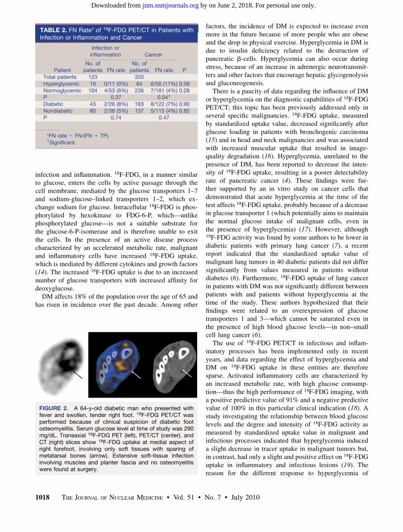
FIGURE 2. A 64-y-old diabetic man who presented withfever and swollen, tender right foot. 18F-FDG PET/CT wasperformed because of clinical suspicion of diabetic footosteomyelitis. Serum glucose level at time of study was 290mg/dL. Transaxial 18F-FDG PET (left), PET/CT (center), andCT (right) slices show 18F-FDG uptake at medial aspect ofright forefoot, involving only soft tissues with sparing ofmetatarsal bones (arrow). Extensive soft-tissue infectioninvolving muscles and planter fascia and no osteomyelitiswere found at surgery.
1018 THE JOURNAL OF NUCLEAR MEDICINE • Vol. 51 • No. 7 • July 2010
by on June 2, 2018. For personal use only. jnm.snmjournals.org Downloaded from

inflammatory and malignant cells is unclear and could berelated to multiple factors. One potential explanation forthe less significant effect of glucose levels on 18F-FDGuptake in infectious and inflammatory processes, as com-pared with malignancy, could rely on their different in-tracellular glycogen storage. Although tumor cells exhibitan increased glycolytic activity, have low glycogen storagecapabilities, and fully depend on extracellular glucosesupply, inflammatory cells are capable of mobilizing in-tracellular glycogen during periods of low plasma glucose(19).
The present study examines, for the first time to the bestof our knowledge, whether altered glucose metabolismaffects the diagnostic accuracy of 18F-FDG PET/CT inpatients with cancer and infection or inflammation. Asdemonstrated by the present data, the detectability rate ofactive disease in the group of patients with a suspectedinfectious or inflammatory process was not affected byelevated glucose levels or DM. The 4 FN studies found inthis patient population could not be characterized by anyparticular clinical features. This low FN rate is in concor-dance with other studies that reported a high diagnosticaccuracy of 18F-FDG PET/CT in infection and inflamma-tion in heterogeneous study populations, unrelated to thepresence of hyperglycemia and DM (9,10,18).
The present data also indicate, however, that hypergly-cemia statistically significantly affected the diagnosticaccuracy of 18F-FDG PET/CT in the group of patientsassessed for the presence of an active malignant tumor, inagreement with some previously published reports (4,17).Thirteen FN studies were found in this patient population.Five of these patients had tumors with a histology known tobe overall less 18F-FDG–avid. DM did not represent aninterfering factor in the 18F-FDG PET/CT assessment ofcancer. However, these results, in addition to those inseveral previously published reports regarding the effectof hyperglycemia on 18F-FDG uptake in cancer (1,4,7,19),are in disagreement with the results of an experimentalstudy in a rat model. That model suggested that 18F-FDGuptake in inflammatory lesions was significantly impairedwith hyperglycemia, and tumor uptake of 18F-FDG was notsignificantly affected (20). These discrepancies may berelated to the differences in the metabolic state of theinvestigated subjects. Although acute, controlled hypergly-cemia was induced in the rat model in healthy subjects,hyperglycemia in patients with or without DM represents inmost cases a chronic systemic condition.
A limitation of this study may be the relatively highproportion of diabetic patients in the randomly selectedoncology group, as compared with the general population.This high proportion may be potentially related—at least inpart—to steroid treatment, commonly administered inoncologic patients, which may lead to a higher rate ofDM and to the higher prevalence of this condition in elderlyindividuals who were represented in significant numbers inthe present study population. A strong association between
hyperglycemia and DM was found in both the oncologyand the infection or inflammation groups. Of patients withDM, 44% in the infection or inflammation and 37% in theoncology group had high glucose levels at the time of thestudy, and 100% and 81% of hyperglycemic patients,respectively, had DM.
CONCLUSION
DM did not significantly affect the diagnostic accuracyof 18F-FDG PET/CT for the assessment of malignancy orinfectious and inflammatory processes. However, hypergly-cemia at the time of the study can lead to a higher FN ratein the oncology group, and imaging in the presence of highblood glucose levels should be avoided in cancer patients.In contrast, high blood glucose levels at the time of the 18F-FDG PET/CT study did not significantly affect the de-tectability rate of infectious and inflammatory processesand had no statistically significant impact on the number ofFN studies in this patient population. On the basis of the re-sults of the present study, hyperglycemia—which is fre-quently detected in acute and chronically ill patients—andDM—which is often an underlying condition in patientspresenting with a suspected infectious process—should notbe considered as potential contraindications for performing18F-FDG PET/CT studies.
REFERENCES
1. Roy FN, Beaulieu S, Boucher L, Bourdeau I, Cohade C. Impact of intravenous
insulin on 18F-FDG PET in diabetic cancer patients. J Nucl Med. 2009;50:
178–183.
2. Bombardieri E, Aktolun C, Baum RP, et al. FDG-PET: procedure guidelines for
tumour imaging. Eur J Nucl Med Mol Imaging. 2003;30:BP115–BP124.
3. Delbeke D, Coleman RE, Guiberteau MJ, et al. Procedure guideline for tumor
imaging with 18F-FDG PET/CT 1.0. J Nucl Med. 2006;47:885–895.
4. Diederichs CG, Staib L, Glatting G, Beger HG, Reske SN. FDG PET: elevated
plasma glucose reduces both uptake and detection rate of pancreatic
malignancies. J Nucl Med. 1998;39:1030–1033.
5. Chang YC, Yen TC, Ng KK, et al. Does diabetes mellitus influence the efficacy
of FDG-PET in the diagnosis of cervical cancer? Eur J Nucl Med Mol Imaging.
2005;32:647–652.
6. Gorenberg M, Hallett WA, O’Doherty MJ. Does diabetes affect [18F]FDG
standardised uptake values in lung cancer? Eur J Nucl Med Mol Imaging. 2002;
29:1324–1327.
7. Torizuka T, Zasadny KR, Wahl RL. Diabetes decreases FDG accumulation in
primary lung cancer. Clin Positron Imaging. 1999;2:281–287.
8. Bleeker-Rovers CP, Vos FJ, Corstens HM, Oyen WJ. Imaging of infectious
diseases using [18F]fluorodeoxyglucose PET. Q J Nucl Med Mol Imaging. 2008;
52:17–29.
9. Keidar Z, Engel A, Hoffman A, Israel O, Nitecki S. Prosthetic vascular graft
infection: the role of 18F-FDG PET/CT. J Nucl Med. 2007;48:1230–1236.
10. Keidar Z, Gurman-Balbir A, Gaitini D, Israel O. Fever of unknown origin: the
role of 18F-FDG PET/CT. J Nucl Med. 2008;49:1980–1985.
11. Kumar R, Basu S, Torigian D, Anand V, Zhuang H, Alavi A. Role of modern
imaging techniques for diagnosis of infection in the era of 18F-fluorodeox-
yglucose positron emission tomography. Clin Microbiol Rev. 2008;21:209–224.
12. De Winter F, Vogelaers D, Gemmel F, Dierckx RA. Promising role of 18-
F-fluoro-D-deoxyglucose positron emission tomography in clinical infectious
diseases. Eur J Clin Microbiol Infect Dis. 2002;21:247–257.
13. Joshi N, Caputo GM, Weitekamp MR, Karchmer AW. Infections in patients with
diabetes mellitus. N Engl J Med. 1999;341:1906–1912.
14. Love C, Tomas MB, Tronco GG, Palestro CJ. FDG PET of infection and
inflammation. Radiographics. 2005;25:1357–1368.
18F-FDG PET/CT IN HYPERGLYCEMIA AND DIABETES • Rabkin et al. 1019
by on June 2, 2018. For personal use only. jnm.snmjournals.org Downloaded from

15. Langen KJ, Braun U, Rota Kops E, et al. The influence of plasma glucose levels
on fluorine-18-fluorodeoxyglucose uptake in bronchial carcinomas. J Nucl Med.
1993;34:355–359.
16. Lindholm P, Minn H, Leskinen-Kallio S, Bergman J, Ruotsalainen U, Joensuu H.
Influence of the blood glucose concentration on FDG uptake in cancer: a PET
study. J Nucl Med. 1993;34:1–6.
17. Torizuka T, Clavo AC, Wahl RL. Effect of hyperglycemia on in vitro tumor uptake of
tritiated FDG, thymidine, L-methionine and L-leucine. J Nucl Med. 1997;38:382–386.
18. Bleeker-Rovers CP, Vos FJ, Wanten GJ, et al. 18F-FDG PET in detecting
metastatic infectious disease. J Nucl Med. 2005;46:2014–2019.
19. Zhuang HM, Cortes-Blanco A, Pourdehnad M, et al. Do high glucose levels have
differential effect on FDG uptake in inflammatory and malignant disorders? Nucl
Med Commun. 2001;22:1123–1128.
20. Zhao S, Kuge Y, Tsukamoto E, et al. Effects of insulin and glucose loading on
FDG uptake in experimental malignant tumours and inflammatory lesions. Eur J
Nucl Med. 2001;28:730–735.
1020 THE JOURNAL OF NUCLEAR MEDICINE • Vol. 51 • No. 7 • July 2010
by on June 2, 2018. For personal use only. jnm.snmjournals.org Downloaded from

Doi: 10.2967/jnumed.109.074294Published online: June 16, 2010.
2010;51:1015-1020.J Nucl Med. Zoya Rabkin, Ora Israel and Zohar Keidar Comparative AnalysisPET/CT Studies in Patients Evaluated for Infection or Inflammation and Cancer? A
F-FDG18Do Hyperglycemia and Diabetes Affect the Incidence of False-Negative
http://jnm.snmjournals.org/content/51/7/1015This article and updated information are available at:
http://jnm.snmjournals.org/site/subscriptions/online.xhtml
Information about subscriptions to JNM can be found at:
http://jnm.snmjournals.org/site/misc/permission.xhtmlInformation about reproducing figures, tables, or other portions of this article can be found online at:
(Print ISSN: 0161-5505, Online ISSN: 2159-662X)1850 Samuel Morse Drive, Reston, VA 20190.SNMMI | Society of Nuclear Medicine and Molecular Imaging
is published monthly.The Journal of Nuclear Medicine
© Copyright 2010 SNMMI; all rights reserved.
by on June 2, 2018. For personal use only. jnm.snmjournals.org Downloaded from