![SZ05-ZN/EN-A10€¦ · spring lever and withdraw filament 1. Pull filament guide tube out of filament intake, 2. Tap [Tools]. leave filament 10cm to pull filament easily. 5. Extruder](https://static.fdocuments.net/doc/165x107/5f8e7086182e8509724132b6/sz05-znen-a10-spring-lever-and-withdraw-filament-1-pull-filament-guide-tube-out.jpg)
dnternexin, a Novel Neuronal Intermediate Filament … · The Journal of Neuroscience, August 1990,...
14
The Journal of Neuroscience, August 1990, W(8): 2735-2748 dnternexin, a Novel Neuronal Intermediate Filament Protein, Precedes the Low Molecular Weight Neurofilament Protein (NF-L) in the Developing Rat Brain Michael P. Kaplan, Steven S. M. Chin, Karsten H. Fliegner, and Ronald K. H. Liem DeDartments of Patholoav and Anatomv and Cell Bioloav, Columbia University College of Physicians and Surgeons, New Yolk, New York 10032 -’ cr-lnternexin is a 66 kDa protein that copurifies with inter- mediate filaments (IF) from rat spinal cord and optic nerve. This protein is axonally transported in rat optic nerve along with the neurofilament triplet proteins in slow component a. Polymerization in vitro and distribution in viva confirm that cr-internexin is a neuronal IF. We raised 2 highly specific monoclonal antibodies to a-internexin which were applied to frozen rat brain sections and Western blots of cytoskeletal extracts. These results indicate that cY-internexin is primarily an axonal protein found in most, if not all, neurons of the CNS. lmmunoreactive proteins of similar molecular weight were found in cytoskeletal extracts of CNS tissue from sev- eral additional species, including mouse and cow. While the distribution of cr-internexin as given by immunocytochemical methods is similar to that of low molecular weight neurofila- ment protein (NF-L) in the adult, its distribution in the embryo is far more extensive. At embryonic day 16, when the expres- sion of NF-L is still limited to a relatively small number of cells and levels of expression are low, a-internexin is already found at much higher levels and in cells not yet expressing NF-L in detectable quantities. Similar results are found at embryonic day 12. These data suggest that neuronal IF in the developing nervous system contain a higher proportion of cu-internexin than their adult counterparts, and that expres- sion of cY-internexin precedes that of NF-L in many or most neurons of the developing brain. Mammalian neuronalintermediate filaments(IF) have long been described in terms of a “neurofilament triplet” of low, medium, and high molecular weight proteins, designated NF-L, NF-M, and NF-H, respectively (Hoffman and Lasek, 1975; Liem et al., 1978).Insoluble at physiological pH, they polymerize in vivo to form 10 nm IFS which traverse the length of the neuronal axo- plasm. Filaments composed of variable proportions of the 3 neurofilament proteins can be detected in the axons (and, to a lesser extent, the dendrites and perikarya) of most mature neu- Received March 7, 1990; revised April 24, 1990; accepted April 25, 1990. We thank Dr. James Goldman and Dr. John Pintar for their expert advice on developmental anatomy, Mr. David Weinstein and Mr. Ray Manson for photo- graphic work, and Ms. Kristy Brown for electron microscopy. This work was supported by Grants NS 15 182 (Jacob Javits Neuroscience Award) and EY03849. K.H.F. and S.S.M.C. are supported by a Medical Scientist Training Program Grant at New York University School of Medicine. Correspondence should be addressed to Dr. Ronald K. H. Liem, Department of Pathology, Columbia University College of Physicians and Surgeons, 630 West 168th Street, New York, NY 10032. Copyright 0 1990 Society for Neuroscience 0270-6474/90/082735-14$03.00/O rons in the central and peripheral nervous system. A fourth neuronal intermediate filament protein has recently been de- scribed(Portier et al., 1984;Parysek and Goldman, 1987;Leon- ard et al., 1988); termed “peripherin,” this 57 kDa protein is found primarily in processes of the peripheral nervous system. While their exact functions remain unclear, neuronal IFS pre- sumably play some structural role in the generation and/or maintenanceof the highly asymmetrical forms of neuronsand their processes; for example, it hasbeensuggested that neuronal IFS may be arbiters of neuronal caliber (for review, see Lasek et al., 1983). Like other IFS, such asglial fibrillary acidic protein (GFA) in astroglia, desminin smooth muscle cells, and vimentin in cells of mesenchymal origin, the neuronal IF proteins each contain a highly conserved a-helical domain, flanked by variable head and tail sequences (for review, seeSteinert and Roop, 1988); the rod-shaped, a-helical domainsform the core of the filament, with the variable end sequences projecting outward. The exten- sive carboxy-terminal end sequence of NF-H is known to par- ticipate in the formation of cross-bridges betweenfilaments(Hi- rokawa et al., 1984). NF-L, but not NF-M or NF-H, is readily able to self-polymerize in vitro at physiological pH (Liem and Hutchison, 1982); inability of the other isolated subunits to polymerize is probably due to ionic repulsion between their highly phosphorylated tail sequences, and it has recently been suggested that this repulsion between phosphate groups may play a role in maintaining the distancebetweenfilaments in vivo (Hirokawa et al., 1984; Carden et al., 1987; Shaw, in press). Developmental studies usingbiochemicaland immunological methods have shown that neurogenesis is accompanied by changes in neuronal IF composition. After an initial stage during which vimentin may be expressed (Bignami et al., 1982; Co- chard and Paulin, 1984), coexpression of NF-L and NF-M pre- cedes that of NF-H in developing rat brain (Shaw and Weber, 1982; Cardenet al., 1987) retina (Shaw and Weber, 1983),and optic nerve (Pachter and Liem, 1984). Neuronal IFS containing NF-L and NF-M can be detected asearly asembryonic day 12 in someregions,while NF-H may not be expressed until days or weeks after birth in someareas.Hippocampal neurons cul- tured in vitro display a similar stepwise pattern of expression (Shaw et al., 1985). The significance of this delayed appearance of the larger subunit is probably related to its putative role as cross-linker betweenfilaments and the stabilization of IF struc- ture presumably accompanyingneuronal maturation. In the pe- ripheral nervous system,this scenario is further complicated by the presence of peripherin, which appears around the same time
Transcript of dnternexin, a Novel Neuronal Intermediate Filament … · The Journal of Neuroscience, August 1990,...
The Journal of Neuroscience, August 1990, W(8): 2735-2748
dnternexin, a Novel Neuronal Intermediate Filament Protein, Precedes the Low Molecular Weight Neurofilament Protein (NF-L) in the Developing Rat Brain
Michael P. Kaplan, Steven S. M. Chin, Karsten H. Fliegner, and Ronald K. H. Liem
DeDartments of Patholoav and Anatomv and Cell Bioloav, Columbia University College of Physicians and Surgeons, New Yolk, New York 10032 -’
cr-lnternexin is a 66 kDa protein that copurifies with inter- mediate filaments (IF) from rat spinal cord and optic nerve. This protein is axonally transported in rat optic nerve along with the neurofilament triplet proteins in slow component a. Polymerization in vitro and distribution in viva confirm that cr-internexin is a neuronal IF. We raised 2 highly specific monoclonal antibodies to a-internexin which were applied to frozen rat brain sections and Western blots of cytoskeletal extracts. These results indicate that cY-internexin is primarily an axonal protein found in most, if not all, neurons of the CNS. lmmunoreactive proteins of similar molecular weight were found in cytoskeletal extracts of CNS tissue from sev- eral additional species, including mouse and cow. While the distribution of cr-internexin as given by immunocytochemical methods is similar to that of low molecular weight neurofila- ment protein (NF-L) in the adult, its distribution in the embryo is far more extensive. At embryonic day 16, when the expres- sion of NF-L is still limited to a relatively small number of cells and levels of expression are low, a-internexin is already found at much higher levels and in cells not yet expressing NF-L in detectable quantities. Similar results are found at embryonic day 12. These data suggest that neuronal IF in the developing nervous system contain a higher proportion of cu-internexin than their adult counterparts, and that expres- sion of cY-internexin precedes that of NF-L in many or most neurons of the developing brain.
Mammalian neuronal intermediate filaments (IF) have long been described in terms of a “neurofilament triplet” of low, medium, and high molecular weight proteins, designated NF-L, NF-M, and NF-H, respectively (Hoffman and Lasek, 1975; Liem et al., 1978). Insoluble at physiological pH, they polymerize in vivo to form 10 nm IFS which traverse the length of the neuronal axo- plasm. Filaments composed of variable proportions of the 3 neurofilament proteins can be detected in the axons (and, to a lesser extent, the dendrites and perikarya) of most mature neu-
Received March 7, 1990; revised April 24, 1990; accepted April 25, 1990. We thank Dr. James Goldman and Dr. John Pintar for their expert advice on
developmental anatomy, Mr. David Weinstein and Mr. Ray Manson for photo- graphic work, and Ms. Kristy Brown for electron microscopy. This work was supported by Grants NS 15 182 (Jacob Javits Neuroscience Award) and EY03849. K.H.F. and S.S.M.C. are supported by a Medical Scientist Training Program Grant at New York University School of Medicine.
Correspondence should be addressed to Dr. Ronald K. H. Liem, Department of Pathology, Columbia University College of Physicians and Surgeons, 630 West 168th Street, New York, NY 10032.
Copyright 0 1990 Society for Neuroscience 0270-6474/90/082735-14$03.00/O
rons in the central and peripheral nervous system. A fourth neuronal intermediate filament protein has recently been de- scribed (Portier et al., 1984; Parysek and Goldman, 1987; Leon- ard et al., 1988); termed “peripherin,” this 57 kDa protein is found primarily in processes of the peripheral nervous system. While their exact functions remain unclear, neuronal IFS pre- sumably play some structural role in the generation and/or maintenance of the highly asymmetrical forms of neurons and their processes; for example, it has been suggested that neuronal IFS may be arbiters of neuronal caliber (for review, see Lasek et al., 1983).
Like other IFS, such as glial fibrillary acidic protein (GFA) in astroglia, desmin in smooth muscle cells, and vimentin in cells of mesenchymal origin, the neuronal IF proteins each contain a highly conserved a-helical domain, flanked by variable head and tail sequences (for review, see Steinert and Roop, 1988); the rod-shaped, a-helical domains form the core of the filament, with the variable end sequences projecting outward. The exten- sive carboxy-terminal end sequence of NF-H is known to par- ticipate in the formation of cross-bridges between filaments (Hi- rokawa et al., 1984). NF-L, but not NF-M or NF-H, is readily able to self-polymerize in vitro at physiological pH (Liem and Hutchison, 1982); inability of the other isolated subunits to polymerize is probably due to ionic repulsion between their highly phosphorylated tail sequences, and it has recently been suggested that this repulsion between phosphate groups may play a role in maintaining the distance between filaments in vivo (Hirokawa et al., 1984; Carden et al., 1987; Shaw, in press).
Developmental studies using biochemical and immunological methods have shown that neurogenesis is accompanied by changes in neuronal IF composition. After an initial stage during which vimentin may be expressed (Bignami et al., 1982; Co- chard and Paulin, 1984), coexpression of NF-L and NF-M pre- cedes that of NF-H in developing rat brain (Shaw and Weber, 1982; Carden et al., 1987) retina (Shaw and Weber, 1983), and optic nerve (Pachter and Liem, 1984). Neuronal IFS containing NF-L and NF-M can be detected as early as embryonic day 12 in some regions, while NF-H may not be expressed until days or weeks after birth in some areas. Hippocampal neurons cul- tured in vitro display a similar stepwise pattern of expression (Shaw et al., 1985). The significance of this delayed appearance of the larger subunit is probably related to its putative role as cross-linker between filaments and the stabilization of IF struc- ture presumably accompanying neuronal maturation. In the pe- ripheral nervous system, this scenario is further complicated by the presence of peripherin, which appears around the same time
2736 Kaplan et al. * dnternexin, a Neuronal IF in the Developing CNS
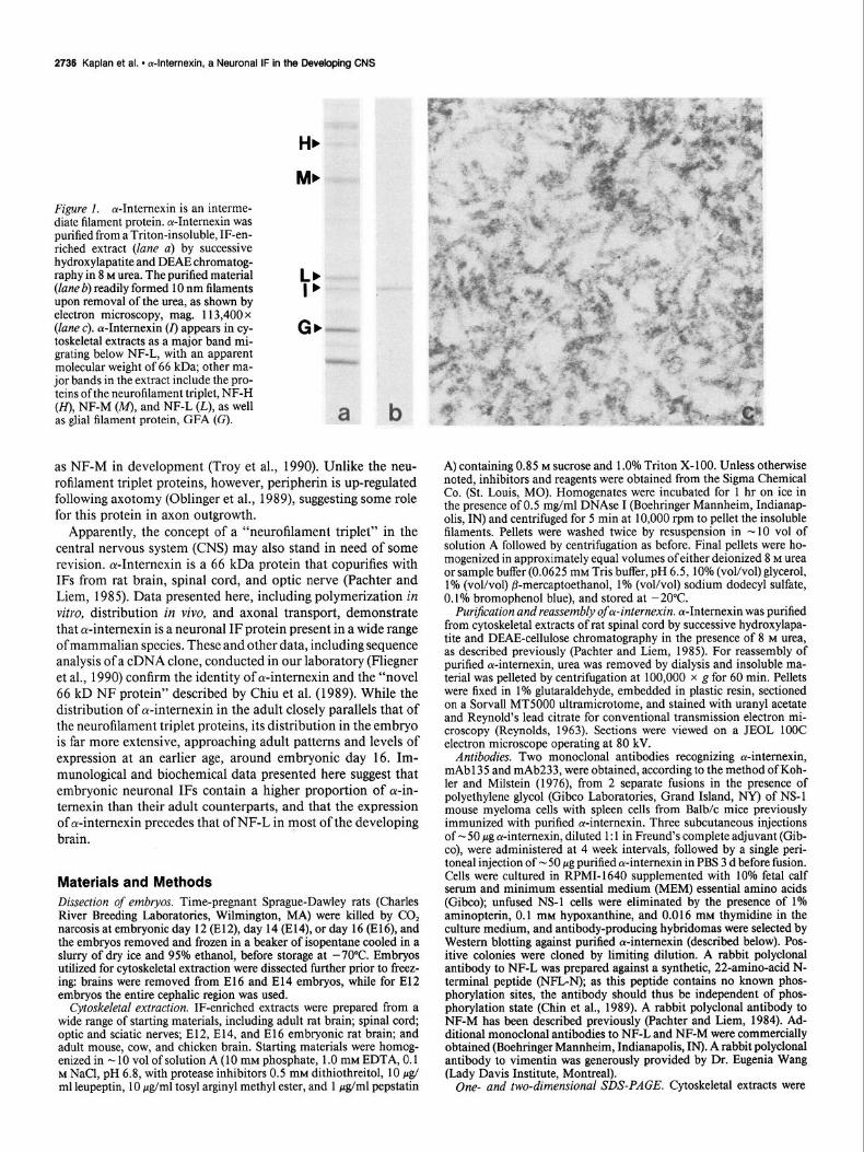
Figure 1. oc-Internexin is an interme- diate filament protein. a-Intemexin was purified from a T&on-insoluble, IF-en- riched extract (lane a) by successive hydroxylapatite and DEAE chromatog- raphy in 8 M urea. The purified material (lane b) readily formed 10 nm filaments upon removal of the urea, as shown by electron microscopy, mag. 113,400 x (lane c). cu-Intemexin (I) appears in cy- toskeletal extracts as a major band mi- grating below NF-L, with an apparent molecular weight of 66 kDa; other ma- jor bands in the extract include the pro- teins ofthe neurofilament triplet, NF-H (H), NF-M (M), and NF-L (L), as well as glial filament protein, GFA (G).
:t
Gbf
a
as NF-M in development (Troy et al., 1990). Unlike the neu- rofilament triplet proteins, however, peripherin is up-regulated following axotomy (Oblinger et al., 1989), suggesting some role for this protein in axon outgrowth.
Apparently, the concept of a “neurofilament triplet” in the central nervous system (CNS) may also stand in need of some revision. cr-Internexin is a 66 kDa protein that copurifies with IFS from rat brain, spinal cord, and optic nerve (Pachter and Liem, 1985). Data presented here, including polymerization in vitro, distribution in vivo, and axonal transport, demonstrate that a-internexin is a neuronal IF protein present in a wide range ofmammalian species. These and other data, including sequence analysis of a cDNA clone, conducted in our laboratory (F’liegner et al., 1990) confirm the identity of a-internexin and the “novel 66 kD NF protein” described by Chiu et al. (1989). While the distribution of ol-internexin in the adult closely parallels that of the neurofilament triplet proteins, its distribution in the embryo is far more extensive, approaching adult patterns and levels of expression at an earlier age, around embryonic day 16. Im- munological and biochemical data presented here suggest that embryonic neuronal IFS contain a higher proportion of cu-in- ternexin than their adult counterparts, and that the expression of cy-internexin precedes that of NF-L in most of the developing brain.
Materials and Methods Dissection of embryos. Time-pregnant Sprague-Dawley rats (Charles River Breeding Laboratories, Wilmington, MA) were killed by CO, narcosis at embryonic day 12 (E12), day 14 (E14), or day 16 (E16), and the embryos removed and frozen in a beaker of isopentane cooled in a slurry of dry ice and 95% ethanol, before storage at -70°C. Embryos utilized for cytoskeletal extraction were dissected further prior to freez- ing: brains were removed from El6 and El4 embryos, while for El2 embryos the entire cephalic region was used.
Cytoskeletal extraction. IF-enriched extracts were prepared from a wide range of starting materials, including adult rat brain; spinal cord, optic and sciatic nerves; E12, E14, and El 6 embryonic rat brain; and adult mouse, cow, and chicken brain. Starting materials were homog- enized in - Ib volof solution A (10 mM phosphate, 1.0 mM EDTA, Or1 M NaCl, pH 6.8, with protease inhibitors 0.5 mM dithiothreitol, 10 & ml leupeptin, 10 fig/ml tosyl arginyl methyl ester, and 1 &ml pepstatin
A) containing 0.85 M sucrose and 1 .O% Triton X-100. Unless otherwise noted, inhibitors and reagents were obtained from the Sigma Chemical Co. (St. Louis, MO). Homogenates were incubated for 1 hr on ice in the presence of 0.5 &/ml DNAse I (Boehringer Mannheim, Indianap- olis. IM and centrifuaed for 5 min at 10.000 mm to uellet the insoluble filaments. Pellets were washed twice by resuspension in w 10 vol of solution A followed by centrifugation as before. Final pellets were ho- mogenized in approximately equal volumes of either deionized 8 M urea or sample buffer (0.0625 mM Tris buffer, pH 6.5, 10% (voVvo1) glycerol, 1% (vol/vol) ,&mercaptoethanol, 1% (vol/vol) sodium dodecyl sulfate, 0.1% bromophenol blue), and stored at -20°C.
Purification and reassembly of Lu-internexin. cu-Intemexin was purified from cytoskeletal extracts of rat spinal cord by successive hydroxylapa- tite and DEAE-cellulose chromatography in the presence of 8 M urea, as described previously (Pachter and Liem, 1985). For reassembly of purified o-intemexin, urea was removed by dialysis and insoluble ma- terial was pelleted by centrifugation at 100,000 x g for 60 min. Pellets were fixed in 1% glutaraldehyde, embedded in plastic resin, sectioned on a Sorvall MT5000 ultramicrotome, and stained with uranyl acetate and Reynolds lead citrate for conventional transmission electron mi- croscopy (Reynolds, 1963). Sections were viewed on a JEOL 1OOC electron microscope operating at 80 kV.
Antibodies. Two monoclonal antibodies recognizing cu-intemexin, mAb 135 and mAb233, were obtained, according to the method of Koh- ler and Milstein (1976), from 2 separate fusions in the presence of polyethylene glycol (Gibco Laboratories, Grand Island, NY) of NS-1 mouse myeloma cells with spleen cells from Balb/c mice previously immunized with purified ol-internexin. Three subcutaneous injections of u 50 pg cu-intemexin, diluted 1: 1 in Freund’s complete adjuvant (Gib- co), were administered at 4 week intervals, followed by a single peri- toneal injection of w 50 pg purified oc-intemexin in PBS 3 d before fusion. Cells were cultured in RPMI-1640 supplemented with 10% fetal calf serum and minimum essential medium (MEM) essential amino acids (Gibco); unfused NS-1 cells were eliminated by the presence of 1% aminopterin, 0.1 mM hypoxanthine, and 0.0 16 mM thymidine in the culture medium, and antibody-producing hybridomas were selected by Western blotting against purified cY-intemexin (described below). Pos- itive colonies were cloned by limiting dilution. A rabbit polyclonal antibody to NF-L was prepared against a synthetic, 22-amino-acid N- terminal peptide (NFL-N); as this peptide contains no known phos- phorylation sites, the antibody should thus be independent of phos- phorylation state (Chin et al., 1989). A rabbit polyclonal antibody to NF-M has been described previously (Pachter and Liem, 1984). Ad- ditional monoclonal antibodies to NEL and NF-M were commercially obtained (Boehringer Mannheim, Indianapolis, IN). A rabbit polyclonal antibody to vimentin was generously provided by Dr. Eugenia Wang (Lady Davis Institute, Montreal).
One- and two-dimensional SDS-PAGE. Cytoskeletal extracts were
The Journal of Neuroscience, August 1990, fCJ(8) 2737
subjected to l-dimensional electrophoresis in the presence of SDS (Laemmli, 1970). Protein concentrations were estimated by Lowry de- termination, and from - 10 to 50 pg of the cytoskeletal extracts was applied to each lane of a 7.5% vertical slab gel (Hoeffer Instruments, San Francisco, CA). Two-dimensional electrophoresis was performed essentially according to the method of O’Farrell (1975). For the first dimension, - 50 rg of samples was diluted in lysis buffer supplemented with 0.1% SDS and applied to polyacrylamide tube gels containing a pH gradient established by the addition of 1.6% (vol/vol) pH 4-6 am- pholines and 0.4% pH 3.5-10 ampholines (LKB Instruments, Bromma, Sweden). The second dimension consisted of a 7.5% SDS-PAGE slab gel, as described above. Gels were either fixed in 10% acetic acid, 50% methanol, and stained with Coomassie blue, or transferred to nitrocel- lulose for Western blotting.
Western blotting. Transfer was performed by a modification of meth- ods previously described (Towbin et al., 1979). Gels were placed in contact with nitrocellulose in a buffer containing 19.2 mM glycine, 2.5 mM Tris, and 20% methanol (vol/vol) at a pH of 8.3, and a constant current of 20 mA was applied overnight in a transfer apparatus (Bio- Rad Instruments, Richmond, CA). Blots were then incubated for 2 hr in blocking buffer (PBS containing 5% nonfat dry milk) before incu- bation for 2 hr with primary antibodies. Monoclonal and polyclonal antibodies to NF-L were used at dilutions of 1: 10 and 1: 1000, respec- tively. For the monoclonal antibodies to a-intemexin, culture super- natants were employed directly. Blots were then reacted with a strep- tavidin/biotin/horseradish peroxidase complex (Amersham Inc., Arlington Heights, IL) and visualized using diaminobenzidine and hy- drogen peroxide.
Immunojluorescence on cryostat sections. Frozen rat brains and em- bryos were mounted in Tissue-Tek (Miles Laboratories) and 10 pm sections were taken at - 20°C on a Reinhart cryostat (Bright Instrument Co., Huntington, England). Sagittal, coronal, and longitudinal sections were placed on gelatin-coated glass slides and allowed to air-dry before storage at - 70°C. Sections were fixed for 15 min in cold methanol at -20°C and rinsed 3 times in PBS prior to staining. Monoclonal anti- bodies to NF-L and NF-M were diluted 1: 10 in PBS; rabbit polyclonal antibodies to NF-L, NF-M, and vimentin were employed at working dilutions of 1:200, 1: 100, and 1: 100, respectively, in PBS. In the case of mAb135 and mAb233, the monoclonal antibodies to oc-intemexin, culture supematants were employed directly. For double-labeling ex- periments, each of the rabbit antibodies was diluted directly in mAb135 or mAb233; the order of application of the antibodies did not appear to affect results. All primary antibodies were applied for 2 hr at room temperature and then rinsed 5 times for 3 min with PBS. For the second antibody, fluorescein-conjugated goat anti-mouse IgG and rhodamine- conjugated anti-rabbit IgG (Cappel Laboratories, Malvem, PA) were applied at 1: 100 in PBS for 1 hr. After 5 more rinses in PBS, 1 drop of a mixture consisting of 30% glycerol (voVvo1) and 16% polyvinyl alcohol was added to each section, and coverslips were applied. Sections were viewed and photographed on an Optiphot fluorescence microscope (Ni- kon Instruments, Tokyo, Japan).
Axonal transport. Neonatal and young adult albino rats were anes- thetized with methoxyflurane (Metofane, Pitman-Moore, Washington Crossing, NJ) and injected intraocularly with loo-250 &i [‘S]-methi- onine (NEN/DuPont, Wilmington, DE) using a Hamilton syringe fitted with a 32G needle. All injections were performed with the aid of a dissection microscope and a mechanical micromanipulator. Newborn pups required an incision to be made along the still fused eyelid borders. The needle was positioned for entry into the vitreous chamber at the posterior aspect of the scleral-cornea1 junction. Volumes up to 10 hl were then slowly infused into the eye. After allowing sufficient time for transport of newly synthesized retinal ganglion cell proteins into the optic nerve, the animals were killed by CO, narcosis and decapitation. The radiolabeled optic pathway was dissected out and the individual optic nerves and tracts were then carefully cut into 1 mm segments and homogenized. The T&on-soluble and insoluble fractions were prepared as previously described, an aliquot of each was placed in Aquasol liquid scintillation cocktail (NEN) and counted, and remaining samples were then subjected to l- or 2-dimensional SDS-PAGE and fluorography.
Results Protein chemical and axonal transport studies The electrophoretic profile of the T&on-insoluble, IF-enriched extract from rat optic nerve is shown in Figure 1 (lane a), along
>
*
10 12 14 16 16
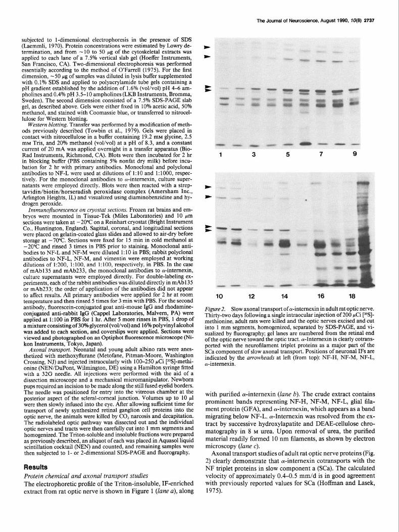
Figure 2. Slow axonal transport of oc-intemexin in adult rat optic nerve. Thirty-two days following a single intraocular injection of 200 pCi [35S]- methionine, adult rats were killed and the optic nerves excised and cut into 1 mm segments, homogenized, separated by SDS-PAGE, and vi- sualized by fluorography; gel lanes are numbered from the retinal end of the optic nerve toward the optic tract. c+Intemexin is clearly cotrans- ported with the neurofilament triplet proteins as a major part of the SCa component of slow axonal transport. Positions of neuronal IFS are indicated by the arrowheads at left (from top): NF-H, NF-M, NF-L, cu-intemexin.
with purified oc-internexin (lane b). The crude extract contains prominent bands representing NF-H, NF-M, NF-L, gIia1 fila- ment protein (GFA), and ol-interuexin, which appears as a band migrating below NF-L. ol-Intemexin was resolved from the ex- tract by successive hydroxylapatite and DEAE-cellulose chro- matography in 8 M urea. Upon removal of urea, the purified material readily formed 10 nm filaments, as shown by electron microscopy (lane c).
Axonal transport studies of adult rat optic nerve proteins (Fig. 2) clearly demonstrate that oc-intemexin cotransports with the NF triplet proteins in slow component a (SCa). The calculated velocity of approximately 0.4-0.5 mm/d is in good agreement with previously reported values for SCa (Hoffman and Lasek, 1975).
2738 Kaplan et al. l a-lnternexin, a Neuronal IF in the Developing CNS
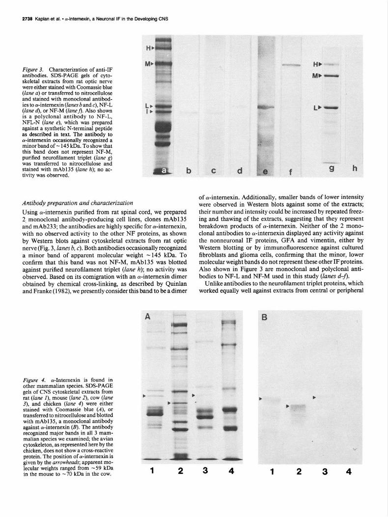
Figure 3. Characterization of anti-IF antibodies. SDS-PAGE gels of cyto- skeletal extracts from rat optic nerve were either stained with Coomassie blue (lane a) or transferred to nitrocellulose and stained with monoclonal antibod- ies to a-intemexin (lanes b and c), NF-L (lane d), or NF-M (laneA. Also shown is a polyclonal antibody to NF-L, NFL-N (lane e), which was prepared against a synthetic N-terminal peptide as described in text. The antibody to cY-intemexin occasionally recognized a minor band of - 145 kDa. To show that this band does not represent NF-M, purified neurofilament triplet (lane g) was transferred to nitrocellulose and stained with mAb135 (lane h); no ac- tivity was observed.
Antibody preparation and characterization Using cu-internexin purified from rat spinal cord, we prepared 2, monoclonal antibody-producing cell lines, clones mAb135 and mAb233; the antibodies are highly specific for cu-internexin, with no observed activity to the other NF proteins, as shown by Western blots against cytoskeletal extracts from rat optic nerve (Fig. 3, lanes b, c). Both antibodies occasionally recognized a minor band of apparent molecular weight - 145 kDa. To confirm that this band was not NF-M, mAb135 was blotted against purified neurofilament triplet (lane h); no activity was observed. Based on its comigration with an cy-internexin dimer obtained by chemical cross-linking, as described by Quinlan and Franke (1982), we presently consider this band to be a dimer
Figure 4. cY-Intemexin is found in other mammalian species. SDS-PAGE gels of CNS cytoskeletal extracts from rat (lane I), mouse (lane 2), cow (lane 3), and chicken (lane 4) were either stained with Coomassie blue (A), or transferred to nitrocellulose and blotted with mAb135, a monoclonal antibody against cY-intemexin (B). The antibody recognized major bands in all 3 mam- malian species we examined, the avian cytoskeleton, as represented here by the
of a-intemexin. Additionally, smaller bands of lower intensity were observed in Western blots against some of the extracts; their number and intensity could be increased by repeated freez- ing and thawing of the extracts, suggesting that they represent breakdown products of cu-intemexin. Neither of the 2 mono- clonal antibodies to ar-intemexin displayed any activity against the nonneuronal IF proteins, GFA and vimentin, either by Western blotting or by immunofluorescence against cultured fibroblasts and glioma cells, confirming that the minor, lower molecular weight bands do not represent these other IF proteins. Also shown in Figure 3 are monoclonal and polyclonal anti- bodies to NF-L and NF-M used in this study (lanes d-J).
Unlike antibodies to the neurofilament triplet proteins, which worked equally well against extracts from central or peripheral
chicken, does not show a cross-reactive protein. The position of cw-intemexin is given by the arrowheads; apparent mo-
-. -I_ lecular weights ranged from -59 kDa in the mouse to -70 kDa in the cow. 1 2 3 4 1 2 3 4
The Journal of Neuroscience, August 1990, 70(8) 2739
H- A H _x(,.
L Ic -I
L -
v v
sources, antibodies to a-internexin displayed comparatively lit- tle activity when cytoskeletal extracts of adult rat sciatic nerve were employed for Western blotting (not shown).
CNS cytoskeletal preparations from other mammalian species displayed similar electrophoretic profiles. In mouse, rat, and cow brain extracts (Fig. 4A, lanes 1-3) the 3 neurofilament pro- teins and GFA are easily identified, along with an additional major band migrating below NF-L which was recognized by the monoclonal antibody to rat cr-mternexin (Fig. 4B). The apparent molecular weight of this band ranged from - 59 kDa in mouse to -70 kDa in cow brain extracts. No comparable reactive protein was found in chicken, the only nonmammalian species we examined (lane 4). Reactivity was also shown against a cy- toskeletal preparation obtained from human brain, but this ma- terial was highly degraded owing to the long postmortem period. The highest molecular weight band corresponded to a protein of -65 kDa.
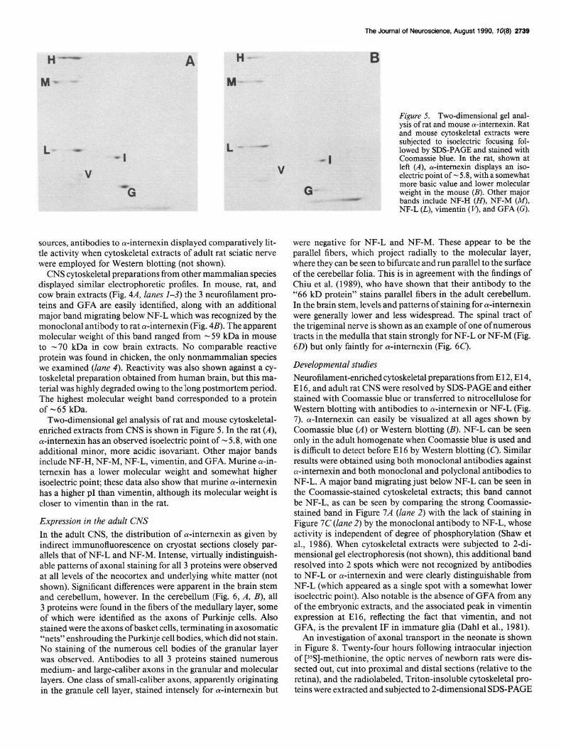
Two-dimensional gel analysis of rat and mouse cytoskeletal- enriched extracts from CNS is shown in Figure 5. In the rat (A), ar-internexin has an observed isoelectric point of - 5.8, with one additional minor, more acidic isovariant. Other major bands include NF-H, NF-M, NF-L, vimentin, and GFA. Murine cu-in- ternexin has a lower molecular weight and somewhat higher isoelectric point; these data also show that murine cu-intemexin has a higher p1 than vimentin, although its molecular weight is closer to vimentin than in the rat.
Expression in the adult CNS
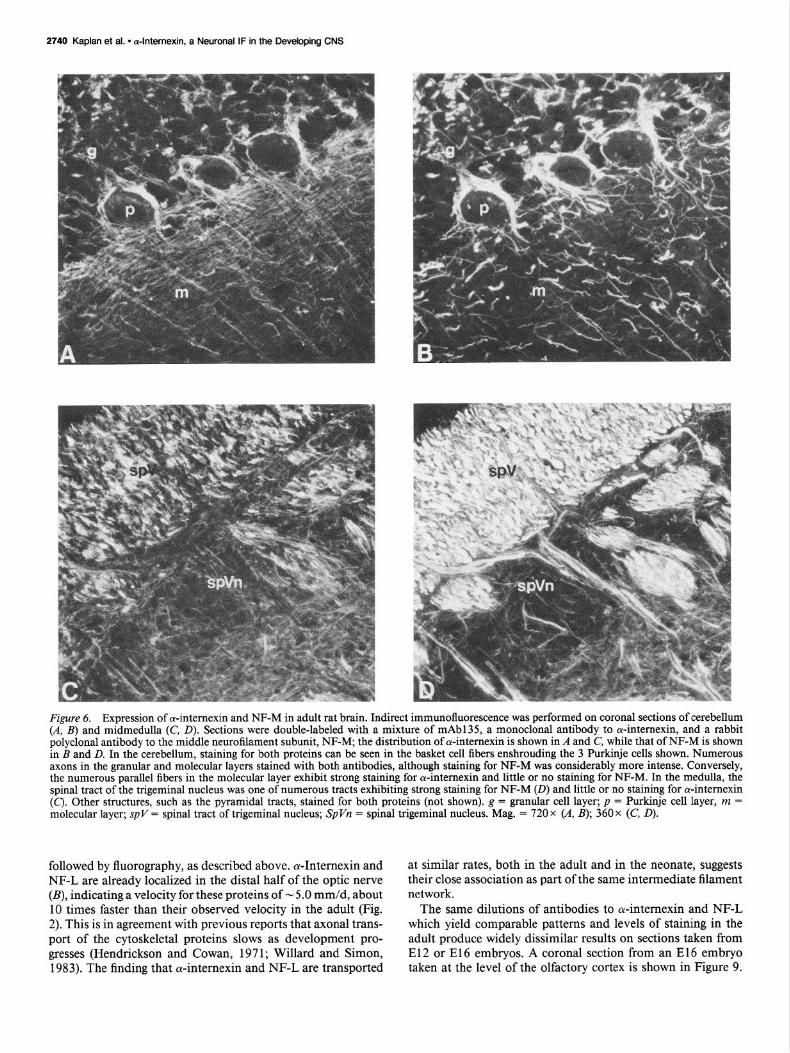
In the adult CNS, the distribution of a-internexin as given by indirect immunofluorescence on cryostat sections closely par- allels that of NF-L and NF-M. Intense, virtually indistinguish- able patterns of axonal staining for all 3 proteins were observed at all levels of the neocortex and underlying white matter (not shown). Significant differences were apparent in the brain stem and cerebellum, however. In the cerebellum (Fig. 6, A, B), all 3 proteins were found in the fibers of the medullary layer, some of which were identified as the axons of Purkinje cells. Also stained were the axons ofbasket cells, terminating in axosomatic “nets” enshrouding the Purkinje cell bodies, which did not stain. No staining of the numerous cell bodies of the granular layer was observed. Antibodies to all 3 proteins stained numerous medium- and large-caliber axons in the granular and molecular layers. One class of small-caliber axons, apparently originating in the granule cell layer, stained intensely for a-intemexin but
B
Figure 5. Two-dimensional gel anal- ysis of rat and mouse a-intemexin. Rat and mouse cytoskeletal extracts were subjected to -isoelectric focusing fol- lowed bv SDS-PAGE and stained with
-I
G
Coomassie blue. In the rat, shown at left (A), a-intemexin displays an iso- electric point of - 5.8, with a somewhat more basic value and lower molecular weight in the mouse (B). Other major bands include NF-H (m. NF-M CM). NF-L (L), vimentin (r$ and GFA IG).
were negative for NF-L and NF-M. These appear to be the parallel fibers, which project radially to the molecular layer, where they can be seen to bifurcate and run parallel to the surface of the cerebellar folia. This is in agreement with the findings of Chiu et al. (1989), who have shown that their antibody to the “66 kD protein” stains parallel fibers in the adult cerebellum. In the brain stem, levels and patterns of staining for ol-intemexin were generally lower and less widespread. The spinal tract of the trigeminal nerve is shown as an example of one of numerous tracts in the medulla that stain strongly for NF-L or NF-M (Fig. 60) but only faintly for ol-internexin (Fig. 6C).
Developmental studies
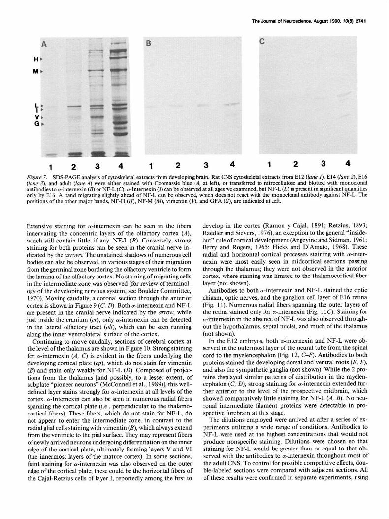
Neurofilament-enriched cytoskeletal preparations from E 12, E 14, E 16, and adult rat CNS were resolved by SDS-PAGE and either stained with Coomassie blue or transferred to nitrocellulose for Western blotting with antibodies to ol-intemexin or NF-L (Fig. 7). cu-Intemexin can easily be visualized at all ages shown by Coomassie blue (A) or Western blotting (B). NF-L can be seen only in the adult homogenate when Coomassie blue is used and is difficult to detect before El 6 by Western blotting (C). Similar results were obtained using both monoclonal antibodies against a-intemexin and both monoclonal and polyclonal antibodies to NF-L. A major band migrating just below NF-L can be seen in the Coomassie-stained cytoskeletal extracts; this band cannot be NF-L, as can be seen by comparing the strong Coomassie- stained band in Figure 7A (lane 2) with the lack of staining in Figure 7C (lane 2) by the monoclonal antibody to NF-L, whose activity is independent of degree of phosphorylation (Shaw et al., 1986). When cytoskeletal extracts were subjected to 2-di- mensional gel electrophoresis (not shown), this additional band resolved into 2 spots which were not recognized by antibodies to NF-L or cu-intemexin and were clearly distinguishable from NF-L (which appeared as a single spot with a somewhat lower isoelectric point). Also notable is the absence of GFA from any of the embryonic extracts, and the associated peak in vimentin expression at E16, reflecting the fact that vimentin, and not GFA, is the prevalent IF in immature glia (Dahl et al., 198 1).
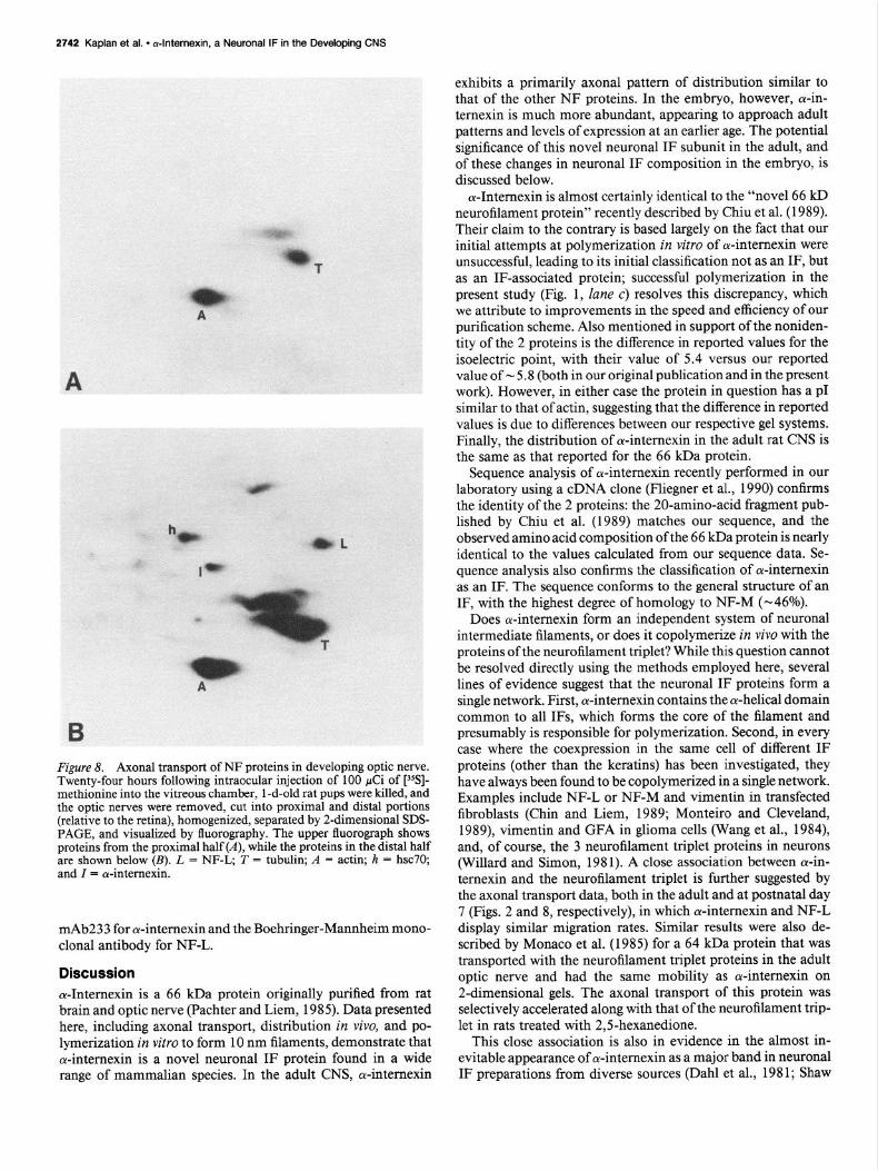
An investigation of axonal transport in the neonate is shown in Figure 8. Twenty-four hours following intraocular injection of [35S]-methionine, the optic nerves of newborn rats were dis- sected out, cut into proximal and distal sections (relative to the retina), and the radiolabeled, Triton-insoluble cytoskeletal pro- teins were extracted and subjected to 2-dimensional SDS-PAGE
2740 Kaplan et al. l dntemexin, a Neuronal IF in the Developing CNS
Figure 6. Expression of a-intemexin and NF-M in adult rat brain. Indirect immunofluorescence was performed on coronal sections of cerebellum (A, B) and midmedulla (C, D). Sections were double-labeled with a mixture of mAb135, a monoclonal antibody to ol-intemexin, and a rabbit polyclonal antibody to the middle neurofilament subunit, NF-M; the distribution of a-intemexin is shown in A and C, while that of NF-M is shown in B and D. In the cerebellum, staining for both proteins can be seen in the basket cell fibers enshrouding the 3 Purkinje cells shown. Numerous axons in the granular and molecular layers stained with both antibodies, although staining for NF-M was considerably more intense. Conversely, the numerous parallel fibers in the molecular layer exhibit strong staining for Antemexin and little or no staining for NF-M. In the medulla, the spinal tract of the trigeminal nucleus was one of numerous tracts exhibiting strong staining for NF-M (0) and little or no staining for a-intemexin (C). Other structures, such as the pyramidal tracts, stained for both proteins (not shown). g = granular cell layer; p = Purkinje cell layer, m = molecular layer; spV = spinal tract of trigeminal nucleus; SpVn = spinal trigeminal nucleus. Mag. = 720 x (A, B); 360x (C, D).
followed by fluorography, as described above. cu-Internexin and NF-L are already localized in the distal half of the optic nerve (B), indicating a velocity for these proteins of - 5.0 mm/d, about 10 times faster than their observed velocity in the adult (Fig. 2). This is in agreement with previous reports that axonal trans- port of the cytoskeletal proteins slows as development pro- gresses (Hendrickson and Cowan, 197 1; Willard and Simon, 1983). The finding that cr-internexin and NF-L are transported
at similar rates, both in the adult and in the neonate, suggests their close association as part of the same intermediate filament network.
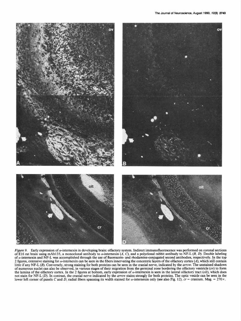
The same dilutions of antibodies to cu-internexin and NF-L which yield comparable patterns and levels of staining in the adult produce widely dissimilar results on sections taken from El 2 or El6 embryos. A coronal section from an El 6 embryo taken at the level of the olfactory cortex is shown in Figure 9.
The Journal of Neuroscience, August 1990, 10(E) 2741
Figure 7. SDS-PAGE analysis of cytoskeletal extracts from developing brain. Rat CNS cytoskeletal extracts from El2 (lane I), El4 (lane 2), El6 (lane 3), and adult (lane 4) were either stained with Coomassie blue (A. at left), or transferred to nitrocellulose and blotted with monoclonal antibodies to cu-internexin (B) or NF-L (c). a-Intemexin (I) can be observed at all ages we examined, but NF-L (L) is present in significant quantities only by E16. A band migrating slightly ahead of NF-L can be observed, which does not react with the monoclonal antibody against NF-L. The positions of the other major bands, NF-H (H), NF-M (M), vimentin (J’), and GFA (G), are indicated at left.
Extensive staining for a-internexin can be seen in the fibers innervating the concentric layers of the olfactory cortex (A), which still contain little, if any, NF-L (B). Conversely, strong staining for both proteins can be seen in the cranial nerve in- dicated by the arrows. The unstained shadows of numerous cell bodies can also be observed, in various stages of their migration from the germinal zone bordering the olfactory ventricle to form the lamina of the olfactory cortex. No staining of migrating cells in the intermediate zone was observed (for review of terminol- ogy of the developing nervous system, see Boulder Committee, 1970). Moving caudally, a coronal section through the anterior cortex is shown in Figure 9 (C, 0). Both ol-internexin and NF-L are present in the cranial nerve indicated by the arrow, while just inside the cranium (cr), only ol-internexin can be detected in the lateral olfactory tract (olt), which can be seen running along the inner ventrolateral surface of the cortex.
Continuing to move caudally, sections of cerebral cortex at the level of the thalamus are shown in Figure 10. Strong staining for a-internexin (A, C) is evident in the fibers underlying the developing cortical plate (cp), which do not stain for vimentin (B) and stain only weakly for NF-L (0). Composed of projec- tions from the thalamus [and possibly, to a lesser extent, of subplate “pioneer neurons” (McConnell et al., 1989)], this well- defined layer stains strongly for oc-internexin at all levels of the cortex. c+Internexin can also be seen in numerous radial fibers spanning the cortical plate (i.e., perpendicular to the thalamo- cortical fibers). These fibers, which do not stain for NF-L, do not appear to enter the intermediate zone, in contrast to the radial glial cells staining with vimentin (B), which always extend from the ventricle to the pial surface. They may represent fibers of newly arrived neurons undergoing differentiation on the inner edge of the cortical plate, ultimately forming layers V and VI (the innermost layers of the mature cortex). In some sections, faint staining for ol-internexin was also observed on the outer edge of the cortical plate; these could be the horizontal fibers of the Cajal-Retzius cells of layer I, reportedly among the first to
develop in the cortex (Ramon y Cajal, 1891; Retzius, 1893; Raedler and Sievers, 1976), an exception to the general “inside- out” rule of cortical development (Angevine and Sidman, 196 1; Berry and Rogers, 1965; Hicks and D’Amato, 1968). These radial and horizontal cortical processes staining with ar-inter- nexin were most easily seen in midcortical sections passing through the thalamus; they were not observed in the anterior cortex, where staining was limited to the thalamocortical fiber layer (not shown).
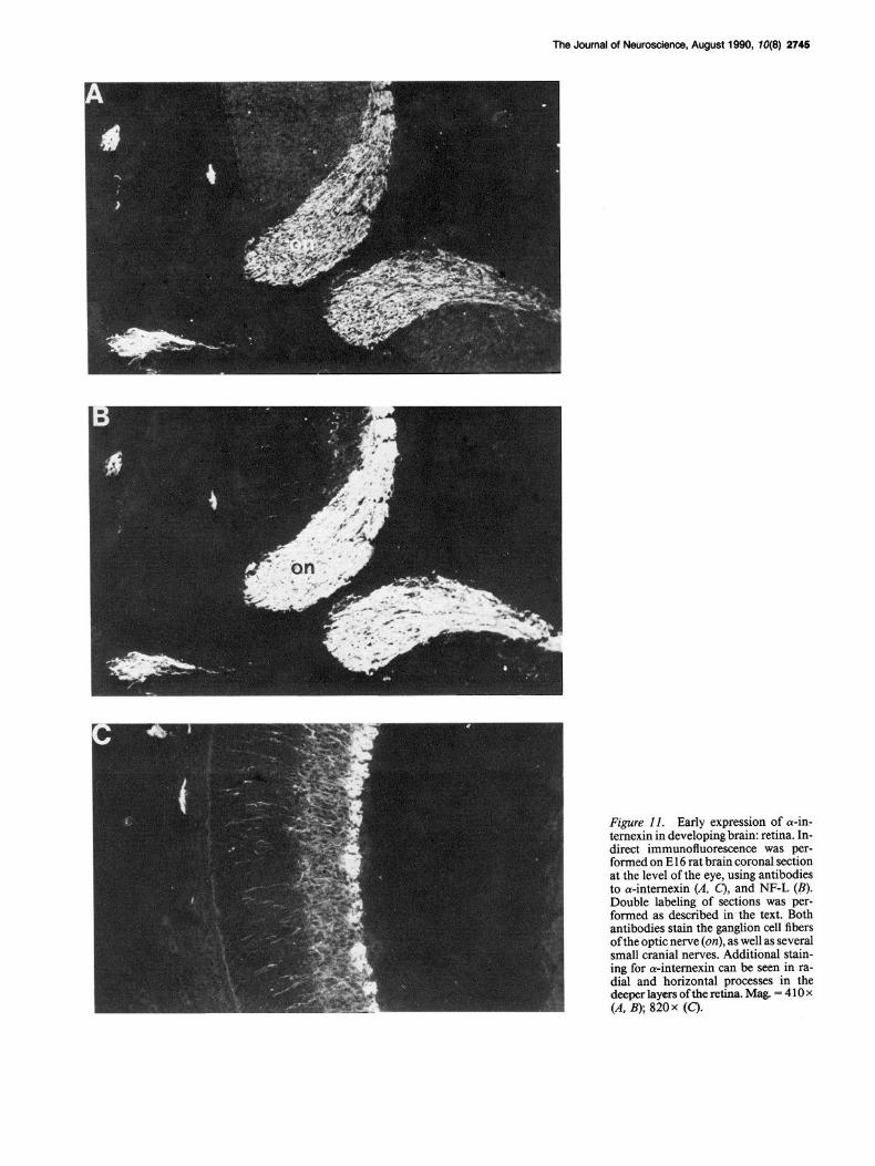
Antibodies to both oc-intemexin and NF-L stained the optic chiasm, optic nerves, and the ganglion cell layer of El6 retina (Fig. 11). Numerous radial fibers spanning the outer layers of the retina stained only for a-internexin (Fig. 11 C). Staining for a-intemexin in the absence of NF-L was also observed through- out the hypothalamus, septal nuclei, and much of the thalamus (not shown).
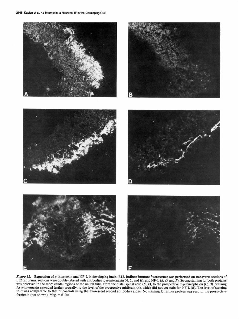
In the El2 embryos, both a-intemexin and NF-L were ob- served in the outermost layer of the neural tube from the spinal cord to the myelencephalon (Fig. 12, C-F). Antibodies to both proteins stained the developing dorsal and ventral roots (E. F), and also the sympathetic ganglia (not shown). While the 2 pro- teins displayed similar patterns of distribution in the myelen- cephalon (C, O), strong staining for cu-intemexin extended fur- ther anterior to the level of the prospective midbrain, which showed comparatively little staining for NF-L (A, B). No neu- ronal intermediate filament proteins were detectable in pro- spective forebrain at this stage.
The dilutions employed were arrived at after a series of ex- periments utilizing a wide range of conditions. Antibodies to NF-L were used at the highest concentrations that would not produce nonspecific staining. Dilutions were chosen so that staining for NF-L would be greater than or equal to that ob- served with the antibodies to cu-intemexin throughout most of the adult CNS. To control for possible competitive effects, dou- ble-labeled sections were compared with adjacent sections. All of these results were confirmed in separate experiments, using
2742 Kaplan et al. l dnternexin, a Neuronal IF in the Developing CNS
Figure 8. Axonal transport of NF proteins in developing optic nerve. Twenty-four hours following intraocular injection of 100 &i of [“%]- methionine into the vitreous chamber, 1 -d-old rat pups were killed, and the optic nerves were removed, cut into proximal and distal portions (relative to the retina), homogenized, separated by 2-dimensional SDS- PAGE, and visualized by &orography. The upper fluorograph shows proteins from the proximal half (A), while the proteins in the distal half are shown below (B). L = NF-L, T = tubulin; A = actin; h = hsc70; and Z = cY-internexin.
mAb233 for ol-internexin and the Boehringer-Mannheim mono- clonal antibody for NF-L.
Discussion cr-Internexin is a 66 kDa protein originally purified from rat brain and optic nerve (Pachter and Liem, 1985). Data presented here, including axonal transport, distribution in vivo, and po- lymerization in vitro to form 10 nm filaments, demonstrate that ar-internexin is a novel neuronal IF protein found in a wide range of mammalian species. In the adult CNS, cu-internexin
exhibits a primarily axonal pattern of distribution similar to that of the other NF proteins. In the embryo, however, a-in- ternexin is much more abundant, appearing to approach adult patterns and levels of expression at an earlier age. The potential significance of this novel neuronal IF subunit in the adult, and of these changes in neuronal IF composition in the embryo, is discussed below.
a-Intemexin is almost certainly identical to the “novel 66 kD neurofilament protein” recently described by Chiu et al. (1989). Their claim to the contrary is based largely on the fact that our initial attempts at polymerization in vitro of ol-intemexin were unsuccessful, leading to its initial classification not as an IF, but as an IF-associated protein; successful polymerization in the present study (Fig. 1, lane c) resolves this discrepancy, which we attribute to improvements in the speed and efficiency of our purification scheme. Also mentioned in support of the noniden- tity of the 2 proteins is the difference in reported values for the isoelectric point, with their value of 5.4 versus our reported value of - 5.8 (both in our original publication and in the present work). However, in either case the protein in question has a p1 similar to that of actin, suggesting that the difference in reported values is due to differences between our respective gel systems. Finally, the distribution of a-intemexin in the adult rat CNS is the same as that reported for the 66 kDa protein.
Sequence analysis of ol-internexin recently performed in our laboratory using a cDNA clone (Fliegner et al., 1990) confirms the identity of the 2 proteins: the 20-amino-acid fragment pub- lished by Chiu et al. (1989) matches our sequence, and the observed amino acid composition of the 66 kDa protein is nearly identical to the values calculated from our sequence data. Se- quence analysis also confirms the classification of ol-intemexin as an IF. The sequence conforms to the general structure of an IF, with the highest degree of homology to NF-M (-46%).
Does ol-intemexin form an independent system of neuronal intermediate filaments, or does it copolymerize in vivo with the proteins of the neurofilament triplet? While this question cannot be resolved directly using the methods employed here, several lines of evidence suggest that the neuronal IF proteins form a single network. First, a-intemexin contains the a-helical domain common to all IFS, which forms the core of the filament and presumably is responsible for polymerization. Second, in every case where the coexpression in the same cell of different IF proteins (other than the keratins) has been investigated, they have always been found to be copolymerized in a single network. Examples include NF-L or NF-M and vimentin in transfected fibroblasts (Chin and Liem, 1989; Monteiro and Cleveland, 1989), vimentin and GFA in glioma cells (Wang et al., 1984), and, of course, the 3 neurofilament triplet proteins in neurons (Willard and Simon, 1981). A close association between a-in- temexin and the neurofilament triplet is further suggested by the axonal transport data, both in the adult and at postnatal day 7 (Figs. 2 and 8, respectively), in which cY-intemexin and NF-L display similar migration rates. Similar results were also de- scribed by Monaco et al. (1985) for a 64 kDa protein that was transported with the neurofilament triplet proteins in the adult optic nerve and had the same mobility as cY-intemexin on 2-dimensional gels. The axonal transport of this protein was selectively accelerated along with that of the neurofilament trip- let in rats treated with 2,5-hexanedione.
This close association is also in evidence in the almost in- evitable appearance of a-internexin as a major band in neuronal IF preparations from diverse sources (Dahl et al., 1981; Shaw
The Journal of Neuroscience, August 1990, IO(Q) 2743
Figure 9. Early expression of 4nternexin in developing brain olfactory system. Indirect immunofluorescence was performed on coronal sections of El6 rat brain using mAbl35, a monoclonal antibody to a-intemexin (A, C), and a polyclonal rabbit antibody to NF-L (B, D). Double labeling of oc-intemexin and NF-L was accomplished through the use of fluorescein- and rhodamine-conjugated second antibodies, respectively. In the top 2 figures, extensive staining for cY-intemexin can be seen in the fibers innervating the concentric layers of the olfactory cortex (A), which still contain little if any NF-L (B). Conversely, strong staining for both proteins can be seen in the cranial nerve, indicated by the mow. The unstained shadows of numerous nuclei can also be observed, in various stages of their migration from the germinal zone bordering the olfactory ventricle (ov) to form the lamina of the olfactory cortex. In the 2 figures at bottom, early expression of cu-intemexin is seen in the lateral olfactory tract (ok), which does not stain for NF-L (0). In contrast, the cranial nerve indicated by the arrow stains strongly for both proteins. The optic vesicle can be seen in the lower left comer of panels C and D; radial fibers spanning its width stained for cu-intemexin only (see also Fig. 12). cr = cranium. Mag. = 270 x .

2744 Kaplan et al. l dntemexin, a Neuronal IF in the Developing CNS
Figure 10. Early expression of oc-internexin in developing brain: cerebral cortex. Indirect immunofluorescence was performed on El6 rat brain sections as described above, using antibodies to ol-intemexin (A, C), vimentin (B), and NF-L (0). A-C show the same section viewed by double labeling, while D shows a section taken somewhat more caudally. Strong staining for cY-intemexin is evident in the fibers underlying the developing cortical plate, which do not stain for vimentin and stain only weakly for NF-L. Expression of ar-intemexin is also seen in the habenula (ha), and in numerous radial fibers spanning the cortical plate. These fibers do not appear to enter the intermediate zone (iz), in contrast to the radial fibers staining with vimentin, which extend from the ventricle (v), to the pial surface. Mag. = 270 x (A, B, D); 540 x (C).
and Weber, 1982). This raises the question of why this major component of the neuronal cytoskeleton has not been previously identified. One reason for this is its ease of degradation (see Fig. 4), also reported by Chiu et al. for their 66 kDa protein (1989). A second is the possibility that this protein represented a break- down of NF-L, since it always migrates ahead of NF-L in 1 -dimensional SDS-PAGE. Finally, the molecular weight of cu-internexin, -59 kDa in the mouse, allows it to be mistaken for either vimentin or tubulin on 1 -dimensional gels; these pos- sibilities are discounted in the present study by both Western blotting and 2-dimensional gel analysis.
The absence of an immunoreactive band in the sample ob- tained from chick optic nerve does not preclude the possibility that cu-internexin may be present in nonmammalian species. Many antibodies to mammalian IFS do not recognize their avian counterpart and it is therefore possible that our 2 monoclonal antibodies similarly fail to recognize chick a-internexin. A pro- tein of similar molecular weight, NAPA-73, has been described in chick as a neurofilament-associated protein, which migrates behind NF-L and is associated with the stage of terminal dif- ferentiation of chick brain neurons (Ciment et al., 1986; Wu and de Vellis, 1987). Comparisons and homologies between
The Journal of Neuroscience, August 1990, W(8) 2745
Figure 11. Early expression of a-in- ternexin in developing brain: retina. In- direct immunofluorescence was per- formed on El 6 rat brain coronal section at the level of the eye, using antibodies to cY-intemexin CA. 0 and NF-L (B). Double labeling‘ of sections was tier- formed as described in, the text. Both antibodies stain the ganglion cell fibers of the optic nerve (on), as well as several small cranial nerves. Additional stain- ing for a-intemexin can be seen in ra- dial and horizontal processes in the deeper layers of the retina. Mag. = 4 10 x (A, B); 820x (C).
2748 Kaplan et al. l dnternexin, a Neuronal IF in the Developing CNS
Figure 12. Expression of cu-internexin and NF-L in developing brain: E12. Indirect immunofluorescence was performed on transverse sections of E 12 rat brains; sections were double-labeled with antibodies to oc-internexin (A. C, and E), and NF-L (B, D, and F). Strong staining for both proteins was observed in the more caudal regions of the neural tube, from the distal spinal cord (E, F), to the prospective myelencephalon (C, D). Staining for or-internexin extended further rostrally, to the level of the prospective midbrain (A), which did not yet stain for NF-L (B). The level of staining in B was comparable to that of controls using the fluorescent second antibodies alone. No staining for either protein was seen in the prospective forebrain (not shown). Mag. = 6 10 x .
The Journal of Neuroscience, August 1990, W(8) 2747
NAPA- and oc-internexin await the publication of the se- quence of NAPA- 3.
It has long been known that neuronal differentiation is ac- companied by changes in neuronal IF composition, usually de- scribed as a stepwise process in which expression of NF-H fol- lows coexpression of the 2 smaller subunits (Shaw and Weber, 1982, 1983; Pachter and Liem, 1985; Carden et al., 1987). Ad- ditionally, some neurons also express vimentin early in devel- opment (Bignami et al., 1982; Cochard and Paulin, 1984), while neurepithelial stem cells express nestin, a novel neuronal IF protein (Lendahl et al., 1990). Our data indicate that the expres- sion of oc-internexin precedes that of NF-L throughout most of the developing brain and that embryonic neuronal IFS contain an elevated proportion of ol-internexin relative to their adult counterparts. This can be seen most clearly in the El6 embryos (Figs. 9-l 1). The same antibodies and dilutions that produced comparable patterns and levels of staining in the adult yielded widely dissimilar results in the embryos, with strong staining for cu-internexin in processes that still displayed very little NF- L. For example, strong staining for both proteins could be seen in the optic nerve, the brain stem and spinal cord, and also the cranial nerves and their ganglia, structures already well devel- oped by E 16. Both proteins can be seen in the thalamocortical projections underlying the cortical plate, but staining for a-in- ternexin is far stronger and more extensive. In the cerebral cor- tex, which is still relatively undeveloped at E16, cy-internexin can be detected in the processes of the horizontal cells of layer I, and also in the radial processes of neurons in layers V and VI; expression of NF-L in the cortex at El6 is limited to the aforementioned thalamocortical projections, whose develop- ment precedes that of the cortical plate (Hicks and D’Amato, 1968; Altman and Bayer, 1979). While further studies are need- ed to clarify the time course and significance of this early expres- sion of cu-intemexin [in particular, its relationship to early expression of vimentin (Bignami et al., 1982; Cochard and Pau- lin, 1984), nestin (Lendahl et al., 1990), and peripherin (Troy et al., 1990)], the early onset, high intensity, and long duration of its expression suggest that a-internexin may be highly useful as an early neuronal marker.
A consideration of these changes in neuronal IF composition accompanying development may provide some insight into the respective functional contributions of the constituent proteins to neuronal IF function in the adult. The axons of the early, developing nervous system are distinguished most obviously from their adult counterparts by their smaller diameter. In light of the widely accepted theory of neuronal IFS as arbiters of axonal caliber (Lasek et al., 1983), the preponderance of a-in- temexin in the neuronal IF networks of these smaller axons suggests that the ability of neuronal IFS to increase axonal di- ameter must derive from the properties of one or more of the other neuronal IF proteins. The most obvious candidates for such a role are the 2 larger subunits, NF-M and NF-H, which could affect axonal diameter either by the formation of radially oriented cross-bridges between filaments or by simple, me- chanical “space filling” by their large carboxy-terminal projec- tions. Filaments composed predominantly of oc-intemexin would have fewer of these bulky radial projections. Neuronal IFS may thus regulate axonal diameter through changes not only in their number, but also in their composition, with the changes de- scribed above allowing for a gradual increase in axonal diameter. This model would also account for the high levels of cY-intemexin we observed in the small-caliber parallel fibers of the adult
cerebellum, which appeared to lack other NF triplet proteins (as opposed to the numerous larger fibers in the cerebellum, which expressed all 4 neuronal IF proteins). The absence of lengthy carboxy-terminal extension would seem to make a-in- ternexin ideally suited to form IFS in developing axons, but does not explain why this function might not be performed just as well by NF-L, which shares this feature. The answer may relate to the relative abilities of these proteins to mediate interactions between IFS and other organelles, such as (but not only) the microtubules. Interactions between IFS and microtubules would presumably be mediated by microtubule-associated proteins (MAPS), which are believed to bind to NF-L (Heimann et al., 1985); in fact, oc-internexin has the highest degree of homology not to NF-L, but to NF-M, which reportedly does not bind MAPS. These differences in binding characteristics (which need to be further elucidated) may provide for additional control of axonal diameter and/or regulation of the dynamics and stability of the neurofilament network.
Changes in IF composition have also been observed in the peripheral nervous system following axotomy. Expression of peripherin and vimentin increased immediately following ax- otomy, while expression of the neurofilament triplet proteins was markedly reduced (Oblinger et al., 1989). Although we found only low levels of oc-internexin in adult peripheral structures such as sciatic nerve, in agreement with the findings of Chiu et al. for their 66 kDa protein (1989), and also with those of Mona- co et al. for their 64 kDa protein (1985), significant levels of ol-intemexin were observed at El 2 in the developing dorsal roots (Fig. 12) and at E 16 in at least one peripheral nervous system (PNS) structure, the trigeminal ganglia (not shown). While a detailed consideration of neuronal IF expression in the PNS is beyond the scope of this paper, these preliminary results suggest an elevated peripheral expression of a-intemexin in the embryo, which is down-regulated as development proceeds. This view is also supported by the work of Chiu et al. (1989), who report elevated amounts of their 66 kDa protein in sciatic nerve at postnatal day 1, compared to adult levels. In light of the pro- posed recapitulation of the developmental program of cyto- skeletal expression during axon regeneration (Hoffman and Cleveland, 1988), it will be interesting to see whether PNS expression of oc-internexin increases following axotomy, or rath- er, whether oc-internexin and peripherin may play analogous roles in the CNS and PNS, respectively.
References Altman J, Bayer S (1979) Development of the diencephalon of the
rat. J Comp Neurol 188:455-524. Angevine JB Jr, Sidman RL (1961) Autoradiographic study of cell
migration during histogenesis of cerebral cortex in the mouse. Nature 192:766-768.
Berry M, Rogers AW (1965) The migration of neuroblasts in the developing cerebral cortex. J Anat 99:691-709.
Bignami A, Raju T, Dahl D (1982) Localization of vimentin, the nonspecific intermediate filament protein, in embryonal glia and in early differentiating neurons. Dev Viol 9 1:286-295: -
Boulder Committee (1970) Embrvonic vertebrate central nervous svs- tern: revised terminolog;. Anat kec 166:257-26 1.
Carden MJ, Trojanowski JQ, Schlaepfer WW, Lee VMY (1987) Two- stage expression of neurofilament polypeptides during rat neurogene- sis with early establishment of adult phosphorylation patterns. J Neu- rosci 7:3489-3504.
Chin SSM, Liem RKH (1989) Expression ofrat neurofilament proteins NF-L and NF-M in transfected non-neuronal cells. Eur J Cell Biol 5oz475-490.
Chin SSM, Hashim GA, Liem RKH (1989) Expression of truncated
2748 Kaplan et al. * dnternexin, a Neuronal IF in the Developing CNS
neurofilament proteins in transiently transfected cells. J Cell Biol Abstr 109(4):69a.
Chiu FC, Barnes EA, Das K, Haley J, Socolow P, Macaluso FP, Fant J (1989) Characterization of a novel 66-kD subunit of mammalian neurofilaments. Neuron 2:1435-1445.
Ciment G, Ressler A, Letoumeau PC, Weston JA (1986) A novel intermediate filament-associated protein, NAPA-73, that binds to different filament types at different stages of nervous system devel- opment. J Cell Biol 102:246-25 1.
Cochard P, Paulin D (1984) Initial expression of neurofilaments and vimentin in the central and peripheral nervous system of the mouse embryo in vivo. J Neurosci 4:2080-2094.
Dahl D, Rueger DC, Bignami A, Weber K, Osbom M (198 1) Vimen- tin, the 57,000 kD protein of fibroblast filaments, is the major cy- toskeletal component in immature glia. Eur J Cell Biol 24: 191-196.
Fliegner KH, Ching GY, Liem RKH (1990) The predicted amino acid sequence of a-intemexin is that of a novel neuronal intermediate filament protein. EMBO J 9:749-755.
Heimann R, Shelanski ML, Liem RKH (1985) Specific binding of microtubule-associated proteins to the 70,000 dalton neurofilament subunit. J Biol Chem 260:2 160-2 166.
Hendrickson AE, Cowan WM (197 1) Changes in the rate of axoplas- mic transport during postnatal development ofthe rabbit’s optic nerve and tract: Exp Nemo1 30:403-422.
Hicks SP. D’Amato CJ f 1968) Cell miarations to the isocortex in the rat. Anat Ret 160:6191634.’ -
Hirokawa N, Glicksman MA, Willard M (1984) Organization of mam- malian neurofilament polypeptides within the neuronal cytoskeleton. J Cell Biol 98:1523-1536.
Hoffman PN, Cleveland DW (1988) Neurofilament and tubulin expression recapitulates the developmental program during axonal regeneration: induction of a P-specific isotype. Proc Nat1 Acad Sci USA 5:4530-4533.
Hoffman PN, Lasek RJ (1975) The slow component of axonal trans- port. Identification of major structural polypeptides of the axon and their generality among mammalian neurons. J Cell Biol66:35 l-366.
Kohler G, Milstein C (1976) Derivation of specific antibody-producing tissue culture and tumor lines by cell fusion. Eur J Immunol 6:5 1 l- 519.
Laemmli UK (1970) Cleavage of structural proteins during the assem- bly of the head of bacteriophage T4. Nature 7:680-685.
Lasek RJ, Oblinger MM, Drake PF (1983) The molecular biology of neuronal geometry: the expression of neurofilament genes influences axonal diameter. Cold Spring Harbor Symp Quant Biol48:43 l-444.
Lendahl U. Zimmerman LB. McKav RDG (1990) CNS stem cells express a new class of intermediate filament proteins. Cell 60:585- 595.
Leonard GB, Gorham DG, Cole P, Greene LA, Ziff EB (1988) A nerve growth factor-regulated messenger RNA encodes a new intermediate filament protein: J Cell Biol 106:181-193.
Liem RKH. Hutchison SB (1982) Purification of the individual com- ponents ofthe neurofilameht triplet: filament assembly from the 70,000 dalton subunit. Biochemistry 2 1:3221-3226.
Liem RKH, Yen S-H, Salomon GD, Shelanski ML (1978) Interme- diate filaments in nervous tissue. J Cell Biol 79:637-645.
McConnell SK, Gnosh A, Shatz CJ (1989) Subplate neurons pioneer the first axon pathway from the cerebral cortex. Science 245:978- 982.
Monaco S, Antilio-Gambetti L, Zabel D, Gambetti P (1985) Giant axonal neuropathy: acceleration of neurofilament transport in optic axons. Proc Nat1 Acad Sci USA 82:920-924.
Oblinger MM, Wong J, Parysek LM (1989) Axotomy-induced changes in the expression of a type III neuronal intermediate filament gene. J Neurosci 9~3766-3775.
O’Farrell PH (1975) High-resolution two-dimensional electrophoresis of proteins. J Biol Chem 250:4007-402 1.
Pachter JS, Liem RKH (1984) The differential appearance of neuro- filament triplet polypeptides in the developing rat optic nerve. Dev Biol 103:200-210.
Pachter JS, Liem RKH (1985) cu-Internexin, a 66-kD intermediate filament-binding protein from mammalian central nervous tissues. J Cell Biol 101:1316-1322.
Parvsek LM, Goldman R (1987) Characterization of intermediate filaments in PC1 2 cells. J Neurosci 7:78 l-79 1.
Portier M-M. deNechaud B. Gros F (1984) Periuherin. a new member ofthe intermediate filament protein family. De; Neurosci 6:335-344.
Quinlan R, Franke WW (1982) Heteropolymeric filaments ofvimentin and desmin in vascular smooth muscle tissue and cultured baby ham- ster kidney cells demonstrated by chemical cross-linking. Proc Nat1 Acad Sci USA 7913452-3456.
Raedler A, Sievers J (1976) Light and electron microscopical studies on specific cells of the marginal zone in the developing rat cerebral cortex. Anat Embryo1 149:173-181.
Ramon y Cajal S (1891) Sur la structure de l’ecorce cerebrale de quelques mammiferes. Cellule 7: 125-l 76.
Retzius G (1893) Die Cajal’schen zellen des Grosshimrinde beim Menschen und bei Saugetieren. Biol Untersuch 5: 1-9.
Reynolds ES (1963) The use of lead citrate at high pH as an electron- opaque stain in electron microscopy. J Cell Biol 17:208-2 12.
Shaw G (1990) Neurofilament proteins. In: The neuronal cytoskeleton (Burgoyne RD, ed). New York: Liss (in press).
Shaw G, Weber K (1982) Differential expression of neurofilament triplet proteins in brain development. Nature 298:277-279.
Shaw G, Weber K (1983) The structure and development of the rat retina: an immunofluorescence microscopical study using antibodies specific for intermediate filament proteins. Eur J Cell Biol 30:219- 232.
Shaw G, Banker GA, Weber K (1985) An immunofluorescence study of neurofilament protein expression by developing hippocampal neu- rons in tissue culture. Eur J Cell Biol 39:205-216.
Shaw G, Osborn M, Weber K (1986) Reactivity of a panel of neu- rofilament antibodies on phosphorylated and dephosphorylated neu- rofilaments. Eur J Cell Biol 42: l-9.
Steinert PM, Roop DR (1988) Molecular and cellular biology of in- termediate filaments. Annu Rev Biochem 57:593-625.
Towbin H, Staehlin T, Gordon J (1979) Electrophoretic transfer of proteins nitrocellulose: procedure and some applications. Proc Nat1 Acad Sci USA 7614350-4354.
Troy C, Brown K, Greene LA, Shelanski ML (1990) The ontogeny of peripherin, a type III neuronal intermediate filament protein. Neu- roscience (in press).
Wang E, Caimcross JG, Liem RKH (1984) Identification of glial fil- ament protein and vimentin in the same intermediate filament system in human glioma cells. Proc Nat1 Acad Sci USA 8 1:2102-2 106.
Willard M, Simon C (198 1) Antibody decoration of neurofilaments. J Cell Biol 89:198-205.
Willard M, Simon C (1983) Modulations of neurofilament axonal transport during the development of rabbit retinal ganglion cells. Cell 35:551-559.
Wu DK, de Vellis J (1987) The expression of the intermediate fila- ment-associated protein (NAPA-73) is associated with the stage of terminal differentiation of chick brain neurons. Brain Res 42 1: 186-
Monteiro M, Cleveland D (1989) Expression of NF-L and NF-M in fibroblasts reveals coassembly of neurofilament and vimentin sub- units. J Cell Biol 108:579-593.
193.