DNA Replication Stress
370
DNA Replication Stress Robert M. Brosh Jr. www.mdpi.com/journal/ijms Edited by Printed Edition of the Special Issue Published in International Journal of Molecular Sciences International Journal of Molecular Sciences
Transcript of DNA Replication Stress
DNA Replication Stress
Robert M. Brosh Jr.
www.mdpi.com/journal/ijms
Edited by
Printed Edition of the Special Issue Published in International Journal of Molecular Sciences
International Journal of Molecular Sciences

DNA Replication Stress


DNA Replication Stress
Special Issue Editor
Robert M. Brosh Jr.
MDPI • Basel • Beijing • Wuhan • Barcelona • Belgrade

Special Issue Editor
Robert M. Brosh Jr.
National Institute on Aging
USA
Editorial Office
MDPI
St. Alban-Anlage 66
4052 Basel, Switzerland
This is a reprint of articles from the Special Issue published online in the open access journal
International Journal of Molecular Sciences (ISSN 1422-0067) from 2018 to 2019 (available at: https:
//www.mdpi.com/journal/ijms/special issues/DNA Replication Stress).
For citation purposes, cite each article independently as indicated on the article page online and as
indicated below:
LastName, A.A.; LastName, B.B.; LastName, C.C. Article Title. Journal Name Year, Article Number,
Page Range.
ISBN 978-3-03921-389-4 (Pbk)
ISBN 978-3-03921-390-0 (PDF)
c© 2019 by the authors. Articles in this book are Open Access and distributed under the Creative
Commons Attribution (CC BY) license, which allows users to download, copy and build upon
published articles, as long as the author and publisher are properly credited, which ensures maximum
dissemination and a wider impact of our publications.
The book as a whole is distributed by MDPI under the terms and conditions of the Creative Commons
license CC BY-NC-ND.

Contents
About the Special Issue Editor . . . . . . . . . . . . . . . . . . . . . . . . . . . . . . . . . . . . . . vii
Robert M. Brosh Jr.
Special Issue on DNA Replication Stress: Summary of Topics CoveredReprinted from: Int. J. Mol. Sci. 2019, 20, 2934, doi:10.3390/ijms20122934 . . . . . . . . . . . . . . 1
Garrett M. Warren, Richard A. Stein, Hassane S. Mchaourab and Brandt F. Eichman
Movement of the RecG Motor Domain upon DNA Binding Is Required for Efficient ForkReversalReprinted from: Int. J. Mol. Sci. 2018, 19, 3049, doi:10.3390/ijms19103049 . . . . . . . . . . . . . . 6
Jolanta Kwasniewska, Karolina Zubrzycka and Arita Kus
Impact of Mutagens on DNA Replication in Barley ChromosomesReprinted from: Int. J. Mol. Sci. 2018, 19, 1070, doi:10.3390/ijms19041070 . . . . . . . . . . . . . . 19
Maılyn Yates and Alexandre Marechal
Ubiquitylation at the Fork: Making and Breaking Chains to Complete DNA ReplicationReprinted from: Int. J. Mol. Sci. 2018, 19, 2909, doi:10.3390/ijms19102909 . . . . . . . . . . . . . . 32
Wei-Chung Tsao and Kristin A. Eckert
Detours to Replication: Functions of Specialized DNA Polymerases during Oncogene-inducedReplication StressReprinted from: Int. J. Mol. Sci. 2018, 19, 3255, doi:10.3390/ijms19103255 . . . . . . . . . . . . . . 64
Anna Konopka and Julie D Atkin
The Emerging Role of DNA Damage in the Pathogenesis of the C9orf72 Repeat Expansion inAmyotrophic Lateral SclerosisReprinted from: Int. J. Mol. Sci. 2018, 19, 3137, doi:10.3390/ijms19103137 . . . . . . . . . . . . . . 89
Jolene Michelle Helena, Anna Margaretha Joubert, Simone Grobbelaar,
Elsie Magdalena Nolte, Marcel Nel, Michael Sean Pepper, Magdalena Coetzee and
Anne Elisabeth Mercier
Deoxyribonucleic Acid Damage and Repair: Capitalizing on Our Understanding of theMechanisms of Maintaining Genomic Integrity for Therapeutic PurposesReprinted from: Int. J. Mol. Sci. 2018, 19, 1148, doi:10.3390/ijms19041148 . . . . . . . . . . . . . . 110
Synnøve Brandt Ræder, Anala Nepal, Karine Øian Bjøras, Mareike Seelinger,
Rønnaug Steen Kolve, Aina Nedal, Rebekka Muller and Marit Otterlei
APIM-Mediated REV3L–PCNA Interaction Important for Error Free TLS Over UV-InducedDNA Lesions in Human CellsReprinted from: Int. J. Mol. Sci. 2019, 20, 100, doi:10.3390/ijms20010100 . . . . . . . . . . . . . . . 133
Wei-Wei Wang, Huan Zhou, Juan-Juan Xie, Gang-Shun Yi, Jian-Hua He, Feng-Ping Wang,
Xiang Xiao and Xi-Peng Liu
Thermococcus Eurythermalis Endonuclease IV Can Cleave Various Apurinic/ApyrimidinicSite Analogues in ssDNA and dsDNAReprinted from: Int. J. Mol. Sci. 2019, 20, 69, doi:10.3390/ijms20010069 . . . . . . . . . . . . . . . 150
v
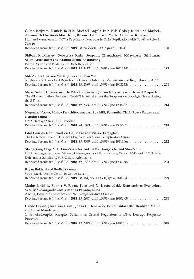
Guido Keijzers, Daniela Bakula, Michael Angelo Petr, Nils Gedsig Kirkelund Madsen,
Amanuel Teklu, Garik Mkrtchyan, Brenna Osborne and Morten Scheibye-Knudsen
Human Exonuclease 1 (EXO1) Regulatory Functions in DNA Replication with Putative Roles inCancerReprinted from: Int. J. Mol. Sci. 2019, 20, 74, doi:10.3390/ijms20010074 . . . . . . . . . . . . . . . 168
Shibani Mukherjee, Debapriya Sinha, Souparno Bhattacharya, Kalayarasan Srinivasan,
Salim Abdisalaam and Aroumougame Asaithamby
Werner Syndrome Protein and DNA ReplicationReprinted from: Int. J. Mol. Sci. 2018, 19, 3442, doi:10.3390/ijms19113442 . . . . . . . . . . . . . . 183
Md. Akram Hossain, Yunfeng Lin and Shan Yan
Single-Strand Break End Resection in Genome Integrity: Mechanism and Regulation by APE2Reprinted from: Int. J. Mol. Sci. 2018, 19, 2389, doi:10.3390/ijms19082389 . . . . . . . . . . . . . . 201
Miiko Sokka, Dennis Koalick, Peter Hemmerich, Juhani E. Syvaoja and Helmut Pospiech
The ATR-Activation Domain of TopBP1 Is Required for the Suppression of Origin Firing duringthe S PhaseReprinted from: Int. J. Mol. Sci. 2018, 19, 2376, doi:10.3390/ijms19082376 . . . . . . . . . . . . . . 214
Nagendra Verma, Matteo Franchitto, Azzurra Zonfrilli, Samantha Cialfi, Rocco Palermo and
Claudio Talora
DNA Damage Stress: Cui Prodest?Reprinted from: Int. J. Mol. Sci. 2019, 20, 1073, doi:10.3390/ijms20051073 . . . . . . . . . . . . . . 229
Lilas Courtot, Jean-Sebastien Hoffmann and Valerie Bergoglio
The Protective Role of Dormant Origins in Response to Replicative StressReprinted from: Int. J. Mol. Sci. 2018, 19, 3569, doi:10.3390/ijms19113569 . . . . . . . . . . . . . . 242
Sheng-Yong Yang, Yi Li, Guo-Shun An, Ju-Hua Ni, Hong-Ti Jia and Shu-Yan Li
DNA Damage-Response Pathway Heterogeneity of Human Lung Cancer A549 and H1299 CellsDetermines Sensitivity to 8-Chloro-AdenosineReprinted from: Int. J. Mol. Sci. 2018, 19, 1587, doi:10.3390/ijms19061587 . . . . . . . . . . . . . . 264
Bayan Bokhari and Sudha Sharma
Stress Marks on the Genome: Use or Lose?Reprinted from: Int. J. Mol. Sci. 2019, 20, 364, doi:10.3390/ijms20020364 . . . . . . . . . . . . . . . 279
Marios Kritsilis, Sophia V. Rizou, Paraskevi N. Koutsoudaki, Konstantinos Evangelou,
Vassilis G. Gorgoulis and Dimitrios Papadopoulos
Ageing, Cellular Senescence and Neurodegenerative DiseaseReprinted from: Int. J. Mol. Sci. 2018, 19, 2937, doi:10.3390/ijms19102937 . . . . . . . . . . . . . . 291
Hanne Leysen, Jaana van Gastel, Jhana O. Hendrickx, Paula Santos-Otte, Bronwen Martin
and Stuart Maudsley
G Protein-Coupled Receptor Systems as Crucial Regulators of DNA Damage ResponseProcessesReprinted from: Int. J. Mol. Sci. 2018, 19, 2919, doi:10.3390/ijms19102919 . . . . . . . . . . . . . . 328
vi

About the Special Issue Editor
Robert Brosh, Dr., received his B.Sc. in chemistry from Bethany College in 1985, M.Sc. in biochemistry
from Texas A & M University in 1988, and Ph.D. in biology from the University of North Carolina,
Chapel Hill, in 1996. Brosh conducted postdoctoral studies at NIH before assuming his present
position as Principal Investigator in the Laboratory of Molecular Gerontology, NIA, where he became
a tenured Senior Investigator in 2006. Brosh’s group is engaged in biomedical studies of DNA repair
diseases. His research is supported by the NIA Intramural Research Program (NIH), and he has
received funding from the Fanconi Anemia Research Fund, NIH R03 Grant, NIA Inter-Lab Funding
Awards and, most recently, from an NIA-NIEHS Inter-Institute Award. Brosh serves as Mentor
for the NIH Summer Student Intramural Research Training Award Program, NIH Undergraduate
Scholarship Program, and NIH Community College Enrichment Program. In 2015, he received
the NIH National Institute on Aging Post Baccalaureate Distinguished Mentor Award and the NIA
Director’s Merit Award in 2017. He has served on the Editorial Board for Journal of Biological
Chemistry and other journals, including Aging and Gene, and is Associate Editor of Ageing Research
Reviews. Brosh is a Guest Lecturer at Johns Hopkins University and has participated on several grant
review committees, including NIH, German Research Foundation, and Italian Telethon Scientific
Committee. Brosh is also involved in outreach programs that provide children with hands-on
learning opportunities in science.
vii

International Journal of
Molecular Sciences
Editorial
Special Issue on DNA Replication Stress: Summaryof Topics Covered
Robert M. Brosh, Jr. †
Laboratory of Molecular Gerontology, National Institute on Aging, National Institutes of Health, NIHBiomedical Research Center, 251 Bayview Blvd, Baltimore, MD 21224, USA; [email protected]† Guest Editor of International Journal of Molecular Sciences Special Issue on DNA Replication Stress.
Received: 10 June 2019; Accepted: 13 June 2019; Published: 15 June 2019
A Special Issue of International Journal of Molecular Sciences (IJMS) is dedicated to mechanismsmediated at the molecular and cellular levels to respond to adverse genomic perturbations and DNAreplication stress (https://www.mdpi.com/journal/ijms/special_issues/DNA_Replication_Stress). Therelevant proteins and processes play paramount roles in nucleic acid transactions to maintain genomicstability and cellular homeostasis. A total of 18 articles are comprised in the series, encompassing abroad range of highly relevant topics in genome biology. These include replication fork dynamics,DNA repair processes, DNA damage signaling and cell cycle control, cancer biology, epigenetics,cellular senescence, neurodegeneration, and aging. Below are highlighting primers for the articleswhich constitute this recently published IJMS Special Issue.
1. DNA Replication Fork Dynamics
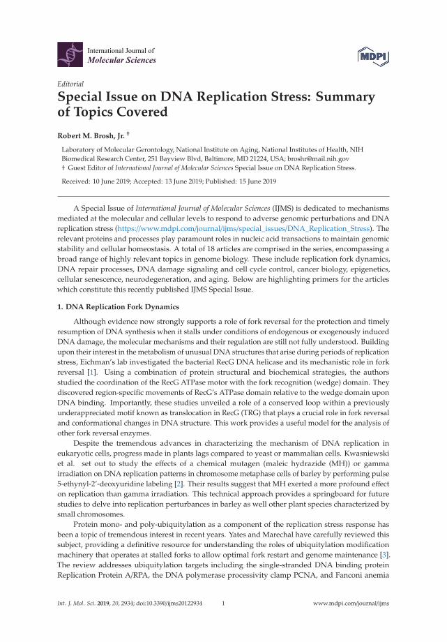
Although evidence now strongly supports a role of fork reversal for the protection and timelyresumption of DNA synthesis when it stalls under conditions of endogenous or exogenously inducedDNA damage, the molecular mechanisms and their regulation are still not fully understood. Buildingupon their interest in the metabolism of unusual DNA structures that arise during periods of replicationstress, Eichman’s lab investigated the bacterial RecG DNA helicase and its mechanistic role in forkreversal [1]. Using a combination of protein structural and biochemical strategies, the authorsstudied the coordination of the RecG ATPase motor with the fork recognition (wedge) domain. Theydiscovered region-specific movements of RecG’s ATPase domain relative to the wedge domain uponDNA binding. Importantly, these studies unveiled a role of a conserved loop within a previouslyunderappreciated motif known as translocation in RecG (TRG) that plays a crucial role in fork reversaland conformational changes in DNA structure. This work provides a useful model for the analysis ofother fork reversal enzymes.
Despite the tremendous advances in characterizing the mechanism of DNA replication ineukaryotic cells, progress made in plants lags compared to yeast or mammalian cells. Kwasniewskiet al. set out to study the effects of a chemical mutagen (maleic hydrazide (MH)) or gammairradiation on DNA replication patterns in chromosome metaphase cells of barley by performing pulse5-ethynyl-2’-deoxyuridine labeling [2]. Their results suggest that MH exerted a more profound effecton replication than gamma irradiation. This technical approach provides a springboard for futurestudies to delve into replication perturbances in barley as well other plant species characterized bysmall chromosomes.
Protein mono- and poly-ubiquitylation as a component of the replication stress response hasbeen a topic of tremendous interest in recent years. Yates and Marechal have carefully reviewed thissubject, providing a definitive resource for understanding the roles of ubiquitylation modificationmachinery that operates at stalled forks to allow optimal fork restart and genome maintenance [3].The review addresses ubiquitylation targets including the single-stranded DNA binding proteinReplication Protein A/RPA, the DNA polymerase processivity clamp PCNA, and Fanconi anemia
Int. J. Mol. Sci. 2019, 20, 2934; doi:10.3390/ijms20122934 www.mdpi.com/journal/ijms1

Int. J. Mol. Sci. 2019, 20, 2934
protein complex FANCD2/I. Also discussed are the reversible ubiquitylation processes that prevailduring DNA replication stress.
2. Alternate DNA Structures
Difficult-to-replicate sequences pose a unique challenge to the DNA polymerases delegated todeal with noncanonical DNA structures and copy the genome. This is the very topic of a review articlefrom Kristin Eckert’s lab [4]. Specialized DNA polymerases help to cope with such unusual DNAstructures, and their regulation plays profound roles during oncogene-induced replication stress. Tsaoand Eckert provide a very comprehensive and current assessment of the field that is a useful resourcemoving forward in this hotly studied area of genome metabolism.
Alternate DNA structures and DNA damage have far-reaching effects on human physiology,including neurodegenerative diseases. This topic is addressed by Konopka and Atkin in the context ofamyotrophic lateral sclerosis (ALS), a debilitating progressive neurodegenerative disorder characterizedby hexanucleotide repeat expansions [5]. The central role of DNA damage is discussed in the reviewarticle, as well as potential therapeutic strategies to treat ALS.
3. DNA Repair Proteins and Processes
DNA is considered the quintessential information molecule in genome biology. Therefore, themechanisms of DNA damage and repair are highly valuable to cellular metabolism. Helena et al. reviewthe DNA repair pathways which exist to protect the genome and preserve cellular homeostasis [6]. Theanalysis is not only relevant to understanding disease pathogenesis but also diagnosis and therapeuticstrategies for combating various cancers.
Dr. Marit Otterlei and colleagues have had a longstanding interest in the in vivo response to DNAdamage in human cells. Combining both elegant microscopy and mutation analysis, they report theirfindings from an investigation of the interaction of the replication processivity clamp PCNA with atranslesion synthesis (TLS) DNA polymerase known as REV3L that is implicated in DNA synthesispast ultraviolet light-induced lesions [7]. They discovered that a specialized PCNA interacting motifdesignated APIM is critical for the function and specificity of REV3L in TLS. Moreover, the studyrevealed that mutation frequencies and spectra could be modulated in vivo by a PCNA-targetingcell-penetrating peptide, suggesting the potential use of the peptide in chemotherapy strategies todownregulate mutation frequency, as it preferentially targets TLS compared to error-free DNA repair.
Wang et al. characterized the catalytic activity of an archaeal thermophilic endonuclease IV toincise apurinic/apyrimidinic (AP) analogues in single-stranded or double-stranded DNA [8]. Using abattery of AP analogues with different length alkane, polyethylene glycol, cyclic, or two-carbon atomchain spacers, the authors were able to systematically assess substrate specificity and biochemicalactivity for the recognition and cleavage of phosphodiester bonds by the Thermococcus eurythermalisendonuclease IV, providing a model for studies of related enzymes and shedding light on the repair ofAP sites in hyperthermophilic archaea.
Human exonuclease I (EXO1) is a DNA processing enzyme with important pleiotropic roles incellular DNA metabolism. Guido Keijzers et al. review the replication and post-replication functionsof EXO1 to help the reader appreciate the involvement of EXO1 mutations in various cancers [9].Mismatch repair deficiencies caused by molecular defects of EXO1 mutant alleles is associated withmultiple cancers. Some of these mutations reside in the nuclease domain, whereas others reside indomains delegated for protein interaction with the mismatch repair factors MLH1 and MSH2. Thus,microsatellite instability driven by EXO1 mutational defects may very well underlie chromosomaldestabilization and be a major driver of tumorigenesis.
Since the discovery over two decades ago that recessive mutations in the RecQ helicase geneWRN are linked to the premature aging disorder Werner syndrome, the hereditary disease has servedas a window to understanding the molecular basis for genomic stability, yet its precise functions innucleic acid transactions are still not well understood. The Asaithamby lab addresses the role(s) of
2
Int. J. Mol. Sci. 2019, 20, 2934
WRN in replication fork processing and the post-translational modifications that fine-tune its pathwayactivities [10]. The authors discuss the proposed dual roles of WRN in replication fork stabilizationand pathway choice for double-strand break repair. Interpretations of WRN’s involvement in cellularsenescence and genome maintenance place the experimental studies of WRN in a useful perspectivefor potential clinical implications.
Single-strand breaks are one of the most common DNA lesions in the cell and pose a source ofgenomic instability by interfering with cellular DNA replication and transcription. A comprehensivereview from the Yan lab discusses single-strand break DNA end resection and its step-by-stepmechanism [11]. An emphasis is placed on the role of AP endonuclease 2 (APE2) in the process ofsingle-strand break end resection. A valuable perspective for future studies in this area is provided.
4. Cell Cycle Control
Understanding how DNA replication initiation is controlled during the DNA synthesis S-phase inmammalian cells is of considerable interest given the number of proteins involved and the importanceof ensuring that genome duplication occurs only once per cell cycle. Sokka et al. focused their analysison the importance of the ATR-activation domain of the activator DNA topoisomerase-2-bindingprotein 1 (TopBP1) for the suppression of origin firing within the S-phase [12]. By employing DNAfiber assays and human cells expressing a conditionally expressed TopBP1 mutant that is defective inATR activation, they observed the loss of dormant origin suppression underlying the elevated DNAreplication initiation. A model is presented whereby TopBP1 binds to the pre-initiation complex toinitiate new forks and activate ATR to inhibit the firing of nearby dormant origins.
A review by Claudio Talora and co-workers addresses the topic of checkpoint adaptation, aprocess whereby cancer cells acquire mutations in the face of DNA damage and replication stress tosurvive and continue to proliferate [13]. Although there is much known about DNA damage-inducedcell cycle surveillance systems (including checkpoints mediated by CHK1 and CHK2), as well as thesensors, transducers, and mediators involved in the DNA damage response, the molecular mechanismsof checkpoint adaptation are less well understood, particularly in mammalian cells. The key factors inyeast and Xenopus are discussed. In addition, the consequences of checkpoint adaptation are described.This review provides a nice perspective of the cellular response to DNA damage stress, placing it inthe context of cancer cell survival.
The Bergoglio lab provides a very comprehensive assessment of dormant origins and their rolein response to replicative stress to preserve the genome [14]. Origin licensing and firing as well asthe spatial and temporal organization of replication origins is discussed. The selection of origins isa complex process that deserves further attention. How dormant origins are affected by replicativestress and the significance of fork speed are active areas of investigation. The mechanisms wherebycells regulate dormant origins and their firing is considered. The functional roles of such proteinsas those implicated in Fanconi anemia, Rap1-Interacting Factor, and MCM are described, as are theconsequence of deficiencies (e.g., genomic instability) due to loss of these proteins.
5. Cancer Biology
Shu-Yan Li and colleagues investigated the basis for variability in the sensitivity of humanlung cancer cells as a function of p53 status to the potential anticancer drug 8-chloro-adenosine(8-Cl-Ado) currently in a phase I clinical trial for treatment of chronic lymphocytic leukemia [15]. Theydetermined that p53-null lung cancer cells are hypersensitive to the agent due to elevated double-strandbreaks. Their results suggest that several factors play into the heterogeneity of the DNA damageresponse including defective p53-p21 signaling, poor induction of the DNA repair protein p53R2,and cleavage of the DNA damage sensor PARP-1. In this age of emerging personalized medicine,characterization of the DNA damage response in specific mutant backgrounds of cancer cells mayenhance chemotherapeutic strategies.
3

Int. J. Mol. Sci. 2019, 20, 2934
6. DNA Damage and Epigenetics
A review from Sudha Sharma’s lab provides a fresh perspective on oxidative DNA damage inthe context of the cellular response and repair mechanisms, as well as the effects of oxidative DNAdamage on gene expression [16]. A particularly unique and interesting viewpoint on the epigeneticfunctions of oxidative DNA lesions, the so-called “stress marks on the genome”, is provided. Thepreferential occurrence of guanine oxidation in gene promoters may provide a cellular signal to affectthe expression of redox-regulated genes. A potential role of G-quadruplexes in this regulation isdiscussed. Readers are encouraged to read the Sharma paper to acquire new insights into the oxidativestress DNA damage response and the latest developments in this new area of study.
7. Aging, DNA Damage Signaling, and Cellular Senescence
Cellular senescence and its role in aging and neurodegenerative disease is the subject of acomprehensive review contributed jointly by the Gorgoulis and Papadopoulos labs [17]. Thecounterproductive effects of cellular senescence on chronic inflammation, compromised regenerativecapacity, and loss of nerve cell, tissue, and cerebral function are discussed. This informative backgroundprovides the authors an opportunity to comment on new and emerging neuroprotection treatmentstrategies that involve cellular senescence as a therapeutic target.
Stuart Maudsley and colleagues present a review article on the importance of G protein-coupledreceptor (GPCR) systems as stress sensors for intracellular damage and as regulators of DNA damageresponse systems [18]. The various GPCR signaling systems are described systematically and discussedin the context of DNA damage signaling pathways. This leads the authors to propose an emergingfield of GPCR therapeutics to regulate DNA damage and repair processes that would in turn influenceaging processes.
8. Perspective
As Guest Editor for this IJMS Special Issue, I am very pleased to offer the collection of rivetingarticles centered on the theme of DNA replication stress. The blend of articles builds upon a theme thatDNA damage has profound consequences for genomic stability and cellular homeostasis that affecttissue function, disease, cancer, and aging at multiple levels and by unique mechanisms. I thank theauthors for their excellent contributions which provide new insight into this fascinating and highlyrelevant area of genome biology.
Conflicts of Interest: The author declares no conflict of interest.
References
1. Warren, G.M.; Stein, R.A.; Mchaourab, H.S.; Eichman, B.F. Movement of the RecG Motor Domain upon DNABinding Is Required for Efficient Fork Reversal. Int. J. Mol. Sci. 2018, 19, 3049. [CrossRef] [PubMed]
2. Kwasniewska, J.; Zubrzycka, K.; Kus, A. Impact of Mutagens on DNA Replication in Barley Chromosomes.Int. J. Mol. Sci. 2018, 19, 1070. [CrossRef] [PubMed]
3. Yates, M.; Maréchal, A. Ubiquitylation at the Fork: Making and Breaking Chains to Complete DNAReplication. Int. J. Mol. Sci. 2018, 19, 2909. [CrossRef] [PubMed]
4. Tsao, W.-C.; Eckert, K.A. Detours to Replication: Functions of Specialized DNA Polymerases duringOncogene-induced Replication Stress. Int. J. Mol. Sci. 2018, 19, 3255. [CrossRef] [PubMed]
5. Konopka, A.; Atkin, J.D. The Emerging Role of DNA Damage in the Pathogenesis of the C9orf72 RepeatExpansion in Amyotrophic Lateral Sclerosis. Int. J. Mol. Sci. 2018, 19, 3137. [CrossRef] [PubMed]
6. Helena, J.M.; Joubert, A.M.; Grobbelaar, S.; Nolte, E.M.; Nel, M.; Pepper, M.S.; Coetzee, M.; Mercier, A.E.Deoxyribonucleic Acid Damage and Repair: Capitalizing on Our Understanding of the Mechanisms ofMaintaining Genomic Integrity for Therapeutic Purposes. Int. J. Mol. Sci. 2018, 19, 1148. [CrossRef][PubMed]
4
Int. J. Mol. Sci. 2019, 20, 2934
7. Ræder, S.B.; Nepal, A.; Bjørås, K.; Seelinger, M.; Kolve, R.S.; Nedal, A.; Müller, R.; Otterlei, M. APIM-MediatedREV3L–PCNA Interaction Important for Error Free TLS Over UV-Induced DNA Lesions in Human Cells.Int. J. Mol. Sci. 2018, 20, 100. [CrossRef] [PubMed]
8. Wang, W.-W.; Zhou, H.; Xie, J.-J.; Yi, G.-S.; He, J.-H.; Wang, F.-P.; Xiao, X.; Liu, X.-P. ThermococcusEurythermalis Endonuclease IV Can Cleave Various Apurinic/Apyrimidinic Site Analogues in ssDNA anddsDNA. Int. J. Mol. Sci. 2018, 20, 69. [CrossRef] [PubMed]
9. Keijzers, G.; Bakula, D.; Petr, M.A.; Madsen, N.G.K.; Teklu, A.; Mkrtchyan, G.; Osborne, B.;Scheibye-Knudsen, M. Human Exonuclease 1 (EXO1) Regulatory Functions in DNA Replication withPutative Roles in Cancer. Int. J. Mol. Sci. 2018, 20, 74. [CrossRef] [PubMed]
10. Mukherjee, S.; Sinha, D.; Bhattacharya, S.; Srinivasan, K.; Abdisalaam, S.; Asaithamby, A. Werner SyndromeProtein and DNA Replication. Int. J. Mol. Sci. 2018, 19, 3442. [CrossRef] [PubMed]
11. Hossain, M.A.; Lin, Y.; Yan, S. Single-Strand Break End Resection in Genome Integrity: Mechanism andRegulation by APE2. Int. J. Mol. Sci. 2018, 19, 2389. [CrossRef] [PubMed]
12. Sokka, M.; Koalick, D.; Hemmerich, P.; Syväoja, J.E.; Pospiech, H. The ATR-Activation Domain of TopBP1 IsRequired for the Suppression of Origin Firing during the S Phase. Int. J. Mol. Sci. 2018, 19, 2376. [CrossRef][PubMed]
13. Verma, N.; Franchitto, M.; Zonfrilli, A.; Cialfi, S.; Palermo, R.; Talora, C. DNA Damage Stress: Cui Prodest?Int. J. Mol. Sci. 2019, 20, 1073. [CrossRef] [PubMed]
14. Courtot, L.; Hoffmann, J.-S.; Bergoglio, V. The Protective Role of Dormant Origins in Response to ReplicativeStress. Int. J. Mol. Sci. 2018, 19, 3569. [CrossRef] [PubMed]
15. Yang, S.-Y.; Li, Y.; An, G.-S.; Ni, J.-H.; Jia, H.-T.; Li, S.-Y. DNA Damage-Response Pathway Heterogeneity ofHuman Lung Cancer A549 and H1299 Cells Determines Sensitivity to 8-Chloro-Adenosine. Int. J. Mol. Sci.2018, 19, 1587. [CrossRef] [PubMed]
16. Bokhari, B.; Sharma, S. Stress Marks on the Genome: Use or Lose? Int. J. Mol. Sci. 2019, 20, 364. [CrossRef][PubMed]
17. Kritsilis, M.; Rizou, S.V.; Koutsoudaki, P.N.; Evangelou, K.; Gorgoulis, V.G.; Papadopoulos, D. Ageing,Cellular Senescence and Neurodegenerative Disease. Int. J. Mol. Sci. 2018, 19, 2937. [CrossRef] [PubMed]
18. Leysen, H.; Van Gastel, J.; Hendrickx, J.O.; Santos-Otte, P.; Martin, B.; Maudsley, S. G Protein-CoupledReceptor Systems as Crucial Regulators of DNA Damage Response Processes. Int. J. Mol. Sci. 2018, 19, 2919.[CrossRef] [PubMed]
© 2019 by the author. Licensee MDPI, Basel, Switzerland. This article is an open accessarticle distributed under the terms and conditions of the Creative Commons Attribution(CC BY) license (http://creativecommons.org/licenses/by/4.0/).
5

International Journal of
Molecular Sciences
Article
Movement of the RecG Motor Domain upon DNABinding Is Required for Efficient Fork Reversal
Garrett M. Warren 1, Richard A. Stein 2, Hassane S. Mchaourab 2 and Brandt F. Eichman 1,*
1 Department of Biological Sciences, Vanderbilt University, Nashville, TN 37232, USA;[email protected]
2 Department of Molecular Physiology and Biophysics, Vanderbilt University, Nashville, TN 37232, USA;[email protected] (R.A.S.); [email protected] (H.S.M.)
* Correspondence: [email protected]; Tel.: +1-615-936-5233
Received: 1 September 2018; Accepted: 4 October 2018; Published: 6 October 2018
Abstract: RecG catalyzes reversal of stalled replication forks in response to replication stress inbacteria. The protein contains a fork recognition (“wedge”) domain that binds branched DNAand a superfamily II (SF2) ATPase motor that drives translocation on double-stranded (ds)DNA.The mechanism by which the wedge and motor domains collaborate to catalyze fork reversal inRecG and analogous eukaryotic fork remodelers is unknown. Here, we used electron paramagneticresonance (EPR) spectroscopy to probe conformational changes between the wedge and ATPasedomains in response to fork DNA binding by Thermotoga maritima RecG. Upon binding DNA,the ATPase-C lobe moves away from both the wedge and ATPase-N domains. This conformationalchange is consistent with a model of RecG fully engaged with a DNA fork substrate constructed froma crystal structure of RecG bound to a DNA junction together with recent cryo-electron microscopy(EM) structures of chromatin remodelers in complex with dsDNA. We show by mutational analysisthat a conserved loop within the translocation in RecG (TRG) motif that was unstructured in theRecG crystal structure is essential for fork reversal and DNA-dependent conformational changes.Together, this work helps provide a more coherent model of fork binding and remodeling by RecGand related eukaryotic enzymes.
Keywords: DNA replication; DNA repair; DNA damage response; DNA translocation; DNA helicase;superfamily 2 ATPase; replication restart; fork reversal; fork regression; chromatin remodeler
1. Introduction
Faithful DNA replication at every round of cell division is critical for transmission of geneticinformation. Replisomes assembled at progressing replication forks regularly encounter a numberof impediments including DNA damage, aberrant DNA structures, difficult to replicate nucleotidesequences, and transcription complexes [1]. Stalled replication forks can lead to replisome disassembly,strand breaks and other pathogenic DNA structures, and are a potential source of genome instabilityassociated with a number of diseases [1,2]. To ensure complete genome duplication, a numberof pathways operate to mitigate fork stalling or to restart replication through reassembly of thereplication fork in an origin independent manner [3,4]. One important mechanism for stabilizingor restarting stalled forks is fork reversal (or fork regression), in which specialized motor proteinspush the fork backward to convert the three-way fork into a four-way junction (Figure 1a) [5–8].The Holliday junction-like structure serves as an important intermediate for recombination-coupledrepair and can also promote template switching to enable DNA synthesis from an unhindered nascentstrand template [3]. Fork reversal may also promote excision repair of fork-stalling DNA lesions bysequestering them away from the fork and back into the context of dsDNA.
Int. J. Mol. Sci. 2018, 19, 3049; doi:10.3390/ijms19103049 www.mdpi.com/journal/ijms6
Int. J. Mol. Sci. 2018, 19, 3049
a
5′3′
5′3′
5′3′
wedge
proteinmovement
motor
b3′
3′
3′5′
5′
5′wedge
ATPasemotor
ATPase-N
ATPase-Cparentalduplex
laggingstrand
leadingstrand
linker
TRG
Figure 1. RecG catalyzes replication fork reversal. (a) Schematic of fork reversal. Template DNAstrands are black and nascent strands are brown. RecG is colored according to domains: ATPase-N and-C lobes are blue and red, respectively, and the wedge domain is green. (b) Crystal structure of RecGbound to fork DNA, Protein Data Bank (PDB) ID 1GM5. The protein is colored as in panel a, with thetranslocation in RecG (TRG) motif yellow and DNA orange.
Fork reversal mechanisms are operative in both prokaryotes and eukaryotes [3,7,8]. In bacteria,the dsDNA translocase RecG is a key player in this process and is important for maintenance ofgenome stability via DNA repair and recombination [9–11]. Inactivation of RecG sensitizes cells to theinterstrand crosslinking agent mitomycin C and to UV and ionizing radiation [12,13], and leadsto over-replication of the terminus region in circular DNA [14,15]. The molecular rationale forthese phenotypes remains under debate [16], but may result from the generation of DNA structuresnecessary for origin-independent replication restart by PriA [9,10,17,18] or recombination repair byRecA/RecBCD or RuvABC machinery [9,19,20].
In vitro, RecG catalyzes regression of replication forks and branch migration of Hollidayjunctions [21,22], even in the presence of stalled replisome components [23], and also unwindsD-loops and R-loops [24–26]. These remodeling activities rely on ATP-dependent dsDNA translocationcatalyzed by a superfamily 2 (SF2) helicase motor comprised of two RecA-like ATPase lobes [27].RecG preferentially binds Holliday junctions and model replication forks that contain ssDNA on theleading strand and dsDNA on the lagging strand [28,29]. The basis for RecG’s preference for branchedstructures was illustrated by a crystal structure of the Thermotoga maritima enzyme bound to a modelreplication fork, which revealed an N-terminal oligonucleotide/oligosaccharide (OB)-fold (“wedge”)domain that engaged both leading and lagging template strands at the branch point, and that isconnected to the motor by a helical linker (Figure 1b) [30]. DNA remodeling is presumably catalyzedby dsDNA translocation by the motor tracking with 3′→5′ polarity on the lagging strand of theparental duplex toward the fork [29,31], while the wedge domain aids unwinding of parental-nascentduplexes and possibly annealing of nascent strands to form the four-way Holliday junction [30,32](Figure 1a).
How the motor domain engages DNA and how translocation is coupled to fork stabilization bythe wedge domain to remodel a branched nucleic acid substrate is not entirely clear, in part becausethe DNA corresponding to the parental duplex template in the structure was too short to contactthe ATPase motor (Figure 1b). One clue for DNA translocation was provided by the identificationof a conserved helical hairpin—the TRG (translocation in RecG) motif—in RecG and TRCF/Mfd(transcription-repair coupling factor), a bacterial SF2 helicase that translocates on dsDNA to terminatetranscription [33–36]. Mutagenesis of the TRG motif impaired fork reversal by RecG and displacementof RNA polymerase from DNA by TRCF/Mfd, and thus this motif is essential for DNA translocaseactivities in both proteins [33,34]. In RecG, the TRG motif is centrally located between the wedge andmotor domains, but the TRG region predicted to lie in the path of the DNA was disordered in thecrystal structure, and thus how it enables DNA translocation remains speculative [33,35,37,38].
7

Int. J. Mol. Sci. 2018, 19, 3049
In this study, we aimed to understand the role of the TRG motif and how the RecG motor engagesparental DNA in the context of a fork. Using a combination of electron paramagnetic resonance(EPR) spectroscopy and mutagenesis, we found that T. maritima RecG undergoes a conformationalchange in the ATPase motor relative to the wedge domain upon binding a model DNA replicationfork. DNA binding is required to activate the ATPase activity and fork reversal activity, and thereforeour EPR distance distributions provide insight into the operation of a DNA fork remodeling enzymefully bound to a relevant DNA substrate in solution. In addition, we expanded on the previous TRGanalysis [33] by showing that the conserved loop region C-terminal to the TRG motif is critical for ATPhydrolysis and fork reversal activity, and that mutations in the loop attenuate conformational changesinduced by DNA binding. Our data support a model whereby the TRG loop is required for stabilizingthe DNA-bound motor in an active conformation.
2. Results
2.1. Reorientation of the RecG Motor Domain to Accommodate the Parental DNA Duplex
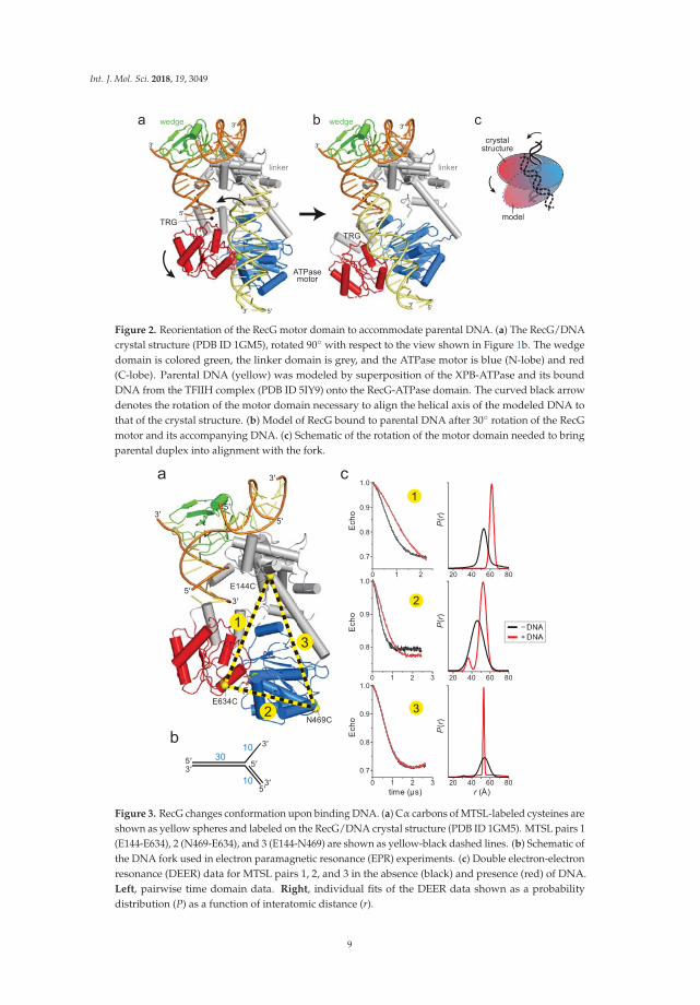
The RecG crystal structure illustrated how the wedge domain engages the branch point of aDNA fork [30], but did not address the interaction of the motor domain with DNA or its relativeconformation in the DNA-bound state because the 10 base pairs (bps) of parental duplex used inthe structure did not reach the motor domain (Figure 1b). The structure predicts that at least 25 bpsare necessary to fully engage the motor, consistent with DNase I footprinting showing that RecGprotects a significant portion of the parental DNA duplex [39]. To gain insight into how the motorand wedge domains might collaborate in a fully bound DNA complex, we constructed a model ofDNA bound to the motor domain using available structures of SF2 ATPase motors bound to dsDNA(Figure 2a, Supplemental Figure S1). Recent cryo-EM structures of chromatin remodeling complexesCHD1, SNF2, INO80 bound to nucleosomes [40–44] and of Xeroderma pigmentosum B (XPB) helicasewithin the TFIIH component of the transcription pre-initiation complex [43] showed a conserved pathof DNA across the N- and C-terminal lobes of the ATPase in a manner predicted from an archaealRad54 homolog bound to DNA in an open conformation [45]. Superposition of the DNA from thesestructures onto RecG using the motor domain as a guide shows that the modeled and crystalizedDNA duplexes are misaligned (Figure 2a). Alignment of these two DNA segments into a continuousparental duplex requires either a 25–40◦ bend in the DNA helical axis or rotation of the motor domainin which the ATPase-C lobe swings away from the wedge domain (Figure 2b,c).
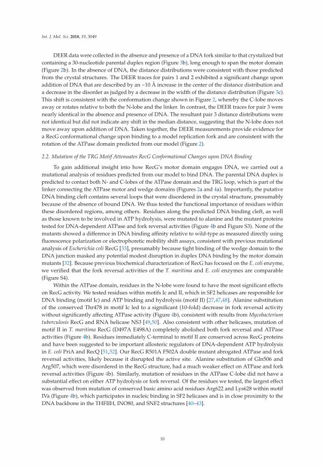
To determine if DNA binding causes a conformational change within the protein, we usedelectron paramagnetic resonance (EPR) to determine the distances between domains upon additionof DNA. The four-pulse, double electron-electron resonance (DEER) technique provides probabilitydistributions of the distances between spin-labeled residue pairs [46]. Our experimental designwas to place spin-labels in three domains—the linker that connects the wedge to the ATPasemotor, the ATPase N-lobe connected to the linker, and the ATPase C-lobe (Figure 3a). The linkerregion is predicted to be relatively inflexible based on the network of centrally located α-helices,whereas the C-lobe is likely more mobile given its peripheral location. We used the ThermotogaRecG protein for our experiments in order to correspond to the crystal structure [30]. The spinlabel (1-oxy-2,2,5,5-tetramethyl-pyrolline-3-methyl)-methanethiosulfonate (MTSL) was introducedat positions Glu144, Asn469, and Glu634, which were chosen on the basis of their surface exposedlocations. After substitution of native cysteine residues to serine, non-native cysteines were introducedpairwise to produce E144C-E634C (pair 1), N469C-E634C (pair 2), and E144C-N469C (pair 3) mutantsnecessary for thiol conjugation of MTSL (Figure 3a). We verified that neither the Cys mutations nor thespin-labels affected the DNA dependent ATPase activity of the protein (Figure S2a,b). Continuouswave (CW) spectra of each MTSL-RecG protein were consistent with surface exposed sites (Figure S2c).
8
Int. J. Mol. Sci. 2018, 19, 3049
a b cwedge
ATPasemotor
3′
3′
5′
5′
5′
5′
3′
3′
linker
TRGTRG
wedge
3′
3′
5′
5′
5′
3′
linker
crystalstructure
model
Figure 2. Reorientation of the RecG motor domain to accommodate parental DNA. (a) The RecG/DNAcrystal structure (PDB ID 1GM5), rotated 90◦ with respect to the view shown in Figure 1b. The wedgedomain is colored green, the linker domain is grey, and the ATPase motor is blue (N-lobe) and red(C-lobe). Parental DNA (yellow) was modeled by superposition of the XPB-ATPase and its boundDNA from the TFIIH complex (PDB ID 5IY9) onto the RecG-ATPase domain. The curved black arrowdenotes the rotation of the motor domain necessary to align the helical axis of the modeled DNA tothat of the crystal structure. (b) Model of RecG bound to parental DNA after 30◦ rotation of the RecGmotor and its accompanying DNA. (c) Schematic of the rotation of the motor domain needed to bringparental duplex into alignment with the fork.
2
13
5′
5′
5′
3′
E144C
E634C
N469C
3′
3′
b
a c
10
10
303′
3′5′
5′3′
5′
3
0 1 2 3
0.7
0.8
0.9
1.0
20 40 60 80time (μs) r (Å)
Ech
o
P(r
)
2
Ech
o
P(r
)
0 1 2 3
0.8
0.9
1.0
20 40 60 80
1
0 1 2
0.7
0.8
0.9
Ech
o
P(r)
1.0
20 40 60 80
Figure 3. RecG changes conformation upon binding DNA. (a) Cα carbons of MTSL-labeled cysteines areshown as yellow spheres and labeled on the RecG/DNA crystal structure (PDB ID 1GM5). MTSL pairs 1(E144-E634), 2 (N469-E634), and 3 (E144-N469) are shown as yellow-black dashed lines. (b) Schematic ofthe DNA fork used in electron paramagnetic resonance (EPR) experiments. (c) Double electron-electronresonance (DEER) data for MTSL pairs 1, 2, and 3 in the absence (black) and presence (red) of DNA.Left, pairwise time domain data. Right, individual fits of the DEER data shown as a probabilitydistribution (P) as a function of interatomic distance (r).
9
Int. J. Mol. Sci. 2018, 19, 3049
DEER data were collected in the absence and presence of a DNA fork similar to that crystalized butcontaining a 30-nucleotide parental duplex region (Figure 3b), long enough to span the motor domain(Figure 2b). In the absence of DNA, the distance distributions were consistent with those predictedfrom the crystal structures. The DEER traces for pairs 1 and 2 exhibited a significant change uponaddition of DNA that are described by an ~10 Å increase in the center of the distance distribution anda decrease in the disorder as judged by a decrease in the width of the distance distribution (Figure 3c).This shift is consistent with the conformation change shown in Figure 2, whereby the C-lobe movesaway or rotates relative to both the N-lobe and the linker. In contrast, the DEER traces for pair 3 werenearly identical in the absence and presence of DNA. The resultant pair 3 distance distributions werenot identical but did not indicate any shift in the median distance, suggesting that the N-lobe does notmove away upon addition of DNA. Taken together, the DEER measurements provide evidence fora RecG conformational change upon binding to a model replication fork and are consistent with therotation of the ATPase domain predicted from our model (Figure 2).
2.2. Mutation of the TRG Motif Attenuates RecG Conformational Changes upon DNA Binding
To gain additional insight into how RecG’s motor domain engages DNA, we carried out amutational analysis of residues predicted from our model to bind DNA. The parental DNA duplex ispredicted to contact both N- and C-lobes of the ATPase domain and the TRG loop, which is part of thelinker connecting the ATPase motor and wedge domains (Figures 2a and 4a). Importantly, the putativeDNA binding cleft contains several loops that were disordered in the crystal structure, presumablybecause of the absence of bound DNA. We thus tested the functional importance of residues withinthese disordered regions, among others. Residues along the predicted DNA binding cleft, as wellas those known to be involved in ATP hydrolysis, were mutated to alanine and the mutant proteinstested for DNA-dependent ATPase and fork reversal activities (Figure 4b and Figure S3). None of themutants showed a difference in DNA binding affinity relative to wild-type as measured directly usingfluorescence polarization or electrophoretic mobility shift assays, consistent with previous mutationalanalysis of Escherichia coli RecG [33], presumably because tight binding of the wedge domain to theDNA junction masked any potential modest disruption in duplex DNA binding by the motor domainmutants [32]. Because previous biochemical characterization of RecG has focused on the E. coli enzyme,we verified that the fork reversal activities of the T. maritima and E. coli enzymes are comparable(Figure S4).
Within the ATPase domain, residues in the N-lobe were found to have the most significant effectson RecG activity. We tested residues within motifs Ic and II, which in SF2 helicases are responsible forDNA binding (motif Ic) and ATP binding and hydrolysis (motif II) [27,47,48]. Alanine substitutionof the conserved Thr478 in motif Ic led to a significant (10-fold) decrease in fork reversal activitywithout significantly affecting ATPase activity (Figure 4b), consistent with results from Mycobacteriumtuberculosis RecG and RNA helicase NS3 [49,50]. Also consistent with other helicases, mutation ofmotif II in T. maritima RecG (D497A E498A) completely abolished both fork reversal and ATPaseactivities (Figure 4b). Residues immediately C-terminal to motif II are conserved across RecG proteinsand have been suggested to be important allosteric regulators of DNA-dependent ATP hydrolysisin E. coli PriA and RecQ [51,52]. Our RecG R501A F502A double mutant abrogated ATPase and forkreversal activities, likely because it disrupted the active site. Alanine substitution of Gln506 andArg507, which were disordered in the RecG structure, had a much weaker effect on ATPase and forkreversal activities (Figure 4b). Similarly, mutation of residues in the ATPase C-lobe did not have asubstantial effect on either ATP hydrolysis or fork reversal. Of the residues we tested, the largest effectwas observed from mutation of conserved basic amino acid residues Arg622 and Lys628 within motifIVa (Figure 4b), which participates in nucleic binding in SF2 helicases and is in close proximity to theDNA backbone in the THFIIH, INO80, and SNF2 structures [40–43].
10

Int. J. Mol. Sci. 2018, 19, 3049
Figure 4. Loops within the TRG motif are essential for DNA-dependent ATP hydrolysis and forkreversal activity. (a) Structure of the ATPase domain (blue and red) with residues lining the putativeDNA binding surface shown as Cα spheres. The TRG hairpin and loop are colored yellow. Dashedlines represent disordered regions in the crystal structure. (b) Relative DNA-dependent ATP hydrolysis(black bars) and fork reversal activities (white bars) of alanine mutants. Shading corresponds to thelocation of each mutant in the structure shown in panel a. Raw data and rates are shown in Figure S3.(c,d) DEER measurements for spin-label pairs 1 (c) and 2 (d) in the TRG loop mutant, G726A P727AG728A. Pairwise time domain data and individual fits of the DEER data are shown on the left and rightof each panel, respectively.
In contrast to the SF2 motor domain, mutation of the TRG motif had the most severe impact onRecG function. The TRG motif contains a highly conserved loop that was unstructured in the RecGstructure and that lies directly in the proposed path of DNA binding [30]. Two separate mutantsof this loop (G726A P727A G728A and F730A F731A) abrogated fork reversal and ATP hydrolysis(Figure 4b). Loss of activity by these mutants indicates that the TRG loop is important for bindingDNA during translocation, facilitating interdomain movement by the motor during the ATPase cycle,or both. Indeed, the TRG loop lies at the intersection of the two ATPase lobes and the wedge domain,directly in the proposed path of DNA and near helicase motifs III and VI, which coordinate ATPhydrolysis and translocation (motif III) and facilitate ATP binding and hydrolysis (motif VI) in otherSF2 helicases [27,48].
To test the role of the TRG loop in RecG DNA-dependent conformation changes, we usedEPR to measure interdomain distances in the dysfunctional TRG loop mutant, G726A P727AG728A. Spin labels were introduced into the mutant in the same location as the wild-type protein.We hypothesized that if the TRG loop mediates DNA binding or the DNA-induced conformationalchange observed in the wild-type protein, then addition of DNA to the mutant would not affect thedistance distributions. Indeed, the increase in spin label pair 1 distance upon addition of DNA wasreduced without the concomitant decrease in disorder compared to wild-type (Figure 4c and FigureS2d). The TRG loop mutation showed an even greater effect on spin label pair 2, from which only amodest shift in distance was observed upon addition of DNA (Figure 4c and Figure S2d). Therefore,we conclude that the loop C-terminal to the TRG motif mediates DNA-induced conformational changeswithin the motor, and likely couples motor domain dynamics to the fork-binding wedge domain todrive translocation.
3. Discussion
Coupling of an SF2 motor to a fork recognition domain is a conserved feature in the eukaryoticfork remodelers SMARCAL1, HLTF, and ZRANB3 [53–55], and thus it is important to understandhow the two domains collaborate to drive fork reversal. By extrapolation from ssDNA translocation
11

Int. J. Mol. Sci. 2018, 19, 3049
mechanisms of SF1 and SF2 helicases, the current model for dsDNA translocation by the fork andchromatin remodelers entails conversion of an open to closed conformation of ATPase lobes uponbinding DNA [44,45,56]. DNA duplex binding along the interface of the two ATPase lobes places thetracking (3′→ 5′) strand in contact with motif Ia in the ATPase-N lobe and motif IV in the ATPaseC-lobe. Consequently, ATP-induced conformational changes between the two ATPase lobes woulddrive an inchworm movement of the tracking strand and concomitant rotary motion of the duplex [57].As the fork recognition domain keeps the protein anchored to the junction [32], DNA translocationwould effectively pull the unwound template strands back into the protein, facilitating their annealingto each other and unwinding from nascent strands as they encounter the junction. This collaborationbetween motor and fork binding domains is analogous to INO80 chromatin remodeling machinery,which uses the ARP5 subunit to bind both histone and DNA in order to position the INO80 motorto pump DNA into the nucleosome [40,41]. Both mechanisms require an anchor point to grip thesubstrate to facilitate productive translocation by the motor.
Our EPR results revealed a DNA-induced movement of RecG’s ATPase-C lobe relative to thepositions of the ATPase-N lobe and the wedge domain. This motion can be modeled by a simplepivoting of the motor at the ATPase-N lobe, or a more complex rotation between the two ATPase lobes.The range of motion that we observe between RecG’s two ATPase lobes is not as dramatic as thatobserved in fluorescence resonance energy transfer studies of an archaeal homolog of Rad54, a relatedSNF2-like dsDNA translocase [56]. Although we cannot say with certainty the nature of the open andclosed conformations of the motor domain from our distance measurements, the two ATPase lobes inthe ADP-bound crystal structure are already well-positioned to accommodate dsDNA in a catalyticorientation. The motion of the motor with respect to the wedge that we observe is more striking, sinceit is clear that the relative positions of the motor and wedge in the crystal structure cannot support acontiguous parental DNA duplex without a rotation of the motor or a sharp bend in the helical axis ofthe DNA. The latter is unlikely since coupling motor activity to fork stabilization by the wedge domainwould place tension on the DNA segment between the two domains. Moreover, the position of themotor domain observed in the crystal structure is constrained by a neighboring protein molecule in thecrystal that pushes the motor closer to the wedge. Thus, our data supports a conformational transitionfrom a more compact state in the absence of DNA to a more extended state upon engaging a fork.
Our mutational analysis of the relatively unstructured DNA binding surface of the ATPase domainis consistent with and extends the previous studies showing the TRG motif to be essential for RecGfunction [33]. The previous mutational analysis focused on the helical hairpin itself, but it is the loopextending from the C-terminal end of the helical hairpin that resides in the path of the DNA and atthe intersection of the motor and wedge domains, and that is likely the mechanical element directlyresponsible for DNA translocation. It was hypothesized that an ATP-induced conformational changein the TRG helical hairpin, propagated through motif VI, would restructure the TRG loop to act asa lever or ratchet to mechanically move or stabilize the DNA in a new conformation [33]. This TRGloop is highly conserved among RecG and Mfd orthologs, with the consensus sequence G(P/A/V)GdΦΦGxxQ(S/T)G (where Φ is a hydrophobic residue). Mutation of the invariant glutamine (Q640) inE. coli RecG demonstrated that the TRG loop was essential for RecG activity in vivo [33]. We nowshow by mutation of the GPG and ΦΦ residues in the T. maritima enzyme that this loop is essential forATPase and fork reversal activities. More importantly, we found that disruption of the GPG sequencecurtailed the range of DNA-induced interdomain motion, implying that this loop region is importantfor coupling motor and wedge domains. We hypothesize, based on our DEER distance measurements,that the TRG motif loop is required to stabilize an activated conformation of the ATPase domains uponDNA binding to promote ATP hydrolysis [33], similar to the postulated role of the brace helix in thechromatin remodelers [40–42,44,58]. In those structures, the brace helix spans the two ATPase lobesand likely stabilizes a closed conformation through interaction of hydrophobic residues on the bracehelix and the ATPase N-lobe. It may be that the conserved hydrophobic residues in the TRG loop thatare essential for RecG activity may help to organize the two ATPase lobes in a similar manner.
12
Int. J. Mol. Sci. 2018, 19, 3049
4. Materials and Methods
All experiments were carried out using T. maritima RecG containing a C-terminal hexahistidinetag (TmRecG-His6). We verified that addition of the His6 tag did not affect enzyme activity (Figure S4).
4.1. Protein Purification
TmRecG-His6 was overexpressed from a pET28a+-TmrecG vector [59] in E. coli Tuner (DE3)cells at 37 ◦C for 3 h in Lysogeny broth (LB) medium supplemented with 100 μg/mL kanamycinand 500 μM isopropyl β-D-1 thiogalactopyranoside (IPTG). Cells were lysed by sonication in buffercontaining 50 mM Tris pH 7.5, 600 mM NaCl, 20% glycerol (v/v), 1 mM dithiothreitol (DTT), 1 mMphenylmethylsulfonyl fluoride, 0.5 μg/ml leupeptin, and 0.5 μg/ml aprotinin. The lysate was clarifiedby centrifugation at 50,000× g at 4 ◦C for 45 min. RecG-His6 was purified by nickel nitrilotriaceticacid (Ni-NTA) agarose affinity chromatography in buffer containing 50 mM Tris pH 7.5, 600 mMNaCl, 25 mM imidazole, 5% glycerol, and 1 mM tris(2-carboxyethyl)phosphine (TCEP) and elutedin buffer containing 50 mM Tris pH 7.5, 600 mM NaCl, 250 mM imidazole, 5% glycerol, 1 mM TCEP.RecG-His6-containing fractions were subjected to heparin sepharose chromatography using a 0.1–1 MNaCl gradient in buffer containing 50 mM Tris pH 7.5, 100 mM NaCl, and 15% glycerol.
Mutant RecG expression vectors were generated using the Q5 mutagenesis kit (New EnglandBiolabs) and sequence verified prior to use. All mutant proteins were overexpressed the same aswild-type protein. Alanine mutants were purified by Ni-NTA affinity chromatography, flash frozen,and stored at −80 ◦C in buffer containing 50 mM Tris pH 7.5, 600 mM NaCl, 250 mM imidazole, 5%glycerol (v/v), and 1 mM DTT. To prepare cysteine mutants for spin-labeling, all five native cysteinesin RecG were first mutated to serine to generate a Cys-less RecG, which was then used to generatethree separate double mutants (E144C N469C, E144C E634C, and N469C E634C). Cysteine mutantproteins were purified using Ni-NTA and heparin chromatography and stored at −80 ◦C in buffercontaining 50 mM Tris pH 7.5, 600 mM NaCl, and 10% glycerol (v/v). Spin-labeling was carried out byincubating cysteine mutants with a 20-fold molar excess of MTSL for 2 h at room temperature, followedby addition of another 20-fold molar excess of MTSL and incubation for 2 h at room temperature andthen overnight at 4 ◦C. Excess MTSL was removed using a HiTrap Sephadex G-25 desalting column(GE Healthcare, Chicago, IL, USA) in buffer containing 50 mM Tris pH 7.5, 500 mM NaCl, and 10%(v/v) glycerol.
To test the effect of the C-terminal His6-tag, we generated a cleavable pET-28a/RecG-3C-His6
construct in which the His6-tag could be removed with Rhinovirus 3C protease. Q5 mutagenesiskit (New England Biolabs, Ipswich, MA) was used to replace the sequence K776LIEVG781KLAAALE(non-native residues italicized) in the pET28a+-TmrecG vector with the 3C recognition sequenceLEVLFQGP. Proteolytic cleavage generates a 781-residue protein with I775LEVLFQ sequence at theC-terminus. RecG-3C-His6 protein was overexpressed and purified the same as TmRecG-His6.The His6-tag was removed by a 16-hr incubation with 3C protease after elution from the Ni-NTA column.
E. coli RecG was purified from a pGS772-RecG expression plasmid [21] as previouslydescribed [60], with an added heparin-sepharose purification step at the end.
4.2. EPR
Spin-labeled TmRecG-3C-His6 protein was buffer exchanged using Amicon Ultra 15 mLcentrifugal units 30 kDa MWCO (MilliporeSigma, Burlington, MA, USA) into buffer containing50 mM Tris pH 7.5, 100 mM NaCl, and 30% (w/v) glycerol. Fork DNA was prepared by annealingstrands F1/F2/F3 (Table 1) in SSC buffer (15 mM sodium citrate pH 7.0 and 150 mM NaCl). A 2-foldmolar excess of DNA was added to 25–50 μM protein and the complex flash frozen in liquid nitrogen.DEER experiments were performed at 83 K on a Bruker 580 pulsed EPR spectrometer at Q-bandfrequency (33.5 GHz) using a standard four-pulse protocol [61]. Analysis of the DEER data to determineP(r) distance distributions was carried out using homemade software running in MATLAB [62,63].
13
Int. J. Mol. Sci. 2018, 19, 3049
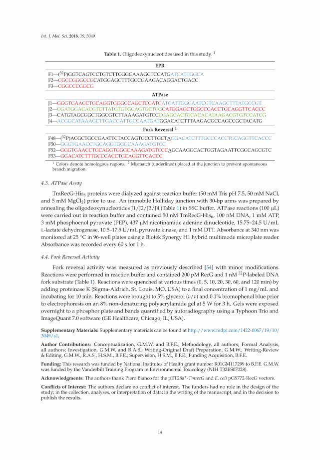
Table 1. Oligodeoxynucleotides used in this study. 1
EPR
F1—(32P)GGTCAGTCCTGTCTTCGGCAAAGCTCCATGATCATTGGCAF2—CGCCGGGCCGCATGGAGCTTTGCCGAAGACAGGACTGACCF3—CGGCCCGGCG
ATPase
J1—GGGTGAACCTGCAGGTGGGCCAGCTCCATGATCATTGGCAATCGTCAAGCTTTATGCCGTJ2—CGATGGACACGTCTTATGTGTGCAGTGCTCGCATGGAGCTGGCCCACCTGCAGGTTCACCCJ3—CATGTAGCGGCTGGCGTCTTAAAGATGTCCCGAGCACTGCACACATAAGACGTGTCCATCGJ4—ACGGCATAAAGCTTGACGATTGCCAATGATGGACATCTTTAAGACGCCAGCCGCTACATG
Fork Reversal 2
F48—(32P)ACGCTGCCGAATTCTACCAGTGCCTTGCTAGGACATCTTTGCCCACCTGCAGGTTCACCCF50—GGGTGAACCTGCAGGTGGGCAAAGATGTCCF52—GGGTGAACCTGCAGGTGGGCAAAGATGTCCCAGCAAGGCACTGGTAGAATTCGGCAGCGTCF53—GGACATCTTTGCCCACCTGCAGGTTCACCC
1 Colors denote homologous regions. 2 Mismatch (underlined) placed at the junction to prevent spontaneousbranch migration.
4.3. ATPase Assay
TmRecG-His6 proteins were dialyzed against reaction buffer (50 mM Tris pH 7.5, 50 mM NaCl,and 5 mM MgCl2) prior to use. An immobile Holliday junction with 30-bp arms was prepared byannealing the oligodeoxynucleotides J1/J2/J3/J4 (Table 1) in SSC buffer. ATPase reactions (100 μL)were carried out in reaction buffer and contained 50 nM TmRecG-His6, 100 nM DNA, 1 mM ATP,3 mM phosphoenol pyruvate (PEP), 437 μM nicotinamide adenine dinucleotide, 15.75–24.5 U/mLL-lactate dehydrogenase, 10.5–17.5 U/mL pyruvate kinase, and 1 mM DTT. Absorbance at 340 nm wasmonitored at 25 ◦C in 96-well plates using a Biotek Synergy H1 hybrid multimode microplate reader.Absorbance was recorded every 60 s for 1 h.
4.4. Fork Reversal Activity
Fork reversal activity was measured as previously described [54] with minor modifications.Reactions were performed in reaction buffer and contained 200 pM RecG and 1 nM 32P-labeled DNAfork substrate (Table 1). Reactions were quenched at various times (0, 5, 10, 20, 30, 60, and 120 min) byadding proteinase K (Sigma-Aldrich, St. Louis, MO, USA) to a final concentration of 1 mg/mL andincubating for 10 min. Reactions were brought to 5% glycerol (v/v) and 0.1% bromophenol blue priorto electrophoresis on an 8% non-denaturing polyacrylamide gel at 5 W for 3 h. Gels were exposedovernight to a phosphor plate and bands quantified by autoradiography using a Typhoon Trio andImageQuant 7.0 software (GE Healthcare, Chicago, IL, USA).
Supplementary Materials: Supplementary materials can be found at http://www.mdpi.com/1422-0067/19/10/3049/s1.
Author Contributions: Conceptualization, G.M.W. and B.F.E.; Methodology, all authors; Formal Analysis,all authors; Investigation, G.M.W. and R.A.S.; Writing-Original Draft Preparation, G.M.W.; Writing-Review& Editing, G.M.W., R.A.S., H.S.M., B.F.E.; Supervision, H.S.M., B.F.E.; Funding Acquisition, B.F.E.
Funding: This research was funded by National Institutes of Health grant number R01GM117299 to B.F.E. G.M.W.was funded by the Vanderbilt Training Program in Environmental Toxicology (NIH T32ES07028).
Acknowledgments: The authors thank Piero Bianco for the pET28a+-TmrecG and E. coli pGS772-RecG vectors.
Conflicts of Interest: The authors declare no conflict of interest. The funders had no role in the design of thestudy; in the collection, analyses, or interpretation of data; in the writing of the manuscript, and in the decision topublish the results.
14

Int. J. Mol. Sci. 2018, 19, 3049
Abbreviations
ATP adenosine 5′-triphosphateATPase adenosine triphosphataseDEER double electron-electron resonanceDTT dithiothreitolEDTA ethylenediaminetetraacetic acidEPR electron paramagnetic resonanceMTSL [1-oxy-2,2,5,5-tetramethyl-pyrolline-3-methyl]-methanethiosulfonateNTA nitrilotriacetic acidSF2 superfamily 2SRD substrate recognition domainSSC saline-sodium citrateTCEP tris(2-carboxyethyl)phosphineTRG translocation in RecG
References
1. Zeman, M.K.; Cimprich, K.A. Causes and consequences of replication stress. Nat. Cell Biol. 2014, 16, 2–9.[CrossRef] [PubMed]
2. Cortez, D. Preventing replication fork collapse to maintain genome integrity. DNA Repair (Amst) 2015, 32,149–157. [CrossRef] [PubMed]
3. Marians, K.J. Lesion Bypass and the Reactivation of Stalled Replication Forks. Annu. Rev. Biochem. 2018, 87,217–238. [CrossRef] [PubMed]
4. Berti, M.; Vindigni, A. Replication stress: Getting back on track. Nat. Struct. Mol. Biol. 2016, 23, 103–109.[CrossRef] [PubMed]
5. Fujiwara, Y.; Tatsumi, M. Replicative bypass repair of ultraviolet damage to DNA of mammalian cells:Caffeine sensitive and caffeine resistant mechanisms. Mutat. Res. 1976, 37, 91–110. [CrossRef]
6. Higgins, N.P.; Kato, K.; Strauss, B. A model for replication repair in mammalian cells. J. Mol. Biol. 1976, 101,417–425. [CrossRef]
7. Atkinson, J.; McGlynn, P. Replication fork reversal and the maintenance of genome stability. Nucleic AcidsRes. 2009, 37, 3475–3492. [CrossRef] [PubMed]
8. Neelsen, K.J.; Lopes, M. Replication fork reversal in eukaryotes: From dead end to dynamic response.Nat. Rev. Mol. Cell Biol. 2015, 16, 207–220. [CrossRef] [PubMed]
9. Lloyd, R.G.; Rudolph, C.J. 25 years on and no end in sight: A perspective on the role of RecG protein.Curr. Genet. 2016, 62, 827–840. [CrossRef] [PubMed]
10. McGlynn, P.; Lloyd, R.G. Genome stability and the processing of damaged replication forks by RecG.Trends Genet. 2002, 18, 413–419. [CrossRef]
11. Bianco, P.R. I came to a fork in the DNA and there was RecG. Prog. Biophys. Mol. Biol. 2015, 117, 166–173.[CrossRef] [PubMed]
12. Lloyd, R.G. Conjugational recombination in resolvase-deficient ruvC mutants of Escherichia coli K-12depends on recG. J. Bacteriol. 1991, 173, 5414–5418. [CrossRef] [PubMed]
13. Lloyd, R.G.; Buckman, C. Genetic analysis of the recG locus of Escherichia coli K-12 and of its role inrecombination and DNA repair. J. Bacteriol. 1991, 173, 1004–1011. [CrossRef] [PubMed]
14. Rudolph, C.J.; Upton, A.L.; Lloyd, R.G. Replication fork collisions cause pathological chromosomalamplification in cells lacking RecG DNA translocase. Mol. Microbiol. 2009, 74, 940–955. [CrossRef] [PubMed]
15. Rudolph, C.J.; Upton, A.L.; Stockum, A.; Nieduszynski, C.A.; Lloyd, R.G. Avoiding chromosome pathologywhen replication forks collide. Nature 2013, 500, 608–611. [CrossRef] [PubMed]
16. Courcelle, J.; Hanawalt, P.C. RecA-dependent recovery of arrested DNA replication forks. Annu. Rev. Genet.2003, 37, 611–646. [CrossRef] [PubMed]
17. Gregg, A.V.; McGlynn, P.; Jaktaji, R.P.; Lloyd, R.G. Direct rescue of stalled DNA replication forks via thecombined action of PriA and RecG helicase activities. Mol. Cell 2002, 9, 241–251. [CrossRef]
18. Rudolph, C.J.; Upton, A.L.; Briggs, G.S.; Lloyd, R.G. Is RecG a general guardian of the bacterial genome?DNA Repair (Amst) 2010, 9, 210–223. [CrossRef] [PubMed]
15

Int. J. Mol. Sci. 2018, 19, 3049
19. Kowalczykowski, S.C. Initiation of genetic recombination and recombination-dependent replication.Trends Biochem. Sci. 2000, 25, 156–165. [CrossRef]
20. West, S.C. Processing of recombination intermediates by the RuvABC proteins. Annu. Rev. Genet. 1997, 31,213–244. [CrossRef] [PubMed]
21. Lloyd, R.G.; Sharples, G.J. Dissociation of synthetic Holliday junctions by E. coli RecG protein. EMBO J.1993, 12, 17–22. [CrossRef] [PubMed]
22. Whitby, M.C.; Ryder, L.; Lloyd, R.G. Reverse branch migration of Holliday junctions by RecG protein:A new mechanism for resolution of intermediates in recombination and DNA repair. Cell 1993, 75, 341–350.[CrossRef]
23. Gupta, S.; Yeeles, J.T.; Marians, K.J. Regression of replication forks stalled by leading-strand template damage:I. Both RecG and RuvAB catalyze regression, but RuvC cleaves the holliday junctions formed by RecGpreferentially. J. Biol Chem 2014, 289, 28376–28387. [CrossRef] [PubMed]
24. Azeroglu, B.; Mawer, J.S.; Cockram, C.A.; White, M.A.; Hasan, A.M.; Filatenkova, M.; Leach, D.R.RecG Directs DNA Synthesis during Double-Strand Break Repair. PLoS Genet. 2016, 12, e1005799. [CrossRef][PubMed]
25. Azeroglu, B.; Leach, D.R.F. RecG controls DNA amplification at double-strand breaks and arrested replicationforks. FEBS Lett. 2017, 591, 1101–1113. [CrossRef] [PubMed]
26. Midgley-Smith, S.L.; Dimude, J.U.; Taylor, T.; Forrester, N.M.; Upton, A.L.; Lloyd, R.G.; Rudolph, C.J.Chromosomal over-replication in Escherichia coli recG cells is triggered by replication fork fusion andamplified if replichore symmetry is disturbed. Nucleic Acids Res. 2018. [CrossRef] [PubMed]
27. Fairman-Williams, M.E.; Guenther, U.P.; Jankowsky, E. SF1 and SF2 helicases: Family matters. Curr. Opin.Struct. Biol. 2010, 20, 313–324. [CrossRef] [PubMed]
28. Abd Wahab, S.; Choi, M.; Bianco, P.R. Characterization of the ATPase activity of RecG and RuvAB proteinson model fork structures reveals insight into stalled DNA replication fork repair. J. Biol. Chem. 2013, 288,26397–26409. [CrossRef] [PubMed]
29. McGlynn, P.; Lloyd, R.G. Rescue of stalled replication forks by RecG: Simultaneous translocation on theleading and lagging strand templates supports an active DNA unwinding model of fork reversal andHolliday junction formation. Proc. Natl. Acad. Sci. USA 2001, 98, 8227–8234. [CrossRef] [PubMed]
30. Singleton, M.R.; Scaife, S.; Wigley, D.B. Structural analysis of DNA replication fork reversal by RecG. Cell2001, 107, 79–89. [CrossRef]
31. Manosas, M.; Perumal, S.K.; Bianco, P.R.; Ritort, F.; Benkovic, S.J.; Croquette, V. RecG and UvsW catalyserobust DNA rewinding critical for stalled DNA replication fork rescue. Nat. Commun. 2013, 4, 2368.[CrossRef] [PubMed]
32. Briggs, G.S.; Mahdi, A.A.; Wen, Q.; Lloyd, R.G. DNA binding by the substrate specificity (wedge) domain ofRecG helicase suggests a role in processivity. J. Biol. Chem. 2005, 280, 13921–13927. [CrossRef] [PubMed]
33. Mahdi, A.A.; Briggs, G.S.; Sharples, G.J.; Wen, Q.; Lloyd, R.G. A model for dsDNA translocation revealed bya structural motif common to RecG and Mfd proteins. EMBO J. 2003, 22, 724–734. [CrossRef] [PubMed]
34. Chambers, A.L.; Smith, A.J.; Savery, N.J. A DNA translocation motif in the bacterial transcription–repaircoupling factor, Mfd. Nucleic Acids Res. 2003, 31, 6409–6418. [CrossRef] [PubMed]
35. Deaconescu, A.M.; Chambers, A.L.; Smith, A.J.; Nickels, B.E.; Hochschild, A.; Savery, N.J.; Darst, S.A.Structural basis for bacterial transcription-coupled DNA repair. Cell 2006, 124, 507–520. [CrossRef] [PubMed]
36. Park, J.S.; Marr, M.T.; Roberts, J.W. E. coli Transcription repair coupling factor (Mfd protein) rescues arrestedcomplexes by promoting forward translocation. Cell 2002, 109, 757–767. [CrossRef]
37. Deaconescu, A.M.; Savery, N.; Darst, S.A. The bacterial transcription repair coupling factor. Curr. Opin.Struct. Biol. 2007, 17, 96–102. [CrossRef] [PubMed]
38. Savery, N.J. The molecular mechanism of transcription-coupled DNA repair. Trends Microbiol. 2007, 15,326–333. [CrossRef] [PubMed]
39. Tanaka, T.; Masai, H. Stabilization of a stalled replication fork by concerted actions of two helicases. J. Biol.Chem. 2006, 281, 3484–3493. [CrossRef] [PubMed]
40. Ayala, R.; Willhoft, O.; Aramayo, R.J.; Wilkinson, M.; McCormack, E.A.; Ocloo, L.; Wigley, D.B.; Zhang, X.Structure and regulation of the human INO80-nucleosome complex. Nature 2018, 556, 391–395. [CrossRef][PubMed]
16

Int. J. Mol. Sci. 2018, 19, 3049
41. Eustermann, S.; Schall, K.; Kostrewa, D.; Lakomek, K.; Strauss, M.; Moldt, M.; Hopfner, K.P. Structural basisfor ATP-dependent chromatin remodelling by the INO80 complex. Nature 2018, 556, 386–390. [CrossRef][PubMed]
42. Liu, X.; Li, M.; Xia, X.; Li, X.; Chen, Z. Mechanism of chromatin remodelling revealed by the Snf2-nucleosomestructure. Nature 2017, 544, 440–445. [CrossRef] [PubMed]
43. He, Y.; Yan, C.; Fang, J.; Inouye, C.; Tjian, R.; Ivanov, I.; Nogales, E. Near-atomic resolution visualization ofhuman transcription promoter opening. Nature 2016, 533, 359–365. [CrossRef] [PubMed]
44. Farnung, L.; Vos, S.M.; Wigge, C.; Cramer, P. Nucleosome-Chd1 structure and implications for chromatinremodelling. Nature 2017, 550, 539–542. [CrossRef] [PubMed]
45. Durr, H.; Korner, C.; Muller, M.; Hickmann, V.; Hopfner, K.P. X-ray structures of the Sulfolobus solfataricusSWI2/SNF2 ATPase core and its complex with DNA. Cell 2005, 121, 363–373. [CrossRef] [PubMed]
46. McHaourab, H.S.; Steed, P.R.; Kazmier, K. Toward the fourth dimension of membrane protein structure:Insight into dynamics from spin-labeling EPR spectroscopy. Structure 2011, 19, 1549–1561. [CrossRef][PubMed]
47. Singleton, M.R.; Dillingham, M.S.; Wigley, D.B. Structure and mechanism of helicases and nucleic acidtranslocases. Annu. Rev. Biochem. 2007, 76, 23–50. [CrossRef] [PubMed]
48. Pyle, A.M. Translocation and unwinding mechanisms of RNA and DNA helicases. Annu. Rev. Biophys. 2008,37, 317–336. [CrossRef] [PubMed]
49. Zegeye, E.D.; Balasingham, S.V.; Laerdahl, J.K.; Homberset, H.; Kristiansen, P.E.; Tonjum, T. Effects ofconserved residues and naturally occurring mutations on Mycobacterium tuberculosis RecG helicase activity.Microbiology 2014, 160, 217–227. [CrossRef] [PubMed]
50. Lin, C.; Kim, J.L. Structure-based mutagenesis study of hepatitis C virus NS3 helicase. J. Virol. 1999, 73,8798–8807. [PubMed]
51. Windgassen, T.A.; Keck, J.L. An aromatic-rich loop couples DNA binding and ATP hydrolysis in the PriADNA helicase. Nucleic Acids Res. 2016, 44, 9745–9757. [CrossRef] [PubMed]
52. Zittel, M.C.; Keck, J.L. Coupling DNA-binding and ATP hydrolysis in Escherichia coli RecQ: Role of a highlyconserved aromatic-rich sequence. Nucleic Acids Res. 2005, 33, 6982–6991. [CrossRef] [PubMed]
53. Poole, L.A.; Cortez, D. Functions of SMARCAL1, ZRANB3, and HLTF in maintaining genome stability.Crit. Rev. Biochem. Mol. Biol. 2017, 52, 696–714. [CrossRef] [PubMed]
54. Mason, A.C.; Rambo, R.P.; Greer, B.; Pritchett, M.; Tainer, J.A.; Cortez, D.; Eichman, B.F. A structure-specificnucleic acid-binding domain conserved among DNA repair proteins. Proc. Natl. Acad. Sci. USA 2014, 111,7618–7623. [CrossRef] [PubMed]
55. Kile, A.C.; Chavez, D.A.; Bacal, J.; Eldirany, S.; Korzhnev, D.M.; Bezsonova, I.; Eichman, B.F.; Cimprich, K.A.HLTF’s Ancient HIRAN Domain Binds 3′ DNA Ends to Drive Replication Fork Reversal. Mol. Cell. 2015, 58,1090–1100. [CrossRef] [PubMed]
56. Lewis, R.; Durr, H.; Hopfner, K.P.; Michaelis, J. Conformational changes of a Swi2/Snf2 ATPase during itsmechano-chemical cycle. Nucleic Acids Res. 2008, 36, 1881–1890. [CrossRef] [PubMed]
57. Hopfner, K.P.; Michaelis, J. Mechanisms of nucleic acid translocases: Lessons from structural biology andsingle-molecule biophysics. Curr. Opin. Struct. Biol. 2007, 17, 87–95. [CrossRef] [PubMed]
58. Yan, L.; Wang, L.; Tian, Y.; Xia, X.; Chen, Z. Structure and regulation of the chromatin remodeller ISWI.Nature 2016, 540, 466–469. [CrossRef] [PubMed]
59. Bianco, P.R.; Pottinger, S.; Tan, H.Y.; Nguyenduc, T.; Rex, K.; Varshney, U. The IDL of E. coli SSB links ssDNAand protein binding by mediating protein-protein interactions. Protein Sci. 2017, 26, 227–241. [CrossRef][PubMed]
60. Betous, R.; Couch, F.B.; Mason, A.C.; Eichman, B.F.; Manosas, M.; Cortez, D. Substrate-selective repair andrestart of replication forks by DNA translocases. Cell. Rep. 2013, 3, 1958–1969. [CrossRef] [PubMed]
61. Jeschke, G. DEER distance measurements on proteins. Annu. Rev. Phys. Chem. 2012, 63, 419–446. [CrossRef][PubMed]
17

Int. J. Mol. Sci. 2018, 19, 3049
62. Mishra, S.; Verhalen, B.; Stein, R.A.; Wen, P.C.; Tajkhorshid, E.; McHaourab, H.S. Conformational dynamicsof the nucleotide binding domains and the power stroke of a heterodimeric ABC transporter. Elife 2014, 3,e02740. [CrossRef] [PubMed]
63. Stein, R.A.; Beth, A.H.; Hustedt, E.J. A Straightforward Approach to the Analysis of Double Electron-ElectronResonance Data. Methods Enzymol. 2015, 563, 531–567. [CrossRef] [PubMed]
© 2018 by the authors. Licensee MDPI, Basel, Switzerland. This article is an open accessarticle distributed under the terms and conditions of the Creative Commons Attribution(CC BY) license (http://creativecommons.org/licenses/by/4.0/).
18

International Journal of
Molecular Sciences
Article
Impact of Mutagens on DNA Replication inBarley Chromosomes
Jolanta Kwasniewska *,†, Karolina Zubrzycka † and Arita Kus
Department of Plant Anatomy and Cytology, University of Silesia, Jagiellonska 28, 40-032 Katowice, Poland;[email protected] (K.Z.); [email protected] (A.K.)* Correspondence: [email protected]; Tel.: +48-32-2009-468† These authors contributed equally to this work.
Received: 13 February 2018; Accepted: 31 March 2018; Published: 3 April 2018
Abstract: Replication errors that are caused by mutagens are critical for living cells. The aim of thestudy was to analyze the distribution of a DNA replication pattern on chromosomes of the H. vulgare‘Start’ variety using pulse 5-ethynyl-2′-deoxyuridine (EdU) labeling, as well as its relationship tothe DNA damage that is induced by mutagenic treatment with maleic hydrazide (MH) and γ ray.To the best of our knowledge, this is the first example of a study of the effects of mutagens on theDNA replication pattern in chromosomes, as well as the first to use EdU labeling for these purposes.The duration of the cell cycle of the Hordeum vulgare ‘Start’ variety was estimated for the first time,as well as the influence of MH and γ ray on it. The distribution of the signals of DNA replicationalong the chromosomes revealed relationships between DNA replication, the chromatin structure,and DNA damage. MH has a stronger impact on replication than γ ray. Application of EdU seems tobe promising for precise analyses of cell cycle disturbances in the future, especially in plant specieswith small genomes.
Keywords: barley; chromosome; DNA replication pattern; EdU; mutagens
1. Introduction
Data regarding the effects of mutagens on plant nuclear genomes and DNA replication are ofgreat importance. The spatiotemporal patterns of DNA replication in nuclei were recently characterizedin detail in control cells [1], as well as in relation to DNA damage and mutagenesis [2] usinga quantitative analysis. However, to date there is no similar data on the effects of mutagens onthe pattern of DNA replication on chromosomes. Analyses of the distribution of the signals of DNAreplication on the chromosomes can be more informative when exploring the relationships betweenDNA replication, the chromatin structure, and DNA damage than studies using non-dividing cells.
Until now, the localisation of replicated chromatin was only possible usingbromodeoxyuridine (BrdU). One of the disadvantages of using BrdU is degradation of thechromatin structure during denaturation step, which is especially inconvenient in the context ofan analysis of DNA damage during mutagenesis. The relatively large size of the detection sitescaused by the need to use specific antibodies to detect BrdU is an unfavorable feature of an analysisof DNA replication sites, especially in the case of an analysis of the signals in chromosomes.Currently, the “click” reaction using 5-ethynyl-2′-deoxyuridine (EdU) [3,4] is commonly used. Its goodpreservation of chromatin and high resolution make this technique useful in a detailed analysis of theeffects of mutagens on the S-phase [2].
In this study, we present the distribution of the DNA replication pattern on chromosomes usingpulse EdU labeling and analyze its relationship with the DNA damage that is induced by mutagenictreatment with maleic hydrazide (MH) and γ ray. To the best of our knowledge, this is the first example
Int. J. Mol. Sci. 2018, 19, 1070; doi:10.3390/ijms19041070 www.mdpi.com/journal/ijms19

Int. J. Mol. Sci. 2018, 19, 1070
of a study of the effects of mutagens on the DNA replication pattern in chromosomes, as well as thefirst to use EdU labeling for these purposes.
We used barley (Hordeum vulgare ‘Start’ variety, 2n = 14) as the model plant species. Barley, whichis characterized by relatively large chromosomes and a specific heterochromatin distribution [5,6],is a convenient species for an analysis of the distribution of the DNA replication pattern alongthe chromosomes, as well as in the context of mutagenesis. Barley is regarded to be a modelspecies in analyses of the cytogenetic effects of mutagens, especially due to its chromosome size.Chromosome rearrangements [7,8], as well as disturbances of the cell cycle [2] after mutagenictreatment, have previously been shown in barley cells. In our study, the duration of the cell cycleof the Hordeum vulgare ‘Start’ variety was estimated, as well as the influence of MH and γ ray on it,by applying the EdU method.
2. Results
The cells that passed through the S-phase during EdU incorporation were characterized by thepresence of green Alexa Fluor 488 replication signals on the chromosomes (Figure 1A,A‘). Cells withno replication signals on the chromosomes were also observed (Figure 1B,B‘), which indicated thatthey were not in the S-phase during EdU incorporation.
Figure 1. Barley metaphase cells from the control (untreated) roots with green Alexa Fluor 488replication signals (A,A‘) and with no signals (B,B‘). (A,B) DAPI staining, all chromosomes stained.(A‘,B‘) results of 5-ethynyl-2′-deoxyuridine (EdU) incorporation and detection with Alexa Fluor488 azide. Bars represent 10 μm.
20
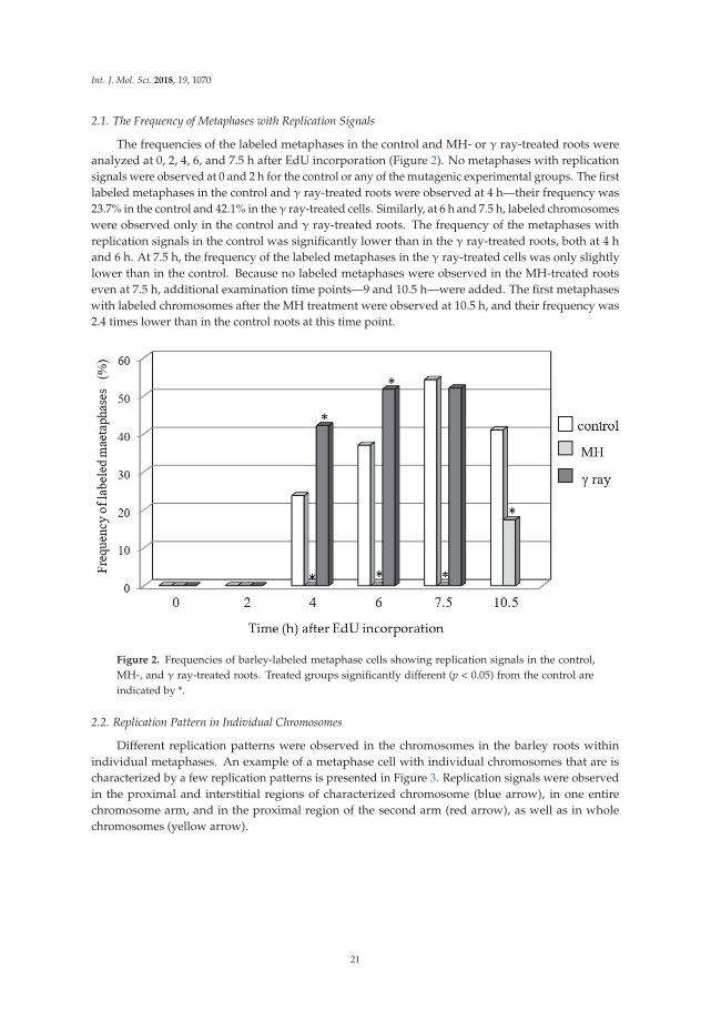
Int. J. Mol. Sci. 2018, 19, 1070
2.1. The Frequency of Metaphases with Replication Signals
The frequencies of the labeled metaphases in the control and MH- or γ ray-treated roots wereanalyzed at 0, 2, 4, 6, and 7.5 h after EdU incorporation (Figure 2). No metaphases with replicationsignals were observed at 0 and 2 h for the control or any of the mutagenic experimental groups. The firstlabeled metaphases in the control and γ ray-treated roots were observed at 4 h—their frequency was23.7% in the control and 42.1% in the γ ray-treated cells. Similarly, at 6 h and 7.5 h, labeled chromosomeswere observed only in the control and γ ray-treated roots. The frequency of the metaphases withreplication signals in the control was significantly lower than in the γ ray-treated roots, both at 4 hand 6 h. At 7.5 h, the frequency of the labeled metaphases in the γ ray-treated cells was only slightlylower than in the control. Because no labeled metaphases were observed in the MH-treated rootseven at 7.5 h, additional examination time points—9 and 10.5 h—were added. The first metaphaseswith labeled chromosomes after the MH treatment were observed at 10.5 h, and their frequency was2.4 times lower than in the control roots at this time point.
Figure 2. Frequencies of barley-labeled metaphase cells showing replication signals in the control,MH-, and γ ray-treated roots. Treated groups significantly different (p < 0.05) from the control areindicated by *.
2.2. Replication Pattern in Individual Chromosomes
Different replication patterns were observed in the chromosomes in the barley roots withinindividual metaphases. An example of a metaphase cell with individual chromosomes that are ischaracterized by a few replication patterns is presented in Figure 3. Replication signals were observedin the proximal and interstitial regions of characterized chromosome (blue arrow), in one entirechromosome arm, and in the proximal region of the second arm (red arrow), as well as in wholechromosomes (yellow arrow).
21

Int. J. Mol. Sci. 2018, 19, 1070
Figure 3. Different replication patterns of the barley chromosome within one metaphase cell.Replication signals were observed in the proximal and interstitial regions of the chromosome(blue arrow), in one entire chromosome arm, and in the proximal region of the second arm (red arrow)and in whole chromosomes (yellow arrow). (A) DAPI staining. (B) results of 5-ethynyl-2′-deoxyuridine(EdU) incorporation and detection with Alexa Fluor 488 azide. Bars represent 10 μm.
2.3. Chromosome Replication Pattern in the Control and Mutagen-Treated Roots
The differences in the chromosome replication pattern for a particular experimental group and atspecific time points were observed. Figures 4–6 showed the replication patterns in the chromosomesin the control, γ ray-, and MH-treated roots at specific time points: 0, 2, 4, 6, 7.5, and 10.5 h.Chromosomes with different replication patterns and their corresponding schematic presentationare shown. Only one morphological type of chromosome is presented in order to simplify the schemes.
In the control at 4 h, large replication signals were observed in the chromosome centromericregions and small signals were also observed in the distal and interstitial regions (Figure 4).These chromosome regions are replicated in the late S-phase, passed the G2-phase as a first,and thus can be observed in the first labeled metaphases. At 6 h, five more chromosomereplication patterns were observed than at 4 h. Chromosomes that were completely labeled wereobserved most often. Another pattern was characterized by replication signals that covered onechromosome arm and the pericentromeric region of the second arm, sometimes with additionaldistal signal(s). Chromosomes that had small replication signals in the distal and centromeric regionswere also observed. Only two new replication patterns were observed at 7.5 h—both with signalsin the distal regions, which is characteristic for early S-phase cells. In addition to the new typesof signals, the most-frequent replication pattern in this hour was also fully labeled chromosomes.The chromosome labeling that was characteristic at 6 and 7.5 h involved the regions that had beenreplicated in the early, middle, and late S-phase. Generally, at 10.5 h, the same chromosome replicationpatterns were observed as at 7.5 h. The most frequently appearing pattern of replication at thistime was characterised by the signals in the distal parts. However, at 10.5 h, new chromosomereplication patterns were additionally observed—with only one sister chromatid labeled withinan individual chromosome. The other types of patterns that were observed at 10.5 h were chromosomeswith both sister chromatids labeled, although each of them was labeled in a different part.
22

Int. J. Mol. Sci. 2018, 19, 1070
Figure 4. Types of replication patterns in barley chromosomes in the control roots at 4 h (A), 6 h (B),7.5 h (C), and 10.5 h (D) after EdU incorporation. Chromosomes with different replication patterns andtheir corresponding schematic presentation are shown. Only one morphological type of chromosomeis presented in order to simplify the scheme.
After treatment with maleic hydrazide, only six replication patterns were observed.Figure 5 shows the different replication patterns in the MH-treated root at 10.5 h. No replicationsignals were observed at the earlier time points. Fully labeled chromosomes were the mostfrequently observed.
23

Int. J. Mol. Sci. 2018, 19, 1070
Figure 5. Types of replication patterns in the barley chromosomes in MH-treated roots at 10.5 hafter EdU incorporation. Chromosomes with different replication patterns and their correspondingschematic presentation are shown. Only one morphological type of chromosome is presented in orderto simplify the scheme.
The same thirteen replication patterns were observed in the γ ray-treated roots as in thecontrol cells (Figure 6). Basically, the same patterns of replication dominated at given hours asin the control cells. However, differences in their occurrence at specific time points in the controland γ ray-treated roots were observed. Interestingly, the labeling of whole chromosomes in thecontrol was observed at 6 h, whereas in the γ ray-treated roots, it was already observed at 4 h.Similarly, the chromosomes with labeling in the terminal regions of a chromosome were observedat 6 h in the γ ray-treated roots, whereas in the control, they were observed at 7.5 h. Additionally,the chromosomes with Alexa Fluor 488 fluorescence in large bands in the pericentromeric regions wereobserved at 4 h and 6 h in the control and at 4, 6, and 7.5 h in the γ ray-treated roots.
Figure 6. Cont.
24

Int. J. Mol. Sci. 2018, 19, 1070
Figure 6. Types of replication patterns in the barley chromosomes in the γ ray-treated roots at 4 h (A),6 h (B), and 7.5 h (C) after EdU incorporation. Chromosomes with different replication patterns andtheir corresponding schematic presentation are shown. Only one morphological type of chromosomeis presented in order to simplify the scheme.
In the present study, the application of 5-ethynyl-2′-deoxyuridine (EdU) incorporation anddetection allowed differences in the replication pattern at different time points after EdU incorporationin all of the experimental groups to be determined. Figure 7 presents a comparison of the replicationpatterns in the barley chromosomes in the control, and MH- and γ ray-treated roots, at different timepoints after EdU incorporation.
25

Int. J. Mol. Sci. 2018, 19, 1070
Fig
ure
7.
Com
pari
son
ofth
ere
plic
atio
npa
tter
nsin
the
barl
eych
rom
osom
esin
the
cont
rol,
and
MH
-and
γra
y-tr
eate
dro
ots,
and
thei
roc
curr
ence
atdi
ffer
entt
ime
poin
tsaf
ter
EdU
inco
rpor
atio
n.“+
”m
eans
the
pres
ence
ofpa
rtic
ular
DN
Are
plic
atio
npa
tter
n,“−
”m
eans
the
lack
ofpa
rtic
ular
DN
Are
plic
atio
npa
tter
n.
26

Int. J. Mol. Sci. 2018, 19, 1070
3. Discussion
In present study we analyzed the distribution of a DNA replication pattern on theH. vulgare ‘Start’ variety chromosomes, as well as its relationship to the DNA damage, usingEdU method. Different replication patterns were observed in the chromosomes in the barleyroots within individual metaphases. This may be due to differences in the DNA packing ofindividual chromosomes. It is well known that euchromatin and heterochromatin regions replicateat different times during the S-phase. Individual barley chromosomes are characterized bya different localisation of constitutive heterochromatin, as was demonstrated by the Giemsa C-bandingtechnique [9]. Therefore, the chromosomes belonging to one metaphase plate may have a differentDNA replication pattern. Due to results, we can state that DNA replication in barley chromosomesbegins in the terminal chromosome regions (early S-phase), after which its pattern is observed in wholechromosomes, and at the end—in the centromeric regions (late S-phase). These analyses indirectlyprovide information about the localisation of euchromatin and heterochromatin in the chromosomesof the H. vulgare ‘Start’ variety. Since it is known that DNA replication starts in euchromatin andthen continues in the heterochromatin regions, this implies the presence of transcriptionally activegenes in the terminal chromosome regions and the inactive heterochromatin in the centromeric regions.A similar localisation of euchromatin and heterochromatin has previously been shown using the BrdUincorporation and detection methods, and fluorescence, in situ hybridisation with centromeric andtelomeric probes [10].
Thirteen types of replication patterns were distinguished in the control barley chromosomes withEdU incorporation and detection, whereas only five patterns had previously been observed using BrdUincorporation and detection [11]. This is probably due to the possibility of discovering small signalswith EdU method by using a small size detection azide and eliminating the denaturation step, which isnecessary for detection of BrdU. For example, the chromosomes with just centromeric signals wereobserved using BrdU, whereas patterns with centromeric signals, as well as terminal, or terminal andinterstitial, signals, were observed using the EdU method. The accuracy of this method allowed one tonotice the differences between the occurrence times of individual replication patterns in control andtreated cells. This can prove that EdU can potentially be applied to studying the effects of mutagenson cell cycle disturbances, especially in plant species that are characterized by small chromosomes.
Beside the typical replication signals comprising two chromatids, specific new chromosomereplication patterns were observed in control at 10.5 h—with only one sister chromatid labeledwithin an individual chromosome. This labeling pattern was characteristic for the cells in whichthe DNA replication occurred twice—first, the DNA synthesis in the presence of EdU, and thesecond, without EdU. Other new patterns observed at this time were chromosomes with bothchromatids labeled, although each of them in different localisation. This is characteristic of thesister chromatid exchange (SCE), which is commonly known in plants, as well as in animals andhumans. The occurrence of SCEs was first demonstrated using autoradiography, and later a procedurethat had a much greater resolution using BrdU was introduced [12]. It is known that BrdU itself inducesthe SCEs due to its mutagenic effect. Until now, there is no data on the possible application of EdU inthe SCE method and, consequently, on its effects on the induction of sister chromatid exchanges.
Additionally to the main aim, the results of this study provided new data about the duration ofthe G2-phase and the cell cycle of the H. vulgare ‘Start’ variety cells. The first labeled metaphases in thecontrol roots were observed at 4 h after the incorporation of EdU, while at 2 h after the incorporationof EdU no labeling has been observed within the chromosomes. This indicates that the duration ofthe G2-phase is between 2 h and 4 h. The replication pattern with labeling in only one chromatid ofthe chromosome was observed at 10.5 h. This proves that during 10.5 h, in addition to the G2-phase,there was also a complete next cell cycle without the presence of thymidine analogs. Considering thatthe length of the G2 phase is more than 2 h, it could be concluded that the duration of the cell cyclein Hordeum vulgare ‘Start’ variety is at most 8.5 h. It should also be emphasized that the pattern ofreplication with labeling in only one chromatid within the chromosome could also be observed in
27

Int. J. Mol. Sci. 2018, 19, 1070
the earlier hours after incorporation of EdU—between 7.5 h and 10.5 h. It is also possible that theduration of the G2 phase may be longer than 2 h. In both cases, this would mean that the durationof the cell cycle in this variety of barley can be even shorter than 8.5 h. The experiments for the‘Start’ variety were planned according to the mean duration of the cell cycle of other varieties ofbarley, e.g., ‘Brage’—10.4 h [13], ‘Sultan’—12.4 h, ‘Maris Otter’—12 h [14], and ‘Amethyst’—9.2 h [15].The results of this study indirectly prove that the duration of the cell cycle of the H. vulgare ‘Start’variety is definitely shorter than in the previously described varieties of this species.
The analyses showed the differences in the chromosome replication pattern for a particularexperimental group and at specific time points. After treatment with maleic hydrazide, only sixreplication patterns were observed. First chromosomes with replication patterns in the MH-treatedroots were observed only at 10.5 h. This may be due to the mechanism of MH action, namely,its influence on the synthesis of the nucleic acids and enzymes that are involved in the mitoticspindle [16,17]. The mitotic activity can even be totally stopped after MH treatment [18]. The patternsof replication that were observed after MH treatment were characteristic of the regions that have beenreplicated in the early and middle S-phase, even though the patterns for the late S-phase should beobserved first. This may indicate that MH led to a complete cell cycle arrest in the cells that were inthe late S-phase during the incorporation of EdU. After treatment with MH, the observed patternsdiffered considerably from patterns that were observed at the same time point in the control cells.No replication patterns with signals involving one sister chromatid have been observed, thus excludinga second DNA synthesis without the presence of EdU. MH is a clastogenic and mutagenic agent thatmay cause the S-phase to be extended. We found that the first labeled metaphases in the MH-treatedcells were observed at 10.5 h while in the control cells already at 4 h. This may be due to both theextension of the S-phase or a delayed G2/M transition. For the first time, comparing the time ofappearance of the first labeled metaphases in control and treated material, we can precisely evaluatethat after MH treatment the duration of cell transitions from the S-phase of the cell cycle to mitosiswas extended for about 6.5 h.
Differences at specific time points in the control and γ ray-treated roots were also observed.The labeling of whole chromosomes in the control was observed at 6 h, whereas in the γ ray-treatedroots, it was already observed at 4 h. Similarly, the chromosomes with labeling in the terminal regionsof a chromosome were observed at 6 h in the γ ray-treated roots, whereas in the control, they wereobserved at 7.5 h. It is known that γ ray acts during the G1, S, and G2 cell cycle phases. We concludethat γ ray can lead to a shortening of the S-phase or acceleration of the G2/M transition, which is judgedby the presence of the replication patterns that are characteristic for the early and middle S-phaseearlier than in the control cells. This physical mutagen may also have the opposite effect—extendingthe S-phase of the cell cycle or G2/M transition, as was evidenced by the presence of replicationpatterns that are characteristic for the late S-phase longer after the end of the EdU incorporation thanin the control cells.
Summarizing, differences in the temporal distribution of the replication patterns between thecontrol, γ ray-, and MH-treated roots were found. Slight differences were observed regarding thereplication patterns in the control and γ ray-treated roots, while differences in replication patterns inthe control and MH-treated cells were more significant. The results obtained in this work are consistentwith those previously obtained by Kwasniewska et al. [2] during an analysis of the replication processin the barley nuclei. MH has a stronger effect on DNA replication than γ radiation. It was demonstratedthat treatment with MH and γ ray did not change the characteristic S-phase patterns in the nuclei;however, the frequencies of the S-phase labeled cells after mutagenic treatment were different than inthe control cells. The results of this study on the pattern of replication in barley chromosomes alsoconfirm that no new replication patterns are observed after mutagenic treatment. However, differenceswere found in the temporal distribution of the replication patterns between the control, γ ray-,and MH-treated roots, as well as differences in the frequency of the labeled metaphases. Moreover,previous studies on the replication patterns in barley cells have also shown that the frequencies of
28
Int. J. Mol. Sci. 2018, 19, 1070
EdU-labeled cell nuclei in the early, middle, and late S-phase were different in the control cells andcells that had been treated with mutagens. After treatment with MH, a significant increase has beenobserved in the frequency of labeled cells in the middle S-phase. This may indicate an extension ofthe S-phase of the cell cycle after treatment with this chemical mutagen, as is also confirmed by thedata presented in this paper. After treatment with γ ray, in turn, a significant increase in the frequencyof labeled cells in the late S-phase has been observed before, which confirms that γ ray can extendthe late S-phase in barley cells. This is followed by the observation that the pattern of replication inchromosomes that are characteristic for late S-phase phase occurs longer after the incorporation ofEdU than in the control cells.
4. Materials and Methods
4.1. Mutagenic Treatment
Seeds of the barley (Hordeum vulgare, 2n = 14) ‘Start’ variety were used as the plant material.Maleic acid hydrazide dissolved in water (4 mM MH; Sigma-Aldrich, St. Louis, MO, USA, CAS 123-3301)and a gamma ray (175 Gy) were used for the mutagenic treatment. The mutagen doses used in the studyhad been applied in previous experiments in which their cytogenetic effects were well characterized [7,8].Before chemical treatment, the barley seeds were pre-soaked in distilled water for 8 h and then treated withMH for 3 h. Two treatment experiments using MH were performed. After the treatment, the seeds werewashed three times in distilled water and then germinated in Petri dishes lined with moist filter paper at21 ◦C in the dark for 3 days. Our previous findings showed that the cytogenetic effect of MH treatmentis observed in the roots of three-day seedlings [7]. The irradiation was performed at the InternationalAtomic Energy Agency, Seibersdorf Laboratory, Austria in 2015. For about half a year, the seeds had beenkept in a refrigerator. Keeping the seeds in the 4 ◦C is commonly practiced (also in mutagenesis studies).Additionally we ourselves confirmed, in the number of cytological analyses, that during this storage thecytogenetic effect of irradiation still persisted and did not diminish. We observed numerous breaks in thechromosomes and the formation of dicentric chromosomes, which is one of the characteristic effects ofthe mutagenic effect of gamma radiation (these results are not presented here). Similar storage time hasbeen used in our previous experiments [8]. After irradiation, the seeds were pre-soaked in distilled waterfor 8 h and germinated in Petri dishes at 21 ◦C in the dark. Two experiments were performed for eachmutagenic treatment.
4.2. EdU Incorporation and Detection
The incorporation and detection of 5-ethynyl-2′-deoxyuridine (EdU; Click-iT EdU Imaging KitsAlexa Fluor 488, Invitrogen, Carlsbad, CA, USA) were applied according to the manufacturer’sprocedure with minor modifications. The 3-day barley seedlings were incubated for 30 min in thedark in a 10 mM EdU solution. The seedlings were rinsed in distilled water 2 × 5 min and fixed inethanol:glacial acetic acid (3:1) for 2 h at room temperature (RT) and 2 h at 4 ◦C at 0, 2, 4, 6, and 7.5 hafter EdU incorporation. Additional fixation time points—9 h and 10.5 h—were applied for the controland MH-treated roots. The times for fixation and examination of replication pattern after the end ofEdU incorporation were selected according Kakeda and Yamagata [11] with modifications. The rootsof the seedlings were used as the source of the meristems for the investigations. For chromosomepreparation, the material was washed with a 0.01 mM sodium citrate buffer (pH 4.8) for 30 min anddigested with 2% cellulase (w/v, Onozuka, Serva, Heidelberg, Germany) and 20% pectinase (v/v,Sigma-Aldrich) for 2 h at 37 ◦C. After digestion, the material was washed with a sodium citrate bufferfor 30 min. Squash preparations were made in a drop of 45% acetic acid. After freezing and removingthe coverslips, the slides were dried. Prior to EdU detection, the slides were permeabilized with 0.5%Triton X-100 for 20 min and then washed in PBS at RT. The slides were incubated for 30 min at RT inan EdU reaction cocktail (Click-iT EdU Imaging Kits Alexa Fluor 488, Invitrogen), which was preparedaccording the manufacturer’s procedure. For one sample reaction, the following components were
29

Int. J. Mol. Sci. 2018, 19, 1070
added: 43 μL of a 1× Click-iT reaction buffer, 2 μL of CuSO4 (Component E, 100 mM), 0.12 μL AlexaFluor 488 azide (Component B), and a 5 μL reaction buffer additive (Component F). After 2 × 5 minwashes, the slides were stained with 2 μg/mL DAPI (Sigma-Aldrich), washed with PBS, and mountedin a Vectashield medium (Vector, Burlingame, CA, USA).
4.3. Analysis
Preparations were examined using a Zeiss Axio Imager.Z.2 wide-field fluorescence microscope(Zeiss, Oberkochen, Germany) equipped with an AxioCam Mrm monochromatic camera(Zeiss, Oberkochen, Germany). For the analyses of the distribution of the EdU pattern on the barleychromosomes, images were captured and processed using Adobe Photoshop 4.0 (San Jose, CA, USA).The frequencies of the metaphases with Alexa Fluor 488 signals were calculated. For each experimentalgroup (control, MH, and γ ray) and individual time point, 100 metaphases were evaluated. The numberof analyzed plants was different depending on the experimental group. Due to significant decreasein the mitotic index in roots after MH treatment, about 10 plants have been used for each hourafter the incorporation of EdU. For control and gamma-irradiated samples, not less than 5 plantswere evaluated.
The significance of differences between control and treated groups was evaluated with Student’st-test (p < 0.05).
5. Conclusions
This work demonstrates the usefulness of the EdU method in a detailed analysis of the replicationpatterns in barley. Such a high resolution of the EdU method indicates that it can be used formore precise analyses of cell cycle disturbances, not only for plant species with large chromosomes,but especially for those with small chromosomes. Analyses of the distribution of the signals of DNAreplication on the chromosomes revealed relationships between DNA replication, the chromatinstructure, and DNA damage, and thus they are more informative than studies using non-dividing cells.We proved that MH has a stronger impact on replication than γ ray by its action of extending theduration of cell transitions from the S-phase of the cell cycle to mitosis by 6.5 h. Data regarding theduration of the cell cycle in the H. vulgare ‘Start’ variety are also presented for first time.
Author Contributions: Jolanta Kwasniewska conceived and designed the experiments; Jolanta Kwasniewska,Karolina Zubrzycka, and Arita Kus performed the experiments; Karolina Zubrzycka analyzed the data;Jolanta Kwasniewska wrote the paper.
Conflicts of Interest: The authors declare no conflict of interest.
References
1. Bass, H.W.; Hoffman, G.G.; Lee, T.J.; Wear, E.E.; Joseph, S.R.; Allen, G.C.; Hanley-Bowdoin, L.; Thompson, W.F.Defining multiple, distinct, and shared spatiotemporal patterns of DNA replication and endoreduplicationfrom 3D image analysis of developing maize (Zea mays L.) root tip nuclei. Plant Mol. Biol. 2015, 89, 339–351.[CrossRef] [PubMed]
2. Kwasniewska, J.; Kus, A.; Swoboda, M.; Braszewska-Zalewska, A. DNA replication after mutagenictreatment in Hordeum vulgare. Mutat. Res. 2016, 812, 20–28. [CrossRef] [PubMed]
3. Buck, S.B.; Bradford, J.; Gee, K.R.; Agnew, B.J.; Clarke, S.T.; Salic, A. Detection of S-phase cell cycleprogression using 5-ethynyl-2′-deoxyuridine incorporation with click chemistry, an alternative to using5-bromo-2’-deoxyuridine antibodies. BioTechniques 2008, 44, 927–929. [CrossRef] [PubMed]
4. Cavanagh, B.L.; Walker, T.; Norazit, A.; Meedeiniya, A.C.B. Thymidine analogues for tracking DNA synthesis.Molecules 2011, 16, 7980–7993. [CrossRef] [PubMed]
5. Santos, A.P.; Shaw, P. Interphase chromosomes and the Rabl configuration: Does genome size matter.J. Microsc. 2004, 214, 201–206. [CrossRef] [PubMed]
6. Cowan, C.R.; Carlton, P.M.; Cande, W.Z. The polar arrangement of telomeres in interphase and meiosis.Rabl organization and the bouquet. Plant Physiol. 2001, 125, 532–538. [CrossRef] [PubMed]
30

Int. J. Mol. Sci. 2018, 19, 1070
7. Juchimiuk, J.; Hering, B.; Maluszynska, J. Multicolour FISH in an analysis of chromosome aberrationsinduced by N-nitroso-N-methylurea and maleic hydrazide in barley cells. J. Appl. Genet. 2007, 48, 99–106.[CrossRef] [PubMed]
8. Juchimiuk-Kwasniewska, J.; Brodziak, L.; Maluszynska, J. FISH in analysis of gamma ray-inducedmicronuclei formation in barley. J. Appl. Genet. 2011, 52, 23–29. [CrossRef] [PubMed]
9. Linde-Laursen, I. Giemsa C-banding of barley chromosomes. I. Banding pattern polymorphism. Hereditas1978, 88, 55–64. [CrossRef]
10. Jasencakova, Z.; Meister, A.; Schubert, I. Chromatin organization and its relation to replication and histoneacetylation during the cell cycle in barley. Chromosoma 2001, 110, 83–92. [CrossRef] [PubMed]
11. Kakeda, K.; Yamagata, H. Immunological analysis of chromosome replication in barley, rye, and durumwheat by using an anti-BrdU antibody. Hereditas 1992, 116, 67–70. [CrossRef]
12. Levi, M.; Sparvoli, E.; Sgorbati, S.; Chiatante, D. Rapid immunofluorescent determination of cells in theS phase in pea root meristems: An alternative to autoradiography. Physiol. Plant. 1987, 71, 68–72. [CrossRef]
13. Van’t Hof, J. The Duration of Chromosomal DNA Synthesis, of the Mitotic Cycle, and of Meiosis ofHigher Plants. In Handbook of Genetics; King, R.C., Ed.; Springer: Boston, MA, USA, 1974; pp. 363–377.
14. Bennett, M.D.; Finch, R.A. The mitotic cycle time of root meristem cells of Hordeum vulgare. Caryologia1972, 25, 439–444. [CrossRef]
15. Schwammenhoferova, K.; Ondrej, M. Mitotic Cycle Kinetics of Root Meristems of Isolated Barley Embryosand Intact Seedlings. Labelling of Nuclei by 3H-thymidine and its Cytogenetic Consequences. Biol. Plant.1978, 20, 351–358. [CrossRef]
16. Marcano, L.; Carruyo, I.; Del Campo, A.; Montiel, X. Cytotoxicity and mode of action of maleic hydrazide inroot tips of Allium cepa L. Environ. Res. 2004, 94, 221–226. [CrossRef]
17. Jabee, F.; Ansari, M.Y.; Shahab, D. Studies on the Effect of Maleic Hydrazide on Root Tip Cells and PollenFertility in Trigonella foenum-graceum L. Turk. J. Bot. 2008, 32, 332–344.
18. Kaymak, F. Cytogenetic Effects of Maleic Hydrazide on Helianthus annuus L. Pak. J. Biol. Sci. 2005, 8, 104–108.
© 2018 by the authors. Licensee MDPI, Basel, Switzerland. This article is an open accessarticle distributed under the terms and conditions of the Creative Commons Attribution(CC BY) license (http://creativecommons.org/licenses/by/4.0/).
31

International Journal of
Molecular Sciences
Review
Ubiquitylation at the Fork: Making and BreakingChains to Complete DNA Replication
Maïlyn Yates and Alexandre Maréchal *
Department of Biology, Université de Sherbrooke, Sherbrooke, QC J1K 2R1, Canada;[email protected]* Correspondence: [email protected]; Tel.: +1-819-821-8000 (ext.65409)
Received: 31 August 2018; Accepted: 24 September 2018; Published: 25 September 2018
Abstract: The complete and accurate replication of the genome is a crucial aspect of cell proliferationthat is often perturbed during oncogenesis. Replication stress arising from a variety of obstacles toreplication fork progression and processivity is an important contributor to genome destabilization.Accordingly, cells mount a complex response to this stress that allows the stabilization and restartof stalled replication forks and enables the full duplication of the genetic material. This responsearticulates itself on three important platforms, Replication Protein A/RPA-coated single-strandedDNA, the DNA polymerase processivity clamp PCNA and the FANCD2/I Fanconi Anemia complex.On these platforms, the recruitment, activation and release of a variety of genome maintenance factors isregulated by post-translational modifications including mono- and poly-ubiquitylation. Here, we reviewrecent insights into the control of replication fork stability and restart by the ubiquitin system duringreplication stress with a particular focus on human cells. We highlight the roles of E3 ubiquitin ligases,ubiquitin readers and deubiquitylases that provide the required flexibility at stalled forks to selectthe optimal restart pathways and rescue genome stability during stressful conditions.
Keywords: DNA replication stress; genome stability; ubiquitin; replication fork restart; translesionsynthesis; template-switching; homologous recombination; Fanconi Anemia
1. Introduction
Precise and thorough replication of the genome is a pre-requisite for cell proliferation andthe faithful transmission of genetic information to the progeny of all living organisms. In humans,this is a complex and difficult task considering the sheer number of bases that must be accuratelyreplicated to produce an adult person composed of an estimated >1013 cells [1]. To complexifymatters, a variety of obstacles can impede replication fork progression and processivity and createcircumstances that lead to point mutations and rearrangements. These situations include repetitiveand/or secondary-structure prone DNA sequences, DNA lesions, RNA:DNA hybrids, insufficientnucleotide levels, oncogene activation and many other replication hurdles that are collectively referredto as DNA replication stress [2,3].
Recent reports have highlighted a unifying early response to replication stress in mammaliancells in which stalled forks rapidly regress to form 4-branched structures reminiscent of chickenfeet by re-annealing of parental strands and concomitant annealing of the nascent DNA strands([4,5] and Figure 1). This appears to be a universal response as fork reversal is well documentedin prokaryotic systems and was also detected upon topoisomerase I inhibition in yeast, mouse andhuman cells as well as in Xenopus laevis egg extracts [5–7]. Moreover, this response is inducedby treatments with mild concentrations of many replication disruptors indicating that it maybe an invariable event during replication stress [4]. The reversal of ongoing forks into 4-wayHolliday-like junctions had been proposed as a strategy to bypass DNA damage for a long time
Int. J. Mol. Sci. 2018, 19, 2909; doi:10.3390/ijms19102909 www.mdpi.com/journal/ijms32
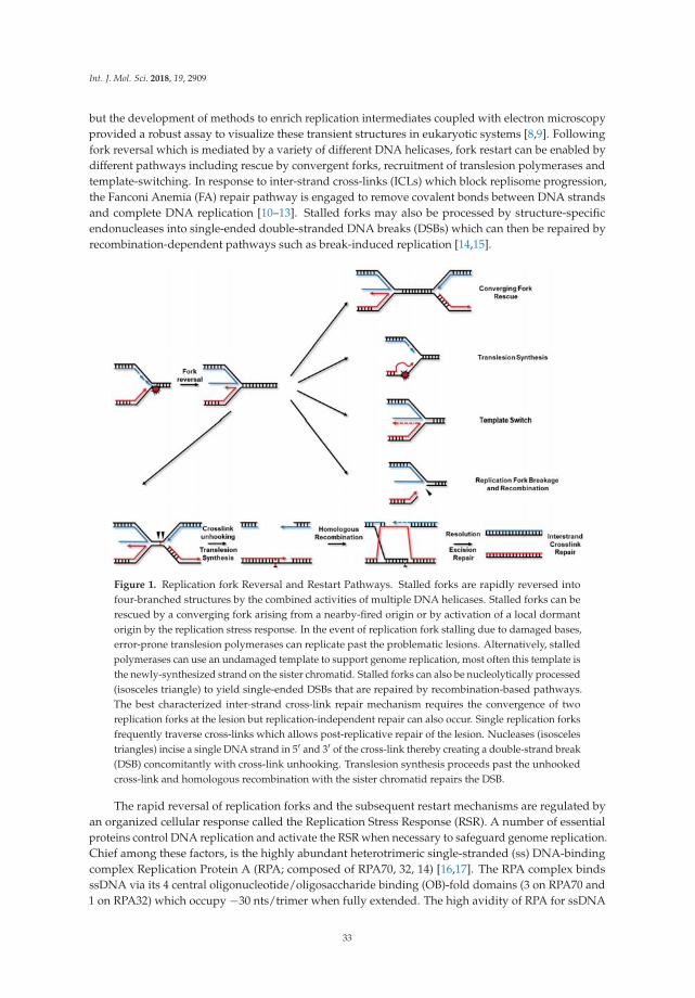
Int. J. Mol. Sci. 2018, 19, 2909
but the development of methods to enrich replication intermediates coupled with electron microscopyprovided a robust assay to visualize these transient structures in eukaryotic systems [8,9]. Followingfork reversal which is mediated by a variety of different DNA helicases, fork restart can be enabled bydifferent pathways including rescue by convergent forks, recruitment of translesion polymerases andtemplate-switching. In response to inter-strand cross-links (ICLs) which block replisome progression,the Fanconi Anemia (FA) repair pathway is engaged to remove covalent bonds between DNA strandsand complete DNA replication [10–13]. Stalled forks may also be processed by structure-specificendonucleases into single-ended double-stranded DNA breaks (DSBs) which can then be repaired byrecombination-dependent pathways such as break-induced replication [14,15].
Figure 1. Replication fork Reversal and Restart Pathways. Stalled forks are rapidly reversed intofour-branched structures by the combined activities of multiple DNA helicases. Stalled forks can berescued by a converging fork arising from a nearby-fired origin or by activation of a local dormantorigin by the replication stress response. In the event of replication fork stalling due to damaged bases,error-prone translesion polymerases can replicate past the problematic lesions. Alternatively, stalledpolymerases can use an undamaged template to support genome replication, most often this template isthe newly-synthesized strand on the sister chromatid. Stalled forks can also be nucleolytically processed(isosceles triangle) to yield single-ended DSBs that are repaired by recombination-based pathways.The best characterized inter-strand cross-link repair mechanism requires the convergence of tworeplication forks at the lesion but replication-independent repair can also occur. Single replication forksfrequently traverse cross-links which allows post-replicative repair of the lesion. Nucleases (isoscelestriangles) incise a single DNA strand in 5′ and 3′ of the cross-link thereby creating a double-strand break(DSB) concomitantly with cross-link unhooking. Translesion synthesis proceeds past the unhookedcross-link and homologous recombination with the sister chromatid repairs the DSB.
The rapid reversal of replication forks and the subsequent restart mechanisms are regulated byan organized cellular response called the Replication Stress Response (RSR). A number of essentialproteins control DNA replication and activate the RSR when necessary to safeguard genome replication.Chief among these factors, is the highly abundant heterotrimeric single-stranded (ss) DNA-bindingcomplex Replication Protein A (RPA; composed of RPA70, 32, 14) [16,17]. The RPA complex bindsssDNA via its 4 central oligonucleotide/oligosaccharide binding (OB)-fold domains (3 on RPA70 and1 on RPA32) which occupy −30 nts/trimer when fully extended. The high avidity of RPA for ssDNA
33

Int. J. Mol. Sci. 2018, 19, 2909
supports unperturbed DNA replication by protecting these fragile regions against enzymatic processingand by disrupting secondary DNA structures that could slow down or block DNA polymerases.Additionally, RPA contains two protein-protein interaction modules: the RPA70 N-terminal OB-folddomain and the winged helix domain at the C-terminus of RPA32. These features allow the RPA-ssDNAplatform to orchestrate the recruitment and activation of a large number of DNA damage signalingand repair factors to maintain genome stability [18–21].
The RSR is switched on by the detection of a deoxyribonucleic structure composed of persistentRPA-ssDNA and an adjacent single-/double-stranded (ds) DNA junction [22–24]. This structureresults at least in part from the functional uncoupling of the replicative DNA helicase and polymerasesduring replication stress and activates the ATR-ATRIP master checkpoint kinase [25,26]. A similarATR-activating structure can also arise from DSB resection, conferring an unusual flexibility to ATR inthe detection of genome destabilizing lesions [27]. ATR is brought onto RPA-ssDNA by an interactionbetween its obligate partner ATRIP and RPA [28,29]. Once there, it is activated by direct contactwith proteins that possess ATR-activating domains (AAD). In human cells, two such ATR activatorshave been described thus far: TOPBP1 (Topoisomerase II Binding Protein 1) and ETAA1 (Ewing’sTumor Associated Antigen 1) [30–33]. It is thought that the physical contact between ATR-ATRIPand the AAD domain of its activators induces conformational changes that lead to ATR activation.Once activated via auto-phosphorylation, ATR modifies a variety of RSR effectors including RPA itselfand the downstream kinase CHK1 to prevent the firing of new replication origins, activate fork repairfactors and inhibit cell cycle progression to support the recovery and eventual completion of DNAreplication prior to mitosis entry [34–38]. In addition to ATR activation, the ability of RPA to interactwith numerous other DNA replication, recombination and repair factors implicates it in virtually allsub-pathways of replication fork processing and restart during replication stress.
Another key platform for the early RSR is the homotrimeric DNA polymerase sliding clampPCNA (Proliferating Cell Nuclear Antigen) [39,40]. During normal replication, the ring-shapedhomotrimeric PCNA is loaded onto primer template junctions by the Replication Factor C complexand encircles dsDNA while interacting with DNA polymerases to enhance their processivity [41–43].PCNA is also important to mediate polymerase switches during replication and is a critical regulatorof Okazaki fragment processing. Similarly to RPA, PCNA exerts its cellular functions by interactingwith myriad genome maintenance factors that often bind PCNA through a short conserved PIP (PCNAinteracting-peptide; consensus sequence Q-x-x-[I/L/M/V]-x-x-[F/Y]-[F/Y]) making this platform anessential component of DNA replication and the RSR [44]. Because of their symmetrical architecture,PCNA and its bacterial equivalent the beta sliding clamp can in principle interact productively withmultiple different partners simultaneously giving rise to the tool belt hypothesis [45]. The alternative tothis model is the sequential interaction of PCNA with individual enzymes and experimental support forboth models exists in Okazaki fragment maturation by the FEN1, POL δ and Ligase I enzymes [46,47].During DNA replication stress, PCNA interacts with various DNA helicases and polymerases andplays key roles in fork reversal, template-switching, homologous recombination (HR) and translesionDNA synthesis to maintain genome stability [8,48,49].
Finally, the FA DNA repair pathway processes highly toxic inter-strand cross-links (ICLs). FA is arare human syndrome characterized by bone marrow failure, cellular hypersensitivity to cross-linkingagents, cancer predisposition and skeletal defects among a host of other clinical manifestations.The mutations of 22 DNA repair genes result in at least a subset of FA symptoms and the proteinsthat they encode are considered bona fide FA factors [12]. The critical platform of this pathway isthe FANCD2/I complex which initially recognizes the lesion and coordinates the recruitment ofendonucleases to unhook the cross-link. Following the unhooking reaction, translesion polymerases,HR and nucleotide excision repair effectors collaborate to remove the chemical adduct and completeDNA replication (Figure 1). More recently, several FA proteins were also shown to function in variousaspects of the RSR indicating that at least some components of this pathway may play importantgenome maintenance roles beyond the repair of ICLs [50–54].
34

Int. J. Mol. Sci. 2018, 19, 2909
Recruitment and exchange of interacting partners on RPA, PCNA and FANCD2/I, particularlyin response to various types of DNA damage is controlled by post-translational modifications(PTMs) [16,40,55,56]. For instance, hyper-phosphorylation of the N-terminus of RPA32 by the ATR,ATM and DNA-PK DNA damage kinases regulates its association with the MRN complex, the PALB2HR protein and the PRP19 E3 ubiquitin ligase among others [57–59]. Ubiquitylation has also emergedas a prevalent PTM that occurs on a large number of genome maintenance factors in response toreplication stress and DSBs ([60–62] and reviewed in [63–66]). Modifications of PCNA, FANCD2/Iand more recently of RPA by ubiquitin and ubiquitin-like modifiers have been shown to controltheir functions in genome maintenance. Here, we review recent findings on the ubiquitin ligases,ubiquitin-binding genome guardians and de-ubiquitylases that play key roles on the main RSRplatforms to faithfully complete DNA replication under adverse conditions.
2. Ubiquitylation on the RPA-ssDNA Platform
The first indication that the RPA complex is ubiquitylated upon DNA damage came fromproteomics analyses which showed that RPA70, RPA32 and RPA14 are all modified by ubiquitinin UV-treated cells [61]. Subsequently, RPA ubiquitylation was also shown to be enhanced bycamptothecin (CPT; a topoisomerase I inhibitor that creates DSBs at ongoing forks), hydroxyurea(HU), the DNA polymerase inhibitor aphidicolin and the cross-linking agent mitomycin C but not byγ-irradiation (IR) indicating that this modification is particularly relevant to the RSR [21,67,68]. Two E3ubiquitin ligases function on RPA-ssDNA and control RPA ubiquitylation during replication stress:PRP19 and RFWD3 [21,59,67–69].
2.1. PRP19, at the Intersection of mRNA Maturation and Genome Maintenance
PRP19 is a multifunctional and essential U-BOX family E3 ubiquitin ligase best known forits role as an evolutionarily conserved pre-mRNA processing factor that functions within thePRP19/CDC5L core complex, an important spliceosome co-factor composed of PRP19, CDC5L,PLRG1 and BCAS2 [70–74]. In addition to its U-BOX E3 ligase domain, PRP19 also contains acentral coiled-coil domain which allows its tetramerization and a C-terminal substrate recognitionWD40-repeat module [69,75,76]. It was first discovered in yeast as a mutant that accumulatesintron-containing pre-mRNA at non-permissive temperature and was later shown to physicallyassociate with the spliceosome and promote its activation [77–79]. Mechanistically, PRP19 is part ofa ubiquitylation/de-ubiquitylation cycle that targets the PRP3 subunit of the U4/U6 snRNP withnon-degradative K63-linked ubiquitin chains to promote spliceosome maturation and productiverounds of mRNA splicing [80].
Saccharomyces cerevisiae, PRP19 is also known as PSO4 and was independently found in a geneticscreen for photoactivated psoralen-sensitive mutants suggesting that it may play a role in ICLrepair [81]. Depletion of the PRP19/CDC5L complex also inhibits psoralen cross-link repair in vivo inhuman cells [82]. Additional early clues for a DNA repair function of PRP19 came from the observationthat its overexpression enhances the resistance to genotoxic insults and increases the replication lifespanof umbilical vein endothelial cells [83]. Knockdown (KD) of the core subunits of the PRP19/CDC5Lcomplex also results in sensitivity to the replication stress-inducing agents mitomycin C, UV andHU [84,85]. Support for a more direct link between the PRP19 complex and the RSR came from thediscovery that the complete PRP19/CDC5L core complex relocates onto RPA-ssDNA upon replicationstress or at resected DSBs [21,69,86]. On RPA-ssDNA, the PRP19 complex promotes ATR activation.Indeed KD of PRP19, CDC5L, PLRG1 or BCAS2 all strongly decrease RSR signaling by ATR and therecruitment of this master checkpoint kinase to stalled forks as measured by phosphorylation of itssubstrates (RPA and/or CHK1) and by ATRIP foci formation [21,69,84,86]. Depletion of PRP19 andother splicing factors was also shown to impede DSB resection which may contribute to ATR-activationand RPA phosphorylation defects [21,69,87,88]. Furthermore, PRP19 complex KD impedes replicationfork restart and HR indicative of extensive roles in the RSR [21,87,89]. Importantly, a WD40-repeat
35

Int. J. Mol. Sci. 2018, 19, 2909
point mutant defective for the PRP19-RPA interaction but still able to form the PRP19-CDC5L splicingcomplex is unable to support ATR activation, HR and timely repair of collapsed replication forks,demonstrating the dual roles of PRP19 in mRNA processing and the RSR [21,59]. A deletion of theU-BOX domain also impedes ATR activation and HR linking them to PRP19-mediated ubiquitylation.Mechanistically, PRP19 KD decreases RPA70 and RPA32 ubiquitylation upon CPT treatment and thiscan be complemented by the re-expression of WT PRP19 but not by ubiquitin ligase or RPA-bindingmutants [21,59]. More recent data indicates that RPA ubiquitylation also depends on PLRG1 which isrequired to activate the E3 ligase activity of the PRP19/CDC5L core complex [69]. Thus, in responseto replication stress, the PRP19 complex transforms into a sensor of RPA-ssDNA and functions as aubiquitin ligase on RPA-ssDNA to promote RSR signaling and replication fork repair.
2.2. RFWD3, a Novel Fanconi Anemia Player on RPA-ssDNA
RFWD3 is a RING (Really Interesting New Gene) domain E3 ubiquitin ligase that contains acoiled-coil domain and a WD40-repeat substrate-binding module. The first link between RFWD3and the DNA damage response came from the demonstration that RFWD3 works together with theMDM2 E3 ligase to control the length of ubiquitin chains polymerized onto p53. In this context,RFWD3-mediated ubiquitylation is non-degradative and promotes the stability of p53 in responseto DSBs [90]. Initial evidence for an implication of RFWD3 in the RSR came when it was isolated asan RPA32 interactor and shown to be required for optimal RPA and CHK1 phosphorylation [91,92].The magnitude of the CHK1 phosphorylation defect induced by RFWD3 KD appears to be somewhatcell-type dependent perhaps due to variations in ATR activation thresholds [67,91,92]. However, in linewith an ATR-activating role for RFWD3, its downregulation leads to increased new origin firing duringreplication stress, as might be expected if the S-phase checkpoint is defective [67]. This particularfunction of RFWD3 in ATR signaling appears to be independent from its ubiquitin ligase activity butto require its interaction with RPA [68]. Early phenotypic characterization also noted the inability ofRFWD3-depleted cells to resolve HU-induced RPA and RAD51 foci in a timely manner suggestingthat some step(s) in replication fork restart might depend on this E3 ubiquitin ligase [92]. Accordingly,RFWD3 depletion or mutation renders cells sensitive to a variety of replication stressors includingCPT, mitomycin C, cisplatin and olaparib with milder sensitivities to HU and γ-irradiation [68,91–94].
A breakthrough came from the discovery that RFWD3 also acts as a ubiquitin ligase on RPA70and RPA32 during replication stress and promotes HR repair of DSBs and stalled replicationforks [67]. Downregulation of RFWD3 abrogrates ubiquitylation of RPA in response to HU, 4-NQO(4-nitroquinoline), mitomycin C and CPT treatments [59,67,68]. Some of the phenotypes seen uponRFWD3 depletion could be recapitulated by a RPA32 ubiquitylation mutant (K37/38R) which exhibitedreplication fork repair defects. Nevertheless, this mutant was still able to support HR at stalled forks,possibly due to the fact that at least 18 potentially interchangeable lysines are modified on the RPAcomplex, which makes it difficult to create fully ubiquitylation-defective RPA [67]. Interestingly,a RPA32 C-terminal winged-helix domain mutant that cannot interact with RFWD3 mutant also hadimpaired phosphorylation suggesting crosstalk between these 2 PTMs on RPA-ssDNA. In responseto replication stress, it does not appear that ubiquitylation mediated by UV or 4-NQO treatment isdegradative as no increase in ubiquitylated RPA species or in total RPA levels could be observed inresponse to proteasome inhibition [67].
Another important development was recently made with the implication of RFWD3 as a novel FApathway factor and the discovery that RFWD3-mediated ubiquitylation is an important contributor toICL repair [68,93,94]. Mechanistically, it was found that both RPA and the RAD51 HR recombinasedirectly interact with RFWD3 and are ubiquitylated in response to mitomycin C [68]. In contrast tothe ubiquitylation induced by short term UV or 4-NQO treatments, long-term mitomycin C treatmentdestabilizes RPA32 and RAD51 proteins in a RFWD3-dependent manner [67,68]. Enhanced levels ofRPA32 and RAD51 ubiquitylation induced by mitomycin C were observed following proteasome orVCP (Valosin-containing protein/p97) inhibition and the turnover of RPA32 in mitomycin C-induced
36

Int. J. Mol. Sci. 2018, 19, 2909
foci was decreased upon RFWD3 KD but not in response to PRP19 depletion, indicating a specificrole of RFWD3 in this genotoxic context. In vitro, ubiquitylation of RPA70 and 32 and of RAD51decreased their affinity for ssDNA. RPA32 or RAD51 ubiquitylation mutants also showed someimpairment in turnover rates as well as defective HR repair and sensitivity to ICL agents implicatingtheir modification directly in these processes. The loading of the RAD54 SWI2/SNF2 ATPase motorprotein and the MCM8 helicase were perturbed by RFWD3 KO indicating extensive defects in the latersteps of ICL repair.
RFWD3 was also directly implicated in FA when a patient was found with a frameshift leadingto premature termination on one RFWD3 allele and a missense mutation (I639K) in the WD40-repeatdomain of the other. This patient presented FA symptoms including absent thumbs, pan-cytopeniaof the bone marrow and hypersensitivity of cells to mitomycin C and diepoxybutane. Chromosomalbreakage, G2-arrest and cell death induced by mitomycin C treatment of patient fibroblasts couldbe complemented by re-expression of WT RFWD3 establishing this ubiquitin ligase as a novel FAfactor [68,93,94]. Mutation of I639K and other residues on the WD40-repeat domain abrogates therecruitment of RFWD3 to sites of damage, its interaction with RPA and RAD51, the ubiquitylationof these substrates, and HR repair. Altogether these studies convincingly implicate RFWD3 in theregulation of HR that occurs towards the end of ICL repair [68,93,94].
2.3. Potential Interplay between PRP19 and RFWD3 on RPA-ssDNA
How could RFWD3 and PRP19 collaborate to regulate the RSR? Whereas RFWD3 is constitutivelyassociated with the RPA32 C-terminal winged-helix domain, the PRP19 complex is specifically recruitedto RPA-ssDNA during replication stress [21,59,62]. Upon damage, PRP19 also interacts with RFWD3,perhaps by joining it on the RPA complex as PRP19 was shown to associate with RPA70 whereasRFWD3 is tethered to the C-terminus of RPA32 [21,59,86,93]. The PRP19-RPA interaction is stronglystimulated by CPT treatment which creates DSB at replication forks. This CPT-triggered PRP19-RPAinteraction is decreased by ATR kinase inhibition and completely abrogated when ATR, ATM andDNA-PK are jointly inactivated [21,59]. All 3 of these DNA damage response kinases phosphorylatetheir own specific substrates in response to DNA damage but they act together on the RPA32N-terminus to promote its full hyper-phosphorylation during replication-associated DSBs [55,95].Interestingly, an RPA32 mutant that cannot be phosphorylated still binds RFWD3 but cannot interactwith PRP19 or be ubiquitylated in response to CPT. Reciprocally, a PRP19 point mutant that cannotinteract with phosphorylated RPA is unable to support CPT-mediated RPA ubiquitylation [59,69].Part of the ubiquitylation occurring on RPA occurs as non-degradative K63-linked chains, which areimportant regulators of protein-protein interactions [21,67,96,97]. In vitro, ATRIP exhibits affinity forK63-linked ubiquitin chains that may favor ATR-ATRIP recruitment onto ubiquitylated RPA-ssDNA.This led to the proposal of a feed-forward loop for ATR activation in which RPA hyper-phosphorylationby ATR, ATM and DNA-PK enhances its interaction with PRP19 and its ubiquitylation by both RFWD3and PRP19 which may further promote fork restart and HR (Figure 2, [21,59]). In this context, the E3ubiquitin ligase activity of PRP19 on RPA-ssDNA would promote ATR signaling, HR and fork restartwhereas RFWD3 would be geared more towards fork restart and HR. Use of different sites or types ofubiquitin chains by each ligase on RPA may explain their differential requirements in these aspects ofthe RSR. Interestingly, a notable exception to this model is for mitomycin C treatment which increasesthe RFWD3-RPA interaction [93]. This enhanced recruitment of RFWD3 to RPA in response to ICLsmay explain the degradative nature of RFWD3-mediated ubiquitylation in this context. Additionalwork will be required to understand the mechanisms which control the balance of degradative andnon-degradative ubiquitylation on RPA-ssDNA.
37

Int. J. Mol. Sci. 2018, 19, 2909
Figure 2. Ubiquitylation on the RPA-ssDNA platform during Replication Stress. (1) Fork uncouplingleads to RPA-ssDNA production which is constitutively associated with the RFWD3 (R3) ubiquitinligase; (2) RPA hyper-phosphorylation by ATR, ATM and DNA-PK enhances the recruitment ofPRP19 (19); (3) PRP19 and RFWD3 poly-ubiquitylate the RPA complex. K63-linked chains help tetherATR-ATRIP to RPA-ssDNA; (4) this produces a feed-forward loop that results in the spreading of RPAphosphorylation and ubiquitylation across RPA-ssDNA filaments. These modifications stimulate RSRsignaling, fork restart and homologous recombination.
Another potential point of intersection between PRP19 and RFWD3 could be at the level ofRFWD3 phosphorylation. It was shown that RFWD3 is phosphorylated by ATM/ATR in responseto replication stress and this phosphorylation is required for mitomycin C resistance as well as forRPA ubiquitylation [68,90,93]. Because of PRP19’s role in ATR activation it is possible that some ofthe negative impact of PRP19 depletion on RPA ubiquitylation may be through a decrease in thestimulatory phosphorylation of RFWD3 during the RSR. It is also possible that these two ligasesmay confer some flexibility in the response to specific genotoxic circumstances that may lead to RPAubiquitylation. In this regard, it has been shown that RPA is also SUMOylated (small ubiquitin-likemodifier) in response to DSBs. SUMO-RPA enhances RAD51 recruitment and promotes HR repair ofthe breaks [98]. It was suggested that SUMO-RPA may be recognized and targeted by the RNF4SUMO-targeted ubiquitin ligase (STUbL) to facilitate RPA removal from ssDNA and promoteHR-mediated repair of breaks [98,99]. However, in contrast to the PRP19 and RFWD3 ligases, directubiquitylation of RPA by RNF4 remains to be demonstrated. Under different genotoxic circumstances,RFWD3, PRP19 and RNF4 may be called into action to modify RPA and regulate its many genomemaintenance functions. Finally, as RPA-ssDNA orchestrates the recruitment of multiple genomeguardians during the RSR, the ubiquitylation of additional substrates at stalled forks may explain someof the phenotypic differences observed in PRP19- and RFWD3-deficient cells as well. How exactly,these different E3 ligases work together on RPA-ssDNA will be an interesting avenue of future research.
3. Ubiquitylation on the PCNA Platform
Besides the RPA-ssDNA platform, PCNA is essential for DNA replication, DNA damage toleranceand genome stability. PCNA is a homotrimeric DNA polymerase processivity factor. With its ring-shapedstructure, PCNA encircles DNA to form a sliding clamp and acts as a protein loading scaffold duringunperturbed DNA replication. In addition to its critical roles during replication, PCNA serves as arecruitment platform for genome maintenance proteins and choreographs their activities withinmultiple repair pathways (reviewed in [39,40]). Similarly to the RPA-ssDNA platform, studies revealedthat ubiquitylation and/or SUMOylation of PCNA are important for cell survival in response toDNA damaging agents that block replication fork progression such as UV, methyl methanesulfonate,mitomycin C or HU [100,101]. Whereas DNA damaging agents that cause fork stalling lead to PCNAubiquitylation, others such as CPT or bleomycin that cause DSBs do not, underlining the relevance ofPCNA ubiquitylation for the restoration of blocked replication forks.
In response to replication fork stalling or during normal S-phase, mono- or poly-ubiquitylationand/or SUMOylation on PCNA occurs on a major site, the highly conserved lysine 164 (K164)residue [102–105]. PCNA mono-ubiquitylation in yeast and in mammals is mainly catalyzed bythe conserved Rad18(E3)/Rad6(E2) complex [106]. Poly-ubiquitylation of PCNA by addition ofK63-linked ubiquitin chains onto mono-ubiquitylated PCNA depends on the heterodimeric complexUbc13-Mms2 (E2) first discovered in yeast (UBC13-UEV1 in humans) in association with the Rad5
38

Int. J. Mol. Sci. 2018, 19, 2909
ubiquitin ligase (HLTF or SHPRH in humans) [103,107,108]. In yeast, PCNA is also SUMOylatedto a lesser extent on K127 and additional sites are also found in human cells [102,109]. In yeast,PCNA SUMOylation involves Ubc9 (E2) and Siz1 and Siz2 (E3a) whereas the SUMO E3 in humansin currently unknown [102–104,110]. In human cells, endogenous DNA damage also promotes K164PCNA mono-ubiquitylation by CRL4Cdt2, instead of RAD18 [111]. PCNA ubiquitylation can alsobe stimulated by accessory co-factors such as SIVA1, a PCNA and RAD18-interacting protein [112].A notable exception to K164-targeted ubiquitylation was discovered in DNA ligase I-defective yeastcells. In this context, deficient Okazaki fragment ligation leads to ubiquitylation of PCNA at K107at non-permissive temperatures to promote DNA damage signaling [113]. Although the specificlysine residue remains to be determined, it seems that this alternative modification is also conservedin humans [113,114].
A functional link between the RPA-ssDNA and PCNA ubiquitylation has been established inyeast where RPA-ssDNA serves as a recruitment platform for Rad18-Rad6 at stalled forks and controlsPCNA ubiquitylation [115]. In humans, some evidence also suggests that RPA-ssDNA regulatesPCNA ubiquitylation further supporting close collaboration between both platforms in genomemaintenance [101].
The modification of PCNA by ubiquitin and ubiquitin-like molecules controls repair pathwaychoice at replication-blocking obstacles. Two major pathways mediate DNA damage tolerancemechanisms to allow replication completion in the presence of damage: Translesion synthesis (TLS) andtemplate switching (TS) (Figure 3 and reviewed in [116]). While PCNA mono-ubiquitylation promotesTLS that constitutes to some extent an error-prone lesion bypass mode, PCNA poly-ubiquitylationpromotes TS, an error-free recombination-based repair pathway.
Figure 3. Ubiquitylation on PCNA during Replication Stress. When forks encounter DNA lesions,PCNA is either mono-ubiquitylated to engage translesion synthesis or poly-ubiquitylated to triggerlesion bypass by template switching. RAD18(18)-RAD6(6) is recruited and mono-ubiquitylates PCNAat K164. Mono-ubiquitylated PCNA is recognized by specialized TLS polymerases that containPCNA-interacting and ubiquitin-binding domains (e.g., POL η, κ, ι, REV1). TLS polymerases replacereplicative polymerases to continue DNA synthesis across the lesions. Addition of K63-linked ubiquitinchains on mono-ubiquitylated PCNA is mediated by the UBC13-UEV1 (13) E2 complex which functionswith either HLTF, SHPRH and/or currently unknown E3 ligases (X). Poly-ubiquitylated PCNA recruitsfork remodeler ZRANB3 via its ubiquitin binding zinc finger to allow fork regression.
39
Int. J. Mol. Sci. 2018, 19, 2909
3.1. PCNA Mono-Ubiquitylation and Translesion Synthesis
TLS is a highly conserved repair mechanism that uses low fidelity polymerases to replicatedamaged DNA segments thus allowing completion of DNA replication (reviewed in [116]). DuringTLS, DNA polymerase switches happen during which replicative polymerases (i.e., Pol δ and Polε B-family polymerases) are replaced by specialized TLS polymerases that are capable of insertingnucleotides opposite lesions [117–119]. Once nucleotide incorporation opposite the lesion is made, theTLS patch is then extended by the same or another TLS polymerase. The extension step can rangefrom 5–60 nucleotides depending on the lesion and polymerase involved. The low processivity of TLSpolymerases enables a final switch that brings back a replicative DNA polymerase to resume bulkgenome replication.
The best characterized TLS enzymes include members of the Y-family of DNA polymerases Polη (eta; POLH/XPV/RAD30A), Pol ι (iota; POLI/RAD30B), Pol κ (kappa; POLK/DINB1) and REV1along with the B-family polymerase Pol ζ (zeta), a tetrameric complex composed of the catalyticsubunit REV3L, REV7/MAD2L2/FANCV, POLD2 and POLD3 ([120–123] and reviewed in [124–127]).When compared with replicative polymerases, these specialized polymerases generally exhibit lowernucleotide incorporation fidelity due to a more spacious/flexible active site and lack exonucleolyticproofreading function. TLS polymerases each have distinct properties that make them either mutagenicor accurate depending on the type of lesion encountered. Care must thus be taken at the selectionstep during polymerase switch to pick the best enzyme for the job at hand. For example, POL η/XPVis the major TLS polymerase selected to bypass UV-induced cis-syn cyclobutane thymine-thyminedimers (CPD) in a largely error-free manner [128–131]. The importance of POL η/XPV in UV-induceddamage repair is emphasized by the fact that its mutation in humans causes xeroderma pigmentosum,a syndrome characterized by extreme sensitivity to sunlight and a high incidence of skin cancer [132,133]. In the absence of POL η, TLS at CPDs is believed to be catalyzed by a different specializedpolymerase which frequently incorporates incorrect nucleotides thereby generating mutations thatpromote cancer [134].
PCNA is the main conductor of polymerase switches during TLS and this is highly regulated byits mono-ubiquitylation. TLS polymerases of the Y-family POL η, POL ι and POL κ possess PIP motifswhile REV1 interacts with PCNA via its BRCT (BRCA1 C Terminus) domain [135–139]. Besides thesePCNA-interacting regions, the Y-family of TLS polymerases also possess evolutionarily conservedubiquitin-binding motifs (UBM) and/or ubiquitin-binding zinc finger (UBZ) domains that mediate therecognition of mono-ubiquitylated PCNA [140,141]. These ubiquitin-binding domains are importantfor genome stability as exemplified by the requirement of the Pol η UBZ to restore normal response toUV in XP-V fibroblasts [142]. Despite strong experimental support that PCNA mono-ubiquitylationis required for maximal TLS in mammalian cells, this modification is not absolutely essential for thisprocess to occur. KD of REV3L, POL η or REV1 in PCNA K164R mouse embryonic fibroblasts (MEFs)leads to increased UV sensitivity indicative of additional modes of recruitment for TLS polymerasesat lesions [143]. These alternative recruitment modes vary; certain TLS polymerases such as POL η
and POL κ interact with RAD18 to facilitate their access to DNA templates. Furthermore, REV1 bindsssDNA and primer termini and recruits different TLS polymerase partners including POL ζ, POL η,POL ι and POL κ to sites of damage. In essence, mono-ubiquitylated PCNA works with REV1 and itspartner TLS polymerases to coordinate both steps of the TLS reaction: nucleotide insertion oppositethe lesion and extension of the nascent strand [117,144–146].
3.2. PCNA Poly-Ubiquitylation and Template Switching
TS is a damage avoidance repair mechanism based on recombination that uses the undamagedsister chromatid as the template to carry out limited DNA replication that allows the replication forkto bypass problematic lesions in a mostly error-free manner (reviewed in [147–149]). Even though themechanistic details have not been completely worked out, TS at the fork is thought to happen afterfork reversal. DNA synthesis would then allow the stalled nascent DNA to extend past the lesion
40

Int. J. Mol. Sci. 2018, 19, 2909
on the undamaged switched template (Figure 1 and [147]). TS can also occur post-replicatively andinitiate behind the fork at gapped lesions that were left behind [150,151].
In yeast, one of the major actors of this pathway is Rad5, a member of the SWI/SNF2ATPase family which is also a RING domain E3 ubiquitin ligase. Rad5 mediates K63-linked PCNApoly-ubiquitylation with its partner E2 Ubc13-Mms2 to extend the mono-ubiquitylation product createdby Rad18/Rad6 [102,103,108]. Moreover, the ATP-dependent helicase activity of Rad5 allows it tounwind and anneal nascent and parental strands together to promote replication fork reversal [107,152].Both the ubiquitin ligase and ATPase activities of Rad5 were shown to be required for genome stabilityin response to a variety of different replication stressors [153–155].
Despite years of efforts, it is still unclear how PCNA poly-ubiquitylation in yeast promotes TSand error-free damage bypass. What is clear is that sister chromatid junction formation during TSrequires PCNA SUMOylation and the Rad51 recombinase [156–158]. During an unperturbed S-phase,unscheduled recombination is actively counteracted when DNA replication is proceeding smoothly.Indeed, PCNA SUMOylation directly mediates the recruitment of the anti-recombinase/UvrD familyhelicase Srs2 which is equipped with SUMO-interacting (SIMs) and PIP-like motifs. On SUMO-PCNA,Srs2 minimizes Rad51-dependent recombination at ongoing forks by disturbing the formation ofRad51-ssDNA filaments [159–164]. The alternative clamp loader Elg1 is also tethered to SUMO-PCNAvia SIMs and PIP-like motifs to promote PCNA unloading after Okazaki fragment maturation andthis role is shared by the Elg1 homolog in human cells, ATAD5 [165–168]. Moreover, SUMO-PCNApositively regulates its damage-induced mono- and poly-ubiquitylation by recruiting the Rad6-Rad18ligase which exhibits at least partial STUbL behavior on this platform. In this case however, the STUbLactivity does not appear to be shared by the human RAD18 homolog [169].
How Rad51 comes into action at stalled replication forks in yeast to induce TS when necessaryhas recently been brought to light. It was found that the adaptor protein Esc2 possesses SUMO-likedomains (SLDs) which interact with SIMs on Srs2. Esc2 can also bind directly to branched DNAstructures in vitro and to stalled replication forks in vivo. At stalled forks, Esc2 and the STUbL Slx5-Slx8increase the turnover of SUMOylated Srs2 thereby allowing Rad51 recruitment and local induction ofrecombination [170,171]. Downstream of PCNA poly-ubiquitylation and Rad51-mediated TS, the Sgs1helicase, together with Top3 and Rmi1 (BLM/TOP3A/RMI1/2 in humans) dissolves recombinationintermediates to promote the formation of non-crossover products [156,172]. Backup sister chromatidjunction resolution is also provided by the Mus81-Mms4 complex with assistance by Esc2 [173–175].
In this context, the role of poly-ubiquitylated PCNA in promoting this cascade of events stillremains a bit obscure. Thus far in yeast, the only known interactor of poly-ubiquitylated PCNA is theAAA+ ATPase Mgs1 [176]. Interestingly, MGS1 exhibits synthetic lethality with RAD6 and syntheticsickness with RAD18 and RAD5, implicating it in the protection and repair of stalled replicationforks [177]. Mgs1 interacts with poly-ubiquitylated PCNA via its UBZ domain. This domain allowsMgs1 to outcompete the Pol δ subunit Pol32 on PCNA which may be important for a variety ofoutcomes at stalled forks. However, a Mgs1 UBZ domain mutant still rescues synthetic lethalityof mgs1 rad18 mutants as well as the WT and the lack of DNA damage sensitivity of MGS1 singlemutants does not support a critical role for this protein in damage bypass [177,178]. The identificationof poly-ubiquitylated PCNA-binding effector(s) that may promote TS in yeast will be a very significantstep forward in our understanding of this error-free repair mechanism. Another scenario stems fromthe very recent finding that K63-linked poly-ubiquitin chains can bind to DNA [179]. This opens upthe possibility that PCNA poly-ubiquitylation by itself may locally influence the architecture of stalledreplication forks and perhaps help their eventual restart.
In human cells, a similar pathway appears to promote damage avoidance and two Rad5 functionalhomologues were identified: Helicase-like transcription factor (HLTF) and SNF2 histone-linkerPHD-finger RING-finger helicase (SHPRH) [180,181]. Both SHPRH and HLTF present extensivestructural and functional similarities with yeast Rad5 including RING domains that function with theE2 complex UBC13-MMS2 to poly-ubiquitylate PCNA in response to replication stress [180–184].
41

Int. J. Mol. Sci. 2018, 19, 2909
Additional E3 ligase(s) may also take part in PCNA poly-ubiquitylation in mammals as doubleHLTF/SHPRH mutant MEFs showed a decrease but not an abrogation of PCNA poly-ubiquitylation inresponse to UV [185]. In vitro, HLTF can carry out fork reversal and also complements the UV sensitivityof the rad5 yeast mutant further supporting the notion that it acts as a Rad5 homolog to promote genomemaintenance [181,186,187]. In addition to its SWI/SNF helicase domain, HLTF has a HIRAN (HIP116and RAD5 N-terminus) region which functions as a substrate recognition module. The HIRAN domainis structurally similar to an OB-fold motif and mediates binding of HLTF at 3′ ssDNA ends to drivereplication fork reversal [188–191]. This domain is not found in SHPRH which also cannot complementrad5 mutant UV sensitivity suggesting that despite their common function in PCNA poly-ubiquitylation,HLTF and SHPRH may also have distinct roles in genome maintenance [184].
Little is known about how HLTF and SHPRH collaborate or how they split the work to regulatereplicative lesion bypass. One possibility is that each ligase may be activated in response to differentlesions or genotoxic circumstances. Supporting this idea, is the fact that HLTF and SHPRH wereshown to play distinct roles in the response to UV or MMS respectively [192]. MMS induces HLTFdegradation perhaps via auto-ubiquitylation and the formation of a RAD18-SHPRH-Pol κ complexthat allows bypass of the lesions. Contrastingly, following UV irradiation HLTF but not SHPRHpromotes the recruitment of Pol η which can bypass UV lesions in an error-free manner [192]. Theseroles of HLTF and SHPRH in controlling some aspects of TLS are also supported experimentallyfor the Rad6-Rad18-Rad5-Ubc13-Mms2 axis in fission yeast [193]. Interestingly, Pol η has a singleubiquitin-binding motif whereas Pol κ and other TLS polymerases have 2 [142]. It is possible that PCNAmono- and poly-ubiquitylation may be induced differentially depending on the source of replicationstress. In turn, this may influence TLS polymerase selection to maximize error-free repair of lesions.
In human cells, the SWI/SNF2 family ATPase ZRANB3/AH2 (zinc finger, RAN-binding domaincontaining 3) is recruited onto poly-ubiquitylated PCNA and mediates replication fork reversal andrestart. In addition to fork regression activity, ZRANB3 can dismantle D-loops in vitro and limitsister-chromatid exchange in vivo [194–197]. The recruitment of ZRANB3 to stalled forks requires aPIP and an APIM (AlkB2 PCNA interaction motif) motif that bind directly to PCNA, a NZF (NPL4zinc finger) which recognizes K63-linked ubiquitin chains and a HNH nuclease domain [195,197–199].RAD18 or UBC13 depletion impaired the retention but not the initial recruitment of ZRANB3 atUV stripes indicating that ZRANB3 may also be recruited in a PCNA ubiquitylation-independentmanner to sites of damage. HLTF and ZRANB3 along with a number of other factors function as forkremodelers at stalled forks. One explanation for the requirement of many enzymes for this processmay lie in the variety of structures created in response to different types of lesions [200]. It is alsopossible that different remodelers act in a sequential manner at different steps of the reversal reaction.In agreement with this hypothesis, SMARCAL1, HLTF or ZRANB3 KO all led to a complete abrogationof stalled fork degradation by the MRE11 nuclease in BRCA1/BRCA2 mutant cells suggesting thatthese enzymes are all necessary for fork reversal [196,201]. Importantly, PCNA ubiquitylation, UBC13and the interaction between ZRANB3 and poly-ubiquitylated PCNA were recently shown to berequired for fork reversal in vivo, suggesting a sequential model where the PCNA-RAD18-UBC13system would promote the arrival of ZRANB3 to stalled forks and drive their remodeling to supportgenome stability [194]. Whether HLTF, SHPRH or perhaps another E3 ubiquitin ligase controls thisprocess remains to be determined.
The WRN-interacting protein 1 (WRNIP1) AAA+ ATPase is the human homolog of Mgs1 andlike its yeast counterpart, it can interact with ubiquitylated PCNA [202]. The localization of WRNIP1to replication factories or micro-irradiation stripes depends on its UBZ domain which, in vitro showspreference for ubiquitin chains over single ubiquitin polypeptides [202–204]. WRNIP1 foci formationwas decreased albeit not totally abrogated upon mutation of the K164 ubiquitin-acceptor site onPCNA suggesting that additional ubiquitylated proteins might tether WRNIP1 to sites of damage [205].WRNIP1 is also able to bind directly to forked DNA structures resembling stalled forks and to interactwith RAD18 [205,206]. WRNIP1 plays multiple roles at stalled forks which include ATM activation,
42

Int. J. Mol. Sci. 2018, 19, 2909
suppression of sister chromatid exchange and the stabilization of RAD51 on ssDNA to preventMRE11-mediated fork degradation during replication stress but whether these functions depend on itsUBZ domain remains undisclosed at the moment [205,207,208].
Finally, in human cells, PCNA is also SUMOylated although at lower levels than in yeast [109,209].Similar to the situation in yeast, SUMOylated human PCNA is bound by an anti-recombinaseto prevent unscheduled recombination. Like its yeast equivalent Srs2, PARI (PCNA-associatedrecombination inhibitor) also contains a UvrD helicase domain, a SIM and a PIP box. PARI bindsSUMOylated PCNA and limits recombination during ongoing DNA replication. In vitro, purifiedPARI can disrupt RAD51 nucleofilaments and its PIP and SIM domains were also shown to mediateCPT resistance [209]. PARI also has a helicase-independent but PCNA-interaction dependent rolein the suppression of HR. In this latter study, RAD51 nucleofilament disruption by PARI was notdetected but a defect in D-loop extension attributed to a competition between PARI and Pol δ forPCNA was reported [210]. Additionally, cells expressing a non-SUMOylatable K164R PCNA mutantstill supported PARI foci indicating that other SUMO-modified proteins help tether PARI to stalledreplication forks. Thus, SUMOylation of PCNA and other proteins during replication stress is anevolutionarily conserved strategy for the recruitment of effectors that prevent undesired recombination.
4. Ubiquitylation and the Fanconi Anemia Pathway
ICLs differ from most other replication stressors in that they completely block the progression ofDNA replication on both strands of the parental duplex. Covalent bonds between both filaments of thedouble helix also obstruct any DNA transaction that requires strand separation. The FA repair pathwayresolves these damaging lesions and ubiquitylation plays prominent roles in this process. Here we willprovide a brief overview of this pathway and discuss recent discoveries on the importance of ubiquitinin the regulation of ICL repair. For additional details, there are numerous excellent detailed reviews onthe FA machinery to which we refer readers [10–13].
Activation of the FA pathway is triggered by ATR-mediated phosphorylation and subsequentmono-ubiquitylation of the FANCD2/I dimer by the core FA ubiquitin ligase complex followingICL detection [211–213]. The FA core complex contains the FANCL RING E3 ubiquitin ligase whichworks with its partner E2 UBE2T to mono-ubiquitylate FANCD2 on lysine 561 and FANCI on lysine523 [214–217]. Activation of the FANCD2/I complex at ICLs is also regulated by the FAAP20 accessorysubunit of the FA core complex that contains a UBZ domain with preference for K63-linked ubiquitinchains. FAAP20 is recruited via its UBZ domain to ICLs and promotes chromatin loading of the corecomplex in response to cross-linking agents [218–220]. The recognition of RNF8-UBC13 axis-mediatedubiquitylation products on chromatin by the UBZ domain of FAAP20 was proposed to be requiredfor full activation of the FA pathway but the functional importance of the UBZ domain for ICL repairwas inconclusive in one of the studies [218,220]. Once activated, mono-ubiquitylated FANCD2/I thenguides the recruitment of structure-specific endonucleases to incise in 5′ and 3′ of the covalently linkednucleotides which results in cross-link unhooking (Figure 4). Among the enzymes that have beenimplicated in ICL repair, three are associated with the SLX4/FANCP nuclease scaffold/cofactor protein:XPF/ERCC4/FANCQ-ERCC1, MUS81-EME1 and SLX1 [221–229]. Cumulative evidence suggeststhat XPF-ERCC1 is the prevailing player in this process with smaller contributions from the othernucleases [230–233]. SLX4 orchestrates the activity of endonucleases by recruiting them to differenttypes of lesions [234]. In the case of ICLs, SLX4 is tethered via direct binding of mono-ubiquitylatedFANCD2/I and/or another ubiquitylated protein via its ubiquitin-reading dual UBZ domains (UBZ1appears to have a predominant role in this) which enables the unhooking of the cross-link byXPF-ERCC1 [230,232,235–238]. Functionally, mutation/deletion of the UBZ domains of SLX4 causesFA, hypersensitivity to ICL agents and abrogation of its recruitment to psoralen-UVA micro-irradiationtracks in human cells and MMC-induced foci in DT40 chicken cells [221,227,235,236]. The presenceof dual UBZ domains on SLX4 along with an in vitro preference of these domains for K63-linked
43

Int. J. Mol. Sci. 2018, 19, 2909
ubiquitin chains suggest that poly-ubiquitylated proteins could also help mediate its recruitment tocross-linked DNA [227,236].
Figure 4. Ubiquitylation and the Fanconi Anemia Cross-link Repair Pathway. Upon cross-linkdetection, the FANCD2/I (D2/I) complex is activated by ATR phosphorylation and FA core complexubiquitylation. This ubiquitylation promotes the cross-link unhooking step via the recruitment ofnucleases containing ubiquitin binding-domains. These nucleases include SLX1, XPF and MUS81 (81)which are bound to the scaffold SLX4 ubiquitin-binding protein, SNM1A and FAN1. The mode ofrecruitment of SLX4 to ICLs is currently unclear and may depend on still unknown ubiquitylatedfactors (X). After lesion unhooking is completed, translesion polymerases are recruited to synthesizeDNA on the parental duplex containing the lesion and the double-stranded break on the other duplexis resected to promote HR. RFWD3 (R3) associates with and ubiquitylates the ssDNA-binding RPAand the RAD51 recombinase and promotes their VCP/p97 and proteasome-dependent removal anddegradation to drive HR to completion.
In addition to the SLX4 nuclease organizer, other ubiquitin-reading nucleases function in ICLrepair. For instance, FAN1, a 5′ flap endonuclease and 5′-3′ exonuclease tethered to mono-ubiquitylatedFANCD2/I via a UBZ domain was shown to be required for optimal resistance to cross-linkingagents [239–242]. Despite strong molecular evidence supporting its implication in ICL repair, mutationof FAN1 in humans does not lead to FA but causes karyomegalic interstitial nephritis (KIN) andincreased susceptibility to colorectal cancer [243–247]. Kidney defects are also observed in FAN1 KOor nuclease-dead knock-in mouse models [243,244,248]. Cells from KIN patients with mutations inFAN1 are sensitive to MMC but when treated with diepoxybutane do not show chromosomal breaksor cell cycle arrest as is typical for FA-patient cells [246]. Additionally, in DT40 cells, FAN1 deletionshows additive impairments in ICL repair with the rest of the FA pathway and this is also observedin co-depletion experiments in human cells suggesting that FAN1 participates in repair of ICLs in aunique manner [239,246]. Even more surprisingly, the mitomycin C sensitivity of KIN patient cellsor FAN1 KO cells can be rescued by a UBZ-defective mutant but not by a nuclease-dead mutantindicating that FAN1’s role in ICL repair depends on its catalytic activity but not on its interaction withmono-ubiquitylated FANCD2 [244,246,249]. A UBZ FAN1 mutant is also still able to rapidly localizeto psoralen-UV-A micro-irradiation sites via its SAP DNA-binding domain to mediate its ICL repairfunction [244]. More recently, it was shown that chromosomal abnormalities caused by HU-inducedreplication stress in MEFs cannot be rescued by a FAN1 UBZ mutant implicating this nuclease in otheraspects of the RSR. Double nuclease-defective FAN1 and FANCD2 KO MEFs did not display additivelevels of radial structures and chromatid breaks supporting the idea that FAN1 and mono-ubiquitylatedFANCD2 work together to restrain fork elongation during HU-induced replication stress [249,250].Contrastingly, FAN1 plays a UBZ domain- and FA core complex-independent role in replicationfork restart in response to APH and in the absence of FANCD2 its unchecked activity can lead to
44
Int. J. Mol. Sci. 2018, 19, 2909
degradation of APH-stalled forks [251]. Interestingly, G-quadruplex stabilization or UV-treatment wasshown to induce UBZ- and PIP-box-mediated FAN1 interaction with ubiquitylated PCNA indicatingthat different RSR platforms can share ubiquitin-reading effectors to promote genomic integrity in acontext-specific manner [252]. Altogether, these data support ubiquitin-dependent and -independentroles for the FAN1 nuclease in the RSR.
Apart from FAN1, in vitro nuclease assays have shown that the 5′-3′ exonucleaseSNM1A/DCLRE1A (Sensitive to Nitrogen Mustard 1A/DNA Cross-link Repair 1A) can also performunhooking of ICL lesions [253–255]. In humans, SNM1A is part of a family of three similar nucleasesfrom the β-CASP metallo-β-lactamase group with SNM1B/Apollo and SNM1C/Artemis [256].SNM1A KO mice or SNM1A-depleted human cells exhibit increased sensitivity to MMC andSNM1A or SNM1B disruption lead to cross-linking agent sensitivity in a non-epistatic way in DT40cells [255,257,258]. SNM1C KO in DT40 cells does not induce ICL sensitivity [257]. Moreover, SNM1Awas the only member of the family able to complement the ICL agent sensitivity of the pso2 buddingyeast mutant [259]. In fission yeast, SpPso2 (SNM1A homolog) and SpFan1 (FAN1 homolog) functionin redundant pathways that support ICL repair [260]. This functional redundancy is conserved inmammals as well [244]. SNM1A can also collaborate with XPF-ERCC1 to resolve ICLs before theygenerate DSBs induced by the MUS81-EME1-mediated processing of stalled forks [255,261]. In vitro,processing of ICLs in replication fork-like structures bearing nascent leading strands by XPF-ERCC1and SNM1A is stimulated by RPA [262]. Like FAN1, SNM1A possesses UBZ and PIP box domainsthat are both required for its recruitment to lesions by ubiquitylated PCNA in response to MMC orUV [263]. However, the functional significance of UBZ-dependent recruitment of SNM1A for cross-linkrepair has not yet been examined.
Following cross-link unhooking by nucleases, the lesion bypass step of the FA pathway requiresthe recruitment of translesion polymerases to replicate across the cross-link remnant. In vertebrates,REV1, REV3 and REV7 are proposed to be the main players during ICL repair [122,264–268]. TLS isorchestrated by mono-ubiquitylated PCNA and a ubiquitin-binding motif in REV1 may bring itto ICLs [269]. The current model posits that REV1 recruits POL ζ at the unhooked cross-link topromote the extension step of the TLS reaction [268]. Very recently, biallelic inactivating mutations inREV7/MAD2L2/FANCV were found to cause FA [270]. Interestingly, one of the mutations falls withinthe HORMA domain of REV7, known to mediate its interaction with REV1 and REV3L suggesting thatthe incapacity of patient cells to form an active Pol ζ is responsible for the defect in ICL repair [271].The TLS-independent function of REV7 as part of the end-joining SHIELDIN complex that controlsthe extent of DNA resection at DSBs could suggest defects in downstream ICL repair steps in REV7mutant cells as well [272–279]. Finally, the recent implication of the RFWD3 ubiquitin ligase in the FApathway supports roles for ubiquitylation beyond cross-link unhooking, nuclease recruitment andtranslesion synthesis into the HR-dependent steps of ICL repair [68,93,94].
5. De-Ubiquitylation and Replication Stress
De-ubiquitylation of the RSR platforms and more generally of genome maintenance factorsis also a critical regulatory aspect of DNA damage signaling and repair (reviewed in [280,281]).Removal of ubiquitin moieties from their targets is carried out by multiple cysteine proteases known asde-ubiquitinating enzymes (DUBs). Here, we present the key DUBs that function on PCNA, FANCD2/Iand RPA.
The USP1/UAF1 complex is an important de-ubiquitylase for the RSR that can remove ubiquitinfrom PCNA and the FANCD2/I complex. It was first identified via a targeted siRNA screen as the DUBthat works on FANCD2 and subsequently found to target FANCI as well [216,282]. Disruption of USP1in DT40 cells or in mice leads to cross-linker sensitivity and epistasis for ICL repair was shown betweenchicken USP1 and FANCI. In mice, double FANCD2/USP1 KO leads to additive mitomycin C sensitivityindicating at least partly independent ICL repair functions for these 2 proteins [283,284]. In USP1−/−
MEFs, spontaneous and damage-induced FANCD2 ubiquitylation were increased. Moreover, FANCD2
45

Int. J. Mol. Sci. 2018, 19, 2909
foci assembly and HR were also defective. Similar ICL sensitivity and HR defects were also observedin UAF1−/+ MEFs [285]. The recruitment mechanism of USP1/UAF1 onto FANCD2/I involves theUAF1 subunit of the complex which contains SLDs at its C-terminus. These SLDs are bound by aSIM on FANCI which is critical for FANCD2/I de-ubiquitylation [286]. It was also recently shown byelegant in vitro experiments that FANCD2/I de-ubiquitylation occurs when DNA is disengaged whichwould correlate with the completion of DNA repair in vivo [287].
USP1 also targets PCNA during replication stress. Exposure of cells to UV irradiation inducesauto-cleavage of USP1 which is subsequently degraded by the proteasome. This decrease in USP1activity allows PCNA ubiquitylation levels to increase and promote TLS [288]. Interestingly, similar tothe tethering of USP1/UAF1 onto FANCD2/I, the UAF1 SLDs mediate its recruitment onto PCNAvia a SIM on ELG1, suggesting that USP1/UAF1 may coordinate HR and TLS [286]. Apart fromUSP1, ubiquitin-specific protease 10 (USP10) is another negative regulator of ubiquitylated PCNA.In this case, USP10 specifically recognizes PCNA that is decorated by ubiquitin but also by ISG15(Interferon-stimulated gene 15, a ubiquitin-like molecule). USP10 activity on PCNA promotes therelease of Pol η and terminates TLS [289]. In both situations, PCNA de-ubiquitylation signals thecompletion of DNA repair and allows the resumption of DNA replication.
More recently, a novel class of DUB was identified and shown to play a role in the RSR.Three groups simultaneously identified ZUFSP (zinc finger with UFM1-specific peptidase domainprotein) as a DUB with a high preference for long K63-linked ubiquitin chains [290–292]. ZUFSPinteracts with and cleaves K63-linked chains via tandem ubiquitin-binding domains and a c-terminalC78 papain-like peptidase domain. It co-localizes with RPA-ssDNA at micro-irradiation stripes and atFokI-induced DSBs and its recruitment is mediated by ubiquitin-binding domains and a series of zincfingers at its N-terminus. KD of UBC13 strongly decreases ZUFSP accumulation at damage sites furthersupporting the role of K63-linked ubiquitin chains in this process [290]. An interaction with RPA wasalso reported by all three studies but RPA depletion did not impair the recruitment of ZUFSP to damagesites [96,290,291]. In agreement with a role for ZUFSP in the RSR, its KD increased the duration ofS-phase and enhanced micronuclei formation in HU-treated cells. Prevention of micronuclei formationrequired the catalytic activity, the ubiquitin-binding domains and the N-terminal zinc fingers ofZUFSP [290]. Increased sensitivities to CPT and γ-irradiation along with spontaneous formation ofγ-H2A.X and 53BP1 foci were also observed upon ZUFSP depletion further supporting its genomemaintenance roles [291]. Interestingly, ZUFSP inhibition increased the levels of HU-induced andbasal K63-linked ubiquitylation on RPA70, RPA32 suggesting that it may curb ubiquitylation on theRPA-ssDNA platform [96]. Additional work will be required to identify the full substrate complementof this enzyme during replication stress and to better understand its role(s) in the RSR.
6. Conclusions and Future Perspectives
Over the last few decades, the combined efforts of many research groups working with a varietyof different model organisms have shown that the ubiquitin system coordinates the selection andguidance of DNA repair activities on RPA-ssDNA, PCNA and FANCD2/I, in a highly context-specificmanner to safeguard the genome. At the same time, extensive crosstalk was documented between theseplatforms which by no means evolve in silos. Case in point, RPA-ssDNA generated rapidly at stalledforks promotes PCNA and FANCD2/I ubiquitylation by recruiting Rad18-Rad6 and by activating theATR-ATRIP kinase respectively [28,115]. At the same time, PCNA can promote RPA-ssDNA formationby enhancing EXO1-mediated resection and many more connections exist between the three RSRcomrades in arms [293]. RSR platforms also share ubiquitin-binding effectors as is the case for thenuclease FAN1 that is tethered to both ubiquitylated PCNA or FANCD2/I. The influence that theseplatforms have on each other and how DNA repair is achieved in a mostly error-free manner despitethis extensive interplay will be important topics of future research.
In addition to the numerous E3 ubiquitin ligases discussed here, others for which substrates arestill poorly characterized have strong experimental support for roles in the RSR and more ligases that
46

Int. J. Mol. Sci. 2018, 19, 2909
function in DNA damage repair are still being discovered [294–298]. Delineating how these novelfactors fit in the larger picture of the RSR will require the development of high-throughput substrateidentification methods to define their ubiquitylomes and help us understand the full extent of theirgenome maintenance functions. With so many ligases converging to adjacent or even to the same RSRplatforms, an outstanding question is how do different ligases that act on similar sets of substratescoordinate their activities to promote genome stability? A complete and fulfilling answer to thisquestion will require in vitro reconstitution and careful examination of these ubiquitylation reactions.
Finally, impairment of the RPA, PCNA and FANCD2/I platforms by pharmacological means hasthe potential to synergize with current RSR-targeting drugs such as gemcitabine, ATR, CHK1 andWEE1 inhibitors [24,299]. Already, inhibitors of RPA DNA-binding and protein-protein interactionshave shown promise in vitro and in cell models [300–302]. Peptides that impede the interaction ofPCNA-binding proteins with their target also show cancer-specific cytotoxicity [303,304]. Furthermore,targeting the FA pathway by siRNA or small molecules increases cancer cell response to cisplatin andgemcitabine [305,306]. Understanding the complex exchange of factors that occurs at the surface ofthese genome maintenance platforms and its regulation by ubiquitin writers, readers and erasers, willhelp us develop complementary strategies to impair RSR activation on its main hubs and hopefullysensitize cancer cells even more to chemo- and radio-therapies.
Author Contributions: Writing—Review & Editing, M.Y. and A.M. Funding Acquisition, A.M.
Funding: Work in the Maréchal lab is supported by the Natural Sciences and Engineering Research Council ofCanada (Discovery Grant #5026), the Canadian Institutes of Health Research (Project Grant #376288) and theFonds de Recherche Québec-Santé (Research Scholar Junior I #32568).
Acknowledgments: We thank François-Michel Boisvert, four anonymous reviewers and all members of theMaréchal lab for critical reading of this manuscript. We apologize to our colleagues whose work could not becited here due to space constraints.
Conflicts of Interest: The authors declare no conflict of interest.
Abbreviations
4-NQO 4-NitroquinolineAAD ATR-activating domainAPH AphidicolinBRCT BRCA1 C TerminusCPD Cyclobutane thymine-thymine dimerCPT CamptothecinDCLRE1A DNA Cross-link Repair 1ADSB Double-stranded DNA breakDUB Deubiquitylating enzymeETAA1 Ewing’s Tumor Associated Antigen 1FA Fanconi anemiaHIRAN HIP116 and RAD5 N-terminusHLTF Helicase-like transcription factorHR Homologous recombinationHU HydroxyureaICL Inter-strand cross-linkIR IrradiationISG15 Interferon-stimulated gene 15KD KnockdownKIN Karyomegalic interstitial nephritis
47
Int. J. Mol. Sci. 2018, 19, 2909
KO KnockoutMEF Mouse embryonic fibroblastNZF NPL4 zinf fingerOB Oligonucleotide/oligosaccharide bindingPARI PCNA-associated recombination inhibitorPCNA Proliferating Cell Nuclear AntigenPIP PCNA interacting-peptidePTM Post-translational modificationRING Really Interesting New GeneRPA Replication Protein ARSR Replication stress responseSHPRH SNF2 histone-linker PHD-finger RING-finger helicaseSIM SUMO-interacting motifSLD SUMO-like domainSNM1A Sensitive to Nitrogen Mustard 1ASTUbL SUMO-targeted ubiquitin ligaseSUMO Small ubiquitin-like modifierTLS Translesion synthesisTOPBP1 Topoisomerase II Binding protein 1TS Template switchUBM Ubiquitin-binding motifUBZ Ubiquitin-binding zinc fingerUSP1 Ubiquitin specific protease 1USP10 Ubiquitin specific protease 10VCP Vasolin-containing proteinWRNIP1 WRN-interacting protein 1ZRANB3 Zinc finger RAN-binding domain containing 3ZUFSP Zinc finger with UFM1-specific peptidase domain protein
References
1. Bianconi, E.; Piovesan, A.; Facchin, F.; Beraudi, A.; Casadei, R.; Frabetti, F.; Vitale, L.; Pelleri, M.C.; Tassani, S.;Piva, F.; et al. An estimation of the number of cells in the human body. Ann. Hum. Biol. 2013, 40, 463–471.[CrossRef] [PubMed]
2. Zeman, M.K.; Cimprich, K. Causes and consequences of replication stress. Nat. Cell Biol. 2013, 16, 2–9.[CrossRef] [PubMed]
3. Kotsantis, P.; Petermann, E.; Boulton, S.J. Mechanisms of Oncogene-Induced Replication Stress: JigsawFalling into Place. Cancer Discov. 2018, 1. [CrossRef] [PubMed]
4. Zellweger, R.; Dalcher, D.; Mutreja, K.; Berti, M.; Schmid, J.A.; Herrador, R.; Vindigni, A.; Lopes, M.Rad51-mediated replication fork reversal is a global response to genotoxic treatments in human cells.J. Cell Biol. 2015, 208, 563–579. [CrossRef] [PubMed]
5. Chaudhuri, A.R.; Hashimoto, Y.; Herrador, R.; Neelsen, K.J.; Fachinetti, D.; Bermejo, R.; Cocito, A.;Costanzo, V.; Lopes, M. Topoisomerase I poisoning results in PARP-mediated replication fork reversal.Nat. Struct. Mol. Biol. 2012, 19, 417–423. [CrossRef] [PubMed]
6. Neelsen, K.J.; Lopes, M. Replication fork reversal in eukaryotes: From dead end to dynamic response.Nat. Rev. Mol. Cell Biol. 2015, 9. [CrossRef]
7. Atkinson, J.; McGlynn, P. Replication fork reversal and the maintenance of genome stability. Nucleic Acids Res.2009, 37, 3475–3492. [CrossRef]
8. Quinet, A.; Lemaçon, D.; Vindigni, A. Replication Fork Reversal: Players and Guardians. Mol. Cell 2017, 68,830–833. [CrossRef]
9. Neelsen, K.J.; Chaudhuri, A.R.; Follonier, C.; Herrador, R.; Lopes, M. Visualization and Interpretation ofEukaryotic DNA Replication Intermediates In Vivo by Electron Microscopy. In Functional Analysis of DNAand Chromatin; Stockert, J.C., Espada, J., Blázquez-Castro, A., Eds.; Humana Press: Totowa, NJ, USA, 2014;pp. 177–208. ISBN 978-1-62703-706-8.
48
Int. J. Mol. Sci. 2018, 19, 2909
10. Duxin, J.P.; Walter, J.C. What is the DNA repair defect underlying Fanconi anemia? Curr. Opin. Cell Biol.2015, 37, 49–60. [CrossRef]
11. Kottemann, M.C.; Smogorzewska, A. Fanconi anaemia and the repair of Watson and Crick DNA crosslinks.Nature 2013, 493, 356–363. [CrossRef]
12. Che, R.; Zhang, J.; Nepal, M.; Han, B.; Fei, P. Multifaceted Fanconi Anemia Signaling. Trends Genet. 2017, 34,171–183. [CrossRef] [PubMed]
13. Ceccaldi, R.; Sarangi, P.; D’Andrea, A.D. The Fanconi anaemia pathway: New players and new functions.Nat. Rev. Mol. Cell Biol. 2016, 17, 337–349. [CrossRef] [PubMed]
14. Kramara, J.; Osia, B.; Malkova, A. Break-induced replication: An unhealthy choice for stress relief? Nat. Struct.Mol. Biol. 2017, 24, 11–12. [CrossRef] [PubMed]
15. Berti, M.; Vindigni, A. Replication stress: Getting back on track. Nat. Struct. Mol. Biol. 2016, 23, 103–109.[CrossRef] [PubMed]
16. Wold, M.S. Replication protein A: A heterotrimeric, single-stranded DNA-binding protein required foreukaryotic DNA metabolism. Annu. Rev. Biochem. 1997, 66, 61–92. [CrossRef] [PubMed]
17. Flynn, R.L.; Zou, L. Oligonucleotide/oligosaccharide-binding fold proteins: A growing family of genomeguardians. Crit. Rev. Biochem. Mol. Biol. 2010, 45, 266–275. [CrossRef] [PubMed]
18. Tkác, J.; Xu, G.; Adhikary, H.; Young, J.T.F.; Gallo, D.; Escribano-Díaz, C.; Krietsch, J.; Orthwein, A.;Munro, M.; Sol, W.; et al. HELB Is a Feedback Inhibitor of DNA End Resection. Mol. Cell 2016, 61, 405–418.[CrossRef]
19. Fanning, E.; Klimovich, V.; Nager, A.R. A dynamic model for replication protein A (RPA) function in DNAprocessing pathways. Nucleic Acids Res. 2006, 34, 4126–4137. [CrossRef]
20. Wan, L.; Lou, J.; Xia, Y.; Su, B.; Liu, T.; Cui, J.; Sun, Y.; Lou, H.; Huang, J. hPrimpol1/CCDC111 is a humanDNA primase-polymerase required for the maintenance of genome integrity. EMBO Rep. 2013, 14, 1104–1112.[CrossRef]
21. Maréchal, A.; Li, J.M.; Ji, X.Y.; Wu, C.S.; Yazinski, S.A.; Nguyen, H.D.; Liu, S.; Jiménez, A.E.; Jin, J.; Zou, L.PRP19 Transforms into a Sensor of RPA-ssDNA after DNA Damage and Drives ATR Activation via aUbiquitin-Mediated Circuitry. Mol. Cell 2014, 53, 235–246. [CrossRef]
22. Saldivar, J.C.; Cortez, D.; Cimprich, K.A. The essential kinase ATR: Ensuring faithful duplication of achallenging genome. Nat. Rev. Mol. Cell Biol. 2017, 18, 622–636. [CrossRef] [PubMed]
23. Maréchal, A.; Zou, L. DNA damage sensing by the ATM and ATR kinases. Cold Spring Harb. Perspect. Biol.2013, 5, 1–17. [CrossRef] [PubMed]
24. Yazinski, S.A.; Zou, L. Functions, Regulation, and Therapeutic Implications of the ATR Checkpoint Pathway.Annu. Rev. Genet. 2016, 50, 155–173. [CrossRef] [PubMed]
25. Byun, T.S.; Pacek, M.; Yee, M.; Walter, J.C.; Cimprich, K.A. Functional uncoupling of MCM helicase and DNApolymerase activities activates the ATR-dependent checkpoint. Genes Dev. 2005, 19, 1040–1052. [CrossRef][PubMed]
26. Lopes, M.; Foiani, M.; Sogo, J.M. Multiple mechanisms control chromosome integrity after replication forkuncoupling and restart at irreparable UV lesions. Mol. Cell 2006, 21, 15–27. [CrossRef] [PubMed]
27. Shiotani, B.; Zou, L. Single-stranded DNA orchestrates an ATM-to-ATR switch at DNA breaks. Mol. Cell2009, 33, 547–558. [CrossRef] [PubMed]
28. Zou, L.; Elledge, S.J. Sensing DNA damage through ATRIP recognition of RPA-ssDNA complexes. Science2003, 300, 1542–1548. [CrossRef]
29. Cortez, D.; Guntuku, S.; Qin, J.; Elledge, S.J. ATR and ATRIP: Partners in checkpoint signaling. Science 2001,294, 1713–1716. [CrossRef]
30. Kumagai, A.; Lee, J.; Yoo, H.Y.; Dunphy, W.G. TopBP1 activates the ATR-ATRIP complex. Cell 2006, 124,943–955. [CrossRef]
31. Lee, Y.C.; Zhou, Q.; Chen, J.; Yuan, J. RPA-Binding Protein ETAA1 Is an ATR Activator Involved in DNAReplication Stress Response. Curr. Biol. 2016, 26, 3257–3268. [CrossRef]
32. Bass, T.E.; Luzwick, J.W.; Kavanaugh, G.; Carroll, C.; Dungrawala, H.; Glick, G.G.; Feldkamp, M.D.;Putney, R.; Chazin, W.J.; Cortez, D. ETAA1 acts at stalled replication forks to maintain genome integrity.Nat. Cell Biol. 2016, 18, 1185–1195. [CrossRef] [PubMed]
49

Int. J. Mol. Sci. 2018, 19, 2909
33. Haahr, P.; Hoffmann, S.; Tollenaere, M.A.X.; Ho, T.; Toledo, L.I.; Mann, M.; Bekker-Jensen, S.; Räschle, M.;Mailand, N. Activation of the ATR kinase by the RPA-binding protein ETAA1. Nat. Cell Biol. 2016, 18,1196–1207. [CrossRef] [PubMed]
34. Liu, S.; Shiotani, B.; Lahiri, M.; Maréchal, A.; Tse, A.; Leung, C.C.Y.; Glover, J.; Yang, X.H.; Zou, L. ATRAutophosphorylation as a Molecular Switch for Checkpoint Activation. Mol. Cell 2011, 43, 192–202.[CrossRef] [PubMed]
35. Nam, E.A.; Zhao, R.; Glick, G.G.; Bansbach, C.E.; Friedman, D.B.; Cortez, D. T1989 phosphorylation isa marker of active ataxia telangiectasia-mutated and rad3-related (ATR) kinase. J. Biol. Chem. 2011, 286,28707–28714. [CrossRef] [PubMed]
36. Matsuoka, S.; Ballif, B.A.; Smogorzewska, A.; McDonald, E.R.; Hurov, K.E.; Luo, J.; Bakalarski, C.E.; Zhao, Z.;Solimini, N.; Lerenthal, Y.; et al. ATM and ATR substrate analysis reveals extensive protein networksresponsive to DNA damage. Science 2007, 316, 1160–1166. [CrossRef] [PubMed]
37. Wagner, S.A.; Oehler, H.; Voigt, A.; Dalic, D.; Freiwald, A.; Serve, H.; Beli, P. ATR inhibition rewires cellularsignaling networks induced by replication stress. Proteomics 2016, 16, 402–416. [CrossRef] [PubMed]
38. Stokes, M.P.; Rush, J.; Macneill, J.; Ren, J.M.; Sprott, K.; Nardone, J.; Yang, V.; Beausoleil, S.A.; Gygi, S.P.;Livingstone, M.; et al. Profiling of UV-induced ATM/ATR signaling pathways. Proc. Natl. Acad. Sci. USA2007, 104, 19855–19860. [CrossRef]
39. Moldovan, G.L.; Pfander, B.; Jentsch, S. PCNA, the Maestro of the Replication Fork. Cell 2007, 129, 665–679.[CrossRef]
40. Choe, K.N.; Moldovan, G.L. Forging Ahead through Darkness: PCNA, Still the Principal Conductor at theReplication Fork. Mol. Cell 2017, 65, 380–392. [CrossRef]
41. Prelich, G.; Tan, C.K.; Kostura, M.; Mathews, M.B.; So, A.G.; Downey, K.M.; Stillman, B. Functional identityof proliferating cell nuclear antigen and a DNA polymerase-δ auxiliary protein. Nature 1987, 326, 517–520.[CrossRef]
42. O’Donnell, M.; Langston, L.; Stillman, B. Principles and concepts of DNA replication in bacteria, archaea,and eukarya. Cold Spring Harb. Perspect. Biol. 2013, 5, 1–13. [CrossRef] [PubMed]
43. Ohashi, E.; Tsurimoto, T. Functions of Multiple Clamp and Clamp-Loader Complexes in Eukaryotic DNAReplication. In DNA Replication: From Old Principles to New Discoveries; Masai, H., Foiani, M., Eds.; Springer:Singapore, 2017; pp. 135–162. ISBN 978-981-10-6955-0.
44. Mailand, N.; Gibbs-Seymour, I.; Bekker-Jensen, S. Regulation of PCNA-protein interactions for genomestability. Nat. Rev. Mol. Cell Biol. 2013, 14, 269–282. [CrossRef] [PubMed]
45. Indiani, C.; McInerney, P.; Georgescu, R.; Goodman, M.F.; O’Donnell, M. A sliding-clamp toolbelt binds high-and low-fidelity DNA polymerases simultaneously. Mol. Cell 2005, 19, 805–815. [CrossRef] [PubMed]
46. Dovrat, D.; Stodola, J.L.; Burgers, P.M.J.; Aharoni, A. Sequential switching of binding partners on PCNAduring in vitro Okazaki fragment maturation. Proc. Natl. Acad. Sci. USA 2014, 111, 14118–14123. [CrossRef][PubMed]
47. Stodola, J.L.; Burgers, P.M. Resolving individual steps of Okazaki-fragment maturation at a millisecondtimescale. Nat. Struct. Mol. Biol. 2016, 23, 402–408. [CrossRef] [PubMed]
48. Yang, W.; Gao, Y. Translesion and Repair DNA Polymerases: Diverse Structure and Mechanism. Annu. Rev. Biochem.2018, 87, 239–261. [CrossRef] [PubMed]
49. Branzei, D.; Psakhye, I. DNA damage tolerance. Curr. Opin. Cell Biol. 2016, 40, 137–144. [CrossRef]50. Schwab, R.A.; Nieminuszczy, J.; Shah, F.; Langton, J.; Lopez Martinez, D.; Liang, C.C.; Cohn, M.A.;
Gibbons, R.J.; Deans, A.J.; Niedzwiedz, W. The Fanconi Anemia Pathway Maintains Genome Stabilityby Coordinating Replication and Transcription. Mol. Cell 2015, 60, 351–361. [CrossRef]
51. García-Rubio, M.L.; Pérez-Calero, C.; Barroso, S.I.; Tumini, E.; Herrera-Moyano, E.; Rosado, I.V.; Aguilera, A.The Fanconi Anemia Pathway Protects Genome Integrity from R-loops. PLoS Genet. 2015, 11, e1005674.[CrossRef] [PubMed]
52. Michl, J.; Zimmer, J.; Buffa, F.M.; McDermott, U.; Tarsounas, M. FANCD2 limits replication stress and genomeinstability in cells lacking BRCA2. Nat. Struct. Mol. Biol. 2016, 23, 755–757. [CrossRef]
53. Bosch, P.C.; Segura-Bayona, S.; Koole, W.; van Heteren, J.T.; Dewar, J.M.; Tijsterman, M.; Knipscheer, P.FANCJ promotes DNA synthesis through G-quadruplex structures. EMBO J. 2014, 33, 2521–2533. [CrossRef]
50

Int. J. Mol. Sci. 2018, 19, 2909
54. Sobinoff, A.P.; Allen, J.A.M.; Neumann, A.A.; Yang, S.F.; Walsh, M.E.; Henson, J.D.; Reddel, R.R.; Pickett, H.A.BLM and SLX 4 play opposing roles in recombination-dependent replication at human telomeres. EMBO J.2017, 36, 2907–2919. [CrossRef] [PubMed]
55. Maréchal, A.; Zou, L. RPA-coated single-stranded DNA as a platform for post-translational modifications inthe DNA damage response. Cell Res. 2015, 25, 9–23. [CrossRef] [PubMed]
56. Oakley, G.G.; Patrick, S.M. Replication protein A: Directing traffic at the intersection of replication and repair.Front. Biosci. (Landmark Ed.) 2010, 15, 883–900. [CrossRef] [PubMed]
57. Oakley, G.G.; Tillison, K.; Opiyo, S.A.; Glanzer, J.G.; Horn, J.M.; Patrick, S.M. Physical interaction betweenreplication protein A (RPA) and MRN: Involvement of RPA2 phosphorylation and the N-terminus of RPA1.Biochemistry 2009, 48, 7473–7481. [CrossRef] [PubMed]
58. Murphy, A.K.; Fitzgerald, M.; Ro, T.; Kim, J.H.; Rabinowitsch, A.I.; Chowdhury, D.; Schildkraut, C.L.;Borowiec, J.A. Phosphorylated RPA recruits PALB2 to stalled DNA replication forks to facilitate forkrecovery. J. Cell Biol. 2014, 206, 493–507. [CrossRef]
59. Dubois, J.C.; Yates, M.; Gaudreau-Lapierre, A.; Clément, G.; Cappadocia, L.; Gaudreau, L.; Zou, L.;Maréchal, A. A phosphorylation-and-ubiquitylation circuitry driving ATR activation and homologousrecombination. Nucleic Acids Res. 2017, 45, 8859–8872. [CrossRef] [PubMed]
60. Elia, A.E.H.; Boardman, A.P.; Wang, D.C.; Koren, I.; Gygi, S.P.; Elledge, S.J.; Elia, A.E.H.; Boardman, A.P.;Wang, D.C.; Huttlin, E.L.; et al. Quantitative Proteomic Atlas of Ubiquitination and Acetylation in the DNADamage Response Resource. Mol. Cell 2015, 59, 867–881. [CrossRef] [PubMed]
61. Povlsen, L.K.; Beli, P.; Wagner, S.A.; Poulsen, S.L.; Sylvestersen, K.B.; Poulsen, J.W.; Nielsen, M.L.;Bekker-Jensen, S.; Mailand, N.; Choudhary, C. Systems-wide analysis of ubiquitylation dynamics reveals akey role for PAF15 ubiquitylation in DNA-damage bypass. Nat. Cell Biol. 2012, 14, 1089–1098. [CrossRef]
62. Dungrawala, H.; Rose, K.L.; Bhat, K.P.; Glick, G.G.; Couch, F.B.; Cortez, D.; Dungrawala, H.; Rose, K.L.;Bhat, K.P.; Mohni, K.N.; et al. The Replication Checkpoint Prevents Two Types of Fork Collapse withoutRegulating Replisome Stability. Mol. Cell 2015, 59, 1–13. [CrossRef] [PubMed]
63. Jackson, S.P.; Durocher, D. Regulation of DNA Damage Responses by Ubiquitin and SUMO. Mol. Cell 2013,49, 795–807. [CrossRef] [PubMed]
64. García-Rodríguez, N.; Wong, R.P.; Ulrich, H.D. Functions of Ubiquitin and SUMO in DNA Replication andReplication Stress. Front. Genet. 2016, 7, 1–28. [CrossRef] [PubMed]
65. Schwertman, P.; Bekker-Jensen, S.; Mailand, N. Regulation of DNA double-strand break repair by ubiquitinand ubiquitin-like modifiers. Nat. Rev. Mol. Cell Biol. 2016, 17, 379–394. [CrossRef] [PubMed]
66. Jentsch, S.; Müller, S. Regulatory Functions of Ubiquitin and SUMO in DNA Repair Pathways. In Conjugationand Deconjugation of Ubiquitin Family Modifiers: Subcellular Biochemistry; Groettrup, M., Ed.; Springer:New York, NY, USA, 2010; pp. 184–194. ISBN 978-1-4419-6676-6.
67. Elia, A.E.H.; Wang, D.C.; Willis, N.A.; Gygi, S.P.; Scully, R.; Elledge, S.J. RFWD3-Dependent Ubiquitinationof RPA Regulates Repair at Stalled Replication Forks. Mol. Cell 2015, 60, 280–293. [CrossRef] [PubMed]
68. Inano, S.; Sato, K.; Katsuki, Y.; Kobayashi, W.; Tanaka, H.; Nakajima, K.; Nakada, S.; Miyoshi, H.; Knies, K.;Takaori-Kondo, A.; et al. RFWD3-Mediated Ubiquitination Promotes Timely Removal of Both RPA andRAD51 from DNA Damage Sites to Facilitate Homologous Recombination. Mol. Cell 2017, 66, 622–634.[CrossRef] [PubMed]
69. De Moura, T.R.; Mozaffari-Jovin, S.; Szabó, C.Z.K.; Schmitzová, J.; Dybkov, O.; Cretu, C.; Kachala, M.;Svergun, D.; Urlaub, H.; Lührmann, R.; et al. Prp19/Pso4 Is an Autoinhibited Ubiquitin Ligase Activated byStepwise Assembly of Three Splicing Factors. Mol. Cell 2018, 69, 979–992. [CrossRef] [PubMed]
70. Wahl, M.C.; Will, C.L.; Lührmann, R. The spliceosome: Design principles of a dynamic RNP machine. Cell2009, 136, 701–718. [CrossRef]
71. Hatakeyama, S.; Yada, M.; Matsumoto, M.; Ishida, N.; Nakayama, K.I. U box proteins as a new family ofubiquitin-protein ligases. J. Biol. Chem. 2001, 276, 33111–33120. [CrossRef] [PubMed]
72. Ohi, M.D.; Vander Kooi, C.W.; Rosenberg, J.A.; Ren, L.; Hirsch, J.P.; Chazin, W.J.; Walz, T.; Gould, K.L.Structural and Functional Analysis of Essential pre-mRNA Splicing Factor Prp19p. Mol. Cell Biol. 2005, 25,451–460. [CrossRef]
73. Fortschegger, K.; Wagner, B.; Voglauer, R.; Katinger, H.; Sibilia, M.; Grillari, J. Early embryonic lethality ofmice lacking the essential protein SNEV. Mol. Cell Biol. 2007, 27, 3123–3130. [CrossRef]
51

Int. J. Mol. Sci. 2018, 19, 2909
74. Grote, M.; Wolf, E.; Will, C.L.; Lemm, I.; Agafonov, D.E.; Schomburg, A.; Fischle, W.; Urlaub, H.; Lührmann, R.Molecular architecture of the human Prp19/CDC5L complex. Mol. Cell Biol. 2010, 30, 2105–2119. [CrossRef][PubMed]
75. Vander Kooi, C.W.; Ren, L.; Xu, P.; Ohi, M.D.; Gould, K.L.; Chazin, W.J. The Prp19 WD40 domain contains aconserved protein interaction region essential for its function. Structure 2010, 18, 584–593. [CrossRef]
76. Ohi, M.D.; Vander Kooi, C.W.; Rosenberg, J.A.; Chazin, W.J.; Gould, K.L. Structural insights into the U-box,a domain associated with multi-ubiquitination. Nat. Struct. Biol. 2003, 10, 250–255. [CrossRef] [PubMed]
77. Vijayraghavan, U.; Company, M.; Abelson, J. Isolation and characterization of pre-mRNA splicing mutantsof Saccharomyces cerevisiae. Genes Dev. 1989, 3, 1206–1216. [CrossRef] [PubMed]
78. Cheng, S.C.; Tarn, W.Y.; Tsao, T.Y.; Abelson, J. PRP19: A novel spliceosomal component. Mol. Cell Biol. 1993,13, 1876–1882. [CrossRef] [PubMed]
79. Chan, S.-P.; Kao, D.-I.; Tsai, W.-Y.; Cheng, S.-C. The Prp19p-associated complex in spliceosome activation.Science 2003, 302, 279–282. [CrossRef] [PubMed]
80. Song, E.J.; Werner, S.L.; Neubauer, J.; Stegmeier, F.; Aspden, J.; Rio, D.; Harper, J.W.; Elledge, S.J.;Kirschner, M.W.; Rape, M. The Prp19 complex and the Usp4Sart3 deubiquitinating enzyme control reversibleubiquitination at the spliceosome. Genes Dev. 2010, 24, 1434–1447. [CrossRef]
81. Grey, M.; Düsterhöft, A.; Henriques, J.A.; Brendel, M. Allelism of PSO4 and PRP19 links pre-mRNAprocessing with recombination and error-prone DNA repair in Saccharomyces cerevisiae. Nucleic Acids Res.1996, 24, 4009–4014. [CrossRef] [PubMed]
82. Zhang, N.; Kaur, R.; Lu, X.; Shen, X.; Li, L.; Legerski, R.J. The Pso4 mRNA splicing and DNA repair complexinteracts with WRN for processing of DNA interstrand cross-links. J. Biol. Chem. 2005, 280, 40559–40567.[CrossRef]
83. Voglauer, R.; Chang, M.W.-F.; Dampier, B.; Wieser, M.; Baumann, K.; Sterovsky, T.; Schreiber, M.; Katinger, H.;Grillari, J. SNEV overexpression extends the life span of human endothelial cells. Exp. Cell Res. 2006, 312,746–759. [CrossRef]
84. Zhang, N.; Kaur, R.; Akhter, S.; Legerski, R.J. Cdc5L interacts with ATR and is required for the S-phasecell-cycle checkpoint. EMBO Rep. 2009, 10, 1029–1035. [CrossRef] [PubMed]
85. Monteforte, R.; Beilhack, G.F.; Grausenburger, R.; Mayerhofer, B.; Bittner, R.; Grillari-Voglauer, R.; Sibilia, M.;Dellago, H.; Tschachler, E.; Gruber, F.; et al. SNEVPrp19/PSO4deficiency increases PUVA-induced senescencein mouse skin. Exp. Dermatol. 2016, 25, 212–217. [CrossRef] [PubMed]
86. Wan, L.; Huang, J. The PSO4 Complex Associates with RPA and Modulates the Activation of ATR.J. Biol. Chem. 2014, 289, 6619–6626. [CrossRef] [PubMed]
87. Onyango, D.O.; Howard, S.M.; Neherin, K.; Yanez, D.A.; Stark, J.M. Tetratricopeptide repeat factor XAB2mediates the end resection step of homologous recombination. Nucleic Acids Res. 2016, 44, 5702–5716.[CrossRef] [PubMed]
88. Onyango, D.O.; Lee, G.; Stark, J.M. PRPF8 is important for BRCA1-mediated homologous recombination.Oncotarget 2017, 8, 93319–93337. [CrossRef] [PubMed]
89. Abbas, M.; Shanmugam, I.; Bsaili, M.; Hromas, R.; Shaheen, M. The role of the human Psoralen 4 (hPso4)complex in replication stress and homologous recombination. J. Biol. Chem. 2014, 289, 14009–14019.[CrossRef] [PubMed]
90. Fu, X.; Yucer, N.; Liu, S.; Li, M.; Yi, P.; Mu, J.-J.; Yang, T.; Chu, J.; Jung, S.Y.; O’Malley, B.W.; et al.RFWD3-Mdm2 ubiquitin ligase complex positively regulates p53 stability in response to DNA damage.Proc. Natl. Acad. Sci. USA 2010, 107, 4579–4584. [CrossRef] [PubMed]
91. Gong, Z.; Chen, J. E3 ligase RFWD3 participates in replication checkpoint control. J. Biol. Chem. 2011, 286,22308–22313. [CrossRef] [PubMed]
92. Liu, S.; Chu, J.; Yucer, N.; Leng, M.; Wang, S.-Y.; Chen, B.P.C.; Hittelman, W.N.; Wang, Y. RING finger andWD repeat domain 3 (RFWD3) associates with replication protein A (RPA) and facilitates RPA-mediatedDNA damage response. J. Biol. Chem. 2011, 286, 22314–22322. [CrossRef]
93. Feeney, L.; Muñoz, I.M.; Lachaud, C.; Toth, R.; Appleton, P.L.; Schindler, D.; Rouse, J. RPA-MediatedRecruitment of the E3 Ligase RFWD3 Is Vital for Interstrand Crosslink Repair and Human Health. Mol. Cell2017, 66, 610–621. [CrossRef]
94. Knies, K.; Inano, S.; Ramírez, M.J.; Ishiai, M.; Surrallés, J.; Takata, M.; Schindler, D. Biallelic mutations in theubiquitin ligase RFWD3 cause Fanconi anemia. J. Clin. Investig. 2017, 127, 3013–3027. [CrossRef] [PubMed]
52
Int. J. Mol. Sci. 2018, 19, 2909
95. Binz, S.K.; Sheehan, A.M.; Wold, M.S. Replication protein A phosphorylation and the cellular response toDNA damage. DNA Repair 2004, 3, 1015–1024. [CrossRef] [PubMed]
96. Hewings, D.S.; Heideker, J.; Ma, T.P.; AhYoung, A.P.; El Oualid, F.; Amore, A.; Costakes, G.T.; Kirchhofer, D.;Brasher, B.; Pillow, T.; et al. Reactive-site-centric chemoproteomics identifies a distinct class of deubiquitinaseenzymes. Nat. Commun. 2018, 9, 1162. [CrossRef] [PubMed]
97. Husnjak, K.; Dikic, I. Ubiquitin-Binding Proteins: Decoders of Ubiquitin-Mediated Cellular Functions.Annu. Rev. Biochem. 2012, 81, 291–322. [CrossRef] [PubMed]
98. Dou, H.; Huang, C.; Singh, M.; Carpenter, P.B.; Yeh, E.T.H. Regulation of DNA repair throughdeSUMOylation and SUMOylation of replication protein A complex. Mol. Cell 2010, 39, 333–345. [CrossRef][PubMed]
99. Galanty, Y.; Belotserkovskaya, R.; Coates, J.; Jackson, S.P. RNF4, a SUMO-targeted ubiquitin E3 ligase,promotes DNA double-strand break repair. Genes Dev. 2012, 26, 1179–1195. [CrossRef] [PubMed]
100. Brown, S.; Niimi, A.; Lehmann, A.R. Ubiquitination and deubiquitination of PCNA in response to stalling ofthe replication fork. Cell Cycle 2009, 8, 689–692. [CrossRef]
101. Niimi, A.; Brown, S.; Sabbioneda, S.; Kannouche, P.L.; Scott, A.; Yasui, A.; Green, C.M.; Lehmann, A.R.Regulation of proliferating cell nuclear antigen ubiquitination in mammalian cells. Proc. Natl. Acad. Sci. USA2008, 105, 16125–16130. [CrossRef]
102. Hoege, C.; Pfander, B.; Moldovan, G.L.; Pyrowolakis, G.; Jentsch, S. RAD6-dependent DNA repair is linkedto modification of PCNA by ubiquitin and SUMO. Nature 2002, 419, 135–141. [CrossRef] [PubMed]
103. Parker, J.L.; Ulrich, H.D. Mechanistic analysis of PCNA poly-ubiquitylation by the ubiquitin protein ligasesRad18 and Rad5. EMBO J. 2009, 28, 3657–3666. [CrossRef]
104. Masuda, Y.; Suzuki, M.; Kawai, H.; Hishiki, A.; Hashimoto, H.; Masutani, C.; Hishida, T.; Suzuki, F.;Kamiya, K. En bloc transfer of polyubiquitin chains to PCNA in vitro is mediated by two different humanE2-E3 pairs. Nucleic Acids Res. 2012, 40, 10394–10407. [CrossRef]
105. Stelter, P.; Ulrich, H.D. Control of spontaneous and damage-induced mutagenesis by SUMO and ubiquitinconjugation. Nature 2003, 425, 188–191. [CrossRef] [PubMed]
106. Watanabe, K.; Tateishi, S.; Kawasuji, M.; Tsurimoto, T.; Inoue, H.; Yamaizumi, M. Rad18 guides polη toreplication stalling sites through physical interaction and PCNA monoubiquitination. EMBO J. 2004, 23,3886–3896. [CrossRef] [PubMed]
107. Blastyák, A.; Pintér, L.; Unk, I.; Prakash, L.; Prakash, S.; Haracska, L. Yeast Rad5 Protein Required forPostreplication Repair Has a DNA Helicase Activity Specific for Replication Fork Regression. Mol. Cell 2007,28, 167–175. [CrossRef] [PubMed]
108. Unk, I.; Hajdú, I.; Blastyák, A.; Haracska, L. Role of yeast Rad5 and its human orthologs, HLTF and SHPRHin DNA damage tolerance. DNA Repair 2010, 9, 257–267. [CrossRef] [PubMed]
109. Gali, H.; Juhasz, S.; Morocz, M.; Hajdu, I.; Fatyol, K.; Szukacsov, V.; Burkovics, P.; Haracska, L. Role of SUMOmodification of human PCNA at stalled replication fork. Nucleic Acids Res. 2012, 40, 6049–6059. [CrossRef][PubMed]
110. Parker, J.L.; Bucceri, A.; Davies, A.A.; Heidrich, K.; Windecker, H.; Ulrich, H.D. SUMO modification ofPCNA is controlled by DNA. EMBO J. 2008, 27, 2422–2431. [CrossRef] [PubMed]
111. Terai, K.; Abbas, T.; Jazaeri, A.A.; Dutta, A. CRL4Cdt2E3 Ubiquitin Ligase Monoubiquitinates PCNA toPromote Translesion DNA Synthesis. Mol. Cell 2010, 37, 143–149. [CrossRef]
112. Han, J.; Liu, T.; Huen, M.S.Y.; Hu, L.; Chen, Z.; Huang, J. SIVA1 directs the E3 ubiquitin ligase RAD18 forPCNA monoubiquitination. J. Cell Biol. 2014, 205, 811–827. [CrossRef]
113. Das-Bradoo, S.; Nguyen, H.D.; Wood, J.L.; Ricke, R.M.; Haworth, J.C.; Bielinsky, A.K. Defects in DNA ligasei trigger PCNA ubiquitylation at Lys 107. Nat. Cell Biol. 2010, 12, 74–79. [CrossRef]
114. Da Nguyen, H.; Becker, J.; Thu, Y.M.; Costanzo, M.; Koch, E.N.; Smith, S.; Myung, K.; Myers, C.L.;Boone, C.; Bielinsky, A.K. Unligated Okazaki Fragments Induce PCNA Ubiquitination and a Requirementfor Rad59-Dependent Replication Fork Progression. PLoS ONE 2013, 8. [CrossRef] [PubMed]
115. Davies, A.A.; Huttner, D.; Daigaku, Y.; Chen, S.; Ulrich, H.D. Activation of Ubiquitin-Dependent DNADamage Bypass Is Mediated by Replication Protein A. Mol. Cell 2008, 29, 625–636. [CrossRef] [PubMed]
116. Ulrich, H.D.; Walden, H. Ubiquitin signalling in DNA replication and repair. Nat. Rev. Mol. Cell Biol. 2010,11, 479–489. [CrossRef] [PubMed]
53

Int. J. Mol. Sci. 2018, 19, 2909
117. Boehm, E.M.; Spies, M.; Washington, M.T. PCNA tool belts and polymerase bridges form during translesionsynthesis. Nucleic Acids Res. 2016, 44, 8250–8260. [CrossRef] [PubMed]
118. Jansen, J.G.; Tsaalbi-Shtylik, A.; de Wind, N. Roles of mutagenic translesion synthesis in mammalian genomestability, health and disease. DNA Repair 2015, 29, 56–64. [CrossRef] [PubMed]
119. Vaisman, A.; Woodgate, R. Translesion DNA polymerases in eukaryotes: What makes them tick? Crit. Rev.Biochem. Mol. Biol. 2017, 52, 274–303. [CrossRef] [PubMed]
120. Makarova, A.V.; Stodola, J.L.; Burgers, P.M. A four-subunit DNA polymerase ζ complex containing Pol δaccessory subunits is essential for PCNA-mediated mutagenesis. Nucleic Acids Res. 2012, 40, 11618–11626.[CrossRef] [PubMed]
121. Johnson, R.E.; Prakash, L.; Prakash, S. Pol31 and Pol32 subunits of yeast DNA polymerase are also essentialsubunits of DNA polymerase ζ. Proc. Natl. Acad. Sci. USA 2012, 109, 12455–12460. [CrossRef] [PubMed]
122. Lee, Y.-S.; Gregory, M.T.; Yang, W. Human Pol ζ purified with accessory subunits is active in translesionDNA synthesis and complements Pol η in cisplatin bypass. Proc. Natl. Acad. Sci. USA 2014, 111, 2954–2959.[CrossRef] [PubMed]
123. Baranovskiy, A.G.; Lada, A.G.; Siebler, H.M.; Zhang, Y.; Pavlov, Y.I.; Tahirov, T.H. DNA polymerase delta andzeta switch by sharing accessory subunits of DNA polymerase delta. J. Biol. Chem. 2012, 287, 17281–17287.[CrossRef]
124. Sale, J.E. Translesion DNA synthesis and mutagenesis in eukaryotes. Cold Spring Harb. Perspect. Biol. 2013, 5.[CrossRef] [PubMed]
125. Prakash, S.; Johnson, R.E.; Prakash, L. Eukaryotic Translesion Synthesis DNA Polymerases: Specificity ofStructure and Function. Annu. Rev. Biochem. 2005, 74, 317–353. [CrossRef] [PubMed]
126. Goodman, M.F.; Woodgate, R. Translesion DNA Polymerases. Cold Spring Harb. Perspect. Biol. 2013, 5,a010363. [CrossRef] [PubMed]
127. Jain, R.; Aggarwal, A.K.; Rechkoblit, O. Eukaryotic DNA polymerases. Curr. Opin. Struct. Biol. 2018, 53,77–87. [CrossRef] [PubMed]
128. Johnson, R.E.; Washington, M.T.; Prakash, S.; Prakash, L. Fidelity of human DNA polymerase η. J. Biol. Chem.2000, 275, 7447–7450. [CrossRef] [PubMed]
129. Johnson, R.E.; Haracska, L.; Prakash, S.; Prakash, L. Role of DNA Polymerase η in the Bypass of a (6-4) TTPhotoproduct. Mol. Cell. Biol. 2001, 21, 1–7. [CrossRef]
130. Kannouche, P.L.; Wing, J.; Lehmann, A.R. Interaction of human DNA polymerase eta with monoubiquitinatedPCNA: A possible mechanism for the polymerase switch in response to DNA damage. Mol. Cell 2004, 14,491–500. [CrossRef]
131. Johnson, R.E.; Prakash, S.; Prakash, L. Efficient bypass of a thymine-thymine dimer by yeast DNA polymerase,Polη. Science 1999, 283, 1001–1004. [CrossRef]
132. Masutani, C.; Kusumoto, R.; Yamada, A.; Dohmae, N.; Yokoi, M.; Yuasa, M.; Araki, M.; Iwai, S.; Takio, K.;Hanaoka, F. The XPV (xeroderma pigmentosum variant) gene encodes human DNA polymerase eta. Nature1999, 399, 700–704. [CrossRef]
133. Johnson, R.E.; Kondratick, C.M.; Prakash, S.; Prakash, L. hRAD30 mutations in the variant form of xerodermapigmentosum. Science 1999, 285, 263–265. [CrossRef]
134. Kannouche, P.; Stary, A. Xeroderma pigmentosum variant and error-prone DNA polymerases. Biochimie2003, 85, 1123–1132. [CrossRef] [PubMed]
135. Vidal, A.E.; Kannouche, P.; Podust, V.N.; Yang, W.; Lehmann, A.R.; Woodgate, R. Proliferating cell nuclearantigen-dependent coordination of the biological functions of human DNA polymerase iota. J. Biol. Chem.2004, 279, 48360–48368. [CrossRef] [PubMed]
136. Yoon, J.-H.; Acharya, N.; Park, J.; Basu, D.; Prakash, S.; Prakash, L. Identification of two functionalPCNA-binding domains in human DNA polymerase κ. Genes Cells 2014, 19, 594–601. [CrossRef] [PubMed]
137. Acharya, N.; Yoon, J.-H.; Gali, H.; Unk, I.; Haracska, L.; Johnson, R.E.; Hurwitz, J.; Prakash, L.; Prakash, S.Roles of PCNA-binding and ubiquitin-binding domains in human DNA polymerase in translesion DNAsynthesis. Proc. Natl. Acad. Sci. USA 2008, 105, 17724–17729. [CrossRef] [PubMed]
138. Masuda, Y.; Kanao, R.; Kaji, K.; Ohmori, H.; Hanaoka, F.; Masutani, C. Different types of interaction betweenPCNA and PIP boxes contribute to distinct cellular functions of Y-family DNA polymerases. Nucleic Acids Res.2015, 43, 7898–7910. [CrossRef] [PubMed]
54

Int. J. Mol. Sci. 2018, 19, 2909
139. Guo, C.; Sonoda, E.; Tang, T.S.; Parker, J.L.; Bielen, A.B.; Takeda, S.; Ulrich, H.D.; Friedberg, E.C. REV1Protein Interacts with PCNA: Significance of the REV1 BRCT Domain In Vitro and In Vivo. Mol. Cell 2006, 23,265–271. [CrossRef] [PubMed]
140. Hibbert, R.G.; Sixma, T.K. Intrinsic flexibility of ubiquitin on proliferating cell nuclear antigen (PCNA) intranslesion synthesis. J. Biol. Chem. 2012, 287, 39216–39223. [CrossRef] [PubMed]
141. Plosky, B.S.; Vidal, A.E.; De Henestrosa, A.R.F.; McLenigan, M.P.; McDonald, J.P.; Mead, S.; Woodgate, R.Controlling the subcellular localization of DNA polymerases ι and η via interactions with ubiquitin. EMBO J.2006, 25, 2847–2855. [CrossRef]
142. Bienko, M.; Green, C.M.; Crosetto, N.; Rudolf, F.; Zapart, G.; Coull, B.; Kannouche, P.; Wider, G.; Peter, M.;Lehmann, A.R.; et al. Ubiquitin-binding domains in translesion synthesis polymerases. Science 2005, 310,1821–1824. [CrossRef]
143. Hendel, A.; Krijger, P.H.L.; Diamant, N.; Goren, Z.; Langerak, P.; Kim, J.; Reißner, T.; Lee, K.Y.; Geacintov, N.E.;Carell, T.; et al. PCNA ubiquitination is important, but not essential for translesion DNA synthesis inmammalian cells. PLoS Genet. 2011, 7, e1002262. [CrossRef]
144. Friedberg, E.C.; Lehmann, A.R.; Fuchs, R.P.P. Trading Places: How Do DNA Polymerases Switch duringTranslesion DNA Synthesis? Mol. Cell 2005, 18, 499–505. [CrossRef] [PubMed]
145. Guo, C.; Fischhaber, P.L.; Luk-Paszyc, M.J.; Masuda, Y.; Zhou, J.; Kamiya, K.; Kisker, C.; Friedberg, E.C.Mouse Rev1 protein interacts with multiple DNA polymerases involved in translesion DNA synthesis.EMBO J. 2003, 22, 6621–6630. [CrossRef] [PubMed]
146. Ohashi, E.; Murakumo, Y.; Kanjo, N.; Akagi, J.I.; Masutani, C.; Hanaoka, F.; Ohmori, H. Interaction of hREV1with three human Y-family DNA polymerases. Genes Cells 2004, 9, 523–531. [CrossRef] [PubMed]
147. Marians, K.J. Lesion Bypass and the Reactivation of Stalled Replication Forks. Annu. Rev. Biochem. 2018, 87,217–238. [CrossRef]
148. Branzei, D. Ubiquitin family modifications and template switching. FEBS Lett. 2011, 585, 2810–2817.[CrossRef]
149. Ulrich, H.D. Timing and spacing of ubiquitin-dependent DNA damage bypass. FEBS Lett. 2011, 585,2861–2867. [CrossRef]
150. Daigaku, Y.; Davies, A.A.; Ulrich, H.D. Ubiquitin-dependent DNA damage bypass is separable from genomereplication. Nature 2010, 465, 951–955. [CrossRef]
151. Karras, G.I.; Jentsch, S. The RAD6 DNA damage tolerance pathway operates uncoupled from the replicationfork and is functional beyond S phase. Cell 2010, 141, 255–267. [CrossRef]
152. Shin, S.; Hyun, K.; Kim, J.; Hohng, S. ATP Binding to Rad5 Initiates Replication Fork Reversal by Inducingthe Unwinding of the Leading Arm and the Formation of the Holliday Junction. Cell Rep. 2018, 23, 1831–1839.[CrossRef]
153. Gangavarapu, V.; Haracska, L.; Unk, I.; Johnson, R.E.; Prakash, S.; Prakash, L. Mms2-Ubc13-Dependent and-Independent Roles of Rad5 Ubiquitin Ligase in Postreplication Repair and Translesion DNA Synthesis inSaccharomyces cerevisiae. Mol. Cell. Biol. 2006, 26, 7783–7790. [CrossRef]
154. Gangavarapu, V.; Prakash, S.; Prakash, L. Requirement of RAD52 Group Genes for Postreplication Repair ofUV-Damaged DNA in Saccharomyces cerevisiae. Mol. Cell. Biol. 2007, 27, 7758–7764. [CrossRef] [PubMed]
155. Ortiz-Bazán, M.Á.; Gallo-Fernández, M.; Saugar, I.; Jiménez-Martín, A.; Vázquez, M.V.; Tercero, J.A. Rad5Plays a Major Role in the Cellular Response to DNA Damage during Chromosome Replication. Cell Rep.2014, 23, 460–468. [CrossRef] [PubMed]
156. Branzei, D.; Vanoli, F.; Foiani, M. SUMOylation regulates Rad18-mediated template switch. Nature 2008, 456,915–920. [CrossRef] [PubMed]
157. Branzei, D.; Sollier, J.; Liberi, G.; Zhao, X.; Maeda, D.; Seki, M.; Enomoto, T.; Ohta, K.; Foiani, M. Ubc9- andMms21-Mediated Sumoylation Counteracts Recombinogenic Events at Damaged Replication Forks. Cell2006, 127, 509–522. [CrossRef] [PubMed]
158. Liberi, G.; Maffioletti, G.; Lucca, C.; Chiolo, I.; Baryshnikova, A.; Cotta-ramusino, C.; Lopes, M.; Pellicioli, A.;Haber, J.E.; Foiani, M. Rad51-dependent DNA structures accumulate at damaged replication forks in sgs1mutants defective in the yeast ortholog of BLM RecQ helicase. Genes Dev. 2005, 19, 339–350. [CrossRef]
159. Pfander, B.; Moldovan, G.L.; Sacher, M.; Hoege, C.; Jentsch, S. SUMO-modified PCNA recruits Srs2 toprevent recombination during S phase. Nature 2005, 436, 428–433. [CrossRef] [PubMed]
55

Int. J. Mol. Sci. 2018, 19, 2909
160. Papouli, E.; Chen, S.; Davies, A.A.; Huttner, D.; Krejci, L.; Sung, P.; Ulrich, H.D. Crosstalk between SUMOand ubiquitin on PCNA is mediated by recruitment of the helicase Srs2p. Mol. Cell 2005, 19, 123–133.[CrossRef]
161. Armstrong, A.A.; Mohideen, F.; Lima, C.D. Recognition of SUMO-modified PCNA requires tandem receptormotifs in Srs2. Nature 2012, 483, 59–65. [CrossRef]
162. Veaute, X.; Jeusset, J.; Soustelle, C.; Kowalczykowski, S.C.; Le Cam, E.; Fahre, F. The Srs2 helicase preventsrecombination by disrupting Rad51 nucleoprotein filaments. Nature 2003, 423, 309–312. [CrossRef]
163. Krejci, L.; Van Komen, S.; Li, Y.; Villemain, J.; Reddy, M.S.; Klein, H.; Ellenberger, T.; Sung, P. DNA helicaseSrs2 disrupts the Rad51 presynaptic filament. Nature 2003, 423, 305–309. [CrossRef]
164. Kolesar, P.; Sarangi, P.; Altmannova, V.; Zhao, X.; Krejci, L. Dual roles of the SUMO-interacting motif in theregulation of Srs2 sumoylation. Nucleic Acids Res. 2012, 40, 7831–7843. [CrossRef] [PubMed]
165. Parnas, O.; Zipin-Roitman, A.; Pfander, B.; Liefshitz, B.; Mazor, Y.; Ben-Aroya, S.; Jentsch, S.; Kupiec, M. Elg1,an alternative subunit of the RFC clamp loader, preferentially interacts with SUMOylated PCNA. EMBO J.2010, 29, 2611–2622. [CrossRef] [PubMed]
166. Kubota, T.; Nishimura, K.; Kanemaki, M.T.; Donaldson, A.D. The Elg1 Replication Factor C-like ComplexFunctions in PCNA Unloading during DNA Replication. Mol. Cell 2013, 50, 273–280. [CrossRef] [PubMed]
167. Kubota, T.; Katou, Y.; Nakato, R.; Shirahige, K.; Donaldson, A.D. Replication-Coupled PCNA Unloadingby the Elg1 Complex Occurs Genome-wide and Requires Okazaki Fragment Ligation. Cell Rep. 2015, 12,774–787. [CrossRef] [PubMed]
168. Lee, K.Y.; Fu, H.; Aladjem, M.I.; Myung, K. ATAD5 regulates the lifespan of DNA replication factories bymodulating PCNA level on the chromatin. J. Cell Biol. 2013, 200, 31–44. [CrossRef] [PubMed]
169. Parker, J.L.; Ulrich, H.D. A SUMO-interacting motif activates budding yeast ubiquitin ligase Rad18 towardsSUMO-modified PCNA. Nucleic Acids Res. 2012, 40, 11380–11388. [CrossRef] [PubMed]
170. Urulangodi, M.; Sebesta, M.; Menolfi, D.; Szakal, B.; Sollier, J.; Sisakova, A.; Krejci, L.; Branzei, D. Localregulation of the Srs2 helicase by the SUMO-like domain protein Esc2 promotes recombination at sites ofstalled replication. 2015, 29, 2067–2080. [CrossRef] [PubMed]
171. Saponaro, M.; Callahan, D.; Zheng, X.; Krejci, L.; Haber, J.E.; Klein, H.L.; Liberi, G. Cdk1 targets Srs2 tocomplete synthesis-dependent strand annealing and to promote recombinational repair. PLoS Genet. 2010, 6,e1000858. [CrossRef]
172. Wu, L.; Hickson, I.O. The Bloom’s syndrome helicase suppresses crossing over during homologousrecombination. Nature 2003, 426, 870–874. [CrossRef]
173. Sebesta, M.; Urulangodi, M.; Stefanovie, B.; Szakal, B.; Pacesa, M.; Lisby, M.; Branzei, D.; Krejci, L. Esc2promotes Mus81 complex-activity via its SUMO-like and DNA binding domains. Nucleic Acids Res. 2017, 45,215–230. [CrossRef]
174. Szakal, B.; Branzei, D. Premature Cdk1/Cdc5/Mus81 pathway activation induces aberrant replication anddeleterious crossover. EMBO J. 2013, 32, 1155–1167. [CrossRef] [PubMed]
175. Ashton, T.M.; Mankouri, H.W.; Heidenblut, A.; McHugh, P.J.; Hickson, I.D. Pathways for Holliday JunctionProcessing during Homologous Recombination in Saccharomyces cerevisiae. Mol. Cell. Biol. 2011, 31,1921–1933. [CrossRef] [PubMed]
176. Saugar, I.; Parker, J.L.; Zhao, S.; Ulrich, H.D. The genome maintenance factor Mgs1 is targeted to sites ofreplication stress by ubiquitylated PCNA. Nucleic Acids Res. 2012, 40, 245–257. [CrossRef] [PubMed]
177. Hishida, T.; Ohno, T. Saccharomyces cerevisiae MGS1 is essential in strains deficient in the RAD6 -dependentDNA damage tolerance pathway. EMBO J. 2002, 21, 2019–2029. [CrossRef]
178. Hishida, T.; Iwasaki, H.; Ohno, T.; Morishita, T.; Shinagawa, H. A yeast gene, MGS1, encoding aDNA-dependent AAA+ ATPase is required to maintain genome stability. Proc. Natl. Acad. Sci. USA2001, 98, 8283–8289. [CrossRef]
179. Liu, P.; Gan, W.; Su, S.; Hauenstein, A.V.; Fu, T.; Brasher, B.; Schwerdtfeger, C.; Liang, A.C.; Xu, M.; Wei, W.K63-linked polyubiquitin chains bind to DNA to facilitate DNA damage repair. Sci. Signal. 2018, 11, eaar8133.[CrossRef]
180. Motegi, A.; Liaw, H.-J.; Lee, K.-Y.; Roest, H.P.; Maas, A.; Wu, X.; Moinova, H.; Markowitz, S.D.; Ding, H.;Hoeijmakers, J.H.J.; et al. Polyubiquitination of proliferating cell nuclear antigen by HLTF and SHPRHprevents genomic instability from stalled replication forks. Proc. Natl. Acad. Sci. USA 2008, 105, 12411–12416.[CrossRef] [PubMed]
56
Int. J. Mol. Sci. 2018, 19, 2909
181. Unk, I.; Hajdu, I.; Fatyol, K.; Hurwitz, J.; Yoon, J.-H.; Prakash, L.; Prakash, S.; Haracska, L. Human HLTFfunctions as a ubiquitin ligase for proliferating cell nuclear antigen polyubiquitination. Proc. Natl. Acad.Sci. USA 2008, 105, 3768–3773. [CrossRef] [PubMed]
182. Unk, I.; Hajdú, I.; Fátyol, K.; Szakál, B.; Blastyák, A.; Bermudez, V.; Hurwitz, J.; Prakash, L.; Prakash, S.;Haracska, L. Human SHPRH is a ubiquitin ligase for Mms2-Ubc13-dependent polyubiquitylation ofproliferating cell nuclear antigen. Proc. Natl. Acad. Sci. USA 2006, 103, 18107–18112. [CrossRef]
183. Burkovics, P.; Sebesta, M.; Balogh, D.; Haracska, L.; Krejci, L. Strand invasion by HLTF as a mechanism fortemplate switch in fork rescue. Nucleic Acids Res. 2014, 42, 1711–1720. [CrossRef] [PubMed]
184. Motegi, A.; Sood, R.; Moinova, H.; Markowitz, S.D.; Liu, P.P.; Myung, K. Human SHPRH suppresses genomicinstability through proliferating cell nuclear antigen polyubiquitination. J. Cell Biol. 2006, 175, 703–708.[CrossRef] [PubMed]
185. Krijger, P.H.L.; Lee, K.Y.; Wit, N.; van den Berk, P.C.M.; Wu, X.; Roest, H.P.; Maas, A.; Ding, H.;Hoeijmakers, J.H.J.; Myung, K.; et al. HLTF and SHPRH are not essential for PCNA polyubiquitination,survival and somatic hypermutation: Existence of an alternative E3 ligase. DNA Repair 2011, 10, 438–444.[CrossRef]
186. Blastyak, A.; Hajdu, I.; Unk, I.; Haracska, L. Role of Double-Stranded DNA Translocase Activity of HumanHLTF in Replication of Damaged DNA. Mol. Cell. Biol. 2010, 30, 684–693. [CrossRef] [PubMed]
187. Achar, Y.J.; Balogh, D.; Haracska, L. Coordinated protein and DNA remodeling by human HLTF on stalledreplication fork. Proc. Natl. Acad. Sci. USA 2011, 108, 14073–14078. [CrossRef] [PubMed]
188. Kile, A.C.; Chavez, D.A.; Bacal, J.; Eldirany, S.; Korzhnev, D.M.; Bezsonova, I.; Eichman, B.F.; Cimprich, K.A.HLTF’s Ancient HIRAN Domain Binds 3′ DNA Ends to Drive Replication Fork Reversal. Mol. Cell 2015, 58,1090–1100. [CrossRef]
189. Hishiki, A.; Hara, K.; Ikegaya, Y.; Yokoyama, H.; Shimizu, T.; Sato, M.; Hashimoto, H. Structure of a novelDNA-binding domain of Helicase-like Transcription Factor (HLTF) and its functional implication in DNAdamage tolerance. J. Biol. Chem. 2015, 290, 13215–13223. [CrossRef] [PubMed]
190. Achar, Y.J.; Balogh, D.; Neculai, D.; Juhasz, S.; Morocz, M.; Gali, H.; Dhe-Paganon, S.; Venclovas, C.;Haracska, L. Human HLTF mediates postreplication repair by its HIRAN domain-dependent replicationfork remodelling. Nucleic Acids Res. 2015, 43, 10277–10291. [CrossRef] [PubMed]
191. Chavez, D.A.; Greer, B.H.; Eichman, B.F. The HIRAN domain of helicase-like transcription factor positionsthe DNA translocase motor to drive efficient DNA fork regression. J. Biol. Chem. 2018. [CrossRef] [PubMed]
192. Lin, J.R.; Zeman, M.K.; Chen, J.Y.; Yee, M.C.; Cimprich, K.A. SHPRH and HLTF Act in a Damage-SpecificManner to Coordinate Different Forms of Postreplication Repair and Prevent Mutagenesis. Mol. Cell 2011,42, 237–249. [CrossRef] [PubMed]
193. Coulon, S.; Ramasubramanyan, S.; Alies, C.; Philippin, G.; Lehmann, A.; Fuchs, R.P. Rad8Rad5/Mms2-Ubc13ubiquitin ligase complex controls translesion synthesis in fission yeast. EMBO J. 2010, 29, 2048–2058.[CrossRef] [PubMed]
194. Vujanovic, M.; Krietsch, J.; Raso, M.C.; Terraneo, N.; Zellweger, R.; Schmid, J.A.; Taglialatela, A.; Huang, J.W.;Holland, C.L.; Zwicky, K.; et al. Replication Fork Slowing and Reversal upon DNA Damage RequirePCNA Polyubiquitination and ZRANB3 DNA Translocase Activity. Mol. Cell 2017, 67, 882–890. [CrossRef][PubMed]
195. Ciccia, A.; Nimonkar, A.V.; Hu, Y.; Hajdu, I.; Achar, Y.J.; Izhar, L.; Petit, S.A.; Adamson, B.; Yoon, J.C.;Kowalczykowski, S.C.; et al. Polyubiquitinated PCNA Recruits the ZRANB3 Translocase to MaintainGenomic Integrity after Replication Stress. Mol. Cell 2012, 1–14. [CrossRef] [PubMed]
196. Taglialatela, A.; Alvarez, S.; Leuzzi, G.; Sannino, V.; Ranjha, L.; Huang, J.W.; Madubata, C.; Anand, R.;Levy, B.; Rabadan, R.; et al. Restoration of Replication Fork Stability in BRCA1- and BRCA2-Deficient Cellsby Inactivation of SNF2-Family Fork Remodelers. Mol. Cell 2017, 68, 414–430. [CrossRef] [PubMed]
197. Yuan, J.; Ghosal, G.; Chen, J. The HARP-like Domain-Containing Protein AH2/ZRANB3 Binds to PCNAand Participates in Cellular Response to Replication Stress. Mol. Cell 2012, 1–12. [CrossRef] [PubMed]
198. Weston, R.; Peeters, H.; Ahel, D. ZRANB3 is a structure-specific ATP-dependent endonuclease involved inreplication stress response. Genes Dev. 2012, 26, 1558–1572. [CrossRef] [PubMed]
199. Sebesta, M.; Cooper, C.D.O.; Ariza, A.; Carnie, C.J.; Ahel, D. Structural insights into the function of ZRANB3in replication stress response. Nat. Commun. 2017, 8, 15847. [CrossRef] [PubMed]
57
Int. J. Mol. Sci. 2018, 19, 2909
200. Badu-Nkansah, A.; Mason, A.C.; Eichman, B.F.; Cortez, D. Identification of a substrate recognition domainin the replication stress response protein zinc finger ran-binding domain-containing protein 3 (ZRANB3).J. Biol. Chem. 2016, 291, 8251–8257. [CrossRef]
201. Kolinjivadi, A.M.; Sannino, V.; De Antoni, A.; Zadorozhny, K.; Kilkenny, M.; Técher, H.; Baldi, G.; Shen, R.;Ciccia, A.; Pellegrini, L.; et al. Smarcal1-Mediated Fork Reversal Triggers Mre11-Dependent Degradationof Nascent DNA in the Absence of Brca2 and Stable Rad51 Nucleofilaments. Mol. Cell 2017, 67, 867–881.[CrossRef]
202. Crosetto, N.; Bienko, M.; Hibbert, R.G.; Perica, T.; Ambrogio, C.; Kensche, T.; Hofmann, K.; Sixma, T.K.;Dikic, I. Human Wrnip1 is localized in replication factories in a ubiquitin-binding zinc finger-dependentmanner. J. Biol. Chem. 2008, 283, 35173–35185. [CrossRef]
203. Bish, R.A.; Myers, M.P. Werner helicase-interacting protein 1 binds polyubiquitin via its zinc finger domain.J. Biol. Chem. 2007, 282, 23184–23193. [CrossRef]
204. Nomura, H.; Yoshimura, A.; Edo, T.; Kanno, S.I.; Tada, S.; Seki, M.; Yasui, A.; Enomoto, T. WRNIP1accumulates at laser light irradiated sites rapidly via its ubiquitin-binding zinc finger domain andindependently from its ATPase domain. Biochem. Biophys. Res. Commun. 2012, 417, 1145–1150. [CrossRef]
205. Kanu, N.; Zhang, T.; Burrell, R.A.; Chakraborty, A.; Cronshaw, J.; Costa, C.D.; Grönroos, E.; Pemberton, H.N.;Anderton, E.; Gonzalez, L.; et al. RAD18, WRNIP1 and ATMIN promote ATM signalling in response toreplication stress. Oncogene 2016, 35, 4009–4019. [CrossRef]
206. Yoshimura, A.; Seki, M.; Kanamori, M.; Tateishi, S.; Tsurimoto, T.; Tada, S.; Enomoto, T. Physical andfunctional interaction between WRNIP1 and RAD18. Genes Genet. Syst. 2009, 84, 171–178. [CrossRef]
207. Leuzzi, G.; Marabitti, V.; Pichierri, P.; Franchitto, A. WRNIP1 protects stalled forks from degradation andpromotes fork restart after replication stress. EMBO J. 2016, 35, 1437–1451. [CrossRef]
208. Hayashi, T.; Seki, M.; Inoue, E.; Yoshimura, A.; Kusa, Y.; Tada, S.; Enomoto, T. Vertebrate WRNIP1 and BLMare required for efficient maintenance of genome stability. Genes Genet. Syst. 2008, 83, 95–100. [CrossRef][PubMed]
209. Moldovan, G.-L.; Dejsuphong, D.; Petalcorin, M.I.R.; Hofmann, K.; Takeda, S.; Boulton, S.J.; D’Andrea, A.D.Inhibition of homologous recombination by the PCNA-interacting protein PARI. Mol. Cell 2012, 45, 75–86.[CrossRef] [PubMed]
210. Burkovics, P.; Dome, L.; Juhasz, S.; Altmannova, V.; Sebesta, M.; Pacesa, M.; Fugger, K.; Sorensen, C.S.;Lee, Y.W.T.; Haracska, L.; et al. The PCNA-associated protein PARI negatively regulates homologousrecombination via the inhibition of DNA repair synthesis. Nucleic Acids Res. 2016, 44, 3176–3189. [CrossRef][PubMed]
211. Andreassen, P.R.; D’Andrea, A.D.; Taniguchi, T. ATR couples FANCD2 monoubiquitination to theDNA-damage responsey. Genes Dev. 2004, 18, 1958–1963. [CrossRef] [PubMed]
212. Ishiai, M.; Kitao, H.; Smogorzewska, A.; Tomida, J.; Kinomura, A.; Uchida, E.; Saberi, A.; Kinoshita, E.;Kinoshita-Kikuta, E.; Koike, T.; et al. FANCI phosphorylation functions as a molecular switch to turn on theFanconi anemia pathway. Nat. Struct. Mol. Biol. 2008, 15, 1138–1146. [CrossRef] [PubMed]
213. Knipscheer, P.; Räschle, M.; Smogorzewska, A.; Enoiu, M.; Ho, T.V.; Schärer, O.D.; Elledge, S.J.; Walter, J.C.The Fanconi Anemia Pathway Promotes Replication-Dependent DNA Interstrand Cross-Link Repair. Science2009, 2, 1698–1701. [CrossRef] [PubMed]
214. Meetei, A.R.; de Winter, J.P.; Medhurst, A.L.; Wallisch, M.; Waisfisz, Q.; van de Vrugt, H.J.; Oostra, A.B.;Yan, Z.; Ling, C.; Bishop, C.E.; et al. A novel ubiquitin ligase is deficient in Fanconi anemia. Nat. Genet. 2003,35, 165–170. [CrossRef] [PubMed]
215. Machida, Y.J.; Machida, Y.; Chen, Y.; Gurtan, A.M.; Kupfer, G.M.; D’Andrea, A.D.; Dutta, A. UBE2T Is theE2 in the Fanconi Anemia Pathway and Undergoes Negative Autoregulation. Mol. Cell 2006, 23, 589–596.[CrossRef]
216. Smogorzewska, A.; Matsuoka, S.; Vinciguerra, P.; McDonald, E.R.; Hurov, K.E.; Luo, J.; Ballif, B.A.; Gygi, S.P.;Hofmann, K.; D’Andrea, A.D.; et al. Identification of the FANCI Protein, a Monoubiquitinated FANCD2Paralog Required for DNA Repair. Cell 2007, 129, 289–301. [CrossRef]
217. Alpi, A.F.; Pace, P.E.; Babu, M.M.; Patel, K.J. Mechanistic Insight into Site-Restricted Monoubiquitination ofFANCD2 by Ube2t, FANCL, and FANCI. Mol. Cell 2008, 32, 767–777. [CrossRef]
58

Int. J. Mol. Sci. 2018, 19, 2909
218. Yan, Z.; Guo, R.; Paramasivam, M.; Shen, W.; Ling, C.; Fox, D.; Wang, Y.; Oostra, A.B.; Kuehl, J.; Lee, D.Y.; et al.A Ubiquitin-Binding Protein, FAAP20, Links RNF8-Mediated Ubiquitination to the Fanconi Anemia DNARepair Network. Mol. Cell 2012, 47, 61–75. [CrossRef]
219. Ali, A.M.; Pradhan, A.; Singh, T.R.; Du, C.; Li, J.; Wahengbam, K.; Grassman, E.; Auerbach, A.D.; Pang, Q.;Meetei, A.R. FAAP20: A novel ubiquitin-binding FA nuclear core-complex protein required for functionalintegrity of the FA-BRCA DNA repair pathway. Blood 2012, 119, 3285–3294. [CrossRef] [PubMed]
220. Leung, J.W.C.; Wang, Y.; Fong, K.W.; Huen, M.S.Y.; Li, L.; Chen, J. Fanconi anemia (FA) binding proteinFAAP20 stabilizes FA complementation group A (FANCA) and participates in interstrand cross-link repair.Proc. Natl. Acad. Sci. USA 2012, 109, 4491–4496. [CrossRef]
221. Stoepker, C.; Hain, K.; Schuster, B.; Hilhorst-Hofstee, Y.; Rooimans, M.A.; Steltenpool, J.; Oostra, A.B.;Eirich, K.; Korthof, E.T.; Nieuwint, A.W.M.; et al. SLX4, a coordinator of structure-specific endonucleases, ismutated in a new Fanconi anemia subtype. Nat. Genet. 2011, 43, 138–141. [CrossRef] [PubMed]
222. Svendsen, J.M.; Smogorzewska, A.; Sowa, M.E.; O’Connell, B.C.; Gygi, S.P.; Elledge, S.J.; Harper, J.W.Mammalian BTBD12/SLX4 Assembles A Holliday Junction Resolvase and Is Required for DNA Repair. Cell2009, 138, 63–77. [CrossRef] [PubMed]
223. Andersen, S.L.; Bergstralh, D.T.; Kohl, K.P.; LaRocque, J.R.; Moore, C.B.; Sekelsky, J. Drosophila MUS312 andthe Vertebrate Ortholog BTBD12 Interact with DNA Structure-Specific Endonucleases in DNA Repair andRecombination. Mol. Cell 2009, 35, 128–135. [CrossRef] [PubMed]
224. Fekairi, S.; Scaglione, S.; Chahwan, C.; Taylor, E.R.; Tissier, A.; Coulon, S.; Dong, M.Q.; Ruse, C.; Yates, J.R.;Russell, P.; et al. Human SLX4 Is a Holliday Junction Resolvase Subunit that Binds Multiple DNARepair/Recombination Endonucleases. Cell 2009, 138, 78–89. [CrossRef]
225. Muñoz, I.M.; Hain, K.; Déclais, A.C.; Gardiner, M.; Toh, G.W.; Sanchez-Pulido, L.; Heuckmann, J.M.; Toth, R.;Macartney, T.; Eppink, B.; et al. Coordination of Structure-Specific Nucleases by Human SLX4/BTBD12 IsRequired for DNA Repair. Mol. Cell 2009, 35, 116–127. [CrossRef] [PubMed]
226. Crossan, G.P.; Van Der Weyden, L.; Rosado, I.V.; Langevin, F.; Gaillard, P.H.L.; McIntyre, R.E.; Gallagher, F.;Kettunen, M.I.; Lewis, D.Y.; Brindle, K.; et al. Disruption of mouse Slx4, a regulator of structure-specificnucleases, phenocopies Fanconi anemia. Nat. Genet. 2011, 43, 147–152. [CrossRef] [PubMed]
227. Kim, Y.; Lach, F.P.; Desetty, R.; Hanenberg, H.; Auerbach, A.D.; Smogorzewska, A. Mutations of the SLX4gene in Fanconi anemia. Nat. Genet. 2011, 43, 142–146. [CrossRef]
228. Kashiyama, K.; Nakazawa, Y.; Pilz, D.T.; Guo, C.; Shimada, M.; Sasaki, K.; Fawcett, H.; Wing, J.F.; Lewin, S.O.;Carr, L.; et al. Malfunction of nuclease ERCC1-XPF results in diverse clinical manifestations and causesCockayne syndrome, xeroderma pigmentosum, and Fanconi anemia. Am. J. Hum. Genet. 2013, 92, 807–819.[CrossRef] [PubMed]
229. Bogliolo, M.; Schuster, B.; Stoepker, C.; Derkunt, B.; Su, Y.; Raams, A.; Trujillo, J.P.; Minguillón, J.;Ramírez, M.J.; Pujol, R.; et al. Mutations in ERCC4, encoding the DNA-repair endonuclease XPF, causeFanconi anemia. Am. J. Hum. Genet. 2013, 92, 800–806. [CrossRef] [PubMed]
230. Kim, Y.; Spitz, G.S.; Veturi, U.; Lach, F.P.; Auerbach, A.D.; Smogorzewska, A. Regulation of multiple DNArepair pathways by the Fanconi anemia protein SLX4. Blood 2013, 121, 54–63. [CrossRef] [PubMed]
231. Bhagwat, N.; Olsen, A.L.; Wang, A.T.; Hanada, K.; Stuckert, P.; Kanaar, R.; D’Andrea, A.; Niedernhofer, L.J.;McHugh, P.J. XPF-ERCC1 Participates in the Fanconi Anemia Pathway of Cross-Link Repair. Mol. Cell. Biol.2009, 29, 6427–6437. [CrossRef] [PubMed]
232. Douwel, D.K.; Boonen, R.A.; Long, D.T.; Szypowska, A.A.; Räschle, M.; Walter, J.C.; Knipscheer, P.XPF-ERCC1 Acts in Unhooking DNA Interstrand Crosslinks in Cooperation with FANCD2 andFANCP/SLX4. Mol. Cell 2014, 54, 460–471. [CrossRef]
233. Castor, D.; Nair, N.; Déclais, A.C.; Lachaud, C.; Toth, R.; Macartney, T.J.; Lilley, D.M.J.; Arthur, J.S.C.;Rouse, J. Cooperative control of holliday junction resolution and DNA Repair by the SLX1 and MUS81-EME1nucleases. Mol. Cell 2013, 52, 221–233. [CrossRef]
234. Nowotny, M.; Gaur, V. Structure and mechanism of nucleases regulated by SLX4. Curr. Opin. Struct. Biol.2016, 36, 97–105. [CrossRef] [PubMed]
235. Yamamoto, K.N.; Kobayashi, S.; Tsuda, M.; Kurumizaka, H.; Takata, M.; Kono, K.; Jiricny, J.; Takeda, S.;Hirota, K. Involvement of SLX4 in interstrand cross-link repair is regulated by the Fanconi anemia pathway.Proc. Natl. Acad. Sci. USA 2011, 108, 6492–6496. [CrossRef]
59
Int. J. Mol. Sci. 2018, 19, 2909
236. Lachaud, C.; Castor, D.; Hain, K.; Muñoz, I.; Wilson, J.; MacArtney, T.J.; Schindler, D.; Rouse, J. Distinctfunctional roles for the two SLX4 ubiquitin-binding UBZ domains mutated in Fanconi anemia. J. Cell Sci.2014, 127, 2811–2817. [CrossRef] [PubMed]
237. Ouyang, J.; Garner, E.; Hallet, A.; Nguyen, H.D.; Rickman, K.A.; Gill, G.; Smogorzewska, A.; Zou, L.Noncovalent Interactions with SUMO and Ubiquitin Orchestrate Distinct Functions of the SLX4 Complex inGenome Maintenance. Mol. Cell 2015, 57, 108–122. [CrossRef] [PubMed]
238. Guervilly, J.H.; Takedachi, A.; Naim, V.; Scaglione, S.; Chawhan, C.; Lovera, Y.; Despras, E.; Kuraoka, I.;Kannouche, P.; Rosselli, F.; et al. The SLX4 complex is a SUMO E3 ligase that impacts on replication stressoutcome and genome stability. Mol. Cell 2015, 57, 123–137. [CrossRef] [PubMed]
239. Yoshikiyo, K.; Kratz, K.; Hirota, K.; Nishihara, K.; Takata, M.; Kurumizaka, H.; Horimoto, S.; Takeda, S.;Jiricny, J. KIAA1018/FAN1 nuclease protects cells against genomic instability induced by interstrandcross-linking agents. Proc. Natl. Acad. Sci. USA 2010, 107, 21553–21557. [CrossRef] [PubMed]
240. Smogorzewska, A.; Desetty, R.; Saito, T.T.; Schlabach, M.; Lach, F.P.; Sowa, M.E.; Clark, A.B.; Kunkel, T.A.;Harper, J.W.; Colaiácovo, M.P.; et al. A Genetic Screen Identifies FAN1, a Fanconi Anemia-AssociatedNuclease Necessary for DNA Interstrand Crosslink Repair. Mol. Cell 2010, 39, 36–47. [CrossRef] [PubMed]
241. Liu, T.; Ghosal, G.; Yuan, J.; Chen, J.; Huang, J. FAN1 acts with FANCI-FANCD2 to promote DNA interstrandcross-link repair. Science 2010, 329, 693–696. [CrossRef]
242. MacKay, C.; Déclais, A.C.; Lundin, C.; Agostinho, A.; Deans, A.J.; MacArtney, T.J.; Hofmann, K.; Gartner, A.;West, S.C.; Helleday, T.; et al. Identification of KIAA1018/FAN1, a DNA Repair Nuclease Recruited to DNADamage by Monoubiquitinated FANCD2. Cell 2010, 142, 65–76. [CrossRef]
243. Lachaud, C.; Slean, M.; Marchesi, F.; Lock, C.; Odell, E.; Castor, D.; Toth, R.; Rouse, J. Karyomegalic interstitialnephritis and DNA damage-induced polyploidy in Fan1 nuclease-defective knock-in mice. Genes Dev. 2016,30, 639–644. [CrossRef]
244. Thongthip, S.; Bellani, M.; Gregg, S.Q.; Sridhar, S.; Conti, B.A.; Chen, Y.; Seidman, M.M.; Smogorzewska, A.Fan1 deficiency results in DNA interstrand cross-link repair defects, enhanced tissue karyomegaly, andorgan dysfunction. Genes Dev. 2016, 30, 645–659. [CrossRef] [PubMed]
245. Trujillo, J.P.; Mina, L.B.; Pujol, R.; Bogliolo, M.; Andrieux, J.; Holder, M.; Schuster, B.; Schindler, D.; Surrallés, J.;Dc, W.; et al. On the role of FAN1 in Fanconi anemia. Blood 2013, 120, 86–89. [CrossRef] [PubMed]
246. Zhou, W.; Otto, E.A.; Cluckey, A.; Airik, R.; Hurd, T.W.; Chaki, M.; Diaz, K.; Lach, F.P.; Bennett, G.R.;Gee, H.Y.; et al. FAN1 mutations cause karyomegalic interstitial nephritis, linking chronic kidney failure todefective DNA damage repair. Nat. Genet. 2012, 44, 910–915. [CrossRef] [PubMed]
247. Seguí, N.; Mina, L.B.; Lázaro, C.; Sanz-Pamplona, R.; Pons, T.; Navarro, M.; Bellido, F.; López-Doriga, A.;Valdés-Mas, R.; Pineda, M.; et al. Germline Mutations in FAN1 Cause Hereditary Colorectal Cancer byImpairing DNA Repair. Gastroenterology 2015, 149, 563–566. [CrossRef] [PubMed]
248. Airik, R.; Schueler, M.; Airik, M.; Cho, J.; Porath, J.D.; Mukherjee, E.; Sims-Lucas, S.; Hildebrandt, F. AFANCD2/FANCI-Associated Nuclease 1-Knockout Model Develops Karyomegalic Interstitial Nephritis.J. Am. Soc. Nephrol. 2016, 27, 3552–3559. [CrossRef]
249. Lachaud, C.; Moreno, A.; Marchesi, F.; Toth, R.; Blow, J.J.; Rouse, J. Ubiquitinated Fancd2 recruits Fan1 tostalled replication forks to prevent genome instability. Science 2016, 5634, 1–8. [CrossRef]
250. Lossaint, G.; Larroque, M.; Ribeyre, C.; Bec, N.; Larroque, C.; Décaillet, C.; Gari, K.; Constantinou, A.FANCD2 Binds MCM Proteins and Controls Replisome Function upon Activation of S Phase CheckpointSignaling. Mol. Cell 2013, 51, 678–690. [CrossRef]
251. Chaudhury, I.; Stroik, D.R.; Sobeck, A. FANCD2-Controlled Chromatin Access of the Fanconi-AssociatedNuclease FAN1 Is Crucial for the Recovery of Stalled Replication Forks. Mol. Cell. Biol. 2014, 34, 3939–3954.[CrossRef] [PubMed]
252. Porro, A.; Berti, M.; Pizzolato, J.; Bologna, S.; Kaden, S.; Saxer, A.; Ma, Y.; Nagasawa, K.; Sartori, A.A.;Jiricny, J. FAN1 interaction with ubiquitylated PCNA alleviates replication stress and preserves genomicintegrity independently of BRCA2. Nat. Commun. 2017, 8, 1–13. [CrossRef]
253. Pizzolato, J.; Mukherjee, S.; Schaerer, O.D.; Jiricny, J. FANCD2-associated nuclease 1, but not exonuclease 1or flap endonuclease 1, is able to unhook DNA interstrand cross-links in vitro. J. Biol. Chem. 2015. [CrossRef][PubMed]
60

Int. J. Mol. Sci. 2018, 19, 2909
254. Wang, R.; Persky, N.S.; Yoo, B.; Ouerfelli, O.; Smogorzewska, A.; Elledge, S.J.; Pavletich, N.P. DNA repair.Mechanism of DNA interstrand cross-link processing by repair nuclease FAN1. Science 2014, 346, 1127–1130.[CrossRef] [PubMed]
255. Wang, A.T.; Sengerová, B.; Cattell, E.; Inagawa, T.; Hartley, J.M.; Kiakos, K.; Burgess-Brown, N.A.; Swift, L.P.;Enzlin, J.H.; Schofield, C.J.; et al. Human SNM1A and XPF—ERCC1 collaborate to initiate DNA interstrandcross-link repair. Genes Dev. 2011, 25, 1859–1870. [CrossRef] [PubMed]
256. Yan, Y.; Akhter, S.; Zhang, X.; Legerski, R. The multifunctional SNM1 gene family: Not just nucleases.Future Oncol. 2010, 6, 1015–1029. [CrossRef]
257. Ishiai, M.; Kimura, M.; Namikoshi, K.; Yamazoe, M.; Yamamoto, K.; Arakawa, H.; Agematsu, K.;Matsushita, N.; Takeda, S.; Buerstedde, J.-M.; et al. DNA Cross-Link Repair Protein SNM1A Interactswith PIAS1 in Nuclear Focus Formation. Mol. Cell. Biol. 2004, 24, 10733–10741. [CrossRef] [PubMed]
258. Dronkert, M.L.G.; de Wit, J.; Boeve, M.; Vasconcelos, M.L.; van Steeg, H.; Tan, T.L.R.; Hoeijmakers, J.H.J.;Kanaar, R. Disruption of Mouse SNM1 Causes Increased Sensitivity to the DNA Interstrand Cross-LinkingAgent Mitomycin C. Mol. Cell. Biol. 2000, 20, 4553–4561. [CrossRef] [PubMed]
259. Hazrati, A.; Ramis-Castelltort, M.; Sarkar, S.; Barber, L.J.; Schofield, C.J.; Hartley, J.A.; McHugh, P.J. HumanSNM1A suppresses the DNA repair defects of yeast pso2 mutants. DNA Repair 2008, 7, 230–238. [CrossRef][PubMed]
260. Fontebasso, Y.; Etheridge, T.J.; Oliver, A.W.; Murray, J.M.; Carr, A.M. The conserved Fanconi anemia nucleaseFan1 and the SUMO E3 ligase Pli1 act in two novel Pso2-independent pathways of DNA interstrand crosslinkrepair in yeast. DNA Repair 2013, 12, 1011–1023. [CrossRef]
261. Hemphill, A.W.; Bruun, D.; Thrun, L.; Akkari, Y.; Torimaru, Y.; Hejna, K.; Jakobs, P.M.; Hejna, J.; Jones, S.;Olson, S.B.; et al. Mammalian SNM1 is required for genome stability. Mol. Genet. Metab. 2008, 94, 38–45.[CrossRef]
262. Abdullah, U.B.; McGouran, J.F.; Brolih, S.; Ptchelkine, D.; El-Sagheer, A.H.; Brown, T.; McHugh, P.J. RPAactivates the XPF-ERCC1 endonuclease to initiate processing of DNA interstrand crosslinks. EMBO J. 2017,e201796664. [CrossRef]
263. Yang, K.; Moldovan, G.L.; D’Andrea, A.D. RAD18-dependent recruitment of SNM1A to DNA repaircomplexes by a ubiquitin-binding zinc finger. J. Biol. Chem. 2010, 285, 19085–19091. [CrossRef]
264. Niedzwiedz, W.; Mosedale, G.; Johnson, M.; Ong, C.Y.; Pace, P.; Patel, K.J. The Fanconi anaemia gene FANCCpromotes homologous recombination and error-prone DNA repair. Mol. Cell 2004, 15, 607–620. [CrossRef][PubMed]
265. Simpson, L.J.; Sale, J.E. Rev1 is essential for DNA damage tolerance and non-templated immunoglobulingene mutation in a vertebrate cell line. EMBO J. 2003, 22, 1654–1664. [CrossRef] [PubMed]
266. Okada, T.; Sonoda, E.; Yoshimura, M.; Kawano, Y.; Saya, H.; Kohzaki, M. Multiple Roles of Vertebrate REVGenes in DNA Repair and Recombination. Mol. Cell. Biol. 2005, 25, 6103–6111. [CrossRef] [PubMed]
267. Räschle, M.; Knipsheer, P.; Enoiu, M.; Angelov, T.; Sun, J.; Griffith, J.D.; Ellenberger, T.E.; Schärer, O.D.;Walter, J.C. Mechanism of Replication-Coupled DNA Interstrand Crosslink Repair. Cell 2008, 134, 969–980.[CrossRef] [PubMed]
268. Budzowska, M.; Graham, T.G.; Sobeck, A.; Waga, S.; Walter, J.C. Regulation of the Rev1-pol complex duringbypass of a DNA interstrand cross-link. EMBO J. 2015, 34, 1971–1985. [CrossRef] [PubMed]
269. Guo, C.; Tang, T.-S.; Bienko, M.; Parker, J.L.; Bielen, A.B.; Sonoda, E.; Takeda, S.; Ulrich, H.D.; Dikic, I.;Friedberg, E.C. Ubiquitin-Binding Motifs in REV1 Protein Are Required for Its Role in the Tolerance of DNADamage. Mol. Cell. Biol. 2006, 26, 8892–8900. [CrossRef] [PubMed]
270. Bluteau, D.; Masliah-Planchon, J.; Clairmont, C.; Rousseau, A.; Ceccaldi, R.; D’Enghien, C.D.; Bluteau, O.;Cuccuini, W.; Gachet, S.; De Latour, R.P.; et al. Biallelic inactivation of REV7 is associated with Fanconianemia. J. Clin. Investig. 2016, 126, 3580–3584. [CrossRef] [PubMed]
271. Tomida, J.; Takata, K.I.; Lange, S.S.; Schibler, A.C.; Yousefzadeh, M.J.; Bhetawal, S.; Dent, S.Y.R.; Wood, R.D.REV7 is essential for DNA damage tolerance via two REV3L binding sites in mammalian DNA polymeraseζ. Nucleic Acids Res. 2015, 43, 1000–1011. [CrossRef] [PubMed]
272. Gupta, R.; Somyajit, K.; Narita, T.; Maskey, E.; Stanlie, A.; Kremer, M.; Typas, D.; Lammers, M.; Mailand, N.;Nussenzweig, A.; et al. DNA Repair Network Analysis Reveals Shieldin as a Key Regulator of NHEJ andPARP Inhibitor Sensitivity. Cell 2018, 173, 972–988. [CrossRef]
61

Int. J. Mol. Sci. 2018, 19, 2909
273. Dev, H.; Chiang, T.W.W.; Lescale, C.; de Krijger, I.; Martin, A.G.; Pilger, D.; Coates, J.; Sczaniecka-Clift, M.;Wei, W.; Ostermaier, M.; et al. Shieldin complex promotes DNA end-joining and counters homologousrecombination in BRCA1-null cells. Nat. Cell Biol. 2018, 20, 954–965. [CrossRef]
274. Mirman, Z.; Lottersberger, F.; Takai, H.; Kibe, T.; Gong, Y.; Takai, K.; Bianchi, A.; Zimmermann, M.;Durocher, D.; de Lange, T. 53BP1–RIF1–shieldin counteracts DSB resection through CST- and Polα-dependentfill-in. Nature 2018, 560, 112–116. [CrossRef]
275. Noordermeer, S.M.; Adam, S.; Setiaputra, D.; Barazas, M.; Pettitt, S.J.; Ling, A.K.; Olivieri, M.;Álvarez-Quilón, A.; Moatti, N.; Zimmermann, M.; et al. The shieldin complex mediates 53BP1-dependentDNA repair. Nature 2018, 560, 117–121. [CrossRef] [PubMed]
276. Tomida, J.; Takata, K.; Bhetawal, S.; Person, M.D.; Chao, H.; Tang, D.G.; Wood, R.D. FAM35A associates withREV7 and modulates DNA damage responses of normal and BRCA1-defective cells. EMBO J. 2018, e99543.[CrossRef] [PubMed]
277. Findlay, S.; Heath, J.; Luo, V.M.; Malina, A.; Morin, T.; Coulombe, Y.; Djerir, B.; Li, Z.; Samiei, A.;Simo-Cheyou, E.; et al. FAM35A co-operates with REV7 to coordinate DNA double-strand break repairpathway choice. EMBO J. 2018, e100158. [CrossRef] [PubMed]
278. Xu, G.; Chapman, J.R.; Brandsma, I.; Yuan, J.; Mistrik, M.; Bouwman, P.; Bartkova, J.; Gogola, E.;Warmerdam, D.; Barazas, M.; et al. REV7 counteracts DNA double-strand break resection and affectsPARP inhibition. Nature 2015, 521, 541–544. [CrossRef] [PubMed]
279. Boersma, V.; Moatti, N.; Segura-Bayona, S.; Peuscher, M.H.; van der Torre, J.; Wevers, B.A.; Orthwein, A.;Durocher, D.; Jacobs, J.J.L. MAD2L2 controls DNA repair at telomeres and DNA breaks by inhibiting 5′ endresection. Nature 2015, 521, 537–540. [CrossRef] [PubMed]
280. Kee, Y.; Huang, T.T. Role of Deubiquitinating Enzymes in DNA Repair. Mol. Cell. Biol. 2016, 36, 524–544.[CrossRef] [PubMed]
281. Jacq, X.; Kemp, M.; Martin, N.M.B.; Jackson, S.P. Deubiquitylating Enzymes and DNA Damage ResponsePathways. Cell Biochem. Biophys. 2013, 67, 25–43. [CrossRef]
282. Nijman, S.M.B.; Huang, T.T.; Dirac, A.M.G.; Brummelkamp, T.R.; Kerkhoven, R.M.; D’Andrea, A.D.;Bernards, R. The deubiquitinating enzyme USP1 regulates the fanconi anemia pathway. Mol. Cell 2005, 17,331–339. [CrossRef] [PubMed]
283. Kim, J.M.; Parmar, K.; Huang, M.; Weinstock, D.M.; Ruit, C.A.; Kutok, J.L.; D’Andrea, A.D. Inactivation ofMurine Usp1 Results in Genomic Instability and a Fanconi Anemia Phenotype. Dev. Cell 2009, 16, 314–320.[CrossRef]
284. Oestergaard, V.H.; Langevin, F.; Kuiken, H.J.; Pace, P.; Niedzwiedz, W.; Simpson, L.J.; Ohzeki, M.; Takata, M.;Sale, J.E.; Patel, K.J. Deubiquitination of FANCD2 Is Required for DNA Crosslink Repair. Mol. Cell 2007, 28,798–809. [CrossRef] [PubMed]
285. Park, E.; Kim, J.M.; Primack, B.; Weinstock, D.M.; Moreau, L.A.; Parmar, K.; D’Andrea, A.D. Inactivation ofUaf1 Causes Defective Homologous Recombination and Early Embryonic Lethality in Mice. Mol. Cell. Biol.2013, 33, 4360–4370. [CrossRef] [PubMed]
286. Yang, K.; Moldovan, G.L.; Vinciguerra, P.; Murai, J.; Takeda, S.; D’Andrea, A.D. Regulation of the Fanconianemia pathway by a SUMO-like delivery network. Genes Dev. 2011, 25, 1847–1858. [CrossRef] [PubMed]
287. van Twest, S.; Murphy, V.J.; Hodson, C.; Tan, W.; Swuec, P.; O’Rourke, J.J.; Heierhorst, J.; Crismani, W.;Deans, A.J. Mechanism of Ubiquitination and Deubiquitination in the Fanconi Anemia Pathway. Mol. Cell2017, 65, 247–259.
288. Huang, T.T.; Nijman, S.M.B.; Mirchandani, K.D.; Galardy, P.J.; Cohn, M.A.; Haas, W.; Gygi, S.P.; Ploegh, H.L.;Bernards, R.; D’Andrea, A.D. Regulation of monoubiquitinated PCNA by DUB autocleavage. Nat. Cell Biol.2006, 8, 339–347. [CrossRef] [PubMed]
289. Park, J.M.; Yang, S.W.; Yu, K.R.; Ka, S.H.; Lee, S.W.; Seol, J.H.; Jeon, Y.J.; Chung, C.H. Modification of PCNAby ISG15 Plays a Crucial Role in Termination of Error-Prone Translesion DNA Synthesis. Mol. Cell 2014, 54,626–638. [CrossRef] [PubMed]
290. Haahr, P.; Borgermann, N.; Guo, X.; Typas, D.; Achuthankutty, D.; Hoffman, S.; Shearer, R.; Sixma, T.;Mailand, N. ZUFSP deubiquitylates K63-linked polyubiquitin chains to promote genome stability. Mol. Cell2018, 70, 165–174. [CrossRef]
62
Int. J. Mol. Sci. 2018, 19, 2909
291. Kwasna, D.; Rehman, S.A.A.; Natarajan, J.; Matthews, S.; Madden, R.; De Cesare, V.; Weidlich, S.; Virdee, S.;Ahel, I.; Gibbs Seymour, I.; et al. Discovery and characterization of ZUFSP, a novel DUB class important forgenome stability. Mol. Cell 2018, 70, 150–164. [CrossRef]
292. Hermanns, T.; Pichlo, C.; Woiwode, I.; Klopffleisch, K.; Witting, K.F.; Ovaa, H.; Baumann, U.; Hofmann, K. Afamily of unconventional deubiquitinases with modular chain specificity determinants. Nat. Commun. 2018,9, 799. [CrossRef]
293. Chen, X.; Paudyal, S.C.; Chin, R.I.; You, Z. PCNA promotes processive DNA end resection by Exo1.Nucleic Acids Res. 2013, 41, 9325–9338. [CrossRef]
294. Soo Lee, N.; Jin Chung, H.; Kim, H.-J.; Yun Lee, S.; Ji, J.-H.; Seo, Y.; Hun Han, S.; Choi, M.; Yun, M.;Lee, S.-G.; et al. TRAIP/RNF206 is required for recruitment of RAP80 to sites of DNA damage. Nat. Commun.2016, 7, 10463. [CrossRef] [PubMed]
295. Harley, M.E.; Murina, O.; Leitch, A.; Higgs, M.R.; Bicknell, L.S.; Yigit, G.; Blackford, A.N.; Zlatanou, A.;Mackenzie, K.J.; Reddy, K.; et al. TRAIP promotes DNA damage response during genome replication and ismutated in primordial dwarfism. Nat. Genet. 2016, 48, 36–43. [CrossRef] [PubMed]
296. Feng, W.; Guo, Y.; Huang, J.; Deng, Y.; Zang, J.; Huen, M.S. TRAIP regulates replication fork recovery andprogression via PCNA. Cell Discov. 2016, 2, 16016. [CrossRef] [PubMed]
297. Hoffmann, S.; Smedegaard, S.; Nakamura, K.; Mortuza, G.B.; Räschle, M.; de Opakua, A.I.; Oka, Y.; Feng, Y.;Blanco, F.J.; Mann, M.; et al. TRAIP is a PCNA-binding ubiquitin ligase that protects genome stability afterreplication stress. J. Cell Biol. 2015. [CrossRef]
298. Baranes-Bachar, K.; Levy-Barda, A.; Oehler, J.; Reid, D.A.; Soria-Bretones, I.; Voss, T.C.; Chung, D.;Park, Y.; Liu, C.; Yoon, J.-B.; et al. The Ubiquitin E3/E4 Ligase UBE4A Adjusts Protein Ubiquitylationand Accumulation at Sites of DNA Damage, Facilitating Double-Strand Break Repair. Mol. Cell 2018, 69,866–878. [CrossRef] [PubMed]
299. Forment, J.V.; O’Connor, M.J. Targeting the replication stress response in cancer. Pharmacol. Ther. 2018, 188,155–167. [CrossRef]
300. Glanzer, J.G.; Liu, S.; Wang, L.; Mosel, A.; Peng, A.; Oakley, G.G. RPA Inhibition increases Replication Stressand Suppresses Tumor Growth. Cancer Res. 2014, 74, 5165–5172. [CrossRef] [PubMed]
301. Feldkamp, M.D.; Frank, A.O.; Kennedy, J.P.; Patrone, J.D.; Vangamudi, B.; Waterson, A.G.; Fesik, S.W.;Chazin, W.J. Surface Reengineering of RPA70N Enables Cocrystallization with an Inhibitor of the ReplicationProtein A Interaction Motif of ATR Interacting Protein. Biochemistry 2013, 52, 6515–6524. [CrossRef]
302. Shuck, S.C.; Turchi, J.J. Targeted inhibition of RPA reveals cytotoxic activity, synergy with chemotherapeuticDNA damaging agents and insight into cellular function. Cancer Res. 2010, 70, 3189–3198. [CrossRef]
303. Smith, S.J.; Gu, L.; Phipps, E.A.; Dobrolecki, L.E.; Mabrey, K.S.; Gulley, P.; Dillehay, K.L.; Dong, Z.; Fields, G.B.;Chen, Y.-R.; et al. A Peptide Mimicking a Region in Proliferating Cell Nuclear Antigen Specific to Key ProteinInteractions Is Cytotoxic to Breast Cancer. Mol. Pharmacol. 2015, 87, 263–276. [CrossRef]
304. Inoue, A.; Kikuchi, S.; Hishiki, A.; Shao, Y.; Heath, R.; Evison, B.J.; Actis, M.; Canman, C.E.; Hashimoto, H.;Fujii, N. A Small Molecule Inhibitor of Monoubiquitinated Proliferating Cell Nuclear Antigen (PCNA)inhibits Repair of Interstrand DNA Crosslink, enhances DNA Double-strand Break, and sensitizes CancerCells to Cisplatin. J. Biol. Chem. 2014, 289, 7109–7120. [CrossRef] [PubMed]
305. Chirnomas, D. Chemosensitization to cisplatin by inhibitors of the Fanconi anemia/BRCA pathway.Mol. Cancer Ther. 2006, 5, 952–961. [CrossRef] [PubMed]
306. Dai, C.-H.; Wang, Y.; Chen, P.; Jiang, Q.; Lan, T.; Li, M.-Y.; Su, J.-Y.; Wu, Y.; Li, J. Suppression of the FApathway combined with CHK1 inhibitor hypersensitize lung cancer cells to gemcitabine. Sci. Rep. 2017,7, 15031. [CrossRef] [PubMed]
© 2018 by the authors. Licensee MDPI, Basel, Switzerland. This article is an open accessarticle distributed under the terms and conditions of the Creative Commons Attribution(CC BY) license (http://creativecommons.org/licenses/by/4.0/).
63
International Journal of
Molecular Sciences
Review
Detours to Replication: Functions of SpecializedDNA Polymerases during Oncogene-inducedReplication Stress
Wei-Chung Tsao and Kristin A. Eckert *
Department of Pathology, The Jake Gittlen Laboratories for Cancer Research, Hershey, PA 17033, USA;[email protected]* Correspondence: [email protected]; Tel.: +1-717-531-4065
Received: 8 September 2018; Accepted: 15 October 2018; Published: 20 October 2018
Abstract: Incomplete and low-fidelity genome duplication contribute to genomic instability andcancer development. Difficult-to-Replicate Sequences, or DiToRS, are natural impediments inthe genome that require specialized DNA polymerases and repair pathways to complete andmaintain faithful DNA synthesis. DiToRS include non B-DNA secondary structures formed byrepetitive sequences, for example within chromosomal fragile sites and telomeres, which inhibitDNA replication under endogenous stress conditions. Oncogene activation alters DNA replicationdynamics and creates oncogenic replication stress, resulting in persistent activation of the DNAdamage and replication stress responses, cell cycle arrest, and cell death. The response to oncogenicreplication stress is highly complex and must be tightly regulated to prevent mutations andtumorigenesis. In this review, we summarize types of known DiToRS and the experimental evidencesupporting replication inhibition, with a focus on the specialized DNA polymerases utilized tocope with these obstacles. In addition, we discuss different causes of oncogenic replication stressand its impact on DiToRS stability. We highlight recent findings regarding the regulation of DNApolymerases during oncogenic replication stress and the implications for cancer development.
Keywords: Difficult-to-Replicate Sequences; replication stress; non-B DNA; Polymerase eta;Polymerase kappa; genome instability; common fragile sites; Microsatellites
1. Introduction
To maintain genome integrity, complete genome duplication requires careful orchestration ofthe replication machinery through active coordination of DNA synthesis and repair. Encounterswith structural impediments in the genome can lead to the slowing or stalling of the replication fork.This phenomenon, termed replication stress, results in uncoupling of the helicase from the replisomepolymerases, creating long stretches of single-stranded DNA and activating a cascade of signalingpathways referred to here as the replication stress response [1]. DNA replication stress has emerged asa key factor driving genome instability during tumor cell evolution [2–4]. Persistent replicative stressand unresolved fork stalling leads to repair processing and/or collapse of stalled DNA replicationforks, ultimately resulting in double strand breaks [3,5]. The replication stress response coordinatesDNA replication initiation and elongation, with the ability to rescue and resume synthesis at stalledreplication forks [1,5].
Replicative polymerases encounter many Difficult-To-Replicate Sequences, or DiToRS, regions ofthe genome that hinder DNA synthesis elongation [6–8]. Impediments to genome replication arisefrom both endogenous and exogenous sources. Endogenous replication fork barriers include naturallyarising physical obstacles, such as protein-DNA complexes or transcription-replication collisions (e.g.,R-loops), and non-B DNA secondary structures, such as those formed within microsatellites, common
Int. J. Mol. Sci. 2018, 19, 3255; doi:10.3390/ijms19103255 www.mdpi.com/journal/ijms64

Int. J. Mol. Sci. 2018, 19, 3255
fragile sites (CFSs), and telomeres [5,9–13]. Here, we will focus on DiToRS formed by non B-DNAsecondary structures and repetitive DNA. For a detailed review of physical obstacles from DiToRS,we recommend these sources [7,14]. Exogenous exposures that impede DNA replication include DNAlesions formed by environmental and physical insults, such as carcinogenic chemicals (i.e. tobaccosmoke), chemotherapeutic agents, or irradiation [5].
DiToRS present a challenge to DNA synthesis by replicative DNA polymerases and therebycontribute to endogenous replication stress. The bulk of eukaryotic genome duplication is carried outby the B-family replicative polymerases δ and ε. These polymerases are highly processive enzymeswith proofreading domains that maintain faithful DNA synthesis. However, the biochemical natureof the replicative DNA polymerases renders the catalytic site inefficient at synthesizing past DiToRSand DNA lesions. For this, cells utilize specialized DNA polymerases that have increased flexibilityfor substrates, allowing enzymatic bypass of unusual DNA structures [15]. Importantly, decades ofresearch have revealed the importance of specialized DNA polymerases in mediating DNA synthesispast DiToRS as well as DNA lesions to prevent replication stress.
Oncogene activation is a hallmark of cancer and typically exerts a myriad of effects on cellularprocesses including, but not limited to, metabolism, proliferation, cell cycle progression, transcriptionand DNA replication. Of note, oncogenes can disrupt replication dynamics by altering origin licensing,origin firing, deoxynucleotide pools and transcription, leading to persistent replicative stress andgenome instability. Additionally, hyper-replication causes increased chromosomal breakage at DiToRSsuch as CFSs [3,4,16], theoretically placing a reliance on specialized DNA polymerases to completegenome duplication and cell proliferation. In this review, we summarize the types of DiToRS with afocus on sequences that adopt non-B DNA structures and the importance of specialized polymerases forcompleting DiToRS replication. Furthermore, we discuss how oncogenic replication stress affects theregulation of specialized DNA polymerases during oncogenic replication stress, and the implicationsfor carcinogenesis.
2. Endogenous Genome DiToRS
The human genome is characterized by its DNA sequence complexity and high repetitive DNAcontent [17]. With distinct sequence properties such as base composition, symmetry, and length,repetitive sequences can form DNA secondary structures alternative to the right-handed B-DNAhelix [18] more favorably than random DNA sequences (reviewed in [7,11]). For instance, left-handedZ-DNA duplexes form within alternating purine-pyrimidine sequences [19]; H-DNA triplexes formwithin polypurine/polypyrimidine tracts and mirror repeats [20,21]; and both intramolecular andintermolecular four stranded structures form within repeats rich in adjacent guanines [22] such astelomeric repeats [23]. In addition, specific motifs can form unique, localized structures, including, butnot limited to, bent DNA within A-rich tracts [24], G quartets or G quadruplex (G4) structures formedwithin G-rich tracts [25], and cruciform structures and hairpins formed between inverted repeats orquasipalindromes [26,27].
The presence of repetitive sequences is a major factor impacting genome stability, and non-B DNAsecondary structures play an important, causative role in human cell mutagenesis and disease [28–30].DiToRS exert their effects on genome instability by interfering with DNA synthesis accompanying anyphase of DNA metabolism: replication, repair, or recombination. Below, we summarize evidence forthe effect of DiToRS on replication obtained through in vitro studies of purified polymerases, ex vivostudies using reporter plasmids and two-dimensional (2D) gel analyses of fork progression, and in vivostudies analyzing replication progression within individual DNA molecules. Such DiToRS are linkedmechanistically to genome variations that underlie inherited microsatellite expansion diseases [31],de novo genomic disorders [32,33], cancer genome instability [34–36], and genome evolution [37].Evolutionarily, conserved repetitive elements prone to breakage or viral integration provide idealregions for chromosomal rearrangement and species divergence [38].
65

Int. J. Mol. Sci. 2018, 19, 3255
DiToRS best described at the nucleotide level as inhibiting replication are associated withmicrosatellites and chromosomal fragile sites. Microsatellites are short tandem repeats of 1–6 basepairsper unit that are distributed throughout the human genome in both inter- and intragenic regions [39,40].As detailed below, many microsatellite sequences can adopt alternative secondary structures, the formand stability being dependent on the repeat unit sequence composition and total allele length. Fragilesites are specific chromosomal regions where a high frequency of gaps/breaks can be observedin metaphase chromosomes [41], and include CFS and rare fragile sites [42,43], early replicatingfragile sites [10], and telomeres [12]. CFS regions are associated with recurrent translocations,interstitial deletions, and amplifications in cancer genomes [44,45], copy number variation in stemcells [46], and viral DNA integration events [47,48]. A vast literature supports a role for DiToRSas contributing to CFS etiology, and breakage within CFS regions is enhanced by replication stress(reviewed in [8,41,49,50]). However, additional genome features and mechanisms contribute to difficultreplication through CFS regions, including a paucity of replication origins [51], inefficient replicationinitiation [52,53], and the formation of R loops during transcription and collision with replication forksduring S phase [54].
2.1. AT-Rich Repeats
CFSs are enriched in Alu repeats and contain highly AT-rich regions, particularly mononucleotide[A/T] microsatellites [2,55]. Such AT-rich, high DNA “flexibility regions” may affect replication byhindering efficient topoisomerase activity ahead of the replication fork [42,56]. The Flex 1 region ofFRA16D contains a [AT/TA]34 microsatellite that induces replication fork stalling and chromosomalfragility in an S. cerevisiae model [13]. Using locus-specific fiber analyses and FANCD2-deficient humancells, replication forks were shown to stall within the AT-rich flexibility core regions of FRA16D [57].Similarly, DNA fiber analyses demonstrated that replication through the FRA16C locus was slowednear AT-rich regions [52]. The rare fragile site FRA16B spans the same genomic locus as FRA16C,but is an expanded, AT-rich minisatellite repeat. In vitro, 14 copies of the 33mer minisatellite repeatwere shown to form alternative DNA secondary structures and when present in reporter plasmids,inhibited replication in human cells [58].
Our laboratory provided direct experimental evidence that specific DiToRS within CFSs,namely [A/T] and [AT/TA] microsatellites, are inhibitory to human replicative DNA polymerases.A mononucleotide [A/T] repeat of 28 units within the FRA16D Flex 5 region inhibited DNA synthesisin vitro by the replicative polymerases α-primase and δ, and inhibit DNA synthesis in cell-free humanextracts [59]. The human Pol δ holoenzyme dissociates from the DNA template at such repeatelements [60], which may contribute to impaired replication fork progression observed within FRA16D.Polymerase pausing may be due to the formation of bent DNA within the [A/T] tract [61], rather thanH-DNA formation [59]. Hairpin structures formed within long, CFS-derived [AT] repeats (25 unitsor greater) also impede Pol δ holoenzyme synthesis [62], consistent with the length dependence ofreplication inhibition and chromosomal instability at [AT/TA] tracts observed in vivo [13]. Interestingly,a genome-wide analysis of structural variation in cancer genomes found a significant enrichmentof [AT/TA] repeats at translocation endpoints, whereas [A/T] repeats were found preferentially atdeletion endpoints [34].
2.2. GC-Rich Repeats
Arguably the best studied DiToRS in the human genome are those formed within expandedmicrosatellites associated with over 30 neurological and neuromuscular disorders. The types ofDiToRS formed within these repetitive sequences and their effects on DNA metabolism have beenrecently reviewed [31,63]. The [CCG/CGG] repeats can form both hairpins and G4 structures. Earlystudies from the Usdin lab showed that [CGG] and other G-rich sequences are barriers to in vitroDNA synthesis by prokaryotic polymerases, consistent with formation of intrastrand quadruplexstructures [64,65]. Using reporter plasmids, these repeats were shown by 2D gel analysis to stall
66

Int. J. Mol. Sci. 2018, 19, 3255
replication in a length-dependent manner, in both yeast and primate cells [66,67]. Telomeric sequencesalso encode GC-rich repeats that can fold into G4 structures [68]. Pol δ is the major DNA polymeraseresponsible for human telomere ([TTAGGG] repeat) synthesis [69]. However, the Opresko lab showedthat while Pol δ pauses during synthesis of telomeric repeats in vitro, this pausing is not the result ofG4 structure formation [70]. Never-the-less, predicted G4-motifs are enriched at the breakpoints ofsomatic copy number variations found in human cancers [71].
2.3. Triplex DNA (H-DNA)
Naturally occurring H DNA-sequences are a source of double strand breaks and genomeinstability [36,72], and sequences with H-DNA potential, particularly [GAA] and [GAAA]microsatellites, are associated with translocation breakpoints in tumor cells [34,36]. Our lab hasshown that the formation of H-DNA during long [TC] microsatellite DNA synthesis in vitro inhibitsreplicative Pol α-primase [73]. However, the best studied example of an H-DNA forming DiToRS isthe expanded [GAA/CTT] repeat causing Freidrich’s ataxia. Using plasmid reporter assays and 2Dgel analyses, replication pausing within the repeats was observed in yeast, mammalian, and humancell systems [74–76]. Direct visualization of replication fork intermediates using electron microscopyconfirmed the presence of aberrant structures within the long [GAA/CTT] repeats [76].
2.4. Inverted Repeats and Quasipalindromes
Inverted repeats and quasipalindromes are hot spots of double-strand breaks and rearrangementsthat contribute to genomic instability [77,78]. Palindromes formed by Alu elements (longinverted repeats) cause replication stalling in vivo [79]. Our lab demonstrated that much shorter,quasipalindrome repeats (from 29–37 nucleotides in length) found within CFSs can directly impedelagging strand polymerases in vitro [59,60]. Short inverted repeats (<30 bp) are enriched at cancergenome translocation breakpoints, and a short inverted repeat present in a reporter plasmid wassufficient to impede DNA replication fork progression in primate cells [80].
3. Specialized DNA Polymerases and the Maintenance of DiToRS Stability
Of the 15 human DNA polymerases, Pol zeta (Pol ζ) from the B-family, and Pols eta (Pol η), kappa(Pol κ), and Rev1 from the Y-family are known regulators of DiToRS stability. These polymerasesare best known for their ability to carry out bypass of specific DNA lesions that block replicativepolymerases, hence their common description as translesion synthesis (TLS) polymerases [81]. Weproposed the terminology “specialized polymerases” as a more general term than “TLS polymerases”,given the known cellular roles of these same enzymes in DiToRS replication [82]. For detailed reviews ofthe replicative and specialized DNA polymerases required for DNA repair, including TLS, homologousrecombination, and non-homologous end joining, see Sale, 2013 [83]; Barnes, 2017 [84]; Bournique,2018 [85]; Vaisman, 2017 [15]. Specialized polymerases are generally considered to be error-pronebecause of their low fidelity compared to replicative polymerases when copying an undamaged,B-form DNA templates. However, when utilizing templates containing DNA lesions or non-B DNAstructures, these polymerases can replicate DNA with remarkable accuracy and efficiency [86].
Given their known functions in maintaining genome stability, surprisingly little is known aboutaltered expression or mutation of specialized DNA polymerase genes in tumors. Using cBioPortalanalyses [87,88], we observed that POLH (Pol η), POLK (Pol κ), REV3L (Pol ζ) and REV1 genesdisplay different types of genomic alterations (Figure 1A). In total, an average of 6% of all tumorsamples queried have variant specialized polymerase genes, although alterations within certain typesof cancer reach up to 18%. The POLH locus is primarily amplified in cancers, and this amplification iscorrelated with increased mRNA expression (Figure 1B). Increased POLH expression has also beenreported in Non-Small Cell Lung Cancers [89] and Head and Neck Squamous Cell Cancers [90].Inherited loss-of-function POLH mutations cause Xeroderma Pigmentosum Variant (XPV), a diseasecharacterized by skin UV hypersensitivity and predisposition to skin cancer [91,92]. Correspondingly,
67

Int. J. Mol. Sci. 2018, 19, 3255
the most studied biochemical activity of Pol η is its ability to accurately replicate UV-inducedcyclobutane pyrimidine dimers and other lesions [92,93]. Structurally, Pol η has a unique little fingerdomain that may act as a molecular splint by forcing the DNA to adopt a B-DNA form during DNAsynthesis [94]. Loss of Pol η results in increased mutagenesis induced by UV and other DNA damagingagents [95,96], and increases genome instability at CFSs (see below). Furthermore, Pol η has roles inadditional cellular processes, including mismatch repair [97], homologous recombination [98], andsomatic hypermutation [99]. A putative Pol η signature has been found in several cancers, includingmelanoma and esophageal cancer [100], both of which are amplified in our analyses (Figure 1B).Thus, Pol η’s role in tumorigenesis is more complex than once thought, and could be either tumorsuppressive or oncogenic, depending on the cellular context.
Figure 1. Specialized DNA Polymerase Gene Alterations in Cancer. Thirty tumor groups (n = 65,690samples) were queried using cBioportal (www.cbioportal.org) and public datasets available as of 1September 2018. (A) The total number of POLH, POLK, REV3L, and REV1 gene alterations found in alltumor types are categorized into mutations (green bars), amplifications (red bars), deep deletions (bluebars), fusions (orange bars). Fusion events found in tumors were extremely rare: POLH has 2; POLK has3; REV1 has 3; REV3L has 5. Multiple alterations with mutations were classified as mutations. Multiplealterations with fusions only occurred with amplifications and thus were designated as amplifications.(B) Correlation of POLH gene expression with copy number status. Individual TCGA PanCancer Atlasdatasets for each type of cancer with mRNA expression from RNA-seq data sets were extracted asRSEM and graphed as box-and-whisker plots.
In contrast to POLH, the POLK locus is highly deleted in cancers. Decreased POLK expressionhas been noted in several studies, including ovarian, stomach, lung, and colorectal cancers [101–103].Biochemically, Pol κ specializes in error-free bypass of bulky minor groove N2-deoxyguanine lesions,such as benzo(a)pyrene diolepoxide (BPDE) adducts. This ability is due, in part, to its unique N-claspdomain that allows the enzyme to encircle the DNA while accommodating bulkier lesions in theclosed conformation [104]. Pol κ also plays a role in DNA repair processes such as nucleotide excisionrepair [105], double-stranded break repair [106], and induction of replication stress signaling via ATR(see below). Pol κ-deficient cells have elevated levels of BPDE-induced mutagenesis [107] and enhancedATR checkpoint signaling [108]. However, the presence of Pol κ is also a source of mutagenesis due toits low fidelity on undamaged, non-repetitive DNA templates [109], and Pol κ overexpression increasesDNA damage foci and homologous recombination [110]. Thus, Pol κ must be tightly regulated duringcellular replication and repair processes to maintain genome stability.
Roles for REV1 and REV3 in lung cancer [111] have also been documented, and were among themost altered tumor samples in our analysis. Rev1 is a deoxycytidyl transferase that is restricted toinserting dCTPs opposite guanines and abasic sites [112,113]. Rev1 incorporates dCTPs by evicting
68
Int. J. Mol. Sci. 2018, 19, 3255
the template guanine and instead relying on an arginine residue in the catalytic site to bind incomingdCTPs [114]. This method of incorporation ensures that only a dCTP can be inserted. Interestingly, thecatalytic activity of Rev1 is not its most crucial function. In cells, Rev1 is required for tolerance of manyDNA lesions, even though biochemically, Rev1 does not support TLS [115]. These findings suggest arole for Rev1 that regulates other polymerase activities. Indeed, Rev1 interacts with Pols η, κ, ι, andRev7 as a scaffolding protein in response to exogenous damage [116,117]. Human Pol ζ consists offour subunits: the catalytic subunit (Rev3), an accessory subunit (Rev7) and two subunits shared withthe replicative Pol δ [118]. The catalytic subunit of the Pol ζ holoenzyme can function alone, but itsefficiency is enhanced when in complex with the other subunits [119,120]. Rev3 lacks a proofreadingdomain, making it error-prone [121]. A major known function of Pol ζ is its role in promotingmutagenesis. In mouse cells, decreased expression of REV3 reduces UV-induced mutagenesis, butdoes not affect UV sensitivity [122]. Reducing REV3 levels in lung tumor cells resulted in enhancedtumor cell killing by cisplatin and reduced therapy-induced mutagenesis [111].
3.1. Specialized Polymerases and Common Fragile Site Replication
Specialized Pols η, κ and ζ have been implicated in maintaining CFS stability. Pol η is presentat the replication fork in unperturbed human cells [123]. Jean Sebastian-Hoffman and colleaguespublished a series of papers demonstrating that Pol η is required to maintain genomic stability atCFSs and prevent under-replicated DNA. Pol η-deficient human cells display increased formation ofspontaneous chromosomal abnormalities and CFS breakage, suggesting that Pol η is important for CFSstability during unperturbed DNA replication [124]. Pol η-deficiency enhanced the formation of RPAfoci and 53BP1 nuclear bodies, indicating the presence of under-replicated DNA [125]. Additionally,using chromatin immunoprecipitation and Pol η expression constructs, Pol η was found to be enrichedat FRA7D and FRA16D CFS loci. Our laboratory used CFS-derived DNA template sequences todemonstrate biochemically that Pol η is more efficient than Pol δ for synthesis of CFS-derived DiToRS,including AT-rich repeats and quasipalindromes [125]. Recently, we used a dual-polymerase in vitromodel and demonstrated directly that Pol η can take over DNA synthesis when the replicativePol δ holoenzyme is stalled at CFS-derived DiToRS, particularly in the presence of aphidicolin [62].Additionally, Pol η may participate in HR-associated mechanisms to restart replication forks stalledwithin CFS, due to its association with other proteins known to affect CFS stability, such as RAD51,BRCA2 and PALB2 [98,126]. Together, these studies demonstrate that Pol η is recruited to CFS duringunperturbed and stressed conditions to synthesize DiToRS, facilitating complete genome duplicationand DNA repair.
Pol κ also has roles in maintaining fragile site stability. In vitro, Pol κ efficiently extendsDNA templates through [A/T], [AT/TA], and quasipalindrome DiToRS that inhibit replicativepolymerases [60,61]. Like Pol η, Pol κ can freely exchange with the Pol δ holoenzyme to completeDiToRS synthesis, particularly in the presence of aphidicolin [62]. Pol κ has a characteristichigh accuracy for slippage errors during microsatellite synthesis, greater than that of replicativePol δ [82,127]. Recently, Nussensweig and colleagues identified regions in the genome termed“early-replicating fragile sites” that are AT-rich rich and display a Pol κ mutational signature ofnontemplated insertion errors within [A/T] repeats [10,128]. However, in cancer cells, POLK depletioncauses instability at the FRA7H CFS locus [129]. Further studies are needed to determine the roles ofPol κ for cellular DiToRS replication.
Evidence for the ability of Pol ζ to maintain DiToRS stability comes from two independent groupsusing Saccharomyces cerevisiae and human cells. In yeast, Northam et al. showed that Pol ζ is importantfor replication of undamaged DNA [130]. Their later work revealed that Pol ζ is specifically recruitedto hairpin-forming DiToRS that cause stalling of replicative polymerases [131]. Using human cancercells, Bhat et al. found that knockdown of REV3 enhanced mitotic defects including anaphase bridges,lagging chromosomes and chromosomal breakage at CFS, indicating that Pol ζ is important for CFSmaintenance [132].
69

Int. J. Mol. Sci. 2018, 19, 3255
3.2. Specialized Polymerase Synthesis of G4 Motifs
Specialized Pols η and κ, and Rev1, are involved in processing G4-quadruplexes. Rev1 actsas a major mediator of G4-quadruplex synthesis by regulating histone recycling and polymeraseexchange [133]. Rev1 deficiency leads to changes in histone modifications flanking the G4-motifs,resulting in loss of parental chromatin marks [134]. Moreover, Rev1 destabilizes G4-quadruplexesby acting in concert with helicases such as FANCJ, BLM, or WRN [135,136]. Sale and colleaguesproposed a handoff model wherein Rev1, with its favorable binding to poly-dG sequences, binds toG4-quadruplexes and initiates DNA synthesis, followed by exchange with Pol η or κ [133]. In vitro,Pol η and κ favor synthesis utilizing G4-quadruplex DNA templates over B-DNA templates [137].Rev1, Pol κ, and Pol η perform complementary biochemical activities, efficiently replicating differentnucleotide positions flanking and within G4-quadruplexes [137–139]. Indirect evidence supports rolesfor Pols η and κ in cellular replication of G4 motifs. Treatment of Pol η or κ- deficient cells with theG4 stabilizing agent, telomestatin, increases DSBs at G-rich loci [140], and stabilization of G4 DNAstructures in Pol κ-deficient HeLa cells decreases viability [140]. Recently, an unbiased proteomicanalysis of telomeric DNA uncovered a novel role for Pol η in maintaining telomere stability viaa process known as alternative lengthening of telomeres (ALT) [141]. Telomerase-deficient cancercells can utilize ALT to maintain telomeric DNA by forming ALT-associated PML bodies to facilitatehomology-directed repair. Pol η, but not Pol κ, is co-localized to such bodies to resolve D-loops incooperation with Pol δ. More studies are needed to understand why Pol η is specifically required fortelomere synthesis.
The human mitochondrial (mt) genome also has sequences with non-B DNA forming potential,including G4-forming sequences, that are associated with mitochondrial diseases, cancer andaging [142–145]. For example, mtDNA deletion breakpoints are associated with non-B DNA formingsequences [143] and G quadruplex structures [144,145]. However, unlike DiToRS in the nucleargenome, relatively little is known regarding the extent to which such sequences in human mtDNArepresent DiToRS. Brosh and colleagues showed that Twinkle, the replicative mitochondrial helicase, isinefficient at unwinding specific G4 sequences found in the mtDNA [145], supporting the concept thatthe formation of G4 structures perturbs mitochondrial genome replication, leading to DNA strandbreaks and deletions.
4. Specialized DNA Polymerases and the Replication Stress Response
Genome DiToRS can lead to replisome stalling and the persistence of ssDNA during synthesis.The long stretches of ssDNA are bound by replication protein A (RPA) and in turn, the Ataxiatelangiectasia and Rad3-related (ATR) protein kinase and its binding partner, ATR-interactingprotein (ATRIP), are localized to the RPA-bound ssDNA. This causes chromatin localization of DNAtopoisomerase 2-binding protein 1 (TOPBP1), an allosteric activator of ATR-ATRIP phosphorylationactivity. Specifically, the interaction between TOPBP1 and DNA polymerase α-primase mediatesrecruitment of Rad9-Rad1-Hus1 (9-1-1) complex onto stalled forks [146]. Pol κ also plays a role inactivating the 9-1-1 complex by interacting with Rad9, and is required for maintenance of genomestability and recovery from replication stress [147]. In response to mitomycin C-induced interstrandcrosslinks, Rev1 functions to assemble the ATR/ATRIP and 9-1-1 complex [148].
ATR phosphorylation of Chk1 mediates the phosphorylation and repression of factors that slowdown cell cycle progression (e.g., WEE1) and induce cell cycle arrest (e.g., CDC25A/C). Moreover,Chk1 orchestrates the inhibition of origin firing at new replication factories while simultaneouslyactivating dormant origins within existing replication factories to prevent under-replicated DNAand genome instability [149]. ATR modulates the functions of numerous repair proteins involvedin DNA unwinding (WRN, BLM) [150], homologous recombination [151], the Fanconi Anemiapathway [152,153], and the TLS pathway [154,155]. Consequently, ATR deficient (Seckel syndrome)cells display spontaneously increased CFS breakage in the absence of DNA damaging treatments [156].ATR also is an important regulator and mediator of DNA polymerase activity and localization. Pol η
70

Int. J. Mol. Sci. 2018, 19, 3255
is directly phosphorylated by ATR [154]. ATR-mediated phosphorylation of Pol η in response toUV damage, cisplatin and gemcitabine treatment leads to Pol η chromatin localization [157]. BPDEtreatment of lung cancer cells results in ATR/Chk1-mediated recruitment of Pol κ via Rad18 andthe subsequent monoubiquitination of PCNA, and inhibition of ATR/Chk1 signaling prevents theinteraction between Pol κ and PCNA [155]. Additionally, chronic replication stress and endogenousDNA damage may deplete cellular RPA pools, leading to unprotected ssDNA and activationof the DNA damage response and cell death/senescence pathways controlled by the p53 andATM/Chk2 [5,158]. Together, ATR, ATM, and p53 act in concert to maintain genome integrity anddetermine cell fate. Because of this, ATR, ATM, and p53 pathways are often under selective pressure tobe altered or mutated during cancer cell evolution in order bypass tumor suppressive mechanisms.
While the cellular effects of replication stress induced by exogenous agents have been studiedextensively, the discovery of physiologically-relevant models of replication stress was crucial tounderstanding mechanisms of genome instability during carcinogenesis. Di Micco et al. and Bartkovaet al. first showed that oncogene activation induced replication stress which led to activation of theDNA damage response and senescence [159,160]. Moreover, hyper-replication was accompaniedby increased origin firing and partly replicated DNA which were reminiscent of aphidicolin-treatedcells [159]. Indeed, Miron et al. later showed that oncogene overexpression causes CFS instabilitysimilar to aphidicolin-treated cells [161]. However, while some overlapping regions of CFS instabilitywere observed between oncogenes and aphidicolin, different oncogenes have a unique landscape offragile sites, presumably caused by the different mechanisms of oncogenic stress. These studies pavedthe way for more than a decade of research on the sources of oncogenic replication stress.
5. Oncogenic Replication Stress Mechanisms
Oncogenes control a variety of physiological processes that are vital to cellular homeostasisincluding proliferation, apoptosis, epigenetics, metabolism, cell cycle regulation, transcription,DNA replication and DNA damage repair. For reviews on the functions of different oncogenes,refer to references [162,163]. Oncogene activation in pre-neoplastic cells causes genome instabilitypreferentially at CFS loci [2,164–166]. The current paradigm of oncogene-mediated genome instabilityand tumorigenesis posits that excessive proliferative signaling leads to persistent replication stress,activation of the DNA damage response, and cellular senescence or programmed cell death, all ofwhich are fail-safe mechanisms that shut down cellular proliferation. However, this presents a selectivepressure for tumor cells to acquire mutations that allow bypass of cell cycle arrest and continueproliferation. In the presence of oncogene addiction, the replication stress response is constitutivelyactive in cancer cells to alleviate the constant obstacles to genome replication, such as altered originfiring and nucleotide depletion [167–169].
Currently, 27 oncogenes have been studied for their impact on replication stress and eachhave distinct mechanisms [170]. Here, we focus on Ras, CCNE1 (Cyclin E), and c-Myc which are,arguably, the three most studied oncogenes in the field of replication stress. We will highlight thecollaboration between c-Myc and Ras or CCNE1, and how DiToRS replication may play a role inneoplastic transformation.
5.1. Balancing Cell Proliferation, Apoptosis, and Cell Death
Ras, Cyclin E and c-Myc proteins are all signaling hubs connected by upstream and downstreammitogenic pathways. Ras family members (H-Ras, K-Ras, N-Ras) affect signaling pathwaysdownstream of the oncogenic MAPK pathway, including the Raf/MEK/ERK and PI3K/Aktpathways [171]. Constitutive activation of Ras signaling promotes expression of several growthfactors and causes sustained growth and inhibition of apoptosis [172,173]. Cyclin E is a cell cycleregulator that dictates the G1/S transition and S phase progression. Hyperactivation of CyclinE/CDK2 causes premature entry into S phase, which can be detrimental to genome integrity [174].Moreover, Cyclin E can inhibit the pro-apoptotic FOXO1 transcriptional factor, thereby increasing
71

Int. J. Mol. Sci. 2018, 19, 3255
proliferation [175]. The Myc family includes three oncoproteins (c-Myc, l-Myc, and n-Myc) [176].These helix-loop-helix leucine zipper transcription factors are downstream of many mitogenic andsignaling pathways that activate a plethora of cellular processes including metabolism, differentiation,cell size and pluripotency [177]. The impact of oncogene activation on cell fate (e.g., apoptosis versuscell proliferation) is dictated by the intra- and extracellular environment. For example, high levelsof c-Myc promotes apoptosis under limiting growth factor conditions, whereas cells with plentifulgrowth factors respond with rapid proliferation [178]. Thus, it is likely that normal cells generate apro-apoptotic program in response to c-Myc activation whereas transformed cells only respond to itsproliferative signals.
The tumor protective mechanisms apoptosis and senescence are robust responses that preventa single oncogene activation from promoting tumorigenesis. Senescence is a state of cell cycle arrestin which cells shut down proliferation but retain metabolic activity [179]. For years, senescencewas referred to as an irreversible state of arrest. However, recent studies suggest that, in somecases, alterations in CDKs, p53, p16(INK4A), or Rb can reverse senescence and restore cell cycleprogression [180,181]. These findings suggest that different oncogenes may collaborate to reverse orbypass the onset of senescence. Indeed, co-expression of c-Myc suppresses Ras-induced senescencevia Cdk2 activity [182].
5.2. Regulation of DNA Replication and S Phase
DNA replication is a highly coordinated process that precisely duplicates DNA once duringeach cell cycle [183]. Initiation of DNA synthesis includes two stages: origin licensing and activation(Figure 2). Licensing begins with ORC binding in late mitosis to G1 and requires little to no CDKactivity, whereas activation occurs only after entry into S-phase and requires high CDK activity. Originlicensing proceeds through a series of steps, beginning with recruitment of pre-replicative complex(pre-RC) proteins (Cdt1 and Cdc6) and the minichromosome maintenance 2–7 (MCM2–7) helicase.Subsequently, the pre-initiation (pre-IC) complex is formed by Cdk- and Dbf4-dependent kinase(DDK)-mediated phosphorylation and activation of the MCM helicases. This mediates recruitmentof MCM10 which is important for the recruitment of Cdc45 and GINS complex to form the CMGcomplex (Cdc45/MCM2-7/GINS) [184–186]. Origin activation occurs after phosphorylation of theCMG complex splits the MCM proteins into two separate hexamers for the two bi-directionalreplisomes. This is accompanied by the recruitment of several replication proteins, including TopBP1.Simultaneously, the clamp loader RFC and sliding clamp PCNA are recruited, followed by CMG-and MCM10-mediated recruitment and interaction of the replicative polymerases δ, ε, and α for theinitiation of synthesis [187,188].
The highly complex process of origin activation is readily altered by oncogene activation (Figure 2).One of the main sources of replication stress is the dysregulation of origin usage via altered originactivation or inappropriate re-firing of origins. Indeed, overexpression of Cdt1 or Cdc6 inducesreplication stress [189,190]. Moreover, depletion of pre-RC proteins in the presence of oncogenescyclin E, H-Ras, K-Ras, or c-Myc sensitizes cancer cells to replication stress-inducing agents [191,192].The Ras, c-Myc and cyclin E oncogenes also alter CDK activity which, in turn, causes excessiveorigin licensing and activation. Depending on the cellular context, Cyclin E can either inhibit pre-RCformation via impairment of MCM loading, or promote pre-RC formation, forcing the G1/S phasetransition [193,194]. Myc overexpression creates DNA replication stress in two ways: by regulatingorigin activation and by directly promoting the G1/S transition. Myc facilitates pre-RC formationby interacting with Orc1/2, Cdc45, TOPBP1, and MCMs, and by transcriptionally regulating Cdt1expression [195,196]. Myc also promotes tumorigenesis by directly activating the replication stressresponse. Hypomorphic levels of ATR prevents the development of Myc-induced lymphomas andpancreatic tumors [197], and Myc activates transcription of CHEK1, CHEK2, and WRN genes [198,199].Re-firing of origins, also known as re-replication, is caused by aberrant expression of Cdt1 and Cdc6 aswell as by Cyclin E and c-Myc overexpression [180,200]. Re-replication leads to genome instability,
72

Int. J. Mol. Sci. 2018, 19, 3255
in part, through increased head-to-tail collisions between newly formed and existing replicationforks [201].
Figure 2. Oncogenes induce origin licensing and E2F transcriptional activity to drive G1/S transitionand DNA hyper-replication. Myc and Cyclin E directly increase origin licensing by facilitatingrecruitment of pre-initiation complex (pre-IC) factors, including Cdt1, Cdc6, and MCMs. High levels ofRas, Myc or Cyclin E lead to increased CDK phosphorylation activity and E2F signaling that promotesthe G1/S transition and initiation of DNA synthesis.
Aberrant regulation of the G1/S transition is a common feature of oncogenes that createsreplication stress, and the activation/inactivation of oncogenes and tumor suppressor genes thatinfluence the G1/S transition are found in most human cancers [202]. Recently, Macheret andHalazonetis found that overexpression of cyclin E and and c-Myc leads to an increase of fired DNAreplication origins within highly transcribed intragenic regions [203]. Moreover, they found increasedreplication stress and collapsed forks resulting in double stranded breaks at these newly fired origins,which was alleviated by inhibiting transcription. These data suggest that during oncogene activation,the shortened G1 phase may leave transcription of G1 and S phase genes unfinished, which leads toincreased collisions with the replisome.
5.3. Alterations in Metabolism
Ras and c-Myc have extensive connections to metabolic pathways. Oncogenic Ras promotesmetabolic reprogramming of the cell through induction of anabolic glycolysis and autophagy-mediated
73

Int. J. Mol. Sci. 2018, 19, 3255
protein recycling to support its increase in cell proliferation and biomass [204–206]. As a transcriptionfactor, c-Myc can directly increase transcription of genes involved in nucleotide biosynthesis [207],mitochondrial biogenesis [208], glycolysis [209], and glutaminolysis [210]. Because of the overarchingreach of c-Myc on metabolism, c-Myc often collaborates with other oncogenes to drive tumorigenesis.In fact, c-Myc can be spontaneously activated by oncogenic Ras to promote transformation and increasecellular metabolism [211–213]. Recently, Myc and Ras overexpression were shown to have distinctmetabolic consequences [214], although both Myc and Ras overexpression led to increased replicativestress. Ras enhanced metabolic activity of glycolysis and oxygen consumption that correlated withslower DNA replication fork progression, whereas Myc-induced less drastic metabolic changes butincreased oxidative stress.
Oncogene-induced changes in metabolism also may lead to replication stress via depletionof dNTP nucleotide precursors. Alteration of dNTP pools is a well-known physiologic sourceof replication stress in cancer cells [8,215], and dNTP pools are crucial for DNA polymerasebiochemistry and fidelity [216–218]. dNTPs are synthesized via ribonucleotide reductase (RNR), whichis composed of a catalytic (RRM1) and two regulatory (RRM2, RRM2B) domains. RRM2 expressionis specifically increased during S-phase to regulate dNTP levels, and oncogene activation repressesRRM2 gene expression [219,220]. Knockdown of ATM rescued Ras-induced dNTP depletion andsenescence [213] and was accompanied by upregulation of c-Myc-mediated nucleotide biosynthesis.Disruptions of de novo dNTP pool homeostasis also can be caused by metabolic changes, such as lowglutamine levels [221]. Expression of the HPV E6/E7 oncoproteins and cyclin E cause replicationstress, altered dNTP levels, and genome instability at DiToRS [222]. Interestingly, co-expressionof c-Myc in cells with cyclin E or E6/E7 led to upregulation of nucleotide biosynthesis-associatedgenes [222,223]. Together, these results further highlight the role of c-Myc in cooperating with otheroncogenes and the importance of maintaining dNTP homeostasis and metabolic reprogrammingduring oncogene-induced tumorigenesis.
6. Tolerance of Oncogenic Replication Stress via Specialized DNA Polymerases
The impact of oncogenic replication stress on cellular transformation is well established; however,the precise mechanisms by which cells tolerate oncogenic-replication stress are less clear. DNApolymerase functions are central to preventing replication stress, especially during DiToRS synthesis.Two recent studies have elucidated a role for specialized DNA polymerases η and κ in the cellulartolerance of oncogenic replication stress. Yang et al. showed that overexpression of cyclin E andH-Ras in human cells caused Pol κ re-localization to chromatin in a Rad18-dependent manner [215].Overexpression of cyclin E or Ras was accompanied by increased PCNA monoubiquitination, whichcould be suppressed by treatment with roscovitine, a CDK2 inhibitor. Using the WEE1 inhibitor,MK-1775, the authors showed that Pol κ is required to prevent ssDNA accumulation and decreased cellviability induced by aberrant CDK2 activity. Moreover, cyclin E, but not Cdt1 or Cdc6, overexpressionincreased chromatin binding of Cdc45 and PCNA monoubiquitination, suggesting that Rad18 activityis mediated by origin activation but not by origin licensing. Importantly, activation of Rad18-mediatedPol κ activity is attenuated by p53 in cells with aberrant Cdk2 activity. Because p53 is often mutatedin tumors, these results suggest that specialized polymerase activity may be important for bypass ofoncogene-induced senescence or cell death. Kurashima et al. uncovered a role for Pol η in alleviatingMyc-induced replication stress [224]. They proposed that Pol η directly suppresses Myc-inducedreplication stress by mediating fork progression. Depletion of Cdc45 decreased Myc-inducedreplication stress, suggesting origin hyper-activation was the source of oncogenic replication stress inthis model. Myc activation in Pol η-deficient and mutant cells synergistically increased double strandbreak formation compared to cells with proficient Pol η. These two papers suggest that the role of Polsη and κ in the tolerance of oncogenic stress is context dependent. It is intriguing that H-Ras and cyclinE are more reliant on Pol κ, whereas Myc relies on Pol η. Moreover, the roles of Rev1 and/or Pol ζ inalleviating oncogenic replication stress are still unknown.
74

Int. J. Mol. Sci. 2018, 19, 3255
7. Perspective
Although our understanding of the fate of the replication fork during oncogenic replication stressis becoming clearer, much remains to be discovered by characterizing the effects of different oncogenes,unique DiToRS in the genome, and the precise mechanisms by which cells tolerate oncogene replicationstress. We propose a conceptual framework wherein oncogene activation cooperates with replicationforks stalled at DiToRS, leading to the recruitment of specialized DNA polymerases to resolve forkstalling and resume DNA elongation and cell cycle progression (Figure 3). One possible modelis that different polymerases are required to tolerate distinct forms of replication stress or DiToRSobstacles. Rad18-mediated PCNA monubiquitination is one mechanism to promote recruitment ofdifferent specialized DNA polymerases, depending on oncogenic cellular context and the distinctforms of DiToRS. However, PCNA monubiquitination is not required for specialized/replicativepolymerase exchange at DiToRS [62,225], so other regulators of DNA polymerases in the contextof oncogene induced replication stress remain to be discovered. Additional studies are neededto determine whether other post-translational modifications of specialized polymerases, such assumoylation or phosphorylation, are involved in engaging these polymerases during oncogenereplication stress. Another open question is the extent to which specialized DNA polymerases cancompensate for each other. Interestingly, Pol η deficient cells were able to resist the negative effects ofCDK2 activation, possibly because Pol κ can localize to replication stressed loci without competingwith Pol η [215]. Clearly, more studies are necessary to understand the precise mechanisms bywhich specialized polymerases promote tolerance of oncogenic replication stress. Finally, specializedpolymerases may play a role in oncogene activation. Myc is of particular importance regardingDiToRS, due to the structure of its promoter region. The c-Myc promoter consists of sequences thatcan form H-DNA, Z-DNA, and G4 quadruplexes [226–229]. It is therefore tempting to speculatethat the deregulation of specialized polymerases may cause spontaneous amplification of c-Myc orother cancer-associated genes encoding DiToRS. Interestingly, human tumors show a bias for POLHamplification but POLK deletion (Figure 1A), raising the possibility that alterations in these two DNApolymerases may favor bypass of senescence and promote cellular transformation. Identifying whichDNA polymerases are crucial for cells to tolerate the activation of different oncogenes will provideinsight into carcinogenesis and responses to therapy. With the advent of exploiting DNA repair andreplication stress pathways as cancer therapies, it is crucial to understand how specialized polymerasescan contribute to chemoresistance and tumor relapse. Recently, we have found that exogenousreplicative stress inducers increase POLH mRNA and Pol η protein expression to prevent cell cyclearrest [230]. Moreover, inhibiting ATR in Pol η-depleted or deficient cells undergoing replicative stressresulted in synthetic lethality, suggesting a possible therapeutic option. Importantly, ATR and Chk1inhibitors are a promising avenue of drug therapy with both currently undergoing clinical trials [231].Thus, targeting specific tumors based on expression of specialized DNA polymerases to generatesynthetic lethality undergoing “oncogene addiction” may be a viable therapeutic option in the future.
75

Int. J. Mol. Sci. 2018, 19, 3255
Figure 3. Conceptual framework for the role of specialized polymerases in oncogene inducedreplication stress. Oncogene activation promotes DNA replication thereby increasing replication forkencounters with DiToRS. Fork stalling activates the ATR/Chk1 axis and the subsequent recruitmentof specialized DNA polymerase to resolve impediments. Rad18-mediated ubiquitination of PCNA isresponsible, in part, for the recruitment of specialized DNA polymerase for distinct forms of DiToRSdepending on the oncogenic cellular context. Engagement of specialized polymerases allows forkprogression past DiToRS and continued cell cycle progression.
Funding: Research in our laboratory was supported by supported by NIH grant R01-GM087472, the Donald B.and Dorothy L. Stabler Foundation, and generous donations to the Jake Gittlen Cancer Research Foundation.
Acknowledgments: We are grateful to Suzanne Hile and Ryan Barnes, Katherine Aird, and George-LucianMoldovan for critical reading of the manuscript.
Conflicts of Interest: The authors declare no conflict of interest.
Abbreviations
CFS Common Fragile SiteDiToRS Difficult-to-Replicate SequencesNon-B DNA DNA secondary structures other than B form
References
1. Macheret, M.; Halazonetis, T.D. DNA replication stress as a hallmark of cancer. Annu. Rev. Pathol. 2015, 10,425–448. [CrossRef] [PubMed]
2. Tsantoulis, P.K.; Kotsinas, A.; Sfikakis, P.P.; Evangelou, K.; Sideridou, M.; Levy, B.; Mo, L.; Kittas, C.; Wu, X.R.;Papavassiliou, A.G.; et al. Oncogene-induced replication stress preferentially targets common fragile sites inpreneoplastic lesions. A genome-wide study. Oncogene 2008, 27, 3256–3264. [CrossRef] [PubMed]
3. Halazonetis, T.D.; Gorgoulis, V.G.; Bartek, J. An oncogene-induced DNA damage model for cancerdevelopment. Science 2008, 319, 1352–1355. [CrossRef] [PubMed]
4. Negrini, S.; Gorgoulis, V.G.; Halazonetis, T.D. Genomic instability—An evolving hallmark of cancer. Nat. Rev.Mol. Cell Biol. 2010, 11, 220–228. [CrossRef] [PubMed]
5. Zeman, M.K.; Cimprich, K.A. Causes and consequences of replication stress. Nat. Cell Biol. 2014, 16, 2–9.[CrossRef] [PubMed]
6. Bochman, M.L.; Paeschke, K.; Zakian, V.A. DNA secondary structures: Stability and function of G-quadruplexstructures. Nat. Rev. Genet. 2012, 13, 770–780. [CrossRef] [PubMed]
76

Int. J. Mol. Sci. 2018, 19, 3255
7. Mirkin, E.V.; Mirkin, S.M. Replication fork stalling at natural impediments. Microb. Mol. Biol. Rev. 2007, 71,13–35. [CrossRef] [PubMed]
8. Técher, H.; Koundrioukoff, S.; Nicolas, A.; Debatisse, M. The impact of replication stress on replicationdynamics and DNA damage in vertebrate cells. Nat. Rev. Genet. 2017, 18, 535–550. [CrossRef] [PubMed]
9. Le Tallec, B.; Koundrioukoff, S.; Wilhelm, T.; Letessier, A.; Brison, O.; Debatisse, M. Updating the mechanismsof common fragile site instability: How to reconcile the different views? Cell. Mol. Life Sci. 2014, 71, 4489–4494.[CrossRef] [PubMed]
10. Barlow, J.H.; Faryabi, R.B.; Callén, E.; Wong, N.; Malhowski, A.; Chen, H.T.; Gutierrez-Cruz, G.; Sun, H.W.;McKinnon, P.; Wright, G.; et al. Identification of early replicating fragile sites that contribute to genomeinstability. Cell 2013, 152, 620–632. [CrossRef] [PubMed]
11. Mirkin, S.M. Expandable DNA repeats and human disease. Nature 2007, 447, 932–940. [CrossRef] [PubMed]12. Sfeir, A.; Kosiyatrakul, S.T.; Hockemeyer, D.; MacRae, S.L.; Karlseder, J.; Schildkraut, C.L.; de Lange, T.
Mammalian telomeres resemble fragile sites and require TRF1 for efficient replication. Cell 2009, 138, 90–103.[CrossRef] [PubMed]
13. Zhang, H.; Freudenreich, C.H. An AT-rich sequence in human common fragile site FRA16D causes forkstalling and chromosome breakage in S. cerevisiae. Mol. Cell 2007, 27, 367–379. [CrossRef] [PubMed]
14. Hamperl, S.; Cimprich, K.A. Conflict Resolution in the Genome: How Transcription and Replication Make ItWork. Cell 2016, 167, 1455–1467. [CrossRef] [PubMed]
15. Vaisman, A.; Woodgate, R. Translesion DNA polymerases in eukaryotes: What makes them tick? Crit. Rev.Biochem. Mol. Biol. 2017, 52, 274–303. [CrossRef] [PubMed]
16. Burrell, R.A.; McClelland, S.E.; Endesfelder, D.; Groth, P.; Weller, M.C.; Shaikh, N.; Domingo, E.; Kanu, N.;Dewhurst, S.M.; Gronroos, E.; et al. Replication stress links structural and numerical cancer chromosomalinstability. Nature 2013, 494, 492–496. [CrossRef] [PubMed]
17. de Koning, A.P.; Gu, W.; Castoe, T.A.; Batzer, M.A.; Pollock, D.D. Repetitive elements may comprise overtwo-thirds of the human genome. PLoS Genet. 2011, 7, e1002384. [CrossRef] [PubMed]
18. Watson, J.D.; Crick, F.H. A structure for deoxyribose nucleic acid. 1953. Nature 2003, 421, 397–398. [PubMed]19. Wang, A.H.; Quigley, G.J.; Kolpak, F.J.; Crawford, J.L.; van Boom, J.H.; van der Marel, G.; Rich, A. Molecular
structure of a left-handed double helical DNA fragment at atomic resolution. Nature 1979, 282, 680–686.[CrossRef] [PubMed]
20. Mirkin, S.M.; Lyamichev, V.I.; Drushlyak, K.N.; Dobrynin, V.N.; Filippov, S.A.; Frank-Kamenetskii, M.D.DNA H form requires a homopurine-homopyrimidine mirror repeat. Nature 1987, 330, 495–497. [CrossRef][PubMed]
21. Schroth, G.P.; Ho, P.S. Occurrence of potential cruciform and H-DNA forming sequences in genomic DNA.Nucleic Acids Res. 1995, 23, 1977–1983. [CrossRef] [PubMed]
22. Sen, D.; Gilbert, W. Formation of parallel four-stranded complexes by guanine-rich motifs in DNA and itsimplications for meiosis. Nature 1988, 334, 364–366. [CrossRef] [PubMed]
23. Parkinson, G.N.; Lee, M.P.; Neidle, S. Crystal structure of parallel quadruplexes from human telomeric DNA.Nature 2002, 417, 876–880. [CrossRef] [PubMed]
24. Koo, H.S.; Wu, H.M.; Crothers, D.M. DNA bending at adenine. thymine tracts. Nature 1986, 320, 501–506.[CrossRef] [PubMed]
25. Huppert, J.L. Structure, location and interactions of G-quadruplexes. FEBS J. 2010, 277, 3452–3458. [CrossRef][PubMed]
26. Inagaki, H.; Ohye, T.; Kogo, H.; Kato, T.; Bolor, H.; Taniguchi, M.; Shaikh, T.H.; Emanuel, B.S.; Kurahashi, H.Chromosomal instability mediated by non-B DNA: Cruciform conformation and not DNA sequence isresponsible for recurrent translocation in humans. Genome Res. 2009, 19, 191–198. [CrossRef] [PubMed]
27. Bissler, J.J. DNA inverted repeats and human disease. Front. Biosci. 1998, 3, d408–d418. [CrossRef] [PubMed]28. Thys, R.G.; Lehman, C.E.; Pierce, L.C.; Wang, Y.H. DNA secondary structure at chromosomal fragile sites in
human disease. Curr. Genom. 2015, 16, 60–70. [CrossRef] [PubMed]29. Wang, H.; Pierce, L.J.; Spangrude, G.J. Distinct roles of IL-7 and stem cell factor in the OP9-DL1 T-cell
differentiation culture system. Exp. Hematol. 2006, 34, 1730–1740. [CrossRef] [PubMed]30. Wang, G.; Carbajal, S.; Vijg, J.; DiGiovanni, J.; Vasquez, K.M. DNA structure-induced genomic instability
in vivo. J. Natl. Cancer Inst. 2008, 100, 1815–1817. [CrossRef] [PubMed]
77
Int. J. Mol. Sci. 2018, 19, 3255
31. McGinty, R.J.; Mirkin, S.M. Cis- and Trans-Modifiers of Repeat Expansions: Blending Model Systems withHuman Genetics. Trends Genet. 2018, 34, 448–465. [CrossRef] [PubMed]
32. Lee, J.A.; Carvalho, C.M.; Lupski, J.R. A DNA replication mechanism for generating nonrecurrentrearrangements associated with genomic disorders. Cell 2007, 131, 1235–1247. [CrossRef] [PubMed]
33. Colnaghi, R.; Carpenter, G.; Volker, M.; O’Driscoll, M. The consequences of structural genomic alterationsin humans: Genomic disorders, genomic instability and cancer. Semin. Cell Dev. Biol. 2011, 22, 875–885.[CrossRef] [PubMed]
34. Bacolla, A.; Tainer, J.A.; Vasquez, K.M.; Cooper, D.N. Translocation and deletion breakpoints in cancergenomes are associated with potential non-B DNA-forming sequences. Nucleic Acids Res. 2016, 44, 5673–5688.[CrossRef] [PubMed]
35. Burrow, A.A.; Williams, L.E.; Pierce, L.C.; Wang, Y.H. Over half of breakpoints in gene pairs involved incancer-specific recurrent translocations are mapped to human chromosomal fragile sites. BMC Genom. 2009,10, 59. [CrossRef] [PubMed]
36. Zhao, J.; Wang, G.; Del Mundo, I.M.; McKinney, J.A.; Lu, X.; Bacolla, A.; Boulware, S.B.; Zhang, C.; Zhang, H.;Ren, P.; et al. Distinct Mechanisms of Nuclease-Directed DNA-Structure-Induced Genetic Instability inCancer Genomes. Cell Rep. 2018, 22, 1200–1210. [CrossRef] [PubMed]
37. Zhao, J.; Bacolla, A.; Wang, G.; Vasquez, K.M. Non-B DNA structure-induced genetic instability and evolution.Cell. Mol. Life Sci. 2010, 67, 43–62. [CrossRef] [PubMed]
38. Biscotti, M.A.; Olmo, E.; Heslop-Harrison, J.S. Repetitive DNA in eukaryotic genomes. Chromosome Res.2015, 23, 415–420. [CrossRef] [PubMed]
39. Gemayel, R.; Vinces, M.D.; Legendre, M.; Verstrepen, K.J. Variable tandem repeats accelerate evolution ofcoding and regulatory sequences. Annu. Rev. Genet. 2010, 44, 445–477. [CrossRef] [PubMed]
40. Li, Y.C.; Korol, A.B.; Fahima, T.; Beiles, A.; Nevo, E. Microsatellites: Genomic distribution, putative functionsand mutational mechanisms: A review. Mol. Ecol. 2002, 11, 2453–2465. [CrossRef] [PubMed]
41. Durkin, S.G.; Glover, T.W. Chromosome fragile sites. Annu. Rev. Genet. 2007, 41, 169–192. [CrossRef][PubMed]
42. Zlotorynski, E.; Rahat, A.; Skaug, J.; Ben-Porat, N.; Ozeri, E.; Hershberg, R.; Levi, A.; Scherer, S.W.;Margalit, H.; Kerem, B. Molecular basis for expression of common and rare fragile sites. Mol. Cell. Biol. 2003,23, 7143–7151. [CrossRef] [PubMed]
43. Helmrich, A.; Stout-Weider, K.; Hermann, K.; Schrock, E.; Heiden, T. Common fragile sites are conservedfeatures of human and mouse chromosomes and relate to large active genes. Genome Res. 2006, 16, 1222–1230.[CrossRef] [PubMed]
44. Schoder, C.; Liehr, T.; Velleuer, E.; Wilhelm, K.; Blaurock, N.; Weise, A.; Mrasek, K. New aspects onchromosomal instability: Chromosomal break-points in Fanconi anemia patients co-localize on the molecularlevel with fragile sites. Int. J. Oncol. 2010, 36, 307–312. [PubMed]
45. Bignell, G.R.; Greenman, C.D.; Davies, H.; Butler, A.P.; Edkins, S.; Andrews, J.M.; Buck, G.; Chen, L.; Beare, D.;Latimer, C.; et al. Signatures of mutation and selection in the cancer genome. Nature 2010, 463, 893–898.[CrossRef] [PubMed]
46. Hussein, S.M.; Batada, N.N.; Vuoristo, S.; Ching, R.W.; Autio, R.; Narva, E.; Ng, S.; Sourour, M.;Hamalainen, R.; Olsson, C.; et al. Copy number variation and selection during reprogramming topluripotency. Nature 2011, 471, 58–62. [CrossRef] [PubMed]
47. Dall, K.L.; Scarpini, C.G.; Roberts, I.; Winder, D.M.; Stanley, M.A.; Muralidhar, B.; Herdman, M.T.;Pett, M.R.; Coleman, N. Characterization of naturally occurring HPV16 integration sites isolated fromcervical keratinocytes under noncompetitive conditions. Cancer Res. 2008, 68, 8249–8259. [CrossRef][PubMed]
48. Bester, A.C.; Schwartz, M.; Schmidt, M.; Garrigue, A.; Hacein-Bey-Abina, S.; Cavazzana-Calvo, M.;Ben-Porat, N.; Von Kalle, C.; Fischer, A.; Kerem, B. Fragile sites are preferential targets for integrations ofMLV vectors in gene therapy. Gene Ther. 2006, 13, 1057–1059. [CrossRef] [PubMed]
49. Dillon, L.W.; Burrow, A.A.; Wang, Y.H. DNA instability at chromosomal fragile sites in cancer. Curr. Genom.2010, 11, 326–337. [CrossRef] [PubMed]
50. Lukusa, T.; Fryns, J.P. Human chromosome fragility. Biochim. Biophys. Acta 2008, 1779, 3–16. [CrossRef][PubMed]
78
Int. J. Mol. Sci. 2018, 19, 3255
51. Letessier, A.; Millot, G.A.; Koundrioukoff, S.; Lachagès, A.M.; Vogt, N.; Hansen, R.S.; Malfoy, B.; Brison, O.;Debatisse, M. Cell-type-specific replication initiation programs set fragility of the FRA3B fragile site. Nature2011, 470, 120–123. [CrossRef] [PubMed]
52. Ozeri-Galai, E.; Lebofsky, R.; Rahat, A.; Bester, A.C.; Bensimon, A.; Kerem, B. Failure of origin activationin response to fork stalling leads to chromosomal instability at fragile sites. Mol. Cell 2011, 43, 122–131.[CrossRef] [PubMed]
53. Palakodeti, A.; Lucas, I.; Jiang, Y.; Young, D.J.; Fernald, A.A.; Karrison, T.; Le Beau, M.M. Impaired replicationdynamics at the FRA3B common fragile site. Hum. Mol. Genet. 2010, 19, 99–110. [CrossRef] [PubMed]
54. Helmrich, A.; Ballarino, M.; Tora, L. Collisions between replication and transcription complexes causecommon fragile site instability at the longest human genes. Mol. Cell 2011, 44, 966–977. [CrossRef] [PubMed]
55. Fungtammasan, A.; Walsh, E.; Chiaromonte, F.; Eckert, K.A.; Makova, K.D. A genome-wide analysis ofcommon fragile sites: What features determine chromosomal instability in the human genome? Genome Res.2012, 22, 993–1005. [CrossRef] [PubMed]
56. Mishmar, D.; Rahat, A.; Scherer, S.W.; Nyakatura, G.; Hinzmann, B.; Kohwi, Y.; Mandel-Gutfroind, Y.;Lee, J.R.; Drescher, B.; Sas, D.E.; et al. Molecular characterization of a common fragile site (FRA7H) onhuman chromosome 7 by the cloning of a simian virus 40 integration site. Proc. Natl. Acad. Sci. USA 1998, 95,8141–8146. [CrossRef] [PubMed]
57. Madireddy, A.; Kosiyatrakul, S.T.; Boisvert, R.A.; Herrera-Moyano, E.; Garcia-Rubio, M.L.; Gerhardt, J.;Vuono, E.A.; Owen, N.; Yan, Z.; Olson, S.; et al. FANCD2 Facilitates Replication through Common FragileSites. Mol. Cell 2016, 64, 388–404. [CrossRef] [PubMed]
58. Burrow, A.A.; Marullo, A.; Holder, L.R.; Wang, Y.H. Secondary structure formation and DNA instability atfragile site FRA16B. Nucleic Acids Res. 2010, 38, 2865–2877. [CrossRef] [PubMed]
59. Shah, S.N.; Opresko, P.L.; Meng, X.; Lee, M.Y.; Eckert, K.A. DNA structure and the Werner protein modulatehuman DNA polymerase Delta-dependent replication dynamics within the common fragile site FRA16D.Nucleic Acids Res. 2010, 38, 1149–1162. [CrossRef] [PubMed]
60. Walsh, E.; Wang, X.; Lee, M.Y.; Eckert, K.A. Mechanism of replicative DNA polymerase Delta pausing and apotential role for DNA polymerase Kappa in common fragile site replication. J. Mol. Biol. 2013, 425, 232–243.[CrossRef] [PubMed]
61. Hile, S.E.; Eckert, K.A. DNA polymerase kappa produces interrupted mutations and displays polar pausingwithin mononucleotide microsatellite sequences. Nucleic Acids Res. 2008, 36, 688–696. [CrossRef] [PubMed]
62. Barnes, R.P.; Hile, S.E.; Lee, M.Y.; Eckert, K.A. DNA polymerases Eta and Kappa exchange with thepolymerase delta holoenzyme to complete common fragile site synthesis. DNA Repair (Amst) 2017, 57,1–11. [CrossRef] [PubMed]
63. Usdin, K.; House, N.C.; Freudenreich, C.H. Repeat instability during DNA repair: Insights from modelsystems. Crit. Rev. Biochem. Mol. Biol. 2015, 50, 142–167. [CrossRef] [PubMed]
64. Usdin, K.; Woodford, K.J. CGG repeats associated with DNA instability and chromosome fragility formstructures that block DNA synthesis in vitro. Nucleic Acids Res. 1995, 23, 4202–4209. [CrossRef] [PubMed]
65. Weitzmann, M.N.; Woodford, K.J.; Usdin, K. The development and use of a DNA polymerase arrest assay forthe evaluation of parameters affecting intrastrand tetraplex formation. J. Biol. Chem. 1996, 271, 20958–20964.[CrossRef] [PubMed]
66. Pelletier, R.; Krasilnikova, M.M.; Samadashwily, G.M.; Lahue, R.; Mirkin, S.M. Replication and expansion oftrinucleotide repeats in yeast. Mol. Cell. Biol. 2003, 23, 1349–1357. [CrossRef] [PubMed]
67. Voineagu, I.; Surka, C.F.; Shishkin, A.A.; Krasilnikova, M.M.; Mirkin, S.M. Replisome stalling and stabilizationat CGG repeats, which are responsible for chromosomal fragility. Nat. Struct. Mol. Biol. 2009, 16, 226–228.[CrossRef] [PubMed]
68. Burge, S.; Parkinson, G.N.; Hazel, P.; Todd, A.K.; Neidle, S. Quadruplex DNA: Sequence, topology andstructure. Nucleic Acids Res. 2006, 34, 5402–5415. [CrossRef] [PubMed]
69. Dilley, R.L.; Verma, P.; Cho, N.W.; Winters, H.D.; Wondisford, A.R.; Greenberg, R.A. Break-induced telomeresynthesis underlies alternative telomere maintenance. Nature 2016, 539, 54–58. [CrossRef] [PubMed]
70. Lormand, J.D.; Buncher, N.; Murphy, C.T.; Kaur, P.; Lee, M.Y.; Burgers, P.; Wang, H.; Kunkel, T.A.;Opresko, P.L. DNA polymerase delta stalls on telomeric lagging strand templates independently fromG-quadruplex formation. Nucleic Acids Res. 2013, 41, 10323–10333. [CrossRef] [PubMed]
79

Int. J. Mol. Sci. 2018, 19, 3255
71. De, S.; Michor, F. DNA secondary structures and epigenetic determinants of cancer genome evolution.Nat. Struct. Mol. Biol. 2011, 18, 950–955. [CrossRef] [PubMed]
72. Wang, G.; Vasquez, K.M. Naturally occurring H-DNA-forming sequences are mutagenic in mammalian cells.Proc. Natl. Acad. Sci. USA 2004, 101, 13448–13453. [CrossRef] [PubMed]
73. Hile, S.E.; Eckert, K.A. Positive correlation between DNA polymerase alpha-primase pausing andmutagenesis within polypyrimidine/polypurine microsatellite sequences. J. Mol. Biol. 2004, 335, 745–759.[CrossRef] [PubMed]
74. Krasilnikova, M.M.; Mirkin, S.M. Replication stalling at Friedreich’s ataxia (GAA)n repeats in vivo.Mol. Cell. Biol. 2004, 24, 2286–2295. [CrossRef] [PubMed]
75. Chandok, G.S.; Patel, M.P.; Mirkin, S.M.; Krasilnikova, M.M. Effects of Friedreich’s ataxia GAA repeats onDNA replication in mammalian cells. Nucleic Acids Res. 2012, 40, 3964–3974. [CrossRef] [PubMed]
76. Follonier, C.; Oehler, J.; Herrador, R.; Lopes, M. Friedreich’s ataxia-associated GAA repeats inducereplication-fork reversal and unusual molecular junctions. Nat. Struct. Mol. Biol. 2013, 20, 486–494.[CrossRef] [PubMed]
77. Casper, A.M.; Greenwell, P.W.; Tang, W.; Petes, T.D. Chromosome aberrations resulting from double-strandDNA breaks at a naturally occurring yeast fragile site composed of inverted ty elements are independent ofMre11p and Sae2p. Genetics 2009, 183, 423–439, 1SI–26SI. [CrossRef] [PubMed]
78. Seier, T.; Padgett, D.R.; Zilberberg, G.; Sutera, V.A., Jr.; Toha, N.; Lovett, S.T. Insights into mutagenesis usingEscherichia coli chromosomal lacZ strains that enable detection of a wide spectrum of mutational events.Genetics 2011, 188, 247–262. [CrossRef] [PubMed]
79. Voineagu, I.; Narayanan, V.; Lobachev, K.S.; Mirkin, S.M. Replication stalling at unstable inverted repeats:Interplay between DNA hairpins and fork stabilizing proteins. Proc. Natl. Acad. Sci. USA 2008, 105,9936–9941. [CrossRef] [PubMed]
80. Lu, S.; Wang, G.; Bacolla, A.; Zhao, J.; Spitser, S.; Vasquez, K.M. Short Inverted Repeats Are Hotspots forGenetic Instability: Relevance to Cancer Genomes. Cell Rep. 2015, 10, 1674–1680. [CrossRef] [PubMed]
81. Waters, L.S.; Minesinger, B.K.; Wiltrout, M.E.; D’Souza, S.; Woodruff, R.V.; Walker, G.C. Eukaryotic translesionpolymerases and their roles and regulation in DNA damage tolerance. Microb. Mol. Biol. Rev. 2009, 73,134–154. [CrossRef] [PubMed]
82. Hile, S.E.; Wang, X.; Lee, M.Y.; Eckert, K.A. Beyond translesion synthesis: Polymerase κ fidelity as a potentialdeterminant of microsatellite stability. Nucleic Acids Res. 2012, 40, 1636–1647. [CrossRef] [PubMed]
83. Sale, J.E. Translesion DNA synthesis and mutagenesis in eukaryotes. Cold Spring Harb. Perspect. Biol. 2013, 5,a012708. [CrossRef] [PubMed]
84. Barnes, R.; Eckert, K. Maintenance of Genome Integrity: How Mammalian Cells Orchestrate GenomeDuplication by Coordinating Replicative and Specialized DNA Polymerases. Genes (Basel) 2017, 8, 19.[CrossRef] [PubMed]
85. Bournique, E.; Dall’Osto, M.; Hoffmann, J.S.; Bergoglio, V. Role of specialized DNA polymerases in thelimitation of replicative stress and DNA damage transmission. Mutat. Res. 2018, 808, 62–73. [CrossRef][PubMed]
86. Boyer, A.S.; Grgurevic, S.; Cazaux, C.; Hoffmann, J.S. The human specialized DNA polymerases and non-BDNA: Vital relationships to preserve genome integrity. J. Mol. Biol. 2013, 425, 4767–4781. [CrossRef][PubMed]
87. Cerami, E.; Gao, J.; Dogrusoz, U.; Gross, B.E.; Sumer, S.O.; Aksoy, B.A.; Jacobsen, A.; Byrne, C.J.; Heuer, M.L.;Larsson, E.; et al. The cBio cancer genomics portal: An open platform for exploring multidimensional cancergenomics data. Cancer Discov. 2012, 2, 401–404. [CrossRef] [PubMed]
88. Gao, J.; Aksoy, B.A.; Dogrusoz, U.; Dresdner, G.; Gross, B.; Sumer, S.O.; Sun, Y.; Jacobsen, A.; Sinha, R.;Larsson, E.; et al. Integrative analysis of complex cancer genomics and clinical profiles using the cBioPortal.Sci Signal 2013, 6, l1. [CrossRef] [PubMed]
89. Ceppi, P.; Novello, S.; Cambieri, A.; Longo, M.; Monica, V.; Lo Iacono, M.; Giaj-Levra, M.; Saviozzi, S.;Volante, M.; Papotti, M.; et al. Polymerase eta mRNA expression predicts survival of non-small cell lungcancer patients treated with platinum-based chemotherapy. Clin. Cancer Res. 2009, 15, 1039–1045. [CrossRef][PubMed]
80

Int. J. Mol. Sci. 2018, 19, 3255
90. Zhou, W.; Chen, Y.W.; Liu, X.; Chu, P.; Loria, S.; Wang, Y.; Yen, Y.; Chou, K.M. Expression of DNA translesionsynthesis polymerase eta in head and neck squamous cell cancer predicts resistance to gemcitabine andcisplatin-based chemotherapy. PLoS ONE 2013, 8, e83978. [CrossRef] [PubMed]
91. Masutani, C.; Kusumoto, R.; Yamada, A.; Dohmae, N.; Yokoi, M.; Yuasa, M.; Araki, M.; Iwai, S.; Takio, K.;Hanaoka, F. The XPV (xeroderma pigmentosum variant) gene encodes human DNA polymerase eta. Nature1999, 399, 700–704. [CrossRef] [PubMed]
92. Broughton, B.C.; Cordonnier, A.; Kleijer, W.J.; Jaspers, N.G.; Fawcett, H.; Raams, A.; Garritsen, V.H.; Stary, A.;Avril, M.F.; Boudsocq, F.; et al. Molecular analysis of mutations in DNA polymerase eta in xerodermapigmentosum-variant patients. Proc. Natl. Acad. Sci. USA 2002, 99, 815–820. [CrossRef] [PubMed]
93. Masutani, C.; Kusumoto, R.; Iwai, S.; Hanaoka, F. Mechanisms of accurate translesion synthesis by humanDNA polymerase eta. EMBO J. 2000, 19, 3100–3109. [CrossRef] [PubMed]
94. Biertümpfel, C.; Zhao, Y.; Kondo, Y.; Ramón-Maiques, S.; Gregory, M.; Lee, J.Y.; Masutani, C.; Lehmann, A.R.;Hanaoka, F.; Yang, W. Structure and mechanism of human DNA polymerase eta. Nature 2010, 465, 1044–1048.[CrossRef] [PubMed]
95. Stary, A.; Kannouche, P.; Lehmann, A.R.; Sarasin, A. Role of DNA polymerase eta in the UV mutationspectrum in human cells. J. Biol. Chem. 2003, 278, 18767–18775. [CrossRef] [PubMed]
96. Wang, Y.C.; Maher, V.M.; McCormick, J.J. Xeroderma pigmentosum variant cells are less likely than normalcells to incorporate dAMP opposite photoproducts during replication of UV-irradiated plasmids. Proc. Natl.Acad. Sci. USA 1991, 88, 7810–7814. [CrossRef] [PubMed]
97. Peña-Diaz, J.; Bregenhorn, S.; Ghodgaonkar, M.; Follonier, C.; Artola-Borán, M.; Castor, D.; Lopes, M.;Sartori, A.A.; Jiricny, J. Noncanonical mismatch repair as a source of genomic instability in human cells.Mol. Cell 2012, 47, 669–680. [CrossRef] [PubMed]
98. McIlwraith, M.J.; Mcllwraith, M.J.; Vaisman, A.; Liu, Y.; Fanning, E.; Woodgate, R.; West, S.C. Human DNApolymerase eta promotes DNA synthesis from strand invasion intermediates of homologous recombination.Mol. Cell 2005, 20, 783–792. [CrossRef] [PubMed]
99. Zeng, X.; Winter, D.B.; Kasmer, C.; Kraemer, K.H.; Lehmann, A.R.; Gearhart, P.J. DNA polymerase eta is anA-T mutator in somatic hypermutation of immunoglobulin variable genes. Nat. Immunol. 2001, 2, 537–541.[CrossRef] [PubMed]
100. Rogozin, I.B.; Goncearenco, A.; Lada, A.G.; De, S.; Yurchenko, V.; Nudelman, G.; Panchenko, A.R.;Cooper, D.N.; Pavlov, Y.I. DNA polymerase η mutational signatures are found in a variety of differenttypes of cancer. Cell Cycle 2018, 17, 348–355. [CrossRef] [PubMed]
101. Albertella, M.R.; Lau, A.; O’Connor, M.J. The overexpression of specialized DNA polymerases in cancer.DNA Repair (Amst) 2005, 4, 583–593. [CrossRef] [PubMed]
102. Pan, Q.; Fang, Y.; Xu, Y.; Zhang, K.; Hu, X. Down-regulation of DNA polymerases κ, η, ι, and ζ in humanlung, stomach, and colorectal cancers. Cancer Lett. 2005, 217, 139–147. [CrossRef] [PubMed]
103. Pillaire, M.J.; Selves, J.; Gordien, K.; Gourraud, P.A.; Gouraud, P.A.; Gentil, C.; Danjoux, M.; Do, C.; Negre, V.;Bieth, A.; et al. A ‘DNA replication’ signature of progression and negative outcome in colorectal cancer.Oncogene 2010, 29, 876–887. [CrossRef] [PubMed]
104. Lone, S.; Townson, S.A.; Uljon, S.N.; Johnson, R.E.; Brahma, A.; Nair, D.T.; Prakash, S.; Prakash, L.;Aggarwal, A.K. Human DNA polymerase Kappa encircles DNA: Implications for mismatch extensionand lesion bypass. Mol. Cell 2007, 25, 601–614. [CrossRef] [PubMed]
105. Ogi, T.; Lehmann, A.R. The Y-family DNA polymerase κ (pol κ) functions in mammalian nucleotide-excisionrepair. Nat. Cell Biol. 2006, 8, 640–642. [CrossRef] [PubMed]
106. Zhang, X.; Lv, L.; Chen, Q.; Yuan, F.; Zhang, T.; Yang, Y.; Zhang, H.; Wang, Y.; Jia, Y.; Qian, L.; et al. MouseDNA polymerase Kappa has a functional role in the repair of DNA strand breaks. DNA Repair (Amst) 2013,12, 377–388. [CrossRef] [PubMed]
107. Avkin, S.; Goldsmith, M.; Velasco-Miguel, S.; Geacintov, N.; Friedberg, E.C.; Livneh, Z. Quantitative analysisof translesion DNA synthesis across a benzo[a]pyrene-guanine adduct in mammalian cells: The role of DNApolymerase kappa. J. Biol. Chem. 2004, 279, 53298–53305. [CrossRef] [PubMed]
108. Bi, X.; Slater, D.M.; Ohmori, H.; Vaziri, C. DNA polymerase kappa is specifically required for recoveryfrom the benzo[a]pyrene-dihydrodiol epoxide (BPDE)-induced S-phase checkpoint. J. Biol. Chem. 2005, 280,22343–22355. [CrossRef] [PubMed]
81

Int. J. Mol. Sci. 2018, 19, 3255
109. Ohashi, E.; Bebenek, K.; Matsuda, T.; Feaver, W.J.; Gerlach, V.L.; Friedberg, E.C.; Ohmori, H.; Kunkel, T.A.Fidelity and processivity of DNA synthesis by DNA polymerase κ, the product of the human DINB1 gene.J. Biol. Chem. 2000, 275, 39678–39684. [CrossRef] [PubMed]
110. Bavoux, C.; Leopoldino, A.M.; Bergoglio, V.J.O.W.; Ogi, T.; Bieth, A.; Judde, J.G.; Pena, S.D.; Poupon, M.F.;Helleday, T.; et al. Up-regulation of the error-prone DNA polymerase κ promotes pleiotropic geneticalterations and tumorigenesis. Cancer Res. 2005, 65, 325–330. [PubMed]
111. Doles, J.; Oliver, T.G.; Cameron, E.R.; Hsu, G.; Jacks, T.; Walker, G.C.; Hemann, M.T. Suppression of Rev3, thecatalytic subunit of Polζ, sensitizes drug-resistant lung tumors to chemotherapy. Proc. Natl. Acad. Sci. USA2010, 107, 20786–20791. [CrossRef] [PubMed]
112. Nelson, J.R.; Lawrence, C.W.; Hinkle, D.C. Deoxycytidyl transferase activity of yeast REV1 protein. Nature1996, 382, 729–731. [CrossRef] [PubMed]
113. Zhang, Y.; Wu, X.; Rechkoblit, O.; Geacintov, N.E.; Taylor, J.S.; Wang, Z. Response of human REV1 to differentDNA damage: Preferential dCMP insertion opposite the lesion. Nucleic Acids Res. 2002, 30, 1630–1638.[CrossRef] [PubMed]
114. Nair, D.T.; Johnson, R.E.; Prakash, L.; Prakash, S.; Aggarwal, A.K. Rev1 employs a novel mechanism of DNAsynthesis using a protein template. Science 2005, 309, 2219–2222. [CrossRef] [PubMed]
115. Ross, A.L.; Simpson, L.J.; Sale, J.E. Vertebrate DNA damage tolerance requires the C-terminus but not BRCTor transferase domains of REV1. Nucleic Acids Res. 2005, 33, 1280–1289. [CrossRef] [PubMed]
116. Guo, C.; Fischhaber, P.L.; Luk-Paszyc, M.J.; Masuda, Y.; Zhou, J.; Kamiya, K.; Kisker, C.; Friedberg, E.C.Mouse Rev1 protein interacts with multiple DNA polymerases involved in translesion DNA synthesis.EMBO J. 2003, 22, 6621–6630. [CrossRef] [PubMed]
117. Ohashi, E.; Murakumo, Y.; Kanjo, N.; Akagi, J.; Masutani, C.; Hanaoka, F.; Ohmori, H. Interaction of hREV1with three human Y-family DNA polymerases. Genes Cells 2004, 9, 523–531. [CrossRef] [PubMed]
118. Lee, Y.S.; Gregory, M.T.; Yang, W. Human Pol ζ purified with accessory subunits is active in translesionDNA synthesis and complements Pol η in cisplatin bypass. Proc. Natl. Acad. Sci. USA 2014, 111, 2954–2959.[CrossRef] [PubMed]
119. Shachar, S.; Ziv, O.; Avkin, S.; Adar, S.; Wittschieben, J.; Reissner, T.; Chaney, S.; Friedberg, E.C.; Wang, Z.;Carell, T.; et al. Two-polymerase mechanisms dictate error-free and error-prone translesion DNA synthesisin mammals. EMBO J. 2009, 28, 383–393. [CrossRef] [PubMed]
120. Makarova, A.V.; Stodola, J.L.; Burgers, P.M. A four-subunit DNA polymerase ζ complex containing Pol δaccessory subunits is essential for PCNA-mediated mutagenesis. Nucleic Acids Res. 2012, 40, 11618–11626.[CrossRef] [PubMed]
121. Friedberg, E.C.; Fischhaber, P.L.; Kisker, C. Error-prone DNA polymerases: Novel structures and the benefitsof infidelity. Cell 2001, 107, 9–12. [CrossRef]
122. Diaz, M.; Watson, N.B.; Turkington, G.; Verkoczy, L.K.; Klinman, N.R.; McGregor, W.G. Decreased frequencyand highly aberrant spectrum of ultraviolet-induced mutations in the hprt gene of mouse fibroblastsexpressing antisense RNA to DNA polymerase zeta. Mol. Cancer Res. 2003, 1, 836–847. [PubMed]
123. Despras, E.; Sittewelle, M.; Pouvelle, C.; Delrieu, N.; Cordonnier, A.M.; Kannouche, P.L. Rad18-dependentSUMOylation of human specialized DNA polymerase eta is required to prevent under-replicated DNA.Nat. Commun. 2016, 7, 13326. [CrossRef] [PubMed]
124. Rey, L.; Sidorova, J.M.; Puget, N.; Boudsocq, F.; Biard, D.S.; Monnat, R.J., Jr.; Cazaux, C.; Hoffmann, J.S.Human DNA polymerase eta is required for common fragile site stability during unperturbed DNAreplication. Mol. Cell. Biol. 2009, 29, 3344–3354. [CrossRef] [PubMed]
125. Bergoglio, V.; Boyer, A.S.; Walsh, E.; Naim, V.; Legube, G.; Lee, M.Y.; Rey, L.; Rosselli, F.; Cazaux, C.;Eckert, K.A.; et al. DNA synthesis by Pol eta promotes fragile site stability by preventing under-replicatedDNA in mitosis. J. Cell Biol. 2013, 201, 395–408. [CrossRef] [PubMed]
126. Buisson, R.; Niraj, J.; Pauty, J.; Maity, R.; Zhao, W.; Coulombe, Y.; Sung, P.; Masson, J.Y. Breast cancerproteins PALB2 and BRCA2 stimulate polymerase eta in recombination-associated DNA synthesis at blockedreplication forks. Cell Rep. 2014, 6, 553–564. [CrossRef] [PubMed]
127. Baptiste, B.A.; Eckert, K.A. DNA polymerase kappa microsatellite synthesis: Two distinct mechanisms ofslippage-mediated errors. Environ. Mol. Mutagen. 2012, 53, 787–796. [CrossRef] [PubMed]
82

Int. J. Mol. Sci. 2018, 19, 3255
128. Tubbs, A.; Sridharan, S.; van Wietmarschen, N.; Maman, Y.; Callen, E.; Stanlie, A.; Wu, W.; Wu, X.; Day, A.;Wong, N.; et al. Dual Roles of Poly(dA:dT) Tracts in Replication Initiation and Fork Collapse. Cell 2018, 174,1127–1142. [CrossRef] [PubMed]
129. Mansilla, S.F.; Bertolin, A.P.; Bergoglio, V.; Pillaire, M.J.; Gonzalez Besteiro, M.A.; Luzzani, C.; Miriuka, S.G.;Cazaux, C.; Hoffmann, J.S.; Gottifredi, V. Cyclin Kinase-independent role of p21(CDKN1A) in the promotionof nascent DNA elongation in unstressed cells. Elife 2016, 5, e18020. [CrossRef] [PubMed]
130. Northam, M.R.; Robinson, H.A.; Kochenova, O.V.; Shcherbakova, P.V. Participation of DNA polymerasezeta in replication of undamaged DNA in Saccharomyces cerevisiae. Genetics 2010, 184, 27–42. [CrossRef][PubMed]
131. Northam, M.R.; Moore, E.A.; Mertz, T.M.; Binz, S.K.; Stith, C.M.; Stepchenkova, E.I.; Wendt, K.L.;Burgers, P.M.; Shcherbakova, P.V. DNA polymerases ζ and Rev1 mediate error-prone bypass of non-BDNA structures. Nucleic Acids Res. 2014, 42, 290–306. [CrossRef] [PubMed]
132. Bhat, A.; Andersen, P.L.; Qin, Z.; Xiao, W. Rev3, the catalytic subunit of Polzeta, is required for maintainingfragile site stability in human cells. Nucleic Acids Res. 2013, 41, 2328–2339. [CrossRef] [PubMed]
133. Wickramasinghe, C.M.; Arzouk, H.; Frey, A.; Maiter, A.; Sale, J.E. Contributions of the specialised DNApolymerases to replication of structured DNA. DNA Repair (Amst) 2015, 29, 83–90. [CrossRef] [PubMed]
134. Sarkies, P.; Reams, C.; Simpson, L.J.; Sale, J.E. Epigenetic instability due to defective replication of structuredDNA. Mol. Cell 2010, 40, 703–713. [CrossRef] [PubMed]
135. Wu, C.G.; Spies, M. G-quadruplex recognition and remodeling by the FANCJ helicase. Nucleic Acids Res.2016, 44, 8742–8753. [CrossRef] [PubMed]
136. Sarkies, P.; Murat, P.; Phillips, L.G.; Patel, K.J.; Balasubramanian, S.; Sale, J.E. FANCJ coordinates twopathways that maintain epigenetic stability at G-quadruplex DNA. Nucleic Acids Res. 2012, 40, 1485–1498.[CrossRef] [PubMed]
137. Eddy, S.; Maddukuri, L.; Ketkar, A.; Zafar, M.K.; Henninger, E.E.; Pursell, Z.F.; Eoff, R.L. Evidence forthe kinetic partitioning of polymerase activity on G-quadruplex DNA. Biochemistry 2015, 54, 3218–3230.[CrossRef] [PubMed]
138. Eddy, S.; Ketkar, A.; Zafar, M.K.; Maddukuri, L.; Choi, J.Y.; Eoff, R.L. Human Rev1 polymerase disruptsG-quadruplex DNA. Nucleic Acids Res. 2014, 42, 3272–3285. [CrossRef] [PubMed]
139. Eddy, S.; Tillman, M.; Maddukuri, L.; Ketkar, A.; Zafar, M.K.; Eoff, R.L. Human Translesion Polymerase κ
Exhibits Enhanced Activity and Reduced Fidelity Two Nucleotides from G-Quadruplex DNA. Biochemistry2016, 55, 5218–5229. [CrossRef] [PubMed]
140. Betous, R.; Rey, L.; Wang, G.; Pillaire, M.J.; Puget, N.; Selves, J.; Biard, D.S.; Shin-ya, K.; Vasquez, K.M.;Cazaux, C.; Hoffmann, J.S. Role of TLS DNA polymerases eta and kappa in processing naturally occurringstructured DNA in human cells. Mol. Carcinog. 2009, 48, 369–378. [CrossRef] [PubMed]
141. Garcia-Exposito, L.; Bournique, E.; Bergoglio, V.; Bose, A.; Barroso-Gonzalez, J.; Zhang, S.; Roncaioli, J.L.;Lee, M.; Wallace, C.T.; Watkins, S.C.; et al. Proteomic Profiling Reveals a Specific Role for TranslesionDNA Polymerase η in the Alternative Lengthening of Telomeres. Cell Rep. 2016, 17, 1858–1871. [CrossRef][PubMed]
142. Falabella, M.; Fernandez, R.J.; Johnson, B.; Kaufman, B.A. Potential roles for G-quadruplexes in mitochondria.Curr. Med. Chem. 2018. [CrossRef] [PubMed]
143. Damas, J.; Carneiro, J.; Goncalves, J.; Stewart, J.B.; Samuels, D.C.; Amorim, A.; Pereira, F. Mitochondrial DNAdeletions are associated with non-B DNA conformations. Nucleic Acids Res. 2012, 40, 7606–7621. [CrossRef][PubMed]
144. Dong, D.W.; Pereira, F.; Barrett, S.P.; Kolesar, J.E.; Cao, K.; Damas, J.; Yatsunyk, L.A.; Johnson, F.B.;Kaufman, B.A. Association of G-quadruplex forming sequences with human mtDNA deletion breakpoints.BMC Genom. 2014, 15, 677. [CrossRef] [PubMed]
145. Bharti, S.K.; Sommers, J.A.; Zhou, J.; Kaplan, D.L.; Spelbrink, J.N.; Mergny, J.L.; Brosh, R.M., Jr. DNAsequences proximal to human mitochondrial DNA deletion breakpoints prevalent in human disease formG-quadruplexes, a class of DNA structures inefficiently unwound by the mitochondrial replicative Twinklehelicase. J. Biol. Chem. 2014, 289, 29975–29993. [CrossRef] [PubMed]
146. Yan, S.; Michael, W.M. TopBP1 and DNA polymerase-α directly recruit the 9-1-1 complex to stalled DNAreplication forks. J. Cell Biol. 2009, 184, 793–804. [CrossRef] [PubMed]
83

Int. J. Mol. Sci. 2018, 19, 3255
147. Bétous, R.; Pillaire, M.J.; Pierini, L.; van der Laan, S.; Recolin, B.; Ohl-Séguy, E.; Guo, C.; Niimi, N.; Grúz, P.;Nohmi, T.; et al. DNA polymerase κ-dependent DNA synthesis at stalled replication forks is important forCHK1 activation. EMBO J. 2013, 32, 2172–2185. [CrossRef] [PubMed]
148. DeStephanis, D.; McLeod, M.; Yan, S. REV1 is important for the ATR-Chk1 DNA damage response pathwayin Xenopus egg extracts. Biochem. Biophys. Res. Commun. 2015, 460, 609–615. [CrossRef] [PubMed]
149. McIntosh, D.; Blow, J.J. Dormant origins, the licensing checkpoint, and the response to replicative stresses.Cold Spring Harb. Perspect. Biol. 2012, 4, a012955. [CrossRef] [PubMed]
150. Futami, K.; Furuichi, Y. RECQL1 and WRN DNA repair helicases: Potential therapeutic targets andproliferative markers against cancers. Front. Genet. 2014, 5, 441. [CrossRef] [PubMed]
151. Buisson, R.; Niraj, J.; Rodrigue, A.; Ho, C.K.; Kreuzer, J.; Foo, T.K.; Hardy, E.J.; Dellaire, G.; Haas, W.; Xia, B.;et al. Coupling of Homologous Recombination and the Checkpoint by ATR. Mol. Cell 2017, 65, 336–346.[CrossRef] [PubMed]
152. Tomida, J.; Itaya, A.; Shigechi, T.; Unno, J.; Uchida, E.; Ikura, M.; Masuda, Y.; Matsuda, S.; Adachi, J.;Kobayashi, M.; et al. A novel interplay between the Fanconi anemia core complex and ATR-ATRIP kinaseduring DNA cross-link repair. Nucleic Acids Res. 2013, 41, 6930–6941. [CrossRef] [PubMed]
153. Nepal, M.; Che, R.; Zhang, J.; Ma, C.; Fei, P. Fanconi Anemia Signaling and Cancer. Trends Cancer 2017, 3,840–856. [CrossRef] [PubMed]
154. Göhler, T.; Sabbioneda, S.; Green, C.M.; Lehmann, A.R. ATR-mediated phosphorylation of DNA polymeraseη is needed for efficient recovery from UV damage. J. Cell Biol. 2011, 192, 219–227. [CrossRef] [PubMed]
155. Bi, X.; Barkley, L.R.; Slater, D.M.; Tateishi, S.; Yamaizumi, M.; Ohmori, H.; Vaziri, C. Rad18 regulates DNApolymerase kappa and is required for recovery from S-phase checkpoint-mediated arrest. Mol. Cell. Biol.2006, 26, 3527–3540. [CrossRef] [PubMed]
156. Casper, A.M.; Nghiem, P.; Arlt, M.F.; Glover, T.W. ATR regulates fragile site stability. Cell 2002, 111, 779–789.[CrossRef]
157. Chen, Y.W.; Cleaver, J.E.; Hatahet, Z.; Honkanen, R.E.; Chang, J.Y.; Yen, Y.; Chou, K.M. Human DNApolymerase eta activity and translocation is regulated by phosphorylation. Proc. Natl. Acad. Sci. USA 2008,105, 16578–16583. [CrossRef] [PubMed]
158. Zannini, L.; Delia, D.; Buscemi, G. CHK2 kinase in the DNA damage response and beyond. J. Mol. Cell. Biol.2014, 6, 442–457. [CrossRef] [PubMed]
159. Di Micco, R.; Fumagalli, M.; Cicalese, A.; Piccinin, S.; Gasparini, P.; Luise, C.; Schurra, C.; Garre, M.;Nuciforo, P.G.; Bensimon, A.; et al. Oncogene-induced senescence is a DNA damage response triggered byDNA hyper-replication. Nature 2006, 444, 638–642. [CrossRef] [PubMed]
160. Bartkova, J.; Rezaei, N.; Liontos, M.; Karakaidos, P.; Kletsas, D.; Issaeva, N.; Vassiliou, L.V.; Kolettas, E.;Niforou, K.; Zoumpourlis, V.C.; et al. Oncogene-induced senescence is part of the tumorigenesis barrierimposed by DNA damage checkpoints. Nature 2006, 444, 633–637. [CrossRef] [PubMed]
161. Miron, K.; Golan-Lev, T.; Dvir, R.; Ben-David, E.; Kerem, B. Oncogenes create a unique landscape of fragilesites. Nat. Commun. 2015, 6, 7094. [CrossRef] [PubMed]
162. Croce, C.M. Oncogenes and cancer. N. Engl. J. Med. 2008, 358, 502–511. [CrossRef] [PubMed]163. Shortt, J.; Johnstone, R.W. Oncogenes in cell survival and cell death. Cold Spring Harb. Perspect. Biol. 2012, 4,
a009829. [CrossRef] [PubMed]164. Dereli-Öz, A.; Versini, G.; Halazonetis, T.D. Studies of genomic copy number changes in human cancers
reveal signatures of DNA replication stress. Mol. Oncol. 2011, 5, 308–314. [CrossRef] [PubMed]165. Gorgoulis, V.G.; Vassiliou, L.V.; Karakaidos, P.; Zacharatos, P.; Kotsinas, A.; Liloglou, T.; Venere, M.;
Ditullio, R.A.; Kastrinakis, N.G.; Levy, B.; et al. Activation of the DNA damage checkpoint and genomicinstability in human precancerous lesions. Nature 2005, 434, 907–913. [CrossRef] [PubMed]
166. Herold, S.; Herkert, B.; Eilers, M. Facilitating replication under stress: An oncogenic function of MYC?Nat. Rev. Cancer 2009, 9, 441–444. [CrossRef] [PubMed]
167. Neelsen, K.J.; Zanini, I.M.; Herrador, R.; Lopes, M. Oncogenes induce genotoxic stress by mitotic processingof unusual replication intermediates. J. Cell Biol. 2013, 200, 699–708. [CrossRef] [PubMed]
168. Sanjiv, K.; Hagenkort, A.; Calderón-Montaño, J.M.; Koolmeister, T.; Reaper, P.M.; Mortusewicz, O.;Jacques, S.A.; Kuiper, R.V.; Schultz, N.; Scobie, M.; et al. Cancer-Specific Synthetic Lethality betweenATR and CHK1 Kinase Activities. Cell Rep. 2016, 14, 298–309. [CrossRef] [PubMed]
84

Int. J. Mol. Sci. 2018, 19, 3255
169. Karnitz, L.M.; Zou, L. Molecular Pathways: Targeting ATR in Cancer Therapy. Clin. Cancer Res. 2015, 21,4780–4785. [CrossRef] [PubMed]
170. Kotsantis, P.; Petermann, E.; Boulton, S.J. Mechanisms of Oncogene-Induced Replication Stress: JigsawFalling into Place. Cancer Discov. 2018, 8, 537–555. [CrossRef] [PubMed]
171. Pylayeva-Gupta, Y.; Grabocka, E.; Bar-Sagi, D. RAS oncogenes: Weaving a tumorigenic web. Nat. Rev. Cancer2011, 11, 761–774. [CrossRef] [PubMed]
172. Fang, X.; Yu, S.; Eder, A.; Mao, M.; Bast, R.C.; Boyd, D.; Mills, G.B. Regulation of BAD phosphorylation atserine 112 by the Ras-mitogen-activated protein kinase pathway. Oncogene 1999, 18, 6635–6640. [CrossRef][PubMed]
173. Rosen, K.; Rak, J.; Jin, J.; Kerbel, R.S.; Newman, M.J.; Filmus, J. Downregulation of the pro-apoptotic proteinBak is required for the ras-induced transformation of intestinal epithelial cells. Curr. Biol. 1998, 8, 1331–1334.[CrossRef]
174. Zhao, J.; Kennedy, B.K.; Lawrence, B.D.; Barbie, D.A.; Matera, A.G.; Fletcher, J.A.; Harlow, E. NPAT linkscyclin E-Cdk2 to the regulation of replication-dependent histone gene transcription. Genes Dev. 2000, 14,2283–2297. [CrossRef] [PubMed]
175. Cooley, A.; Zelivianski, S.; Jeruss, J.S. Impact of cyclin E overexpression on Smad3 activity in breast cancercell lines. Cell Cycle 2010, 9, 4900–4907. [CrossRef] [PubMed]
176. Conacci-Sorrell, M.; McFerrin, L.; Eisenman, R.N. An overview of MYC and its interactome. Cold SpringHarb. Perspect. Med. 2014, 4, a014357. [CrossRef] [PubMed]
177. Chappell, J.; Dalton, S. Roles for MYC in the establishment and maintenance of pluripotency. Cold SpringHarb. Perspect. Med. 2013, 3, a014381. [CrossRef] [PubMed]
178. Neiman, P.E.; Thomas, S.J.; Loring, G. Induction of apoptosis during normal and neoplastic B-celldevelopment in the bursa of Fabricius. Proc. Natl. Acad. Sci. USA 1991, 88, 5857–5861. [CrossRef][PubMed]
179. Hernandez-Segura, A.; Nehme, J.; Demaria, M. Hallmarks of Cellular Senescence. Trends Cell Biol. 2018, 28,436–453. [CrossRef] [PubMed]
180. Alevizopoulos, K.; Vlach, J.; Hennecke, S.; Amati, B. Cyclin E and c-Myc promote cell proliferation inthe presence of p16INK4a and of hypophosphorylated retinoblastoma family proteins. EMBO J. 1997, 16,5322–5333. [CrossRef] [PubMed]
181. Chakradeo, S.; Elmore, L.W.; Gewirtz, D.A. Is Senescence Reversible? Curr. Drug Targets 2016, 17, 460–466.[CrossRef] [PubMed]
182. Hydbring, P.; Larsson, L.G. Cdk2: A key regulator of the senescence control function of Myc. Aging (AlbanyNY) 2010, 2, 244–250. [CrossRef] [PubMed]
183. Fragkos, M.; Ganier, O.; Coulombe, P.; Méchali, M. DNA replication origin activation in space and time.Nat. Rev. Mol. Cell Biol. 2015, 16, 360–374. [CrossRef] [PubMed]
184. Langston, L.D.; Mayle, R.; Schauer, G.D.; Yurieva, O.; Zhang, D.; Yao, N.Y.; Georgescu, R.E.; O’Donnell, M.E.Mcm10 promotes rapid isomerization of CMG-DNA for replisome bypass of lagging strand DNA blocks.Elife 2017, 6, e29118. [CrossRef] [PubMed]
185. Ricke, R.M.; Bielinsky, A.K. Mcm10 regulates the stability and chromatin association of DNApolymerase-alpha. Mol. Cell 2004, 16, 173–185. [CrossRef] [PubMed]
186. Sawyer, S.L.; Cheng, I.H.; Chai, W.; Tye, B.K. Mcm10 and Cdc45 cooperate in origin activation inSaccharomyces cerevisiae. J. Mol. Biol. 2004, 340, 195–202. [CrossRef] [PubMed]
187. Langston, L.D.; Zhang, D.; Yurieva, O.; Georgescu, R.E.; Finkelstein, J.; Yao, N.Y.; Indiani, C.; O’Donnell, M.E.CMG helicase and DNA polymerase epsilon form a functional 15-subunit holoenzyme for eukaryoticleading-strand DNA replication. Proc. Natl. Acad. Sci. USA 2014, 111, 15390–15395. [CrossRef] [PubMed]
188. Sanada, I.; Nakada, K.; Furugen, S.; Kumagai, E.; Yamaguchi, K.; Yoshida, M.; Takatsuki, K. Chromosomalabnormalities in a patient with smoldering adult T-cell leukemia: Evidence for a multistep pathogenesis.Leuk. Res. 1986, 10, 1377–1382. [CrossRef]
189. Borlado, L.R.; Mendez, J. CDC6: From DNA replication to cell cycle checkpoints and oncogenesis.Carcinogenesis 2008, 29, 237–243. [CrossRef] [PubMed]
190. Tatsumi, Y.; Sugimoto, N.; Yugawa, T.; Narisawa-Saito, M.; Kiyono, T.; Fujita, M. Deregulation of Cdt1induces chromosomal damage without rereplication and leads to chromosomal instability. J. Cell Sci. 2006,119 Pt 15, 3128–3140. [CrossRef]
85
Int. J. Mol. Sci. 2018, 19, 3255
191. Hills, S.A.; Diffley, J.F. DNA replication and oncogene-induced replicative stress. Curr. Biol. 2014, 24,R435–R444. [CrossRef] [PubMed]
192. Zheng, D.; Ye, S.; Wang, X.; Zhang, Y.; Yan, D.; Cai, X.; Gao, W.; Shan, H.; Gao, Y.; Chen, J.; et al. Pre-RCProtein MCM7 depletion promotes mitotic exit by Inhibiting CDK1 activity. Sci. Rep. 2017, 7, 2854. [CrossRef][PubMed]
193. Ekholm-Reed, S.; Méndez, J.; Tedesco, D.; Zetterberg, A.; Stillman, B.; Reed, S.I. Deregulation of cyclin Ein human cells interferes with prereplication complex assembly. J. Cell Biol. 2004, 165, 789–800. [CrossRef][PubMed]
194. Jones, R.M.; Mortusewicz, O.; Afzal, I.; Lorvellec, M.; García, P.; Helleday, T.; Petermann, E. Increasedreplication initiation and conflicts with transcription underlie Cyclin E-induced replication stress. Oncogene2013, 32, 3744–3753. [CrossRef] [PubMed]
195. Dominguez-Sola, D.; Ying, C.Y.; Grandori, C.; Ruggiero, L.; Chen, B.; Li, M.; Galloway, D.A.; Gu, W.;Gautier, J.; Dalla-Favera, R. Non-transcriptional control of DNA replication by c-Myc. Nature 2007, 448,445–451. [CrossRef] [PubMed]
196. Valovka, T.; Schönfeld, M.; Raffeiner, P.; Breuker, K.; Dunzendorfer-Matt, T.; Hartl, M.; Bister, K.Transcriptional control of DNA replication licensing by Myc. Sci. Rep. 2013, 3, 3444. [CrossRef] [PubMed]
197. Murga, M.; Campaner, S.; Lopez-Contreras, A.J.; Toledo, L.I.; Soria, R.; Montaña, M.F.; Artista, L.; Schleker, T.;Guerra, C.; Garcia, E.; et al. Exploiting oncogene-induced replicative stress for the selective killing ofMyc-driven tumors. Nat. Struct. Mol. Biol. 2011, 18, 1331–1335. [CrossRef] [PubMed]
198. Wang, W.J.; Wu, S.P.; Liu, J.B.; Shi, Y.S.; Huang, X.; Zhang, Q.B.; Yao, K.T. MYC regulation of CHK1 andCHK2 promotes radioresistance in a stem cell-like population of nasopharyngeal carcinoma cells. Cancer Res.2013, 73, 1219–1231. [CrossRef] [PubMed]
199. Moser, R.; Toyoshima, M.; Robinson, K.; Gurley, K.E.; Howie, H.L.; Davison, J.; Morgan, M.; Kemp, C.J.;Grandori, C. MYC-driven tumorigenesis is inhibited by WRN syndrome gene deficiency. Mol. Cancer Res.2012, 10, 535–545. [CrossRef] [PubMed]
200. Hall, J.R.; Lee, H.O.; Bunker, B.D.; Dorn, E.S.; Rogers, G.C.; Duronio, R.J.; Cook, J.G. Cdt1 and Cdc6 aredestabilized by rereplication-induced DNA damage. J. Biol. Chem. 2008, 283, 25356–25363. [CrossRef][PubMed]
201. Davidson, I.F.; Li, A.; Blow, J.J. Deregulated replication licensing causes DNA fragmentation consistent withhead-to-tail fork collision. Mol. Cell 2006, 24, 433–443. [CrossRef] [PubMed]
202. Ho, A.; Dowdy, S.F. Regulation of G(1) cell-cycle progression by oncogenes and tumor suppressor genes.Curr. Opin. Genet. Dev. 2002, 12, 47–52. [CrossRef]
203. Macheret, M.; Halazonetis, T.D. Intragenic origins due to short G1 phases underlie oncogene-induced DNAreplication stress. Nature 2018, 555, 112–116. [CrossRef] [PubMed]
204. Kimmelman, A.C. Metabolic Dependencies in RAS-Driven Cancers. Clin. Cancer Res. 2015, 21, 1828–1834.[CrossRef] [PubMed]
205. Guo, J.Y.; Chen, H.Y.; Mathew, R.; Fan, J.; Strohecker, A.M.; Karsli-Uzunbas, G.; Kamphorst, J.J.; Chen, G.;Lemons, J.M.; Karantza, V.; et al. Activated Ras requires autophagy to maintain oxidative metabolism andtumorigenesis. Genes Dev. 2011, 25, 460–470. [CrossRef] [PubMed]
206. Lim, J.H.; Lee, E.S.; You, H.J.; Lee, J.W.; Park, J.W.; Chun, Y.S. Ras-dependent induction of HIF-1α785 viathe Raf/MEK/ERK pathway: A novel mechanism of Ras-mediated tumor promotion. Oncogene 2004, 23,9427–9431. [CrossRef] [PubMed]
207. Liu, Y.C.; Li, F.; Handler, J.; Huang, C.R.; Xiang, Y.; Neretti, N.; Sedivy, J.M.; Zeller, K.I.; Dang, C.V. Globalregulation of nucleotide biosynthetic genes by c-Myc. PLoS ONE 2008, 3, e2722. [CrossRef] [PubMed]
208. Li, F.; Wang, Y.; Zeller, K.I.; Potter, J.J.; Wonsey, D.R.; O’Donnell, K.A.; Kim, J.W.; Yustein, J.T.;Lee, L.A.; Dang, C.V. Myc stimulates nuclearly encoded mitochondrial genes and mitochondrial biogenesis.Mol. Cell. Biol. 2005, 25, 6225–6234. [CrossRef] [PubMed]
209. Kim, J.W.; Zeller, K.I.; Wang, Y.; Jegga, A.G.; Aronow, B.J.; O’Donnell, K.A.; Dang, C.V. Evaluation of mycE-box phylogenetic footprints in glycolytic genes by chromatin immunoprecipitation assays. Mol. Cell. Biol.2004, 24, 5923–5936. [CrossRef] [PubMed]
86

Int. J. Mol. Sci. 2018, 19, 3255
210. Wise, D.R.; DeBerardinis, R.J.; Mancuso, A.; Sayed, N.; Zhang, X.Y.; Pfeiffer, H.K.; Nissim, I.; Daikhin, E.;Yudkoff, M.; McMahon, S.B.; et al. Myc regulates a transcriptional program that stimulates mitochondrialglutaminolysis and leads to glutamine addiction. Proc. Natl. Acad. Sci. USA 2008, 105, 18782–18787.[CrossRef] [PubMed]
211. Kelekar, A.; Cole, M.D. Immortalization by c-myc, H-ras, and Ela oncogenes induces differential cellulargene expression and growth factor responses. Mol. Cell. Biol. 1987, 7, 3899–3907. [CrossRef] [PubMed]
212. Elenbaas, B.; Spirio, L.; Koerner, F.; Fleming, M.D.; Zimonjic, D.B.; Donaher, J.L.; Popescu, N.C.; Hahn, W.C.;Weinberg, R.A. Human breast cancer cells generated by oncogenic transformation of primary mammaryepithelial cells. Genes Dev. 2001, 15, 50–65. [CrossRef] [PubMed]
213. Aird, K.M.; Worth, A.J.; Snyder, N.W.; Lee, J.V.; Sivanand, S.; Liu, Q.; Blair, I.A.; Wellen, K.E.; Zhang, R.ATM couples replication stress and metabolic reprogramming during cellular senescence. Cell Rep. 2015, 11,893–901. [CrossRef] [PubMed]
214. Maya-Mendoza, A.; Ostrakova, J.; Kosar, M.; Hall, A.; Duskova, P.; Mistrik, M.; Merchut-Maya, J.M.;Hodny, Z.; Bartkova, J.; Christensen, C.; et al. Myc and Ras oncogenes engage different energy metabolismprograms and evoke distinct patterns of oxidative and DNA replication stress. Mol. Oncol. 2015, 9, 601–616.[CrossRef] [PubMed]
215. Yang, Y.; Gao, Y.; Mutter-Rottmayer, L.; Zlatanou, A.; Durando, M.; Ding, W.; Wyatt, D.; Ramsden, D.;Tanoue, Y.; Tateishi, S.; et al. DNA repair factor RAD18 and DNA polymerase Polκ confer tolerance ofoncogenic DNA replication stress. J. Cell Biol. 2017, 216, 3097–3115. [CrossRef] [PubMed]
216. Mertz, T.M.; Sharma, S.; Chabes, A.; Shcherbakova, P.V. Colon cancer-associated mutator DNA polymerase δ
variant causes expansion of dNTP pools increasing its own infidelity. Proc. Natl. Acad. Sci. USA 2015, 112,E2467–E2476. [CrossRef] [PubMed]
217. Williams, L.N.; Marjavaara, L.; Knowels, G.M.; Schultz, E.M.; Fox, E.J.; Chabes, A.; Herr, A.J. dNTP poollevels modulate mutator phenotypes of error-prone DNA polymerase ε variants. Proc. Natl. Acad. Sci. USA2015, 112, E2457–E2466. [CrossRef] [PubMed]
218. Mathews, C.K. Deoxyribonucleotide metabolism, mutagenesis and cancer. Nat. Rev. Cancer 2015, 15, 528–539.[CrossRef] [PubMed]
219. Aird, K.M.; Zhang, G.; Li, H.; Tu, Z.; Bitler, B.G.; Garipov, A.; Wu, H.; Wei, Z.; Wagner, S.N.; Herlyn, M.; et al.Suppression of nucleotide metabolism underlies the establishment and maintenance of oncogene-inducedsenescence. Cell Rep. 2013, 3, 1252–1265. [CrossRef] [PubMed]
220. Mannava, S.; Moparthy, K.C.; Wheeler, L.J.; Natarajan, V.; Zucker, S.N.; Fink, E.E.; Im, M.; Flanagan, S.;Burhans, W.C.; Zeitouni, N.C.; et al. Depletion of deoxyribonucleotide pools is an endogenous source of DNAdamage in cells undergoing oncogene-induced senescence. Am. J. Pathol. 2013, 182, 142–151. [CrossRef][PubMed]
221. Schmidt, T.T.; Reyes, G.; Gries, K.; Ceylan, C.; Sharma, S.; Meurer, M.; Knop, M.; Chabes, A.; Hombauer, H.Alterations in cellular metabolism triggered by. Proc. Natl. Acad. Sci. USA 2017, 114, E4442–E4451. [CrossRef][PubMed]
222. Bester, A.C.; Roniger, M.; Oren, Y.S.; Im, M.M.; Sarni, D.; Chaoat, M.; Bensimon, A.; Zamir, G.; Shewach, D.S.;Kerem, B. Nucleotide deficiency promotes genomic instability in early stages of cancer development. Cell2011, 145, 435–446. [CrossRef] [PubMed]
223. Liu, X.; Disbrow, G.L.; Yuan, H.; Tomaic, V.; Schlegel, R. Myc and human papillomavirus type 16 E7 genescooperate to immortalize human keratinocytes. J. Virol. 2007, 81, 12689–12695. [CrossRef] [PubMed]
224. Kurashima, K.; Sekimoto, T.; Oda, T.; Kawabata, T.; Hanaoka, F.; Yamashita, T. Polη, a Y-family translesionsynthesis polymerase, promotes cellular tolerance of Myc-induced replication stress. J. Cell Sci. 2018, 131.[CrossRef] [PubMed]
225. Hedglin, M.; Pandey, B.; Benkovic, S.J. Characterization of human translesion DNA synthesis across aUV-induced DNA lesion. Elife 2016, 5, e19788. [CrossRef] [PubMed]
226. Wittig, B.; Wölfl, S.; Dorbic, T.; Vahrson, W.; Rich, A. Transcription of human c-myc in permeabilized nucleiis associated with formation of Z-DNA in three discrete regions of the gene. EMBO J. 1992, 11, 4653–4663.[CrossRef] [PubMed]
227. Belotserkovskii, B.P.; De Silva, E.; Tornaletti, S.; Wang, G.; Vasquez, K.M.; Hanawalt, P.C. A triplex-formingsequence from the human c-MYC promoter interferes with DNA transcription. J. Biol. Chem. 2007, 282,32433–32441. [CrossRef] [PubMed]
87

Int. J. Mol. Sci. 2018, 19, 3255
228. Siddiqui-Jain, A.; Grand, C.L.; Bearss, D.J.; Hurley, L.H. Direct evidence for a G-quadruplex in a promoterregion and its targeting with a small molecule to repress c-MYC transcription. Proc. Natl. Acad. Sci. USA2002, 99, 11593–11598. [CrossRef] [PubMed]
229. Del Mundo, I.M.A.; Zewail-Foote, M.; Kerwin, S.M.; Vasquez, K.M. Alternative DNA structure formation inthe mutagenic human c-MYC promoter. Nucleic Acids Res. 2017, 45, 4929–4943. [CrossRef] [PubMed]
230. Barnes, R.P.; Tsao, W.; Moldovan, G.; Eckert, K. DNA Polymerase Eta Prevents Tumor Cell Cycle Arrest andCell Death During Recovery from Replication Stress. Cancer Res. 2018. [CrossRef] [PubMed]
231. Desai, A.; Yan, Y.; Gerson, S.L. Advances in therapeutic targeting of the DNA damage response in cancer.DNA Repair (Amst) 2018, 66–67, 24–29. [CrossRef] [PubMed]
© 2018 by the authors. Licensee MDPI, Basel, Switzerland. This article is an open accessarticle distributed under the terms and conditions of the Creative Commons Attribution(CC BY) license (http://creativecommons.org/licenses/by/4.0/).
88

International Journal of
Molecular Sciences
Review
The Emerging Role of DNA Damage in thePathogenesis of the C9orf72 Repeat Expansion inAmyotrophic Lateral Sclerosis
Anna Konopka 1 and Julie D Atkin 1,2,*
1 Centre for MND Research, Department of Biomedical Sciences, Faculty of Medicine & Health Sciences,Macquarie University, Sydney, NSW 2109, Australia; [email protected]
2 La Trobe Institute for Molecular Science, Melbourne, VIC 3086, Australia* Correspondence: [email protected]; Tel.: +61-2-9850-2772
Received: 20 September 2018; Accepted: 9 October 2018; Published: 12 October 2018
Abstract: Amyotrophic lateral sclerosis (ALS) is a fatal, rapidly progressing neurodegenerativedisease affecting motor neurons, and frontotemporal dementia (FTD) is a behavioural disorderresulting in early-onset dementia. Hexanucleotide (G4C2) repeat expansions in the gene encodingchromosome 9 open reading frame 72 (C9orf72) are the major cause of familial forms of both ALS(~40%) and FTD (~20%) worldwide. The C9orf72 repeat expansion is known to form abnormal nucleiacid structures, such as hairpins, G-quadruplexes, and R-loops, which are increasingly associatedwith human diseases involving microsatellite repeats. These configurations form during normalcellular processes, but if they persist they also damage DNA, and hence are a serious threat to genomeintegrity. It is unclear how the repeat expansion in C9orf72 causes ALS, but recent evidence implicatesDNA damage in neurodegeneration. This may arise from abnormal nucleic acid structures, the greatlyexpanded C9orf72 RNA, or by repeat-associated non-ATG (RAN) translation, which generates toxicdipeptide repeat proteins. In this review, we detail recent advances implicating DNA damage inC9orf72-ALS. Furthermore, we also discuss increasing evidence that targeting these aberrant C9orf72confirmations may have therapeutic value for ALS, thus revealing new avenues for drug discoveryfor this disorder.
Keywords: C9orf72; ALS; motor neuron disease; R loops, nucleolar stress; neurodegeneration
1. Introduction
Maintaining the stability and integrity of the genome is essential for normal cellular viability.Damage to DNA can arise from both endogenous and exogenous sources, and every cell receivesnumerous DNA injuries per day [1]. These injuries can generate mutations and compromise cellularviability, so safeguarding genetic integrity is of fundamental importance to human health [1].DNA damage occurs in many forms. Single-stranded breaks (SSBs) involve a cut in the phosphodiesterbackbone of one DNA strand, whereas both DNA strands are severed in double-stranded breaks(DSBs). Although DSBs are much less common than SSBs, DSBs are difficult to repair and are the mostcytotoxic lesion [2]. Alternatively, mismatch or modification of individual bases is another form ofDNA damage [2].
Neurons are particularly vulnerable to DNA damage, because they are post-mitotic with highmetabolic rates, and they are highly susceptible to oxidative stress, which is a major source of DNAdamage [3]. Furthermore, SSBs are predicted to be more detrimental in post-mitotic neurons than inother cell types, because there are fewer options for repairing SSBs compared to proliferating cells.Hence, SSBs in neurons are more likely to be converted to highly cytotoxic DSBs than in other cell
Int. J. Mol. Sci. 2018, 19, 3137; doi:10.3390/ijms19103137 www.mdpi.com/journal/ijms89

Int. J. Mol. Sci. 2018, 19, 3137
types [4]. In addition, DNA damage increases with advancing age, which is a major risk factor forneurodegenerative disorders [5].
DNA damage is now well-documented in neurodegenerative diseases, including ataxia-telangiectasia,Parkinson’s disease, and Alzheimer’s disease [6,7]. Dysfunctional DNA repair and DNA damage isalso a growing area of interest in amyotrophic lateral sclerosis (ALS). The greatest proportion of familialcases of ALS are caused by a hexanucleotide repeat expansion in the gene encoding chromosome 9open reading frame 72 (C9orf72). This review will focus on recent findings revealing a relationshipbetween the formation of abnormal DNA structures, DNA damage, nucleolar stress, and C9orf72-ALS.These studies highlight the importance of genomic integrity in maintaining neuronal viability and theydemonstrate a role for DNA damage in the pathogenesis of ALS.
2. Amyotrophic Lateral Sclerosis
ALS is a rapidly progressing and ultimately fatal neurodegenerative disorder affecting both uppermotor neurons in the motor cortex and lower motor neurons in the brainstem and spinal cord. [8].The clinical symptoms are varied, but involve progressive muscle weakness, spasticity, fasciculations,and eventually extensive paralysis [9], resulting in death from respiratory muscle failure usually within2–5 years of diagnosis [10]. ALS is closely related to frontotemporal dementia (FTD), which affectsthe frontal lobes of the brain, and is characterised by behavioural changes in personality, emotion,and behaviour. FTD is diagnosed in approximately 20% of ALS cases [11], and an overlap betweenALS and FTD exists at the clinical, genetic, and pathological levels. In fact, the discovery of the C9orf72mutation in both ALS and FTD confirmed that these two disorders represent opposite extremes ofthe same, continuous clinical disease spectrum [12]. Approximately 10% of ALS cases are caused bydominantly inherited mutations (familial ALS), unlike most cases, which arise sporadically (sALS,90% of cases). The aetiology of ALS/FTD remains unclear, and the disease is thought to involve bothenvironmental and genetic components.
Hexanucleotide repeat expansions (GGGGCC) in a non-coding region of C9orf72 are the mostcommon genetic abnormality in both ALS and FTD, which is responsible for approximately 40%of familial ALS, 5–10% of sporadic ALS, 40% of familial FTD, and 4–21% of sporadic FTD [13–15].Mutations in the genes encoding the TAR-DNA binding protein (TDP-43) (TARDBP), fused in sarcoma(FUS) (FUS), TANK-binding kinase-1 (TBK-1), Ubiquilin 2 (UBQLN2), optineurin (OPTN), and CyclinF (CCNF), are also present in both ALS and FTD patients [16–31]. In contrast, mutations in other genesare present in ALS only, including SOD1 and VAPB, encoding superoxide dismutase 1 and VAMP(vesicle-associated membrane protein)-associated protein B and C, respectively [32].
As in other neurodegenerative diseases, the pathological hallmark of ALS is the presence ofmisfolded protein inclusions in affected tissues [33]. In ALS patients, motor neurons contain theseinclusions, and in familial forms of disease, the inclusions contain the specific proteins that are mutatedin each case [34]. In sporadic ALS, these inclusions contain several different proteins, includingwildtype, misfolded, TDP-43, which is also ubiquitinated, hyper-phosphorylated, and aberrantlymis-localised from the nucleus to the cytoplasm [35]. In fact, this pathological form of TDP-43 ispresent in motor neurons of almost all cases of ALS/FTD (97%) [36]. Similarly, TDP-43 pathologyis present in 45% of FTD cases, implying that TDP-43 is a signature pathological lesion in both ALSand FTD [37]. TDP-43 is also strikingly similar to FUS in terms of its normal cellular functionsand its pathological characteristics. Both TDP-43 and FUS are heterogenous nuclear riboproteins(hnRNPs) that perform multiple roles in RNA processing, including alternative splicing and regulationof transcription and translation [38]. The ALS mutations in FUS are primarily found in the nuclearlocalisation signal (NLS); like TDP-43, FUS mislocalises from the nucleus to the cytoplasm, where itforms stress granules and aggregates [39–41].
The accumulation of misfolded proteins in ALS implies that dysfunctional protein homeostasis(proteostasis) mechanisms are central to pathogenesis, and several of these processes are implicatedin neurodegeneration, including defects in protein degradation (autophagy and the proteasome),
90

Int. J. Mol. Sci. 2018, 19, 3137
protein trafficking (particularly nucleo-cytoplasmic transport), and protein folding. Similarly, severalof the genes mutated in ALS encode proteins that function in proteostasis. However, the growingabundance of RNA binding proteins linked to ALS/FTD has also revealed a role for abnormal RNAmetabolism in pathophysiology [42]. Whilst these two mechanisms are often highlighted as beingcentral to ALS, this rather simplistic division does not fully capture the complexity of the rangeof functions performed by the proteins associated with ALS, nor the cellular signalling pathwaysthat are known to be dysfunctional in this disorder. Recently, DNA damage has been linked toALS [40,43–47]. Interestingly, many of the signalling pathways associated with DNA damage are alsoimplicated in ALS, including oxidative stress, mitochondrial function, RNA metabolism, autophagy,and proteosomal function [48–52].
3. DNA Damage Signalling
Cells have developed elaborate signalling systems to detect and repair damage to DNA, termed the“DNA damage response” (DDR). Many normal physiological events induce DNA damage, particularlytranscription and mitochondrial respiration, which generates reactive oxygen species [53]. It is thereforeessential that the cell normally maintains genomic stability. Depending on the extent of damage andrisk of mutation, the cell induces DNA repair pathways, or following chronic activation of DDR,induces apoptosis to protect the organism. Various sensor proteins detect DNA damage, and themost widely used sensor experimentally is the phosphorylated histone variant H2AX (γH2AX).The formation of SSBs or DSBs activates phosphorylation of H2AX, hence γH2AX flanks sites ofDNA damage. This initiates recruitment of the ataxia-telangiectasia mutated (ATM) and Rad3-related(ATR) protein kinases, which trigger the DDR. Poly (ADP-ribose) polymerase (PARP) and p53 bindingprotein (53-BP1) are other important sensors that signal DNA damage during DDR. Depending onthe type of DNA damage, the DDR can activate several different DNA repair pathways. SSBs arerepaired primarily by excision repair mechanisms, whereas DSBs are repaired by either homologousrecombination (HR) or non-homologous end-joining (NHEJ). However, in neurons, NHEJ is theprimary mechanism, because HR requires active progression through the cell cycle [54,55]
The nucleolus is a prominent cellular compartment located within the nucleus, and it is implicatedin the DDR because it contains over 160 DNA repair proteins [56]. However, it is unclear whetherthe nucleolus is simply a storage facility for DDR proteins, or if these proteins have specific roles inDNA repair in the nucleolus. The nucleolus is also responsible for the biogenesis of ribosomes and theregulation of cellular stress responses.
4. DNA Damage and Neurodegeneration
Nucleotide repeat elements are common in the eukaryotic genome. Microsatellite (short-repeat)expansions are responsible for almost 40 different diseases, including many neurological disorders [57,58].These repeat expansions lead to instability of the repeat and the formation of abnormal DNA structures.Interestingly, mutations in DNA repair genes lead to disorders that produce neurological phenotypes:xeroderma pigmentosum and ataxia-telangiectasia [59]. Furthermore, defects in DNA repair canbe manifested primarily in neural tissues, leading to neurological conditions [60]. It is thereforenot surprising that DNA damage has also been detected in neurodegenerative disorders, includingAlzheimer’s, Parkinson’s, and Huntington’s diseases [6,7], as well as ALS. These findings thereforeimply a close relationship between DNA damage/repair and neuronal function.
Most previous historical studies on DNA damage in ALS were examined in relation to oxidativestress, which occurs in mitochondrial rather than nuclear DNA; they also preceded the discovery of therelationship between TDP-43, C9orf72, and ALS. DNA damage was present in sporadic ALS patients inregions of the CNS that contain motor neurons, but not in other regions [61]. Apoptosis in spinal motorneurons follows DNA damage [62] and DNA repair enzymes are up-regulated in the brain, indicatingincreased DNA damage [63]. Similarly, in ALS mouse models based on transgenic expression ofmutant SOD1, motor neuron degeneration was associated with DNA damage [64], although these
91
Int. J. Mol. Sci. 2018, 19, 3137
animals do not possess the TDP-43 pathology present in almost all ALS cases. DNA damage has alsobeen detected in neuronal cells expressing G93A mutant SOD1 [65]. DNA repair activity detectedby 8-hydroxy-2-deoxyguanosine (OHdG) immunoreactivity is also increased in the motor cortex ofsALS patients [66], and elevated levels of 8-OHdG were also identified in familial ALS spinal cordsbearing SOD1 mutations [66]. Apurinic/apyrimidinic endonuclease 1 (APE1) is elevated in the brainand spinal cord of sporadic ALS patients [45]. Activation of cellular DNA repair processes has beenpreviously implicated in motor neuron degeneration [61,67], and mice lacking the gene encodingERCC1, which is essential for SSB nucleotide excision repair and repair of DSBs, show age-relatedmotor dysfunction [68].
Further evidence linking DNA damage to ALS is the increasing number of proteins mutated inthis disorder that possess normal cellular functions in DNA repair, particularly FUS. DNA damage ispresent in transgenic mice expressing ALS-associated mutant FUS-R521C in cortical neurons and spinalmotor neurons [69]. DSBs trigger FUS phosphorylation by ATM and DNA-dependent protein kinase(DNA-PK), both of which are involved in the DDR [70,71]. FUS binds to histone deacetylase 1 (HDAC1),and therefore indirectly regulates HR and NHEJ in primary mouse neurons [72]. ALS-associated FUSmutations in the nuclear localization sequence NLS cause impartment of PARP-dependent DDR,which leads to neurodegeneration and the formation of FUS aggregates [40]. FUS mutations are alsoresponsible for defects in DNA nick ligation and oxidative damage repair in ALS patients [43]. Similarly,increased expression of γH2AX has been found in ALS patients carrying FUS-R521C or FUS-P525Lmutations [72]. Moreover, FUS and TDP-43 function in the prevention or repair of transcription-associated DNA damage [73]. These findings therefore indicate that FUS is a DDR protein thatfunctions in DNA repair, whereas in ALS, DNA repair is defective. However, despite the markedfunctional and pathological similarities between TDP-43 and FUS, a convincing role for TDP-43 inDNA repair has not yet been demonstrated.
Similarly, senataxin is a helicase that can resolve R-loops [74], and which is mutated in juvenileforms of ALS [75]. Mutations in other DNA damage/repair proteins are also present in more typicalforms of ALS, such as valosin-containing protein (VCP) [76] and cyclin F [31]. VCP is implicated in therepair of DSBs [77], and cyclin F controls genome stability through ubiquitin-mediated proteolysis [78].NIMA-related kinase 1 (NEK1), which is mutated in familial and sporadic ALS [22,79], is necessaryfor cellular checkpoint control in the DDR, independent of ATM or ATR [80], where it functions inreplication fork stability in the completion of HR [81]. ALS-associated NEK1 mutations were shown toinduce DNA damage in induced pluripotent stem cells (iPSC)-derived motor neurons [44]. C21ORF2interacts with NEK1 and functions in HR, but not in NHEJ-mediated DSB repair, and it is also mutatedin sporadic ALS [82].
5. Chromosome 9 Open Reading Frame 72 and Amyotrophic Lateral Sclerosis
Hexanucleotide repeat expansions in C9orf72 are central to both ALS and FTD. Whilst the normalpopulation bears fewer than eight GGGGCC repeats, and 50% of these individuals possesses onlytwo repeats [83], in ALS and FTD this region is expanded up to several thousand times [84]. C9orf72uses alternative splicing to produce at least three different transcript variants. V2 and V3 encodea long isoform, whereas V1 encodes a short isoform. The repeat expansion is located either in intron1 for transcripts V1 and V3 or within the promoter sequence for V2 [13]. Despite a common geneticcause, however, C9orf72 repeat expansion carriers exhibit remarkably heterogeneous clinical andpathological characteristics. There also appears to be no unambiguous clinical correlation betweenthe length of the repeat and disease onset or progression [85]. Interestingly, genetic analysis of theC9orf72 repeat expansion has identified a common haplotype, but the lengths of the repeat varyamong carriers. This implies that the repeats are either unstable and result from a single founder [86],or alternatively, that the repeat sequence is inherently prone to instability and results from differentfounders [87]. The C9orf72 repeat expansion is linked to other neurological conditions including
92

Int. J. Mol. Sci. 2018, 19, 3137
Alzheimer’s disease [88], multiple system atrophy [89], Huntington’s disease [90], cerebellar ataxia [91],multiple sclerosis [92], Parkinson’s disease [93], bipolar disorder [94,95], and schizophrenia [96].
The mechanism of how the C9orf72 repeat expansion induces motor neuron death is unclear,but this may reflect the intronic nature of the repeat expansion. Three major mechanisms have beenproposed, and their relative contributions to pathogenicity are consistently debated. Haploinsufficiencywas initially proposed, given that C9orf72 carriers express reduced levels of the C9orf72 transcriptcompared to individuals without the repeat expansion [97]. However, mice with C9orf72 deficiency,or those expressing loss-of-function mutations, develop immune defects, increased expression ofinflammatory cytokines, and autoimmunity [98,99], but no neurodegenerative phenotype, arguingagainst haploinsufficiency as a single causative factor in ALS/FTD. However, these findings may bereflective of the strong expression of C9orf72 in myeloid cells. Furthermore, a recent study concludedthat both loss- and gain-of-function mechanisms cooperate and lead to neurodegeneration in ALS/FTDby a process involving the impairment of vesicle trafficking [100]. In contrast, there is more evidencein favour of gain of a toxic function in C9orf72-ALS/FTD. One possible mechanism is the inductionof toxicity by transcription of the C9orf72 repeat expansion, producing greatly expanded RNA,which forms predominately nuclear RNA foci in affected tissues [101]. These RNA foci are thoughtto sequester important RNA-binding proteins (RBPs), such as those involved in alternative splicing,leading to impairment of RNA processing [102–104]. Furthermore, many studies report widespreadtranscriptome changes in ALS carrying the C9orf72 repeat expansion [102,105–108]. One report alsohighlighted the splicing factor hnRNP H as a major C9orf72 binding protein, which was linked to theformation of abnormal nucleic acid structures [109].
Another possible process associated with a gain of toxic function of the C9orf72 repeat expansionis repeat-associated non-ATG dependent (RAN) translation [101], whereby expanded repeat sequencesare translated in the absence of an ATG initiation codon. RAN translation has now been describedfor several non-coding repeat expansions, including C9orf72-ALS/FTD [110]. Recent studies haverevealed that translation of the C9orf72 repeat is initiated from a CUG codon upstream from the repeatsequence, which is induced in response to stress stimuli and depends on phosphorylation of theα-subunit of eukaryotic initiation factor-2 (eIF2α) [111–113]. In ALS/FTD, RAN translation producesdipeptide repeat proteins (DRPs), which result from translation on the both sense and antisensestrands. This results in expression of five DPRs: poly GA, poly GR, poly PA, poly PR, and poly GP(which is produced on both sense and anti-sense strands). The biochemical properties of each DPRare quite distinct, and the arginine-containing peptides (poly GR and poly PR) appear to be the mosttoxic, at least in disease models [114,115]. Furthermore, these peptides display features associatedwith neurodegeneration, including liquid–liquid phase separation, perturbation of nucleocytoplasmictransport, and stress granule formation [116,117].
The cause of the selective neurodegeneration of motor neurons in ALS associated with C9orf72repeat expansions is unknown. However, there are several hypotheses, based on the unique characteristicsof motor neurons. As explained above, neurons themselves are particularly susceptible to DNA damage.However, in addition, motor neurons are extremely large cells with high levels of cellular respiration,and thus they are especially prone to oxidative stress. This may render them particularly susceptibleto DNA damage, even compared to other types of neurons. There are also other possibilities to explainthe selective neurodegeneration of motor neurons in ALS. It has been shown that the excitabilitiesof corticospinal tract pathways are abnormally increased in ALS, especially in the early stages ofdisease [118], and an imbalance in excitatory to inhibitory synaptic input precedes motor neurondegeneration in animal models [119]. Interestingly, it has also been demonstrated that physiologicalneuronal activity causes DNA damage [120,121]. Whilst this may be effectively repaired in normalphysiological conditions, the presence of the C9orf72 repeat expansion may disrupt the natural cellularsafeguarding mechanisms, thus contributing to neurodegeneration in ALS.
93
Int. J. Mol. Sci. 2018, 19, 3137
6. Abnormal Nucleotide Structures: R-Loops, G-Quadruplexes, and Hairpins
Nucleic acids are structurally polymorphic. Whilst the double-stranded, right-handed helix is theregular conformation employed by both DNA and RNA, non-canonical alternative structures, such ashairpins, branched junctions, and quadruplexes, also exist [122]. Normal cellular processes leadingto transient separation of nucleic acid strands, such as DNA replication, recombination, repair andtranscription, can lead to instability in their sequences. Not all non-canonical conformations arestable under physiological conditions, but increasing evidence links the formation of these structureswith different biological functions and pathological conditions. Importantly, aberrant nucleic acidstructures are increasingly acknowledged to be an important contributor to human disease [123,124].They are also major sources of DNA damage and are thus recognised to be a serious threat to genomicintegrity [125]. The ability of nucleic acids to form unusual secondary structures is also related to theinstability of repeat sequences [126]. Below we discuss two important nucleic acid structures that areformed by the C9orf72 repeat sequence.
G-quadruplex structures are formed in nucleic acids by G-rich sequences, which is not surprisingbecause G-rich DNA is prone to forming stable secondary structures. G-quadruplexes contain Gtetrad structures that are stacked on top of one another. G tetrads consist of four guanine basesHoogsteen hydrogen-bonded to each other and a cation. They possess normal physiological roles,such as in immunoglobulin heavy chain switching [127], and they are often found at importantpositions in the genome, such as telomeres [128]. However, aberrant, detrimental roles have also beendescribed in disease. G-quadruplexes are gaining increasing interest because of their involvementin signalling pathways that are relevant to cancer and neurodegeneration. In neurological disorders,G-quadruplexes have been implicated in pathogenesis through two main mechanisms. The first is byexpansions of G-repeats, which lead to the formation of G-quadruplexes that induce toxicity, such as inC9orf72-ALS. The second mechanism is through mutations that affect the expression of G-quadruplexbinding proteins, as in the fragile X mental retardation 1 (FMR1) gene and Fragile X syndrome [129].
R-loops are naturally occurring hybrids between DNA and RNA. They form when an RNAstrand displaces a strand of the original DNA double helix, because the stability of the RNA–DNAinteraction is greater than DNA–DNA interactions. Hence, the resulting R-loops can be extremelystable. The formation of R loops often occurs in G-rich sequences, again reflecting the propensityof single-stranded G-rich sequences to form stable secondary structures. R-loops occur normallyduring many cellular processes, including DNA replication, transcription (including reversetranscription), and telomere function, but in these situations, they normally form transiently anddo not persist [130,131]. However, the persistence of R-loops can have deleterious effects, resulting ingenome instability and DNA damage. This is mediated by at least two distinct mechanisms. Firstly,ssDNA that is exposed via an R-loop is chemically labile, and hence more prone to damage. Secondly,R-loops can block replication fork progression, leading to replication stress and error-prone repairmechanisms [130]. R loops can also lead to reduced protein expression by transcriptional stalling,or by negatively regulating RNA polymerases, thus inhibiting transcription [132]. Furthermore,R-loops can mediate other mechanisms of transcriptional repression, such as the methylation ofhistones [133,134]. R loops are therefore closely associated with RNA metabolism, which is implicatedas a major pathogenic mechanism in ALS [135]. Therefore, it is not surprising that R-loops are linkedto various diseases, including multiple cancers and neurodegenerative disorders. Normal cellularmechanisms exist to prevent the formation of R-loops, including senataxin [74]. Interestingly, SETX,the gene encoding senataxin, is mutated in juvenile ALS [58], as is ataxin with oculomotor apraxiatype 2 (AOA2) [136].
7. The Chromosome 9 Open Reading Frame 72 Repeat Expansion Induces DNA Damage
The properties of the G-rich GGGGCC repeat render the C9orf72 repeat expansion highly favorablefor forming abnormal DNA structures, such as G-quadruplexes and R-loops [137–139]. Circulardichroism (CD) and nuclear magnetic resonance spectroscopy (NMR) studies [140,141] have revealed
94

Int. J. Mol. Sci. 2018, 19, 3137
that the C9orf72 repeat expansion forms a heterogenous mixture of G quadruplex conformations,involving both parallel and anti-parallel G structures. Importantly, this interferes with the function ofRNA polymerase at repeat sites, leading to abortive transcripts and less full-length transcripts [140].Treatment with RNase A and RNase H to remove RNA alters the mobility of in vitro transcriptionproducts, also providing evidence for the presence of R-loops [140]. In addition, more R-loopswere detected by immunohistochemistry in spinal cord motor neurons from C9orf72 ALS patients,compared to controls [46]. Similarly, expression of DPRs in cell culture resulted in the productionof more R-loops by immunocytochemistry, which could be reduced by expression of senataxin [46].R-loops are often found at cytosine–phosphate–guanine (CpG) islands and are proposed to suppressDNA methylation [142]. Interestingly, there are two CpG islands flanking the C9orf72 repeat expansionthat are differentially methylated [143].
The formation of G-quadruplexes and R-loops by the C9orf72 repeat expansion implies that thesestructures could damage DNA. Consistent with this notion, elevated levels of DNA damage markersγH2AX, ATR, GADD45, and p53 were present in motor neurons differentiated from iPSC lines fromC9orf72 ALS patients in response to oxidative stress, which could be reduced by pharmacologicalor genetic reduction of oxidative stress [144]. Similarly, we demonstrated that markers of the DDR,including γH2AX, phosphorylated-ATM, cleaved PARP-1, and 53-BP1, were up-regulated in C9orf72ALS patient spinal cord motor neurons [47]. This was confirmed using constructs expressing poly(GR)100 and poly (PR)100, but not the native GGGGCC RNA, revealing that DNA damage is activatedby the DPRs produced by RAN translation of the C9orf72 repeat expansion in ALS. A subsequentstudy also found that the DPRs induce DNA damage, and that in addition, the C9orf72 RNA is capableof inducing damage [30]. Expression of the C9orf72 DPRs resulted in suppression of the recruitmentof 53BP1 to DSBs. This led to defective ATM signalling and hence DNA repair, which appeared tobe driven by the accumulation of p62, and subsequently, defective H2A ubiquitylation. However,a second mechanism was also implicated; the persistent accumulation of R-loops resulting in theformation of DSBs, increased heterochromatin, and splicing defects [46].
8. The Nucleolus and the Chromosome 9 Open Reading Frame 72 Repeat Expansion
The main function of the nucleolus is the rapid production of ribosomal subunits, a process thatmust be highly regulated to achieve proper cellular proliferation and cell growth [145]. This involvesthree main events: pre-rRNA transcription, processing, and ribosomal RNP assembly. These functionsare concentrated in three distinct sub-nucleolar compartments, the fibrillar center (FC), the densefibrillar component (DFC), and the granular component (GC). The varied effects on ribosome subunitproduction and cell growth induced by cellular stress are often accompanied by dramatic changes inthe organization and composition of the nucleolus, and the nucleolus is recognised to be a central hubin cellular stress responses. During DNA damage, the nucleolus segregates, resulting in condensationand separation of FC and GC, as well as the formation of “nucleolar caps” around the nucleolarremnant (also called central body) [146].
Dysfunction in the nucleolus is now implicated as an important mechanism related to toxicityof the C9orf72 repeat expansion. The C9orf72 repeat RNA binds nucleolar proteins in vitro [104,140].Over-expression of poly GR or poly PR repeats in cell culture leads to their localisation in thenucleolus, resulting in abnormal nucleoli, altered ribosomal RNA processing, nucleolar stress, and celldeath [140,147,148]. Additionally, in yeast several nucleolar proteins modify poly PR toxicity [116].Furthermore, nucleolin, an important nucleolar protein involved in the synthesis and maturationof ribosomes, binds specifically to G-quadruplexes formed by the C9orf72 repeat expansion [140].Whilst the C9orf72 DPRs do not localise to the nucleolus in C9orf72-ALS brains, their neuronal nucleolidisplay abnormalities [149]. Disrupted nucleocytoplasmic transport is emerging as a central pathogenicmechanism in ALS that is also closely associated with nucleolar stress. The C9orf72 DPRs inhibitnuclear import and export, and enhancement of nuclear import or suppression of nuclear export,suppressed neurodegeneration in yeast and Drosophila [116,150].
95
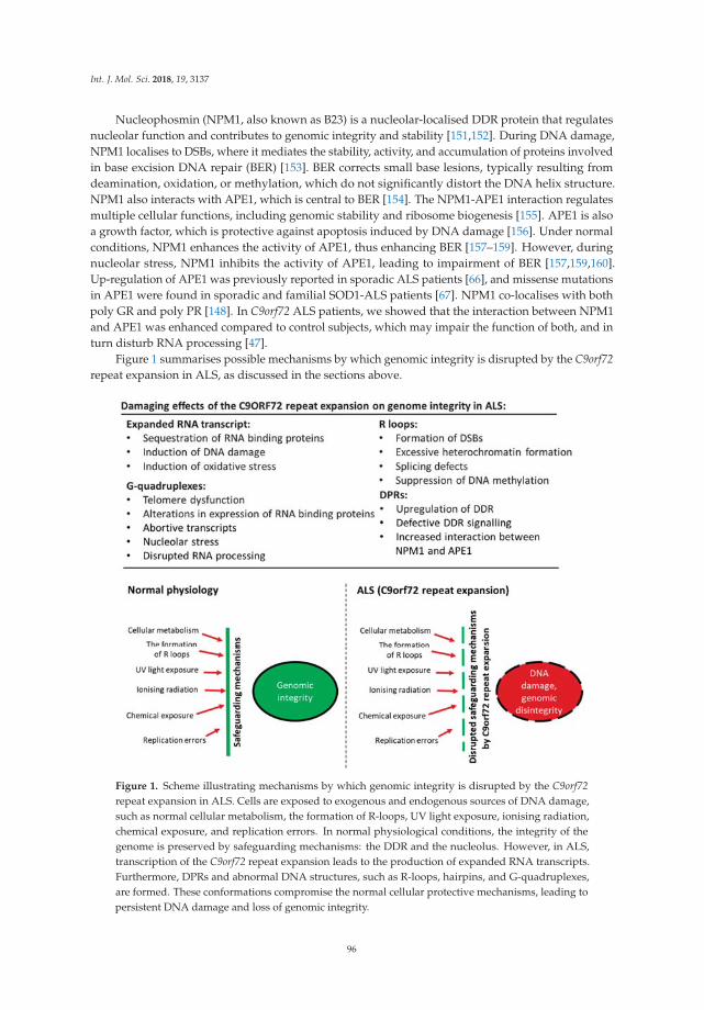
Int. J. Mol. Sci. 2018, 19, 3137
Nucleophosmin (NPM1, also known as B23) is a nucleolar-localised DDR protein that regulatesnucleolar function and contributes to genomic integrity and stability [151,152]. During DNA damage,NPM1 localises to DSBs, where it mediates the stability, activity, and accumulation of proteins involvedin base excision DNA repair (BER) [153]. BER corrects small base lesions, typically resulting fromdeamination, oxidation, or methylation, which do not significantly distort the DNA helix structure.NPM1 also interacts with APE1, which is central to BER [154]. The NPM1-APE1 interaction regulatesmultiple cellular functions, including genomic stability and ribosome biogenesis [155]. APE1 is alsoa growth factor, which is protective against apoptosis induced by DNA damage [156]. Under normalconditions, NPM1 enhances the activity of APE1, thus enhancing BER [157–159]. However, duringnucleolar stress, NPM1 inhibits the activity of APE1, leading to impairment of BER [157,159,160].Up-regulation of APE1 was previously reported in sporadic ALS patients [66], and missense mutationsin APE1 were found in sporadic and familial SOD1-ALS patients [67]. NPM1 co-localises with bothpoly GR and poly PR [148]. In C9orf72 ALS patients, we showed that the interaction between NPM1and APE1 was enhanced compared to control subjects, which may impair the function of both, and inturn disturb RNA processing [47].
Figure 1 summarises possible mechanisms by which genomic integrity is disrupted by the C9orf72repeat expansion in ALS, as discussed in the sections above.
Figure 1. Scheme illustrating mechanisms by which genomic integrity is disrupted by the C9orf72repeat expansion in ALS. Cells are exposed to exogenous and endogenous sources of DNA damage,such as normal cellular metabolism, the formation of R-loops, UV light exposure, ionising radiation,chemical exposure, and replication errors. In normal physiological conditions, the integrity of thegenome is preserved by safeguarding mechanisms: the DDR and the nucleolus. However, in ALS,transcription of the C9orf72 repeat expansion leads to the production of expanded RNA transcripts.Furthermore, DPRs and abnormal DNA structures, such as R-loops, hairpins, and G-quadruplexes,are formed. These conformations compromise the normal cellular protective mechanisms, leading topersistent DNA damage and loss of genomic integrity.
96

Int. J. Mol. Sci. 2018, 19, 3137
9. Novel Therapeutic Strategies for Amyotrophic Lateral Sclerosis Based on the Inhibition ofDNA Damage and Abnormal DNA Structures.
The increasing evidence that both the repeat RNA and DPRs contribute to toxicity implies thattherapeutic strategies based on targeting both factors may be effective in ALS. In addition, there isevidence that the C9orf72 repeat expansion is upstream of the TDP-43 pathology which is present inalmost all ALS cases [161,162], further implying that targeting this region could be an effective strategyin ALS. Abnormal nucleic acid structures, such as G-quadruplexes and R-loops, are increasinglyrecognised as promising drug targets, and several small molecules are being developed to targetthese sites.
There are several lines of evidence implying that the targeting the G-quadruplexes formed by theC9orf72 repeat expansion could be an effective therapeutic strategy for C9orf72-ALS and FTD. Targetingthe hairpin conformation of the C9orf72 repeat expansion with small chemical lead compounds wasprotective against the formation of RNA foci and RAN translation [163]. Recently, three small moleculeswere isolated from chemical libraries that bind and stabilise the C9orf72 G-quadruplex structure [164].Moreover, these compounds reduced the formation of both C9orf72 RNA foci and DPRs in iPSC-derivedmotor neurons, and they improved survival in fly models expressing poly GR [164]. A porphyrincompound, TMPyP4, also stabilized C9orf72 G-quadruplexes, reduced the affinity of RanGAP1, a keyregulator of nucleocytoplasmic transport, and suppressed nuclear import deficits in fly models [150].
Recent studies indicating that DNA repair pathways are dysregulated by the C9orf72 repeatexpansion implies that modulation of the DDR or enhancement of DNA repair processing are alsopromising therapeutic strategies for ALS. Indeed, there are already many compounds in developmentthat modulate the DDR for cancer therapy. Hence, similar approaches could be examined for ALSand other neurodegenerative disorders. An important tumour suppressor, p53, protects the genomeby regulating a variety of DDR mechanisms and controls the induction of apoptosis in genomicallycompromised cells. Not surprisingly, a broad range of approaches for modulating or inhibiting p53activity are already underway in studies of cancer therapy [165–167]. Interestingly, partial inhibitionof the p53 pathway partially suppressed poly GR80 induced toxicity in iPSC-derived patient motorneurons [144]. Another approach is based on the impairment of ATM activation and the suppressionof 53BP1 recruitment to DSBs [46] by the C9orf72 repeat expansion. This implies that inhibition ofnegative regulators of DNA repair, such as the PI3K/AKT mammalian target of rapamycin (mTOR)pathway, which negatively controls ATM [168], may be beneficial for ALS. Consistent with this notion,we also demonstrated the down-regulation of PI3K and p-eIF4G in C9orf72 patient tissues comparedto controls [47], in concordance with previous studies demonstrating dysregulation of the AKT/PI3Kpathway in ALS motor neurons [169–171]. Alternatively, D52 is another recently recognised negativeregulator of ATM signalling [172]. Similarly, nucleolar stress also triggers down-regulation of themTOR pathway [173], and the evidence that the C9orf72 repeat expansion induces nucleolar stressimplies that targeting this mechanism could be an important therapeutic strategy in ALS. We alsopreviously showed that overexpression of NPM1 inhibited apoptosis in neuronal cells expressingpoly (PR)100 or poly (GR)100, suggesting that depletion of NPM1 is linked to cell death in ALS [47].These findings suggest that inhibition of nucleolar stress should be investigated in more detail inrelation to C9orf72-ALS.
Ultimately however, a potentially more effective way to reduce toxicity of the C9orf72 repeatexpansion is by inhibiting its production in the first place. Antisense oligonucleotides (ASOs) areshort, single-stranded oligonucleotides that can be designed to hybridize to specific RNAs and thusmodulate gene expression [174]. Importantly, ASOs targeting the C9orf72 repeat expansion are currentlyshowing promise for ALS. ASO treatment targeting poly GP reduced both repeat-containing RNA fociand poly GP concentrations in C9orf72 ALS iPSC-derived neurons, although poly GP is particularlystable and required 10 days of ASO treatment to be significantly reduced [175]. In ASO-treated mice,concentrations of C9orf72 repeat-containing RNA were reduced approximately 50%, without affectingendogenous C9orf72 mRNA. Similarly, poly GP concentrations in cerebrospinal fluid (CSF) and the
97

Int. J. Mol. Sci. 2018, 19, 3137
brain, as well as RNA foci and poly GP-containing inclusions, were reduced significantly in themotor cortex of the mouse model of C9orf72 ALS [175]. Furthermore, ASOs targeting C9orf72 RNAprevented nuclear import impairment by the C9orf72 repeat expansion in fly models, as well as inC9orf72 iPSC-derived neurons, and suppressed neurodegeneration [150]. In addition, ASOs selectivelyreduced the accumulation of C9orf72 GGGGCC sense strand-containing RNA foci, without significantlyaffecting the level of RNAs encoding C9orf72 itself. Similarly, ASOs targeting the C9orf72 transcriptsuppressed GGGGCC repeat-containing RNA foci formation, and reversed membrane excitabilitydefects in C9orf72-ALS motor neurons differentiated from iPSCs [106]. Hence these studies revealthat ASOs are capable of reducing pathogenic, expansion-containing RNAs without inducing C9orf72protein loss [176].
10. Conclusions
There is now convincing evidence that diseases resulting from the expansion of abnormal repeatsequences are nucleic acid diseases, and in C9orf72-ALS, DNA damage and loss of genome integrityare implicated in pathophysiology. In Figure 2, we illustrate the possible mechanisms by whichthe C9orf72 mutation induces toxicity and genomic instability in ALS. The production of aberrant,toxic nucleic acid conformations formed by the C9orf72 hexanucleotide (GGGGCC) repeat expansion(particularly R-loops) induces DNA damage. This results from impairment of DDR signalling anddysfunction in RNA processing, which compromises genetic integrity and triggers neurodegeneration.Furthermore, the DPRs produced by RAN translation of the C9orf72 repeat expansion, particularly thearginine-containing peptides poly GR and poly PR, are also capable of triggering DNA damage byinducing stress in the nucleolus. They also perturb DNA repair by p62 accumulation and impairingNPM1–APE1 functions. In addition, oxidative stress involving APE1 also further compounds thedamage. Hence, expression of the C9orf72 repeat expansion results in a “double whammy” for the cell:both RNA- and DPR-driven mechanisms conspire to produce extensive DNA damage and genomicinstability, leading to motor neuron death. Further studies are now warranted to determine exactlyhow DNA repair mechanisms are compromised in ALS, the extent to which the DDR is induced,and which specific pathways are compromised. Modulation of the DDR, enhancement of DNA repairprocesses, and the targeting of G-quadruplexes and R-loops, may therefore be novel neuroprotectivestrategies for ALS that should be harnessed in future studies.
98

Int. J. Mol. Sci. 2018, 19, 3137
Figure 2. Mechanisms of amyotrophic lateral sclerosis (ALS) pathogenesis induced by the chromosome9 open reading frame 72 (C9orf72) repeat expansion. The C9orf72 repeat expansion forms abnormalnucleic acid structures, including R-loops, which perturb DNA repair processes involving ATM,and probably other mechanisms. The expanded RNA forms foci and sequesters RNA bindingproteins, leading to dysfunction in RNA processing. The C9orf72 DPRs, produced by RAN translation,also accumulate in the nucleolus, leading to perturbations in nucleolar function, including DNArepair processes, ribosomal biogenesis, and APE-dependent mechanisms (including oxidative stress).They also impair DNA repair by p62-dependent mechanisms. Together, these events result in theaccumulation of DNA damage, genome instability, and motor neuron death.
99
Int. J. Mol. Sci. 2018, 19, 3137
Author Contributions: Both authors, J.D.A. and A.K., conceived, co-wrote, and edited the article throughout forcontent and style consistency; and prepared the figures.
Funding: This research was funded by National Health and Medical Research Council: 10305133, 1086887, 1095215.
Acknowledgments: This work was supported by the National Health and Medical Research Council of Australia(NHMRC) Project grants (10305133 and 1086887), the MND Research Institute of Australia, and an NHMRCDementia Team research grant (1095215).
Conflicts of Interest: The authors have no conflict of interest to declare.
References
1. Liu, W.F.; Yu, S.S.; Chen, G.J.; Li, Y.Z. DNA damage checkpoint, damage repair, and genome stability.Yi Chuan Xue Bao 2006, 33, 381–390. [CrossRef]
2. Martin, L.J. DNA damage and repair: Relevance to mechanisms of neurodegeneration. J. Neuropathol.Exp. Neurol. 2008, 67, 377–387. [CrossRef] [PubMed]
3. Pan, L.; Penney, J.; Tsai, L.H. Chromatin regulation of DNA damage repair and genome integrity in thecentral nervous system. J. Mol. Biol. 2014, 426, 3376–3388. [CrossRef] [PubMed]
4. Hegde, M.L.; Mantha, A.K.; Hazra, T.K.; Bhakat, K.K.; Mitra, S.; Szczesny, B. Oxidative genome damageand its repair: Implications in aging and neurodegenerative diseases. Mech. Ageing Dev. 2012, 133, 157–168.[CrossRef] [PubMed]
5. Madabhushi, R.; Pan, L.; Tsai, L.H. DNA damage and its links to neurodegeneration. Neuron 2014, 83,266–282. [CrossRef] [PubMed]
6. Obulesu, M.; Rao, D.M. DNA damage and impairment of DNA repair in Alzheimer’s disease. Int. J. Neurosci.2010, 120, 397–403. [CrossRef] [PubMed]
7. McKinnon, P.J. ATM and the molecular pathogenesis of ataxia telangiectasia. Ann. Rev. Pathol. 2012, 7,303–321. [CrossRef] [PubMed]
8. Dorst, J.; Ludolph, A.C.; Huebers, A. Disease-modifying and symptomatic treatment of amyotrophic lateralsclerosis. Ther. Adv. Neurol. Disord. 2018, 11, 1756285617734734. [CrossRef] [PubMed]
9. Wijesekera, L.C.; Leigh, P.N. Amyotrophic lateral sclerosis Orphanet. J. Rare Dis. 2009, 4, 3. [CrossRef]10. Ludolph, A.C.; Brettschneider, J.; Weishaupt, J.H. Amyotrophic lateral sclerosis. Curr. Opin. Neurol. 2012, 25,
530–535. [CrossRef] [PubMed]11. Liscic, R.M. Als and Ftd: Insights into the disease mechanisms and therapeutic targets. Eur. J. Pharmacol.
2017, 817, 2–6. [CrossRef] [PubMed]12. Shahheydari, H.; Ragagnin, A.; Walker, A.K.; Toth, R.P.; Vidal, M.; Jagaraj, C.J.; Perri, E.R.; Konopka, A.;
Sultana, J.M.; Atkin, J.D. Protein Quality Control and the Amyotrophic Lateral Sclerosis/FrontotemporalDementia Continuum. Front. Mol. Neurosci. 2017, 10, 119. [CrossRef] [PubMed]
13. DeJesus-Hernandez, M.; Mackenzie, I.R.; Boeve, B.F.; Boxer, A.L.; Baker, M.; Rutherford, N.J.;Nicholson, A.M.; Finch, N.A.; Flynn, H.; Adamson, J. Expanded GGGGCC hexanucleotide repeat innoncoding region of C9ORF72 causes chromosome 9p-linked FTD and ALS. Neuron 2011, 72, 245–256.[CrossRef] [PubMed]
14. Renton, A.E.; Majounie, E.; Waite, A.; Simón-Sánchez, J.; Rollinson, S.; Gibbs, J.R.; Schymick, J.C.;Laaksovirta, H.; Van Swieten, J.C.; Myllykangas, L. A hexanucleotide repeat expansion in C9ORF72 isthe cause of chromosome 9p21-linked ALS-FTD. Neuron 2011, 72, 257–268. [CrossRef] [PubMed]
15. Devenney, E.; Hornberger, M.; Irish, M.; Mioshi, E.; Burrell, J.; Tan, R.; Kiernan, M.C.; Hodges, J.R.Frontotemporal dementia associated with the C9ORF72 mutation: A unique clinical profile. JAMA Neurol.2014, 71, 331–339. [CrossRef] [PubMed]
16. Sreedharan, J.; Blair, I.P.; Tripathi, V.B.; Hu, X.; Vance, C.; Rogelj, B.; Ackerley, S.; Durnall, J.C.; Williams, K.L.;Buratti, E.; et al. TDP-43 mutations in familial and sporadic amyotrophic lateral sclerosis. Science 2008, 319,1668–1672. [CrossRef] [PubMed]
17. Rutherford, N.J.; Zhang, Y.J.; Baker, M.; Gass, J.M.; Finch, N.A.; Xu, Y.F.; Stewart, H.; Kelley, B.J.; Kuntz, K.;Crook, R.J.; et al. Novel mutations in TARDBP (TDP-43) in patients with familial amyotrophic lateralsclerosis. PLoS Genet. 2008, 4, e1000193. [CrossRef] [PubMed]
100

Int. J. Mol. Sci. 2018, 19, 3137
18. Borroni, B.; Alberici, A.; Archetti, S.; Magnani, E.; Di Luca, M.; Padovani, A. New insights into biologicalmarkers of frontotemporal lobar degeneration spectrum. Curr. Med. Chem. 2010, 17, 1002–1009. [CrossRef][PubMed]
19. Vance, C.; Rogelj, B.; Hortobagyi, T.; De Vos, K.J.; Nishimura, A.L.; Sreedharan, J.; Hu, X.; Smith, B.; Ruddy, D.;Wright, P.; et al. Mutations in FUS, an RNA processing protein, cause familial amyotrophic lateral sclerosistype 6. Science 2009, 323, 1208–1211. [CrossRef] [PubMed]
20. Kwiatkowski, T.J., Jr.; Bosco, D.A.; Leclerc, A.L.; Tamrazian, E.; Vanderburg, C.R.; Russ, C.; Davis, A.;Gilchrist, J.; Kasarskis, E.J.; Munsat, T.; et al. Mutations in the FUS/TLS gene on chromosome 16 causefamilial amyotrophic lateral sclerosis. Science 2009, 323, 1205–1208. [CrossRef] [PubMed]
21. Yan, J.; Deng, H.X.; Siddique, N.; Fecto, F.; Chen, W.; Yang, Y.; Liu, E.; Donkervoort, S.; Zheng, J.G.; Shi, Y.; et al.Frameshift and novel mutations in FUS in familial amyotrophic lateral sclerosis and ALS/dementia.Neurology 2010, 75, 807–814. [CrossRef] [PubMed]
22. Cirulli, E.T.; Lasseigne, B.N.; Petrovski, S.; Sapp, P.C.; Dion, P.A.; Leblond, C.S.; Couthouis, J.; Lu, Y.F.;Wang, Q.; Krueger, B.J.; et al. Exome sequencing in amyotrophic lateral sclerosis identifies risk genes andpathways. Science 2015, 347, 1436–1441. [CrossRef] [PubMed]
23. Freischmidt, A.; Wieland, T.; Richter, B.; Ruf, W.; Schaeffer, V.; Muller, K.; Marroquin, N.; Nordin, F.;Hubers, A.; Weydt, P.; et al. Haploinsufficiency of TBK1 causes familial ALS and fronto-temporal dementia.Nat. Neurosci. 2015, 18, 631–636. [CrossRef] [PubMed]
24. van der Zee, J.; Gijselinck, I.; Van Mossevelde, S.; Perrone, F.; Dillen, L.; Heeman, B.; Baumer, V.;Engelborghs, S.; De Bleecker, J.; Baets, J.; et al. TBK1 Mutation Spectrum in an Extended European PatientCohort with Frontotemporal Dementia and Amyotrophic Lateral Sclerosis. Hum. Mutat. 2017, 38, 297–309.[CrossRef] [PubMed]
25. Deng, H.X.; Chen, W.; Hong, S.T.; Boycott, K.M.; Gorrie, G.H.; Siddique, N.; Yang, Y.; Fecto, F.; Shi, Y.;Zhai, H.; et al. Mutations in UBQLN2 cause dominant X-linked juvenile and adult-onset ALS andALS/dementia. Nature 2011, 477, 211–215. [CrossRef] [PubMed]
26. Williams, K.L.; Warraich, S.T.; Yang, S.; Solski, J.A.; Fernando, R.; Rouleau, G.A.; Nicholson, G.A.; Blair, I.P.UBQLN2/ubiquilin 2 mutation and pathology in familial amyotrophic lateral sclerosis. Neurobiol. Aging2012, 33, 2527.e3–2527.e10. [CrossRef] [PubMed]
27. Gellera, C.; Tiloca, C.; Del Bo, R.; Corrado, L.; Pensato, V.; Agostini, J.; Cereda, C.; Ratti, A.; Castellotti, B.;Corti, S.; et al. Ubiquilin 2 mutations in Italian patients with amyotrophic lateral sclerosis and frontotemporaldementia. J. Neurol. Neurosurg. Psychiatry 2013, 84, 183–187. [CrossRef] [PubMed]
28. Synofzik, M.; Maetzler, W.; Grehl, T.; Prudlo, J.; Vom Hagen, J.M.; Haack, T.; Rebassoo, P.; Munz, M.;Schols, L.; Biskup, S. Screening in ALS and FTD patients reveals 3 novel UBQLN2 mutations outside thePXX domain and a pure FTD phenotype. Neurobiol. Aging 2012, 33, 2949.e13–2949.e17. [CrossRef] [PubMed]
29. Maruyama, H.; Morino, H.; Ito, H.; Izumi, Y.; Kato, H.; Watanabe, Y.; Kinoshita, Y.; Kamada, M.; Nodera, H.;Suzuki, H.; et al. Mutations of optineurin in amyotrophic lateral sclerosis. Nature 2010, 465, 223–226.[CrossRef] [PubMed]
30. Pottier, C.; Bieniek, K.F.; Finch, N.; van de Vorst, M.; Baker, M.; Perkersen, R.; Brown, P.; Ravenscroft, T.; vanBlitterswijk, M.; Nicholson, A.M.; et al. Whole-genome sequencing reveals important role for TBK1 andOPTN mutations in frontotemporal lobar degeneration without motor neuron disease. Acta Neuropathol.2015, 130, 77–92. [CrossRef] [PubMed]
31. Williams, K.L.; Topp, S.; Yang, S.; Smith, B.; Fifita, J.A.; Warraich, S.T.; Zhang, K.Y.; Farrawell, N.; Vance, C.;Hu, X.; et al. CCNF mutations in amyotrophic lateral sclerosis and frontotemporal dementia. Nat. Commun.2016, 7, 11253. [CrossRef] [PubMed]
32. Therrien, M.; Dion, P.A.; Rouleau, G.A. ALS: Recent Developments from Genetics Studies. Curr. Neurol.Neurosci. Rep. 2016, 16, 59. [CrossRef] [PubMed]
33. Parakh, S.; Atkin, J.D. Protein folding alterations in amyotrophic lateral sclerosis. Brain Res. 2016, 1648,633–649. [CrossRef] [PubMed]
34. Blokhuis, A.M.; Groen, E.J.; Koppers, M.; van den Berg, L.H.; Pasterkamp, R.J. Protein aggregation inamyotrophic lateral sclerosis. Acta Neuropathol. 2013, 125, 777–794. [CrossRef] [PubMed]
101

Int. J. Mol. Sci. 2018, 19, 3137
35. Neumann, M.; Kwong, L.K.; Lee, E.B.; Kremmer, E.; Flatley, A.; Xu, Y.; Forman, M.S.; Troost, D.;Kretzschmar, H.A.; Trojanowski, J.Q.; et al. Phosphorylation of S409/410 of TDP-43 is a consistent feature inall sporadic and familial forms of TDP-43 proteinopathies. Acta Neuropathol. 2009, 117, 137–149. [CrossRef][PubMed]
36. Mackenzie, I.R.; Neumann, M. FET proteins in frontotemporal dementia and amyotrophic lateral sclerosis.Brain Res. 2012, 1462, 40–43. [CrossRef] [PubMed]
37. Ling, S.C.; Polymenidou, M.; Cleveland, D.W. Converging mechanisms in ALS and FTD: Disrupted RNAand protein homeostasis. Neuron 2013, 79, 416–438. [CrossRef] [PubMed]
38. Buratti, E.; Baralle, F.E. The multiple roles of TDP-43 in pre-mRNA processing and gene expression regulation.RNA Biol. 2010, 7, 420–429. [CrossRef] [PubMed]
39. Ito, D.; Seki, M.; Tsunoda, Y.; Uchiyama, H.; Suzuki, N. Nuclear transport impairment of amyotrophic lateralsclerosis-linked mutations in FUS/TLS. Ann. Neurol. 2011, 69, 152–162. [CrossRef] [PubMed]
40. Naumann, M.; Pal, A.; Goswami, A.; Lojewski, X.; Japtok, J.; Vehlow, A.; Naujock, M.; Gunther, R.;Jin, M.; Stanslowsky, N.; et al. Impaired DNA damage response signaling by FUS-NLS mutations leads toneurodegeneration and FUS aggregate formation. Nat. Commun. 2018, 9, 335. [CrossRef] [PubMed]
41. Dormann, D.; Rodde, R.; Edbauer, D.; Bentmann, E.; Fischer, I.; Hruscha, A.; Than, M.E.; Mackenzie, I.R.;Capell, A.; Schmid, B.; et al. ALS-associated fused in sarcoma (FUS) mutations disrupt Transportin-mediatednuclear import. EMBO J. 2010, 29, 2841–2857. [CrossRef] [PubMed]
42. Liu, Y.J.; Tsai, P.Y.; Chern, Y. Energy Homeostasis and Abnormal RNA Metabolism in Amyotrophic LateralSclerosis. Front. Cell Neurosci. 2017, 11, 126. [CrossRef] [PubMed]
43. Wang, H.; Guo, W.; Mitra, J.; Hegde, P.M.; Vandoorne, T.; Eckelmann, B.J.; Mitra, S.; Tomkinson, A.E.;Van Den Bosch, L.; Hegde, M.L. Mutant FUS causes DNA ligation defects to inhibit oxidative damage repairin Amyotrophic Lateral Sclerosis. Nat. Commun. 2018, 9, 3683. [CrossRef] [PubMed]
44. Higelin, J.; Catanese, A.; Semelink-Sedlacek, L.L.; Oeztuerk, S.; Lutz, A.K.; Bausinger, J.; Barbi, G.; Speit, G.;Andersen, P.M.; Ludolph, A.C.; et al. NEK1 loss-of-function mutation induces DNA damage accumulationin ALS patient-derived motoneurons. Stem. Cell Res. 2018, 30, 150–162. [CrossRef] [PubMed]
45. Gong, J.; Huang, M.; Wang, F.; Ma, X.; Liu, H.; Tu, Y.; Xing, L.; Zhu, X.; Zheng, H.; Fang, J.; et al. RBM45competes with HDAC1 for binding to FUS in response to DNA damage. Nucleic. Acids Res. 2017, 45,12862–12876. [CrossRef] [PubMed]
46. Walker, C.; Herranz-Martin, S.; Karyka, E.; Liao, C.; Lewis, K.; Elsayed, W.; Lukashchuk, V.; Chiang, S.C.;Ray, S.; Mulcahy, P.J.; et al. C9orf72 expansion disrupts ATM-mediated chromosomal break repair.Nat. Neurosci. 2017, 20, 1225–1235. [CrossRef] [PubMed]
47. Farg, M.A.; Konopka, A.; Ying Soo, K.; Ito, D.; Atkin, J.D. The DNA damage response (DDR) is induced bythe C9orf72 repeat expansion in Amyotrophic Lateral Sclerosis. Hum. Mol. Genet. 2017. [CrossRef] [PubMed]
48. Muyderman, H.; Chen, T. Mitochondrial dysfunction in amyotrophic lateral sclerosis-a valid pharmacologicaltarget? Br. J. Pharmacol. 2014, 171, 2191–2205. [CrossRef] [PubMed]
49. Liu, E.Y.; Cali, C.P.; Lee, E.B. RNA metabolism in neurodegenerative disease. Dis. Model. Mech. 2017, 10,509–518. [CrossRef] [PubMed]
50. Ramesh, N.; Pandey, U.B. Autophagy Dysregulation in ALS: When Protein Aggregates Get Out of Hand.Front. Mol. Neurosci. 2017, 10, 263. [CrossRef] [PubMed]
51. Kabashi, E.; Agar, J.N.; Strong, M.J.; Durham, H.D. Impaired proteasome function in sporadic amyotrophiclateral sclerosis. Amyotroph. Lateral Scler. 2012, 13, 367–371. [CrossRef] [PubMed]
52. Pollari, E.; Goldsteins, G.; Bart, G.; Koistinaho, J.; Giniatullin, R. The role of oxidative stress in degenerationof the neuromuscular junction in amyotrophic lateral sclerosis. Front. Cell. Neurosci. 2014, 8, 131. [CrossRef][PubMed]
53. Dutertre, M.; Lambert, S.; Carreira, A.; Amor-Gueret, M.; Vagner, S. DNA damage: RNA-binding proteinsprotect from near and far. Trends Biochem. Sci. 2014, 39, 141–149. [CrossRef] [PubMed]
54. Sharma, S. Age-related nonhomologous end joining activity in rat neurons. Brain Res. Bull. 2007, 73, 48–54.[CrossRef] [PubMed]
55. Rodgers, K.; McVey, M. Error-Prone Repair of DNA Double-Strand Breaks. J. Cell Physiol. 2016, 231, 15–24.[CrossRef] [PubMed]
56. Ogawa, L.M.; Baserga, S.J. Crosstalk between the nucleolus and the DNA damage response. Mol. Biosyst.2017, 13, 443–455. [CrossRef] [PubMed]
102

Int. J. Mol. Sci. 2018, 19, 3137
57. Todd, P.K.; Paulson, H.L. RNA-mediated neurodegeneration in repeat expansion disorders. Ann. Neurol.2010, 67, 291–300. [CrossRef] [PubMed]
58. La Spada, A.R.; Taylor, J.P. Repeat expansion disease: Progress and puzzles in disease pathogenesis.Nat. Rev. Genet. 2010, 11, 247–258. [CrossRef] [PubMed]
59. Sordet, O.; Redon, C.E.; Guirouilh-Barbat, J.; Smith, S.; Solier, S.; Douarre, C.; Conti, C.; Nakamura, A.J.;Das, B.B.; Nicolas, E.; et al. Ataxia telangiectasia mutated activation by transcription- and topoisomeraseI-induced DNA double-strand breaks. EMBO Rep. 2009, 10, 887–893. [CrossRef] [PubMed]
60. Bosshard, M.; Markkanen, E.; van Loon, B. Base excision repair in physiology and pathology of the centralnervous system. Int. J. Mol. Sci. 2012, 13, 16172–16222. [CrossRef] [PubMed]
61. Shaikh, A.Y.; Martin, L.J. DNA base-excision repair enzyme apurinic/apyrimidinic endonuclease/redoxfactor-1 is increased and competent in the brain and spinal cord of individuals with amyotrophic lateralsclerosis. Neuromol. Med. 2002, 2, 47–60. [CrossRef] [PubMed]
62. Martin, L.J.; Liu, Z.; Chen, K.; Price, A.C.; Pan, Y.; Swaby, J.A.; Golden, W.C. Motor neuron degeneration inamyotrophic lateral sclerosis mutant superoxide dismutase-1 transgenic mice: Mechanisms of mitochondriopathyand cell death. J. Comp. Neurol. 2007, 500, 20–46. [CrossRef] [PubMed]
63. Kim, S.H.; Engelhardt, J.I.; Henkel, J.S.; Siklos, L.; Soos, J.; Goodman, C.; Appel, S.H. Widespread increasedexpression of the DNA repair enzyme PARP in brain in ALS. Neurology 2004, 62, 319–322. [CrossRef][PubMed]
64. Martin, L.J. Transgenic mice with human mutant genes causing Parkinson’s disease and amyotrophic lateralsclerosis provide common insight into mechanisms of motor neuron selective vulnerability to degeneration.Rev. Neurosci. 2007, 18, 115–136. [CrossRef] [PubMed]
65. Barbosa, L.F.; Cerqueira, F.M.; Macedo, A.F.; Garcia, C.C.; Angeli, J.P.; Schumacher, R.I.; Sogayar, M.C.;Augusto, O.; Carri, M.T.; Di Mascio, P.; et al. Increased SOD1 association with chromatin, DNA damage, p53activation, and apoptosis in a cellular model of SOD1-linked ALS. Biochim. Biophys. Acta 2010, 1802, 462–471.[CrossRef] [PubMed]
66. Ferrante, R.J.; Browne, S.E.; Shinobu, L.A.; Bowling, A.C.; Baik, M.J.; MacGarvey, U.; Kowall, N.W.;Brown, R.H., Jr.; Beal, M.F. Evidence of increased oxidative damage in both sporadic and familial amyotrophiclateral sclerosis. J. Neurochem. 1997, 69, 2064–2074. [CrossRef] [PubMed]
67. Olkowski, Z.L. Mutant AP endonuclease in patients with amyotrophic lateral sclerosis. Neuroreport 1998, 9,239–242. [CrossRef] [PubMed]
68. Selfridge, J.; Song, L.; Brownstein, D.G.; Melton, D.W. Mice with DNA repair gene Ercc1 deficiency in a neuralcrest lineage are a model for late-onset Hirschsprung disease. DNA Repair 2010, 9, 653–660. [CrossRef][PubMed]
69. Qiu, H.; Lee, S.; Shang, Y.; Wang, W.Y.; Au, K.F.; Kamiya, S.; Barmada, S.J.; Finkbeiner, S.; Lui, H.;Carlton, C.E.; et al. ALS-associated mutation FUS-R521C causes DNA damage and RNA splicing defects.J. Clin. Investig. 2014, 124, 981–999. [CrossRef] [PubMed]
70. Deng, Q.; Holler, C.J.; Taylor, G.; Hudson, K.F.; Watkins, W.; Gearing, M.; Ito, D.; Murray, M.E.; Dickson, D.W.;Seyfried, N.T.; et al. FUS is phosphorylated by DNA-PK and accumulates in the cytoplasm after DNAdamage. J. Neurosci. 2014, 34, 7802–7813. [CrossRef] [PubMed]
71. Gardiner, M.; Toth, R.; Vandermoere, F.; Morrice, N.A.; Rouse, J. Identification and characterization ofFUS/TLS as a new target of ATM. Biochem. J. 2008, 415, 297–307. [CrossRef] [PubMed]
72. Wang, W.Y.; Pan, L.; Su, S.C.; Quinn, E.J.; Sasaki, M.; Jimenez, J.C.; Mackenzie, I.R.; Huang, E.J.; Tsai, L.H.Interaction of FUS and HDAC1 regulates DNA damage response and repair in neurons. Nat. Neurosci. 2013,16, 1383–1391. [CrossRef] [PubMed]
73. Hill, S.J.; Mordes, D.A.; Cameron, L.A.; Neuberg, D.S.; Landini, S.; Eggan, K.; Livingston, D.M. Two familialALS proteins function in prevention/repair of transcription-associated DNA damage. Proc. Natl. Acad.Sci. USA 2016, 113, E7701–E7709. [CrossRef] [PubMed]
74. Skourti-Stathaki, K.; Proudfoot, N.J.; Gromak, N. Human senataxin resolves RNA/DNA hybrids formed attranscriptional pause sites to promote Xrn2-dependent termination. Mol. Cell 2011, 42, 794–805. [CrossRef][PubMed]
75. Chen, Y.Z.; Bennett, C.L.; Huynh, H.M.; Blair, I.P.; Puls, I.; Irobi, J.; Dierick, I.; Abel, A.; Kennerson, M.L.;Rabin, B.A.; et al. DNA/RNA helicase gene mutations in a form of juvenile amyotrophic lateral sclerosis(ALS4). Am. J. Hum. Genet. 2004, 74, 1128–1135. [CrossRef] [PubMed]
103

Int. J. Mol. Sci. 2018, 19, 3137
76. Koppers, M.; van Blitterswijk, M.M.; Vlam, L.; Rowicka, P.A.; van Vught, P.W.; Groen, E.J.; Spliet, W.G.;Engelen-Lee, J.; Schelhaas, H.J.; de Visser, M.; et al. VCP mutations in familial and sporadic amyotrophiclateral sclerosis. Neurobiol. Aging 2012, 33, e813–e837. [CrossRef] [PubMed]
77. Vaz, B.; Halder, S.; Ramadan, K. Role of p97/VCP (Cdc48) in genome stability. Front. Genet. 2013, 4, 60.[CrossRef] [PubMed]
78. D’Angiolella, V.; Esencay, M.; Pagano, M. A cyclin without cyclin-dependent kinases: Cyclin F controlsgenome stability through ubiquitin-mediated proteolysis. Trends Cell Biol. 2013, 23, 135–140. [CrossRef][PubMed]
79. Brenner, D.; Muller, K.; Wieland, T.; Weydt, P.; Bohm, S.; Lule, D.; Hubers, A.; Neuwirth, C.; Weber, M.;Borck, G.; et al. NEK1 mutations in familial amyotrophic lateral sclerosis. Brain 2016, 139, e28. [CrossRef][PubMed]
80. Chen, Y.; Chen, C.F.; Riley, D.J.; Chen, P.L. Nek1 kinase functions in DNA damage response and checkpointcontrol through a pathway independent of ATM and ATR. Cell Cycle 2011, 10, 655–663. [CrossRef] [PubMed]
81. Spies, J.; Waizenegger, A.; Barton, O.; Surder, M.; Wright, W.D.; Heyer, W.D.; Lobrich, M. Nek1 RegulatesRad54 to Orchestrate Homologous Recombination and Replication Fork Stability. Mol. Cell 2016, 62, 903–917.[CrossRef] [PubMed]
82. Fang, X.; Lin, H.; Wang, X.; Zuo, Q.; Qin, J.; Zhang, P. The NEK1 interactor, C21ORF2, is required for efficientDNA damage repair. Acta Biochim. Biophys. Sin. 2015, 47, 834–841. [CrossRef] [PubMed]
83. Rutherford, N.J.; Heckman, M.G.; Dejesus-Hernandez, M.; Baker, M.C.; Soto-Ortolaza, A.I.; Rayaprolu, S.;Stewart, H.; Finger, E.; Volkening, K.; Seeley, W.W.; et al. Length of normal alleles of C9ORF72 GGGGCCrepeat do not influence disease phenotype. Neurobiol. Aging 2012, 33, 2950.e5–2950.e7. [CrossRef] [PubMed]
84. Hubers, A.; Marroquin, N.; Schmoll, B.; Vielhaber, S.; Just, M.; Mayer, B.; Hogel, J.; Dorst, J.; Mertens, T.;Just, W.; et al. Polymerase chain reaction and Southern blot-based analysis of the C9orf72 hexanucleotiderepeat in different motor neuron diseases. Neurobiol Aging 2014, 35, 1214.e1–1214.e6. [CrossRef] [PubMed]
85. Van Mossevelde, S.; van der Zee, J.; Cruts, M.; Van Broeckhoven, C. Relationship between C9orf72 repeatsize and clinical phenotype. Curr. Opin. Genet. Dev. 2017, 44, 117–124. [CrossRef] [PubMed]
86. Smith, B.N.; Newhouse, S.; Shatunov, A.; Vance, C.; Topp, S.; Johnson, L.; Miller, J.; Lee, Y.; Troakes, C.;Scott, K.M.; et al. The C9ORF72 expansion mutation is a common cause of ALS+/-FTD in Europe and hasa single founder. Eur. J. Hum. Genet. 2013, 21, 102–108. [CrossRef] [PubMed]
87. Fratta, P.; Polke, J.M.; Newcombe, J.; Mizielinska, S.; Lashley, T.; Poulter, M.; Beck, J.; Preza, E.; Devoy, A.;Sidle, K.; et al. Screening a UK amyotrophic lateral sclerosis cohort provides evidence of multiple origins ofthe C9orf72 expansion. Neurobiol. Aging 2015, 36, e541–e547. [CrossRef] [PubMed]
88. Rollinson, S.; Halliwell, N.; Young, K.; Callister, J.B.; Toulson, G.; Gibbons, L.; Davidson, Y.S.; Robinson, A.C.;Gerhard, A.; Richardson, A.; et al. Analysis of the hexanucleotide repeat in C9ORF72 in Alzheimer’s disease.Neurobiol Aging 2012, 33, e1845–e1846. [CrossRef] [PubMed]
89. Goldman, J.S.; Quinzii, C.; Dunning-Broadbent, J.; Waters, C.; Mitsumoto, H.; Brannagan, T.H., 3rd;Cosentino, S.; Huey, E.D.; Nagy, P.; Kuo, S.H. Multiple system atrophy and amyotrophic lateral sclerosisin a family with hexanucleotide repeat expansions in C9orf72. JAMA Neurol. 2014, 71, 771–774. [CrossRef][PubMed]
90. Hensman Moss, D.J.; Poulter, M.; Beck, J.; Hehir, J.; Polke, J.M.; Campbell, T.; Adamson, G.; Mudanohwo, E.;McColgan, P.; Haworth, A.; et al. C9orf72 expansions are the most common genetic cause of Huntingtondisease phenocopies. Neurology 2014, 82, 292–299. [CrossRef] [PubMed]
91. Corcia, P.; Vourc’h, P.; Guennoc, A.M.; Del Mar Amador, M.; Blasco, H.; Andres, C.; Couratier, P.; Gordon, P.H.;Meininger, V. Pure cerebellar ataxia linked to large C9orf72 repeat expansion. Amyotroph. Lateral Scler.Frontotemporal. Degener. 2016, 17, 301–303. [CrossRef] [PubMed]
92. Ismail, A.; Cooper-Knock, J.; Highley, J.R.; Milano, A.; Kirby, J.; Goodall, E.; Lowe, J.; Scott, I.;Constantinescu, C.S.; Walters, S.J.; et al. Concurrence of multiple sclerosis and amyotrophic lateral sclerosisin patients with hexanucleotide repeat expansions of C9ORF72. J. Neurol. Neurosurg. Psychiatry 2013, 84,79–87. [CrossRef] [PubMed]
93. Lesage, S.; Le Ber, I.; Condroyer, C.; Broussolle, E.; Gabelle, A.; Thobois, S.; Pasquier, F.; Mondon, K.;Dion, P.A.; Rochefort, D.; et al. C9orf72 repeat expansions are a rare genetic cause of parkinsonism. Brain2013, 136, 385–391. [CrossRef] [PubMed]
104
Int. J. Mol. Sci. 2018, 19, 3137
94. Floris, G.; Borghero, G.; Cannas, A.; Stefano, F.D.; Murru, M.R.; Corongiu, D.; Cuccu, S.; Tranquilli, S.;Marrosu, M.G.; Chio, A.; et al. Bipolar affective disorder preceding frontotemporal dementia in a patientwith C9ORF72 mutation: Is there a genetic link between these two disorders? J. Neurol. 2013, 260, 1155–1157.[CrossRef] [PubMed]
95. Galimberti, D.; Reif, A.; Dell’Osso, B.; Palazzo, C.; Villa, C.; Fenoglio, C.; Kittel-Schneider, S.; Leonhard, C.;Olmes, D.G.; Serpente, M.; et al. C9ORF72 hexanucleotide repeat expansion as a rare cause of bipolardisorder. Bipolar. Disord. 2014, 16, 448–449. [CrossRef] [PubMed]
96. Galimberti, D.; Reif, A.; Dell’osso, B.; Kittel-Schneider, S.; Leonhard, C.; Herr, A.; Palazzo, C.; Villa, C.;Fenoglio, C.; Serpente, M.; et al. C9ORF72 hexanucleotide repeat expansion is a rare cause of schizophrenia.Neurobiol. Aging 2014, 35, e1214–e1217. [CrossRef] [PubMed]
97. Waite, A.J.; Baumer, D.; East, S.; Neal, J.; Morris, H.R.; Ansorge, O.; Blake, D.J. Reduced C9orf72 protein levelsin frontal cortex of amyotrophic lateral sclerosis and frontotemporal degeneration brain with the C9ORF72hexanucleotide repeat expansion. Neurobiol. Aging 2014, 35, 1779.e5–1779.e13. [CrossRef] [PubMed]
98. Burberry, A.; Suzuki, N.; Wang, J.Y.; Moccia, R.; Mordes, D.A.; Stewart, M.H.; Suzuki-Uematsu, S.; Ghosh, S.;Singh, A.; Merkle, F.T.; et al. Loss-of-function mutations in the C9ORF72 mouse ortholog cause fatalautoimmune disease. Sci. Transl. Med. 2016, 8, 347. [CrossRef] [PubMed]
99. Atanasio, A.; Decman, V.; White, D.; Ramos, M.; Ikiz, B.; Lee, H.C.; Siao, C.J.; Brydges, S.; LaRosa, E.;Bai, Y.; et al. C9orf72 ablation causes immune dysregulation characterized by leukocyte expansion,autoantibody production, and glomerulonephropathy in mice. Sci. Rep. 2016, 6, 23204. [CrossRef] [PubMed]
100. Shi, Y.; Lin, S.; Staats, K.A.; Li, Y.; Chang, W.H.; Hung, S.T.; Hendricks, E.; Linares, G.R.; Wang, Y.;Son, E.Y.; et al. Haploinsufficiency leads to neurodegeneration in C9ORF72 ALS/FTD human induced motorneurons. Nat. Med. 2018, 24, 313–325. [CrossRef] [PubMed]
101. Zu, T.; Liu, Y.; Banez-Coronel, M.; Reid, T.; Pletnikova, O.; Lewis, J.; Miller, T.M.; Harms, M.B.; Falchook, A.E.;Subramony, S.H.; et al. RAN proteins and RNA foci from antisense transcripts in C9ORF72 ALS andfrontotemporal dementia. Proc. Natl. Acad. Sci. USA 2013, 110, E4968–E4977. [CrossRef] [PubMed]
102. Donnelly, C.J.; Zhang, P.W.; Pham, J.T.; Haeusler, A.R.; Mistry, N.A.; Vidensky, S.; Daley, E.L.; Poth, E.M.;Hoover, B.; Fines, D.M.; et al. RNA toxicity from the ALS/FTD C9ORF72 expansion is mitigated by antisenseintervention. Neuron 2013, 80, 415–428. [CrossRef] [PubMed]
103. Lee, Y.B.; Chen, H.J.; Peres, J.N.; Gomez-Deza, J.; Attig, J.; Stalekar, M.; Troakes, C.; Nishimura, A.L.;Scotter, E.L.; Vance, C.; et al. Hexanucleotide repeats in ALS/FTD form length-dependent RNA foci,sequester RNA binding proteins, and are neurotoxic. Cell Rep. 2013, 5, 1178–1186. [CrossRef] [PubMed]
104. Cooper-Knock, J.; Walsh, M.J.; Higginbottom, A.; Robin Highley, J.; Dickman, M.J.; Edbauer, D.; Ince, P.G.;Wharton, S.B.; Wilson, S.A.; Kirby, J.; et al. Sequestration of multiple RNA recognition motif-containingproteins by C9orf72 repeat expansions. Brain 2014, 137, 2040–2051. [CrossRef] [PubMed]
105. Prudencio, M.; Belzil, V.V.; Batra, R.; Ross, C.A.; Gendron, T.F.; Pregent, L.J.; Murray, M.E.; Overstreet, K.K.;Piazza-Johnston, A.E.; Desaro, P.; et al. Distinct brain transcriptome profiles in C9orf72-associated andsporadic ALS. Nat. Neurosci. 2015, 18, 1175–1182. [CrossRef] [PubMed]
106. Sareen, D.; O’Rourke, J.G.; Meera, P.; Muhammad, A.K.; Grant, S.; Simpkinson, M.; Bell, S.; Carmona, S.;Ornelas, L.; Sahabian, A.; et al. Targeting RNA foci in iPSC-derived motor neurons from ALS patients witha C9ORF72 repeat expansion. Sci. Transl. Med. 2013, 5, 208. [CrossRef] [PubMed]
107. Cooper-Knock, J.; Bury, J.J.; Heath, P.R.; Wyles, M.; Higginbottom, A.; Gelsthorpe, C.; Highley, J.R.;Hautbergue, G.; Rattray, M.; Kirby, J.; et al. C9ORF72 GGGGCC Expanded Repeats Produce SplicingDysregulation which Correlates with Disease Severity in Amyotrophic Lateral Sclerosis. PLoS ONE 2015, 10,e0127376. [CrossRef] [PubMed]
108. Highley, J.R.; Kirby, J.; Jansweijer, J.A.; Webb, P.S.; Hewamadduma, C.A.; Heath, P.R.; Higginbottom, A.;Raman, R.; Ferraiuolo, L.; Cooper-Knock, J.; et al. Loss of nuclear TDP-43 in amyotrophic lateral sclerosis(ALS) causes altered expression of splicing machinery and widespread dysregulation of RNA splicing inmotor neurones. Neuropathol. Appl. Neurobiol. 2014, 40, 670–685. [CrossRef] [PubMed]
109. Conlon, E.G.; Lu, L.; Sharma, A.; Yamazaki, T.; Tang, T.; Shneider, N.A.; Manley, J.L. The C9ORF72 GGGGCCexpansion forms RNA G-quadruplex inclusions and sequesters hnRNP H to disrupt splicing in ALS brains.Elife 2016, 5. [CrossRef] [PubMed]
110. Cleary, J.D.; Ranum, L.P. Repeat-associated non-ATG (RAN) translation in neurological disease. Hum. Mol. Genet.2013, 22, R45–R51. [CrossRef] [PubMed]
105
Int. J. Mol. Sci. 2018, 19, 3137
111. Sonobe, Y.; Ghadge, G.; Masaki, K.; Sendoel, A.; Fuchs, E.; Roos, R.P. Translation of dipeptide repeat proteinsfrom the C9ORF72 expanded repeat is associated with cellular stress. Neurobiol. Dis. 2018, 116, 155–165.[CrossRef] [PubMed]
112. Cheng, W.; Wang, S.; Mestre, A.A.; Fu, C.; Makarem, A.; Xian, F.; Hayes, L.R.; Lopez-Gonzalez, R.; Drenner, K.;Jiang, J.; et al. C9ORF72 GGGGCC repeat-associated non-AUG translation is upregulated by stress througheIF2alpha phosphorylation. Nat. Commun. 2018, 9, 51. [CrossRef] [PubMed]
113. Tabet, R.; Schaeffer, L.; Freyermuth, F.; Jambeau, M.; Workman, M.; Lee, C.Z.; Lin, C.C.; Jiang, J.;Jansen-West, K.; Abou-Hamdan, H.; et al. CUG initiation and frameshifting enable production of dipeptiderepeat proteins from ALS/FTD C9ORF72 transcripts. Nat. Commun. 2018, 9, 152. [CrossRef] [PubMed]
114. Mizielinska, S.; Gronke, S.; Niccoli, T.; Ridler, C.E.; Clayton, E.L.; Devoy, A.; Moens, T.; Norona, F.E.;Woollacott, I.O.C.; Pietrzyk, J.; et al. C9orf72 repeat expansions cause neurodegeneration in Drosophilathrough arginine-rich proteins. Science 2014, 345, 1192–1194. [CrossRef] [PubMed]
115. Moens, T.G.; Mizielinska, S.; Niccoli, T.; Mitchell, J.S.; Thoeng, A.; Ridler, C.E.; Gronke, S.; Esser, J.;Heslegrave, A.; Zetterberg, H.; et al. Sense and antisense RNA are not toxic in Drosophila models ofC9orf72-associated ALS/FTD. Acta Neuropathol. 2018, 135, 445–457. [CrossRef] [PubMed]
116. Jovicic, A.; Mertens, J.; Boeynaems, S.; Bogaert, E.; Chai, N.; Yamada, S.B.; Paul, J.W., 3rd; Sun, S.; Herdy, J.R.;Bieri, G.; et al. Modifiers of C9orf72 dipeptide repeat toxicity connect nucleocytoplasmic transport defects toFTD/ALS. Nat. Neurosci. 2015, 18, 1226–1229. [CrossRef] [PubMed]
117. Lee, K.H.; Zhang, P.; Kim, H.J.; Mitrea, D.M.; Sarkar, M.; Freibaum, B.D.; Cika, J.; Coughlin, M.;Messing, J.; Molliex, A.; et al. C9orf72 Dipeptide Repeats Impair the Assembly, Dynamics, and Function ofMembrane-Less Organelles. Cell 2016, 167, 774–788.e717. [CrossRef] [PubMed]
118. Kohara, N.; Kaji, R.; Kojima, Y.; Mills, K.R.; Fujii, H.; Hamano, T.; Kimura, J.; Takamatsu, N.; Uchiyama, T.Abnormal excitability of the corticospinal pathway in patients with amyotrophic lateral sclerosis: A singlemotor unit study using transcranial magnetic stimulation. Electroencephalogr. Clin. Neurophysiol. 1996, 101,32–41. [CrossRef]
119. Schutz, B. Imbalanced excitatory to inhibitory synaptic input precedes motor neuron degeneration inan animal model of amyotrophic lateral sclerosis. Neurobiol. Dis. 2005, 20, 131–140. [CrossRef] [PubMed]
120. Suberbielle, E.; Sanchez, P.E.; Kravitz, A.V.; Wang, X.; Ho, K.; Eilertson, K.; Devidze, N.; Kreitzer, A.C.;Mucke, L. Physiologic brain activity causes DNA double-strand breaks in neurons, with exacerbation byamyloid-beta. Nat. Neurosci. 2013, 16, 613–621. [CrossRef] [PubMed]
121. Madabhushi, R.; Gao, F.; Pfenning, A.R.; Pan, L.; Yamakawa, S.; Seo, J.; Rueda, R.; Phan, T.X.; Yamakawa, H.;Pao, P.C.; et al. Activity-Induced DNA Breaks Govern the Expression of Neuronal Early-Response Genes.Cell 2015, 161, 1592–1605. [CrossRef] [PubMed]
122. Mirkin, S.M. Discovery of alternative DNA structures: A heroic decade (1979–1989). Front. Biosci. 2008, 13,1064–1071. [CrossRef] [PubMed]
123. Richard, P.; Manley, J.L. R Loops and Links to Human Disease. J. Mol. Biol. 2017, 429, 3168–3180. [CrossRef][PubMed]
124. Wu, Y.; Brosh, R.M., Jr. G-quadruplex nucleic acids and human disease. FEBS J. 2010, 277, 3470–3488.[CrossRef] [PubMed]
125. Hamperl, S.; Cimprich, K.A. The contribution of co-transcriptional RNA:DNA hybrid structures to DNAdamage and genome instability. DNA Repair 2014, 19, 84–94. [CrossRef] [PubMed]
126. Pearson, C.E.; Nichol Edamura, K.; Cleary, J.D. Repeat instability: Mechanisms of dynamic mutations.Nat. Rev. Genet. 2005, 6, 729–742. [CrossRef] [PubMed]
127. Yu, K.; Chedin, F.; Hsieh, C.L.; Wilson, T.E.; Lieber, M.R. R-loops at immunoglobulin class switch regions inthe chromosomes of stimulated B. cells. Nat. Immunol. 2003, 4, 442–451. [CrossRef] [PubMed]
128. Huppert, J.L.; Balasubramanian, S. G-quadruplexes in promoters throughout the human genome. Nucleic. Acids Res.2007, 35, 406–413. [CrossRef] [PubMed]
129. Simone, R.; Fratta, P.; Neidle, S.; Parkinson, G.N.; Isaacs, A.M. G-quadruplexes: Emerging roles inneurodegenerative diseases and the non-coding transcriptome. FEBS Lett. 2015, 589, 1653–1668. [CrossRef][PubMed]
130. Aguilera, A.; Garcia-Muse, T. R loops: From transcription byproducts to threats to genome stability. Mol. Cell2012, 46, 115–124. [CrossRef] [PubMed]
106

Int. J. Mol. Sci. 2018, 19, 3137
131. Graf, M.; Bonetti, D.; Lockhart, A.; Serhal, K.; Kellner, V.; Maicher, A.; Jolivet, P.; Teixeira, M.T.; Luke, B.Telomere Length Determines TERRA and R-Loop Regulation through the Cell Cycle. Cell 2017, 170, 72–85.e14.[CrossRef] [PubMed]
132. Belotserkovskii, B.P.; Hanawalt, P.C. PNA binding to the non-template DNA strand interferes withtranscription, suggesting a blockage mechanism mediated by R.-loop formation. Mol. Carcinog. 2015, 54,1508–1512. [CrossRef] [PubMed]
133. Groh, M.; Lufino, M.M.; Wade-Martins, R.; Gromak, N. R-loops associated with triplet repeat expansionspromote gene silencing in Friedreich ataxia and fragile X. syndrome. PLoS Genet. 2014, 10, e1004318.[CrossRef] [PubMed]
134. Skourti-Stathaki, K.; Kamieniarz-Gdula, K.; Proudfoot, N.J. R-loops induce repressive chromatin marks overmammalian gene terminators. Nature 2014, 516, 436–439. [CrossRef] [PubMed]
135. Droppelmann, C.A.; Campos-Melo, D.; Ishtiaq, M.; Volkening, K.; Strong, M.J. RNA metabolism in ALS:When normal processes become pathological. Amyotroph Lateral Scler. 2014, 15, 321–336. [CrossRef] [PubMed]
136. Groh, M.; Albulescu, L.O.; Cristini, A.; Gromak, N. Senataxin: Genome Guardian at the Interface ofTranscription and Neurodegeneration. J. Mol. Biol. 2017, 429, 3181–3195. [CrossRef] [PubMed]
137. Chan, Y.A.; Aristizabal, M.J.; Lu, P.Y.; Luo, Z.; Hamza, A.; Kobor, M.S.; Stirling, P.C.; Hieter, P. Genome-wideprofiling of yeast DNA:RNA hybrid prone sites with DRIP-chip. PLoS Genet. 2014, 10, e1004288. [CrossRef][PubMed]
138. Huertas, P.; Aguilera, A. Cotranscriptionally formed DNA:RNA hybrids mediate transcription elongationimpairment and transcription-associated recombination. Mol. Cell 2003, 12, 711–721. [CrossRef] [PubMed]
139. Panigrahi, G.B.; Lau, R.; Montgomery, S.E.; Leonard, M.R.; Pearson, C.E. Slipped (CTG)·(CAG) repeats canbe correctly repaired, escape repair or undergo error-prone repair. Nat. Struct. Mol. Biol. 2005, 12, 654–662.[CrossRef] [PubMed]
140. Haeusler, A.R.; Donnelly, C.J.; Periz, G.; Simko, E.A.; Shaw, P.G.; Kim, M.S.; Maragakis, N.J.; Troncoso, J.C.;Pandey, A.; Sattler, R.; et al. C9orf72 nucleotide repeat structures initiate molecular cascades of disease.Nature 2014, 507, 195–200. [CrossRef] [PubMed]
141. Zhou, B.; Liu, C.; Geng, Y.; Zhu, G. Topology of a G-quadruplex DNA formed by C9orf72 hexanucleotiderepeats associated with ALS and FTD. Sci. Rep. 2015, 5, 16673. [CrossRef] [PubMed]
142. Ginno, P.A.; Lott, P.L.; Christensen, H.C.; Korf, I.; Chedin, F. R-loop formation is a distinctive characteristicof unmethylated human CpG island promoters. Mol. Cell 2012, 45, 814–825. [CrossRef] [PubMed]
143. Xi, Z.; Zinman, L.; Moreno, D.; Schymick, J.; Liang, Y.; Sato, C.; Zheng, Y.; Ghani, M.; Dib, S.; Keith, J.; et al.Hypermethylation of the CpG island near the G4C2 repeat in ALS with a C9orf72 expansion. Am. J.Hum. Genet. 2013, 92, 981–989. [CrossRef] [PubMed]
144. Lopez-Gonzalez, R.; Lu, Y.; Gendron, T.F.; Karydas, A.; Tran, H.; Yang, D.; Petrucelli, L.; Miller, B.L.;Almeida, S.; Gao, F.B. Poly(GR) in C9ORF72-Related ALS/FTD Compromises Mitochondrial Function andIncreases Oxidative Stress and DNA Damage in iPSC-Derived Motor Neurons. Neuron 2016, 92, 383–391.[CrossRef] [PubMed]
145. Lempiainen, H.; Shore, D. Growth control and ribosome biogenesis. Curr. Opin. Cell Biol. 2009, 21, 855–863.[CrossRef] [PubMed]
146. Shav-Tal, Y.; Blechman, J.; Darzacq, X.; Montagna, C.; Dye, B.T.; Patton, J.G.; Singer, R.H.; Zipori, D. Dynamicsorting of nuclear components into distinct nucleolar caps during transcriptional inhibition. Mol. Biol. Cell2005, 16, 2395–2413. [CrossRef] [PubMed]
147. Kwon, I.; Xiang, S.; Kato, M.; Wu, L.; Theodoropoulos, P.; Wang, T.; Kim, J.; Yun, J.; Xie, Y.; McKnight, S.L.Poly-dipeptides encoded by the C9orf72 repeats bind nucleoli, impede RNA biogenesis, and kill cells. Science2014, 345, 1139–1145. [CrossRef] [PubMed]
148. Tao, Z.; Wang, H.; Xia, Q.; Li, K.; Li, K.; Jiang, X.; Xu, G.; Wang, G.; Ying, Z. Nucleolar stress and impairedstress granule formation contribute to C9orf72 RAN translation-induced cytotoxicity. Hum. Mol. Genet. 2015,24, 2426–2441. [CrossRef] [PubMed]
149. Mizielinska, S.; Ridler, C.E.; Balendra, R.; Thoeng, A.; Woodling, N.S.; Grasser, F.A.; Plagnol, V.; Lashley, T.;Partridge, L.; Isaacs, A.M. Bidirectional nucleolar dysfunction in C9orf72 frontotemporal lobar degeneration.Acta Neuropathol. Commun. 2017, 5, 29. [CrossRef] [PubMed]
107

Int. J. Mol. Sci. 2018, 19, 3137
150. Zhang, K.; Donnelly, C.J.; Haeusler, A.R.; Grima, J.C.; Machamer, J.B.; Steinwald, P.; Daley, E.L.; Miller, S.J.;Cunningham, K.M.; Vidensky, S.; et al. The C9orf72 repeat expansion disrupts nucleocytoplasmic transport.Nature 2015, 525, 56–61. [CrossRef] [PubMed]
151. Yun, C.; Wang, Y.; Mukhopadhyay, D.; Backlund, P.; Kolli, N.; Yergey, A.; Wilkinson, K.D.; Dasso, M.Nucleolar protein B23/nucleophosmin regulates the vertebrate SUMO pathway through SENP3 and SENP5proteases. J. Cell Biol. 2008, 183, 589–595. [CrossRef] [PubMed]
152. Haindl, M.; Harasim, T.; Eick, D.; Muller, S. The nucleolar SUMO-specific protease SENP3 reverses SUMOmodification of nucleophosmin and is required for rRNA processing. EMBO Rep. 2008, 9, 273–279. [CrossRef][PubMed]
153. Koike, A.; Nishikawa, H.; Wu, W.; Okada, Y.; Venkitaraman, A.R.; Ohta, T. Recruitment of phosphorylatedNPM1 to sites of DNA damage through RNF8-dependent ubiquitin conjugates. Cancer Res. 2010, 70, 6746–6756.[CrossRef] [PubMed]
154. Xu, M.; Lai, Y.; Torner, J.; Zhang, Y.; Zhang, Z.; Liu, Y. Base excision repair of oxidative DNA damage coupledwith removal of a CAG repeat hairpin attenuates trinucleotide repeat expansion. Nucleic Acids Res. 2014, 42,3675–3691. [CrossRef] [PubMed]
155. Vascotto, C.; Fantini, D.; Romanello, M.; Cesaratto, L.; Deganuto, M.; Leonardi, A.; Radicella, J.P.; Kelley, M.R.;D’Ambrosio, C.; Scaloni, A.; et al. APE1/Ref-1 Interacts with NPM1 within Nucleoli and Plays a Role in therRNA Quality Control Process. Mol. Cell Biol. 2009, 29, 1834–1854. [CrossRef] [PubMed]
156. Takeuchi, K.; Motoda, Y.; Ito, F. Role of transcription factor activator protein 1 (AP1) in epidermal growthfactor-mediated protection against apoptosis induced by a DNA-damaging agent. FEBS J. 2006, 273, 3743–3755.[CrossRef] [PubMed]
157. Vascotto, C.; Lirussi, L.; Poletto, M.; Tiribelli, M.; Damiani, D.; Fabbro, D.; Damante, G.; Demple, B.;Colombo, E.; Tell, G. Functional regulation of the apurinic/apyrimidinic endonuclease 1 by nucleophosmin:Impact on tumor biology. Oncogene 2014, 33, 2876–2887. [CrossRef] [PubMed]
158. Londero, A.P.; Orsaria, M.; Tell, G.; Marzinotto, S.; Capodicasa, V.; Poletto, M.; Vascotto, C.; Sacco, C.;Mariuzzi, L. Expression and prognostic significance of APE1/Ref-1 and NPM1 proteins in high-gradeovarian serous cancer. Am. J. Clin. Pathol. 2014, 141, 404–414. [CrossRef] [PubMed]
159. Poletto, M.; Lirussi, L.; Wilson, D.M., 3rd; Tell, G. Nucleophosmin modulates stability, activity, and nucleolaraccumulation of base excision repair proteins. Mol. Biol. Cell 2014, 25, 1641–1652. [CrossRef] [PubMed]
160. Lindstrom, M.S. NPM1/B23: A Multifunctional Chaperone in Ribosome Biogenesis and ChromatinRemodeling. Biochem. Res. Int. 2011, 2011, 195209. [CrossRef] [PubMed]
161. Baborie, A.; Griffiths, T.D.; Jaros, E.; Perry, R.; McKeith, I.G.; Burn, D.J.; Masuda-Suzukake, M.; Hasegawa, M.;Rollinson, S.; Pickering-Brown, S.; et al. Accumulation of dipeptide repeat proteins predates that of TDP-43in frontotemporal lobar degeneration associated with hexanucleotide repeat expansions in C9ORF72 gene.Neuropathol. Appl. Neurobiol. 2015, 41, 601–612. [CrossRef] [PubMed]
162. Nonaka, T.; Masuda-Suzukake, M.; Hosokawa, M.; Shimozawa, A.; Hirai, S.; Okado, H.; Hasegawa, M.C9ORF72 dipeptide repeat poly-GA inclusions promote: Intracellular aggregation of phosphorylated TDP-43.Hum. Mol. Genet. 2018. [CrossRef] [PubMed]
163. Su, Z.; Zhang, Y.; Gendron, T.F.; Bauer, P.O.; Chew, J.; Yang, W.Y.; Fostvedt, E.; Jansen-West, K.; Belzil, V.V.;Desaro, P.; et al. Discovery of a Biomarker and Lead Small Molecules to Target r(GGGGCC)-AssociatedDefects in c9FTD/ALS. Neuron 2014, 84, 239. [CrossRef] [PubMed]
164. Simone, R.; Balendra, R.; Moens, T.G.; Preza, E.; Wilson, K.M.; Heslegrave, A.; Woodling, N.S.; Niccoli, T.;Gilbert-Jaramillo, J.; Abdelkarim, S.; et al. G-quadruplex-binding small molecules ameliorate C9orf72FTD/ALS pathology in vitro and in vivo. EMBO Mol. Med. 2018, 10, 22–31. [CrossRef] [PubMed]
165. Lane, D.P.; Cheok, C.F.; Lain, S. p53-based cancer therapy. Cold Spring Harb. Perspect. Biol. 2010, 2, a001222.[CrossRef] [PubMed]
166. Komarov, P.G.; Komarova, E.A.; Kondratov, R.V.; Christov-Tselkov, K.; Coon, J.S.; Chernov, M.V.; Gudkov, A.V.A chemical inhibitor of p53 that protects mice from the side effects of cancer therapy. Science 1999, 285,1733–1737. [CrossRef] [PubMed]
167. Komarova, E.A.; Gudkov, A.V. Suppression of p53: A new approach to overcome side effects of antitumortherapy. Biochemistry (Mosc) 2000, 65, 41–48. [PubMed]
168. Shen, C.; Houghton, P.J. The mTOR pathway negatively controls ATM by up-regulating miRNAs. Proc. Natl.Acad. Sci. USA 2013, 110, 11869–11874. [CrossRef] [PubMed]
108

Int. J. Mol. Sci. 2018, 19, 3137
169. Leger, B.; Vergani, L.; Soraru, G.; Hespel, P.; Derave, W.; Gobelet, C.; D’Ascenzio, C.; Angelini, C.; Russell, A.P.Human skeletal muscle atrophy in amyotrophic lateral sclerosis reveals a reduction in Akt and an increasein atrogin-1. FASEB J. 2006, 20, 583–585. [CrossRef] [PubMed]
170. Peviani, M.; Salvaneschi, E.; Bontempi, L.; Petese, A.; Manzo, A.; Rossi, D.; Salmona, M.; Collina, S.; Bigini, P.;Curti, D. Neuroprotective effects of the Sigma-1 receptor (S1R) agonist PRE-084, in a mouse model of motorneuron disease not linked to SOD1 mutation. Neurobiol. Dis. 2014, 62, 218–232. [CrossRef] [PubMed]
171. Peviani, M.; Cheroni, C.; Troglio, F.; Quarto, M.; Pelicci, G.; Bendotti, C. Lack of changes in the PI3K/AKTsurvival pathway in the spinal cord motor neurons of a mouse model of familial amyotrophic lateral sclerosis.Mol. Cell Neurosci. 2007, 34, 592–602. [CrossRef] [PubMed]
172. Chen, Y.; Kamili, A.; Hardy, J.R.; Groblewski, G.E.; Khanna, K.K.; Byrne, J.A. Tumor protein D52 representsa negative regulator of ATM protein levels. Cell Cycle 2013, 12, 3083–3097. [CrossRef] [PubMed]
173. Rieker, C.; Engblom, D.; Kreiner, G.; Domanskyi, A.; Schober, A.; Stotz, S.; Neumann, M.; Yuan, X.; Grummt, I.;Schutz, G.; et al. Nucleolar disruption in dopaminergic neurons leads to oxidative damage and parkinsonismthrough repression of mammalian target of rapamycin signaling. J. Neurosci. 2011, 31, 453–460. [CrossRef][PubMed]
174. Rinaldi, C.; Wood, M.J.A. Antisense oligonucleotides: The next frontier for treatment of neurologicaldisorders. Nat. Rev. Neurol. 2018, 14, 9–21. [CrossRef] [PubMed]
175. Gendron, T.F.; Chew, J.; Stankowski, J.N.; Hayes, L.R.; Zhang, Y.J.; Prudencio, M.; Carlomagno, Y.;Daughrity, L.M.; Jansen-West, K.; Perkerson, E.A.; et al. Poly(GP) proteins are a useful pharmacodynamicmarker for C9ORF72-associated amyotrophic lateral sclerosis. Sci. Transl. Med. 2017, 9. [CrossRef] [PubMed]
176. Lagier-Tourenne, C.; Baughn, M.; Rigo, F.; Sun, S.; Liu, P.; Li, H.R.; Jiang, J.; Watt, A.T.; Chun, S.; Katz, M.; et al.Targeted degradation of sense and antisense C9orf72 RNA foci as therapy for ALS and frontotemporaldegeneration. Proc. Natl. Acad. Sci. USA 2013, 110, E4530–4539. [CrossRef] [PubMed]
© 2018 by the authors. Licensee MDPI, Basel, Switzerland. This article is an open accessarticle distributed under the terms and conditions of the Creative Commons Attribution(CC BY) license (http://creativecommons.org/licenses/by/4.0/).
109

International Journal of
Molecular Sciences
Review
Deoxyribonucleic Acid Damage and Repair:Capitalizing on Our Understanding of theMechanisms of Maintaining GenomicIntegrity for Therapeutic Purposes
Jolene Michelle Helena 1 ID , Anna Margaretha Joubert 1 ID , Simone Grobbelaar 1,
Elsie Magdalena Nolte 1, Marcel Nel 1, Michael Sean Pepper 2 ID , Magdalena Coetzee 1 ID
and Anne Elisabeth Mercier 1,*
1 Department of Physiology, School of Medicine, Faculty of Health Sciences, University of Pretoria,Pretoria 0002, South Africa; [email protected] (J.M.H.); [email protected] (A.M.J.);[email protected] (S.G.); [email protected] (E.M.N.); [email protected] (M.N.);[email protected] (M.C.)
2 Institute for Cellular and Molecular Medicine, Department of Immunology, South African Medical ResearchCouncil (SAMRC) Extramural Unit for Stem Cell Research and Therapy, Faculty of Health Sciences,University of Pretoria, Pretoria 0002, South Africa; [email protected]
* Correspondence: [email protected]; Tel.: +27-(0)12-319-2141
Received: 9 February 2018; Accepted: 23 March 2018; Published: 11 April 2018
Abstract: Deoxyribonucleic acid (DNA) is the self-replicating hereditary material that provides ablueprint which, in collaboration with environmental influences, produces a structural and functionalphenotype. As DNA coordinates and directs differentiation, growth, survival, and reproduction, it isresponsible for life and the continuation of our species. Genome integrity requires the maintenance ofDNA stability for the correct preservation of genetic information. This is facilitated by accurate DNAreplication and precise DNA repair. DNA damage may arise from a wide range of both endogenousand exogenous sources but may be repaired through highly specific mechanisms. The most commonmechanisms include mismatch, base excision, nucleotide excision, and double-strand DNA (dsDNA)break repair. Concurrent with regulation of the cell cycle, these mechanisms are precisely executed toensure full restoration of damaged DNA. Failure or inaccuracy in DNA repair contributes to genomeinstability and loss of genetic information which may lead to mutations resulting in disease or loss oflife. A detailed understanding of the mechanisms of DNA damage and its repair provides insightinto disease pathogeneses and may facilitate diagnosis and the development of targeted therapies.
Keywords: DNA replication; DNA damage; DNA repair; genome integrity
1. Deoxyribonucleic Acid as Hereditary Material
Deoxyribonucleic acid (DNA) is the hereditary material found in humans, other eukaryotes,and prokaryotes that carries instructions for structure and function [1]. Acting as a blueprint incollaboration with environment cues, DNA gives rise to phenotype. Accordingly, its integrity isessential for life [2]. Genomic stability is maintained by the accurate replication and adequate repair ofDNA; failure of these crucial processes results in DNA damage and the inability to ensure continuationof a given species [3]. The occurrence of DNA damage is more likely to occur at genomic loci whichhave increased transcriptional activity [4]. Failure to maintain DNA integrity as a result of inadequaterepair leads to mutations inducing structural, biochemical, and/or functional aberrations which arethe cause of several diseases [2].
Int. J. Mol. Sci. 2018, 19, 1148; doi:10.3390/ijms19041148 www.mdpi.com/journal/ijms110

Int. J. Mol. Sci. 2018, 19, 1148
2. Cell Growth
The purpose of the cell cycle is to generate two genetically identical daughter cells from asingle parent cell [5]. This is achieved by the coordination of cell growth, DNA replication, and celldivision [5]. The cell cycle is responsive to a variety of cues and signals: internal cellular cues involvingDNA damage, external cellular cues, or molecular signals that contribute to the regulation of itsprogression [5,6]. Cellular cues include hormones and growth factors such as insulin and insulin-likegrowth factor, nutrients such as amino acids and glucose, and cellular stressors such as hypoxiaand osmotic stress [5,6]. The mammalian target of rapamycin (mTOR) protein kinase acts as anenvironmental sensor to these cues and promotes critical processes of the cell cycle [6].
The cell cycle is divided into interphase and mitosis [7]. During interphase, cell growth andDNA synthesis occur to prepare the cell for mitosis [7]. Interphase consists of the growth 1/gap 1 (G1)phase, the DNA synthesis (S) phase, and the pre-mitotic/gap 2 (G2) phase, while mitosis comprisesthe mitotic (M) phase [7]. In the M phase, mitosis is marked by nuclear division and cytokinesis(cytoplasmic division) [8,9]. In the G1 phase, cells are metabolically active and grow continuously [8,9].DNA synthesis and replication occur during the S phase [8,9]. During the G2 phase, cells continueto grow and specific proteins are synthesized in preparation for mitosis [8,9]. The resting (G0) phasesignifies quiescence in which non-dividing cells exit the cell cycle [8,9].
3. Cell Cycle Control and Checkpoints
Cell cycle checkpoints are important regulatory mechanisms through which DNA integrity ismaintained [10,11]. They only allow cells with stable DNA to undergo DNA replication in the S phase,and only cells with correctly replicated DNA enter the M phase for cell division [10,11]. Any failureof cell cycle control mechanisms leads to a range of mutations resulting from the replication andpreservation of damaged and unrepaired DNA [10,11].
Cell cycle control may be described as a three-step process [12,13]. First, DNA synthesis(S phase) and chromosome segregation (M phase) are qualitatively controlled by phosphorylation ofvarious proteins by specific kinases [12,13]. Second, the activity of cyclin-dependent kinases (CDKs)determines the progression of cells through the cell cycle [12,13]. CDKs stimulate the transitionbetween cell cycle phases via phosphorylation of effector protein substrates [5]. CDKs are activatedby cyclins and inhibited by CDK inhibitors (CDKIs) [5]. Third, cell cycle-related regulators includingcyclins and CDKIs are quantitatively controlled by ubiquitination, an important post-translationalmodification [12,13]. Ubiquitination results from an enzymatic cascade that involves the attachmentof ubiquitin to a lysine residue of the target protein [14]. Target proteins are defined as polypeptidesenriched in proline, glutamic acid, serine, and threonine residues which serve as intramolecular signalsfor proteolytic degradation [15]. This post-translational modification regulates vital cellular activitiessuch as cell growth and death, chromatin organization and dynamics, gene expression, and the DNAdamage response (DDR) [14].
CDK regulation is controlled by the nuclear availability of cyclins throughout the cell cycle,phosphorylation by CDK activating kinases (CAKs), and the activity of CDKI peptide inhibitors [16].Cyclins are a family of proteins that control the progression of the cell cycle by forming complexes withCDKs thereby modulating CDK activation and activity [16,17]. Cyclin D-CDK4 and -D-CDK6 regulatethe G1 phase, cyclin E-CDK2 is responsible for the G1/S phase transition, cyclin A-CDK2 regulatesthe S phase, cyclin A-CDK1 regulates the G2 phase, and cyclin B-CDK1 is involved in the regulationof the M phase [17]. CAKs activate all CDKs, whereas only a few inhibited by Wee1- and myelintranscription factor 1 (Myt1) kinases and promoted by cell division cycle 25 (cdc25) phosphatases [16].Active cyclin-CDK complexes are inactivated by the binding of CDKIs from either the CDK4 inhibitor(INK) family (p15, p16, p18 and p19) or the CDK inhibitor (KIP) family (p25, p27 and p57) [16]. The INKfamily of CDKIs is capable of inhibiting all CDKs, whereas the KIP family of CDKIs can only inhibitCDKs involved in the G1 phase (Figure 1) [5].
111

Int. J. Mol. Sci. 2018, 19, 1148
Figure 1. Control of the cell cycle. Metabolically active growing cells are present in the growth 1/gap1 (G1) phase. DNA replication occurs in the DNA synthesis (S) phase. Cells prepare for mitosis inthe pre-mitotic/gap 2 (G2) phase. Cells undergo nuclear- and cytoplasmic division in the mitotic (M)phase. Non-dividing cells exit the cell cycle in the resting (G0) phase. The different cell cycle phases areregulated by specific cyclin/cyclin-dependent kinase (CDK) complexes.
The G1 checkpoint ensures that cell size is adequate, that nutrient supply is sufficient, that growthfactors are present, and that there is no DNA damage [10,18]. The G2 checkpoint ensures that error-freeDNA replication occurs by activating DNA repair mechanisms during an induced pause in the cycleif required [10,18]. Near the end of the M phase, the spindle assembly checkpoint ensures thatchromosomes are stably attached to the mitotic spindle to facilitate chromosome separation [10,18].
4. Disruption of Genome Integrity
DNA damage is defined as chemical (dynamic) and physical (structural) alterations to the DNAdouble helix that are derived from endogenous or exogenous origins and impair the function andintegrity of DNA [19,20].
If the damaged DNA is repairable, the necessary cell cycle checkpoints are activated, the DNAis repaired, genome integrity is restored, and the cell survives [21]. If the extent of DNA damageis irreparable, cells containing damaged DNA are directed to undergo senescence or programmedcell death to prevent the proliferation of mutant cells and the replication of erroneous DNA [21].Should DNA repair mechanisms and DNA damage elimination processes fail, mutations andchromosomal aberrations arise which may lead to malignant and pathological transformation ofthe cell [21,22].
5. Endogenous Deoxyribonucleic Acid Damage
Endogenous DNA damage, originating from internal metabolic processes, includes damagecaused by reactive oxygen species (ROS) and reactive nitrogen species (RNS) [5,20]. These productsare formed during oxidative stress, metabolic processes, and the inflammatory response [5,20].Endogenous DNA damage also includes depurination and depyrimidination at certain loci [19,20].This occurs through the hydrolysis of N-glycosidic bonds between nitrogenous bases and deoxyriboseresidues, resulting in apurinic and apyrimidinic site formation [19,20,23]. In addition, the spontaneoushydrolytic deamination of cytosine bases can alter DNA, resulting in a non-native uracil base [19,20].Replication stress represents another form of spontaneous endogenous DNA damage whichoccurs during the S phase and causes the stalling of replication forks [24]. The intra-S phasecheckpoint is responsible for slowing replication forks to allow DNA damage to be repaired and
112

Int. J. Mol. Sci. 2018, 19, 1148
to prevent genetically aberrant cells from progressing to the next phase of the cell cycle [25].Furthermore, a complex interaction between checkpoint kinase 1 (Chk1), Claspin, and the Timeless(Tim)-Tim-interacting protein (Tipin) complex mediates the intra-S phase checkpoint [26,27].
6. Exogenous Deoxyribonucleic Acid Damage
Exogenous DNA damage, originating from external environmental processes, includes ionizingand solar ultraviolet radiation [19,20]. Ionizing radiation generates a wide variety of DNA lesions [19,20].These include single and dsDNA breaks as well as oxidative modifications of nucleobases anddeoxyribose moieties [19,20]. Solar ultraviolet radiation forms cyclobutane pyrimidine dimers whichare strongly linked to the aetiology of skin cancer [19,20,23]. Exogenous DNA damage also includesenvironmental pollutants present in air, water, and food [20]. Harmful chemicals such as second-handsmoke, pesticides (e.g., organophosphates), and toxic metals (e.g., mercury) are metabolised into highlyreactive metabolites that chemically react with nitrogenous bases [20]. Ultimately, these chemicals leadto deleterious DNA strand breaks and DNA adducts [20].
7. Deoxyribonucleic Acid Damage Response Pathway
The DDR is an integrated signaling and genomic maintenance network which enables cells towithstand threats posed by DNA damage [28–30]. The DDR is involved in signaling the presenceof DNA damage to DNA repair machinery [28–30]. Sensor proteins recognize DNA lesions andprevent replication fork stalling by mediating the amplification of signaling pathways and stimulatingtransducers and effectors to impact various cellular processes [31]. These cellular processes includestabilizing replisomes (protein complexes responsible for DNA replication), regulating transcription,monitoring the cell cycle, providing energy through autophagy, remodeling chromatin, repairingdamaged DNA, processing ribonucleic acid (RNA), and inducing apoptosis [31].
The DNA damage checkpoint complex is composed of sensors, signal transducers, and effectorpathways [32]. The fundamental components are the phosphoinositide 3 kinase-related kinases(PIKKs), namely ataxia telangiectasia mutated (ATM), ataxia telangiectasia, and rad3-related (ATR)and DNA-dependent protein kinase (DNA-PK) (Figure 2) [32,33]. These proteins play vital roles intelomere-length regulation to protect chromosome ends from deterioration and in the preventionof their ends fusing with other chromosomes [31,32]. Additionally, the substrates of these proteins,such as Chk1 and checkpoint kinase 2 (Chk2), mediate cell cycle arrest in the G1, S, and G2 phases ofthe cell cycle, thus mediating DNA repair and cell death [31,32].
ATM is an important protein involved in the activation of cell cycle checkpoints [34,35]. ATM isrecruited to double-strand DNA (dsDNA) breaks by the dsDNA break repair nuclease MRE11-dsDNAbreak repair protein RAD50 (RAD50)-nibrin (NBS1) (MRN) complex [36]. Upon recruitment, ATM isactivated by autophosphorylation at three serine (Ser) sites namely Ser367, Ser1893, and Ser1981 [37].In addition, ATM is acetylated at lysine (Lys)3016 [37]. As a result, ATM phosphorylates the MRNcomplex and downstream effector proteins such as Chk1 and -2 to initiate cell cycle checkpoints [38,39].Cell cycle checkpoints allow for increased time to repair DNA damage before the cells enter either theS phase for DNA replication or the M phase for cell division [10,11].
The ATM protein is activated by dsDNA breaks, whereas the ATR protein responds tosingle-strand DNA (ssDNA) breaks [32,40]. ATM and ATR activate checkpoint regulator substratesChk2 and Chk1 respectively [31,41]. These checkpoint regulator substrates are responsible forregulating CDK activity [31,32]. Chk1 activates cdc25c phosphatase by phosphorylation whichsubsequently inhibits CDK2 activity [41,42]. Inhibition of the cyclin E-CDK2 complex results in G1/Sphase arrest [41]. Chk2 activates cdc25a phosphatase by phosphorylation which subsequently inhibitsCDK1 activity [41,42]. Inhibition of the cyclin B-CDK1 complex results in G2/M phase arrest [41].Chk1 also phosphorylates and activates Wee1 kinase to inhibit the G2/M phase transition [41,42].ATM, ATR, DNA-PK, Chk1, and Chk2 are all capable of phosphorylating p53 which regulatesthe transcriptional activation of p21 to contribute to CDK1- and CDK2-mediated inhibition [40,41].
113

Int. J. Mol. Sci. 2018, 19, 1148
p53 phosphoprotein mediates various cellular responses to DNA damage, including the regulationof transcription, induction of cell death, and promotion of DNA repair, and is stabilized bypost-translational modifications (Figure 2) [31,32]. Cell cycle arrest is promoted for the transcriptional-or post-transcriptional activation of DNA repair proteins [31,32].
Figure 2. Deoxyribonucleic acid damage checkpoint complex. DNA damage presenting as double-strandDNA (dsDNA)- or single-strand DNA (ssDNA) breaks initiate the DNA damage response (DDR)via ataxia telangiectasia mutated (ATM), DNA-dependent protein kinase (DNA-PK) and ataxiatelangiectasia and rad3-related protein (ATR). Checkpoint kinase 2 (Chk2) is expressed throughout thecell cycle and is activated by ATM, whereas checkpoint kinase 1 (Chk1) expression is restricted to theG1- and S phases and is activated by ATR. These checkpoint kinases phosphorylate and subsequentlyactivate p53 which integrates stress signals to determine the fate of the cell.
8. Preservation of Genome Integrity
Cell cycle checkpoint prolongation and DDR protein recruitment is highly dependent on thecharacteristics and complexity of the DNA damage sustained [2,43]. Therefore, specific DNA repairmechanisms and DNA repair genes are responsible for correcting particular types of DNAlesions [2,43]. The prominent DNA repair mechanisms include mismatch repair, base-excision repair,nucleotide-excision repair, and dsDNA break repair [43,44]. dsDNA break repair can be further dividedinto three subtypes, namely non-homologous end-joining (NHEJ), homologous recombination (HR),and microhomology-mediated end joining (MMEJ) (Figure 3) [43,44].
Figure 3. Deoxyribonucleic acid damage and repair mechanisms. Various DNA damaging agents causea range of DNA lesions. Each are corrected by a specific DNA repair mechanism, namely mismatchrepair, base-excision repair, transcription-coupled/global genome repair, or homologous recombination(HR)/non-homologous end-joining (NHEJ).
114

Int. J. Mol. Sci. 2018, 19, 1148
9. Mismatch Repair
Mismatch repair is responsible for correcting base pair mismatches which occur whenadenosine-guanine and cytosine-thymidine do not pair correctly [45]. The specific pathway thatdetects and removes misincorporated bases was discovered by Paul Modrich (Nobel Prize in Chemistry,2015) [45]. Mismatch repair also corrects DNA insertions and deletions resulting from erroneous DNAreplication or DNA polymerase misincorporation errors (Figure 3) [44,46,47]. Mismatch repair involvesthree sequential processes: recognition of the mismatch, excision of the incorrect DNA sequence, andresynthesis of the correct DNA sequence [48].
In initiating mismatch repair, the Mutator S (MutS) complex is responsible for detecting DNAmismatches [48–50]. The excision of the ssDNA mismatch lesion occurs upon detection of base–basemismatches and insertion/deletion loops (dsDNA with one or more unpaired nucleotides) [31,43,51].Nuclease, polymerase, and ligase enzymes act on the subsequent ssDNA excision to ensure the newDNA strand is inserted correctly [31]. As mismatch repair is an immediate post-replicative correctionmechanism, proteins involved in the repair process are regulated by the cell cycle [43,52]. These DNAmismatch repair proteins include MutS homolog 1 (MSH1) and Mutator L (MutL) homolog 1 (MLH1)which recognise base-base mismatches as well as insertion/deletion loops [43,52].
In the recognition step, MutS is recruited ahead of proliferating cell nuclear antigen (PCNA),an essential DNA replication accessory protein [53]. MutSα (MutS homolog 2 (MSH2)-MutShomolog 6 (MSH6) heteroduplex) recognizes base-base mismatches whereas MutSβ (MSH2-MutShomolog 3 (MSH3) heteroduplex) recognizes insertion/deletion loops [48–50,54]. Upon MutSrecruitment, the DNA mismatch repair protein MutL is recruited to MutS [48–50]. In the excision step,MutL activates Mutator H (MutH) endonuclease to generate a ssDNA break (DNA nick) containingthe mismatch which allows for the attachment of exonuclease 1 (Exo1) [48–50]. Exo1 excises themismatched DNA strand. This is proceeded by the recruitment of replication protein A (RPA) toprotect the resulting ssDNA [48–50]. In the resynthesis step, DNA polymerase synthesizes a newcomplementary DNA strand to correct the mismatch and DNA ligase repairs the DNA nick andrestores the DNA double helix [48–50].
10. Base-Excision Repair
Base-excision repair is involved in the removal and replacement of damaged DNA bases [55].Tomas Lindahl described the pathway in which these modified bases are repaired, for whichhe was awarded the Nobel Prize in Chemistry (2015) [55]. Glycosylases are responsible for thedetection and removal of the damaged DNA base(s) and the subsequent forming of an abasicsite [56]. DNA damage-causing agents that specifically induce the base-excision repair pathwayinclude ROS (e.g., superoxide (O2
−) and hydrogen peroxide (H2O2)), X-rays (e.g., computed axialtomography scans), alkylating agents (e.g., cisplatin), and spontaneous reactions (e.g., replication forkstalling) [44,57]. DNA lesions arise from these mutagens via alkylation, deamination, and oxidationreactions, and as a result of ROS-induced oxidative damage to guanine and the subsequent formationof 8-oxoguanine (Figure 3) [43,58].
ROS are generated during normal cellular respiration [23,59]. Electron transfer occurs betweenvarious metabolic intermediates and a terminal electron acceptor, namely molecular oxygen (O2)during aerobic respiration [23]. ROS have the potential to cause oxidative damage to DNA as aresult of their unpaired electrons which make them highly reactive [23]. H2O2 is a by-product ofnumerous biochemical reactions such as uric acid formation, but may also be generated by ionizingradiation [23]. H2O2 produces two oxidized base products, namely 8-oxoguanine, which binds toadenine or cytosine to form a transversion mutation (conversion from a purine to a pyrimidine andvice versa), and thymine glycol which inhibits DNA replication [23,59].
In base-excision repair, a substrate-specific DNA glycosylase enzyme detects a damaged DNAbase and removes it by cleaving the N-glycosidic bond between deoxyribose and the damagedbase [43,56,58,60–62]. Nuclease, polymerase, and ligase enzymes are subsequently recruited in order
115

Int. J. Mol. Sci. 2018, 19, 1148
to complete DNA repair in a similar manner as in ssDNA break repair [56,60–62]. Upon recognitionand excision of a damaged DNA base by a substrate-specific DNA glycosylase enzyme, an abasic site isformed and is cleaved by the apurinic-apyrimidinic endonuclease [56,58,60–62]. Scaffolding proteins,namely poly (adenosine diphosphate (ADP)-ribose) polymerase 1 (PARP1) and X-ray repaircross-complementation protein 1 (XRCC1), protect the resulting ssDNA and recruit downstreambase-excision repair proteins [56,60–62]. In short-patch base excision repair, DNA polymerase β insertsthe modified base and DNA ligase I or -III seal the remaining DNA nick [56,58,60–62]. In long-patchbase excision repair, DNA polymerases δ and -ε insert the correct bases past the gap, while flapendonuclease 1 (FEN1) cleaves the displaced DNA and DNA ligase 1 along with PCNA which sealsthe nick [56,58,60–62].
Base excision repair corrects large numbers of small DNA base lesions caused by alkylation,deamination, and oxidation reactions [43,44]. Components of base excision repair machinery suchas glycosylases are regulated in a cell cycle-specific manner [43]. The expression of uracil-DNAglycosylase, encoded by the UNG gene, peaks in the late G1 phase continuing throughout the Sphase [43,58]. The expression of thymine/uracil mismatch glycosylase, encoded by the TDG gene,peaks in the G1 phase and declines in the S phase [43,58].
11. Nucleotide-Excision Repair
Nucleotide-excision repair controls the removal of DNA adducts (segments of DNA covalentlybound to carcinogenic chemicals) from DNA by excising an oligonucleotide containing the lesionto replace it with newly synthesised DNA [63]. The discovery of the mechanism by which this isachieved is attributed to Aziz Sancar (Nobel Prize in Chemistry, 2015) [63]. DNA-damaging agentsthat induce the nucleotide-excision repair pathway include ultraviolet light and polycyclic aromatichydrocarbons which contribute to destabilization of the DNA double helix [44,64]. These agentsmay cause DNA adducts, such as etheno-DNA adducts (e.g., 1,N6-ethenodeoxyadenosine and3,N4-ethenodeoxycytidine), which are generated from exogenous carcinogen metabolism andendogenous lipid peroxidation, as well as intrastrand crosslinks, characterised by the covalent bindingof nucleotides within the same DNA strand (Figure 3) [43,65,66].
DNA double helix-distorting lesions are recognized by the xeroderma pigmentosum groupA (XPA) protein and undergo repair in one of two pathways depending on the type of lesion i.e.,transcription-coupled nucleotide-excision repair or global genome nucleotide excision repair [31,67].DNA double helix distortion is most commonly caused by pyrimidine dimers formed by ultravioletlight [43,44]. Transcription-coupled nucleotide excision repair targets lesions blocking transcriptionwhile global genome nucleotide-excision repair targets lesions in both transcribed- and non-transcribedDNA [31,67]. Nucleotide excision repair is characterised by the excision of the 25–30 base oligonucleotidesegments containing the adduct, resulting in ssDNA on which DNA polymerases act before ligationoccurs [68].
Global genome nucleotide excision repair is initiated by the xeroderma pigmentosum groupC-RAD23 homolog B (XPC-RAD23B) complex which binds to the non-damaged DNA strand oppositeto the lesion [68–70]. Transcription factor II human (TFIIH) interacts with XPC-RAD23B to recruit thegroup B subunit (XPB) to separate DNA strands and allow the group D subunit (XPD) to detect DNAdamage and verify the chemical composition of the lesion [68–70]. The pre-incision complex is formedwith the recruitment of RPA, the group A subunit (XPA) and the group G subunit (XPG) [68–70].The excision repair cross-complementation group 1-xeroderma pigmentosum group F (ERCC1-XPF)complex interacts with XPA to form a 5′ DNA incision at the lesion [68–70]. DNA repair synthesis isinitiated by polymerases δ and -κ or polymerase ε and is followed by a 3′ DNA incision at the lesion byXPG [68–70]. The DNA nick is sealed by DNA ligase I or the DNA ligase IIIa-XRCC1 complex [68–70].
116

Int. J. Mol. Sci. 2018, 19, 1148
12. Double-Strand Deoxyribonucleic Acid Break Repair
DsDNA breaks occur when the sugar-phosphate backbones of both DNA strands are brokenat a similar position or in close proximity to one other [71]. Subsequently, physical dissociationof the DNA double helix takes place resulting in the formation of two separate single-strandedmolecules [71]. Genetic information is lost as a result of the absence of a DNA template for accuraterepair in the newly synthesized DNA [71]. DNA-damaging agents that cause dsDNA breaks andthe repair pathway include X-rays, ionizing radiation, and anti-cancer drugs (e.g., cisplatin) [44,72].These agents may also cause other DNA lesions such as interstrand crosslinks (covalent bondswhich form between complementary strands thereby inhibiting their separation and replication)(Figure 3) [43]. Replication fork stalling may be the result of dsDNA-damaging agents or may beresponsible for the formation of dsDNA breaks as a result of origin re-firing in an attempt to promotereplication fork speed [25,73]. Thus, dsDNA break repair mechanisms are essential for replication forkprogression and stable DNA replication [25,73].
The MRN complex detects dsDNA breaks and subsequently recruits and activates membersof the DDR machinery such as those of the phosphatidylinositol 3 kinase (PI3K) family [74,75].Activated ATM phosphorylates histone variant H2AX at Ser139 resulting in the formation of foci at sitesof DNA damage lending to the recruitment of repair proteins [76,77]. Although the phosphorylatedform of histone H2AX (γ-H2AX) may be regarded as a sensitive quantitative indicator of dsDNAdamage, specificity is not high as it may also serve as evidence of other DNA stressors such as stalledreplication forks [78].
With dsDNA breaks, two principle mechanisms may be implemented in the repair process,namely NHEJ and HR [79]. This classification depends on whether sequence homology (templateDNA sequence) is used to join dsDNA break ends [79,80]. In NHEJ, sequence homology is notrequired for dsDNA break end-joining and it involves minimal DNA processing [79,80]. In HR,sequence homology is required in order to align dsDNA break ends prior to ligation [79,80].
The Ku70/Ku80 heterodimer recognizes dsDNA breaks by binding to both blunt or near-bluntbroken DNA ends to elicit NHEJ [81–83]. Additionally, it binds and activates the DNA-PK catalyticsubunit (DNA-PKCS) [81,82]. NHEJ is facilitated by scaffold proteins, namely X-ray repaircross-complementation protein 4 (XRCC4) and XRCC4-like factor (XLF), which bind to DNA ligaseIV to seal the DNA nick [81–83]. DNA end-processing occurs prior to ligation to ensure compatibleDNA ends by either the DNA-PKCS-interacting protein Artemis endonuclease trimming DNA endsor polymerases filling DNA ends (Figure 4) [81,82,84]. MMEJ, also known as alternative end-joining,is a Ku protein-independent NHEJ pathway that commonly results in DNA sequence deletions [71].Both NHEJ and MMEJ can function in all phases of the cell cycle [71].
A paralog of XRCC4 and XLF (PAXX), a member of the XRCC4 superfamily, is recruited to dsDNAdamage breaks and interacts directly with the Ku70/Ku80 heterodimer to initiate NHEJ [85,86].Moreover, PAXX functions with structurally similar scaffold proteins XRCC4 and XLF to facilitateligation of the DNA nick and conclude dsDNA break repair [85,86]. PAXX is a novel component of theNHEJ machinery and promotes cell survival in response to dsDNA break-inducing agents [85,86].
In HR, replicated sister chromatid DNA sequences are used as templates to restore missing DNAsequences on the damaged chromatid [87,88]. For this reason, HR can only operate in the S and G2
phases of the cell cycle when replicated sister chromatids are available [87,88]. HR is initiated byresection of broken DNA ends by the MRN complex and the C-terminal-binding protein interactingprotein (CtIP) generating 3′-ssDNA tails [87–89]. RPA coats 3′-ssDNA tails and is replaced by RAD51with the help of breast cancer gene 1 (BRCA1) and 2 (BRCA2) [79,87–89]. A nucleoprotein complexis created that detects the homologous sequence on the sister chromatid, a process referred to asstrand invasion [89,90]. RAD51 catalyzes strand invasion on the homologous template to allow therestoration of lost sequence information [87,90]. Broken DNA ends are resolved by junctions calledHoliday junctions that result in crossover and non-crossover products (Figure 4) [84]. As HR makes
117

Int. J. Mol. Sci. 2018, 19, 1148
use of a non-damaged DNA template to restore chromosome integrity, it is a more precise method ofDNA repair than that of NHEJ and MMEJ [71].
Figure 4. Double-strand deoxyribonucleic acid break repair. DsDNA breaks are repaired by HR orNHEJ. HR involves the restoration of DNA sequences using sister chromatid sequence homology as atemplate and functions in all phases of the cell cycle, whereas NHEJ involves damaged DNA sequencedeletions and functions in only S and G2 phases.
Two additional dsDNA break repair pathways exist that require sequence homology, namelysingle-strand annealing (SSA) and alternative non-homologous end-joining (alt-NHEJ) [91]. SSA ismediated by a single-strand annealing protein (SSAP) (such as RAD52) which uses a shortsingle-stranded region to locate sequence identity and initiate HR [91,92]. In the SSA pathway,resection of the dsDNA break exposes complementary sequences in the ssDNA tails of the two ends [93].Complementary sequences anneal, leaving non-complementary flaps of ssDNA [93]. Nucleolytic flapremoval and ligation finalizes the repair of the dsDNA break [93]. In the absence of XRCC4 and DNAligase IV, which are responsible for concluding the classical NHEJ repair pathway, alt-NHEJ resolvesthe dsDNA break [94]. Alt-NHEJ relies on CtIP-dependent resection as in HR but requires a unique setof repair factors, namely PARP1 together with DNA ligase I or III [95,96].
Aurora kinase A and ninein-interacting protein (AUNIP) acts as a dsDNA damage sensor andinteracts with CtIP to ensure adequate accumulation of CtIP at dsDNA breaks to initiate DNAend resection and subsequently HR [97,98]. AUNIP is recruited to dsDNA damage sites througha DNA-binding motif displaying a preference for substrates that are structurally similar to thoseformed at replication forks during replication stalling [97]. The absence of AUNIP results in thefailure of the HR pathway and is accompanied by hypersensitivity to DNA damage agents that causereplication-associated dsDNA breaks [97]. When AUNIP is overexpressed, it promotes HR and inhibitsNHEJ; however, when AUNIP is inhibited, the frequency of NHEJ is increased [97].
Ubiquitin-specific protease 4 (USP4) facilitates HR by directly participating in dsDNA breakend-resection through the post-translational process of autoubiquitination [99,100]. USP4 interactswith both CtIP and the MRN complex via a specific conserved region on USP4 and the catalyticdomain of USP4, respectively, to promote the recruitment of the DNA end-resection factor CtIP todsDNA break sites [100]. USP4 contains several ubiquitinated sites, mostly on cysteine residues [99].USP4 catalytic activity is responsible for the deubiquitination of these cysteine residues to promoteCtIP recruitment [99]. USP4 is a novel HR regulator whose enzymatic activity is regulated by ubiquitinadducts [99,100].
118

Int. J. Mol. Sci. 2018, 19, 1148
13. Pathophysiology of Deoxyribonucleic Acid Repair Failure
Rare hereditary diseases characterized by DNA repair deficiencies arise when repair machineryand mechanisms are defective [101]. Germline mutations in the relevant DNA repair genes areresponsible for a range of diseases including Werner, Bloom, and Cockayne syndromes [101].
Mismatch repair deficiencies are present in adult-onset autosomal dominant Lynch syndrome andchild-onset autosomal recessive constitutional mismatch repair deficiency syndrome, which includewell-established colorectal and endometrial cancer syndromes [31,101,102]. Both adult- and child-onsethereditary tumour syndromes are characterized by mutated MSH and MLH genes which areresponsible for sensing base-base mismatches and insertion/deletion loops (Table 1) [31,101,102].Xeroderma pigmentosum is a non-curable genetic disease characterized by germline mutationsin nucleotide-excision repair genes causing neurodegeneration, photosensitivity, and skincancer [31,101,103]. Mutated xeroderma pigmentosum genes foster the creation of altered proteinproducts which are responsible for the inability to repair DNA adducts and intrastrand crosslinksresulting from defective recognition and signaling of these nucleotide lesions (Table 1) [103].Spinocerebellar ataxia with axonal neuropathy (SCAN1) results from a tyrosyl-DNA phosphodiesterasedeficiency, an enzyme involved in controlling DNA winding by topoisomerase during base excisionrepair [104]. Ataxia with oculomotor apraxia 1 (AOA1) results from an aprataxin deficiency which isassociated with scaffolding proteins which facilitate accurate base-excision repair [105]. Both SCAN1 andAOA1 are characterized by base excision repair deficiencies that produce ataxia and neurodegeneration(Table 1) [104,105]. DDR defects are present in Li-Fraumeni syndrome in which soft tissue sarcomas, breastcancer, and brain tumours are prevalent [31,101,106]. A mutation in the p53 gene inhibits the DDR andinterferes with cell cycle regulation and tumour suppression (Table 1) [106]. If any component of the DDRmachinery is impaired, it results in defective DNA damage sensing and signaling, potentially leading topathological conditions that include Alzheimer’s, Parkinson’s, and Huntington’s diseases [31,101].
Table 1. Diseases and disorders associated with defective DNA repair.
DNA RepairMechanism
Associated Disease/Disorder Mutation/Deficiency Responsible Clinical Presentation
Mismatch repairLynch syndrome
Constitutional mismatchrepair deficiency syndrome
MSH and MLH mutations [103] Colorectal cancer,endometrial cancer [103]
Nucleotide-excisionrepair
Xeroderma pigmentosumdisorder
Mutations in xerodermapigmentosum complexes [104]
Neurodegeneration,photosensitivity,skin cancer [104]
Base-excisionrepair
Spinocerebellar ataxia withaxonal neuropathy (SCAN1)
Ataxia with oculomotorapraxia 1 (AOA1)
Tyrosyl-DNA phosphodiesterasedeficiency [105]
Aprataxin deficiency [106]
Ataxia,neurodegeneration [105,106]
DNA damageresponse (DDR) Li-Fraumeni syndrome p53 mutation [107] Soft tissue sarcomas, breast
cancer, brain tumours [107]
The identification of various pathologies related to inadequate DNA damage detection andDNA repair mechanisms highlights the fundamental role these processes play in maintaining genomicstability [107]. These networks are of particular importance in the prevention of neurodegeneration andmalignant transformation [107]. Furthermore, they govern normal growth, neurogenesis, and immunesystem development [107].
14. Deoxyribonucleic Acid Repair Pathways as Therapeutic Targets and Future Directions
A defective DDR system is a hallmark of certain cancers which allows tumour cells to proliferateand acquire mutations [108]. These DNA repair defects serve as a platform for the discovery of specifictreatments for selected cancers [108].
119

Int. J. Mol. Sci. 2018, 19, 1148
PARP1 binds to damaged DNA ends via two homologous N-terminal zinc (Zn) finger domains,Zn1 and Zn2 [109]. The carboxylic catalytic domain of PARP1 uses nicotinamide adenine dinucleotide(NAD+) as a substrate to synthesize poly (ADP-ribose) chains by an autoregulated process [109].A structurally unique third Zn finger domain, Zn3, plays a vital role in the synthesis of these poly(ADP-ribose) chains [110]. Poly (ADP-ribose) chain synthesis at the carboxylic catalytic domain leadsto the recruitment of the base-excision repair complex through interaction with condensin I andXRCC1 [110,111]. When PARP1 is bound to damaged DNA ends it prevents the conversion of ssDNAbreaks into dsDNA breaks until base excision repair is completed [110].
Defects in HR proteins, specifically BRCA1 and -2, lead to the failure of dsDNA break repairand increase the likelihood of breast and ovarian cancer [112,113]. Cancer cells harbouring BRCAmutations are unable to recruit RAD51 to dsDNA break sites during HR, thus forcing cells into the moreerror-prone NHEJ repair pathway [114]. This HR defect promotes tumour cell sensitivity to treatmentsthat induce ssDNA breaks [115]. One such treatment strategy is the inhibition of scaffold proteinPARP1 which is involved in the repair of ssDNA lesions [114,116,117]. Furthermore, PARP inhibitionleads to an accumulation of dsDNA aberrations giving rise to cell death, a process referred to assynthetic lethality [114,116,117].
ATM regulates responses associated with dsDNA break repair by phosphorylating downstreamregulatory proteins and repair factors such as BRCA1, Chk2 and p53 [118]. Williamson et al. (2012)showed that mantle cell lymphoma expressing ATM and p53 mutations exhibit enhanced cytotoxicityto olaparib (PARP inhibitor) treatment both in vitro and in vivo [119]. In addition, intact DNA-PK,together with mutated ATM/p53, contribute to the induction of NHEJ as well as the syntheticlethal response consequent to PARP inhibition [119]. PARP activity is required for the detectionand resumption of stalled replication forks following replication stress [120]. Following recognition byPARP, the MRN complex is recruited and the HR repair pathway repairs the damage in order to restartthe replication fork [121,122]. PARP inhibition thus prevents the downstream processes required forthe continuation of replication forks and subsequent DNA replication [122]. Cytogenetic aberrationsinvolving chromosome 11q, which contains cancer-associated genes such as ATM and Chk1, have beenimplicated in neuroblastoma [123]. Defective DDR systems display a sensitivity to PARP inhibition,and thus PARP inhibitors are promising neuroblastoma therapeutics [123].
Olaparib was approved in 2014 by the Food and Drug Administration (FDA) as a monotherapyfor women diagnosed with BRCA-deficient or -mutant ovarian cancer who had undergone three ormore failed chemotherapy regimens [124]. The administration of olaparib within this patient subsetresulted in progression-free survival that was significantly longer in the olaparib treatment group(48%) when compared to the placebo group (15%) [125]. Olaparib has a good oral bioavailability butmyelodysplastic syndrome and acute myeloid leukaemia have been reported as more substantiveunwanted effects [124,126]. Olaparib is the first clinical chemotherapeutic agent inhibiting PARP inorder to target DNA repair defects in malignant cells [127].
DNA strand break bait (Dbait) molecules are DNA repair inhibitors that mimic dsDNA breaksand sequester dsDNA break repair proteins such as DNA-PK and PARP1 [128]. These large moleculesare comprised of 32-base pair double helices that interfere with dsDNA break signaling by actingas bait for repair enzymes and thus inhibit HR and NHEJ [128]. Dbait molecules cause DNA-PKhyper-activation, resulting in the phosphorylation of DNA damage signaling molecules, includingH2AX, Chk2, and p53, ultimately preventing the recruitment of DNA repair complexes to DNAdamage sites [129].
Biau et al. (2014) conducted a preclinical study in which a cholesterol-conjugated Dbait molecule,DT01, sensitized melanoma cells to radiotherapy both in vitro and in vivo [128]. In addition, DT01 hasbeen shown to improve the efficacy of the chemotherapeutic doxorubicin in mouse models bearinghepatocellular carcinoma [130]. Herath et al. (2016) investigated the chemosensitizing effects of DT01in combination with a two-drug chemotherapeutic regimen (oxaliplatin and 5-fluorouracil) in anin vivo colorectal liver metastases model, and have reported significant anti-tumour effects using the
120

Int. J. Mol. Sci. 2018, 19, 1148
combined treatment [131]. Moreover, H2AX phosphorylation by DNA-PK was exclusive to tumourcells, thus indicating sparing of surrounding non-tumourigenic tissue [131]. A signal-interfering DNA(AsiDNA), which is a cholesterol-conjugated member of the Dbait family, induces preferential toxicitytowards tumourigenic tissue whilst sparing non-tumourigenic hematologic cells and preservingimmune function [132]. Thierry et al. (2017) reported the induction of necrotic and apoptotic celldeath by AsiDNA through p53-independent mechanisms in several lymphoma and leukaemia celllines [132]. AsiDNA enters cells through low density lipoprotein (LDL) receptors and subsequentlyactivates DNA-PK [132]. Dbait molecules improve the clinical outcomes of chemo- and radiotherapyby disturbing DNA repair processes in treated tumour tissue [128,132,133]. The combination of PARPinhibitor and Dbait leads to increased unrepaired dsDNA breaks, resulting in amplified tumour celldeath while sparing non-tumour cells [133].
PARP inhibitors constitute a major emerging class of promising therapeutics; however,various other DNA repair pathway inhibitors are also currently being investigated [134]. Preclinicaland clinical development of highly selective small molecule inhibitors of ATM and ATR is aimedat targeting the DDR and subsequently DNA repair [135–137]. Base excision repair inhibitorsinclude apurinic-apyrimidinic endonuclease inhibitors that prevent abasic site cleavage and DNApolymerase β inhibitors, preventing the insertion of modified bases [134,138]. Protein-protein andprotein-DNA interactions involved in nucleotide-excision repair have been identified as targets of therepair pathway and include ERCC1-XPF, ERCC1-XPA, and RPA-DNA [134,139]. Inhibition of vitalproteins involved in NHEJ, such as DNA-PK and the Ku70/Ku80 heterodimer, inhibit recognition oftermini and end-bridging, thus implicating dsDNA break repair [140,141]. Furthermore, inhibition ofvital components of HR machinery such as RAD51 targets the alternative dsDNA break repairpathway [142].
Genetic engineering is an emergent experimental treatment strategy with potential applicationsin incurable genetic disorders [143]. One area which has received considerable attention is geneediting and, in particular, the use of the clustered regularly interspaced short palindromic repeats(CRISPR)/CRISPR-associated nuclease (Cas) system [143]. There are three CRISPR/Cas systems whichare classified according to a specific Cas protein [143]. Type I is identifiable by the presence of Cas3,type II by Cas9, and type III by Cas10 [143]. Each system uses a unique mechanism to recognize andcleave nucleic acids [144]. Type I and III use Cas complexes to target specific DNA sites; however, typeII requires only a single Cas protein [144]. For this reason, the type II CRISPR/Cas9 system has beenused preferentially for genetic engineering [144,145]. CRISPR/Cas9 uses a complementary guide-RNA(gRNA) sequence and a protospacer adjacent motif (PAM) sequence to recognize a specific targetedDNA sequence [144,145]. PAM sequences, usually 5′-asparagine-glycine-glycine-3′(5′-NGG-3′),are used by both type I and -II CRISPR/Cas’ systems to recognize target DNA [146]. PAM sequences liewithin the target DNA sequence and, if absent, Cas9-binding will not occur even if the gRNA sequenceis complementary to the target DNA sequence [147]. The C-terminal of Cas9 interacts with the PAMsequence via arginine (Arg) motifs 1333 and -1335 to trigger the separation of the upstream strandsof the target sequence at the first base pair position [148]. Cas9 has two catalytic nuclease domains,histidine-asparagine-histidine (HNH) and RuvC, each of which are responsible for cleaving one DNAstrand in a coordinated manner [149,150]. The HNH domain cleaves the complementary strand whilethe non-complementary strand is cleaved by the RuvC domain [149,150]. This concurrent cleavingactivity produces a dsDNA break which is repairable by either HR or NHEJ [150–152]. Error-pronerepair pathways such as NHEJ may result in gene-silencing or insertion/deletion mutations whichare generally used for gene knock-out experiments [150–152]. The homology-directed repair pathwayensures precise DNA repair through a ‘copy-paste’ mechanism using a donor template; however,NHEJ is the preferred repair pathway in response to Cas9 cleavage [151,153].
Although the potential clinical applications of CRISPR/Cas9 are numerous, these are inearly stages of research [154]. CRISPR/Cas9 may be applicable in the treatment of cancer andgenetic disorders such as Duchenne muscular dystrophy, retinitis pigmentosa, β-thalassaemia,
121

Int. J. Mol. Sci. 2018, 19, 1148
and xeroderma pigmentosum disorder [154–156]. Huntington’s disease is characterized by anexpansion of cytosine-adenine-guanine/glutamine repeats; a potential therapeutic target may lie in theuse of CRISPR/Cas9 to silence the mutant form of the huntingtin gene (mHtt) [152].Yang et al. (2017)demonstrated successful CRISPR/Cas9 suppression of mHtt in mouse striatal neuronal cells whichresulted in the alleviation of motor impairments and neurotoxicity [152]. Ou et al. (2016) combinedCRISPR/Cas9 technology with that of Takahashi and Yamanaka’s (2006) induced pluripotent stemcells (iPSCs) with the aim of curing β-thalassaemia by correcting the defective β-globin gene [157,158].Corrected iPSCs were used to generate haematopoietic stem cells that could successfully differentiateand survive in mice without exhibiting tumourigenic properties [158]. CRISPR/Cas9 is a promisingtool harnessing DNA damage as well as repair pathways and mechanisms in the treatment of incurablediseases, disease mapping, drug screening, and personalized medicine [144,145].
15. Conclusions
DNA is responsible for carrying hereditary information across generations; it accomplishesthis by controlling the production and function of proteins. As a result, it is essential for growth,survival, and reproduction. Should aberrations occur in DNA, genome integrity is maintained throughaccurate DNA replication and adequate DNA repair. DNA damaging agents may be of endogenous orexogenous origin and the resulting DNA lesions may cause morbidity and mortality if not repaired.Specific DNA repair mechanisms that are closely associated with the cell cycle exist to correct thedifferent types of DNA lesion that occur. Failure of these vital repair processes may lead to a variety ofmutations and, consequently, diseases. Through an understanding of the causes of DNA damage andthe corresponding repair mechanisms, it is possible to design strategies to create or improve methodsfor the prevention, diagnosis, and treatment of pathologies related to deficiencies in the mechanismsof DNA repair.
Acknowledgments: Funding and support of this research is gratefully acknowledged from the Cancer Associationof South Africa (CANSA) (A0V741) (A0W228), the National Research Foundation (NRF) (105992) (90523) (85818),the South African Medical Research Council (SAMRC) (A0W110) (University Flagship and Stem Cell ExtramuralUnit awards to MSP), Struwig-Germeshuysen Trust (A0N074), the School of Medicine Research Committee ofthe University of Pretoria (RESCOM) (A0R984), and the Research Development Programme of the University ofPretoria (RDP-UP). All images were created using Microsoft PowerPoint 2016.
Conflicts of Interest: The authors declare no conflict of interest.
Abbreviations
ADP adenosine diphosphatealt-NHEJ alternative non-homologous end-joiningAOA1 ataxia with oculomotor apraxia 1Arg arginineAsiDNA a signal interfering DNAATM ataxia telangiectasia mutatedATR ataxia telangiectasia and rad3-relatedAUNIP aurora kinase A and ninein interacting proteinBRCA1 breast cancer gene 1BRCA2 breast cancer gene 2CAKs CDK activating kinasesCas CRISPR-associated nucleasecdc25 cell division cycle 25CDKs cyclin-dependent kinasesCDKIs CDK inhibitorsChk1 checkpoint kinase 1Chk2 checkpoint kinase 2CRISPR clustered regularly interspaced short palindromic repeatsCtIP C-terminal-binding protein interacting protein
122

Int. J. Mol. Sci. 2018, 19, 1148
Dbait DNA strand break baitDDR DNA damage responseDNA deoxyribonucleic acidDNA-PK DNA-dependent protein kinaseDNA-PKCS DNA-PK catalytic subunitdsDNA double-strand DNAERCC1 excision-repair cross-complementation group 1Exo1 exonuclease 1FDA Food and Drug AdministrationFEN1 flap endonuclease 1G glycineG0 restingG1 growth 1/gap 1G2 pre-mitotic/gap 2gRNA guide RNAH histidineH2O2 hydrogen peroxideHR homologous recombinationINK CDK4 inhibitoriPSCs induced pluripotent stem cellsKIP CDK inhibitorLDL low density lipoproteinLys lysineM mitoticmHtt mutant huntingtin geneMLH1 MutL homolog 1MMEJ microhomology-mediated end-joiningMRE11 dsDNA break repair nuclease MRE11MRN MRE11-RAD50-NBS1MSH1 MutS homolog 1MSH2 MutS homolog 2MSH3 MutS homolog 3MSH6 MutS homolog 6mTOR mammalian target of rapamycinMutH Mutator HMutL Mutator LMutS Mutator SMutSα MSH2-MSH6 heteroduplexMutSβ MSH2-MSH3 heteroduplexMyt1 myelin transcription factor 1N asparagineNAD+ nicotinamide adenine dinucleotideNBS1 nibrinNHEJ non-homologous end-joiningO2 oxygenO2
− superoxidePAM protospacer adjacent motifPARP1 poly (ADP-ribose) polymerase 1PAXX paralog of XRCC4 and XLFPCNA proliferating cell nuclear antigenPI3K phosphatidylinositol 3 kinasePIKKs phosphoinositide 3 kinase-related kinasesRAD50 dsDNA break repair protein RAD50RNA ribonucleic acidRNS reactive nitrogen species
123
Int. J. Mol. Sci. 2018, 19, 1148
ROS reactive oxygen speciesRPA replication protein AS DNA synthesisSCAN1 spinocerebellar ataxia with axonal neuropathySer serineSSA single-strand annealingSSAP single-strand annealing proteinssDNA single-strand DNATFIIH transcription factor II humanTim TimelessTipin Tim-interacting proteinUSP4 ubiquitin-specific protease 4XLF XRCC4-like factorXPA xeroderma pigmentosum group AXPB xeroderma pigmentosum group BXPC-RAD23B xeroderma pigmentosum group C-RAD23 homolog BXPD xeroderma pigmentosum group DXPF xeroderma pigmentosum group FXPG xeroderma pigmentosum group GXRCC1 X-ray repair cross-complementation protein 1XRCC4 X-ray repair cross-complementation protein 4Zn zinc
References
1. Watson, J.D.; Crick, F.H. Genetical implications of the structure of deoxyribonucleic acid. Nature 1953, 171,964–967. [CrossRef] [PubMed]
2. Clancy, S. DNA damage and repair: Mechanisms for maintaining DNA integrity. Nat. Educ. 2008, 1, 103.3. Wang, J.; Lindahl, T. Maintenance of genome stability. Genom. Proteom. Bioinform. 2016, 14, 119–121.
[CrossRef] [PubMed]4. Kim, N.; Abdulovic, A.L.; Gealy, R.; Lippert, M.J.; Jinks-Robertson, S. Transcription-associated mutagenesis
in yeast is directly proportional to the level of gene expression and influenced by the direction of DNAreplication. DNA Repair 2007, 6, 1285–1296. [CrossRef] [PubMed]
5. Khanna, K.K.; Shiloh, Y. The DNA Damage Response: Implications on Cancer Formation and Treatment; Springer:Dordrecht, The Netherlands, 2009.
6. Ekim, B.; Magnuson, B.; Acosta-Jaquez, H.A.; Keller, J.A.; Feener, E.P.; Fingar, D.C. mTOR kinase domainphosphorylation promotes mTORC1 signaling, Cell Growth, and Cell Cycle Progression. Mol. Cell. Biol.2011, 31, 2787–2801. [CrossRef] [PubMed]
7. Ly, T.; Ahmad, Y.; Shlien, A.; Soroka, D.; Mills, A.; Emanuele, M.J.; Stratton, M.R.; Lamond, A.I. A proteomicchronology of gene expression through the cell cycle in human myeloid leukemia cells. eLife 2014, 3, e01630.[CrossRef] [PubMed]
8. Noguchi, E.; Gadaleta, M.C. Cell Cycle Control: Mechanisms and Protocols; Springer: New York, NY, USA, 2014.9. Cooper, G.M. The Cell: A Molecular Approach, 2nd ed.; Sinauer Associates: Sunderland, MA, USA, 2000.10. Gabrielli, B.; Brooks, K.; Pavey, S. Defective cell cycle checkpoints as targets for anti-cancer therapies.
Front. Pharmacol. 2012, 3, 9. [CrossRef] [PubMed]11. Barnum, K.; O’Connell, M. Cell cycle regulation by checkpoints. In Cell Cycle Control. Methods in Molecular
Biology; Noguchi, E., Gadaleta, M.C., Eds.; Springer: New York, NY, USA, 2014; pp. 29–40.12. Robert, J. Cell Cycle Control. Textbook of Cell Signalling in Cancer; Springer: Cham, Switzerland, 2015; pp. 203–219.13. Morgan, D.O. The Cell Cycle: Principles of Control; New Science Press: London, UK, 2007.14. Levy-Cohen, G.; Blank, M. Functional analysis of protein ubiquitination. Anal. Biochem. 2015, 484, 37–39.
[CrossRef] [PubMed]15. Barnes, J.; Gomes, A. PEST sequences in calmodulin-binding proteins. In Signal Transduction Mechanisms.
Developments in Molecular and Cellular Biochemistry; Barnes, J., Coore, H., Mohammed, A., Sharma, R., Eds.;Springer: New York, NY, USA, 1995; pp. 17–27.
124

Int. J. Mol. Sci. 2018, 19, 1148
16. Weis, M.C.; Avva, J.; Jacobberger, J.W.; Sreenath, S.N. A data-driven, mathematical model of mammalian cellcycle regulation. PLoS ONE 2014, 9, e97130. [CrossRef] [PubMed]
17. Goodman, S.R. Medical Cell Biology, 3rd ed.; Elsevier: Amsterdam, The Netherlands, 2007.18. Zachos, G.; Black, E.J.; Walker, M.; Scott, M.T.; Vagnarelli, P.; Earnshaw, W.C.; Gillespie, D.A. Chk1 is required
for spindle checkpoint function. Dev. Cell 2007, 12, 247–260. [CrossRef] [PubMed]19. Siede, W.; Doetsch, P.W. DNA Damage Recognition; CRC Press: Boca Raton, FL, USA, 2005.20. Geacintov, N.E.; Broyde, S. The Chemical Biology of DNA Damage; Wiley: Hoboken, NJ, USA, 2011.21. Speidel, D. The role of DNA damage responses in p53 biology. Arch. Toxicol. 2015, 89, 501–517. [CrossRef]
[PubMed]22. Jeggo, P.A.; Pearl, L.H.; Carr, A.M. DNA repair, genome stability and cancer: A historical perspective.
Nat. Rev. Cancer 2016, 16, 35–42. [CrossRef] [PubMed]23. Tropp, B.E. Molecular Biology: Genes to Proteins, 3rd ed.; Jones and Bartlett Publishers: Burlington, MA,
USA, 2008.24. Henriksson, S.; Groth, P.; Gustafsson, N.; Helleday, T. Distinct mechanistic responses to replication fork
stalling induced by either nucleotide or protein deprivation. Cell Cycle 2017. [CrossRef] [PubMed]25. Iyer, D.R.; Rhind, N. Replication fork slowing and stalling are distinct, checkpoint-independent consequences
of replicating damaged DNA. PLoS Genet. 2017, 13, e1006958. [CrossRef] [PubMed]26. Ünsal-Kaçmaz, K.; Chastain, P.D.; Qu, P.-P.; Minoo, P.; Cordeiro-Stone, M.; Sancar, A.; Kaufmann, W.K. The
human Tim/Tipin complex coordinates an intra-S checkpoint response to UV that slows replication forkdisplacement. Mol. Cell. Biol. 2007, 27, 3131–3142. [CrossRef] [PubMed]
27. Gagou, M.E.; Zuazua-Villar, P.; Meuth, M. Enhanced H2AX phosphorylation, DNA replication fork arrest,and cell death in the absence of Chk1. Mol. Biol. Cell 2010, 21, 739–752. [CrossRef] [PubMed]
28. Karl, S.; Pritschow, Y.; Volcic, M.; Häcker, S.; Baumann, B.; Wiesmüller, L.; Debatin, K.M.; Fulda, S.Identification of a novel pro-apopotic function of NF-κB in the DNA damage response. J. Cell. Mol. Med.2009, 13, 4239–4256. [CrossRef] [PubMed]
29. Li, Z.; Musich, P.R.; Zou, Y. Differential DNA damage responses in p53 proficient and deficient cells:Cisplatin-induced nuclear import of XPA is independent of ATR checkpoint in p53-deficient lung cancercells. Int. J. Biochem. Mol. Biol. 2011, 2, 138–145. [PubMed]
30. Pabla, N.; Huang, S.; Mi, Q.-S.; Daniel, R.; Dong, Z. ATR-Chk2 signaling in p53 activation and DNA damageresponse during cisplatin-induced apoptosis. J. Biol. Chem. 2008, 283, 6572–6583. [CrossRef] [PubMed]
31. Jackson, S.P.; Bartek, J. The DNA-damage response in human biology and disease. Nature 2009, 461,1071–1078. [CrossRef] [PubMed]
32. Bartek, J.; Lukas, J. DNA damage checkpoints: From initiation to recovery or adaptation. Curr. Opin. Cell Biol.2007, 19, 238–245. [CrossRef] [PubMed]
33. Rodier, F.; Campisi, J.; Bhaumik, D. Two faces of p53: Aging and tumor suppression. Nucleic Acids Res. 2007,35, 7475–7484. [CrossRef] [PubMed]
34. Bakkenist, C.J.; Kastan, M.B. DNA damage activates ATM through intermolecular autophosphorylation anddimer dissociation. Nature 2003, 421, 499. [CrossRef] [PubMed]
35. Falck, J.; Coates, J.; Jackson, S.P. Conserved modes of recruitment of ATM, ATR and DNA-PKcs to sites ofDNA damage. Nature 2005, 434, 605. [CrossRef] [PubMed]
36. Uziel, T.; Lerenthal, Y.; Moyal, L.; Andegeko, Y.; Mittelman, L.; Shiloh, Y. Requirement of the MRN complexfor ATM activation by DNA damage. EMBO J. 2003, 22, 5612–5621. [CrossRef] [PubMed]
37. Kozlov, S.V.; Graham, M.E.; Jakob, B.; Tobias, F.; Kijas, A.W.; Tanuji, M.; Chen, P.; Robinson, P.J.;Taucher-Scholz, G.; Suzuki, K.; et al. Autophosphorylation and ATM activation: Additional sites addto the complexity. J. Biol. Chem. 2011, 286, 9107–9119. [CrossRef] [PubMed]
38. Riballo, E.; Kühne, M.; Rief, N.; Doherty, A.; Smith, G.C.; Recio, M.J.; Reis, C.; Dahm, K.; Fricke, A.;Krempler, A.; et al. A pathway of double-strand break rejoining dependent upon ATM, Artemis, andproteins locating to gamma-H2AX foci. Mol. Cell 2004, 16, 715–724. [CrossRef] [PubMed]
39. Beucher, A.; Birraux, J.; Tchouandong, L.; Barton, O.; Shibata, A.; Conrad, S.; Goodarzi, A.A.; Krempler, A.;Jeggo, P.A.; Löbrich, M. ATM and Artemis promote homologous recombination of radiation-induced DNAdouble-strand breaks in G2. EMBO J. 2009, 28, 3413–3427. [CrossRef] [PubMed]
40. Maréchal, A.; Zou, L. DNA damage sensing by the ATM and ATR kinases. Cold Spring Harb. Perspect. Biol.2013, 5, a012716. [CrossRef] [PubMed]
125

Int. J. Mol. Sci. 2018, 19, 1148
41. Bartek, J.; Lukas, J. Chk1 and Chk2 kinases in checkpoint control and cancer. Cancer Cell 2003, 3, 421–429.[CrossRef]
42. Busino, L.; Donzelli, M.; Chiesa, M.; Guardavaccaro, D.; Ganoth, D.; Dorrello, N.V.; Hershko, A.; Pagano, M.;Draetta, G.F. Degradation of Cdc25A by β-TrCP during S phase and in response to DNA damage. Nature2003, 426, 87–91. [CrossRef] [PubMed]
43. Mjelle, R.; Hegre, S.A.; Aas, P.A.; Slupphaug, G.; Drabløs, F.; Sætrom, P.; Krokan, H.E. Cell cycle regulationof human DNA repair and chromatin remodeling genes. DNA Repair 2015, 30, 53–67. [CrossRef] [PubMed]
44. Torgovnick, A.; Schumacher, B. DNA repair mechanisms in cancer development and therapy. Front. Genet.2015, 6, 157. [CrossRef] [PubMed]
45. Modrich, P. Mechanisms and biological effects of mismatch repair. Annu. Rev. Genet. 1991, 25, 229–253.[CrossRef] [PubMed]
46. Tropp, B.E. Molecular Biology: Genes to Proteins, 4th ed.; Jones & Bartlett Learning: Burlington, MA, USA, 2012.47. Spies, M.; Fishel, R. Mismatch Repair during homologous and homeologous recombination. Cold Spring
Harb. Perspect. Biol. 2015, 7, a022657. [CrossRef] [PubMed]48. Constantin, N.; Dzantiev, L.; Kadyrov, F.A.; Modrich, P. Human mismatch repair: Reconstitution of a
nick-directed bidirectional reaction. J. Biol. Chem. 2005, 280, 39752–39761. [CrossRef] [PubMed]49. Wei, K.; Clark, A.B.; Wong, E.; Kane, M.F.; Mazur, D.J.; Parris, T.; Kolas, N.K.; Russel, R.; Hou, H.; Kneitz, B.; et al.
Inactivation of exonuclease 1 in mice results in DNA mismatch repair defects, increased cancer susceptibility,and male and female sterility. Genes Dev. 2003, 17, 603–614. [CrossRef] [PubMed]
50. Dherin, C.; Gueneau, E.; Francin, M.; Nunez, M.; Miron, S.; Liberti, S.E.; Rasmussen, L.J.; Zinn-Justin, S.;Gilquin, B.; Charbonnier, J.B.; et al. Characterization of a highly conserved binding site of Mlh1 required forexonuclease I-dependent mismatch repair. Mol. Cell. Biol. 2009, 29, 907–918. [CrossRef] [PubMed]
51. Ganten, D.; Ruckpaul, K. Encyclopedic Reference of Genomics and Proteomics in Molecular Medicine; Springer:Berlin, Germany, 2006.
52. Wood, R.D.; Mitchell, M.; Sgouros, J.; Lindahl, T. Human DNA repair genes. Science 2001, 291, 1284–1289.[CrossRef] [PubMed]
53. Bowers, J.; Tran, P.T.; Joshi, A.; Liskay, R.M.; Alani, E. MSH-MLH complexes formed at a DNA mismatch aredisrupted by the PCNA sliding clamp. J. Mol. Biol. 2001, 306, 957–968. [CrossRef] [PubMed]
54. Sharma, M.; Predeus, A.V.; Kovacs, N.; Feig, M. Differential mismatch recognition specificities of eukaryoticMutS homologs, MutSα and MutSβ. Biophys. J. 2014, 106, 2483–2492. [CrossRef] [PubMed]
55. Lindahl, T. Instability and decay of the primary structure of DNA. Nature 1993, 362, 709. [CrossRef] [PubMed]56. Odell, I.D.; Barbour, J.-E.; Murphy, D.L.; Della-Maria, J.A.; Sweasy, J.B.; Tomkinson, A.E.; Wallace, S.S.;
Pederson, D.S. Nucleosome disruption by DNA ligase III-XRCC1 promotes efficient base excision repair.Mol. Cell. Biol. 2011, 31, 4623–4632. [CrossRef] [PubMed]
57. Ying, S.; Chen, Z.; Medhurst, A.L.; Neal, J.A.; Bao, Z.; Mortusewicz, O.; McGouran, J.; Song, X.; Shen, H.;Hamdy, F.C.; et al. DNA-PKcs and PARP1 bind to unresected stalled DNA replication forks where theyrecruit XRCC1 to mediate repair. Cancer Res. 2016, 76, 1078–1088. [CrossRef] [PubMed]
58. Krokan, H.E.; Bjørås, M. Base excision repair. Cold Spring Harb. Perspect. Biol. 2013, 5, a012583. [CrossRef][PubMed]
59. Aguiar, P.H.N.; Furtado, C.; Repolês, B.M.; Ribeiro, G.A.; Mendes, I.C.; Peloso, E.F.; Gadelha, F.R.;Macedo, A.M.; Franco, G.R.; Pena, S.D.; et al. Oxidative stress and DNA lesions: The role of 8-oxoguaninelesions in trypanosoma cruzi cell viability. PLoS Negl. Trop. Dis. 2013, 7, e2279. [CrossRef] [PubMed]
60. Dianova, I.I.; Sleeth, K.M.; Allinson, S.L.; Parsons, J.L.; Breslin, C.; Caldecott, K.W.; Dianov, G.L. XRCC1–DNApolymerase β interaction is required for efficient base excision repair. Nucleic Acids Res. 2004, 32, 2550–2555.[CrossRef] [PubMed]
61. Asagoshi, K.; Tano, K.; Chastain, P.D.; Adachi, N.; Sonoda, E.; Kikuchi, K.; Koyama, H.; Nagata, K.;Kaufman, D.G.; Takeda, S.; et al. FEN1 functions in long patch base excision repair under conditions ofoxidative stress in vertebrate cells. Mol. Cancer Res. 2010, 8, 204–215. [CrossRef] [PubMed]
62. Wiederhold, L.; Leppard, J.B.; Kedar, P.; Karimi-Busheri, F.; Rasouli-Nia, A.; Weinfeld, M.; Tomkinson, A.E.;Izumi, T.; Prasad, R.; Wilson, S.H.; et al. AP endonuclease-independent DNA base excision repair in humancells. Mol. Cell 2004, 15, 209–220. [CrossRef] [PubMed]
63. Petit, C.; Sancar, A. Nucleotide excision repair: From, E. coli to man. Biochimie 1999, 81, 15–25. [CrossRef]
126

Int. J. Mol. Sci. 2018, 19, 1148
64. Ranes, M.; Boeing, S.; Wang, Y.; Wienholz, F.; Menoni, H.; Walker, J.; Encheva, V.; Chakravarty, P.; Mari, P.O.;Stewart, A.; et al. A ubiquitylation site in Cockayne syndrome B required for repair of oxidative DNAdamage, but not for transcription-coupled nucleotide excision repair. Nucleic Acids Res. 2016, 44, 5246–5255.[CrossRef] [PubMed]
65. Cui, S.; Li, H.; Wang, S.; Jiang, X.; Zhang, S.; Zhang, R.; Fu, P.P.; Sun, X. Ultrasensitive UPLC-MS-MS methodfor the quantitation of etheno-DNA adducts in human urine. Int. J. Environ. Res. Public Health 2014, 11, 10902.[CrossRef] [PubMed]
66. Chaim, I.A.; Gardner, A.; Wu, J.; Iyama, T.; Wilson, D.M.; Samson, L.D. A novel role for transcription-couplednucleotide excision repair for the in vivo repair of 3,N4-ethenocytosine. Nucleic Acids Res. 2017, 45, 3242–3252.[PubMed]
67. Bee, L.; Marini, S.; Pontarin, G.; Ferraro, P.; Costa, R.; Albrecht, U.; Celotti, L. Nucleotide excision repairefficiency in quiescent human fibroblasts is modulated by circadian clock. Nucleic Acids Res. 2015, 43,2126–2137. [CrossRef] [PubMed]
68. Hoogstraten, D.; Bergink, S.; Ng, J.M.Y.; Verbiest, V.H.M.; Luijsterburg, M.S.; Geverts, B.; Raams, A.;Dinant, C.; Hoeijmakers, J.H.; Vermeulen, W.; et al. Versatile DNA damage detection by the global genomenucleotide excision repair protein XPC. J. Cell Sci. 2008, 121, 2850–2859. [CrossRef] [PubMed]
69. Sugasawa, K.; Okamoto, T.; Shimizu, Y.; Masutani, C.; Iwai, S.; Hanaoka, F. A multistep damage recognitionmechanism for global genomic nucleotide excision repair. Genes Dev. 2001, 15, 507–521. [CrossRef] [PubMed]
70. Lans, H.; Marteijn, J.A.; Schumacher, B.; Hoeijmakers, J.H.J.; Jansen, G.; Vermeulen, W. Involvement ofglobal genome repair, transcription coupled repair, and chromatin remodeling in UV DNA damage responsechanges during development. PLoS Genet. 2010, 6, e1000941. [CrossRef] [PubMed]
71. Aparicio, T.; Baer, R.; Gautier, J. DNA double-strand break repair pathway choice and cancer. DNA Repair2014, 19, 169–175. [CrossRef] [PubMed]
72. Weterings, E.; Gallegos, A.C.; Dominick, L.N.; Cooke, L.S.; Bartels, T.N.; Vagner, J.; Matsunaga, T.O.;Mahadevan, D. A novel small molecule inhibitor of the DNA repair protein Ku70/80. DNA Repair 2016, 43,98–106. [CrossRef] [PubMed]
73. Alexander, J.L.; Barrasa, M.I.; Orr-Weaver, T.L. Replication fork progression during re-replication requiresthe DNA damage checkpoint and double-strand break repair. Curr. Biol. 2015, 25, 1654–1660. [CrossRef][PubMed]
74. Jacobs, K.M.; Misri, S.; Meyer, B.; Raj, S.; Zobel, C.L.; Sleckman, B.P.; Hallahan, D.E.; Sharma, G.G. Uniqueepigenetic influence of H2AX phosphorylation and H3K56 acetylation on normal stem cell radioresponses.Mol. Biol. Cell 2016, 27, 1332–1345. [CrossRef] [PubMed]
75. Burma, S.; Chen, B.P.; Murphy, M.; Kurimasa, A.; Chen, D.J. ATM phosphorylates histone H2AX in responseto DNA double-strand breaks. J. Biol. Chem. 2001, 276, 42462–42467. [CrossRef] [PubMed]
76. Redon, C.E.; Nakamura, A.J.; Zhang, Y.-W.; Ji, J.; Bonner, W.M.; Kinders, R.J.; Parchment, R.E.; Doroshow, J.H.;Pommier, Y. Histone γH2AX and poly(ADP-Rribose) as clinical pharmacodynamic biomarkers. Clin. Cancer Res.2010, 16, 4532–4542. [CrossRef] [PubMed]
77. Löbrich, M.; Shibata, A.; Beucher, A.; Fisher, A.; Ensminger, M.; Goodarzi, A.A.; Barton, O.; Jeggo, P.A. γH2AXfoci analysis for monitoring DNA double-strand break repair: Strengths, limitations and optimization.Cell Cycle 2010, 9, 662–669. [CrossRef] [PubMed]
78. Cleaver, J.E. γH2Ax: Biomarker of damage or functional participant in DNA repair “all that glitters is notgold!”. Photochem. Photobiol. 2011, 87, 1230–1239. [CrossRef] [PubMed]
79. Kass, E.M.; Helgadottir, H.R.; Chen, C.-C.; Barbera, M.; Wang, R.; Westermark, U.K.; Ludwig, T.;Moynahan, M.E.; Jasin, M. Double-strand break repair by homologous recombination in primary mousesomatic cells requires BRCA1 but not the ATM kinase. Proc. Natl. Acad. Sci. USA 2013, 110, 5564–5569.[CrossRef] [PubMed]
80. Mao, Z.; Bozzella, M.; Seluanov, A.; Gorbunova, V. Comparison of nonhomologous end joining andhomologous recombination in human cells. DNA Repair 2008, 7, 1765–1771. [CrossRef] [PubMed]
81. Adachi, N.; Suzuki, H.; Iiizumi, S.; Koyama, H. Hypersensitivity of nonhomologous DNA end-joiningmutants to VP-16 and ICRF-193: Implications for the repair of topoisomerase II-mediated DNA damage.J. Biol. Chem. 2003, 278, 35897–35902. [CrossRef] [PubMed]
127
Int. J. Mol. Sci. 2018, 19, 1148
82. Ma, Y.; Lu, H.; Tippin, B.; Goodman, M.F.; Shimazaki, N.; Koiwai, O.; Hsieh, C.L.; Schwarz, K.; Lieber, M.R.A biochemically defined system for mammalian nonhomologous DNA end joining. Mol. Cell 2004, 16,701–713. [CrossRef] [PubMed]
83. Schulte-Uentrop, L.; El-Awady, R.A.; Schliecker, L.; Willers, H.; Dahm-Daphi, J. Distinct roles of XRCC4 andKu80 in non-homologous end-joining of endonuclease- and ionizing radiation-induced DNA double-strandbreaks. Nucleic Acids Res. 2008, 36, 2561–2569. [CrossRef] [PubMed]
84. Brandsma, I.; van Gent, D.C. Pathway choice in DNA double strand break repair: Observations of a balancingact. Genome Integr. 2012, 3, 9. [CrossRef] [PubMed]
85. Ochi, T.; Blackford, A.N.; Coates, J.; Jhujh, S.; Mehmood, S.; Tamura, N.; Travers, J.; Wu, Q.; Draviam, V.M.;Robinson, C.V.; et al. PAXX, a paralog of XRCC4 and XLF, interacts with Ku to promote DNA double-strandbreak repair. Science 2015, 347, 185–188. [CrossRef] [PubMed]
86. Liu, X.; Shao, Z.; Jiang, W.; Lee, B.J.; Zha, S. PAXX promotes Ku accumulation at DNA breaks and is essentialfor end-joining in XLF-deficient mice. Nat. Commun. 2017, 8, 13816. [CrossRef] [PubMed]
87. Falck, J.; Forment, J.V.; Coates, J.; Mistrik, M.; Lukas, J.; Bartek, J.; Jackson, S.P. Cyclin-dependentkinase targeting of NBS1 promotes DNA-end resection, replication restart and homologous recombination.EMBO Rep. 2012, 13, 561–568. [CrossRef] [PubMed]
88. Limbo, O.; Chahwan, C.; Yamada, Y.; de Bruin, R.A.; Wittenberg, C.; Russell, P. Ctp1 is a cell-cycle-regulatedprotein that functions with Mre11 complex to control double-strand break repair by homologousrecombination. Mol. Cell 2007, 28, 134–146. [CrossRef] [PubMed]
89. Shibata, A.; Moiani, D.; Arvai, A.S.; Perry, J.; Harding, S.M.; Genois, M.M.; Maity, R.; van Rossum-Fikkert, S.;Kertokalio, A.; Romoli, F.; et al. DNA double-strand break repair pathway choice is directed by distinctMRE11 nuclease activities. Mol. Cell 2014, 53, 7–18. [CrossRef] [PubMed]
90. Stark, J.M.; Pierce, A.J.; Oh, J.; Pastink, A.; Jasin, M. Genetic steps of mammalian homologous repair withdistinct mutagenic consequences. Mol. Cell. Biol. 2004, 24, 9305–9316. [CrossRef] [PubMed]
91. Schumacher, A.J.; Mohni, K.N.; Kan, Y.; Hendrickson, E.A.; Stark, J.M.; Weller, S.K. The HSV-1 exonuclease,UL12, stimulates recombination by a single strand annealing mechanism. PLoS Pathog. 2012, 8, e1002862.[CrossRef] [PubMed]
92. Ander, M.; Subramaniam, S.; Fahmy, K.; Stewart, A.F.; Schäffer, E. A single-strand annealing protein clampsDNA to detect and secure homology. PLoS Biol. 2015, 13, e1002213. [CrossRef] [PubMed]
93. Morrical, S.W. DNA-pairing and annealing processes in homologous recombination and homology-directedrepair. Cold Spring Harb. Perspect. Biol. 2015, 7, a016444. [CrossRef] [PubMed]
94. Nussenzweig, A.; Nussenzweig, M.C. A backup DNA repair pathway moves to the forefront. Cell 2007, 131,223–225. [CrossRef] [PubMed]
95. Biehs, R.; Steinlage, M.; Barton, O.; Juhász, S.; Künzel, J.; Spies, J.; Shibata, A.; Jeggo, P.A.; Löbrich, M. DNAdouble-strand break resection occurs during non-homologous end joining in G1 but is distinct from resectionduring homologous recombination. Mol. Cell 2017, 65, 671–684. [CrossRef] [PubMed]
96. Newman, E.A.; Lu, F.; Bashllari, D.; Wang, L.; Opipari, A.W.; Castle, V.P. Alternative NHEJ pathwaycomponents are therapeutic targets in high-risk neuroblastoma. Mol. Cancer Res. 2015, 13, 470–482. [CrossRef][PubMed]
97. Lou, J.; Chen, H.; Han, J.; He, H.; Huen, M.S.Y.; Feng, X.; Liu, T.; Huang, J. AUNIP/C1orf135 directs DNAdouble-strand breaks towards the homologous recombination repair pathway. Nat. Commun. 2017, 8, 895.[CrossRef] [PubMed]
98. Lieu, A.S.; Chen, T.S.; Chou, C.H.; Wu, C.H.; Hsu, C.Y.; Huang, C.Y.; Chang, L.K.; Loh, J.K.; Chang, C.S.;Hsu, C.M.; et al. Functional characterization of AIBp, a novel Aurora-A binding protein in centrosomestructure and spindle formation. Int. J. Oncol. 2010, 37, 429–436. [PubMed]
99. Wijnhoven, P.; Konietzny, R.; Blackford, A.N.; Travers, J.; Kessler, B.M.; Nishi, R.; Jackson, S.P. USP4auto-deubiquitylation promotes homologous recombination. Mol. Cell 2015, 60, 362–373. [CrossRef][PubMed]
100. Liu, H.; Zhang, H.; Wang, X.; Tian, Q.; Hu, Z.; Peng, C.; Jiang, P.; Wang, T.; Guo, W.; Chen, Y.; et al. Thedeubiquitylating enzyme USP4 cooperates with CtIP in DNA double-strand break end resection. Cell Rep.2015, 13, 93–107. [CrossRef] [PubMed]
101. Griffiths, A.J.F.; Miller, J.H.; Suzuki, D.T.; Lewontin, R.C.; Gelbart, W.M. An Introduction to Genetic Analysis,7th ed.; W.H. Freeman: New York, NY, USA, 2000.
128
Int. J. Mol. Sci. 2018, 19, 1148
102. Maletzki, C.; Huehns, M.; Bauer, I.; Ripperger, T.; Mork, M.M.; Vilar, E.; Klöcking, S.; Zettl, H.; Prall, F.;Linnebacher, M. Frameshift mutational target gene analysis identifies similarities and differences inconstitutional mismatch repair-deficiency and Lynch syndrome. Mol. Carcinog. 2017, 56, 1753–1764.[CrossRef] [PubMed]
103. Bowden, N.A.; Beveridge, N.J.; Ashton, K.A.; Baines, K.J.; Scott, R.J. Understanding xeroderma pigmentosumcomplementation groups using gene expression profiling after UV-light exposure. Int. J. Mol. Sci. 2015, 16,15985–15996. [CrossRef] [PubMed]
104. Hirano, R.; Interthal, H.; Huang, C.; Nakamura, T.; Deguchi, K.; Choi, K.; Bhattacharjee, M.B.; Arimura, K.;Umehara, F.; Izumo, S.; et al. Spinocerebellar ataxia with axonal neuropathy: Consequence of a Tdp1recessive neomorphic mutation? EMBO J. 2007, 26, 4732–4743. [CrossRef] [PubMed]
105. Çaglayan, M.; Horton, J.K.; Prasad, R.; Wilson, S.H. Complementation of aprataxin deficiency by baseexcision repair enzymes. Nucleic Acids Res. 2015, 43, 2271–2281. [CrossRef] [PubMed]
106. Akouchekian, M.; Hemati, S.; Jafari, D.; Jalilian, N.; Dehghan Manshadi, M. Does PTEN gene mutation playany role in Li-Fraumeni syndrome. Med. J. Islam. Repub. Iran 2016, 30, 378. [PubMed]
107. Lawrence, K.S.; Chau, T.; Engebrecht, J. DNA damage response and spindle assembly checkpoint functionthroughout the cell cycle to ensure genomic integrity. PLoS Genet. 2015, 11, e1005150. [CrossRef] [PubMed]
108. Hanahan, D.; Weinberg, R.A. Hallmarks of cancer: The next generation. Cell 2011, 144, 646–674. [CrossRef][PubMed]
109. Steffen, J.D.; McCauley, M.M.; Pascal, J.M. Fluorescent sensors of PARP-1 structural dynamics and allostericregulation in response to DNA damage. Nucleic Acids Res. 2016, 44, 9771–9783. [CrossRef] [PubMed]
110. Langelier, M.F.; Ruhl, D.D.; Planck, J.L.; Kraus, W.L.; Pascal, J.M. The Zn3 domain of human poly(ADP-ribose)polymerase-1 (PARP-1) functions in both DNA-dependent poly(ADP-ribose) synthesis activity andchromatin compaction. J. Biol. Chem. 2010, 285, 18877–18887. [CrossRef] [PubMed]
111. Heale, J.T.; Ball, A.R.; Schmiesing, J.A.; Kim, J.-S.; Kong, X.; Zhou, S.; Hudson, D.F.; Earnshaw, W.C.;Yokomori, K. Condensin I interacts with the PARP-1-XRCC1 complex and functions in DNA single-strandbreak repair. Mol. Cell 2006, 21, 837–848. [CrossRef] [PubMed]
112. Audeh, M.W.; Carmichael, J.; Penson, R.T.; Friedlander, M.; Powell, B.; Bell-McGuinn, K.M.; Scott, C.;Weitzel, J.N.; Oaknin, A.; Loman, N.; et al. Oral poly(ADP-ribose) polymerase inhibitor olaparib in patientswith BRCA1 or BRCA2 mutations and recurrent ovarian cancer: A proof-of-concept trial. Lancet 2010, 376,245–251. [CrossRef]
113. Tutt, A.; Robson, M.; Garber, J.E.; Domchek, S.M.; Audeh, M.W.; Weitzel, J.N.; Friedlander, M.; Arun, B.;Loman, N.; Schmutzler, R.K.; et al. Oral poly(ADP-ribose) polymerase inhibitor olaparib in patients withBRCA1 or BRCA2 mutations and advanced breast cancer: A proof-of-concept trial. Lancet 2010, 376, 235–244.[CrossRef]
114. Wiener, D.; Gajardo-Meneses, P.; Ortega-Hernández, V.; Herrera-Cares, C.B.; Díaz, S.N.; Fernández, W.;Cornejo, V.; Gamboa, J.; Tapia, T.; Alvarez, C.; et al. BRCA1 and BARD1 colocalize mainly in the cytoplasmof breast cancer tumors, and their isoforms show differential expression. Breast Cancer Res. Treat. 2015, 153,669–678. [CrossRef] [PubMed]
115. Sartori, A.A.; Lukas, C.; Coates, J.; Mistrik, M.; Fu, S.; Bartek, J.; Baer, R.; Lukas, J.; Jackson, S.P. Human CtIPpromotes DNA end resection. Nature 2007, 450, 509. [CrossRef] [PubMed]
116. Farmer, H.; McCabe, N.; Lord, C.J.; Tutt, A.N.; Johnson, D.A.; Richardson, T.B.; Santarosa, M.; Dillon, K.J.;Hickson, I.; Knights, C.; et al. Targeting the DNA repair defect in BRCA mutant cells as a therapeutic strategy.Nature 2005, 434, 917. [CrossRef] [PubMed]
117. Bryant, H.E.; Schultz, N.; Thomas, H.D.; Parker, K.M.; Flower, D.; Lopez, E.; Kyle, S.; Meuth, M.; Curtin, N.J.;Helleday, T. Specific killing of BRCA2-deficient tumours with inhibitors of poly(ADP-ribose) polymerase.Nature 2005, 434, 913. [CrossRef] [PubMed]
118. Kitagawa, R.; Bakkenist, C.J.; McKinnon, P.J.; Kastan, M.B. Phosphorylation of SMC1 is a critical downstreamevent in the ATM–NBS1–BRCA1 pathway. Genes Dev. 2004, 18, 1423–1438. [CrossRef] [PubMed]
119. Williamson, C.T.; Kubota, E.; Hamill, J.D.; Klimowicz, A.; Ye, R.; Muzik, H.; Dean, M.; Tu, L.; Gilley, D.;Magliocco, A.M.; et al. Enhanced cytotoxicity of PARP inhibition in mantle cell lymphoma harbouringmutations in both ATM and p53. EMBO Mol. Med. 2012, 4, 515–527. [CrossRef] [PubMed]
129

Int. J. Mol. Sci. 2018, 19, 1148
120. Ronson, G.; Piberger, A.L.; Higgs, M.; Olsen, A.; Stewart, G.; McHugh, P.; Petermann, E.; Lakin, N. PARP1and PARP2 stabilise replication forks at base excision repair intermediates through Fbh1-dependent Rad51regulation. Nat. Commun. 2018, 9, 746. [CrossRef] [PubMed]
121. Ray Chaudhuri, A.; Callen, E.; Ding, X.; Gogola, E.; Duarte, A.A.; Lee, J.E.; Wong, N.; Lafarga, V.; Calvo, J.A.;Panzarino, N.J.; et al. Replication fork stability confers chemoresistance in BRCA-deficient cells. Nature 2016,535, 382. [CrossRef] [PubMed]
122. Bryant, H.E.; Petermann, E.; Schultz, N.; Jemth, A.-S.; Loseva, O.; Issaeva, N.; Johansson, F.; Fernandez, S.;McGlynn, P.; Helleday, T. PARP is activated at stalled forks to mediate Mre11-dependent replication restartand recombination. EMBO J. 2009, 28, 2601–2615. [CrossRef] [PubMed]
123. Takagi, M.; Yoshida, M.; Nemoto, Y.; Tamaichi, H.; Tsuchida, R.; Seki, M.; Uryu, K.; Nishii, R.; Miyamoto, S.;Saito, M.; et al. Loss of DNA damage response in neuroblastoma and utility of a PARP inhibitor. J. Natl.Cancer Inst. 2017, 109. [CrossRef] [PubMed]
124. Kim, G.; Ison, G.; McKee, A.E.; Zhang, H.; Tang, S.; Gwise, T.; Sridhara, R.; Lee, E.; Tzou, A.; Philip, R.;et al. FDA approval summary: Olaparib monotherapy in patients with deleterious germline BRCA-mutatedadvanced ovarian cancer treated with three or more lines of chemotherapy. Clin. Cancer Res. 2015, 21,4257–4261. [CrossRef] [PubMed]
125. Pujade-Lauraine, E.; Ledermann, J.A.; Selle, F.; Gebski, V.; Penson, R.T.; Oza, A.M.; Korach, J.; Huzarski, T.;Poveda, A.; Pignata, S.; et al. Olaparib tablets as maintenance therapy in patients with platinum-sensitive,relapsed ovarian cancer and a BRCA1/2 mutation (SOLO2/ENGOT-Ov21): A double-blind, randomised,placebo-controlled, phase 3 trial. Lancet Oncol. 2017, 18, 1274–1284. [CrossRef]
126. Bundred, N.; Gardovskis, J.; Jaskiewicz, J.; Eglitis, J.; Paramonov, V.; McCormack, P.; Swaisland, H.;Cavallin, M.; Parry, T.; Carmichael, J.; et al. Evaluation of the pharmacodynamics and pharmacokineticsof the PARP inhibitor olaparib: A phase I multicentre trial in patients scheduled for elective breast cancersurgery. Investig. New Drugs 2013, 31, 949–958. [CrossRef] [PubMed]
127. Fong, P.C.; Boss, D.S.; Yap, T.A.; Tutt, A.; Wu, P.; Mergui-Roelvink, M.; Mortimer, P.; Swaisland, H.; Lau, A.;O’Connor, M.J.; et al. Inhibition of poly(ADP-Ribose) polymerase in tumors from BRCA mutation carriers.N. Engl. J. Med. 2009, 361, 123–134. [CrossRef] [PubMed]
128. Biau, J.; Devun, F.; Jdey, W.; Kotula, E.; Quanz, M.; Chautard, E.; Sayarath, M.; Sun, J.S.; Verrelle, P.;Dutreix, M. A preclinical study combining the DNA repair inhibitor Dbait with radiotherapy for thetreatment of melanoma. Neoplasia 2014, 16, 835–844. [CrossRef] [PubMed]
129. Yao, H.; Qiu, H.; Shao, Z.; Wang, G.; Wang, J.; Yao, Y.; Xin, Y.; Zhou, M.; Wang, A.Z.; Zhang, L. Nanoparticleformulation of small DNA molecules, Dbait, improves the sensitivity of hormone-independent prostatecancer to radiotherapy. Nanomed. Nanotechnol. Biol. Med. 2016, 12, 2261–2271. [CrossRef] [PubMed]
130. Herath, N.I.; Devun, F.; Herbette, A.; Lienafa, M.C.; Sun, J.S.; Dutreix, M.; Denys, A. Potentiation ofdoxorubicin efficacy in hepatocellular carcinoma by the DNA repair inhibitor DT01 in preclinical models.Eur. Radiol. 2017, 27, 4435–4444. [CrossRef] [PubMed]
131. Herath, N.I.; Devun, F.; Lienafa, M.C.; Herbette, A.; Denys, A.; Sun, J.S.; Dutreix, M. The DNA repair inhibitorDT01 as a novel therapeutic strategy for chemosensitization of colorectal liver metastasis. Mol. Cancer Ther.2016, 15, 15–22. [CrossRef] [PubMed]
132. Thierry, S.; Jdey, W.; Alculumbre, S.; Soumelis, V.; Noguiez-Hellin, P.; Dutreix, M. The DNA repair inhibitorDbait is specific for malignant hematologic cells in blood. Mol. Cancer Ther. 2017, 16, 2817–2827. [CrossRef][PubMed]
133. Jdey, W.; Thierry, S.; Russo, C.; Devun, F.; Al Abo, M.; Noguiez-Hellin, P.; Sun, J.S.; Barillot, E.; Zinovyev, A.;Kuperstein, I.; et al. Drug driven synthetic lethality: Bypassing tumor cell genetics with a combination ofDbait and PARP inhibitors. Clin. Cancer Res. 2017, 23, 1001–1011. [CrossRef] [PubMed]
134. Gavande, N.S.; VanderVere-Carozza, P.S.; Hinshaw, H.D.; Jalal, S.I.; Sears, C.R.; Pawelczak, K.S.; Turchi, J.J.DNA repair targeted therapy: The past or future of cancer treatment? Pharmacol. Ther. 2016, 160, 65–83.[CrossRef] [PubMed]
135. Batey, M.A.; Zhao, Y.; Kyle, S.; Richardson, C.; Slade, A.; Martin, N.M.B.; Lau, A.; Newell, D.R.; Curtin, N.J.Preclinical evaluation of a novel ATM inhibitor, KU59403, in vitro and in vivo in p53 functional anddysfunctional models of human cancer. Mol. Cancer Ther. 2013, 12, 959–967. [CrossRef] [PubMed]
130
Int. J. Mol. Sci. 2018, 19, 1148
136. Biddlestone-Thorpe, L.; Sajjad, M.; Rosenberg, E.; Beckta, J.M.; Valerie, N.C.K.; Tokarz, M.; Adams, B.R.;Wagner, A.F.; Khalil, A.; Gilfor, D.; et al. ATM kinase inhibition preferentially sensitizes p53-mutant gliomato ionizing radiation. Clin. Cancer Res. 2013, 19, 3189–3200. [CrossRef] [PubMed]
137. Foote, K.M.; Blades, K.; Cronin, A.; Fillery, S.; Guichard, S.S.; Hassall, L.; Hickson, I.; Jacq, X.; Jewsbury, P.J.;McGuire, T.M.; et al. Discovery of 4-{4-[(3R)-3-Methylmorpholin-4-yl]-6-[1-(methylsulfonyl) cyclopropyl]pyrimidin-2-yl}-1H-indole (AZ20): A potent and selective inhibitor of ATR protein kinase with monotherapyin vivo antitumor activity. J. Med. Chem. 2013, 56, 2125–2138. [CrossRef] [PubMed]
138. Madhusudan, S.; Smart, F.; Shrimpton, P.; Parsons, J.L.; Gardiner, L.; Houlbrook, S.; Talbot, D.C.;Hammonds, T.; Freemont, P.A.; Sternberg, M.J.; et al. Isolation of a small molecule inhibitor of DNAbase excision repair. Nucleic Acids Res. 2005, 33, 4711–4724. [CrossRef] [PubMed]
139. Andrews, B.J.; Turchi, J.J. Development of a high-throughput screen for inhibitors of replication protein Aand its role in nucleotide excision repair. Mol. Cancer Ther. 2004, 3, 385–391. [PubMed]
140. Boeckman, H.J.; Trego, K.S.; Turchi, J.J. Cisplatin sensitizes cancer cells to ionizing radiation via inhibition ofnonhomologous end joining. Mol. Cancer Res. 2005, 3, 277–285. [CrossRef] [PubMed]
141. Munck, J.M.; Batey, M.A.; Zhao, Y.; Jenkins, H.; Richardson, C.J.; Cano, C.; Tavecchio, M.; Barbeau, J.;Bardos, J.; Cornell, L.; et al. Chemosensitization of cancer cells by KU-0060648, a dual inhibitor of DNA-PKand PI-3K. Mol. Cancer Ther. 2012, 11, 1789–1798. [CrossRef] [PubMed]
142. Alagpulinsa, D.A.; Ayyadevara, S.; Shmookler Reis, R.J. A small-molecule inhibitor of RAD51 reduceshomologous recombination and sensitizes multiple myeloma cells to doxorubicin. Front. Oncol. 2014, 4, 289.[CrossRef] [PubMed]
143. Makarova, K.S.; Wolf, Y.I.; Alkhnbashi, O.S.; Costa, F.; Shah, S.A.; Saunders, S.J.; Barrangou, R.; Brouns, S.J.;Charpentier, E.; Haft, D.H.; et al. An updated evolutionary classification of CRISPR–Cas systems.Nat. Rev. Microbiol. 2015, 13, 722–736. [CrossRef] [PubMed]
144. Jinek, M.; East, A.; Cheng, A.; Lin, S.; Ma, E.; Doudna, J. RNA-programmed genome editing in human cells.eLife 2013, 2, e00471. [CrossRef] [PubMed]
145. Ran, F.A.; Hsu, P.D.; Wright, J.; Agarwala, V.; Scott, D.A.; Zhang, F. Genome engineering using theCRISPR-Cas9 system. Nat. Protoc. 2013, 8, 2281–2308. [CrossRef] [PubMed]
146. Shah, S.A.; Erdmann, S.; Mojica, F.J.M.; Garrett, R.A. Protospacer recognition motifs: Mixed identities andfunctional diversity. RNA Biol. 2013, 10, 891–899. [CrossRef] [PubMed]
147. Sternberg, S.H.; Redding, S.; Jinek, M.; Greene, E.C.; Doudna, J.A. DNA interrogation by the CRISPRRNA-guided endonuclease Cas9. Nature 2014, 507, 62–67. [CrossRef] [PubMed]
148. Anders, C.; Niewoehner, O.; Duerst, A.; Jinek, M. Structural basis of PAM-dependent target DNA recognitionby the Cas9 endonuclease. Nature 2014, 513, 569–573. [CrossRef] [PubMed]
149. Jinek, M.; Chylinski, K.; Fonfara, I.; Hauer, M.; Doudna, J.A.; Charpentier, E. A programmabledual-RNA-guided DNA endonuclease in adaptive bacterial immunity. Science 2012, 337, 816–821. [CrossRef][PubMed]
150. Bothmer, A.; Phadke, T.; Barrera, L.A.; Margulies, C.M.; Lee, C.S.; Buquicchio, F.; Moss, S.;Abdulkerim, H.S.; Selleck, W.; Jayaram, H.; et al. Characterization of the interplay between DNA repairand CRISPR/Cas9-induced DNA lesions at an endogenous locus. Nat. Commun. 2017, 8, 13905. [CrossRef][PubMed]
151. Zhu, Z.; González, F.; Huangfu, D. The iCRISPR platform for rapid genome editing in human PluripotentStem Cells. Methods Enzymol. 2014, 546, 215–250. [PubMed]
152. Yang, S.; Chang, R.; Yang, H.; Zhao, T.; Hong, Y.; Kong, H.E.; Sun, X.; Qin, Z.; Jin, P.; Li, S.; et al.CRISPR/Cas9-mediated gene editing ameliorates neurotoxicity in mouse model of Huntington’s disease.J. Clin. Investig. 2017, 127, 2719–2724. [CrossRef] [PubMed]
153. Carroll, D. Genome editing: Progress and challenges for medical applications. Genome Med. 2016, 8, 120.[CrossRef] [PubMed]
154. Liang, P.; Xu, Y.; Zhang, X.; Ding, C.; Huang, R.; Zhang, Z.; Lv, J.; Xie, X.; Chen, Y.; Li, Y.; et al.CRISPR/Cas9-mediated gene editing in human tripronuclear zygotes. Protein Cell 2015, 6, 363–372.[CrossRef] [PubMed]
155. Nelson, C.E.; Hakim, C.H.; Ousterout, D.G.; Thakore, P.I.; Moreb, E.A.; Castellanos Rivera, R.M.;Madhavan, S.; Pan, X.; Ran, F.A.; Yan, W.X.; et al. In vivo genome editing improves muscle functionin a mouse model of Duchenne muscular dystrophy. Science 2016, 351, 403–407. [CrossRef] [PubMed]
131

Int. J. Mol. Sci. 2018, 19, 1148
156. Xu, P.; Tong, Y.; Liu, X.Z.; Wang, T.T.; Cheng, L.; Wang, B.Y.; Lv, X.; Huang, Y.; Liu, D.P. Both TALENs andCRISPR/Cas9 directly target the HBB IVS2–654 (C > T) mutation in β-thalassemia-derived iPSCs. Sci. Rep.2015, 5, 12065. [CrossRef] [PubMed]
157. Ou, Z.; Niu, X.; He, W.; Chen, Y.; Song, B.; Xian, Y.; Fan, D.; Tang, D.; Sun, X. The combination of CRISPR/Cas9and iPSC technologies in the gene therapy of human β-thalassemia in mice. Sci. Rep. 2016, 6, 32463.[CrossRef] [PubMed]
158. Takahashi, K.; Yamanaka, S. Induction of pluripotent stem cells from mouse embryonic and adult fibroblastcultures by defined factors. Cell 2006, 126, 663–676. [CrossRef] [PubMed]
© 2018 by the authors. Licensee MDPI, Basel, Switzerland. This article is an open accessarticle distributed under the terms and conditions of the Creative Commons Attribution(CC BY) license (http://creativecommons.org/licenses/by/4.0/).
132

International Journal of
Molecular Sciences
Article
APIM-Mediated REV3L–PCNA Interaction Importantfor Error Free TLS Over UV-Induced DNA Lesions inHuman Cells
Synnøve Brandt Ræder 1, Anala Nepal 1,2,†, Karine Øian Bjørås 1,†, Mareike Seelinger 1,†,
Rønnaug Steen Kolve 1, Aina Nedal 1, Rebekka Müller 1 and Marit Otterlei 1,2,*
1 Department of Clinical and Molecular Medicine, Faculty of Medicine and Health Sciences,Norwegian University of Science and Technology (NTNU), N-7491 Trondheim, Norway;[email protected] (S.B.R.); [email protected] (A.N.); [email protected] (K.Ø.B.);[email protected] (M.S.); [email protected] (R.S.K.); [email protected] (A.N.);[email protected] (R.M.)
2 Clinic of Surgery, St. Olavs Hospital, Trondheim University Hospital, N-7006 Trondheim, Norway* Correspondence: [email protected]; Tel.: +47-92889422† These authors contributed equally to this work.
Received: 11 December 2018; Accepted: 22 December 2018; Published: 28 December 2018
Abstract: Proliferating cell nuclear antigen (PCNA) is essential for the organization of DNAreplication and the bypass of DNA lesions via translesion synthesis (TLS). TLS is mediated byspecialized DNA polymerases, which all interact, directly or indirectly, with PCNA. How interactionsbetween the TLS polymerases and PCNA affects TLS specificity and/or coordination is not fullyunderstood. Here we show that the catalytic subunit of the essential mammalian TLS polymerasePOLζ, REV3L, contains a functional AlkB homolog 2 PCNA interacting motif, APIM. APIMfrom REV3L fused to YFP, and full-length REV3L-YFP colocalizes with PCNA in replication foci.Colocalization of REV3L-YFP with PCNA is strongly reduced when an APIM-CFP construct isoverexpressed. We also found that overexpression of full-length REV3L with mutated APIM leads tosignificantly altered mutation frequencies and mutation spectra, when compared to overexpressionof full-length REV3L wild-type (WT) protein in multiple cell lines. Altogether, these data suggestthat APIM is a functional PCNA-interacting motif in REV3L, and that the APIM-mediated PCNAinteraction is important for the function and specificity of POLζ in TLS. Finally, a PCNA-targetingcell-penetrating peptide, containing APIM, reduced the mutation frequencies and changed themutation spectra in several cell lines, suggesting that efficient TLS requires coordination mediated byinteractions with PCNA.
Keywords: POLζ; mutation frequency; mutations spectra; SupF; mutagenicity
1. Introduction
DNA damage is continuously induced by exogenous and endogenous sources. If not repairedprior to replication, these may result in replication fork collapse, strand breaks, cell death, or genomicinstability. Cells have; therefore, evolved fine-tuned systems to handle replication fork stalling via twomain pathways: translesion DNA synthesis (TLS) and template switching (TS). TLS is intrinsicallyerror-prone and a major source of mutations, while TS is mostly error-free [1].
Proliferating cell nuclear antigen (PCNA) belongs to the conserved DNA clamp family, andthe earliest known function of PCNA was docking of replicative polymerases to DNA. PCNA is ahub protein and essential for multiple DNA replication-associated processes, for example, chromatinremodeling/epigenetics, DNA repair, recombination/TS, and TLS [2,3]. When the replication fork
Int. J. Mol. Sci. 2019, 20, 100; doi:10.3390/ijms20010100 www.mdpi.com/journal/ijms133
Int. J. Mol. Sci. 2019, 20, 100
encounters a DNA lesion, mono-ubiquitination of PCNA is suggested to be important for mediating apolymerase switch, from the replicative polymerase to a TLS polymerase, which is able to synthesizeover the lesion. In addition to the polymerase switch at replication forks, TLS polymerases are alsobelieved to be important for the filling of post-replicative gaps left by replicative polymerases [1].
Several hundred proteins contain one or two of the PCNA-interacting motifs, called PCNAinteracting peptide (PIP)-box and AlkB homolog 2 PCNA interacting motif (APIM), both of whichare conserved in yeast [4,5]. These PCNA-binding motifs have an overlapping interaction site onPCNA [6–9]. The selection of which proteins interact with PCNA at any given time is likely coordinatedby multi-layered regulatory mechanisms, including affinity-driven competition, post translationalmodifications (PTMs) of PCNA or PCNA-binding proteins, complex partners, as well as translationaland proteolytic regulations [2].
The main polymerases in TLS are the Y-family polymerases, REV1, POLη, POLι POLκ and theB-family polymerase, POLζ. POLζ is an extender polymerase (i.e., it extends from the mismatchgenerated by the “inserter” TLS polymerases, POLη, POLι, or POLκ. However, POLζ has also beenshown to insert bases opposite lesions [1]. The POLζ complex (here called POLζ consists of foursubunits; REV3L, REV7, p50 (POLD2), and p66 (POLD3) [10]. The latter two are shared with thelagging strand replicative polymerase, POLδ. REV3L was recently also shown to be localized inmitochondria, where it associated with POLγ [11]. REV3L, the catalytic subunit, is essential fordevelopment and survival, for example, embryonic lethality is observed after REV3L knock out (KO)in mice [12,13], and higher sensitivity to UVC irradiation and chemotherapeutics, such as mitomycin-C(MMC), is seen in human cells with catalytically dead REV3L and REV3L KO cells [14]. The mammalianREV3L is 3130 amino acids (aa) long, which is twice the size of its yeast ortholog [12], and it containsthe PCNA-interacting motif APIM in the predicted unstructured region (PCD) (aa 1240–1244), whichis not present in yeast [5,15]. Whether APIM in mammalian REV3L is functional is not known.
REV1 acts as a scaffold for TLS via interactions with POLη, POLι, POLκ, POLζ subunit REV7,and PCNA. It is suggested that REV1 interacts with PCNA via its N-terminus BRCA1 C terminus(B RCT) domain and/or polymerase-associated (PAD) domain. POLη, POLι and POLκ contain thePIP-box, and interact directly with PCNA [1], but still they are dependent on REV1 for replicatingover UV lesions [16]. POLζ, which contains a potential APIM in REV3L, can replicate over UV lesionsindependently of REV1. How exactly the most appropriate TLS polymerase is selected when neededlikely depends both on the type of DNA lesion and on their ability to interact with their two main hubproteins, REV1 and PCNA.
In this study, we examined the in vivo properties of overexpressed full-length REV3L, andthe functionality of APIM found in the predicted unstructured region of REV3L. Colocalizationexperiments, as well as analysis of mutation frequencies and mutations spectra after overexpression offull-length REV3L, supports that APIM in REV3L, and its direct interaction with PCNA, is importantfor REV3L’s function in TLS. Furthermore, we found that an APIM-containing cell-penetrating peptide(APIM-peptide) targeting PCNA [6,17] reduced the mutation frequency more in the isogenic normalcell line than in POLζ-mutated cells. This data supports a role of APIM–PCNA interactions in TLS,and specifically in POLζ-mediated TLS.
2. Results and Discussion
2.1. REV3L Localization Increases in the Nuclei upon Genotoxic Stress and Inhibition of Nuclear Export
To determine REV3L localization, we overexpressed full-length REV3L tagged with YFP. REV3Llocalized both in the nucleus and cytosol, but the fraction of cells with nuclear localization increasedafter MMC treatment and UV irradiation (Figure 1a,b, and data not shown). This is in accordance withincreased chromatin association after genotoxic stress previously reported [18]. Recently, REV3L wasfound to contain both functional nuclear and mitochondrial localization signals, and to associate withPOLγ in mitochondria [11]. Some of the cytosolic REV3L could; therefore, be mitochondrial, but it
134
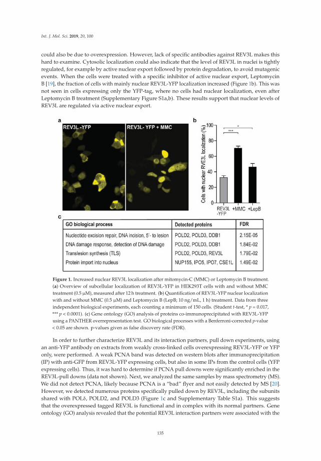
Int. J. Mol. Sci. 2019, 20, 100
could also be due to overexpression. However, lack of specific antibodies against REV3L makes thishard to examine. Cytosolic localization could also indicate that the level of REV3L in nuclei is tightlyregulated, for example by active nuclear export followed by protein degradation, to avoid mutagenicevents. When the cells were treated with a specific inhibitor of active nuclear export, LeptomycinB [19], the fraction of cells with mainly nuclear REV3L-YFP localization increased (Figure 1b). This wasnot seen in cells expressing only the YFP-tag, where no cells had nuclear localization, even afterLeptomycin B treatment (Supplementary Figure S1a,b). These results support that nuclear levels ofREV3L are regulated via active nuclear export.
Figure 1. Increased nuclear REV3L localization after mitomycin-C (MMC) or Leptomycin B treatment.(a) Overview of subcellular localization of REV3L-YFP in HEK293T cells with and without MMCtreatment (0.5 μM), measured after 12 h treatment. (b) Quantification of REV3L-YFP nuclear localizationwith and without MMC (0.5 μM) and Leptomycin B (LepB; 10 ng/mL, 1 h) treatment. Data from threeindependent biological experiments, each counting a minimum of 150 cells. (Student t-test, * p = 0.017,*** p < 0.0001). (c) Gene ontology (GO) analysis of proteins co-immunoprecipitated with REV3L-YFPusing a PANTHER overrepresentation test. GO biological processes with a Benferroni-corrected p-value< 0.05 are shown. p-values given as false discovery rate (FDR).
In order to further characterize REV3L and its interaction partners, pull down experiments, usingan anti-YFP antibody on extracts from weakly cross-linked cells overexpressing REV3L-YFP or YFPonly, were performed. A weak PCNA band was detected on western blots after immunoprecipitation(IP) with anti-GFP from REV3L-YFP expressing cells, but also in some IPs from the control cells (YFPexpressing cells). Thus, it was hard to determine if PCNA pull downs were significantly enriched in theREV3L-pull downs (data not shown). Next, we analyzed the same samples by mass spectrometry (MS).We did not detect PCNA, likely because PCNA is a “bad” flyer and not easily detected by MS [20].However, we detected numerous proteins specifically pulled down by REV3L, including the subunitsshared with POLδ, POLD2, and POLD3 (Figure 1c and Supplementary Table S1a). This suggeststhat the overexpressed tagged REV3L is functional and in complex with its normal partners. Geneontology (GO) analysis revealed that the potential REV3L interaction partners were associated with the
135

Int. J. Mol. Sci. 2019, 20, 100
following biological processes: nucleotide excision repair, DNA damage response, detection of DNAdamage, protein import into nucleus and TLS (Figure 1c). We filtered proteins using the “CRAPome”database (www.crapome.org) prior to this analysis, however the biological processes detected did notchange much from the list, including all proteins regarded as significantly enriched in REV3L-YFPpull downs (Supplementary Table S1b).
2.2. REV3L Colocalizes with PCNA and Contains a Functional APIM Sequence
The four subunit yeast POLζ complex is reported to have higher activity in presence of PCNA [10],thus how the different subunits interact with PCNA may be important, both for proper regulationof their activity and possibly also for fidelity/substrate specificity. The POLζ complex has twoPCNA interacting motifs, POLD3 contains a PIP-box and REV3L contains the APIM sequence KFVLK(1240-1244). Previous data has shown that the second amino acid in APIM (consensus sequence:R/K-F/W/Y-L/I/V/A-L/I/V/A-K/R) is vital for affinity to PCNA [5,6,17,21]. After mutation ofthis amino acid, we found that both REV3L and REV3L F1241A colocalized in PCNA foci resemblingreplication foci (Figure 2a). This was not unexpected since REV3L pulled down both POLD2 andPOLD3, both having the ability to interact directly or indirectly with PCNA [22–24]. Therefore,reducing the APIM-mediated REV3L–PCNA interaction by the F1241A mutation might not besufficient to abolish colocalization with PCNA. However, KFVLK is a functional PCNA interactingmotif, as KFVLK-YFP colocalizes with HcRed-PCNA in foci resembling replication foci (Figure 2b),similarly to the previously reported hABH21–7F4W APIM-variant (RWLVK) with increased affinity [5](Supplementary Figure S1c), here shown as a CFP-fusion. Furthermore, when F in KFVLK is mutated(corresponds to F1241A in REV3L), colocalization with PCNA is strongly reduced (Figure 2c).
Figure 2. Overexpressed full-length REV3L and APIM peptide from REV3L (KFVLK-YFP) colocalizes withendogenous and overexpressed PCNA. White line on merged images represents 5μm scale. Representativeimages display: (a) REV3L or REV3L F1241A (α-FLAG) and endogenous PCNA (α-PCNA) in transfectedHEK293T cells (STED image); (b) KFVLK-YFP (REV3L APIM), RWLVK-CFP, and HcRed-PCNA intransfected HeLa cells (live cell images); and (c) KAVLK-YFP (REV3L F1241A-APIM), KFVLK-CFP(REV3L APIM), and HcRed-PCNA in transfected HeLa cells (live cell image).
136
Int. J. Mol. Sci. 2019, 20, 100
In order to visually detect reduced colocalization/affinity, a large change is required. Fluorescenceresonance energy transfer (FRET) measurements would have been the preferred technique, as it canquantifiably differentiate between direct interaction (<20 nm distance) and colocalization (~50–100 nmdistance). For example, in a previous study, we found that the direct Xeroderma pigmentosum group-Acomplementing protein (XPA)–PCNA interaction determined by FRET was abolished when APIM in XPAwas mutated; however, XPA with mutated APIM still colocalized with PCNA in replication foci [25]. Wehave made several attempts to measure FRET between REV3L-YFP (WT and F1241A) and CFP-PCNA.However, the large size of REV3L results in low expression levels and; therefore, low fluorescence intensitycompared to PCNA. Therefore, FRET measurements were not technically possible. Second best to FRET,we have measured fold increase in intensity in foci over the background of REV3L-YFP (WT and F1241A),in the absence and presence of overexpressed APIM-CFP. When selecting images taken with the sameconfocal settings and comparing only cells with equal protein expression levels, we detected higherintensity in foci of REV3L than REV3L F1241A-YFP (Figure 3a). The foci intensities of both full-lengthproteins were reduced upon overexpression of the APIM motif in REV3L (KFVLK) and importantly, wedetected a larger (>2×) reduction for REV3L F1241A than REV3L (26% versus 11%, Figure 3b), suggestingthat REV3L F1241A has lower affinity for PCNA than REV3L.
When the high affinity APIM variant (RWLVK) was overexpressed, it nearly abolished the fociformation of both REV3L and REV3L F1241A (Figure 3c). APIM and PIP-box motifs have overlappinginteraction sites on PCNA [6,9], and different PIP-box variants are shown to have up to 700× differencesin their affinities for PCNA [23], with the PIP-box from p21 being the strongest. The high affinity APIMvariant (RWLVK), which has a ~5× lower dissociation constant than the p21 PIP-box in microscalethermophoresis (MST) experiments [17], may be able to compete with both the PIP-box in POLD3 andthe APIM in REV3L, for binding to PCNA, explaining the strong reduction of REV3L foci localizationobserved. We could not detect any differences in colocalization and/or intensities of REV3L in PCNAfoci after treatments with DNA inducing damaging agents such as cisplatin, MMC, or UV irradiation(data not shown), although a stronger nuclear localization of REV3L was detected. In summary, theseresults suggest that REV3L is present in unperturbed replication foci and that the REV3L APIM–PCNAinteraction is important for its affinity for PCNA.
2.3. Mutation of APIM in REV3L Affects the Mutation Frequency
Biochemical assays to test APIM functionality in REV3L is very difficult because REV3L is a verylarge protein (3130 aa) in complex with multiple other proteins, and interactions with PCNA is aprocess which is tightly regulated via, for example, PTMs. Therefore, in order to investigate APIMfunctionality in REV3L, REV3L-YFP (WT and F1241A) were overexpressed in one repair-proficient(HEK293) and one TLS-deficient cell line (POLη KO), together with an UV-irradiated reporter plasmid(SupF mutagenesis assay). The transfection efficiency was 30–50%, and no differences were detectedbetween expression of REV3L and REV3L F1241A (Supplementary Figure S2a). The difference inmutation frequencies between independent experiments detected in the SupF assays was large,still we repeatedly detected a 2–3 times reduction in mutation frequency in cells overexpressingREV3L, compared to cells overexpressing REV3L F1241A, in both cell lines (Figure 4a). Differences inmutation frequencies after REV3L and REV3L F1241A overexpression were also found in two DNArepair-deficient cell lines; a nucleotide excision repair (NER) deficient fibroblast cell line (XPA−/−) anda mismatch repair (MMR) deficient cell line (MLH1−/−) (Supplementary Figure S2b). Overexpressionof REV3L in HEK293 and POLη KO cells reduced the mutation frequency compared to both the controland the REV3L F1241A expressing cells, and this could suggest that APIM in REV3L, and; thus, adirect REV3L–PCNA interaction, contributes to the correct bypass of UV-lesions. Human POLζ haspreviously been reported to perform error-free replicative bypass of (6-4) photoproducts [26]. However,whether REV3L can bypass lesions correctly or not likely depends upon the damage type and load,and, the DNA repair capacity of the cells. Human POLζ is reported to be able to replicate over UVlesions independently of REV1 [16]. Whether the APIM-mediated PCNA interaction in POLζ is more
137
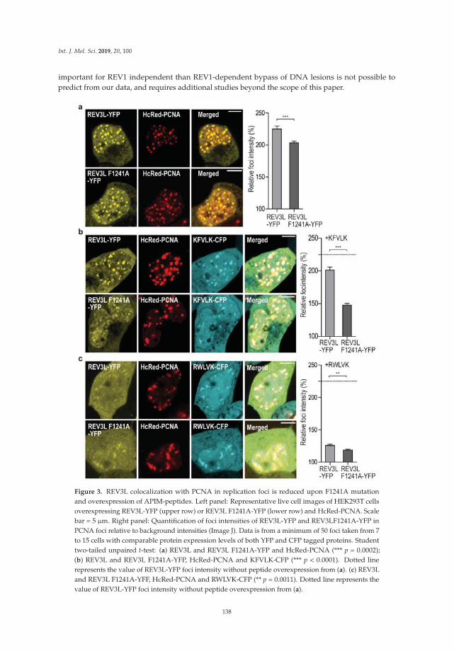
Int. J. Mol. Sci. 2019, 20, 100
important for REV1 independent than REV1-dependent bypass of DNA lesions is not possible topredict from our data, and requires additional studies beyond the scope of this paper.
Figure 3. REV3L colocalization with PCNA in replication foci is reduced upon F1241A mutationand overexpression of APIM-peptides. Left panel: Representative live cell images of HEK293T cellsoverexpressing REV3L-YFP (upper row) or REV3L F1241A-YFP (lower row) and HcRed-PCNA. Scalebar = 5 μm. Right panel: Quantification of foci intensities of REV3L-YFP and REV3LF1241A-YFP inPCNA foci relative to background intensities (Image J). Data is from a minimum of 50 foci taken from 7to 15 cells with comparable protein expression levels of both YFP and CFP tagged proteins. Studenttwo-tailed unpaired t-test: (a) REV3L and REV3L F1241A-YFP and HcRed-PCNA (*** p = 0.0002);(b) REV3L and REV3L F1241A-YFP, HcRed-PCNA and KFVLK-CFP (*** p < 0.0001). Dotted linerepresents the value of REV3L-YFP foci intensity without peptide overexpression from (a). (c) REV3Land REV3L F1241A-YFF, HcRed-PCNA and RWLVK-CFP (** p = 0.0011). Dotted line represents thevalue of REV3L-YFP foci intensity without peptide overexpression from (a).
138

Int. J. Mol. Sci. 2019, 20, 100
Figure 4. Mutation in APIM in REV3L (F1241A) affects the mutation frequency and pattern. (a) Mutationfrequency determined by the supF assay from overexpression of REV3L-YFP or REV3L F1241A-YFPin HEK 293 and POLη KO (HAP-1) cells. Cells expressing only UVB-irradiated pSP189 (supF reporterplasmid) represents the control. Four independent experiments are shown for each cell line. Studentstwo-tailed paired t-test, * p < 0.05. (b,c) Mutation spectra (supF gene) isolated from cells overexpressingREV3L-YFP, REV3L F1241A-YFP, or only pSP189 (control) in HEK293 cells (b) and POLη KO cells (c) fromfour independent experiments. Left panel: Mutations at sites in the supF gene occurring with a frequency> 10% in either control (white bars), REV3L (light grey bars), or REV3L F1241A-expressing cells (dark greybars). Right panel: Mutation spectra. The number in subscript indicates how many times the specificmutation was detected in the same transformation. Tandem and quadruple mutations are underlined.
139

Int. J. Mol. Sci. 2019, 20, 100
2.4. Mutation of APIM in REV3L Affects the Mutation Spectra in Four Cell Lines
The mutation spectra of the supF gene isolated from REV3L and REV3L F1241A overexpressingHEK293 cells showed a clear reduction of mutations at position 168 compared to the control cells(Figure 4b, left panel). This verifies that both proteins were expressed in sufficient levels to affect TLS.The spectra importantly also revealed differences between REV3L and REV3L F1241A overexpression(e.g., in the frequency of mutations at position 172 and 156). This suggests that the mutation of APIMin REV3L affected the specificity/function of POLζ.
The mutation spectra from the POLη-deficient cells also showed clear differences between cellsoverexpressing REV3L and REV3L F1241A at multiple positions, further supporting that APIM inREV3L is important for POLζ’s specificity (Figure 4c). Additionally, and importantly in this context,the mutation spectra from cells overexpressing REV3L and REV3L F1241A were different also in theXPA−/− and MLH1−/− cell lines (Supplementary Figure S2c,d).
Because of the large size of POLζ, previous studies on the polymerase functionality have beendone on yeast protein [15] or truncated human REV3L variants [10,14]. This is the first studyof the functionality of full-length proteins including the APIM-containing PCD region in REV3L.The differences detected in both the mutation frequencies and the mutation spectra after overexpressionof REV3L and REV3L F1241A, in all four cell lines tested, further suggest that APIM in REV3L is afunctional PCNA interacting motif. The F1241A mutation is not expected to change the REV3L bindingto REV7 nor REV3L’s catalytic activity, because both the REV7 binding region and the catalytic domainare located distant to the PCD region [15]. We hypothesize that the changes in mutation frequency andmutation spectra observed by mutating APIM is due to reduced direct interaction between REV3L andPCNA. Impairing the direct interaction between REV3L and PCNA could either slightly change theproximity of REV3L to DNA, or the switch between the inserter and the extender TLS polymerase,and this could affect TLS and give rise to the differences in mutation frequency and mutation spectraobserved between REV3L and REV3F1241A.
POLη interacts with PCNA via the two modules, PIP-box and ubiquitin-binding zinc-fingerdomain (UBZ), and mutations of one of these modules only partly reduce POLη’s ability to complementXeroderma pigmentosum variant (XP-V) cells after UV-irradiation [27]. Thus, multiple PCNAinteracting modules working cooperatively to stabilize interaction of TLS POLs with PCNA in vivois known also for other TLS polymerases. Recent data additionally suggests POLη travels withunperturbed replication forks [28]. If TLS polymerases are following a “piggyback” model as suggested(reviewed in [29]) (i.e., they ride on the PCNA ring until the replicative polymerases encounter a DNAlesion), then several interaction motifs of the functional polymerase complex might be required forregulation of their activities.
2.5. Targeting PCNA with APIM-Containing Peptides Reduce the Mutation Frequency
In order to further investigate the potential importance of APIM in REV3L, we wanted to make acell line with an endogenous mutation in the APIM sequence of REV3L. We were not able to create aguide RNA including APIM in REV3L, and therefore decided to use a guide RNA targeting a sequenceupstream of APIM. We initially selected mutated cells based on their hypersensitivity to MMC andUV-irradiation, and normal sensitivity to methyl methanesulfonate (MMS), and, surprisingly, themost sensitive clone obtained contained a homozygote single amino acid deletion, two amino acidsupstream of APIM (REV3L ΔA1237) (Supplementary Figure S3a–d). No off-targets of significance couldexplain the observed phenotype in the REV3L ΔA1237 cell line (Supplementary Figure S3e). Becausecommercial antibodies against REV3L are not available, we tried to establish a targeted MS/MSmethod for determination of cellular REV3L levels. The level of endogenous REV3L in HEK293 was, asalso found by others [11], low and below the detection limit, thus MS/MS detection was not technicallypossible (levels not detected in four out of five experiments, data not shown). In order to explore theconsequence of the ΔA1237 deletion in REV3L, we overexpressed REV3L ΔA1237 as an YFP fusion(REV3L ΔA1237-YFP) and detected reduced nuclear localization of REV3L ΔA1237-YFP compared to
140

Int. J. Mol. Sci. 2019, 20, 100
REV3L-YFP (Figure 5a,b). Despite this difference, overexpressed REV3L ΔA1237-YFP still colocalizedwith PCNA when co-expressed with HcRed-PCNA (Figure 5c). Reduced nuclear localization wasnot detected for the REV3L F1241A mutant (Supplementary Figure S1d), even though both REV3LΔA1237 and REV3L F1241A have reduced foci intensities compared to REV3L WT (Figures 5c and3a, respectively). Overexpression of APIM from REV3L (KFVLK) reduced REV3L ΔA1237-YFP fociless than REV3L F1241A-YPF (13% versus 26%, respectively), but slightly more than REV3L WT (11%)(Figures 5c and 3b), indicating that the reduced nuclear localization caused by the ΔA1237 deletion isnot mainly due to its reduced PCNA affinity. The single amino acid deletion does not affect nuclearexport of REV3L ΔA1237, as Leptomycin B treatment still increased nuclear fraction (data not shown).Thus, reduced nuclear localization and/or stability of REV3L ΔA1237 compared to REV3L WT is likelythe main reason for the observed hypersensitivity towards UV-irradiation and MMC in this cell line.
Figure 5. Deletion of A1237 in REV3L affects nuclear localization. (a) Overview of cells expressingREV3L-YFP and REV3L ΔA1237-YFP in HEK293T cells (live cell images). (b) Quantification of the cellswith nuclear localization of REV3L-YFP (also shown in Figure 1b) compared to REV3L ΔA1237-YFP.Data from three independent biological experiments from a minimum of 150 cells. (Student t-test,*** p < 0.0001). (c) Left panel: Representative confocal images of HEK293T cells overexpressing REV3LΔA1237-YFP and HcRed-PCNA (upper row); REV3L ΔA1237–YFP, HcRed-PCNA, and KFVLK-CFP(mid row); and REV3L ΔA1237-YFP, HcRed-PCNA, and RWLVK-CFP (bottom row). White line onmerged images represents 5 μm scale. Right panel: Quantification of foci intensities of REV3L ΔA1237-YFP in PCNA foci over background intensities (Image J). Data from a minimum of 50 foci taken froma minimum of 7 to 10 cells with comparable protein expression levels of both YFP and CFP taggedproteins. (Student two-tailed unpaired t-test, *** p ≤ 0.0002). Dotted line represents the value of REV3Lfoci intensity without peptide overexpression from Figure 1a.
141

Int. J. Mol. Sci. 2019, 20, 100
The mutation spectra of the supF gene isolated from REV3L ΔA1237 cells and its isogenic controlHEK293 were different, supporting a reduced level of REV3L and an altered TLS pattern in the REV3LΔA1237 cell line (Figure 6b). For example, mutations in position 168 that are frequent in HEK293 cellswere absent in REV3L ΔA1237, and mutations in position 108 were found only in REV3L ΔA1237(white bars, Figure 6a,b, left panel). Cumulatively, these results show that the REV3L ΔA1237 cell linehas an altered TLS response, which, together with its hypersensitivity to MMC, might suggest that it ispartly POLζ deficient.
We have previously designed a cell penetrating peptide containing the APIM sequence RWLVK(APIM-peptide), with high PCNA affinity, and a mutated version of this peptide (W4A) with ~50%reduced PCNA affinity [6,17]. Co-treatments with the APIM-peptide are shown to increase the efficacyof multiple chemotherapeutic drugs in multiple cancer cells and preclinical animal models [6,30–32].In Figure 3c we showed that overexpression of RWLVK strongly reduced the colocalization betweenREV3L and PCNA. We have unpublished data indicating that a significant part of the increased growthinhibitory efficacy observed when combining APIM-peptide with cisplatin (another inter-strandcrosslinking agent), is mediated via REV3L inhibition, because siRNA knock down of REV3L had lesseffect in cells overexpressing the APIM-peptide (data not shown). In order to explore the impact ofinhibiting the APIM-mediated REV3L–PCNA interaction on TLS, HEK293 and the REV3L ΔA1237 cellswere treated with the APIM-peptide during the SupF assay. In agreement with previous results [6,30],this treatment did not inhibit replication and we were able to isolate newly replicated SupF reporterplasmid. Interestingly, APIM-peptide treatment reduced the mutation frequency in HEK293 cellsby ~70% compared to the control, while the reduction was only ~30% in the REV3L ΔA1237 cells(Figure 6c, p < 0.05). No reduction in the mutation frequency was detected in similar experimentsusing the mutant APIM-peptide (Supplementary Figure S4a). There is a tendency towards reducedmutation frequency in REV3L ΔA1237 compared to HEK293 cells when no peptide is added (notsignificant, p = 0.08), suggesting lower levels of REV3L, and reduced TLS, in this cell line. Furthermore,the APIM-peptide was >2× more efficient in reducing the mutation frequency in HEK293 thanREV3L ΔA1237 cells. The latter suggests that part of the APIM-peptide’s effect in HEK293 cells isthe inhibition of POLζ-mediated TLS. The mutation frequency in the XPA−/− cells was, as expected,elevated compared to the repair proficient HEK293 (~2×); however, a ~50% reduction in mutationfrequency was still detected in this cell line after treatments with the APIM-peptide (Figure 6c).
2.6. Targeting PCNA with APIM-Peptide Affects the Mutation Spectra
The mutation spectra in the supF gene, isolated from both HEK293 and the REV3L ΔA1237 cells,were changed by the APIM-peptide treatment. For example, the mutations at position 168 werestrongly reduced compared to the control in HEK293 (Figure 6a, left panel), whereas, in the REV3LΔA1237 cells, mutations at this position were only detected after treatment with the APIM-peptide(Figure 6b). Similar effects of the APIM-peptide were also detected in the two cell lines, for example,the relative amount of mutations at position 156 were increased after treatment with the APIM-peptidein both cell lines. The APIM-peptide treatment reduced the mutation frequency and changed themutation spectra in both cell lines, but not similarly. Because the main difference in these two celllines likely is the level of REV3L, we hypothesize that a major part of the APIM-peptide’s effect onTLS is due to inhibition of the REV3L APIM–PCNA interaction. However, in addition to REV3L, thetwo Rad5 homologs, HLTF and SHPRH, believed to be important in regulation of TLS [33], and XPA,essential for NER [25], also contain APIM [5]. The function of these proteins could; therefore, also beaffected by the APIM-peptide treatment. Of note, the reduction in mutation frequencies shows that theAPIM-peptide reduces TLS more efficiently than error free DNA repair, and this could be beneficial incancer therapies if the APIM-peptide were used in combination with chemotherapeutics.
142
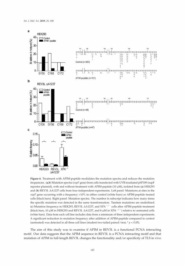
Int. J. Mol. Sci. 2019, 20, 100
Figure 6. Treatment with APIM-peptide modulates the mutation spectra and reduces the mutationfrequencies. (a,b) Mutation spectra (supF gene) from cells transfected with UVB irradiated pSP189 (supFreporter plasmid), with and without treatment with APIM-peptide (10 μM), isolated from (a) HEK293and (b) REV3L ΔA1237 cells from four independent experiments. Left panel: Mutations at sites in thesupF gene occurring with a frequency >10% in either control (white bars) or APIM-peptide treatedcells (black bars). Right panel: Mutation spectra. The number in subscript indicates how many timesthe specific mutation was detected in the same transformation. Tandem mutations are underlined.(c) Mutation frequency in HEK293, REV3L ΔA1237, and XPA−/− cells after APIM-peptide treatment(black bars, 10 μM in HEK293 and REV3L ΔA1237, and 8 μM in XPA−/−) relative to untreated cells(white bars). Data from each cell line includes data from a minimum of three independent experiments.A significant reduction in mutation frequency after addition of APIM-peptide compared to control(untreated) was detected in all three cell lines (student two-tailed paired t-test, * p < 0.05).
The aim of this study was to examine if APIM in REV3L is a functional PCNA interactingmotif. Our data suggests that the APIM sequence in REV3L is a PCNA interacting motif and thatmutation of APIM in full-length REV3L changes the functionality and/or specificity of TLS in vivo.
143

Int. J. Mol. Sci. 2019, 20, 100
Additionally, we find that targeting PCNA with the APIM-peptide reduces mutagenesis, likely byimpairing the efficacy of POLζ. Cumulatively, our data suggests that APIM in REV3L is a functionalPCNA interacting motif and that direct interaction with PCNA is important for TLS coordination.
3. Material and Methods
3.1. Expression Constructs
KFVLK-YFP/CFP and KAVLK-YFP were constructed by annealing MDKFVLK and MDKAVLKencoding oligonucleotides (Sigma-Aldrich, Saint Louis, MO, USA) with EcoRI and XhoI overhang.These were cloned into yellow (YFP) and cyan (CFP) variants of green fluorescent protein (GFP)(Clonetech/TaKaRa Bio USA, Montain View, CA, USA), using the pEYFP-N1 or pECFP-N1 plasmidswith mutated ATGsimilarly to RWLVK-CFP [5]. pREV3L-3xFLAG was a kind gift from Christine E.Canman, Department of Pharmacology, University of Michigan, USA [34]. A site-specific mutationF1241A was generated in pREV3L3xFLAG using Quick Change II (Agilent Technologies, Santa Clara,CA, USA). The Amp resistance in this plasmid was switched to Kanamycin (Km) using a Km-resistancegene flanked with AatII and FspI from the pUC57 vector. pREV3L-YFP was generated by GenScript(Piscataway, NJ, , USA) by replacement of the 3xFLAG tag with YFP using the pEYFP-N1. CFP-PCNAand HcRed-PCNA has previously been described [5,35]. pSP189 reporter plasmid and Escherichia colistrain MBM7070 were a gift from Professor Karlene Cimprich, Department of Chemical and SystemsBiology, Stanford University, Stanford, CA, USA [33].
3.2. Cell Lines
HEK293, HEK293T, HCT116, and HeLa cells (ATCC: CRL 1573, CRL 11268, CCL-247, and CCL2,respectively) were cultured in Dulbecco´s Modified Eagle Medium (DMEM) (4.5 g/L; Sigma-Aldrich);and HAP1 cells (POLη KO, Horizon Genomics, Cambridge, UK) were cultured in Iscove’s ModifiedMedium (IMDM) (Sigma Aldrich). Media was supplemented with 10% fetal bovine serum (FBS;Sigma-Aldrich), 2.5 μg/mL Fungizone® Amphotericin B (Gibco, Thermo Fischer Scientific, Waltham,MA, USA), 1 mM L-Glutamin (Sigma-Aldrich), and an antibiotic mixture containing 100 μg/mLpenicillin and 100 μg/mL streptromycin (Gibco). The XP-A deficient cell line (XPA−/−; CoriellInstitute, GM04429) were cultured in Minimum Essential Medium-alpha (MEM-alpha, 4.5 g/L;Gibco) supplemented with 10% FBS, 2.5 μg/mL Fungizone® Amphotericin B, 1 mM L-Glutamin, and100 μg/mL gentamicin (Gibco). The cells were cultured at 37 ◦C in a 5% CO2-humidified atmosphere.
3.3. SupF Assay
The supF mutagenicity assay was performed essentially as previously reported [33]. Briefly, thereporter plasmid pSP189 was irradiated with 600 or 800 mJ/cm2 UVB, depending on the cell line,with UV lamp, Vilber Lourmat Bio Spectra V5, 312 nm. Cells were transfected with UVB-irradiatedpSP189 (including plasmids not exposed to UVR as controls) and co-transfected with constructsof interest or treated with APIM-peptide. Transfections were performed using X-treme GENE HPtransfection reagent according to manufacturer protocol (Roche diagnostics, Basel, Switzerland). Cellswere harvested after 48 h for both isolation of plasmid and western analysis and/or confocal analysis.Isolated plasmids were DpnI (NEB) restriction digested to exclude original bacterial plasmids in orderto only look at replicated plasmids. Blue/white screening was performed by transformation of theisolated plasmids into E. coli MBM7070 cells, followed by plating on indicator X-gal/IPTG/Amp agarplates. Mutation frequency (white/blue colonies) was calculated for the different samples for severaltransformations. White mutant colonies were picked for re-streak and DNA sequencing of supF gene.
3.4. Imaging
HEK293T, transfected with pREV3L-3xFLAG, were fixed in paraformaldehyde (2%) andpermeabilized in ice-cold methanol for 5 min at −20 ◦C. The cells were washed in phosphate buffered
144
Int. J. Mol. Sci. 2019, 20, 100
saline (PBS) and blocked in bovine serum albumin (BSA)-PBS (2%), prior to incubation with primaryantibodies against PCNA (ab18197) and FLAG-peptide (α-FLAG, mouse monoclonal, SIGMA F1804)overnight at 4 ◦C. Samples were washed in PBS and stained with tetramethylrhodamine (TRITC) goatα-rabbit and Alexa fluor 532 goat α-mouse (Life Technologies), and then diluted 1:200 in BSA-PBS(2%) for 1 hour at room temperature (RT). Samples were washed and maintained in PBS. Imageswere captured on a Leica SP8 stimulated emission depletion (STED) 3X confocal microscope using a100×/1.4 oil immersion objective, using the 660 and 775 nm lasers.
For immunofluorescence staining and confocal imaging related to supF assay, cells weretransfected in parallel with the SupF-assay transfection, with proportional amounts of cells andtransfection mix. Cells were fixed with 2% paraformaldehyde, treated with methanol (−20 ◦C), andincubated overnight at 4 ◦C with primary antibody (α-FLAG), diluted 1:500 in FBS-PBS. The followingday the cells were washed and treated with secondary antibody (goat α-mouse Alexa fluor 532), diluted1:2000 in FBS-PBS for 45 min (37 ◦C). Images were captured using the Zeiss LSM 510 Meta confocalmicroscope (argon laser 514 nm and BP 530–600 nm for YFP; 543 nm argon laser and LP 560 for Alexafluor 532). Live cell imaging was performed using a Zeiss LSM 510 Meta laser scanning microscopeequipped with a Plan-Apochromate 63x/1.4 oil immersion objective. YFP, CFP, and HcRed wereexcited and detected at λ = 514 nm/530–600 nm, λ = 458 nm/470–500 nm, and λ = 543 nm/>615 nm,respectively, using consecutive scans. The thickness of the scanned optical slices was 1 μm.
3.5. Fluorescence Measurements and Fluorescence Resonance Energy Transfer (FRET)
FRET was done as previously described [6]. Fluorescence intensities was measured using theimaging processing software, Fiji (ImageJ) version 1.06.2016, and all images were taken with the exactsame confocal settings. Average intensities within an area of interest (foci) was measured and dividedwith average intensity in the nucleus outside foci. We selected and compared only cells with equalfluorescence intensities (YFP and CFP) (i.e., equal levels of expressed proteins), and we selected cellswithin a narrow region of intensities. A minimum of 50 foci were measured per sample from 7 to15 cells.
3.6. Preparation of Cell Lysates
Exponentially grown HEK293T cells were transfected with p-REV3L-YFP or pYFP usingX-tremeGENE HP transfection reagent (Roche diagnostics). The cells were crosslinked in 0.25%formaldehyde for 20 min at room temperature, and harvested as previously described [36]. Briefly,the cell pellet was resuspended in 3× packed cell volume in buffer I: 20 mM, pH 7.8, HEPES-KOH,100 mM KCl, 1.5 mM MgCl2, 0.2 mM EDTA, 20% glycerol, 0.5% NP-40, 1 mM DTT, 1× completeprotease inhibitor, and 1x phosphatase-inhibitor cocktails I and II (Sigma-Aldrich). 200 U OmniCleaveEndonuclease (Epicenter Technologies, Thane, India) was added to each 100 μL of cell pellet beforesonication (Branson Sonifier 250). Residual DNA/RNA in the lysates were digested for 1 h at 37 ◦Cusing an endonuclease cocktail of 400 U OmniCleave, 10 U DNase I (Roche diagnostics), 250 Ubenzonase (Merck, Darmstadt, Germany), 100–300 U micrococcal nuclease (Sigma-Aldrich), and20 μg RNase (Sigma-Aldrich) per 30 mg protein in the lysate. Digestion was followed by clearanceby centrifugation.
3.7. Immunoprecipitation
Immunoprecipitations were performed using Dynabeads protein A magnetic beads coupled topolyclonal GFP antibodies (ab290, Abcam, Cambridge, UK), which also recognize YFP and CFP, usingthe crosslinker, Bis(sulfosuccinimidyl)suberate (BS3), according to the manufacturer’s instructions(Thermo Fisher Scientific). Coupled beads were incubated with cleared lysates, under gentle rotationat 4 ◦C overnight, and further washed, three times, with 10 mM Tris-HCl, pH 7.5, 100 mM KCl beforeelution. Immunoprecipitated proteins were eluted in lithium dodecyl sulfate (LDS) loading buffer
145

Int. J. Mol. Sci. 2019, 20, 100
(Invitrogen, Thermo Fisher Scientific), containing 100 mM DTT, by heating the beads for 10 minutes at70 ◦C, and separated briefly on a NuPAGE 3–8% Tris-Acetate protein gel (Invitrogen).
3.8. Mass Spectrometry (MS) Analysis
The gel lanes were cut into pieces (~100 mg) and submitted to in-gel tryptic digestion, as describedby [37]. Tryptic digests were dried out, resuspended in 0.1% formic acid, and analyzed on an OrbitrapElite mass spectrometer (Thermo Scientific) coupled to an Easy-nLC 1000 UHPLC system (ThermoScientific). Peptides were injected into an Acclaim PepMap100 C-18 column (75 μm i.d. × 2 cm,C18, 3 μm, 100 Å, Thermo Scientific) and further separated in an Acclaim PepMap RSLC Nanoviperanalytical column (75 μm i.d. × 50 cm, C18, 2 μm, 100 Å, Thermo Scientific). A 120-minute methodwith a 250 nL/minute flow rate was employed; it started with an 80-minute gradient from 2% to 40%of buffer B (99.9% acetonitrile, 0.1% formic acid; buffer A was 0.1% formic acid in water), then it wasincreased to 55% of buffer B in 15 min, and then to 100% of buffer B in 15 min, it was then kept at100% of buffer B for 10 min. The peptides eluting from the column were analyzed in positive-ionmode using data dependent acquisition, using collision-induced dissociation (CID) fragmentation withnormalized collision energy 35. Each profile MS scan (m/z 400–1600) was acquired at a resolution of120,000 FWHM in the orbitrap, followed by 10 centroid MS/MS scans in the ion trap at rapid scan rate,with an isolation width of 2.0 m/z and an activation time of 10 ms. A 60-second dynamic exclusionwas employed. MS spectra were analyzed using Proteome Discoverer (Thermo Scientific) version1.4.0.288 software, running Mascot and the Sequest HT database search algorithms. Spectra weresearched against a human proteome database from UniProt with the following parameters: maximummissed cleavage = 2, precursor mass tolerance = 10 ppm, fragment mass tolerance = 0.6 Da, anddynamic modification = carbamidomethyl (C: +57.021 Da). Peptides were identified with a high degreeof confidence (defined as false discovery rate (FDR) ≤0.01) using Percolator. From three biologicalreplicas, possible REV3L interaction partners were identified as proteins detected with a Sequest score>5 in at least two or more experiments in the REV3L-YFP IP, compared to the YFP control sample.
3.9. Guide RNA Cloning
LentiCRISPRv2 vector (Addgene plasmid #52961), containing two expression cassettes, hSpCas9and the chimeric guide RNA, was used as a vector for the CRISPR/Cas9 system. The guide RNA waschosen to target REV3L upstream of its APIM sequence. The following oligonucleotides were used:5′caccgAAAATCTCAGTCTGGTGCTG-3′ on plus-strand and 5′aaacCAGCACCAGACTGAGATTTTc-3′
on minus-strand. The vector was digested using BsmBI (NEB) and the annealed oligonucleotides (guideRNA) were cloned into the guide RNA scaffold by using Quick Ligase (NEB). Constructs were heat shocktransformed into Stbl3 chemically-competent E. coli, and plated on LB Ampicillin plates (100 μg/mL).Plasmids from three bacterial colonies were isolated, digested by restriction enzyme HindIII (NEB), andapplied on a 0.8% agarose gel for screening. Further, the constructs were sequenced to verify if the guideRNA was cloned correctly into the vector.
3.10. Transfection of CRISPR/Cas9 Vector and Single Clone Expansion.
HEK293 cells, seeded in a 12-well plate (150,000 cells/well), were transfected (Xtreme Gene HP,Roche) with 1 μg lentiCRISPR v2 guide plasmid per well. Selection medium containing 2 μg/mLpuromycin was added 72 h after transfection and renewed every 3 to 4 days. Potential single-cell coloniescould be observed after 14 days. Cell colonies were washed with PBS, trypsinized, resuspended, andtransferred into a 24-well plate. Cells were further expanded and DNA was harvested for screening.Briefly, 100,000 cells were resuspended in 50 μL of alkaline solution (25 mM NaOH/0.2 mM EDTA,pH 12) and heated for 10 min at 95 ◦C. After cooling, 50 μL of neutralizing solution (40mM tris-HCl,pH 5) was added and the lysate was isolated after centrifugation. The target sequence in the REV3Lwas PCR amplified using forward primer 5′ ATTCTTCTCCACCTCGCTGC-3′ and reverse primer
146

Int. J. Mol. Sci. 2019, 20, 100
3′CCGCTATGCACACAATCTGC-5′, and the PCR product was sequenced using forward primer 5′
GCGCAAGAGCACAGATTAAG-3′ and reverse primer 3′ TGGGTAGGGAAGCAGAAAGG-5′.
3.11. Viability
HEK293 (4000 cells/well) and REV3L ΔA1237 (5000 cells/well) were seeded in 96-well plates.After 4 h, cells were treated with mitomycin-C (MMC, Medac) (0.05, 0.1, 0.2, and 0.4 μM), exposedto UVB-irradiation (20, 40, and 60 mJ/cm2) or treated with methyl methanesulfonate (MMS)(0.025, 0.05, 0.1, and 0.2 μM). Cell viability was measured at different timepoints by the MTT(3-(4.5-Dimethylthiazol-2-yl)-2.5 diphenyl-tetrazolium bromide) assay as previously described [5].
Supplementary Materials: The following are available online at http://www.mdpi.com/1422-0067/20/1/100/s1.
Author Contributions: M.O., R.M., S.B.R., K.Ø.B., and A.N. designed the study; S.B.R., A.N., K.Ø.B., M.S., R.S.K.,and A.N. performed the experiments; M.O., S.B.R., K.Ø.B., A.N., and M.S. wrote the manuscript.
Funding: This work was supported by grants from the Norwegian University of Science and Technology (NTNU),Trondheim, Norway, and The Liaison Committee for Education, Research, and Innovation in Central Norway, andthe Norwegian University of Science and Technology (NTNU). The funders had no role in the study design, datacollection and analysis, decision to publish, or the preparation of the manuscript.
Acknowledgments: The microscopy and MS analyses were done at the Cellular and Molecular Imaging CoreFacility (CMIC) and the Proteomic and Metabolomics Core Facility (PROMEC), NTNU, respectively. CMIC andPROMEC are funded by the Faculty of Medicine at NTNU and Central Norway Regional Health Authority.We thank Barbara van Loon, NTNU, for valuable feedback on the manuscript.
Conflicts of Interest: APIM Therapeutics is a spin-off company of the Norwegian University of Science andTechnology, where Professor Marit Otterlei is an inventor, founder, minority shareholder, and CSO. APIMTherapeutics is developing APIM-peptides for use in cancer therapy.
References
1. Zhao, L.; Washington, M.T. Translesion Synthesis: Insights into the Selection and Switching of DNAPolymerases. Genes 2017, 8, 24. [CrossRef] [PubMed]
2. Choe, K.N.; Moldovan, G.L. Forging Ahead through Darkness: PCNA, Still the Principal Conductor at theReplication Fork. Mol. Cell 2017, 65, 380–392. [CrossRef] [PubMed]
3. Mailand, N.; Gibbs-Seymour, I.; Bekker-Jensen, S. Regulation of PCNA-protein interactions for genomestability. Nat. Rev. Mol. Cell Biol. 2013, 14, 269–282. [CrossRef] [PubMed]
4. Olaisen, C.; Kvitvang, H.F.N.; Lee, S.; Almaas, E.; Bruheim, P.; Drablos, F.; Otterlei, M. The role of PCNA as ascaffold protein in cellular signaling is functionally conserved between yeast and humans. FEBS Open Biol.2018, 8, 1135–1145. [CrossRef] [PubMed]
5. Gilljam, K.M.; Feyzi, E.; Aas, P.A.; Sousa, M.M.; Muller, R.; Vagbo, C.B.; Catterall, T.C.; Liabakk, N.B.; Slupphaug, G.;Drablos, F.; et al. Identification of a novel, widespread, and functionally important PCNA-binding motif. J. Cell Biol.2009, 186, 645–654. [CrossRef] [PubMed]
6. Müller, R.; Misund, K.; Holien, T.; Bachke, S.; Gilljam, K.M.; Våtsveen, T.K.; Rø, T.B.; Bellacchio, E.; Sundan, A.;Otterlei, M. Targeting Proliferating Cell Nuclear Antigen and Its Protein Interactions Induces Apoptosis inMultiple Myeloma Cells. PLoS ONE 2013, 8, e70430. [CrossRef]
7. Bacquin, A.; Pouvelle, C.; Siaud, N.; Perderiset, M.; Salomé-Desnoulez, S.; Tellier-Lebegue, C.; Lopez, B.;Charbonnier, J.-B.; Kannouche, P.L. The helicase FBH1 is tightly regulated by PCNA via CRL4 (Cdt2)-mediatedproteolysis in human cells. Nucleic Acids Res. 2013, 41, 6501–6513. [CrossRef]
8. Fu, D.; Samson, L.D.; Hubscher, U.; van Loon, B. The interaction between ALKBH2 DNA repair enzymeand PCNA is direct, mediated by the hydrophobic pocket of PCNA and perturbed in naturally-occurringALKBH2 variants. DNA Repair 2015, 35, 13–18. [CrossRef]
9. Sebesta, M.; Cooper, C.D.O.; Ariza, A.; Carnie, C.J.; Ahel, D. Structural insights into the function of ZRANB3in replication stress response. Nat. Commun. 2017, 8, 15847. [CrossRef]
10. Makarova, A.V.; Stodola, J.L.; Burgers, P.M. A four-subunit DNA polymerase zeta complex containingPol delta accessory subunits is essential for PCNA-mediated mutagenesis. Nucleic Acids Res. 2012, 40,11618–11626. [CrossRef]
147
Int. J. Mol. Sci. 2019, 20, 100
11. Singh, B.; Li, X.R.; Owens, K.M.; Vanniarajan, A.; Liang, P.; Singh, K.K. Human REV3 DNA PolymeraseZeta Localizes to Mitochondria and Protects the Mitochondrial Genome. PLoS ONE 2015, 10, 18. [CrossRef][PubMed]
12. Van Sloun, P.P.H.; Romeijn, R.J.; Eeken, J.C.J. Molecular cloning, expression and chromosomal localisation ofthe mouse Rev31 gene, encoding the catalytic subunit of polymerase zeta. Mutat. Res. DNA Repair 1999, 433,109–116. [CrossRef]
13. Van Sloun, P.P.H.; Varlet, I.; Sonneveld, E.; Boei, J.; Romeijn, R.J.; Eeken, J.C.J.; De Wind, N. Involvement ofmouse Rev3 in tolerance of endogenous and exogenous DNA damage. Mol. Cell. Biol. 2002, 22, 2159–2169.[CrossRef] [PubMed]
14. Suzuki, T.; Gruz, P.; Honma, M.; Adachi, N.; Nohmi, T. The role of DNA polymerase zeta in translesionsynthesis across bulky DNA adducts and cross-links in human cells. Mutat. Res. 2016, 791–792, 35–41.[CrossRef] [PubMed]
15. Lee, Y.S.; Gregory, M.T.; Yang, W. Human Pol zeta purified with accessory subunits is active in translesionDNA synthesis and complements Pol. in cisplatin bypass. Proc. Natl. Acad. Sci. USA 2014, 111, 2954–2959.[CrossRef] [PubMed]
16. Yoon, J.H.; Park, J.; Conde, J.; Wakamiya, M.; Prakash, L.; Prakash, S. Rev1 promotes replication through UVlesions in conjunction with DNA polymerases eta, iota, and kappa but not DNA polymerase zeta. Genes Dev.2015, 29, 2588–2602. [PubMed]
17. Olaisen, C.; Muller, R.; Nedal, A.; Otterlei, M. PCNA-interacting peptides reduce Akt phosphorylation andTLR-mediated cytokine secretion suggesting a role of PCNA in cellular signaling. Cell. Signal. 2015, 27,1478–1487. [CrossRef] [PubMed]
18. Brondello, J.M.; Pillaire, M.J.; Rodriguez, C.; Gourraud, P.A.; Selves, J.; Cazaux, C.; Piette, J. Novel evidencesfor a tumor suppressor role of Rev3, the catalytic subunit of Pol zeta. Oncogene 2008, 27, 6093–6101. [CrossRef]
19. Kudo, N.; Matsumori, N.; Taoka, H.; Fujiwara, D.; Schreiner, E.P.; Wolff, B.; Yoshida, M.; Horinouchi, S.Leptomycin B inactivates CRM1/exportin 1 by covalent modification at a cysteine residue in the centralconserved region. Proc. Natl. Acad. Sci. USA 1999, 96, 9112–9117. [CrossRef]
20. Bj Ras, K.O.; Sousa, M.M.L.; Sharma, A.; Fonseca, D.M.; CK, S.G.; Bj Ras, M.; Otterlei, M. Monitoring of thespatial and temporal dynamics of BER/SSBR pathway proteins, including MYH, UNG2, MPG, NTH1 andNEIL1-3, during DNA replication. Nucleic Acids Res. 2017, 45, 8291–8301. [CrossRef]
21. Hara, K.; Uchida, M.; Tagata, R.; Yokoyama, H.; Ishikawa, Y.; Hishiki, A.; Hashimoto, H. Structure ofproliferating cell nuclear antigen (PCNA) bound to an APIM peptide reveals the universality of PCNAinteraction. Acta Crystallogr. F Struct. Biol. Commun. 2018, 74 Pt 4, 214–221. [CrossRef]
22. Ducoux, M.; Urbach, S.; Baldacci, G.; Hubscher, U.; Koundrioukoff, S.; Christensen, J.; Hughes, P. Mediationof proliferating cell nuclear antigen (PCNA)-dependent DNA replication through a conserved p21(Cip1)-likePCNA-binding motif present in the third subunit of human DNA polymerase delta. J. Biol. Chem. 2001, 276,49258–49266. [CrossRef] [PubMed]
23. Bruning, J.B.; Shamoo, Y. Structural and thermodynamic analysis of human PCNA with peptides derivedfrom DNA polymerase-delta p66 subunit and flap endonuclease-1. Structure 2004, 12, 2209–2219. [CrossRef][PubMed]
24. Pustovalova, Y.; Magalhaes, M.T.Q.; D’Souza, S.; Rizzo, A.A.; Korza, G.; Walker, G.C.; Korzhnev, D.M.Interaction between the Rev1 C-Terminal Domain and the PolD3 Subunit of Pol xi Suggests a Mechanism ofPolymerase Exchange upon Rev1/Pol xi-Dependent Translesion Synthesis. Biochemistry 2016, 55, 2043–2053.[CrossRef] [PubMed]
25. Gilljam, K.M.; Muller, R.; Liabakk, N.B.; Otterlei, M. Nucleotide Excision Repair Is Associated with theReplisome and Its Efficiency Depends on a Direct Interaction between XPA and PCNA. PLoS ONE 2012,7, e49199. [CrossRef] [PubMed]
26. Yoon, J.H.; Prakash, L.; Prakash, S. Error-free replicative bypass of (6-4) photoproducts by DNA polymerasezeta in mouse and human cells. Genes Dev. 2010, 24, 123–128. [CrossRef]
27. Bienko, M.; Green, C.M.; Sabbioneda, S.; Crosetto, N.; Matic, I.; Hibbert, R.G.; Begovic, T.; Niimi, A.; Mann, M.;Lehmann, A.R.; et al. Regulation of translesion synthesis DNA polymerase eta by monoubiquitination.Mol. Cell 2010, 37, 396–407. [CrossRef]
148

Int. J. Mol. Sci. 2019, 20, 100
28. Despras, E.; Sittewelle, M.; Pouvelle, C.; Delrieu, N.; Cordonnier, A.M.; Kannouche, P.L. Rad18-dependentSUMOylation of human specialized DNA polymerase eta is required to prevent under-replicated DNA.Nat. Commun. 2016, 7, 15. [CrossRef]
29. Slade, D. Maneuvers on PCNA Rings during DNA Replication and Repair. Genes 2018, 9, 28. [CrossRef]30. Søgaard, C.K.; Moestue, S.A.; Rye, M.B.; Kim, J.; Nepal, A.; Liabakk, Ni.; Bachke, S.; Bathen, T.F.; Otterlei, M.;
Hill, D.K. APIM-peptide targeting PCNA improves the efficacy of docetaxel treatment in the TRAMP mousemodel of prostate cancer. Oncotarget 2018, 9, 11752–11766. [CrossRef]
31. Gederaas, O.A.; Sogaard, C.D.; Viset, T.; Bachke, S.; Bruheim, P.; Arum, C.J.; Otterlei, M. Increased AnticancerEfficacy of Intravesical Mitomycin C Therapy when Combined with a PCNA Targeting Peptide. Transl. Oncol.2014, 7, 812–823. [CrossRef] [PubMed]
32. Søgaard, C.K.; Blindheim, A.; Røst, L.M.; Petrovic, V.; Nepal, A.; Bachke, S.; Liabakk, N.B.; Gederaas, O.A.;Viset, T.; Arum, C.J.; et al. “Two hits—One stone”; increased efficacy of cisplatin-based therapies by targetingPCNA’s role in both DNA repair and cellular signaling. Oncotarget 2018, 9, 32448. [CrossRef] [PubMed]
33. Lin, J.R.; Zeman, M.K.; Chen, J.Y.; Yee, M.C.; Cimprich, K.A. SHPRH and HLTF act in a damage-specificmanner to coordinate different forms of postreplication repair and prevent mutagenesis. Mol. Cell 2011, 42,237–249. [CrossRef] [PubMed]
34. Sharma, S.; Hicks, J.K.; Chute, C.L.; Brennan, J.R.; Ahn, J.Y.; Glover, T.W.; Canman, C.E. REV1 and polymerasezeta facilitate homologous recombination repair. Nucleic Acids Res. 2012, 40, 682–691. [CrossRef] [PubMed]
35. Aas, P.A.; Otterlei, M.; Falnes, P.O.; Vagbo, C.B.; Skorpen, F.; Akbari, M.; Sundheim, O.; Bjoras, M.; Slupphaug, G.;Seeberg, E.; et al. Human and bacterial oxidative demethylases repair alkylation damage in both RNA andDNA. Nature 2003, 421, 859–863. [CrossRef] [PubMed]
36. Akbari, M.; Solvang-Garten, K.; Hanssen-Bauer, A.; Lieske, N.V.; Pettersen, H.S.; Pettersen, G.K.; Wilson, D.M., 3rd;Krokan, H.E.; Otterlei, M. Direct interaction between XRCC1 and UNG2 facilitates rapid repair of uracil in DNAby XRCC1 complexes. DNA Repair 2010, 9, 785–795. [CrossRef]
37. Shevchenko, A.; Wilm, M.; Vorm, O.; Mann, M. Mass spectrometric sequencing of proteins silver-stainedpolyacrylamide gels. Anal. Chem. 1996, 68, 850–858. [CrossRef]
© 2018 by the authors. Licensee MDPI, Basel, Switzerland. This article is an open accessarticle distributed under the terms and conditions of the Creative Commons Attribution(CC BY) license (http://creativecommons.org/licenses/by/4.0/).
149

International Journal of
Molecular Sciences
Article
Thermococcus Eurythermalis Endonuclease IVCan Cleave Various Apurinic/Apyrimidinic SiteAnalogues in ssDNA and dsDNA
Wei-Wei Wang 1,†, Huan Zhou 2,†, Juan-Juan Xie 1, Gang-Shun Yi 1, Jian-Hua He 2,
Feng-Ping Wang 1,3, Xiang Xiao 1,3 and Xi-Peng Liu 1,3,*
1 State Key Laboratory of Microbial Metabolism, School of Life Sciences and Biotechnology, Shanghai JiaoTong University, 800 Dong-Chuan Road, Shanghai 200240, China; [email protected] (W.-W.W.);[email protected] (J.-J.X.); [email protected] (G.-S.Y.); [email protected] (F.-P.W.);[email protected] (X.X.)
2 Shanghai Institute of Applied Physics, Chinese Academy of Sciences, No. 239 Zhangheng Road,Shanghai 201204, China; [email protected] (H.Z.); [email protected] (J.-H.H.)
3 State Key Laboratory of Ocean Engineering, School of Naval Architecture, Ocean and Civil Engineering,Shanghai Jiao Tong University, 800 Dong-Chuan Road, Shanghai 200240, China
* Correspondence: [email protected]† These authors contributed equally to this work.
Received: 31 October 2018; Accepted: 18 December 2018; Published: 24 December 2018
Abstract: Endonuclease IV (EndoIV) is a DNA damage-specific endonuclease that mainly hydrolyzesthe phosphodiester bond located at 5′ of an apurinic/apyrimidinic (AP) site in DNA. EndoIValso possesses 3′-exonuclease activity for removing 3′-blocking groups and normal nucleotides.Here, we report that Thermococcus eurythermalis EndoIV (TeuendoIV) shows AP endonuclease and3′-exonuclease activities. The effect of AP site structures, positions and clustered patterns on theactivity was characterized. The AP endonuclease activity of TeuendoIV can incise DNA 5′ to variousAP site analogues, including the alkane chain Spacer and polyethylene glycol Spacer. However,the short Spacer C2 strongly inhibits the AP endonuclease activity. The kinetic parameters alsosupport its preference to various AP site analogues. In addition, the efficient cleavage at AP sitesrequires ≥2 normal nucleotides existing at the 5′-terminus. The 3′-exonuclease activity of TeuendoIVcan remove one or more consecutive AP sites at the 3′-terminus. Mutations on the residues forsubstrate recognition show that binding AP site-containing or complementary strand plays a keyrole for the hydrolysis of phosphodiester bonds. Our results provide a comprehensive biochemicalcharacterization of the cleavage/removal of AP site analogues and some insight for repairing APsites in hyperthermophile cells.
Keywords: Thermococcus eurythermalis; endonuclease IV; AP site analogue; spacer; DNA repair
1. Introduction
Both physical and chemical factors in the cell and the environment can cause varioustypes of DNA damage, which will cause some potential mutagenic and toxic effects on thecell. This DNA damage mainly include hydrolytic deamination of cytosine, methylation of bases,oxidized bases, base losses, base crosslinking, DNA strand breaks, and misincorporation in DNAreplication [1–3]. Among these types of DNA damage, base losses, also called apurinic/apyrimidinic(AP) sites, are the most common lesion [2]. AP sites mainly result from spontaneous base lossvia depurination/depyrimidination, as well as removing damaged bases by all kinds of DNAglycosylases [4]. Depurination/depyrimidination creates 2000–10,000 AP sites per cell per day inmammalian cells [5]. AP sites cannot guide the incorporation of a correct (d) NMP because of the
Int. J. Mol. Sci. 2019, 20, 69; doi:10.3390/ijms20010069 www.mdpi.com/journal/ijms150
Int. J. Mol. Sci. 2019, 20, 69
inability to form the required hydrogen bonds, and then it will inhibit replication or transcription.When the DNA polymerase encounters an AP site on the template strand, generally dAMP is preferredfor incorporation into the extending strand, i.e., the “A-rule” [6]. Therefore, if AP sites are not quicklyrepaired, the cells will be in danger of serious toxicity.
In the cell, AP sites are mainly repaired by the base excision repair (BER) pathway. BER isinitiated by DNA glycosylases, which generate the immediate product AP site [4]. In BER, the enzymesresponsible for cleaving the DNA backbone at AP sites are classified into two groups, the AP lyase andthe AP endonuclease. The AP lyases cleave the AP site (deoxyribose) through a β-elimination reaction,generating 5′-phosphate and 3′-α,β unsaturated aldehyde [7]. The AP endonucleases, includingendonuclease IV (EndoIV) and exonuclease III (ExoIII), incise DNA 5′ to AP sites through hydrolysisof the phosphodiester bond to yield a free 3′-OH and a 5′-deoxyribose-phosphate (dRP) group [8].In addition to the natural AP site, the C1′-reduced (tetrahydrofuranyl) and deoxyribose-oxidizedsites (C1′-oxidized 2-deoxyribonolactone and the C4′-oxidized AP site) are also recognized by APendonuclease [9,10]. In addition to AP endonuclease activity, EndoIV and ExoIII have 3′-exonucleaseactivity, and function as 3′-repair diesterases by removing DNA 3′-blocking groups such as3′-phosphates, 3′-phosphoglycolates and 3′-α,β unsaturated aldehydes [11,12]. On cleaving theDNA 5′ to an AP site or removing the 3′-blocking groups, DNA polymerase will resynthesize amatched DNA strand by incorporating correct dNMPs into the 3′-OH end under the direction ofa complementary template strand, and then DNA ligase will seal the nick generated in the repairprocess [8].
Though both EndoIV and ExoIII primarily function as an AP endonuclease and a 3′-repairdiesterase, they have many contrasting properties. ExoIII has strong 3′-exonuclease activity ondouble-stranded (ds) DNA and endonuclease activity at urea damage site in DNA [13,14]. BacterialExoIII also possesses a strong ribonuclease H activity [11]. Human APE1, the homologue of bacterialExoIII, has several novel activities, such as endoribonuclease [15,16], 3′-RNA phosphatase and3′-exoribonuclease activities [17]. In addition to the weaker 3′-exonuclease activity on normal3′-nucleotides [18,19], EndoIV has an additional activity that cleaves DNA 5′ to some oxidativebases [20–22]. ExoIII is constitutively expressed in Escherichia coli (E. coli); however, EndoIV is inducedby oxidative stress [23]. Human cells use an ExoIII homolog APE1 for treating AP sites [24]; however,Saccharomyces cerevisiae (S. cerevisiae) uses a homolog of EndoIV, APN-1, for repairing AP sites [25].
The hydrolysis mechanism of the AP site has been interpreted based on the crystal structuresof bacterial E. coli EndoIV (EcoendoIV) [26–28], Thermus thermophilus (T. thermophilus) EndoIV [29],human APE1 complexed with AP-site-containing double-stranded DNA (dsDNA) [30,31], and bacterialExoIII from E. coli [32]. The recognition of the AP site and subsequent hydrolysis of the phosphodiesterbond are involved in the interaction between EcoendoIV and two strands of DNA duplex [26]. HumanAPE1 interacts with 9–10 nucleotides around the AP site, mainly through weak additive contactswith phosphate groups [30]. The crystal structure of E. coli ExoIII gives a detailed interpretation onthe catalytic mechanism of the AP endonuclease activity [32]. A crystal structure of ExoIII from ahyperthermophilic archaea, Archaeoglobus fulgidus, was also solved [33]. However, until now, no crystalstructure of archaeal EndoIV has been solved.
High temperature results in more DNA damage, such as hydrolytic deamination of cytosine andAP sites [5,34], so hyperthermophiles face a serious high-temperature threat to genome integrity andbear more stress to repair DNA damages than mesophiles. Thermococcus eurythermalis (T. eurythermalis)A501 is a conditional piezophilic hyperthermophilic archaea, isolated from the Guaymas Basin, that iswell adapted to the hydrothermal environment [35]. Except for the EndoIV from Pyrococcus furiosus [36],reports on archaeal EndoIV are scarce. P. furiosus EndoIV (PfuendoIV) possesses both AP endonucleaseand 3′ exonuclease activities, and its 3′-exonuclease activity, but not its AP endonuclease activity,is stimulated by PCNA [36]. Meanwhile, the effects of the structure and context of AP site analogues onEndoIV activity are less known. T. eurythermalis encodes a homologue of EndoIV that shows very lowsequence similarity to EcoendoIV. As the only AP endonuclease, T. eurythermalis EndoIV (TeuendoIV)
151
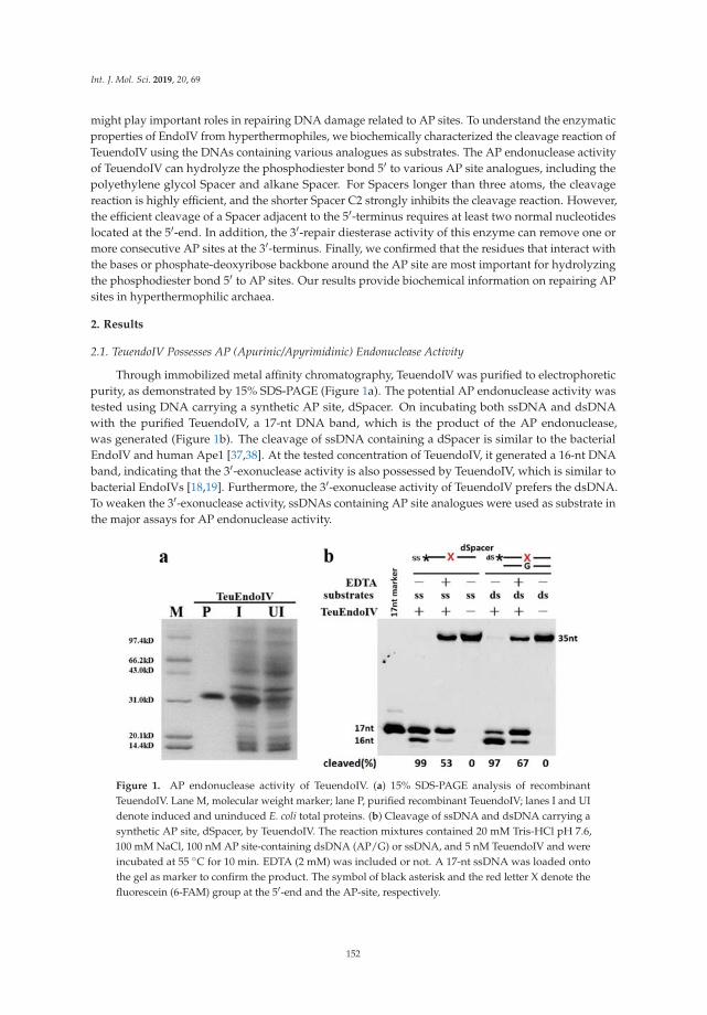
Int. J. Mol. Sci. 2019, 20, 69
might play important roles in repairing DNA damage related to AP sites. To understand the enzymaticproperties of EndoIV from hyperthermophiles, we biochemically characterized the cleavage reaction ofTeuendoIV using the DNAs containing various analogues as substrates. The AP endonuclease activityof TeuendoIV can hydrolyze the phosphodiester bond 5′ to various AP site analogues, including thepolyethylene glycol Spacer and alkane Spacer. For Spacers longer than three atoms, the cleavagereaction is highly efficient, and the shorter Spacer C2 strongly inhibits the cleavage reaction. However,the efficient cleavage of a Spacer adjacent to the 5′-terminus requires at least two normal nucleotideslocated at the 5′-end. In addition, the 3′-repair diesterase activity of this enzyme can remove one ormore consecutive AP sites at the 3′-terminus. Finally, we confirmed that the residues that interact withthe bases or phosphate-deoxyribose backbone around the AP site are most important for hydrolyzingthe phosphodiester bond 5′ to AP sites. Our results provide biochemical information on repairing APsites in hyperthermophilic archaea.
2. Results
2.1. TeuendoIV Possesses AP (Apurinic/Apyrimidinic) Endonuclease Activity
Through immobilized metal affinity chromatography, TeuendoIV was purified to electrophoreticpurity, as demonstrated by 15% SDS-PAGE (Figure 1a). The potential AP endonuclease activity wastested using DNA carrying a synthetic AP site, dSpacer. On incubating both ssDNA and dsDNAwith the purified TeuendoIV, a 17-nt DNA band, which is the product of the AP endonuclease,was generated (Figure 1b). The cleavage of ssDNA containing a dSpacer is similar to the bacterialEndoIV and human Ape1 [37,38]. At the tested concentration of TeuendoIV, it generated a 16-nt DNAband, indicating that the 3′-exonuclease activity is also possessed by TeuendoIV, which is similar tobacterial EndoIVs [18,19]. Furthermore, the 3′-exonuclease activity of TeuendoIV prefers the dsDNA.To weaken the 3′-exonuclease activity, ssDNAs containing AP site analogues were used as substrate inthe major assays for AP endonuclease activity.
Figure 1. AP endonuclease activity of TeuendoIV. (a) 15% SDS-PAGE analysis of recombinantTeuendoIV. Lane M, molecular weight marker; lane P, purified recombinant TeuendoIV; lanes I and UIdenote induced and uninduced E. coli total proteins. (b) Cleavage of ssDNA and dsDNA carrying asynthetic AP site, dSpacer, by TeuendoIV. The reaction mixtures contained 20 mM Tris-HCl pH 7.6,100 mM NaCl, 100 nM AP site-containing dsDNA (AP/G) or ssDNA, and 5 nM TeuendoIV and wereincubated at 55 ◦C for 10 min. EDTA (2 mM) was included or not. A 17-nt ssDNA was loaded ontothe gel as marker to confirm the product. The symbol of black asterisk and the red letter X denote thefluorescein (6-FAM) group at the 5′-end and the AP-site, respectively.
152

Int. J. Mol. Sci. 2019, 20, 69
After confirmation of AP endonuclease activity, the optimal reaction parameters were determinedfor TeuendoIV using ssDNA carrying an internal dSpacer as substrate (Figure S1). TeuendoIV showedhigher activity at pH values ranging from 7.5 to 9.0, with a maximum activity at pH 8.5. AlthoughEDTA did not inactivate the AP endonuclease, addition of EDTA inhibited the enzymatic activity tosome extent, implying the metal ion is necessary for EndoIV. A high concentration of DTT inhibited theAP endonuclease activity. As an enzyme from hyperthermophiles, TeuendoIV showed higher activityat temperatures ranging from 50 to 65 ◦C.
Divalent metal ions showed different effects on activity (Figure S2). Mg2+ promoted APendonuclease activity, and Mn2+ had no clear effect on activity. However, Ni2+ and Zn2+ showed clearinhibition on activity. In particular, Zn2+, which is the preferred cofactor for EcoendoIV [26], is a stronginhibitor of TeuendoIV. Interestingly, TeuendoIV showed clear AP endonuclease activity at the absenceof divalent mental ions, implying that it bound some metal ions during recombinant expression inE. coli cell.
2.2. TeuendoIV Shows AP Endonuclease on Various AP Site Analogues
The natural AP site is not stable under high temperature, so it is generally replaced by severalAP site analogues in AP endonuclease activity assays [10]. Various AP site analogues have differentmolecular structures, which might largely affect the AP endonuclease activity. To understand thecleavage reaction on AP sites, we utilized DNAs containing a range of AP site analogues withdifferent molecular structures (Figure 2) as substrates in cleavage assay. TeuendoIV can hydrolyzethe phosphodiester bond 5′ to various alkane chain and polyethylene glycol Spacers with differentcleavage efficiency (Figure 3a,b). For alkane chains longer than three carbon atoms, TeuendoIVefficiently cleaved the phosphodiester bond 5′ to the alkane chain (Figure 3a). However, the cleavageefficiency is not proportional to the length of the alkane chain and is almost constant for Spacers longerthan three carbon atoms. These results imply that the alkane chains longer than C3 can be efficientlybent out of the DNA backbone to perfectly orient the phosphodiester bond for attacking by the catalyticwater molecule. However, under our assay conditions, ssDNA containing an internal Spacer C2 wasnot cleaved because of the short alkane chain (Figure 3a), implying that the phosphodiester bond5′ to Spacer C2 is not oriented perfectly into the substrate-binding pocket and cannot be attacked bythe coordinated water molecule [26]. For polyethylene glycol Spacers, TeuendoIV hydrolyzed thephosphodiester bond 5′ to Spacers 9 and 18 with a comparable efficiency (Figure 3b). Furthermore,the cleavage efficiencies of Spacer C9 and Spacer 9 were similar (Figure S3), suggesting that the oxygenatoms of polyethylene glycol Spacers have no influence on the cleavage reaction.
The cyclic Spacers have a structure similar to natural AP site deoxyribose, so they might berecognized and cleaved at higher efficiency than linear Spacers. Both Spacer C3 and dSpacer have athree-carbon atom chain between two phosphate groups, but dSpacer has a cyclic deoxyribose-likemolecular structure, and Spacer C3 is a linear carbon chain (Figure 2). Our results showed thatthe AP endonuclease activity of TeuendoIV cleaved the ssDNA containing dSpacer more efficientlythan Spacer C3 (Figure 3c). These results indicate that the cyclic larger tetrahydrofuran structureis beneficial to correctly orient the phosphodiester bond 5′ to AP sites into the enzymatic catalyticcenter for attacking by a water molecule. When the amount of TeuendoIV was increased, ssDNAcontaining a Spacer C2 was weakly cleaved (Figure 3d). However, the two Spacers Dual SH anddisulfide (S-S, the assays were performed in the presence of 1.0 mM NAD+), which have the sametwo-carbon atom chain as Spacer C2, completely blocked the hydrolysis of the phosphodiester bond(Figure 3d). When a five-fold concentration of TeuendoIV was used in the assay, the Spacer C2 wascleaved completely and the Spacer Dual SH was not cleaved (Figure S4). Meanwhile, major DNAswere digested by the 3′-exonuclease activity because of the blockage of AP endonuclease activity bythe Spacers C2 and Dual SH.
153

Int. J. Mol. Sci. 2019, 20, 69
Figure 2. Structures of various AP site analogues. The molecular structure of (a) alkane chain Spacers(Spacer Cn), (b) cyclic Spacers (natural Spacer, and synthetic dSpacer and rSpacer), (c) polyethyleneglycol Spacers (Spacer 9 and Spacer 18), and (d) two-carbon atom chain Spacers (Spacer C2, Dual SH,and disulfide (S-S)).
Figure 3. AP endonuclease activity on ssDNAs containing various Spacers. Different ssDNAs (100 nM)containing (a) alkane spacers, (b) polyethylene glycol spacers, (c) dSpacer and spacer C3, and (d) sulfuratom spacers were incubated with 5 nM (a–c) or 10 nM (d) of TeuendoIV at 55 ◦C for the indicated time.For the Spacer disulfide bond (S-S), the NAD+ (1.0 mM) was substituted for DTT in the reaction buffer.Uppercase letter M denotes oligonucleotides marker. The cleavage percentages of substrates are listedat the bottom of each panel. The symbol of black asterisk and the red letter X denote the fluorescein(6-FAM) group at the 5′-end and the AP-sites, respectively.
154

Int. J. Mol. Sci. 2019, 20, 69
The kinetic parameters for various AP site analogues (Table 1) show that, except for Spacer C2,the tested AP-site analogues have the comparable Km and kcat values, with a little substrate preferenceto dSpacer. Although the Km of Spacer C2 is 2-fold higher than those of other AP-site analogues, its kcat
show at least 100-fold decrease, resulting in the large decrease of kcat/Km.
Table 1. Kinetic parameters Km and kcat of TeuendoIV for AP-site analogues.
Substrates Km (μM) kcat (min−1) kcat/Km (min−1·μM−1)
dSpacer 0.35 ± 0.03 0.59 ± 0.05 1.68 ± 0.15Spacer C2 0.16 ± 0.02 0.0021 ± 0.0002 0.013 ± 0.002Spacer C3 0.37 ± 0.03 0.23 ± 0.02 0.62 ± 0.06Spacer C4 0.41 ± 0.04 0.33 ± 0.03 0.80 ± 0.08Spacer C6 0.36 ± 0.03 0.37 ± 0.04 1.03 ± 0.10
Spacer C12 0.41 ± 0.04 0.35 ± 0.03 0.85 ± 0.08Spacer 9 0.34 ± 0.03 0.36 ± 0.03 1.06 ± 0.10Spacer 18 0.37 ± 0.04 0.42 ± 0.04 1.14 ± 0.11
Km and kcat for cleaving ssDNAs containing dSpacer and Spacer C3, C4, C6, C12, 9, 18 were calculated by doublereciprocal plotting using the initial reaction rates at various substrate concentrations (0.05, 0.1, 0.2, 0.5, 1.0, 2.0and 5.0 μM) and 25 nM TeuendoIV where incubation time is 5 min. Considering that the very low activity ofTeuendoIV on Spacer C2, the concentration of TeuendoIV and the incubation time were increased to 500 nM and 30min, respectively. The cleaved products are less than 10% (Table S4), which indicate that the kinetic experimentswere performed during periods when initial rates hold. The initial reaction rates (Table S4) were used to calculatethe values of Km and kcat, and the graphs of the double reciprocal plotting are shown in Figure S5. All data are themeans of three independent experiments.
2.3. Cleavage of DNA Containing Clustered AP Site Analogues
Although DNA containing a single Spacer can be cleaved, the clustered AP sites may haveeffect on cleavage. We performed the cleavage reactions of ssDNAs containing more than oneconsecutive AP site analogue. Our results showed that TeuendoIV efficiently cleaved the upstreamphosphodiester bond of several consecutive AP site analogues, such as Spacer C12 and Spacer 18(Figure 4a). Even ssDNA containing seven consecutive Spacer 18 damages was cleaved efficientlyat the first phosphodiester bond located 5′ to the consecutive AP sites. The DNA containing threeclustered cyclic AP sites, dSpacers, also was cleaved with a comparable efficiency to DNA with asingle dSpacer (Figure S6). In addition to the consecutive identical AP sites, the phosphodiester bond5′ to two tandem different AP site analogues was also hydrolyzed efficiently, and the tandem orderof two AP site analogues affected the cleavage efficiency to some degree (Figure 4b). If ≥4 normalnucleotides existed between two identical AP sites, the cleavage took place at the 5′-sides of both APsites (Figure 4c and Figure S7). If the two identical Spacers were separated by another different Spacer,including the Spacer C2, the cleavage site almost was exclusively happened at the phosphodiesterbond 5′ to the first AP site (Figure 4d).
We also characterized the cleavage of ssDNAs containing the clustered AP sites analogues thatare poorly cleaved when existing alone, such as Spacer C2 and Dual SH. The cleavage efficiency ofthe phosphodiester bond is proportional to the number of consecutive Spacer C2, even at a lowerconcentration of TeuendoIV (Figure S8), suggesting that Spacer C2 actually does not inhibit thehydrolysis reaction and only blocks the perfect orientation of the cleaved phosphodiester bond intothe enzymatic active center. The perfect orientation became easy when ≥2 Spacer C2 are clusteredconsecutively. In contrast to a cleavage percentage of 86% for clustered Spacer C2×2, only 12% ofthe clustered Spacer Dual SH×2 was cleaved even at a 10-fold concentration of enzyme (Figure S8),indicating that the Dual SH group actually inhibits the hydrolysis reaction.
155

Int. J. Mol. Sci. 2019, 20, 69
Figure 4. Cleavage of ssDNAs containing clustered AP site analogues. The ssDNAs (100 nM) containing(a) consecutive Spacer C12 or 18, (b) tandem Spacers C12 and 18, or (c) tandem Spacer 18 separated byfour normal bases or (d) a different middle AP site analogue were incubated with TeuendoIV (5 nM)at 55 ◦C for the indicated time. The combinations of AP site analogues are listed on the top of eachpanel. Uppercase letter M denotes oligonucleotides marker. The cleavage percentages of substrates arelisted at the bottom of each panel. The symbol of black asterisk and the red letters X, Y and Z denotethe fluorescein (6-FAM) group at the 5′-end and the AP-sites, respectively.
2.4. The Base Opposite the AP Site Has Little Effect on dsDNA Cleavage
The AP endonuclease activities of TeuendoIV on dsDNA and ssDNA were compared. Generally,TeuendoIV preferred dsDNA to ssDNA for all tested Spacers (Figure 5). The bases (A, T, C, and G)opposite Spacer C3 or 18 had an effect on the dsDNA cleavage reaction in the following order: AP/C >AP/T > AP/A > AP/G (Spacer C3), and AP/A > AP/A > AP/C > AP/T (Spacer 18). However, the APendonuclease activity of TeuendoIV did not show a clear preference to the base opposite dSpacer.
156

Int. J. Mol. Sci. 2019, 20, 69
Figure 5. Effect of the base opposite AP site on dsDNA cleavage. TeuendoIV (5 nM) was incubatedwith 100 nM ssDNA or dsDNAs opposite each of four bases at 55 ◦C for the indicated time. The Spacersare (a) dSpacer, (b) Spacer C3, and (c) Spacer 18. Uppercase letter M denotes oligonucleotides marker.The cleavage percentages of substrates are listed at the bottom of each panel.
2.5. Cleavage of DNA Containing AP Sites Adjacent to Termini
The EndoIV has 3′-exonuclease and 3′-repair diesterase activities; the 3′-exonuclease removesnormal 3′-nucleotides, and the 3′-repair activity removes abnormal 3′-terminal groups, such as3′-phosphates, 3′-phosphoglycolates, and 3′-α,β unsaturated aldehydes [11,12]. To cleave thephosphodiester bond 5′ to an AP site, it is generally required that the AP site should be locatedat the appropriate position of the DNA backbone. For the Spacer adjacent to the 5′-terminus, at leasttwo normal nucleotides were required to be located 5′ to an AP site (Figure 6a). If only one normalnucleotide was placed on the 5′-terminus, almost no product of AP endonuclease was generated.However, the 3′-exonuclease of TeuendoIV was promoted to hydrolyze the 3′-nucleotide, resulting in
157

Int. J. Mol. Sci. 2019, 20, 69
generation of substantial 3′-FAM mononucleotide (Figure 6a), suggesting that TeuendoIV has strongintrinsic 3′-exonuclease similar to bacterial EndoIV [18,19]. For the AP sites adjacent to the 3′-terminus,the existence of additional normal 3′-nucleotides is not necessary for hydrolyzing the phosphodiesterbond 5′ to the AP site (Figure 6b). However, the normal 3′-nucleotides showed promotion on theAP endonuclease activity. When the single Spacer C6 in Figure 6b was changed to three consecutiveclustered ones, the hydrolysis model did not change, predominantly hydrolyzing the phosphodiesterbond located at the 5′ side of the first Spacer (Figure 6c). Furthermore, if more than one Spacer C6 waslocated at the 3′-terminus, the hydrolysis reaction also took place at the first 3′-terminal phosphodiesterbond, i.e., the one between two 3′-terminal Spacer C6 (Figure 6c, second substrate, 0.3 min).
Figure 6. Cleavage of ssDNAs with a Spacer near the 3′-/5′-terminus. TeuendoIV (5 nM) was incubatedwith 100 nM ssDNA containing one Spacer C6 adjacent to the (a) 5′-terminus, (b) 3′-terminus, or (c) threeSpacers (Spacer C6) adjacent to the 3′-terminus at 55 ◦C for the indicated time. Uppercase letter Mdenotes oligonucleotides marker. The cleavage percentages of substrates are listed at the bottom ofeach panel. AP denotes AP endonuclease activity and exo denotes 3′-exonuclease activity. The symbolof black asterisk and the red letter X denote the fluorescein (6-FAM) group at the 5′- or 3’-end and theAP-site, respectively.
158
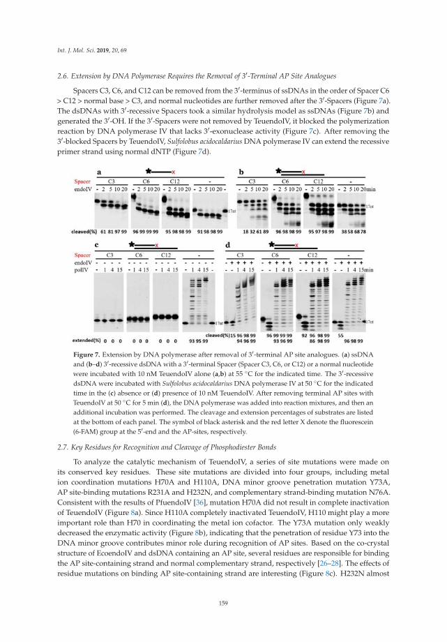
Int. J. Mol. Sci. 2019, 20, 69
2.6. Extension by DNA Polymerase Requires the Removal of 3′-Terminal AP Site Analogues
Spacers C3, C6, and C12 can be removed from the 3′-terminus of ssDNAs in the order of Spacer C6> C12 > normal base > C3, and normal nucleotides are further removed after the 3′-Spacers (Figure 7a).The dsDNAs with 3′-recessive Spacers took a similar hydrolysis model as ssDNAs (Figure 7b) andgenerated the 3′-OH. If the 3′-Spacers were not removed by TeuendoIV, it blocked the polymerizationreaction by DNA polymerase IV that lacks 3′-exonuclease activity (Figure 7c). After removing the3′-blocked Spacers by TeuendoIV, Sulfolobus acidocaldarius DNA polymerase IV can extend the recessiveprimer strand using normal dNTP (Figure 7d).
Figure 7. Extension by DNA polymerase after removal of 3′-terminal AP site analogues. (a) ssDNAand (b–d) 3′-recessive dsDNA with a 3′-terminal Spacer (Spacer C3, C6, or C12) or a normal nucleotidewere incubated with 10 nM TeuendoIV alone (a,b) at 55 ◦C for the indicated time. The 3′-recessivedsDNA were incubated with Sulfolobus acidocaldarius DNA polymerase IV at 50 ◦C for the indicatedtime in the (c) absence or (d) presence of 10 nM TeuendoIV. After removing terminal AP sites withTeuendoIV at 50 ◦C for 5 min (d), the DNA polymerase was added into reaction mixtures, and then anadditional incubation was performed. The cleavage and extension percentages of substrates are listedat the bottom of each panel. The symbol of black asterisk and the red letter X denote the fluorescein(6-FAM) group at the 5′-end and the AP-sites, respectively.
2.7. Key Residues for Recognition and Cleavage of Phosphodiester Bonds
To analyze the catalytic mechanism of TeuendoIV, a series of site mutations were made onits conserved key residues. These site mutations are divided into four groups, including metalion coordination mutations H70A and H110A, DNA minor groove penetration mutation Y73A,AP site-binding mutations R231A and H232N, and complementary strand-binding mutation N76A.Consistent with the results of PfuendoIV [36], mutation H70A did not result in complete inactivationof TeuendoIV (Figure 8a). Since H110A completely inactivated TeuendoIV, H110 might play a moreimportant role than H70 in coordinating the metal ion cofactor. The Y73A mutation only weaklydecreased the enzymatic activity (Figure 8b), indicating that the penetration of residue Y73 into theDNA minor groove contributes minor role during recognition of AP sites. Based on the co-crystalstructure of EcoendoIV and dsDNA containing an AP site, several residues are responsible for bindingthe AP site-containing strand and normal complementary strand, respectively [26–28]. The effects ofresidue mutations on binding AP site-containing strand are interesting (Figure 8c). H232N almost
159

Int. J. Mol. Sci. 2019, 20, 69
completely lost the AP endonuclease activity on ssDNA and dsDNA. In addition to binding the5′-side base of the AP site, the residue H231 in EcoendoIV (corresponding to the residue H232 inTeuendoIV) also coordinates the divalent metal ions [26]. Therefore, the possibility is that H232Astrongly inactivate the AP endonuclease activity of TeuendoIV because of its inability to coordinatemetal ion cofactor. In contrast to H232A, R231A selectively inactivated the AP endonuclease activityon dsDNA, suggesting that the residue R231 only participates in the recognition of AP sites in dsDNA.Considering that N76 is only involved in binding the complementary strand [26,27], it is plausible thatthe N76A obtained minor activity on ssDNA and completely lost the activity on dsDNA (Figure 8d),indicating that the disruption of binding the complementary strand might cause serious defects inrecognizing and binding the AP site in ssDNA.
Figure 8. Cleavage reactions of wt (wild type) and mutant TeuendoIV. Mutations on key conservedresidues potentially involved in (a) coordinating metal ion cofactors, (b) penetrating into the duplexminor groove, and binding the (c) AP site-containing strand and (d) complementary strand were usedto assay AP endonuclease activities (5 nM enzymes) on ssDNA and dsDNA with an internal dSpacer(100 nM) at 55 ◦C for the indicated time. The cleavage percentages of substrates are listed at the bottomof each panel. The symbol of black asterisk and the red letter X denote the fluorescein (6-FAM) groupat the 5′-end and the AP-site, respectively.
2.8. Structure Comparison of TeuendoIV and EcoendoIV
Because no crystal structure of archaeal EndoIV has been solved, a modeled topology ofTeuendoIV was built based on the bacterial EcoendoIV crystal structure (Figure 9a), which is composedof nine α-helixes, five DNA-binding recognition loops (R-loop), and eight β-strands [26]. Comparedwith EcoendoIV, TeuendoIV has a more disordered N-terminal domain and lacks an R-loop2 (Figure 9).
Archaeal TeuendoIV shows very weak sequence similarity to these from bacteria and S. cerevisiae(Figure 9c). The sequence identity between TeuendoIV and EcoendoIV is just 24% (Figure 9c), implyingthat there are differences between their topologies. The modeled structure of TeuendoIV also supportedthis speculation (Figure 9a,b). TeuendoIV lacks a short peptide, the R-loop2 in bacterial and S. cerevisiae
160

Int. J. Mol. Sci. 2019, 20, 69
EndoIV. In the crystal structure of EcoendoIV and dsDNA, this R-loop2 has much interactions withDNA, including deoxyriboses, bases and phosphates, and might play important roles in hydrolyzingthe phosphodiester bonds 5′ to the AP site [26,27]. The absence of R-loop2 did not result in enzymaticinactivity, but the removal of N-terminal 21 residues inactivated the TeuendoIV [39], suggesting thatthe N-terminal secondary structures β1 and R-loop1 are required for catalytic activity. The phylogenictree also shows that TeuendoIV branches from bacterial and eukaryotic EndoIV (Figure S9).
Figure 9. Homology model of TeuendoIV and multi-alignment of EndoIVs. (a) Modeled structure ofTeuendoIV. (b) Superimposition of TeuendoIV (green) and EcoendoIV (cyan). (c) Multiple sequencealignment of EndoIVs. The EndoIVs are from T. eurythermalis, T. thermophilus (PDB ID: 3AAM),Chlamydophila pneumoniae, S. cerevisiae and E. coli (PDB ID: 1QTW). The secondary structures ofEcoendoIV are shown at the top of sequences. Cylinders indicate α-helices, arrows indicate β-strands,and five green rectangles indicate DNA-binding recognition loops 1–5. Identical and similar residuesare shaded in black and gray, respectively. The mutated residues are marked by asterisks.
161
Int. J. Mol. Sci. 2019, 20, 69
3. Materials and Methods
3.1. Materials
KOD-plus DNA polymerase was purchased from Toyobo (Osaka, Japan). Expression vectorpET28a and E. coli Rosetta 2(DE3)pLysS were purchased from Merck (Darmstadt, Germany).The Ni-NTA resin was bought from Bio-Rad (Hercules, CA, USA). The Spacer phosphoramiditeswere purchased from Glen Research (Sterling, VA, USA) and ChemGenes (Wilmington, MA, USA).Oligonucleotides were synthesized in Biosune (Shanghai, China). All the other chemicals and reagentswere of analytical grade.
3.2. Construction of Expression Plasmids
T. eurythermalis A501 was cultured at 85 ◦C, and its genomic DNA was extracted byphenol-chloroform and precipitated using isopropyl alcohol [35]. The gene encoding TeuendoIVwas amplified from the genomic DNA using a pair of specific primers (Table S1) by PCR andinserted into pET28a between the Nde I and BamH I sites, producing a recombinant expressionvector, pET28a-TeuEndoIV. The site-directed mutations were constructed on the basis of the TeuendoIVexpression plasmid according to the protocol of the QuikChange® Site-Directed Mutagenesis Kit fromAgilent (Santa Clara, CA, USA) using respective primers (Table S1). The base sequences of insertedgenes were confirmed by DNA sequencing.
3.3. Expression and Purification of TeuendoIV
Various expression plasmids were transformed into E. coli Rosetta 2(DE3)pLysS to expressrecombinant EndoIV and its mutants. Isopropyl-1-thio-b-D-galactopyranoside (IPTG) was addedinto bacterial cultures (OD600 ≈ 0.8) with a 0.5 mM final concentration to induce the expressionof target proteins for 20 h at 16 ◦C. Bacterial pellets were resuspended with ice-cold lysis buffer(20 mM Tris-HCl pH 7.9, 300 mM NaCl, 10 mM imidazole, 5 mM β-mercaptoethanol (β-ME), 1 mMphenylmethylsulfonyl fluoride (PMSF) and 10% glycerol) for breaking cells by sonication. Lysateswere clarified by centrifugation at 16,000× g for 30 min at 4 ◦C. Then the supernatants were purifiedby the immobilized Ni2+ affinity chromatography. After loading the supernatant onto a columnpre-equilibrated with lysis buffer, the resin was washed with 100 column volumes of lysis buffercontaining 20 mM imidazole. Finally, the target proteins were eluted with lysis buffer containing200 mM imidazole. The purity of eluted fractions was confirmed by 15% SDS-PAGE. The purifiedproteins were stored in small aliquots at −20 ◦C
3.4. Activity Assay of TeuendoIV
The base sequences of oligodeoxyribonucleotides used in the activity assay are listed in Table S2.The AP site analogues, dSpacer, alkane chain (Spacers C2, C3, C4, C6, C9 and C12), polyethyleneglycol (Spacers 9 and 18), Dual SH, and disulfide bond (S-S) were introduced into oligonucleotidesthat were used as substrates to determine the effect of AP site structure on DNA cleavage reaction.The disulfide bond (S-S) was introduced by oxidizing the dual SH by NAD+. The strands carrying theAP site analogues were fluorescently labeled with FAM at the 5′-end. The dsDNAs with different basepairs (N/AP, N denotes one of A, T, C, G) were used to characterize the effects of the base opposite theAP site analogues on AP endonuclease activity. To prepare dsDNA substrates, the FAM-labeled strandwas annealed with unlabeled complementary strand in a mole ratio of 1:1.5 by boiling for 5 min at95 ◦C and slowly cooling down to room temperature.
AP endonuclease activity was determined as described with some modification [12]. Standardreaction solutions contained 100 nM 5′-FAM-labeled AP site-containing ssDNA or dsDNA and thespecified amount of TeuendoIV in buffer consisting of 20 mM Tris-HCl pH 7.6, 100 mM NaCl,and 0.2 mM EDTA. After optimization of reaction buffer, all reactions were performed at 55 ◦Cin buffer consisting of 20 mM Tris-HCl pH 7.5, 50 mM NaCl, 1 mM MgCl2, 0.5 mM DTT, and 0.5 mM
162

Int. J. Mol. Sci. 2019, 20, 69
EDTA. For the DNA substrate containing a disulfide bond Spacer, the NAD+ (1.0 mM) was substitutedfor DTT in the reaction buffer. The exonuclease activity of TeuendoIV and DNA polymerase activity ofSulfolobus acidocaldarius DNA polymerase IV were performed in buffer consisting of 20 mM Tris-HCl pH8.8, 10 mM KCl, 10 mM (NH4)2SO4, 2 mM MgSO4, 100 ng/mL BSA and 0.1% TritonX-100. The reactionswere incubated at 55 ◦C for the indicated time and were stopped by adding an equal volume of loadingbuffer (50 mM EDTA, 8 M urea, 0.2% SDS, 0.1% bromophenol blue, 0.1% xylene cyan) to reactionsamples. Then, the reaction products were analyzed by electrophoresis in 15% polyacrylamide gels(8 M urea). After electrophoresis, the gels were imaged using an FL9500 fluorescent scanner andquantified with the analysis software (GE Healthcare, Chicago, IL, USA).
To quantitatively compare the cleavage efficiency between various AP site analogues, the kineticparameters (Km and kcat) of TeuendoIV on ssDNAs containing various AP-site analogues werecalculated using double reciprocal plotting. The 5′-FAM-labelled ssDNAs containing Spacer C3,C4, C6, C12, 9, 18 and dSpacer (0.05, 0.1, 0.25, 0.5, 1.0, 2.0 and 5.0 μM) were incubated with 25 nMTeuendoIV for 5 min. For cleavage reaction of Spacer C2, 500 nM TeuendoIV was incubated withthe above concentration of ssDNA for 30 min. The cleavage products were quantitated and used tocalculate the initial reaction rates, which were used to calculate the values of Km and kcat.
3.5. Multiple Sequence Alignment and Constructing Phylogenetic Tree
The multiple sequence alignment was performed using MUSCLE (Multiple Sequence Comparisonby Log Expectation) on the website of http://www.ebi.ac.uk/Tools/msa/muscle/. The phylogenetictree was built by MEGA using sequences from archaea and bacteria (Table S3).
3.6. Structure Modeling of EndoIV
The modeled structure of TeuendoIV was constructed using ProMod3 on the SWISS-MODELserver (https://www.swissmodel.expasy.org/) [40]. The crystal structure of EcoendoIV (PDB ID:1QTW) was used as the template during homologous modeling. The two structures of EcoendoIV andTeuendoIV were aligned using Pymol (Schrodinger LLC, New York, NY, USA). The PyMOL MolecularGraphics System, version 1.5.0.3).
4. Discussion
Bacterial and archaeal EndoIV can hydrolyze natural AP site [12,36]. We confirm that archaealEndoIV can also hydrolyze various AP site analogues with different molecular structures. The lengthof the AP site is an important factor in the hydrolysis efficiency of the phosphodiester bond 5′ to anAP site analogue. Our results show that the length of the AP site should be longer than two carbonatoms for efficient hydrolysis, and too short an alkane chain (Spacer C2) cannot be efficiently treatedby TeuendoIV (Figure 3a). The molecular conformation is another determinant of cleavage efficiency.The dSpacer, which has a molecular structure similar to the natural AP site, is preferably recognizedand cleaved than Spacer C3, implying that the circular ribose-like structure is a promoter to hydrolyzethe phosphodiester bond 5′ to dSpacer. Compared with the weak cleavage of phosphodiester bond 5′ toSpacer C2, two Spacers, dual thiol and disulfide bond structures, completely block the cleavage reaction,implying that, in addition to the short length, the groups of the disulfide bond and dual thiol alsocause hindrance for orienting and/or attacking the 5′-side phosphodiester bond by water molecule.
The position of an AP site in DNA also determines the efficiency of the hydrolysis reaction.When the AP site analogues are located near the 5′-terminus, at least two normal 5′-nucleotidesare required for the efficient hydrolysis of phosphodiester bonds. However, the 3′-terminal AP siteanalogues, even several clustered Spacers, can be removed efficiently, implying that both the APendonuclease and 3′-exonuclease activities of EndoIV might use a similar substrate-binding andhydrolysis mechanism. For the clustered farther 3′-Spacers, the cleavage reaction can take placeat the first 3′-terminal phosphodiester bond (Figure 6c, second substrate, 0.3 min), indicating the3′-exonuclease of TeuendoIV can remove the first Spacer of the farther 3′-terminal consecutive Spacers.
163

Int. J. Mol. Sci. 2019, 20, 69
During hydrolysis of phosphodiester bonds by AP endonucleases, the DNA backbone is bound byseveral conserved residues [26–28]. The EcoendoIV-DNA interactions are mainly provided by residuesR230 and H231 for binding the AP site, residue N75 for binding the complementary strand, and residueY72 DNA for penetrating into the minor groove. These conserved residues correspond to R231 andH232, N76, and Y73 in TeuendoIV. Residue Y72 in EcoendoIV plays a central role in recognizing theAP site, and the mutation Y72A results in a 1000-fold decrease in activity [27]. Similarly, the mutationY73A leads to a strong loss of activity of TeuendoIV (Figure 8b), indicating that R-loop3 plays a similarrole in recognizing the AP site. The mutations on residues for binding the AP site strand shoulddecrease the cleavage reaction of dsDNA and ssDNA, and our results confirm their harmful effects onthe AP endonuclease activity of TeuendoIV (Figure 8c). It is conceivable that the mutations on residuesfor binding the AP site-containing strand are harmful to cleaving both ssDNA and dsDNA. However,the R231A mutation only seriously decreases the activity on the dsDNA and has a minor harmful effecton the cleavage of ssDNA. The mutations on the residues for binding the complementary strand arethought to be destructive for the hydrolysis of phosphodiester bonds in dsDNA, not those in ssDNA.Actually, the N76A mutation completely inactivates the AP endonuclease activity on dsDNA and leadsto partial activity on ssDNA (Figure 8d).
Although R-loop2 in EcoendoIV also provides interactions with the DNA backbone and bases,the residues corresponding to R-loop2 are absent or largely changed in TeuendoIV (Figure 9c).When the N-terminal 21 residues are deleted, the truncated TeuendoIV loses its AP endonucleaseactivity [39], indicating the R-loop1 and R-loop2 are important for binding DNA. These results mightbe interpreted by the complex crystal structure of TeuendoIV and dsDNA or ssDNA. Therefore, tocompare the differences between bacterial and archaeal EndoIV in their structural and biochemicalproperties, solving the crystal structure of TeuendoIV will be important.
EndoIV possesses both AP endonuclease and 3′-phosphodiesterase activities, both of whichmight use the same water molecule to attack the phosphodiester bonds, i.e., the same catalysismechanism for two enzymatic activities. The AP endonuclease requires EndoIV to interact withthe DNA backbone upstream and downstream of an AP site, whereas the 3′-phosphodiesterase(3′-repair diesterase/3′-exonuclease) cannot interact with the backbone downstream of an AP site,which does not exist for the 3′-terminal Spacer. Therefore, the mutations on residues that interact withthe 3′ backbone of an internal AP site should have little effect on the hydrolysis of the 3′-terminalphosphodiester bond by 3′-exonuclease, as well as the removal of 3′-terminal AP sites or 3′-blockinggroups by AP endonuclease/3′-repair diesterase.
Compared with ExoIII, EndoIV has more homologues in archaea, and even more than onehomologue can exist in one species. For example, two and three EndoIV homologues are present inthe P. furiosus and Methanothermobacter thermautotrophicus genomes, respectively [36]. Among theseEndoIV homologues, generally only one possesses AP endonuclease and 3′-exonuclease activities [36].M. thermautotrophicus also has a homologue of ExoIII, which does not possess the AP endonucleaseactivity but functions as an endonuclease specific to dU damage [41]. Unlike the usually coupled APendonuclease/3′-repair diesterase/3′-exonuclease, in the human pathogen Neisseria meningitides theseactivities are endowed to two separated homologues [42]. Meanwhile, the lower level of sequenceidentity between TeuendoIV and EcoendoIV and the phylogenetic tree of EndoIV (Figure S9) imply afarther ancestral relationship between archaea and bacteria EndoIV.
Depurination/depyrimidination occurs more frequently at high temperatures [5]. Hence,hyperthermophiles face a serious spontaneous mutation resulting from AP sites and dU damage [5,34].The BER pathway, as the main AP site repair pathway, is important for tolerating high temperatureand avoiding mutagenesis in archaea. T. eurythermalis does not encode ExoIII, implying EndoIV playsan important role in the BER pathway. Considering that bacterial EndoIV possesses RNA-specific3′-exonuclease activity [43–45] and human Ape1 can process abasic and oxidized ribonucleotidesembedded in DNA [46], the TeuendoIV might function in the repair of non-AP site-type damage andRNA metabolism.
164
Int. J. Mol. Sci. 2019, 20, 69
Supplementary Materials: Supplementary materials can be found at http://www.mdpi.com/1422-0067/20/1/69/s1.
Author Contributions: X.-P.L. designed the study. W.-W.W. and H.Z. performed experiments. X.-P.L. and W.-W.W.wrote the paper. All authors analyzed the data and approved the final version of the manuscript.
Funding: This work was supported by the National Key R&D Program of China (Grant Nos. 2018YFC0310704),the National Natural Science Foundation of China (Grant Nos. U1832161, 91428308, 41530967), and the ChinaOcean Mineral Resources R&D Association (Grant No. DY135-B2-12).
Conflicts of Interest: The authors declare no conflict of interest.
References
1. Lengauer, C.; Kinzler, K.W.; Vogelstein, B. DNA methylation and genetic instability in colorectal cancer cells.Proc. Natl. Acad. Sci. USA 1997, 94, 2545–2550. [CrossRef] [PubMed]
2. Lindahl, T. Instability and decay of the primary structure of DNA. Nature 1993, 362, 709–715. [CrossRef][PubMed]
3. Ho, E.; Ames, B.N. Low intracellular zinc induces oxidative DNA damage, disrupts p53, NF kappa B,and AP1 DNA binding, and affects DNA repair in a rat glioma cell line. Proc. Natl. Acad. Sci. USA 2002,99, 16770–16775. [CrossRef] [PubMed]
4. Cunningham, R.P. DNA glycosylases. Mutat. Res. 1997, 383, 189–196. [CrossRef]5. Lindahl, T.; Nyberg, B. Rate of depurination of native deoxyribonucleic acid. Biochemistry 1972, 11, 3610–3618.
[CrossRef] [PubMed]6. Cuniasse, P.; Fazakerley, G.V.; Guschlbauer, W.; Kaplan, B.E.; Sowers, L.C. The abasic site as a challenge
to DNA polymerase. A nuclear magnetic resonance study of G, C and T opposite a model abasic site.J. Mol. Biol. 1990, 213, 303–314. [CrossRef]
7. Piersen, C.E.; McCullough, A.K.; Lloyd, R.S. AP lyases and dRPases: Commonality of mechanism.Mutat. ‘Res. 2000, 459, 43–53. [CrossRef]
8. Erzberger, J.P.; Barsky, D.; Scharer, O.D.; Colvin, M.E.; Wilson, D.M. Elements in abasic site recognitionby the major human and Escherichia coli apurinic/apyrimidinic endonucleases. Nucleic Acids Res. 1998,26, 2771–2778. [CrossRef]
9. Greenberg, M.M.; Weledji, Y.N.; Kim, J.; Bales, B.C. Repair of oxidized abasic sites by exonuclease III,endonuclease IV, and endonuclease III. Biochemistry 2004, 43, 8178–8183. [CrossRef]
10. Shida, T.; Noda, M.; Sekiguchi, J. Cleavage of single- and double-stranded DNAs containing an abasicresidue by Escherichia coli exonuclease III (AP endonuclease VI). Nucleic Acids Res. 1996, 24, 4572–4576.[CrossRef]
11. Shida, T.; Ogawa, T.; Ogasawara, N.; Sekiguchi, J. Characterization of Bacillus subtilis ExoA protein:A multifunctional DNA-repair enzyme similar to the Escherichia coli exonuclease III. Biosci. Biotechnol.Biochem. 1999, 63, 1528–1534. [CrossRef] [PubMed]
12. Haas, B.J.; Sandigursky, M.; Tainer, J.A.; Franklin, W.A.; Cunningham, R.P. Purification and characterizationof Thermotoga maritima endonuclease IV, a thermostable apurinic/apyrimidinic endonuclease and 3′-repairdiesterase. J. Bacteriol. 1999, 181, 2834–2839. [PubMed]
13. Kow, Y.W.; Wallace, S.S. Exonuclease III recognizes urea residues in oxidized DNA. Proc. Natl. Acad. Sci. USA1985, 82, 8354–8358. [CrossRef] [PubMed]
14. Hoheisel, J.D. On the activities of Escherichia coli exonuclease III. Anal. Biochem. 1993, 209, 238–246. [CrossRef][PubMed]
15. Barnes, T.; Kim, W.C.; Mantha, A.K.; Kim, S.E.; Izumi, T.; Mitra, S.; Lee, C.H. Identification ofApurinic/apyrimidinic endonuclease 1 (APE1) as the endoribonuclease that cleaves c-myc mRNA.Nucleic Acids Res. 2009, 37, 3946–3958. [CrossRef] [PubMed]
16. Kim, W.C.; Berquist, B.R.; Chohan, M.; Uy, C.; Wilson, D.M., 3rd; Lee, C.H. Characterization of theendoribonuclease active site of human apurinic/apyrimidinic endonuclease 1. J. Mol. Biol. 2011, 411, 960–971.[CrossRef] [PubMed]
17. Chohan, M.; Mackedenski, S.; Li, W.M.; Lee, C.H. Human apurinic/apyrimidinic endonuclease 1 (APE1) has3′ RNA phosphatase and 3′ exoribonuclease activities. J. Mol. Biol. 2015, 427, 298–311. [CrossRef]
18. SKerins, M.; Collins, R.; McCarthy, T.V. Characterization of an endonuclease IV 3′–5′ exonuclease activity.J. Biol. Chem. 2003, 278, 3048–3054. [CrossRef]
165

Int. J. Mol. Sci. 2019, 20, 69
19. Xie, J.J.; Liu, X.P.; Han, Z.; Yuan, H.; Wang, Y.; Hou, J.L.; Liu, J.H. Chlamydophila pneumoniae endonucleaseIV prefers to remove mismatched 3′ ribonucleotides: Implication in proofreading mismatched 3′-terminalnucleotides in short-patch repair synthesis. DNA Repair 2013, 12, 140–147. [CrossRef]
20. Gros, L.; Ishchenko, A.A.; Ide, H.; Elder, R.H.; Saparbaev, M.K. The major human AP endonuclease (Ape1)is involved in the nucleotide incision repair pathway. Nucleic Acids Res. 2004, 32, 73–81. [CrossRef]
21. Ischenko, A.A.; Saparbaev, M.K. Alternative nucleotide incision repair pathway for oxidative DNA damage.Nature 2002, 415, 183–187. [CrossRef] [PubMed]
22. Golan, G.; Ishchenko, A.A.; Khassenov, B.; Shoham, G.; Saparbaev, M.K. Coupling of the nucleotide incisionand 3′->5′ exonuclease activities in Escherichia coli endonuclease IV: Structural and genetic evidences.Mutat. Res. 2010, 685, 70–79. [CrossRef] [PubMed]
23. Chan, E.; Weiss, B. Endonuclease IV of Escherichia coli is induced by paraquat. Proc. Natl. Acad. Sci. USA1987, 84, 3189–3193. [CrossRef] [PubMed]
24. Demple, B.; Herman, T.; Chen, D.S. Cloning and expression of APE, the cDNA encoding the major humanapurinic endonuclease: Definition of a family of DNA repair enzymes. Proc. Natl. Acad. Sci. USA 1991,88, 11450–11454. [CrossRef] [PubMed]
25. Johnson, A.W.; Demple, B. Yeast DNA 3′-repair diesterase is the major cellular apurinic/apyrimidinicendonuclease: Substrate specificity and kinetics. J. Biol. Chem. 1988, 263, 18017–18022. [PubMed]
26. Hosfield, D.J.; Guan, Y.; Haas, B.J.; Cunningham, R.P.; Tainer, J.A. Structure of the DNA repair enzymeendonuclease IV and its DNA complex: Double-nucleotide flipping at abasic sites and three-metal-ioncatalysis. Cell 1999, 98, 397–408. [CrossRef]
27. Garcin, E.D.; Hosfield, D.J.; Desai, S.A.; Haas, B.J.; Björas, M.; Cunningham, R.P.; Tainer, J.A. DNAapurinicapyrimidinic site binding and excision by endonuclease IV. Nat. Struct. Mol. Biol. 2008, 15, 515–522.[CrossRef]
28. Ivanov, I.; Tainer, J.A.; McCammon, J.A. Unraveling the three-metal-ion catalytic mechanism of the DNArepair enzyme endonuclease IV. Proc. Natl. Acad. Sci. USA 2007, 104, 1465–1470. [CrossRef]
29. Asano, R.; Ishikawa, H.; Nakane, S.; Nakagawa, N.; Kuramitsu, S.; Masui, R. An additional C-terminal loop inendonuclease IV, an apurinic/apyrimidinic endonuclease, controls binding affinity to DNA. Acta Crystallogr.D Biol. Crystallogr. 2011, 67, 149–155. [CrossRef]
30. Mol, C.D.; Izumi, T.; Mitra, S.; Tainer, J.A. DNA-bound structures and mutants reveal abasic DNA bindingby APE1 and DNA repair coordination. Nature 2000, 403, 451–456. [CrossRef]
31. Freudenthal, B.D.; Beard, W.A.; Cuneo, M.J.; Dyrkheeva, N.S.; Wilson, S.H. Capturing snapshots of APE1processing DNA damage. Nat. Struct. Mol. Biol. 2015, 22, 924–931. [CrossRef] [PubMed]
32. Mol, C.D.; Kuo, C.F.; Thayer, M.M.; Cunningham, R.P.; Tainer, J.A. Structure and function of themultifunctional DNA-repair enzyme exonuclease III. Nature 1995, 374, 381–386. [CrossRef] [PubMed]
33. Schmiedel, R.; Kuettner, E.B.; Keim, A.; Strater, N.; Greiner-Stoffele, T. Structure and function of the abasicsite specificity pocket of an AP endonuclease from Archaeoglobus fulgidus. DNA Repair 2009, 8, 219–231.[CrossRef] [PubMed]
34. Lindahl, T.; Nyberg, B. Heat-induced deamination of cytosine residues in deoxyribonucleic acid. Biochemistry1974, 13, 3405–3410. [CrossRef] [PubMed]
35. Zhao, W.; Xiao, X. Complete genome sequence of Thermococcus eurythermalis A501, a conditional piezophilichyperthermophilic archaeon with a wide temperature range, isolated from an oil-immersed deep-seahydrothermal chimney on Guaymas Basin. J. Biotechnol. 2015, 193, 14–15. [CrossRef] [PubMed]
36. Kiyonari, S.; Tahara, S.; Shirai, T.; Iwai, S.; Ishino, S.; Ishino, Y. Biochemical properties and base excisionrepair complex formation of apurinic/apyrimidinic endonuclease from Pyrococcus furiosus. Nucleic Acids Res.2009, 37, 6439–6453. [CrossRef] [PubMed]
37. Liu, X.P.; Liu, J.H. Chlamydia pneumoniae AP endonuclease IV could cleave AP sites of double- andsingle-stranded DNA. Biochim. Biophys. Acta 2005, 1753, 217–225. [CrossRef]
38. Marenstein, D.R.; Wilson, D.M., 3rd; Teebor, G.W. Human AP endonuclease (APE1) demonstratesendonucleolytic activity against AP sites in single-stranded DNA. DNA Repair 2004, 3, 527–533. [CrossRef]
39. Wang, W.W.; Xie, J.J.; Zhou, H.; Liu, X.P. The function of N-terminal peptide for endoIV during cleavage ofAP site. Gene. in preparation.
166

Int. J. Mol. Sci. 2019, 20, 69
40. Biasini, M.; Bienert, S.; Waterhouse, A.; Arnold, K.; Studer, G.; Schmidt, T.; Kiefer, F.; Cassarino, T.G.;Bertoni, M.; Bordoli, L.; et al. SWISS-MODEL: Modelling protein tertiary and quaternary structure usingevolutionary information. Nucleic Acids Res. 2014, 42, 252–258. [CrossRef]
41. Georg, J.; Schomacher, L.; Chong, J.P.; Majerník, A.I.; Raabe, M.; Urlaub, H.; Müller, S.; Ciirdaeva, E.;Kramer, W.; Fritz, H.J. The Methanothermobacter thermautotrophicus ExoIII homologue Mth212 is a DNAuridine endonuclease. Nucleic Acids Res. 2006, 34, 5325–5336. [CrossRef] [PubMed]
42. Carpenter, E.P.; Corbett, A.; Thomson, H.; Adacha, J.; Jensen, K.; Bergeron, J.; Kasampalidis, I.; Exley, R.;Winterbotham, M.; Tang, C.; et al. AP endonuclease paralogues with distinct activities in DNA repair andbacterial pathogenesis. EMBO J. 2007, 26, 1363–1372. [CrossRef] [PubMed]
43. Liu, X.P.; Zhang, Y.; Liang, R.B.; Hou, J.L.; Liu, J.H. Characterization of the 3′ exonuclease of Chlamydophilapneumoniae endonuclease IV on double-stranded DNA and the RNA strand of RNA/DNA hybrid.Biochem. Biophys. Res. Commun. 2007, 361, 987–993. [CrossRef] [PubMed]
44. Ishchenko, A.A.; Ide, H.; Ramotar, D.; Nevinsky, G.; Saparbaev, M. α-Anomeric deoxynucleotides, anoxicproducts of ionizing radiation, are substrates for the endonuclease IV-type AP endonucleases. Biochemistry2004, 43, 15210–15216. [CrossRef] [PubMed]
45. Ide, H.; Tedzuka, K.; Shimzu, H.; Kimura, Y.; Purmal, A.A.; Wallace, S.S.; Kow, Y.W. α-Deoxyadenosine,a major anoxic radiolysis product of adenine in DNA, is a substrate for Escherichia coli endonuclease IV.Biochemistry 1994, 33, 7842–7847. [CrossRef] [PubMed]
46. Malfatti, M.C.; Balachander, S.; Antoniali, G.; Koh, K.D.; Saint-Pierre, C.; Gasparutto, D.; Chon, H.;Crouch, R.J.; Storici, F.; Tell, G. Abasic and oxidized ribonucleotides embedded in DNA are processedby human APE1 and not by RNase H2. Nucleic Acids Res. 2017, 45, 11193–11212. [CrossRef] [PubMed]
© 2018 by the authors. Licensee MDPI, Basel, Switzerland. This article is an open accessarticle distributed under the terms and conditions of the Creative Commons Attribution(CC BY) license (http://creativecommons.org/licenses/by/4.0/).
167

International Journal of
Molecular Sciences
Review
Human Exonuclease 1 (EXO1) Regulatory Functionsin DNA Replication with Putative Roles in Cancer
Guido Keijzers 1,* , Daniela Bakula 1, Michael Angelo Petr 1,2, Nils Gedsig Kirkelund Madsen 1,
Amanuel Teklu 1, Garik Mkrtchyan 1, Brenna Osborne 1 and Morten Scheibye-Knudsen 1
1 Department of Cellular and Molecular Medicine, Center for Healthy Aging, University of Copenhagen,DK-2200 Copenhagen, Denmark; [email protected] (D.B.); [email protected] (M.A.P.);[email protected] (N.G.K.M.); [email protected] (A.T.); [email protected] (G.M.);[email protected] (B.O.); [email protected] (M.S.-K.)
2 Experimental Gerontology Section, Translational Gerontology Branch, National Institute on Aging, NIH,Baltimore, MD 21224, USA
* Correspondence: [email protected]; Tel.: +45-29-17-65-32
Received: 20 November 2018; Accepted: 19 December 2018; Published: 25 December 2018
Abstract: Human exonuclease 1 (EXO1), a 5′→3′ exonuclease, contributes to the regulation of thecell cycle checkpoints, replication fork maintenance, and post replicative DNA repair pathways.These processes are required for the resolution of stalled or blocked DNA replication that can lead toreplication stress and potential collapse of the replication fork. Failure to restart the DNA replicationprocess can result in double-strand breaks, cell-cycle arrest, cell death, or cellular transformation. Inthis review, we summarize the involvement of EXO1 in the replication, DNA repair pathways, cellcycle checkpoints, and the link between EXO1 and cancer.
Keywords: DNA repair; double strand break repair; exonuclease 1; EXO1; mismatch repair; MMR;NER; nucleotide excision repair; strand displacements; TLS; translesion DNA synthesis
1. Introduction
Human exonuclease 1 (EXO1) contributes to checkpoint progression and to several DNA repairpathways involved in reducing DNA replication stress, for example, in mismatch repair (MMR),translesion DNA synthesis (TLS), nucleotide excision repair (NER), double-strand break repair (DSBR),and checkpoint activation to restart stalled DNA forks [1–6]. The multifarious and crucial roles ofEXO1 in these DNA repair pathways are summarized in Figure 1.
EXO1 is a member of the Rad2/XPG family of nucleases [7], and contains an active domain,located at the N-terminal region of the protein (Figure 2). The EXO1 transcript has 5′→3′ exonucleaseactivity, as well as 5′ structure specific DNA endonuclease activity and 5′→3′ RNase H activity [7,8].EXO1 has a high affinity for processing double stranded DNA (dsDNA), DNA nicks, gaps, and DNAfork structures, and is involved in resolving double Holliday junctions [9–12]. During DNA replicationin the S-phase of the cell cycle, a polymerase can incorporate a mismatched DNA base or encountersecondary DNA structures, which can stall the replication fork and lead to replication stress. Thecollapse of a replication fork can have severe consequences, and failure to restart a stalled fork maylead to double-strand breaks, chromosomal rearrangement, cell-cycle arrest, cell death, or malignanttransformation [13,14].
The contribution of EXO1 in the safeguarding stability of the genome during DNA replicativeand post-replicative processes is well-established. EXO1 activity contributes to several DNA repairprocesses; however, it is not clear if the absence or malfunction of EXO1 can contribute to cancerdevelopment. We will herein examine the putative wider roles of EXO1 as a guardian of our genomeand investigate its possible role in cancer progression and initiation.
Int. J. Mol. Sci. 2019, 20, 74; doi:10.3390/ijms20010074 www.mdpi.com/journal/ijms168

Int. J. Mol. Sci. 2019, 20, 74
Figure 1. Human EXO1 participates in both replicative and post-replicative processes. In thereplicative process, EXO1 contributes to DNA replication by assisting in the removal of mismatches,bypassing the lesion using translesion synthesis, or by assisting with nucleotide excision repair byactivating the NER repair pathway. EXO1 also has a role in DNA resection during the process ofhomologous recombination.
Figure 2. Interaction domains in EXO1. Schematic overview of the relevant interaction domainsin the human EXO1 protein, denoting interaction domains with mismatch repair proteins MSH3,MLH1, MSH2, and other significant interaction regions, including with PARP1, PCNA, and the nuclearlocalization signal (NLS).
2. DNA Replication
Enzymes able to metabolize DNA are required for modulating DNA replication. EXO1is intricately involved in this process both as an enzyme involved in replication and in DNArepair pathways such as homologous recombination, but it is also an essential enzyme in thereplication process, such as DNA strand displacement. Strand displacement describes the removalof single stranded RNA or DNA from an RNA:DNA or DNA:DNA duplex, a process required formultiple essential cellular processes, such as DNA replication and DNA repair. Accordingly, flapstructure-specific endonuclease 1 (FEN1), EXO1, and polymerase δ are the main factors in primerremoval and Okazaki fragment maturation at the lagging strand in the process of strand displacementduring replication [8,15–19]. In yeast, EXO1 can substitute for RAD27 (FEN1 is the human homolog) inRNA primer removal [11]. Indeed, in vitro assays suggest that 5’ flaps (<5 nt) generated by polymeraseδ during replication are efficiently removed by FEN1 or EXO1 [9,11,15,16]. The 3’-exonuclease activityof polymerase δ avoids excessive strand displacement [19]. Deletion of POL32 (third subunit ofpolymerase δ) can suppress the lethality of growth defects of RAD27 and polymerase δ D520V mutantsin yeast (defective for RAD27 and the 3’→5’ exonuclease of polymerase δ) [20]. In support of thisobservation, synthetic lethality is seen in yeast exo1Δ, rad27Δ double knockout cells [17–19,21]. Thissuggests significant overlap in the functionality of these enzymes. Accordingly, both human FEN1
169

Int. J. Mol. Sci. 2019, 20, 74
and EXO1 have weak flap activity at long 5’ flap overhangs (5–20 nucleotides), but efficiently removemono- or dinucleotide overhangs [8,11,15]. Further, both EXO1 and FEN1 have been demonstrated tohave RNA and DNA displacement activity in vitro [8,11,15,21]. In addition, in biochemical assays, itwas demonstrated that the human RecQL helicases, RECQ1 and WRN, physically and functionallyinteract with human EXO1 and increase its exo- and endonucleolytic incision activities catalyzedby EXO1 [22,23]. Both RecQL helicases efficiently unwind the 5’ flap DNA substrate [22,23], whichis a critical intermediate that arises during the DNA strand displacement process. Therefore, thecombined helicase and physical interaction of EXO1 with RECQL1 or WRN may play an importantrole in the enhancement of DNA strand displacement, such as that occurring during lagging strandDNA synthesis at the replication fork, or during the DNA repair (for example, long patch base excisionrepair) that also potentially leads to strand displacement. These findings highlight the role of EXO1 inDNA replication and underscore the need for a multitude of enzymatic processes required for humanDNA synthesis. Longer DNA flaps with more than 25 nucleotides are processed in the presence of RPA,FEN1, and helicase partner with either the ATP-dependent helicase Petite Integration Frequency 1(PIF1) or DNA replication helicase/nuclease 2 (DNA2) in vitro [24–28]. However, it was recentlydemonstrated that DNA2 and RPA can process long flaps independent of RAD27 in yeast [29,30].In vitro data suggest that POL32 has no effect on the generation of short flaps. Notably, longer flapsonly accumulate in the presence of POL32, indicating that polymerase δ and FEN1 team up in shortflap removal. The role of EXO1 in the removal of long DNA flaps of more than 25 nucleotides hasnot yet been extensively studied [9,11]. It is possible that EXO1 could potentially act as a back-up toFEN1 during circumstances of cellular stress. However, it has to be taken into account that the actualcontribution of EXO1 in humans remains understudied and there is much scope for further work inthis area.
3. Mismatch Repair
High-fidelity DNA replication is required to maintain an unaltered genetic code during celldivision. The MMR pathway is a post-replicative DNA repair system, which mainly corrects DNApolymerase slippage and damaged bases, such as chemically-induced base adducts; base mismatches;and base insertions, deletions, and loops. The MMR pathway consists of several steps, which aredetailed below. The initial recognition step of eukaryotic MMR utilizes the MutSα complex made up ofmutS homolog 2 (MSH2) and mutS homolog 6 (MSH6) or MutSβ complex (MSH2 and mutS homolog 3(MSH3)). The MutSα mainly recognizes single base mismatches, while the MutSβ complex detectslarger lesions, insertion/deletions, or loops [31–33]. The MutSα or β complex operates by binding tothe DNA mismatched base or DNA distortion. Following the initial DNA distortion recognition, theMutLα complex (heterodimer of MLH1/Postmeiotic Segregation Increased 2 (PMS2)), proliferatingcell nuclear antigen (PCNA), and replication factor C (RFC) are recruited. MutSα or Mutsβ forms atetrameric complex with MutLα at the site of the replication error. In the presence of PCNA and RFC,the MutLα nicks the DNA at 3’ or 5’ to the lesion by use of the intrinsic endonuclease activity in PMS2.EXO1’s contribution to the MMR was identified in fission yeast (Schizosaccharomyces pombe) after itwas co-purified with mismatch repair factor MSH2 [2]. EXO1 is the only known nuclease active inthe MMR pathway by interacting with the mismatch repair factors mutL homolog 1 (MLH1), MSH2,MSH3, and PCNA (Table 1) [34–42]. EXO1 is recruited to excise the newly synthesized DNA containingthe replication error in a MutSα or β, and in a MutLα-dependent manner. Additional factors, such asreplication protein A (RPA), guide the resection of the single stranded DNA (ssDNA) intermediatesduring the DNA repair process to avoid the formation of secondary DNA structures or excessiveDNA degradation [43]. The repair reaction is completed by the joint activities of the PCNA and DNApolymerase δ/ε resynthesizing the DNA, and DNA ligase I sealing the nick [44]. Malfunction of MMRis associated with increased microsatellite instability (MSI), a hallmark of certain types of colon cancer,such as hereditary nonpolyposis colorectal cancer (HNPCC), also known as Lynch Syndrome (OnlineMendelian Inheritance in Man (OMIM) #120435) [45–47].
170

Int. J. Mol. Sci. 2019, 20, 74
More recently, it was shown that MMR occurs in the absence of EXO1 [48,49], suggesting that aproportion of MMR is EXO1-independent and relies on either strand displacement or involvementof other helicases or nucleases. Indeed, several members of the RecQL family of helicases have beenproposed to be involved in MMR. The WRN helicase/exonuclease interacts with MutL1α, MutSα,MutSβ, and RPA. However, only MutSα, MutSβ, and RPA stimulate the DNA helicase activity ofWRN on naked DNA [50–52]. Interestingly, it is reported that in some cases, cells from patients withWerner Syndrome (OMIM#277700) show a malfunction in the MMR [32,53–55]. Nonsense mutationsin the BLM gene lead to Bloom Syndrome disease (OMIM#210900). Some Bloom Syndrome casesshow immunodeficiency and increased MSI [56]. Furthermore, the RECQL helicases, RECQL1 andBLM, physically interact with MutLα, MutSα, and RPA [23,57–60]. Only MutSα and RPA enhance thehelicase activity of RECQ1 and BLM [23,58–61]. However, the above is in contrast to in vitro assayswith human cell extracts of BLM−/− and WRN−/− that show no defective MMR [62,63]. Altogether,this suggests that the RECQL helicase has some stimulatory role in the MMR pathway, but does nothave a significant contribution in the absence of EXO1. Nonetheless, deficiency in the MMR pathwayin human cell lines in the absence of helicases WRN or BLM in combination with the depletion ofEXO1 has not been reported. In addition, some nucleases have been suggested to back up MMR in theabsence of EXO1, including the MRE11 homolog A (MRE11) and FAN1 (FANCD2/FANCI-AssociatedNuclease 1) [64]. The contribution of MRE11 to the MMR pathway and to MSI has recently beenreviewed [32]. A recent study showed that overexpression of the human polymerase δ D316A;E318Amutant resulted in mild MMR deficiency [65]. In vitro experiments with cell extracts show that theoverexpression or addition of human EXO1 protein compliments the mild mutator phenotype ofpolymerase δ D316A;E318A, indicating that EXO1 can provide backup to polymerase δ in its MMRactivity [65]. It has been suggested that the polymersase δ strand displacement activity may indeeddepend on the endo-nuclease activity of MutLα in the absence of EXO1 [66]; however, the mechanismis so far unknown. While the role of EXO1 in MMR is well-established, EXO1-independent MMR ineukaryotic cells is still not understood.
4. Translesion DNA Synthesis
Translesion DNA synthesis (TLS) describes the process by which a DNA polymerase cansynthesize a DNA strand across a lesion on the template strand. This process is critical to maintainingfunctional DNA replication in the face of genotoxic stress and may act as a pathway to cope with ultraviolet (UV) induced DNA damage [3]. Indeed, in human cell lines, it was demonstrated that EXO1recruits the TLS DNA polymerases κ and ι to sites of UV damage [3]. Interestingly, an inactivatingmutation in the aspartate at position 173 to alanine in EXO1 (EXO1-D173A) results in an inability torecruit the TLS polymerase κ/ι to the damage site, suggesting an active role of EXO1 in TLS [3]. Notably,in yeast, the EXO1 mutant strain (FF447AA) shows defective MMR due to the loss of interaction withMLH1, but is still active in TLS [67]. However, it remains unclear if such an EXO1 variant can assist inUV-induced TLS in mammals. In addition, the yeast 9-1-1 complex (three distinct subunits complex ofDdc1, Mec3, and Rad17 in yeast and RAD9, HUS1, and RAD1 in humans) and EXO1 also contribute toan error-free TLS pathway in a PCNA monoubiquitinylation manner that makes use of undamagedsister chromatids as templates for repair [68]. Overall, EXO1 appears to have an emerging role in TLS,requiring further investigation.
5. Nucleotide Excision Repair
UV radiation from sunlight mainly damages DNA by causing cyclobutane pyrimidine dimers,and 6–4 photoproducts, lesions typically repaired by the nucleotide excision repair pathway (NER)independent of replication [69]. However, during the S-phase of the cell cycle, UV radiation-inducedbase lesions block DNA replication. EXO1 belongs to the same family of nucleases as xerodermapigmentosum complementation group G (XPG), a protein involved in NER. Accordingly, cells damagedby UV exposure and inhibited in translesion synthesis show an accumulation of EXO1 at the DNA
171
Int. J. Mol. Sci. 2019, 20, 74
damage sites [3]. Indeed, an additive UV-sensitivity effect is observed in yeast when both rad2 (XPGhomolog in human) and exo1 are knocked out [69]. In addition, yeast EXO1 competes with thetranslesion synthesis pathways, and converts the NER intermediates to long ssDNA gaps, leading tocheckpoint activation [4]. In human cell lines, EXO1 enlarges ssDNA gaps to stretch over 30 nucleotideslong to activate the ATR checkpoint [70]. The contribution of EXO1 to NER is likely limited toenlarging the DNA gaps that occur as part of NER leading to checkpoint activation; although this isnot well-understood.
6. Homologous Recombination and DNA End Resection
Homologous recombination (HR) is an essential process involved in the repair of doublestrand DNA breaks, mainly in the S and G2-phases of the cell cycle. A possible piece of evidencesuggesting the involvement of EXO1 in double strand DNA repair is the observation that Exo1null/null
mice show an increase in chromosomal breaks and base substitution, and predominately developlymphomas [71]. In addition, human cell lines depleted in EXO1 exert chromosomal instability anddemonstrate a hypersensitivity to ionizing radiation (IR), a hallmark of cells defective in homologousrecombination [5]. This provides support that EXO1 is required for the HR repair of DSBs in humancells. In contrast, yeast exo1-/- has no significant defect in recombinational repair, with only minordefects in DNA end processing [16,18,19,72]. Data also suggests that EXO1 is involved in DNA damagesignaling upon replication fork stalling [73]. The 5′→3′ DNA resection of DSB ends to produce a 3’single stranded DNA overhang is a critical step in the repair of DSBs by HR [74]. In mouse embryonicfibroblasts (MEF), Exo1null/null cells showed a defect in the DNA damage response [71]. Treatment ofExo1null/null cells with the topoisomerase inhibitor camptothecin, which creates single strand breaks(SSB) that ultimately lead to DSB during the S-phase, results in a reduction in phosphorylatedRPA (pRPA) foci at the DSBs [71]. Recruitment of pRPA is regulated by DNA damage responseprotein-kinases, such as ataxia telangiectasia mutated (ATM) and ataxia telangiectasia mutated andRad3 related (ATR) [71]. PARP1, a factor involved in DSB repair, physically interacts with EXO1at the PAR interaction motif (PIN) at the N-terminus of EXO1 [75,76] and stimulates EXO1 in its 5’excision activity in an in vitro MMR assay [77]. Poly (ADP-Ribose) Polymerase 1 (PARP1) promotesPAR-mediated polyADP-ribosylation (PARylation) recruitment to the DNA damage site, followedby additional DNA repair factors [75,76]. The EXO1-R93G variant, mutated in its PIN domain, ispoorly recruited to damaged DNA [76]. This suggests that PARP1 is potentially essential in the earlyrecruitment of EXO1. The interplay between the MRE11-RAD50-NBS1 (MNR)-complex and EXO1 iswell-documented [78–81] and deletion in Mre11, Rad50, or Nbs1 genes has been shown to be lethalin mice [82]. Mice that carry a hypomorphic allele of Nbs1 (Nbs1ΔB/ΔB) are viable, but show severedevelopmental impairment, embryonic death, and chromosomal instability when Exo1 is lost [82].The Nbs1ΔB/ΔB MEFs depleted in EXO1 strongly influenced DNA replication, DNA repair, checkpointsignaling, and the DNA damage response [82].
The single-stranded DNA binding protein RPA has a central role in DNA replication, DNA repair,recombination, and DNA resection [83]. DNA resection after double strand DNA breaks is proposedto occur via two different routes. In the RPA-BLM-DNA2-MRN mediated route, RPA stimulates DNAunwinding by the DNA helicase BLM in a 5’→3’ direction, leading to the formation of single strandedDNA that can be resected by the nuclease DNA2 [79]. The other resection route is mediated by EXO1and is stimulated by BLM, MRN, and RPA [79]. Indeed, yeast depleted in RPA and loss of Mre11eliminates both SGS1-DNA2 mediated and EXO1-dependent resection pathways [43], suggestingthat RPA and MRN are essential for resection. DNA-resection by EXO1 is probably inhibited by theDNA binders RPA, Ku70/80, and/or C-terminal-binding protein interacting protein (CtIP) (the yeasthomolog is SAE2) [43,81,84–86]. Accordingly, in nonhomologous end joining, the Ku70/80 heterodimerprotects the DNA in a complex with DNA-PKcs for DNA end resection [86–88]. Therefore, EXO1has a limited role in this pathway. In contrast, EXO1 likely collaborates in an alternative end joiningpathway with the WRN in trimming the DNA ends [89–91]. EXO1 interacts with WRN and enhances
172

Int. J. Mol. Sci. 2019, 20, 74
the exonuclease activity of EXO1 by the C-terminal region of WRN. Biochemical assays suggest thatWRN and EXO1 function in replication stress, where WRN enhances EXO1 in processing stalled forksor regressed replication forks [92]. More recently, it was shown that the WRN exonuclease activityprevents unscheduled degradation by MRE11 and EXO1 during replication re-start [93]. Human cellsdepleted in WRN show an enhanced degradation of the nascent DNA strand by MRE11 and EXO1after camptothecin treatment [93]. In summary, EXO1 is required for homologous recombination,while it is less essential for nonhomologous end joining.
7. Cell Cycle Regulation
Several lines of evidence suggest that EXO1 may be a central regulator of the cell cycle.For example, in S-phase, EXO1 co-localizes with MMR protein MSH2 and cell cycle regulatorPCNA [39]. In humans, EXO1 interacts physically with PCNA via the PCNA-interacting protein(PIP box) motif located in the C-terminal region of EXO1 [40,41,94]. Indeed, PCNA stimulates theexonuclease activity of EXO1 on dsDNA substrates [95].
Further evidence for a regulatory function of EXO1 in the cell cycle comes from yeast, where theabsence of cell cycle regulator 14-3-3 leads to checkpoint defects [96]. In humans, EXO1 physicallyinteracts with six of the seven 14-3-3 isoforms and is stimulated by isoform 14-3-3η and 14-3-3σ in itsexonuclease activity in vitro [96]. The EXO1-dependent resection pathway is restrained by 14-3-3σ,thereby counteracting EXO1 stimulation by PCNA [97,98]. In addition to the 14-3-3 complex, the 9-1-1complex functions on the crossroads between checkpoint activation and DNA repair, and stimulatesDNA resection of yeast EXO1 [99,100]. In total, EXO1 physically and functionally interacts withmultiple central proteins involved in cell-cycle regulation and is therefore likely to be important inthese processes.
Table 1. EXO1 interactor proteins in humans and yeast. Significant interaction partners of EXO1 inhumans and yeast during different cellular processes.
Repair ProcessEXO1 Interaction
Proteins in HumanReference
EXO1 InteractionProteins in Yeast
Reference
Mismatch repair
MSH2MSH3MLH1PCNA
[36,38][32,33][38,41]
[40]
MSH2MSH3MLH1
[2,34][72][72]
Homologous recombination/DNA replication/DNA end resection
PARP1BLMWRN
RECQ1CTIP
[75,76][57,79]
[22][23][85]
SGS1SAE2
[74][74]
Cell cycle regulationPCNA14-3-3η14-3-3σ
[40,41,95][97,98][97,98]
9-1-114-3-3
[99,100][96]
8. Link to Cancer
EXO1 has been associated with different types of cancers, including Lynch Syndrome, breast,ovarian, lung, pancreatic, and gastric tract cancer (see Table 2) [101–117]. Lynch Syndrome is commonlycaused by mutations in the MLH1 and MSH2 genes in humans that give rise to almost two-thirds of allLynch Syndrome cases [45,118]. A hallmark of MMR deficiency in MSH2–/– and MLH1–/– cells is thepresence of MSI, leading to increased chromosomal instability, which is believed to be the underlyingmolecular driver of tumor formation in Lynch syndrome [21,45,118]. Several studies have beenconducted on single-nucleotide polymorphisms (SNP) in EXO1 related to MSI in tumors in humans;however, it remains inconclusive if EXO1 defects contribute to MSI. However, in genomic-wideassociation studies (GWAS), specific mutations in EXO1 have been identified as risk alleles for thedevelopment of multiple types of cancer [112,116]. Notably, at least some of these mutations canlead to the loss of protein function. For example, the A153V and N279S mutations are located in the
173

Int. J. Mol. Sci. 2019, 20, 74
active nuclease domain (as highlighted in both Table 2, and shown graphically in Figure 2) and arelikely related to the malfunction of the nuclease activity of EXO1. Other mutations in EXO1, includingT439M, E670G, and P757L, are located in the MLH1 and MSH2 binding domains (Figure 2). One ofthe most studied mutations is the E109K, which was suggested to be dysfunctional in the nucleasedomain [71,101]. However, biochemistry studies revealed that EXO1 E109K is functional in its nucleaseactivity [119,120]. The mutation is located in the EXO1 PAR-binding motif, and therefore potentiallynot recruited to sites of DNA damage [76]. The clinical data is supported by mouse models, wherethe loss of Exo1 leads to an increased incidence of lymphomas, but interestingly not to increasedMSI [71]. Pathogenic mutations in both introns, exons and the untranslated regions of EXO1 havebeen described [112]. Nevertheless, the overexpression of EXO1 has also been reported in severalother cancers, which in part is related to increased DNA repair activity [121–124]. However, EXO1is in general expressed at low levels, independent of the cell-cycle progression or proliferative statusof the cell, and increased levels of EXO1 are harmful and lead to genomic instability [6]. Severalother nucleases including FEN1 and MRE11 have also been demonstrated to have elevated levels ofexpression in tumors [125–127]. Clearly, the connection between EXO1 and cancer has been establishedand could represent a druggable target in cancers where the EXO1 protein is overexpressed.
Table 2. Mutations in EXO1 in relation to different cancers. Represents the most commonly reportedpoint mutations in EXO1 in relation to different cancer types. Abbreviations: CRC- colorectal cancer,IC- cancer of the small intestine, BC- breast cancer, PC- pancreatic cancer, GC- gastric cancer, LC- lungcancer, HCC- hepatocellular carcinoma, OC- oral cancer, and CC- cervical cancer.
Mutations inEXO1 Region
CorrespondingDNA Sequence
MutationReported SNP
Coding andNon-Coding
Region
Type ofCancer/Remark
Reference
p.E109K c.326A>G rs756251971 exon CRC [101]
p.A153V c.458C>G rs143955774 exon
CRC, ICCombined withpolε c.1373A>T,
p.Y458F
[102]
p.N279S c.836A>G rs4149909 exon BC, PC [103,104]
p.T439M c.1317G>A rs4149963 exon CRC [105]
p.E589K c.1765G>A rs1047840 exonGC, LC, HCC,
Melanoma,Glioblastoma
[106–113]
p.E670G c.2009A>G rs1776148 exonGC, BC, OC,
LC, Melanoma,Glioblastoma
[106–109,111–113]
p.R723G/p.R723S c.2167C>A/c.2167C>T rs1635498 exon GC, BC, OC,LC [107–109,111,112]
p.P757L c.2270C>T rs9350 exonCRC, PC, GC,OC, LC, BC,Melanoma
[105,107–109,111–114]
Non coding region
c.2212-1G>C rs4150000 Intron, splicingvariant PC [115]
rs72755295 Intron, splicingvariant [116]
rs1776177 UTR region GC, BC, OC,LC [107–109,111]
rs1635517 UTR region GC, BC, OC,LC [107–109,111]
rs3754093 UTR region GC, BC, OC,LC [107–109,111]
rs851797 UTR region GC, BC, OC,LC [107–109,111,112,117]
c.C-908G rs10802996 UTR region CC, GC, BC,OC, LC [107–109,111,112]
174

Int. J. Mol. Sci. 2019, 20, 74
9. Conclusions and Perspectives
Evidently, EXO1 is a central player in DNA metabolic processes. As elucidated herein, EXO1contributes to several DNA repair pathways, which safeguard DNA replication, including MMR, TLS,HR, and cell cycle regulation (Figure 2). Replication fork collapse and checkpoint failure during DNAreplication can lead to chromosomal instability or abnormal DNA repair, leading to translocation,transformation, and cell death, all processes where EXO1 has been implicated.
Nonetheless, several questions remain to be answered. For example, given the putative centralrole of EXO1, it remains a mystery why the knockout of EXO1 in mice, as well as loss of function, leadsto a relatively mild phenotype. Further, the mechanism of EXO1-independent MMR is still unclear,particularly regarding at what point this specific pathway is active. Given the biochemical activityof EXO1, it is possible that an unknown helicase or exonuclease can contribute to MMR repair in theabsence of EXO1. DNA polymerase δ is a strong candidate, as well as the helicases BLM and/or WRNwith minor contributions [50–54,56–58,65]. However, unknown contributors with a more prominentrole in MMR may still remain to be discovered.
EXO1 gene variants have been associated with different types of cancers. Interestingly, largeGWAS analyses support that specific mutations in domains required for interaction with otherproteins in EXO1 are more commonly occurring in particular types of cancer, as summarized inTable 2 [107–112,116]. The central role of EXO1 in replication and post-replication processes, includingcheckpoint activation, suggests that EXO1 dysfunction could alter other DNA repair pathways, leadingto replication stress followed by genomic instability and the development of cancer. Deregulation ofEXO1 protein levels in tumors is commonly reported [121,122]. Furthermore, EXO1 has been addressedas a candidate gene in cancer therapeutics through its increased expression in tumors [123]. Giventhe large number of processes that involve EXO1, it is not surprising that EXO1 has emerged as acritical protein in cancer research. Nevertheless, several enigmas remain and the EXO1 field is fertilefor future explorations.
Author Contributions: Initiation, conceptualization, writing–original draft preparation G.K. and M.S.-K.,writing–review and editing and visualization G.K., D.B., M.A.P., N.G.K.M., A.T., G.K., B.O. and M.S.-K., fundingacquisition G.K., D.B. and M.S.-K. All authors critically reviewed the manuscript and gave their final approval.
Funding: G.K. is supported by grants from the Arvid Nilssons Fond, Fabrikant Einer Willumsens Mindelegat,Oda og Hans Svenningsens Fond, Harboe fonden [grant number 15292] and Vissing fonden [grant number 54622].D.B. is supported by the German Research Foundation Forschungsstipendium [grant number BA 6276/1-1].M.S.-K. is supported by funds of the Danish Council for Independent Research [grant number DFF-7016-00230]and the Danish Cancer Society [grant number R167-A11015-17-S2]. None of these funds had an influence on thewriting of the manuscript or the decision to submit to this journal.
Conflicts of Interest: The authors declare no conflict of interest.
References
1. Cotta-Ramusino, C.; Fachinetti, D.; Lucca, C.; Doksani, Y.; Lopes, M.; Sogo, J.; Foiani, M. Exo1 processesstalled replication forks and counteracts fork reversal in checkpoint-defective cells. Mol. Cell 2005, 17,153–159. [CrossRef] [PubMed]
2. Szankasi, P.; Smith, G.R. A role for exonuclease I. from S. pombe in mutation avoidance and mismatchcorrection. Science 1995, 267, 1166–1169. [CrossRef] [PubMed]
3. Sertic, S.; Mollica, A.; Campus, I.; Roma, S.; Tumini, E.; Aguilera, A.; Muzi-Falconi, M. Coordinated Activityof Y Family TLS Polymerases and EXO1 Protects Non-S Phase Cells from UV-Induced Cytotoxic Lesions.Mol. Cell 2018, 70, 34–47. [CrossRef] [PubMed]
4. Qiu, J.; Guan, M.X.; Bailis, A.M.; Shen, B. Saccharomyces cerevisiae exonuclease-1 plays a role in UVresistance that is distinct from nucleotide excision repair. Nucleic Acids Res. 1998, 26, 3077–3083. [CrossRef][PubMed]
5. Bolderson, E.; Tomimatsu, N.; Richard, D.J.; Boucher, D.; Kumar, R.; Pandita, T.K.; Burma, S.; Khanna, K.K.Phosphorylation of Exo1 modulates homologous recombination repair of DNA double-strand breaks. NucleicAcids Res. 2010, 38, 1821–1831. [CrossRef]
175

Int. J. Mol. Sci. 2019, 20, 74
6. Keijzers, G.; Liu, D.; Rasmussen, L.J. Exonuclease 1 and its versatile roles in DNA repair. Crit. Rev. Biochem.Mol. Biol. 2016, 51, 440–451. [CrossRef]
7. Wilson, D.M., 3rd; Carney, J.P.; Coleman, M.A.; Adamson, A.W.; Christensen, M.; Lamerdin, J.E. Hex1: Anew human Rad2 nuclease family member with homology to yeast exonuclease 1. Nucleic Acids Res. 1998,26, 3762–3768. [CrossRef] [PubMed]
8. Qiu, J.; Qian, Y.; Chen, V.; Guan, M.X.; Shen, B. Human exonuclease 1 functionally complements its yeasthomologues in DNA recombination, RNA primer removal, and mutation avoidance. J. Biol. Chem. 1999, 274,17893–17900. [CrossRef]
9. Lee, B.I.; Wilson, D.M., 3rd. The RAD2 domain of human exonuclease 1 exhibits 5’ to 3’ exonuclease and flapstructure-specific endonuclease activities. J. Biol. Chem. 1999, 274, 37763–37769. [CrossRef]
10. Genschel, J.; Modrich, P. Mechanism of 5’-directed excision in human mismatch repair. Mol. Cell 2003, 12,1077–1086. [CrossRef]
11. Keijzers, G.; Bohr, V.A.; Rasmussen, L.J. Human exonuclease 1 (EXO1) activity characterization and itsfunction on flap structures. Biosci. Rep. 2015, 35, e00206. [CrossRef]
12. Zakharyevich, K.; Ma, Y.; Tang, S.; Hwang, P.Y.; Boiteux, S.; Hunter, N. Temporally and biochemically distinctactivities of Exo1 during meiosis: Double-strand break resection and resolution of double Holliday junctions.Mol. Cell 2010, 40, 1001–1015. [CrossRef]
13. Segurado, M.; Diffley, J.F. Separate roles for the DNA damage checkpoint protein kinases in stabilizing DNAreplication forks. Genes Dev. 2008, 22, 1816–1827. [CrossRef] [PubMed]
14. Kim, H.S.; Nickoloff, J.A.; Wu, Y.; Williamson, E.A.; Sidhu, G.S.; Reinert, B.L.; Jaiswal, A.S.; Srinivasan, G.;Patel, B.; Kong, K.; et al. Endonuclease EEPD1 Is a Gatekeeper for Repair of Stressed Replication Forks.J. Biol. Chem. 2017, 292, 2795–2804. [CrossRef]
15. Rossi, M.L.; Bambara, R.A. Reconstituted Okazaki fragment processing indicates two pathways of primerremoval. J. Biol. Chem. 2006, 281, 26051–26061. [CrossRef]
16. Moreau, S.; Morgan, E.A.; Symington, L.S. Overlapping functions of the Saccharomyces cerevisiae Mre11,Exo1 and Rad27 nucleases in DNA metabolism. Genetics 2001, 159, 1423–1433. [PubMed]
17. Sparks, J.L.; Chon, H.; Cerritelli, S.M.; Kunkel, T.A.; Johansson, E.; Crouch, R.J.; Burgers, P.M. RNaseH2-initiated ribonucleotide excision repair. Mol. Cell 2012, 47, 980–986. [CrossRef] [PubMed]
18. Fiorentini, P.; Huang, K.N.; Tishkoff, D.X.; Kolodner, R.D.; Symington, L.S. Exonuclease I of Saccharomycescerevisiae functions in mitotic recombination in vivo and in vitro. Mol. Cell. Biol. 1997, 17, 2764–2773.[CrossRef] [PubMed]
19. Llorente, B.; Symington, L.S. The Mre11 nuclease is not required for 5’ to 3’ resection at multiple HO-induceddouble-strand breaks. Mol. Cell. Biol. 2004, 24, 9682–9694. [CrossRef]
20. Stith, C.M.; Sterling, J.; Resnick, M.A.; Gordenin, D.A.; Burgers, P.M. Flexibility of eukaryotic Okazakifragment maturation through regulated strand displacement synthesis. J. Biol. Chem. 2008, 283, 34129–34140.[CrossRef]
21. Tishkoff, D.X.; Filosi, N.; Gaida, G.M.; Kolodner, R.D. A novel mutation avoidance mechanism dependenton S. cerevisiae RAD27 is distinct from DNA mismatch repair. Cell 1997, 88, 253–263. [CrossRef]
22. Sharma, S.; Sommers, J.A.; Driscoll, H.C.; Uzdilla, L.; Wilson, T.M.; Brosh, R.M., Jr. The exonucleolyticand endonucleolytic cleavage activities of human exonuclease 1 are stimulated by an interaction with thecarboxyl-terminal region of the Werner syndrome protein. J. Biol. Chem. 2003, 278, 23487–23496. [CrossRef][PubMed]
23. Doherty, K.M.; Sharma, S.; Uzdilla, L.A.; Wilson, T.M.; Cui, S.; Vindigni, A.; Brosh, R.M., Jr. RECQ1 helicaseinteracts with human mismatch repair factors that regulate genetic recombination. J. Biol. Chem. 2005, 280,28085–28094. [CrossRef] [PubMed]
24. Pike, J.E.; Henry, R.A.; Burgers, P.M.; Campbell, J.L.; Bambara, R.A. An alternative pathway for Okazakifragment processing: Resolution of fold-back flaps by Pif1 helicase. J. Biol. Chem. 2010, 285, 41712–41723.[CrossRef] [PubMed]
25. Rossi, M.L.; Pike, J.E.; Wang, W.; Burgers, P.M.; Campbell, J.L.; Bambara, R.A. Pif1 helicase directs eukaryoticOkazaki fragments toward the two-nuclease cleavage pathway for primer removal. J. Biol. Chem. 2008, 283,27483–27493. [CrossRef]
176
Int. J. Mol. Sci. 2019, 20, 74
26. Gloor, J.W.; Balakrishnan, L.; Campbell, J.L.; Bambara, R.A. Biochemical analyses indicate that binding andcleavage specificities define the ordered processing of human Okazaki fragments by Dna2 and FEN1. NucleicAcids Res. 2012, 40, 6774–6786. [CrossRef] [PubMed]
27. Munashingha, P.R.; Lee, C.H.; Kang, Y.H.; Shin, Y.K.; Nguyen, T.A.; Seo, Y.S. The trans-autostimulatoryactivity of Rad27 suppresses dna2 defects in Okazaki fragment processing. J. Biol. Chem. 2012, 287, 8675–8687.[CrossRef]
28. Zaher, M.S.; Rashid, F.; Song, B.; Joudeh, L.I.; Sobhy, M.A.; Tehseen, M.; Hingorani, M.M.; Hamdan, S.M.Missed cleavage opportunities by FEN1 lead to Okazaki fragment maturation via the long-flap pathway.Nucleic Acids Res. 2018, 46, 2956–2974. [CrossRef]
29. Duxin, J.P.; Moore, H.R.; Sidorova, J.; Karanja, K.; Honaker, Y.; Dao, B.; Piwnica-Worms, H.; Campbell, J.L.;Monnat, R.J., Jr.; Stewart, S.A. Okazaki fragment processing-independent role for human Dna2 enzymeduring DNA replication. J. Biol. Chem. 2012, 287, 21980–21991. [CrossRef]
30. Levikova, M.; Cejka, P. The Saccharomyces cerevisiae Dna2 can function as a sole nuclease in the processingof Okazaki fragments in DNA replication. Nucleic Acids Res. 2015, 43, 7888–7897. [CrossRef]
31. Hsieh, P.; Yamane, K. DNA mismatch repair: Molecular mechanism, cancer, and ageing. Mech. Ageing Dev.2008, 129, 391–407. [CrossRef] [PubMed]
32. Liu, D.; Keijzers, G.; Rasmussen, L.J. DNA mismatch repair and its many roles in eukaryotic cells. Mutat.Res. 2017, 773, 174–187. [CrossRef] [PubMed]
33. Kunkel, T.A.; Erie, D.A. DNA mismatch repair. Annu. Rev. Biochem. 2005, 74, 681–710. [CrossRef] [PubMed]34. Tishkoff, D.X.; Boerger, A.L.; Bertrand, P.; Filosi, N.; Gaida, G.M.; Kane, M.F.; Kolodner, R.D. Identification
and characterization of Saccharomyces cerevisiae EXO1, a gene encoding an exonuclease that interacts withMSH2. Proc. Natl. Acad. Sci. USA 1997, 94, 7487–7492. [CrossRef] [PubMed]
35. Tran, H.T.; Gordenin, D.A.; Resnick, M.A. The 3’–>5’ exonucleases of DNA polymerases delta and epsilonand the 5’–>3’ exonuclease Exo1 have major roles in postreplication mutation avoidance in Saccharomycescerevisiae. Mol. Cell. Biol. 1999, 19, 2000–2007. [CrossRef] [PubMed]
36. Schmutte, C.; Sadoff, M.M.; Shim, K.S.; Acharya, S.; Fishel, R. The interaction of DNA mismatch repairproteins with human exonuclease I. J. Biol. Chem. 2001, 276, 33011–33018. [CrossRef] [PubMed]
37. Schmutte, C.; Marinescu, R.C.; Sadoff, M.M.; Guerrette, S.; Overhauser, J.; Fishel, R. Human exonuclease Iinteracts with the mismatch repair protein hMSH2. Cancer Res. 1998, 58, 4537–4542.
38. Jäger, A.C.; Rasmussen, M.; Bisgaard, H.C.; Singh, K.K.; Nielsen, F.C.; Rasmussen, L.J. HNPCC mutationsin the human DNA mismatch repair gene hMLH1 influence assembly of hMutLalpha and hMLH1-hEXO1complexes. Oncogene 2001, 20, 3590–3595. [CrossRef]
39. Amin, N.S.; Nguyen, M.N.; Oh, S.; Kolodner, R.D. exo1-Dependent mutator mutations: Model system forstudying functional interactions in mismatch repair. Mol. Cell. Biol. 2001, 21, 5142–5155. [CrossRef]
40. Nielsen, F.C.; Jäger, A.C.; Lützen, A.; Bundgaard, J.R.; Rasmussen, L.J. Characterization of human exonuclease1 in complex with mismatch repair proteins, subcellular localization and association with PCNA. Oncogene2004, 23, 1457–1468. [CrossRef]
41. Liberti, S.E.; Andersen, S.D.; Wang, J.; May, A.; Miron, S.; Perderiset, M.; Keijzers, G.; Nielsen, F.C.;Charbonnier, J.B.; Bohr, V.A.; et al. Bi-directional routing of DNA mismatch repair protein human exonuclease1 to replication foci and DNA double strand breaks. DNA Repair 2011, 10, 73–86. [CrossRef]
42. Goellner, E.M.; Putnam, C.D.; Graham, W.J., 5th; Rahal, C.M.; Li, B.Z.; Kolodner, R.D. Identification ofExo1-Msh2 interaction motifs in DNA mismatch repair and new Msh2-binding partners. Nat. Struct.Mol. Biol. 2018, 25, 650–659. [CrossRef]
43. Chen, H.; Lisby, M.; Symington, L.S. RPA coordinates DNA end resection and prevents formation of DNAhairpins. Mol. Cell 2013, 50, 589–600. [CrossRef]
44. Li, G.M. Mechanisms and functions of DNA mismatch repair. Cell Res. 2008, 18, 85–98. [CrossRef] [PubMed]45. Pedroni, M.; Tamassia, M.G.; Percesepe, A.; Roncucci, L.; Benatti, P.; Lanza, G., Jr.; Gafà, R.; Di Gregorio, C.;
Fante, R.; Losi, L.; et al. Microsatellite instability in multiple colorectal tumors. Int. J. Cancer 1999, 81, 1–5.[CrossRef]
46. Keijzers, G.; Bakula, D.; Scheibye-Knudsen, M. Monogenic Diseases of DNA Repair. N. Engl. J. Med. 2017,377, 1868–1876. [CrossRef] [PubMed]
47. Nicolaides, N.C.; Littman, S.J.; Modrich, P.; Kinzler, K.W.; Vogelstein, B. A naturally occurring hPMS2mutation can confer a dominant negative mutator phenotype. Mol. Cell. Biol. 1998, 18, 1635–1641. [CrossRef]
177

Int. J. Mol. Sci. 2019, 20, 74
48. Kadyrov, F.A.; Genschel, J.; Fang, Y.; Penland, E.; Edelmann, W.; Modrich, P. A possible mechanism forexonuclease 1-independent eukaryotic mismatch repair. Proc. Natl. Acad. Sci. USA 2009, 106, 8495–8500.[CrossRef]
49. Goellner, E.M.; Smith, C.E.; Campbell, C.S.; Hombauer, H.; Desai, A.; Putnam, C.D.; Kolodner, R.D. PCNAand Msh2-Msh6 activate an Mlh1-Pms1 endonuclease pathway required for Exo1-independent mismatchrepair. Mol. Cell 2014, 55, 291–304. [CrossRef]
50. Saydam, N.; Kanagaraj, R.; Dietschy, T.; Garcia, P.L.; Peña-Diaz, J.; Shevelev, I.; Stagljar, I.; Janscak, P. Physicaland functional interactions between Werner syndrome helicase and mismatch-repair initiation factors.Nucleic Acids Res. 2007, 35, 5706–57016. [CrossRef]
51. Machwe, A.; Lozada, E.; Wold, M.S.; Li, G.M.; Orren, D.K. Molecular cooperation between the Wernersyndrome protein and replication protein A in relation to replication fork blockage. J. Biol. Chem. 2011, 286,3497–3508. [CrossRef] [PubMed]
52. Kawasaki, T.; Ohnishi, M.; Suemoto, Y.; Kirkner, G.J.; Liu, Z.; Yamamoto, H.; Loda, M.; Fuchs, C.S.; Ogino, S.WRN promoter methylation possibly connects mucinous differentiation, microsatellite instability and CpGisland methylator phenotype in colorectal cancer. Mod. Pathol. 2008, 21, 150–158. [CrossRef] [PubMed]
53. Gray, M.D.; Shen, J.C.; Kamath-Loeb, A.S.; Blank, A.; Sopher, B.L.; Martin, G.M.; Oshima, J.; Loeb, L.A. TheWerner syndrome protein is a DNA helicase. Nat. Genet. 1997, 17, 100–103. [CrossRef] [PubMed]
54. Sommers, J.A.; Sharma, S.; Doherty, K.M.; Karmakar, P.; Yang, Q.; Kenny, M.K.; Harris, C.C.; Brosh, R.M., Jr.p53 modulates RPA-dependent and RPA-independent WRN helicase activity. Cancer Res. 2005, 65, 1223–1233.[CrossRef] [PubMed]
55. Constantinou, A.; Tarsounas, M.; Karow, J.K.; Brosh, R.M.; Bohr, V.A.; Hickson, I.D.; West, S.C. Werner’ssyndrome protein (WRN) migrates Holliday junctions and co-localizes with RPA upon replication arrest.EMBO Rep. 2000, 1, 80–84. [CrossRef] [PubMed]
56. Cunniff, C.; Bassetti, J.A.; Ellis, N.A. Bloom’s Syndrome: Clinical Spectrum, Molecular Pathogenesis, andCancer Predisposition. Mol. Syndromol. 2017, 8, 4–23. [CrossRef] [PubMed]
57. Pedrazzi, G.; Perrera, C.; Blaser, H.; Kuster, P.; Marra, G.; Davies, S.L.; Ryu, G.H.; Freire, R.; Hickson, I.D.;Jiricny, J.; et al. Direct association of Bloom’s syndrome gene product with the human mismatch repairprotein MLH1. Nucleic Acids Res. 2001, 29, 4378–4386. [CrossRef] [PubMed]
58. Pedrazzi, G.; Bachrati, C.Z.; Selak, N.; Studer, I.; Petkovic, M.; Hickson, I.D.; Jiricny, J.; Stagljar, I. The Bloom’ssyndrome helicase interacts directly with the human DNA mismatch repair protein hMSH6. Biol. Chem.2003, 384, 1155–1164. [CrossRef] [PubMed]
59. Sommers, J.A.; Banerjee, T.; Hinds, T.; Wan, B.; Wold, M.S.; Lei, M.; Brosh, R.M., Jr. Novel function of theFanconi anemia group J or RECQ1 helicase to disrupt protein-DNA complexes in a replication proteinA-stimulated manner. J. Biol. Chem. 2014, 289, 19928–19941. [CrossRef]
60. Banerjee, T.; Sommers, J.A.; Huang, J.; Seidman, M.M.; Brosh, R.M., Jr. Catalytic strand separation by RECQ1is required for RPA-mediated response to replication stress. Curr. Biol. 2015, 25, 2830–2838. [CrossRef]
61. Doherty, K.M.; Sommers, J.A.; Gray, M.D.; Lee, J.W.; von Kobbe, C.; Thoma, N.H.; Kureekattil, R.P.;Kenny, M.K.; Brosh, R.M., Jr. Physical and functional mapping of the replication protein a interactiondomain of the werner and bloom syndrome helicases. J. Biol. Chem. 2005, 280, 29494–29505. [CrossRef][PubMed]
62. Langland, G.; Kordich, J.; Creaney, J.; Goss, K.H.; Lillard-Wetherell, K.; Bebenek, K.; Kunkel, T.A.; Groden, J.The Bloom’s syndrome protein (BLM) interacts with MLH1 but is not required for DNA mismatch repair.J. Biol. Chem. 2001, 276, 30031–30035. [CrossRef] [PubMed]
63. Bennett, S.E.; Umar, A.; Oshima, J.; Monnat, R.J., Jr.; Kunkel, T.A. Mismatch repair in extracts of Wernersyndrome cell lines. Cancer Res. 1997, 57, 2956–2960. [PubMed]
64. Desai, A.; Gerson, S. Exo1 independent DNA mismatch repair involves multiple compensatory nucleases.DNA Repair 2014, 21, 55–64. [CrossRef] [PubMed]
65. Liu, D.; Frederiksen, J.H.; Liberti, S.E.; Lützen, A.; Keijzers, G.; Pena-Diaz, J.; Rasmussen, L.J. Human DNApolymerase delta double-mutant D316A; E318A interferes with DNA mismatch repair in vitro. Nucleic AcidsRes. 2017, 45, 9427–9440. [CrossRef] [PubMed]
178

Int. J. Mol. Sci. 2019, 20, 74
66. Smith, C.E.; Mendillo, M.L.; Bowen, N.; Hombauer, H.; Campbell, C.S.; Desai, A.; Putnam, C.D.;Kolodner, R.D. Dominant mutations in S. cerevisiae PMS1 identify the Mlh1- Pms1 endonuclease active siteand an exonuclease 1-independent mismatch repair pathway. PLoS Genet. 2013, 9, e1003869. [CrossRef][PubMed]
67. Tran, P.T.; Fey, J.P.; Erdeniz, N.; Gellon, L.; Boiteux, S.; Liskay, R.M. A mutation in EXO1 defines separableroles in DNA mismatch repair and post-replication repair. DNA Repair 2007, 6, 1572–1583. [CrossRef]
68. Karras, G.I.; Fumasoni, M.; Sienski, G.; Vanoli, F.; Branzei, D.; Jentsch, S. Noncanonical role of the 9-1-1 clampin the error-free DNA damage tolerance pathway. Mol. Cell 2013, 49, 536–546. [CrossRef]
69. Friedberg, E.C. How nucleotide excision repair protects against cancer. Nat. Rev. Cancer 2001, 1, 22–33.[CrossRef]
70. Giannattasio, M.; Follonier, C.; Tourrière, H.; Puddu, F.; Lazzaro, F.; Pasero, P.; Lopes, M.; Plevani, P.;Muzi-Falconi, M. Exo1 competes with repair synthesis, converts NER intermediates to long ssDNA gaps,and promotes checkpoint activation. Mol. Cell 2010, 40, 50–62. [CrossRef]
71. Schaetzlein, S.; Chahwan, R.; Avdievich, E.; Roa, S.; Wei, K.; Eoff, R.L.; Sellers, R.S.; Clark, A.B.; Kunkel, T.A.;Scharff, M.D.; et al. Mammalian Exo1 encodes both structural and catalytic functions that play distinct rolesin essential biological processes. Proc. Natl. Acad. Sci. USA 2013, 110, E2470–E2479. [CrossRef] [PubMed]
72. Modrich, P.; Lahue, R. Mismatch repair in replication fidelity, genetic recombination, and cancer biology.Annu. Rev. Biochem. 1996, 65, 101–133. [CrossRef] [PubMed]
73. El-Shemerly, M.; Hess, D.; Pyakurel, A.K.; Moselhy, S.; Ferrari, S. ATR-dependent pathways control hEXO1stability in response to stalled forks. Nucleic Acids Res. 2008, 6, 511–519. [CrossRef] [PubMed]
74. Mimitou, E.P.; Symington, L.S. Sae2, Exo1 and Sgs1 collaborate in DNA double-strand break processing.Nature 2008, 455, 770–774. [CrossRef] [PubMed]
75. Cheruiyot, A.; Paudyal, S.C.; Kim, I.K.; Sparks, M.; Ellenberger, T.; Piwnica-Worms, H.; You, Z.Poly(ADP-ribose)-binding promotes Exo1 damage recruitment and suppresses its nuclease activities. DNARepair 2015, 35, 106–115. [CrossRef] [PubMed]
76. Zhang, F.; Shi, J.; Chen, S.H.; Bian, C.; Yu, X. The PIN domain of EXO1 recognizes poly(ADP-ribose) in DNAdamage response. Nucleic Acids Res. 2015, 43, 10782–10794. [CrossRef] [PubMed]
77. Liu, Y.; Kadyrov, F.A.; Modrich, P. PARP-1 enhances the mismatch-dependence of 5’-directed excision inhuman mismatch repair in vitro. DNA Repair 2011, 10, 1145–1153. [CrossRef] [PubMed]
78. Farah, J.A.; Cromie, G.A.; Smith, G.R. Ctp1 and Exonuclease 1, alternative nucleases regulated by the MRNcomplex, are required for efficient meiotic recombination. Proc. Natl. Acad. Sci. USA 2009, 106, 9356–9361.[CrossRef]
79. Nimonkar, A.V.; Genschel, J.; Kinoshita, E.; Polaczek, P.; Campbell, J.L.; Wyman, C.; Modrich, P.;Kowalczykowski, S.C. BLM-DNA2-RPA-MRN and EXO1-BLM-RPA-MRN constitute two DNA end resectionmachineries for human DNA break repair. Genes Dev. 2011, 25, 350–362. [CrossRef]
80. Yang, S.H.; Zhou, R.; Campbell, J.; Chen, J.; Ha, T.; Paull, T.T. The SOSS1 single-stranded DNA bindingcomplex promotes DNA end resection in concert with Exo1. EMBO J. 2013, 32, 126–139. [CrossRef]
81. Williams, B.R.; Mirzoeva, O.K.; Morgan, W.F.; Lin, J.; Dunnick, W.; Petrini, J.H. A murine model of Nijmegenbreakage syndrome. Curr. Biol. 2002, 12, 648–653. [CrossRef]
82. Rein, K.; Yanez, D.A.; Terré, B.; Palenzuela, L.; Aivio, S.; Wei, K.; Edelmann, W.; Stark, J.M.; Stracker, T.H.EXO1 is critical for embryogenesis and the DNA damage response in mice with a hypomorphic Nbs1 allele.Nucleic Acids Res. 2015, 43, 7371–7387. [CrossRef] [PubMed]
83. Wang, Y.; Putnam, C.D.; Kane, M.F.; Zhang, W.; Edelmann, L.; Russell, R.; Carrión, D.V.; Chin, L.;Kucherlapati, R.; Kolodner, R.D.; et al. Mutation in Rpa1 results in defective DNA double-strand breakrepair, chromosomal instability and cancer in mice. Nat. Genet. 2005, 37, 750–755. [CrossRef] [PubMed]
84. Ferrari, S. DNA end resection by CtIP and exonuclease 1 prevents genomic instability. EMBO Rep. 2010, 11,962–968.
85. Shim, E.Y.; Chung, W.H.; Nicolette, M.L.; Zhang, Y.; Davis, M.; Zhu, Z.; Paull, T.T.; Ira, G.; Lee, S.E.Saccharomyces cerevisiae Mre11/Rad50/Xrs2 and Ku proteins regulate association of Exo1 and Dna2 withDNA breaks. EMBO J. 2010, 29, 3370–3380. [CrossRef] [PubMed]
86. Wang, W.; Daley, J.M.; Kwon, Y.; Krasner, D.S.; Sung, P. Plasticity of the Mre11-Rad50-Xrs2-Sae2 nucleaseensemble in the processing of DNA-bound obstacles. Genes Dev. 2017, 31, 2331–2336. [CrossRef] [PubMed]
179
Int. J. Mol. Sci. 2019, 20, 74
87. Rulten, S.L.; Grundy, G.J. Non-homologous end joining: Common interaction sites and exchange of multiplefactors in the DNA repair process. Bioessays 2017, 39, 1600209. [CrossRef]
88. Kragelund, B.B.; Weterings, E.; Hartmann-Petersen, R.; Keijzers, G. The Ku70/80 ring in Non-HomologousEnd-Joining: Easy to slip on, hard to remove. Front. Biosci. 2016, 21, 514–527.
89. Shamanna, R.A.; Lu, H.; de Freitas, J.K.; Tian, J.; Croteau, D.L.; Bohr, V.A. WRN regulates pathway choicebetween classical and alternative non-homologous end joining. Nat. Commun. 2016, 7, 13785. [CrossRef]
90. Yu, A.M.; McVey, M. Synthesis-dependent microhomology-mediated end joining accounts for multiple typesof repair junctions. Nucleic Acids Res. 2010, 38, 5706–5717. [CrossRef]
91. Keijzers, G.; Maynard, S.; Shamanna, R.A.; Rasmussen, L.J.; Croteau, D.L.; Bohr, V.A. The role of RecQhelicases in non-homologous end-joining. Crit. Rev. Biochem. Mol. Biol. 2014, 49, 463–472. [CrossRef][PubMed]
92. Aggarwal, M.; Sommers, J.A.; Morris, C.; Brosh, R.M., Jr. Delineation of WRN helicase function with EXO1in the replicational stress response. DNA Repair 2010, 9, 765–776. [CrossRef] [PubMed]
93. Iannascoli, C.; Palermo, V.; Murfuni, I.; Franchitto, A.; Pichierri, P. The WRN exonuclease domain protectsnascent strands from pathological MRE11/EXO1-dependent degradation. Nucleic Acids Res. 2015, 43,9788–9803. [CrossRef] [PubMed]
94. Genschel, J.; Kadyrova, L.Y.; Iyer, R.R.; Dahal, B.K.; Kadyrov, F.A.; Modrich, P. Interaction of proliferatingcell nuclear antigen with PMS2 is required for MutLα activation and function in mismatch repair. Proc. Natl.Acad. Sci. USA 2017, 114, 4930–4935. [CrossRef] [PubMed]
95. Chen, X.; Paudyal, S.C.; Chin, R.I.; You, Z. PCNA promotes processive DNA end resection by Exo1. NucleicAcids Res. 2013, 41, 9325–9338. [CrossRef] [PubMed]
96. Engels, K.; Giannattasio, M.; Muzi-Falconi, M.; Lopes, M.; Ferrari, S. 14-3-3 Proteins regulate exonuclease1-dependent processing of stalled replication forks. PLoS Genet. 2011, 7, e1001367. [CrossRef] [PubMed]
97. Andersen, S.D.; Keijzers, G.; Rampakakis, E.; Engels, K.; Luhn, P.; El-Shemerly, M.; Nielsen, F.C.; Du, Y.;May, A.; Bohr, V.A.; et al. 14-3-3 checkpoint regulatory proteins interact specifically with DNA repair proteinhuman exonuclease 1 (hEXO1) via a semi-conserved motif. DNA Repair 2012, 11, 267–277. [CrossRef][PubMed]
98. Chen, X.; Kim, I.K.; Honaker, Y.; Paudyal, S.C.; Koh, W.K.; Sparks, M.; Li, S.; Piwnica-Worms, H.;Ellenberger, T.; You, Z. 14-3-3 proteins restrain the Exo1 nuclease to prevent overresection. J. Biol. Chem.2015, 290, 12300–12312. [CrossRef]
99. Ngo, G.H.; Balakrishnan, L.; Dubarry, M.; Campbell, J.L.; Lydall, D. The 9-1-1 checkpoint clamp stimulatesDNA resection by Dna2-Sgs1 and Exo1. Nucleic Acids Res. 2014, 42, 10516–10528. [CrossRef]
100. Ngo, G.H.; Lydall, D. The 9-1-1 checkpoint clamp coordinates resection at DNA double strand breaks. NucleicAcids Res. 2015, 43, 5017–5032. [CrossRef]
101. Sun, X.; Zheng, L.; Shen, B. Functional alterations of human exonuclease 1 mutants identified in atypicalhereditary nonpolyposis colorectal cancer syndrome. Cancer Res. 2002, 62, 6026–6030. [PubMed]
102. Hansen, M.F.; Johansen, J.; Bjørnevoll, I.; Sylvander, A.E.; Steinsbekk, K.S.; Sætrom, P.; Sandvik, A.K.;Drabløs, F.; Sjursen, W. A novel POLE mutation associated with cancers of colon, pancreas, ovaries andsmall intestine. Fam. Cancer 2015, 14, 437–448. [CrossRef] [PubMed]
103. Sokolenko, A.P.; Preobrazhenskaya, E.V.; Aleksakhina, S.N.; Iyevleva, A.G.; Mitiushkina, N.V.; Zaitseva, O.A.;Yatsuk, O.S.; Tiurin, V.I.; Strelkova, T.N.; Togo, A.V.; et al. Candidate gene analysis of BRCA1/2mutation-negative high-risk Russian breast cancer patients. Cancer Lett. 2015, 359, 259–261. [CrossRef][PubMed]
104. Dong, X.; Li, Y.; Hess, K.R.; Abbruzzese, J.L.; Li, D. DNA mismatch repair gene polymorphisms affectsurvival in pancreatic cancer. Oncologist 2011, 16, 61–70. [CrossRef] [PubMed]
105. Yamamoto, H.; Hanafusa, H.; Ouchida, M.; Yano, M.; Suzuki, H.; Murakami, M.; Aoe, M.; Shimizu, N.;Nakachi, K.; Shimizu, K. Single nucleotide polymorphisms in the EXO1 gene and risk of colorectal cancer ina Japanese population. Carcinogenesis 2005, 26, 411–416. [CrossRef] [PubMed]
106. Bayram, S.; Akkız, H.; Bekar, A.; Akgöllü, E.; Yıldırım, S. The significance of Exonuclease 1 K589Epolymorphism on hepatocellular carcinoma susceptibility in the Turkish population: A case-control study.Mol. Biol. Rep. 2012, 39, 943–951. [CrossRef] [PubMed]
180

Int. J. Mol. Sci. 2019, 20, 74
107. Tsai, M.H.; Tseng, H.C.; Liu, C.S.; Chang, C.L.; Tsai, C.W.; Tsou, Y.A.; Wang, R.F.; Lin, C.C.; Wang, H.C.;Chiu, C.F.; et al. Interaction of Exo1 genotypes and smoking habit in oral cancer in Taiwan. Oral Oncol. 2009,45, e90–e94. [CrossRef]
108. Wang, H.C.; Chiu, C.F.; Tsai, R.Y.; Kuo, Y.S.; Chen, H.S.; Wang, R.F.; Tsai, C.W.; Chang, C.H.; Lin, C.C.;Bau, D.T. Association of genetic polymorphisms of EXO1 gene with risk of breast cancer in Taiwan. AnticancerRes. 2009, 29, 3897–3901.
109. Hsu, N.Y.; Wang, H.C.; Wang, C.H.; Chiu, C.F.; Tseng, H.C.; Liang, S.Y.; Tsai, C.W.; Lin, C.C.; Bau, D.T. Lungcancer susceptibility and genetic polymorphisms of Exo1 gene in Taiwan. Anticancer Res. 2009, 29, 725–730.
110. Jin, G.; Wang, H.; Hu, Z.; Liu, H.; Sun, W.; Ma, H.; Chen, D.; Miao, R.; Tian, T.; Jin, L.; et al. Potentiallyfunctional polymorphisms of EXO1 and risk of lung cancer in a Chinese population: A case-control analysis.Lung Cancer 2008, 60, 340–346. [CrossRef]
111. Bau, D.T.; Wang, H.C.; Liu, C.S.; Chang, C.L.; Chiang, S.Y.; Wang, R.F.; Tsai, C.W.; Lo, Y.L.; Hsiung, C.A.;Lin, C.C.; et al. Single-nucleotide polymorphism of the Exo1 gene: Association with gastric cancersusceptibility and interaction with smoking in Taiwan. Chin. J. Physiol. 2009, 52, 411–418. [CrossRef][PubMed]
112. Zhang, M.; Zhao, D.; Yan, C.; Zhang, L.; Liang, C. Associations between Nine Polymorphisms in EXO1 andCancer Susceptibility: A Systematic Review and Meta-Analysis of 39 Case-control Studies. Sci. Rep. 2016, 6,29270. [CrossRef] [PubMed]
113. Ibarrola-Villava, M.; Peña-Chilet, M.; Fernandez, L.P.; Aviles, J.A.; Mayor, M.; Martin-Gonzalez, M.;Gomez-Fernandez, C.; Casado, B.; Lazaro, P.; Lluch, A.; et al. Genetic polymorphisms in DNA repairand oxidative stress pathways associated with malignant melanoma susceptibility. Eur. J. Cancer 2011, 47,2618–2625. [CrossRef] [PubMed]
114. Haghighi, M.M.; Taleghani, M.Y.; Mohebbi, S.R.; Vahedi, M.; Fatemi, S.R.; Zali, N.; Shemirani, A.I.; Zali, M.R.Impact of EXO1 polymorphism in susceptibility to colorectal cancer. Genet. Test. Mol. Biomarkers 2010, 14,649–652. [CrossRef] [PubMed]
115. Alimirzaie, S.; Mohamadkhani, A.; Masoudi, S.; Sellars, E.; Boffetta, P.; Malekzadeh, R.; Akbari, M.R.;Pourshams, A. Mutations in Known and Novel cancer Susceptibility Genes in Young Patients with PancreaticCancer. Arch. Iran Med. 2018, 21, 228–233. [PubMed]
116. Michailidou, K.; Beesley, J.; Lindstrom, S.; Canisius, S.; Dennis, J.; Lush, M.J.; Maranian, M.J.; Bolla, M.K.;Wang, Q.; Shah, M.; et al. Genome-wide association analysis of more than 120,000 individuals identifies 15new susceptibility loci for breast cancer. Nat. Genet. 2015, 47, 373–380. [CrossRef] [PubMed]
117. Shi, T.; Jiang, R.; Wang, P.; Xu, Y.; Yin, S.; Cheng, X.; Zang, R. Significant association of the EXO1 rs851797polymorphism with clinical outcome of ovarian cancer. Onco. Targets Ther. 2017, 10, 4841–4851. [CrossRef][PubMed]
118. Peltomäki, P.; Vasen, H.F. Mutations predisposing to hereditary nonpolyposis colorectal cancer: Databaseand results of a collaborative study. The International Collaborative Group on Hereditary NonpolyposisColorectal Cancer. Gastroenterology 1997, 113, 1146–1158. [CrossRef] [PubMed]
119. Shao, H.; Baitinger, C.; Soderblom, E.J.; Burdett, V.; Modrich, P. Hydrolytic function of Exo1 in mammalianmismatch repair. Nucleic Acids Res. 2014, 42, 7104–7112. [CrossRef]
120. Bregenhorn, S.; Jiricny, J. Biochemical characterization of a cancer-associated E109K missense variant ofhuman exonuclease 1. Nucleic Acids Res. 2014, 42, 7096–7103. [CrossRef]
121. Axelsen, J.B.; Lotem, J.; Sachs, L.; Domany, E. Genes overexpressed in different human solid cancers exhibitdifferent tissue-specific expression profiles. Proc. Natl. Acad. Sci. USA 2007, 104, 13122–13127. [CrossRef][PubMed]
122. Dai, Y.; Tang, Z.; Yang, Z.; Zhang, L.; Deng, Q.; Zhang, X.; Yu, Y.; Liu, X.; Zhu, J. EXO1 overexpressionis associated with poor prognosis of hepatocellular carcinoma patients. Cell Cycle 2018, 17, 2386–2397.[CrossRef] [PubMed]
123. Muthuswami, M.; Ramesh, V.; Banerjee, S.; Viveka Thangaraj, S.; Periasamy, J.; Bhaskar Rao, D.;Barnabas, G.D.; Raghavan, S.; Ganesan, K. Breast tumors with elevated expression of 1q candidate genesconfer poor clinical outcome and sensitivity to Ras/PI3K inhibition. PLoS ONE 2013, 8, e77553. [CrossRef][PubMed]
181

Int. J. Mol. Sci. 2019, 20, 74
124. de Sousa, J.F.; Torrieri, R.; Serafim, R.B.; Di Cristofaro, L.F.; Escanfella, F.D.; Ribeiro, R.; Zanette, D.L.;Paçó-Larson, M.L.; da Silva, W.A., Jr.; Tirapelli, D.P.; et al. Expression of signautes of DNA repair genescorrelate with survival prognosis of astrocytomapatients. Tumour. Biol. 2017, 39, 010428317694552. [CrossRef][PubMed]
125. Yuan, S.S.; Hou, M.F.; Hsieh, Y.C.; Huang, C.Y.; Lee, Y.C.; Chen, Y.J.; Lo, S. Role of Mre11 in cell proliferation,tumor invasion and DNA repair in breast cancer. J. Natl. Cancer Inst. 2012, 104, 1485–1502. [CrossRef][PubMed]
126. Zhang, K.; Keymeulen, S.; Nelson, R.; Tong, T.R.; Yuan, Y.C.; Yun, X.; Liu, Z.; Lopez, J.; Raz, D.J.; Kim, J.Y.Overexpression of Flap Endonuclease 1 Correlates with Enhanced Proliferation and Poor Prognosis ofNon-Small-Cell Lung Cancer. Am. J. Pathol. 2018, 188, 242–251. [CrossRef] [PubMed]
127. Abdel-Fatah, T.M.; Russell, R.; Albarakati, N.; Maloney, D.J.; Dorjsuren, D.; Rueda, O.M.; Moseley, P.;Mohan, V.; Sun, H.; Abbotts, R.; et al. Genomic and protein expression analysis reveals flap endonuclease 1(FEN1) as a key biomarker in breast and ovarian cancer. Mol. Oncol. 2014, 7, 1326–1338. [CrossRef]
© 2018 by the authors. Licensee MDPI, Basel, Switzerland. This article is an open accessarticle distributed under the terms and conditions of the Creative Commons Attribution(CC BY) license (http://creativecommons.org/licenses/by/4.0/).
182

International Journal of
Molecular Sciences
Review
Werner Syndrome Protein and DNA Replication
Shibani Mukherjee, Debapriya Sinha, Souparno Bhattacharya, Kalayarasan Srinivasan,
Salim Abdisalaam and Aroumougame Asaithamby *
Division of Molecular Radiation Biology, Department of Radiation Oncology, University of Texas SouthwesternMedical Center, Dallas, TX 75390, USA; [email protected] (S.M.);[email protected] (D.S.); [email protected] (S.B.);[email protected] (K.S.); [email protected] (S.A.)* Correspondence: [email protected]; Fax: +1-214-648-5995
Received: 17 September 2018; Accepted: 25 October 2018; Published: 2 November 2018
Abstract: Werner Syndrome (WS) is an autosomal recessive disorder characterized by the prematuredevelopment of aging features. Individuals with WS also have a greater predisposition to rarecancers that are mesenchymal in origin. Werner Syndrome Protein (WRN), the protein mutated inWS, is unique among RecQ family proteins in that it possesses exonuclease and 3′ to 5′ helicaseactivities. WRN forms dynamic sub-complexes with different factors involved in DNA replication,recombination and repair. WRN binding partners either facilitate its DNA metabolic activities orutilize it to execute their specific functions. Furthermore, WRN is phosphorylated by multiplekinases, including Ataxia telangiectasia mutated, Ataxia telangiectasia and Rad3 related, c-Abl,Cyclin-dependent kinase 1 and DNA-dependent protein kinase catalytic subunit, in response togenotoxic stress. These post-translational modifications are critical for WRN to function properlyin DNA repair, replication and recombination. Accumulating evidence suggests that WRN plays acrucial role in one or more genome stability maintenance pathways, through which it suppressescancer and premature aging. Among its many functions, WRN helps in replication fork progression,facilitates the repair of stalled replication forks and DNA double-strand breaks associated withreplication forks, and blocks nuclease-mediated excessive processing of replication forks. Inthis review, we specifically focus on human WRN’s contribution to replication fork processingfor maintaining genome stability and suppressing premature aging. Understanding WRN’smolecular role in timely and faithful DNA replication will further advance our understandingof the pathophysiology of WS.
Keywords: cancer; DNA double-strand repair; premature aging; post-translational modification;protein stability; replication stress; Werner Syndrome; Werner Syndrome Protein
1. Introduction
Werner Syndrome (WS) is an autosomal recessive genetic disorder that causes symptoms ofpremature aging and is accompanied by a higher risk of cancer [1–3]. Individuals with WS show agreater predisposition to diseases usually observed in older age, such as arteriosclerosis, cataracts,osteoporosis, and type II diabetes mellitus [4–6]. In addition, individuals with WS are more susceptibleto rare cancers that are mesenchymal in origin [1,2]. Myocardial infarction and cancer are the mostcommon causes of death among patients with WS [2]. Primary cells derived from these patientsexhibit elevated levels of chromosomal translocations, inversions, and deletions of large segmentsof DNA, and they have a high spontaneous mutation rate [7,8]. Additionally, WS fibroblasts havea markedly shorter replicative life span than age-matched controls in culture [4,9]. Most WS caseshave been linked to mutations in a single gene, the Werner syndrome gene (WRN), which is located onchromosome 8 [10].
Int. J. Mol. Sci. 2018, 19, 3442; doi:10.3390/ijms19113442 www.mdpi.com/journal/ijms183

Int. J. Mol. Sci. 2018, 19, 3442
WRN, the protein defective in WS, belongs to the RecQ helicase family. The human genomecontains five RecQ genes: RecQ1, Bloom syndrome protein (BLM), WRN, RecQ4, and RecQ5. WRN is a1432 amino acid-long multifunctional protein that comprises four distinct functional domains (Figure 1).WRN has an exonuclease (E84) domain (38–236 aa) and a WRN-WRN interaction (multimerizationor oligomerization) domain (251–333 aa) in the N-terminal region. It has adenosine triphosphatase(ATPase), helicase (K577) (558–724 aa), and RecQ C-terminal (RQC) (749–899 aa) domains in the middleregion and a helicase-and-ribonuclease D-C-terminal (HRDC) domain (940–1432 aa) in the C-terminalregion. Though the crystal structure for full-length WRN is not available yet, crystal structures of theexonuclease and HRDC domains have been solved. The crystal structure of the exonuclease domain(1–333 aa) at 2.0 angstrom resolution showed a ring of six WRN exonuclease domains, the perfect sizeto slip around a DNA helix, with their binding and catalytic sites oriented inward toward the encircledDNA [11]. This study further revealed that WRN’s exonuclease domain possesses Mg2+ and Mn2+
binding sites, which help modulate WRN’s exonuclease activities [11]. Additionally, full-length WRNforms a trimer [12], and the WRN exonuclease construct (1–333 aa) forms a trimer when purified bygel filtration analysis and homohexamers upon interaction with DNA or with Proliferating cell nuclearantigen (PCNA), as examined by atomic force microscope [13,14]. Subsequently, Perry et al. (2010)identified the 250–333 amino acids as being not only responsible for WRN’s homomultimerization, butalso critical for its exonuclease processivity [15]. The HRDC domain’s crystal structure revealed thatthis domain exists as a monomer in solution and has weak DNA binding ability in vitro [16]. However,the HRDC domain is known to interact with many different proteins, which suggests that WRN’sDNA binding specificity is dictated by another domain. Thus, structural analyses of N- and C-terminaldomains have provided a wealth of information about WRN’s exonuclease activities and its ability toact on different DNA structures.
Figure 1. Schematic showing different functional domains, exonuclease (E84), helicase (K577)active sites, and DNA-PKcs (S440 and S467), ATM (S1058, S1141 and S1292), ATR (S991, S1411,T1152 and S1256) and CDK1 (S1133) phosphorylation, and acetylation (K366, K887, K1117, K1127,K1389 and K1413) sites in WRN. TDD-Trimerization (oligomerization/multimerization) domain(250–333aa); A-acidic repeats (2X27; 424–477 aa); RQC-RecQ C-terminal (749–899 aa); NLS-nuclearlocalization signal; aa-amino acid; black dotted lines denote acetylation events; solid red arrowsindicate DNA-PKcs-mediated phosphorylation sites; solid dark blue lines represent ATM-mediatedphosphorylation events; dotted orange arrows represent ATR-dependent phosphorylation sites; lightblue dotted line represents CDK1-dependent phosphorylation site.
184
Int. J. Mol. Sci. 2018, 19, 3442
WRN exonuclease functions on a variety of structured DNA substrates, including bubbles,stem-loops, forks, and Holliday junctions, as well as RNA-DNA duplexes, which suggests that WRNmay have roles in DNA replication, recombination, and repair [17,18]. WRN’s 3′ to 5′ DNA helicaseactivity [19] may coordinate with its exonuclease activity, as both show similar substrate specificity.WRN also performs non-enzymatic functions during DNA replication and repair [20–22], though theregulation of these activities is poorly understood.
WRN plays roles in many biological processes, as it forms dynamic sub-complexes with factorsinvolved in those processes. WRN directly binds to Nijmegen breakage syndrome protein’s (NBS1)forkhead-associated (FHA) domain in response to DNA double-strand breaks (DSBs). This interactionis important for the post-translational modification of WRN [23]. WRN also interacts with Meioticrecombination 11 homolog A (MRE11) nuclease via NBS1 [24]; MRE11 promotes WRN helicase activity,but WRN does not modulate MRE11’s nuclease activities [24]. WRN interacts with RAD51, but thisinteraction does not affect WRN’s nuclease activities [25]. WRN physically interacts with Xerodermapigmentosum complementation group G (XPG) protein, a DNA endonuclease; this interaction iscritical for stimulating WRN’s helicase activity [26]. WRN not only directly interacts with Replicationprotein A (RPA), but also displaces it from the replication forks [27]. WRN interacts with telomericrepeat binding factor 2 (TRF2), which helps WRN’s exonuclease activity to process telomeric repeatDNA [28]. BLM and RecQ5L helicases functionally interact with WRN to modulate its exonucleaseand helicase activities, respectively [29]. WRN also interacts with ATR [25] and DNA-dependentprotein kinase catalytic subunit (DNA-PKCS) [30,31]. Importantly, mutations in most of these geneslead to disorders that predispose to cancer. Yet, the contributions of WRN and its partnering factors tomaintaining genome stability and suppressing premature aging phenotypes in response to replicationstress are not fully understood.
2. WRN in DNA Replication
DNA replication is an intricate process that is monitored closely by a plethora of proteins actingsynchronously to create two identical daughter DNA strands from one parental DNA strand whileensuring maximum fidelity. However, a chance for error always remains, which could eventuallychange the fate of the daughter cells. The various checkpoints in the cell cycle exist to ensure theaccurate segregation of DNA to the daughter cells. Numerous studies characterizing WRN haveshown that it facilitates replication fork progression, helps restart stalled DNA replication [32–34],and protects replication forks [22]. In most cases, either post-translationally modified WRN cooperateswith other replication fork processing factors, or its nuclease activities are modulated by its interactingpartner and by post-translational modifications under replication stress conditions.
2.1. Role of WRN in Replication Fork Progression
WRN has been implicated in replication fork progression and efficient restart of DNA replicationunder normal physiological and genotoxic agent-induced replication stress conditions [22,32–35].An elegant study by Rodriguez-Lopez et al. (2002), using a single-molecule DNA fiber technique,showed that WRN is important in the elongation stage of DNA replication [35]. In the absence ofWRN, cells fail to maintain bidirectional DNA replication, resulting in asymmetrical bidirectional forks.Based on these observations, they proposed that the WRN helicase is involved either in preventingthe collapse of stalled replication forks or in resolving intermediates present at collapsed forks.A study by Sidorova et al. (2008) found that WRN affects replication fork progression after methylmethanesulfonate (MMS)-induced replication fork damage [33]. Another study by Su et al. (2014)found defects in replication fork progression in response to collapsed replication forks [22]. Collectively,these findings suggest that WRN facilitates the progression of stalled or collapsed replication forksgenerated under normal physiological conditions and under genotoxic agents induced replicationstress conditions.
185

Int. J. Mol. Sci. 2018, 19, 3442
2.2. Role of WRN in Replication Fork Arrest Recovery
WRN function has been strongly implicated in the recovery of arrested replication forks [36,37].Most of these functions of WRN are carried out in concert with its interacting partners with known rolesin DNA replication and its post-translational modifications. WRN interacts with replication checkpointfactors—specifically, the RAD9-RAD1-hydroxyurea-sensitive 1 (HUS1; 9.1.1) complex [38]—to preventDSBs from forming at stalled replication forks. A study by Pichierri et al. (2012) found that theRAD1 subunit of the 9.1.1 complex binds to WRN’s N-terminal region, which contributes to WRN’sre-localization to nuclear foci and phosphorylation in response to replication fork stalling. Theyalso showed that WRN affects the ATR signaling pathway, promoting checkpoint activation inresponse to stalled replication forks, and is phosphorylated by ATR in a replication checkpointmediated protein (TopBP1)-dependent manner, upon replication fork arrest [38]. These coordinatedactivities of WRN-9.1.1-ATR-TopBP1 are critical for the stability of fragile sites [38]. Replicationstress triggers co-localization of WRN with RPA at nuclear foci in a manner that depends onWRN phosphorylation by ATR [34]. Cells expressing an ATR-unphosphorylatable mutant of WRNbehave like WRN-deficient cells; stalled replication forks collapse, leading to DSB formation [34].The accumulation of replication-associated DSBs in WRN-deficient cells depends on structure-specificendonuclease MMS and ultraviolet-sensitive 81 (MUS81) activity [39]. Replication stress leads notonly to DSB accumulation but also to increased expression of common fragile sites in WRN-deficientcells [40]. Thus, ATR/ATM-mediated WRN phosphorylation and WRN’s interacting partners arecrucial for replication fork arrest recovery.
2.3. Role of WRN in Replication Fork Protection
In addition to replication fork processing and recovery functions, recent findings strongly suggestroles for WRN in protecting replication forks (Figure 2). An elegant study by Su et al. (2014) reporteda non-enzymatic role for WRN in stabilizing newly replicated DNA strands at collapsed replicationforks [22]. They found that WRN blocks excessive processing of newly replicated genomic DNAby MRE11 in response to collapsed replication forks. This is an interesting study, because most ofthe work on WRN focuses on its nuclease activities in various DNA metabolic pathways, but thisstudy identified the physical presence of WRN at the sites of collapsed replication forks as the key forRAD51 stabilization, which blocks MRE11 activities on the newly replicated genome. Another study byIannascoli et al. (2015) showed that the exonuclease activity of WRN protects MRE11- and Exonuclease1 (EXO1)-mediated degradation of nascent DNA strands at regressed forks in the absence of significantnumbers of DSBs [41]. Additionally, Kehrli et al. (2016) found that WRN not only interacts with ClassI Histone Deacetylase (HDAC1), but also co-localizes with HDAC1 on newly replicated DNA. Thisinteraction helps to protect replication forks upon hydroxyurea-induced replication fork arrest [42].Thus, in addition to roles in replication fork progression and efficient restart, WRN is also involvedin maintaining nascent DNA strands by at least three distinct mechanisms in response to replicationstress. However, the molecular mechanism by which cells utilize WRN and its exonuclease activity toprotect nascent DNA strands in response to replication stress is not clear.
186

Int. J. Mol. Sci. 2018, 19, 3442
Figure 2. Diagram depicting the mechanism of WRN-mediated replication fork stabilization. WRN isrecruited to the sites of collapsed replication forks and is phosphorylated at multiple Ser/Thr sites byATM, ATR and CDK1 kinases. WRN binding to perturbed replication forks not only stabilizes RAD51but also prevents excessive nuclease activities of MRE11 and/or EXO1. Eventually, WRN is degradedby the ubiquitin-dependent proteasomal pathway, resulting in the protection of newly replicatedgenome, chromosome stability and suppression of premature senescence. In the absence of WRN,replication forks will be degraded by MRE11 and/or EXO1, and that will lead to genomic instabilityand premature senescence. HR—homologous recombination; EXO1—exonuclease 1; RPA—replicationprotein A; EME1-essential meiotic structure-specific endonuclease 1; red P—phosphorylation events;⊥-represents blocking of nuclease activities. 187
Int. J. Mol. Sci. 2018, 19, 3442
3. WRN and Phosphorylation
Phosphorylation is one of the most common post-translational modifications in proteins thatoccurs in response to genotoxic stress. WRN has been shown to be phosphorylated at serine/threonineand tyrosine in vivo when cells are exposed to different DNA damaging agents [43]. WRN isphosphorylated by several kinases, including ATM, ATR [34,44], DNA-PKCS [30,31,45], c-Abl [46],and CDK1 [47] (Figure 1). WRN phosphorylation may affect its enzymatic activities, protein-proteininteractions, stability, and sub-nuclear redistribution. These phosphorylation events in WRN arenecessary for maintaining genome stability and suppressing premature aging.
3.1. Role of DNA-PKCS-Mediated WRN Phosphorylation
WRN is phosphorylated by DNA-PKCS both in vitro and in vivo. A first study by Yannone et al.(2001) demonstrated that WRN associates with the catalytic subunit of DNA-PK (DNA-PKCS) andrequires KU70/80 to form a stable WRN·DNA-PK·DNA complex in vitro [30]. The association of WRNwith DNA-PKCS inhibits WRN’s exonuclease and helicase activities, but adding KU70/80 to formthe WRN·DNA-PK·DNA complex activates exonuclease and helicase activities in vitro. Moreover,DNA-PKCS–dependent WRN phosphorylation is critical for processing ionizing radiation-inducedDSBs [30]. A second report by Karmakar et al. (2002) revealed that WRN interacts with DNA-PKCS, andKU70/80 mediates this interaction [31]. Similar to Yannone et al., (2001) they also found that DNA-PKCS
phosphorylates WRN both in vitro and in vivo. They also showed that, in contrast to Yannone et al.’sfindings, WRN phosphorylation by DNA-PKCS inhibits WRN’s exonuclease activity in the presence ofKU70/80, whereas WRN dephosphorylation enhances its exonuclease and helicase activities. A thirdstudy identified S440 and S467 in WRN as major phosphorylation sites mediated by DNA-PK [45].Additionally, the phosphorylation of S440 and S467 in WRN is important for re-localizing WRN tonucleoli, which is required for efficient DSB repair. Thus, WRN is a target of DNA-PK phosphorylation,and its catalytic activities and re-localization are regulated by phosphorylation [30,31,45]. However,no study has addressed whether DNA-PKCS-mediated WRN phosphorylation could also occur inresponse to replication stress.
3.2. Role of ATR-Mediated WRN Phosphorylation
In response to replication stress, ATR, one of the members of the phosphatidylinositol3-kinase-related kinase (PIKK) family, phosphorylates WRN. Ammazzalorso et al. (2010) usedanti-phospho Serine-Glutamine (SQ) / Threonine-Glutamine (TQ) antibodies to analyze randomSQ/TQ mutations in WRN and found that S991, T1152, and S1256 residues are substrates of ATRkinase [34]. By mutating multiple amino acid residues simultaneously and extrapolating results frommulti-site mutants, they found that WRN phosphorylation by ATR prevents the formation of DSBs atstalled replication forks by regulating WRN’s sub-nuclear re-localization and interaction with RPA [34].Another study by Su et al. (2016), using mass-spectrometry followed by phospho-specific antibodiesthat recognize the phosphorylated S1141 antibody, found that ATR phosphorylates the S1141 residuein response to replication stress in vivo, but only during active DNA replication [48]. They also foundthat WRN, RPA2, and RAD51 may be recruited to the sites of perturbed DNA replication withoutWRN phosphorylation at S1141, but S1141 phosphorylation in vivo helps dissociate RAD51 and WRNfrom the damaged DNA ends. Thus, ATR-mediated WRN S1141 phosphorylation is important forsuppressing chromosome instability in response to collapsed replication forks. Collectively, thesestudies show that WRN is a substrate of ATR kinase and that WRN phosphorylation by ATR is criticalfor suppressing genome instability following replication stress induced by genotoxic agents.
3.3. Role of ATM-Mediated WRN Phosphorylation
WRN also serves as a substrate for ATM. An in vitro kinase assay-based peptide screening methodidentified S1141 and S1292 in WRN as ATM phosphorylation sites [49]. Subsequently, Ammazzalorso
188

Int. J. Mol. Sci. 2018, 19, 3442
et al. (2010) found that ATM phosphorylates WRN specifically at S1058, S1141, and S1292 in response toreplication stress [34]. Furthermore, using multi-ATM phosphorylation site WRN mutants, the authorsdeduced that ATM phosphorylation promoted homologous recombination (HR) repair of collapsedforks by influencing RAD51’s ability to form nuclear foci after cells were exposed to hydroxyurea [34].However, additional experiments are needed to define how ATM-dependent phosphorylation of WRNmodulates RAD51 functions at perturbed replication forks.
3.4. Role of CDK1-Mediated WRN Phosphorylation
WRN is phosphorylated at S1133 by cyclin-dependent kinase 1 (CDK1) [47]. WRNphosphorylation by CDK1 has multiple consequences: 1. promotion of the DNA replicationhelicase/nuclease 2 (DNA2)-dependent long-range step in end resection of replication-associatedDSBs; 2. dynamic interaction of MRE11 with replication forks; 3. facilitation of HR; 4. replicationfork recovery; and 5. genome stability maintenance. It will be interesting to further understand howCDK1-mediated WRN phosphorylation modulates other post-translational modifications in WRN.
4. WRN Stability and Degradation
Regulated protein degradation rapidly and irreversibly turns off a protein’s function. This processis critical, since maintaining genomic integrity and cellular homeostasis requires that cells eliminatevarious proteins related to genome curation and stability, including DNA damage response signalingfactors, properly and at the right time [50]. WRN undergoes proteasome-mediated degradation inresponse to genotoxic stress, and this destabilization of WRN is critical for maintaining genomestability and suppressing premature aging.
4.1. Role of WRN Ubiquitination
Results from three independent research groups have identified ubiquitin-dependentdestabilization of WRN in response to replication stress [23,48,51]. The first study, by Kobayashiet al. (2010), found that ATM/NBS1-dependent WRN phosphorylation facilitates ubiquitin-dependentdegradation of WRN in response to replication stress [23]. The second study, by Su et al. (2016), showedthat ATR-mediated WRN phosphorylation influences WRN destabilization and genome stabilitymaintenance. According to Su et al. (2016), ATR-mediated WRN phosphorylation at S1141 leadsto proteasome-mediated degradation of WRN following replication stress induced by camptothecin(CPT). Their study used enhanced green fluorescent protein-tagged wild-type, phospho-mutant(S1141A), and phospho-mimetic (S1141D) WRN in combination with fluorescence redistribution afterphotobleaching (FRAP) technique, and it revealed that ATR-mediated S1141 phosphorylation is criticalfor the reversible interaction of WRN with collapsed replication forks [48]. Similarly, a third study, byShamanna et al. (2016), found that replication stress induced by CPT triggered WRN degradation bya ubiquitin-mediated proteasome pathway [51]. Taken together, these reports indicate that WRN isubiquitinated in response to replication stress, which impacts its interaction with binding partnersand its stability. However, neither ubiquitination sites nor ubiquitin ligases that mediate replicationstress-dependent WRN ubiquitination have been identified.
ATM- and ATR-dependent WRN phosphorylation is important for proper cellular recovery fromreplication stress, maintaining genome stability, and avoiding premature senescence. Phosphorylationalso redirects WRN to ubiquitin-mediated degradation. However, it is unclear whether WRNdegradation takes place after DNA metabolic activities have completed or it facilitates subsequentsteps involved in cell recovery from genotoxic stress. Furthermore, WRN phosphorylation facilitatesWRN’s dynamic interaction with perturbed replication forks, and in the absence of phosphorylation,WRN tightly interacts with replication-associated DSBs, culminating in chromosome instability [48].Therefore, it is possible that phosphorylation facilitates WRN’s initial role in DNA processing and thatsubsequent ubiquitination-mediated proteasomal degradation helps recruit additional factors to the
189

Int. J. Mol. Sci. 2018, 19, 3442
replication forks for faithful replication fork processing. However, further experiments are needed toverify these speculations.
4.2. Role of WRN Acetylation
In addition to being phosphorylated and ubiquitinated, WRN is also acetylated in response toreplication stress. Li et al. (2010) identified six lysine acetylation sites (K366, K887, K1117, K1127, K1389and K1413) in WRN, and these acetylation events were mediated by cyclic adenosine monophosphate(cAMP) response element binding (CREB)-binding protein (CBP) and p300 acetyltransferases aftercellular exposure to the DNA cross-linking agent mitomycin c (MMC). Furthermore, deacetylaseSirtuin 1 reversed the effects of WRN acetylation [52]. According to Lozada et al. (2014), endogenousWRN is mildly acetylated under normal physiological conditions, and its acetylation level increasesin response to stalled replication forks [53]. WRN acetylation in response to replication stress servesmultiple functions: 1. it translocates WRN from nucleoplasm to nucleoli; 2. it directs WRN’s bindingaffinity to four-stranded replication fork structures; 3. it modulates WRN’s exonuclease and helicaseactivities; and 4. it redirects WRN to the ubiquitin-mediated proteasomal degradation pathway.Though the impact of acetylation on WRN’s functions is similar to that of phosphorylation induced byreplication stress, it remains unknown how WRN acetylation influences its phosphorylation, and viceversa, to maintain genome stability or suppress premature aging phenotypes.
5. WRN and DSB Repair Pathway Choice
DNA repair is an essential and spontaneous phenomenon that occurs in cells in response togenomic damage induced by various endogenous and exogenous agents. Repairing damaged genomeis imperative to avoid complications like mutations and replication stress. The different pathways thatthe cell adopts to repair DNA lesions are base excision repair (BER), nucleotide excision repair (NER),mis-match repair (MMR), and DSB repair. DSBs are repaired by two major pathways: non-homologousend-joining (NHEJ) and HR. These two pathways differ in the templates they use and have markeddifferences in their ultimate repair fidelity. NHEJ processes the broken DNA ends and later ligatesthem, while HR uses an undamaged sister chromatid as a template for repair and, thus, is moreaccurate than NHEJ. The two pathways’ specific requirements indicate that the choice between themmay depend on which stage the cell is in the cell cycle. Numerous studies have established that HR ismost active in mid-S (synthetic) to early G2 (second growth) phases, while NHEJ, which can be activein all four phases of the cell cycle, is mostly predominant in the G1 (first growth) and G2/M (mitotic)phases [54,55]. Biochemical and cell biological evidence suggests a potential role for WRN in NHEJ(both c- and alt-NHEJ) and HR, owing to the 3′ to 5′ directionality of the helicase and exonuclease, thehypersensitivity of WS cells to certain genotoxic agents, and WRN’s interaction with proteins involvedin these DNA repair pathways [22,48].
5.1. Role of WRN in Classical NHEJ
The NHEJ pathway has two sub-pathways [56,57]: 1. Classical NHEJ (c-NHEJ) and 2. AlternativeNHEJ (alt-NHEJ). WRN interacts not only with major c-NHEJ factors, such as KU 70/80 [58] andDNA-PKCS [30,31], but also a substrate for DNA-PKCS [30,31] (Figure 3). Because DNA-PK andWRN assemble at DNA ends, DNA-PKCS can phosphorylate WRN, and thus regulate WRN’senzymatic activity and facilitate DNA end processing before ligation [30,31]. It has been shownthat WRN deficiency results in the excessive degradation of non-homologous DNA ends during NHEJpathway repair of exogenously transfected linear plasmid DNA substrate [59]. Cellular end-joiningassays using linearized reporter plasmids showed that WRN’s exonuclease and helicase activitiesinfluence DNA end-joining [20]. Cellular assays that distinguish microhomology-mediated DSBrepair from unmediated repair of linear plasmids demonstrated that WS cells display increasedlevels of microhomology-mediated DSB repair; introducing WT WRN, but not the exonuclease- or
190

Int. J. Mol. Sci. 2018, 19, 3442
helicase-mutant WRN, in WS cells completely rescued the WS phenotype [11]. Despite physical andfunctional interactions with c-NHEJ pathway factors, the exact role of WRN in c-NHEJ is not clear.
Figure 3. Schematic showing the involvement of WRN in classical non-homologous end-joining(c-NHEJ) pathway-mediated DNA double-strand break (DSB) repair in response to both endogenousand exogenous genotoxic stress. DNA-PK (DNA-PKCS+KU70/80) complex not only recruits WRN toDSB sites but also phosphorylates at multiple amino acid residues in WRN. In addition, physicalbinding of WRN to damaged DNA prevents excessive enzymatic activities of MRE11 and CtIP.These events are necessary for preventing genomic DNA deletions, microhomology-mediated DSBrepair, and alt-NHEJ-mediated DSB repair. DNA-PKCS—DNA-dependent protein kinase catalyticsubunit; NHEJ—non-homologous end joining; P—phosphorylation events; XRCC4-X-ray repair crosscomplementing 4; CtIP-C-terminal binding protein 1 (CtBP1) interacting protein; rose red ⊥-representsblocking of nuclease activities; red lightening shape arrow denotes genotoxic stress.
191

Int. J. Mol. Sci. 2018, 19, 3442
5.2. Role of WRN in Alternative NHEJ Pathway
Cells utilize alt-NHEJ as a back-up DSB repair pathway when the c-NHEJ pathway is defective.Paradoxically, alt-NHEJ has both beneficial and harmful outcomes. For example, alt-NHEJ playsa beneficial role during class switch recombination, an essential process that generates antibodyisotopes [60], but DSB repair by the alt-NHEJ pathway also results in genome instability [61,62].A recent study showed that WRN regulates the pathway choice between c- and alt-NHEJ duringDSB repair [63,64]. According to this study, WRN is recruited to the sites of DSBs, in anuclease activities-dependent manner, which prevents DNA end-resection by blocking MRE11’sand CtIP’s access to DSBs. Thus, the physical presence of WRN at the sites of DSBs blocksMRE11-CtIP-dependent DNA end-resection, thus promoting c-NHEJ via its helicase and exonucleaseactivities. However, if WRN is absent, MRE11 and CtIP are recruited to the DSBs, which leads to DNAend-resection and culminates in alt-NHEJ. Ultimately, WRN-mediated c-NHEJ protects DSBs fromMRE11/CtIP-mediated resection, which prevents the deletion of large genomic regions and telomerefusions. However, further research is needed to understand how WRN-proficient S/G2 phase cellsallow DNA end-resection to proceed during HR-mediated replication fork processing.
5.3. Role of WRN in HR
HR repair (HRR) is a multi-step process that requires the coordinated activities of differentproteins. HRR involves three major steps (Figure 2). First, DNA end-resection generates single-strandDNA overhangs, resulting in the recruitment of RPA, RAD51 and their interacting partners. Then,strand invasion results in the formation of D-loops and Holiday junctions (HJ). Finally, HJ resolutionresults in processed duplex DNA. Since HRR uses the sister chromatid to repair the complementarysequence, this mode of DNA repair is well-suited to repairing a multitude of DNA lesions, includingstalled or collapsed replication forks, that may be triggered during normal physiological conditions orby various genotoxic agents.
Multiple studies have shown that DNA damaging agents that cause replication fork stallingand treatments with replication inhibitors have deleterious effects on WS cells [65–69]. For example,WRN-deficient primary fibroblasts are hypersensitive to 4-nitroquinoline-1-oxide (4NQO) [70,71],topoisomerase I inhibitors (camptothecin) and DNA cross-linking agents [72]. In addition, WS cells arehypersensitive to hydroxyurea, which stalls replication forks without inducing DNA adducts [32,39].Because interstrand crosslinks and perturbed replication forks are believed to be repaired through HR,these results suggest an important role for WRN in recombinational resolution of stalled and collapsedreplication forks.
Mounting in vitro evidence suggests that WRN acts on a plethora of DNA replication forkstructures. For instance: a. WRN’s unwinding and pairing activities function in the regression of areplication fork substrate model [27,73]; b. WRN performs a strand-exchange function through itsDNA unwinding activities [74]; c. WRN not only binds to D-loop structures, but also uses its helicaseand exonuclease activities to disrupt and degrade the D-loops [75,76]; d. WRN acts on HJ substrate toconvert it into a four-stranded replication fork structure, a key step of branch migration [77]; and e.WRN works on G-rich telomeric sequences to form t-loops [78]. Overall, these studies provide strongevidence that DNA substrates that mimic various HRR intermediate structures are suitable substratesfor WRN’s nuclease activities, through which WRN contributes to the HRR pathway.
HRR not only requires the regulated activities of various factors, it also involves thepost-translational modifications of these factors. WRN interacts with a range of key proteins ofthe HRR pathway. For example, WRN binds to the MRE11/RAD50/NBS1 (MRN) complex [24] andthe recombination mediator, RAD52 [36]. WRN also interacts physically and functionally with RPA [79]and Breast cancer associated protein 1 (BRCA1 [80]. Furthermore, WRN co-localizes with RAD51, akey enzyme in HRR, at the sites of perturbed replication forks [81]. In addition to these protein-proteininteractions, WRN is also post-translationally modified by ATM, ATR, c-Abl and CDK1 in response
192

Int. J. Mol. Sci. 2018, 19, 3442
to replication stress (see Section 3 for details). These studies imply that WRN acts in concert with avariety of factors to facilitate HRR.
6. Consequences of WRN Deficiency and Replication Stress
Premature aging, cancer, and cardiomyopathy are the major symptoms exhibited by patients withWS. Most of the studies involving wild type WRN or WRN mutants harboring exonuclease, helicase,ATPase, post-translational modifications, or fragmented WRN concluded that WRN is important bothfor maintaining genome stability and for suppressing premature senescence. However, determininghow cells decide whether to undergo premature senescence or cancer in the absence of WRN requiresadditional experimental evidence. Nonetheless, genome instability and premature senescence are thetwo major phenotypes that have been well studied in WRN-defective cells.
6.1. Role of WRN in Maintaining Genome Stability
Patients with WS have a high incidence of malignant neoplasms, and the tumor type of theneoplasms that appear in these patients differs from that observed in normal aging: in patients withWS, mesenchymal and epithelial cancers are equally common, even though epithelial cancers appearten times more often than mesenchymal cancers in the normal aging population [82–86]. Furthermore,somatic cells derived from patients with WS are predisposed to various genetic mutations, includingchromosomal translocations, inversions, and deletions; and WS fibroblasts transformed with simianvirus 40 (SV40) showed a high rate of spontaneous mutations [4]. Improper DNA repair accountsfor the cellular phenotypes often found in WS, such as variegated translocation mosaicism [87] andextensive deletions of endogenous genomic loci [8]. WRN can be viewed as a tumor-suppressor genebecause of its involvement in genome stability maintenance functions.
According to a model proposed by Su et al. (2016), after genotoxic stress-induced replicationstress, WRN is recruited to the collapsed replication forks by NBS1 [22] and phosphorylated at S1141by ATR [48]. This phosphorylation facilitates WRN ubiquitination, which modulates its dynamicinteraction with collapsed replication forks (probably by affecting the interaction between NBS1 andWRN). This, in turn, facilitates HR-mediated repair of replication-associated DSBs by granting access tofactors (probably MUS81/EME1) involved in these processes. Finally, ubiquitinated WRN is targeted tothe proteasome-dependent degradation pathway. These processes work together to maintain genomicstability under collapsed replication fork conditions. If WRN is not phosphorylated by ATR, WRNubiquitination is reduced, resulting in a stable interaction between WRN and replication-associatedDSBs, which prevents other factors involved in replication and repair from binding to thosereplication-associated DSBs. As a result, cells cannot resolve recombination intermediates thatarise after RAD51-dependent strand invasion, eventually leading to anaphase bridge formation andchromosome instability.
The contribution of chromosome instability to the initiation of cancer in individuals with WS is stillelusive. Though exposure to different replication stress inducers compromises WS cells’ survival, somecells with chromosomal aberrations enter mitosis. Every subsequent round of replication increases theoverall mutation level in surviving cells. Thus, defective replication fork processes are biologicallysignificant, because replicating a damaged genome provides opportunities for genomic rearrangementsand can increase genomic instability, leading to the genetic changes that make an initiated cell progressto a malignant cell.
6.2. Role of WRN in Suppressing Premature Aging
Premature aging is the major phenotype of patients with WS [88]. We can distinguish two typesof cellular senescence: replicative senescence, which depends on telomere shortening, and prematuresenescence induced by genotoxic stress, which occurs without telomere shortening. Though replicativesenescence mediated by telomere shortening is very slow, genotoxic stress initiates rapid prematuresenescence. Telomere shortening, defective telomere maintenance and imperfect replication fork
193

Int. J. Mol. Sci. 2018, 19, 3442
processing are the most studied causes of cellular senescence in WRN-defective cells [51,64]. Thoughthe initial signal for cellular senescence can originate from various regions in the genome, evidenceindicates that these genomic insults, irrespective of the genomic loci, proceed through the same DNAdamage response signaling pathways. Specifically, double-stranded DNA ends revealed by telomeredeprotection or generated by DNA damaging agents or replication stress initiate the ATM-dependentpathway; similarly, regions of single-stranded DNA at telomeres or at stalled replication forks initiatethe ATR-dependent pathway. Both the ATM and ATR pathways can induce a senescence phenotype inresponse to an appropriate genomic lesion, whether WRN-deficiency is involved in producing that setof circumstances or not. Thus, several different genomic events can induce senescence in the absence ofa functional WRN. However, it remains unclear how much telomere shortening, deprotected telomeresand replication stress induced by genotoxic agents in both telomeric and non-telomeric genomic locieach contribute to premature senescence in the absence of a functional WRN.
7. Conclusions and Future Perspectives
Though WRN’s nuclease and non-nuclease activities have been implicated in a multitude ofDNA metabolic pathways, the mechanisms that regulate WRN activity to prevent carcinogenesis andpremature aging at the nucleotide level have not been well established. Deciphering the molecularchoreography of WRN and its biochemical activities, post-translational modifications, and interactionpartners in promoting faithful replication fork processing will provide new insight into the molecularorigin of cancer and aging phenotypes in individuals with WS. Identifying WRN phosphorylationevents and interacting proteins will reveal novel mechanisms that explain how post-translationalmodifications induced by replication stress contribute to WRN’s biological functions. Decoding themechanism that WRN uses to stabilize its interacting partners at replication-associated DSBs willfurther advance our understanding of the pathophysiology of aging.
How can we exploit what we know about WRN’s contributions to replication fork stability totreat cancer or prevent premature aging-associated phenotypes? 1. WRN phosphorylation redirectsWRN to degrade in response to replication stress. Can this property of WRN be used to eliminateWRN-proficient cancer cells using combination therapy approaches? 2. Recently, a small moleculeinhibitor that prevents WRN’s helicase activity was identified [89]. Since the helicase activity of WRNis critical for resolving perturbed replication forks, WRN helicase inhibitors in combination with cancertherapeutics that cause replication stress can be used to sensitize WRN-proficient cancer cells. However,it will be challenging to apply WRN’s known functions in replication to treat or prevent symptomsassociated with premature aging due to WRN deficiency. On the brighter side, lessons learned fromindividuals with WS can be applied to treat diseases associated with normal biological aging.
While the accumulation of senescent cells and the induction of the senescence-associated secretoryphenotype (SASP) could contribute to aging features in WS, we do not know what signalingevents, beyond the activation of ATM and ATR checkpoint pathways, induce and maintain thesenescence phenotype in the absence of a functional WRN. Evidence indicates that the accumulationof chromatin fragments, or micronuclei, in the cytoplasm due to genomic instability can initiate acytosolic DNA sensing pathway-mediated premature senescence mechanism that does not involvetelomere shortening [90–94]. A recent study showed that cytosolic DNA fragments released inresponse to replication stress and DNA damage are sensed by the cyclic GMP-AMP synthase(cGAS)-stimulator of interferon genes (STING)-mediated cytosolic DNA sensing pathway, whichactivates both nuclear factor kappa-light-chain-enhancer of B-cells (NF-κB) and signal transducer andactivator of transcription 1 (STAT1)-mediated immune signaling and cellular senescence [95]. Similarly,Bhattacharya et al. (2017) found that lack of RAD51 results in excessive processing of newly replicatedgenomic DNA by MRE11, and the resulting genomic DNA fragments accumulate in the cytosol,ultimately activating inflammatory signaling [96]. In the absence of WRN, MRE11’s exonucleaseactivity also acts on the newly replicated genomic DNA in response to replication stress; however,whether these degraded DNA trigger immune signaling that contributes to the premature senescence
194
Int. J. Mol. Sci. 2018, 19, 3442
phenotype in WS cells remains to be investigated [22]. It is worth noting that fibroblasts derivedfrom patients with WS and helicase-mutant mice and serum from patients with WS show heightenedinflammatory signaling [97–99]. However, further experiments are required to determine whetherthe cytosolic DNA sensing pathway plays a role in initiating immune signaling-driven prematuresenescence in patients with WS.
Cellular senescence restricts unlimited cellular proliferation and plays critical roles in both agingand tumor suppression. However, it is intriguing that individuals with WS exhibit the symptomsof both. Several studies show that senescent cells develop SASP that may induce changes in thetissue microenvironment, causing it to gain control over cell behavior and promoting tumorigenesis.However, future experiments are required to show that immune signaling initiated by genomicinstability is the primary trigger for the development of SASP-mediated cancer in patients with WS.
Author Contributions: Conceptualization, S.M. and A.A.; Writing-Original Draft Preparation, S.M., D.S. andA.A.; Writing-Review & Editing, S.A., S.B. and K.S.; Supervision, A.A.; Funding Acquisition A.A.
Acknowledgments: This work was supported by the National Institute of Aging (R01AG053341) grant (to A.A.).Jonathan Feinberg edited this review article.
Conflicts of Interest: The authors declare no conflict of interest.
References
1. Friedrich, K.; Lee, L.; Leistritz, D.F.; Nurnberg, G.; Saha, B.; Hisama, F.M.; Eyman, D.K.; Lessel, D.;Nurnberg, P.; Li, C.; et al. WRN mutations in Werner syndrome patients: Genomic rearrangements, unusualintronic mutations and ethnic-specific alterations. Hum. Genet. 2010, 128, 103–111. [CrossRef] [PubMed]
2. Goto, M. Hierarchical deterioration of body systems in Werner’s syndrome: Implications for normal ageing.Mech. Ageing Dev. 1997, 98, 239–254. [CrossRef]
3. Oshima, J.; Sidorova, J.M.; Monnat, R.J., Jr. Werner syndrome: Clinical features, pathogenesis and potentialtherapeutic interventions. Ageing Res. Rev. 2017, 33, 105–114. [CrossRef] [PubMed]
4. Salk, D. Werner’s syndrome: A review of recent research with an analysis of connective tissue metabolism,growth control of cultured cells, and chromosomal aberrations. Hum. Genet. 1982, 62, 1–5. [CrossRef][PubMed]
5. Epstein, C.J.; Motulsky, A.G. Werner syndrome: Entering the helicase era. Bioessays 1996, 18, 1025–1027.[CrossRef] [PubMed]
6. Goto, M.; Miller, R.W.; Ishikawa, Y.; Sugano, H. Excess of rare cancers in Werner syndrome (adult progeria).Cancer Epidemiol. Biomark. Prev. 1996, 5, 239–246.
7. Salk, D.; Au, K.; Hoehn, H.; Martin, G.M. Cytogenetics of Werner’s syndrome cultured skin fibroblasts:Variegated translocation mosaicism. Cytogenet. Genome Res. 1981, 30, 92–107. [CrossRef] [PubMed]
8. Fukuchi, K.; Martin, G.M.; Monnat, R.J., Jr. Mutator phenotype of Werner syndrome is characterized byextensive deletions. Proc. Natl. Acad. Sci. USA 1989, 86, 5893–5897. [CrossRef] [PubMed]
9. Martin, G.M.; Sprague, C.A.; Epstein, C.J. Replicative life-span of cultivated human cells. Effects of donor’sage, tissue, and genotype. Lab. Investig. 1970, 23, 86–92. [PubMed]
10. Yu, C.E.; Oshima, J.; Fu, Y.H.; Wijsman, E.M.; Hisama, F.; Alisch, R.; Matthews, S.; Nakura, J.; Miki, T.;Ouais, S.; et al. Positional cloning of the Werner’s syndrome gene. Science 1996, 272, 258–262. [CrossRef][PubMed]
11. Perry, J.J.; Yannone, S.M.; Holden, L.G.; Hitomi, C.; Asaithamby, A.; Han, S.; Cooper, P.K.; Chen, D.J.;Tainer, J.A. WRN exonuclease structure and molecular mechanism imply an editing role in DNA endprocessing. Nat. Struct. Mol. Biol. 2006, 13, 414. [CrossRef] [PubMed]
12. Huang, S.; Beresten, S.; Li, B.; Oshima, J.; Ellis, N.A.; Campisi, J. Characterization of the human and mouseWRN 3′→5′ exonuclease. Nucleic Acids Res. 2000, 28, 2396–2405. [CrossRef] [PubMed]
13. Xue, Y.; Ratcliff, G.C.; Wang, H.; Davis-Searles, P.R.; Gray, M.D.; Erie, D.A.; Redinbo, M.R. A minimalexonuclease domain of WRN forms a hexamer on DNA and possesses both 3′–5′ exonuclease and5′-protruding strand endonuclease activities. Biochemistry 2002, 41, 2901–2912. [CrossRef] [PubMed]
14. Choi, J.M.; Kang, S.Y.; Bae, W.J.; Jin, K.S.; Ree, M.; Cho, Y. Probing the roles of active site residues in the 3’-5’exonuclease of the Werner syndrome protein. J. Biol. Chem. 2007, 282, 9941–9951. [CrossRef] [PubMed]
195

Int. J. Mol. Sci. 2018, 19, 3442
15. Perry, J.J.; Asaithamby, A.; Barnebey, A.; Kiamanesch, F.; Chen, D.J.; Han, S.; Tainer, J.A.; Yannone, S.M.Identification of a coiled coil in werner syndrome protein that facilitates multimerization and promotesexonuclease processivity. J. Biol. Chem. 2010, 285, 25699–25707. [CrossRef] [PubMed]
16. Kitano, K.; Yoshihara, N.; Hakoshima, T. Crystal structure of the HRDC domain of human Werner syndromeprotein, WRN. J. Biol. Chem. 2007, 282, 2717–2728. [CrossRef] [PubMed]
17. Von Kobbe, C.; Thoma, N.H.; Czyzewski, B.K.; Pavletich, N.P.; Bohr, V.A. Werner syndrome protein containsthree structure-specific DNA binding domains. J. Biol. Chem. 2003, 278, 52997–53006. [CrossRef] [PubMed]
18. Compton, S.A.; Tolun, G.; Kamath-Loeb, A.S.; Loeb, L.A.; Griffith, J.D. The Werner syndrome protein bindsreplication fork and holliday junction DNAs as an oligomer. J. Biol. Chem. 2008, 283, 24478–24483. [CrossRef][PubMed]
19. Gray, M.D.; Shen, J.C.; Kamath-Loeb, A.S.; Blank, A.; Sopher, B.L.; Martin, G.M.; Oshima, J.; Loeb, L.A. TheWerner syndrome protein is a DNA helicase. Nat. Genet. 1997, 17, 100–103. [CrossRef] [PubMed]
20. Chen, L.; Huang, S.; Lee, L.; Davalos, A.; Schiestl, R.H.; Campisi, J.; Oshima, J. WRN, the protein deficientin Werner syndrome, plays a critical structural role in optimizing DNA repair. Aging Cell 2003, 2, 191–199.[CrossRef] [PubMed]
21. Kamath-Loeb, A.; Loeb, L.A.; Fry, M. The Werner syndrome protein is distinguished from the Bloomsyndrome protein by its capacity to tightly bind diverse DNA structures. PLoS ONE 2012, 7, e30189.[CrossRef] [PubMed]
22. Su, F.; Mukherjee, S.; Yang, Y.; Mori, E.; Bhattacharya, S.; Kobayashi, J.; Yannone, S.M.; Chen, D.J.;Asaithamby, A. Nonenzymatic role for WRN in preserving nascent DNA strands after replication stress.Cell Rep. 2014, 9, 1387–1401. [CrossRef] [PubMed]
23. Kobayashi, J.; Okui, M.; Asaithamby, A.; Burma, S.; Chen, B.P.; Tanimoto, K.; Matsuura, S.;Komatsu, K.; Chen, D.J. WRN participates in translesion synthesis pathway through interaction withNBS1. Mech. Ageing Dev. 2010, 131, 436–444. [CrossRef] [PubMed]
24. Cheng, W.H.; von Kobbe, C.; Opresko, P.L.; Arthur, L.M.; Komatsu, K.; Seidman, M.M.; Carney, J.P.; Bohr, V.A.Linkage between Werner syndrome protein and the Mre11 complex via Nbs1. J. Biol. Chem. 2004, 279,21169–21176. [CrossRef] [PubMed]
25. Otterlei, M.; Bruheim, P.; Ahn, B.; Bussen, W.; Karmakar, P.; Baynton, K.; Bohr, V.A. Werner syndrome proteinparticipates in a complex with RAD51, RAD54, RAD54B and ATR in response to ICL-induced replicationarrest. J. Cell Sci. 2006, 119, 5137–5146. [CrossRef] [PubMed]
26. Trego, K.S.; Chernikova, S.B.; Davalos, A.R.; Perry, J.J.; Finger, L.D.; Ng, C.; Tsai, M.S.; Yannone, S.M.;Tainer, J.A.; Campisi, J.; et al. The DNA repair endonuclease XPG interacts directly and functionally with theWRN helicase defective in Werner syndrome. Cell Cycle 2011, 10, 1998–2007. [CrossRef] [PubMed]
27. Machwe, A.; Lozada, E.; Wold, M.S.; Li, G.M.; Orren, D.K. Molecular cooperation between the Wernersyndrome protein and replication protein A in relation to replication fork blockage. J. Biol. Chem. 2011, 286,3497–3508. [CrossRef] [PubMed]
28. Machwe, A.; Xiao, L.; Orren, D.K. TRF2 recruits the Werner syndrome (WRN) exonuclease for processing oftelomeric DNA. Oncogene 2004, 23, 149–156. [CrossRef] [PubMed]
29. Popuri, V.; Huang, J.; Ramamoorthy, M.; Tadokoro, T.; Croteau, D.L.; Bohr, V.A. RECQL5 plays co-operativeand complementary roles with WRN syndrome helicase. Nucleic Acids Res. 2013, 41, 881–899. [CrossRef][PubMed]
30. Yannone, S.M.; Roy, S.; Chan, D.W.; Murphy, M.B.; Huang, S.; Campisi, J.; Chen, D.J. Werner syndromeprotein is regulated and phosphorylated by DNA-dependent protein kinase. J. Biol. Chem. 2001, 276,38242–38248. [PubMed]
31. Karmakar, P.; Piotrowski, J.; Brosh, R.M., Jr.; Sommers, J.A.; Miller, S.P.; Cheng, W.H.; Snowden, C.M.;Ramsden, D.A.; Bohr, V.A. Werner protein is a target of DNA-dependent protein kinase in vivo and in vitro,and its catalytic activities are regulated by phosphorylation. J. Biol. Chem. 2002, 277, 18291–18302. [CrossRef][PubMed]
32. Sidorova, J.M.; Kehrli, K.; Mao, F.; Monnat, R., Jr. Distinct functions of human RECQ helicases WRN andBLM in replication fork recovery and progression after hydroxyurea-induced stalling. DNA Repair. 2013, 12,128–139. [CrossRef] [PubMed]
196
Int. J. Mol. Sci. 2018, 19, 3442
33. Sidorova, J.M.; Li, N.; Folch, A.; Monnat, R.J., Jr. The RecQ helicase WRN is required for normal replicationfork progression after DNA damage or replication fork arrest. Cell Cycle 2008, 7, 796–807. [CrossRef][PubMed]
34. Ammazzalorso, F.; Pirzio, L.M.; Bignami, M.; Franchitto, A.; Pichierri, P. ATR and ATM differently regulateWRN to prevent DSBs at stalled replication forks and promote replication fork recovery. EMBO J. 2010, 29,3156–3169. [CrossRef] [PubMed]
35. Rodriguez-Lopez, A.M.; Jackson, D.A.; Iborra, F.; Cox, L.S. Asymmetry of DNA replication fork progressionin Werner’s syndrome. Aging Cell 2002, 1, 30–39. [CrossRef] [PubMed]
36. Baynton, K.; Otterlei, M.; Bjoras, M.; von Kobbe, C.; Bohr, V.A.; Seeberg, E. WRN interacts physicallyand functionally with the recombination mediator protein RAD52. J. Biol. Chem. 2003, 278, 36476–36486.[CrossRef] [PubMed]
37. Sidorova, J.M. Roles of the Werner syndrome RecQ helicase in DNA replication. DNA Repair. 2008, 7,1776–1786. [CrossRef] [PubMed]
38. Pichierri, P.; Nicolai, S.; Cignolo, L.; Bignami, M.; Franchitto, A. The RAD9-RAD1-HUS1 (9.1.1) complexinteracts with WRN and is crucial to regulate its response to replication fork stalling. Oncogene 2012, 31,2809–2823. [CrossRef] [PubMed]
39. Franchitto, A.; Pirzio, L.M.; Prosperi, E.; Sapora, O.; Bignami, M.; Pichierri, P. Replication fork stalling inWRN-deficient cells is overcome by prompt activation of a MUS81-dependent pathway. J. Cell Biol. 2008,183, 241–252. [CrossRef] [PubMed]
40. Murfuni, I.; De Santis, A.; Federico, M.; Bignami, M.; Pichierri, P.; Franchitto, A. Perturbed replication inducedgenome wide or at common fragile sites is differently managed in the absence of WRN. Carcinogenesis 2012,33, 1655–1663. [CrossRef] [PubMed]
41. Iannascoli, C.; Palermo, V.; Murfuni, I.; Franchitto, A.; Pichierri, P. The WRN exonuclease domain protectsnascent strands from pathological MRE11/EXO1-dependent degradation. Nucleic Acids Res. 2015, 43,9788–9803. [CrossRef] [PubMed]
42. Kehrli, K.; Phelps, M.; Lazarchuk, P.; Chen, E.; Monnat, R., Jr.; Sidorova, J.M. Class I HistoneDeacetylase HDAC1 and WRN RECQ Helicase Contribute Additively to Protect Replication Forks uponHydroxyurea-induced Arrest. J. Biol. Chem. 2016, 291, 24487–24503. [CrossRef] [PubMed]
43. Kusumoto, R.; Muftuoglu, M.; Bohr, V.A. The role of WRN in DNA repair is affected by post-translationalmodifications. Mech. Ageing Dev. 2007, 128, 50–57. [CrossRef] [PubMed]
44. Pichierri, P.; Rosselli, F.; Franchitto, A. Werner’s syndrome protein is phosphorylated in anATR/ATM-dependent manner following replication arrest and DNA damage induced during the S phase ofthe cell cycle. Oncogene 2003, 22, 1491–1500. [CrossRef] [PubMed]
45. Kusumoto-Matsuo, R.; Ghosh, D.; Karmakar, P.; May, A.; Ramsden, D.; Bohr, V.A. Serines 440 and 467 in theWerner syndrome protein are phosphorylated by DNA-PK and affects its dynamics in response to DNAdouble strand breaks. Aging 2014, 6, 70–81. [CrossRef] [PubMed]
46. Cheng, W.H.; von Kobbe, C.; Opresko, P.L.; Fields, K.M.; Ren, J.; Kufe, D.; Bohr, V.A. Werner syndromeprotein phosphorylation by abl tyrosine kinase regulates its activity and distribution. Mol. Cell. Biol. 2003,23, 6385–6395. [CrossRef] [PubMed]
47. Palermo, V.; Rinalducci, S.; Sanchez, M.; Grillini, F.; Sommers, J.A.; Brosh, R.M., Jr.; Zolla, L.; Franchitto, A.;Pichierri, P. CDK1 phosphorylates WRN at collapsed replication forks. Nat. Commun. 2016, 7, 12880.[CrossRef] [PubMed]
48. Su, F.; Bhattacharya, S.; Abdisalaam, S.; Mukherjee, S.; Yajima, H.; Yang, Y.; Mishra, R.; Srinivasan, K.;Ghose, S.; Chen, D.J.; et al. Replication stress induced site-specific phosphorylation targets WRN to theubiquitin-proteasome pathway. Oncotarget 2016, 7, 46–65. [CrossRef] [PubMed]
49. Kim, S.T.; Lim, D.S.; Canman, C.E.; Kastan, M.B. Substrate specificities and identification of putativesubstrates of ATM kinase family members. J. Biol. Chem. 1999, 274, 37538–37543. [CrossRef] [PubMed]
50. Buszczak, M.; Signer, R.A.; Morrison, S.J. Cellular differences in protein synthesis regulate tissue homeostasis.Cell 2014, 159, 242–251. [CrossRef] [PubMed]
51. Shamanna, R.A.; Lu, H.; Croteau, D.L.; Arora, A.; Agarwal, D.; Ball, G.; Aleskandarany, M.A.; Ellis, I.O.;Pommier, Y.; Madhusudan, S.; et al. Camptothecin targets WRN protein: Mechanism and relevance inclinical breast cancer. Oncotarget 2016, 7, 13269–13284. [CrossRef] [PubMed]
197
Int. J. Mol. Sci. 2018, 19, 3442
52. Li, K.; Wang, R.; Lozada, E.; Fan, W.; Orren, D.K.; Luo, J. Acetylation of WRN protein regulates its stabilityby inhibiting ubiquitination. PLoS ONE 2010, 5, e10341. [CrossRef] [PubMed]
53. Lozada, E.; Yi, J.; Luo, J.; Orren, D.K. Acetylation of Werner syndrome protein (WRN): Relationships withDNA damage, DNA replication and DNA metabolic activities. Biogerontology 2014, 15, 347–366. [CrossRef][PubMed]
54. Mao, Z.; Bozzella, M.; Seluanov, A.; Gorbunova, V. DNA repair by nonhomologous end joining andhomologous recombination during cell cycle in human cells. Cell Cycle 2008, 7, 2902–2906. [CrossRef][PubMed]
55. Ceccaldi, R.; Rondinelli, B.; D’Andrea, A.D. Repair Pathway Choices and Consequences at the Double-StrandBreak. Trends Cell Biol. 2016, 26, 52–64. [CrossRef] [PubMed]
56. Mladenov, E.; Magin, S.; Soni, A.; Iliakis, G. DNA double-strand-break repair in higher eukaryotes and itsrole in genomic instability and cancer: Cell cycle and proliferation-dependent regulation. Semin. Cancer Biol.2016, 37–38, 51–64. [CrossRef] [PubMed]
57. Sallmyr, A.; Tomkinson, A.E. Repair of DNA double-strand breaks by mammalian alternative end-joiningpathways. J. Biol. Chem. 2018, 293, 10536–10546. [CrossRef] [PubMed]
58. Cooper, M.P.; Machwe, A.; Orren, D.K.; Brosh, R.M.; Ramsden, D.; Bohr, V.A. Ku complex interacts with andstimulates the Werner protein. Genes Dev. 2000, 14, 907–912. [PubMed]
59. Oshima, J.; Huang, S.; Pae, C.; Campisi, J.; Schiestl, R.H. Lack of WRN results in extensive deletion atnonhomologous joining ends. Cancer Res. 2002, 62, 547–551. [PubMed]
60. Lee-Theilen, M.; Matthews, A.J.; Kelly, D.; Zheng, S.; Chaudhuri, J. CtIP promotes microhomology-mediatedalternative end joining during class-switch recombination. Nat. Struct. Mol. Biol. 2011, 18, 75–79. [CrossRef][PubMed]
61. Rai, R.; Zheng, H.; He, H.; Luo, Y.; Multani, A.; Carpenter, P.B.; Chang, S. The function of classical andalternative non-homologous end-joining pathways in the fusion of dysfunctional telomeres. EMBO J. 2010,29, 2598–2610. [CrossRef] [PubMed]
62. Badie, S.; Carlos, A.R.; Folio, C.; Okamoto, K.; Bouwman, P.; Jonkers, J.; Tarsounas, M. BRCA1 and CtIPpromote alternative non-homologous end-joining at uncapped telomeres. EMBO J. 2015, 34, 410–424.[CrossRef] [PubMed]
63. Shamanna, R.A.; Lu, H.; de Freitas, J.K.; Tian, J.; Croteau, D.L.; Bohr, V.A. WRN regulates pathway choicebetween classical and alternative non-homologous end joining. Nat. Commun. 2016, 7, 13785. [CrossRef][PubMed]
64. Shamanna, R.A.; Croteau, D.L.; Lee, J.H.; Bohr, V.A. Recent Advances in Understanding Werner Syndrome.F1000Research 2017, 6, 1779. [CrossRef] [PubMed]
65. Dhillon, K.K.; Sidorova, J.; Saintigny, Y.; Poot, M.; Gollahon, K.; Rabinovitch, P.S.; Monnat, R.J., Jr. Functionalrole of the Werner syndrome RecQ helicase in human fibroblasts. Aging Cell 2007, 6, 53–61. [CrossRef][PubMed]
66. Poot, M.; Hoehn, H.; Runger, T.M.; Martin, G.M. Impaired S-phase transit of Werner syndrome cells expressedin lymphoblastoid cell lines. Exp. Cell Res. 1992, 202, 267–273. [CrossRef]
67. Pichierri, P.; Franchitto, A.; Mosesso, P.; Palitti, F. Werner’s syndrome protein is required for correct recoveryafter replication arrest and DNA damage induced in S-phase of cell cycle. Mol. Biol. Cell 2001, 12, 2412–2421.[CrossRef] [PubMed]
68. Rodriguez-Lopez, A.M.; Whitby, M.C.; Borer, C.M.; Bachler, M.A.; Cox, L.S. Correction of proliferationand drug sensitivity defects in the progeroid Werner’s Syndrome by Holliday junction resolution.Rejuvenation Res. 2007, 10, 27–40. [CrossRef] [PubMed]
69. Salk, D.; Bryant, E.; Hoehn, H.; Johnston, P.; Martin, G.M. Growth characteristics of Werner syndrome cellsin vitro. Adv. Exp. Med. Biol. 1985, 190, 305–311. [PubMed]
70. Ogburn, C.E.; Oshima, J.; Poot, M.; Chen, R.; Hunt, K.E.; Gollahon, K.A.; Rabinovitch, P.S.; Martin, G.M.An apoptosis-inducing genotoxin differentiates heterozygotic carriers for Werner helicase mutations fromwild-type and homozygous mutants. Hum. Genet. 1997, 101, 121–125. [CrossRef] [PubMed]
71. Poot, M.; Gollahon, K.A.; Emond, M.J.; Silber, J.R.; Rabinovitch, P.S. Werner syndrome diploid fibroblastsare sensitive to 4-nitroquinoline-N-oxide and 8-methoxypsoralen: Implications for the disease phenotype.FASEB J. 2002, 16, 757–758. [CrossRef] [PubMed]
198

Int. J. Mol. Sci. 2018, 19, 3442
72. Poot, M.; Yom, J.S.; Whang, S.H.; Kato, J.T.; Gollahon, K.A.; Rabinovitch, P.S. Werner syndrome cells aresensitive to DNA cross-linking drugs. FASEB J. 2001, 15, 1224–1226. [CrossRef] [PubMed]
73. Machwe, A.; Xiao, L.; Groden, J.; Orren, D.K. The Werner and Bloom syndrome proteins catalyze regressionof a model replication fork. Biochemistry 2006, 45, 13939–13946. [CrossRef] [PubMed]
74. Machwe, A.; Xiao, L.; Groden, J.; Matson, S.W.; Orren, D.K. RecQ family members combine strand pairingand unwinding activities to catalyze strand exchange. J. Biol. Chem. 2005, 280, 23397–23407. [CrossRef][PubMed]
75. Orren, D.K.; Theodore, S.; Machwe, A. The Werner syndrome helicase/exonuclease (WRN) disrupts anddegrades D-loops in vitro. Biochemistry 2002, 41, 13483–13488. [CrossRef] [PubMed]
76. Mohaghegh, P.; Karow, J.K.; Brosh, R.M., Jr.; Bohr, V.A.; Hickson, I.D. The Bloom’s and Werner’s syndromeproteins are DNA structure-specific helicases. Nucleic Acids Res. 2001, 29, 2843–2849. [CrossRef] [PubMed]
77. Machwe, A.; Karale, R.; Xu, X.; Liu, Y.; Orren, D.K. The Werner and Bloom syndrome proteins help resolvereplication blockage by converting (regressed) holliday junctions to functional replication forks. Biochemistry2011, 50, 6774–6788. [CrossRef] [PubMed]
78. Edwards, D.N.; Machwe, A.; Chen, L.; Bohr, V.A.; Orren, D.K. The DNA structure and sequence preferencesof WRN underlie its function in telomeric recombination events. Nat. Commun. 2015, 6, 8331. [CrossRef][PubMed]
79. Constantinou, A.; Tarsounas, M.; Karow, J.K.; Brosh, R.M.; Bohr, V.A.; Hickson, I.D.; West, S.C. Werner’ssyndrome protein (WRN) migrates Holliday junctions and co-localizes with RPA upon replication arrest.EMBO Rep. 2000, 1, 80–84. [CrossRef] [PubMed]
80. Cheng, W.H.; Kusumoto, R.; Opresko, P.L.; Sui, X.; Huang, S.; Nicolette, M.L.; Paull, T.T.; Campisi, J.;Seidman, M.; Bohr, V.A. Collaboration of Werner syndrome protein and BRCA1 in cellular responses to DNAinterstrand cross-links. Nucleic Acids Res. 2006, 34, 2751–2760. [CrossRef] [PubMed]
81. Sakamoto, S.; Nishikawa, K.; Heo, S.J.; Goto, M.; Furuichi, Y.; Shimamoto, A. Werner helicase relocates intonuclear foci in response to DNA damaging agents and co-localizes with RPA and Rad51. Genes Cells 2001, 6,421–430. [CrossRef] [PubMed]
82. Agrelo, R. A new molecular model of cellular aging based on Werner syndrome. Med. Hypotheses 2007, 68,770–780. [CrossRef] [PubMed]
83. Anitei, M.G.; Zeitoun, G.; Mlecnik, B.; Marliot, F.; Haicheur, N.; Todosi, A.M.; Kirilovsky, A.; Lagorce, C.;Bindea, G.; Ferariu, D.; et al. Prognostic and predictive values of the immunoscore in patients with rectalcancer. Clin. Cancer Res. 2014, 20, 1891–1899. [CrossRef] [PubMed]
84. Lauper, J.M.; Krause, A.; Vaughan, T.L.; Monnat, R.J., Jr. Spectrum and risk of neoplasia in Werner syndrome:A systematic review. PLoS ONE 2013, 8, e59709. [CrossRef] [PubMed]
85. Yamaga, M.; Takemoto, M.; Takada-Watanabe, A.; Koizumi, N.; Kitamoto, T.; Sakamoto, K.; Ishikawa, T.;Koshizaka, M.; Maezawa, Y.; Yokote, K. Recent Trends in WRN Gene Mutation Patterns in Individuals withWerner Syndrome. J. Am. Geriatr. Soc. 2017, 65, 1853–1856. [CrossRef] [PubMed]
86. Yokote, K.; Chanprasert, S.; Lee, L.; Eirich, K.; Takemoto, M.; Watanabe, A.; Koizumi, N.; Lessel, D.; Mori, T.;Hisama, F.M.; et al. WRN Mutation Update: Mutation Spectrum, Patient Registries, and TranslationalProspects. Hum. Mutat. 2017, 38, 7–15. [CrossRef] [PubMed]
87. Hoehn, H.; Bryant, E.M.; Au, K.; Norwood, T.H.; Boman, H.; Martin, G.M. Variegated translocationmosaicism in human skin fibroblast cultures. Cytogenet. Genome Res. 1975, 15, 282–298. [CrossRef] [PubMed]
88. Lebel, M.; Monnat, R.J., Jr. Werner syndrome (WRN) gene variants and their association with altered functionand age-associated diseases. Ageing Res. Rev. 2018, 41, 82–97. [CrossRef] [PubMed]
89. Aggarwal, M.; Sommers, J.A.; Shoemaker, R.H.; Brosh, R.M., Jr. Inhibition of helicase activity by a smallmolecule impairs Werner syndrome helicase (WRN) function in the cellular response to DNA damage orreplication stress. Proc. Natl. Acad. Sci. USA 2011, 108, 1525–1530. [CrossRef] [PubMed]
90. Yang, H.; Wang, H.; Ren, J.; Chen, Q.; Chen, Z.J. cGAS is essential for cellular senescence. Proc. Natl. Acad.Sci. USA 2017, 114, E4612–E4620. [CrossRef] [PubMed]
91. Mackenzie, K.J.; Carroll, P.; Martin, C.A.; Murina, O.; Fluteau, A.; Simpson, D.J.; Olova, N.; Sutcliffe, H.;Rainger, J.K.; Leitch, A.; et al. cGAS surveillance of micronuclei links genome instability to innate immunity.Nature 2017, 548, 461–465. [CrossRef] [PubMed]
199
Int. J. Mol. Sci. 2018, 19, 3442
92. Harding, S.M.; Benci, J.L.; Irianto, J.; Discher, D.E.; Minn, A.J.; Greenberg, R.A. Mitotic progression followingDNA damage enables pattern recognition within micronuclei. Nature 2017, 548, 466–470. [CrossRef][PubMed]
93. Gluck, S.; Guey, B.; Gulen, M.F.; Wolter, K.; Kang, T.W.; Schmacke, N.A.; Bridgeman, A.; Rehwinkel, J.;Zender, L.; Ablasser, A. Innate immune sensing of cytosolic chromatin fragments through cGAS promotessenescence. Nat. Cell Biol. 2017, 19, 1061–1070. [CrossRef] [PubMed]
94. Dou, Z.; Ghosh, K.; Vizioli, M.G.; Zhu, J.; Sen, P.; Wangensteen, K.J.; Simithy, J.; Lan, Y.; Lin, Y.; Zhou, Z.;et al. Cytoplasmic chromatin triggers inflammation in senescence and cancer. Nature 2017, 550, 402–406.[CrossRef] [PubMed]
95. Kreienkamp, R.; Graziano, S.; Coll-Bonfill, N.; Bedia-Diaz, G.; Cybulla, E.; Vindigni, A.; Dorsett, D.;Kubben, N.; Batista, L.F.Z.; Gonzalo, S. A Cell-Intrinsic Interferon-like Response Links Replication Stress toCellular Aging Caused by Progerin. Cell Rep. 2018, 22, 2006–2015. [CrossRef] [PubMed]
96. Bhattacharya, S.; Srinivasan, K.; Abdisalaam, S.; Su, F.T.; Raj, P.; Dozmorov, I.; Mishra, R.; Wakeland, E.K.;Ghose, S.; Mukherjee, S.; et al. RAD51 interconnects between DNA replication, DNA repair and immunity.Nucleic Acids Res. 2017, 45, 4590–4605. [CrossRef] [PubMed]
97. Turaga, R.V.; Paquet, E.R.; Sild, M.; Vignard, J.; Garand, C.; Johnson, F.B.; Masson, J.Y.; Lebel, M. The Wernersyndrome protein affects the expression of genes involved in adipogenesis and inflammation in addition tocell cycle and DNA damage responses. Cell Cycle 2009, 8, 2080–2092. [CrossRef] [PubMed]
98. Massip, L.; Garand, C.; Paquet, E.R.; Cogger, V.C.; O’Reilly, J.N.; Tworek, L.; Hatherell, A.; Taylor, C.G.;Thorin, E.; Zahradka, P.; et al. Vitamin C restores healthy aging in a mouse model for Werner syndrome.FASEB J. 2010, 24, 158–172. [CrossRef] [PubMed]
99. Goto, M.; Sugimoto, K.; Hayashi, S.; Ogino, T.; Sugimoto, M.; Furuichi, Y.; Matsuura, M.; Ishikawa, Y.;Iwaki-Egawa, S.; Watanabe, Y. Aging-associated inflammation in healthy Japanese individuals and patientswith Werner syndrome. Exp. Gerontol. 2012, 47, 936–939. [CrossRef] [PubMed]
© 2018 by the authors. Licensee MDPI, Basel, Switzerland. This article is an open accessarticle distributed under the terms and conditions of the Creative Commons Attribution(CC BY) license (http://creativecommons.org/licenses/by/4.0/).
200
International Journal of
Molecular Sciences
Review
Single-Strand Break End Resection in GenomeIntegrity: Mechanism and Regulation by APE2
Md. Akram Hossain, Yunfeng Lin and Shan Yan *
Department of Biological Sciences, University of North Carolina at Charlotte, Charlotte, NC 28223, USA;[email protected] (M.A.H.); [email protected] (Y.L)* Correspondence: [email protected]; Tel.: +1-704-687-8528
Received: 19 June 2018; Accepted: 11 August 2018; Published: 14 August 2018
Abstract: DNA single-strand breaks (SSBs) occur more than 10,000 times per mammalian cell eachday, representing the most common type of DNA damage. Unrepaired SSBs compromise DNAreplication and transcription programs, leading to genome instability. Unrepaired SSBs are associatedwith diseases such as cancer and neurodegenerative disorders. Although canonical SSB repairpathway is activated to repair most SSBs, it remains unclear whether and how unrepaired SSBsare sensed and signaled. In this review, we propose a new concept of SSB end resection for genomeintegrity. We propose a four-step mechanism of SSB end resection: SSB end sensing and processing,as well as initiation, continuation, and termination of SSB end resection. We also compare differentmechanisms of SSB end resection and DSB end resection in DNA repair and DNA damage response(DDR) pathways. We further discuss how SSB end resection contributes to SSB signaling and repair.We focus on the mechanism and regulation by APE2 in SSB end resection in genome integrity. Finally,we identify areas of future study that may help us gain further mechanistic insight into the processof SSB end resection. Overall, this review provides the first comprehensive perspective on SSB endresection in genome integrity.
Keywords: APE2; ATR-Chk1 DDR pathway; Genome integrity; SSB end resection; SSB repair;SSB signaling
1. Introduction
DNA single-strand breaks (SSBs) are discontinuities in one strand of the DNA double helix,and are often associated with damaged or mismatched 5′- and/or 3′-termini at the sites of SSBs [1].SSBs can arise from oxidized nucleotides/bases during oxidative stress, intermediate products ofDNA repair pathways (e.g., base excision repair (BER)), and aborted activity of cellular enzymes(e.g., DNA topoisomerase 1) (Figure 1) [1,2]. Oxidative stress is an imbalance of the generation ofreactive oxygen species (ROS) and anti-oxidant agents [2]. It has been estimated that more than10,000 SSBs are generated per mammalian cell each day, representing the most common type of DNAlesions [3,4]. Unrepaired SSBs are localized primarily in nucleus and mitochondria and may resultin DNA replication stress, transcriptional stalling, and excessive PARP activation, leading to genomeinstability (Figure 1) [1]. Accumulating evidence suggests that SSBs are implicated in the pathologiesof cancer, neurodegenerative diseases, and heart failure (Figure 1) [1,2,5–7].
It is generally accepted that SSBs are repaired by various DNA repair mechanisms. Rapid globalSSB repair mechanism includes SSB detection, DNA end processing, DNA gap filling, and DNAligation, which is canonical SSB repair pathway [1]. The SSB repair pathway is sometimes consideredas a specialized sub-pathway of BER [8]. Notably, PARP1 (Poly ADP ribose polymerase 1) andXRCC1 (X-ray repair cross-complementing protein 1) play essential roles in this canonical SSBrepair pathway [9–11]. Alternatively, recent evidence shows that SSBs can also be resolved by either
Int. J. Mol. Sci. 2018, 19, 2389; doi:10.3390/ijms19082389 www.mdpi.com/journal/ijms201

Int. J. Mol. Sci. 2018, 19, 2389
homologous recombination (HR) or alternative homologue-mediated SSB repair pathway [11,12].Unrepaired SSBs during DNA replication can be converted to more deleterious DNA double-strandbreaks (DSBs) [13]. The DNA replication-derived DSBs from SSBs result in chromosome breakages andtranslocations, leading to severe genome instability [14], although cohesion-dependent sister-chromatidexchange is available for repairing SSB-derived DSBs [15]. More details of various SSB repair pathwayscan be found from several recent reviews on the topic [8,11,16]. However, understanding how SSBs aregenerated, sensed, repaired, and signaled remains incomplete, largely because of the lack of efficientin vivo or in vitro experimental systems.
DNA repair intermediate
3’5’
SSB
DNA sugar or base damage
Abortive Top1
DNA replication
stress
Transcription stalling
Excessive PARP
activation
Subcellular localization:1) Nucleus 2) Mitochondria
Cancer Heart failureNeuro-degenerative
diseases
Genome Instability
Figure 1. Generation and role of single-strand break (SSB) in genome integrity. SSBs may be derivedfrom DNA sugar or base damage, defective DNA repair, and abortive Top1 activity, and are localizedin nucleus and mitochondria. Unrepaired SSBs result in DNA replication stress, transcription stalling,and excessive PARP (Poly ADP-ribose polymerase) activation, leading to genome instability and humandiseases such as cancer, heart failure, and neurodegenerative disorders.
In this review, we will introduce a new concept “SSB end resection” in the field of genomeintegrity, and summarize the current molecular understanding of SSB end resection. We comparethe major features of SSB end resection with DSB end resection. We then focus on the critical roles ofSSB end resection in SSB signaling and repair. Finally, we identify several outstanding questions forfuture studies of SSB end resection. This perspective serves the first comprehensive review of SSB endresection for mechanistic studies on this topic in the field of genome integrity.
2. Concept of SSB End Resection
In general, SSB end resection is defined as the enzymatic end processing at SSB sites.The directionalities of SSB end resection include 3′ to 5′ direction and 5′ to 3′ direction, which aredesignated as 3′–5′ SSB end resection and 5′–3′ SSB end resection, respectively. After SSB endresection, an ssDNA (single-stranded DNA) gap of context-specific length is generated. Due to thetechnical difficulty in determining whether the resection of the two SSB ends are dependent on ormutually exclusive to each other, we cannot exclude the possibility of bidirectional SSB end resection;for simplicity, we propose the two possible SSB end resection with different directionality (i.e., 3′–5′ SSBend resection and 5′–3′ SSB end resection).
Recent studies are in support of the concept of 3′–5′ SSB end resection. It has been demonstratedthat oxidative DNA damage-derived indirect SSBs are processed by APE2 (AP endonuclease 2,also known as APEX2 or APN2) in the 3′ to 5′ direction to promote ATR-Chk1 DNA damage response(DDR) pathway in Xenopus cell-free egg extract system [17,18]. Interestingly, it has also been shown thata defined site-specific SSB structure can be resected in the 3′ to 5′ direction by APE2 in Xenopus systemand reconstitution experimental system [19]. Importantly, it was recently demonstrated that a 9nt-gap
202

Int. J. Mol. Sci. 2018, 19, 2389
is formed in the 5′ side of a defined SSB structure for subsequent DNA repair in living cells [20].These findings are consistent with the critical roles of 3′–5′ SSB end resection in genome integrity.
Some DNA metabolism enzymes, such as TDP2 (Tyrosyl-DNA phosphodiesterase 2) and APTX(Aprataxin), may digest SSB end in the 5′ to 3′ direction, suggesting a possible mechanism of 5′–3′ SSBend resection [8,21]. However, there are almost no in-depth studies showing whether and how the5′–3′ SSB end resection happens. Thus, the potential biological or physiological relevance of 5′–3′ SSBend resection remains unclear. The long-patch BER pathway involves PCNA (Proliferating cellularnuclear antigen)-mediated DNA repair synthesis and FEN1 (Flap structure-specific endonuclease1)-mediated degradation of a DNA strand [22], which is excluded from our defined 5′–3′ SSB endresection. Future investigations are still needed to test whether SSB end can be resected in the 5′ to 3′
direction in various different model systems. Thus, we focus on the 3’–5’ SSB end resection processesin this review.
Here, we propose a four-step molecular mechanism involved in the processes of 3′–5′ SSB endresection (Figure 2): (I) Step 1 is SSB end sensing and processing; (II) Step 2 is the initiation phase ofSSB end resection; (III) Step 3 is the continuation phase of SSB end resection; and (IV) Step 4 is thetermination of SSB end resection. In the next section, we delineate the details of these four steps forSSB end resection.
SSB with complex end 3 5
3 –5 SSB end resection
Step 1: SSB end sensing and processing
Step 2: Initiation of SSB end resection
Step 3: Continuation of SSB end resection
Step 4: Termination of SSB end resection
SSB with simple end
ssDNA gap
Stop
3 5
3 5
3 5
3 5
Figure 2. Proposed four steps of 3′–5′ SSB end resection: End sensing and processing, initiation,continuation, and termination of SSB end resection.
3. Molecular Mechanism of SSB End Resection
3.1. SSB End Sensing and Processing
During the canonical SSB repair, SSB end sensing by sensor protein such as PARP1 is critical for thesubsequent DNA repair process [1,23]. SSBs with the “-OH” groups at both ends are designated as SSBswith simple ends. On the other hand, SSBs with chemically heterogeneous structures, such as 3′-Top1adduct, 3′-phosphate, 3′-phosphoglycolate, 5′-Top2 adduct, 5′-aldehyde, 5′-deoxyribose phosphate,or 5’-adenylate (AMP), are designated as SSBs with complex ends [8,21]. These complex ends ofSSBs are recognized and processed or removed by various DNA metabolism enzymes such as TDP1(Tyrosyl-DNA phosphodiesterase 1), APE1 (AP endonuclease 1), Polymerase beta, FEN1, and APTX,among others [8,21]. Such SSB end processing is important for canonical SSB repair pathway. However,it remains unclear how cells decide to proceed with the canonical SSB repair pathway, or alternatively,the SSB end resection-mediated non-canonical SSB repair pathway. Mechanistic studies are neededto find out whether the SSB end processing is critical for making decisions on choice of various SSBrepair pathways.
203

Int. J. Mol. Sci. 2018, 19, 2389
3.2. Initiation of SSB End Resection
It is critical for cells to resect SSBs in the 3′–5′ direction only when necessary, leading to a ssDNAgap. However, such a ssDNA gap is more deleterious than just a nick or 1-nt gap in genome. Thus,this initiation phase of SSB end resection must be highly regulated via essential regulatory mechanisms.It has been demonstrated that several DNA metabolism enzymes may resect SSBs to initiate the SSBend resection process in vitro.
DNA exonucleases such as APE2, APE1, and Mre11 may be involved in SSB end resectioninitiation. APE2 has strong 3′–5′ exonuclease activity but weak AP endonuclease activity [24,25].It has been shown that APE2 resects ~3nt on a defined SSB structure in the 3′–5′ direction even inthe absence of PCNA in vitro [24]. Notably, ~1–4 nt ssDNA gap structure will significantly enhanceAPE2’s 3′–5′ exonuclease activity in vitro [24]. However, a defined SSB structure is still resectedinto ~1–3 nt ssDNA gap in the 3’–5’ direction when APE2 is absent in Xenopus cell-free system [19].These observations suggest that APE2 may contribute to the initiation of SSB end resection in in vitroassays, or alternatively, that other exonuclease resects SSB in the absence of APE2 using cell-free eggextracts. Considering the requirement of ssDNA for APE2’s PCNA-mediated 3′–5′ exonuclease activity,APE2 may not be the exonuclease to initiate the SSB end resection process in vivo. Therefore, it remainsunclear whether APE2 initiates SSB end resection in in vivo systems. APE1 (AP endonuclease 1,also known as Ref-1 or APN1) has weak 3′–5′ exonuclease activity but strong AP endonucleaseactivity [26]. It has been demonstrated that APE1 can resect a SSB structure into ~1–3nt ssDNAgap structure in the 3′ to 5′ direction in vitro [19,27]. Of note, APE1 can also resect 1-nt gap or 2-ntgap structures in vitro. APE1’s 3′–5′ exonuclease activity was shown to prevent trinucleotide repeatexpansions [28]. APE1 is also shown to remove mismatches at the 3′-end of SSB site [29]. Interestingly,APE1 mutants at the F266 and W280 residues significantly enhance its 3′–5′ exonuclease activity [30].Structure determinant of such APE1’s 3′–5′ exonuclease activity from a SSB structure has been recentlyelucidated in more details [31]. However, it remains elusive whether APE1 resects SSB in the 3′ to 5′
direction in vivo. Furthermore, Mre11’s exonuclease from the Mre11-Rad50-Nbs1 (MRN) complex canresect SSB with simple end in the 3′–5′ direction in reconstitution system with purified proteins [32];however, the potential role of Mre11 in SSB end resection initiation requires a nearby DSB end [33,34].Future studies are needed to determine whether SSB structure without a nearby DSB can be resectedby Mre11’s 3′–5′ exonuclease activity.
Furthermore, other type of DNA metabolism enzymes such as helicase and endonuclease mayalso be involved in the SSB end resection initiation. It has been reported that a 9-nt ssDNA gap isformed in the 3′–5′ direction of an oxidative or alkylation lesion in living cells [20]. Mechanistic studieshave revealed that the ssDNA gap formation is mediated by DNA helicase RECQ1 and endonucleaseERCC1-XPF in cooperation of PARP1 and RPA, and that the ssDNA gap formation in the 5′ side ofDNA lesion promotes subsequent DNA repair [20]. Consistent with this observation, Rad1-Rad10nuclease in budding yeast (counterpart of human ERCC1-XPF) can remove 3’ complex end of SSB andfurther resect SSB several nt in the 3′–5′ direction to promote the repair of hydrogen peroxide-inducedSSBs [35]. In addition, it is also possible that some previously unidentified DNA exonucleases andhelicases/endonucleases can initiate the SSB end resection. Unbiased de novo identification andfunctional characterization of these DNA metabolism enzymes are needed to reveal more moleculardetails in the initiation phase of SSB end resection.
3.3. Continuation of SSB End Resection
After the initiation phase, SSB end resection processing is continued by DNA metabolism enzymeswith higher processivity of 3′–5′ exonuclease activities. The first important player in continuation ofSSB end resection is APE2, which has strong PCNA-mediated 3′–5′ exonuclease activity. Since theapparent outcome of SSB end resection is to generate a longer stretch of ssDNA gap, the APE2-mediatedSSB end resection continuation must be under tight regulations, and such 3′–5′ SSB end resection
204

Int. J. Mol. Sci. 2018, 19, 2389
only happens when it is necessary. At least three different types of regulatory mechanisms have beensuggested to determine how APE2 contributes to SSB end resection continuation:
The first regulatory mechanism is to regulate how APE2 is recruited to SSB sites. APE2 is localizedin nucleus and mitochondria [36], although there is no report on underlying mechanism of howexactly APE2 is imported into these organelles, respectively. APE2 interacts with PCNA via APE2’sPIP (PCNA-interacting protein) box and PCNA’s IDCL (interdomain connector loop) motif, which isdesigned as the first mode of APE2-PCNA interaction and is critical for the recruitment of APE2 tooxidative stress-damaged chromatin DNA [17,24,25,37,38]. Therefore, PCNA may play an importantrole in the recruitment of APE2 to SSB sites on damaged chromatin.
The second regulatory mechanism is to enhance APE2’s 3′–5′ exonuclease activity via APE2interaction with ssDNA. APE2 interaction with PCNA is not sufficient for promoting its 3′–5′
exonuclease activity. Recent studies have demonstrated that a unique Zf-GRF motif within APE2C-terminus plays an essential role for its recognition and binding to ssDNA region and associated3′–5′ exonuclease activity [18]. Once APE2 Zf-GRF interacts with ssDNA, the conformation of thecatalytic domain within APE2 N-terminus may be changed for maximum 3′–5′ exonuclease activity.More structure/function analysis is needed to clarify how such ssDNA interaction within APE2 Zf-GRFpromotes its 3′–5′ exonuclease activity.
The third regulatory mechanism is to promote APE2’s 3′–5′ exonuclease activity via two distinctmodes of the APE2-PCNA interaction. In addition to the first mode of APE2-PCNA interaction,APE2 Zf-GRF motif interacts with PCNA’s C-terminus, which is designated as the second modeof APE2-PCNA interaction [19]. Several separation-of-function mutants within APE2 have beencharacterized in Xenopus APE2 to distinguish the two modes of APE2-PCNA interaction [19]. Notably,the two modes of APE2-PCNA interaction are neither dependent on nor exclusive to each other.Both modes of APE2-PCNA interaction are critical to promote APE2’s 3′–5′ exonuclease activity inXenopus [19]. Similarly, yeast APE2 binds to the PCNA IDCL motif and C-terminus to enhance its 3′–5′
exonuclease activity [25]. Based on the high similarity within APE2 Zf-GRF region among differentspecies, the critical role of the second mode of APE2-PCNA interaction for APE2’s exonuclease activityis likely conserved in mammalian cells.
Notably, APE2’s 3′–5′ exonuclease activity has been demonstrated and characterized in vitrofrom experimental model organisms including Arabidopsis thaliana, Trypanosoma cruzi, Ciona intestinalis,Saccharomyces cerevisiae, Schizosaccharomyces pombe, Xenopus laevis, and Homo sapiens [19,36,39–43],suggesting that the role of APE2’s 3′–5′ exonuclease activity in SSB end resection is highly conservedduring evolution. APE2 3′–5′ exonuclease activity is important for the removal of 3′-blocked terminito repair DNA lesions from hydrogen peroxide treatment in Saccharomyces cerevisiae [38]. In addition,Ciona intestinalis APE2 has 3′–5′ exonuclease activity and contributes to protection and survivalfrom oxidative stress [43]. Human APE2 is mostly localized in the nuclei and to some extentin the mitochondria [36]. Furthermore, PCNA interacts with human APE2 to stimulate APE2’s3′–5′ exonuclease activity, which is important for removing 3′-end adenine opposite from 8-oxoG(7,8-dihydro-8-oxoguanine) and subsequent 3′–5′ end resection [37]. Notably, oxidative stress alsopromotes the colocalization of APE2 with PCNA in living cells [37].
3.4. Termination of SSB End Resection
The apparent outcome of the 3′–5′ SSB end resection is DNA strand degradation, making itdeleterious for genome stability if the 3′–5′ SSB end resection is not terminated when necessary. In vitrodata have shown that a 3′ recessed ssDNA/dsDNA structure can be resected almost completely byPCNA-mediated APE2’s 3′–5′ exonuclease activity [18]. It is reasoned that some regulatory mechanismsare necessary to negatively regulate APE2’s 3′–5′ exonuclease activity in vivo once sufficient ssDNAgap is generated. More studies are needed to dissect the molecular details of how SSB end resectionis terminated.
205

Int. J. Mol. Sci. 2018, 19, 2389
4. SSB End Resection and DSB End Resection
It is well documented that DSB end resection in the 5′–3′ direction is critical for DSBrepair and DSB signaling [44–46]. Notably, Mre11 contributes to DNA repair of DSB ends orprotein-DNA crosslinks via its 3′–5′ exonuclease activity [32,47]. Accumulating evidence suggeststhat Mre11’s endonuclease activity is required for generating SSBs at DSB ends, which are furtherresected by 3′–5′ exonuclease activity of the MRN complex and 5′–3′ exonuclease activity ofEXO1 (Exonuclease 1) (Figure 3) [33,34,48]. This mechanism is designated as bidirectional DSB endresection [49]. After EXO1’s initial end resection, the 5′–3′ DSB end resection is further continued byother DNA metabolism enzymes such as DNA2, which is known as the two-step mechanism for 5′–3′
DSB end resection (Figure 3) [50].In contrast to DSB end resection, SSB end resection has several distinctive features. First,
the directionality of SSB end resection is in the 3′ to 5′ direction, whereas overall DSB end resectionis in the 5′ to 3′ direction. It remains unclear whether a SSB can be resected in the 5′ to 3′ direction.The functionalities of DNA end resection are likely through different DNA metabolism enzymesinvolved in the two different DNA end resection (i.e., DSB and SSB end resection) pathways. Second,the ssDNA gap generated after 3′–5′ SSB end resection is relatively short (~18–26 nt), whereasthe ssDNA after DSB end resection is large (~800 nt) (Figure 3) [19,48]. Reconstitution evidencesuggests that CtIP/Sae2 promotes endonuclease activity within Mre11 to make SSB from a nearbyprotein-occluded DSB end [51,52]. Although the size of ssDNA region generated from DSB endresection or SSB end resection is different, the RPA-coated ssDNA serves the platform for assembly ofthe DDR protein complex to trigger ATR-Chk1 DDR pathway activation (Figure 4). Third, the 3′–5′
SSB end resection near a DSB end requires Mre11 exonuclease activity, whereas 3′–5′ SSB end resectionwithout a nearby DSB end requires APE2 exonuclease activity (Figure 3) [19,53]. Interestingly, it appearsthat while APE2 and Mre11 have related functions, they cannot compensate for the absence of eachother in regards to 3′–5′ SSB end resection with or without DSB end, respectively.
CtIP P
Bidirectional end resection
Continuation of 5 –3 DSB end resection
APE2 recruitment and activation
Continuation of 3 –5 SSB end resection
SSB
MRN
MRN EXO1
DNA2
3 –5 SSB end resection 5 –3 DSB end resection
~800 nt ~18–26 nt
SSB generation by MRN endonuclease
DSB
APE2
PCNA
Initiation of 3 –5 SSB end resection
Termination of 5 –3 DSB end resection
Termination of 3 –5 SSB end resection
APE2
PCNA
3 5
3 5
3 5
3 5
3 5
3 5
3 5
3 5
3 5
3 5
Figure 3. SSB end resection and DSB end resection. Left panel shows molecular details of 3′–5′ SSBend resection. Following initiation of 3′–5′ SSB end resection via an unknown mechanism, APE2 isrecruited and activated by PCNA interaction and ssDNA association. The 3′–5′ SSB end resectionis continued by APE2’s 3′–5′ exonuclease activity to generate ssDNA (~18–26 nt) and terminatedby an unknown mechanism. The right panel shows molecular details of 5′–3′ DSB end resection.DSB end is recognized by MRN complex and nicked by CtIP-mediated Mre11’s endonuclease activity,followed by bidirectional end resection through 3′–5′ exonuclease activity of the MRN complex and5′–3′ exonuclease activity of EXO1 (Exonuclease 1). The 5′–3′ DSB end resection is continued by DNA2to generate a longer stretch of ssDNA (~800 nt).
206

Int. J. Mol. Sci. 2018, 19, 2389
APE2 recruitment and activation
ATR recruitment and activation
Continuation of 3 –5 SSB end resection
RPA
SSB C
hk1
P
P
TopBP1
9-1-1
ATR
ATRIP
One-end DSB
5 3 SSB
Leftward fork
SSB
Rightward fork
ATM recruitment and activation
MRN recruitment
MRN
ATM
H2A
X
P P Chk2
Replication-independent SSB signaling
SSB SSB
One-end DSB
MRN
H2A
X
Replication-dependent SSB signaling
Initiation of SSB end resection
APE2
PCNA
APE2
PCNA
APE2
PCNA
3 5
3 5
3 5
3 5
3 5
3 5
3 5
3 5
3 5
3 5
3 5
3 5
Figure 4. SSB signaling. Left panel demonstrates replication-independent SSB signaling. Followinginitiation of SSB end resection, APE2 is recruited and activated by PCNA interaction and ssDNAassociation. SSB end is resected by APE2 in the 3′–5′ direction to generate ssDNA for RPA recruitmentand assembly of ATR DDR protein complex including ATR, ATRIP, TopBP1, and the 9-1-1 complex.Activated ATR phosphorylates Chk1 and RPA32. Right panel shows replication-dependent SSBsignaling. When replication fork (rightward or leftward) meets SSB site, one-end DSB and new SSBare generated. The replication-derived SSB may proceed with 3′–5′ SSB end resection and subsequentATR DDR pathway. The one-end DSB triggers MRN complex recruitment and ATM DDR activationincluding γ–H2AX and Chk2 phosphorylation.
5. Roles of SSB End Resection in SSB Signaling, SSB Repair, and Beyond
The 3′–5′ SSB end resection mediated by APE2 is essential for the ATR-Chk1 DDRpathway following oxidative stress in Xenopus egg extracts [17,18,54–57]. Especially, APE2’s 3′–5′
exonuclease activity is particularly critical for oxidative stress-induced DDR pathway activation [17].Furthermore, a defined site-specific SSB structure triggers ATR-Chk1 DDR pathway in a DNAreplication-independent fashion in Xenopus egg extracts [19]. Notably, APE2’s 3′–5′ exonuclease activityis essential for the defined SSB-induced ATR-Chk1 DDR pathway, whereas CDK (Cyclin-dependentkinase) kinase activity is dispensable for the SSB-induced DDR pathway [19]. Furthermore,the APE2-mediated 3′–5′ SSB end resection is required for ssDNA generation and assembly of ATR,ATRIP, TopBP1, and the 9-1-1 (Rad9-Rad1-Hus1) complex onto SSB sites to trigger the ATR-Chk1DDR pathway (Figure 4) [19]. Therefore, the APE2-mediated 3′–5′ SSB end resection is essential forSSB signaling.
Oxidative DNA damage-derived SSBs can trigger ATM-Chk2 DDR pathway activation inmammalian cells [58]. Although unrepaired SSBs in XRCC1-deficient cells trigger ATM activationto prevent the generation of DSBs, the underlying mechanism of SSB-induced ATM DDR pathwayactivation remains unclear. It has been shown that one-end DSB is generated when DNA replicationfork meets with SSB (Figure 4) [13]. Furthermore, replication-derived BER-processed SSBs frommethylation damage can trigger checkpoint signaling such as γ-H2AX, leading to chromatid breaksand chromosome translocations [14]. It is conceivable that replication-derived DSBs from SSBs can
207

Int. J. Mol. Sci. 2018, 19, 2389
activate the ATM-Chk2 DDR pathway (Figure 4). In addition, such DSBs from SSBs during replicationcan be repaired by cohesion-dependent sister-chromatid exchange [15].
What is the role of 3′–5′ SSB end resection for SSB repair? XRCC1-mediated canonical SSBrepair have been revealed in reconstitution system with recombinant human proteins or culturedmammalian cells [59,60]. XRCC1 promotes PNKP (Polynucleotide kinase phosphatase)-mediated SSBend termini processing, followed by gap filling by DNA polymerase beta and sealing by DNA ligase3. More details of the canonical SSB repair can be found from recent comprehensive reviews [1,11].It has been demonstrated that defined plasmid-based SSB structure is repaired in about 30 min inthe Xenopus system [19]. Because of the involvement of 3′–5′ SSB end resection, this distinct repairpathway is designated as non-canonical SSB repair. The capacity of SSB repair in Xenopus systemis comparable to that characterized in human SSB repair systems [59]. However, it remains unclearhow the XRCC1-mediated canonical SSB repair pathway and the APE2-mediated non-canonical SSBrepair pathway contribute to the overall SSB repair. Do they work coordinately or independently?Notably, one distinct feature of the non-canonical SSB repair pathway is dependence on the ATR-Chk1DDR pathway activation in Xenopus egg extracts [19]. Although the precise mechanism underlyingthe non-canonical SSB repair remains to be elucidated, we speculate two possible mechanisms: oneor more SSB repair regulators are phosphorylated by ATR or Chk1 kinases, which are required forpromoting SSB repair; alternatively, efficient SSB repair is suppressed by an inhibitory factor that canbe phosphorylated by ATR or Chk1 to relieve the suppression. Although it is currently unknownwhether the role of APE2 in SSB end resection-mediated non-canonical SSB repair is conserved inmammalians, previous studies have shown that APE2 is important for overall SSB repair followingoxidative stress in mammalian cells [36,37]. The APE2-mediated non-canonical SSB repair in Xenopushas important implications in mammalian cells, especially in terminally differentiated cells such asneuron cells, most of which remain in the G0 or G1 phase of the cell cycle.
Unrepaired SSBs are implicated in human diseases such as neurodegenerative disorders, cancer,and heart failure [1,5–7]. SSB repair has been associated with hereditary genetic diseases includingAtaxia-oculomotor apraxia 1 (AOA1) and spinocerebellar ataxia with axonal neuropathy 1 (SCAN1) [1].Both germline and tumor-associated variants of genes encoding SSB repair proteins (e.g., XRCC1, APE1,and Polymerase beta) have been identified in humans, suggesting SSB repair as a tumor suppressormechanism [61]. However, it remains unclear whether the SSB-induced SSB signaling or the associatedSSB repair pathways play direct or indirect roles in tumorigenesis. It has been shown recently insenescent epithelial cells that ROS induces more SSBs and downregulates PARP1 expression, leading todefective SSB repair and emergence of post-senescent transformed and mutated precancerous cells [5].Thus, the mutagenicity of accumulated unrepaired SSBs in epithelial cells is proposed as the driver ofcancer development [62]. APE2 mutants have been found in several cancer patients, suggesting thatdefective SSB end resection is implicated in cancer development [19]. Future mechanistic studies usingmammalian cell lines and genetically engineered mouse models will allow us to better understandhow APE2-medaited SSB end resection is involved in cancer development. Interestingly, accumulationof SSBs was found in cardiomyocytes of the failing heart and unrepaired SSB triggers DDR pathway,and increases inflammatory response through NF-κB signaling [6]. In addition, newly defined distincthomology-dependent SSB repair pathways are proposed to support gene correction or editing usingssDNA donors at sites of SSBs [12,63]. The initiation of HR at SSBs is distinct from HR from DSB sites,and a current understanding of SSB-induced HR is summarized in a recent review [16]. Together,findings from SSB end resection studies will contribute to the field of genome integrity.
6. Concluding Remarks and Perspectives for Future Studies
Although we have just begun to understand role and mechanism of SSB end resection in genomeintegrity, many significant questions in studies of SSB end resection remain unanswered. What isthe molecular mechanism underlying the initiation phase of SSB end resection in vivo? Althougha few DNA nucleases demonstrate 3′–5′ exonuclease activity in vitro, it is vital to determine how SSB
208

Int. J. Mol. Sci. 2018, 19, 2389
end resection is initiated exactly. We speculate that type and complexity of SSB ends (e.g., simpleends or complex ends) may be important for the initiation of SSB end resection. How is SSB endresection terminated or negatively regulated? Whereas SSB end resection initiates and continues inthe 3′–5′ direction, leading to ssDNA generation, some regulatory mechanisms should be in place toterminate SSB end resection when necessary. Otherwise, a longer stretch of ssDNA will be generated,leading to more severe genome instability such as DSBs and chromosome translocations. How does cellmake decisions to repair SSBs via canonical or non-canonical SSB repair pathway? We speculate thatmost SSBs are repaired via XRCC1-mediated canonical SSB repair pathway, and that APE2-mediatenon-canonical SSB repair may take place only when the amount of SSBs is more than a repair threshold.Although non-canonical SSB repair requires ATR DDR pathway [19], future work is still needed toreveal molecular details of this non-canonical SSB repair. Will nucleosome and chromatin remodelingcomplex at or near SSB sites regulate SSB end resection? A recent report has demonstrated that PARP3recognizes site-specific SSB in nucleosome and monoribosylates Histone 2B in DT40 cells [64]. It is alsoshown that SNF2 chromatin remodeling protein ALC1 is important for chromatin relaxation and SSBrepair [65]. Thus, it is important to determine whether SSB end resection is regulated by the context inchromatin including nucleosome and chromatin remodeling complex.
Various experimental systems including the Xenopus egg extract system and mammalian cellsin culture have been developed to study SSB repair and signaling. The Xenopus egg extractssystem has been utilized and optimized to dissect different aspects of SSB end resection directly:hydrogen peroxide-induced indirect SSBs on chromatin DNA in Xenopus low speed supernatant anddefined site-specific plasmid-based SSBs in Xenopus high-speed supernatant [17–19]. Thus, Xenopusegg extracts system provides an excellent experimental system to reveal the molecular details ofreplication-dependent and -independent SSB end resection in SSB repair and signaling. In addition,SSBs are accumulated after treatment of DNA damaging agents (e.g., methyl methanesulfonateand hydrogen peroxide) in mammalian cells such as terminally differentiated muscle cells andcardiomyocyte [6,66]. SSBs are also generated when BER proteins such as XRCC1 is knocked down ordeficient in mammalian cells [6,58]. SSBs, but not DSBs, can be induced after local UVC irradiationin XPA-UVDE cells which express UV damage endonuclease (UVDE), but which are deficient innucleotide excision repair protein XPA [67,68]. Site-specific SSB can be introduced by transienttransfection with Cas9 and gRNA expression vectors in human osteosarcoma cells (U2OS-DR-GFP)and mouse embryonic stem cells (ES-DR-GFP) harboring a single genetically integrated copy of theDR-GFP reporter [16,69]. A recent study has demonstrated that a small ssDNA gap is generated in the5’ side of SSB in plasmid-transfected cells, suggesting that the 3′–5′ SSB end resection is conserved inmammalian cells [20]. Future studies of the outstanding questions using these various experimentalsystems will provide a better understanding of all aspects of SSB end resection in genome integrity.
Taking together, we introduce the concept and mechanism of SSB end resection and summarizethe current understanding on the biological significance of SSB end resection in genome integrity.
Funding: The Yan lab was supported, in part, by funds from University of North Carolina at Charlotte (DukeEnergy Endowment Special Initiatives Fund and Faculty Research Grants) and grants from the National Instituteof General Medical Sciences (R15GM101571 and R15GM114713) and the National Cancer Institute (R01CA225637)in the National Institutes of Health.
Acknowledgments: We thank Kausik Chakrabarti and Andrew Truman for constructive comments and proofreading.
Conflicts of Interest: The authors declare no conflict of interest.
209
Int. J. Mol. Sci. 2018, 19, 2389
Abbreviations
9-1-1 complex Rad9-Rad1-Hus1 complex8-oxoG 7,8-dihydro-8-oxoguanineAPE1 AP endonuclease 1APE2 AP endonuclease 2APTX AprataxinBER Base excision repairCDK Cyclin-dependent kinaseDDR DNA damage responseDSB Double-strand breakEXO1 Exonuclease 1FEN1 Flap structure-specific endonuclease 1HR Homologous recombinationIDCL motif Interdomain connector loop motifROS Reactive oxygen speciesSSB Single-strand breakPARP1 Poly ADP ribose polymerase 1PARP3 Poly ADP ribose polymerase 3PCNA Proliferating cellular nuclear antigenPIP box PCNA-interacting protein boxPNKP Polynucleotide kinase phosphatasessDNA Single-stranded DNATDP1 Tyrosyl-DNA phosphodiesterase 1TDP2 Tyrosyl-DNA phosphodiesterase 2UVDE UV damage endonucleaseXRCC1 X-ray repair cross-complementing protein 1
References
1. Caldecott, K.W. Single-strand break repair and genetic disease. Nat. Rev. Genet. 2008, 9, 619–631. [CrossRef][PubMed]
2. Yan, S.; Sorrell, M.; Berman, Z. Functional interplay between ATM/ATR-mediated DNA damage responseand DNA repair pathways in oxidative stress. Cell. Mol. Life Sci. 2014, 71, 3951–3967. [CrossRef] [PubMed]
3. Ciccia, A.; Elledge, S.J. The DNA damage response: Making it safe to play with knives. Mol. Cell 2010, 40,179–204. [CrossRef] [PubMed]
4. Tubbs, A.; Nussenzweig, A. Endogenous DNA damage as a source of genomic instability in cancer. Cell2017, 168, 644–656. [CrossRef] [PubMed]
5. Nassour, J.; Martien, S.; Martin, N.; Deruy, E.; Tomellini, E.; Malaquin, N.; Bouali, F.; Sabatier, L.; Wernert, N.;Pinte, S.; et al. Defective DNA single-strand break repair is responsible for senescence and neoplastic escapeof epithelial cells. Nat. Commun. 2016, 7, 10399. [CrossRef] [PubMed]
6. Higo, T.; Naito, A.T.; Sumida, T.; Shibamoto, M.; Okada, K.; Nomura, S.; Nakagawa, A.; Yamaguchi, T.;Sakai, T.; Hashimoto, A.; et al. DNA single-strand break-induced DNA damage response causes heart failure.Nat. Commun. 2017, 8, 15104. [CrossRef] [PubMed]
7. Hoch, N.C.; Hanzlikova, H.; Rulten, S.L.; Tetreault, M.; Komulainen, E.; Ju, L.; Hornyak, P.; Zeng, Z.;Gittens, W.; Rey, S.A.; et al. XRCC1 mutation is associated with PARP1 hyperactivation and cerebellar ataxia.Nature 2017, 541, 87–91. [CrossRef] [PubMed]
8. Abbotts, R.; Wilson, D.M., III. Coordination of DNA single strand break repair. Free Radic. Biol. Med. 2017,107, 228–244. [CrossRef] [PubMed]
9. Hanssen-Bauer, A.; Solvang-Garten, K.; Akbari, M.; Otterlei, M. X-ray repair cross complementing protein 1in base excision repair. Int. J. Mol. Sci. 2012, 13, 17210–17229. [CrossRef] [PubMed]
10. Tallis, M.; Morra, R.; Barkauskaite, E.; Ahel, I. Poly(ADP-ribosyl)ation in regulation of chromatin structureand the DNA damage response. Chromosoma 2014, 123, 79–90. [CrossRef] [PubMed]
11. Caldecott, K.W. DNA single-strand break repair. Exp. Cell Res. 2014, 329, 2–8. [CrossRef] [PubMed]
210

Int. J. Mol. Sci. 2018, 19, 2389
12. Davis, L.; Maizels, N. Homology-directed repair of DNA nicks via pathways distinct from canonicaldouble-strand break repair. Proc. Natl. Acad. Sci. USA 2014, 111, E924–E932. [CrossRef] [PubMed]
13. Kuzminov, A. Single-strand interruptions in replicating chromosomes cause double-strand breaks. Proc. Natl.Acad. Sci. USA 2001, 98, 8241–8246. [CrossRef] [PubMed]
14. Ensminger, M.; Iloff, L.; Ebel, C.; Nikolova, T.; Kaina, B.; Lbrich, M. DNA breaks and chromosomalaberrations arise when replication meets base excision repair. J. Cell Biol. 2014, 206, 29–43. [CrossRef][PubMed]
15. Cortes-Ledesma, F.; Aguilera, A. Double-strand breaks arising by replication through a nick are repaired bycohesin-dependent sister-chromatid exchange. EMBO Rep. 2006, 7, 919–926. [CrossRef] [PubMed]
16. Maizels, N.; Davis, L. Initiation of homologous recombination at DNA nicks. Nucleic Acids Res. 2018.[CrossRef] [PubMed]
17. Willis, J.; Patel, Y.; Lentz, B.L.; Yan, S. APE2 is required for ATR-Chk1 checkpoint activation in response tooxidative stress. Proc. Natl. Acad. Sci. USA 2013, 110, 10592–10597. [CrossRef] [PubMed]
18. Wallace, B.D.; Berman, Z.; Mueller, G.A.; Lin, Y.; Chang, T.; Andres, S.N.; Wojtaszek, J.L.; DeRose, E.F.;Appel, C.D.; London, R.E.; et al. APE2 Zf-GRF facilitates 3′–5′ resection of DNA damage following oxidativestress. Proc. Natl. Acad. Sci. USA 2017, 114, 304–309. [CrossRef] [PubMed]
19. Lin, Y.; Bai, L.; Cupello, S.; Hossain, M.A.; Deem, B.; McLeod, M.; Raj, J.; Yan, S. APE2 promotes DNAdamage response pathway from a single-strand break. Nucleic Acids Res. 2018, 46, 2479–2494. [CrossRef][PubMed]
20. Woodrick, J.; Gupta, S.; Camacho, S.; Parvathaneni, S.; Choudhury, S.; Cheema, A.; Bai, Y.; Khatkar, P.;Erkizan, H.V.; Sami, F.; et al. A new sub-pathway of long-patch base excision repair involving 5′ gapformation. EMBO J. 2017, 36, 1605–1622. [CrossRef] [PubMed]
21. Andres, S.N.; Schellenberg, M.J.; Wallace, B.D.; Tumbale, P.; Williams, R.S. Recognition and repair ofchemically heterogeneous structures at DNA ends. Environ. Mol. Mutagen. 2015, 56, 1–21. [CrossRef][PubMed]
22. Krokan, H.E.; Bjoras, M. Base excision repair. Cold Spring Harb. Perspect. Biol. 2013, 5, a012583. [CrossRef][PubMed]
23. Ray Chaudhuri, A.; Nussenzweig, A. The multifaceted roles of PARP1 in DNA repair and chromatinremodelling. Nat. Rev. Mol. Cell Biol. 2017, 18, 610–621. [CrossRef] [PubMed]
24. Burkovics, P.; Szukacsov, V.; Unk, I.; Haracska, L. Human Ape2 protein has a 3′–5′ exonuclease activity thatacts preferentially on mismatched base pairs. Nucleic Acids Res. 2006, 34, 2508–2515. [CrossRef] [PubMed]
25. Unk, I.; Haracska, L.; Gomes, X.V.; Burgers, P.M.J.; Prakash, L.; Prakash, S. Stimulation of 3′ → 5′ exonucleaseand 3′-phosphodiesterase activities of yeast Apn2 by proliferating cell nuclear antigen. Mol. Cell. Biol. 2002,22, 6480–6486. [CrossRef] [PubMed]
26. Choi, S.; Joo, H.K.; Jeon, B.H. Dynamic Regulation of APE1/Ref-1 as a Therapeutic Target Protein. ChonnamMed. J. 2016, 52, 75–80. [CrossRef] [PubMed]
27. Wilson, D.M., III. Properties of and substrate determinants for the exonuclease activity of human apurinicendonuclease Ape1. J. Mol. Biol. 2003, 330, 1027–1037. [CrossRef]
28. Beaver, J.M.; Lai, Y.; Xu, M.; Casin, A.H.; Laverde, E.E.; Liu, Y. AP endonuclease 1 prevents trinucleotiderepeat expansion via a novel mechanism during base excision repair. Nucleic Acids Res. 2015, 43, 5948–5960.[CrossRef] [PubMed]
29. Lai, Y.; Jiang, Z.; Zhou, J.; Osemota, E.; Liu, Y. AP endonuclease 1 prevents the extension of a T/G mismatchby DNA polymerase beta to prevent mutations in CpGs during base excision repair. DNA Repair 2016, 43,89–97. [CrossRef] [PubMed]
30. Hadi, M.Z.; Ginalski, K.; Nguyen, L.H.; Wilson, D.M., III. Determinants in nuclease specificity of Ape1 andApe2, human homologues of Escherichia coli exonuclease III. J. Mol. Biol. 2002, 316, 853–866. [CrossRef][PubMed]
31. Whitaker, A.M.; Flynn, T.S.; Freudenthal, B.D. Molecular snapshots of APE1 proofreading mismatches andremoving DNA damage. Nat. Commun. 2018, 9, 399. [CrossRef] [PubMed]
32. Deshpande, R.A.; Lee, J.H.; Arora, S.; Paull, T.T. Nbs1 converts the human Mre11/Rad50 nuclease complexinto an endo/exonuclease machine specific for protein-DNA adducts. Mol. Cell 2016, 64, 593–606. [CrossRef][PubMed]
211

Int. J. Mol. Sci. 2018, 19, 2389
33. Wang, W.; Daley, J.M.; Kwon, Y.; Krasner, D.S.; Sung, P. Plasticity of the Mre11-Rad50-Xrs2-Sae2 nucleaseensemble in the processing of DNA-bound obstacles. Genes Dev. 2017, 31, 2331–2336. [CrossRef] [PubMed]
34. Reginato, G.; Cannavo, E.; Cejka, P. Physiological protein blocks direct the Mre11-Rad50-Xrs2 and Sae2nuclease complex to initiate DNA end resection. Genes Dev. 2017, 31, 2325–2330. [CrossRef] [PubMed]
35. Guzder, S.N.; Torres-Ramos, C.; Johnson, R.E.; Haracska, L.; Prakash, L.; Prakash, S. Requirement of yeastRad1-Rad10 nuclease for the removal of 3′-blocked termini from DNA strand breaks induced by reactiveoxygen species. Genes Dev. 2004, 18, 2283–2291. [CrossRef] [PubMed]
36. Tsuchimoto, D.; Sakai, Y.; Sakumi, K.; Nishioka, K.; Sasaki, M.; Fujiwara, T.; Nakabeppu, Y. Human APE2protein is mostly localized in the nuclei and to some extent in the mitochondria, while nuclear APE2 ispartly associated with proliferating cell nuclear antigen. Nucleic Acids Res. 2001, 29, 2349–2360. [CrossRef][PubMed]
37. Burkovics, P.; Hajdu, I.; Szukacsov, V.; Unk, I.; Haracska, L. Role of PCNA-dependent stimulation of3′-phosphodiesterase and 3′–5′ exonuclease activities of human Ape2 in repair of oxidative DNA damage.Nucleic Acids Res. 2009, 37, 4247–4255. [CrossRef] [PubMed]
38. Unk, I.; Haracska, L.; Prakash, S.; Prakash, L. 3′-phosphodiesterase and 3′ → 5′ exonuclease activities ofyeast Apn2 protein and requirement of these activities for repair of oxidative DNA damage. Mol. Cell. Biol.2001, 21, 1656–1661. [CrossRef] [PubMed]
39. Hadi, M.Z.; Wilson, D.M., III. Second human protein with homology to the Escherichia coli abasicendonuclease exonuclease III. Environ. Mol. Mutagen. 2000, 36, 312–324. [CrossRef]
40. Sepulveda, S.; Valenzuela, L.; Ponce, I.; Sierra, S.; Bahamondes, P.; Ramirez, S.; Rojas, V.; Kemmerling, U.;Galanti, N.; Cabrera, G. Expression, functionality, and localization of apurinic/apyrimidinic endonucleasesin replicative and non-replicative forms of Trypanosoma cruzi. J. Cell. Biochem. 2014, 115, 397–409. [CrossRef][PubMed]
41. Lee, J.; Jang, H.; Shin, H.; Choi, W.L.; Mok, Y.G.; Huh, J.H. AP endonucleases process 5-methylcytosineexcision intermediates during active DNA demethylation in Arabidopsis. Nucleic Acids Res. 2014, 42,11408–11418. [CrossRef] [PubMed]
42. Johnson, R.E.; Torres-Ramos, C.A.; Izumi, T.; Mitra, S.; Prakash, S.; Prakash, L. Identification of APN2, theSaccharomyces cerevisiae homolog of the major human AP endonuclease HAP1, and its role in the repair ofabasic sites. Genes Dev. 1998, 12, 3137–3143. [CrossRef] [PubMed]
43. Funakoshi, M.; Nambara, D.; Hayashi, Y.; Zhang-Akiyama, Q.M. CiAPEX2 and CiP0, candidates of APendonucleases in Ciona intestinalis, have 3′–5′ exonuclease activity and contribute to protection againstoxidative stress. Genes Environ. 2017, 39, 27. [CrossRef] [PubMed]
44. Levikova, M.; Pinto, C.; Cejka, P. The motor activity of DNA2 functions as an ssDNA translocase to promoteDNA end resection. Genes Dev. 2017, 31, 493–502. [CrossRef] [PubMed]
45. Tkac, J.; Xu, G.; Adhikary, H.; Young, J.T.F.; Gallo, D.; Escribano-Diaz, C.; Krietsch, J.; Orthwein, A.;Munro, M.; Sol, W.; et al. HELB is a feedback inhibitor of DNA end resection. Mol. Cell 2016, 61, 405–418.[CrossRef] [PubMed]
46. Ismail, I.H.; Gagne, J.P.; Genois, M.M.; Strickfaden, H.; McDonald, D.; Xu, Z.; Poirier, G.G.; Masson, J.Y.;Hendzel, M.J. The RNF138 E3 ligase displaces Ku to promote DNA end resection and regulate DNA repairpathway choice. Nat. Cell Biol. 2015, 17, 1446–1457. [CrossRef] [PubMed]
47. Shibata, A.; Moiani, D.; Arvai, A.S.; Perry, J.; Harding, S.M.; Genois, M.M.; Maity, R.; van Rossum-Fikkert, S.;Kertokalio, A.; Romoli, F.; et al. DNA double-strand break repair pathway choice is directed by distinctMRE11 nuclease activities. Mol. Cell 2014, 53, 7–18. [CrossRef] [PubMed]
48. Daley, J.M.; Niu, H.; Miller, A.S.; Sung, P. Biochemical mechanism of DSB end resection and its regulation.DNA Repair 2015, 32, 66–74. [CrossRef] [PubMed]
49. Rein, K.; Stracker, T.H. The MRE11 complex: An important source of stress relief. Exp. Cell Res. 2014, 329,162–169. [CrossRef] [PubMed]
50. Miller, A.S.; Daley, J.M.; Pham, N.T.; Niu, H.; Xue, X.; Ira, G.; Sung, P. A novel role of the Dna2 translocasefunction in DNA break resection. Genes Dev. 2017, 31, 503–510. [CrossRef] [PubMed]
51. Cannavo, E.; Cejka, P. Sae2 promotes dsDNA endonuclease activity within Mre11-Rad50-Xrs2 to resect DNAbreaks. Nature 2014, 514, 122–125. [CrossRef] [PubMed]
52. Garcia, V.; Phelps, S.E.; Gray, S.; Neale, M.J. Bidirectional resection of DNA double-strand breaks by Mre11and Exo1. Nature 2011, 479, 241–244. [CrossRef] [PubMed]
212

Int. J. Mol. Sci. 2018, 19, 2389
53. Hoa, N.N.; Shimizu, T.; Zhou, Z.W.; Wang, Z.Q.; Deshpande, R.A.; Paull, T.T.; Akter, S.; Tsuda, M.; Furuta, R.;Tsutsui, K.; et al. Mre11 Is essential for the removal of lethal topoisomerase 2 covalent cleavage complexes.Mol. Cell 2016, 64, 580–592. [CrossRef] [PubMed]
54. Yan, S.; Willis, J. WD40-repeat protein WDR18 collaborates with TopBP1 to facilitate DNA damage checkpointsignaling. Biochem. Biophys. Res. Commun. 2013, 431, 466–471. [CrossRef] [PubMed]
55. Cupello, S.; Richardson, C.; Yan, S. Cell-free Xenopus egg extracts for studying DNA damage responsepathways. Int. J. Dev. Biol. 2016, 60, 229–236. [CrossRef] [PubMed]
56. Bai, L.; Michael, W.M.; Yan, S. Importin beta-dependent nuclear import of TopBP1 in ATR-Chk1 checkpointin Xenopus egg extracts. Cell. Signal. 2014, 26, 857–867. [CrossRef] [PubMed]
57. DeStephanis, D.; McLeod, M.; Yan, S. REV1 is important for the ATR-Chk1 DNA damage response pathwayin Xenopus egg extracts. Biochem. Biophys. Res. Commun. 2015, 460, 609–615. [CrossRef] [PubMed]
58. Khoronenkova, S.V.; Dianov, G.L. ATM prevents DSB formation by coordinating SSB repair and cell cycleprogression. Proc. Natl. Acad. Sci. USA 2015, 112, 3997–4002. [CrossRef] [PubMed]
59. Whitehouse, C.J.; Taylor, R.M.; Thistlethwaite, A.; Zhang, H.; Karimi-Busheri, F.; Lasko, D.D.; Weinfeld, M.;Caldecott, K.W. XRCC1 stimulates human polynucleotide kinase activity at damaged DNA termini andaccelerates DNA single-strand break repair. Cell 2001, 104, 107–117. [CrossRef]
60. Brem, R.; Hall, J. XRCC1 is required for DNA single-strand break repair in human cells. Nucleic Acids Res.2005, 33, 2512–2520. [CrossRef] [PubMed]
61. Sweasy, J.B.; Lang, T.; DiMaio, D. Is base excision repair a tumor suppressor mechanism? Cell Cycle 2006, 5,250–259. [CrossRef] [PubMed]
62. Abbadie, C.; Pluquet, O.; Pourtier, A. Epithelial cell senescence: An adaptive response to pre-carcinogenicstresses? Cell. Mol. Life Sci. 2017, 74, 4471–4509. [CrossRef] [PubMed]
63. Davis, L.; Maizels, N. Two Distinct pathways support gene correction by single-stranded donors at DNAnicks. Cell Rep. 2016, 17, 1872–1881. [CrossRef] [PubMed]
64. Grundy, G.J.; Polo, L.M.; Zeng, Z.; Rulten, S.L.; Hoch, N.C.; Paomephan, P.; Xu, Y.; Sweet, S.M.; Thorne, A.W.;Oliver, A.W.; et al. PARP3 is a sensor of nicked nucleosomes and monoribosylates histone H2B(Glu2).Nat. Commun. 2016, 7, 12404. [CrossRef] [PubMed]
65. Tsuda, M.; Cho, K.; Ooka, M.; Shimizu, N.; Watanabe, R.; Yasui, A.; Nakazawa, Y.; Ogi, T.; Harada, H.;Agama, K.; et al. ALC1/CHD1L, a chromatin-remodeling enzyme, is required for efficient base excisionrepair. PLoS ONE 2017, 12, e0188320. [CrossRef] [PubMed]
66. Fortini, P.; Ferretti, C.; Pascucci, B.; Narciso, L.; Pajalunga, D.; Puggioni, E.M.; Castino, R.; Isidoro, C.;Crescenzi, M.; Dogliotti, E. DNA damage response by single-strand breaks in terminally differentiatedmuscle cells and the control of muscle integrity. Cell Death Differ. 2012, 19, 1741–1749. [CrossRef] [PubMed]
67. Okano, S.; Lan, L.; Caldecott, K.W.; Mori, T.; Yasui, A. Spatial and temporal cellular responses to single-strandbreaks in human cells. Mol. Cell. Biol. 2003, 23, 3974–3981. [CrossRef] [PubMed]
68. Gao, Y.; Li, C.; Wei, L.; Teng, Y.; Nakajima, S.; Chen, X.; Xu, J.; Leger, B.; Ma, H.; Spagnol, S.T.; et al.SSRP1 cooperates with PARP and XRCC1 to facilitate single-strand DNA break repair by chromatin priming.Cancer Res. 2017, 77, 2674–2685. [CrossRef] [PubMed]
69. Vriend, L.E.; Prakash, R.; Chen, C.C.; Vanoli, F.; Cavallo, F.; Zhang, Y.; Jasin, M.; Krawczyk, P.M. Distinctgenetic control of homologous recombination repair of Cas9-induced double-strand breaks, nicks and pairednicks. Nucleic Acids Res. 2016, 44, 5204–5217. [CrossRef] [PubMed]
© 2018 by the authors. Licensee MDPI, Basel, Switzerland. This article is an open accessarticle distributed under the terms and conditions of the Creative Commons Attribution(CC BY) license (http://creativecommons.org/licenses/by/4.0/).
213
International Journal of
Molecular Sciences
Article
The ATR-Activation Domain of TopBP1 Is Requiredfor the Suppression of Origin Firing during theS Phase
Miiko Sokka 1,2,*,†,‡ ID , Dennis Koalick 3,†,§, Peter Hemmerich 3, Juhani E. Syväoja 2
and Helmut Pospiech 3,4,*
1 Department of Biology, University of Eastern Finland, FI-80101 Joensuu, Finland2 Institute of Biomedicine, University of Eastern Finland, FI-70211 Kuopio, Finland; [email protected] Leibniz Institute on Aging—Fritz Lipmann Institute, DE-07745 Jena, Germany;
[email protected] (D.K.); [email protected] (P.H.)4 Faculty of Biochemistry and Molecular Medicine, University of Oulu, FI-90014 Oulu, Finland* Correspondence: [email protected] (M.S.); [email protected] (H.P.);
Tel.: +1-401-863-1665 (M.S.); +49-3641-656-290 (H.P.)† The authors wish it to be known that, in their opinion, the first two authors should be regarded as joint
First Authors.‡ Present address: Department of Molecular Biology, Cell Biology and Biochemistry, Brown University,
Providence, RI 02912, USA.§ Present address: Umweltlabor Rhön-Rennsteig GmbH Marktwasserweg 2, 98617 Meiningen, Germany.
Received: 22 July 2018; Accepted: 6 August 2018; Published: 13 August 2018
Abstract: The mammalian DNA replication program is controlled at two phases, the licensing ofpotential origins of DNA replication in early gap 1 (G1), and the selective firing of a subset oflicenced origins in the synthesis (S) phase. Upon entry into the S phase, serine/threonine-proteinkinase ATR (ATR) is required for successful completion of the DNA replication program by limitingunnecessary dormant origin activation. Equally important is its activator, DNA topoisomerase2-binding protein 1 (TopBP1), which is also required for the initiation of DNA replication after a risein S-phase kinase levels. However, it is unknown how the ATR activation domain of TopBP1 affectsDNA replication dynamics. Using human cells conditionally expressing a TopBP1 mutant deficientfor ATR activation, we show that functional TopBP1 is required in suppressing local dormant originactivation. Our results demonstrate a regulatory role for TopBP1 in the local balancing of replicationfork firing within the S phase.
Keywords: DNA replication; S phase; origin firing; TopBP1; ATR; DNA fiber assay
1. Introduction
The DNA replication program of a mammalian cell is controlled at two distinct phases to guaranteethat duplication of the genome occurs once and only once every cell cycle. Early in the gap 1(G1) phase, all the potential origins of DNA replication are licensed by loading a pre-replicationcomplex consisting of the following core components: a replicative helicase core, composed ofDNA replication licensing factor MCM2–7 (MCM2–7), DNA replication factor Cdt1 (Cdt1), andcell division control protein 6 homolog (Cdc6) [1]. The loading of the pre-replication complexis functionally separated from origin firing, which requires elevation of the levels of synthesis(S)-phase-specific cyclin-dependent kinase (S-CDK) and Dbf4-dependent kinase (DDK) at the G1/Sborder. S-CDKs phosphorylate the key proteins, ATP-dependent DNA helicase Q4 (RecQL4), Treslin,and geminin coiled-coil domain-containing protein 1 (GEMC1), which bind to DNA topoisomerase2-binding protein (TopBP1) [2–5]. 1 TopBP1 is subsequently involved in loading Cdc45 to the
Int. J. Mol. Sci. 2018, 19, 2376; doi:10.3390/ijms19082376 www.mdpi.com/journal/ijms214
Int. J. Mol. Sci. 2018, 19, 2376
pre-initiation complex that activates the replicative helicase CMG (cell division control protein 45homolog (Cdc45)/MCM2–7/Sld5-Psf1-Psf2-Psf3 complex (go-ichi-ni-san; GINS) complex), leading toDNA polymerase loading and the initiation of DNA synthesis.
A crucial protein for completion of a successful DNA replication program is TopBP1, which isrequired for the firing of DNA replication forks, but is also a key activator of the serine/threonine-protein kinase ATR (ATR) checkpoint kinase [6]. ATR does not participate in the firing of replicationorigins, but limits unnecessary activation of dormant replication origins and prevents the accumulationof DNA damage during the S phase [7]. How ATR restricts origin firing is not completely understood.Both ATR and TopBP1 are essential proteins required for the survival of proliferating cells [8–10].Mutating the ATR-activation domain (AAD) of TopBP1 resulted in embryonic lethality and cellularsenescence in a mouse model [11]. However, it is not well understood why the AAD of TopBP1 isimportant for cell survival and how it affects the decisions regarding the initiation of DNA replication.
Using a DNA fiber assay and human osteosarcoma (U2OS) cell lines inducibly expressing eitherwild-type or mutant TopBP1, we investigated how an AAD mutant of TopBP1 affects the initiation ofDNA replication during the S phase. We report that the cells expressing an AAD mutant of TopBP1show increased initiation of DNA replication in the S phase by losing local dormant origin suppression.
2. Results
2.1. Cells Expressing the TopBP1 AAD Mutant Arrest at G1 and Enter Senescence
To study the role of the ATR-activation domain (AAD) of TopBP1 during an unperturbed cellcycle, we exploited an ATR-inactivating point mutation (W1145R) of TopBP1 [6]. U2OS cells designedto conditionally express the enhanced GFP (eGFP)-TopBP1 wild-type (WT) and the eGFP-TopBP1W1145R mutant were described in our previous study [12].
Expression was induced by increasing amounts of doxycycline to ascertain that the cell lineexpressed the eGFP-TopBP1 W1145R mutant as desired. Whole-cell extracts were immunoblottedwith an anti-GFP antibody, showing a doxycycline-dependent signal at around 200 kD, resulting fromthe expression of eGFP-TopBP1 (Figure 1a, upper panel). We did not observe leakage of expressionin either cell line when not induced with doxycycline (Figure 1a). For the eGFP-TopBP1 W1145Rmutant, we noticed that the level of expression was lower compared to that of the eGFP-TopBP1 WTline. Both eGFP-TopBP1 WT and W1145R localized predominantly in the nucleus (Figure 1b).
215
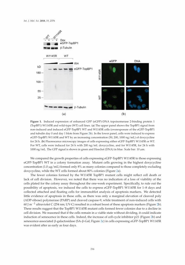
Int. J. Mol. Sci. 2018, 19, 2376
(a) (b)
Figure 1. Induced expression of enhanced GFP (eGFP)-DNA topoisomerase 2-binding protein 1(TopBP1) W1145R and wild-type (WT) cell lines. (a) The upper panel shows the TopBP1 signal fromnon-induced and induced eGFP-TopBP1 WT and W1145R cells (overexposure of the eGFP-TopBP1and tubulin day 0 and day 1 blots from Figure 2b). In the lower panel, cells were induced to expresseGFP-TopBP1 W1145R and WT by an increasing concentration (50, 200, 1000 ng/mL) of doxycyclinefor 24 h. (b) Fluorescence microscopy images of cells expressing either eGFP-TopBP1 W1145R or WT.For WT, cells were induced for 24 h with 200 ng/mL doxycycline, and for W1145R, for 24 h with1000 ng/mL. The GFP signal is shown in green and Hoechst (DNA) in blue. Scale bar: 10 μm.
We compared the growth properties of cells expressing eGFP-TopBP1 W1145R to those expressingeGFP-TopBP1 WT in a colony formation assay. Mutant cells growing in the highest doxycyclineconcentration (1.0 μg/mL) formed only 8% as many colonies compared to those completely excludingdoxycycline, while the WT cells formed about 80% colonies (Figure 2a).
The fewer colonies formed by the W1145R TopBP1 mutant cells might reflect cell death orlack of cell division. However, we noted that there was no indication of a loss of viability of thecells plated for the colony assay throughout the one-week experiment. Specifically, to rule out thepossibility of apoptosis, we induced the cells to express eGFP-TopBP1 W1145R for 1–8 days andcollected attached and floating cells for immunoblot analysis of apoptosis markers. We detectedlittle evidence of apoptosis in these cells, as there was only a marginal elevation of cleaved poly(ADP-ribose) polymerase (PARP) and cleaved caspase-9, while treatment of non-induced cells with60 J m−2 ultraviolet C (254 nm; UV-C) resulted in a robust boost of these apoptosis markers (Figure 2b).These results suggest that the TopBP1 W1145R mutant cells formed fewer colonies due to a decline incell division. We reasoned that if the cells remain in a viable state without dividing, it could indicateinduction of senescence in these cells. Indeed, the increase of cell-cycle inhibitor p21 (Figure 2b) andsenescence-associated β-galactosidase (SA-β-Gal; Figure 2c) in cells expressing eGFP-TopBP1 W1145Rwas evident after as early as four days.
216
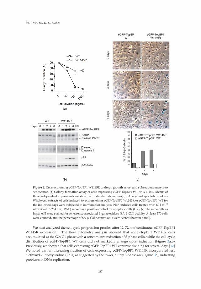
Int. J. Mol. Sci. 2018, 19, 2376
(a)
(b) (c)
Figure 2. Cells expressing eGFP-TopBP1 W1145R undergo growth arrest and subsequent entry intosenescence. (a) Colony formation assay of cells expressing eGFP-TopBP1 WT or W1145R. Means ofthree independent experiments are shown with standard deviations; (b) Analysis of apoptotic markers.Whole-cell extracts of cells induced to express either eGFP-TopBP1 W1145R or eGFP-TopBP1 WT forthe indicated days were subjected to immunoblot analysis. Non-induced cells treated with 60 J m−2
ultraviolet C (254 nm; UV-C) served as a positive control for apoptotic cells (UV); (c) The same cells asin panel B were stained for senescence-associated β-galactosidase (SA-β-Gal) activity. At least 170 cellswere counted, and the percentage of SA-β-Gal positive cells were scored (bottom panel).
We next analyzed the cell-cycle progression profiles after 12–72 h of continuous eGFP-TopBP1W1145R expression. The flow cytometry analysis showed that eGFP-TopBP1 W1145R cellsaccumulated at the G0/G1 phase with a concomitant reduction of S-phase cells, while the cell-cycledistribution of eGFP-TopBP1 WT cells did not markedly change upon induction (Figure 3a,b).Previously, we showed that cells expressing eGFP-TopBP1 WT continue dividing for several days [12].We noted that an increasing fraction of cells expressing eGFP-TopBP1 W1145R incorporated less5-ethynyl-2′-deoxyuridine (EdU) as suggested by the lower, blurry S-phase arc (Figure 3b), indicatingproblems in DNA replication.
217

Int. J. Mol. Sci. 2018, 19, 2376
(a)
(b)
Figure 3. Cells expressing eGFP-TopBP1 W1145R arrest predominantly in gap 1 (G1) phase. (a) Flowcytometry analysis of cells. Cells were induced to express eGFP-TopBP1 WT or W1145R for theindicated times, and were pulsed with 5-ethynyl-2′-deoxyuridine (EdU) prior to sample collection tolabel synthesis (S)-phase cells. (b) Flow cytometry profiles of selected samples from panel A.
Together, these results show that the cells expressing the TopBP1 AAD mutant remain viable,but a large fraction of cells arrest with a G1-phase DNA content, and, if the expression is prolonged,these cells enter senescence. Since our cells still contain endogenous TopBP1, we conclude that theTopBP1 AAD mutant has a dominant negative effect on cell-cycle progression.
2.2. The TopBP1 AAD Mutant Slows Down the DNA Replication Elongation Rate and Increases the Number ofFired Origins
The shift in EdU-labeling intensity in cells expressing eGFP-TopBP1 W1145R (Figure 3b) promptedus to concentrate on subsequent analyses of S-phase cells. We performed DNA fiber assays on cellsthat were induced to express either eGFP-TopBP1 WT or W1145R for 24 h. The cells were sequentially
218

Int. J. Mol. Sci. 2018, 19, 2376
pulse-labeled with 5-chloro-2′-deoxyuridine (CldU) and 5-iodo-2′-deoxyuridine (IdU) for 20 min beforeanalysis (Figure 4a). We observed a dramatic decrease in the average DNA replication elongation ratefrom 1.0 kb min−1 to 0.4 kb min−1 (Figure 4b) and a clear shift in the distribution of elongation rates(Figure 4c) when cells were induced to express eGFP-TopBP1 W1145R. Slowed replication fork rateswere observed in unperturbed conditions after ATR or serine/threonine-protein kinase Chk1 (Chk1)inhibition or depletion [13–16], or after Cdc45 overexpression [17]. To further explore if origin firingis increased in mutant TopBP1 cells, we analyzed the inter-origin distance and the fraction of neworigins during the combined first and second pulses of labeling. Indeed, the average distance betweenorigins dropped almost by half from 78 to 43 kb (Figure 4d) when expression of eGFP-TopBP1 W1145Rwas induced, indicating an excessive firing of dormant origins. In line with this, we also observeda significant increase in newly fired origins in cells expressing eGFP-TopBP1 W1145R (Figure 4e).Thus, cells expressing the TopBP1 AAD mutant displayed a severe replication stress phenotype [18].The strong asymmetry of elongation of fork pairs from the same origin (Figure 4f) suggests that thereduced elongation rate is caused by recurrent stalling of DNA replication. These results stronglysuggest that the limitation of (dormant) origin firing is controlled by a TopBP1-mediated pathwaythat is dependent on a functional AAD. As the excess of WT TopBP1 after doxycycline induction didnot affect origin firing or elongation rate, TopBP1 appears not to be rate limiting for DNA replicationorigin firing.
(a) (b)
Figure 4. Cont.
219
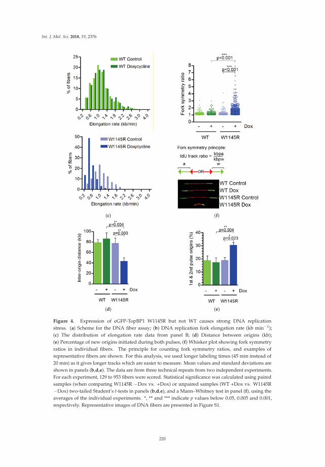
Int. J. Mol. Sci. 2018, 19, 2376
(c) (f)
(d) (e)
Figure 4. Expression of eGFP-TopBP1 W1145R but not WT causes strong DNA replicationstress. (a) Scheme for the DNA fiber assay; (b) DNA replication fork elongation rate (kb min−1);(c) The distribution of elongation rate data from panel B; (d) Distance between origins (kb);(e) Percentage of new origins initiated during both pulses; (f) Whisker plot showing fork symmetryratios in individual fibers. The principle for counting fork symmetry ratios, and examples ofrepresentative fibers are shown. For this analysis, we used longer labeling times (45 min instead of20 min) as it gives longer tracks which are easier to measure. Mean values and standard deviations areshown in panels (b,d,e). The data are from three technical repeats from two independent experiments.For each experiment, 129 to 953 fibers were scored. Statistical significance was calculated using pairedsamples (when comparing W1145R −Dox vs. +Dox) or unpaired samples (WT +Dox vs. W1145R−Dox) two-tailed Student’s t-tests in panels (b,d,e), and a Mann–Whitney test in panel (f), using theaverages of the individual experiments. *, ** and *** indicate p values below 0.05, 0.005 and 0.001,respectively. Representative images of DNA fibers are presented in Figure S1.
220

Int. J. Mol. Sci. 2018, 19, 2376
2.3. The TopBP1 AAD Mutant Induces Accumulation of Single-Stranded DNA
ATR or Chk1 inhibition is known to lead to the generation of excess single-stranded DNA(ssDNA) [19,20]. We tested if ssDNA was also present in cells expressing eGFP-TopBP1 W1145R.Indeed, we found that after 24 or 48 h of expression, ssDNA was present in about 30% of the cells,while it was not detected in non-induced or eGFP-TopBP1 WT expressing cells (Figure 5a,b andFigure S2). We noted that ssDNA foci were more enlarged in cells which expressed mutant TopBP1for 48 h than in cells expressing it for only 24 h. In the latter, the ssDNA foci were more similar toreplication foci and overlapped with sites of DNA replication (Figure 5c).
(a)
(b)
(c)
Figure 5. Cont.
221

Int. J. Mol. Sci. 2018, 19, 2376
(d)
Figure 5. Expression of eGFP-TopBP1 W1145R induces accumulation of single-stranded DNA (ssDNA),but no DNA damage response. (a) ssDNA analysis of eGFP-TopBP1 WT and W1145R cells leftnon-induced or induced for 24 or 48 h. Means of three independent experiments with standarddeviations are shown; (b) Representative examples of nuclei from panel A. DNA replication foci werelabeled with a short pulse of EdU; (c) Co-localization of DNA replication foci (yellow) and ssDNAis shown in the overlay image (EdU + ssDNA) and in a magnified region marked by white frame(bottom frame); (d) Immunoblot analysis of whole-cell extracts from cells induced to express eithereGFP-TopBP1 WT or W1145R for the indicated times. Etoposide was used as a positive control toinduce the intra-S-phase damage response (normal human osteosarcoma (U2OS) cells).
The generation of ssDNA in wild-type cells normally results in the amplification of ATR signalingvia the independent recruitment of TopBP1 and ATR to the ssDNA. In our mutant cells, endogenousTopBP1 was still present, which, in principle, could initiate the DNA replication stress response. Indeed,the cells induced to express mutant TopBP1 were still fully capable of activating the Chk1 responsewhen irradiated with UV-C (Figure S3A). Depletion of TopBP1 by two different small interfering RNAs(siRNAs) from the parental U2OS cell strain completely abrogated the Chk1 response to UV-C, showingthat the Chk1 response is dependent on TopBP1 in these cells (Figure S3B). To test if the DNA damageresponse was induced in cells expressing mutant TopBP1, we analyzed the expressions of p21, p27,and phosphorylated serine-protein kinase ATM (ATM), Chk1, p53, and histone H2AX phosphorylatedat serine 139 (γH2AX) in immunoblots of whole-cell extracts. While the cells showed elevated levels ofp21 and p27, no accumulation of the phosphorylated DNA damage checkpoint markers, ATM S1981,Chk1 S345, p53 S15, or γH2AX was observed (Figure 5d). Increased p21 and p27 protein levels furthersupport the notion of senescence-associated G1 arrest in response to defective TopBP1 signaling ratherthan an intra-S-phase damage response. Expression of WT TopBP1 did not affect the levels of DNAreplication stress markers (Figure 5d).
Taken together, these results show that the failure of TopBP1 signaling during unperturbedDNA replication leads to excess origin firing, decreased replication fork elongation due to excessivefork stalling, and an accumulation of ssDNA. These results resemble the phenotypes of ATR orChk1-inhibited cells that show excess local origin firing causing defective progression of replication
222

Int. J. Mol. Sci. 2018, 19, 2376
forks [14,20]. Despite a lack of replication checkpoint signaling, the mutant TopBP1 cells did not gointo an “intrinsic replication catastrophe” as do cells overexpressing Cdc45 [17].
3. Discussion
Using an AAD mutant of TopBP1, we demonstrated that regulation of origin firing is compromisedin cells expressing the TopBP1 mutant in the S phase. This observation is in accordance with theessential role of TopBP1 in activating ATR, and with findings that inhibiting the ATR-Chk1 pathwayrestricts dormant origin firing [14,21–24]. However, until now, it was not clear if TopBP1 withan inactivating mutation in its ATR-activation domain had a similar effect on replication fork dynamicsas occurs after the inhibition of ATR or Chk1. We also report that cells expressing the AAD mutant ofTopBP1 accumulate at the G1 phase and enter senescence if expression is prolonged. This is consistentwith deletion mutation experiments in mouse, where wild-type TopBP1 was replaced with an AADmutant [11]. Since our cell lines still had endogenous TopBP1 present, the cell-cycle blocking effect ofthe AAD mutant TopBP1 is dominant negative, in contrast to the damage response induced by UV-C(Figure S3). Thus, the AAD of TopBP1 appears to be essential for cell-cycle progression, and for theinitiation of DNA replication.
DNA replication origins are licensed by loading pre-replication complexes in early G1 well beforethe restriction point [25]. It is notable that pre-replication complexes are loaded in excess to thatwhich is ultimately used during a given S phase. Most origins remain dormant and are only firedwhen replication stress leads to problems in fork progression [26]. During stress, dormant originscan be activated to ensure continuation of replication without dramatic slowing down of the wholereplication program. However, too many simultaneously active replication forks can be deleterious tocells, leading to replication stress, accumulation of ssDNA, and ultimately, DNA shattering [17,20].Interestingly, we did not observe initiation of the DNA damage response in cells expressing the mutantTopBP1, despite the severe DNA replication phenotype and accumulation of ssDNA. It is even moresurprising considering our observation that the endogenous TopBP1 present in our expression celllines is capable of inducing Chk1 in response to UV-C (Figure S3). This may be explained by the(replication-dependent) replication protein A (RPA) depletion that we observed after expression ofthe TopBP1 AAD mutant. It should be noted that the phenotype observed here after expression ofTopBP1 AAD is not merely phenocopying the effects of ATR inhibition or depletion. ATR inhibitiondoes not lead to a severe loss of cellular viability in the absence of induced replication stress [15,16,27].For instance, Jossé et al. [27] did not observe any effect on elongation rates after ATR inhibition,whereas Moiseeva et al. [15] observed reduced elongation rates and increased origin firing, but nofork asymmetry, indicating no augmentation of fork stalling. This is consistent with the role of ATRduring replication stress, especially preventing exhaustion of the RPA pool [20]. Here, we observedthat expression of the AAD mutant, but not WT TopBP1, induces both replication stress and suppressesATR-dependent damage responses. Whereas the latter could be a secondary effect of suppressing ATRactivation by the AAD mutant, the phenotype is more consistent with a dual role of TopBP1 couplingthe initiation of DNA replication with the suppression of neighboring origins.
Moreover, several recent studies identified Ewing’s tumor-associated antigen 1 (ETAA1) asa second ATR activator [28–31]. It was noted previously that U2OS cells have very low ETAA1 proteinlevels [30]. This could explain the lack of replication stress-induced Chk1 phosphorylation, as well asthe severity of the phenotype of the AAD mutant expression observed in this study. ETAA1 activatesATR in response to DNA replication stress independently of TopBP1, but is not implicated to participatein DNA replication initiation or elongation.
It was proposed previously that a fired origin suppresses the firing of nearby origins withina replication cluster. This negative origin interference was first found in yeast [32,33] and laterapplied to human cells as well [34]. How negative origin interference functions mechanistically is notunderstood. Two models can be envisioned that are not necessarily mutually exclusive.
223

Int. J. Mol. Sci. 2018, 19, 2376
The more efficiently firing origin could deplete replication factors resulting in differential firingefficiency in a local cluster of origins. The less efficient origin in the cluster would thereafterbe inactivated due to its replication by a traversing fork from the nearby more efficient origin.Alternatively, there could be a factor or factors that inhibit nearby origin activation in the vicinity ofa fired origin.
TopBP1 was not previously observed to be required for the elongation of DNA replication [35–37],which would explain the increased stalling of forks we observe. It is possible, however, that fork stallingis a naturally frequent event in cells, which would require continuous TopBP1-ATR for re-initiation.
Another possibility that is consistent with the negative origin interference is that, once TopBP1binds to the pre-initiation complex and activates an origin, it simultaneously induces local activation ofthe ATR pathway leading to an inhibition of nearby origin activation (see Figure 6). Such an essentialfunction of TopBP1 would explain the ostensibly contradictory roles in activating and suppressingorigin firing.
Figure 6. A model for the role of TopBP1 in the S phase. During the S phase, TopBP1 activates the firingof new forks, and the TopBP1 local activation of serine/threonine-protein kinase ATR (ATR) inhibitsthe firing of nearby dormant origins.
4. Materials and Methods
4.1. Cell Culture
The preparation and cultivation of eGFP-TopBP1 WT and W1145R cells were describedpreviously [12]. All cell lines were regularly checked for contamination. The SA-β-Gal assay wasperformed according to the manufacturer’s instructions (Millipore).
4.2. Colony Formation Assay
Approximately 5000 cells were plated per 10-cm dish and then grown with or without doxycyclinefor eight days until colonies of over 50 cells appeared in the no-doxycycline control plates. Colonieswere fixed with methanol and stained with 0.5% crystal violet in 25% methanol. The number ofcolonies was counted with a custom automated analysis using CellProfiler (r11710) [38].
4.3. Flow Cytometry
Cells were labeled with 10 μM EdU for 15 min before harvesting. Harvested cells werewashed with 1% bovine serum albumin/phosphate-buffered saline (BSA/PBS) and fixed with 2%paraformaldehyde for 15 min. EdU detection (Click-iT EdU Alexa Fluor 647) was done accordingto the manufacturer’s instructions (Life Technologies, Carlsbad, CA, USA). DNA was stained with4′,6-diamidino-2-phenylindole (DAPI; Sigma-Aldrich, St. Louis, MI, USA). Flow cytometry data wereacquired with a FACSCanto machine using the FACSDiva software (BD Biosciences, Franklin Lakes,NJ, USA). Data analysis was done using the FlowJo software (FlowJo, LLC, Ashland, OR, USA).
224

Int. J. Mol. Sci. 2018, 19, 2376
4.4. DNA Fiber Assays
For the DNA fiber assays, cells were either non-induced or induced to express eGFP-TopBP1WT or W1145R for 24 h before supplementing the medium with 25 μM IdU (Sigma-Aldrich) for20 min (first labeling). The cells were then washed, and 250 μM CldU (Sigma-Aldrich) was addedto fresh medium for 20 min (second labeling). Preparation of DNA spreads was done as describedpreviously [17].
4.5. Single-Stranded DNA (ssDNA) Analysis and Immunofluorescence Microscopy
DNA was labeled with 10 μM 5-bromo-2′-deoxyuridine (BrdU) for 36 h. Sample preparationwas performed as described previously [17]. To detect incorporated BrdU only in regions of ssDNA,the DNA was directly immunolabelled (without denaturation) with a primary monoclonal antibody(1:1500 dilution) of rat-anti-BrdU (Clone BU1/75 ABD Serotec) and a secondary anti-rat Alexa Fluor555 conjugate. Total DNA was counterstained with DAPI. Fluorescence images were acquired usinga Zeiss Axio Imager.Z1 at 630-fold magnification.
For the fluorescence microscopy in Figure 1b, cells were prepared as described previously [12].Briefly, cells were fixed with 3% paraformaldehyde, permeabilized with 0.2% Triton X-100-PBS, andstained for DNA with Hoechst 33258. Wide-field fluorescent images were obtained with Axiocam HRcolor using a Zeiss Axioplan 2 microscope with a 40× Zeiss Plan-Neofluar objective.
4.6. Immunoblotting
Immunoblotting was performed as described previously [12]. Primary antibodies (1:1000 dilutionsunless otherwise stated) were from Cell Signaling (α-GFP D5.1 (1:2000), α-PARP #9542, α-caspase9 #9502 (1:2000), α-ATM D2E2, α-ATM phospho-S1981 10H11.E12, α-Chk1 phospho-S345 133D3,α-p53 phospho-S15 16G8 (1:2000), α-p27 D69C12, and α-phospho-S780 pRB #9307), Millipore (α-p21#05-345, α-β-Tubulin KMX-1 (1:20000), and α-γH2AX JBW301), and Santa Cruz Biothechnology (α-p53sc-6243 (1:400), α-Chk1 sc-8408, and α-Cyclin A sc-751). Secondary antibodies (1:40000 dilution)for immunoblotting were peroxidase-conjugated goat α-rabbit or α-mouse immunoglobulin G (IgG;Jackson Immunoresearch Laboratories).
Supplementary Materials: Supplementary materials can be found at http://www.mdpi.com/1422-0067/19/8/2376/s1.
Author Contributions: Conceptualization, M.S. and H.P. Funding acquisition, J.E.S. Investigation, M.S. and D.K.Resources, P.H. Supervision, H.P. and J.E.S. Writing (original draft, review and editing), M.S. and H.P.
Funding: This research was funded by the Academy of Finland grant 251576 to J.E.S., the Finnish CulturalFoundation, North Carelia Regional fund to M.S., and the UFA Grant 3610S30016 by the German Federal Officefor Radiation Protection as part of the “Kompetenzverbund für Strahlenforschung” to H.P. The Fritz LipmannInstitute is a member of the Science Association “Gottfried Wilhelm Leibniz” (WGL) and financially supported bythe Federal Government of Germany and the State of Thuringia. Funding for open access charge: Leibniz Insituteon Aging—Fritz Lipmann Institute.
Acknowledgments: We are grateful to Susan A. Gerbi for comments on the manuscript. We thank LeenaPääkkönen and Annerose Gleiche for technical assistance in the laboratory.
Conflicts of Interest: The authors declare no conflict of interest.
Abbreviations
AAD ATR activating domain (in TopBP1)ATM Serine-protein kinase ATMATR Serine/threonine-protein kinase ATRBSA Bovine serum albuminCdc45 Cell division control protein 45 homologCdc6 Cell division control protein 6 homolog
225
Int. J. Mol. Sci. 2018, 19, 2376
Cdt1 DNA replication factor Cdt1Chk1 Serine/threonine-protein kinase Chk1CldU 5-Chloro-2′-deoxyuridineCMG Cdc45-MCM-GINS complexDDK Dbf4-dependent kinaseEdU 5-Ethynyl-2′-deoxyuridineETAA1 Ewing’s tumor-associated antigen 1eGFP Enhanced green fluorescent proteinGEMC1 Geminin coiled-coil domain-containing protein 1GINS Sld5-Psf1-Psf2-Psf3 complex (go-ichi-ni-san)H2A.X/γH2AX Phosphorylated histone H2AXIdU 5-iodo-2′-deoxyuridinekb kilobaseMCM2-7 DNA replication licensing factor MCM2-7ORI Origin of DNA replicationp21 Cyclin-dependent kinase inhibitor 1p27 Cyclin-dependent kinase inhibitor 1Bp53 Cellular tumor antigen p53PARP Poly (ADP-ribose) polymerasePBS Phosphate-buffered salineRecQL4 ATP-dependent DNA helicase Q4RPA Replication protein A complexS-CDK S-phase-specific cyclin dependent kinaseSA-β-Gal Senescence-associated beta-galactosidasessDNA Single-stranded DNATopBP1 DNA topoisomerase 2-binding protein 1U2OS Human osteosarcoma cell lineUV-C Ultraviolet C (254 nm)WT Wild type
References
1. Aladjem, M.I.; Redon, C.E. Order from clutter: Selective interactions at mammalian replication origins.Nat. Rev. Genet. 2017, 18, 101–116. [CrossRef] [PubMed]
2. Kumagai, A.; Shevchenko, A.A.; Shevchenko, A.A.; Dunphy, W.G. Treslin collaborates with TopBP1 intriggering the initiation of DNA replication. Cell 2010, 140, 349–359. [CrossRef] [PubMed]
3. Balestrini, A.; Cosentino, C.; Errico, A.; Garner, E.; Costanzo, V. GEMC1 is a TopBP1-interacting proteinrequired for chromosomal DNA replication. Nat. Cell Biol. 2010, 12, 484–491. [CrossRef] [PubMed]
4. Matsuno, K.; Kumano, M.; Kubota, Y.; Hashimoto, Y.; Takisawa, H. The N-terminal noncatalytic regionof Xenopus RecQ4 is required for chromatin binding of DNA polymerase alpha in the initiation of DNAreplication. Mol. Cell. Biol. 2006, 26, 4843–4852. [CrossRef] [PubMed]
5. Sangrithi, M.N.; Bernal, J.A.; Madine, M.; Philpott, A.; Lee, J.; Dunphy, W.G.; Venkitaraman, A.R. Initiationof DNA replication requires the RECQL4 protein mutated in Rothmund-Thomson syndrome. Cell 2005, 121,887–898. [CrossRef] [PubMed]
6. Kumagai, A.; Lee, J.; Yoo, H.Y.; Dunphy, W.G. TopBP1 activates the ATR-ATRIP complex. Cell 2006, 124,943–955. [CrossRef] [PubMed]
7. Saldivar, J.C.; Cortez, D.; Cimprich, K.A. The essential kinase ATR: Ensuring faithful duplication ofa challenging genome. Nat. Rev. Mol. Cell Biol. 2017, 18, 622–636. [CrossRef] [PubMed]
8. Brown, E.J.; Baltimore, D. ATR disruption leads to chromosomal fragmentation and early embryonic lethality.Genes Dev. 2000, 14, 397–402. [CrossRef] [PubMed]
9. Klein, A.D.; Muijtjens, M.; Os, R.V.; Verhoeven, Y.; Smit, B.; Carr, A.M.; Lehmann, A.R.; Hoeijmakers, J.H.J.Targeted disruption of the cell-cycle checkpoint gene ATR leads to early embryonic lethality in mice. Curr. Biol.2000, 10, 479–482. [CrossRef]
226
Int. J. Mol. Sci. 2018, 19, 2376
10. Jeon, Y.; Ko, E.; Lee, K.Y.; Ko, M.J.; Park, S.Y.; Kang, J.; Jeon, C.H.; Lee, H.; Hwang, D.S. TopBP1 deficiencycauses an early embryonic lethality and induces cellular senescence in primary cells. J. Biol. Chem. 2011, 286,5414–5422. [CrossRef] [PubMed]
11. Zhou, Z.-W.; Liu, C.; Li, T.-L.; Bruhn, C.; Krueger, A.; Min, W.; Wang, Z.-Q.; Carr, A.M. An EssentialFunction for the ATR-Activation-Domain (AAD) of TopBP1 in Mouse Development and Cellular Senescence.PLoS Genet. 2013, 9, e1003702. [CrossRef] [PubMed]
12. Sokka, M.; Rilla, K.; Miinalainen, I.; Pospiech, H.; Syväoja, J.E. High levels of TopBP1 induce ATR-dependentshut-down of rRNA transcription and nucleolar segregation. Nucleic Acids Res. 2015, 43, 4975–4989.[CrossRef] [PubMed]
13. Petermann, E.; Maya-Mendoza, A.; Zachos, G.; Gillespie, D.A.F.; Jackson, D.A.; Caldecott, K.W. Chk1requirement for high global rates of replication fork progression during normal vertebrate S phase.Mol. Cell. Biol. 2006, 26, 3319–3326. [CrossRef] [PubMed]
14. Petermann, E.; Woodcock, M.; Helleday, T. Chk1 promotes replication fork progression by controllingreplication initiation. PNAS 2010, 107, 16090–16095. [CrossRef] [PubMed]
15. Moiseeva, T.; Hood, B.; Schamus, S.; O’Connor, M.J.; Conrads, T.P.; Bakkenist, C.J. ATR kinase inhibitioninduces unscheduled origin firing through a Cdc7-dependent association between GINS and And-1.Nat. Commun. 2017, 8, 1392. [CrossRef] [PubMed]
16. Couch, F.B.; Bansbach, C.E.; Driscoll, R.; Luzwick, J.W.; Glick, G.G.; Bétous, R.; Carroll, C.M.; Jung, S.Y.;Qin, J.; Cimprich, K.A.; et al. ATR phosphorylates SMARCAL1 to prevent replication fork collapse. Genes Dev.2013, 27, 1610–1623. [CrossRef] [PubMed]
17. Köhler, C.; Koalick, D.; Fabricius, A.; Parplys, A.C.; Borgmann, K.; Pospiech, H.; Grosse, F. Cdc45 is limitingfor replication initiation in humans. Cell Cycle 2016, 15, 974–985. [CrossRef] [PubMed]
18. Marheineke, K.; Hyrien, O.; Krude, T. Visualization of bidirectional initiation of chromosomal DNAreplication in a human cell free system. Nucleic Acids Res. 2005, 33, 6931–6941. [CrossRef] [PubMed]
19. Syljuåsen, R.G.; Sørensen, C.S.; Hansen, L.T.; Fugger, K.; Lundin, C.; Johansson, F.; Helleday, T.;Sehested, M.; Lukas, J.; Bartek, J. Inhibition of Human Chk1 Causes Increased Initiation of DNA Replication,Phosphorylation of ATR Targets, and DNA Breakage. Mol. Cell. Biol. 2005, 25, 3553–3562. [CrossRef][PubMed]
20. Toledo, L.I.; Altmeyer, M.; Rask, M.-B.; Lukas, C.; Larsen, D.H.; Povlsen, L.K.; Bekker-Jensen, S.; Mailand, N.;Bartek, J.; Lukas, J. ATR Prohibits Replication Catastrophe by Preventing Global Exhaustion of RPA. Cell2013, 155, 1088–1103. [CrossRef] [PubMed]
21. Heffernan, T.P.; Simpson, D.A.; Frank, A.R.; Heinloth, A.N.; Paules, R.S.; Cordeiro-Stone, M.; Kaufmann, W.K.An ATR- and Chk1-dependent S checkpoint inhibits replicon initiation following UVC-induced DNA damage.Mol. Cell. Biol. 2002, 22, 8552–8561. [CrossRef] [PubMed]
22. Maya-Mendoza, A.; Petermann, E.; Gillespie, D.A.F.; Caldecott, K.W.; Jackson, D.A. Chk1 regulates thedensity of active replication origins during the vertebrate S phase. EMBO J. 2007, 26, 2719–2731. [CrossRef][PubMed]
23. Seiler, J.A.; Conti, C.; Syed, A.; Aladjem, M.I.; Pommier, Y. The intra-S-phase checkpoint affects both DNAreplication initiation and elongation: Single-cell and -DNA fiber analyses. Mol. Cell. Biol. 2007, 27, 5806–5818.[CrossRef] [PubMed]
24. Shechter, D.; Costanzo, V.; Gautier, J. ATR and ATM regulate the timing of DNA replication origin firing.Nat. Cell Biol. 2004, 6, 648–655. [CrossRef] [PubMed]
25. Fragkos, M.; Ganier, O.; Coulombe, P.; Méchali, M. DNA replication origin activation in space and time.Nat. Rev. Mol. Cell Biol. 2015, 16, 360–374. [CrossRef] [PubMed]
26. Ge, X.Q.; Jackson, D.A.; Blow, J.J. Dormant origins licensed by excess Mcm2–7 are required for human cellsto survive replicative stress. Genes Dev. 2007, 21, 3331–3341. [CrossRef] [PubMed]
27. Jossé, R.; Martin, S.E.; Guha, R.; Ormanoglu, P.; Pfister, T.D.; Reaper, P.M.; Barnes, C.S.; Jones, J.; Charlton, P.;Pollard, J.R.; et al. ATR inhibitors VE-821 and VX-970 sensitize cancer cells to topoisomerase I inhibitorsby disabling DNA replication initiation and fork elongation responses. Cancer Res. 2014, 74, 6968–6978.[CrossRef] [PubMed]
28. Bass, T.E.; Luzwick, J.W.; Kavanaugh, G.; Carroll, C.; Dungrawala, H.; Glick, G.G.; Feldkamp, M.D.;Putney, R.; Chazin, W.J.; Cortez, D. ETAA1 acts at stalled replication forks to maintain genome integrity.Nat. Cell Biol. 2016, 18, 1185–1195. [CrossRef] [PubMed]
227

Int. J. Mol. Sci. 2018, 19, 2376
29. Lee, Y.-C.; Zhou, Q.; Chen, J.; Yuan, J. RPA-Binding Protein ETAA1 Is an ATR Activator Involved in DNAReplication Stress Response. Curr. Biol. 2016, 26, 3257–3268. [CrossRef] [PubMed]
30. Haahr, P.; Hoffmann, S.; Tollenaere, M.A.X.; Ho, T.; Toledo, L.I.; Mann, M.; Bekker-Jensen, S.; Räschle, M.;Mailand, N. Activation of the ATR kinase by the RPA-binding protein ETAA1. Nat. Cell Biol. 2016, 18,1196–1207. [CrossRef] [PubMed]
31. Feng, S.; Zhao, Y.; Xu, Y.; Ning, S.; Huo, W.; Hou, M.; Gao, G.; Ji, J.; Guo, R.; Xu, D. Ewing tumor-associatedantigen 1 interacts with replication protein A to promote restart of stalled replication forks. J. Biol. Chem.2016, 291, 21956–21962. [CrossRef] [PubMed]
32. Brewer, B.J.; Fangman, W.L. Initiation at closely spaced replication origins in a yeast chromosome. Science1993, 262, 1728–1731. [CrossRef] [PubMed]
33. Marahrens, Y.; Stillman, B. Replicator dominance in a eukaryotic chromosome. EMBO J. 1994, 13, 3395–3400.[PubMed]
34. Lebofsky, R.; Heilig, R.; Sonnleitner, M.; Weissenbach, J.; Bensimon, A. DNA replication origin interferenceincreases the spacing between initiation events in human cells. Mol. Biol. Cell 2006, 17, 5337–5345. [CrossRef][PubMed]
35. Hashimoto, Y.; Takisawa, H. Xenopus Cut5 is essential for a CDK-dependent process in the initiation of DNAreplication. EMBO J. 2003, 22, 2526–2535. [CrossRef] [PubMed]
36. Mäkiniemi, M.; Hillukkala, T.; Tuusa, J.; Reini, K.; Vaara, M.; Huang, D.; Pospiech, H.; Majuri, I.; Westerling, T.;Mäkelä, T.P.P.; et al. BRCT domain-containing protein TopBP1 functions in DNA replication and damageresponse. J. Biol. Chem. 2001, 276, 30399–30406. [CrossRef] [PubMed]
37. Kim, J.; Mcavoy, S.A.; Smith, D.I.; Chen, J. Human TopBP1 Ensures Genome Integrity during Normal SPhase. Mol. Cell. Biol. 2005, 25, 10907–10915. [CrossRef] [PubMed]
38. Carpenter, A.E.; Jones, T.R.; Lamprecht, M.R.; Clarke, C.; Kang, I.H.; Friman, O.; Guertin, D.A.; Chang, J.H.;Lindquist, R.A.; Moffat, J.; et al. CellProfiler: Image analysis software for identifying and quantifying cellphenotypes. Genome Biol. 2006, 7, R100. [CrossRef] [PubMed]
© 2018 by the authors. Licensee MDPI, Basel, Switzerland. This article is an open accessarticle distributed under the terms and conditions of the Creative Commons Attribution(CC BY) license (http://creativecommons.org/licenses/by/4.0/).
228
International Journal of
Molecular Sciences
Review
DNA Damage Stress: Cui Prodest?
Nagendra Verma , Matteo Franchitto, Azzurra Zonfrilli, Samantha Cialfi, Rocco Palermo and
Claudio Talora *
Department of Molecular Medicine, Sapienza University of Rome, 00161 Rome, Italy;[email protected] (N.V.); [email protected] (M.F.); [email protected] (A.Z.);[email protected] (S.C.); [email protected] (R.P.)* Correspondence: [email protected]
Received: 18 January 2019; Accepted: 26 February 2019; Published: 1 March 2019
Abstract: DNA is an entity shielded by mechanisms that maintain genomic stability and are essentialfor living cells; however, DNA is constantly subject to assaults from the environment throughoutthe cellular life span, making the genome susceptible to mutation and irreparable damage. Cells areprepared to mend such events through cell death as an extrema ratio to solve those threats from amulticellular perspective. However, in cells under various stress conditions, checkpoint mechanismsare activated to allow cells to have enough time to repair the damaged DNA. In yeast, entry intothe cell cycle when damage is not completely repaired represents an adaptive mechanism to copewith stressful conditions. In multicellular organisms, entry into cell cycle with damaged DNA isstrictly forbidden. However, in cancer development, individual cells undergo checkpoint adaptation,in which most cells die, but some survive acquiring advantageous mutations and selfishly evolve aconflictual behavior. In this review, we focus on how, in cancer development, cells rely on checkpointadaptation to escape DNA stress and ultimately to cell death.
Keywords: cell cycle checkpoints; genomic instability; G2-arrest; cell death; repair of DNAdamage; adaptation
1. Introduction
While questionable, one of the most well-known and widely reported aspect in cancer biology isthe acquisition of genetic mutations that underlie cell transformation and tumor progression. From thisperspective, cell transformation is a genetic process of tumor cells adapted to stressful environmentalconditions; if to ‘cell adaptation’ can be conferred the Darwinian concept to respond to life’s needs forsurvival, the nature of what adaptation means for tumor cells is extremely elusive. Either physicalor chemical environmental agents can cause DNA damage and consequently genetic mutations thatpromote cell transformation.
Examples of physical agents promoting mutations are ionizing radiation, ultraviolet light present insunlight which can promote the estimated rate of up to 10,000 DNA lesions per cell per day [1,2]; chemicalagents such as benzo(a)pyrene B(a)P, 7,12-dimethylbenz[a]anthracene (DMBA), that generate DNAadducts, leading to mutations [3]. Beside exogenously, DNA damage can also occur endogenouslyas cells divide, with tens of thousands events every day in each single cell [2]. Thus, DNA damagemight potentially affect the function of central regulators of many biological processes, ultimatelyleading to cancer development. Additionally, infectious pathogens elicit an oncogenic spiral thatis one of the causes of cancer development [4]. If we assess the concept that ‘adaptation’ meansthe optimization of the phenotype whereby the organism acquires changes that increase its survivaland reproductive success, when this concept is applied to cell transformation it remains extremelyvague. Although this concept is suitable for viral carcinogenesis that hijacking cellular pathwayspromotes the survival and proliferation of infected cells, in a multicellular organism, cells do not need
Int. J. Mol. Sci. 2019, 20, 1073; doi:10.3390/ijms20051073 www.mdpi.com/journal/ijms229

Int. J. Mol. Sci. 2019, 20, 1073
to adapt their phenotype to a non-permissive environment. Unquestionably, in multicellular organisms,cells are immersed in growth conditions favorable to their replication. However, there is an obviousdifference in the relationship between adaptation and environment in unicellular versus multicellularorganisms. Life and replication in unicellular organisms are dependent on the conditions presentin the environment and they survive if they are able to adapt to environmental changes. In sharpcontrast, in multicellular organisms cell division is tightly regulated to control cell shape, tissuepatterns, and morphogenesis [5], although cells are typically immersed in permissive environmentalconditions. Preservation of the integrity of multicellular organisms relies on these extra layers ofdevelopmental control that function to restrain cellular proliferation that may change in response toenvironmental or intracellular stress signals. This implies that, as previously defined [6,7], cancer cellsarise from cells adapted to respond to holistic control system and the escape from these host defensemechanisms represents an important strategy for cell transformation.
2. Cell Cycle Surveillance System
Genetic damage produced by either exogenous or endogenous mechanisms represents an ongoingthreat to the cell. To preserve genome integrity, eukaryotic cells have evolved repair mechanismsspecific for different types of DNA Damage (for an extensive review see [8,9]). However, regardlessof the type of damage a sophisticated surveillance mechanism, called DNA damage checkpoint,detects and signals its presence to the DNA repair machinery. DNA damage checkpoint has beenfunctionally conserved throughout eukaryotic evolution, with most of the relevant players in thecheckpoint response highly conserved from yeast to human [10]. Checkpoints are induced to delaycell cycle progression and to allow cells to repair damaged DNA (Figure 1). Once the damaged DNAis repaired, the checkpoint machinery triggers signals that will resume cell cycle progression [11].In cells, multiple pathways contribute to DNA repair, but independently of the specific pathwayinvolved, three phase are traditionally identified: Sensing of damage, signal, and downstream effects(Figure 2). The sensor phase recognizes the damage and activates the signal transduction phase to selectthe appropriate repair pathway. For example, cells pose at least four independent mechanisms forrepairing Double-Strand-Breaks (DSBs): Non-Homologous End-Joining (NHEJ), either classic-NHEJ oralternative-NHEJ, Homologous Recombination (HR), and single-strand annealing (SSA) [1,10,12,13].Furthermore, highlighting the complexity of the DNA damage response, in mammals, at least four, inpart, independent sensors can detect DSBs: Mre11-Rad50-Xrs2 (MRN), Poly ADP-Ribose polymerase(PARP), Ku70/Ku80 and Replication protein A (RPA) that binds single stranded DNA permittingthe further processing of DSBs [1,14]. In the presence of DSBs, the activation of the DNA damageresponse and the mobilization of the repair proteins give rise to the formation of nuclear foci at thesites of damage. In yeast, the MRX-complex (Mre11-Rad50-Xrs2) is recruited at the site of DSBs [15].Localization of MRX-complex to the damaged site is required to recruit and activate the protein kinaseTel1, which initiates DSBs signaling [13,16]. A similar mechanism is employed by MRN-complex inmammal cells (in which Nbs1 is the mammalian ortholog of Xrs2). MRN-complex orchestrates thecellular response to DBSs by physically interacting and activating the kinase Ataxia-TelangiectasiaMutated (ATM, the mammalian ortholog of Tel1). The signal is transduced by ATM that phosphorylatesthe histone variant Histone-2AX (H2AX) generating g-H2AX that promotes the recruitment ofMediator of DNA-Damage Checkpoin 1 (MDC1) protein at the site of damage. MDC1 amplifiesthe DNA-Damage Response (DDR) signal through the iterated recruitment of the MRN-ATM complexat the damage site that further phosphorylates adjacent H2AX molecules extending the γ-H2AXmark [13,16]. Additionally, MDC1 functions as an interaction platform for other DDR componentsincluding chromatin remodelers and ubiquitin ligase complexes [13,16]. The recruitment of thesefactors is essential to create a more open and accessible chromatin conformation to facilitate accessat sites of DNA lesions and to allow ubiquitin-mediated accumulation of DNA repair factors, whichwill ultimately contribute to DNA repair pathways [13,16,17]. An integral part of the DNA damageresponse is the parallel induction of repair mechanisms and reversible cell cycle arrest that delays cell
230
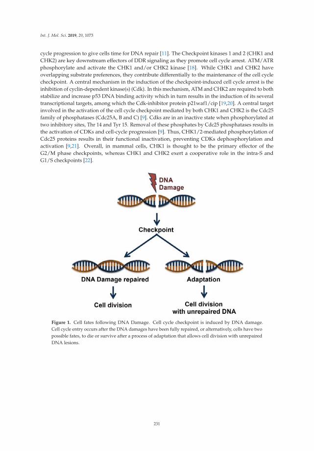
Int. J. Mol. Sci. 2019, 20, 1073
cycle progression to give cells time for DNA repair [11]. The Checkpoint kinases 1 and 2 (CHK1 andCHK2) are key downstream effectors of DDR signaling as they promote cell cycle arrest. ATM/ATRphosphorylate and activate the CHK1 and/or CHK2 kinase [18]. While CHK1 and CHK2 haveoverlapping substrate preferences, they contribute differentially to the maintenance of the cell cyclecheckpoint. A central mechanism in the induction of the checkpoint-induced cell cycle arrest is theinhibition of cyclin-dependent kinase(s) (Cdk). In this mechanism, ATM and CHK2 are required to bothstabilize and increase p53 DNA binding activity which in turn results in the induction of its severaltranscriptional targets, among which the Cdk-inhibitor protein p21waf1/cip [19,20]. A central targetinvolved in the activation of the cell cycle checkpoint mediated by both CHK1 and CHK2 is the Cdc25family of phosphatases (Cdc25A, B and C) [9]. Cdks are in an inactive state when phosphorylated attwo inhibitory sites, Thr 14 and Tyr 15. Removal of these phosphates by Cdc25 phosphatases results inthe activation of CDKs and cell-cycle progression [9]. Thus, CHK1/2-mediated phosphorylation ofCdc25 proteins results in their functional inactivation, preventing CDKs dephosphorylation andactivation [9,21]. Overall, in mammal cells, CHK1 is thought to be the primary effector of theG2/M phase checkpoints, whereas CHK1 and CHK2 exert a cooperative role in the intra-S andG1/S checkpoints [22].
Figure 1. Cell fates following DNA Damage. Cell cycle checkpoint is induced by DNA damage.Cell cycle entry occurs after the DNA damages have been fully repaired, or alternatively, cells have twopossible fates, to die or survive after a process of adaptation that allows cell division with unrepairedDNA lesions.
231
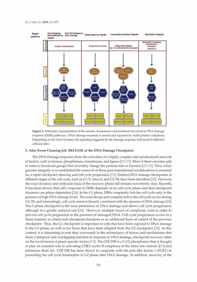
Int. J. Mol. Sci. 2019, 20, 1073
Figure 2. Schematic representation of the sensors, transducers and mediators involved in DNA damageresponse (DDR) pathways. DNA damage response is sensed and repaired by multi-protein complexes.Depending on the level of injury, the signaling triggered by the damage response will result in differentcellular fates.
3. After Event Cleaning Job: RELEASE of the DNA Damage Checkpoint
The DNA Damage response elicits the activation of a highly complex and synchronized networkof factors, such as kinases, phosphatases, transferases, and ligases [23–27]. Most of these enzymes addto remove functional groups that reversibly change the proteins fate or function [23–27]. Thus, whengenome integrity is re-established the removal of these post-translational modifications is essentialfor a rapid checkpoint silencing and cell cycle progression [13]. Distinct DNA damage checkpoints atdifferent stages of the cell cycle, such as G1/S, intra-S, and G2/M, have been described [28]. However,the exact dynamic and molecular basis of the recovery phase still remains not entirely clear. Recently,it has been shown that cell’s response to DSBs depends on its cell cycle phase and that checkpointdynamics are phase-dependent [28]. In the G1 phase, DBSs completely halt the cell cycle only in thepresence of high DNA damage levels. The most abrupt and complete halt to the cell cycle occurs duringG2/M, and interestingly, cell cycle arrest is linearly correlated with the amount of DNA damage [28].The S phase checkpoint is the more permissive to DNA damage and allows cell cycle progression,although at a greatly reduced rate [28]. However, multiple layers of complexity exist in order toprevent cell cycle progression in the presence of damaged DNA. Cell cycle progression occurs in alinear manner, in which each checkpoint functions as an additional layer of control of the previouscheckpoint. Thus, the G1 checkpoint is important in cells that have been exposed to DNA damagein the G1-phase, as well as for those that have been adapted from the G2 checkpoint [29]. In thiscontext, it is interesting to note that, conversely to the redundancy of factors and mechanisms thatshare a temporal and overlapping function in response to DNA damage, checkpoint recovery relieson the involvement of phase-specific factors [13]. The CDC25B is a S/G2 phosphatase that is thoughtto play an essential role in activating CDK1-cyclin B complexes at the entry into mitosis ([13] andreferences there in). CDC25B has been shown to cooperate with the polo-like kinase 1 (PLK1) inpromoting the cell cycle resumption in G2 phase after DNA damage. In addition, recovery of the
232

Int. J. Mol. Sci. 2019, 20, 1073
G2 DNA damage checkpoint appears to be distinct from G1. Indeed, both PLK1 and Cdc25B arenot expressed in G1 and do not influence cell cycle resumption in G1 (Reference [13] and referencestherein). Essentially the same activation pathways promote mitotic entry in an unperturbed cell cycleand checkpoint recovery [30]. However, these pathways are thought to be differentially involvedin these two processes. PLK1 is not essential for mitotic entry in cells progressing through normalcell cycles; it has been shown that the complete inhibition of PLK1 can only delay G2/M transitionleaving the importance of PLK1 for mitotic entry during unperturbed cell cycle controversy [13,31].Conversely, it is well established that initiation of the DNA damage response repress pro-mitoticmachinery and leads to the inhibition of pro-mitotic kinases among which CDK1, Aurora A, andPLK1 [32–34]. Additionally, the degradation of Cdc25 and Bora, as well as of several other proteinsinvolved in mitotic entry, is critical for cell cycle arrest [35,36]. While PLK1 is dispensable for the onsetof mitosis in an unperturbed cell cycle, in sharp contrast PLK1, is essential for mitotic entry followingrecovery from DNA Damage-induced cell cycle arrest [37]. Cell cycle re-entry relies on the Aurora-Akinase and its co-factor Bora, which phosphorylates PLK1 at Thr210 in its activation loop; thus, Plk1 isactivated and promotes mitotic entry by stimulating cyclin B1-Cdk1 activation [25,30,37,38]. PLK1 canpromote cyclinB1/CDK1 activation by several mechanisms. Early works in Xenopus have establishedthat Plx1 (PLK1) phosphorylates and activates Cdc25C, and this activates the Cyclin B–CDK1 complex.In vertebrates, the Cdc25 paralogues (Cdc25A, B and C), all have been shown to be target of PLK1activity [39], but it remains poorly characterized, with Cdc25 phosphatase(s) the substrate of PLK1during the G2 recovery. However, it has been suggested that G2 recovery is dependent on the specificisoform Cdc25B, which is stabilized after damage, while Cdc25A expression is reduced [37,40]. Besideits implication in the re-activation of cyclin-B1–CDK1 complex, PLK1 controls the silencing of DDRsignals by inactivating the ATM/CHK2 pathway. Within the DNA damage response mechanism,53BP1 is an adaptor protein required to tether several checkpoint components at the damaged sites,including CHK2 and ATM. In PLK1-mediated inactivation of the DNA damage checkpoint, it hasbeen shown that PLK1 phosphorylated 53BP1 that thus fails to form foci after DNA damage [41].Additionally, it has been shown that PLK1 also directly phosphorylates and inactivates CHK2 [41].Thus, PLK1 negatively regulates the ATM-CHK2 branch of the DNA damage to inactivate checkpointsignaling and to control checkpoint duration [41]. Similarly, PLK1 negatively controls Claspin andCHK1 and the inactivation of these components results in a shutdown of the checkpoint [42–44].Specifically, phosphorylation of Claspin by PLK1 creates a docking site for β-TrCP protein, resulting inthe efficient ubiquitin-mediated degradation of this protein [42–44]. In conclusion, PLK1 is capableof driving entry into mitosis after DNA damage-induced cell cycle arrest and to promote checkpointsilencing and recovery.
4. DNA Damage and the Balance between Survival and Death
A central question in cells responding to DNA damage is how DDR pathway controls cell fatedecision. The accepted paradigm implies that the level of damage may trigger different responses;thus, low-level promotes the initiation of repair and the activation of survival mechanisms, whereashigh-levels promote cell death. This concept includes the tacit assumption that, if the damage isirreparable, cells undergo apoptosis; however, there currently is not a clear biochemical mechanism forhow cells distinguish between reparable and irreparable DNA damage. Evidence suggests that cellsrespond to DNA damage by simultaneously activating DNA repair and cell death pathways [45,46];p53 protein and its functional ambiguity might play a central role in this context, given the ability ofp53 to control the transcription of genes involved in either survival or death [47]. p53 influences severalpathways, which are essential for progression through the cell cycle, including G1/S, G2/M and spindleassembly checkpoints [48]. Thus, it is not surprising that several signaling pathways can converge onp53 to control cellular outcomes. Among them, PLK1 was shown to physically bind to p53 inhibitingits transactivation activity, as well as its pro-apoptotic function [49]. As mentioned above, upon DNAdamage, ATM/ATR alone lead to phosphorylation of several hundreds of proteins, among them
233
Int. J. Mol. Sci. 2019, 20, 1073
p53 [50]. The Mouse Double Minute 2 protein (MDM2) represents one of the predominant and critical E3ubiquitin ligase for p53, responsible for the dynamic regulation of p53 function [51–54]. MDM2 mediatesp53 ubiquitination through a RING domain (Really Interesting New Gene domain). Additionally,p53 and MDM2 function in a negative feedback loop, in which MDM2 transcription is activated by p53and under normal stress conditions, MDM2 maintains low levels of p53 protein [51–54]. Furthermore,it has been observed that MDM2 binds to the promoters of p53-responsive genes and form a complexwith p53 by interacting with its transactivation domain, thus MDM2 mediates histone ubiquitylation andtranscriptional repression of p53 targets genes [51–54]. Upon DNA damage, ATM/ATR either directly orthrough CHK1/CHK2 phosphorylate p53 (Reference [46] and references there in). Similarly, it has beenshown that ATM phosphorylates MDM2 (References [46,55] and references therein); phosphorylationof p53 and MDM2 in response to DNA damage by ATM/CHK1/CHK2 is thought to abrogate theMDM2-p53 protein-protein interaction leading to p53 stabilization and activation. (References [46,55]and references therein). In this context, it is thought that a low-level of DNA damage causes atransiently expression and response of p53 whereas a higher-level of DNA damage leads to sustainedp53 activation. Thus, upon DNA damage cell fate is determined by tunable threshold of p53. Previousstudies have indicated that p53 may selectively contribute to the differential expression of pro-survivaland pro-apoptotic genes, due to the higher affinity of p53 for its binding sites in promoter associatedwith cell cycle arrest, e.g p21/CDKN1A and lower affinity for those associated with apoptosis [47].It has been shown that both pro-arrest and pro-apoptotic p53 target genes are expressed proportionallyto the p53 expression levels [47]. It is conceivable that, upon DNA damage triggering apoptosis, cellsmust reach the pro-apoptotic threshold of p53 activity, whose level is determined by expression levelsof p53 itself. Interestingly, it has been shown that lowering this pro-apoptotic threshold with inhibitorsof antiapoptotic Bcl-2 family proteins sensitized cells to p53-induced apoptosis [47]. DNA damage canactivate both p53 and Nuclear Factor kappa-light-chain-enhancer of activated B cells (NF-κB). A modeldescribing the crosstalk between p53 and NF-κB was proposed by Puszynski and co-workers [56].This work suggested that the diverse outcome of the p53/NF-κB crosstalk in balancing survival anddeath depended on the dynamic context of p53 and NF-κB pathways activation. It has been proposedthat NF-κB activation preceding p53 activation render cells more resistant to DNA damage-relateddeath [56]. Remarkably, data from gain and loss of function approaches demonstrated that sustainedanti-apoptotic NF-κB activity in tumors might depend on mutant p53 activity [57]. Thus, the regulationof p53 and its downstream effects are likely to be dependent on its interaction with other signaltransduction pathways, which may influence the final response to p53 activation. In addition to theabove-discussed mechanisms that control p53′s duality in cell fate, site-specific phosphorylation of p53also seems to be important in promoting its pro-apoptotic function. It has been observed that promoterselectivity of p53 is regulated by post-translational modifications [58]. In this context, the increasedaffinity of p53 to the regulatory regions of pro-apoptotic genes is related to its phosphorylation atserine-46 (ser46) [58]. Thus, in stress-conditions, phosphorylation of p53 at S-46 regulates its pro-deathfunction through the induction of apoptotic genes such as NOXA [59] PTEN [60] and TP53AIP1 [61].Several kinases phosphorylate p53 on S-46 either directly (HIPK2, p38, PKCδ, and DYRK2) or indirectlythrough ATM/ATR, with the effect to promote upregulation of pro-apoptotic p53-target genes [62–66].In addition to its role as regulator of the cell fate of genomically compromised cells, several studieshave shown that p53 also directly impacts the activity of various DNA-repair pathways [67]. Thus,p53 appears a multitasking factor providing protection from cancer development by maintaininggenome stability. In conclusion, p53 is a central component of the signaling network activated bythe DNA damage response and the tight regulation and balance of its activity must be maintained topreserve the dynamic principle of the damage checkpoint.
5. Molecular Mechanisms of Checkpoint Adaptation
Cells have evolved a complex network to maintain the integrity of the genome. An essential eventin the DNA damage response is represented by the cell cycle arrest that allows cells to repair damaged
234
Int. J. Mol. Sci. 2019, 20, 1073
DNA before entering the subsequent phases of the cell cycle [11]. Thus, the expected consequence in thepresence of DNA damage is that cell cycle re-entry will only occur following DNA repair [11]. However,cells can enter into cell cycle before repairing their DNA through a mechanism originally describedas checkpoint adaptation [68–70]. While in mammal cells the molecular mechanism of checkpointadaptation has remained controversial and largely unknown until recently, it has been extensivelystudied in Xenopus and yeast. Since the checkpoint adaptation and checkpoint recovery mechanismshare keys factors, it is not surprising that components of the checkpoint adaptation response arehighly conserved throughout the eukaryotic evolution [10]. In the yeast S. cerevisiae, analysis ofdeletion mutants indicates that multiple factors are involved in checkpoint adaptation, among them:Cdc5 (PLK1), Tel1 (ATM), and Mec1 (ATR) [16]. In response to different kinds of DNA damage,checkpoint activation promotes the recruitment of Tel1/Mec1 to the lesion site [15]. The Tel1/Mec1kinases directly phosphorylate the adaptor proteins Rad9 and Mrc1 that are able to recruit andto activate the checkpoint Kinase Rad53, the structural homolog of human CHK2, but consideredfunctionally similar to CHK1 [71]. Phosphorylation of Rad53 as well as that of CHK1 promotes cellcycle arrest [15,71–73]. Several observations indicate that inhibition of Rad53 plays a crucial role inthe control of the adaptation process; in particular, Rad53 over-activation was observed in diverseadaptation-defective mutants [73]. Moreover, it has been shown that Cdc5-mediated phosphorylationof Rad53 is required for checkpoint adaptation [74]; consistently with the finding that a dominantnegative Rad53 mutant was shown to bypass the requirement of cdc5, in a cdc5 adaptation-defectivemutant [73]. Finally, Rad53 de-phosphorylation mediated by both the phosphatases Ptc2 and Ptc3 hasbeen shown to bypass the DNA damage checkpoint [65,72,75]. Thus, most of the common pathwaysinvolved in checkpoint adaptation inhibit Rad53 to promote entry into the cell cycle.
A consistent link between the Plx1 (PLK1) and Chk1 has been also observed in Xenopus laevis [76].Persistent replication stress promotes the interaction between Claspin and Plx1, which causes thephosphorylation and release of Claspin from the chromatin and thereby Chk1 inactivation [76].While checkpoint adaptation has been extensively studied in both lower and higher eukaryotes,its existence in mammal cells has long been considered controversial [10,77]. However, soon afterthe studies cited above, several authors reported a similar type of functional interaction betweenPLK1 and CHK1 in human cells. Overall these studies depict a model in which PLK1 phosphorylatesand promotes SCFβ-TrCP ubiquitin ligase-mediated processing of Claspin, thereby promoting CHK1de-phosphorylation and inactivation [43,44,78]. Based on these studies, PLK1 has attracted a lotof interest for understanding the molecular mechanism controlling checkpoint adaptation. Thus,a number of experimental observations have provided mechanistic insight into the involvement ofPLK1 in checkpoint adaptation. Interestingly, was observed that in the presence of DNA damage PLK1degradation is required to achieve a proper G2 arrest [79], consistently with previous observationsindicating that sustained PLK1 activity following DNA damage increases the fraction of mitoticcells [33]. In addition to Claspin, it was shown that in checkpoint adaptation WEE1 kinase is a directdownstream target of PLK1 (Reference [37] and references there in) WEE1 negatively regulates entryinto mitosis by promoting the phosphorylation of CDK1, thus inhibiting the CDK1/cyclin B complex.PLK1 phosphorylates and leads to degradation WEE1, thereby promoting entry into mitosis [Reference37 and references therein]. The requirement of PLK1 activity in cells entering in mitosis it has beenelegantly confirmed by using a fluorescence-based probe for PLK1 activity at single cell level [80].It has been reported that increased PLK1 activity is detected in cells entering mitosis in unperturbedcell cycle and when cells recover from DNA damage checkpoint by addition of caffeine that force ashutdown of the checkpoint [25,80,81]. An interesting observation arising from these studies is that,once PLK1 activity increases beyond a certain level, it overrides damage checkpoint regardless ofwhether DNA damage persists [80].
However, while a number of studies favor the notion of a central role of PLK1 to drive checkpointadaptation, likely there are multiple factors that contribute to the DNA damage recovery. CDK1 is akey regulator of mitotic entry, and as discussed above, PLK1 itself can phosphorylate it. Thus, it is
235

Int. J. Mol. Sci. 2019, 20, 1073
likely that signaling pathways able to influence Cyclin B/CDK1 activity in conjunction with PLK1potentially might regulate adaptation [13,16,37].
6. Consequences of Checkpoint Adaptation
Cell cycle checkpoints and DNA repair mechanisms are important processes to maintain theintegrity of the genome and the faithful transfer of genetic information to daughter cells [10].This surveillance mechanism provides time to repair the damage, and only when repair has beensuccessful, the checkpoint is extinguished and cells re-enter into the cell cycle [1,10,12,46,77,82,83].In unicellular organisms, if DNA repair is not possible, cells can overcome DNA Damage throughcheckpoint adaptation [15,21,71,77,84]. Interestingly, mounting evidence indicates that this conceptis not only found in unicellular eukaryotes like yeast but it might be extended also in multicellularorganisms [10,16,76,77,85]. While the critical determinants of the outcomes of checkpoint adaptation arenot yet precisely understood, checkpoint adaptation has several possible consequences. For instancemost cells that undergo checkpoint adaptation die, whereas some cells survive; surviving cells face twodifferent fates: Some cells will die in subsequent phases of the cell cycle, but a small number of cellswill survive and divide with damaged DNA [References [85–87] and references there in]. In line withthis model, it has been demonstrated that in repair-defective diploid yeast, nearly all cells undergocheckpoint adaptation, resulting in the generation of aneuploid cells with whole chromosome lossesthat have acquired resistance to the initial genotoxic challenge [84]. An important consequence ofthis finding was the demonstration that adaptation inhibition, either pharmacologically or genetically,drastically reduces the occurrence of resistant cells [87–89]. Thus, both in unicellular and multicellularorganisms checkpoint adaptation might represent a mechanism that increases cells survival andincreases the risk of propagation of damaged DNA to daughter cells [86,87,89]. Understandingthis aspect is particularly important as a weakened checkpoint, it has been shown, enhances bothspontaneous and carcinogen-mediated tumorigenesis [90,91]. Additionally, DNA damaging agents arewidely used in oncology to treat many forms of cancer [92]. Unfortunately, resistance to these agentscan result from a variety of factors that significantly reduce their efficacy in cancer therapy [93]. There isevidence that checkpoint adaptation may drive the selection of therapy-resistant cells (Reference [92]and references therein). A better understanding of the mechanisms that determine either survival ordeath following checkpoint adaptation might provide insight into the potential mechanisms for thefailure of cancer therapies, thereby facilitating further improvement of current cancer treatments.
7. Future Directions
Cancer is often regarded as an asexual evolution in which cancer cells arise through thesequential acquisition of beneficial mutations that should confer an increased fitness to the adaptedcells [94–96]. Checkpoint adaptation serves as a mechanism by which cells become adapted to stressfulconditions [16,77,84,85,89,92]. As described above, in this process the interaction between DNArepair pathways and cell cycle checkpoints determines cell fate decision and prevents neoplastictransformation. Preservation of integrity of multicellular organisms relies on these extra layers ofdevelopmental control. While the nature of what adaptation means for tumor cells in a multicellularorganism remains puzzling, several observations indicate that the DNA Damage response may alsoaffect the biology of the surrounding cellular microenvironment (for review see Reference [97]). In thisprocess, the DNA damage response in cancer cells produces a paracrine signaling to induce changes innearby microenvironment. However, DNA-damage response plays a crucial role, not only in cancers,but also in a wide variety of hereditary as well as non-genetic diseases [98–102]. A better understandingof how the DDR-driven signals are regulated and received by the surrounding microenvironmentcould represent an opportunity to understand how the systemic homeostasis controls cell fitness.
Funding: This resaerch was funded by the Associazione Italiana per la Ricerca sul Cancro, AIRC and by theItalian Ministry of Education, University and Research—Dipartimenti di Eccellenza—L. 232/2016. The APC wasfunded by Associazione Malati di Hailey-Hailey Disease, A.AMA.HHD-Onlus.
236
Int. J. Mol. Sci. 2019, 20, 1073
Conflicts of Interest: The authors declare no conflict of interest.
References
1. Ciccia, A.; Elledge, S.J. The DNA damage response: Making it safe to play with knives. Mol. Cell 2010, 40,179–204. [CrossRef] [PubMed]
2. Hoeijmakers, J.H. DNA damage, aging, and cancer. N. Engl. J. Med. 2009, 361, 1475–1485. [CrossRef][PubMed]
3. Basu, A.K. DNA Damage, Mutagenesis and Cancer. Int. J. Mol. Sci. 2018, 19, 970. [CrossRef] [PubMed]4. Yasunaga, J.I.; Matsuoka, M. Oncogenic spiral by infectious pathogens: Cooperation of multiple factors in
cancer development. Cancer Sci. 2018, 109, 24–32. [CrossRef] [PubMed]5. Rue, P.; Martinez Arias, A. Cell dynamics and gene expression control in tissue homeostasis and development.
Mol. Syst. Biol. 2015, 11, 792. [CrossRef] [PubMed]6. Chigira, M. Selfish cells in altruistic cell society—A theoretical oncology. Int. J. Oncol. 1993, 3, 441–455.
[CrossRef] [PubMed]7. Hersh, E.M.; Gutterman, J.U.; Mavligit, G.M. Cancer and host defense mechanisms. Pathobiol. Annu. 1975, 5,
133–167. [PubMed]8. Burgess, R.C.; Misteli, T. Not All DDRs Are Created Equal: Non-Canonical DNA Damage Responses. Cell
2015, 162, 944–947. [CrossRef] [PubMed]9. Donzelli, M.; Draetta, G.F. Regulating mammalian checkpoints through Cdc25 inactivation. EMBO Rep.
2003, 4, 671–677. [CrossRef] [PubMed]10. Harrison, J.C.; Haber, J.E. Surviving the breakup: The DNA damage checkpoint. Annu. Rev. Genet. 2006, 40,
209–235. [CrossRef] [PubMed]11. Bartek, J.; Lukas, J. DNA damage checkpoints: From initiation to recovery or adaptation. Curr. Opin. Cell Biol.
2007, 19, 238–245. [CrossRef] [PubMed]12. Chaudhury, I.; Koepp, D.M. Recovery from the DNA Replication Checkpoint. Genes 2016, 7, 94. [CrossRef]
[PubMed]13. Shaltiel, I.A.; Krenning, L.; Bruinsma, W.; Medema, R.H. The same, only different—DNA damage checkpoints
and their reversal throughout the cell cycle. J. Cell Sci. 2015, 128, 607–620. [CrossRef] [PubMed]14. Cannan, W.J.; Pederson, D.S. Mechanisms and Consequences of Double-Strand DNA Break Formation in
Chromatin. J. Cell. Physiol. 2016, 231, 3–14. [CrossRef] [PubMed]15. Baldo, V.; Liang, J.; Wang, G.; Zhou, H. Preserving Yeast Genetic Heritage through DNA Damage Checkpoint
Regulation and Telomere Maintenance. Biomolecules 2012, 2, 505–523. [CrossRef] [PubMed]16. Syljuasen, R.G. Checkpoint adaptation in human cells. Oncogene 2007, 26, 5833–5839. [CrossRef] [PubMed]17. Uckelmann, M.; Sixma, T.K. Histone ubiquitination in the DNA damage response. DNA Repair 2017, 56,
92–101. [CrossRef] [PubMed]18. Bartek, J.; Lukas, J. Chk1 and Chk2 kinases in checkpoint control and cancer. Cancer Cell 2003, 3, 421–429.
[CrossRef]19. Harper, J.W.; Adami, G.R.; Wei, N.; Keyomarsi, K.; Elledge, S.J. The p21 Cdk-interacting protein Cip1 is a
potent inhibitor of G1 cyclin-dependent kinases. Cell 1993, 75, 805–816. [CrossRef]20. Harper, J.W.; Elledge, S.J.; Keyomarsi, K.; Dynlacht, B.; Tsai, L.H.; Zhang, P.; Dobrowolski, S.; Bai, C.;
Connell-Crowley, L.; Swindell, E.; et al. Inhibition of cyclin-dependent kinases by p21. Mol. Biol. Cell 1995, 6,387–400. [CrossRef] [PubMed]
21. Stracker, T.H.; Usui, T.; Petrini, J.H. Taking the time to make important decisions: The checkpoint effectorkinases Chk1 and Chk2 and the DNA damage response. DNA Repair 2009, 8, 1047–1054. [CrossRef] [PubMed]
22. Niida, H.; Murata, K.; Shimada, M.; Ogawa, K.; Ohta, K.; Suzuki, K.; Fujigaki, H.; Khaw, A.K.; Banerjee, B.;Hande, M.P.; et al. Cooperative functions of Chk1 and Chk2 reduce tumour susceptibility in vivo. EMBO J.2010, 29, 3558–3570. [CrossRef] [PubMed]
23. Gong, F.; Clouaire, T.; Aguirrebengoa, M.; Legube, G.; Miller, K.M. Histone demethylase KDM5A regulatesthe ZMYND8-NuRD chromatin remodeler to promote DNA repair. J. Cell Biol. 2017, 216, 1959–1974.[CrossRef] [PubMed]
24. Liu, C.; Vyas, A.; Kassab, M.A.; Singh, A.K.; Yu, X. The role of poly ADP-ribosylation in the first wave ofDNA damage response. Nucleic Acids Res. 2017, 45, 8129–8141. [CrossRef] [PubMed]
237
Int. J. Mol. Sci. 2019, 20, 1073
25. Macurek, L.; Lindqvist, A.; Lim, D.; Lampson, M.A.; Klompmaker, R.; Freire, R.; Clouin, C.; Taylor, S.S.;Yaffe, M.B.; Medema, R.H. Polo-like kinase-1 is activated by aurora A to promote checkpoint recovery. Nature2008, 455, 119–123. [CrossRef] [PubMed]
26. Nie, M.; Boddy, M.N. Cooperativity of the SUMO and Ubiquitin Pathways in Genome Stability. Biomolecules2016, 6, 14. [CrossRef] [PubMed]
27. Piekna-Przybylska, D.; Bambara, R.A.; Balakrishnan, L. Acetylation regulates DNA repair mechanisms inhuman cells. Cell Cycle 2016, 15, 1506–1517. [CrossRef] [PubMed]
28. Chao, H.X.; Poovey, C.E.; Privette, A.A.; Grant, G.D.; Chao, H.Y.; Cook, J.G.; Purvis, J.E. Orchestration ofDNA Damage Checkpoint Dynamics across the Human Cell Cycle. Cell Syst. 2017, 5, 445–459.e5. [CrossRef][PubMed]
29. Giunta, S.; Belotserkovskaya, R.; Jackson, S.P. DNA damage signaling in response to double-strand breaksduring mitosis. J. Cell Biol. 2010, 190, 197–207. [CrossRef] [PubMed]
30. Lindqvist, A.; de Bruijn, M.; Macurek, L.; Bras, A.; Mensinga, A.; Bruinsma, W.; Voets, O.; Kranenburg, O.;Medema, R.H. Wip1 confers G2 checkpoint recovery competence by counteracting p53-dependenttranscriptional repression. EMBO J. 2009, 28, 3196–3206. [CrossRef] [PubMed]
31. Pintard, L.; Archambault, V. A unified view of spatio-temporal control of mitotic entry: Polo kinase as thekey. Open Biol. 2018, 8, 180114. [CrossRef] [PubMed]
32. Lock, R.B.; Ross, W.E. Possible role for p34cdc2 kinase in etoposide-induced cell death of Chinese hamsterovary cells. Cancer Res. 1990, 50, 3767–3771. [PubMed]
33. Smits, V.A.; Klompmaker, R.; Arnaud, L.; Rijksen, G.; Nigg, E.A.; Medema, R.H. Polo-like kinase-1 is a targetof the DNA damage checkpoint. Nat. Cell Biol. 2000, 2, 672–676. [CrossRef] [PubMed]
34. Smits, V.A.; Klompmaker, R.; Vallenius, T.; Rijksen, G.; Makela, T.P.; Medema, R.H. p21 inhibits Thr161phosphorylation of Cdc2 to enforce the G2 DNA damage checkpoint. J. Biol. Chem. 2000, 275, 30638–30643.[CrossRef] [PubMed]
35. Falck, J.; Lukas, C.; Protopopova, M.; Lukas, J.; Selivanova, G.; Bartek, J. Functional impact of concomitantversus alternative defects in the Chk2-p53 tumour suppressor pathway. Oncogene 2001, 20, 5503–5510.[CrossRef] [PubMed]
36. Qin, B.; Gao, B.; Yu, J.; Yuan, J.; Lou, Z. Ataxia telangiectasia-mutated- and Rad3-related protein regulates theDNA damage-induced G2/M checkpoint through the Aurora A cofactor Bora protein. J. Biol. Chem. 2013,288, 16139–16144. [CrossRef] [PubMed]
37. Van Vugt, M.A.; Bras, A.; Medema, R.H. Polo-like kinase-1 controls recovery from a G2 DNA damage-inducedarrest in mammalian cells. Mol. Cell 2004, 15, 799–811. [CrossRef] [PubMed]
38. Archambault, V.; Carmena, M. Polo-like kinase-activating kinases: Aurora A, Aurora B and what else?Cell Cycle 2012, 11, 1490–1495. [CrossRef] [PubMed]
39. Sur, S.; Agrawal, D.K. Phosphatases and kinases regulating CDC25 activity in the cell cycle: Clinicalimplications of CDC25 overexpression and potential treatment strategies. Mol. Cell. Biochem. 2016, 416,33–46. [CrossRef] [PubMed]
40. Jullien, D.; Bugler, B.; Dozier, C.; Cazales, M.; Ducommun, B. Identification of N-terminally truncated stablenuclear isoforms of CDC25B that are specifically involved in G2/M checkpoint recovery. Cancer Res. 2011,71, 1968–1977. [CrossRef] [PubMed]
41. Van Vugt, M.A.; Gardino, A.K.; Linding, R.; Ostheimer, G.J.; Reinhardt, H.C.; Ong, S.E.; Tan, C.S.; Miao, H.;Keezer, S.M.; Li, J.; et al. A mitotic phosphorylation feedback network connects Cdk1, Plk1, 53BP1, and Chk2to inactivate the G(2)/M DNA damage checkpoint. PLoS Biol. 2010, 8, e1000287. [CrossRef] [PubMed]
42. Freire, R.; van Vugt, M.A.; Mamely, I.; Medema, R.H. Claspin: Timing the cell cycle arrest when the genomeis damaged. Cell Cycle 2006, 5, 2831–2834. [CrossRef] [PubMed]
43. Mamely, I.; van Vugt, M.A.; Smits, V.A.; Semple, J.I.; Lemmens, B.; Perrakis, A.; Medema, R.H.; Freire, R.Polo-like kinase-1 controls proteasome-dependent degradation of Claspin during checkpoint recovery.Curr. Biol. 2006, 16, 1950–1955. [CrossRef] [PubMed]
44. Peschiaroli, A.; Dorrello, N.V.; Guardavaccaro, D.; Venere, M.; Halazonetis, T.; Sherman, N.E.; Pagano, M.SCFbetaTrCP-mediated degradation of Claspin regulates recovery from the DNA replication checkpointresponse. Mol. Cell 2006, 23, 319–329. [CrossRef] [PubMed]
45. Borges, H.L.; Linden, R.; Wang, J.Y. DNA damage-induced cell death: Lessons from the central nervoussystem. Cell Res. 2008, 18, 17–26. [CrossRef] [PubMed]
238

Int. J. Mol. Sci. 2019, 20, 1073
46. Roos, W.P.; Thomas, A.D.; Kaina, B. DNA damage and the balance between survival and death in cancerbiology. Nat. Rev. Cancer 2016, 16, 20–33. [CrossRef] [PubMed]
47. Kracikova, M.; Akiri, G.; George, A.; Sachidanandam, R.; Aaronson, S.A. A threshold mechanism mediatesp53 cell fate decision between growth arrest and apoptosis. Cell Death Differ. 2013, 20, 576–588. [CrossRef][PubMed]
48. Engeland, K. Cell cycle arrest through indirect transcriptional repression by p53: I have a DREAM.Cell Death Differ. 2018, 25, 114–132. [CrossRef] [PubMed]
49. Ando, K.; Ozaki, T.; Yamamoto, H.; Furuya, K.; Hosoda, M.; Hayashi, S.; Fukuzawa, M.; Nakagawara, A.Polo-like kinase 1 (Plk1) inhibits p53 function by physical interaction and phosphorylation. J. Biol. Chem.2004, 279, 25549–25561. [CrossRef] [PubMed]
50. Marechal, A.; Zou, L. DNA damage sensing by the ATM and ATR kinases. Cold Spring Harb. Perspect. Biol.2013, 5, a012716. [CrossRef] [PubMed]
51. Sullivan, K.D.; Galbraith, M.D.; Andrysik, Z.; Espinosa, J.M. Mechanisms of transcriptional regulation byp53. Cell Death Differ. 2018, 25, 133–143. [CrossRef] [PubMed]
52. Minsky, N.; Oren, M. The RING domain of Mdm2 mediates histone ubiquitylation and transcriptionalrepression. Mol. Cell 2004, 16, 631–639. [CrossRef] [PubMed]
53. Tang, Y.; Zhao, W.; Chen, Y.; Zhao, Y.; Gu, W. Acetylation is indispensable for p53 activation. Cell 2008, 133,612–626. [CrossRef] [PubMed]
54. Shi, D.; Gu, W. Dual Roles of MDM2 in the Regulation of p53: Ubiquitination Dependent and UbiquitinationIndependent Mechanisms of MDM2 Repression of p53 Activity. Genes Cancer 2012, 3, 240–248. [CrossRef][PubMed]
55. Cheng, Q.; Chen, J. Mechanism of p53 stabilization by ATM after DNA damage. Cell Cycle 2010, 9, 472–478.[CrossRef] [PubMed]
56. Puszynski, K.; Bertolusso, R.; Lipniacki, T. Crosstalk between p53 and nuclear factor-B systems: Pro- andanti-apoptotic functions of NF-B. IET Syst. Biol. 2009, 3, 356–367. [CrossRef] [PubMed]
57. Schneider, G.; Henrich, A.; Greiner, G.; Wolf, V.; Lovas, A.; Wieczorek, M.; Wagner, T.; Reichardt, S.;von Werder, A.; Schmid, R.M.; et al. Cross talk between stimulated NF-κB and the tumor suppressor p53.Oncogene 2010, 29, 2795–2806. [CrossRef] [PubMed]
58. Pietsch, E.C.; Sykes, S.M.; McMahon, S.B.; Murphy, M.E. The p53 family and programmed cell death.Oncogene 2008, 27, 6507–6521. [CrossRef] [PubMed]
59. Ichwan, S.J.; Yamada, S.; Sumrejkanchanakij, P.; Ibrahim-Auerkari, E.; Eto, K.; Ikeda, M.A. Defect in serine 46phosphorylation of p53 contributes to acquisition of p53 resistance in oral squamous cell carcinoma cells.Oncogene 2006, 25, 1216–1224. [CrossRef] [PubMed]
60. Mayo, L.D.; Seo, Y.R.; Jackson, M.W.; Smith, M.L.; Rivera Guzman, J.; Korgaonkar, C.K.; Donner, D.B.Phosphorylation of human p53 at serine 46 determines promoter selection and whether apoptosis isattenuated or amplified. J. Biol. Chem. 2005, 280, 25953–25959. [CrossRef] [PubMed]
61. Oda, K.; Arakawa, H.; Tanaka, T.; Matsuda, K.; Tanikawa, C.; Mori, T.; Nishimori, H.; Tamai, K.; Tokino, T.;Nakamura, Y.; et al. p53AIP1, a potential mediator of p53-dependent apoptosis, and its regulation bySer-46-phosphorylated p53. Cell 2000, 102, 849–862. [CrossRef]
62. Bulavin, D.V.; Saito, S.; Hollander, M.C.; Sakaguchi, K.; Anderson, C.W.; Appella, E.; Fornace, A.J., Jr.Phosphorylation of human p53 by p38 kinase coordinates N-terminal phosphorylation and apoptosis inresponse to UV radiation. EMBO J. 1999, 18, 6845–6854. [CrossRef] [PubMed]
63. Hofmann, T.G.; Moller, A.; Sirma, H.; Zentgraf, H.; Taya, Y.; Droge, W.; Will, H.; Schmitz, M.L. Regulation ofp53 activity by its interaction with homeodomain-interacting protein kinase-2. Nat. Cell Biol. 2002, 4, 1–10.[CrossRef] [PubMed]
64. Taira, N.; Nihira, K.; Yamaguchi, T.; Miki, Y.; Yoshida, K. DYRK2 is targeted to the nucleus and controls p53via Ser46 phosphorylation in the apoptotic response to DNA damage. Mol. Cell 2007, 25, 725–738. [CrossRef][PubMed]
65. Toczyski, D.P.; Galgoczy, D.J.; Hartwell, L.H. CDC5 and CKII control adaptation to the yeast DNA damagecheckpoint. Cell 1997, 90, 1097–1106. [CrossRef]
66. Yoshida, K.; Liu, H.; Miki, Y. Protein kinase C delta regulates Ser46 phosphorylation of p53 tumor suppressorin the apoptotic response to DNA damage. J. Biol. Chem. 2006, 281, 5734–5740. [CrossRef] [PubMed]
239

Int. J. Mol. Sci. 2019, 20, 1073
67. Williams, A.B.; Schumacher, B. p53 in the DNA-Damage-Repair Process. Cold Spring Harb. Perspect. Med.2016, 6, a026070. [CrossRef] [PubMed]
68. Paulovich, A.G.; Margulies, R.U.; Garvik, B.M.; Hartwell, L.H. RAD9, RAD17, and RAD24 are requiredfor S phase regulation in Saccharomyces cerevisiae in response to DNA damage. Genetics 1997, 145, 45–62.[PubMed]
69. Paulovich, A.G.; Toczyski, D.P.; Hartwell, L.H. When checkpoints fail. Cell 1997, 88, 315–321. [CrossRef]70. Sandell, L.L.; Zakian, V.A. Loss of a yeast telomere: Arrest, recovery, and chromosome loss. Cell 1993, 75,
729–739. [CrossRef]71. Pardo, B.; Crabbe, L.; Pasero, P. Signaling pathways of replication stress in yeast. FEMS Yeast Res. 2017, 17.
[CrossRef] [PubMed]72. Leroy, C.; Lee, S.E.; Vaze, M.B.; Ochsenbein, F.; Guerois, R.; Haber, J.E.; Marsolier-Kergoat, M.C. PP2C
phosphatases Ptc2 and Ptc3 are required for DNA checkpoint inactivation after a double-strand break.Mol. Cell 2003, 11, 827–835. [CrossRef]
73. Pellicioli, A.; Lee, S.E.; Lucca, C.; Foiani, M.; Haber, J.E. Regulation of Saccharomyces Rad53 checkpointkinase during adaptation from DNA damage-induced G2/M arrest. Mol. Cell 2001, 7, 293–300. [CrossRef]
74. Vidanes, G.M.; Sweeney, F.D.; Galicia, S.; Cheung, S.; Doyle, J.P.; Durocher, D.; Toczyski, D.P. CDC5 inhibitsthe hyperphosphorylation of the checkpoint kinase Rad53, leading to checkpoint adaptation. PLoS Biol. 2010,8, e1000286. [CrossRef] [PubMed]
75. Guillemain, G.; Ma, E.; Mauger, S.; Miron, S.; Thai, R.; Guerois, R.; Ochsenbein, F.; Marsolier-Kergoat, M.C.Mechanisms of checkpoint kinase Rad53 inactivation after a double-strand break in Saccharomyces cerevisiae.Mol. Cell. Biol. 2007, 27, 3378–3389. [CrossRef] [PubMed]
76. Yoo, H.Y.; Kumagai, A.; Shevchenko, A.; Shevchenko, A.; Dunphy, W.G. Adaptation of a DNA replicationcheckpoint response depends upon inactivation of Claspin by the Polo-like kinase. Cell 2004, 117, 575–588.[CrossRef]
77. Lupardus, P.J.; Cimprich, K.A. Checkpoint adaptation; molecular mechanisms uncovered. Cell 2004, 117,555–556. [CrossRef] [PubMed]
78. Mailand, N.; Bekker-Jensen, S.; Bartek, J.; Lukas, J. Destruction of Claspin by SCFbetaTrCP restrains Chk1activation and facilitates recovery from genotoxic stress. Mol. Cell 2006, 23, 307–318. [CrossRef] [PubMed]
79. Bassermann, F.; Frescas, D.; Guardavaccaro, D.; Busino, L.; Peschiaroli, A.; Pagano, M. The Cdc14B-Cdh1-Plk1axis controls the G2 DNA-damage-response checkpoint. Cell 2008, 134, 256–267. [CrossRef] [PubMed]
80. Liang, H.; Esposito, A.; De, S.; Ber, S.; Collin, P.; Surana, U.; Venkitaraman, A.R. Homeostatic control ofpolo-like kinase-1 engenders non-genetic heterogeneity in G2 checkpoint fidelity and timing. Nat. Commun.2014, 5, 4048. [CrossRef] [PubMed]
81. Jaiswal, H.; Benada, J.; Mullers, E.; Akopyan, K.; Burdova, K.; Koolmeister, T.; Helleday, T.; Medema, R.H.;Macurek, L.; Lindqvist, A. ATM/Wip1 activities at chromatin control Plk1 re-activation to determine G2checkpoint duration. EMBO J. 2017, 36, 2161–2176. [CrossRef] [PubMed]
82. Bartek, J.; Lukas, J. DNA repair: Damage alert. Nature 2003, 421, 486–488. [CrossRef] [PubMed]83. Van Vugt, M.A.; Bras, A.; Medema, R.H. Restarting the cell cycle when the checkpoint comes to a halt.
Cancer Res. 2005, 65, 7037–7040. [CrossRef] [PubMed]84. Bender, K.; Vydzhak, O.; Klermund, J.; Busch, A.; Grimm, S.; Luke, B. Checkpoint adaptation in
repair-deficient cells drives aneuploidy and resistance to genotoxic agents. BioRxiv 2018. [CrossRef]85. Lewis, C.W.; Golsteyn, R.M. Cancer cells that survive checkpoint adaptation contain micronuclei that harbor
damaged DNA. Cell Cycle 2016, 15, 3131–3145. [CrossRef] [PubMed]86. Kubara, P.M.; Kerneis-Golsteyn, S.; Studeny, A.; Lanser, B.B.; Meijer, L.; Golsteyn, R.M. Human cells enter
mitosis with damaged DNA after treatment with pharmacological concentrations of genotoxic agents.Biochem. J. 2012, 446, 373–381. [CrossRef] [PubMed]
87. Swift, L.H.; Golsteyn, R.M. Cytotoxic amounts of cisplatin induce either checkpoint adaptation or apoptosisin a concentration-dependent manner in cancer cells. Biol. Cell 2016, 108, 127–148. [CrossRef] [PubMed]
88. Galgoczy, D.J.; Toczyski, D.P. Checkpoint adaptation precedes spontaneous and damage-induced genomicinstability in yeast. Mol. Cell. Biol. 2001, 21, 1710–1718. [CrossRef] [PubMed]
89. Syljuasen, R.G.; Jensen, S.; Bartek, J.; Lukas, J. Adaptation to the ionizing radiation-induced G2 checkpointoccurs in human cells and depends on checkpoint kinase 1 and Polo-like kinase 1 kinases. Cancer Res. 2006,66, 10253–10257. [CrossRef] [PubMed]
240

Int. J. Mol. Sci. 2019, 20, 1073
90. Jiang, X.; Wang, J.; Xing, L.; Shen, H.; Lian, W.; Yi, L.; Zhang, D.; Yang, H.; Liu, J.; Zhang, X.Sterigmatocystin-induced checkpoint adaptation depends on Chk1 in immortalized human gastric epithelialcells in vitro. Arch. Toxicol. 2017, 91, 259–270. [CrossRef] [PubMed]
91. Weaver, B.A.; Cleveland, D.W. Decoding the links between mitosis, cancer, and chemotherapy: The mitoticcheckpoint, adaptation, and cell death. Cancer Cell 2005, 8, 7–12. [CrossRef] [PubMed]
92. Swift, L.H.; Golsteyn, R.M. Genotoxic anti-cancer agents and their relationship to DNA damage, mitosis, andcheckpoint adaptation in proliferating cancer cells. Int. J. Mol. Sci. 2014, 15, 3403–3431. [CrossRef] [PubMed]
93. Choi, J.S.; Berdis, A. Combating resistance to DNA damaging agents. Oncoscience 2018, 5, 134–136. [PubMed]94. Sprouffske, K.; Merlo, L.M.; Gerrish, P.J.; Maley, C.C.; Sniegowski, P.D. Cancer in light of experimental
evolution. Curr. Biol. 2012, 22, R762–R771. [CrossRef] [PubMed]95. Taylor, T.B.; Johnson, L.J.; Jackson, R.W.; Brockhurst, M.A.; Dash, P.R. First steps in experimental cancer
evolution. Evol. Appl. 2013, 6, 535–548. [CrossRef] [PubMed]96. Taylor, T.B.; Wass, A.V.; Johnson, L.J.; Dash, P. Resource competition promotes tumour expansion in
experimentally evolved cancer. BMC Evol. Biol. 2017, 17, 268. [CrossRef] [PubMed]97. Malaquin, N.; Carrier-Leclerc, A.; Dessureault, M.; Rodier, F. DDR-mediated crosstalk between
DNA-damaged cells and their microenvironment. Front. Genet. 2015, 6, 94. [CrossRef] [PubMed]98. Cialfi, S.; Le Pera, L.; De Blasio, C.; Mariano, G.; Palermo, R.; Zonfrilli, A.; Uccelletti, D.; Palleschi, C.;
Biolcati, G.; Barbieri, L.; et al. The loss of ATP2C1 impairs the DNA damage response and induces alteredskin homeostasis: Consequences for epidermal biology in Hailey-Hailey disease. Sci. Rep. 2016, 6, 31567.[CrossRef] [PubMed]
99. Horwitz, E.; Krogvold, L.; Zhitomirsky, S.; Swisa, A.; Fischman, M.; Lax, T.; Dahan, T.; Hurvitz, N.;Weinberg-Corem, N.; Klochendler, A.; et al. β-Cell DNA Damage Response Promotes Islet Inflammation inType 1 Diabetes. Diabetes 2018, 67, 2305–2318. [CrossRef] [PubMed]
100. Milanese, C.; Cerri, S.; Ulusoy, A.; Gornati, S.V.; Plat, A.; Gabriels, S.; Blandini, F.; Di Monte, D.A.;Hoeijmakers, J.H.; Mastroberardino, P.G. Activation of the DNA damage response in vivo in synucleinopathymodels of Parkinson’s disease. Cell Death Dis. 2018, 9, 818. [CrossRef] [PubMed]
101. Waller, R.; Murphy, M.; Garwood, C.J.; Jennings, L.; Heath, P.R.; Chambers, A.; Matthews, F.E.; Brayne, C.;Ince, P.G.; Wharton, S.B.; et al. Metallothionein-I/II expression associates with the astrocyte DNA damageresponse and not Alzheimer-type pathology in the aging brain. Glia 2018, 66, 2316–2323. [CrossRef][PubMed]
102. Wang, H.; Li, S.; Oaks, J.; Ren, J.; Li, L.; Wu, X. The concerted roles of FANCM and Rad52 in the protection ofcommon fragile sites. Nat. Commun. 2018, 9, 2791. [CrossRef] [PubMed]
© 2019 by the authors. Licensee MDPI, Basel, Switzerland. This article is an open accessarticle distributed under the terms and conditions of the Creative Commons Attribution(CC BY) license (http://creativecommons.org/licenses/by/4.0/).
241

International Journal of
Molecular Sciences
Review
The Protective Role of Dormant Origins in Responseto Replicative Stress
Lilas Courtot, Jean-Sébastien Hoffmann and Valérie Bergoglio *
CRCT, Université de Toulouse, Inserm, CNRS, UPS; Equipe Labellisée Ligue Contre le Cancer, Laboratoired’excellence Toulouse Cancer, 2 Avenue Hubert Curien, 31037 Toulouse, France; [email protected] (L.C.);[email protected] (J.-S.H.)* Correspondence: [email protected]; Tel.: +33-(0)5-82-74-16-57
Received: 2 October 2018; Accepted: 7 November 2018; Published: 12 November 2018
Abstract: Genome stability requires tight regulation of DNA replication to ensure that the entiregenome of the cell is duplicated once and only once per cell cycle. In mammalian cells, originactivation is controlled in space and time by a cell-specific and robust program called replicationtiming. About 100,000 potential replication origins form on the chromatin in the gap 1 (G1) phase butonly 20–30% of them are active during the DNA replication of a given cell in the synthesis (S) phase.When the progress of replication forks is slowed by exogenous or endogenous impediments, the cellmust activate some of the inactive or “dormant” origins to complete replication on time. Thus, themany origins that may be activated are probably key to protect the genome against replication stress.This review aims to discuss the role of these dormant origins as safeguards of the human genomeduring replicative stress.
Keywords: dormant origins; replicative stress; replication timing; DNA damage; genomeinstability; cancer
1. Introduction: Eukaryotic Origins and the Replication Program
Because of their large genomes, mammalian cells need thousands of replication forks, whichinitiate from replication origins, to ensure the complete duplication of their DNA within a specific timeframe before they can divide. In human cells, the replication process takes about 10 h and involves theactivation of roughly 30,000 replication origins. In normal replication conditions, replication origins arespread over about 100 kb of DNA, and only a single origin will be active within an individual DNA unitthat we call a replicon. A coordinated group of adjacent replicons, “replicon cluster”, can be visualizedas DNA replication foci [1]. Several studies, which compared replication timing (RT) and genometopology, suggested the term “replication domains” for replicons clustered inside large chromatinregions (~1 Mb), close to the size of one replication foci. They are located at discrete territories of thenucleus in the gap 1 (G1) phase and replicate at the same moment during the synthesis (S) phase [2–4].At any given time of the S phase, about 10% of replicons are activated and replicate simultaneously [5].In addition, the temporal activation of origins in a specific region of the genome correlates with adistinct pattern of replication foci as cells progress from early to late S phase. The sequential activationof potential origins within replication domains is thought to play a direct role in defining the S phaseprogram or replication program. Temporal and spatial organization of DNA replication was adoptedby metazoans cells to finely control the challenging goal of replicating the entire genome in a limitedtime and to overcome any obstacles that replication forks may encounter.
1.1. Origin Licensing and Firing
Complete and robust DNA duplication requires loading of minichromosome maintenance DNAhelicase complex (MCM2–7) onto the replication origins. This step, called origin licensing, is restricted
Int. J. Mol. Sci. 2018, 19, 3569; doi:10.3390/ijms19113569 www.mdpi.com/journal/ijms242
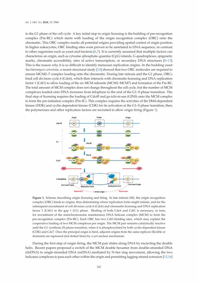
Int. J. Mol. Sci. 2018, 19, 3569
to the G1 phase of the cell cycle. A key initial step in origin licensing is the building of pre-recognitioncomplex (Pre-RC) which starts with loading of the origin recognition complex (ORC) onto thechromatin. This ORC complex marks all potential origins providing spatial control of origin position.In higher eukaryotes, ORC binding sites were proven to be unrelated to DNA sequence, in contrastto other organisms such as yeast and bacteria [6,7]. It is currently assumed that multiple factors cancharacterize an origin, such as cytosine–phosphate–guanine (CpG) islands, G-quadruplexes, epigeneticmarks, chromatin accessibility, sites of active transcription, or secondary DNA structures [8–13].This is the reason why it is so difficult to identify metazoan replication origins. In the budding yeastSaccharomyces cerevisiae, a recent structural study [14] showed that two ORC molecules are required toensure MCM2–7 complex loading onto the chromatin. During late mitosis and the G1 phase, ORCsbind cell division cycle 6 (Cdc6), which then interacts with chromatin licensing and DNA replicationfactor 1 (Cdt1) to allow loading of the six MCM subunits (MCM2–MCM7) and formation of the Pre-RC.The total amount of MCM complex does not change throughout the cell cycle, but the number of MCMcomplexes loaded onto DNA increases from telophase to the end of the G1–S phase transition. Thefinal step of licensing requires the loading of Cdc45 and go-ichi-ni-san (GINS) onto the MCM complexto form the pre-initiation complex (Pre-IC). This complex requires the activities of the Dbf4-dependentkinase (DDK) and cyclin-dependent kinase (CDK) for its activation at the G1–S phase transition; then,the polymerases and other replication factors are recruited to allow origin firing (Figure 1).
Figure 1. Scheme describing origin licensing and firing. In late mitosis (M), the origin recognitioncomplex (ORC) binds to origins, thus determining where replication forks might initiate, and for thesubsequent recruitment of cell division cycle 6 (Cdc6) and chromatin licensing and DNA replicationfactor 1 (Cdt1) in the gap 1 (G1) phase. Binding of both Cdc6 and Cdt1 is necessary, in turn,for recruitment of the minichromosome maintenance DNA helicase complex (MCM) to form thepre-recognition complex (Pre-RC). Each ORC has two Cdt1-binding sites, which may explain thecooperative loading of two MCM complexes per origin. The MCM pair remains catalytically inactiveuntil the G1–synthesis (S) phase transition, when it is phosphorylated by both cyclin-dependent kinase(CDK) and Cdc7. Once the principal origin is fired, adjacent origins from the same replicon (flexible ordormant) are repressed (red dotted lines) by a yet unclear mechanism.
During the first step of origin firing, the MCM pair slides along DNA by encircling the doublehelix. Recent papers proposed a switch of the MCM double hexamer from double-stranded DNA(dsDNA) to single-stranded DNA (ssDNA) mediated by N-tier ring movement, allowing the twohelicases complexes to pass each other within the origin and permitting lagging-strand extrusion [15,16]
243

Int. J. Mol. Sci. 2018, 19, 3569
(Figure 1). During the elongation step, excess MCMs that are not initiated are removed by the passageof the replication fork [17].
The cell must balance its need for sufficient origins to replicate the entire genome against the riskof re-replication of DNA in the S phase due to an excess of origins. Thus, the control of origin licensingis crucial. Repression of new origin licensing during the S phase is important to avoid re-replication,which can lead to aneuploidy, DNA double-strand breaks, gene amplification, and general genomeinstability [18–20]. DNA that is not replicated due to an insufficient number of origins or to replicationfork stalling, by contrast, can also lead to genome instability and rearrangements if the DNA replicationcheckpoint is inactive or deficient [21–23].
1.2. Spatial and Temporal Organization of Replication Origins
Origin usage in eukaryotes is mainly dependent on two important factors: space and time.Replication origins fire at a defined time that remains the same among cell generations and is closelyrelated to their spatial organization. Early replicating origins are mainly found in replication domainsthat are enriched in active epigenetic modifications and highly transcribed genes [24–29]. Thesechromosomal regions have a consequent amount of MCMs, providing potential origins that replicateearly in the S phase [7,30]. Conversely, late replication occurs in origin-poor domains with low genedensity, and enriched in heterochromatin hallmarks [29,31–33].
Replication clusters are organized in the three-dimensional (3D) nuclear space, whereearly-replicating domains locate mainly at the center of the nucleus while late-replicating domains arefound predominantly at the nuclear periphery (Figure 2B). Chromatin conformation mapping methodssuch as Hi-C are very powerful for visualizing the spatial organization of early- and late-replicatingdomains [34,35]. Replication domains are created by topological reorganization of the chromatinin nuclear space. In metazoans, the association of particular replication domains with sub-nuclearcompartments determines their replication timing. The set-up of this compartmentalization occurs at aspecific time of the G1 phase and is called the timing decision point (TDP) [36,37] (Figure 2).
Figure 2. Spatial organization of origins and replication timing. (A) In the early G1 phase, Pre-RCs(black) are assembled on the chromatin and mark potential origins; early-replicating domains (green)and late-replicating domains (red) are disordered in the nuclear space. (B) After the timing decisionpoint (TDP), in the late G1 phase, early-replicating domains are close to center of the nucleus whereaslate-replication domains are associated with the lamina, close to the nuclear periphery. (C) Activeorigins (yellow) cluster in replication domains that are associated to the nuclear matrix (NM), leavinginactive (dormant or flexible) origins in DNA loops (gray).
Accumulating evidence indicates that DNA attachment to the nuclear matrix is important forthe initiation of DNA replication [38–42]. The nuclear matrix permits the separation of chromosometerritories and allows the formation of replication clusters [39]. The organization of replicon clusters
244

Int. J. Mol. Sci. 2018, 19, 3569
might, thus, reflect chromatin looping to bring the origins from different replicons into a singledomain and to exclude the flexible and/or dormant origins from this replication factory (Figure 2C).The cohesin complex may be a key player in chromatin looping because it was found to interactphysically with the MCM complex and to be enriched at origin sites [43].
1.3. Techniques to Detect and Identify Origins
The first quantitative method for determining origin density in the genomes of bacteria andmammalians was DNA fiber autoradiography [44,45]. This time-consuming technique is now replacedby other assays, such as DNA combing or spreading, which label newly replicated DNA withnucleosides analogs, including bromo-, chloro-, and iododeoxyuridine, and visualize the newlyreplicated DNA by immunofluorescence microscopy using antibodies specific for the analog [46].
The use of next-generation DNA sequencing led to the discovery of tens of thousands of potentialreplication origins in the human genome. Several independent approaches were used that exploitthe direct identification of DNA replication initiation intermediates. The first approach is basedon the purification and quantification of short nascent strands (SNS) of DNA [26]. In this method,1.5–2.5-kb nascent strands specific to replication origins are purified thanks to their resistance toλ-exonuclease digestion due to the incorporation, by the primase, of small RNA primers at their5′ ends [47]. The exonuclease digests the large excess of broken genomic DNA that would generate abackground signal if not correctly removed. These genome-wide SNS analyses showed that activeorigins often co-localize with transcription start sites (TSS) and are located in GC-rich regions, closeto CpG islands or G-quadruplexes, confirming previous microarray hybridization results [24,25,48].A second approach [29] is based on the sequencing of an early intermediate called the DNA replicationbubble, which forms when two replication forks diverge from a single origin. The technique consistsof fragmenting the replicating DNA via a restriction endonuclease, and then trapping the circularreplication bubbles in agarose gel [29]. This so-called “bubble-seq” method led to the mappingof more than 100,000 origins in the human genome. A third genome-wide approach relies onsequencing purified Okazaki fragments (“OK-seq”) to determine replication fork polarity, whichallows the identification of initiation and termination sites [49]. With this approach, between 5000 and10,000 broad initiation zones of up to 150 kb were detected. These sites are mainly non-transcribedbut often surrounded by active genes, and they contain a single randomly located initiation event.Finally, a fourth method for identifying metazoan replication origins is called initiation-site sequencing(“ini-seq”) [50]. In this method, initiation events are synchronized biochemically in a cell-free systemin which newly replicated DNA, synthesized a few minutes after initiation, is directly labeled andsubsequently immuno-precipitated. This original approach has the important advantage of allowingfunctional genome-wide studies of origin activation. As these approaches become more and moreaccurate and complementary to each other, they provide an increasingly large, novel dataset on thecharacteristics of replication origins.
1.4. Origin Flexibility, Dormancy, and Efficiency
The replication initiation program of metazoan cells is remarkably flexible, with many originsfiring at disparate frequencies depending on the cell lineage. MCM complexes and all the componentsof the Pre-RC are loaded in excess onto the chromatin in the G1 phase to provide this flexibility.In addition to differences between cell lineages, origin flexibility is also observed within a cellpopulation [42,51].
Very few origins are activated almost all the time; they are called “constitutive” origins [52].The majority of origins do not initiate replication in all cell cycles; these are called “flexible” origins.Origins that are activated only when replication from adjacent origins is compromised are called“dormant” origins. Unlike constitutive and flexible origins, dormant origins are not detectable inwhole-genome analyses. Inter-origin distances measured by whole-genome sequencing are shorterthan those measured by single-fiber analyses. This discrepancy may be explained by the flexibility
245
Int. J. Mol. Sci. 2018, 19, 3569
of origin choice within replicons [53], which might also help coordinate DNA replication withtranscription [54,55] and other nuclear processes, such as DNA repair, in order to facilitate recoverywhen replication is compromised. Given that there is no DNA consensus sequence for metazoanorigins and that there exists such a flexibility in establishing which potential origins are activated,one might wonder how initiation ever occurs accurately and at consistent origins [56].
There are currently two theories to explain how origins are selected. One relies on the idea ofan origin decision point (ODP)—which occurs in the G1 phase, after the timing decision point—thatdetermines which origins are activated during replication [57]. The second theory postulates increasingorigin efficiency based on the random use of replication origins [58], with the idea that the efficiencyof origin firing increases throughout the S phase as the replicative DNA polymerases recycle to neworigins. Moreover, replication origin efficiency also depends on their location in the nucleus, epigeneticmarks, and mainly on the amount of loaded MCM complexes [7,59,60] or nucleosome occupancy [61].Chromosome architecture also plays an important role in the regulation of DNA replication originlocalization and activation [62], although chromosomal loops and loop anchors are still poorly definedbiochemically. Further studies using single-cell technologies will be required in the future to betterunderstand the mechanism of origin choice.
2. Dormant Origin Activation in Response to Replicative Stress
2.1. The Notion of DNA Replication Stress
During DNA replication, the appearance of endogenous or exogenous sources of stress leads toreplication forks slowing or stalling. Exogenous sources of stress comprise mainly genotoxic chemicals,and ultraviolet and ionizing radiation. Endogenous sources of stress that are considered to be barriersto replication include repetitive sequences, G-quadruplexes, telomeres, DNA–RNA hybrids, errorsin the incorporation of ribonucleotides, collisions between replication and transcription machineries,compaction of chromatin, deregulation of origin activity, and reduction of the deoxyribonucleotidetriphosphate (dNTP) pool. Some regions of the genome, such as early-replicating fragile sites (ERFSs)and common fragile sites (CFSs), are more prone than others to replicative stress. Moreover, evidenceis emerging that constitutive activation or overexpression of oncogenes, such as Harvey rat sarcoma(HRas) and myelocytomatosis (c-Myc), are a potential source of replication stress [63]. These oncogenespromote replication initiation or origin firing, leading to an elevated risk of nucleotide pool depletionand/or increased collisions with transcription complexes [64,65]. This may explain why supplementingcancer cells with exogenous nucleosides helps decrease chromosomal instability [66].
The first consequence of replication stress is fork collapse, creating DNA single-strand breaksand/or double-strand breaks. These lesions must be resolved before cell division by repair mechanismssuch as homologous recombination (HR), non-homologous end-joining (NHEJ). or micro-homologymediated end-joining (MMEJ). In normal cells, the ataxia telangiectasia mutated (ATM) and ataxiatelangiectasia Rad3-related (ATR) checkpoint signaling pathways prevent cell division when thegenome is damaged. When some proteins of the checkpoint pathway, for example p53, are mutated,the cell can divide despite the presence of DNA lesions (including breaks and unreplicated DNA),which may lead to chromosome fragmentation, rearrangements, and genomic instability [67–70].
2.2. The Discovery of Dormant Origins and Their Link to Replicative Stress
In 1977, J. Herbert Taylor [71] first described the firing of new origins in response to replicationfork stalling during DNA replication in Chinese hamster ovary (CHO) cells, a finding that latersuggested the existence of dormant origins. Moreover, several studies in a range of eukaryotes,including S. cerevisiae, Xenopus laevis, and human cells, demonstrated that MCM complexes are loadedonto DNA in a large excess when compared to the number of DNA-bound ORCs and the numberof active replication origins [72–77]. It was later shown in X. laevis [78] and in human cells that thisexcess of MCM provides a reservoir of dormant origins, which are activated when replication forks
246
Int. J. Mol. Sci. 2018, 19, 3569
are arrested by agents such as aphidicolin (APH) or hydroxyurea (HU) [79,80]. These studies alsoshowed that depletion of MCM by small interfering RNAs leads to hypersensitivity to replicationinhibitors due to the lack of dormant origins [79,80]. Moreover, checkpoint kinase 1 (Chk1) activationis required for firing of dormant origins within active replication clusters, as well as for repressionof other replicons that are not yet active [81], suggesting a link between the DNA damage responseand dormant origin activation. Indeed, in vertebrates, inactivation or depletion of various proteinsinvolved in genome maintenance, such as ATR [82,83], Chk1 [84–87], Wee1 [88,89], bloom syndromeprotein (BLM) [90], Claspin [91,92], breast cancer type 2 susceptibility protein (BRCA2), and Rad51 [93],slows replication forks and also increases the number of initiation events, at least in studies whereinitiation events were examined. This finding indicates a link between fork speed and the number ofactive origins, as we examine further below.
2.3. The Density of Active Origins Depends on Replication Fork Speed
Under normal conditions, dormant origins do not fire and are passively replicated by the forkcoming from adjacent activated origins. Thus, it makes sense to assume that replication fork speedcan be a regulator of active origin density. In two complementary studies on CHO cells [62,94], it wasdemonstrated that replication fork speed has a direct impact on the number of active origins. When thefork is slowed down by HU treatment, the density of active origins increases. In contrast, in conditionsthat accelerate fork speed (addition of adenine and uridine to the culture medium), fewer origins areactive. These studies further showed that the cell starts compensating for the decrease in fork speedwithin half an hour of treatment by activating dormant origins, which are then able to change theirstatus within the S phase. Regulation of the number of initiation events occurs at the level of individualclusters, consistent with the functional organization of origins into replicon clusters [95]. Anotherstudy demonstrated that, in the absence of Cdc7 or ORC1, replication forks progress more rapidlythan in control cells and fewer origins fire [96], again suggesting that the number of active originsand the fork rate are interdependent. Similarly, using chemical inhibitors of origin activity (a Cdc7kinase inhibitor) and of DNA synthesis (APH), a more recent study found that the primary effectsof replicative stress on fork rate can be distinguished from those on origin firing [97]. Collectively,these results support the conclusion that the density of origin firing depends on fork speed and, thus,is affected by endogenous or exogenous replicative stress.
2.4. CFS Fragility Due to the Lack of Dormant Origins
CFSs play a major role in cancer initiation because of their instability in conditions of replicationstress. CFSs were first described as gaps and constrictions in the metaphase chromosomes of humanlymphocytes grown under mild replication stress conditions (i.e., a low dose of APH) [98]. Theseobservations were since seen in other organisms and are very likely to be the consequence ofunder-replication and/or DNA breaks caused by replication stress [99,100].
Although CFSs have been known for over two decades, the cause of their fragility is stillcontroversial [55,101]. CFS fragility was first linked to non-B DNA sequences, such as AT-richsequences, which are able to adopt secondary structures, constituting barriers to replicationforks [102–105]. Deletion of these sequences from some cancer cell lines does not prevent breaksat these loci [106–108], suggesting that DNA sequence is not the sole reason for the instability of CFSs.Genome-wide analysis of replication and DNA combing experiments found a paucity of replicationorigins within the core of CFSs [109,110] and an incapacity to activate additional origins in responseto replicative stress [111]. This suggests that, in order to replicate these regions, the fork must passthrough long stretches of DNA containing multiple non-B DNA conformation sequences, and thattheir fragility correlates with the absence of additional replication origin firing when replication isslowed down. Most CFSs correspond to long genes (>300 kb), which might increase the risk of collisionbetween the transcription and replication machineries [112]. Although one study showed that thetranscription of large genes does not systematically dictate CFS fragility [113], other studies found that
247

Int. J. Mol. Sci. 2018, 19, 3569
replication stress induces locus- and cell-type-specific genomic instability at active, large transcriptionunits corresponding to CFSs [114,115]. Moreover, it is thought that fragility of these sites result fromentry into mitosis before their complete replication [116,117]. Taken together, these observationssuggest that replication defects at fragile sites may be due to a low density of licensed origins or mayreflect inefficient or delayed activation of replication forks under replication stress.
3. Regulation of Dormant Origins: A Passive or Active Mechanism?
3.1. Activation of Dormant Origins by a “Passive” Mechanism
It is currently not clear what drives the firing of dormant origins when forks are slowed down orinhibited. One first hypothesis could be that it does not involve an active mechanism, but occurs asa consequence of the stochastic nature of origin firing [18,79]. Dormant origins have a precise lap oftime to fire before being passively replicated then inactivated by forks from adjacent origins. Whenfork progression is impeded, the replication at dormant origins is delayed and, therefore, they havean increased probability to fire. By means of computational modeling, a study showed that the samelevels of dormant origin activation seen in vivo can be reproduced by a passive mechanism [118].In this model, the mechanism relies simply on the stochastic nature of origin firing, without any needfor additional regulatory pathways.
This simple theory can be sufficient to explain the activation of dormant origins in response toreplicative stress. Nonetheless, it cannot be ruled out that dormant origins may also be regulated byactive mechanisms, involving DNA damage response and other replication-related pathways.
3.2. Regulation of Dormant Origins by “Active” Mechanisms
3.2.1. ATR/Chk1 Kinases as Modulators of Origin Activation
The inhibition of replication forks activates the DNA damage checkpoint kinases ATR–Chk1 andATM–Chk2, which have many different functions, including stabilizing replication forks, delayingor blocking the progress of the cell cycle, and promoting DNA lesion repair [119–121]. It may seemsurprising that, in response to replication stress, the cell can both activate dormant origins and suppressoverall origin initiation; however, when replication forks stall, it makes sense that dormant originsshould be activated in their vicinity and not elsewhere in the genome.
In the normal S phase, Chk1 affects replication fork speed by inhibiting excess originfiring [23,85,86]. In response to low levels of replication stress induced by APH or HU, ATR andChk1 impede the activation of new replicon clusters while allowing dormant origins to fire withinthose already activated and affected by the drug [79,81], thereby avoiding the deleterious impactof replication fork stalling (Figure 3). The mechanism responsible for this phenomenon is not yetelucidated, but one possibility is that ATR and Chk1 mildly reduce CDK levels, resulting in activationof fewer replication clusters [122]. Alternatively, Chk1 might directly inhibit the initiation processthrough an interaction with Treslin, which is required to stabilize Cdc45, GINS, and the MCM complextogether with topoisomerase 2-binding protein 1 (TOPBP1) [123–127]. Moreover, a recent study foundthat an ATR inhibitor not only induced unscheduled origin firing, but also revealed another mechanismof origin regulation through a Cdc7-dependent phosphorylation of GINS [128]. Finally, a very recentstudy found that the ATR-activation domain of TOPBP1 is required to suppress origin firing duringthe S phase [129], further supporting an important role for the ATR–Chk1 pathway in regulating theactivation of origins.
248

Int. J. Mol. Sci. 2018, 19, 3569
Figure 3. Ataxia telangiectasia Rad3-related (ATR)/checkpoint kinase 1 (Chk1) involvement in thedifferential regulation of origin firing under replicative stress. In response to replication stress, theATR/Chk1 kinases allow the activation of dormant origins within active replicon clusters (activeorigin(s) in red) while repressing any firing within those that are not yet activated (inactive originsin blue).
3.2.2. Mannose Receptor C-Type 1 (Mrc1)/Claspin Is a Central Regulator of Origin Firing underNormal and Stressed Replication
S. cerevisiae Mrc1 and its metazoan ortholog Claspin are not only involved in the S phasecheckpoint signaling pathway, but are also important components of replication forks. They interactwith many factors known to function in or to regulate DNA replication, including MCM4, MCM10,ATR, Chk1, Cdc7, Cdc45, DNA polymerases α, δ, and ε, and proliferating cell nuclear antigen(PCNA) [130–133]. The presence of Mrc1/Claspin is necessary for normal DNA replication [91,92,134,135],probably by making a connection between the helicase components and replicative polymerases at thereplication fork. Also, Claspin plays another role in the initiation of DNA replication in human cellsduring the normal S phase by recruiting Cdc7 to facilitate phosphorylation of MCM proteins [136].It was recently discovered in yeast that Mrc1 has two crucial functions in regulating the firing oforigins: a checkpoint independent-role to activate early-firing origins during normal replication, and acheckpoint dependent-function to inhibit late/dormant origins in the presence of HU [137].
3.2.3. Fanconi Anemia Proteins in the Regulation of Dormant Origins
The role of the Fanconi anemia (FA) pathway in the DNA repair of interstrand cross-links (ICLs)was studied for many years. A clear model emerged describing that FA proteins orchestrate theinterplay between multiple DNA repair pathways, including homologous recombination (HR) andtranslesion synthesis (TLS) [138–140]. However, treatment of cells with a low dose APH robustlyactivates the FA pathway, indicating a role of the FA proteins during DNA replication [141].
FA complementation group 1 (FANCI) was shown to be involved in dormant origin firing uponlow replication stress through a FA pathway-independent mechanism [142]. FANCI associates withMCM3 and MCM5, localizes with replication origins, and acts as a regulator of DDK activity to allowthe activation of the MCM2–7 helicase complex in response to mild replicative stress. In contrast,under high replicative stress, FANCI is phosphorylated by ATR. This phosphorylated form of FANCInegatively regulates dormant origin firing and activates replication fork restart/DNA repair that isFA-dependent. In this context, FA complementation group D2 (FANCD2), which is known as a closepartner of FANCI, acts as a negative regulator of dormant origin firing [142].
Finally, FANCD2 was shown to facilitate replication of repeat-rich genomic regions such as CFSsby decreasing DNA–RNA hybrid accumulation, thus reducing the need for dormant origin firing [143].
3.2.4. Rap1-Interacting Factor 1 (RIF1) Orchestrates Origins and Replication Timing
RIF1 (Rap1-interacting factor 1) was first discovered in budding yeast as a telomericchromatin-interacting protein required for the regulation of telomere length via its interaction withRap1 [144,145]. It was then demonstrated in S. cerevisiae that RIF1 inhibits activation of the DNA
249
Int. J. Mol. Sci. 2018, 19, 3569
damage checkpoint close to telomeres [146,147] and affects telomere replication timing [148]. Althoughthe RIF1 protein is evolutionarily conserved, in metazoans, it was described not to play a specificrole at telomeres, but rather to orchestrate the DNA double-strand break repair pathway and DNArecombination [149–153].
Further studies implicated RIF1 from the fission yeast Schizosaccharomyces pombe and mammalianRIF1 in regulating genome-wide DNA replication. S. pombe RIF1 binds selectively not only to telomeres,but also to specific regions of the genome where it may regulate the choice and timing of origin firingin late-replicating regions of chromosomes [154]. In RIF1-deficient cells, activation of dormant or lateorigins is concomitant with suppression of some active early-firing origins, indicating that RIF1 is acrucial player in the genome-wide origin activation program in S. pombe. In human cells, depletionof RIF1 results in increased early-S phase initiation events, loss of mid-S phase replication foci, andglobal changes in replication timing domain structures. Domains that normally replicate in the early Sphase are delayed, whereas those that normally replicate in the late S phase are advanced [155]. Thus,replication timing is completely disturbed in the absence of RIF1. Another study observed that, in theabsence of RIF1, the distance between origins is greater than in control cells during the normal S phase,and there are fewer dormant origins upon replication stress [156].
Also, RIF1 binds tightly to insoluble nuclear structures in late mitosis and the early G1 phase,and regulates chromatin-loop size [155]. Interestingly, RIF1 binding to consensus G-quadruplex-likesequences in fission yeast was identified [157]. These sequences tend to be near dormant origins, andthe binding of RIF1 on these sites would allow their repression over a great distance. Overall, thesefindings indicate that RIF1, through its role in organizing higher-order chromatin architecture, is anessential regulator of replication timing.
Thus, the accumulating data suggest that, through its interaction with chromatin and nuclearstructures, RIF1 plays an important role in the regulation of dormant origin availability not only inresponse to replicative stress, but also in normal conditions.
3.2.5. Chromatin Loop Size Correlates with Dormant Origin Activation
The fluorescent DNA halo technique was essential for establishing the link between chromatinloops and replicon size [158], and for describing the importance of replicon remodeling eventsin Xenopus embryonic development [159]. Basically, the technique relies on cell permeabilizationand soluble protein extraction, allowing supercoiled DNA loops to unroll around an insolublescaffold, the nuclear matrix. Those structures called DNA “halos” can be visualized by4′,6-diamidino-2-phenylindole (DAPI) fluorescent staining. Active origins are in or near the nuclearmatrix, whereas dormant/inactive origins are in the DNA loops [160] (Figure 2C).
Using the fluorescent DNA halo technique, one study [62] observed a strict correlation betweendormant origin activation at a given S phase and reduced chromatin loop size in the next G1/S phase.Combining the DNA halo experiment with fluorescent in situ hybridization (FISH) using a probetargeting the highly amplified adenosine monophosphate deaminase 2 (AMPD2)-specific locus inCHO cells, they demonstrated that, in response to replication stress, activation of dormant originsrelocates this locus toward the nuclear matrix.
Cohesin also influences the size of interphase chromatin loops since its absence results in longerchromatin loops due to a limited origin usage [43], showing that, independently of the effect of cohesinacetylation on replication fork progression [161], this structural protein is present at origins and impactstheir activity. Finally, chromatin loop size increases in RIF1-depleted cells [155], suggesting that theRIF1 protein is required for proper chromatin loop formation, as already mentioned above.
250

Int. J. Mol. Sci. 2018, 19, 3569
4. Dormant Origin Deficiency, Genome Stability, and Pathologies
4.1. MCM Mutants and Dormant Origins in Mice
Homozygosity for a null allele of any of the six Mcm genes in mice (Mcm2–7) causes embryoniclethality [162–164], consistent with the evidence that these Mcm genes are essential for DNA replication.Only hypomorphic alleles such as Mcm4Chaos3 and Mcm2IRES-CreERT2 (IRES, internal ribosome entrysite; ERT2, estrogen receptor 2) result in mice that are viable into adulthood. The Mcm2IRES-CreERT2
allele expresses a tamoxifen-inducible Cre recombinase (CreERT2) inserted into the 3′ untranslatedregion (UTR) of the endogenous Mcm2 locus, which reduces the expression of MCM2 by 65% whencompared to wild-type cells [165]. The Mcm4Chaos3 allele produces an MCM4 protein with a Phe345Ilemutation, which does not affect the helicase activity of the MCM complex in vitro, but does reduce theefficiency of its assembly [164].
Surprisingly, mouse embryonic fibroblasts (MEFs) from Mcm4Chaos3 mice also have a reducedMCM7 protein level in addition to MCM4 [164]. Moreover, immortalized homozygous Mcm4Chaos3
cells display less stable association of MCM2–7 at replication forks compared to wild-type cells [166].Finally, Mcm4Chaos3/Chaos3 MEFs exhibit about a half reduction in chromatin bound MCM2–7 that causesa lower ability to activate dormant origins in response to treatment with low doses of APH [162,167].
Mice with only one-third of the normal MCM2 level were shown to develop lymphomas at avery young age, and have diverse stem cell proliferation defects. Similarly to Mcm4Chaos3, these micealso have 27% less MCM7 protein than wild-type mice. Moreover, Mcm2IRES-CreERT2 cells exhibitdecreased replication origin usage due to lower dormant origin availability even in the presence ofHU, as demonstrated by DNA combing experiments [165,168].
Hence, these two mouse models are close phenotypically, showing dormant origin deficiencydue to reduced levels of loaded MCM onto the chromatin. Even in an unchallenged S phase,the inability to activate dormant origins leads to accumulation of stalled replication forks thatreach mitosis and interfere with chromosome segregation. Both phenotypes lead to improperchromosome stability and premature tumorigenesis, with several differences in the latency of diseasedevelopment [165,166,168,169].
4.2. MCM Mutants and Dormant Origins in Stem/Progenitor Cells
The fact that Mcm2 expression has a global effect on cell proliferation within many tissues mightexplain why the majority of Mcm2IRES-CreERT2 mice develop tumors and display a range of additionalhallmarks of age-related disorders. A study that set out to determine the effect of Mcm2 deficiencyobserved an approximately threefold reduction in the level of neurogenesis within the sub-ventricularzone in Mcm2IRES-CreERT2 mouse brains [165], fewer stem cells in intestinal crypts and in skeletal muscle,and a modest increase in DNA damage.
Consistent with the conclusion that Mcm mutants affect stem cells, neural stem-cell progenitors inMcm4Chaos3/Chaos3 mouse embryos display a high level of Chk1 activation, increased phosphorylatedH2A histone family X (γH2AX) and p53-binding protein 1 (53BP1) foci, an accumulation in theG2–M phase, and more apoptosis, resulting in a reduced ability to form neurospheres in vitro [170].The renewal of stem cells in the brain appears to be normal, but their ability to differentiate intointermediate progenitors is highly reduced due to an increase of apoptotic cells in the sub-ventricularand intermediate zones [170].
These observations suggest that normal expression of MCM complex proteins is essential forstem/progenitor cell function by reducing the risk of replication-associated genome instability, an ideathat was supported by two other studies. One demonstrated that human embryonic stem cells,which have a remarkably short G1 phase, load MCM onto chromatin very rapidly when comparedto differentiated cells, in order to have a similar total amount of loaded MCM at the G1–S phasetransition [171]. In the second study, hypomorphic expression of the origin licensing factor MCM3in mouse reduced the number of licensed origins and affected the function of hematopoietic stem
251

Int. J. Mol. Sci. 2018, 19, 3569
cells, as well as the differentiation of highly proliferative erythrocyte precursors, thus demonstratingthat the rate of MCM loading is crucial for correct organism development [163]. These observationssuggest that hematopoietic progenitors are exceptionally sensitive to replication stress, and that theymust license an excess of origins to ensure their correct differentiation and function.
Intriguingly, aging hematopoietic stem cells suffer from replication stress even in wild-type mice.This might be due to the fact that old stem cells have reduced expression of MCM complex proteins,resulting in reduced numbers of dormant origins and, as a consequence, more chromosome instabilityand cell-cycle defects [172].
4.3. Consequences of Limited Licensing and Firing in Humans
A mutation in the Mcm4 gene, which results in a truncated form of this protein lacking the N-terminalserine/threonine-rich domain, was identified in a group of patients with a syndrome including growthdelay, natural killer cell deficiency, adrenal insufficiency, and genome instability [173–175]. Thistruncated form of MCM4 does not affect MCM complex loading [174]. Nevertheless, immortalizedfibroblasts from these patients have a high level of chromosome breakage and defects in cell-cycleprogression, and they are sensitive to low doses of APH [174], suggesting that the N-terminal aminoacids of MCM4 protein are involved in the maintenance of genome integrity during replication.Further studies will be necessary to elucidate the mechanism via which normal MCM4 ensures genomemaintenance. One possibility is the role of MCM4 phosphorylation in the checkpoint response, whereit was shown that the N-terminal domain of MCM4 has a crucial role. In unperturbed replication,this domain exerts an inhibitory effect on replication initiation, and this inhibitory effect is relievedupon its phosphorylation by DDK. However, in the context of replication stress, this N-terminalphosphorylation by DDK becomes a prerequisite for proper checkpoint activation [176].
Another disease that appears to involve defective replication origin licensing is Meier–Gorlinsyndrome (MGS), an autosomal recessive primordial dwarfism syndrome characterized by pre- andpost-natal impaired growth. Several studies identified marked locus heterogeneity in this syndromeincluding mutations in five genes encoding components of the Pre-RC: Orc1, Orc4, Orc6, Cdt1, andCdc6 [177,178]. The molecular and cellular phenotypes include impaired licensing, altered S phaseprogression, and proliferation defects, which partially overlap with the phenotypes due to MCMmutations, except for chromosomal instability, and an increased predisposition to cancer. Nonetheless,MGS mutations in Orc1 and Orc6 can cause quite a significant reduction in MCM loading andreplication origin licensing [177,179,180].
Mice and human phenotypes caused by mutations in the licensing system illustrate our limitedunderstanding of what happens to cells when the DNA replication program is compromised.For example, the threshold value for the number of licensed origins needed to activate the licensingcheckpoint is still not known, nor whether this value varies between cell types.
5. Conclusion and Prospects
Dormant origins are now recognized as an important safeguard against under-replication of thegenome, thus ensuring genome maintenance. Activation of dormant origins plays a central role inthe rescue of stalled forks in the context of replicative stress, contributing to the complete replicationof the DNA. The interactions between dormant origins and other fork restart mechanisms (such asTLS) are mostly unknown, even though some links with DNA damage checkpoint or FA pathways arebecoming evident. What determines whether the cell activates dormant origins or induces these othermechanisms in response to fork stalling still remains to be investigated.
How replicon clusters are activated at the molecular level remains unclear, although we know thatorigin activation is regulated by both Chk1 and CDKs. The RIF1 protein might be the most interestingfactor in this process since it is present both at the replication fork and at replication origins, where itplays a role in the DNA damage response, as well as in replication timing.
252
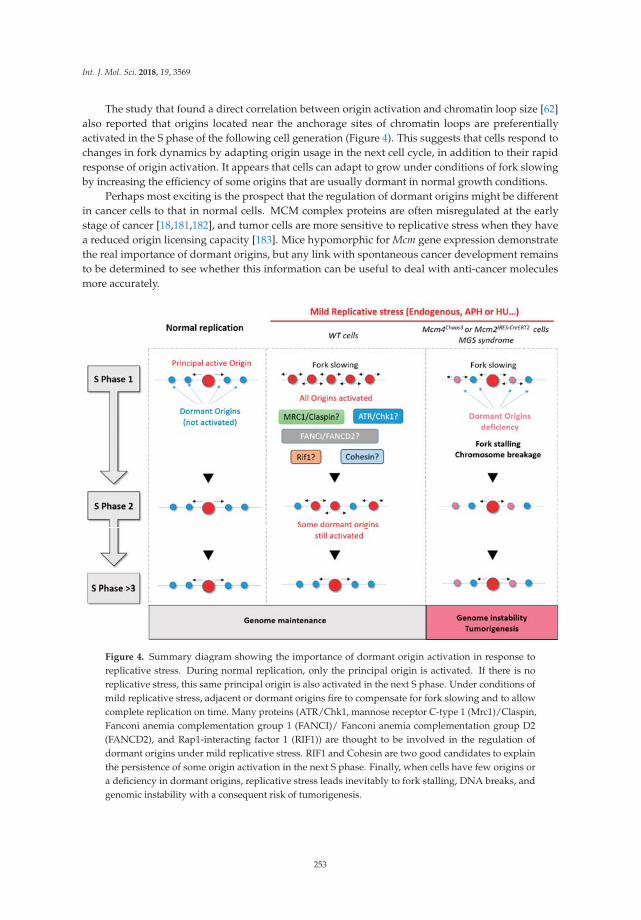
Int. J. Mol. Sci. 2018, 19, 3569
The study that found a direct correlation between origin activation and chromatin loop size [62]also reported that origins located near the anchorage sites of chromatin loops are preferentiallyactivated in the S phase of the following cell generation (Figure 4). This suggests that cells respond tochanges in fork dynamics by adapting origin usage in the next cell cycle, in addition to their rapidresponse of origin activation. It appears that cells can adapt to grow under conditions of fork slowingby increasing the efficiency of some origins that are usually dormant in normal growth conditions.
Perhaps most exciting is the prospect that the regulation of dormant origins might be differentin cancer cells to that in normal cells. MCM complex proteins are often misregulated at the earlystage of cancer [18,181,182], and tumor cells are more sensitive to replicative stress when they havea reduced origin licensing capacity [183]. Mice hypomorphic for Mcm gene expression demonstratethe real importance of dormant origins, but any link with spontaneous cancer development remainsto be determined to see whether this information can be useful to deal with anti-cancer moleculesmore accurately.
Figure 4. Summary diagram showing the importance of dormant origin activation in response toreplicative stress. During normal replication, only the principal origin is activated. If there is noreplicative stress, this same principal origin is also activated in the next S phase. Under conditions ofmild replicative stress, adjacent or dormant origins fire to compensate for fork slowing and to allowcomplete replication on time. Many proteins (ATR/Chk1, mannose receptor C-type 1 (Mrc1)/Claspin,Fanconi anemia complementation group 1 (FANCI)/ Fanconi anemia complementation group D2(FANCD2), and Rap1-interacting factor 1 (RIF1)) are thought to be involved in the regulation ofdormant origins under mild replicative stress. RIF1 and Cohesin are two good candidates to explainthe persistence of some origin activation in the next S phase. Finally, when cells have few origins ora deficiency in dormant origins, replicative stress leads inevitably to fork stalling, DNA breaks, andgenomic instability with a consequent risk of tumorigenesis.
253
Int. J. Mol. Sci. 2018, 19, 3569
Author Contributions: L.C. wrote the manuscript, J.-S.H. proofread it, and V.B designed and proofread it.
Funding: Work in J.-S.H.’s laboratory is supported by funding from the Institut National du Cancer (PLBIO 2016),the Agence Nationale de la Recherche (ANR PRC 2016), Labex TOUCAN, and La Ligue contre le Cancer (Equipelabellisée 2017).
Acknowledgments: We kindly thank Dana Hodroj for proofreading and precious comments.
Conflicts of Interest: The authors declare no conflicts of interest.
Abbreviations
53BP1 p53-Binding protein 1APH AphidicolinATM Ataxia telangiectasia mutatedATR Ataxia telangiectasia and Rad3-related proteinBLM Bloom syndrome RecQ like helicaseBRCA2 Breast cancer 2Cdc45 Cell division cycle protein 45Cdc6/7 Cell division cycle 6/7CDK Cyclin-dependent kinaseCdt1 Chromatin licensing and DNA replication factor 1CFS Common fragile siteChk1 Checkpoint kinase 1Chk2 Checkpoint kinase 2CHO Chinese hamster ovaryc-Myc MyelocytomatosisDDK Dbf4-dependent kinaseDNA Deoxyribonucleic aciddNTP DeoxyribonucleotidesERFS Early-replicating fragile siteFANCI/D2 Fanconi anemia complementation group I/D2FISH Fluorescence in situ hybridizationGINS Go-ichi-ni-sanHR Homologous recombinationHRAS Harvey rat sarcomaHU HydroxyureaICL Interstrand cross-linkMCM Minichromosome maintenanceMGS Meier–Glorin syndromeMMEJ Micro-homology mediated end-joiningMrc1 Mannose receptor C-type 1NHEJ Non-homologous end-joiningODP Origin decision pointORC Origin recognition complexPCNA Proliferating cell nuclear antigenPre-IC Pre-initiation complexPre-RC Pre-recognition complexRIF1 Rap1-interacting factor 1RNA Ribonucleic acidRT Replication timingSNS Short nascent strandSVZ Sub-ventricular zoneTDP Timing decision pointTOPBP1 Topoisomerase 2-binding protein 1TLS Translesion synthesisTSS Transcription start sites
254
Int. J. Mol. Sci. 2018, 19, 3569
References
1. Berezney, R.; Dubey, D.D.; Huberman, J.A. Heterogeneity of eukaryotic replicons, replicon clusters, andreplication foci. Chromosoma 2000, 108, 471–484. [CrossRef] [PubMed]
2. Pope, B.D.; Ryba, T.; Dileep, V.; Yue, F.; Wu, W.; Denas, O.; Vera, D.L.; Wang, Y.; Hansen, R.S.; Canfield, T.K.;et al. Topologically associating domains are stable units of replication-timing regulation. Nature 2014, 515,402–405. [CrossRef] [PubMed]
3. Takebayashi, S.; Ogata, M.; Okumura, K. Anatomy of Mammalian Replication Domains. Genes 2017, 8, 110.[CrossRef] [PubMed]
4. Boulos, R.E.; Drillon, G.; Argoul, F.; Arneodo, A.; Audit, B. Structural organization of human replicationtiming domains. FEBS Lett. 2015, 589, 2944–2957. [CrossRef] [PubMed]
5. Löb, D.; Lengert, N.; Chagin, V.O.; Reinhart, M.; Casas-Delucchi, C.S.; Cardoso, M.C.; Drossel, B. 3D replicondistributions arise from stochastic initiation and domino-like DNA replication progression. Nat. Commun.2016, 7, 11207. [CrossRef] [PubMed]
6. Hyrien, O. Peaks cloaked in the mist: The landscape of mammalian replication origins. J. Cell Biol. 2015, 208,147–160. [CrossRef] [PubMed]
7. Hyrien, O. How MCM loading and spreading specify eukaryotic DNA replication initiation sites.F1000Research 2016, 5. [CrossRef] [PubMed]
8. Karnani, N.; Taylor, C.M.; Malhotra, A.; Dutta, A. Genomic Study of Replication Initiation in HumanChromosomes Reveals the Influence of Transcription Regulation and Chromatin Structure on Origin Selection.Mol. Biol. Cell 2010, 21, 393–404. [CrossRef] [PubMed]
9. Méchali, M.; Yoshida, K.; Coulombe, P.; Pasero, P. Genetic and epigenetic determinants of DNA replicationorigins, position and activation. Curr. Opin. Genet. Dev. 2013, 23, 124–131. [CrossRef] [PubMed]
10. Martin, M.M.; Ryan, M.; Kim, R.; Zakas, A.L.; Fu, H.; Lin, C.M.; Reinhold, W.C.; Davis, S.R.; Bilke, S.; Liu, H.;et al. Genome-wide depletion of replication initiation events in highly transcribed regions. Genome Res. 2011,21, 1822–1832. [CrossRef] [PubMed]
11. Bartholdy, B.; Mukhopadhyay, R.; Lajugie, J.; Aladjem, M.I.; Bouhassira, E.E. Allele-specific analysis of DNAreplication origins in mammalian cells. Nat. Commun. 2015, 6, 7051. [CrossRef] [PubMed]
12. Besnard, E.; Babled, A.; Lapasset, L.; Milhavet, O.; Parrinello, H.; Dantec, C.; Marin, J.-M.; Lemaitre, J.-M.Unraveling cell type—Specific and reprogrammable human replication origin signatures associated withG-quadruplex consensus motifs. Nat. Struct. Mol. Biol. 2012, 19, 837–844. [CrossRef] [PubMed]
13. Cayrou, C.; Ballester, B.; Peiffer, I.; Fenouil, R.; Coulombe, P.; Andrau, J.-C.; van Helden, J.; Méchali, M. Thechromatin environment shapes DNA replication origin organization and defines origin classes. Genome Res.2015, 25, 1873–1885. [CrossRef] [PubMed]
14. Coster, G.; Diffley, J.F.X. Bidirectional eukaryotic DNA replication is established by quasi-symmetricalhelicase loading. Science 2017, 357, 314–318. [CrossRef] [PubMed]
15. Noguchi, Y.; Yuan, Z.; Bai, L.; Schneider, S.; Zhao, G.; Stillman, B.; Speck, C.; Li, H. Cryo-EM structureof Mcm2-7 double hexamer on DNA suggests a lagging-strand DNA extrusion model. Proc. Natl. Acad.Sci. USA 2017, 114, E9529–E9538. [CrossRef] [PubMed]
16. Douglas, M.E.; Ali, F.A.; Costa, A.; Diffley, J.F.X. The mechanism of eukaryotic CMG helicase activation.Nature 2018, 555, 265–268. [CrossRef] [PubMed]
17. Fragkos, M.; Ganier, O.; Coulombe, P.; Méchali, M. DNA replication origin activation in space and time.Nat. Rev. Mol. Cell Biol. 2015, 16, 360–374. [CrossRef] [PubMed]
18. Blow, J.J.; Ge, X.Q. Replication forks, chromatin loops and dormant replication origins. Genome Biol. 2008, 9,244. [CrossRef] [PubMed]
19. Muñoz, S.; Búa, S.; Rodríguez-Acebes, S.; Megías, D.; Ortega, S.; de Martino, A.; Méndez, J. In Vivo DNARe-replication Elicits Lethal Tissue Dysplasias. Cell Rep. 2017, 19, 928–938. [CrossRef] [PubMed]
20. Neelsen, K.J.; Zanini, I.M.Y.; Mijic, S.; Herrador, R.; Zellweger, R.; Chaudhuri, A.R.; Creavin, K.D.; Blow, J.J.;Lopes, M. Deregulated origin licensing leads to chromosomal breaks by rereplication of a gapped DNAtemplate. Genes Dev. 2013, 27, 2537–2542. [CrossRef] [PubMed]
21. Lukas, C.; Savic, V.; Bekker-Jensen, S.; Doil, C.; Neumann, B.; Pedersen, R.S.; Grøfte, M.; Chan, K.L.;Hickson, I.D.; Bartek, J.; et al. 53BP1 nuclear bodies form around DNA lesions generated by mitotictransmission of chromosomes under replication stress. Nat. Cell Biol. 2011, 13, 243–253. [CrossRef] [PubMed]
255

Int. J. Mol. Sci. 2018, 19, 3569
22. Moreno, A.; Carrington, J.T.; Albergante, L.; Al Mamun, M.; Haagensen, E.J.; Komseli, E.-S.; Gorgoulis, V.G.;Newman, T.J.; Blow, J.J. Unreplicated DNA remaining from unperturbed S phases passes through mitosis forresolution in daughter cells. Proc. Natl. Acad. Sci. USA 2016, 113, E5757–E5764. [CrossRef] [PubMed]
23. Syljuåsen, R.G.; Sørensen, C.S.; Hansen, L.T.; Fugger, K.; Lundin, C.; Johansson, F.; Helleday, T.;Sehested, M.; Lukas, J.; Bartek, J. Inhibition of Human Chk1 Causes Increased Initiation of DNA Replication,Phosphorylation of ATR Targets, and DNA Breakage. Mol. Cell. Biol. 2005, 25, 3553–3562. [CrossRef][PubMed]
24. Sequeira-Mendes, J.; Díaz-Uriarte, R.; Apedaile, A.; Huntley, D.; Brockdorff, N.; Gómez, M. TranscriptionInitiation Activity Sets Replication Origin Efficiency in Mammalian Cells. PLOS Genet. 2009, 5, e1000446.[CrossRef] [PubMed]
25. Cadoret, J.-C.; Meisch, F.; Hassan-Zadeh, V.; Luyten, I.; Guillet, C.; Duret, L.; Quesneville, H.; Prioleau, M.-N.Genome-wide studies highlight indirect links between human replication origins and gene regulation.Proc. Natl. Acad. Sci. USA 2008, 105, 15837–15842. [CrossRef] [PubMed]
26. Prioleau, M.-N.; MacAlpine, D.M. DNA replication origins—Where do we begin? Genes Dev. 2016, 30,1683–1697. [CrossRef] [PubMed]
27. Sugimoto, N.; Maehara, K.; Yoshida, K.; Ohkawa, Y.; Fujita, M. Genome-wide analysis of the spatiotemporalregulation of firing and dormant replication origins in human cells. Nucleic Acids Res. 2018, 46, 6683–6696.[CrossRef] [PubMed]
28. Lucas, I.; Palakodeti, A.; Jiang, Y.; Young, D.J.; Jiang, N.; Fernald, A.A.; Beau, M.M.L. High-throughputmapping of origins of replication in human cells. EMBO Rep. 2007, 8, 770–777. [CrossRef] [PubMed]
29. Mesner, L.D.; Valsakumar, V.; Cieslik, M.; Pickin, R.; Hamlin, J.L.; Bekiranov, S. Bubble-seq analysis of thehuman genome reveals distinct chromatin-mediated mechanisms for regulating early- and late-firing origins.Genome Res. 2013, 23, 1774–1788. [CrossRef] [PubMed]
30. Das, S.P.; Borrman, T.; Liu, V.W.T.; Yang, S.C.-H.; Bechhoefer, J.; Rhind, N. Replication timing is regulated bythe number of MCMs loaded at origins. Genome Res. 2015, 25, 1886–1892. [CrossRef] [PubMed]
31. Smith, O.K.; Kim, R.; Fu, H.; Martin, M.M.; Lin, C.M.; Utani, K.; Zhang, Y.; Marks, A.B.; Lalande, M.;Chamberlain, S.; et al. Distinct epigenetic features of differentiation-regulated replication origins. Epigenet.Chromatin 2016, 9, 18. [CrossRef] [PubMed]
32. Suzuki, M.; Oda, M.; Ramos, M.-P.; Pascual, M.; Lau, K.; Stasiek, E.; Agyiri, F.; Thompson, R.F.; Glass, J.L.;Jing, Q.; et al. Late-replicating heterochromatin is characterized by decreased cytosine methylation in thehuman genome. Genome Res 2011, 21, 1833–1840. [CrossRef] [PubMed]
33. Casas-Delucchi, C.S.; van Bemmel, J.G.; Haase, S.; Herce, H.D.; Nowak, D.; Meilinger, D.; Stear, J.H.;Leonhardt, H.; Cardoso, M.C. Histone hypoacetylation is required to maintain late replication timing ofconstitutive heterochromatin. Nucleic Acids Res. 2012, 40, 159–169. [CrossRef] [PubMed]
34. Rivera-Mulia, J.C.; Gilbert, D.M. Replication timing and transcriptional control: Beyond cause andeffect—Part III. Curr. Opin. Cell Biol. 2016, 40, 168–178. [CrossRef] [PubMed]
35. Ryba, T.; Hiratani, I.; Lu, J.; Itoh, M.; Kulik, M.; Zhang, J.; Schulz, T.C.; Robins, A.J.; Dalton, S.;Gilbert, D.M. Evolutionarily conserved replication timing profiles predict long-range chromatin interactionsand distinguish closely related cell types. Genome Res. 2010, 20, 761–770. [CrossRef] [PubMed]
36. Dimitrova, D.S.; Berezney, R. The spatio-temporal organization of DNA replication sites is identical inprimary, immortalized and transformed mammalian cells. J. Cell Sci. 2002, 115, 4037–4051. [CrossRef][PubMed]
37. Gilbert, D.M. Cell fate transitions and the replication timing decision point. J. Cell Biol. 2010, 191, 899–903.[CrossRef] [PubMed]
38. Anachkova, B.; Djeliova, V.; Russev, G. Nuclear matrix support of DNA replication. J. Cell. Biochem. 2005, 96,951–961. [CrossRef] [PubMed]
39. Wilson, R.H.C.; Coverley, D. Relationship between DNA replication and the nuclear matrix. Genes Cells 2013,18, 17–31. [CrossRef] [PubMed]
40. Djeliova, V.; Russev, G.; Anachkova, B. Dynamics of association of origins of DNA replication with thenuclear matrix during the cell cycle. Nucleic Acids Res. 2001, 29, 3181–3187. [CrossRef] [PubMed]
41. Radichev, I.; Parashkevova, A.; Anachkova, B. Initiation of DNA replication at a nuclear matrix-attachedchromatin fraction. J. Cell. Physiol. 2005, 203, 71–77. [CrossRef] [PubMed]
256

Int. J. Mol. Sci. 2018, 19, 3569
42. Debatisse, M.; Toledo, F.; Anglana, M. Replication initiation in Mammalian Cells: Changing Preferences.Cell Cycle 2004, 3, 18–20. [CrossRef]
43. Guillou, E.; Ibarra, A.; Coulon, V.; Casado-Vela, J.; Rico, D.; Casal, I.; Schwob, E.; Losada, A.; Méndez, J.Cohesin organizes chromatin loops at DNA replication factories. Genes Dev. 2010, 24, 2812–2822. [CrossRef][PubMed]
44. Hand, R. Deoxyribonucleic acid fiber autoradiography as a technique for studying the replication of themammalian chromosome. J. Histochem. Cytochem. 1975, 23, 475–481. [CrossRef] [PubMed]
45. Cairns, J. The bacterial chromosome and its manner of replication as seen by autoradiography. J. Mol. Biol.1963, 6, 208–213. [CrossRef]
46. Jackson, D.A.; Pombo, A. Replicon Clusters Are Stable Units of Chromosome Structure: Evidence ThatNuclear Organization Contributes to the Efficient Activation and Propagation of S Phase in Human Cells.J. Cell Biol. 1998, 140, 1285–1295. [CrossRef] [PubMed]
47. Bielinsky, A.K.; Gerbi, S.A. Discrete start sites for DNA synthesis in the yeast ARS1 origin. Science 1998, 279,95–98. [CrossRef] [PubMed]
48. Cayrou, C.; Coulombe, P.; Puy, A.; Rialle, S.; Kaplan, N.; Segal, E.; Méchali, M. New insights into replicationorigin characteristics in metazoans. Cell Cycle 2012, 11, 658–667. [CrossRef] [PubMed]
49. Petryk, N.; Kahli, M.; d’Aubenton-Carafa, Y.; Jaszczyszyn, Y.; Shen, Y.; Silvain, M.; Thermes, C.; Chen, C.-L.;Hyrien, O. Replication landscape of the human genome. Nat. Commun. 2016, 7, 10208. [CrossRef] [PubMed]
50. Langley, A.R.; Gräf, S.; Smith, J.C.; Krude, T. Genome-wide identification and characterisation of humanDNA replication origins by initiation site sequencing (ini-seq). Nucleic Acids Res. 2016, 44, 10230–10247.[CrossRef] [PubMed]
51. Cayrou, C.; Coulombe, P.; Vigneron, A.; Stanojcic, S.; Ganier, O.; Peiffer, I.; Rivals, E.; Puy, A.;Laurent-Chabalier, S.; Desprat, R.; et al. Genome-scale analysis of metazoan replication origins reveals theirorganization in specific but flexible sites defined by conserved features. Genome Res. 2011, 21, 1438–1449.[CrossRef] [PubMed]
52. Méchali, M. Eukaryotic DNA replication origins: Many choices for appropriate answers. Nat. Rev. Mol.Cell Biol. 2010, 11, 728–738. [CrossRef] [PubMed]
53. Lebofsky, R.; Heilig, R.; Sonnleitner, M.; Weissenbach, J.; Bensimon, A.; Matera, A.G. DNA ReplicationOrigin Interference Increases the Spacing between Initiation Events in Human Cells. Mol. Biol. Cell 2006, 17,5337–5345. [CrossRef] [PubMed]
54. Lõoke, M.; Reimand, J.; Sedman, T.; Sedman, J.; Järvinen, L.; Värv, S.; Peil, K.; Kristjuhan, K.; Vilo, J.;Kristjuhan, A. Relicensing of Transcriptionally Inactivated Replication Origins in Budding Yeast. J. Biol.Chem. 2010, 285, 40004–40011. [CrossRef] [PubMed]
55. Glover, T.W.; Wilson, T.E.; Arlt, M.F. Fragile sites in cancer: More than meets the eye. Nat. Rev. Cancer 2017,17, 489–501. [CrossRef] [PubMed]
56. Aladjem, M.I.; Redon, C.E. Order from clutter: Selective interactions at mammalian replication origins. Nat.Rev. Genet. 2017, 18, 101–116. [CrossRef] [PubMed]
57. Wu, J.R.; Gilbert, D.M. The replication origin decision point is a mitogen-independent,2-aminopurine-sensitive, G1-phase event that precedes restriction point control. Mol. Cell. Biol.1997, 17, 4312–4321. [CrossRef] [PubMed]
58. Rhind, N. DNA replication timing: Random thoughts about origin firing. Nat. Cell Biol. 2006, 8, 1313–1316.[CrossRef] [PubMed]
59. Das, S.P.; Rhind, N. How and Why Multiple MCMs are Loaded at Origins of DNA Replication. Bioessays2016, 38, 613–617. [CrossRef] [PubMed]
60. Das, M.; Singh, S.; Pradhan, S.; Narayan, G. MCM Paradox: Abundance of Eukaryotic Replicative Helicasesand Genomic Integrity. Mol. Biol. Int. 2014, 2014, 574850. [CrossRef] [PubMed]
61. Rodriguez, J.; Lee, L.; Lynch, B.; Tsukiyama, T. Nucleosome occupancy as a novel chromatin parameter forreplication origin functions. Genome Res. 2017, 27, 269–277. [CrossRef] [PubMed]
62. Courbet, S.; Gay, S.; Arnoult, N.; Wronka, G.; Anglana, M.; Brison, O.; Debatisse, M. Replication forkmovement sets chromatin loop size and origin choice in mammalian cells. Nature 2008, 455, 557–560.[CrossRef] [PubMed]
63. Halazonetis, T.D.; Gorgoulis, V.G.; Bartek, J. An Oncogene-Induced DNA Damage Model for CancerDevelopment. Science 2008, 319, 1352–1355. [CrossRef] [PubMed]
257

Int. J. Mol. Sci. 2018, 19, 3569
64. Bester, A.C.; Roniger, M.; Oren, Y.S.; Im, M.M.; Sarni, D.; Chaoat, M.; Bensimon, A.; Zamir, G.; Shewach, D.S.;Kerem, B. Nucleotide Deficiency Promotes Genomic Instability in Early Stages of Cancer Development. Cell2011, 145, 435–446. [CrossRef] [PubMed]
65. Jones, R.M.; Mortusewicz, O.; Afzal, I.; Lorvellec, M.; García, P.; Helleday, T.; Petermann, E. Increasedreplication initiation and conflicts with transcription underlie Cyclin E-induced replication stress. Oncogene2013, 32, 3744–3753. [CrossRef] [PubMed]
66. Burrell, R.A.; McClelland, S.E.; Endesfelder, D.; Groth, P.; Weller, M.-C.; Shaikh, N.; Domingo, E.; Kanu, N.;Dewhurst, S.M.; Gronroos, E.; et al. Replication stress links structural and numerical cancer chromosomalinstability. Nature 2013, 494, 492–496. [CrossRef] [PubMed]
67. Visconti, R.; Della Monica, R.; Grieco, D. Cell cycle checkpoint in cancer: A therapeutically targetabledouble-edged sword. J. Exp. Clin. Cancer Res. 2016, 35, 153. [CrossRef] [PubMed]
68. Samassekou, O.; Bastien, N.; Lichtensztejn, D.; Yan, J.; Mai, S.; Drouin, R. Different TP53 mutations areassociated with specific chromosomal rearrangements, telomere length changes, and remodeling of thenuclear architecture of telomeres. Genes Chromosomes Cancer 2014, 53, 934–950. [CrossRef] [PubMed]
69. Liu, C.; Li, B.; Li, L.; Zhang, H.; Chen, Y.; Cui, X.; Hu, J.; Jiang, J.; Qi, Y.; Li, F. Correlations of telomerelength, P53 mutation, and chromosomal translocation in soft tissue sarcomas. Int. J. Clin. Exp. Pathol. 2015, 8,5666–5673. [PubMed]
70. Hanel, W.; Moll, U.M. Links Between Mutant p53 and Genomic Instability. J. Cell. Biochem. 2012, 113, 433–439.[CrossRef] [PubMed]
71. Taylor, J.H. Increase in DNA replication sites in cells held at the beginning of S phase. Chromosoma 1977, 62,291–300. [CrossRef] [PubMed]
72. Burkhart, R.; Schulte, D.; Hu, B.; Musahl, C.; Göhring, F.; Knippers, R. Interactions of Human NuclearProteins P1Mcm3 and P1Cdc46. Eur. J. Biochem. 1995, 228, 431–438. [CrossRef] [PubMed]
73. Lei, M.; Kawasaki, Y.; Tye, B.K. Physical interactions among Mcm proteins and effects of Mcm dosage onDNA replication in Saccharomyces cerevisiae. Mol. Cell. Biol. 1996, 16, 5081–5090. [CrossRef] [PubMed]
74. Rowles, A.; Chong, J.P.J.; Brown, L.; Howell, M.; Evan, G.I.; Blow, J.J. Interaction between the OriginRecognition Complex and the Replication Licensing Systemin Xenopus. Cell 1996, 87, 287–296. [CrossRef]
75. Donovan, S.; Harwood, J.; Drury, L.S.; Diffley, J.F.X. Cdc6p-dependent loading of Mcm proteins ontopre-replicative chromatin in budding yeast. Proc. Natl. Acad. Sci. USA 1997, 94, 5611–5616. [CrossRef][PubMed]
76. Mahbubani, H.M.; Chong, J.P.J.; Chevalier, S.; Thömmes, P.; Blow, J.J. Cell Cycle Regulation of the ReplicationLicensing System: Involvement of a Cdk-dependent Inhibitor. J. Cell Biol. 1997, 136, 125–135. [CrossRef][PubMed]
77. Edwards, M.C.; Tutter, A.V.; Cvetic, C.; Gilbert, C.H.; Prokhorova, T.A.; Walter, J.C. MCM2–7 ComplexesBind Chromatin in a Distributed Pattern Surrounding the Origin Recognition Complex inXenopus EggExtracts. J. Biol. Chem. 2002, 277, 33049–33057. [CrossRef] [PubMed]
78. Woodward, A.M.; Göhler, T.; Luciani, M.G.; Oehlmann, M.; Ge, X.; Gartner, A.; Jackson, D.A.; Blow, J.J.Excess Mcm2–7 license dormant origins of replication that can be used under conditions of replicative stress.J. Cell Biol. 2006, 173, 673–683. [CrossRef] [PubMed]
79. Ge, X.Q.; Jackson, D.A.; Blow, J.J. Dormant origins licensed by excess Mcm2–7 are required for human cellsto survive replicative stress. Genes Dev. 2007, 21, 3331–3341. [CrossRef] [PubMed]
80. Ibarra, A.; Schwob, E.; Méndez, J. Excess MCM proteins protect human cells from replicative stress bylicensing backup origins of replication. Proc. Natl. Acad. Sci. USA 2008, 105, 8956–8961. [CrossRef] [PubMed]
81. Ge, X.Q.; Blow, J.J. Chk1 inhibits replication factory activation but allows dormant origin firing in existingfactories. J. Cell Biol. 2010, 191, 1285–1297. [CrossRef] [PubMed]
82. Koundrioukoff, S.; Carignon, S.; Técher, H.; Letessier, A.; Brison, O.; Debatisse, M. Stepwise Activation of theATR Signaling Pathway upon Increasing Replication Stress Impacts Fragile Site Integrity. PLoS Genet. 2013,9, e1003643. [CrossRef] [PubMed]
83. Shechter, D.; Gautier, J. MCM proteins and checkpoint kinases get together at the fork. Proc. Natl. Acad.Sci. USA 2004, 101, 10845–10846. [CrossRef] [PubMed]
84. Katsuno, Y.; Suzuki, A.; Sugimura, K.; Okumura, K.; Zineldeen, D.H.; Shimada, M.; Niida, H.; Mizuno, T.;Hanaoka, F.; Nakanishi, M. Cyclin A–Cdk1 regulates the origin firing program in mammalian cells. Proc. Natl.Acad. Sci. USA 2009, 106, 3184–3189. [CrossRef] [PubMed]
258

Int. J. Mol. Sci. 2018, 19, 3569
85. Maya-Mendoza, A.; Petermann, E.; Gillespie, D.A.; Caldecott, K.W.; Jackson, D.A. Chk1 regulates the densityof active replication origins during the vertebrate S phase. EMBO J. 2007, 26, 2719–2731. [CrossRef] [PubMed]
86. Petermann, E.; Woodcock, M.; Helleday, T. Chk1 promotes replication fork progression by controllingreplication initiation. Proc. Natl. Acad. Sci. USA 2010, 107, 16090–16095. [CrossRef] [PubMed]
87. Técher, H.; Koundrioukoff, S.; Carignon, S.; Wilhelm, T.; Millot, G.A.; Lopez, B.S.; Brison, O.; Debatisse, M.Signaling from Mus81-Eme2-Dependent DNA Damage Elicited by Chk1 Deficiency Modulates ReplicationFork Speed and Origin Usage. Cell Rep. 2016, 14, 1114–1127. [CrossRef] [PubMed]
88. Beck, H.; Nähse-Kumpf, V.; Larsen, M.S.Y.; O’Hanlon, K.A.; Patzke, S.; Holmberg, C.; Mejlvang, J.; Groth, A.;Nielsen, O.; Syljuåsen, R.G.; et al. Cyclin-Dependent Kinase Suppression by WEE1 Kinase Protects theGenome through Control of Replication Initiation and Nucleotide Consumption. Mol. Cell. Biol. 2012, 32,4226–4236. [CrossRef] [PubMed]
89. Domínguez-Kelly, R.; Martín, Y.; Koundrioukoff, S.; Tanenbaum, M.E.; Smits, V.A.J.; Medema, R.H.;Debatisse, M.; Freire, R. Wee1 controls genomic stability during replication by regulating the Mus81-Eme1endonuclease. J. Cell Biol. 2011, 194, 567–579. [CrossRef] [PubMed]
90. Chabosseau, P.; Buhagiar-Labarchède, G.; Onclercq-Delic, R.; Lambert, S.; Debatisse, M.; Brison, O.;Amor-Guéret, M. Pyrimidine pool imbalance induced by BLM helicase deficiency contributes to geneticinstability in Bloom syndrome. Nat. Commun. 2011, 2, 368. [CrossRef] [PubMed]
91. Petermann, E.; Helleday, T.; Caldecott, K.W.; Cohen-Fix, O. Claspin Promotes Normal Replication Fork Ratesin Human Cells. Mol. Biol. Cell 2008, 19, 2373–2378. [CrossRef] [PubMed]
92. Scorah, J.; McGowan, C.H. Claspin and Chk1 Regulate Replication Fork Stability by Different Mechanisms.Cell Cycle 2009, 8, 1036–1043. [CrossRef] [PubMed]
93. Wilhelm, T.; Ragu, S.; Magdalou, I.; Machon, C.; Dardillac, E.; Técher, H.; Guitton, J.; Debatisse, M.; Lopez, B.S.Slow Replication Fork Velocity of Homologous Recombination-Defective Cells Results from EndogenousOxidative Stress. PLoS Genet. 2016, 12, e1006007. [CrossRef] [PubMed]
94. Anglana, M.; Apiou, F.; Bensimon, A.; Debatisse, M. Dynamics of DNA Replication in Mammalian SomaticCells: Nucleotide Pool Modulates Origin Choice and Interorigin Spacing. Cell 2003, 114, 385–394. [CrossRef]
95. Conti, C.; Saccà, B.; Herrick, J.; Lalou, C.; Pommier, Y.; Bensimon, A.; Matera, A.G. Replication Fork Velocitiesat Adjacent Replication Origins Are Coordinately Modified during DNA Replication in Human Cells.Mol. Biol. Cell 2007, 18, 3059–3067. [CrossRef] [PubMed]
96. Zhong, Y.; Nellimoottil, T.; Peace, J.M.; Knott, S.R.V.; Villwock, S.K.; Yee, J.M.; Jancuska, J.M.; Rege, S.;Tecklenburg, M.; Sclafani, R.A.; et al. The level of origin firing inversely affects the rate of replication forkprogression. J. Cell Biol. 2013, 201, 373–383. [CrossRef] [PubMed]
97. Rodriguez-Acebes, S.; Mourón, S.; Méndez, J. Uncoupling fork speed and origin activity to identify theprimary cause of replicative stress phenotypes. J. Biol. Chem. 2018, 293, 12855–12861. [CrossRef] [PubMed]
98. Glover, T.W.; Berger, C.; Coyle, J.; Echo, B. DNA polymerase alpha inhibition by aphidicolin induces gapsand breaks at common fragile sites in human chromosomes. Hum. Genet. 1984, 67, 136–142. [CrossRef][PubMed]
99. Elder, F.F.; Robinson, T.J. Rodent common fragile sites: Are they conserved? Evidence from mouse and rat.Chromosoma 1989, 97, 459–464. [CrossRef] [PubMed]
100. Helmrich, A.; Stout-Weider, K.; Hermann, K.; Schrock, E.; Heiden, T. Common fragile sites are conservedfeatures of human and mouse chromosomes and relate to large active genes. Genome Res. 2006, 16, 1222–1230.[CrossRef] [PubMed]
101. Le Tallec, B.; Koundrioukoff, S.; Wilhelm, T.; Letessier, A.; Brison, O.; Debatisse, M. Updating the mechanismsof common fragile site instability: How to reconcile the different views? Cell. Mol. Life Sci. 2014, 71, 4489–4494.[CrossRef] [PubMed]
102. Bergoglio, V.; Boyer, A.-S.; Walsh, E.; Naim, V.; Legube, G.; Lee, M.Y.W.T.; Rey, L.; Rosselli, F.; Cazaux, C.;Eckert, K.A.; et al. DNA synthesis by Pol η promotes fragile site stability by preventing under-replicatedDNA in mitosis. J. Cell Biol. 2013, 201, 395–408. [CrossRef] [PubMed]
103. Zhang, H.; Freudenreich, C.H. An AT-rich Sequence in Human Common Fragile Site FRA16D Causes ForkStalling and Chromosome Breakage in S. cerevisiae. Mol. Cell 2007, 27, 367–379. [CrossRef] [PubMed]
104. Fungtammasan, A.; Walsh, E.; Chiaromonte, F.; Eckert, K.A.; Makova, K.D. A genome-wide analysis ofcommon fragile sites: What features determine chromosomal instability in the human genome? Genome Res.2012, 22, 993–1005. [CrossRef] [PubMed]
259

Int. J. Mol. Sci. 2018, 19, 3569
105. Dillon, L.W.; Pierce, L.C.T.; Ng, M.C.Y.; Wang, Y.-H. Role of DNA secondary structures in fragile site breakagealong human chromosome 10. Hum. Mol. Genet. 2013, 22, 1443–1456. [CrossRef] [PubMed]
106. Corbin, S.; Neilly, M.E.; Espinosa, R.; Davis, E.M.; McKeithan, T.W.; Beau, M.M.L. Identification of UnstableSequences within the Common Fragile Site at 3p14.2: Implications for the Mechanism of Deletions withinFragile Histidine Triad Gene/Common Fragile Site at 3p14.2 in Tumors. Cancer Res. 2002, 62, 3477–3484.[PubMed]
107. Finnis, M.; Dayan, S.; Hobson, L.; Chenevix-Trench, G.; Friend, K.; Ried, K.; Venter, D.; Woollatt, E.; Baker, E.;Richards, R.I. Common chromosomal fragile site FRA16D mutation in cancer cells. Hum. Mol. Genet. 2005,14, 1341–1349. [CrossRef] [PubMed]
108. Durkin, S.G.; Glover, T.W. Chromosome fragile sites. Annu. Rev. Genet. 2007, 41, 169–192. [CrossRef][PubMed]
109. Palumbo, E.; Matricardi, L.; Tosoni, E.; Bensimon, A.; Russo, A. Replication dynamics at common fragile siteFRA6E. Chromosoma 2010, 119, 575–587. [CrossRef] [PubMed]
110. Letessier, A.; Millot, G.A.; Koundrioukoff, S.; Lachagès, A.-M.; Vogt, N.; Hansen, R.S.; Malfoy, B.; Brison, O.;Debatisse, M. Cell-type-specific replication initiation programs set fragility of the FRA3B fragile site. Nature2011, 470, 120–123. [CrossRef] [PubMed]
111. Ozeri-Galai, E.; Lebofsky, R.; Rahat, A.; Bester, A.C.; Bensimon, A.; Kerem, B. Failure of origin activationin response to fork stalling leads to chromosomal instability at fragile sites. Mol. Cell 2011, 43, 122–131.[CrossRef] [PubMed]
112. Helmrich, A.; Ballarino, M.; Tora, L. Collisions between replication and transcription complexes causecommon fragile site instability at the longest human genes. Mol. Cell 2011, 44, 966–977. [CrossRef] [PubMed]
113. Le Tallec, B.; Millot, G.A.; Blin, M.E.; Brison, O.; Dutrillaux, B.; Debatisse, M. Common fragile site profilingin epithelial and erythroid cells reveals that most recurrent cancer deletions lie in fragile sites hosting largegenes. Cell Rep. 2013, 4, 420–428. [CrossRef] [PubMed]
114. Debatisse, M.; Le Tallec, B.; Letessier, A.; Dutrillaux, B.; Brison, O. Common fragile sites: Mechanisms ofinstability revisited. Trends Genet. 2012, 28, 22–32. [CrossRef] [PubMed]
115. Wilson, T.E.; Arlt, M.F.; Park, S.H.; Rajendran, S.; Paulsen, M.; Ljungman, M.; Glover, T.W. Large transcriptionunits unify copy number variants and common fragile sites arising under replication stress. Genome Res.2015, 25, 189–200. [CrossRef] [PubMed]
116. Naim, V.; Rosselli, F. The FANC pathway and mitosis: A replication legacy. Cell Cycle 2009, 8, 2907–2912.[CrossRef] [PubMed]
117. Chan, K.L.; Palmai-Pallag, T.; Ying, S.; Hickson, I.D. Replication stress induces sister-chromatid bridging atfragile site loci in mitosis. Nat. Cell Biol. 2009, 11, 753–760. [CrossRef] [PubMed]
118. Blow, J.J.; Ge, X.Q. A model for DNA replication showing how dormant origins safeguard against replicationfork failure. EMBO Rep. 2009, 10, 406–412. [CrossRef] [PubMed]
119. Lukas, C.; Melander, F.; Stucki, M.; Falck, J.; Bekker-Jensen, S.; Goldberg, M.; Lerenthal, Y.; Jackson, S.P.;Bartek, J.; Lukas, J. Mdc1 couples DNA double-strand break recognition by Nbs1 with its H2AX-dependentchromatin retention. EMBO J. 2004, 23, 2674–2683. [CrossRef] [PubMed]
120. Branzei, D.; Foiani, M. The DNA damage response during DNA replication. Curr. Opin. Cell Biol. 2005, 17,568–575. [CrossRef] [PubMed]
121. Lambert, S.; Carr, A.M. Checkpoint responses to replication fork barriers. Biochimie 2005, 87, 591–602.[CrossRef] [PubMed]
122. Thomson, A.M.; Gillespie, P.J.; Blow, J.J. Replication factory activation can be decoupled from the replicationtiming program by modulating Cdk levels. J. Cell Biol. 2010, 188, 209–221. [CrossRef] [PubMed]
123. Kumagai, A.; Shevchenko, A.; Shevchenko, A.; Dunphy, W.G. Treslin Collaborates with TopBP1 in Triggeringthe Initiation of DNA Replication. Cell 2010, 140, 349–359. [CrossRef] [PubMed]
124. Wang, J.; Gong, Z.; Chen, J. MDC1 collaborates with TopBP1 in DNA replication checkpoint control.J. Cell Biol. 2011, 193, 267–273. [CrossRef] [PubMed]
125. Sansam, C.L.; Cruz, N.M.; Danielian, P.S.; Amsterdam, A.; Lau, M.L.; Hopkins, N.; Lees, J.A. A vertebrategene, ticrr, is an essential checkpoint and replication regulator. Genes Dev. 2010, 24, 183–194. [CrossRef][PubMed]
126. Kumagai, A.; Shevchenko, A.; Shevchenko, A.; Dunphy, W.G. Direct regulation of Treslin by cyclin-dependentkinase is essential for the onset of DNA replication. J. Cell Biol. 2011, 193, 995–1007. [CrossRef] [PubMed]
260
Int. J. Mol. Sci. 2018, 19, 3569
127. Guo, C.; Kumagai, A.; Schlacher, K.; Shevchenko, A.; Shevchenko, A.; Dunphy, W.G. Interaction of Chk1 withTreslin Negatively Regulates the Initiation of Chromosomal DNA Replication. Mol. Cell 2015, 57, 492–505.[CrossRef] [PubMed]
128. Moiseeva, T.; Hood, B.; Schamus, S.; O’Connor, M.J.; Conrads, T.P.; Bakkenist, C.J. ATR kinase inhibitioninduces unscheduled origin firing through a Cdc7-dependent association between GINS and And-1.Nat. Commun. 2017, 8, 1392. [CrossRef] [PubMed]
129. Sokka, M.; Koalick, D.; Hemmerich, P.; Syväoja, J.; Pospiech, H.; Sokka, M.; Koalick, D.; Hemmerich, P.;Syväoja, J.E.; Pospiech, H. The ATR-Activation Domain of TopBP1 Is Required for the Suppression of OriginFiring during the S Phase. Int. J. Mol. Sci. 2018, 19, 2376. [CrossRef] [PubMed]
130. Gold, D.A.; Dunphy, W.G. Drf1-dependent Kinase Interacts with Claspin through a Conserved Protein Motif.J. Biol. Chem. 2010, 285, 12638–12646. [CrossRef] [PubMed]
131. Serçin, Ö.; Kemp, M.G. Characterization of functional domains in human Claspin. Cell Cycle 2011, 10,1599–1606. [CrossRef] [PubMed]
132. Uno, S.; Masai, H. Efficient expression and purification of human replication fork-stabilizing factor, Claspin,from mammalian cells: DNA-binding activity and novel protein interactions. Genes Cells 2011, 16, 842–856.[CrossRef] [PubMed]
133. Hao, J.; de Renty, C.; Li, Y.; Xiao, H.; Kemp, M.G.; Han, Z.; DePamphilis, M.L.; Zhu, W. And-1 coordinateswith Claspin for efficient Chk1 activation in response to replication stress. EMBO J. 2015, 34, 2096–2110.[CrossRef] [PubMed]
134. Lin, S.-Y.; Li, K.; Stewart, G.S.; Elledge, S.J. Human Claspin works with BRCA1 to both positively andnegatively regulate cell proliferation. Proc. Natl. Acad. Sci. USA 2004, 101, 6484–6489. [CrossRef] [PubMed]
135. Szyjka, S.J.; Viggiani, C.J.; Aparicio, O.M. Mrc1 is required for normal progression of replication forksthroughout chromatin in S. cerevisiae. Mol. Cell 2005, 19, 691–697. [CrossRef] [PubMed]
136. Yang, C.-C.; Suzuki, M.; Yamakawa, S.; Uno, S.; Ishii, A.; Yamazaki, S.; Fukatsu, R.; Fujisawa, R.; Sakimura, K.;Tsurimoto, T.; et al. Claspin recruits Cdc7 kinase for initiation of DNA replication in human cells.Nat. Commun. 2016, 7, 12135. [CrossRef] [PubMed]
137. Matsumoto, S.; Kanoh, Y.; Shimmoto, M.; Hayano, M.; Ueda, K.; Fukatsu, R.; Kakusho, N.; Masai, H.Checkpoint-Independent Regulation of Origin Firing by Mrc1 through Interaction with Hsk1 Kinase.Mol. Cell. Biol. 2017, 37, e00355-16. [CrossRef] [PubMed]
138. Knipscheer, P.; Räschle, M.; Smogorzewska, A.; Enoiu, M.; Ho, T.V.; Schärer, O.D.; Elledge, S.J.; Walter, J.C.The Fanconi anemia pathway promotes replication-dependent DNA interstrand crosslink repair. Science2009, 326, 1698–1701. [CrossRef] [PubMed]
139. Kottemann, M.C.; Smogorzewska, A. Fanconi anemia and the repair of Watson and Crick crosslinks. Nature2013, 493, 356–363. [CrossRef] [PubMed]
140. Räschle, M.; Knipscheer, P.; Enoiu, M.; Angelov, T.; Sun, J.; Griffith, J.D.; Ellenberger, T.E.; Schärer, O.D.;Walter, J.C. Mechanism of Replication-Coupled DNA Interstrand Crosslink Repair. Cell 2008, 134, 969–980.[CrossRef] [PubMed]
141. Howlett, N.G.; Taniguchi, T.; Durkin, S.G.; D’Andrea, A.D.; Glover, T.W. The Fanconi anemia pathway isrequired for the DNA replication stress response and for the regulation of common fragile site stability.Hum. Mol. Genet. 2005, 14, 693–701. [CrossRef] [PubMed]
142. Chen, Y.-H.; Jones, M.J.K.; Yin, Y.; Crist, S.B.; Colnaghi, L.; Sims, R.J.; Rothenberg, E.; Jallepalli, P.V.;Huang, T.T. ATR-mediated phosphorylation of FANCI regulates dormant origin firing in response toreplication stress. Mol. Cell 2015, 58, 323–338. [CrossRef] [PubMed]
143. Madireddy, A.; Kosiyatrakul, S.T.; Boisvert, R.A.; Herrera-Moyano, E.; García-Rubio, M.L.; Gerhardt, J.;Vuono, E.A.; Owen, N.; Yan, Z.; Olson, S.; et al. FANCD2 Facilitates Replication through Common FragileSites. Mol. Cell 2016, 64, 388–404. [CrossRef] [PubMed]
144. Hardy, C.F.; Sussel, L.; Shore, D. A RAP1-interacting protein involved in transcriptional silencing andtelomere length regulation. Genes Dev. 1992, 6, 801–814. [CrossRef] [PubMed]
145. Shi, T.; Bunker, R.D.; Mattarocci, S.; Ribeyre, C.; Faty, M.; Gut, H.; Scrima, A.; Rass, U.; Rubin, S.M.; Shore, D.;et al. Rif1 and Rif2 shape telomere function and architecture through multivalent Rap1 interactions. Cell2013, 153, 1340–1353. [CrossRef] [PubMed]
146. Xue, Y.; Rushton, M.D.; Maringele, L. A Novel Checkpoint and RPA Inhibitory Pathway Regulated by Rif1.PLoS Genet. 2011, 7, e1002417. [CrossRef] [PubMed]
261

Int. J. Mol. Sci. 2018, 19, 3569
147. Ribeyre, C.; Shore, D. Anticheckpoint pathways at telomeres in yeast. Nat. Struct. Mol. Biol. 2012, 19, 307–313.[CrossRef] [PubMed]
148. Lian, H.-Y.; Robertson, E.D.; Hiraga, S.; Alvino, G.M.; Collingwood, D.; McCune, H.J.; Sridhar, A.; Brewer, B.J.;Raghuraman, M.K.; Donaldson, A.D. The effect of Ku on telomere replication time is mediated by telomerelength but is independent of histone tail acetylation. Mol. Biol. Cell 2011, 22, 1753–1765. [CrossRef] [PubMed]
149. Chapman, J.R.; Barral, P.; Vannier, J.-B.; Borel, V.; Steger, M.; Tomas-Loba, A.; Sartori, A.A.; Adams, I.R.;Batista, F.D.; Boulton, S.J. RIF1 Is Essential for 53BP1-Dependent Nonhomologous End Joining andSuppression of DNA Double-Strand Break Resection. Mol. Cell 2013, 49, 858–871. [CrossRef] [PubMed]
150. Virgilio, M.D.; Callen, E.; Yamane, A.; Zhang, W.; Jankovic, M.; Gitlin, A.D.; Feldhahn, N.; Resch, W.;Oliveira, T.Y.; Chait, B.T.; et al. Rif1 Prevents Resection of DNA Breaks and Promotes Immunoglobulin ClassSwitching. Science 2013. [CrossRef] [PubMed]
151. Escribano-Díaz, C.; Orthwein, A.; Fradet-Turcotte, A.; Xing, M.; Young, J.T.F.; Tkác, J.; Cook, M.A.;Rosebrock, A.P.; Munro, M.; Canny, M.D.; et al. A Cell Cycle-Dependent Regulatory Circuit Composed of53BP1-RIF1 and BRCA1-CtIP Controls DNA Repair Pathway Choice. Mol. Cell 2013, 49, 872–883. [CrossRef][PubMed]
152. Zimmermann, M.; Lottersberger, F.; Buonomo, S.B.; Sfeir, A.; de Lange, T. 53BP1 Regulates DSB Repair UsingRif1 to Control 5′ End Resection. Science 2013, 339, 700–704. [CrossRef] [PubMed]
153. Feng, L.; Fong, K.-W.; Wang, J.; Wang, W.; Chen, J. RIF1 Counteracts BRCA1-mediated End Resection duringDNA Repair. J. Biol. Chem. 2013, 288, 11135–11143. [CrossRef] [PubMed]
154. Hayano, M.; Kanoh, Y.; Matsumoto, S.; Renard-Guillet, C.; Shirahige, K.; Masai, H. Rif1 is a global regulatorof timing of replication origin firing in fission yeast. Genes Dev. 2012, 26, 137–150. [CrossRef] [PubMed]
155. Yamazaki, S.; Ishii, A.; Kanoh, Y.; Oda, M.; Nishito, Y.; Masai, H. Rif1 regulates the replication timingdomains on the human genome. EMBO J. 2012, 31, 3667–3677. [CrossRef] [PubMed]
156. Hiraga, S.; Ly, T.; Garzón, J.; Horejší, Z.; Ohkubo, Y.; Endo, A.; Obuse, C.; Boulton, S.J.; Lamond, A.I.;Donaldson, A.D. Human RIF1 and protein phosphatase 1 stimulate DNA replication origin licensing butsuppress origin activation. EMBO Rep. 2017, 18, 403–419. [CrossRef] [PubMed]
157. Kanoh, Y.; Matsumoto, S.; Fukatsu, R.; Kakusho, N.; Kono, N.; Renard-Guillet, C.; Masuda, K.; Iida, K.;Nagasawa, K.; Shirahige, K.; et al. Rif1 binds to G quadruplexes and suppresses replication over longdistances. Nat. Struct. Mol. Biol. 2015, 22, 889–897. [CrossRef] [PubMed]
158. Buongiorno-Nardelli, M.; Micheli, G.; Carri, M.T.; Marilley, M. A relationship between replicon size andsupercoiled loop domains in the eukaryotic genome. Nature 1982, 298, 100–102. [CrossRef] [PubMed]
159. Lemaitre, J.-M.; Danis, E.; Pasero, P.; Vassetzky, Y.; Méchali, M. Mitotic remodeling of the replicon andchromosome structure. Cell 2005, 123, 787–801. [CrossRef] [PubMed]
160. Vogelstein, B.; Pardoll, D.M.; Coffey, D.S. Supercoiled loops and eucaryotic DNA replicaton. Cell 1980, 22,79–85. [CrossRef]
161. Terret, M.-E.; Sherwood, R.; Rahman, S.; Qin, J.; Jallepalli, P.V. Cohesin acetylation speeds the replicationfork. Nature 2009, 462, 231–234. [CrossRef] [PubMed]
162. Chuang, C.-H.; Wallace, M.D.; Abratte, C.; Southard, T.; Schimenti, J.C. Incremental genetic perturbationsto MCM2-7 expression and subcellular distribution reveal exquisite sensitivity of mice to DNA replicationstress. PLoS Genet. 2010, 6, e1001110. [CrossRef] [PubMed]
163. Alvarez, S.; Díaz, M.; Flach, J.; Rodriguez-Acebes, S.; López-Contreras, A.J.; Martínez, D.; Cañamero, M.;Fernández-Capetillo, O.; Isern, J.; Passegué, E.; et al. Replication stress caused by low MCM expressionlimits fetal erythropoiesis and hematopoietic stem cell functionality. Nat. Commun. 2015, 6, 8548. [CrossRef][PubMed]
164. Shima, N.; Alcaraz, A.; Liachko, I.; Buske, T.R.; Andrews, C.A.; Munroe, R.J.; Hartford, S.A.; Tye, B.K.;Schimenti, J.C. A viable allele of Mcm4 causes chromosome instability and mammary adenocarcinomas inmice. Nat. Genet. 2007, 39, 93–98. [CrossRef] [PubMed]
165. Pruitt, S.C.; Bailey, K.J.; Freeland, A. Reduced Mcm2 Expression Results in Severe Stem/Progenitor CellDeficiency and Cancer. Stem Cells 2007, 25, 3121–3132. [CrossRef] [PubMed]
166. Shima, N.; Buske, T.R.; Schimenti, J.C. Genetic Screen for Chromosome Instability in Mice: Mcm4 and BreastCancer. Cell Cycle 2007, 6, 1135–1140. [CrossRef] [PubMed]
262
Int. J. Mol. Sci. 2018, 19, 3569
167. Kawabata, T.; Luebben, S.W.; Yamaguchi, S.; Ilves, I.; Matise, I.; Buske, T.; Botchan, M.R.; Shima, N. StalledFork Rescue via Dormant Replication Origins in Unchallenged S Phase Promotes Proper ChromosomeSegregation and Tumor Suppression. Mol. Cell 2011, 41, 543–553. [CrossRef] [PubMed]
168. Kunnev, D.; Rusiniak, M.E.; Kudla, A.; Freeland, A.; Cady, G.K.; Pruitt, S.C. DNA damage response andtumorigenesis in Mcm2-deficient mice. Oncogene 2010, 29, 3630–3638. [CrossRef] [PubMed]
169. Bai, G.; Smolka, M.B.; Schimenti, J.C. Chronic DNA Replication Stress Reduces Replicative Lifespan ofCells by TRP53-Dependent, microRNA-Assisted MCM2-7 Downregulation. PLoS Genet. 2016, 12, e1005787.[CrossRef] [PubMed]
170. Ge, X.Q.; Han, J.; Cheng, E.-C.; Yamaguchi, S.; Shima, N.; Thomas, J.-L.; Lin, H. Embryonic Stem CellsLicense a High Level of Dormant Origins to Protect the Genome against Replication Stress. Stem Cell Rep.2015, 5, 185–194. [CrossRef] [PubMed]
171. Matson, J.P.; Dumitru, R.; Coryell, P.; Baxley, R.M.; Chen, W.; Twaroski, K.; Webber, B.R.; Tolar, J.;Bielinsky, A.-K.; Purvis, J.E.; et al. Rapid DNA replication origin licensing protects stem cell pluripotency.eLife 2017, 6, e30473. [CrossRef] [PubMed]
172. Flach, J.; Bakker, S.T.; Mohrin, M.; Conroy, P.C.; Pietras, E.M.; Reynaud, D.; Alvarez, S.; Diolaiti, M.E.;Ugarte, F.; Forsberg, E.C.; et al. Replication stress is a potent driver of functional decline in ageinghaematopoietic stem cells. Nature 2014, 512, 198–202. [CrossRef] [PubMed]
173. Casey, J.P.; Nobbs, M.; McGettigan, P.; Lynch, S.; Ennis, S. Recessive mutations in MCM4/PRKDC cause anovel syndrome involving a primary immunodeficiency and a disorder of DNA repair. J. Med. Genet. 2012,49, 242–245. [CrossRef] [PubMed]
174. Gineau, L.; Cognet, C.; Kara, N.; Lach, F.P.; Dunne, J.; Veturi, U.; Picard, C.; Trouillet, C.; Eidenschenk, C.;Aoufouchi, S.; et al. Partial MCM4 deficiency in patients with growth retardation, adrenal insufficiency, andnatural killer cell deficiency. J. Clin. Investig. 2012, 122, 821–832. [CrossRef] [PubMed]
175. Hughes, C.R.; Guasti, L.; Meimaridou, E.; Chuang, C.-H.; Schimenti, J.C.; King, P.J.; Costigan, C.; Clark, A.J.L.;Metherell, L.A. MCM4 mutation causes adrenal failure, short stature, and natural killer cell deficiency inhumans. J. Clin. Investig. 2012, 122, 814–820. [CrossRef] [PubMed]
176. Sheu, Y.-J.; Stillman, B. The Dbf4-Cdc7 kinase promotes S phase by alleviating an inhibitory activity in Mcm4.Nature 2010, 463, 113–117. [CrossRef] [PubMed]
177. Bicknell, L.S.; Bongers, E.M.H.F.; Leitch, A.; Brown, S.; Schoots, J.; Harley, M.E.; Aftimos, S.; Al-Aama, J.Y.;Bober, M.; Brown, P.A.J.; et al. Mutations in the pre-replication complex cause Meier-Gorlin syndrome.Nat. Genet. 2011, 43, 356–359. [CrossRef] [PubMed]
178. Guernsey, D.L.; Matsuoka, M.; Jiang, H.; Evans, S.; Macgillivray, C.; Nightingale, M.; Perry, S.; Ferguson, M.;LeBlanc, M.; Paquette, J.; et al. Mutations in origin recognition complex gene ORC4 cause Meier-Gorlinsyndrome. Nat. Genet. 2011, 43, 360–364. [CrossRef] [PubMed]
179. Bleichert, F.; Balasov, M.; Chesnokov, I.; Nogales, E.; Botchan, M.R.; Berger, J.M. A Meier-Gorlin syndromemutation in a conserved C-terminal helix of Orc6 impedes origin recognition complex formation. eLife 2013,2, e00882. [CrossRef] [PubMed]
180. Stiff, T.; Alagoz, M.; Alcantara, D.; Outwin, E.; Brunner, H.G.; Bongers, E.M.H.F.; O’Driscoll, M.; Jeggo, P.A.Deficiency in origin licensing proteins impairs cilia formation: Implications for the aetiology of Meier-Gorlinsyndrome. PLoS Genet. 2013, 9, e1003360. [CrossRef] [PubMed]
181. Gonzalez, M.A.; Tachibana, K.K.; Laskey, R.A.; Coleman, N. Control of DNA replication and its potentialclinical exploitation. Nat. Rev. Cancer 2005, 5, 135–141. [CrossRef] [PubMed]
182. Blow, J.J.; Gillespie, P.J. Replication Licensing and Cancer—A Fatal Entanglement? Nat. Rev. Cancer 2008, 8,799–806. [CrossRef] [PubMed]
183. Zimmerman, K.M.; Jones, R.M.; Petermann, E.; Jeggo, P.A. Diminished origin licensing capacity specificallysensitises tumour cells to replication stress. Mol. Cancer Res. 2013, 11, 370–380. [CrossRef] [PubMed]
© 2018 by the authors. Licensee MDPI, Basel, Switzerland. This article is an open accessarticle distributed under the terms and conditions of the Creative Commons Attribution(CC BY) license (http://creativecommons.org/licenses/by/4.0/).
263

International Journal of
Molecular Sciences
Article
DNA Damage-Response Pathway Heterogeneity ofHuman Lung Cancer A549 and H1299 CellsDetermines Sensitivity to 8-Chloro-Adenosine
Sheng-Yong Yang 1 ID , Yi Li 1, Guo-Shun An 2, Ju-Hua Ni 2 ID , Hong-Ti Jia 2 and Shu-Yan Li 2,*
1 Department of Biochemistry and Molecular Biology, Molecular Medicine and Cancer Research Center,Chongqing Medical University, Chongqing 400016, China; [email protected] (S.-Y.Y.);[email protected] (Y.L.)
2 Department of Biochemistry and Molecular Biology, Beijing Key Laboratory of Protein PosttranslationalModifications and Cell Function, School of Basic Medical Science, Peking University Health Science Center,Beijing 100191, China; [email protected] (G.-S.A.); [email protected] (J.-H.N.);[email protected] (H.-T.J.)
* Correspondence: [email protected]; Tel.: +86-10-8280-1622; Fax: +86-10-8280-1434
Received: 6 April 2018; Accepted: 21 May 2018; Published: 28 May 2018
Abstract: Human lung cancer H1299 (p53-null) cells often display enhanced susceptibility tochemotherapeutics comparing to A549 (p53-wt) cells. However, little is known regarding to theassociation of DNA damage-response (DDR) pathway heterogeneity with drug sensitivity in thesetwo cells. We investigated the DDR pathway differences between A549 and H1299 cells exposedto 8-chloro-adenosine (8-Cl-Ado), a potential anticancer drug that can induce DNA double-strandbreaks (DSBs), and found that the hypersensitivity of H1299 cells to 8-Cl-Ado is associated with itsDSB overaccumulation. The major causes of excessive DSBs in H1299 cells are as follows: First, defectof p53-p21 signal and phosphorylation of SMC1 increase S phase cells, where replication of DNAcontaining single-strand DNA break (SSB) produces more DSBs in H1299 cells. Second, p53 defect andno available induction of DNA repair protein p53R2 impair DNA repair activity in H1299 cells moreseverely than A549 cells. Third, cleavage of PARP-1 inhibits topoisomerase I and/or topoisomeraseI-like activity of PARP-1, aggravates DNA DSBs and DNA repair mechanism impairment in H1299cells. Together, DDR pathway heterogeneity of cancer cells is linked to cancer susceptibility to DNAdamage-based chemotherapeutics, which may provide aid in design of chemotherapy strategy toimprove treatment outcomes.
Keywords: A549 cells; H1299 cells; heterogeneity; DNA damage response; 8-chloro-adenosine
1. Introduction
The outcomes of DNA damage are diverse, depending on DNA damage types and DNA repairsystems in the cell [1]. Among various types of DNA damage, double-strand breaks (DSBs) are themost lethal. DNA DSBs are induced by ultraviolet, ionizing radiation, and genotoxic chemicals orchemotherapeutics. DSBs can also occur upon replication of DNA containing single-strand breaks(SSBs) [1,2]. DNA damage triggers cell-cycle checkpoint and DNA repair mechanisms, which allowcells to repair damage. Defects in DNA repair result in genomic instability linked to increased riskof tumorigenesis [3]. On the other hand, DNA repair components of cancers can be targets forchemotherapy [4].
Central to the detection of DNA lesions is ATM (ataxia-telangiectasia mutated) and ATR(ATM- and Rad3-related) kinases, which get recruited to DNA damage sites and initiate DNAdamage response (DDR). In the DSB-response cascade, ATM and ATR phosphorylate histone H2AX at
Int. J. Mol. Sci. 2018, 19, 1587; doi:10.3390/ijms19061587 www.mdpi.com/journal/ijms264
Int. J. Mol. Sci. 2018, 19, 1587
Ser139 (γH2AX). γH2AX forms nuclear foci in the DNA domains next to the DSB over a megadaltondistance and recruits DNA damage responsive proteins to integrate cell-cycle checkpoints and repairpathways in cells [1–5]. Meanwhile, ATM and ATR phosphorylate downstream kinases CHK2and CHK1. ATM/CHK2- and ATR/CHK1-controlled checkpoints transiently arrest cells in G1,S or G2/M phases [2]. Arrest in G1, the dominant checkpoint response to DSBs, is mediated via p53.Activation of ATM/CHK2 and ATR/CHK1 leads to modification and stabilization of p53 protein [6].The tumor suppressor p53 encoded by the TP53 gene transcribes downstream genes (e.g., p21CIPI/WAF1,p53R2, Gadd45, etc.), initiating DNA repair, growth arrest, senescence, and/or apoptosis [7–9].
Poly(ADP-ribose) polymerases (PARPs) are implicated in the regulation of chromatin structure,DNA replication, transcription and repair. PARP-1, a major member of PARP family, is activated byDNA strand breaks and involved in DNA repair such as homologous recombination (HR)-mediatedSSB repair during replication and nonhomologous end joining (NHEJ)-mediated DSB repair in G1 [10].Deficiency of PARP-1 leads to increased sensitivities of cells to DNA damage [11].
Cells with different single-gene depletion display distinctive sensitivity to chemotherapeutics [12].Likewise, human non-small-cell lung cancer (NSCLC) H1299 (p53-null) cell is more sensitive tocurcumin than A549 (p53-wt) cells [13]. In that work, the apoptotic molecules p53, bcl-2, andbcl-XL were examined, however, less is known about the link between DDR pathway heterogeneityand susceptibility to DNA damage in the two cancer cells. Our previous work has revealed thatH1299 cells are more susceptible than A549 when exposed to 8-chloro-adenosine (8-Cl-Ado) [14],a potential anticancer drug currently in a phase I clinical trial for treatment of chronic lymphocyticleukemia. 8-Cl-Ado inhibits tumor cell proliferation and induces apoptosis by inhibiting DNA andRNA syntheses [15–17]. We have previously shown that 8-Cl-Ado induces DNA DSBs in humanmyelocytic leukemia K562 cell [18]. Since p53-dependent G1 arrest is critical to DNA repair, we aretherefore concerned about the role of p53 in 8-Cl-Ado-induced DNA DSB response in p53-wt A549and p53-null H1299 cells. We suppose that the heterogeneity of DNA DSB signal pathways inthose cells might be linked to distinctive sensitivity to chemotherapeutics. To test this hypothesis,we comparatively investigated 8-Cl-Ado-induced DDR in A549 and H1299 cells and demonstrated thatdifferent accumulation of DNA DSBs and heterogeneity of DDR pathways determine their distinctivesusceptibilities to 8-Cl-Ado, which may provide aid in the design of future chemotherapy strategies toimprove treatment outcomes.
2. Results
2.1. H1299 Cells Are More Sensitive to 8-Cl-Ado-Induced Growth Inhibition and Apoptosis than A549 Cells
As shown in Figure 1A, 8-Cl-Ado (2 μM) significantly inhibited both A549 (p53-wt) and H1299(p53-null) cell proliferation after 48 h exposure; the inhibitory rates in H1299 cells at 48, 72 and 96 hwere 57%, 75% and 81%, respectively, which were much higher than 44%, 48% and 51% inhibitoryrates in A549 at the same time points. Flow cytometry showed that more apoptotic cells (subG1/<2N)occurred in H1299 (12.7%) than A549 (5.6%) after 48 h 8-Cl-Ado exposure (Figure 1B). Consistently,the active caspase-3 subunits p17 and p21 from procaspase-3, a sign of apoptosis, and the p85 fragmentproduced by the cleavage of caspase-3 substrate PARP-1 (115 kD) were detectable in H1299 cells after12–24 h exposure, but a weak activation of procaspase-3 and cleavage of PARP-1 were not seen in A549cells until 48 h after exposure (Figure 1C,D). These results suggest that H1299 cell is more sensitive to8-Cl-Ado-induced growth inhibition and apoptosis than A549.
265
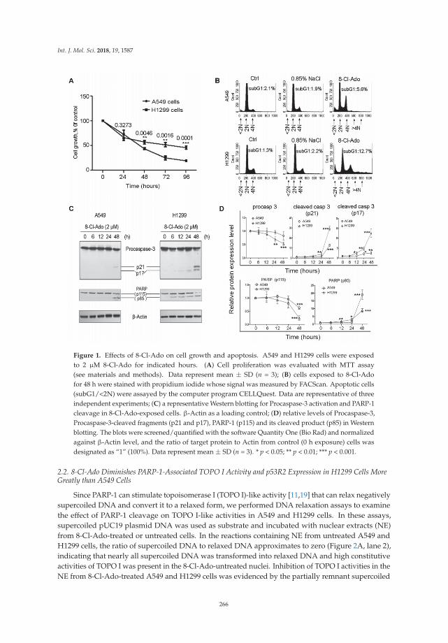
Int. J. Mol. Sci. 2018, 19, 1587
Figure 1. Effects of 8-Cl-Ado on cell growth and apoptosis. A549 and H1299 cells were exposedto 2 μM 8-Cl-Ado for indicated hours. (A) Cell proliferation was evaluated with MTT assay(see materials and methods). Data represent mean ± SD (n = 3); (B) cells exposed to 8-Cl-Adofor 48 h were stained with propidium iodide whose signal was measured by FACScan. Apoptotic cells(subG1/<2N) were assayed by the computer program CELLQuest. Data are representative of threeindependent experiments; (C) a representative Western blotting for Procaspase-3 activation and PARP-1cleavage in 8-Cl-Ado-exposed cells. β-Actin as a loading control; (D) relative levels of Procaspase-3,Procaspase-3-cleaved fragments (p21 and p17), PARP-1 (p115) and its cleaved product (p85) in Westernblotting. The blots were screened/quantified with the software Quantity One (Bio Rad) and normalizedagainst β-Actin level, and the ratio of target protein to Actin from control (0 h exposure) cells wasdesignated as “1” (100%). Data represent mean ± SD (n = 3). * p < 0.05; ** p < 0.01; *** p < 0.001.
2.2. 8-Cl-Ado Diminishes PARP-1-Associated TOPO I Activity and p53R2 Expression in H1299 Cells MoreGreatly than A549 Cells
Since PARP-1 can stimulate topoisomerase I (TOPO I)-like activity [11,19] that can relax negativelysupercoiled DNA and convert it to a relaxed form, we performed DNA relaxation assays to examinethe effect of PARP-1 cleavage on TOPO I-like activities in A549 and H1299 cells. In these assays,supercoiled pUC19 plasmid DNA was used as substrate and incubated with nuclear extracts (NE)from 8-Cl-Ado-treated or untreated cells. In the reactions containing NE from untreated A549 andH1299 cells, the ratio of supercoiled DNA to relaxed DNA approximates to zero (Figure 2A, lane 2),indicating that nearly all supercoiled DNA was transformed into relaxed DNA and high constitutiveactivities of TOPO I was present in the 8-Cl-Ado-untreated nuclei. Inhibition of TOPO I activities in theNE from 8-Cl-Ado-treated A549 and H1299 cells was evidenced by the partially remnant supercoiled
266
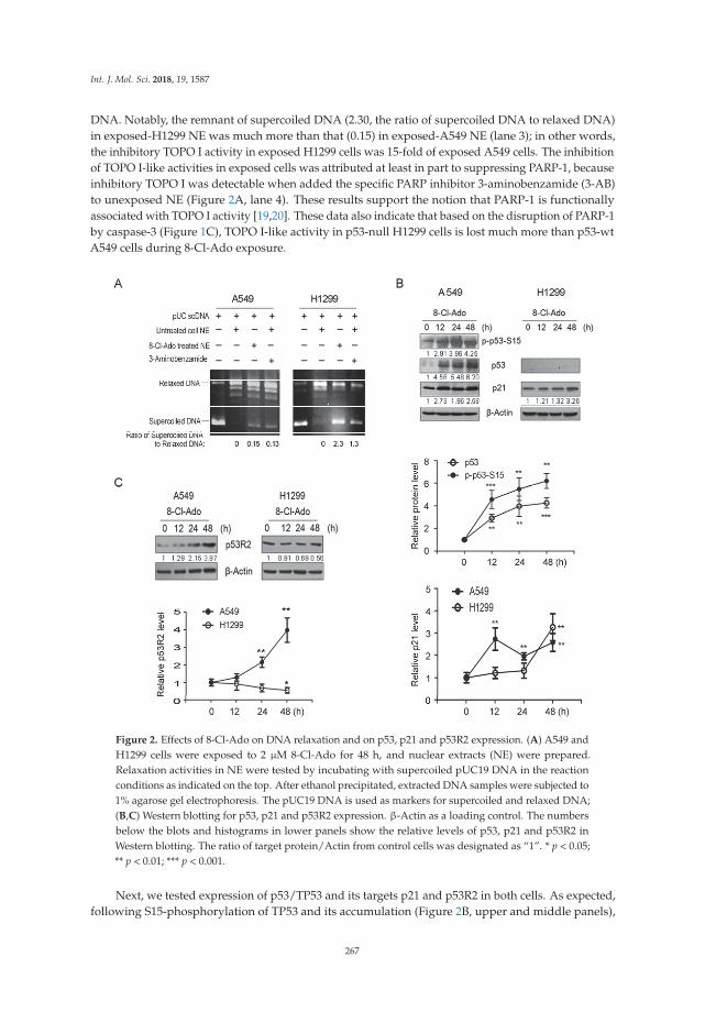
Int. J. Mol. Sci. 2018, 19, 1587
DNA. Notably, the remnant of supercoiled DNA (2.30, the ratio of supercoiled DNA to relaxed DNA)in exposed-H1299 NE was much more than that (0.15) in exposed-A549 NE (lane 3); in other words,the inhibitory TOPO I activity in exposed H1299 cells was 15-fold of exposed A549 cells. The inhibitionof TOPO I-like activities in exposed cells was attributed at least in part to suppressing PARP-1, becauseinhibitory TOPO I was detectable when added the specific PARP inhibitor 3-aminobenzamide (3-AB)to unexposed NE (Figure 2A, lane 4). These results support the notion that PARP-1 is functionallyassociated with TOPO I activity [19,20]. These data also indicate that based on the disruption of PARP-1by caspase-3 (Figure 1C), TOPO I-like activity in p53-null H1299 cells is lost much more than p53-wtA549 cells during 8-Cl-Ado exposure.
Figure 2. Effects of 8-Cl-Ado on DNA relaxation and on p53, p21 and p53R2 expression. (A) A549 andH1299 cells were exposed to 2 μM 8-Cl-Ado for 48 h, and nuclear extracts (NE) were prepared.Relaxation activities in NE were tested by incubating with supercoiled pUC19 DNA in the reactionconditions as indicated on the top. After ethanol precipitated, extracted DNA samples were subjected to1% agarose gel electrophoresis. The pUC19 DNA is used as markers for supercoiled and relaxed DNA;(B,C) Western blotting for p53, p21 and p53R2 expression. β-Actin as a loading control. The numbersbelow the blots and histograms in lower panels show the relative levels of p53, p21 and p53R2 inWestern blotting. The ratio of target protein/Actin from control cells was designated as “1”. * p < 0.05;** p < 0.01; *** p < 0.001.
Next, we tested expression of p53/TP53 and its targets p21 and p53R2 in both cells. As expected,following S15-phosphorylation of TP53 and its accumulation (Figure 2B, upper and middle panels),
267
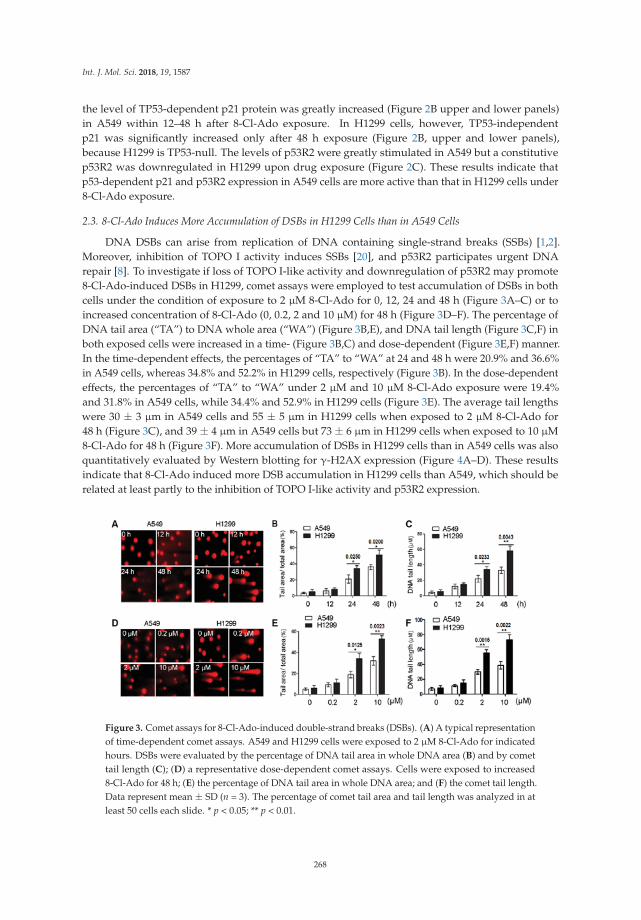
Int. J. Mol. Sci. 2018, 19, 1587
the level of TP53-dependent p21 protein was greatly increased (Figure 2B upper and lower panels)in A549 within 12–48 h after 8-Cl-Ado exposure. In H1299 cells, however, TP53-independentp21 was significantly increased only after 48 h exposure (Figure 2B, upper and lower panels),because H1299 is TP53-null. The levels of p53R2 were greatly stimulated in A549 but a constitutivep53R2 was downregulated in H1299 upon drug exposure (Figure 2C). These results indicate thatp53-dependent p21 and p53R2 expression in A549 cells are more active than that in H1299 cells under8-Cl-Ado exposure.
2.3. 8-Cl-Ado Induces More Accumulation of DSBs in H1299 Cells than in A549 Cells
DNA DSBs can arise from replication of DNA containing single-strand breaks (SSBs) [1,2].Moreover, inhibition of TOPO I activity induces SSBs [20], and p53R2 participates urgent DNArepair [8]. To investigate if loss of TOPO I-like activity and downregulation of p53R2 may promote8-Cl-Ado-induced DSBs in H1299, comet assays were employed to test accumulation of DSBs in bothcells under the condition of exposure to 2 μM 8-Cl-Ado for 0, 12, 24 and 48 h (Figure 3A–C) or toincreased concentration of 8-Cl-Ado (0, 0.2, 2 and 10 μM) for 48 h (Figure 3D–F). The percentage ofDNA tail area (“TA”) to DNA whole area (“WA”) (Figure 3B,E), and DNA tail length (Figure 3C,F) inboth exposed cells were increased in a time- (Figure 3B,C) and dose-dependent (Figure 3E,F) manner.In the time-dependent effects, the percentages of “TA” to “WA” at 24 and 48 h were 20.9% and 36.6%in A549 cells, whereas 34.8% and 52.2% in H1299 cells, respectively (Figure 3B). In the dose-dependenteffects, the percentages of “TA” to “WA” under 2 μM and 10 μM 8-Cl-Ado exposure were 19.4%and 31.8% in A549 cells, while 34.4% and 52.9% in H1299 cells (Figure 3E). The average tail lengthswere 30 ± 3 μm in A549 cells and 55 ± 5 μm in H1299 cells when exposed to 2 μM 8-Cl-Ado for48 h (Figure 3C), and 39 ± 4 μm in A549 cells but 73 ± 6 μm in H1299 cells when exposed to 10 μM8-Cl-Ado for 48 h (Figure 3F). More accumulation of DSBs in H1299 cells than in A549 cells was alsoquantitatively evaluated by Western blotting for γ-H2AX expression (Figure 4A–D). These resultsindicate that 8-Cl-Ado induced more DSB accumulation in H1299 cells than A549, which should berelated at least partly to the inhibition of TOPO I-like activity and p53R2 expression.
Figure 3. Comet assays for 8-Cl-Ado-induced double-strand breaks (DSBs). (A) A typical representationof time-dependent comet assays. A549 and H1299 cells were exposed to 2 μM 8-Cl-Ado for indicatedhours. DSBs were evaluated by the percentage of DNA tail area in whole DNA area (B) and by comettail length (C); (D) a representative dose-dependent comet assays. Cells were exposed to increased8-Cl-Ado for 48 h; (E) the percentage of DNA tail area in whole DNA area; and (F) the comet tail length.Data represent mean ± SD (n = 3). The percentage of comet tail area and tail length was analyzed in atleast 50 cells each slide. * p < 0.05; ** p < 0.01.
268

Int. J. Mol. Sci. 2018, 19, 1587
Figure 4. Western blotting and immunocytochemistry for γ-H2AX expression in A549 and H1299 cells.(A) Cells were exposed to 2 μM 8-Cl-Ado for 0, 6, 12, 24 and 48 h. Western blotting was performedwith specific antibody. β-Actin as a loading control; (B) histograms showing the relative levels ofγ-H2AX in (A) experiments. The ratio of γ-H2AX to Actin at 0 h is normalized to “1”. Data representmean ± SD (n = 3); (C) cells were exposed to 8-Cl-Ado at the indicated concentrations for 48 h, andWestern blotting was performed for a dose-dependent increase of γ-H2AX; (D) histograms showingthe relative levels of γ-H2AX in (C) experiments. * p < 0.05; ** p < 0.01; *** p < 0.001.
2.4. Defect in p53-p21 Signal in H1299 Cells Leads to Increased S Subpopulation upon DSBs
We first examined cell cycle checkpoint signals in DDR. Phospho-ATM-S1981 was significantlyincreased within 48 h upon 8-Cl-Ado exposure in both cells (Figure 5A). Following ATM activation,phospho-CHK1-S345 and phospho-CHK2-T68 were increased within 6–48 h in both H1299 and A549cells. It seemed that CHK1 phosphorylation dominantly occupied in H1299 cells, whereas CHK2phosphorylation predominated in A549 cells. This difference between the two cells presumably isattributable to more SSBs induced by TOPO I inhibition in H1299 cells.
After 48 h exposure, A549 cells increased G1 subpopulation from 65 (control) to 84% but decreasedS phase cells sharply from 23 to 7% and had no significant changes in G2/M cells (from 10 to 9%),while H1299 cells increased G1 phase cells from 47 to 52%, decreased S phase cells from 28 to 23%, andstill remained 22% G2/M cells (Figure 5B). Alternatively, the percentages of S phase and G2/M phasecells in 48 h-exposed H1299 cells are threefold and twofold as much as in exposed A549, respectively,indicating that even in the presence of DSBs, H1299 cells had more cells entering into S and G2/Mphases. Not surprisingly, induction of p21 followed after p53 phosphorylation/accumulation inexposed A549 cells, but no marked increase until 48 h in exposed H1299 cells (Figure 2B). Followingsilence of p53 by RNA interference (RNAi) in A549 cells (Figure 5C, left panel) or over-expressed p53 inH1299 cells (Figure 5C, right panel), p21 was down- or upregulated (Figure 5C). Similar G1, S and G2/Msubpopulations occurred in p53-silenced A549 (Figure 5D, left panel) and p53-overexpressed H1299(Figure 5D, right panel) under exposed and unexposed conditions; it was much the same of G1,S and G2/M subpopulations in unexposed A549 and p53-overexpressed H1299 cells, but a littledifference between them under exposed condition, probably due to over-expressing of exogenous p53.At any rate, these results indicate that p21 increases G1 phase but restricts S and G2/M phase cells.
269

Int. J. Mol. Sci. 2018, 19, 1587
Together, defect in p53-p21 signal leads to more serious impairment of G1 checkpoint and to moreS phase cell accumulation in H1299 cells than in A549 cells during DDR.
Figure 5. Signal pathways and cell-cycle progression in DDR (DNA damage response). (A) A549and H1299 cells were exposed to 2 μM 8-Cl-Ado for indicated hours, and Western blotting wasperformed for components of signal-transduction pathways. The relative levels of target proteinswere normalized against β-Actin; (B) cell-cycle analysis. Cells were exposed to 2 μM 8-Cl-Adofor 48 h. After harvested and fixed, cells were stained with propidium iodide (PI); PI signal wasmeasured by FACScan. G1, G2/M and S populations in the cell-cycle were analyzed by computerprograms. Data present ± SD (n = 3). * p < 0.05; (C) Western blotting for p53 and p21 in p53-silencedA549 and p53-overexpressed H1299 cells. Cells were transiently transfected with pSUPER-basic(control), pSUPER-p53 (for silencing TP53), or p53-WT expression plasmid for 48 h and exposed to8-Cl-Ado for additional 48 h, followed by Western blotting. The relative levels of target proteins werenormalized against β-Actin; (D) G1 and G2/M and S subpopulations in p53-silenced A549 cells andp53-overexpressed H1299 cells.
2.5. 8-Cl-Ado-Induced More Accumulation of DSBs in H1299 Is Associated with DNA Replication in S Phase
DNA DSBs interfere with DNA replication [1]. We thus compared DNA synthesis in both cellsusing BrdU incorporation. In consistence with the results shown in Figure 5B, more BrdU-labeled Sand G2 cells in H1299 cells than A549 cells were detectable after 24 h 8-Cl-Ado-exposure (Figure 6).DNA synthesis was continually decreased in H1299 cells within 12–48 h of exposure, but only seenat earlier steps (<24 h) in A549 cells (Figure 6A). The percentages of BrdU-incorporated S cells inA549 cells after 0, 12, 24 and 48 h exposure were 44.6%, 38.2%, 28.7% and 32.5%; in other words,DNA synthesis was continually decreased before 24 h but became increased by 48 h, indicating thatDNA repair capability initiates a little recovery within 24–48 h. In H1299, however, the percentagesof BrdU positive S cells at the same time-points were 54.9%, 48.2%, 46.7% and 38.7%, respectively.Importantly, the BrdU-incorporated rates at 24 and 48 h in H1299 were significantly higher thanA549(Figure 6B). The continual drops of BrdU-incorporated S cells in H1299 cells suggest that DNA damageremains at all times and the repair capability is unrecovered, probably due to p53 defect and p53R2reduction. In addition, more DNA synthesis in S phase may result in more DSBs in H1299 cells.
270
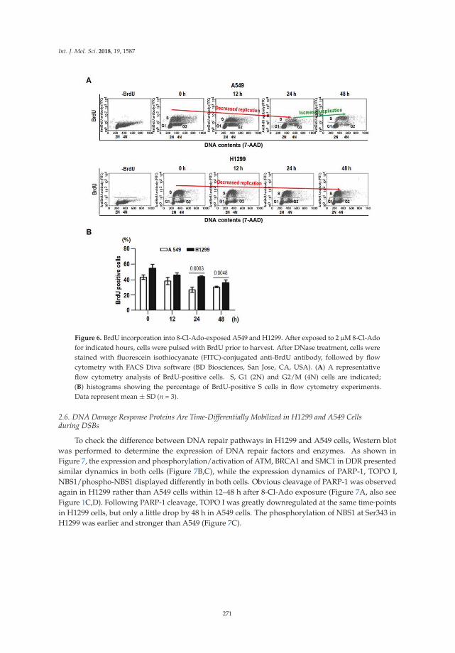
Int. J. Mol. Sci. 2018, 19, 1587
Figure 6. BrdU incorporation into 8-Cl-Ado-exposed A549 and H1299. After exposed to 2 μM 8-Cl-Adofor indicated hours, cells were pulsed with BrdU prior to harvest. After DNase treatment, cells werestained with fluorescein isothiocyanate (FITC)-conjugated anti-BrdU antibody, followed by flowcytometry with FACS Diva software (BD Biosciences, San Jose, CA, USA). (A) A representativeflow cytometry analysis of BrdU-positive cells. S, G1 (2N) and G2/M (4N) cells are indicated;(B) histograms showing the percentage of BrdU-positive S cells in flow cytometry experiments.Data represent mean ± SD (n = 3).
2.6. DNA Damage Response Proteins Are Time-Differentially Mobilized in H1299 and A549 Cellsduring DSBs
To check the difference between DNA repair pathways in H1299 and A549 cells, Western blotwas performed to determine the expression of DNA repair factors and enzymes. As shown inFigure 7, the expression and phosphorylation/activation of ATM, BRCA1 and SMC1 in DDR presentedsimilar dynamics in both cells (Figure 7B,C), while the expression dynamics of PARP-1, TOPO I,NBS1/phospho-NBS1 displayed differently in both cells. Obvious cleavage of PARP-1 was observedagain in H1299 rather than A549 cells within 12–48 h after 8-Cl-Ado exposure (Figure 7A, also seeFigure 1C,D). Following PARP-1 cleavage, TOPO I was greatly downregulated at the same time-pointsin H1299 cells, but only a little drop by 48 h in A549 cells. The phosphorylation of NBS1 at Ser343 inH1299 was earlier and stronger than A549 (Figure 7C).
271

Int. J. Mol. Sci. 2018, 19, 1587
Figure 7. Western blotting for PARP-1, TOPO I and DNA responsive proteins in DDR. A549 andH1299 were exposed to 2 μM 8-Cl-Ado for the indicated hours. The γ-H2AX, PARP-1 and TOPO I (A),SMC1 (B), ATM, BRCA1 and NBS1 and their modifications (C) were analyzed by Western blotting withspecific antibodies. Also see Figure S1 for BRCA1/pBRCA1 full blots of A549 cells in (C). β-Actin asa loading control. The ratio of target protein/Actin from control cells was designated as “1”.
3. Discussion
Computational biology study reveals that intratumor signaling heterogeneity or pathwaydysregulation is associated to clinical outcome of cancers [21]. The DDR signal pathway is composedof a cell cycle checkpoint and DNA repair mechanisms that maintain genomic stability in normalcells and can also serve as targets for cancer chemotherapy. Our hypothesis that the signalingpathway heterogeneity of cellular response to DSBs might determine chemotherapeutic sensitivityof cancer cells is evidenced in this study, and our major finding is that cancer cells (e.g., H1299)lacking ATM-CHK2-p53-p21 mediated G1 checkpoint and p53-dependent DNA repair are muchmore sensitive to chemotherapeutics. In other words, p53-p21 signal may at least in part protectp53-wt cancer cells (e.g., A549) from DNA damage stress. Our finding support the notion that p53signaling suppresses apoptosis following genotoxic stress, facilitating repair of genomic injury underphysiological conditions but having the potential to promote tumor regrowth in response to cancerchemotherapy [22].
Initial studies of cellular response to anticancer drugs suggested that p53-dependent apoptosiswas the common mechanism of cancer chemotherapy. Subsequent work on p53-null cells andanimal models, however, argued that genotoxic agents could also induce significant cytotoxicityin a p53-independent manner [23–25]. Indeed, p53-null H1299 cells were more sensitive top53-independent apoptosis than p53-wt A549 cells when exposed to curcumin [13], which inhibitscell cycle and cell survival by inducing DNA damage [26]. Similarly, we found that H1299 cells weremore sensitive to 8-Cl-Ado-inducd growth inhibition and apoptosis than A549 cells [14] (also seeFigure 1A,B). It seemed that hypersemsitivity of H1299 was linked to 8-Cl-Ado induced DSBs, because
272

Int. J. Mol. Sci. 2018, 19, 1587
8-Cl-Ado induced more severe DSBs in H1299 than A549 (Figures 3 and 4). Several reasons mayaccount for more extensive and severe DSBs in H1299 than A549 cells.
First, p53-p21 signal deficiency and S cell accumulation by SMC1 activation presumablycontributes to more DSBs in H1299 cells than A549 cells. After detection of DSBs by ATM,p53 is phosphorylated/activated and arrests cells in G1 via activating p21 gene expression [6–8].p53-induced p21 not only induces G1 arrest but inhibits DNA replication without interfering withDNA repair through binding to the replication/repair factor PCNA [27] and PARP-1 [28] in DDR.We found that in A549 cells, the p21 protein was rapidly up-regulated following p53 activationand strictly arrested most cells in G1 phase upon DSBs, while p53-null H1299 cells had a delayedinduction of p21 only by 48 h, leading to G1 checkpoint loss and more S cell accumulation (Figure 5).Also, more S cell accumulation in H1299 might be attributed to SMC1 phosphorylation, becausephosphorylation of SMC1 at Ser957 is required for intra-S checkpoint [29,30]. During DDR, SMC1 isphosphorylated by ATM/ATR in the presence of BRCA1 and NBS1 [31]. Activating intra-S checkpointand inhibiting TOPO I can increase DSBs [20,22], which arise from replication of DNA containingSSBs [1,2]. We did find stronger inhibition of TOPO I (Figures 2A and 7A) and activation of SMC1followed by BRCA1 and NBS1 activation at 24 h after 8-Cl-Ado exposure in H1299 cells (Figure 7C), andmore accumulation of S (BrdU positive) cells in H1299 (Figures 5B and 6). The S cells with uncoveredcapability of DNA synthesis are particularly vulnerable to DNA damage, which may cause replicationstress, then replication-stress-induced DSBs. Previous notion [29–31] and our data can explain whymore DSBs occur in H1299 than A549.
Second, defects of p53 and p53-dependent DNA repair capability are associated with moreDNA DSBs and apoptosis in H1299 than A549. DNA DSB is repaired by NHEJ in G1 phase andHR in late S and G2 [1–5]. The p53 protein guards genomic stability through direct or indirect rolesin DNA repair. For instance, p53 modulates Holliday Junctions and broken end reconnecting andannealing in HR repair [4]. The protein can also interact with repair proteins such as replication proteinA (RPA), Rad51 and Rad52 to promote HR repair [32]. In addition to p21, p53 as a transcriptionfactor can also promote controlling DNA repair gene expression, such as BRCA1, p53R2, GADD45and PCNA. For instance, p53 can transcriptionally activate BRCA1 expression [7], and in turn,ATM-phosphorylated BRCA1 interacts with and enhances p53 transactivation function [6,7,32].BRCA1 selectively co-activates p53-dependent genes such as p21 and p53R2 [8,33] targeting DNA repairand cell cycle arrest but not apoptosis [34]. This is because that p21-PCNA interaction inhibits DNAreplication [27]; p21 association with PARP-1 blocks replication fork progression [28]; p53-dependentp21 can also bind procaspase-3 to protect cells from apoptosis [34]. Moreover, p53-induced p53R2supplies dNTPs for urgent DNA repair during G1 and G2 arrests [8,33]. In our case, p53R2 as a directtarget for p53 was strongly induced in A549 cells (Figure 2C), which might promote the capabilityof DNA repair and could be associated with a lesser increase of DSBs in A549 cells than H1299 cells.In addition, transcription factor E2F1 promotes G1/S transition and induces apoptosis by controllingtarget gene expression [35]. We have previously shown that E2F1 is induced in H1299 cells duringDDR [36,37]. E2F1 may therefore counteract p21-inhibited G1/S transition, promoting S phase cellsand apoptosis in H1299 cells. However, E2F1 cannot achieve that in A549 cells, because p53 maycounteract E2F1 effect by association with it [38]. Contrarily, the early nucleolar accumulation of E2F1may release p53 function [36] to activate p21 and p53R2 genes and p53-dependent DNA repair in A549cells. Indeed, A549 cells displayed a partial capability of recovering DNA replication at late time of8-Cl-Ado exposure, but H1299 cells could not (Figure 6). All above-mentioned can explain more G1cells and less DNA DSBs in A549 cells, but more S phase cells and DSBs in H1299 cells.
Third, loss of PARP-1 activity by caspase-3 cleavage is linked to more accumulation of DNA DSBsin H1299 than A549. PARP-1 is activated by DNA strand breaks and functions as a positive regulatorin DNA repair. PARP-1 mediates DSB end-joining in mammalian cells, which may complementthe DNA-PK/XRCC4/ligase IV-dependent NHJE [39]. PARP-1-mediated DSB end-joining dependson its interaction with repair proteins, by which PARP-1 recruits repair proteins/enzymes to DSB
273

Int. J. Mol. Sci. 2018, 19, 1587
sites [10]. Contrarily, the small cleaved fragment (24-kDa) from PARP-1 blocks the access of repairproteins/enzymes to DSBs [40], indicating that PARP-1 cleavage impairs its capability of recruitingrepair proteins/enzymes. Moreover, PARP-1 can stimulate the activity of DNA-PK for NHJE in G1 [10].In addition, PARP1 is required for rapid recruitment of MRE11 and NBS1 at DSB sites during HRrepair in S phase [41]. Therefore, deficiency of PARP-1 may increase sensitivities of cells to DNAdamage agents [10]. Also, PARP-1 can promote TOPO I and TOPO I-like activity [11,19] and reactivatestalled TOPO I activity [42], therefore loss of PARP1 may decrease DNA repair capability and increasesSSBs and SSB-containing DNA replication-mediated DSBs. We thus conclude that loss of PARP-1 bycaspase-3 cleavage is accounted for more DSBs in H1299 than A549. Our data suggest that inhibition ofPARP-1 as well as TOPO I may sensitize cancer cells to chemotherapeutic agents in certain situations.
p53 (TP53) acts as a tumor suppressor by orchestrating various signaling pathways, in whichthe activity of p53 involves several positive and negative feedback loops that determine the cell fatesthrough cell cycle arrest, DNA repair or apoptosis [43]. Growing evidence suggests that p53 can actas a tumor suppressor via p53-microRNA loops. Genome-wide screen for microRNAs revealed thatmany TP53 targeted miRNAs including miR-34a have been implicated in p53-mediated apoptosisduring DDR [44]. Most recently, a study showed a positive p53/Wip1/miR-16 feedback loop for G1/Scheckpoint during DNA damage [45]. Therefore, we cannot exclude the participation of p53 andmicroRNA feedback loops in 8-Cl-Ado-induced DSB response in A549, which might contribute todifferential sensitivities of A549 and H1299 cells to the drug.
In summary, we tested our hypothesis that more extensive and severe DNA damage was linkedto higher sensitivity of H1299 to 8-Cl-Ado treatment, whereas less DNA damage was linked to lowersensitivity of A549. We have clarified the major causes of more extensive DSBs in H1299 cells. Together,the heterogeneity of DDR signaling pathways determines the sensitivity of cancer cells to DNAdamage-based chemotherapeutics. Notably, we comparatively investigated the effects of 8-Cl-Adoon NSCLC H1299 and A549 cells, whether our finding is suited to other genotoxic agents and cancercells remains to be clarified. In addition, we examined only some of the key molecular components ofthe DDR signaling pathways; gene chip analysis is needed for detailed knowledge of the condition inthe future.
4. Materials and Methods
4.1. Cell Culture and Treatment
Human lung cancer A549 (p53-wt) and H1299 (p53-null) cells from ATCC (Manassas, VA, USA)were cultured in Dulbecco minimum essential medium (DMEM, Gibco, Grand Island, NY, USA)supplemented with 10% fetal bovine serum (Gibco, Grand Island, NY, USA), 100 U/mL penicillinand 100 mg/mL streptomycin, and grown at 37 ◦C with 5% CO2. 8-Chloro-adenosine (8-Cl-Ado)(the State Key Laboratory for Natural and Biomimetic Drugs, Peking University HSC, Beijing, China)was dissolved in 0.9% NaCl solution in given concentrations.
4.2. Cell Proliferation Assay
Cells were cultured in 96-well plates (15,000 cells/0.2 mL per well). 8-Cl-Ado (2 μM)was added to cultures, followed by incubation for given hours. Before harvest, 20 μL MTT(3-(4,5-dimethythiazolzyl)-2,5-diphenyl tetrazolium tromide, 5 mg/mL; Sigma, St. Louis, MO, USA)was added to each well. After incubating for 4 h, 0.2 mL dimethyl sulfoxide (DMSO) was addedto terminate reactions. Absorbance values were determined spectrophotometrically at 490 nm ona Microplate Reader (BIO-TEK, Rockville, MA, USA).
4.3. Flow Cytometry Analysis
Typically, 1 × 10⁶ cells were collected, washed twice in ice-cold PBS and fixed in ice-cold70% ethanol overnight at 4 ◦C. Then cells were washed twice in ice-cold PBS and digested with
274

Int. J. Mol. Sci. 2018, 19, 1587
RNase A (10 μg/mL) at 37 ◦C for 30 min. Cells were stained with 10 μg/mL of propidium iodide(Sigma) for 3 min at room temperature before testing. DNA contents of cells (10,000 cells perexperimental group) were analyzed using computer programs CELLQuest and ModFit LT 2.0epfor Power (Becton Dickinson, Franklin Lakes, NJ, USA). Apoptosis was assayed by the appearanceof a sub-G1 (<2N ploidy) population by the computer program CELLQuest (Becton Dickinson,Franklin Lakes, NJ, USA).
4.4. DNA Relaxation
Reaction mixtures containing 0.4 mg pUC19 plasmid DNA (MBI Fermentas, Vilnius, Lithuania)and 2.5 μg nuclear extracts (NE) from 8-Cl-Ado-exposed or -unexposed cells, or 5 mM3-aminobenzamide (PARP inhibitor) in 20 μL relaxation buffer (50 mM Tris-HCl, pH 8.0, 0.1 M NaCl,5 mM MgCl2) were incubated at 37 ◦C for 30 min and stopped by adding sodium dodecyl sulphate(SDS) and ethylenediaminetetraacetic acid (EDTA) to a final concentration of 0.1% and 10 mM,respectively. DNA was ethanol precipitated, and subjected to electrophoresis in 1% agarose gels.DNA was stained with 1 mg/mL ethidium bromide and visualized by ultraviolet (UV) light.
4.5. Comet Assay
As described previously [46], a 80 μL mixture containing 105 cells treated with or without 8-Cl-Adoin 40 μL PBS, and 40 μL 1% low melting point agarose (final concentration 0.5%) was pipetted onto thefirst agarose layer of the full-frosted microscope slides that were precoated with 0.5% normal meltingpoint agarose. After lysis for 2 h at 4 ◦C in fresh lysing solution, slides were placed in a horizontalgel electrophoresis unit filled with fresh electrophoresis solution for 20 min. Following unwinding,electrophoresis was performed for 20 min at 0.7 V/cm (300 mA/25 V) at 4 ◦C. After electrophoresis,slides were neutralized twice with 0.4 M Tris buffer (pH 7.5) for 15 min. Slides were stained withethidium bromide for 10 min in the dark. After staining, slides were examined at 600× magnification,and pictures were taken under fluorescence microscope (Leica, Mannheim, Germany). To score thepercentage of DNA in the tail, the image analysis system was used (Q550CW; Leica, Wetzlar, Germany).The percentage of comet tail area (the ratio of DNA tail area to total DNA area) and comet tail length(from the center of the DNA head to the end of the DNA tail) was analyzed in 50 cells for one slide.
4.6. Constructs and Transfection
pSUPER-p53 plasmid was constructed by ligating the annealed primers 5′-GATCCCCGACTCCAGTGGTAATCTACTTCAAGAGAGTAGATTACCACTGGAGTCTTTTTA-3′, 5′-AGCTTAAAAAGACTCCAGTGGTAATCTACTCTCTTGAAGTAGATTACCACTGGAGTCGGG-3′ into the Bgl IIand Hind III sites of pSUPER-basic (OligoEngine, Seattle, WA, USA), and correct plasmid was confirmedby direct sequencing. p53 wild-type plasmid was a gift from Dr. Bert Vogelstein (Johns HopkinsUniversity, Baltimore, MD, USA). Transfection was performed with Lipofectamine 2000 (Invitrogen,Carlsbad, CA, USA) following manufacturer’s protocol.
4.7. Western Blotting
Whole-cell extracts were prepared in lysis buffer and protein concentration was determinedusing the BCA Protein Assay Reagent Kit (Pierce, Rockford, IL, USA). Fifty micrograms of totalproteins were loaded onto 10–13% sodium dodecyl sulphate-polyacrylamide gel electrophoresis(SDS-PAGE), and transferred onto nitrocellulose membranes. Membranes were incubated with primaryantibodies overnight at 4 ◦C with gentle rocking followed by horseradish peroxidase-conjugatedsecondary antibody for 1 h. Chemiluminescence signals were visualized using Western blottingluminol reagent (Santa Cruz Biotech, Santa Cruz, CA, USA) and exposed to film. The blots werescreened/quantified with the software Quantity One (Bio Rad, Hercules, CA, USA) and normalizedagainst β-Actin level. The target protein/Actin value obtained from control (8-Cl-Ado-exposedfor 0 h) cells was designated as “1”. Anti-p21, anti-p53, anti-p53R2, anti-phospho-p53-S15,
275

Int. J. Mol. Sci. 2018, 19, 1587
anti-CHK1-S345, anti-CHK2-T68, anti-CHK1, anti-CHK2, anti-ATR, anti-phospho-ATR-S428,anti-NBS1, anti-phospho-NBS1-S343, anti-SMC1, anti-phospho-SMC1-S699, anti-β-actin, anti-BRCA1and anti-phospho-BRCA1-S1524 antibodies were purchased from Cell Signaling Technology;anti-phospho-histone H2AX-S139, anti-ATM and antiphospho-ATM-S1981 were acquired from R&DSystems Inc. (Minneapolis, MN, USA).
4.8. BrdU Incorporation Assay
As previously described [18], BrdU incorporation was performed using the fluoresceinisothiocyanate (FITC) BrdU Flow Kit (BD Pharmingen, San Diego, CA, USA), according to themanufacturer’s instructions. After exposed to 2 μM 8-Cl-Ado, cells were pulsed with finalconcentration of 10 μM BrdU for 30 min at 37 ◦C prior to harvest. Cells were washed incold staining buffer (1 × Dulbecco’s phosphate-buffered saline +3% FBS), fixed/permeabilizedwith Cytofix/Cytoperm buffer and washed with Perm/Wash buffer (on ice). Cells were treatedwith 30 μg DNase for 1 h at 37 ◦C, and stained with FITC-conjugated anti-BrdU antibody and7-AAD. DNA contents were analyzed by a FACSCanto flow cytometer with FACSDiva software(BD Biosciences, San Jose, CA, USA).
4.9. Statistical Analysis
The Student’s t-test and ANOVA test were used for univariate analysis. Statistical significancewas defined by a two-tailed p-value of 0.05.
Supplementary Materials: Supplementary materials can be found at http://www.mdpi.com/1422-0067/19/6/1587/s1.
Author Contributions: S.-Y.Y., J.-H.N., H.-T.J. and S.-Y.L. conceived and designed the study and drafted themanuscript. S.-Y.Y., Y.L. and G.-S.A. performed the experiments. S.-Y.Y., H.-T.J. and S.-Y.L. carried out thedata analysis.
Acknowledgments: We thank Bert Vogelstein (Johns Hopkins University) for p53 plasmid. This work wassupported by National Natural Science Foundation of China Grants 81773025, 81230008, 81571368, 91319302,7132120 and 81602159, the Natural Science Foundation of Chongqing (Grant No. cstc2016jcyjA0054), Science andTechnology Planning Project of Yuzhong District, Chongqing City (20170109), and Startup Fund from ChongqingMedical University.
Conflicts of Interest: The authors declare no conflicts of interest.
References
1. Hoeijmakers, J.H. Genome maintenance mechanisms for preventing cancer. Nature 2001, 411, 366–374.[CrossRef] [PubMed]
2. Jackson, S.P.; Bartek, J. The DNA-damage response in human biology and disease. Nature 2009, 461,1071–1078. [CrossRef] [PubMed]
3. Basu, A.K. DNA Damage, Mutagenesis and Cancer. Int. J. Mol. Sci. 2018, 19, 970. [CrossRef] [PubMed]4. Helleday, T.; Lo, J.; van Gent, D.C.; Engelward, B.P. DNA double-strand break repair: From mechanistic
understanding to cancer treatment. DNA Rep. 2007, 6, 923–935. [CrossRef] [PubMed]5. Polo, S.E.; Jackson, S.P. Dynamics of DNA damage response proteins at DNA breaks: A focus on protein
modifications. Genes Dev. 2011, 25, 409–433. [CrossRef] [PubMed]6. Meek, D.W. The p53 response to DNA damage. DNA Rep. 2004, 3, 1049–1056. [CrossRef] [PubMed]7. Niculescu, A.B., 3rd; Chen, X.; Smeets, M.; Hengst, L.; Prives, C.; Reed, S.I. Effects of p21(Cip1/Waf1) at both
the G1/S and the G2/M cell cycle transitions: pRb is a critical determinant in blocking DNA replication andin preventing endoreduplication. Mol. Cell. Biol. 1998, 18, 629–643. [CrossRef] [PubMed]
8. Tanaka, H.; Arakawa, H.; Yamaguchi, T.; Shiraishi, K.; Fukuda, S.; Matsui, K.; Takei, Y.; Nakamura, Y. Aribonucleotide reductase gene involved in a p53-dependent cell-cycle checkpoint for DNA damage. Nature2000, 404, 42–49. [CrossRef] [PubMed]
9. Taylor, W.R.; Stark, G.R. Regulation of the G2/M transition by p53. Oncogene 2001, 20, 1803–1815. [CrossRef][PubMed]
276
Int. J. Mol. Sci. 2018, 19, 1587
10. Petermann, E.; Keil, C.; Oei, S.L. Importance of poly(ADP-ribose) polymerases in the regulation ofDNA-dependent processes. Cell. Mol. Life Sci. 2005, 62, 731–738. [CrossRef] [PubMed]
11. Yung, T.M.; Parent, M.; Ho, E.L.; Satoh, M.S. Camptothecin-sensitive relaxation of supercoiled DNA bythe topoisomerase I-like activity associated with poly(ADP-ribose) polymerase-1. J. Biol. Chem. 2004, 279,11992–11999. [CrossRef] [PubMed]
12. Jensen, L.H.; Dejligbjerg, M.; Hansen, L.T.; Grauslund, M.; Jensen, P.B.; Sehested, M. Characterisation ofcytotoxicity and DNA damage induced by the topoisomerase II-directed bisdioxopiperazine anti-canceragent ICRF-187 (dexrazoxane) in yeast and mammalian cells. BMC Pharmacol. 2004, 4, 31. [CrossRef][PubMed]
13. Radhakrishna Pillai, G.; Srivastava, A.S.; Hassanein, T.I.; Chauhan, D.P.; Carrier, E. Induction of apoptosis inhuman lung cancer cells by curcumin. Cancer Lett. 2004, 208, 163–170. [CrossRef] [PubMed]
14. Zhang, H.Y.; Gu, Y.Y.; Li, Z.G.; Jia, Y.H.; Yuan, L.; Li, S.Y.; An, G.S.; Ni, J.H.; Jia, H.T. Exposure of human lungcancer cells to 8-chloro-adenosine induces G2/M arrest and mitotic catastrophe. Neoplasia 2004, 6, 802–812.[CrossRef] [PubMed]
15. Langeveld, C.H.; Jongenelen, C.A.; Theeuwes, J.W.; Baak, J.P.; Heimans, J.J.; Stoof, J.C.; Peters, G.J.The antiproliferative effect of 8-chloro-adenosine, an active metabolite of 8-chloro-cyclic adenosinemonophosphate, and disturbances in nucleic acid synthesis and cell cycle kinetics. Biochem. Pharmacol. 1997,53, 141–148. [CrossRef]
16. Gandhi, V.; Ayres, M.; Halgren, R.G.; Krett, N.L.; Newman, R.A.; Rosen, S.T. 8-chloro-cAMP and8-chloro-adenosine act by the same mechanism in multiple myeloma cells. Cancer Res. 2001, 61, 5474–5479.[PubMed]
17. Dennison, J.B.; Balakrishnan, K.; Gandhi, V. Preclinical activity of 8-chloroadenosine with mantle celllymphoma: Roles of energy depletion and inhibition of DNA and RNA synthesis. Br. J. Haematol. 2009, 147,297–307. [CrossRef] [PubMed]
18. Yang, S.Y.; Jia, X.Z.; Feng, L.Y.; Li, S.Y.; An, G.S.; Ni, J.H.; Jia, H.T. Inhibition of topoisomerase II by8-chloro-adenosine triphosphate induces DNA double-stranded breaks in 8-chloro-adenosine-exposedhuman myelocytic leukemia K562 cells. Biochem. Pharmacol. 2009, 77, 433–443. [CrossRef] [PubMed]
19. Bauer, P.I.; Buki, K.G.; Comstock, J.A.; Kun, E. Activation of topoisomerase I by poly [ADP-ribose]polymerase. Int. J. Mol. Med. 2000, 5, 533–540. [CrossRef] [PubMed]
20. Tomicic, M.T.; Kaina, B. Topoisomerase degradation, DSB repair, p53 and IAPs in cancer cell resistance tocamptothecin-like topoisomerase I inhibitors. Biochim. Biophys. Acta 2013, 1835, 11–27. [CrossRef] [PubMed]
21. Banerji, C.R.; Severini, S.; Caldas, C.; Teschendorff, A.E. Intra-tumour signalling entropy determines clinicaloutcome in breast and lung cancer. PLoS Comp. Biol. 2015, 11, e1004115. [CrossRef] [PubMed]
22. Mirzayans, R.; Andrais, B.; Kumar, P.; Murray, D. Significance of wild-type p53 signaling in suppressingapoptosis in response to chemical genotoxic agents: Impact on chemotherapy outcome. Int. J. Mol. Sci. 2017,18, 928. [CrossRef] [PubMed]
23. Merritt, A.J.; Allen, T.D.; Potten, C.S.; Hickman, J.A. Apoptosis in small intestinal epithelial from p53-nullmice: Evidence for a delayed, p53-independent G2/M-associated cell death after gamma-irradiation.Oncogene 1997, 14, 2759–2766. [CrossRef] [PubMed]
24. Han, J.W.; Dionne, C.A.; Kedersha, N.L.; Goldmacher, V.S. p53 status affects the rate of the onset but notthe overall extent of doxorubicin-induced cell death in rat-1 fibroblasts constitutively expressing c-Myc.Cancer Res. 1997, 57, 176–182. [PubMed]
25. Tannock, I.F.; Lee, C. Evidence against apoptosis as a major mechanism for reproductive cell death followingtreatment of cell lines with anti-cancer drugs. Br. J. Cancer 2001, 84, 100–105. [CrossRef] [PubMed]
26. Lu, J.J.; Cai, Y.J.; Ding, J. Curcumin induces DNA damage and caffeine-insensitive cell cycle arrest incolorectal carcinoma HCT116 cells. Mol. Cell. Biochem. 2011, 354, 247–252. [CrossRef] [PubMed]
27. Levine, A.J. p53, the cellular gatekeeper for growth and division. Cell 1997, 88, 323–331. [CrossRef]28. Frouin, I.; Maga, G.; Denegri, M.; Riva, F.; Savio, M.; Spadari, S.; Prosperi, E.; Scovassi, A.I. Human
proliferating cell nuclear antigen, poly(ADP-ribose) polymerase-1, and p21waf1/cip1. A dynamic exchangeof partners. J. Biol. Chem. 2003, 278, 39265–39268. [CrossRef] [PubMed]
29. Kim, S.T.; Xu, B.; Kastan, M.B. Involvement of the cohesin protein, Smc1, in Atm-dependent and independentresponses to DNA damage. Genes Dev. 2002, 16, 560–570. [CrossRef] [PubMed]
277
Int. J. Mol. Sci. 2018, 19, 1587
30. Kaufmann, W.K. The human intra-S checkpoint response to UVC-induced DNA damage. Carcinogenesis2010, 31, 751–765. [CrossRef] [PubMed]
31. Matsuoka, S.; Ballif, B.A.; Smogorzewska, A.; McDonald, E.R., 3rd; Hurov, K.E.; Luo, J.; Bakalarski, C.E.;Zhao, Z.; Solimini, N.; Lerenthal, Y.; et al. ATM and ATR substrate analysis reveals extensive proteinnetworks responsive to DNA damage. Science 2007, 316, 1160–1166. [CrossRef] [PubMed]
32. Gebow, D.; Miselis, N.; Liber, H.L. Homologous and nonhomologous recombination resulting in deletion:Effects of p53 status, microhomology, and repetitive DNA length and orientation. Mol. Cell. Biol. 2000, 20,4028–4035. [CrossRef] [PubMed]
33. MacLachlan, T.K.; Takimoto, R.; El-Deiry, W.S. BRCA1 directs a selective p53-dependent transcriptionalresponse towards growth arrest and DNA repair targets. Mol. Cell. Biol. 2002, 22, 4280–4292. [CrossRef][PubMed]
34. Roninson, I.B. Oncogenic functions of tumour suppressor p21(Waf1/Cip1/Sdi1): Association with cellsenescence and tumour-promoting activities of stromal fibroblasts. Cancer Lett. 2002, 179, 1–14. [CrossRef]
35. La Thangue, N.B. The yin and yang of E2F-1: Balancing life and death. Nat. Cell Biol. 2003, 5, 587–589.[CrossRef] [PubMed]
36. Jin, Y.Q.; An, G.S.; Ni, J.H.; Li, S.Y.; Jia, H.T. ATM-dependent E2F1 accumulation in the nucleolus isan indicator of ribosomal stress in early response to DNA damage. Cell Cycle 2014, 13, 1627–1638. [CrossRef][PubMed]
37. Cao, J.X.; Li, S.Y.; An, G.S.; Mao, Z.B.; Jia, H.T.; Ni, J.H. E2F1-regulated DROSHA promotes miR-630biosynthesis in cisplatin-exposed cancer cells. Biochem. Biophys. Res. Commun. 2014, 450, 470–475. [CrossRef][PubMed]
38. O’Connor, D.J.; Lam, E.W.; Griffin, S.; Zhong, S.; Leighton, L.C.; Burbidge, S.A.; Lu, X. Physical and functionalinteractions between p53 and cell cycle co-operating transcription factors, E2F1 and DP1. EMBO J. 1995, 14,6184–6192. [PubMed]
39. Audebert, M.; Salles, B.; Calsou, P. Involvement of poly(ADP-ribose) polymerase-1 and XRCC1/DNA ligaseIII in an alternative route for DNA double-strand breaks rejoining. J. Biol. Chem. 2004, 279, 55117–55126.[CrossRef] [PubMed]
40. D’Amours, D.; Sallmann, F.R.; Dixit, V.M.; Poirier, G.G. Gain-of-function of poly(ADP-ribose) polymerase-1upon cleavage by apoptotic proteases: Implications for apoptosis. J. Cell Sci. 2001, 114, 3771–3778. [PubMed]
41. Haince, J.F.; McDonald, D.; Rodrigue, A.; Dery, U.; Masson, J.Y.; Hendzel, M.J.; Poirier, G.G.PARP1-dependent kinetics of recruitment of MRE11 and NBS1 proteins to multiple DNA damage sites.J. Biol. Chem. 2008, 283, 1197–1208. [CrossRef] [PubMed]
42. Malanga, M.; Althaus, F.R. Poly(ADP-ribose) reactivates stalled DNA topoisomerase I and Induces DNAstrand break resealing. J. Biol. Chem. 2004, 279, 5244–5248. [CrossRef] [PubMed]
43. Harris, S.; Levine, A. The p53 pathway: Positive and negative feedback loops. Oncogene 2005, 24, 2899–2908.[CrossRef] [PubMed]
44. Tarasov, V.; Jung, P.; Verdoodt, B.; Lodygin, D.; Epanchintsev, A. Differential regulation of microRNAs byp53 revealed by massively parallel sequencing: miR-34a is a p53 target that induces apoptosis and G1-arrest.Cell Cycle 2007, 6, 1586–1593. [CrossRef] [PubMed]
45. Issler, M.V.C.; Mombach, J.C.M. MicroRNA-16 feedback loop with p53 and Wip1 can regulate cell fatedetermination between apoptosis and senescence in DNA damage response. PLoS ONE 2017, 12, e0185794.[CrossRef] [PubMed]
46. Anderson, D.; Yu, T.W.; Phillips, B.J.; Schmezer, P. The effect of various antioxidants and other modifyingagents on oxygen-radical-generated DNA damage in human lymphocytes in the COMET assay. Mutat. Res.1994, 307, 261–271. [CrossRef]
© 2018 by the authors. Licensee MDPI, Basel, Switzerland. This article is an open accessarticle distributed under the terms and conditions of the Creative Commons Attribution(CC BY) license (http://creativecommons.org/licenses/by/4.0/).
278

International Journal of
Molecular Sciences
Review
Stress Marks on the Genome: Use or Lose?
Bayan Bokhari 1,2 and Sudha Sharma 1,3,*1 Department of Biochemistry and Molecular Biology, College of Medicine, Howard University, 520 W Street,
NW, Washington, DC 20059, USA; [email protected] Department of Biochemistry, Faculty of Applied Medical Science, Umm Al- Qura University,
Makkah 21421, Saudi Arabia3 National Human Genome Center, College of Medicine, Howard University, 2041 Georgia Avenue, NW,
Washington, DC 20060, USA* Correspondence: [email protected]; Tel.: +1-202-806-3833; Fax: +1-202-806-5784
Received: 4 December 2018; Accepted: 10 January 2019; Published: 16 January 2019
Abstract: Oxidative stress and the resulting damage to DNA are inevitable consequence ofendogenous physiological processes further amplified by cellular responses to environmentalexposures. If left unrepaired, oxidative DNA lesions can block essential processes such astranscription and replication or can induce mutations. Emerging data also indicate that oxidativebase modifications such as 8-oxoG in gene promoters may serve as epigenetic marks, and/or providea platform for coordination of the initial steps of DNA repair and the assembly of the transcriptionalmachinery to launch adequate gene expression alterations. Here, we briefly review the currentunderstanding of oxidative lesions in genome stability maintenance and regulation of basal andinducible transcription.
Keywords: oxidative stress; DNA damage; DNA repair; replication; 8-oxoG; epigenetic; geneexpression; helicase
1. Introduction
Millions of years ago the evolution of photosynthesis provided oxygen to our planet Earth.Subsequently living organisms not only have used oxygen in their energy production and metabolismbut also developed several protective systems in which they can deal with toxicity generated fromreactive oxygen species (ROS). To maintain a healthy status, oxidants and antioxidants should be inequilibrium. The imbalance between oxidants production and detoxification causes oxidative stresswhich is the precursor to oxidative damage to proteins, lipids, and DNA compromising their structureand functions. This in turn can impair normal physiological functions and lead to a variety of diseasesand aging [1–3].
Oxidative damage to DNA is especially problematic since DNA cannot be resynthesized orturned over. Reactive oxygen species, which include reagents such as superoxide anions (O2
•−),hydrogen peroxide (H2O2), and hydroxyl radicals (•OH) can be produced from oxidative metabolismin mitochondria and other endogenous sources such as peroxisomes and inflammatory cells [3]. Manyenvironmental factors have been identified as exogenous sources for ROS initiation, including butnot limited to exposures to chemicals like bisphenol A or toxins like organophosphate insecticides,ultraviolet, and ionizing radiations [1]. The ROS can attach to DNA due to higher reactivity with strongnucleophilic sites on nucleobases. A variety of mutagenic products, such as base modifications or basetransversions can be generated through the reactions with either the DNA bases or the deoxyribosesugars [1]. Furthermore, oxidative damage to DNA may lead to mutations that activate oncogenes orinactivate tumor suppressor genes as well as modification of gene expression [3].
Sequence characteristics of the DNA render certain regions of the genome more susceptible tooxidative stress [4,5]. Mitochondrial DNA (mtDNA) is more accessible to free radical injury owing to its
Int. J. Mol. Sci. 2019, 20, 364; doi:10.3390/ijms20020364 www.mdpi.com/journal/ijms279
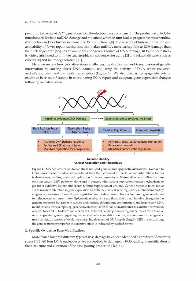
Int. J. Mol. Sci. 2019, 20, 364
proximity to the site of O2•− generation from the electron transport chain [6]. The production of ROS by
mitochondria leads to mtDNA damage and mutations which in turn lead to progressive mitochondrialdysfunction and to a further increase in ROS production [7,8]. The absence of histone protection andavailability of fewer repair mechanism also makes mtDNA more susceptible to ROS damage thanthe nuclear genome [6,9]. As an abundant endogenous source of DNA damage, ROS-induced stressis widely attributed to promote catastrophic consequences for aging [2] and related diseases such ascancer [10] and neurodegeneration [11].
Here we review how oxidative stress challenges the duplication and transmission of geneticinformation by causing direct DNA damage, regulating the activity of DNA repair enzymes,and altering basal and inducible transcription (Figure 1). We also discuss the epigenetic role ofoxidative base modifications in coordinating DNA repair and adequate gene expression changesfollowing oxidative stress.
Figure 1. Mechanisms of oxidative-stress-induced genetic and epigenetic alterations. Damage toDNA bases due to oxidative stress induced from the plethora of extracellular and intracellular factorsis deleterious, leading to stalled replication forks and mutations. Mammalian cells utilize the baseexcision repair (BER) pathway alone and in concert with various replication restart mechanisms toget rid of oxidative lesions and ensure faithful duplication of genome. Genetic response to oxidativestress involves alteration in gene expression by both the classical gene regulatory mechanisms and byepigenetic processes. Classical gene regulation implicates transcription-factor based gene regulationto influence gene transcription. Epigenetic mechanisms are those that do not involve changes in thegenome sequence, but rather in nuclear architecture, chromosome conformation, and histone and DNAmodifications. For example, epigenetic involvement of ROS has been attributed to oxidative conversionof 5-mC to 5-hmC. Oxidative conversion of G to 8-oxoG at the promoter regions activates expression ofredox-regulated genes suggesting that oxidative base modification may also represent an epigeneticmark serving as sensors of oxidative stress. Involvement of DNA repair (largely BER) in coordinatingthe gene regulatory response to oxidative stress is indicated by dashed arrow.
2. Specific Oxidative Base Modifications
More than a hundred different types of base damage have been identified as products of oxidativestress [12]. All four DNA nucleobases are susceptible to damage by ROS leading to modification oftheir structure and alteration of the base-pairing properties (Table 1).
280

Int. J. Mol. Sci. 2019, 20, 364
Table 1. DNA base modifications that commonly exist after oxidative stress.
DNA Base Oxidized Base Modification
Guanine (G)
8-oxo-7,8-dihydro-2′-deoxyguanosine (8-oxoG)8-oxoG is further oxidized to:SpiroiminodihydantoinGuanidinohydantoin2,6-diamino-4-hydroxy-5 formamidopyrimidine (FapyG)
Cytosine (C) 5-hydroxy-2′-deoxycytidine (OH5C)
Adenine (A)8-oxo-7,8-dihydro-2′-deoxyadenosine (8-oxoA)4,6-diamino-5-formamidopyrimidine (FapyA)2-hydroxydeoxyadenosine-5′-triphosphate (2OHA)
Thymine (T)Thymine glycol (Tg)5,6-dihydrothymine (DHT)5-hydroxymethyluracil (5hmU)
Guanine (G) is the most frequently oxidized base due to its low oxidation potential. As aresult, 8-oxo-7,8-dihydro-2′-deoxyguanosine (8-oxoG) is the most abundant oxidative DNA lesionwhich is moderately mutagenic resulting into G:C to T:A transversion and has been associatedwith cellular transformation and cancer initiation [13,14]. Structural studies showed that 8-oxoGinduces only minor distortions to the DNA helical structure that are localized near the modificationsite. The base pairing preference is determined by the conformation; anti-8oxoG base pairs withcytosine (C) whereas syn-8-oxoG functionally mimics thymine (T) and base-pairs with adenine(A) thus giving rise to A:8-oxoG mismatches which potentially results in CG→AT transversionmutations [15]. Replicative DNA polymerases are slowed down at 8-oxoG and insert both correctcytosine and incorrect adenine opposite 8-oxoG, but they preferentially extend A:8-oxoG mispairs.However, during replication events, the cells have an opportunity to utilize the translesion synthesis(TLS) polymerases, mainly the Y-family polymerases, for rapid bypass of 8-oxoG lesion to preventreplication fork arrest [16,17]. The 8-oxoG is also highly susceptible to further oxidative damage,yielding the additional mutagenic base lesions spiroiminodihydantoin and guanidinohydantoin.Oxidation of guanine also results in fragmentation of the purine imidazole ring leading to anothermajor oxidative lesion, 2,6-diamino-4-hydroxy-5-formamidopyrimidine (FapyG) [12,13]. Oxidationof adenine can lead to two major products: 8-oxo-7,8-dihydro-2′-deoxyadenosine (8-oxoA) and4,6-diamino-5-formamidopyrimidine (FapyA). A less prevalent adenine modification upon oxidativedamage is 2-hydroxydeoxyadenosine-5′-triphosphate (2OHA) [12,13].
Cytosine can be the target of oxidation only at the 5,6-double bond to form a major oxidativeproduct 5-hydroxy-2′-deoxycytidine (OH5C) which can be found on DNA spontaneously and afterexposure to ROS generating chemicals [12,18]. In contrast, a cytosine analogue, methylcytosine (5mC),can be attacked by free radicals at both the 5,6-double bond and the 5-methyl group and severaloxidation products of 5mC can be generated [12,18]. Thymine base can also be attacked by freeradicals on either the 5,6-double bond or the 5-methyl group, generating various oxidative products.Thymine glycol (Tg) is one of the most examined oxidative products generated by the ring openingon the 5,6-double bond of thymine causing inhibition to replicative polymerases and a mutagenicsignature indicative of translesion synthesis [17]. Moreover, thymine can be oxidized to produce the5,6-dihydrothymine (DHT) which despite being targeted for repair, does not appear to cause mutationsor cytotoxicity [18]. The free radical attack on the 5-methyl group of thymine produces numerousoxidation products including 5-hydroxymethyluracil (5hmU) which can base pair with both adenineand guanine, thus leading to T:A→C:G transition [19].
3. Repair of Oxidative DNA Damage
As such, cells regularly encounter a spectrum of DNA damage ranging from small non-helixdistorting lesions to bulkier adducts that cause significant structural changes to the DNA double
281

Int. J. Mol. Sci. 2019, 20, 364
helix. Base excision repair (BER) is the main repair pathway of the oxidatively generated 8-oxoG andother non-helix disturbing lesions [14,20–23]. Base excision repair essentially involves (i) excision of adamaged or inappropriate base by DNA glycosylase, (ii) incision of the phosphodiester backbone byapurinic/apyrimidinic (AP) endonuclease at the resulting abasic site creating a single-strand break(SSB), (iii) termini clean-up to permit unabated repair synthesis and/or nick ligation, (iv) gap-filling toreplace the excised nucleotide, and (v) sealing of the final, remaining nick. In addition to BER, otherDNA repair pathways including mismatch repair (MMR) and nucleotide excision repair (NER) alsocontribute to minimize the genotoxic impact of oxidative base lesions as summarized recently [24].
4. Oxidative DNA Damage and Replication Stress
The repair of oxidative DNA lesions is essential to avoid stress when cells enter the replicativephase of the cell cycle [25]. To shield the genome from oxidative damage, DNA replication in yeastis restricted to the reductive stage of the metabolic cycle when oxygen consumption is minimal [26].However, higher eukaryotes must deal with both physiological and pathological levels of ROS whileDNA synthesis is ongoing.
Oxidative lesions and BER intermediates interfere with replication, cause single- (SSB) anddouble-strand breaks (DSB) in DNA, and lead to chromosomal aberrations [27]. Unrepaired SSBs canstall replication machinery which may activate the error-prone damage tolerance mechanism or maylead to fork collapse into a potentially cytotoxic DSB [28]. In addition, closely spaced oxidative lesions,also referred to as oxidative clustered DNA lesions (OCDL), can be converted to DSBs during BER [29].
The single-strand DNA template at the replication fork is more susceptible to oxidative basedamage and strand breaks than the nonreplicating DNA [30]. Replication fork progression is blockedby BER-initiating lesions [31] as well as by the DNA structure intermediates arising from the repair ofoxidized bases [32]. The G-rich sequences at telomeres [5] and promoters that are known to accumulateoxidative DNA damage also display high rates of replication forks stalling [33]. One of the waysby which cells mitigate negative impact of elevated ROS on the replicating genome is by reducingreplication fork speed [34].
Mechanisms that play key roles in the reactivation of arrested replication forks may also act as abarrier against genetic instability triggered by the endogenous oxidative/replication stress axis [34,35].We have recently demonstrated that RecQ like 1 (RECQL1 or RECQ1) helicase which is critical forresetting of replication fork for resumption of normal DNA synthesis [36] is also important for BER [37].Using live in cell-repair assays and biochemical reconstitution, we identified that RECQ1 helicaseactivity and ERCC1-XPF endonuclease in cooperation with poly(ADP-ribose) polymerase (PARP1)and Replication Protein A (RPA) mediate a novel sub-pathway of conventional long-patch BER [37].This process is facilitated by the well-established interaction among RECQ1, PARP1, and RPA [37,38].Although RECQ1 modulates cellular response to oxidative stress [38], whether RECQ1 is requiredto sustain fork progression following oxidative stress is yet unknown. Nevertheless, physical andfunctional cooperation of DNA replication and BER is emerging as a major regulatory mechanism forpreventing genomic instability [39,40].
In addition to inducing DNA damage and nucleotide pool imbalance [35], oxidative stress canalter replication by oxidation induced inactivation of key DNA repair proteins such as RPA [41] orby modulating the levels of Ku70 and Ku80 proteins essential for DSB repair by non-homologousend joining [42]. Oxidative stress leads to activation of the ataxia-telangiectasia mutated (ATM)kinase, the major sensor and regulator of the cellular response to DSBs [43]. Downstream to oxidativestress-dependent activation, ATM protects cells from ROS accumulation by stimulating NADPHproduction and promoting the synthesis of nucleotides required for the repair of DSBs [44] and anumber of other processes to promote restoration of redox homeostasis [45]. Indeed, cells defective inDNA damage response show endogenously elevated levels of ROS [35].
282

Int. J. Mol. Sci. 2019, 20, 364
Collectively, these observations emphasize intricate mechanisms that coordinate replicationdynamics, activation of DNA damage response and DNA repair as directed by the redox status ofthe cell.
5. Oxidative DNA Damage and Gene Expression Changes
Cellular response to oxidative stress involves highly regulated alteration in gene expressionwhich is shared with gene expression patterns observed in aging [46], cancer [47], and otherdiseases [48,49]. The in vivo gene expression signature of oxidative stress suggests p53 plays animportant role and upregulation of p53 targets genes as a common response to oxidative stress acrossdiverse organs and species [50]. The cellular concentration of ROS appears to influence the selectiveactivation of transcription factors involved in signaling pathways including the nuclear factor erythroid2-related factor 2 (Nrf2), mitogen-activated protein (MAP) kinase/AP-1, and nuclear factor-kB (NF-kB)pathways, as well as hypoxia-inducible transcription factor 1α (HIF1A) [51]. Oxidative stress and redoxsignaling may also affect gene expression by altering the functions of histones and DNA modifyingenzymes [52].
The presence of 8-oxoG in the template strand would be expected to impair transcription bystalling of RNA pol II [53,54]; however, 8-oxoG in gene promoters is also associated with geneactivation [55,56]. Oxidation of bases may serve as critical sensors through which ROS signals aresensed and the transcription from the redox responsive genes is regulated [52,57]. Consistent withthis, the increased level of 8-oxoG in the mtDNA of mice lacking 8-oxoguanine DNA glycosylase(OGG1), the enzyme responsible for recognition and repair of 8-oxoG, is associated with differentialexpression of genes involved in ROS-mediated signaling including pro-inflammatory genes [58].In another study, ROS generated by tumor necrosis factor alfa (TNFα) exposure of human cells ledto OGG1 enrichment primarily at the regulatory regions of a large number of genes constitutingsignal transduction pathways that modulate redox-regulated metabolic and immune responses for animmediate global cellular response [59]. Studies from various groups have collectively suggested thatthe redox levels orchestrate OGG1 to play a role either in gene transcription or in lesion repair; and themagnitude of base lesions, predominantly of 8-oxoG, defines the fate of cells [24,60–62].
APE1 is another dual function protein involved both in the BER pathways of DNA lesions, actingas the major apurinic/apyrimidinic endonuclease (APE), and in eukaryotic transcriptional regulationof gene expression as a redox co-activator of several transcription factors including AP-1, HIF1-α,and p53 [63]. APE1 plays a role in the regulation of gene expression during oxidative stress conditionby interactions via its redox-effecter factor-1 (Ref-1) domain with protein factors such as HIF1-α,STAT3, and CBP/p300 that promote transcription [63]. Another study from Tell lab demonstrated thatAPE1-dependent and BER-mediated DNA repair promotes the initiation of transcription of sirtuin 1(SIRT1) gene upon oxidative DNA damage [64].
Although the precise mechanisms are yet unclear, recognition and binding of the oxidativelydamaged base by the repair proteins during the pre-excision step of BER facilitates the recruitmentof specific transcription factors for prompt transcriptional response [61]. Conceivably, oxidativestress-induced gene regulation may act in concert with the repair of DNA damage to protect cells fromaccumulation of oxidative damage.
6. Epigenetic Functions of Oxidative DNA Lesions
Localized formation of 8-oxoG in gene regulatory regions have been suggested to represent anepigenetic modification serving as sensors of oxidative stress. Despite the vulnerability of guanine tooxidation, over 70% of the promoters of human genes, including a large percentage of redox-responsivegene promoters, contain evolutionarily conserved G-rich clusters [65]. Guanine oxidation shows strongdistributional bias in the genome with gene promoter and untranslated regions harboring greater8-oxoG [66].
283

Int. J. Mol. Sci. 2019, 20, 364
Oxidative modification of guanine to 8-oxoG in the promoter may provide a platform for thecoordination of the initial steps of DNA repair, especially BER, and the assembly of the transcriptionalmachinery to launch the prompt and preferential expression of redox-regulated genes in cells that areresponding to oxidative stress [24,57,67,68]. Indeed, the formation of 8-oxoG in the G-rich promotersof the vascular endothelial growth factor (VEGF) [66,69], TNFα [70], and SIRT1 [64] genes can increasetranscription via the BER pathway [61,67].
Transcription of the VEGF gene is known to be regulated by a specific sequence motif in itspromoter, called the G4 motif because of its ability to form G-quadruplex (G4) DNA structure [71].Through complementary biochemical, cellular, and genetic approaches, the Burrows lab demonstratedthat the oxidation of guanine to 8-oxoG in the G-rich promoter element of the VEGF gene facilitatesactivation of transcription in a BER-dependent manner since the OGG1-null cells failed to exhibit anincrease in gene expression [67,69]. One of the suggested mechanisms is that oxidation of guanineto 8-oxoG in the G4 motif provides a structural switch for recruitment of BER proteins such as APE1and transcription factors such as HIF1-α to promote gene transcription [67,69]. Similar mechanismsimplicating other BER proteins and cooperating factors may operate for transcriptional activation ofother redox-regulated genes (Figure 2).
Figure 2. The influence of guanine oxidation at the promoter region on gene expression. Reactiveoxygen species (ROS) induces oxidation of guanine to 8-oxoG. Gene promoters are enriched in guanineand sequence motifs prone to form G4 DNA structures. Formation of 8-oxoG is also shown to inducecritical topological changes in DNA structure. Binding of 8-oxoG by BER proteins may facilitate thesite-specific recruitment of specific transcription factors, chromatin remodelers and other accessoryfactors (shown as ??). These factors likely work in concert to repair the oxidative base lesion (shown bygreen) and activate transcription of redox-regulated genes for an adequate cellular response.
Indeed, the G4 motifs (represented by G≥3NxG≥3NxG≥3NxG≥3) are enriched in the promoterregions of many genes [72]. Gene regulation by modulating the topological superstructures of G4containing promoters, for example VEGF as described above and endonuclease III-like protein 1(NTHL1) genes [67], suggest epigenetic role of 8-oxoG modification.
The regulatory and possible epigenetic roles of 8-oxoG in cells that are responding to oxidativestress can be contrasted with a more traditional 5-methylcytosine (5mC) epigenetic modificationcontributing to the regulation of gene activity during development and differentiation [73–75]. Cytosinemethylation is generally associated with repressed chromatin and inhibition of gene expression [76,77].The methyl moiety of 5mC can be eliminated passively during DNA replication, or actively throughenzymatic DNA demethylation [78]. Base excision repair is implicated in active demethylation of5mC in oxidation independent and dependent manner [78]. During active DNA demethylation,for activation of genes silenced by cytosine methylation, the ten-eleven translocation (TET) proteinsoxidize 5mC in a stepwise fashion to 5-hydroxymethylcytosine (5hmC), 5-formylcytosine (5fC),and 5-carboxylcytosine (5caC). Both 5fC and 5caC can be recognized and excised from DNA bythymine-DNA glycosylase (TDG) followed by subsequent filling in of unmodified cytosine by
284

Int. J. Mol. Sci. 2019, 20, 364
the BER pathway [79]. Moreover, passive elimination of 5mC is also enhanced by active DNAdemethylation [80].
Oxidative conversion of 5mC to 5hmC under oxidative stress changes the DNA methylationpattern resulting in epigenetic alterations [73]. Enrichment of 5hmC within the gene bodies, promoters,and transcription factor-binding regions suggest it may regulate gene expression by modulatingchromatin accessibility of the transcriptional machinery, or by inhibiting repressor binding [73]. Of note,readers of 5hmC include several DNA glycosylases (for example, NEIL1 and NEIL3), replication factors(RFC), helicases (for example, HELLS and RECQ1), and transcriptional repressor protein MeCP2 [76].MeCP2 recognizes methyl-CpG and recruits co-repressor molecules to silence transcription. Oxidationof guanine to 8-oxoG significantly inhibits MeCP2 DNA binding [81]. Proposedly, OGG1 may alleviatethe transcriptional repression by cytosine methylation [61]. By binding to 8-oxoG in the oppositestrand, OGG1 may interfere with the interaction of MeCP2 (and other proteins) with their substratesand recruit transcriptional machinery components to activate transcription [61]. Overall this suggestsan intertwined and DNA repair-involved DNA demethylation pathway for epigenetic regulation ofgene expression.
A recent study suggested that APE1 modulates DNA methyltransferase 1 (DNMT1) expressionand consequent promoter methylation in a redox-mediated manner [82]. These observations highlighta strong possibility that oxidative modification to DNA bases, such as in the form of 8-oxoG or oxidized5mC serve as epigenetic mark and function in a DNA-based mechanism for gene activation.
7. Conclusions and Outlook
Cellular redox status strongly impacts genome duplication and transmission. Therefore, it iscritical to understand how ROS-induced stress affects replication dynamics and activation of DNAdamage response, and how this is coordinated with the transcriptional response for the maintenanceof genomic stability and cellular homeostasis.
The impact of oxidative stress on genetic stability through direct damage to the DNA, suchas oxidized bases or abasic sites, has been documented extensively. As the primary mechanismcounteracting oxidative stress-induced DNA lesions, the BER pathway has been well characterized.Furthermore, activity of BER enzymes such as OGG1 is regulated in a redox-dependent manner [83,84]as well as by posttranslational modifications [85]. The interplay between chromatin status and BERis beginning to be unveiled [86,87], but the molecular mechanisms by which various DNA repairproteins, chromatin remodelers, and transcription factors are targeted to specific oxidative lesions areyet to be delineated. How chromatin remodeling influences BER of oxidative lesions and subsequentgene expression changes remains an exciting open question.
Given the demonstrated role of G4 structures in regulation of redox-sensitive gene expressionchanges [88], factors that modulate the stability of these structures are expected to play significantroles in the process. Interestingly, binding to G4 motifs in target gene promoters and resolutionof G4 DNA structures has been suggested as a mechanism of transcriptional regulation by DNAhelicases RECQ1 [89,90], XPB, XPD [91], BLM [92], and WRN [93]. However, in vitro biochemical datasuggests that the transcriptional regulation by RECQ1 likely does not involve RECQ1 helicase-mediatedunwinding of G4 structures [94]. A potential role of RECQ1 could be to mediate, either directly orthrough protein–protein interactions, repair of oxidative lesions [37,38] in the G4 motif at promotersand elsewhere and facilitate subsequent alterations in gene expression [89,90]. An important nextstep in understanding the molecular role of these helicases in the mechanisms of gene regulation is todetermine the involvement of cooperating transcriptional partners.
Collective data shows that the repair of oxidative DNA damage, a mechanism that protectsgenome integrity, also serves as a proactive mechanism to ensure a prompt and adequate transcriptionalprogram as governed by the cellular cues such as redox status [95]. Studies with APE1 and OGG1suggest that these DNA repair proteins can impact transcription of activator-dependent genes byfacilitating DNA repair, chromatin remodeling and assembly of transcriptional machinery at gene
285

Int. J. Mol. Sci. 2019, 20, 364
promoters, but their roles in constitutive housekeeping transcription is unclear. If 8-oxoG indeed servesas a regulatory mark, epigenetic regulation in this case likely relies on the oxidative DNA damage,possibly induced by the low level of endogenous ROS. If this is the case, then it will be importantto determine if the source of ROS, for example endogenous versus exogenous, by which 8-oxoG isintroduced in the genome dictates the transcriptional outcome in physiological and pathological states.
How prevalent is 8-oxoG-mediated gene regulation in the mammalian genome is unclear andthe epigenetic role of 8-oxoG is yet to be interrogated with respect to biological processes such asdifferentiation, development, tumorigenesis, and metastasis. If 8-oxoG is indeed a bona fide epigeneticmark, an additional consideration is whether oxidation of guanine to 8-oxoG is an active process.Towards this, targeted generation of 8-oxoG in the promoter regions can be coupled with enzymaticallycatalyzed oxidative demethylation of histones by the lysine demethylase (LSD1) as has been shown inthe estrogen receptor- and MYC-activated gene expression models [96,97]. It would be interesting todetermine the relationship between 5mC and 8-oxoG, and the roles of BER and other proteins.
Given the unavoidable exposure to ROS, cells seem to have evolved strategies to utilize ROS asbiological stimuli suitable for the physiological need. Oxidative base modifications, therefore, appearto have both beneficial and deleterious functions. While higher levels of oxidative damage mightinvoke the DNA repair mechanisms to remove the oxidative lesion, lower levels of oxidative damagemay serve to regulate gene expression to the degree required to maintain genomic integrity and cellularhomeostasis. The mechanism that enables cells to distinguish between “regulatory” oxidative DNAdamage from those that cause “undesirable” consequences is yet elusive. Further research is neededto gain a more complete understanding of the molecular details of cellular and genomic context thatdetermine whether to lose or use these stress marks.
Acknowledgments: Research in Sharma Lab was supported by the National Institute of General Medical Sciencesof the National Institutes of Health under Award Number SC1GM093999; National Science Foundation underAward Number NSF1832163. We also acknowledge support from the National Institute on Aging under AwardNumber 1R25 AG047843. B.B. is supported by a graduate student fellowship from Umm Al-Qura University,Saudi Arabia. The content is solely the responsibility of the authors and does not necessarily represent the officialviews of the funding agencies.
Conflicts of Interest: The authors declare no conflict of interest.
References
1. Winterbourn, C.C. Reconciling the chemistry and biology of reactive oxygen species. Nat. Chem. Biol. 2008,4, 278–286. [CrossRef]
2. Beckman, K.B.; Ames, B.N. The free radical theory of aging matures. Physiol. Rev. 1998, 78, 547–581.[CrossRef] [PubMed]
3. Klaunig, J.E.; Kamendulis, L.M.; Hocevar, B.A. Oxidative stress and oxidative damage in carcinogenesis.Toxicol. Pathol. 2010, 38, 96–109. [CrossRef]
4. Evans, M.D.; Cooke, M.S. Factors contributing to the outcome of oxidative damage to nucleic acids. Bioessays2004, 26, 533–542. [CrossRef]
5. Barnes, R.P.; Fouquerel, E.; Opresko, P.L. The impact of oxidative DNA damage and stress on telomerehomeostasis. Mech. Ageing Dev. 2018. [CrossRef] [PubMed]
6. Alexeyev, M.; Shokolenko, I.; Wilson, G.; LeDoux, S. The maintenance of mitochondrial DNAintegrity—Critical analysis and update. Cold Spring Harb. Perspect. Biol. 2013, 5, a012641. [CrossRef][PubMed]
7. Yakes, F.M.; Van Houten, B. Mitochondrial DNA damage is more extensive and persists longer than nuclearDNA damage in human cells following oxidative stress. Proc. Natl. Acad. Sci. USA 1997, 94, 514–519.[CrossRef]
8. Furda, A.M.; Marrangoni, A.M.; Lokshin, A.; Van Houten, B. Oxidants and not alkylating agents inducerapid mtDNA loss and mitochondrial dysfunction. DNA Repair 2012, 11, 684–692. [CrossRef]
9. Bohr, V.A.; Stevnsner, T.; de Souza-Pinto, N.C. Mitochondrial DNA repair of oxidative damage in mammaliancells. Gene 2002, 286, 127–134. [CrossRef]
286
Int. J. Mol. Sci. 2019, 20, 364
10. Klaunig, J.E.; Kamendulis, L.M. The role of oxidative stress in carcinogenesis. Annu. Rev. Pharmacol. Toxicol.2004, 44, 239–267. [CrossRef]
11. Kim, G.H.; Kim, J.E.; Rhie, S.J.; Yoon, S. The Role of Oxidative Stress in Neurodegenerative Diseases.Exp. Neurobiol. 2015, 24, 325–340. [CrossRef] [PubMed]
12. Cadet, J.; Wagner, J.R. DNA base damage by reactive oxygen species, oxidizing agents, and UV radiation.Cold Spring Harb. Perspect. Biol. 2013, 5. [CrossRef]
13. Whitaker, A.M.; Schaich, M.A.; Smith, M.R.; Flynn, T.S.; Freudenthal, B.D. Base excision repair of oxidativeDNA damage: From mechanism to disease. Front. Biosci. 2017, 22, 1493–1522.
14. Demple, B.; Harrison, L. Repair of oxidative damage to DNA: Enzymology and biology. Annu. Rev. Biochem.1994, 63, 915–948. [CrossRef] [PubMed]
15. Beard, W.A.; Batra, V.K.; Wilson, S.H. DNA polymerase structure-based insight on the mutagenic propertiesof 8-oxoguanine. Mutat. Res. 2010, 703, 18–23. [CrossRef] [PubMed]
16. Perry, J.J.P.; Hitomi, K.; Tainer, J.A. Flexibility Promotes Fidelity. Structure 2009, 17, 633–634. [CrossRef] [PubMed]17. McCulloch, S.D.; Kunkel, T.A. The fidelity of DNA synthesis by eukaryotic replicative and translesion
synthesis polymerases. Cell Res. 2008, 18, 148–161. [CrossRef] [PubMed]18. Bjelland, S.; Seeberg, E. Mutagenicity, toxicity and repair of DNA base damage induced by oxidation.
Mutat. Res. 2003, 531, 37–80. [CrossRef]19. Maiti, A.; Drohat, A.C. Thymine DNA glycosylase can rapidly excise 5-formylcytosine and 5-carboxylcytosine:
Potential implications for active demethylation of CpG sites. J. Biol. Chem. 2011, 286, 35334–35338. [CrossRef]20. Lindahl, T.; Wood, R.D. Quality control by DNA repair. Science 1999, 286, 1897–1905. [CrossRef]21. Wilson, D.M., 3rd; Bohr, V.A. The mechanics of base excision repair, and its relationship to aging and disease.
DNA Repair 2007, 6, 544–559. [CrossRef] [PubMed]22. Robertson, A.B.; Klungland, A.; Rognes, T.; Leiros, I. DNA repair in mammalian cells: Base excision repair:
The long and short of it. Cell Mol. Life Sci. 2009, 66, 981–993. [CrossRef]23. Dianov, G.L.; Hubscher, U. Mammalian base excision repair: The forgotten archangel. Nucleic Acids Res.
2013, 41, 3483–3490. [CrossRef] [PubMed]24. Markkanen, E. Not breathing is not an option: How to deal with oxidative DNA damage. DNA Repair 2017,
59, 82–105. [CrossRef] [PubMed]25. Van Loon, B.; Markkanen, E.; Hübscher, U. Oxygen as a friend and enemy: How to combat the mutational
potential of 8-oxo-guanine. DNA Repair 2010, 9, 604–616. [CrossRef] [PubMed]26. Chen, Z.; Odstrcil, E.A.; Tu, B.P.; McKnight, S.L. Restriction of DNA replication to the reductive phase of the
metabolic cycle protects genome integrity. Science 2007, 316, 1916–1919. [CrossRef]27. Ensminger, M.; Iloff, L.; Ebel, C.; Nikolova, T.; Kaina, B.; Lbrich, M. DNA breaks and chromosomal
aberrations arise when replication meets base excision repair. J. Cell Biol. 2014, 206, 29–43. [CrossRef]28. Caldecott, K.W. Single-strand break repair and genetic disease. Nat. Rev. Genet. 2008, 9, 619–631. [CrossRef]
[PubMed]29. Sharma, V.; Collins, L.B.; Chen, T.H.; Herr, N.; Takeda, S.; Sun, W.; Swenberg, J.A.; Nakamura, J. Oxidative
stress at low levels can induce clustered DNA lesions leading to NHEJ mediated mutations. Oncotarget 2016,7, 25377–25390. [CrossRef]
30. Klattenhoff, A.W.; Thakur, M.; Chu, C.S.; Ray, D.; Habib, S.L.; Kidane, D. Loss of NEIL3 DNA glycosylasemarkedly increases replication associated double strand breaks and enhances sensitivity to ATR inhibitor inglioblastoma cells. Oncotarget 2017, 8, 112942–112958. [CrossRef]
31. Groth, P.; Auslander, S.; Majumder, M.M.; Schultz, N.; Johansson, F.; Petermann, E.; Helleday, T. MethylatedDNA causes a physical block to replication forks independently of damage signalling, O(6)-methylguanineor DNA single-strand breaks and results in DNA damage. J. Mol. Biol. 2010, 402, 70–82. [CrossRef] [PubMed]
32. Rangaswamy, S.; Pandey, A.; Mitra, S.; Hegde, M.L. Pre-Replicative Repair of Oxidized Bases MaintainsFidelity in Mammalian Genomes: The Cowcatcher Role of NEIL1 DNA Glycosylase. Genes 2017, 8, 175.[CrossRef] [PubMed]
33. Tubbs, A.; Nussenzweig, A. Endogenous DNA Damage as a Source of Genomic Instability in Cancer. Cell2017, 168, 644–656. [CrossRef]
34. Somyajit, K.; Gupta, R.; Sedlackova, H.; Neelsen, K.J.; Ochs, F.; Rask, M.B.; Choudhary, C.; Lukas, J.Redox-sensitive alteration of replisome architecture safeguards genome integrity. Science 2017, 358, 797–802.[CrossRef] [PubMed]
287
Int. J. Mol. Sci. 2019, 20, 364
35. Wilhelm, T.; Ragu, S.; Magdalou, I.; Machon, C.; Dardillac, E.; Techer, H.; Guitton, J.; Debatisse, M.; Lopez, B.S.Slow Replication Fork Velocity of Homologous Recombination-Defective Cells Results from EndogenousOxidative Stress. PLoS Genet. 2016, 12, e1006007. [CrossRef] [PubMed]
36. Sami, F.; Sharma, S. Probing Genome Maintenance Functions of human RECQ. Comput. Struct. Biotechnol. J.2013, 6, e201303014.
37. Woodrick, J.; Gupta, S.; Camacho, S.; Parvathaneni, S.; Choudhury, S.; Cheema, A.; Bai, Y.; Khatkar, P.;Erkizan, H.V.; Sami, F.; et al. A new sub-pathway of long-patch base excision repair involving 5′ gapformation. EMBO J. 2017, 36, 1605–1622. [CrossRef]
38. Sharma, S.; Phatak, P.; Stortchevoi, A.; Jasin, M.; Larocque, J.R. RECQ1 plays a distinct role in cellularresponse to oxidative DNA damage. DNA Repair 2012, 11, 537–549. [CrossRef]
39. Hegde, M.L.; Hegde, P.M.; Bellot, L.J.; Mandal, S.M.; Hazra, T.K.; Li, G.M.; Boldogh, I.; Tomkinson, A.E.;Mitra, S. Prereplicative repair of oxidized bases in the human genome is mediated by NEIL1 DNA glycosylasetogether with replication proteins. Proc. Natl. Acad. Sci. USA 2013, 110, E3090–E3099. [CrossRef]
40. Parlanti, E.; Locatelli, G.; Maga, G.; Dogliotti, E. Human base excision repair complex is physically associatedto DNA replication and cell cycle regulatory proteins. Nucleic Acids Res. 2007, 35, 1569–1577. [CrossRef]
41. Guven, M.; Brem, R.; Macpherson, P.; Peacock, M.; Karran, P. Oxidative Damage to RPA Limits the NucleotideExcision Repair Capacity of Human Cells. J. Invest. Dermatol. 2015, 135, 2834–2841. [CrossRef] [PubMed]
42. Song, J.Y.; Lim, J.W.; Kim, H.; Morio, T.; Kim, K.H. Oxidative Stress Induces Nuclear Loss of DNARepair Proteins Ku70 and Ku80 and Apoptosis in Pancreatic Acinar AR42J Cells. J. Biol. Chem. 2003,278, 36676–36687. [CrossRef] [PubMed]
43. Guo, Z.; Deshpande, R.; Paull, T.T. ATM activation in the presence of oxidative stress. Cell Cycle 2010, 9,4805–4811. [CrossRef] [PubMed]
44. Cosentino, C.; Grieco, D.; Costanzo, V. ATM activates the pentose phosphate pathway promoting anti-oxidantdefence and DNA repair. EMBO J. 2011, 30, 546–555. [CrossRef]
45. Ditch, S.; Paull, T.T. The ATM protein kinase and cellular redox signaling: Beyond the DNA damage response.Trends Biochem. Sci. 2012, 37, 15–22. [CrossRef] [PubMed]
46. Frenk, S.; Houseley, J. Gene expression hallmarks of cellular ageing. Biogerontology 2018, 19, 547–566.[CrossRef] [PubMed]
47. Leone, A.; Roca, M.S.; Ciardiello, C.; Costantini, S.; Budillon, A. Oxidative Stress Gene Expression ProfileCorrelates with Cancer Patient Poor Prognosis: Identification of Crucial Pathways Might Select NovelTherapeutic Approaches. Oxid. Med. Cell. Longev. 2017, 2017, 2597581. [CrossRef]
48. Reuter, S.; Gupta, S.C.; Chaturvedi, M.M.; Aggarwal, B.B. Oxidative stress, inflammation, and cancer: Howare they linked? Free Radic. Biol. Med. 2010, 49, 1603–1616. [CrossRef] [PubMed]
49. Shi, Q.; Gibson, G.E. Oxidative stress and transcriptional regulation in Alzheimer disease. Alzheimer Dis.Assoc. Disord. 2007, 21, 276–291. [CrossRef]
50. Han, E.S.; Muller, F.L.; Perez, V.I.; Qi, W.; Liang, H.; Xi, L.; Fu, C.; Doyle, E.; Hickey, M.; Cornell, J.; et al. Thein vivo gene expression signature of oxidative stress. Physiol. Genom. 2008, 34, 112–126. [CrossRef]
51. Schieber, M.; Chandel, N.S. ROS function in redox signaling and oxidative stress. Curr. Biol. 2014, 24,R453–R462. [CrossRef]
52. Mikhed, Y.; Gorlach, A.; Knaus, U.G.; Daiber, A. Redox regulation of genome stability by effects on geneexpression, epigenetic pathways and DNA damage/repair. Redox Biol. 2015, 5, 275–289. [CrossRef]
53. Allgayer, J.; Kitsera, N.; Bartelt, S.; Epe, B.; Khobta, A. Widespread transcriptional gene inactivation initiatedby a repair intermediate of 8-oxoguanine. Nucleic Acids Res. 2016, 44, 7267–7280. [CrossRef] [PubMed]
54. Kolbanovskiy, M.; Chowdhury, M.A.; Nadkarni, A.; Broyde, S.; Geacintov, N.E.; Scicchitano, D.A.;Shafirovich, V. The Nonbulky DNA Lesions Spiroiminodihydantoin and 5-Guanidinohydantoin SignificantlyBlock Human RNA Polymerase II Elongation in Vitro. Biochemistry 2017, 56, 3008–3018. [CrossRef] [PubMed]
55. Piccirillo, S.; Filomeni, G.; Brune, B.; Rotilio, G.; Ciriolo, M.R. Redox mechanisms involved in the selectiveactivation of Nrf2-mediated resistance versus p53-dependent apoptosis in adenocarcinoma cells. J. Biol.Chem. 2009, 284, 27721–27733. [CrossRef] [PubMed]
56. Boldogh, I.; Hajas, G.; Aguilera-Aguirre, L.; Hegde, M.L.; Radak, Z.; Bacsi, A.; Sur, S.; Hazra, T.K.; Mitra, S.Activation of ras signaling pathway by 8-oxoguanine DNA glycosylase bound to its excision product,8-oxoguanine. J. Biol. Chem. 2012, 287, 20769–20773. [CrossRef] [PubMed]
288

Int. J. Mol. Sci. 2019, 20, 364
57. Seifermann, M.; Epe, B. Oxidatively generated base modifications in DNA: Not only carcinogenic risk factorbut also regulatory mark? Free Radic. Biol. Med. 2017, 107, 258–265. [CrossRef]
58. Aguilera-Aguirre, L.; Hosoki, K.; Bacsi, A.; Radak, Z.; Sur, S.; Hegde, M.L.; Tian, B.; Saavedra-Molina, A.;Brasier, A.R.; Ba, X.; et al. Whole transcriptome analysis reveals a role for OGG1-initiated DNA repairsignaling in airway remodeling. Free Radic. Biol. Med. 2015, 89, 20–33. [CrossRef]
59. Hao, W.; Qi, T.; Pan, L.; Wang, R.; Zhu, B.; Aguilera-Aguirre, L.; Radak, Z.; Hazra, T.K.; Vlahopoulos, S.A.;Bacsi, A.; et al. Effects of the stimuli-dependent enrichment of 8-oxoguanine DNA glycosylase1 onchromatinized DNA. Redox Biol. 2018, 18, 43–53. [CrossRef]
60. Cyr, A.R.; Domann, F.E. The redox basis of epigenetic modifications: From mechanisms to functionalconsequences. Antioxid. Redox Signal. 2011, 15, 551–589. [CrossRef]
61. Ba, X.; Boldogh, I. 8-Oxoguanine DNA glycosylase 1: Beyond repair of the oxidatively modified base lesions.Redox Biol. 2018, 14, 669–678. [CrossRef] [PubMed]
62. Wang, R.; Li, C.; Qiao, P.; Xue, Y.; Zheng, X.; Chen, H.; Zeng, X.; Liu, W.; Boldogh, I.; Ba, X. OGG1-initiatedbase excision repair exacerbates oxidative stress-induced parthanatos. Cell. Death Dis. 2018, 9, 628. [CrossRef]
63. Bhakat, K.K.; Mantha, A.K.; Mitra, S. Transcriptional regulatory functions of mammalian AP-endonuclease(APE1/Ref-1), an essential multifunctional protein. Antioxid. Redox Signal. 2009, 11, 621–637. [CrossRef][PubMed]
64. Antoniali, G.; Lirussi, L.; D’Ambrosio, C.; Dal Piaz, F.; Vascotto, C.; Casarano, E.; Marasco, D.; Scaloni, A.;Fogolari, F.; Tell, G. SIRT1 gene expression upon genotoxic damage is regulated by APE1 throughnCaRE-promoter elements. Mol. Biol. Cell 2014, 25, 532–547. [CrossRef] [PubMed]
65. Deaton, A.M.; Bird, A. CpG islands and the regulation of transcription. Genes Dev. 2011, 25, 1010–1022.[CrossRef] [PubMed]
66. Pastukh, V.; Roberts, J.T.; Clark, D.W.; Bardwell, G.C.; Patel, M.; Al-Mehdi, A.B.; Borchert, G.M.;Gillespie, M.N. An oxidative DNA “damage” and repair mechanism localized in the VEGF promoteris important for hypoxia-induced VEGF mRNA expression. Am. J. Physiol. Lung Cell. Mol. Physiol. 2015, 309,L1367–L1375. [CrossRef]
67. Fleming, A.M.; Burrows, C.J. 8-Oxo-7,8-dihydroguanine, friend and foe: Epigenetic-like regulator versusinitiator of mutagenesis. DNA Repair 2017, 56, 75–83. [CrossRef]
68. Moore, S.P.; Toomire, K.J.; Strauss, P.R. DNA modifications repaired by base excision repair are epigenetic.DNA Repair 2013, 12, 1152–1158. [CrossRef]
69. Fleming, A.M.; Ding, Y.; Burrows, C.J. Oxidative DNA damage is epigenetic by regulating gene transcriptionvia base excision repair. Proc. Natl. Acad. Sci. USA 2017, 114, 2604–2609. [CrossRef]
70. Ba, X.; Bacsi, A.; Luo, J.; Aguilera-Aguirre, L.; Zeng, X.; Radak, Z.; Brasier, A.R.; Boldogh, I. 8-oxoguanineDNA glycosylase-1 augments proinflammatory gene expression by facilitating the recruitment of site-specifictranscription factors. J. Immunol. 2014, 192, 2384–2394. [CrossRef]
71. Sun, D.; Liu, W.J.; Guo, K.; Rusche, J.J.; Ebbinghaus, S.; Gokhale, V.; Hurley, L.H. The proximal promoterregion of the human vascular endothelial growth factor gene has a G-quadruplex structure that can betargeted by G-quadruplex-interactive agents. Mol. Cancer Ther. 2008, 7, 880–889. [CrossRef] [PubMed]
72. Rhodes, D.; Lipps, H.J. G-quadruplexes and their regulatory roles in biology. Nucleic Acids Res. 2015, 43,8627–8637. [CrossRef]
73. Shi, D.Q.; Ali, I.; Tang, J.; Yang, W.C. New Insights into 5hmC DNA Modification: Generation, Distributionand Function. Front. Genet. 2017, 8, 100. [CrossRef] [PubMed]
74. Menezo, Y.J.; Silvestris, E.; Dale, B.; Elder, K. Oxidative stress and alterations in DNA methylation: Two sidesof the same coin in reproduction. Reprod. Biomed. Online 2016, 33, 668–683. [CrossRef] [PubMed]
75. Lister, R.; Mukamel, E.A.; Nery, J.R.; Urich, M.; Puddifoot, C.A.; Johnson, N.D.; Lucero, J.; Huang, Y.;Dwork, A.J.; Schultz, M.D.; et al. Global epigenomic reconfiguration during mammalian brain development.Science 2013, 341, 1237905. [CrossRef]
76. Spruijt, C.G.; Gnerlich, F.; Smits, A.H.; Pfaffeneder, T.; Jansen, P.W.; Bauer, C.; Munzel, M.; Wagner, M.;Muller, M.; Khan, F.; et al. Dynamic readers for 5-(hydroxy)methylcytosine and its oxidized derivatives. Cell2013, 152, 1146–1159. [CrossRef] [PubMed]
77. Klose, R.J.; Bird, A.P. Genomic DNA methylation: The mark and its mediators. Trends Biochem. Sci. 2006, 31,89–97. [CrossRef]
289

Int. J. Mol. Sci. 2019, 20, 364
78. Wu, H.; Zhang, Y. Reversing DNA methylation: Mechanisms, genomics, and biological functions. Cell 2014,156, 45–68. [CrossRef]
79. He, Y.F.; Li, B.Z.; Li, Z.; Liu, P.; Wang, Y.; Tang, Q.; Ding, J.; Jia, Y.; Chen, Z.; Li, L.; et al. Tet-mediated formationof 5-carboxylcytosine and its excision by TDG in mammalian DNA. Science 2011, 333, 1303–1307. [CrossRef]
80. Ji, D.; Lin, K.; Song, J.; Wang, Y. Effects of Tet-induced oxidation products of 5-methylcytosine on Dnmt1-and DNMT3a-mediated cytosine methylation. Mol. Biosyst. 2014, 10, 1749–1752. [CrossRef]
81. Valinluck, V.; Sowers, L.C. Endogenous cytosine damage products alter the site selectivity of human DNAmaintenance methyltransferase DNMT1. Cancer Res. 2007, 67, 946–950. [CrossRef]
82. Chen, G.; Chen, J.; Yan, Z.; Li, Z.; Yu, M.; Guo, W.; Tian, W. Maternal diabetes modulates dental epithelial stemcells proliferation and self-renewal in offspring through apurinic/apyrimidinicendonuclease 1-mediatedDNA methylation. Sci. Rep. 2017, 7, 40762. [CrossRef]
83. Bravard, A.; Vacher, M.; Gouget, B.; Coutant, A.; de Boisferon, F.H.; Marsin, S.; Chevillard, S.; Radicella, J.P.Redox regulation of human OGG1 activity in response to cellular oxidative stress. Mol. Cell. Biol. 2006, 26,7430–7436. [CrossRef] [PubMed]
84. Bravard, A.; Campalans, A.; Vacher, M.; Gouget, B.; Levalois, C.; Chevillard, S.; Radicella, J.P. Inactivation byoxidation and recruitment into stress granules of hOGG1 but not APE1 in human cells exposed to sub-lethalconcentrations of cadmium. Mutat. Res. 2010, 685, 61–69. [CrossRef] [PubMed]
85. Bhakat, K.K.; Mokkapati, S.K.; Boldogh, I.; Hazra, T.K.; Mitra, S. Acetylation of human 8-oxoguanine-DNAglycosylase by p300 and its role in 8-oxoguanine repair in vivo. Mol. Cell. Biol. 2006, 26, 1654–1665.[CrossRef] [PubMed]
86. Hinz, J.M.; Czaja, W. Facilitation of base excision repair by chromatin remodeling. DNA Repair 2015, 36,91–97. [CrossRef]
87. Balliano, A.J.; Hayes, J.J. Base excision repair in chromatin: Insights from reconstituted systems. DNA Repair2015, 36, 77–85. [CrossRef]
88. Fedeles, B.I. G-quadruplex-forming promoter sequences enable transcriptional activation in response tooxidative stress. Proc. Natl. Acad. Sci. USA 2017, 114, 2788–2790. [CrossRef]
89. Li, X.L.; Lu, X.; Parvathaneni, S.; Bilke, S.; Zhang, H.; Thangavel, S.; Vindigni, A.; Hara, T.; Zhu, Y.;Meltzer, P.S.; et al. Identification of RECQ1-regulated transcriptome uncovers a role of RECQ1 in regulationof cancer cell migration and invasion. Cell Cycle 2014, 13, 2431–2445. [CrossRef]
90. Lu, X.; Parvathaneni, S.; Li, X.L.; Lal, A.; Sharma, S. Transcriptome guided identification of novel functionsof RECQ1 helicase. Methods 2016, 108, 111–117. [CrossRef]
91. Gray, L.T.; Vallur, A.C.; Eddy, J.; Maizels, N. G quadruplexes are genomewide targets of transcriptionalhelicases XPB and XPD. Nat. Chem. Biol. 2014, 10, 313–318. [CrossRef]
92. Nguyen, G.H.; Tang, W.; Robles, A.I.; Beyer, R.P.; Gray, L.T.; Welsh, J.A.; Schetter, A.J.; Kumamoto, K.;Wang, X.W.; Hickson, I.D.; et al. Regulation of gene expression by the BLM helicase correlates with thepresence of G-quadruplex DNA motifs. Proc. Natl. Acad. Sci. USA 2014, 111, 9905–9910. [CrossRef]
93. Tang, W.; Robles, A.I.; Beyer, R.P.; Gray, L.T.; Nguyen, G.H.; Oshima, J.; Maizels, N.; Harris, C.C.; Monnat, R.J.Werner syndrome helicase modulates G4 DNA-dependent transcription and opposes mechanistically distinctsenescence-associated gene expression programs. bioRxiv 2015. [CrossRef]
94. Popuri, V.; Bachrati, C.Z.; Muzzolini, L.; Mosedale, G.; Costantini, S.; Giacomini, E.; Hickson, I.D.; Vindigni, A.The Human RecQ helicases, BLM and RECQ1, display distinct DNA substrate specificities. J. Biol. Chem.2008, 283, 17766–17776. [CrossRef] [PubMed]
95. Fong, Y.W.; Cattoglio, C.; Tjian, R. The Intertwined Roles of Transcription and Repair Proteins. Mol. Cell2013, 52, 291–302. [CrossRef]
96. Amente, S.; Bertoni, A.; Morano, A.; Lania, L.; Avvedimento, E.V.; Majello, B. LSD1-mediated demethylation ofhistone H3 lysine 4 triggers Myc-induced transcription. Oncogene 2010, 29, 3691–3702. [CrossRef] [PubMed]
97. Perillo, B.; Ombra, M.N.; Bertoni, A.; Cuozzo, C.; Sacchetti, S.; Sasso, A.; Chiariotti, L.; Malorni, A.;Abbondanza, C.; Avvedimento, E.V. DNA oxidation as triggered by H3K9me2 demethylation drivesestrogen-induced gene expression. Science 2008, 319, 202–206. [CrossRef]
© 2019 by the authors. Licensee MDPI, Basel, Switzerland. This article is an open accessarticle distributed under the terms and conditions of the Creative Commons Attribution(CC BY) license (http://creativecommons.org/licenses/by/4.0/).
290
International Journal of
Molecular Sciences
Review
Ageing, Cellular Senescence andNeurodegenerative Disease
Marios Kritsilis, Sophia V. Rizou, Paraskevi N. Koutsoudaki, Konstantinos Evangelou,
Vassilis G. Gorgoulis *,† and Dimitrios Papadopoulos *,†
Laboratory of Histology & Embryology, Medical School, National and Kapodistrian University of Athens,75 Mikras Asias Street, Goudi, 115-27 Athens, Greece; [email protected] (M.K.);[email protected] (S.V.R.); [email protected] (P.N.K.); [email protected] (K.E.)* Correspondence: [email protected] (V.G.G.); [email protected] (D.P.);
Tel.: +30-210-746-2352 (V.G.G.)† These authors share equal credit for senior authorship.
Received: 31 August 2018; Accepted: 19 September 2018; Published: 27 September 2018
Abstract: Ageing is a major risk factor for developing many neurodegenerative diseases. Cellularsenescence is a homeostatic biological process that has a key role in driving ageing. There is evidencethat senescent cells accumulate in the nervous system with ageing and neurodegenerative diseaseand may predispose a person to the appearance of a neurodegenerative condition or may aggravateits course. Research into senescence has long been hindered by its variable and cell-type specificfeatures and the lack of a universal marker to unequivocally detect senescent cells. Recent advancesin senescence markers and genetically modified animal models have boosted our knowledge on therole of cellular senescence in ageing and age-related disease. The aim now is to fully elucidate its rolein neurodegeneration in order to efficiently and safely exploit cellular senescence as a therapeutictarget. Here, we review evidence of cellular senescence in neurons and glial cells and we discussits putative role in Alzheimer’s disease, Parkinson’s disease and multiple sclerosis and we provide,for the first time, evidence of senescence in neurons and glia in multiple sclerosis, using the novelGL13 lipofuscin stain as a marker of cellular senescence.
Keywords: neurodegeneration; cellular senescence; ageing; Alzheimer’s disease; multiple sclerosis;Parkinson’s disease; lipofuscin; SenTraGorTM (GL13); senolytics
1. Ageing and Neurodegeneration
Ageing is a universal process characterized by the accumulation of biological changes that lead tothe organism’s functional decline over time. Human ageing is accompanied by a gradual build-up ofcognitive and physical impairment and an increased risk of developing numerous diseases includingcancer, diabetes, cardiovascular, musculoskeletal and neurodegenerative conditions. Age-relateddisability and morbidity adversely affect the quality of life; they are ultimately associated with anincreased risk of death and bear dire consequences for the individual, the family and society.
Ageing is the most important risk factor for the development of neurodegenerative disease andtypically, most neurodegenerative disorders manifest in the elderly [1]. The annual incidence ofAlzheimer’s disease (AD) has been shown to increase exponentially with advancing age [2,3]. Notably,Down syndrome, a progeroid condition, has been associated with AD, and mouse models of prematureageing have been reported to overproduce Aβ and show impaired learning and memory [4–7].Incidence of Parkinson’s disease (PD), the second most common age-related neurodegenerativecondition also increases with age [8,9]. The great majority of AD and PD cases are sporadic andtypically manifest at a much older age than hereditary ones. Despite the differences in pathologyamong the two conditions, they are both typical neurodegenerative diseases characterized by chronic
Int. J. Mol. Sci. 2018, 19, 2937; doi:10.3390/ijms19102937 www.mdpi.com/journal/ijms291

Int. J. Mol. Sci. 2018, 19, 2937
progressive loss of neurons and their synaptic connections manifesting with gradual functionaldecline [4]. But age is a recognized risk factor even for inflammatory demyelinating conditions such asmultiple sclerosis (MS), which also has a strong neurodegenerative component [10]. Age is the strongestpredictor for the transition from the relapsing phase of MS, which is primarily inflammatory to thesecondary progressive phase of the disease, which is thought to be mainly neurodegenerative [10,11].
Although research on the biology of mammalian ageing has recently attracted much attention,our understanding of its underlying mechanisms remains poor. It has been hypothesized that failureof repair mechanisms leads to accumulation of cellular and molecular damage that drives ageing [12].Accumulating damage is thought to occur inherently in a random manner, which explains the greatdiversity in ageing phenotypes, even in monozygotic twins [13]. The interplay among the geneticbackground, environmental factors and the stochastic nature of age-related accumulation of irreparabledamage to the DNA of the organism may also determine the likelihood of developing a particularage-related disease. Genomic instability, telomere attrition, loss of proteostasis, dysregulated nutrientsensing, mitochondrial dysfunction, stem cell exhaustion, altered cellular communication and excessivecellular senescence have all been recognized as hallmarks of ageing [14]. Cellular senescence is aprocess triggered by irreparable DNA damage that underlies normal ageing. Senescent cells becomemore abundant with ageing and a growing body of evidence suggests that their accumulation maycontribute to pathogenesis of age-related diseases. Here, we review the data that support a role forcellular senescence in neurodegeneration, with special focus on AD, PD and MS.
2. Cellular Senescence
Cellular senescence is a homeostatic response aiming to prevent the propagation of damagedcells and neoplastic transformation [15]. Apart from its beneficial role as an anti-tumour response,physiological roles for cellular senescence have also been identified during development [15,16],in adult megakaryocytes, syncytiotrophoblasts, wound healing and placental natural killerlymphocytes [17–19]. However, several lines of evidence indicate that cellular senescence alsocontributes to the loss of function associated with ageing and age-related disease [20]. Accordingto the original observations by Hayflick and Moorhead (1961), when cultures of normal humanfibroblasts were passaged serially they underwent stable cell cycle arrest that was accompanied bystereotypical phenotypic changes [21]. This form of cellular senescence, termed replicative senescenceconstitutes a particular type of cellular senescence and is associated with telomere shortening withsuccessive cell cycles. Nevertheless, besides telomere shortening, there are many more triggers ofcellular senescence including aberrant oncogene activation (oncogene-induced senescence-OIS) [22],stress-induced (stress-induced premature senescence-SIPS) due to oxidative stress [23], ionizingradiation [24], DNA-damaging chemotherapy [25], hyperoxia [26], impaired autophagy [27] or otherstressors and mitochondrial dysfunction [28]. Most of these triggers lead to telomeric or non-telomericDNA damage or altered chromatin structure and typically activate the DNA damage response(DDR) [29,30], although cellular senescence in vitro may also occur without detectable DDR [31,32].When the cell’s repair mechanisms become overwhelmed, the activated DDR elicits cellular senescencevia phosphorylation of p53 [33].
Unlike apoptosis, senescent cells remain viable and metabolically active [30]. Although senescentcells can be recognized by T helper cells and cleared by macrophages and natural killerlymphocytes [27,34,35], their number has been shown to increase with normal ageing in tissues ofhumans, primates and rodents [36,37]. Models of accelerated cellular senescence show prematureageing and increased incidence of age-related pathologies, suggesting that accumulating senescentcells contributes to ageing-related functional compromise and predisposes to age-related disease [38].The features of senescent cells that constitute the senescent phenotype may be responsible for theirputative detrimental effects in ageing and ageing- associated neurodegenerative disease.
292
Int. J. Mol. Sci. 2018, 19, 2937
3. The Senescence Phenotype
Senescent cells exhibit is a multitude of cellular and molecular changes that are neither specificnor pathognomonic of the senescent state. Evidence suggests that the cellular and molecular featuresof senescence depend on both the triggering stimulus and the affected cell type [23]. Although thesenescence phenotype of nervous system cells has not been extensively studied, the key features ofsenescence are described below:
Typically, senescent cells exhibit permanent cell cycle arrest, which is thought to be regulated byp16INK4A and p53-p21-RB (retinoblastoma). Increased expression of p53 upregulates expression ofCDKi(cyclin-dependent kinase inhibitor) p21, which initially arrests the cell cycle. p16INK4A mediatespermanent cells cycle arrest by inhibiting CDK4 and CDK6, which leads to RB hypophosphorylationand blocks entry to S phase [39,40].
Another key feature of cellular senescence is the senescence-associated secretory phenotype(SASP), which is dependent on p38MAPK (p38 mitogen-activated protein kinases), NF-κB(nuclear factor kappa-light-chain-enhancer of activated B cells), cGAS(cyclic GMP-AMPsynthase)/STING(stimulator of IFN genes), NOTCH and mTORmammalian target of rapamycin)signalling [38–42]. SASP consists of chemokines, cytokines, growth factors and metaloproteinases [43].These factors are primarily proinflammatory and act in a paracrine and autocrine manner [44,45],although immunosuppressive mediators have also been identified as part of SASP [46,47]. Recent dataindicate that SASP may also involve small extracellular vesicles in a p53-dependent manner [48–50].
Cellular senescence is also characterized by resistance to apoptotic death, which appears to belargely controlled by the p53 stress response pathway. Both p53 levels and p53 post-translationalmodifications [51] seem to have a role in determining the senescent cell fate while conferring resistanceto apoptosis. Accumulation of intermediate levels of p53 has been reported to favor the expressionof anti-apoptotic bcl-2 family proteins [52]. Nonetheless, p21 has also been shown to be capable todirectly inhibit caspase 3 and apoptosis [53].
Cellular senescence is associated with changes in cellular metabolism. These include upregulationof lysosomal senescence-associated β-galactosidase, a shift from oxidative phosphorylation toglycolysis [54,55] and accumulation of lipofuscin in the cytoplasm [56,57]. Lipofuscin accumulation hasbeen reported as a key feature of cellular senescence that can be used to positively identify senescentcells [57,58]. A recent metabolomic analysis of cultured doxorubicin-treated breast cancer cells revealedthat tricarboxylic acid cycle, pentose phosphate pathway, and nucleotide synthesis pathways weresignificantly upregulated, whereas fatty acid synthesis was reduced [59].
Altered mitochondrial function appears essential in mediating the senescence phenotype. RNAsequencing analysis has shown that a great number of senescence-associated changes involvethe mitochondria and Akt (protein kinsase B), ATM (ataxia-telangiectasia mutated) and mTORC1phosphorylation have been shown to link DDR with mitochondrial biogenesis [60]. Morphologicalchanges in mitochondria are also seen in senescent cells [20,61]. In addition, impaired mitophagyseems to explain accumulation of dysfunctional mitochondria (senescence-associated mitochondrialdysfunction—SAMD) seen in cellular senescence [62,63]. Although mitochondria are not thesole source [63], they are a major generator of ROS, important for both cellular signaling andSASP [47,64–66].
Several epigenetic modifications are also common in cellular senescence. Defects in pericentricheterochromatic silencing at mammalian centromeres, normally regulated by SIRT6 (sirtuin 6) havebeen described [67]. SIRT6 belongs to the sirtuin protein family, whose function has been linked tolongevity [68]. Micro-RNAs, a subclass of regulatory, non-coding RNAs that participate in regulationof cellular senescence may also be epigenetically modified [69]. Chromatic alterations such assenescence-associated heterochromatin formation (SAHF) may accompany cellular senescence in somesettings with deactivation of proliferation-related genes [70–73]. Cellular senescence may also lead tochanges in the organization of nuclear lamina and down regulation of lamin B1 with implications fornuclear morphology and gene expression [74].
293

Int. J. Mol. Sci. 2018, 19, 2937
Finally, senescent cells also undergo morphological changes. Cells become larger and flattened outand acquire an irregular shape. These alterations are more prominent in vitro than in vivo and appearto be caused by cytoskeletal rearrangements [75,76] and changes in cell membrane composition [77].Senescent cells exhibit increased unfolded protein response (UPR), indicative of endoplasmic reticulum(ER) stress [76,77]. The ATF6α pathway of UPR appears to be responsible for both increasing ER sizeassociated with ER stress and regulating the shape and size of senescent cells [76,77].
4. Markers of Cellular Senescence
To better understand the role of senescent cells in physiological and pathological conditions, it isessential to be able to detect them in vitro and in vivo [15]. So far, research on cellular senescence hasbeen hindered by our lack a universal, specific and widely applicable marker of cellular senescenceHere, we discuss the most commonly used markers of senescence:
• p16INK4a: This member of the INK4a family is a cyclin D-dependent kinase CDK4 and CDK6inhibitor, which prevents the phosphorylation of the retinoblastoma protein (Rb), therefore leadingto suspension of the cell cycle before the S-phase [78,79]. Increased levels of p16INK4a have beendocumented in aged and stressed tissues, compared to younger, healthy tissues, whereas theremoval of p16INK4a-expressing senescent cells in mice prevented or delayed tissue dysfunctionand age-related disorders [38,80]. This evidence has established p16INK4a as a widely-acceptedmarker of ageing and cellular senescence [81,82]. Limitations include poor detection of p16INK4a
in mice by the currently available antibodies using immunohistochemistry [79], as well assome situations, where p16INK4a levels are increased, in the absence of other signs of cellularsenescence [81,83,84].
• p21CIP1/WAF1/SDI1: p21 is a member of the second group of CDK inhibitors, namely the CIP/KIP(CDK interacting protein/kinase inhibitory protein) family and can inhibit a variety of CDKs [85].Apart from its role in cellular senescence, it is a key mediator in several biological functions,including cell cycle arrest, cell death, DNA repair processes and even reprogramming ofdifferentiated somatic cells into pluripotent stem cells [86]. In the context of senescence,stress-induced p53 activates p21 in order to trigger cell cycle arrest [39]. Although both p21and p16INK4a upregulation lead to cell cycle arrest, they act through different pathways and havedistinct roles in the induction and progression of cellular senescence [36].
• SA-β-gal: The activity of β-galactosidase detectable at pH 6.0, which is measured using in situstaining with the chromogenic substrate X-gal [senescence-associated β-galactosidase activity(SA-β-gal)] is today the most widely used biomarker for detecting senescent cells [79,87].The lysosomal enzyme β-galactosidase encoded by the GLB1 (galactosidase beta 1) gene, is thesource of SA-β-gal activity and it can, therefore, be elevated in any situation with increasedlysosome number or activity [54,88]. It has also been reported that certain cell culture conditionscan increase the level of SA-β-gal, giving a false positive result [88,89]. Another drawbackof the SA-β-gal assay is that it can only be used on fresh or frozen tissues and not onformalin-fixed paraffin-embedded archival tissue samples, which significantly limits its spectrumof application [15,57].
• Lipofuscin: Intracellular lipofuscin aggregates consist of oxidized protein and lipid degradationresidues and metal cations that cannot be degraded by lysosomal enzymes. Lipofuscinaccumulates with age and its accumulation is a documented hallmark of senescent cells [90–92].GL-13 (SenTraGorTM) is a biotinylated chemical compound derived from Sudan Black-B thatspecifically and strongly binds to lipofuscin [57,58]. Its ability to detect lipofuscin, not only onfresh tissues, but also on formalin-fixed paraffin-embedded archival samples and biological fluidsgives a new perspective in the field of senescence markers [57,58,90,93].
The fact that senescence can be induced by different stimuli and is mediated by several diversemechanisms, as well as the drawbacks of each senescence marker, has led most researchers to abandon
294

Int. J. Mol. Sci. 2018, 19, 2937
the single marker approach and rather utilize a combination of different biomarkers. However,to date, there is no consensus on the optimal combination of markers to detect senescence in vivo andin vitro [23,28,79].
5. Cellular Senescence and Its Putative Role in Neurodegeneration
A primary causative role of cellular senescence in neurodegenerative disease is highly unlikelygiven the great diversity which characterizes ageing-related neurodegenerative pathologies. However,cellular senescence may still substantially contribute to the pathogenesis of neurodegenerative diseaseand thereby determine disease susceptibility, age at disease presentation and rate of progression.Three mechanisms could explain the putative role of cellular senescence in neurodegeneration:
• Promotion of chronic inflammation: Senescence-associated secretory phenotype (SASP) convertssenescent cells into continuous sources of pro-inflammatory mediators, reactive oxygen speciesand metalloproteinases [43]. Senescent cells may sustain a proinflammatory milieu, which canbe damaging for neighboring cells or “contaminating” in the sense of converting neighboringcells to senescent ones in a paracrine manner [44,94,95]. Interleukin-6, a typical SASP mediator,is upregulated in the aged brain and in AD [96–98] and its overexpression has been shown to driveneurodegeneration in vivo [99]. The ageing brain has higher background levels of low-gradeinflammation primarily in the form of dystrophic microglia and increased levels pro-inflammatorycytokines and other mediators, a state known as inflammaging [100–102]. SASP-related mediatorsfrom increased numbers of senescent cells may be what underlies inflammaging [103,104].There are many links between inflammaging and AD and PD pathologies [105,106], which suggestthat SASP may contribute to the pathogenesis of neurodegeneration and may determine diseasesusceptibility or aggravate the course of the disease.
• Exhaustion of the regenerative capacities of the nervous system: There is evidence ofneurogenesis from adult neural stem cells deriving from the subventricular zone (SVZ) andthe subgranular zone (SGZ) of the hippocampal dentate gyrus, that can give rise to neurons,oligodendrocytes and astrocytes [107,108]. Ageing has been shown to significantly reduce adulthippocampal neurogenesis [109]. Cell cycle arrest of adult progenitor cells in the context ofcellular senescence may reduce the regenerative capacities of the CNS. This notion is supportedby recent in vivo evidence from the BUBR1 progeroid mouse model in which adult progenitorproliferation was impaired in the SGZ and SVZ in an age-dependent manner [110]. Although therole of adult neurogenesis in AD remains contentious, studies from animal models indicate thatablation of adult neurogenesis exacerbates memory deficits and upregulates hyperphosphorylatedtau [111], whereas implantation of human neural stem cells alleviates memory deficits and ADpathology [112]. Furthermore, oligodendrocyte progenitor cells (OPCs) are a population of adultstem cells responsible for mediating CNS myelin repair in demyelinating conditions such asMS [113,114]. However, despite remyelination being very efficient at the early stages of thedisease, this process gradually fails over time [115,116]. Evidence from MS and its animal modelssuggests that remyelination can protect demyelinated axons and even correlates with greater ageat death, whereas chronically demyelinated axons are prone to degeneration [117,118]. Inabilityto replenish adult progenitor cells due to cellular senescence could render the CNS susceptibleto neurodegeneration.
• Loss of function: The functional state of senescent cells has not been fully elucidated. However,cell-cycle arrest, changes in gene expression and phenotypic changes that accompany cellularsenescence constitute serious restrictions in the functionality of different cell types [119].The number of senescent cells increases with age [30]. At the same time it must be noted thatageing is associated with loss of brain cells to an extent which may amount to up to 0.4% ofbrain volume, annually [120]. The processes that lead to loss of brain cells with normal ageingare unclear. Both apoptotic and senescent cells can be cleared by the immune system in a highlyregulated manner [35]. Thus, brain volume loss may at least partly be due to immune-mediated
295

Int. J. Mol. Sci. 2018, 19, 2937
clearance of senescent cells. It is also conceivable, that when the number of dysfunctional senescentcells exceeds a certain threshold in a brain with reduced reserves due to age-related cell loss,nervous tissue function is likely to become compromised. Senescent cell accumulation may occurpreferentially in some brain regions e.g., substantia nigra, that are more susceptible to particularstressors, which could explain the ensuing functional deficits.
• Cerebral hypoperfusion and blood-brain barrier (BBB) dysfunction: Cerebral function dependson an adequate blood supply and an intact BBB, which is crucial for maintaining homeostasis ofbrain microenvironment and protecting the parenchyma from pathogens, circulating immunecells, ionic changes and toxic metabolites [121]. There is evidence of an age-related decline incerebral microvascular structure [122] and vascular pathology has been shown to accompanyage-related cognitive impairment and neurodegeneration [123]. BBB leakiness is seen both withnormal ageing and AD [124,125]. In a model of AD in transgenic mice BBB permeability increaseeven preceded neuritic plaque formation [126] and in a neuropathological study, the ApoE4allele, which is a major risk factor for developing AD, was associated with greater likelihood ofBBB disruption [127]. Vascular cells and specifically endothelial cells and pericytes have beenshown to undergo senescence in vitro and in vivo [128]. Accumulation of senescent endothelialcells is associated with impaired tight junction structure and compromised blood-brain barrierintegrity [129] and it is linked to Sirt1 downregulation in senescent endothelial cells [130].Although senescence has not been studied in the cellular components of the choroid plexus, it isknown to undergo several age and disease-related structural and functional alterations [131,132].The choroid plexus produces CSF and forms an interface between blood and CSF. It secretes trophicfactors, such as epidermal growth factor (EGF) and fibroblast growth factor-2 (FGF-2) and may bea route for trafficking lymphocytes to and from the CNS with roles in immune surveillance andneuroinflammation [133,134]. In addition, a shift towards an interferon I-dependent expressionprofile is seen with ageing in human and mouse choroid plexus, which may adversely affectcognitive function and hippocampal neurogenesis [135]. It is conceivable that compromisedcerebrovascular perfusion and altered function of the BBB and/or choroid plexus may adverselyaffect neuronal and glial survival.
6. Evidence of Cellular Senescence in CNS Cell Types
6.1. Astrocytes
The astrocyte is the most abundant cell type of the CNS with a prominent role in the complexfunctions of the healthy CNS, as well as in various pathologies [136]. Over the last few years, evidenceconcerning astrocyte senescence has started to emerge. It has been reported that cultured rat astrocytesshow characteristics of senescence, such as increased SA-β-gal staining, robust ROS production anddecreased mitochondrial activity, resulting in the loss of their ability to maintain neurons and thereforeexerting detrimental effects in the aging brain [137,138].
SASP appears to be another important component of astrocyte senescence [139]. Glutathionedepletion in human astrocyte cultures activated SASP-associated pathways (NF-κB and p38MAPK)and triggered secretion of IL-6 [140]. Other studies also showed that cultured astrocytes of humanand rodent origin can undergo both stress-induced and replicative senescence, which is, interestingly,telomere-independent. They are characterized by an enlarged and flattened morphology and increasedlevels of p53, p21CIP1, p16INK4a and SA-β-gal, as well as the formation of SAHF [141,142].
Several substances and environmental toxins have been associated with astrocyte senescence.The dioxin TCDD can induce premature senescence in rodent astrocytes through activation of theWNT/β-catenin signaling pathway and ROS production and is characterized by increased levels ofsenescence markers, such as SA-β-gal, p16 and p21 [143]. Ammonia has also been shown to triggercellular senescence in cultured rat astrocytes, mediated by ROS and p38MAPK activation leading togrowth arrest and elevated SA-β-gal and p21 levels [144]. Paraquat can induce astrocyte senescence
296

Int. J. Mol. Sci. 2018, 19, 2937
and SASP in vitro, characterized by elevated levels of SA-β-gal and p16INK4a, secretion of IL-6 andincreased number of 53BP1 foci [145]. These data provide a mechanistic link between environmentalfactors, cellular senescence and the risk of neurodegenerative disease [146].
A recent study by Crowe et al. (2016) reported that oxidative stress-induced senescence can causeseveral transcriptomic changes on human astrocytes. More specifically, genes associated with thedevelopment and differentiation of the nervous system, as well as cell cycle genes were downregulated,whereas genes associated with inflammation, extracellular remodeling and apoptosis resistance wereupregulated [147]. Aβ has been shown to trigger astrocyte senescence with increased production ofIL-6 regulated by p38MAPK [148], which corroborates a potential role of astrocytic senescence in ADpathology. In line with these results, Hou et al. also reported that SASP is expressed in senescentastrocytes and regulated by p38MAPK in a NF-κB-dependent manner [149], while Mombach et al.designed a logical model, where p38MAPK has a central role in the regulation of astrocyte senescenceand SASP, in response to DNA damage [150]. Finally, prematurely aged BUBR1 mutant mice displayalterations in gliosis from activated astrocytes, providing in vivo evidence of a link between acceleratedcellular senescence and astrocytic dysfunction [151].
6.2. Microglia
Microglial cells are of mesenchymal origin and are the main representative of innate immuneresponse in the CNS [152]. Microglial cells have been shown to undergo senescence with typicalfeatures. Cultured rat microglial cells have been reported to undergo replicative senescence dueto telomere shortening [153] and the same finding was later reported for microglial cells from ADpatients [154]. Liposaccharide treatment of BV2 microglial cells in culture led to the development of asenescence-like phenotype with growth arrest, SA-β-Gal upregulation and SAHF [155]. With ageing,microglial cells exhibit dystrophic changes, which are thought to be distinct from their typicalreactive morphology. This dystrophic microglial phenotype is also associated with functionalchanges, it is more abundant in neurodegenerative conditions such as AD and may even precede theonset of neurodegeneration, indicating that there may be a causal relationship between microglialsenescence and neurodegeneration [100,156,157]. In addition, an RNA sequencing study of age-relatedtranscriptional changes in astrocytes revealed that in the aged mouse brain astrocytes acquire apro-inflammatory reactive phenotype in response to induction by microglial cells. Nonetheless,this study did not examine any senescence markers that would allow us to attribute this age-relatedpro-inflammatory state of microglia and astrocytes to cellular senescence and SASP [158].
6.3. Oligodendrocytes
Oligodendrocytes are terminally differentiated post-mitotic cells that form the myelin sheathsof myelinated axons. They are known to be extremely vulnerable to oxidative stress [159]. Evidenceof oxidative DNA damage and upregulated SA-β-Gal suggest that oligodendrocytes may undergostress-associated cellular senescence in ageing individuals [160]. Neuroimaging and neuropathologicaldata indicate that there is myelin damage in the white matter associated with ageing and AD [161–163],which could at least be partially explained by oligodendrocyte senescence [164].
6.4. Oligodendrocyte Progenitor Cells
Oligodendrocyte progenitor cells (OPCs) are a population of adult progenitors which constituteapproximately 3–10% of glial cells [165]. Under some circumstances they are capable of undergoingasymmetric division and mediate remyelination by differentiating into myelinating oligodendrocytes,a process highly relevant to myelin repair in multiple sclerosis [113]. Although OPCs do not undergoreplicative senescence [166], there is in vitro evidence that under some circumstances they enter asenescence-like state. OPC senescence is induced by the esophageal cancer-related gene 4 (Ecrg4)and is characterized by cell cycle arrest and increased expression of SA-β-Gal [167]. Interestingly,Ecrg4 exhibits increased expression in OPCs and neural stem cells (NSCs) in the aged mouse brain.
297

Int. J. Mol. Sci. 2018, 19, 2937
It has been noted that spontaneous remyelination in MS fails with ageing [118,168,169]. In addition,BUBR1 insufficiency, which causes a state of accelerated senescence impairs adult OPC proliferationin vivo [170]. Oligodendrocytes and oligodendrocyte precursor cells (OPCs) dysfunction, as wellas myelin breakdown have been suggested to also play an important role in the pathogenesis andprogression of AD, although the exact mechanisms remain unclear [164,171].
6.5. Neurons
Although neurons are post-mitotic and don’t fit the strict definition of cellular senescence,several lines of evidence suggest that even mature post-mitotic neurons develop a senescence-likephenotype. Neurons of aged mice accumulate increased amounts of double strand DNA breaks,SA-β-Gal and proinflammmatory cytokines [172]. About 20–80% of mature neurons of aged miceexhibit a senescence-like phenotype with increased levels of DNA damage, heterochromatinization,SA-β-Gal activity, p38MAPK activation and production of SASP-related mediators including ROS andIL-6 [173]. Interestingly, this senescence-like phenotype was aggravated by a genetic background ofdysfunctional telomeres (terc KO mice) and rescued by a CDKN1A KO background, indicating thatthe senescence-like phenotype is p21-mediated in aged murine neurons [173]. The demonstration ofgranular cytoplasmic lipofuscin deposits with ageing [174] supports the notion that human neuronsmay also acquire an ageing-related senescence-like phenotype. There is little data regarding thefunctional activity of these senescent-like neurons. Nevertheless, neurons from nuclei of the sleep-wakecycle seem to be particularly prone to lipofuscin accumulation with ageing and those lipofuscin positiveneurons exhibited poorer dendritic arborization and decreased neurotransmitter production, indicativeof functional compromise [175]. In addition, neurons deriving from reprogrammed fibroblasts frompatients with Rett syndrome, a neurodegenerative condition due to a MECP loss of function mutation,exhibit evidence of double strand DNA damage and p53-mediated SASP, providing in vitro evidenceof a link between cellular senescence and neurodegenerative disease in humans [176].
6.6. Neural Stem Cells (NSCs)
The therapeutic potential of NSCs in AD has been under thorough investigation in the lastfew years [177]. Meanwhile, accumulating evidence suggests that these cells are also prone tosenescence. NSCs may undergo senescence in vitro in response to various stressors [178]. Specifically,after long-term incubation with Aβ oligomers, cultured NSCs have been reported to exhibitcharacteristics of senescence, such as enlarged and flattened morphology, increased levels of SA-β-galand p16 and decreased level of pRb, a response mediated by the p38MAPK pathway [179,180].These senescent NSCs have also been observed in the dentate gyrus of the APP/PS1 transgenic mouseAD model [179]. NSCs exhibit features of cellular senescence such as telomere shortening, and ROSproduction with ageing [181,182]. Furthermore, there is evidence from the BUBR1 KO mouse thataccelerated cellular senescence impairs adult neurogenesis in vivo [110].
7. Cellular Senescence in Alzheimer’s Disease, Parkinson’s Disease and Multiple Sclerosis
7.1. Alzheimer’s Disease
Cognitive decline in AD is associated with the disseminated formation of extracellular amyloidplaques, intracellular neurofibrillary tangles comprising of hyperphosphorylated tau proteins, as wellas neuronal and synaptic loss [183]. A plethora of evidence links cellular senescence with AD. Aβ42oligomers are reported to trigger the senescent phenotype in in vitro studies with mouse neural stemcells, leading to increased numbers of SA-β-Gal positive cells [179]. Several in vivo studies in mousemodels of AD corroborate these findings [179]. Increased level of SA-β-Gal was also found in plasmasamples from AD patients, compared to controls [184,185]. However, SA-β-Gal was significantlydecreased in monocytes and lymphocytes from AD patients compared to controls, a finding attributedto the up-regulation of miR-128 [186].
298

Int. J. Mol. Sci. 2018, 19, 2937
Cumulative evidence suggests that aberrant cell cycle re-entry of the terminally differentiatedpost-mitotic neurons may play a critical role in the pathogenesis of AD, a theory that is supportedby the re-expression of several cell-cycle regulating proteins in vulnerable neurons [187–190].More specifically, the cyclin-dependent kinase inhibitor p21CIP1, appears to be a critical mediatorof cell-cycle dysregulation in AD [191]. However, the evidence remains inconclusive, as a number ofstudies have reported increased levels in the brains of AD patients compared to controls [192,193],while others have found no significant differences [194]. Interesting are also the results from ADand tauopathy mouse models [195,196], as well as from studies of peripheral blood lymphocytesand monocytes of AD patients [197,198]. The levels of p16INK4a have been reported to be elevated inneurons from AD patients [194,198–200], as well as in neurons from AD mouse models [195]. Increasedlevels of p53, a key mediator of cellular senescence and apoptosis, have been reported in differentbrain regions and in lymphocytes from AD patients [192,201–203], as well as in neurons of mousemodels of AD [204].
Increased p38MAPK activity has been reported in AD brains and lymphocytes [205–208], as wellas in the cortex of a mouse model of AD [209]. Since p38MAPK is a major regulator of SASP [209], it isnot surprising that a number of key components of SASP appear to be up-regulated in AD [210,211].Most notably, IL-6, IL-1β, TGF-β and TNF-α levels have been reported to be elevated in AD braintissue [96,97,212–214], as well as in AD patients’ CSF and serum [215–222], while increased levels ofmetaloproteinases MMP-1, MMP-3 and MMP-10 have also been reported in AD [223–226].
Epigenetic modifications appear to play an important role in the pathogenesis of the disease,as differences in overall methylation have been observed in AD-affected brain regions and abnormalDNA methylation patterns have been reported in several genes associated with AD [227–229].Moreover, elevated phosphorylated histone γH2AX (H2A histone family member X) levels havebeen reported in the hippocampus and lymphocytes from AD patients, indicating an active DNAdamage response [230,231].
Several lines of evidence suggest that deficits in autophagy and lysosomal dysfunction contributeto the etiology and progression of neurodegenerative diseases and especially AD [232,233]. This issupported by a number of studies reporting dysregulation in many autophagic/lysosomal pathwaysin the context of AD [234,235], while the vast majority of AD-associated genes appears to berelated to these same pathways [232]. A recent study attempted to shed light on the interplaybetween autophagic/lysosomal impairment and mitochondrial dysfunction and their relation tostress-induced premature senescence (SIPS) [236]. All aspects of mitochondrial function have beenreported to be impaired in AD [237], including aberrant mitochondrial dynamics and structure [238]and increased oxidative stress, which is already present in the very early stage of the disease andprecedes the major pathologic hallmarks, such as senile plaques and neurofibrillary tangles [132,193].Therefore, mitochondria and lysosomes appear to have a critical role in the progression of SIPS [239],although further research is needed to elucidate their exact contribution to AD and senescence.
Besides neurons, all different cell types that are involved in AD pathology have been reported toundergo senescence. Astrocytes are key players in the initiation and progression of the disease andcan have both beneficial and detrimental effects, depending on different factors [240]. Aβ oligomerscan induce senescence in human astrocytes and through the activation of p38MAPK pathway leadto the production of SASP components, such as IL-6 and MMP-1 [128]. Furthermore, increasedlevels of γH2AX have been found in astrocytes from AD hippocampal samples [241]. Microgliahas long been implicated in the pathogenesis of AD, although the exact underlying mechanismsremain elusive [242,243]. Cultured microglial cells from AD patients have been reported to undergoreplicative senescence due to telomere shortening [153]. Moreover, neuropathological features of ADhave been associated with dystrophic microglial cells that exhibit morphological changes indicative ofsenescence [157]. A recent study reported that in vitro aged microglia from rats [244], after treatmentwith Aβ oligomers acquire a senescent phenotype, characterized by increased levels of SA-β-gal,IL-1β, TNF-α and MMP-2 [245]. Finally, an association between telomere shortening and AD has been
299
Int. J. Mol. Sci. 2018, 19, 2937
suggested [246–248]. However, a large community-based longitudinal study reported no differencein the telomere length between incident pure AD patients and cognitively healthy individuals [249].More studies are needed to shed light on the plausible connection between telomere length and AD.
7.2. Parkinson’s Disease
PD pathology is mainly characterized by loss of neurons from the substantia nigra pars compactain association with the accumulation of ubiquitinated alpha synuclein and other proteins in cytoplasmicinclusions (Lewy bodies) and thread-like proteinaceous inclusions within neurites (Lewy neuritis).However, Lewy bodies are also seen in the cerebral cortex, brainstem nuclei, limbic system, sympatheticganglia, nucleus basalis of Meinert and myenteric plexus [4]. A great deal of data supports a roleof cellular senescence in the pathogenesis of PD. The expression of cell-cycle genes has been foundupregulated in PD. Specifically, p16INK4a mRNA levels were elevated in PD brain samples compared tocontrols [145]. Increased pRb, another important regulator of cell-cycle progression, was reported in thecytoplasm of neurons in the substantia nigra of PD patients compared to age-matched controls [234].The same study showed that the serine 795 phosphorylated, inactive form of pRb (ppRb), had adistinct distribution pattern in PD cases [250]. Another study reported increased levels of the E2F-1transcription factor in dopaminergic neurons in the substantia nigra of PD patients and suggested thatthe pRb/E2F-1 pathway is activated in these neurons which can lead to apoptosis [251]. Increasedlevels of SA-β-gal have also been found in the CSF from PD patients compared to healthy controls [252].
Several SASP-related factors have been found upregulated in PD. IL-1β levels have been reportedto be elevated in the CSF [218], serum [222] and dopaminergic regions of the striatum from patientswith PD compared to controls [253]. Increased levels of IL-6 in PD patients’ serum have been reportedin a number of studies [254–256], while IL-6 levels have also been associated with disease severity [257].IL-6 levels have also been found elevated in the striatal dopaminergic region [253], as well as theCSF from PD patients compared to controls [218]. Elevated TNF-α levels have been reported in thestriatum and the CSF of PD patients compared to controls [258], while MMP-3 was found to co-localizewith α-synuclein in the Lewy bodies in PD patients’ brains [259]. However, it is not clear whether theelevated levels of these cytokines can be attributed to SASP in PD or they are merely part of a separateneuroinflammatory process, which is an established part of the pathophysiology of the disease [260].
Several lines of evidence indicate that mitochondrial dysfunction plays a central role in thepathophysiology of PD. Different mutations in the genes involved in familial PD are associated withpathways of mitochondrial dysfunction, while some of these compromised pathways have beenestablished as important factors in the pathophysiology of sporadic PD [261]. Autophagic/lysosomaldysfunction are also thought to have a key role in the pathogenesis of the disease, with many PDmutations being associated with defects in these pathways [262,263]. Interestingly, a number of keymutations are involved both in mitochondrial and autophagic/lysosomal dysfunction pathways,revealing a compelling crosstalk that lies in the center of the pathophysiology of PD [264].
A recent study by Chinta et al. found increased numbers of senescent astrocytes in substantianigra tissue samples from PD patients, as compared to controls [145]. The same study also reportedthat paraquat, an herbicide that has been strongly associated with the development of sporadic PD,was able to induce senescence in human astrocytes [145].
The evidence concerning telomere length in PD remain inconclusive, [246] with a numberof different studies reporting contradictory results [265–270]. A meta-analysis by Forero et al.,incorporating all these studies, showed that there is no difference in telomere length between PDpatients and age-matched controls [270].
7.3. Multiple Sclerosis
Multiple sclerosis (MS) is a chronic, immune mediated disease characterized by inflammatorydemyelination, astrogliosis, neuronal and axonal loss involving the brain and spinal cord [271].Its aetiology remains unclear but genetic and environmental factors are thought to influence the
300

Int. J. Mol. Sci. 2018, 19, 2937
likelihood of developing the disease [272]. The majority of MS patients follow an initial course withrelapses followed by some degree of remission called relapsing-remitting MS (RR-MS). Relapses inRR-MS are driven by inflammation which can be visualized as new focal inflammatory demyelinatinglesions with magnetic resonance imaging (MRI) techniques. Several immunomodulatory andimmunosuppressive disease-modifying treatments are currently available with moderate to highefficacy in tackling inflammation in RR-MS [273]. Nevertheless, after variable time RR-MS graduallytransforms into secondary progressive MS (SP-MS), a phase with progressive build-up of disability.In the secondary progressive phase of MS (SP-MS) new focal inflammatory demyelinating lesionformation is rare, and the pathological correlate of disability progression is neuroaxonal loss driven byneurodegenerative mechanisms [274–276]. The pathogenesis of ongoing neuroaxonal loss and the timeof shifting from the relapsing to the secondary progressive phase of the disease are poorly understood.Epidemiological evidence suggests that age is the most important determinant for the transition to theprogressive phase of MS [11]. Several immunomodulatory and immunosuppressive therapies havefailed in the progressive forms of MS. Licensed therapeutic options for preventing disease progressionin SP-MS are lacking.
Oxidative damage and mitochondrial dysfunction are key features of MS pathology [277–280].Cellular senescence is an age-dependent process known to be accelerated by oxidative stress andchronic inflammation [14,20]. Random irreparable ROS-mediated damage to the DNA of cellsand mitochondrial dysfunction are strong inducers of cellular senescence [14]. We postulate thataccelerated accumulation of senescent cells above a certain threshold may determine the shift tothe secondary progressive phase of MS and that neurodegeneration in progressive MS is driven bysenescence-associated loss of function. Furthermore, the so-called “compartmentalized within theblood-brain barrier (BBB)” inflammation [276], which is resistant to our immunomodulatory strategies,may represent the SASP-associated low burning inflammation. In line with our hypothesis, currentlyused immunomodulatory and immunosuppressive treatments are modestly or not effective in thesecondary progressive phase of the disease but may delay the onset of the secondary progressive phasewhen used early in the inflammatory relapsing phase [281], probably due to their efficacy in preventingnew inflammatory demyelinating lesion formation and the oxidative DNA damage associated with it.
In the cuprizone-induced demyelination model of multiple sclerosis, increased numbers ofsenescence-associated β-galactosidase positive senescent glial cells were detected in the chronicallydemyelinated corpus callosum. This finding was confirmed with GL13 lipofuscin histochemistry.Correlation analysis revealed a significant association between the number of senescent cells and theextent of demyelination and motor performance, indicating a link between chronic demyelinationand senescent glial cell load and between the senescent glial cell load and loss of function [282].Using GL13 histochemistry as a marker for cellular senescence we detected lipofuscin+ glial cells inacute active (Figure 1A iv) and chronic active demyelinated white matter lesions from SP-MS cases(Figure 1B iv). Lipofuscin positive senescent cells were sparse in chronic inactive demyelinated lesions(Figure 1C iv). No lipofuscin+ glial cells were detected in the normal appearing white matter (NAWM)(data not shown). Cortical demyelination is common and extensive, particularly in the progressivestages of MS [283]. The extent of cortical demyelination has been shown to correlate with disability,cognitive impairment and the likelihood of developing seizures [284]. The most abundant type is thesubpial cortical demyelinated lesion which extends from the pial surface into the deeper cortical layers.A gradient of neuronal loss greater at the most superficial layers I and II and lesser at deeper layersV and VI has been described [285]. Yet, cortical neuronal and synaptic loss have been demonstratedin the absence of demyelination [286]. GL13 histochemistry of subpial demyelinated cortical lesions(Figure 1D iv) and normal appearing cortex revealed granular lipofuscin deposits in numerous neurons(Figure 1E iv), indicating that neurons in SP-MS exhibit a senescence-like phenotype. Evidence oflipofuscin accumulation in glial cells and neurons in grey and white matter demyelinated lesions inSP-MS corroborate the hypothesis of cellular senescence playing a pathogenetic role in progressive MS.Nevertheless, these findings merit further quantitative investigation in order to differentiate the effects
301
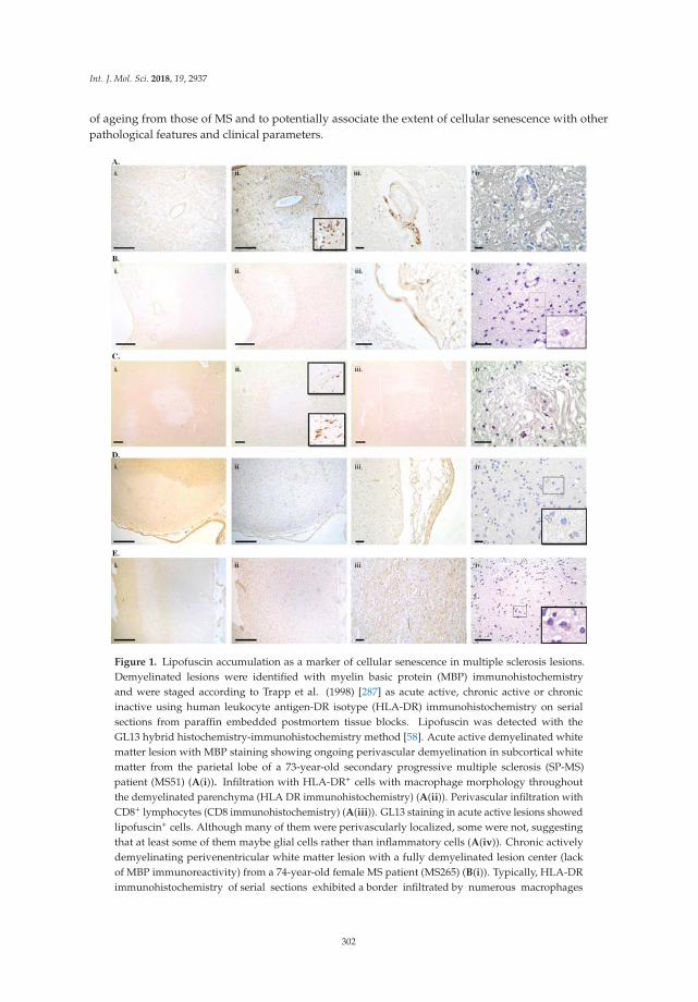
Int. J. Mol. Sci. 2018, 19, 2937
of ageing from those of MS and to potentially associate the extent of cellular senescence with otherpathological features and clinical parameters.
Figure 1. Lipofuscin accumulation as a marker of cellular senescence in multiple sclerosis lesions.Demyelinated lesions were identified with myelin basic protein (MBP) immunohistochemistryand were staged according to Trapp et al. (1998) [287] as acute active, chronic active or chronicinactive using human leukocyte antigen-DR isotype (HLA-DR) immunohistochemistry on serialsections from paraffin embedded postmortem tissue blocks. Lipofuscin was detected with theGL13 hybrid histochemistry-immunohistochemistry method [58]. Acute active demyelinated whitematter lesion with MBP staining showing ongoing perivascular demyelination in subcortical whitematter from the parietal lobe of a 73-year-old secondary progressive multiple sclerosis (SP-MS)patient (MS51) (A(i)). Infiltration with HLA-DR+ cells with macrophage morphology throughoutthe demyelinated parenchyma (HLA DR immunohistochemistry) (A(ii)). Perivascular infiltration withCD8+ lymphocytes (CD8 immunohistochemistry) (A(iii)). GL13 staining in acute active lesions showedlipofuscin+ cells. Although many of them were perivascularly localized, some were not, suggestingthat at least some of them maybe glial cells rather than inflammatory cells (A(iv)). Chronic activelydemyelinating perivenentricular white matter lesion with a fully demyelinated lesion center (lackof MBP immunoreactivity) from a 74-year-old female MS patient (MS265) (B(i)). Typically, HLA-DRimmunohistochemistry of serial sections exhibited a border infiltrated by numerous macrophages
302

Int. J. Mol. Sci. 2018, 19, 2937
whereas the lesion centre is infiltrated by ramified microglia (B(ii)). Few CD8+ lymphocytes are presentperivascularly (CD8 immunohistochemistry) (B(iii)). Lipofuscin+ cells with granular staining werefound in the macrophage infiltrated lesion border (B(iv)). Chronic inactive subcortical white matterdemyelinated lesion (lack of MBP immunoreactivity with a well demarcated border) from the leftparietal lobe of a 71-year-old female SP-MS patient (MS33) (C(i)). Ramified microglial morphologythroughout the demyelinated lesion area and lesion border (HLA-DR immunohistochemistry) typicalof a chronic inactive lesion (C(ii)). Decreased axonal density in the demyelinated lesion seen with200 KDa neurofilament immunohistochemistry (C(iii)). Numerous parenchymal lipofuscin+ cellsin the demyelinated white matter. Lack of HLA-DR+ macrophages from the chronic demyelinatedlesion suggests that the lipofuscin+ cells are glial (C(iv)). Subpial cortical demyelination (lack ofMBP immunoreactivity extending from the pial surface into the deeper cortical layers from theparietal cortex of a 71-year-old female SP-MS patient (MS33) (D(i)). HLA-DR+ ramified microgliain the demyelinated cortical lesion (D(ii)) and few CD8+ lymphocytes infiltrating the adjacentpia matter (D(iii)). Lipofuscin+ cells mostly with neuronal morphology (inset) throughout thedemyelinated cortex (D(iv)). Normal appearing cortex with intact appearing cortical myelin (MBPimmunohistochemistry) from the left parietal lobe of a 71-year-old female SP-MS patient (MS33)(E(i)), HLA-DR immunoreactivity revealing quiescent ramified microglia (E(ii)) and normal-appearingaxonal staining with 200 kDa neurofilament immunohistochemistry on a serial section (E(iii)). GL13staining showed numerous lipofuscin+ cells mostly with neuronal morphology (E(iv)). Scale barsrepresent 500 μm (A(i),A(ii),B(i),B(ii),C(i),C(ii),C(iii),D(i),D(ii),E(i),E(ii)), 50 μm (D(iii),E(iii)) or25 μm (A(iii),A(iv), B(iii), B(iv), C(iv), D(iv), E(iv)).
8. Cellular Senescence as a Therapeutic Target
Currently, there are no available neuroprotective treatments that can effectively modify the diseasecourse and prevent disease progression for AD or PD. Several attempts at targeting Aβ amyloid inAD have failed [288,289]. Interestingly, the Aβ plaques may be found in 35% of cognitively healthyindividuals above the age of 60 [290] and the Aβ load, which has been our main target, correlatesbetter with age than disease severity in AD [291–293], casting doubt on the amyloid hypothesis.Similarly, therapies in PD aim to substitute dopamine and restore the dopaminergic system deficit,with no effect on neuronal cell loss and consequently on disease progression. In MS only siponimod,an S1P1/S1P5 receptor modulator, which is not currently licensed, has shown a modest effect in a phaseIII trial (EXPAND) in secondary- progressive MS, preventing disability progression by 21% in twoyears [294]. Therefore, there is an urgent need for neuroprotective treatments for neurodegenerativedisease. Investigation into new treatment approaches may require a paradigm shift in our view of thepathogenetic mechanisms of neurodegeneration.
Several lines of evidence implicate cellular senescence in the pathogenesis of neurodegenerativedisease. Targeting cellular senescence as a therapeutic strategy is promising yet still at an embryonicstage. There is evidence of a beneficial effect of both pro-senescent and anti-senescent approaches,depending on context. A pro-senescent effect may be desirable in treating cancer [295–297], renal,liver and cutaneous fibrosis [298–301]. An anti-senescent treatment approach may be beneficial inneurodegenerative disease. An anti-senescent or senotherapeutic approach may involve the selectivedeath of senescent cells in order to reduce the burden of senescent cells on a tissue (senolysis) ormodulate senescent cells (senomorphism) in a way that neutralizes the detrimental effects of senescentcells in a tissue i.e., by blocking the expression of SASP or particular mediators of SASP.
Senolysis and/or senomorphism in neurodegenerative disease would aim at preventing cell lossand tissue destruction in order to ultimately prevent disease progression. Given that neurodegenerativediseases seem to have a long presymptomatic phase with pathological changes appearing severalyears before clinical presentation e.g., 50–60% of nigral neurons are already lost at PD diagnosis [302],a great effort is being made to diagnose neurodegenerative disease presymptomatically using differentbiomarkers. In the context of presymptomatic diagnosis, early senotherapeutic treatment could,in theory, even prevent the clinical presentation of neurodegenerative disease. So far, the most
303

Int. J. Mol. Sci. 2018, 19, 2937
convincing senotherapeutic manipulation comes from a sophisticated experiment in geneticallymodified BubR1 progeroid mice. In this setting, P16INK4A senescent cells were eliminated byactivation of an INK-ATTAC transgene by drug treatment. Lifelong elimination of p16INK4A cellssubstantially delayed age-related disease, whereas late life elimination of p16INK4A cells attenuatedthese age-related pathologies [38], supporting the notion that cellular senescence can be successfullyexploited therapeutically.
A number of compounds with senolytic or senomorphic actions have been examined in vitro andin vivo with notable results, summarized in Table 1. From our limited experience so far it is evidentthat, in most cases, the senescence-modifying action is not universal, but rather cell-type dependent,which greatly complicates the therapeutic landscape [303]. Many of the promising compounds withsenolytic or senomorphic activity such as metformin or dasatinib are in use with different indications(diabetes mellitus type 2 and CML/ALL, respectively), which suggests that drug repurposing mayfacilitate our quest for efficacious senotherapeutics. Nevertheless, senotherapeutics have not beenexamined in models of neurodegeneration and supportive evidence remains weak and indirect,and sometimes even contradictory. For example, although some epidemiological studies supported aprotective role for metformin, which crosses the BBB, in preventing cognitive decline in individualswith type 2 diabetes [304], another 12-year cohort study in patients with type 2 diabetes showed atwo-fold increase in the risk of AD and PD in those who took metformin compared to those whodidn’t [305]. The complex physiological and pathophysiological roles of cellular senescence, exertingboth beneficial and detrimental effects according to setting, along with the cell-type specific variabilityin senescence triggers and senescence phenotypes, necessitates a cautious approach to avoid pitfallswhen targeting a such a key biological process therapeutically. Furthermore, the relationship betweensenescence and immune response merits further elucidation. Naturally occurring immune-mediatedclearance of senescent cells could be exploited therapeutically by developing medications that enhanceit. The high specificity of immune responses could be employed to specifically target senescentcell types by developing senolytic vaccines. Cell surface markers of senescence [306] or even theintracellularly localized lipofuscin could potentially be used to prime the cellular or humoral immuneresponse, directing it against senescent cells. Expansion of our knowledge of cellular senescence andits extensive investigation in numerous settings is warranted.
304

Int. J. Mol. Sci. 2018, 19, 2937
Ta
ble
1.
Lice
nsed
and
expe
rim
enta
lcom
poun
dsw
ith
seno
lyti
c,se
nom
orph
icor
sene
scen
ce-i
nduc
ing
acti
onad
apte
dfr
omM
yria
ntho
poul
oset
al.,
2018
[303
].A
LL:
acut
ely
mph
ocyt
icle
ukem
ia,C
ML
:chr
onic
mye
loge
nous
leuk
emia
,HU
VE
C:h
uman
umbi
lical
vein
epit
helia
lcel
ls,J
AK
:Jan
uski
nsas
e,M
EF:
mou
seem
bryo
nic
fibro
blas
ts,M
oA:m
echa
nism
ofac
tion
,PPI
:pro
tein
-pro
tein
inte
ract
ion
inhi
bito
r.
Co
mp
ou
nd
Mo
AE
ffect
Cu
rren
tIn
dic
ati
on
Cla
ssifi
cati
on
Refe
ren
ces
Das
atin
ib(S
pryc
el)
Tyro
sine
kina
sein
hibi
tor,
Inhi
bito
rof
Eph
rece
ptor
sR
educ
edpr
olif
erat
ion
ofse
nesc
entc
ells
invi
tro;
Alle
viat
edag
eing
phen
otyp
esin
trea
ted
anim
als
Phila
delp
hia
chro
mos
ome-
CM
Lan
dA
LLse
noly
tic
[307
–310
]
Que
rcet
inM
odul
ator
ofN
F-κ
B,PI
3K/A
kt,
estr
ogen
rece
ptor
,mTO
R,P
IKδ
kina
sein
hibi
tor,
Pote
ntan
tiox
idan
t
Kill
sse
nesc
enth
uman
endo
thel
ialc
ells
and
mur
ine
bone
mar
row
mes
ench
ymal
stem
cells
expe
rim
enta
lse
noly
tic
[311
–323
]
Nav
itoc
lax
(ABT
-263
)BC
L-2
inhi
bito
r(P
PI)
Red
uced
surv
ival
ofH
UV
EC,h
uman
lung
fibro
blas
tsan
dm
urin
eem
bryo
nic
fibro
blas
tsan
dm
esen
chym
alst
emce
llsin
vitr
oex
peri
men
tal
Seno
lyti
c[3
24–3
30]
ABT
-737
BCL-
XL
inhi
bito
r(P
PI)
Red
uced
viab
ility
ofse
nesc
entc
elli
nvi
tro
and
invi
voex
peri
men
tal
seno
lyti
c[3
31–3
34]
A13
3185
2an
dA
1155
463
BCL-
XL
inhi
bito
rR
educ
edvi
abili
tyof
sene
scen
tcel
lin
vitr
o.ex
peri
men
tal
seno
lyti
c[3
35]
Fise
tin
Inte
ract
sw
ith:
topo
isom
eras
es,
cycl
in-d
epen
dent
kina
ses,
NF-
kB,
PPA
R,P
AR
P1,
PI3K
/Akt
/mTO
R,a
ntio
xida
nt
Del
ayof
age-
rela
ted
CN
Sco
mpl
icat
ions
invi
voex
peri
men
tal
seno
mor
phic
[335
–346
]
Pipe
rlon
gum
ine
NF-
kBm
odul
ator
Indu
ces
apop
tosi
sin
aged
cells
expe
rim
enta
lse
noly
tic
[347
,348
]
Gel
dana
myc
inH
SP90
inhi
bito
r,do
wn-
regu
lati
onof
PI3K
/Akt
Indu
ces
deat
hof
sene
scen
tcel
lsin
vitr
oex
peri
men
tal
seno
lyti
c[3
49,3
50]
Tane
spim
ycin
(17-
AA
G)
HSP
90in
hibi
tor,
dow
n-re
gula
tion
ofPI
3K/A
ktIn
duce
sde
ath
ofse
nesc
entc
ells
invi
tro
expe
rim
enta
lse
noly
tic
[349
,350
]
Pano
bino
stat
(Far
ydak
)no
n-se
lect
ive
hist
one
deac
etyl
ase
inhi
bito
rSy
nerg
isti
cef
fect
wit
hta
xoli
nin
duci
ngde
ath
ofse
nesc
entc
ells
,in
vivo
mul
tipl
em
yelo
ma
seno
lyti
c[3
51]
Api
geni
nan
dK
aem
pfer
olIn
terf
eren
cew
ith
the
NF-
kBp6
5su
buni
tand
IkB
Inhi
bite
dco
mpo
nent
sof
SASP
such
asin
IL-6
,CX
CL
and
GM
-CSF
invi
vo.
expe
rim
enta
lse
nom
orph
ics
[352
]
Rap
amyc
in(R
apam
une)
mTO
Rki
nase
inhi
bito
rsu
ppre
ssio
nof
repl
icat
ive
sene
scen
ceof
rode
ntem
bryo
nic
cells
,life
span
exte
nsio
nin
mod
elsy
stem
s
Lym
phan
gio-
myo
mat
osis
,co
rona
ryst
entc
lot
prev
enti
on,p
reve
ntio
nof
tran
spla
ntre
ject
ion
seno
mor
phic
[353
–356
]
Rux
olit
inib
(Jak
afi)
JAK
inhi
bito
rde
crea
sed
syst
emic
infla
mm
atio
nin
aged
anim
als
and
impr
oved
age-
rela
ted
dysf
unct
ions
and
alle
viat
ing
frai
lty
Poly
cyte
mia
vera
,M
yelo
fibro
sis
seno
mor
phic
[357
]
305
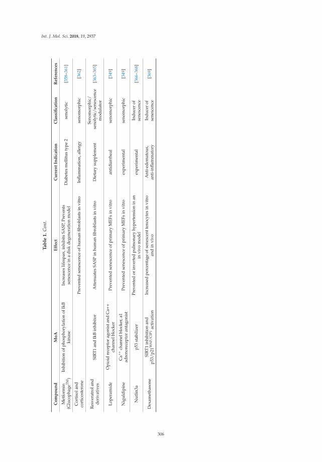
Int. J. Mol. Sci. 2018, 19, 2937
Ta
ble
1.
Con
t.
Co
mp
ou
nd
Mo
AE
ffect
Cu
rren
tIn
dic
ati
on
Cla
ssifi
cati
on
Refe
ren
ces
Met
form
in(G
luco
phag
eTM)
Inhi
biti
onof
phos
phor
ylat
ion
ofIk
Bki
nase
Incr
ease
slif
espa
n,in
hibi
tsSA
SP,P
reve
nts
sene
scen
cein
adi
skde
gene
rati
onm
odel
Dia
bete
sm
ellit
usty
pe2
seno
lyti
c[3
58–3
61]
Cor
tiso
land
cort
icos
tero
nePr
even
ted
sene
scen
ceof
hum
anfib
robl
asts
invi
tro
Infla
mm
atio
n,al
lerg
yse
nom
orph
ic[3
62]
Res
vera
trol
and
deri
vati
ves
SIR
T1an
dIk
Bin
hibi
tor
Att
enua
tes
SASP
inhu
man
fibro
blas
tsin
vitr
oD
ieta
rysu
pple
men
tSe
nom
orph
ic/
seno
lytic
/sen
esce
nce
mod
ulat
or[3
63–3
65]
Lope
ram
ide
Opi
oid
rece
ptor
agon
ista
ndC
a++
chan
nelb
lock
erPr
even
ted
sene
scen
ceof
prim
ary
MEF
sin
vitr
oan
tidi
arrh
eal
seno
mor
phic
[349
]
Nig
uldi
pine
Ca++
chan
nelb
lock
er,a
1ad
reno
rece
ptor
anta
goni
stPr
even
ted
sene
scen
ceof
prim
ary
MEF
sin
vitr
oex
peri
men
tal
seno
mor
phic
[349
]
Nut
lin3a
p53
stab
ilize
rPr
even
ted
orin
vert
edpu
lmon
ary
hype
rten
sion
inan
invi
vom
odel
expe
rim
enta
lIn
duce
rof
sene
scen
ce[3
66–3
68]
Dex
amet
haso
neSI
RT1
inhi
biti
onan
dp5
3/p2
1WA
F/C
IP1
acti
vati
onIn
crea
sed
perc
enta
geof
sene
scen
tten
ocyt
esin
vitr
oan
din
vivo
Ant
i-ed
emat
ous,
anti
-infl
amm
ator
yIn
duce
rof
sene
scen
ce[3
69]
306
Int. J. Mol. Sci. 2018, 19, 2937
9. Conclusions and Future Perspectives
Senescent cells accumulate with ageing and progeroid models have provided in vivo experimentaldata of accelerated ageing-related degenerative pathologies. In addition, experimental senolysisameliorated ageing-related pathologies [38]. Thus, cellular senescence meets the criteria for apotentially causal role in ageing-related disease. There is evidence of cellular senescence affectingastrocytes, microglia, oligodendrocyte progenitors and neural stem cells. A senescence-like phenotypehas also been demonstrated in post-mitotic cells, which suggests that neurons and oligodendrocytesmay also become senescent. Resident brain cells are either post-mitotic or slowly cycling. They are morelikely to exhibit stress-induced premature senescence due to various stressors or insults than developreplicative senescence. However, evidence connecting cellular senescence with the mechanisms ofneurodegeneration remains indirect and further investigation of the putative role of senescence inneurodegeneration is required. Unequivocal identification of senescent cells in vitro and in vivois an important prerequisite to facilitate our understanding of cellular senescence and its role indifferent cell types. Detecting lipofuscin as a marker of cellular senescence using the GL13 compound,which not only detects lipofuscin in situ but also in biological fluids [93], is likely to boost ourunderstanding of the senescence process. Casting light on CNS cell senescence and its role inneurodegeneration is essential to inform any practices that may be senescence- inducing e.g., usingcorticosteroids [368], beta-interferons [370] or DNA-damaging chemotherapeutics in MS, practices thatmay prove detrimental in the long run. Secondly, there is an urgent need for disease-modifyingcures for neurodegenerative diseases. Cellular senescence may be a credible therapeutic targetopening new therapeutic avenues for neurodegenerative disease and senotherapeutics may prove tobe efficacious neuroprotectants.
Funding: This research received no external funding.
Acknowledgments: MS tissue samples were supplied by the UK Multiple Sclerosis Tissue Bank (www.imperial.ac.uk/medicine/multiple-sclerosis-and-parkinsons-tissue-bank/), funded by the MS Society of Great Britain andNorthern Ireland (registered charity 207495). This work received financial support from the Welfare Foundationfor Social & Cultural Sciences (KIKPE), Greece.
Conflicts of Interest: The authors declare no conflict of interest.
Abbreviations
Aβ amyloid-betaAD Alzheimer’s diseaseAkt protein kinase BALL acute lymphocytic leukemiaAPP amyloid beta precursor proteinATF6α activating transcription factor 6 isoform α
ATM ataxia-telangiectasia mutatedBBB blood-brain barrierBcl-2 B-cell lymphoma 2CDK cyclin-dependent kinaseCDKI cyclin-dependent kinase inhibitor proteincGAS cyclic GMP-AMP synthaseCIP/KIP CDK interacting protein/kinase inhibitory proteinCML chronic myelogenous leukemiaCNS central nervous systemCSF cerebrospinal fluidDDR DNA damage responseEcrg4 esophageal cancer-related gene 4EGF epidermal growth factorER endoplasmic reticulumFGF-2 fibroblast growth factor 2
307

Int. J. Mol. Sci. 2018, 19, 2937
GLB1 galactosidase beta 1HLA-DR human leukocyte antigen-DR isotypeγH2AX phosphorylated H2A histone family member XIkB inhibitor of kappa BIL InterleukinMBP myelin basic proteinMECP methyl-CpG-binding proteinMMP matrix metalloproteinaseMRI magnetic resonance imagingmTOR mammalian target of rapamycinMS multiple sclerosisNAWM normal appearing white matter
NF-κBnuclear factor kappa-light-chain-enhancer ofactivated B cells
NSCs neural stem cellsOIS oncogene-induced senescenceOPC oligodendrocyte progenitor cellPD Parkinson’s diseasePS1 presenilin-1p38MAPK p38 mitogen-activated protein kinasesRB RetinoblastomaROS reactive oxygen speciesRR-MS relapsing-remitting multiple sclerosisSA-β-Gal senescence-associated β-galactosidaseSAHF senescence-associated heterochromatin formationSAMD senescence-associated mitochondrial dysfunctionSASP senescence-associated secretory phenotypeSIPS stress-induced premature senescenceSIRT SirtuinSP-MS secondary progressive multiple sclerosisSGZ subgranular zoneSTING stimulator of IFN genesSVZ subventricular zoneS1P sphingosine 1-phosphate receptorTCDD 2,3,7,8-Tetrachlorodibenzo-p-DioxinTerc human telomerase geneTGF-β transforming growth factor betaTNF-α tumour necrosis factor alphaUPR unfolded protein response53BP1 p53-binding protein 1
References
1. Johnson, I.P. Age-related neurodegenerative disease research needs aging models. Front. Aging Neurosci.2015, 7, 168. [CrossRef] [PubMed]
2. Schrijvers, E.M.; Verhaaren, B.F.; Koudstaal, P.J.; Hofman, A.; Ikram, M.A.; Breteler, M.M. Is dementiaincidence declining? Trends in dementia incidence since 1990 in the Rotterdam Study. Neurology 2012,78, 1456–1463. [CrossRef] [PubMed]
3. Rocca, W.A.; Petersen, R.C.; Knopman, D.S.; Hebert, L.E.; Evans, D.A.; Hall, K.S.; Gao, S.; Unverzagt, F.W.;Langa, K.M.; Larson, E.B.; et al. Trends in the incidence and prevalence of Alzheimer’s disease, dementia,and cognitive impairment in the United States. Alzheimers Dement. 2011, 7, 80–93. [CrossRef] [PubMed]
4. Nussbaum, R.L.; Ellis, C.E. Alzheimer’s Disease and Parkinson’s Disease. N. Engl. J. Med. 2003,348, 1356–1364. [CrossRef] [PubMed]
308

Int. J. Mol. Sci. 2018, 19, 2937
5. Lockrow, J.P.; Fortress, A.M.; Granholm, A.C.E. Age-related neurodegeneration and memory loss in downsyndrome. Curr. Gerontol. Geriatr. Res. 2012, 2012, 13. [CrossRef] [PubMed]
6. Takeda, T.; Hosokawa, M.; Takeshita, S.; Irino, M.; Higuchi, K.; Matsushita, T.; Tomita, Y.; Yasuhira, K.;Hamamoto, H.; Shimizu, K.; et al. A new murine model of accelerated senescence. Mech. Ageing Dev. 1981,17, 183–194. [CrossRef]
7. Morley, J.E.; Kumar, V.B.; Bernardo, A.E.; Farr, S.A.; Uezu, K.; Tumosa, N.; Flood, J.F. Beta-amyloid precursorpolypeptide in SAMP8 mice affects learning and memory. Peptides 2000, 21, 1761–1767. [CrossRef]
8. Blin, P.; Dureau-Pournin, C.; Foubert-Samier, A.; Grolleau, A.; Corbillon, E.; Jové, J.; Lassalle, R.; Robinson, P.;Poutignat, N.; Droz-Perroteau, C.; Moore, N. Parkinson’s disease incidence and prevalence assessmentin France using the national healthcare insurance database. Eur. J. Neurol. 2015, 22, 464–471. [CrossRef][PubMed]
9. Duncan, G.W.; Khoo, T.K.; Coleman, S.Y.; Brayne, C.; Yarnall, A.J.; O’Brien, J.T.; Barker, R.A.; Burn, D.J.The incidence of Parkinson’s disease in the North-East of England. Age Ageing 2014, 43, 257–263. [CrossRef][PubMed]
10. Trapp, B.D.; Nave, K.-A. Multiple Sclerosis: An Immune or Neurodegenerative Disorder?Annu. Rev. Neurosci. 2008, 31, 247–269. [CrossRef] [PubMed]
11. Scalfari, A.; Neuhaus, A.; Daumer, M.; Ebers, G.C.; Muraro, P.A. Age and disability accumulation in multiplesclerosis. Neurology 2011, 77, 1246–1252. [CrossRef] [PubMed]
12. Kirkwood, T.B.L.; Melov, S. On the Programmed/Non-Programmed Nature of Ageing within the LifeHistory. Curr. Biol. 2011, 21, R701–R707. [CrossRef] [PubMed]
13. Kirkwood, T.B.L.; Feder, M.; Finch, C.E.; Franceschi, C.; Globerson, A.; Klingenberg, C.P.; LaMarco, K.;Omholt, S.; Westendorp, R.G. What accounts for the wide variation in life span of genetically identicalorganisms reared in a constant environment? Mech. Ageing Dev. 2005, 126, 439–443. [CrossRef] [PubMed]
14. López-Otín, C.; Blasco, M.A.; Partridge, L.; Serrano, M.; Kroemer, G. The Hallmarks of Aging. Cell 2013,153, 1194–1217. [CrossRef] [PubMed]
15. Muñoz-Espín, D.; Serrano, M. Cellular senescence: From physiology to pathology. Nat. Rev. Mol. Cell Biol.2014, 15, 482–496. [CrossRef] [PubMed]
16. Barbouti, A.; Evangelou, K.; Pateras, I.S.; Papoudou-Bai, A.; Patereli, A.; Stefanaki, K.; Rontogianni, D.;Muñoz-Espín, D.; Kanavaros, P.; Gorgoulis, V.G. In situ evidence of cellular senescence in Thymic EpithelialCells (TECs) during human thymic involution. Mech. Ageing Dev. 2018. [CrossRef] [PubMed]
17. Chuprin, A.; Gal, H.; Biron-Shental, T.; Biran, A.; Amiel, A.; Rozenblatt, S.; Krizhanovsky, V. Cell fusioninduced by ERVWE1 or measles virus causes cellular senescence. Genes Dev. 2013, 27, 2356–2366. [CrossRef][PubMed]
18. Demaria, M.; Ohtani, N.; Youssef, S.A.; Rodier, F.; Toussaint, W.; Mitchell, J.R.; Laberge, R.-M.; Vijg, J.;Van Steeg, H.; Dollé, M.E.T.; et al. An Essential Role for Senescent Cells in Optimal Wound Healing throughSecretion of PDGF-AA. Dev. Cell 2014, 31, 722–733. [CrossRef] [PubMed]
19. Rajagopalan, S.; Long, E.O. Cellular senescence induced by CD158d reprograms natural killer cells topromote vascular remodeling. Proc. Natl. Acad. Sci. USA 2012, 109, 20596–20601. [CrossRef] [PubMed]
20. Howcroft, T.K.; Campisi, J.; Louis, G.B.; Smith, M.T.; Wise, B.; Wyss-Coray, T.; Augustine, A.D.;McElhaney, J.E.; Kohanski, R.; Sierra, F. The role of inflammation in age-related disease. Aging (Albany NY)2013, 5, 84–93. [CrossRef] [PubMed]
21. Hayflick, L.; Moorhead, P.S. The serial cultivation of human diploid strains. Exp. Cell Res. 1961, 25, 585–621.[CrossRef]
22. Gorgoulis, V.G.; Halazonetis, T.D. Oncogene-induced senescence: The bright and dark side of the response.Curr. Opin. Cell Biol. 2010, 22, 816–827. [CrossRef] [PubMed]
23. Hernandez-Segura, A.; Nehme, J.; Demaria, M. Hallmarks of Cellular Senescence. Trends Cell Biol. 2018,28, 436–453. [CrossRef] [PubMed]
24. Sabin, R.J.; Anderson, R.M. Cellular Senescence—Its role in cancer and the response to ionizing radiation.Genome Integr. 2011, 2, 7. [CrossRef] [PubMed]
25. Dörr, J.R.; Yu, Y.; Milanovic, M.; Beuster, G.; Zasada, C.; Däbritz, J.H.M.; Lisec, J.; Lenze, D.; Gerhardt, A.;Schleicher, K.; et al. Synthetic lethal metabolic targeting of cellular senescence in cancer therapy. Nature 2013,501, 421–425. [CrossRef] [PubMed]
309

Int. J. Mol. Sci. 2018, 19, 2937
26. Toussaint, O.; Medrano, E.E.; von Zglinicki, T. Cellular and molecular mechanisms of stress-inducedpremature senescence (SIPS) of human diploid fibroblasts and melanocytes. Exp. Gerontol. 2000, 35, 927–945.[CrossRef]
27. Kang, H.T.; Lee, K.B.; Kim, S.Y.; Choi, H.R.; Park, S.C. Autophagy impairment induces premature senescencein primary human fibroblasts. PLoS ONE 2011, 6, e23367. [CrossRef] [PubMed]
28. Wiley, C.D.; Flynn, J.M.; Morrissey, C.; Lebofsky, R.; Shuga, J.; Dong, X.; Unger, M.A.; Vijg, J.; Melov, S.;Campisi, J. Analysis of individual cells identifies cell-to-cell variability following induction of cellularsenescence. Aging Cell 2017, 16, 1043–1050. [CrossRef] [PubMed]
29. Nakamura, A.J.; Chiang, Y.J.; Hathcock, K.S.; Horikawa, I.; Sedelnikova, O.A.; Hodes, R.J.; Bonner, W.M.Both telomeric and non-telomeric DNA damage are determinants of mammalian cellular senescence.Epigenet. Chromatin 2008, 1, 6. [CrossRef] [PubMed]
30. Rodier, F.; Campisi, J. Four faces of cellular senescence. J. Cell Biol. 2011, 192, 547–556. [CrossRef] [PubMed]31. Ramirez, R.D.; Morales, C.P.; Herbert, B.S.; Rohde, J.M.; Passons, C.; Shay, J.W.; Wright, W.E. Putative
telomere-independent mechanisms of replicative aging reflect inadequate growth conditions. Genes Dev.2001, 15, 398–403. [CrossRef] [PubMed]
32. Rodier, F.; Coppé, J.-P.; Patil, C.K.; Hoeijmakers, W.A.M.; Muñoz, D.P.; Raza, S.R.; Freund, A.; Campeau, E.;Davalos, A.R.; Campisi, J. Persistent DNA damage signalling triggers senescence-associated inflammatorycytokine secretion. Nat. Cell Biol. 2009, 11, 973–979. [CrossRef] [PubMed]
33. Turenne, G.A.; Paul, P.; Laflair, L.; Price, B.D. Activation of p53 transcriptional activity requires ATM’s kinasedomain and multiple N-terminal serine residues of p53. Oncogene 2001, 20, 5100–5110. [CrossRef] [PubMed]
34. Xue, W.; Zender, L.; Miething, C.; Dickins, R.A.; Hernando, E.; Krizhanovsky, V.; Cordon-Cardo, C.;Lowe, S.W. Senescence and tumour clearance is triggered by p53 restoration in murine liver carcinomas.Nature 2007, 445, 656–660. [CrossRef] [PubMed]
35. Hoenicke, L.; Zender, L. Immune surveillance of senescent cells—Biological significance in cancer- andnon-cancer pathologies. Carcinogenesis 2012, 33, 1123–1126. [CrossRef] [PubMed]
36. Herbig, U.; Jobling, W.A.; Chen, B.P.C.; Chen, D.J.; Sedivy, J.M. Telomere shortening triggers senescenceof human cells through a pathway involving ATM, p53, and p21CIP1, but not p16INK4a. Mol. Cell 2004,14, 501–513. [CrossRef]
37. Lawless, C.; Wang, C.; Jurk, D.; Merz, A.; Zglinicki, T. von; Passos, J.F. Quantitative assessment of markersfor cell senescence. Exp. Gerontol. 2010, 45, 772–778. [CrossRef] [PubMed]
38. Baker, D.J.; Wijshake, T.; Tchkonia, T.; LeBrasseur, N.K.; Childs, B.G.; van de Sluis, B.; Kirkland, J.L.;van Deursen, J.M. Clearance of p16Ink4a -positive senescent cells delays ageing- associated disorders. Nature2012, 479, 232–236. [CrossRef] [PubMed]
39. Krenning, L.; Feringa, F.M.; Shaltiel, I.A.; VandenBerg, J.; Medema, R.H. Transient activation of p53 in G2phase is sufficient to induce senescence. Mol. Cell 2014, 55, 59–72. [CrossRef] [PubMed]
40. Stein, G.H.; Drullinger, L.F.; Soulard, A.; Dulic, V. Differential Roles for Cyclin-Dependent Kinase Inhibitorsp21 and p16 in the Mechanisms of Senescence and Differentiation in Human Fibroblasts. Mol. Cell. Biol.1999, 19, 2109–2117. [CrossRef] [PubMed]
41. Sharma, V.; Gilhotra, R.; Dhingra, D.; Gilhotra, N. Possible underlying influence of p38MAPK and NF-κBin the diminished anti-anxiety effect of diazepam in stressed mice. J. Pharmacol. Sci. 2011, 116, 257–263.[CrossRef] [PubMed]
42. Yang, H.; Wang, H.; Ren, J.; Chen, Q.; Chen, Z.J. cGAS is essential for cellular senescence. Proc. Natl. Acad.Sci. USA 2017, 114, E4612–E4620. [CrossRef] [PubMed]
43. Coppé, J.-P.; Desprez, P.-Y.; Krtolica, A.; Campisi, J. The Senescence-Associated Secretory Phenotype:The Dark Side of Tumor Suppression. Annu. Rev. Pathol. Mech. Dis. 2010, 5, 99–118. [CrossRef] [PubMed]
44. Acosta, J.C.; Banito, A.; Wuestefeld, T.; Georgilis, A.; Janich, P.; Morton, J.P.; Athineos, D.; Kang, T.-W.;Lasitschka, F.; Andrulis, M.; et al. A complex secretory program orchestrated by the inflammasome controlsparacrine senescence. Nat. Cell Biol. 2013, 15, 978–990. [CrossRef] [PubMed]
45. Chen, H.; Ruiz, P.D.; McKimpson, W.M.; Novikov, L.; Kitsis, R.N.; Gamble, M.J. MacroH2A1 and ATM PlayOpposing Roles in Paracrine Senescence and the Senescence-Associated Secretory Phenotype. Mol. Cell 2015,59, 719–731. [CrossRef] [PubMed]
310
Int. J. Mol. Sci. 2018, 19, 2937
46. Hoare, M.; Ito, Y.; Kang, T.-W.; Weekes, M.P.; Matheson, N.J.; Patten, D.A.; Shetty, S.; Parry, A.J.; Menon, S.;Salama, R.; et al. NOTCH1 mediates a switch between two distinct secretomes during senescence.Nat. Cell Biol. 2016, 18, 979–992. [CrossRef] [PubMed]
47. Passos, J.F.; Nelson, G.; Wang, C.; Richter, T.; Simillion, C.; Proctor, C.J.; Miwa, S.; Olijslagers, S.; Hallinan, J.;Wipat, A.; et al. Feedback between p21 and reactive oxygen production is necessary for cell senescence.Mol. Syst. Biol. 2010, 6, 347. [CrossRef] [PubMed]
48. Hayakawa, T.; Iwai, M.; Aoki, S.; Takimoto, K.; Maruyama, M.; Maruyama, W.; Motoyama, N. SIRT1suppresses the senescence-associated secretory phenotype through epigenetic gene regulation. PLoS ONE2015, 10, 1–16. [CrossRef] [PubMed]
49. Lehmann, B.D.; Paine, M.S.; Brooks, A.M.; McCubrey, J.A.; Renegar, R.H.; Wang, R.; Terrian, D.M.Senescence-Associated Exosome Release from Human Prostate Cancer Cells. Cancer Res. 2008, 68, 7864–7871.[CrossRef] [PubMed]
50. Takasugi, M.; Okada, R.; Takahashi, A.; Virya Chen, D.; Watanabe, S.; Hara, E. Small extracellular vesiclessecreted from senescent cells promote cancer cell proliferation through EphA2. Nat. Commun. 2017, 8, 15729.[CrossRef] [PubMed]
51. Webley, K.; Bond, J.A.; Jones, C.J.; Blaydes, J.P.; Craig, A.; Hupp, T.; Wynford-Thomas, D. Posttranslationalmodifications of p53 in replicative senescence overlapping but distinct from those induced by DNA damage.Mol. Cell. Biol. 2000, 20, 2803–2808. [CrossRef] [PubMed]
52. Tavana, O.; Benjamin, C.L.; Puebla-Osorio, N.; Sang, M.; Ullrich, S.E.; Ananthaswamy, H.N.; Zhu, C. Absenceof p53-dependent apoptosis leads to UV radiation hypersensitivity, enhanced immunosuppression andcellular senescence. Cell Cycle 2010, 9, 3328–3336. [CrossRef] [PubMed]
53. Tang, Y.; Luo, J.; Zhang, W.; Gu, W. Tip60-Dependent Acetylation of p53 Modulates the Decision betweenCell-Cycle Arrest and Apoptosis. Mol. Cell 2006, 24, 827–839. [CrossRef] [PubMed]
54. Lee, B.Y.; Han, J.A.; Im, J.S.; Morrone, A.; Johung, K.; Goodwin, E.C.; Kleijer, W.J.; DiMaio, D.; Hwang, E.S.Senescence-associated β-galactosidase is lysosomal β-galactosidase. Aging Cell 2006, 5, 187–195. [CrossRef][PubMed]
55. Weichhart, T. mTOR as Regulator of Lifespan, Aging, and Cellular Senescence: A Mini-Review. Gerontology2018, 64, 127–134. [CrossRef] [PubMed]
56. Höhn, A.; Grune, T. Lipofuscin: Formation, effects and role of macroautophagy. Redox Biol. 2013, 1, 140–144.[CrossRef] [PubMed]
57. Georgakopoulou, E.A.; Tsimaratou, K.; Evangelou, K.; Fernandez-Marcos, P.J.; Zoumpourlis, V.;Trougakos, I.P.; Kletsas, D.; Bartek, J.; Serrano, M.; Gorgoulis, V.G. Specific lipofuscin staining as a novelbiomarker to detect replicative and stress-induced senescence. A method applicable in cryo-preserved andarchival tissues. Aging (Albany NY) 2013, 5, 37–50. [CrossRef] [PubMed]
58. Evangelou, K.; Lougiakis, N.; Rizou, S.V.; Kotsinas, A.; Kletsas, D.; Muñoz-Espín, D.; Kastrinakis, N.G.;Pouli, N.; Marakos, P.; Townsend, P.; et al. Robust, universal biomarker assay to detect senescent cells inbiological specimens. Aging Cell 2017, 16, 192–197. [CrossRef] [PubMed]
59. Wu, M.; Ye, H.; Shao, C.; Zheng, X.; Li, Q.; Wang, L.; Zhao, M.; Lu, G.; Chen, B.; Zhang, J.; et al.Metabolomics–Proteomics Combined Approach Identifies Differential Metabolism-Associated MolecularEvents between Senescence and Apoptosis. J. Proteome Res. 2017, 16, 2250–2261. [CrossRef] [PubMed]
60. Correia-Melo, C.; Marques, F.D.; Anderson, R.; Hewitt, G.; Hewitt, R.; Cole, J.; Carroll, B.M.; Miwa, S.;Birch, J.; Merz, A.; et al. Mitochondria are required for pro-ageing features of the senescent phenotype.EMBO J. 2016, 35, 724–742. [CrossRef] [PubMed]
61. Lee, S.; Jeong, S.-Y.; Lim, W.-C.; Kim, S.; Park, Y.-Y.; Sun, X.; Youle, R.J.; Cho, H. Mitochondrial Fission andFusion Mediators, hFis1 and OPA1, Modulate Cellular Senescence. J. Biol. Chem. 2007, 282, 22977–22983.[CrossRef] [PubMed]
62. Mai, S.; Klinkenberg, M.; Auburger, G.; Bereiter-Hahn, J.; Jendrach, M. Decreased expression of Drp1 andFis1 mediates mitochondrial elongation in senescent cells and enhances resistance to oxidative stress throughPINK1. J. Cell Sci. 2010, 123, 917–926. [CrossRef] [PubMed]
63. García-Prat, L.; Martínez-Vicente, M.; Perdiguero, E.; Ortet, L.; Rodríguez-Ubreva, J.; Rebollo, E.;Ruiz-Bonilla, V.; Gutarra, S.; Ballestar, E.; Serrano, A.L.; et al. Autophagy maintains stemness by preventingsenescence. Nature 2016, 529, 37–42. [CrossRef] [PubMed]
311

Int. J. Mol. Sci. 2018, 19, 2937
64. Takahashi, Y.; Karbowski, M.; Yamaguchi, H.; Kazi, A.; Wu, J.; Sebti, S.M.; Youle, R.J.; Wang, H.-G. Lossof Bif-1 suppresses Bax/Bak conformational change and mitochondrial apoptosis. Mol. Cell. Biol. 2005,25, 9369–9382. [CrossRef] [PubMed]
65. Correia-Melo, C.; Passos, J.F. Mitochondria: Are they causal players in cellular senescence? Biochim. Biophys.Acta-Bioenerg. 2015, 1847, 1373–1379. [CrossRef] [PubMed]
66. Passos, J.F.; Saretzki, G.; von Zglinicki, T. DNA damage in telomeres and mitochondria during cellularsenescence: Is there a connection? Nucleic Acids Res. 2007, 35, 7505–7513. [CrossRef] [PubMed]
67. Tasselli, L.; Xi, Y.; Zheng, W.; Tennen, R.I.; Odrowaz, Z.; Simeoni, F.; Li, W.; Chua, K.F. SIRT6 deacetylatesH3K18ac at pericentric chromatin to prevent mitotic errors and cellular senescence. Nat. Struct. Mol. Biol.2016, 23, 434–440. [CrossRef] [PubMed]
68. Giblin, W.; Skinner, M.E.; Lombard, D.B. Sirtuins: Guardians of mammalian healthspan. Trends Genet. 2014,30, 271–286. [CrossRef] [PubMed]
69. Komseli, E.-S.; Pateras, I.S.; Krejsgaard, T.; Stawiski, K.; Rizou, S.V.; Polyzos, A.; Roumelioti, F.-M.;Chiourea, M.; Mourkioti, I.; Paparouna, E.; et al. A prototypical non-malignant epithelial model to studygenome dynamics and concurrently monitor micro-RNAs and proteins in situ during oncogene-inducedsenescence. BMC Genomics 2018, 19, 37. [CrossRef] [PubMed]
70. Bannister, A.J.; Zegerman, P.; Partridge, J.F.; Miska, E.A.; Thomas, J.O.; Allshire, R.C.; Kouzarides, T. Selectiverecognition of methylated lysine 9 on histone H3 by the HP1 chromo domain. Nature 2001, 410, 120–124.[CrossRef] [PubMed]
71. Dou, Z.; Ghosh, K.; Vizioli, M.G.; Zhu, J.; Sen, P.; Wangensteen, K.J.; Simithy, J.; Lan, Y.; Lin, Y.; Zhou, Z.;et al. Cytoplasmic chromatin triggers inflammation in senescence and cancer. Nature 2017, 550, 402–406.[CrossRef] [PubMed]
72. Di Micco, R.; Sulli, G.; Dobreva, M.; Liontos, M.; Botrugno, O.A.; Gargiulo, G.; dal Zuffo, R.; Matti, V.;d’Ario, G.; Montani, E.; et al. Interplay between oncogene-induced DNA damage response andheterochromatin in senescence and cancer. Nat. Cell Biol. 2011, 13, 292–302. [CrossRef] [PubMed]
73. Salama, R.; Sadaie, M.; Hoare, M.; Narita, M. Cellular senescence and its effector programs. Genes Dev. 2014,28, 99–114. [CrossRef] [PubMed]
74. Freund, A.; Laberge, R.-M.; Demaria, M.; Campisi, J. Lamin B1 loss is a senescence-associated biomarker.Mol. Biol. Cell 2012, 23, 2066–2075. [CrossRef] [PubMed]
75. Druelle, C.; Drullion, C.; Deslé, J.; Martin, N.; Saas, L.; Cormenier, J.; Malaquin, N.; Huot, L.; Slomianny, C.;Bouali, F.; et al. ATF6α regulates morphological changes associated with senescence in human fibroblasts.Oncotarget 2016, 7, 67699–67715. [CrossRef] [PubMed]
76. Cormenier, J.; Martin, N.; Deslé, J.; Salazar-Cardozo, C.; Pourtier, A.; Abbadie, C.; Pluquet, O. The ATF6αarm of the Unfolded Protein Response mediates replicative senescence in human fibroblasts through aCOX2/prostaglandin E 2 intracrine pathway. Mech. Ageing Dev. 2018, 170, 82–91. [CrossRef] [PubMed]
77. Ohno-Iwashita, Y.; Shimada, Y.; Hayashi, M.; Inomata, M. Plasma membrane microdomains in aging anddisease. Geriatr. Gerontol. Int. 2010, 10, S41–S52. [CrossRef] [PubMed]
78. Serrano, M.; Hannon, G.J.; Beach, D. A new regulatory motif in cell-cycle control causing specific inhibitionof cyclin D/CDK4. Nature 1993, 366, 704–707. [CrossRef] [PubMed]
79. Sharpless, N.E.; Sherr, C.J. Forging a signature of in vivo senescence. Nat. Rev. Cancer 2015, 15, 397–408.[CrossRef] [PubMed]
80. Zindy, F.; Quelle, D.E.; Roussel, M.F.; Sherr, C.J. Expression of the p16(INK4a) tumor suppressor versusother INK4 family members during mouse development and aging. Oncogene 1997, 15, 203–211. [CrossRef][PubMed]
81. Burd, C.E.; Sorrentino, J.A.; Clark, K.S.; Darr, D.B.; Krishnamurthy, J.; Deal, A.M.; Bardeesy, N.;Castrillon, D.H.; Beach, D.H.; Sharpless, N.E. Monitoring tumorigenesis and senescence in vivo with ap16 INK4a-luciferase model. Cell 2013, 152, 340–351. [CrossRef] [PubMed]
82. Krishnamurthy, J.; Torrice, C.; Ramsey, M.R.; Kovalev, G.I.; Al-Regaiey, K.; Su, L.; Sharpless, N.E. Ink4a/Arfexpression is a biomarker of aging. J. Clin. Invest. 2004, 114, 1299–1307. [CrossRef] [PubMed]
83. Shapiro, G.; Edwards, C.; Kobzik, L. Reciprocal Rb Inactivation and p16 INK4 Expression in Primary LungCancers and Cell Lines. Cancer Res. 1995, 55, 505–509. [PubMed]
312
Int. J. Mol. Sci. 2018, 19, 2937
84. Witkiewicz, A.K.; Knudsen, K.E.; Dicker, A.P.; Knudsen, E.S. The meaning of p16 ink4a expression intumors: Functional significance, clinical associations and future developments. Cell Cycle 2011, 10, 2497–2503.[CrossRef] [PubMed]
85. Sherr, C.J.; Roberts, J.M. CDK inhibitors: Positive and negative regulators of G1-phase progression. Genes Dev.1999, 13, 1501–1512. [CrossRef] [PubMed]
86. Jung, Y.-S.; Qian, Y.; Chen, X. Examination of the expanding pathways for the regulation of p21 expressionand activity. Cell. Signal. 2010, 22, 1003–1012. [CrossRef] [PubMed]
87. Dimri, G.P.; Lee, X.; Basile, G.; Acosta, M.; Scott, G.; Roskelley, C.; Medrano, E.E.; Linskens, M.; Rubelj, I.;Pereira-Smith, O. A biomarker that identifies senescent human cells in culture and in aging skin in vivo.Proc. Natl. Acad. Sci. USA 1995, 92, 9363–9367. [CrossRef] [PubMed]
88. Kurz, D.J.; Decary, S.; Hong, Y.; Erusalimsky, J.D. Senescence-associated (beta)-galactosidase reflectsan increase in lysosomal mass during replicative ageing of human endothelial cells. J. Cell Sci. 2000,113, 3613–3622. [PubMed]
89. Severino, J.; Allen, R.G.; Balin, S.; Balin, A.; Cristofalo, V.J. Is β-galactosidase staining a marker of senescencein vitro and in vivo? Exp. Cell Res. 2000, 257, 162–171. [CrossRef] [PubMed]
90. Gorgoulis, V.G.; Pefani, D.; Pateras, I.S.; Trougakos, I.P. Integrating the DNA damage and protein stressresponses during cancer development and treatment. J. Pathol. 2018. [CrossRef] [PubMed]
91. Terman, A.; Brunk, U.T. Lipofuscin. Int. J. Biochem. Cell Biol. 2004, 36, 1400–1404. [CrossRef] [PubMed]92. Jung, T.; Bader, N.; Grune, T. Lipofuscin: Formation, distribution, and metabolic consequences. Ann. N. Y.
Acad. Sci. 2007, 1119, 97–111. [CrossRef] [PubMed]93. Rizou, S.V.; Evangelou, K.; Myrianthopoulos, V.; Mourouzis, I.; Havaki, S.; Vasileiou, P.; Kotsinas, A.;
Kastrinakis, N.; Sfikakis, P.; Townsend, P.; Mikros, E.; Pantos, C.; Gorgoulis, V.G. A novel quantitativemethod for the detection of lipofuscin, the main byproduct of cellular senescence, in fluids. Meth. Mol. Biol.2018, in press.
94. Nelson, G.; Wordsworth, J.; Wang, C.; Jurk, D.; Lawless, C.; Martin-Ruiz, C.; von Zglinicki, T. A senescentcell bystander effect: Senescence-induced senescence. Aging Cell 2012, 11, 345–349. [CrossRef] [PubMed]
95. Ribezzo, F.; Shiloh, Y.; Schumacher, B. Systemic DNA damage responses in aging and diseases.Semin. Cancer Biol. 2016, 37–38, 26–35. [CrossRef] [PubMed]
96. Bauer, J.; Strauss, S.; Schreiter-Gasser, U.; Ganter, U.; Schlegel, P.; Witt, I.; Yolk, B.; Berger, M. Interleukin-6and alpha-2-macroglobulin indicate an acute-phase state in Alzheimer’s disease cortices. FEBS Lett. 1991,285, 111–114. [CrossRef]
97. Huell, M.; Strauss, S.; Volk, B.; Berger, M.; Bauer, J. Interleukin-6 is present in early stages of plaque formationand is restricted to the brains of Alzheimer’s disease patients. Acta Neuropathol. 1995, 89, 544–551. [CrossRef][PubMed]
98. Kiecolt-Glaser, J.K.; Preacher, K.J.; MacCallum, R.C.; Atkinson, C.; Malarkey, W.B.; Glaser, R. Chronicstress and age-related increases in the proinflammatory cytokine IL-6. Proc. Natl. Acad. Sci. USA 2003,100, 9090–9095. [CrossRef] [PubMed]
99. Campbell, I.L.; Abraham, C.R.; Masliah, E.; Kemper, P.; Inglis, J.D.; Oldstone, M.B.; Mucke, L. Neurologicdisease induced in transgenic mice by cerebral overexpression of interleukin 6. Proc. Natl. Acad. Sci. USA1993, 90, 10061–10065. [CrossRef] [PubMed]
100. Streit, W.J.; Sammons, N.W.; Kuhns, A.J.; Sparks, D.L. Dystrophic Microglia in the Aging Human Brain. Glia2004, 45, 208–212. [CrossRef] [PubMed]
101. Rawji, K.S.; Mishra, M.K.; Michaels, N.J.; Rivest, S.; Stys, P.K.; Yong, V.W. Immunosenescence of microgliaand macrophages: Impact on the ageing central nervous system. Brain 2016, 139, 653–661. [CrossRef][PubMed]
102. Olivieri, F.; Prattichizzo, F.; Grillari, J.; Balistreri, C.R. Cellular Senescence and Inflammaging in Age-RelatedDiseases. Med. Inflamm. 2018, 2018, 1–6. [CrossRef] [PubMed]
103. Kuilman, T.; Peeper, D.S. Senescence-messaging secretome: SMS-ing cellular stress. Nat. Rev. Cancer 2009,9, 81–94. [CrossRef] [PubMed]
104. Campisi, J.; d’Adda di Fagagna, F. Cellular senescence: When bad things happen to good cells. Nat. Rev.Mol. Cell Biol. 2007, 8, 729–740. [CrossRef] [PubMed]
105. Giunta, B.; Fernandez, F.; Nikolic, W.V.; Obregon, D.; Rrapo, E.; Town, T.; Tan, J. Inflammaging as a prodrometo Alzheimer’s disease. J. Neuroinflamm. 2008, 5, 51. [CrossRef] [PubMed]
313
Int. J. Mol. Sci. 2018, 19, 2937
106. Calabrese, V.; Santoro, A.; Monti, D.; Crupi, R.; Di Paola, R.; Latteri, S.; Cuzzocrea, S.; Zappia, M.; Giordano, J.;Calabrese, E.J.; et al. Aging and Parkinson’s Disease: Inflammaging, neuroinflammation and biologicalremodeling as key factors in pathogenesis. Free Radic. Biol. Med. 2018, 115, 80–91. [CrossRef] [PubMed]
107. Ming, G.; Song, H. Adult Neurogenesis in the Mammalian Brain: Significant Answers and SignificantQuestions. Neuron 2011, 70, 687–702. [CrossRef] [PubMed]
108. Kriegstein, A.; Alvarez-Buylla, A. The Glial Nature of Embryonic and Adult Neural Stem Cells.Annu. Rev. Neurosci. 2009, 32, 149–184. [CrossRef] [PubMed]
109. Cipriani, S.; Ferrer, I.; Aronica, E.; Kovacs, G.G.; Verney, C.; Nardelli, J.; Khung, S.; Delezoide, A.L.;Milenkovic, I.; Rasika, S.; et al. Hippocampal Radial Glial Subtypes and Their Neurogenic Potentialin Human Fetuses and Healthy and Alzheimer’s Disease Adults. Cereb Cortex 2018, 28, 2458–2478. [CrossRef][PubMed]
110. Yang, Z.; Jun, H.; Choi, C.-I.; Yoo, K.H.; Cho, C.H.; Hussaini, S.M.Q.; Simmons, A.J.; Kim, S.;van Deursen, J.M.; Baker, D.J.; et al. Age-related decline in BubR1 impairs adult hippocampal neurogenesis.Aging Cell 2017, 16, 598–601. [CrossRef] [PubMed]
111. Hollands, C.; Tobin, M.K.; Hsu, M.; Musaraca, K.; Yu, T.-S.; Mishra, R.; Kernie, S.G.; Lazarov, O. Depletion ofadult neurogenesis exacerbates cognitive deficits in Alzheimer’s disease by compromising hippocampalinhibition. Mol. Neurodegener. 2017, 12, 64. [CrossRef] [PubMed]
112. Lee, I.-S.; Jung, K.; Kim, I.-S.; Lee, H.; Kim, M.; Yun, S.; Hwang, K.; Shin, J.E.; Park, K.I. Human neuralstem cells alleviate Alzheimer-like pathology in a mouse model. Mol. Neurodegener. 2015, 10, 38. [CrossRef][PubMed]
113. Reynolds, R.; Dawson, M.; Papadopoulos, D.; Polito, A.; Di Bello, I.C.; Pham-Dinh, D.; Levine, J. The responseof NG2-expressing oligodendrocyte progenitors to demyelination in MOG-EAE and MS. J. Neurocytol. 2002,31, 523–536. [CrossRef] [PubMed]
114. Chang, A.; Tourtellotte, W.W.; Rudick, R.; Trapp, B.D. Premyelinating Oligodendrocytes in Chronic Lesionsof Multiple Sclerosis. N. Engl. J. Med. 2002, 346, 165–173. [CrossRef] [PubMed]
115. Prineas, J.W.; Barnard, R.O.; Kwon, E.E.; Sharer, L.R.; Cho, E.-S. Multiple sclerosis: Remyelination of nascentlesions. Ann. Neurol. 1993, 33, 137–151. [CrossRef] [PubMed]
116. Sim, F.J.; Zhao, C.; Penderis, J.; Franklin, R.J.M. The age-related decrease in CNS remyelination efficiency isattributable to an impairment of both oligodendrocyte progenitor recruitment and differentiation. J. Neurosci.2002, 22, 2451–2459. [CrossRef] [PubMed]
117. Kornek, B.; Storch, M.K.; Weissert, R.; Wallstroem, E.; Stefferl, A.; Olsson, T.; Linington, C.; Schmidbauer, M.;Lassmann, H. Multiple Sclerosis and Chronic Autoimmune Encephalomyelitis. Am. J. Pathol. 2000,157, 267–276. [CrossRef]
118. Patrikios, P.; Stadelmann, C.; Kutzelnigg, A.; Rauschka, H.; Schmidbauer, M.; Laursen, H.; Sorensen, P.S.;Bruck, W.; Lucchinetti, C.; Lassmann, H. Remyelination is extensive in a subset of multiple sclerosis patients.Brain 2006, 129, 3165–3172. [CrossRef] [PubMed]
119. Purcell, M.; Kruger, A.; Tainsky, M.A. Gene expression profiling of replicative and induced senescence.Cell Cycle 2014, 13, 3927–3937. [CrossRef] [PubMed]
120. De Stefano, N.; Stromillo, M.L.; Giorgio, A.; Bartolozzi, M.L.; Battaglini, M.; Baldini, M.; Portaccio, E.;Amato, M.P.; Sormani, M.P. Establishing pathological cut-offs of brain atrophy rates in multiple sclerosis.J. Neurol. Neurosurg. Psychiatry 2016, 87, 93–99. [CrossRef] [PubMed]
121. Hawkins, B.T.; Davis, T.P. The blood-brain barrier/neurovascular unit in health and disease. Pharmacol. Rev.2005, 57, 173–185. [CrossRef] [PubMed]
122. Brown, W.R.; Thore, C.R. Review: Cerebral microvascular pathology in ageing and neurodegeneration.Neuropathol. Appl. Neurobiol. 2011, 37, 56–74. [CrossRef] [PubMed]
123. Bell, R.D.; Zlokovic, B.V. Neurovascular mechanisms and blood-brain barrier disorder in Alzheimer’s disease.Acta Neuropathol. 2009, 118, 103–113. [CrossRef] [PubMed]
124. Farrall, A.J.; Wardlaw, J.M. Blood-brain barrier: Ageing and microvascular disease–systematic review andmeta-analysis. Neurobiol. Aging 2009, 30, 337–352. [CrossRef] [PubMed]
125. van de Haar, H.J.; Burgmans, S.; Jansen, J.F.; van Osch, M.J.; van Buchem, M.A.; Muller, M.; Hofman, P.A.;Verhey, F.R.; Backes, W.H. Blood-Brain Barrier Leakage in Patients with Early Alzheimer Disease. Radiology2016, 281, 527–535. [CrossRef] [PubMed]
314

Int. J. Mol. Sci. 2018, 19, 2937
126. Ujiie, M.; Dickstein, D.L.; Carlow, D.A.; Jefferies, W.A. Blood-brain barrier permeability precedes senileplaque formation in an Alzheimer disease model. Microcirculation 2003, 10, 463–470. [PubMed]
127. Zipser, B.D.; Johanson, C.E.; Gonzalez, L.; Berzin, T.M.; Tavares, R.; Hulette, C.M.; Vitek, M.P.; Hovanesian, V.;Stopa, E.G. Microvascular injury and blood-brain barrier leakage in Alzheimer’s disease. Neurobiol. Aging2007, 28, 977–986. [CrossRef] [PubMed]
128. Erusalimsky, J.D. Vascular endothelial senescence: From mechanisms to pathophysiology. J. Appl. Physiol.2009, 106, 326–332. [CrossRef] [PubMed]
129. Yamazaki, Y.; Baker, D.J.; Tachibana, M.; Liu, C.C.; van Deursen, J.M.; Brott, T.G.; Bu, G.; Kanekiyo, T. VascularCell Senescence Contributes to Blood-Brain Barrier Breakdown. Stroke 2016, 47, 1068–1077. [CrossRef][PubMed]
130. Stamatanovic, S.; Martinez, G.; Hu, A.; Choi, J.; Keep, R.; Andjelkovic, A. Decline in Sirtuin-1 expressionand activity plays a critical role in blood-brain barrier permeability in aging. Neurobiol Dis. 2018,pii: S0969-9961(18)30555-2. [CrossRef]
131. Rubenstein, E. Relationship of senescence of cerebrospinal fluid circulatory system to dementias of the aged.Lancet 1998, 351, 283–285. [CrossRef]
132. Ott, B.R.; Jones, R.N.; Daiello, L.A.; de la Monte, S.M.; Stopa, E.G.; Johanson, C.E.; Denby, C.; Grammas, P.Blood-Cerebrospinal Fluid Barrier Gradients in Mild Cognitive Impairment and Alzheimer’s Disease:Relationship to Inflammatory Cytokines and Chemokines. Front Aging Neurosci. 2018, 10, 245. [CrossRef][PubMed]
133. Kivisäkk, P.; Mahad, D.J.; Callahan, M.K.; Trebst, C.; Tucky, B.; Wei, T.; Wu, L.; Baekkevold, E.S.; Lassmann, H.;Staugaitis, S.M.; et al. Human cerebrospinal fluid central memory CD4+ T cells: Evidence for traffickingthrough choroid plexus and meninges via P-selectin. Proc. Natl. Acad. Sci. USA 2003, 100, 8389–8394.[CrossRef] [PubMed]
134. Praetorius, J.; Damkier, H.H. Transport across the choroid plexus epithelium. Am. J. Physiol. Cell Physiol.2017, 312, C673–C686. [CrossRef] [PubMed]
135. Baruch, K.; Deczkowska, A.; David, E.; Castellano, J.M.; Miller, O.; Kertser, A.; Berkutzki, T.;Barnett-Itzhaki, Z.; Bezalel, D.; Wyss-Coray, T.; et al. Aging. Aging-induced type I interferon response at thechoroid plexus negatively affects brain function. Science 2014, 346, 89–93. [CrossRef] [PubMed]
136. Sofroniew, M.V.; Vinters, H.V. Astrocytes: Biology and pathology. Acta Neuropathol. 2010, 119, 7–35.[CrossRef] [PubMed]
137. Pertusa, M.; García-Matas, S.; Rodríguez-Farré, E.; Sanfeliu, C.; Cristòfol, R. Astrocytes aged in vitro show adecreased neuroprotective capacity. J. Neurochem. 2007, 101, 794–805. [CrossRef] [PubMed]
138. Mansour, H.; Chamberlain, C.G.; Weible, M.W., II; Hughes, S.; Chu, Y.; Chan-Ling, T. Aging-related changes inastrocytes in the rat retina: Imbalance between cell proliferation and cell death reduces astrocyte availability.Aging Cell 2008, 7, 526–540. [CrossRef] [PubMed]
139. Salminen, A.; Ojala, J.; Kaarniranta, K.; Haapasalo, A.; Hiltunen, M.; Soininen, H. Astrocytes in the agingbrain express characteristics of senescence-associated secretory phenotype. Eur. J. Neurosci. 2011, 34, 3–11.[CrossRef] [PubMed]
140. Lee, J.-H.; Yu, W.H.; Kumar, A.; Lee, S.; Mohan, P.S.; Peterhoff, C.M.; Wolfe, D.M.; Martinez-Vicente, M.;Massey, A.C.; Sovak, G.; et al. Lysosomal Proteolysis and Autophagy Require Presenilin 1 and Are Disruptedby Alzheimer-Related PS1 Mutations. Cell 2010, 141, 1146–1158. [CrossRef] [PubMed]
141. Evans, R.J.; Wyllie, F.S.; Wynford-Thomas, D.; Kipling, D.; Jones, C.J. A P53-dependent, telomere-independentproliferative life span barrier in human astrocytes consistent with the molecular genetics of gliomadevelopment. Cancer Res. 2003, 63, 4854–4861. [PubMed]
142. Bitto, A.; Sell, C.; Crowe, E.; Lorenzini, A.; Malaguti, M.; Hrelia, S.; Torres, C. Stress-induced senescence inhuman and rodent astrocytes. Exp. Cell Res. 2010, 316, 2961–2968. [CrossRef] [PubMed]
143. Nie, X.; Liang, L.; Xi, H.; Jiang, S.; Jiang, J.; Tang, C.; Liu, X.; Liu, S.; Wan, C.; Zhao, J.; et al.2,3,7,8-Tetrachlorodibenzo-p-dioxin induces premature senescence of astrocytes via WNT/β-cateninsignaling and ROS production. J. Appl. Toxicol. 2015, 35, 851–860. [CrossRef] [PubMed]
144. Görg, B.; Karababa, A.; Shafigullina, A.; Bidmon, H.J.; Häussinger, D. Ammonia-induced senescence incultured rat astrocytes and in human cerebral cortex in hepatic encephalopathy. Glia 2015, 63, 37–50.[CrossRef] [PubMed]
315
Int. J. Mol. Sci. 2018, 19, 2937
145. Chinta, S.J.; Woods, G.; Demaria, M.; Rane, A.; Zou, Y.; Mcquade, A.; Rajagopalan, S.; Limbad, C.;Madden, D.T.; Campisi, J.; et al. Cellular Senescence Is Induced by the Environmental Neurotoxin Paraquatand Contributes to Neuropathology Linked to Parkinson’s Disease. Cell Rep. 2018, 22, 930–940. [CrossRef][PubMed]
146. Goldman, S.M. Environmental Toxins and Parkinson’s Disease. Annu. Rev. Pharmacol. Toxicol. 2014,54, 141–164. [CrossRef] [PubMed]
147. Crowe, E.P.; Tuzer, F.; Gregory, B.D.; Donahue, G.; Gosai, S.J.; Cohen, J.; Leung, Y.Y.; Yetkin, E.; Nativio, R.;Wang, L.-S.; et al. Changes in the Transcriptome of Human Astrocytes Accompanying OxidativeStress-Induced Senescence. Front. Aging Neurosci. 2016, 8, 208. [CrossRef] [PubMed]
148. Bhat, R.; Crowe, E.P.; Bitto, A.; Moh, M.; Katsetos, C.D.; Garcia, F.U.; Johnson, F.B.; Trojanowski, J.Q.; Sell, C.;Torres, C. Astrocyte Senescence as a Component of Alzheimer’s Disease. PLoS ONE 2012, 7, 1–10. [CrossRef][PubMed]
149. Hou, J.; Cui, C.; Kim, S.; Sung, C.; Choi, C. Ginsenoside F1 suppresses astrocytic senescence-associatedsecretory phenotype. Chem. Biol. Interact. 2018, 283, 75–83. [CrossRef] [PubMed]
150. Mombach, J.C.M.M.; Vendrusculo, B.; Bugs, C.A. A model for p38MAPK-induced astrocyte senescence.PLoS ONE 2015, 10, 1–12. [CrossRef] [PubMed]
151. Hartman, T.K.; Wengenack, T.M.; Poduslo, J.F.; van Deursen, J.M. Mutant mice with small amounts of BubR1display accelerated age-related gliosis. Neurobiol. Aging 2007, 28, 921–927. [CrossRef] [PubMed]
152. Ransohoff, R.M.; Brown, M.A. Innate immunity in the central nervous system. J. Clin. Invest. 2012,122, 1164–1171. [CrossRef] [PubMed]
153. Flanary, B.E.; Streit, W.J. Progressive Telomere Shortening Occurs in Cultured Rat Microglia, but NotAstrocytes. Glia 2004, 45, 75–88. [CrossRef] [PubMed]
154. Flanary, B.E.; Sammons, N.W.; Nguyen, C.; Walker, D.; Streit, W.J. Evidence That Aging and AmyloidPromote Microglial Cell Senescence. Rejuvenation Res. 2007, 10, 61–74. [CrossRef] [PubMed]
155. Yu, H.-M.; Zhao, Y.-M.; Luo, X.-G.; Feng, Y.; Ren, Y.; Shang, H.; He, Z.-Y.; Luo, X.-M.; Chen, S.-D.; Wang, X.-Y.Repeated Lipopolysaccharide Stimulation Induces Cellular Senescence in BV2 Cells. Neuroimmunomodulation2012, 19, 131–136. [CrossRef] [PubMed]
156. Conde, J.R.; Streit, W.J. Microglia in the Aging Brain. J. Neuropathol. Exp. Neurol. 2006, 65, 199–203. [CrossRef][PubMed]
157. Streit, W.J.; Braak, H.; Xue, Q.-S.; Bechmann, I.; Qing-Shan, A.; Ae, X.; Bechmann, I. Dystrophic (senescent)rather than activated microglial cells are associated with tau pathology and likely precede neurodegenerationin Alzheimer’s disease. Acta Neuropathol. 2009, 118, 475–485. [CrossRef] [PubMed]
158. Clarke, L.E.; Liddelow, S.A.; Chakraborty, C.; Münch, A.E.; Heiman, M.; Barres, B.A. Normal aging inducesA1-like astrocyte reactivity. Proc. Natl. Acad. Sci. USA 2018, 115, E1896–E1905. [CrossRef] [PubMed]
159. Giacci, M.K.; Bartlett, C.A.; Smith, N.M.; Iyer, K.S.; Toomey, L.M.; Jiang, H.; Guagliardo, P.; Kilburn, M.R.;Fitzgerald, M. Oligodendroglia Are Particularly Vulnerable to Oxidative Damage after Neurotrauma In Vivo.J. Neurosci. 2018, 38, 6491–6504. [CrossRef] [PubMed]
160. Al-Mashhadi, S.; Simpson, J.E.; Heath, P.R.; Dickman, M.; Forster, G.; Matthews, F.E.; Brayne, C.; Ince, P.G.;Wharton, S.B. Oxidative glial cell damage associated with white matter lesions in the aging human brain.Brain Pathol. 2015, 25, 565–574. [CrossRef] [PubMed]
161. Brickman, A.M. Contemplating Alzheimer’s Disease and the Contribution of White Matter Hyperintensities.Curr. Neurol. Neurosci. Rep. 2013, 13, 415. [CrossRef] [PubMed]
162. Gouw, A.A.; van der Flier, W.M.; Fazekas, F.; van Straaten, E.C.W.; Pantoni, L.; Poggesi, A.; Inzitari, D.;Erkinjuntti, T.; Wahlund, L.O.; Waldemar, G.; et al. Progression of White Matter Hyperintensities andIncidence of New Lacunes Over a 3-Year Period: The Leukoaraiosis and Disability Study. Stroke 2008,39, 1414–1420. [CrossRef] [PubMed]
163. Erten-Lyons, D.; Woltjer, R.; Kaye, J.; Mattek, N.; Dodge, H.H.; Green, S.; Tran, H.; Howieson, D.B.;Wild, K.; Silbert, L.C. Neuropathologic basis of white matter hyperintensity accumulation with advancedage. Neurology 2013, 81, 977–983. [CrossRef] [PubMed]
164. Nasrabady, S.E.; Rizvi, B.; Goldman, J.E.; Brickman, A.M. White matter changes in Alzheimer’s disease:A focus on myelin and oligodendrocytes. Acta Neuropathol. Commun. 2018, 6, 22. [CrossRef] [PubMed]
316

Int. J. Mol. Sci. 2018, 19, 2937
165. Dawson, M.R.L.; Polito, A.; Levine, J.M.; Reynolds, R. NG2-expressing glial progenitor cells: An abundantand widespread population of cycling cells in the adult rat CNS. Mol. Cell. Neurosci. 2003, 24, 476–488.[CrossRef]
166. Tang, D.G.; Tokumoto, Y.M.; Apperly, J.A.; Lloyd, A.C.; Raff, M.C. Lack of replicative senescence in culturedrat oligodendrocyte precursor cells. Science (80-.) 2001, 291, 868–871. [CrossRef] [PubMed]
167. Kujuro, Y.; Suzuki, N.; Kondo, T. Esophageal cancer-related gene 4 is a secreted inducer of cell senescenceexpressed by aged CNS precursor cells. Proc. Natl. Acad. Sci. USA 2010, 107, 8259–8264. [CrossRef] [PubMed]
168. Lucchinetti, C.; Brück, W.; Parisi, J.; Scheithauer, B.; Rodriguez, M.; Lassmann, H. A quantitative analysis ofoligodendrocytes in multiple sclerosis lesions. A study of 113 cases. Brain 1999, 122, 2279–2295. [CrossRef][PubMed]
169. Patani, R.; Balaratnam, M.; Vora, A.; Reynolds, R. Remyelination can be extensive in multiple sclerosisdespite a long disease course. Neuropathol. Appl. Neurobiol. 2007, 33, 277–287. [CrossRef] [PubMed]
170. Choi, C.-I.; Yoo, K.H.; Hussaini, S.M.Q.; Jeon, B.T.; Welby, J.; Gan, H.; Scarisbrick, I.A.; Zhang, Z.; Baker, D.J.;van Deursen, J.M.; et al. The progeroid gene BubR1 regulates axon myelination and motor function.Aging (Albany NY) 2016, 8, 2667–2688. [CrossRef] [PubMed]
171. Cai, Z.; Xiao, M. Oligodendrocytes and Alzheimer’s disease. Int. J. Neurosci. 2016, 126, 97–104. [CrossRef][PubMed]
172. Sedelnikova, O.A.; Horikawa, I.; Zimonjic, D.B.; Popescu, N.C.; Bonner, W.M.; Barrett, J.C. Senescing humancells and ageing mice accumulate DNA lesions with unrepairable double-strand breaks. Nat. Cell Biol. 2004,6, 168–170. [CrossRef] [PubMed]
173. Jurk, D.; Wang, C.; Miwa, S.; Maddick, M.; Korolchuk, V.; Tsolou, A.; Gonos, E.S.; Thrasivoulou, C.;Saffrey, M.J.; Cameron, K.; et al. Postmitotic neurons develop a p21-dependent senescence-like phenotypedriven by a DNA damage response. Aging Cell 2012, 11, 996–1004. [CrossRef] [PubMed]
174. Moreno-García, A.; Kun, A.; Calero, O.; Medina, M.; Calero, M. An Overview of the Role of Lipofuscin inAge-Related Neurodegeneration. Front. Neurosci. 2018, 12, 464. [CrossRef] [PubMed]
175. Panossian, L.; Fenik, P.; Zhu, Y.; Zhan, G.; McBurney, M.W.; Veasey, S. SIRT1 regulation of wakefulness andsenescence-like phenotype in wake neurons. J. Neurosci. 2011, 31, 4025–4036. [CrossRef] [PubMed]
176. Ohashi, M.; Korsakova, E.; Allen, D.; Lee, P.; Fu, K.; Vargas, B.S.; Cinkornpumin, J.; Salas, C.; Park, J.C.;Germanguz, I.; et al. Loss of MECP2 Leads to Activation of P53 and Neuronal Senescence. Stem Cell Rep.2018, 10, 1453–1463. [CrossRef] [PubMed]
177. Duncan, T.; Valenzuela, M. Alzheimer’s disease, dementia, and stem cell therapy. Stem Cell Res. Ther. 2017,8, 111. [CrossRef] [PubMed]
178. Ferron, S.; Mira, H.; Franco, S.; Cano-Jaimez, M.; Bellmunt, E.; Ramírez, C.; Fariñas, I.; Blasco, M.A. Telomereshortening and chromosomal instability abrogates proliferation of adult but not embryonic neural stem cells.Development 2004, 131, 4059–4070. [CrossRef] [PubMed]
179. He, N.; Jin, W.-L.; Lok, K.-H.; Wang, Y.; Yin, M.; Wang, Z.-J. Amyloid-β1–42 oligomer accelerates senescencein adult hippocampal neural stem/progenitor cells via formylpeptide receptor 2. Cell Death Dis. 2013, 4, e924.[CrossRef] [PubMed]
180. Li, W.-Q.; Wang, Z.; Liu, S.; Hu, Y.; Yin, M.; Lu, Y. N-Stearoyl-L-Tyrosine Inhibits the Senescence of NeuralStem/Progenitor Cells Induced by Aβ 1-42 via the CB2 Receptor. Stem Cells Int. 2016, 2016, 1–9.
181. Bose, R.; Moors, M.; Tofighi, R.; Cascante, A.; Hermanson, O.; Ceccatelli, S. Glucocorticoids inducelong-lasting effects in neural stem cells resulting in senescence-related alterations. Cell Death Dis. 2010, 1, e92.[CrossRef] [PubMed]
182. Ferron, S.R.; Marques-Torrejon, M.A.; Mira, H.; Flores, I.; Taylor, K.; Blasco, M.A.; Farinas, I. TelomereShortening in Neural Stem Cells Disrupts Neuronal Differentiation and Neuritogenesis. J. Neurosci. 2009,29, 14394–14407. [CrossRef] [PubMed]
183. Selkoe, D.J.; Hardy, J. The amyloid hypothesis of Alzheimer’s disease at 25 years. EMBO Mol. Med. 2016,8, 595–608. [CrossRef] [PubMed]
184. Magini, A.; Polchi, A.; Tozzi, A.; Tancini, B.; Tantucci, M.; Urbanelli, L.; Borsello, T.; Calabresi, P.; Emiliani, C.Abnormal cortical lysosomal β-hexosaminidase and β-galactosidase activity at post-synaptic sites duringAlzheimer’s disease progression. Int. J. Biochem. Cell Biol. 2015, 58, 62–70. [CrossRef] [PubMed]
317

Int. J. Mol. Sci. 2018, 19, 2937
185. Tiribuzi, R.; Orlacchio, A.A.; Crispoltoni, L.; Maiotti, M.; Zampolini, M.; De Angelis, M.; Mecocci, P.;Cecchetti, R.; Bernardi, G.; Datti, A.; et al. Lysosomal β-galactosidase and β-hexosaminidase activitiescorrelate with clinical stages of dementia associated with Alzheimer’s disease and type 2 diabetes mellitus.J. Alzheimer’s Dis. 2011, 24, 785–797. [CrossRef] [PubMed]
186. Tiribuzi, R.; Crispoltoni, L.; Porcellati, S.; Di Lullo, M.; Florenzano, F.; Pirro, M.; Bagaglia, F.; Kawarai, T.;Zampolini, M.; Orlacchio, A.A.; et al. MiR128 up-regulation correlates with impaired amyloid β(1–42)degradation in monocytes from patients with sporadic Alzheimer’s disease. Neurobiol. Aging 2014,35, 345–356. [CrossRef] [PubMed]
187. Vincent, I.; Rosado, M.; Davies, P. Mitotic mechanisms in Alzheimer’s disease? J. Cell Biol. 1996, 132, 413–425.[CrossRef] [PubMed]
188. Herrup, K.; Yang, Y. Cell cycle regulation in the postmitotic neuron: Oxymoron or new biology?Nat. Rev. Neurosci. 2007, 8, 368–378. [CrossRef] [PubMed]
189. Nagy, Z. The last neuronal division: A unifying hypothesis for the pathogenesis of Alzheimer’s disease.J. Cell. Mol. Med. 2005, 9, 531–541. [CrossRef] [PubMed]
190. Currais, A.; Hortobágyi, T.; Soriano, S. The neuronal cell cycle as a mechanism of pathogenesis in Alzheimer’s.Aging (Albany NY) 2009, 1, 363–371. [CrossRef] [PubMed]
191. Chang, B.D.; Watanabe, K.; Broude, E.V.; Fang, J.; Poole, J.C.; Kalinichenko, T.V.; Roninson, I.B. Effects ofp21Waf1/Cip1/Sdi1 on cellular gene expression: Implications for carcinogenesis, senescence, and age-relateddiseases. Proc. Natl. Acad. Sci. USA 2000, 97, 4291–4296. [CrossRef] [PubMed]
192. Engidawork, E.; Gulesserian, T.; Seidl, R.; Cairns, N.; Lubec, G. Expression of apoptosis related proteins inbrains of patients with Alzheimer’s disease. Neurosci. Lett. 2001, 303, 79–82. [CrossRef]
193. Yates, S.C.; Zafar, A.; Rabai, E.M.; Foxall, J.B.; Nagy, S.; Morrison, K.E.; Clarke, C.; Esiri, M.M.; Christie, S.;Smith, A.D.; et al. The Effects of Two Polymorphisms on p21cip1 Function and Their Association withAlzheimer’s Disease in a Population of European Descent. PLoS ONE 2015, 10, e0114050. [CrossRef][PubMed]
194. Arendt, T.; Holzer, M.; Gärtner, U. Neuronal expression of cycline dependent kinase inhibitors of the INK4family in Alzheimer’s disease. J. Neural Transm. 1998, 105, 949–960. [CrossRef] [PubMed]
195. Esteras, N.; Bartolomé, F.; Alquézar, C.; Antequera, D.; Muñoz, Ú.; Carro, E.; Martín-Requero, Á. Alteredcell cycle-related gene expression in brain and lymphocytes from a transgenic mouse model of Alzheimer’sdisease [amyloid precursor protein/presenilin 1 (PS1)]. Eur. J. Neurosci. 2012, 36, 2609–2618. [CrossRef][PubMed]
196. Delobel, P.; Lavenir, I.; Ghetti, B.; Holzer, M.; Goedert, M. Cell-Cycle Markers in a Transgenic Mouse Modelof Human Tauopathy. Am. J. Pathol. 2006, 168, 878–887. [CrossRef] [PubMed]
197. Tan, M.; Wang, S.; Song, J.; Jia, J. Combination of p53(ser15) and p21/p21(thr145) in peripheral bloodlymphocytes as potential Alzheimer’s disease biomarkers. Neurosci. Lett. 2012, 516, 226–231. [CrossRef][PubMed]
198. Esteras, N.; Alquézar, C.; Bermejo-Pareja, F.; Bialopiotrowicz, E.; Wojda, U.; Martín-Requero, Á.Downregulation of extracellular signal-regulated kinase 1/2 activity by calmodulin KII modulates p21Cip1 levels and survival of immortalized lymphocytes from Alzheimer’s disease patients. Neurobiol. Aging2013, 34, 1090–1100. [CrossRef] [PubMed]
199. Bialopiotrowicz, E.; Kuzniewska, B.; Kachamakova-Trojanowska, N.; Barcikowska, M.; Kuznicki, J.; Wojda, U.Cell cycle regulation distinguishes lymphocytes from sporadic and familial Alzheimer’s disease patients.Neurobiol. Aging 2011, 32, 2319-e13. [CrossRef] [PubMed]
200. Hochstrasser, T.; Marksteiner, J.; Defrancesco, M.; Deisenhammer, E.A.; Kemmler, G.; Humpel, C. Two BloodMonocytic Biomarkers (CCL15 and p21) Combined with the Mini-Mental State Examination DiscriminateAlzheimer’s Disease Patients from Healthy Subjects. Dement. Geriatr. Cogn. Disord. Extra 2011, 1, 297–309.[CrossRef] [PubMed]
201. Arendt, T.; Rödel, L.; Gärtner, U.; Holzer, M. Expression of the cyclin-dependent kinase inhibitor p16 inAlzheimer’s disease. Neuroreport 1996, 7, 3047–3049. [PubMed]
202. McShea, A.; Harris, P.L.; Webster, K.R.; Wahl, A.F.; Smith, M.A. Abnormal expression of the cell cycleregulators P16 and CDK4 in Alzheimer’s disease. Am. J. Pathol. 1997, 150, 1933–1939. [PubMed]
203. Luth, I.H.J.; Holzer, M.; Gertz, H.-J.; Arendt, T. Aberrant expression of nNOS in pyramidal neurons inAlzheimer’s disease is highly co-localized with p21 ras and p16. Brain Res. 2000, 852, 45–55. [CrossRef]
318

Int. J. Mol. Sci. 2018, 19, 2937
204. Monte, S.M. De; Sohn, Y.K.; Wands, J.R. Correlates of p53- and Fas (CD95) -mediated apoptosis in Alzheimer’sdisease. J. Neurol. Sci. 1997, 152, 73–83. [CrossRef]
205. Cenini, G.; Sultana, R.; Memo, M.; Butterfield, D.A. Elevated levels of pro-apoptotic p53 and its oxidativemodification by the lipid peroxidation product, HNE, in brain from subjects with amnestic mild cognitiveimpairment and Alzheimer’s disease. J. Cell. Mol. Med. 2008, 12, 987–994. [CrossRef] [PubMed]
206. Kitamura, Y.; Shimohama, S.; Kamoshima, W.; Matsuoka, Y.; Nomura, Y.; Taniguchi, T. Changes of p53 in thebrains of patients with Alzheimer’s disease. Biochem. Biophys. Res. Commun. 1997, 232, 418–421. [CrossRef][PubMed]
207. Ohyagi, Y.; Asahara, H.; Chui, D.-H.; Tsuruta, Y.; Sakae, N.; Miyoshi, K.; Yamada, T.; Kikuchi, H.; Taniwaki, T.;Murai, H.; et al. Intracellular Aβ42 activates p53 promoter: A pathway to neurodegeneration in Alzheimer’sdisease. FASEB J. 2005, 19, 255–257. [CrossRef] [PubMed]
208. Pei, J.-J.; Braak, E.; Braak, H.; Grundke-Iqbal, I.; Iqbal, K.; Winblad, B.; Cowburn, R.F. Localization of activeforms of C-jun kinase (JNK) and p38 kinase in Alzheimer’s disease brains at different stages of neurofibrillarydegeneration. J. Alzheimers Dis. 2001, 3, 41–48. [CrossRef] [PubMed]
209. Sun, A.; Liu, M.; Nguyen, X.V.; Bing, G. P38 MAP kinase is activated at early stages in Alzheimer’s diseasebrain. Exp. Neurol. 2003, 183, 394–405. [CrossRef]
210. Wang, S.; Zhang, C.; Sheng, X.; Zhang, X.; Wang, B.; Zhang, G. Peripheral expression of MAPK pathways inAlzheimer’s and Parkinson’s diseases. J. Clin. Neurosci. 2014, 21, 810–814. [CrossRef] [PubMed]
211. Savage, M.J.; Lin, Y.-G.; Ciallella, J.R.; Flood, D.G.; Scott, R.W. Activation of c-Jun N-Terminal Kinase and p38in an Alzheimer’s Disease Model Is Associated with Amyloid Deposition. J. Neurosci. 2002, 22, 3376–3385.[CrossRef] [PubMed]
212. Freund, A.; Patil, C.K.; Campisi, J. p38MAPK is a novel DNA damage response-independent regulator of thesenescence-associated secretory phenotype. EMBO J. 2011, 30, 1536–1548. [CrossRef] [PubMed]
213. Lai, K.S.P.; Liu, C.S.; Rau, A.; Lanctôt, K.L.; Köhler, C.A.; Pakosh, M.; Carvalho, A.F.; Herrmann, N. Peripheralinflammatory markers in Alzheimer’s disease: A systematic review and meta-analysis of 175 studies.J. Neurol. Neurosurg. Psychiatry 2017, 88, 876–882. [CrossRef] [PubMed]
214. Rea, I.M.; Gibson, D.S.; McGilligan, V.; McNerlan, S.E.; Alexander, H.D.; Ross, O.A. Age and Age-RelatedDiseases: Role of Inflammation Triggers and Cytokines. Front. Immunol. 2018, 9, 586. [CrossRef] [PubMed]
215. Wood, J.A.; Wood, P.L.; Ryan, R.; Graff-Radford, N.R.; Pilapil, C.; Robitaille, Y.; Quirion, R. Cytokine indicesin Alzheimer’s temporal cortex: No changes in mature IL-1 beta or IL-1RA but increases in the associatedacute phase proteins IL-6, alpha 2-macroglobulin and C-reactive protein. Brain Res. 1993, 629, 245–252.[CrossRef]
216. Cacabelos, R.; Alvarez, X.A.; Fernández-Novoa, L.; Franco, A.; Mangues, R.; Pellicer, A.; Nishimura, T. Braininterleukin-1 beta in Alzheimer’s disease and vascular dementia. Methods Find. Exp. Clin. Pharmacol. 1994,16, 141–151. [PubMed]
217. Luterman, J.D.; Haroutunian, V.; Yemul, S.; Ho, L.; Purohit, D.; Aisen, P.S.; Mohs, R.; Pasinetti, G.M. Cytokinegene expression as a function of the clinical progression of Alzheimer disease dementia. Arch. Neurol. 2000,57, 1153–1160. [CrossRef] [PubMed]
218. Blum-Degen, D.; Müller, T.; Kuhn, W.; Gerlach, M.; Przuntek, H.; Riederer, P. Interleukin-1 beta andinterleukin-6 are elevated in the cerebrospinal fluid of Alzheimer’s and de novo Parkinson’s disease patients.Neurosci. Lett. 1995, 202, 17–20. [CrossRef]
219. Swardfager, W.; Lanctôt, K.; Rothenburg, L.; Wong, A.; Cappell, J.; Herrmann, N. A Meta-Analysis ofCytokines in Alzheimer’s Disease. Biol. Psychiatry 2010, 68, 930–941. [CrossRef] [PubMed]
220. Tarkowski, E.; Liljeroth, A.-M.; Minthon, L.; Tarkowski, A.; Wallin, A.; Blennow, K. Cerebral pattern ofpro-and anti-inflammatory cytokines in dementias. Brain Res. Bull. 2003, 61, 255–260. [CrossRef]
221. Gezen-Ak, D.; Dursun, E.; Hanagası, H.; Bilgiç, B.; Lohman, E.; Araz, Ö.S.; Atasoy, I.L.; Alaylıoglu, M.;Önal, B.; Gürvit, H.; et al. BDNF, TNFα, HSP90, CFH, and IL-10 Serum Levels in Patients with Early or LateOnset Alzheimer’s Disease or Mild Cognitive Impairment. J. Alzheimer’s Dis. 2013, 37, 185–195. [CrossRef][PubMed]
222. Dursun, E.; Gezen-Ak, D.; Hanagası, H.; Bilgiç, B.; Lohmann, E.; Ertan, S.; Atasoy, I.L.; Alaylıoglu, M.; Araz, S.;Önal, B.; et al. The interleukin 1 alpha, interleukin 1 beta, interleukin 6 and alpha-2-macroglobulin serumlevels in patients with early or late onset Alzheimer’s disease, mild cognitive impairment or Parkinson’sdisease. J. Neuroimmunol. 2015, 283, 50–57. [CrossRef] [PubMed]
319

Int. J. Mol. Sci. 2018, 19, 2937
223. Leake, A.; Morris, C.; Whateley, J. Brain matrix metalloproteinase 1 levels are elevated in Alzheimer’s disease.Neurosci. Lett. 2000, 291, 201–203. [CrossRef]
224. Bjerke, M.; Zetterberg, H.; Edman, Å.; Blennow, K.; Wallin, A.; Andreasson, U. Cerebrospinal fluid matrixmetalloproteinases and tissue inhibitor of metalloproteinases in combination with subcortical and corticalbiomarkers in vascular dementia and Alzheimer’s disease. J. Alzheimer’s Dis. 2011, 27, 665–676. [CrossRef][PubMed]
225. Horstmann, S.; Budig, L.; Gardner, H.; Koziol, J.; Deuschle, M.; Schilling, C.; Wagner, S. Matrixmetalloproteinases in peripheral blood and cerebrospinal fluid in patients with Alzheimer’s disease.Int. Psychogeriatr. C Int. Psychogeriatr. Assoc. 2018, 226, 966–972. [CrossRef] [PubMed]
226. Yoshiyama, Y.; Asahina, M.; Hattori, T. Selective distribution of matrix metalloproteinase-3 (MMP-3) inAlzheimer’s disease brain. Acta Neuropathol. 2000, 99, 91–95. [CrossRef] [PubMed]
227. Qazi, T.J.; Quan, Z.; Mir, A.; Qing, H. Epigenetics in Alzheimer’s Disease: Perspective of DNA Methylation.Mol. Neurobiol. 2018, 55, 1026–1044. [CrossRef] [PubMed]
228. Watson, C.T.; Roussos, P.; Garg, P.; Ho, D.J.; Azam, N.; Katsel, P.L.; Haroutunian, V.; Sharp, A.J.Genome-wide12 DNA methylation profiling in the superior temporal gyrus reveals epigenetic signaturesassociated with Alzheimer’s disease. Genome Med. 2016, 8, 5. [CrossRef] [PubMed]
229. Lord, J.; Cruchaga, C. The epigenetic landscape of Alzheimer’s disease. Nat. Neurosci. 2014, 17, 1138–1140.[CrossRef] [PubMed]
230. Silva, A.R.T.; Santos, A.C.F.; Farfel, J.M.; Grinberg, L.T.; Ferretti, R.E.L.; Campos, A.H.J.F.M.; Cunha, I.W.;Begnami, M.D.; Rocha, R.M.; Carraro, D.M.; et al. Repair of Oxidative DNA Damage, Cell-Cycle Regulationand Neuronal Death May Influence the Clinical Manifestation of Alzheimer’s Disease. PLoS ONE 2014,9, e99897. [CrossRef] [PubMed]
231. Siddiqui, M.S.; Francois, M.; Hecker, J.; Faunt, J.; Fenech, M.F.; Leifert, W.R. γH2AX is increased in peripheralblood lymphocytes of Alzheimer’s disease patients in the South Australian Neurodegeneration, Nutritionand DNA Damage (SAND) study of aging. Mutat. Res.-Genet. Toxicol. Environ. Mutagen. 2018, 829–830, 6–18.[CrossRef] [PubMed]
232. Nixon, R.A. The role of autophagy in neurodegenerative disease. Nat. Med. 2013, 19, 983–997. [CrossRef][PubMed]
233. Chung, K.M.; Hernández, N.; Sproul, A.; Yu, W.H. Alzheimer’s disease and the autophagic-lysosomalsystem. Neurosci. Lett. 2018. [CrossRef] [PubMed]
234. Ihara, Y.; Morishima-Kawashima, M.; Nixon, R. The ubiquitin-proteasome system and theautophagic-lysosomal system in Alzheimer disease. Cold Spring Harb. Perspect. Med. 2012, 2, a006361.[CrossRef] [PubMed]
235. Zare-Shahabadi, A.; Masliah, E.; Johnson, G.V.W.; Rezaei, N. Autophagy in Alzheimer’s disease.Rev. Neurosci. 2015, 26, 385–395. [CrossRef] [PubMed]
236. Yoon, S.Y.; Kim, D.H. Alzheimer’s disease genes and autophagy. Brain Res. 2016, 1649, 201–209. [CrossRef][PubMed]
237. Tai, H.; Wang, Z.; Gong, H.; Han, X.; Zhou, J.; Wang, X.; Wei, X.; Ding, Y.; Huang, N.; Qin, J.; et al. Autophagyimpairment with lysosomal and mitochondrial dysfunction is an important characteristic of oxidativestress-induced senescence. Autophagy 2017, 13, 99–113. [CrossRef] [PubMed]
238. Cadonic, C.; Sabbir, M.G.; Albensi, B.C. Mechanisms of Mitochondrial Dysfunction in Alzheimer’s Disease.Mol. Neurobiol. 2016, 53, 6078–6090. [CrossRef] [PubMed]
239. Gao, J.; Wang, L.; Liu, J.; Xie, F.; Su, B.; Wang, X. Abnormalities of Mitochondrial Dynamics inNeurodegenerative Diseases. Antioxid. (Basel, Switz.) 2017, 6, 25. [CrossRef] [PubMed]
240. Chun, H.; Lee, C.J. Reactive astrocytes in Alzheimer’s disease: A double-edged sword. Neurosci. Res. 2018,126, 44–52. [CrossRef] [PubMed]
241. Myung, N.-H.; Zhu, X.; Kruman, I.I.; Castellani, R.J.; Petersen, R.B.; Siedlak, S.L.; Perry, G.; Smith, M.A.;Lee, H.-G. Evidence of DNA damage in Alzheimer disease: Phosphorylation of histone H2AX in astrocytes.Age (Omaha) 2008, 30, 209–215. [CrossRef] [PubMed]
242. Streit, W.J. Microglia and Alzheimer’s disease pathogenesis. J. Neurosci. Res. 2004, 77, 1–8. [CrossRef][PubMed]
243. Mosher, K.I.; Wyss-Coray, T. Microglial dysfunction in brain aging and Alzheimer’s disease.Biochem. Pharmacol. 2014, 88, 594–604. [CrossRef] [PubMed]
320
Int. J. Mol. Sci. 2018, 19, 2937
244. Caldeira, C.; Oliveira, A.F.; Cunha, C.; Vaz, A.R.; Falcão, A.S.; Fernandes, A.; Brites, D. Microglia change froma reactive to an age-like phenotype with the time in culture. Front. Cell. Neurosci. 2014, 8, 152. [CrossRef][PubMed]
245. Caldeira, C.; Cunha, C.; Vaz, A.R.; Falcão, A.S.; Barateiro, A.; Seixas, E.; Fernandes, A.;Brites, D. Key aging-associated alterations in primary microglia response to beta-amyloid stimulation.Front. Aging Neurosci. 2017, 9, 1–23. [CrossRef] [PubMed]
246. Eitan, E.; Hutchison, E.R.; Mattson, M.P. Telomere shortening in neurological disorders: An abundance ofunanswered questions. Trends Neurosci. 2014, 37, 256–263. [CrossRef] [PubMed]
247. Boccardi, V.; Pelini, L.; Ercolani, S.; Ruggiero, C.; Mecocci, P. From cellular senescence to Alzheimer’s disease:The role of telomere shortening. Ageing Res. Rev. 2015, 22, 1–8. [CrossRef] [PubMed]
248. Forero, D.A.; González-Giraldo, Y.; López-Quintero, C.; Castro-Vega, L.J.; Barreto, G.E.; Perry, G.;Kritchevsky, S. Meta-analysis of Telomere Length in Alzheimer’s Disease. J. Gerontol. A Biol. Sci. Med. Sci.2016, 71, 1069–1073. [CrossRef] [PubMed]
249. Hinterberger, M.; Fischer, P.; Huber, K.; Krugluger, W.; Zehetmayer, S. Leukocyte telomere length is linkedto vascular risk factors not to Alzheimer’s disease in the VITA study. J. Neural Transm. 2017, 124, 809–819.[CrossRef] [PubMed]
250. Jordan-Sciutto, K.L.; Dorsey, R.; Chalovich, E.M.; Hammond, R.R.; Achim, C.L. Expression patterns ofretinoblastoma protein in Parkinson disease. J. Neuropathol. Exp. Neurol. 2003, 62, 68–74. [CrossRef][PubMed]
251. Hoglinger, G.U.; Breunig, J.J.; Depboylu, C.; Rouaux, C.; Michel, P.P.; Alvarez-Fischer, D.; Boutillier, A.-L.;DeGregori, J.; Oertel, W.H.; Rakic, P.; et al. The pRb/E2F cell-cycle pathway mediates cell death in Parkinson’sdisease. Proc. Natl. Acad. Sci. USA 2007, 104, 3585–3590. [CrossRef] [PubMed]
252. van Dijk, K.D.; Persichetti, E.; Chiasserini, D.; Eusebi, P.; Beccari, T.; Calabresi, P.; Berendse, H.W.; Parnetti, L.;van de Berg, W.D.J. Changes in endolysosomal enzyme activities in cerebrospinal fluid of patients withParkinson’s disease. Mov. Disord. 2013, 28, 747–754. [CrossRef] [PubMed]
253. Mogi, M.; Harada, M.; Kondo, T.; Riederer, P.; Inagaki, H.; Minami, M.; Nagatsu, T. Interleukin-1β,interleukin-6, epidermal growth factor and transforming growth factor-α are elevated in the brain fromparkinsonian patients. Neurosci. Lett. 1994, 180, 147–150. [CrossRef]
254. Lindqvist, D.; Kaufman, E.; Brundin, L.; Hall, S.; Surova, Y.; Hansson, O. Non-Motor Symptoms in Patientswith Parkinson’s Disease—Correlations with Inflammatory Cytokines in Serum. PLoS ONE 2012, 7, e47387.[CrossRef] [PubMed]
255. Brodacki, B.; Staszewski, J.; Toczyłowska, B.; Kozłowska, E.; Drela, N.; Chalimoniuk, M.; Stepien, A. Seruminterleukin (IL-2, IL-10, IL-6, IL-4), TNFα, and INFγ concentrations are elevated in patients with atypicaland idiopathic parkinsonism. Neurosci. Lett. 2008, 441, 158–162. [CrossRef] [PubMed]
256. Scalzo, P.; Kümmer, A.; Cardoso, F.; Teixeira, A.L. Serum levels of interleukin-6 are elevated in patients withParkinson’s disease and correlate with physical performance. Neurosci. Lett. 2010, 468, 56–58. [CrossRef][PubMed]
257. Hofmann, K.W.; Schuh, A.F.S.; Saute, J.; Townsend, R.; Fricke, D.; Leke, R.; Souza, D.O.; Portela, L.V.;Chaves, M.L.F.; Rieder, C.R.M. Interleukin-6 serum levels in patients with parkinson’s disease.Neurochem. Res. 2009, 34, 1401–1404. [CrossRef] [PubMed]
258. Mogi, M.; Harada, M.; Riederer, P.; Narabayashi, H.; Fujita, K.; Nagatsu, T. Tumor necrosis factor-α (TNF-α)increases both in the brain and in the cerebrospinal fluid from parkinsonian patients. Neurosci. Lett. 1994,165, 208–210. [CrossRef]
259. Choi, D.-H.; Kim, Y.-J.; Kim, Y.-G.; Joh, T.H.; Beal, M.F.; Kim, Y.-S. Role of Matrix Metalloproteinase3-mediated α-Synuclein Cleavage in Dopaminergic Cell Death. J. Biol. Chem. 2011, 286, 14168–14177.[CrossRef] [PubMed]
260. Kaur, K.; Gill, J.S.; Bansal, P.K.; Deshmukh, R. Neuroinflammation—A major cause for striatal dopaminergicdegeneration in Parkinson’s disease. J. Neurol. Sci. 2017, 381, 308–314. [CrossRef] [PubMed]
261. Park, J.-S.; Davis, R.L.; Sue, C.M. Mitochondrial Dysfunction in Parkinson’s Disease: New MechanisticInsights and Therapeutic Perspectives. Curr. Neurol. Neurosci. Rep. 2018, 18, 21. [CrossRef] [PubMed]
262. Pitcairn, C.; Wani, W.Y.; Mazzulli, J.R. Dysregulation of the autophagic-lysosomal pathway in Gaucher andParkinson’s disease. Neurobiol. Dis. 2018. [CrossRef] [PubMed]
321

Int. J. Mol. Sci. 2018, 19, 2937
263. Rivero-Ríos, P.; Madero-Pérez, J.; Fernández, B.; Hilfiker, S. Targeting the Autophagy/LysosomalDegradation Pathway in Parkinson’s Disease. Curr. Neuropharmacol. 2016, 14, 238–249. [CrossRef] [PubMed]
264. Plotegher, N.; Duchen, M.R. Crosstalk between Lysosomes and Mitochondria in Parkinson’s Disease.Front. Cell Dev. Biol. Cell Dev. Biol 2017, 5, 110. [CrossRef] [PubMed]
265. Watfa, G.; Dragonas, C.; Brosche, T.; Dittrich, R.; Sieber, C.C.; Alecu, C.; Benetos, A.; Nzietchueng, R. Studyof telomere length and different markers of oxidative stress in patients with Parkinson’s disease. J. Nutr.Health Aging 2011, 15, 277–281. [CrossRef] [PubMed]
266. Maeda, T.; Guan, J.Z.; Koyanagi, M.; Higuchi, Y.; Makino, N. Aging-Associated Alteration of TelomereLength and Subtelomeric Status in Female Patients with Parkinson’s Disease. J. Neurogenet. 2012, 26, 245–251.[CrossRef] [PubMed]
267. Guan, J.Z.; Maeda, T.; Sugano, M.; Oyama, J.; Higuchi, Y.; Suzuki, T.; Makino, N. A percentage analysisof the telomere length in Parkinson’s disease patients. J. Gerontol. A Biol. Sci. Med. Sci. 2008, 63, 467–473.[CrossRef] [PubMed]
268. Eerola, J.; Kananen, L.; Manninen, K.; Hellstrom, O.; Tienari, P.J.; Hovatta, I. No Evidence for ShorterLeukocyte Telomere Length in Parkinson’s Disease Patients. J. Gerontol. Ser. A Biol. Sci. Med. Sci. 2010,65A, 1181–1184. [CrossRef] [PubMed]
269. Degerman, S.; Domellöf, M.; Landfors, M.; Linder, J.; Lundin, M.; Haraldsson, S.; Elgh, E.; Roos, G.;Forsgren, L. Long leukocyte telomere length at diagnosis is a risk factor for dementia progression inidiopathic parkinsonism. PLoS ONE 2014, 9, e113387. [CrossRef] [PubMed]
270. Forero, D.A.; González-Giraldo, Y.; López-Quintero, C.; Castro-Vega, L.J.; Barreto, G.E.; Perry, G. Telomerelength in Parkinson’s disease: A meta-analysis HHS Public Access. Exp. Gerontol. 2016, 75, 53–55. [CrossRef][PubMed]
271. Compston, A.; Coles, A. Multiple sclerosis. Lancet 2008, 372, 1502–1517. [CrossRef]272. Ebers, G.C. Environmental factors and multiple sclerosis. Lancet Neurol. 2008, 7, 268–277. [CrossRef]273. Pardo, G.; Jones, D.E. The sequence of disease-modifying therapies in relapsing multiple sclerosis: Safety
and immunologic considerations. J. Neurol. 2017, 264, 2351–2374. [CrossRef] [PubMed]274. Papadopoulos, D.; Pham-Dinh, D.; Reynolds, R. Axon loss is responsible for chronic neurological deficit
following inflammatory demyelination in the rat. Exp. Neurol. 2006, 197, 373–385. [CrossRef] [PubMed]275. Papadopoulos, D.; Dukes, S.; Patel, R.; Nicholas, R.; Vora, A.; Reynolds, R. Substantial Archaeocortical
Atrophy and Neuronal Loss in Multiple Sclerosis. Brain Pathol. 2009, 19, 238–253. [CrossRef] [PubMed]276. Lassmann, H.; Brück, W.; Lucchinetti, C.F. The Immunopathology of Multiple Sclerosis: An Overview.
Brain Pathol. 2007, 17, 210–218. [CrossRef] [PubMed]277. Gilgun-Sherki, Y.; Melamed, E.; Offen, D. The role of oxidative stress in the pathogenesis of multiple sclerosis:
The need for effective antioxidant therapy. J. Neurol. 2004, 251, 261–268. [PubMed]278. Mahad, D.; Lassmann, H.; Turnbull, D. Mitochondria and disease progression in multiple sclerosis.
Neuropathol. Appl. Neurobiol. 2008, 34, 577–589. [CrossRef] [PubMed]279. Mahad, D.; Ziabreva, I.; Lassmann, H.; Turnbull, D. Mitochondrial defects in acute multiple sclerosis lesions.
Brain 2008, 131, 1722–1735. [CrossRef] [PubMed]280. Mahad, D.J.; Ziabreva, I.; Campbell, G.; Lax, N.; White, K.; Hanson, P.S.; Lassmann, H.; Turnbull, D.M.
Mitochondrial changes within axons in multiple sclerosis. Brain 2009, 132, 1161–1174. [CrossRef] [PubMed]281. Bergamaschi, R.; Quaglini, S.; Tavazzi, E.; Amato, M.P.; Paolicelli, D.; Zipoli, V.; Romani, A.; Tortorella, C.;
Portaccio, E.; D’Onghia, M.; et al. Immunomodulatory therapies delay disease progression in multiplesclerosis. Mult. Scler. J. 2016, 22, 1732–1740. [CrossRef] [PubMed]
282. Karamita, M.; Nicholas, R.; Kokoti, L.; Rizou, S.; Mitsikostas, D.D.; Gorgoulis, V.; Probert, L.;Papadopoulos, D. Cellular Senescence Correlates with Demyelination, Brain Atrophy and Motor Impairmentin a Model of Multiple Sclerosis (P2.405). Neurology 2018, 90, no 15 supplement.
283. Bø, L.; Vedeler, C.A.; Nyland, H.I.; Trapp, B.D.; Mørk, S.J. Subpial demyelination in the cerebral cortex ofmultiple sclerosis patients. J. Neuropathol. Exp. Neurol. 2003, 62, 723–732. [CrossRef] [PubMed]
284. Calabrese, M.; Filippi, M.; Gallo, P. Cortical lesions in multiple sclerosis. Nat. Rev. Neurol. 2010, 6, 438–444.[CrossRef] [PubMed]
285. Magliozzi, R.; Howell, O.W.; Reeves, C.; Roncaroli, F.; Nicholas, R.; Serafini, B.; Aloisi, F.; Reynolds, R.A Gradient of neuronal loss and meningeal inflammation in multiple sclerosis. Ann. Neurol. 2010, 68, 477–493.[CrossRef] [PubMed]
322

Int. J. Mol. Sci. 2018, 19, 2937
286. Peterson, J.W.; Bö, L.; Mörk, S.; Chang, A.; Trapp, B.D. Transected neurites, apoptotic neurons, and reducedinflammation in cortical multiple sclerosis lesions. Ann. Neurol. 2001, 50, 389–400. [CrossRef] [PubMed]
287. Trapp, B.D.; Peterson, J.; Ransohoff, R.M.; Rudick, R.; Mörk, S.; Bö, L. Axonal transection in the lesions ofmultiple sclerosis. N. Engl. J. Med. 1998, 338, 278–285. [CrossRef] [PubMed]
288. Anderson, R.M.; Hadjichrysanthou, C.; Evans, S.; Wong, M.M. Why do so many clinical trials of therapiesfor Alzheimer’s disease fail? Lancet 2017, 390, 2327–2329. [CrossRef]
289. Cummings, J. Lessons Learned from Alzheimer Disease: Clinical Trials with Negative Outcomes.Clin. Transl. Sci. 2018, 11, 147–152. [CrossRef] [PubMed]
290. Marchant, N.L.; Reed, B.R.; DeCarli, C.S.; Madison, C.M.; Weiner, M.W.; Chui, H.C.; Jagust, W.J.Cerebrovascular disease, β-amyloid, and cognition in aging. Neurobiol. Aging 2012, 33. [CrossRef] [PubMed]
291. Bartzokis, G. Alzheimer’s disease as homeostatic responses to age-related myelin breakdown.Neurobiol. Aging 2011, 32, 1341–1371. [CrossRef] [PubMed]
292. Ossenkoppele, R.; Jansen, W.J.; Rabinovici, G.D.; Knol, D.L.; van der Flier, W.M.; van Berckel, B.N.M.;Scheltens, P.; Visser, P.J.; Verfaillie, S.C.J.; Zwan, M.D.; et al. Prevalence of Amyloid PET Positivity inDementia Syndromes. JAMA 2015, 313, 1939–1949. [CrossRef] [PubMed]
293. Jansen, W.J.; Ossenkoppele, R.; Knol, D.L.; Tijms, B.M.; Scheltens, P.; Verhey, F.R.J.; Visser, P.J.; Aalten, P.;Aarsland, D.; Alcolea, D.; et al. Prevalence of Cerebral Amyloid Pathology in Persons Without Dementia.JAMA 2015, 313, 1924–1938. [CrossRef] [PubMed]
294. Kappos, L.; Bar-Or, A.; Cree, B.A.C.; Fox, R.J.; Giovannoni, G.; Gold, R.; Vermersch, P.; Arnold, D.L.;Arnould, S.; Scherz, T.; et al. Siponimod versus placebo in secondary progressive multiple sclerosis(EXPAND): A double-blind, randomised, phase 3 study. Lancet 2018, 391, 1263–1273. [CrossRef]
295. Rader, J.; Russell, M.R.; Hart, L.S.; Nakazawa, M.S.; Belcastro, L.T.; Martinez, D.; Li, Y.; Carpenter, E.L.;Attiyeh, E.F.; Diskin, S.J.; et al. Dual CDK4/CDK6 Inhibition Induces Cell-Cycle Arrest and Senescence inNeuroblastoma. Clin. Cancer Res. 2013, 19, 6173–6182. [CrossRef] [PubMed]
296. Dickson, M.A.; Tap, W.D.; Keohan, M.L.; D’Angelo, S.P.; Gounder, M.M.; Antonescu, C.R.; Landa, J.;Qin, L.-X.; Rathbone, D.D.; Condy, M.M.; et al. Phase II trial of the CDK4 inhibitor PD0332991 in patientswith advanced CDK4-amplified well-differentiated or dedifferentiated liposarcoma. J. Clin. Oncol. 2013,31, 2024–2028. [CrossRef] [PubMed]
297. Jun, J.-I.; Lau, L.F. Cellular senescence controls fibrosis in wound healing. Aging (Albany NY) 2010, 2, 627–631.[CrossRef] [PubMed]
298. DiRocco, D.P.; Kobayashi, A.; Taketo, M.M.; McMahon, A.P.; Humphreys, B.D. Wnt4/ -Catenin Signaling inMedullary Kidney Myofibroblasts. J. Am. Soc. Nephrol. 2013, 24, 1399–1412. [CrossRef] [PubMed]
299. Kim, Y.-M.; Byun, H.-O.; Jee, B.A.; Cho, H.; Seo, Y.-H.; Kim, Y.-S.; Park, M.H.; Chung, H.-Y.; Woo, H.G.;Yoon, G. Implications of time-series gene expression profiles of replicative senescence. Aging Cell 2013,12, 622–634. [CrossRef] [PubMed]
300. Kong, X.; Feng, D.; Wang, H.; Hong, F.; Bertola, A.; Wang, F.-S.; Gao, B. Interleukin-22 induces hepatic stellatecell senescence and restricts liver fibrosis in mice. Hepatology 2012, 56, 1150–1159. [CrossRef] [PubMed]
301. Borkham-Kamphorst, E.; Schaffrath, C.; Van de Leur, E.; Haas, U.; Tihaa, L.; Meurer, S.K.; Nevzorova, Y.A.;Liedtke, C.; Weiskirchen, R. The anti-fibrotic effects of CCN1/CYR61 in primary portal myofibroblastsare mediated through induction of reactive oxygen species resulting in cellular senescence, apoptosis andattenuated TGF-β signaling. Biochim. Biophys. Acta Mol. Cell Res. 2014, 1843, 902–914. [CrossRef] [PubMed]
302. de la Fuente-Fernández, R.; Schulzer, M.; Kuramoto, L.; Cragg, J.; Ramachandiran, N.; Au, W.L.; Mak, E.;McKenzie, J.; McCormick, S.; Sossi, V.; et al. Age-specific progression of nigrostriatal dysfunction inParkinson’s disease. Ann. Neurol. 2011, 69, 803–810. [CrossRef] [PubMed]
303. Myrianthopoulos, V.; Evangelou, K.; Vasileiou, P.V.S.; Cooks, T.; Vassilakopoulos, T.P.; Pangalis, G.A.;Kouloukoussa, M.; Kittas, C.; Georgakilas, A.G.; Gorgoulis, V.G. Senescence and senotherapeutics: A newfield in cancer therapy. Pharmacol. Ther. 2018. [CrossRef] [PubMed]
304. Ng, T.P.; Feng, L.; Yap, K.B.; Lee, T.S.; Tan, C.H.; Winblad, B. Long-Term Metformin Usage and CognitiveFunction among Older Adults with Diabetes. J. Alzheimer’s Dis. 2014, 41, 61–68. [CrossRef] [PubMed]
305. Kuan, Y.-C.; Huang, K.-W.; Lin, C.-L.; Hu, C.-J.; Kao, C.-H. Effects of metformin exposure onneurodegenerative diseases in elderly patients with type 2 diabetes mellitus. Prog. Neuro-Psychopharmacol.Biol. Psychiatry 2017, 79, 77–83. [CrossRef] [PubMed]
323

Int. J. Mol. Sci. 2018, 19, 2937
306. Althubiti, M.; Lezina, L.; Carrera, S.; Jukes-Jones, R.; Giblett, S.M.; Antonov, A.; Barlev, N.; Saldanha, G.S.;Pritchard, C.A.; Cain, K.; et al. Characterization of novel markers of senescence and their prognostic potentialin cancer. Cell Death Dis. 2014, 5, e1528. [CrossRef] [PubMed]
307. As, J.; Chen, P.; Norris, D.; Padmanabha, R.; Lin, J.; Moquin, R.V.; Shen, Z.; Cook, L.S.;Doweyko, A.M.; Pitt, S.; et al. 2-Aminothiazole as a Novel Kinase Inhibitor Template. Structure−ActivityRelationship Studies toward the Discovery of N-(2-Chloro-6-methylphenyl)-2-[[6-[4-(2-hydroxyethyl)-1-piperazinyl)]-2-methyl-4-pyrimidinyl]amino)]-1,3-thiazole-5-carboxamide (Dasatinib, BMS-354825) as apotent pan-Src Kinase Inhibitor. J. Med. Chem. 2006, 49, 6819–6832.
308. Han, L.; Schuringa, J.J.; Mulder, A.; Vellenga, E. Dasatinib impairs long-term expansion of leukemicprogenitors in a subset of acute myeloid leukemia cases. Ann. Hematol. 2010, 89, 861–871. [CrossRef][PubMed]
309. Keating, G.M. Dasatinib: A Review in Chronic Myeloid Leukaemia and Ph+ Acute Lymphoblastic Leukaemia.Drugs 2017, 77, 85–96. [CrossRef] [PubMed]
310. Li, J.; Rix, U.; Fang, B.; Bai, Y.; Edwards, A.; Colinge, J.; Bennett, K.L.; Gao, J.; Song, L.; Eschrich, S.; et al.A chemical and phosphoproteomic characterization of dasatinib action in lung cancer. Nat. Chem. Biol. 2010,6, 291–299. [CrossRef] [PubMed]
311. Ay, M.; Charli, A.; Jin, H.; Anantharam, V.; Kanthasamy, A.; Kanthasamy, A.G. Quercetin. In Nutraceuticals;Gupta, R.C., Ed.; Academic Press: Boston, MA, USA, 2016; pp. 447–452.
312. Jaffe, R.; Mani, J. Polyphenolics Evoke Healing Responses: Clinical Evidence and Role of PredictiveBiomarkers. In Polyphenols in Human Health and Disease; Watson, R.R., Preedy, V.R., Sherma, Z., Eds.;Academic Press: San Diego, CA, USA, 2014; pp. 695–705.
313. Middleton, E.; Kandaswami, C.; Theoharides, T.C. The effects of plant flavonoids on mammalian cells:Implications for inflammation, heart disease, and cancer. Pharmacol. Rev. 2000, 52, 673–751. [PubMed]
314. Wang, W.; Sun, C.; Mao, L.; Ma, P.; Liu, F.; Yang, J.; Gao, Y. The biological activities, chemical stability,metabolism and delivery systems of quercetin: A review. Trends Food Sci. Technol. 2016, 56, 21–38. [CrossRef]
315. Shankar, G.M.; Antony, J.; Anto, R.J. Quercetin and Tryptanthrin: Two Broad Spectrum Anticancer Agentsfor Future Chemotherapeutic Interventions. In The Enzymes; Bathaie, S.Z., Tamanoi, F., Eds.; Academic Press:Cambridge, MA, USA, 2015; Volume 37, pp. 43–72.
316. Schnekenburger, M.; Diederich, M. Nutritional Epigenetic Regulators in the Field of Cancer: New Avenuesfor Chemopreventive Approaches. In Epigenetic Cancer Therapy; Gray, S.G., Ed.; Academic Press: Boston,MA, USA, 2015; pp. 393–425.
317. Hubbard, G.P.; Stevens, J.M.; Cicmil, M.; Sage, T.; Jordan, P.A.; Williams, C.M.; Lovegrove, J.A.; Gibbins, J.M.Quercetin inhibits collagen-stimulated platelet activation through inhibition of multiple components of theglycoprotein VI signaling pathway. J. Thromb. Haemost. 2003, 1, 1079–1088. [CrossRef] [PubMed]
318. Hubbard, G.P.; Wolffram, S.; Lovegrove, J.A.; Gibbins, J.M. Ingestion of quercetin inhibits plateletaggregation and essential components of the collagen-stimulated platelet activation pathway in humans.J. Thromb. Haemost. 2004, 2, 2138–2145. [CrossRef] [PubMed]
319. Feitelson, M.A.; Arzumanyan, A.; Kulathinal, R.J.; Blain, S.W.; Holcombe, R.F.; Mahajna, J.; Marino, M.;Martinez-Chantar, M.L.; Nawroth, R.; Sanchez-Garcia, I.; et al. Sustained proliferation in cancer: Mechanismsand novel therapeutic targets. Semin. Cancer Biol. 2015, 35, S25–S54. [CrossRef] [PubMed]
320. Williams, R.J.; Spencer, J.P.; Rice-Evans, C. Flavonoids: Antioxidants or signalling molecules? Free Radic.Biol. Med. 2004, 36, 838–849. [CrossRef] [PubMed]
321. Russo, G.L.; Russo, M.; Spagnuolo, C.; Tedesco, I.; Bilotto, S.; Iannitti, R.; Palumbo, R. Quercetin: A PleiotropicKinase Inhibitor Against Cancer BT—Advances in Nutrition and Cancer; Zappia, V., Panico, S., Russo, G.L.,Budillon, A., Della Ragione, F., Eds.; Springer: Berlin/Heidelberg, Germany, 2014; pp. 185–205.
322. Maggiolini, M.; Vivacqua, A.; Fasanella, G.; Recchia, A.G.; Sisci, D.; Pezzi, V.; Montanaro, D.; Musti, A.M.;Picard, D.; Andò, S. The G Protein-coupled Receptor GPR30 Mediates c-fos up-regulation by 17β-Estradioland Phytoestrogens in Breast Cancer Cells. J. Biol. Chem. 2004, 279, 27008–27016. [CrossRef] [PubMed]
323. Zhu, Y.; Tchkonia, T.; Pirtskhalava, T.; Gower, A.C.; Ding, H.; Giorgadze, N.; Palmer, A.K.; Ikeno, Y.;Hubbard, G.B.; Lenburg, M.; et al. The Achilles’ heel of senescent cells: From transcriptome to senolyticdrugs. Aging Cell 2015, 14, 644–658. [CrossRef] [PubMed]
324

Int. J. Mol. Sci. 2018, 19, 2937
324. Zhu, Y.; Tchkonia, T.; Fuhrmann-Stroissnigg, H.; Dai, H.M.; Ling, Y.Y.; Stout, M.B.; Pirtskhalava, T.;Giorgadze, N.; Johnson, K.O.; Giles, C.B.; et al. Identification of a novel senolytic agent, navitoclax, targetingthe Bcl-2 family of anti-apoptotic factors. Aging Cell 2016, 15, 428–435. [CrossRef] [PubMed]
325. Chang, J.; Wang, Y.; Shao, L.; Laberge, R.-M.; Demaria, M.; Campisi, J.; Janakiraman, K.; Sharpless, N.E.;Ding, S.; Feng, W.; et al. Clearance of senescent cells by ABT263 rejuvenates aged hematopoietic stem cells inmice. Nat. Med. 2016, 22, 78–83. [CrossRef] [PubMed]
326. Tse, C.; Shoemaker, A.R.; Adickes, J.; Anderson, M.G.; Chen, J.; Jin, S.; Johnson, E.F.; Marsh, K.C.; Mitten, M.J.;Nimmer, P.; et al. ABT-263: A Potent and Orally Bioavailable Bcl-2 Family Inhibitor. Cancer Res. 2008,68, 3421–3428. [CrossRef] [PubMed]
327. Garland, W.; Benezra, R.; Chaudhary, J. Targeting Protein–Protein Interactions to Treat Cancer—RecentProgress and Future Directions. In Annual Reports in Medicinal Chemistry; Desai, M.C., Ed.; Academic Press:Cambridge, MA, USA, 2013; Volume 48, pp. 227–245.
328. Kim, H.-N.; Chang, J.; Shao, L.; Han, L.; Iyer, S.; Manolagas, S.C.; O’Brien, C.A.; Jilka, R.L.; Zhou, D.;Almeida, M. DNA damage and senescence in osteoprogenitors expressing Osx1 may cause their decreasewith age. Aging Cell 2017, 16, 693–703. [CrossRef] [PubMed]
329. Pan, J.; Li, D.; Xu, Y.; Zhang, J.; Wang, Y.; Chen, M.; Lin, S.; Huang, L.; Chung, E.J.; Citrin, D.E.; et al.Inhibition of Bcl-2/xl With ABT-263 Selectively Kills Senescent Type II Pneumocytes and Reverses PersistentPulmonary Fibrosis Induced by Ionizing Radiation in Mice. Int. J. Radiat. Oncol. 2017, 99, 353–361. [CrossRef][PubMed]
330. Demaria, M.; O’Leary, M.N.; Chang, J.; Shao, L.; Liu, S.; Alimirah, F.; Koenig, K.; Le, C.; Mitin, N.; Deal, A.M.;et al. Cellular Senescence Promotes Adverse Effects of Chemotherapy and Cancer Relapse. Cancer Discov.2017, 7, 165–176. [CrossRef] [PubMed]
331. Yosef, R.; Pilpel, N.; Tokarsky-Amiel, R.; Biran, A.; Ovadya, Y.; Cohen, S.; Vadai, E.; Dassa, L.; Shahar, E.;Condiotti, R.; et al. Directed elimination of senescent cells by inhibition of BCL-W and BCL-XL. Nat. Commun.2016, 7, 11190. [CrossRef] [PubMed]
332. Del Gaizo Moore, V.; Brown, J.R.; Certo, M.; Love, T.M.; Novina, C.D.; Letai, A. Chronic lymphocyticleukemia requires BCL2 to sequester prodeath BIM, explaining sensitivity to BCL2 antagonist ABT-737.J. Clin. Invest. 2007, 117, 112–121. [CrossRef] [PubMed]
333. van Delft, M.F.; Wei, A.H.; Mason, K.D.; Vandenberg, C.J.; Chen, L.; Czabotar, P.E.; Willis, S.N.; Scott, C.L.;Day, C.L.; Cory, S.; et al. The BH3 mimetic ABT-737 targets selective Bcl-2 proteins and efficiently inducesapoptosis via Bak/Bax if Mcl-1 is neutralized. Cancer Cell 2006, 10, 389–399. [CrossRef] [PubMed]
334. Konopleva, M.; Contractor, R.; Tsao, T.; Samudio, I.; Ruvolo, P.P.; Kitada, S.; Deng, X.; Zhai, D.; Shi, Y.-X.;Sneed, T.; et al. Mechanisms of apoptosis sensitivity and resistance to the BH3 mimetic ABT-737 in acutemyeloid leukemia. Cancer Cell 2006, 10, 375–388. [CrossRef] [PubMed]
335. Zhu, Y.; Doornebal, E.J.; Pirtskhalava, T.; Giorgadze, N.; Wentworth, M.; Fuhrmann-Stroissnigg, H.;Niedernhofer, L.J.; Robbins, P.D.; Tchkonia, T.; Kirkland, J.L. New agents that target senescent cells:The flavone, fisetin, and the BCL-XL inhibitors, A1331852 and A1155463. Aging (Albany NY) 2017, 9, 955–963.[CrossRef] [PubMed]
336. Maher, P. Fisetin Acts on Multiple Pathways to Reduce the Impact of Age and Disease on CNS Function.Front. Biosci. (Schol. Ed.) 2015, 7, 58–82. [CrossRef] [PubMed]
337. Naeimi, A.F.; Alizadeh, M. Antioxidant properties of the flavonoid fisetin: An updated review of in vivoand in vitro studies. Trends Food Sci. Technol. 2017, 70, 34–44. [CrossRef]
338. Mathers, J.C.; Strathdee, G.; Relton, C.L. Induction of Epigenetic Alterations by Dietary and OtherEnvironmental Factors. In Advances in Genetics; Herceg, Z., Ushijima, T., Eds.; Academic Press: Cambridge,MA, USA, 2010; Volume 71, pp. 3–39.
339. Kim, J.; Lee, K.W.; Lee, H.J. Polyphenols Suppress and Modulate Inflammation: Possible Roles in Health andDisease. In Polyphenols in Human Health and Disease; Academic Press: San Diego, CA, USA, 2014; pp. 393–408.
340. Esselen, M.; Barth, S.W. Food-Borne Topoisomerase Inhibitors: Risk or Benefit. In Advances in MolecularToxicology; Fishbein, J.C., Heilman, J.M., Eds.; Elsevier: New York, NY, USA, 2014; Volume 8, pp. 123–171.
341. Adhami, V.M.; Syed, D.N.; Khan, N.; Mukhtar, H. Dietary flavonoid fisetin: A novel dual inhibitor ofPI3K/Akt and mTOR for prostate cancer management. Biochem. Pharmacol. 2012, 84, 1277–1281. [CrossRef][PubMed]
325

Int. J. Mol. Sci. 2018, 19, 2937
342. Lu, H.; Chang, D.J.; Baratte, B.; Meijer, L.; Schulze-Gahmen, U. Crystal Structure of a HumanCyclin-Dependent Kinase 6 Complex with a Flavonol Inhibitor, Fisetin. J. Med. Chem. 2005, 48, 737–743.[CrossRef] [PubMed]
343. WEBB, M.R.; EBELER, S.E. Comparative analysis of topoisomerase IB inhibition and DNA intercalationby flavonoids and similar compounds: Structural determinates of activity. Biochem. J. 2004, 384, 527–541.[CrossRef] [PubMed]
344. Syed, D.N.; Adhami, V.M.; Khan, M.I.; Mukhtar, H. Inhibition of Akt/mTOR signaling by the dietaryflavonoid fisetin. Anticancer Agents Med. Chem. 2013, 13, 995–1001. [CrossRef] [PubMed]
345. Gupta, S.C.; Tyagi, A.K.; Deshmukh-Taskar, P.; Hinojosa, M.; Prasad, S.; Aggarwal, B.B. Downregulation oftumor necrosis factor and other proinflammatory biomarkers by polyphenols. Arch. Biochem. Biophys. 2014,559, 91–99. [CrossRef] [PubMed]
346. Khan, N.; Syed, D.N.; Ahmad, N.; Mukhtar, H. Fisetin: A Dietary Antioxidant for Health Promotion.Antioxid. Redox Signal. 2013, 19, 151–162. [CrossRef] [PubMed]
347. Wang, Y.; Chang, J.; Liu, X.; Zhang, X.; Zhang, S.; Zhang, X.; Zhou, D.; Zheng, G. Discovery of piperlongumineas a potential novel lead for the development of senolytic agents. Aging (Albany NY) 2016, 8, 2915–2926.[CrossRef] [PubMed]
348. Zheng, J.; Son, D.J.; Gu, S.M.; Woo, J.R.; Ham, Y.W.; Lee, H.P.; Kim, W.J.; Jung, J.K.; Hong, J.T. Piperlongumineinhibits lung tumor growth via inhibition of nuclear factor kappa B signaling pathway. Sci. Rep. 2016,6, 26357. [CrossRef] [PubMed]
349. Fuhrmann-Stroissnigg, H.; Ling, Y.Y.; Zhao, J.; McGowan, S.J.; Zhu, Y.; Brooks, R.W.; Grassi, D.; Gregg, S.Q.;Stripay, J.L.; Dorronsoro, A.; et al. Identification of HSP90 inhibitors as a novel class of senolytics.Nat. Commun. 2017, 8, 422. [CrossRef] [PubMed]
350. Roe, S.M.; Prodromou, C.; O’Brien, R.; Ladbury, J.E.; Piper, P.W.; Pearl, L.H. Structural Basis for Inhibition ofthe Hsp90 Molecular Chaperone by the Antitumor Antibiotics Radicicol and Geldanamycin. J. Med. Chem.1999, 42, 260–266. [CrossRef] [PubMed]
351. Samaraweera, L.; Adomako, A.; Rodriguez-Gabin, A.; McDaid, H.M. A Novel Indication for Panobinostat asa Senolytic Drug in NSCLC and HNSCC. Sci. Rep. 2017, 7, 1900. [CrossRef] [PubMed]
352. Lim, H.; Park, H.; Kim, H.P. Effects of flavonoids on senescence-associated secretory phenotype formationfrom bleomycin-induced senescence in BJ fibroblasts. Biochem. Pharmacol. 2015, 96, 337–348. [CrossRef][PubMed]
353. Ballou, L.M.; Lin, R.Z. Rapamycin and mTOR kinase inhibitors. J. Chem. Biol. 2008, 1, 27–36. [CrossRef][PubMed]
354. Pospelova, T.V.; Leontieva, O.V.; Bykova, T.V.; Zubova, S.G.; Pospelov, V.A.; Blagosklonny, M.V. Suppressionof replicative senescence by rapamycin in rodent embryonic cells. Cell Cycle 2012, 11, 2402–2407. [CrossRef][PubMed]
355. Li, J.; Kim, S.G.; Blenis, J. Rapamycin: One Drug, Many Effects. Cell Metab. 2014, 19, 373–379. [CrossRef][PubMed]
356. Arriola Apelo, S.I.; Lamming, D.W. Rapamycin: An InhibiTOR of Aging Emerges From the Soil of EasterIsland. J. Gerontol. Ser. A Biol. Sci. Med. Sci. 2016, 71, 841–849. [CrossRef] [PubMed]
357. Xu, M.; Tchkonia, T.; Ding, H.; Ogrodnik, M.; Lubbers, E.R.; Pirtskhalava, T.; White, T.A.; Johnson, K.O.;Stout, M.B.; Mezera, V.; et al. JAK inhibition alleviates the cellular senescence-associated secretory phenotypeand frailty in old age. Proc. Natl. Acad. Sci. USA 2015, 112, E6301–E6310. [CrossRef] [PubMed]
358. Moiseeva, O.; Deschênes-Simard, X.; St-Germain, E.; Igelmann, S.; Huot, G.; Cadar, A.E.; Bourdeau, V.;Pollak, M.N.; Ferbeyre, G. Metformin inhibits the senescence-associated secretory phenotype by interferingwith IKK/NF-κB activation. Aging Cell 2013, 12, 489–498. [CrossRef] [PubMed]
359. Chen, D.; Xia, D.; Pan, Z.; Xu, D.; Zhou, Y.; Wu, Y.; Cai, N.; Tang, Q.; Wang, C.; Yan, M.; et al. Metforminprotects against apoptosis and senescence in nucleus pulposus cells and ameliorates disc degenerationin vivo. Cell Death Dis. 2016, 7, e2441. [CrossRef] [PubMed]
360. Barzilai, N.R. Targeting aging with metformin (tame). Innov. Aging 2017, 743. [CrossRef]361. Rena, G.; Hardie, D.G.; Pearson, E.R. The mechanisms of action of metformin. Diabetologia 2017, 60, 1577–1585.
[CrossRef] [PubMed]
326

Int. J. Mol. Sci. 2018, 19, 2937
362. Laberge, R.-M.; Zhou, L.; Sarantos, M.R.; Rodier, F.; Freund, A.; de Keizer, P.L.J.; Liu, S.; Demaria, M.;Cong, Y.-S.; Kapahi, P.; et al. Glucocorticoids suppress selected components of the senescence-associatedsecretory phenotype. Aging Cell 2012, 11, 569–578. [CrossRef] [PubMed]
363. Sinclair, D.A.; Guarente, L. Small-Molecule Allosteric Activators of Sirtuins. Annu. Rev. Pharmacol. Toxicol.2014, 54, 363–380. [CrossRef] [PubMed]
364. Hubbard, B.P.; Sinclair, D.A. Small molecule SIRT1 activators for the treatment of aging and age-relateddiseases. Trends Pharmacol. Sci. 2014, 35, 146–154. [CrossRef] [PubMed]
365. Borra, M.T.; Smith, B.C.; Denu, J.M. Mechanism of Human SIRT1 Activation by Resveratrol. J. Biol. Chem.2005, 280, 17187–17195. [CrossRef] [PubMed]
366. Pitozzi, V.; Mocali, A.; Laurenzana, A.; Giannoni, E.; Cifola, I.; Battaglia, C.; Chiarugi, P.; Dolara, P.;Giovannelli, L. Chronic Resveratrol Treatment Ameliorates Cell Adhesion and Mitigates the InflammatoryPhenotype in Senescent Human Fibroblasts. J. Gerontol. Ser. A Biol. Sci. Med. Sci. 2013, 68, 371–381.[CrossRef] [PubMed]
367. Vassilev, L.T.; Vu, B.T.; Graves, B.; Carvajal, D.; Podlaski, F.; Filipovic, Z.; Kong, N.; Kammlott, U.; Lukacs, C.;Klein, C.; et al. In Vivo Activation of the p53 Pathway by Small-Molecule Antagonists of MDM2. Science (80-.)2004, 303, 844–848. [CrossRef] [PubMed]
368. Mouraret, N.; Marcos, E.; Abid, S.; Gary-Bobo, G.; Saker, M.; Houssaini, A.; Dubois-Rande, J.-L.; Boyer, L.;Boczkowski, J.; Derumeaux, G.; et al. Activation of Lung p53 by Nutlin-3a Prevents andeverses ExperimentalPulmonary Hypertension. Circulation 2013, 127, 1664–1676. [CrossRef] [PubMed]
369. Poulsen, R.C.; Watts, A.C.; Murphy, R.J.; Snelling, S.J.; Carr, A.J.; Hulley, P.A. Glucocorticoids inducesenescence in primary human tenocytes by inhibition of sirtuin 1 and activation of the p53/p21 pathway:In vivo and in vitro evidence. Ann. Rheum. Dis. 2014, 73, 1405–1413. [CrossRef] [PubMed]
370. Moiseeva, O.; Mallette, F.A.; Mukhopadhyay, U.K.; Moores, A.; Ferbeyre, G. DNA damage signaling andp53-dependent senescence after prolonged beta-interferon stimulation. Mol. Biol. Cell. 2006, 17, 1583–1592.[CrossRef] [PubMed]
© 2018 by the authors. Licensee MDPI, Basel, Switzerland. This article is an open accessarticle distributed under the terms and conditions of the Creative Commons Attribution(CC BY) license (http://creativecommons.org/licenses/by/4.0/).
327

International Journal of
Molecular Sciences
Review
G Protein-Coupled Receptor Systems as CrucialRegulators of DNA Damage Response Processes
Hanne Leysen 1, Jaana van Gastel 1,2, Jhana O. Hendrickx 1,2 , Paula Santos-Otte 3,
Bronwen Martin 1 and Stuart Maudsley 1,2,*
1 Department of Biomedical Sciences, University of Antwerp, 2610 Antwerp, Belgium;[email protected] (H.L.); [email protected] (J.v.G.);[email protected] (J.O.H.); [email protected] (B.M.)
2 Translational Neurobiology Group, Center of Molecular Neurology, VIB, 2610 Antwerp, Belgium3 Institute of Biophysics, Humboldt-Universität zu Berlin, 10115 Berlin, Germany; [email protected]* Correspondence: [email protected]; Tel.: +32-3265-1057
Received: 22 August 2018; Accepted: 15 September 2018; Published: 26 September 2018
Abstract: G protein-coupled receptors (GPCRs) and their associated proteins represent one of themost diverse cellular signaling systems involved in both physiological and pathophysiologicalprocesses. Aging represents perhaps the most complex biological process in humans and involves aprogressive degradation of systemic integrity and physiological resilience. This is in part mediatedby age-related aberrations in energy metabolism, mitochondrial function, protein folding and sorting,inflammatory activity and genomic stability. Indeed, an increased rate of unrepaired DNA damage isconsidered to be one of the ‘hallmarks’ of aging. Over the last two decades our appreciation of thecomplexity of GPCR signaling systems has expanded their functional signaling repertoire. One suchexample of this is the incipient role of GPCRs and GPCR-interacting proteins in DNA damage andrepair mechanisms. Emerging data now suggest that GPCRs could function as stress sensors forintracellular damage, e.g., oxidative stress. Given this role of GPCRs in the DNA damage responseprocess, coupled to the effective history of drug targeting of these receptors, this suggests that oneimportant future activity of GPCR therapeutics is the rational control of DNA damage repair systems.
Keywords: G protein-coupled receptor (GPCR); aging; DNA damage; β-arrestin; G protein-coupledreceptor kinase (GRK); interactome; G protein-coupled receptor kinase interacting protein 2 (GIT2);ataxia telangiectasia mutated (ATM); clock proteins; energy metabolism
1. Introduction
With the knowledge gained about mechanisms underlying health and disease, as well as improvedliving standards and sanitization, there has been a major increase in the global average lifespan [1].The world health organization reported in 2015 that an estimated 900 million people were aged 60or older. By 2050, this number is expected to increase to about two billion people [2]. Despite thispositive result of improved healthcare, a major complication incurred with this increase in the size ofthe worldwide elderly population is the burgeoning prevalence of aging-related diseases includingneurodegenerative disorders, cardiovascular diseases and diabetes mellitus [3]. Indeed, this has beenborne out through multiple studies connecting age-related molecular pathologies and the incidence ofthese disorders [4–6]. These studies suggest that the aging process is an underlying cause for multiplediseases; however, aging itself is not considered a disorder, but a normal physiological process [3].Pathological aging can be defined as a progressive deterioration of physiological functions, which willeventually lead to systemic dysfunction and death [3]. These alterations include metabolic dysfunction,genome instability, telomere attrition and oxidative stress [7,8]. A greater understanding of theseprocesses should improve our capacity to prevent or treat age-related diseases [9].
Int. J. Mol. Sci. 2018, 19, 2919; doi:10.3390/ijms19102919 www.mdpi.com/journal/ijms328

Int. J. Mol. Sci. 2018, 19, 2919
Transmembrane heptahelical GPCRs represent perhaps the most studied and effective drugtargets to date. Their near ubiquitous role in physiological processes, coupled to their capacity torecognize a wide diversity of impinging molecules, makes them ideal targets for pharmacotherapeuticdesign [10,11]. As a testament to the functional efficacy of targeting GPCRs in disease, 475 drugs (~34%of all drugs approved by the FDA, acting on over 108 unique GPCR targets) are currently clinicallyemployed [11]. While currently dominating the realm of therapeutics, there is still a strong impetusfor future GPCR-based drug design. There are over 300 new experimental drugs that are currentlyin clinical trials, of which ~20% target 66 previously unexploited GPCR systems. The major diseaseindications for GPCR modulators have shown a trend towards diabetes, obesity and Alzheimer disease(AD), all of which are strongly age-dependent disorders. While the majority of the worldwide drugdesign effort has been made using the concept of exploiting and controlling the G protein-dependentsignaling modality of GPCRs, there is now a growing field of more ‘engineered efficacy’ therapeuticsthat can utilize alternative modes of non-G protein-mediated GPCR signaling [12–14]. The emergenceof these new and diverse GPCR signaling modes expands our concepts of the types of signalingsystems that can be controlled through GPCR modulation. In this review, we will investigate oneof these new target systems that may hold the key to the future treatment of multiple age-relateddisorders [15–18], i.e., the DNA damage-response (DDR) system.
As life proceeds through the individual’s aging process, both endogenous (e.g., reactive oxygenspecies (ROS)) and environmental (e.g., ionizing radiation) stressors are constantly attacking DNA,causing structural damage [9]. Unrepaired DNA damage negatively affects genome replicationand transcription, causing wide-scale chromosomal aberrations that disrupt critical cell functionssuch as energy metabolism and protein folding/management [19–21]. Given the importance ofDNA-protective activity as an anti-aging strategy, coupled to the feasibility of GPCR druggability,the generation of GPCR-based DDR controlling agents holds considerable promise for improvedtreatments for both disorders of genomic aging such as Werner syndrome or ataxia-telangiectasia,as well as age-related disorders such as metabolic syndrome or Parkinson’s disease.
2. Aging, Metabolic Functionality, DNA Stability, Damage and Repair
2.1. Metabolic Dysfunction, Oxidative Damage and Aging
Systemic aging in humans is strongly associated with the accumulation of deleterious molecularperturbations that negatively affect the functionality of almost all cells, tissues and organs.This progressive and stochastic accumulation of molecular perturbations induces significant cellularsignaling dysfunctions that affect multiple processes related to energy metabolism, cell survival,genomic instability (via sub-optimal damage responsivity and repair efficiency) and aberrantcellular replication.
The molecular control of the aging process has long been associated with the highly-conservedinsulinotropic receptor signaling system. This metabolic system controls the effective uptake andmetabolism of glucose as the primary energy source in the majority of higher organisms. The crucialrole of this signaling system in aging has been evidenced by the demonstration of lifespan extension,in species ranging from nematodes to mice, by mutations affecting insulin receptor signaling [22–26].These mutations affect several cellular functions that are negatively regulated by the insulin receptorand therefore typically observed under fasting conditions where little caloric intake was extant anda likelihood of insulin resistance was low. Concomitant with this, lifespan as well as ‘healthspan’(i.e., period of life in which no overt pathophysiology is extant) extensions have also been inducedby caloric restriction and intermittent fasting interventions [6,27,28]. Considering this evidence,the insulinotropic system represents perhaps the most critical system in organismal developmentand survival. This primacy is due to this system’s ability to generate the optimal adenosinetriphosphate (ATP) yield from catabolized dietary carbohydrate sources. As with all biologicalsystems, the perfect repetition of its function is subject to incremental failure and reduction of
329

Int. J. Mol. Sci. 2018, 19, 2919
sensitivity over time, i.e., the age-dependent inevitability of ‘insulin resistance’ and ‘metabolicsyndrome’ receptor systems [29,30]. Therefore, with increasing age, there is a prevalent system-widereduction in the ability of the body to cope with stress, in part, due to a degradation of theefficiency of energy-generating (e.g., ATP) metabolic systems [29,31,32]. Disruption of the primaryenergy-synthesizing system, i.e., mitochondrial oxidative phosphorylation, leads to both ATP depletion(thus affecting electrical cellular excitability, proteolytic activities, transmembrane transport processesand kinase activity), as well as an increase in the deleterious effects of unregulated hyperglycemia,e.g., systemic inflammation, enhanced ROS generation, arterial stenosis, impaired tissue healing,neuronal damage and renal failure [33,34]. Hence, many characteristic factors of the aging processare linked to effective energy management, i.e., the generation of insulin resistance, disruptions tooxidative phosphorylation of glucose and changes in body fat composition [29].
2.2. Oxidative Aging and DNA Damage Responses
The inexorable generation of systemic metabolic dysfunction (linked to insulinotropic systemaberration) induces a global imbalance between ROS and endogenous antioxidant pathways.This systemic perturbation results in an increased susceptibility of lipids, proteins and nucleic acidsto oxidative radical attack and the creation of sustained oxidative damage [35]. The Harman freeradical/oxidative stress theory stipulates that physiological iron and other metals in the body wouldcause ROS accumulation in cells as a by-product of normal redox reactions. ROS are natural signalingentities, generated as a by-product of a variety of pathways involved in aerobic metabolism [36].ROS-mediated oxidative stress in turn causes DNA and cellular damage in aged cells and organisms,which could trigger cellular apoptosis [35–37]. Depending on the source of damage, DNA canbe altered in different ways, including nucleotide alterations (mutation, substitution, deletion andinsertion) and the creation of bulky adducts, single-strand breaks (SSBs) and double-strand breaks(DSBs) [38]. To guide accurate repair of these lesions, cells activate a highly nuanced signalingnetwork (i.e., the DDR pathway) that: (i) detects the presence of DNA damage sites; (ii) transmitsthe detection of damage to coordinating signal transducers; (iii) stimulates the activation of cell-cyclecheckpoint and DNA damage repair mechanisms [39–41]. There are currently four elucidated DDRmechanisms characterized in mammalian cells: base excision repair (BER); nucleotide excisionrepair (NER); homologous recombination (HR); and non-homologous end-joining (NHEJ) [9,42].BER mainly corrects single lesions or small alterations of bases caused by ROS [42,43]. This pathwayinvolves multiple steps, starting with recognition of the damaged DNA by a DNA glycosylase [44],followed by the activation of a pathway common to SSB repair, involving an apurinic/apyrimidinic(AP) endonuclease to generate the DNA 3′OH terminus [45]. The final repair steps involve a synthesisstage with a DNA polymerase, followed by sealing the DNA lesion via DNA ligase activity [45].NER is a more complex process for removing bulky DNA lesions formed by exposure to radiation,chemicals or through protein-DNA adduct formation [42,44]. DSB repair is performed by either HRor NHEJ [42]. DSBs caused by exogenous stressors can be repaired by either of these pathways [46].Damage produced by a malfunction of DNA replication forks is primarily, or even exclusively,repaired by HR [47]. HR-dependent DSB repair is initiated by forming 3′OH overhangs, which associatewith Rad52 and subsequently with polymerized Rad51 [48]. NHEJ is initiated by the recognitionand binding of the Ku heterodimer (Ku70 and Ku80) to the DSB [49,50]. This then serves as ascaffold to recruit other NHEJ factors to the damaged site, such as the DNA-dependent protein kinase(DNA-PKs) [50].
Stress-induced DNA damage is a routine process in cells; this damage can occur at the level ofwhole chromosome structures, as well as to exposed single- or double-strand entities. ChromosomalDNA stability is provided by nucleoprotein-DNA structures termed telomeres [51]. Mammaliantelomeres are repetitive DNA sequences, which form a lariat-like structure by associating withthe multimeric Shelterin protein complex (also known as the telosome) to shield the exposedends of chromosomal DNA from damage [51–53]. Telomeres shorten progressively with each cell
330

Int. J. Mol. Sci. 2018, 19, 2919
replication cycle [54], thus imposing a functional limit on the number of times a cell can safely divide.Significantly shortened telomeres trigger cellular senescence in normal cells, or genomic instabilityin pre-malignant cells, which contribute to numerous degenerative and aging-related diseases [55].Multiple lines of research, from human, murine and in cellulo studies, have shown that oxidative stressis associated with accelerated telomere shortening and dysfunction [56–63]. Several mechanistic modelshave been proposed to explain how oxidative stress accelerates telomere shortening. One possibilityis that oxidative stress triggers cell death and/or senescence, and as a compensation, the extantcells then undergo further recuperative divisions, leading to increased telomere shortening [55].Another widely-appreciated model hypothesizes that ROS induce SSBs at telomeres directly, or asintermediates in lesion repair, leading to replication fork collapse and telomere loss [64].
Furthering the associations between metabolic dysfunction, aberrant DDR and advanced agingphenotypes, several classical DDR-associated diseases (Hutchinson-Gilford progeria, Werner andCockayne syndromes and ataxia-telangiectasia) are linked to dysglycemic states and insulinresistance [65–70]. Given the strong linkage between insulinotropic decline, oxidative stress,DNA damage and advanced aging, it is clear that molecular interventions that are able tomanipulate this signaling convergence beneficially may represent important future treatments forage-related diseases.
2.3. Metabolic-Clock Process Linked with DDR
It has recently been demonstrated that the cellular clock and circadian rhythm are disrupted inthe aging process [71]. Circadian clock rhythms, present both within the whole-organism and at thesingle-cell level, underpin the everyday fluctuations in biochemical, behavioral and physiologicalfunctions of organisms [72–74]. These circadian signaling systems allow the organism to reliablyrepeat daily patterns of activity throughout its lifespan [75]. The daily rhythm of mammalianenergy metabolism is also subject to the circadian clock system. So-called ‘clock genes’ (factorsthat constitute biological clock regulation) have been revealed not only to constitute the molecularclock of cells, but also to function as facilitators that regulate and interconnect circadian andmetabolic functions. As circadian signals generated by clock genes regulate metabolic rhythms,it is therefore unsurprising that clock gene function is tightly coupled to glucose and lipid metabolism.Clock gene dysfunction has thus also been strongly associated with metabolic disorders includingdiabetes and obesity [76–79]. Changes in energy balance, in turn, conversely affect circadian clockfunctionality [80–82]. Recent research has demonstrated that the application of high-fat diets tomice increases the circadian period of their locomotor activity under constant dark conditions,suggesting molecular disruption to their suprachiasmatic nucleus clock that controls global somatictime measurement [83]. In addition, high-fat diet supplementation has been shown to disrupt therhythmic expression of clock genes in peripheral tissues [84]. Alterations in temporal feeding patternshave also been shown to affect circadian clock gene activity in energy-regulatory peripheral tissues [85].As we have described previously (Section 2.1), dysfunctional metabolic activity may be one of theprime triggers of the pathological aging process that is then associated with telomeric instabilityand DNA damage. To this end, it is unsurprising that clock gene factors can control and integratemetabolic sensation, day-to-day age assessment and DNA stability. Thus, components of circadianclock, such as Aryl hydrocarbon receptor nuclear translocator-like protein 1 (BMAL1-CLOCK),period circadian protein homolog 1 (PER1), period circadian protein homolog 2 (PER2), periodcircadian protein homolog 3 (PER3) and inactive tyrosine-protein kinase transmembrane receptor ROR(ROR1), are suggested to be involved in cellular response to genotoxic stress [72,86–89]. As cellularclocks not only regulate chronological aging, but also the rate/extent of metabolic dysfunction,telomere stability and DNA damage [90–92], it is unsurprising that clock functionality is now linked tomany age-related disorders, e.g., dementia [93,94], glycemic/adiposity disorders [95] and prematureaging diseases associated with attenuated DDR [65–70,96,97]. Therapies targeting clock regulation
331
Int. J. Mol. Sci. 2018, 19, 2919
mechanisms have thus demonstrated promising effects on the treatment of aging-related diseasesincluding metabolic syndrome and psycho-affective disorders [98–100].
In these initial sections (1 and 2), we have outlined how the seemingly impenetrably complexprocess of aging, with its strong association with DDR events, may be more effectively understoodusing signaling network-based concepts. Forming the first level of synergy between the GPCR andDDR systems, we have also detailed how the currently expanding range of GPCR signaling modalitiesalso seems to operate at a network level. At the second level of GPCR-DDR synergy, we have alsodemonstrated that both of these systems interconnect via the observed metabolic dysfunctions inthe aging process. At a third synergistic level, both GPCR and DDR systems, via the alteration ofenergy metabolism, conspire to accelerate aging pathologies via accumulated oxidative damage. In thefinal fourth level of GPCR-DDR synergy, we have shown that these two systems converge via theircommon roles in both circadian clock and metabolic regulation to create a coherent and pervasive roleof GPCR-DDR functionality in the aging process. In the following sections, we shall further refinethese observations and illustrate them with specific exemplary findings.
3. G Protein-Coupled Receptor Systems: Intersections with DNA Damage and Repair Processes
3.1. GPCR Signaling Diversity
The GPCR superfamily represents perhaps the most diverse group of transmembrane proteins inthe human proteome [101]. GPCRs have evolved to provide cells with an incredibly nuanced sensorysystem for entities ranging from photons, small metabolites, chemical neurotransmitters, to complexglycoprotein hormones and exogenous animal toxins [102]. This unparalleled molecular diversity ofGPCR sensitivity has allowed molecular pharmacologists to exploit these complex signaling systemsrationally to combat a plethora of diseases.
GPCRs provide a simple, but highly flexible, mechanism to facilitate the signal transfer ofthe ‘message’ of the extracellular stimulator (i.e., the receptor ‘ligand’ in biomedical terms) to theintracellular milieu. Hence, the stimulated receptor entrains characteristic cell signaling cascaderesponses to generate a productive cellular response to the external input [103]. These versatileheptahelical receptors essentially function as ligand-activated guanine nucleotide exchange factors(GEFs) for heterotrimeric G proteins. G protein activation is initiated through ligand-driven changesin the tertiary structure of the heptahelical core that are then transmitted to the intracellulartransmembrane loops and carboxyl terminus of the receptor. These conformational changes alterthe ability of the receptor to interact with intracellular G proteins and catalyze the exchange of GDPfor GTP on the heterotrimeric G protein α subunit. This nucleotide exchange promotes dissociationof the G protein αβγ subunit heterotrimer, releasing the GTP-bound α subunit and the free βγ
subunit. The GTP-bound α subunit stimulates its cognate downstream effectors, e.g., adenylate cyclaseor phospholipase C, conveying information about the presence of an extracellular stimulus to theintracellular environment. In addition to the Gα subunit, free βγ subunits also possess effectorstimulatory activity, e.g., promotion of G protein-coupled receptor kinase binding to the receptor.This classical ‘G protein-centric’ view of GPCR function still holds true, yet data accumulated over thelast decade have suggested that G protein signaling is not the only physiologically-relevant signalingpathway employed by these receptors [104–108]. The discovery of alternative therapeutically-tractableGPCR signaling pathways, such as the β-arrestin signaling pathway, suggests that additionaldrug design avenues may be fruitful. Luttrell et al. first demonstrated that β-arrestins interactwith Src family kinases and couple beta adrenergic receptors to extracellular signal-regulatedkinase 1/2 (ERK1/2) pathways [12]. β-arrestin molecules were primarily associated with GPCRinternalization and degradation [12,109]. However, in addition to mediating endocytosis of GPCRs,β-arrestins have been demonstrated to scaffold a wide variety of signaling complexes associatedwith GPCR signaling cascades that can occur in parallel, or subsequent to, G protein turnover [104].β-arrestins have subsequently been demonstrated to bind a wide variety of kinases, e.g., E3 ubiquitin
332

Int. J. Mol. Sci. 2018, 19, 2919
ligases, phosphodiesterases and transcription factors [110]. More recently, it has been shown thatactivation of β-arrestin, through the β2-adrenergic receptor (β2AR), leads to increased DNA damage,p53 degradation and the promotion of apoptosis [111,112]. These data suggest that if the activation ofβ2AR could be biased to signal through a more ‘non-β-arrestin’ signaling mode, DNA damage repaircould be promoted. Implicit with the additional complexity of GPCR signaling repertoires, it has beendemonstrated that these additional GPCR transduction mechanisms are facilitated and specified by thecreation of stable multiprotein complexes with the receptor [103,107]. These large multi-proteincomplexes likely represent highly stable, due to the need to regulate multiple protein-proteininteractions, sub-structures that are often termed ‘receptorsomes’. Given the likely presence of both Gprotein and non-G protein GPCR signaling, it is likely that cellular responses to stimulatory ligandswill comprise a range of signaling outcomes dictated by both G protein activity and the expressionprofile of additional proteins that help create stable receptorsome complexes.
In addition to the recent introduction of non-G protein signaling to the functional repertoireof GPCR activity, new theories associated with the enlarged variety and cellular spatial nature ofreceptor activity are redefining our future concepts of therapeutic development. From their initialdiscovery, GPCRs were classically considered to be only ligand responsive when expressed on thecell surface plasma membrane. In contrast to this plasma membrane expression, a large majorityof the total cellular amount of receptor protein was thought to be held in a cytosolic ‘reserve’ asnascent GPCRs ready to replenish the ‘actively signaling’ plasma membrane forms. This classicalview of receptor pharmacology is still valid, especially for rapid extracellular stimulator-based Gprotein activation. There is now considerable evidence however demonstrating that GPCRs canalso signal from intracellular membranes such as endosomes, mitochondria, endoplasmic reticulum,Golgi apparatus and the nucleus [113,114]. This additional signaling capacity suggests that GPCRsalso act as intracellular signal transducers for stimulatory factors generated inside the cell. With thisconcept in mind, it is thus feasible to propose that GPCRs can also act as sensors, at the molecularlevel, for agents that can directly or indirectly induce oxidative stress and/or DNA damage.
3.2. GPCR Functionality in the Context of Molecular Gerontology
A considerable proportion of the global mechanistic process of aging is driven by a degradationof metabolic function resulting in elevated oxidative stress and DNA damage. In recent years, it hasbeen demonstrated that there is a complex neuroendocrine control network of inter-connected GPCRsystems that regulate ‘neurometabolic’ activity. This convergence of GPCR-based systems bridgesthe functional domains of endocrine and neuronal systems in health and disease [115–120]. Here,we also posit that in addition to controlling the aging process via regulation of global metabolism,GPCR systems can also exert a trophic effect on DDR during normal and pathological aging.
Metabolically-driven aging is characterized by the accumulation of adverse changes in cells overtime that attenuates global homeostatic energy control and augments the risk of developing nearly alldiseases [121]. In addition to cellular/tissue damage caused by accumulated protein/DNA damage,molecular aging ‘programs’ (i.e., coherent and repeated pathological patterns of protein expressionleading to stress-related damage) can also generate age-related increases in cellular senescence.Cell growth arrest and hyporesponsiveness to extrinsic stimuli via cell surface receptors, such asGPCRs, are hallmarks of senescent cells [122–125]. Cell senescence describes the process in which cellscease dividing, but do not enter an apoptotic state. These senescent cells possess distinct functionalphenotypes, compared to normal cells, with respect to chromatin remodeling and protein secretorybehavior [126–128]. The discovery of this ‘cell stasis’ process has been attributed to Hayflick andMoorhead [129] after they observed the phenomenon of the irreversible growth arrest of humandiploid cell strains induced by extensive serial passaging in culture. This ‘replicative senescence’is linked with telomeric degradation following each cell cycle. As we have discussed previously,this telomere attenuation [129,130] is strongly associated with DNA frailty. Rather than representinga functional ‘dead end’ of cell physiology, evidence gathered over recent years has demonstrated
333
Int. J. Mol. Sci. 2018, 19, 2919
the importance of senescence-related signaling in processes such as embryonic development [131],wound healing/repair [132,133] and, most importantly, aging [134,135].
In addition to telomeric degradation, additional stressors have been shown to engender cellularsenescence, e.g., certain DNA lesions and ROS attack [136,137], both of which are linked throughthe DDR signaling pathway. It is thought that senescence can be regulated via ATM or ATR(ataxia Telangiectasia Rad3 related) kinases that effectively block cell-cycle progression through thestabilization of p53 and transcriptional activation of the cyclin-dependent kinase (Cdk) inhibitorp21 [138]. Along with cell cycle arrest, the alteration of the functional cellular ‘secretome’ (i.e., the rangeof secreted proteins from a specific cell type) of the specific cell entering a senescent state is one of thecharacteristic features of this aging-associated state [139]. Profound chromatin remodeling representsone of the first steps in age-related senescence; this event causes a coherent cellular response involvingelevation of transcript levels for pro-inflammatory cytokines, chemokines, cell-remodeling growthfactors and proteases [140,141]. This modulatory secretory phenotype has now been codified asthe senescence-associated secretory phenotype (SASP) [139,142,143]. SASP responses, like cell cyclearrest events, can also be dependent on protracted DNA damage signaling [143], caused by thefeed-forward loops that can be generated between DDR signaling and ROS attack [144]. Interestingly,it has been demonstrated that SASP-associated activity is also strongly linked to modifications inGPCR functionality [145,146].
3.3. GPCR Signaling Systems and DNA Damage Repair
While the aging process and the accumulation of age-related damage seem inevitable factsof metabolic life, the strong involvement of GPCR-associated signaling cascades at many levels ofthis process provides a potentially important and effective drug-based mechanism for ameliorationand/or retardation of this process [106,147–149]. Aging, as a molecular process, is clearly a slowlydeveloping entity, coordinated by the interaction of multiple signaling systems across almost allsomatic tissues over decades. This complexity makes it a troublesome process to target usingconventional ‘monolithic target’ therapies, e.g., the failure of anti-amyloid therapies targetingage-related dementia [150]. In contrast, complex mechanistic disease systems may be more effectivelytargeted by therapeutics that possess multidimensional pharmacological efficacy profiles [151–155].The discovery and development of the concept that GPCR systems can effectively target and regulatecomplex transcriptomic/proteomic responses via receptorsome-based non-G protein-dependentsignaling [103] provides a feasible platform upon which multidimensional therapeutic interventionsfor aging can be created [13,14,106]. In their elegant manuscript, Watts and Strogatz [156] demonstratedthat an optimal level of communication between entities, within any specific complex system,is facilitated by a level of organization where some nodes within the network possess a greater degreeof regulatory connectivity compared to other nodes. In the case of molecular signaling networks inthe aging process, it is likely therefore that some proteins possess more profound network-regulatingfunctions than others [8]. These network-controlling factors have been termed ‘keystones’ or ‘hubs’ andare thought to provide a mechanism of dimensional condensation for highly complex cellular signalingsystems. This network organization facilitates the rapid transfer of coherent biological/pathologicalperturbations across a complex series of nodes by making so-called ‘short cuts’ across the network.As such, the super-complex aging process networks can be controlled at a trophic keystone/hub levelrather than by individual sensation/regulation at the individual node (protein or gene) level [157,158].These keystones therefore likely connect and coordinate multiple discrete signaling cascades thatsynergize to regulate multifactorial somatic processes. Demonstrating the efficiency of organizingnetworks in this manner, it has been shown that even networks containing thousands of nodesrequire only the presence of surprisingly few (5–10) keystones to facilitate rapid transfer acrosslarge systems [156]. Targeting these trophic-level proteins, potentially via the recently discoveredGPCR-based transcriptomic efficacy role, facilitates regulation of such complex disorders in a rationalmanner as opposed to the unfeasible proposal of therapeutic aging control at every molecular point in
334

Int. J. Mol. Sci. 2018, 19, 2919
the network. In the next section, we will identify key components of complex GPCR signaling systemsthat possess strong functional roles in the aging-DDR process; by doing so, we hope to illuminate thepotential for effective molecular interventions for neurometabolic aging pathologies.
3.3.1. Heptahelical GPCRs and DNA Damage
Lysophosphatidic Acid Receptor
The GPCR heptahelical core still remains the primary target of therapeutic drug development,but it is clear from considerable research that the functionality of this core transmembrane protein isheavily modulated by accessory protein interactions in addition to the standard G protein associations.These accessory protein interactions have been shown to control receptor dimerization, linkage tonon-receptor signaling adaptors and associations with other complex receptor systems [110,159–161].The involvement of GPCR signaling systems in DDR pathways has received interest from multipleresearch groups recently. For example, LPA2 (lysophosphatidic acid G protein coupled receptorsubtype 2) receptor stimulation has been shown to activate MAPK/ERK, PI3K/AKT and NF-κβsignaling, which leads to enhanced cells survival and repair of radiation injuries [162–164].The activation of an NF-κβ-dependent, ATM-based signaling cascade in turn then controls theexpression of the LPA2 receptor itself [165]. It has furthermore been shown that the activation of thisreceptor leads to the resolution of radiation-induced γH2AX lesions [166] and enhanced long-termsurvival of acutely-irradiated cells [167].
Dopamine D2 Receptor
Protein arginine methylation regulates diverse functions in eukaryotic cells, including geneexpression, the DDR and circadian rhythms. Protein arginine methyltransferase 5 (PRMT5) hasbeen shown to interact directly with and effect the methylation of the dopamine D2 receptor (D2R).This would therefore represent a potential new signaling pathway with which novel pharmacologicalagents could modulate GPCR signaling by changing the methylation status of key cell signalingassociated with DDR responses [168]. In addition to this link between D2Rs and pro-aging mechanisms,therapeutic targeting of D2R-associated DNA damage effects may also yield the creation of novelanti-neoplastic agents [169]. The selective DR2 blocker thioridazine has been shown to induce apoptosisand autophagy in ovarian cancer cell lines, which may be attributed to an increased level of ROSwith associated DNA damage. Thioridazine treatment also resulted in the augmented expressionof various proteins associated with oxidative stress, including nuclear factor E2-related factor 2(NFE2L2), a pivotal transcriptional factor involved in cellular responses to oxidative stress. Conversely,thioridazine treatment has been shown to reduce expression of heme oxygenase 1, NAPDH quinonedehydrogenase 1, hypoxia inducible factor-1α and phosphorylated protein kinase B (Akt-1), factors thattogether represent a concerted pro-DNA damage molecular phenotype.
CXCR4 Receptor
The chemokine receptor, CXCR4, has been strongly associated with the modulation of DDR-relatedactivity and cell cycle control in the context of oncology. Small peptide antagonists, potentially actingvia non-G protein-dependent signaling pathways, have been shown to possess anti-neoplastic activityvia the activation of ‘mitotic catastrophe’, an event associated with a premature or inappropriatecellular entry into mitosis [170]. The experimental peptide antagonist (CTCE-9908) has been shownto induce multinucleation, cell cycle arrest and abnormal mitosis through the deregulation of DNAdamage and spindle assembly checkpoint proteins. The chemokine receptor CXCR4 and its ligand,CXCL12, are critical factors supporting quiescence and bone marrow retention of hematopoieticstem cells (HSCs) during the aging process. Engineered disruption of CXCR4 receptor expressionin mice has been demonstrated to induce an increase in the production of ROS in bone marrow.This elevated ROS activity was subsequently shown to induce apoptosis via enhanced p38 MAPK
335

Int. J. Mol. Sci. 2018, 19, 2919
activation, increase DNA DSBs and apoptosis, leading to a marked reduction in HSC repopulatingpotential. Taken together, these multiple signaling activities result in an increased rate of boneaging [171].
Hydroxycarboxylic Acid (Lactate) Receptor
The primary metabolite lactate was originally considered to be a biomedical waste productof metabolism. Lactate has however been shown to possess important positive signaling roles,especially in the central nervous system (CNS). In the CNS, lactate is released by astrocytes in responseto neuronal activation, after which it is taken up by neurons, oxidized to pyruvate and used forsynthesizing acetyl-CoA to feed oxidative phosphorylation [172]. The discovery of a cognate GPCRfor lactate (hydroxycarboxylic acid) receptor 1 (HCAR1) [173] further reinforced the importance ofthe lactate system in linking cellular metabolism with cognitive function and neuroprotective activity.The lactate GPCR system has subsequently been demonstrated to mediate in part the beneficialneurocognitive aspects of anti-aging interventions such as exercise [174,175]. HCAR1 activity hasbeen implicated in lactate-related enhancement of DNA repair mechanisms in cells, via regulation ofLIG4 (DNA ligase 4), NBS1 (Nijmegen breakage syndrome 1), APTX (aprataxin) and BRCA1 (BRCA1,DNA repair associated) expression, as well as an increase in DNA-PKcs activity [176,177]. In additionto controlling DDR mechanisms, the HCAR1 also appears to control the generation of chemoresistanceto the DNA damaging agent doxorubicin, via a reflexive ABCB1 (ATP binding cassette subfamily Bmember 1) transporter upregulation in HeLa cells [178].
Melanocortin 1 Receptor
While a considerable degree of DNA damage can be induced during the aging process via ROSattack, the long-term exposure to solar ultraviolet radiation can also contribute to age-related genomicfrailty. Recent research has shown that both melanocortin 1 (MC1R), as well as endothelin B (ENDBR)receptors play important roles in the constitutive regulation of melanocytes and their response to solarultraviolet radiation [179]. Ligand-mediated activation of the MC1R has been shown to (i) effectivelyattenuate the extent of damage induced by oxidative stress events and (ii) augment the activity of DNArepair pathways. Specifically, α-MSH (alpha-melanocyte stimulating hormone)-mediated stimulationof MC1R results in the phosphorylation and activation of the DNA damage sensors ATM, ATR andDNA-PK [179,180]. Treatment with α-MSH has also been shown to increase the levels of Chk1 andChk2 (checkpoint kinase 1 and 2), the immediate downstream targets of ATR and ATM, as well as thetranscription factor p53 and γ-H2AX, the phosphorylated form of histone 2AX [179].
Angiotensin II Receptor
Emerging data have demonstrated the importance of maintaining effective aortic vascularcompliance during the metabolic aging process [181]. The therapeutic attenuation of both vascularstiffening and hypertension in the elderly represent a potentially effective pro-longevity interventionstrategy [182]. As advanced aging is commensurate with increased degrees of DNA damage, it istherefore unsurprising that GPCR-associated factors that have strong hemodynamic functions, such asangiotensin II (Ang II), are also important regulators of the DDR process. Activation of the AngII-associated renin-angiotensin-aldosterone system leads to the formation of ROS. Ang II-mediatedstimulation of renal cell lines can induce DNA damage via activation of the Ang II type 1 receptor(AT1R) [183]. AT1R-mediared activation of NADPH oxidase (Nox4 subunit-containing isoform) causesthe production of ROS, resulting in the formation of DNA strand breaks and micronuclei induction.In addition to DNA-damaging effects on renal cell systems, Ang II also has been shown to induceoxidative DNA damage and to accelerate the onset of cellular senescence in vascular smooth musclecells (VSMCs). This pro-aging activity was ultimately shown to occur via telomere-dependent andindependent mechanisms [184].
336
Int. J. Mol. Sci. 2018, 19, 2919
3.3.2. β-Arrestin Family Proteins
Human β-arrestins comprise a small family of cytosolic proteins originally studied for theirrole in the desensitization and intracellular trafficking of GPCRs. Despite this humble beginning,the β-arrestins (β-arrestin1 (ARRB1) and β-arrestin2 (ARRB2)) have emerged as key regulators ofmultiple signaling pathways involved in aging. By acting as cellular scaffolding proteins that linkvital signaling pathway entities to GPCRs, β-arrestins can exert homeostatic and ligand-responsiveallostatic control of intermediary cell metabolic events and long-term cellular functionality [185].As mentioned previously, Luttrell et al. [12] first demonstrated that β-arrestin interacts with Srcfamily kinases and couples the receptor to MAPK ERK1/2 pathways that are associated with theregulation of both oxidative DNA damage [186] and DNA damage-associated cellular senescence [187].β-arrestins have subsequently been demonstrated to bind a wide variety of kinases, E3 ubiquitinligases, phosphodiesterases and transcription factors [110,185]. This ability of β-arrestins to connectGPCRs with these diverse signaling factors has greatly expanded the functional repertoire ofthese receptors. With respect to a direct association with β-arrestin-mediated signaling and DNAdamage/repair pathways, early work indicated that stimulation of beta2-adrenergic receptors(β2ARs) promoted dephosphorylation of β-arrestin2 and its suppression of NF-kappaB (NF-κB)activation. NF-κB activation in response to UV-induced DNA damage is vital to maintain aneffective DDR response [112]. Subsequent research into this intersection between β-arrestin-mediatedsignaling and DNA damage demonstrated that in both murine/human cell lines, β-arrestin1,after association with the active β2AR, induces an Akt-1-mediated activation of the E3 ubiquitinligase, MDM2. This β-arrestin-dependent activation of MDM2 promotes the direct binding of thisligase to p53, thus promoting its degradation resulting in a detrimental effect upon the integrity ofDDR systems, finally leading to increases in nuclear γ-H2AX adducts [188]. The apparent ability ofcirculating catecholamine stimulants of the β2AR, e.g., epinephrine and norepinephrine, to triggerGPCR-β-arrestin-mediated pro-DNA damage effects led to subsequent testing of these findings inmurine models of stress, in which an elevated catecholamine drive would be present. Using anunderwater trauma model of stress, Sood et al. [189] found that there was a steady-state increasein the physical association of the β2AR, β-arrestin1 and p53 with MDM2, thus creating a pro-DNAdamage state in the CNS. Reinforcing this finding, Hara et al. [111] demonstrated that pharmacologicalblockade of this β-arrestin1-dependent p53-MDM2 signaling system was effective in reducing theextent of DNA damage induced by an applied behavioral stress to mice. As β-arrestin1 interacts withnearly all GPCR family proteins, this DNA damage cascade is unlikely to be specific to the β2ARsystem; for example, a simple pro-DNA damage β-arrestin1-p53-MDM2 signaling paradigm has beendemonstrated for the previously mentioned MC1R [190]. These data therefore potentially suggestthat if the activation of β2AR could be biased to signal exclusively through a non-β-arrestin mediatedsignaling paradigm, a reparative DDR response could be promoted. While these data evidently linkthe β-arrestin1 signaling pathway to the generation of DNA damage, in cases where induction of DNAdamage may be desired (i.e., in oncology chemotherapy), the specific drug manipulation of β-arrestin1activity may be beneficial to enhance chemosensitivity to co-administered anti-neoplastic agents [191].
3.3.3. G Protein-Coupled Receptor Kinases and Associated Proteins
As we have discussed, stressful stimulation of GPCR systems, e.g., via circulating norepinephrine,can lead to pro-DNA damaging events. Therefore, the molecular mechanisms that control thesensitivity/activity of GPCRs may also be an important nexus for controlling age-related DNA damage.In response to ligand stimulation, the vast majority of GPCRs are reflexively ‘cut-off’ from generatingfurther G protein-dependent signals via a ‘desensitization’ of the receptor. This tachyphylacticresponse is typically mediated by the phosphorylation of the receptor by heterologous desensitization(via second messenger-dependent protein kinases such as protein kinase A) and/or homologousdesensitization (via a selective phosphorylation through a G protein-coupled receptor kinase (GRK)).Within seconds of receptor stimulation, these kinases phosphorylate serine and threonine residues
337

Int. J. Mol. Sci. 2018, 19, 2919
within the intracellular domains of GPCRs, thereby uncoupling the receptors from heterotrimericG proteins [192–194]. In addition to mediating this reflexive phosphorylation of activated GPCRs,GRKs also control phosphorylation independent cellular responses via their ability to interact with abroad spectrum of proteins involved in signaling and trafficking, e.g., PI3K (phosphoinositide 3-kinase),clathrin, caveolin, RKIP (Raf kinase inhibitor protein), MEK (mitogen-activated protein kinase kinase),Akt (protein kinase B) and GIT (GRK-interacting transcript) proteins [195–198]. This scaffoldingfunction of GRKs allows them to act as potential structural regulators that may control the organizationof GPCR-based receptorsomes.
GRKs belong to a coherent family of associated proteins that all share at least a similar kinaseactivity. The GRK superfamily of related proteins can be subdivided into three main groupsbased on sequence homology: (i) rhodopsin kinase or visual GRK subfamily (GRK1 and GRK7);(ii) the β-adrenergic receptor kinases subfamily (GRK2/GRK3); (iii) the GRK4 subfamily (GRK4,GRK5 and GRK6). These kinases share certain characteristics, but are distinct enzymes withspecific regulatory properties. GRK2, 3, 5 and 6 are ubiquitously expressed in mammalian tissues,whereas GRK1/7 (retina) and 4 (cerebellum, kidney, gonads) demonstrate more tissue-specificexpression patterns [199–201]. With respect to a potential role of GRKs in the DDR realm, it wasfirst noted that genomic reduction of GRK5 expression in osteosarcoma cells inhibited DNAdamage-induced apoptosis via a p53-mediated mechanism [202]. It was subsequently demonstratedthat p53 was a high-affinity substrate of GRK5 and its phosphorylation by this kinase led to itsdegradation and subsequent inhibition of the p53-dependent apoptotic response to genotoxic damage.This association of GRK5 with the DDR pathway was shown to be highly selective, as neither GRK2 norGRK6 could mediate this p53 phosphorylation. Demonstrating the importance of this pathway, it hasbeen shown that GRK5-deficient mice possess an elevated p53 expression level, leading to an elevatedirradiation-induced apoptotic sensitivity. Commensurate with this functional role in DDR processesand cell damage, it has been demonstrated that GRK5 deficiency predisposes model organisms toage-related neurodegeneration, cognitive dysfunction and loss of synaptic plasticity [203–205]. In thecardiovascular setting, however, age-related pathologies have also been associated with elevated GRK5expression [206].
As previously mentioned, many GRK-interacting proteins mediate other significant signalingfunctions. One of these proteins that possesses an important role in controlling DDR isthe GRK-interacting transcript 2 (GIT2). GIT2 is a widely-expressed ADP-ribosylation factorGTPase-activating protein (Arf-GAP) [8,207–209]. GIT2 was identified as an important protein linkedto several aspects of the complex neurometabolic aging process through latent semantic indexing(LSI)-based interrogation of high-dimensionality hypothalamic proteomic datasets gathered fromlongitudinal analysis of aging rats [8]. It was further demonstrated that an age-dependent elevation ofthe expression of GIT2 in the hypothalamus (as well as other brain regions) was found in non-humanprimates, as well as humans [8]. These findings were also supported by the demonstration ofelevated expression levels of GIT2 in human neuronal cells exposed to increasing oxidative stresslevels [37]. Additional investigations revealed that GIT2 interacts with many proteins involved inmultiple signaling pathways linked to aging such as ATM, p53 and BRCA1. All of these proteinsare involved in stress-responsive cascades and play important roles in cell cycle/DDR control,circadian clock regulation [157,210,211] and generation of SASP phenotypes in immune tissues [211].Ectopic elevation of GIT2 expression in neuronal and non-neuronal tissues is able to attenuate theextent of DNA DSB damage induced by both ionizing radiation and chemotherapeutic DNA-damagingagents (cisplatin) [210]. Further reinforcing this permissive role of GIT2 in the aging process, it wasshown that genomic deletion of GIT2 resulted in an accelerated rate of γ-H2AX lesion inclusionin central nervous cortex tissue in experimental mice [210]. In addition to the damage caused tobrain tissues in GIT2 knockout (GIT2KO) mice, it was recently demonstrated that genomic deletionof GIT2 led to a significant co-reduction of multiple circadian clock-related mRNA transcripts ina broad range of immunological tissues including spleen, thymus and multiple lymph nodes [211].
338

Int. J. Mol. Sci. 2018, 19, 2919
This downregulation of GIT2 with associated clock-related proteins has been associated with prematureaging (evidenced by accelerated thymic involution), the creation of a SASP-like phenotype and DDRfunctions [211]. These data together suggest that GIT2 may act as a functional connector betweencellular senescence, clock regulation and DNA damage repair and as such could possess the capacityto alter the accumulation of age-related cellular damage. Therefore, GIT2 might represent a crucialtherapeutic target to attenuate age-related metabolic decline. Classical therapeutic targets howeverare usually receptors, ion channels, kinases and phosphatases; hence, as GIT2 is a scaffolding protein,it does not represent a typical therapeutic target [212]. The demonstration that, in addition to regulatingintermediary cell metabolism events, GPCRs can also effectively regulate the expression profiles ofmultiple signaling proteins via non-G protein-dependent functions [13,14,213] facilitates an importantexpanded capacity for drug development. Hence, it is likely that in the future, GPCRs can be employedto control the expression profile of specific non-canonical signaling proteins, e.g., GIT2 [13,107].In this scenario, the GPCR target would be chosen for its capacity to control the expression ofnetwork-controlling regulators (e.g., GIT2) and their associated factors rather than just modulating asingle protein target. Therefore, in order to target and control GIT2, it is imperative to find a GPCRthat can modulate the function and expression of this scaffolding protein. To identify a GPCR stronglyassociated with GIT2, GIT2KO mice were used recently to investigate expression relationships acrossmultiple tissues [212]. In GIT2KO mice, a consistently downregulated GPCR, the Relaxin 3 familypeptide receptor (RXFP3), was found in the murine CNS, pancreas and liver [212]. The therapeuticcontrol of this GPCR therefore may represent a facile system with which to control the expressionprofile of GIT2 in tissues and therefore regulate aging-related cellular damage in a trophic manner.
3.3.4. Regulator of G Protein Signaling Proteins
The regulation of GPCR activity is highly complex and well controlled, with multiple layers ofinterconnected signaling pathways activated upon receptor stimulation that feedback to modulatereceptor signaling. The most studied GPCR signal ‘conditioning’ mechanisms are mediated by GRKsand β-arrestins; however, an extra level of control is common to many GPCRs, as well, i.e., that exertedby the regulator of G protein-signaling (RGS) proteins [214]. RGS proteins control the activity ofGPCRs via their ability to control heterotrimeric G protein signaling negatively by accelerating the Gα
subunit GTP hydrolytic activity, thus helping to determine the magnitude and duration of the cellularresponse to GPCR stimulation [215]. It is interesting to note, however, given our current knowledgeof non-G protein-dependent GPCR signaling, that indeed RGS proteins may conversely representthemselves as positive stimulators of these recently identified pathways.
Presently, there are thought to be at least twenty canonical RGS protein versions found inmammals [214]. These members of the RGS superfamily are divided into four subfamilies basedon sequence homology and the presence and nature of additional non-RGS domains. With respectto the involvement of RGS proteins in the dynamics of DDR responses, early research indicated thatdisruptions to RGS protein (RGS16, RGSL1/RGSL2 (RGS-like proteins 1/2)) expression/functionswere mediated in human breast carcinomas through DNA fragility within the HPC1 region inchromosome 1 [216]. As with many GPCR-system interactions with the DDR process, the cellcycle regulator p53 clearly exerts a trophic functional role, e.g., within immune cells, the cellularexpression profile of RGS13 was demonstrated to be suppressed by prevailing p53 activity [217].Reinforcing the importance of p53-mediated signaling associated with RGS protein functionality,Huang et al. [218] demonstrated that the anti-neoplastic agent doxorubicin activates ATM and p53through an RGS6- and ROS-dependent signaling process. Interestingly, this ROS/RGS6-dependentATM-activating mechanism was found to be functionally independent of actual physical DNAdamage [218]. This RGS6-dependent ATM/p53 mechanism has also been shown to be relevant inmyocardial apoptosis; this finding therefore introduces the potential to reduce the harmful cardiotoxiceffects of human doxorubicin oncological treatment regimens in the future [219]. Further researchinvestigating the role of RGS6 in ATM activation found that mammary epithelial cells (MECs),
339

Int. J. Mol. Sci. 2018, 19, 2919
isolated from RGS6-null mice, demonstrated a deficit in ATM/p53 activation, ROS generationand apoptosis in response to the DNA damaging agent DMBA (7,12-dimethylbenza[α]anthracene),confirming that RGS6 was required for effective activation of the DDR in these cells [220]. These datasuggested that RGS6 might be a potent natural inhibitor of breast cancer initiation and progress,thereby presenting a new capacity for future breast cancer treatment. An unexpected intersectionbetween the RGS system and DDR responses was recently found by Sjögren et al. [221] duringan unbiased genomic siRNA screening approach to uncover mechanisms that control proteasomaldegradation pathways for RGS2. This research team was able to identify a novel E3 ligase complexcontaining cullin 4B (CUL4B), DNA damage binding protein 1 (DDB1) and F-box protein 44 (FBXO44)that mediates RGS2 protein degradation. DDB1 is a multifunctional DDR-associated factor initiallyisolated as a subunit of a heterodimeric complex that recognizes ultraviolet radiation-induced DNAlesions in the NER pathway [222]. Therefore, within this screen, it was clear that a functional link withthe DDR system was evidenced by the presence of DDB1 in the RGS2-controlling interactome.
3.3.5. Non-Canonical GPCR-Interacting Proteins
It is evident from the growing body of literature concerning the functional and effectiveintersections between the GPCR and DDR systems that future research into this paradigm willhopefully yield actionable therapeutic strategies to mitigate age-associated DNA damage and theage-related disorders this damage triggers. So far, we have shown that GPCRs themselves, β-arrestins,GRKs and their interacting proteins, as well as RGS proteins can play important regulatory roles inDNA-management processes. As we have stated before, however, the true functional spectrumof GPCR-system associated proteins, including ones likely to affect the stoichiometry of GPCRreceptorsome structures, has yet to be conclusively mapped. Therefore, in this final section, we shalldiscuss the role(s) of other non-canonical GPCR-interacting factors that also control the functionalintersection of GPCR and DDR signaling systems.
Regulated in Development and DNA Damage Responses
The availability of cellular nutrients and prevalent metabolic energy levels are functionallydetected by signaling mechanisms that involve the mTORC1 (mammalian target of rapamycincomplex 1) kinase. In response to the presence or absence of these stimuli, mTORC1 can controlcell growth and viability. The cellular ability to maintain energy homeostasis is tightly linkedto a cells’ capacity to maintain DNA integrity and stability. To this end, the catalytic activity ofmTORC1 can be inhibited by the absence of sufficient nutrients or via the sensation of cellular stressorsthrough the responsive overexpression of REDD1 (regulated in development and DNA damageresponses) [223]. REDD1 was initially identified as a crucial developmentally-regulated factor thatconnects p53 signaling to the cellular regulation of ROS-sensitivity, thus suggesting its role in DDRactivities [224]. Researchers have recently shown that this mTORC1-regulatory protein demonstratesa strong functional link to GPCR-systems. Michel and co-workers [225] employed a quantitativeBRET (bioluminescent resonance energy transfer)-based plasma membrane localization assay toscreen for the ability of a panel of endogenously-expressed calcium-mobilizing GPCRs to induceplasma membrane translocation of REDD1. This research team demonstrated that REDD1 and itsmTORC1-inhibitory motif participate in the GPCR-evoked dynamic interaction of REDD1 with theplasma membrane, thus identifying this novel DDR-associated protein as a new effector in GPCRsignaling. Translocation to the plasma membrane appears to be an inactivation mechanism of REDD1by GPCRs. This GPCR-mediated inactivation process is most likely via the resultant sequestration ofREDD1’s functional mTORC1-inhibitory motif.
Fanconi Anemia A Protein
Fanconi anemia (FA) is a rare genetic disease resulting in impaired responses to DNA damage.Among FA patients, the majority develop cancer, most often acute myelogenous leukemia, and 90%
340

Int. J. Mol. Sci. 2018, 19, 2919
develop bone marrow failure (the inability to produce blood cells) by the age of 40. Over two-thirds ofFA patients present with congenital defects including: short stature; abnormalities of the skin, arms,head, eyes, kidneys, ears, developmental disabilities and infertility [226]. FA is the result of a geneticdefect in a cluster of proteins responsible for DNA damage repair via homologous recombination and isconsidered to be a classical ‘genome instability’ disorder. FA is therefore formally defined as an acquiredstate that allows for an increased rate of spontaneous genetic mutations throughout each replicativecell cycle [227,228]. To date, 17 different Fanconi anemia proteins (FANC A, B, C, D1, D2, E, F, G, I, J, L,M, N, P, S and RAD51C, XPF) are currently known to exist; disruption of these can lead to the genomicinstability characteristic of FA [228]. The FANCA protein is found to be responsible for approximately64% of FA cases [229,230], suggesting that this specific FA protein holds a singular position in themaintenance of genome integrity. In addition to its role in genomic stability, FANCA has been shown toalso be a key regulator of GPCR activity. Larder and co-workers [231] demonstrated that the expressionlevels of FANCA, in pituitary gonadotrope cell lines, were controlled by gonadotropin-releasinghormone (GnRH)-mediated stimulation of its cognate GPCR. Upon GnRH-induced expression ofFANCA, it was shown to adopt an intracellular nucleocytoplasmic distribution pattern constitutively.Protracted GnRH receptor stimulation was shown to induce a nuclear accumulation of FANCA beforeeventually trafficking back to the cytoplasm via the nuclear export receptor CRM1 (chromosomeregion maintenance 1 protein homolog). FANCA was subsequently demonstrated to be vitalin allowing GnRH to control the expression of the gonadotropin hormones, i.e., luteinizing andfollicle-stimulating hormones. Regulating the transcriptional control of these two hormones offers aconvincing explanation of the infertility issues found in FA patients. It was concluded from this studythat FANCA could be considered as a novel signal transducer of the GnRH receptor.
Poly(ADP-ribose) Polymerase 1 Protein
The poly(ADP-ribose) polymerase (PARP1) protein is directly involved in the BER DDR pathway.PARP1 catalyzes the poly(ADP-ribosyl)ation of a number of acceptor proteins involved in theregulation of chromatin architecture, as well as DNA metabolism. This poly(ADP-ribosyl)ationtracks DNA damages and represents a crucial step in the sensory signaling pathway leading to therepair of DNA strand breaks [232–235]. Demonstrating the tight functional links between DDR andthe aging process, it has been shown that the prevailing PARP1 activity, measured in the permeabilizedmononuclear leukocytes of thirteen mammalian species (rat, guinea pig, rabbit, marmoset, sheep, pig,cattle, pygmy chimpanzee, horse, donkey, gorilla elephant and man), predictably correlates with themaximum lifespan of these species [236]. In recent years, the scope of functionality of DDR proteins,e.g., BRCA1, has expanded to include effective roles in age-related disorders of cognition such asAD [237]. Recent studies have also indicated that PARP1 may be a new nuclear target in AD-relatedsignal transduction pathways [238]. Further studies into the PARP1 connection with AD have foundthat muscarinic acetylcholine (mAChR) GPCR stimulation can fully activate hippocampal PARP1through a calcium mobilization-dependent and ROS independent process [239]. This cholinergicGPCR-dependent PARP1 activation was abolished by the administration of a pro-AD amyloidogenicpeptide (Amyloid beta 25–35) to experimental mice. This toxic pathological peptide itself significantlystimulated PARP1 activity by inducing ROS-mediated DNA damage. These data suggest that toxicamyloid beta peptides can affect mAChR-dependent signal transduction to PARP1, probably viaROS interdiction and inhibition of ligand-induced calcium mobilization. PARP1 therefore effectivelyserves as a downstream effector of the mAChRs that form the prime functional target of current ADtherapeutics such as the cholinesterase inhibitor Aricept®.
Further to the role of PARP1 in receptor-mediated protection of CNS DNA, it has been shownin human neuronal cells that the aging keystone GIT2 forms active complexes with both PARP1and PARP2 in response to DNA-damaging stress caused by cisplatin treatment or ionizing radiation.The interaction of GIT2 served to enhance the signaling activity of PARP1 in these cells and likelycontributed to the DNA-protecting activity of the GIT2 protein [210].
341

Int. J. Mol. Sci. 2018, 19, 2919
Angiotensin II Type 2 Receptor-Interacting Protein
We have previously indicated that with respect to age-related cardiovascular pathophysiologies,the Ang II ligand-receptor system is an important player in this paradigm (Section 3.3.1). However,in addition to the role of angiotensin receptors in DDR processes, recent research has demonstratedthat additional GPCR interacting proteins can also condition the output of this receptor system.Ang II can functionally interact in a selective manner with two major cell surface GPCRs, angiotensintype 1 receptor (AT1R) and angiotensin type 2 receptor (AT2R). As discussed previously, Ang IIis closely associated with vascular diseases and vascular remodeling. Since vascular senescenceplays a critical role in vascular aging and age-related vascular diseases, this pro-aging process can befunctionally enhanced by AT1R stimulation [240,241]. Conversely, AT2R stimulation generates theopposite signaling output and can functionally antagonize AT1R-mediated vascular senescence [242].It has been demonstrated that multiple non-G protein interacting partners synergize with the Ang IIGPCRs to regulate this complex interplay with respect to cellular senescence control. For example,the AT1R–interacting protein (ATRAP) attenuates the ability of the AT1R to induce vascularsenescence [243,244]. A direct binding partner of the AT2R, i.e., the AT2R-interacting protein(ATIP) [245], has been shown to control vascular senescence behavior, as well [246]. ATIP interactionwith the AT2R appears to play an important role in AT2R control of the senescent process. Hence,Min and co-workers [246] investigated the functional mechanisms of this system in a transgenicmurine system. Transgenic mice were created overexpressing the ATIP protein and were employed toderive primary VSMC cultures. Chronic Ang II stimulation of VSMCs from wild-type mice resulted inthe increase of the DNA damage marker, 8-OHdG. This damaging effect of Ang II was significantlyattenuated in the VSMCs of ATIP transgenic mice after similar treatment with Ang II. VSMCs of ATIPtransgenic mice, in response to chronic Ang II stimulation, showed a greater elevation of the DNArepair factor methyl methanesulfonate-sensitive 2 (MMS2) levels compared to wild-type controls.Significantly less aortic 8-OHdG expression was found, along with a more potent elevation in MMS2levels in the ATIP transgenic mice compared to controls following whole-body irradiation of wild-typeand ATIP transgenic mice. Thus, the ATIP GPCR interacting protein was shown to possess the capacityto attenuate the extent of DNA damage while augmenting the degree of damage repair.
4. The GPCR-DDR Signaling Intersection and Its Potential Therapeutic Exploitation
In recent years, ever stronger connections have been observed between the functional realmsof GPCR and DDR systems. Both signaling systems comprise a highly important and organizedset of interacting proteins that together connect and coordinate physiological responses to multiplestressors experienced during an organism’s lifespan. To illustrate this important functional intersectionin an unbiased manner, we employed latent semantic analysis [247,248] of biomedical text corpora(extracted from all available public texts at PubMed Central) using interrogator text terms associatedwith GPCR or DDR signaling systems (Table S1). This biomedical text interrogation yields protein listswith a measurable extent of scientific text association (cosine similarity score: ranging from 0.1 for thelowest to 1.0 for the strongest associations: [249]) between the interrogator terms and the identifiedproteins. To refine these GPCR- or DDR-associated proteins, we extracted the 95% percentile moststrongly associated proteins (Table S2, GPCR; Table S3, DDR). To investigate the functional crossoversbetween these unbiased protein lists, we cross-interrogated the GPCR-associated protein list (TableS2) with the DDR interrogator terms (Table S1); this generated the protein list detailed in Table S4.In addition, we also cross-interrogated the DDR-associated protein list (Table S3) with the GPCRinterrogator terms (Table S1), generating the protein list detailed in Table S5. This protocol thereforegenerated a list of proteins, identified using unbiased informatic text analysis, linking both GPCRand DDR systems (Figure 1A). For illustrative purposes, we created a wordcloud using 20 proteinsfrom the DDR term interrogation of the GPCR list (Table S4, blue text) and 20 proteins from theGPCR term interrogation of the DDR list (Table S5, red text). In the resulting wordcloud (Figure 1B),the protein term size is proportional to the cumulative cosine similarity score (indicative of strength
342

Int. J. Mol. Sci. 2018, 19, 2919
of GPCR/DDR association) for this protein. Within this cloud, many of the factors discussed in thisreview are evident (e.g., RXFP3, HCAR1, PARP, FANCA, etc.); details of the protein descriptions ofthese GPCR-DDR intersection factors are outlined in Table S6 (GPCR list, blue) and S7 (DDR list,red). In addition to this unbiased interaction between GPCR and DDR systems, we also employedcanonical signaling pathway analysis (ingenuity pathway analysis (IPA)) of the combined proteinlists from Tables S4 and S5. From this pathway-based annotation, a list of significantly regulatedsignaling cascades was generated (Table S8). Plotting the intersection (Figure 1C) between pathwaysthat share the same proteins (>2 common factors) revealed that multiple connections were presentbetween DDR-associated signaling (red) and GPCR-associated signaling (blue) systems. Therefore,using entirely unbiased semantic analysis of publicly-available biomedical texts, the viability of ourposit that these two crucial signaling systems are functionally interconnected has been shown.
Figure 1. Unbiased informatics appraisal of functional intersection between GPCR and DNAdamage-response (DDR) systems. (A) Protein identities, semantically associated with GPCR-related(blue) or DDR-related (red), were generated from whole proteome-wide datasets created from PubMedabstracts. The most strongly GPCR or DDR system-associated protein lists were then cross-interrogatedusing the opposing interrogator term list. (B) Wordcloud representation (using cosine similarityscore values) of both GPCR- (blue text) and DDR-intersectional protein factors. The font size of theprotein term is proportional to the cumulative cosine similarity score values across the multiple (17)interrogator terms. (C) Canonical signaling pathway analysis was applied to the combined GPCR orDDR-associated protein lists (Panel A) created using cross-interrogation. Displaying the pathwayslinked by common signaling proteins reveals the connections between DDR-associated (red) andGPCR-associated (blue) cellular signaling cascades.
With a more advanced understanding of the therapeutically-tractable points of intersectionbetween these two systems, it might be possible to create a novel series of drug-based strategiesrationally to regulate genomic stability and the aging process. These multifunctional GPCR-DDRcontrollers may likely demonstrate the capacity to retard the onset of major debilitating age-associateddiseases. It has been demonstrated over several decades that perhaps the most effective mechanismof drug development lies in the exploitation of GPCR signaling systems. Research into the nuancesof GPCR signaling have revealed the potential for new avenues of therapeutic discovery based
343

Int. J. Mol. Sci. 2018, 19, 2919
on the selective regulation of GPCR signaling. Historically, some of the earliest theories of GPCRsignaling considered that the receptor exists in a simple two-state equilibrium between “on” or “off”states distinguished by their ability to trigger downstream responses [250]. Further advancementof this concept, via the creation of constitutively active mutant receptors, led to the widelyaccepted ternary and extended ternary complex models of GPCR activation [251,252]. Within thesemodels, the mechanisms by which complex ligand signaling behaves could be better appreciated,i.e., stimulating GPCR ligands could alter this equilibrium in different ways and hence were classifiedas agonists, partial agonists, inverse agonists and antagonists [103,253]. Agonists provoke a maximalresponse of the GPCR, whereas partial agonists generate a submaximal response at saturating ligandconcentrations. Classical antagonists were considered to simply lack all receptor efficacy, yet amongthese agents, many were found to possess inverse agonist activity, i.e., the ability to attenuate basalG protein activation status. Such simple ligand/drug classification has largely been relegated tohistorical interest since the advent of the demonstration of multiple signaling system coupling,ligand bias/agonist trafficking and non-G protein-dependent GPCR signaling [12,103,107,253].Therefore, it is clear that with respect to GPCR signaling classification, there is likely to be a broadspectrum of multiple ‘on’ states at all times. It is our proposal that after protein translation andmembrane insertion, that a single GPCR is never ‘off’ or inactive. In this paradigm, depending on thenature and type of GPCR receptorsomes present in the cell, ligands/drugs will possess an ability tostabilize/de-stabilize a percentage of these multiple ‘on’ states to mediate their cellular activity [106].In addition to possessing this ‘spectrum’ functionality of signaling, the subcellular localization aspectof GPCR signaling has a profound impact upon future drug design, especially with respect to themolecular intersection between GPCR and DDR realms. Much of our knowledge of GPCR signaling isconcerned with the analysis of ligand-dependent signals that emanate from stimulated cell surfacereceptors [254]; here, the activated receptors (stimulated via ligand stimulation or constitutive basalactivation) can elicit a broad range of cellular responses depending on receptorsome formation andeventual subcellular trafficking or desensitization. This plasma membrane-focused signaling paradigmwe can describe as ‘Model 1′ signaling. However, considerable emerging data suggest that activelysignaling GPCRs are not solely associated with the plasma membrane. Instead, GPCR signalingcan also emanate from various intracellular membrane structures and can display distinct signalingfeatures such as diverse receptorsome structures, altered lipid environments or differential ‘stimulator’sensitivities [113,121,255–257]. While the classical perspective that GPCRs can be activated at theplasma membrane and subsequently be transported to the intracellular membranes (Model 1) stillholds true, it is now evident that GPCRs can be activated at intracellular membranes throughintracellularly-synthesized stimulators, as well as membrane-permeable or even endocytosed receptorligands [256]. To allow this intracellular signaling, it might be necessary that GPCRs are atypicallyinserted in the intracellular membranes [255] to allow cytoplasmic ligand/stimulator interactions [255].In this case, GPCRs may be able to function as intracellular stress sensors, e.g., for ROS or lactate,and signal from inside the cell to the outside or to other cellular compartments; this differentialsignaling behavior we have codified as ‘Model 2′ signaling. It is interesting to note that the majority ofour receptor activation theories (as well as drug design strategies) have been entirely based upon theModel 1 concept. In addition, our molecular and structural appreciation of GPCR activation has alsobeen driven from a Model 1-biased standpoint. One widely investigated aspect of Model 1 signaling isthe well-characterized rhodopsin-like receptor transmembrane helix 3 Asp-Arg-Tyr (DRY) motif [258]that is thought to control agonist-induced conformational changes in the receptor. Naturally occurringreceptor mutations in the DRY-motif are considered to disrupt normal G protein-dependent signalingin rhodopsin-like GPCRs and increase the amount of intracellularly-retained receptor [259]. Does thisspecific combination of events then pre-dispose such mutated receptor forms to adopt a propensity forModel 2 signaling? If so, then perhaps a re-adjustment of our concepts of receptor activation and ligandsensitivity will be important to potentially exploit the presence of these intracellular Model 2 receptors.A more thorough study of such Model 2 receptors may represent an important resource to identify
344

Int. J. Mol. Sci. 2018, 19, 2919
potential GPCR sensors of damage that can then synergize productively with the DDR machinery toreduce age/metabolism-dependent DNA damage.
5. Conclusions
Taken together, our aggregated findings suggest that multiple components of the GPCR signalingsystem can modulate the activity of signaling proteins directly or indirectly involved in DNA damageand/or repair. As such, GPCR signaling systems may represent multifunctional sensors for DNAdamaging insults, and their rational exploitation via novel drug design may facilitate our abilityto augment DNA repair processes therapeutically. Thus, GPCR systems may have long evolvedside-by-side with emerging DDR systems to act as sensors, and ameliorative effectors, for intracellularDNA damage and age-related stresses.
Supplementary Materials: Supplementary materials can be found at http://www.mdpi.com/1422-0067/19/10/2919/s1.
Author Contributions: Writing-Reviewing & Editing, H.L., J.v.G., J.O.H., P.S.-O., B.M. and S.M.; Visualization,S.M.; Supervision, S.M.; Project Administration, S.M.; Funding Acquisition, J.v.G., J.O.H. and S.M.
Funding: This research was funded by the FWO-OP/Odysseus program #42/FA010100/32/6484, the Universityof Antwerp GOA (Geconcerteerde onderzoeksacties) Program #33931, the FWO Travelling Fellowship Program#V4.161.17N and the EU Erasmus+ training program.
Conflicts of Interest: The authors declare that the research was conducted in the absence of any commercial orfinancial relationships that could be construed as a potential conflict of interest.
References
1. Van Dijk, G.; van Heijningen, S.; Reijne, A.C.; Nyakas, C.; van der Zee, E.A.; Eisel, U.L. Integrativeneurobiology of metabolic diseases, neuroinflammation, and neurodegeneration. Front. Neurosci. 2015, 9,1–19. [CrossRef] [PubMed]
2. World Report on Ageing and Health. Available online: http://www.who.int/ageing/events/world-report-2015-launch/en/ (accessed on 30 September 2015).
3. Nkuipou-Kenfack, E.; Koeck, T.; Mischak, H.; Pich, A.; Schanstra, J.P.; Zürbig, P.; Schumacher, B. Proteomeanalysis in the assessment of ageing. Ageing Res. Rev. 2014, 18, 74–85. [CrossRef] [PubMed]
4. Redman, L.M.; Smith, S.R.; Burton, J.H.; Martin, C.K.; Il’yasova, D.; Ravussin, E. Metabolic Slowing andReduced Oxidative Damage with Sustained Caloric Restriction Support the Rate of Living and OxidativeDamage Theories of Aging. Cell Metab. 2018, 27, 805–815. [CrossRef] [PubMed]
5. Colman, R.J.; Beasley, T.M.; Kemnitz, J.W.; Johnson, S.C.; Weindruch, R.; Anderson, R.M. Caloric restrictionreduces age-related and all-cause mortality in rhesus monkeys. Nat. Commun. 2014, 5, 3557. [CrossRef][PubMed]
6. Colman, R.J.; Anderson, R.M.; Johnson, S.C.; Kastman, E.K.; Kosmatka, K.J.; Beasley, T.M.; Allison, D.B.;Cruzen, C.; Simmons, H.A.; Kemnitz, J.W.; et al. Caloric restriction delays disease onset and mortality inrhesus monkeys. Science 2009, 325, 201–204. [CrossRef] [PubMed]
7. López-Otín, C.; Blasco, M.A.; Partridge, L.; Serrano, M.; Kroemer, G. The hallmarks of aging. Cell 2013, 153,1194–1217. [CrossRef] [PubMed]
8. Chadwick, W.; Martin, B.; Chapter, M.C.; Park, S.-S.; Wang, L.; Daimon, C.M.; Brenneman, R.; Maudsley, S.GIT2 Acts as a Potential Keystone Protein in Functional Hypothalamic Networks Associated withAge-Related Phenotypic Changes in Rats. PLoS ONE 2012, 7, e36975. [CrossRef] [PubMed]
9. Pan, M.-R.; Li, K.; Lin, S.Y.; Hung, W.C. Connecting the Dots: From DNA Damage and Repair to Aging.Int. J. Mol. Sci. 2016, 17, 685. [CrossRef] [PubMed]
10. Overington, J.P.; Al-Lazikani, B.; Hopkins, A.L. How many drug targets are there? Nat. Rev. Drug Discov.2006, 5, 993–996. [CrossRef] [PubMed]
11. Hauser, A.S.; Attwood, M.M.; Rask-Andersen, M.; Schiöth, H.B.; Gloriam, D.E. Trends in GPCR drugdiscovery: New agents, targets and indications. Nat. Rev. Drug Discov. 2017, 16, 829–842. [CrossRef][PubMed]
345

Int. J. Mol. Sci. 2018, 19, 2919
12. Luttrell, L.M.; Ferguson, S.S.; Daaka, Y.; Miller, W.E.; Maudsley, S.; Della Rocca, G.J.; Lin, F.; Kawakatsu, H.;Owada, K.; Luttrell, D.K.; et al. Beta-arrestin-dependent formation of beta2 adrenergic receptor-Src proteinkinase complexes. Science 1999, 283, 655–661. [CrossRef] [PubMed]
13. Maudsley, S.; Martin, B.; Janssens, J.; Etienne, H.; Jushaj, A.; van Gastel, J.; Willemsen, A.; Chen, H.;Gesty-Palmer, D.; Luttrell, L.M. Informatic deconvolution of biased GPCR signaling mechanisms fromin vivo pharmacological experimentation. Methods 2016, 92, 51–63. [CrossRef] [PubMed]
14. Maudsley, S.; Martin, B.; Gesty-Palmer, D.; Cheung, H.; Johnson, C.; Patel, S.; Becker, K.G.; Wood, W.H., 3rd;Zhang, Y.; Lehrmann, E.; et al. Delineation of a Conserved Arrestin-Biased Signaling Repertoire In Vivo.Mol. Pharmacol. 2015, 87, 706–717. [CrossRef] [PubMed]
15. Madabhushi, R.; Pan, L.; Tsai, L.H. DNA damage and its links to neurodegeneration. Neuron 2014, 83,266–282. [CrossRef] [PubMed]
16. Chow, H.M.; Herrup, K. Genomic integrity and the ageing brain. Nat. Rev. Neurosci. 2015, 16, 672–684.[CrossRef] [PubMed]
17. Ishida, T.; Ishida, M.; Tashiro, S.; Yoshizumi, M.; Kihara, Y. Role of DNA damage in cardiovascular disease.Circ. J. 2014, 8, 42–50. [CrossRef]
18. Dobbelstein, M.; Sorensen, C.S. Exploiting replicative stress to treat cancer. Nat. Rev. Drug Discov. 2015, 14,405–423. [CrossRef] [PubMed]
19. De, I.; Dogra, N.; Singh, S. The Mitochondrial Unfolded Protein Response: Role in Cellular Homeostasis andDisease. Curr. Mol. Med. 2017, 17, 587–597. [CrossRef] [PubMed]
20. Chung, J.H. The role of DNA-PK in aging and energy metabolism. FEBS J. 2018, 285, 1959–1972. [CrossRef][PubMed]
21. Awate, S.; Brosh, R.M., Jr. Interactive Roles of DNA Helicases and Translocases with the Single-StrandedDNA Binding Protein RPA in Nucleic Acid Metabolism. Int. J. Mol. Sci. 2017, 18, 1233. [CrossRef] [PubMed]
22. Honda, Y.; Honda, S. The daf-2 gene network for longevity regulates oxidative stress resistance andMn-superoxide dismutase gene expression in Caenorhabditis elegans. FASEB J. 1999, 13, 1385–1393.[CrossRef] [PubMed]
23. Orr, W.C.; Sohal, R.S. Extension of lifespan by overexpression of superoxide dismutase and catalase inDrosophila melanogaster. Science 1994, 263, 1128–1130. [CrossRef] [PubMed]
24. Lin, Y.J.; Seroude, L.; Benzer, S. Extended life-span and stress resistance in the drosophila mutant methuselah.Science 1998, 282, 943–946. [CrossRef] [PubMed]
25. Lee, C.K.; Klopp, R.G.; Weindruch, R.; Prolla, T.A. Gene expression profile of aging and its retardation bycaloric restriction. Science 1999, 285, 1390–1393. [CrossRef] [PubMed]
26. Holzenberger, M.; Dupont, J.; Ducos, B.; Leneuve, P.; Géloën, A.; Even, P.C.; Cervera, P.; Le Bouc, Y. IGF-1receptor regulates lifespan and resistance to oxidative stress in mice. Nature 2003, 421, 182–187. [CrossRef][PubMed]
27. Johnson, J.B.; Summer, W.; Cutler, R.G.; Martin, B.; Hyun, D.H.; Dixit, V.D.; Pearson, M.; Nassar, M.;Telljohann, R.; Maudsley, S.; et al. Alternate day calorie restriction improves clinical findings and reducesmarkers of oxidative stress and inflammation in overweight adults with moderate asthma. Free Radic.Biol. Med. 2007, 42, 665–674. [CrossRef] [PubMed]
28. Carlson, O.; Martin, B.; Stote, K.S.; Golden, E.; Maudsley, S.; Najjar, S.S.; Ferrucci, L.; Ingram, D.K.;Longo, D.L.; Rumpler, W.V.; et al. Impact of reduced meal frequency without caloric restriction onglucose regulation in healthy, normal-weight middle-aged men and women. Metabolism 2007, 56, 1729–1734.[CrossRef] [PubMed]
29. Barzilai, N.; Huffman, D.M.; Muzumdar, R.H.; Bartke, A. The critical role of metabolic pathways in aging.Diabetes 2012, 61, 1315–1322. [CrossRef] [PubMed]
30. Tangvarasittichai, S. Oxidative stress, insulin resistance, dyslipidemia and type 2 diabetes mellitus.World J. Diabetes 2015, 6, 456–480. [CrossRef] [PubMed]
31. Terman, A. Catabolic insufficiency and aging. Ann. N. Y. Acad. Sci. 2006, 1067, 27–36. [CrossRef] [PubMed]32. Daum, B.; Walter, A.; Horst, A.; Osiewacz, H.D.; Kühlbrandt, W. Age-dependent dissociation of ATP
synthase dimers and loss of inner-membrane cristae in mitochondria. Proc. Natl. Acad. Sci. USA 2013, 110,15301–15306. [CrossRef] [PubMed]
33. Brownlee, M. The pathobiology of diabetic complications: A unifying mechanism. Diabetes 2005, 54,1615–1625. [CrossRef] [PubMed]
346
Int. J. Mol. Sci. 2018, 19, 2919
34. Giacco, F.; Brownlee, M. Oxidative stress and diabetic complications. Circ. Res. 2107, 2010, 1058–1070.[CrossRef] [PubMed]
35. Niccoli, T.; Partridge, L. Ageing as a risk factor for disease. Curr. Biol. 2012, 22, R741–R752. [CrossRef][PubMed]
36. Viña, J.; Borras, C.; Abdelaziz, K.M.; Garcia-Valles, R.; Gomez-Cabrera, M.C. The free radical theory of agingrevisited: The cell signaling disruption theory of aging. Antioxid. Redox Signal 2013, 19, 779–787. [CrossRef][PubMed]
37. Chadwick, W.; Zhou, Y.; Park, S.S.; Wang, L.; Mitchell, N.; Stone, M.D.; Becker, K.G.; Martin, B.; Maudsley, S.Minimal Peroxide Exposure of Neuronal Cells Induces Multifaceted Adaptive Responses. PLoS ONE 2010,5, e14352. [CrossRef] [PubMed]
38. Rodriguez-Rocha, H.; Garcia-Garcia, A.; Panayiotidis, M.I.; Franco, R. DNA damage and autophagy.Mutat. Res. 2011, 711, 158–166. [CrossRef] [PubMed]
39. Zhou, B.B.; Elledge, S.J. The DNA damage response: Putting checkpoints in perspective. Nature 2000, 408,433–439. [CrossRef] [PubMed]
40. Seviour, E.G.; Lin, S.Y. The DNA damage response: Balancing the scale between cancer and ageing. Aging2010, 2, 900–907. [CrossRef] [PubMed]
41. Ciccia, A.; Elledge, S.J. The DNA damage response: Making it safe to play with knives. Mol. Cell 2010, 40,179–204. [CrossRef] [PubMed]
42. Ambekar, S.S.; Hattur, S.S.; Bule, P.B. DNA: Damage and Repair Mechanisms in Humans. Glob. J. Pharm.Pharm. Sci. 2017, 3, 555613.
43. David, S.S.; O’Shea, V.L.; Kundu, S. Base-excision repair of oxidative DNA damage. Nature 2007, 447, 941–950.[CrossRef] [PubMed]
44. Sancar, A.; Lindsey-Boltz, L.A.; Ünsal-Kaçmaz, K.; Linn, S. Molecular Mechanisms of Mammalian DNARepair and the DNA Damage Checkpoints. Annu. Rev. Biochem. 2004, 73, 39–85. [CrossRef] [PubMed]
45. Hegde, M.L.; Hazra, T.K.; Mitra, S. Early steps in the DNA base excision/single-strand interruption repairpathway in mammalian cells. Cell Res. 2008, 18, 27–47. [CrossRef] [PubMed]
46. Shrivastav, M.; De Haro, L.P.; Nickoloff, J.A. Regulation of DNA double-strand break repair pathway choice.Cell Res. 2008, 18, 134–147. [CrossRef] [PubMed]
47. Shen, Z.; Nickoloff, J.A. DNA Repair, Genetic Instability, and Cancer; World Scientific: River Edge, NJ, USA,2007; pp. 119–156. [CrossRef]
48. Jasin, M.; Rothstein, R. Repair of strand breaks by homologous recombination. Cold Spring Harb. Perspect. Biol.2013, 5, a012740. [CrossRef] [PubMed]
49. Mari, P.-O.; Florea, B.I.; Persengiev, S.P.; Verkaik, N.S.; Brüggenwirth, H.T.; Modesti, M.; Giglia-Mari, G.;Bezstarosti, K.; Demmers, J.A.; Luider, T.M.; et al. Dynamic assembly of end-joining complexes requiresinteraction between Ku70/80 and XRCC4. Proc. Natl. Acad. Sci. USA 2006, 103, 18597–18602. [CrossRef][PubMed]
50. Davis, A.J.; Chen, D.J. DNA double strand break repair via non-homologous end-joining. Transl. Cancer Res.2013, 2, 130–143. [PubMed]
51. Aubert, G.; Lansdorp, P.M. Telomeres and Aging. Physiol. Rev. 2008, 88, 557–579. [CrossRef] [PubMed]52. Greider, C.W. Telomeres. Curr. Opin. Cell Biol. 1991, 3, 444–451. [CrossRef]53. De Lange, T. Shelterin: The protein complex that shapes and safeguards human telomeres. Genes Dev. 2005,
19, 2100–2110. [CrossRef] [PubMed]54. Harley, C.B.; Futcher, A.B.; Greider, C.W. Telomeres shorten during ageing of human fibroblasts. Nature 1990,
345, 458–460. [CrossRef] [PubMed]55. Barnes, R.P.; Fouquerel, E.; Opresko, P.L. The impact of oxidative DNA damage and stress on telomere
homeostasis. Mech. Ageing Dev. 2018. pii:S0047-6374(18)30052-6. [CrossRef] [PubMed]56. Reichert, S.; Stier, A. Does oxidative stress shorten telomeres in vivo? A review. Biol. Lett. 2017, 13, 20170463.
[CrossRef] [PubMed]57. Kliment, C.R.; Oury, T.D. Oxidative stress, extracellular matrix targets, and idiopathic pulmonary fibrosis.
Free Radic. Biol. Med. 2010, 49, 707–717. [CrossRef] [PubMed]58. Graham, M.K.; Meeker, A. Telomeres and telomerase in prostate cancer development and therapy.
Nat. Rev. Urol. 2017, 14, 607–619. [CrossRef] [PubMed]
347

Int. J. Mol. Sci. 2018, 19, 2919
59. Jurk, D.; Wilson, C.; Passos, J.F.; Oakley, F.; Correia-Melo, C.; Greaves, L.; Saretzki, G.; Fox, C.; Lawless, C.;Anderson, R.; et al. Chronic inflammation induces telomere dysfunction and accelerates ageing in mice.Nat. Commun. 2014, 2, 4172. [CrossRef] [PubMed]
60. Cattan, V.; Mercier, N.; Gardner, J.P.; Regnault, V.; Labat, C.; Maki-Jouppila, J.; Nzietchueng, R.; Benetos, A.;Kimura, M.; et al. Chronic oxidative stress induces a tissue-specific reduction in telomere length in CAST/Eimice. Free Radic. Biol. Med. 2008, 44, 1592–1598. [CrossRef] [PubMed]
61. Kaul, Z.; Cesare, A.J.; Huschtscha, L.I.; Neumann, A.A.; Reddel, R.R. Five dysfunctional telomeres predictonset of senescence in human cells. EMBO Rep. 2012, 13, 52–59. [CrossRef] [PubMed]
62. Shay, J.W. Role of Telomeres and Telomerase in Aging and Cancer. Cancer Discov. 2016, 6, 584–593. [CrossRef][PubMed]
63. Lidzbarsky, G.; Gutman, D.; Shekhidem, H.A.; Sharvit, L.; Atzmon, G. Genomic Instabilities, CellularSenescence, and Aging: In Vitro, In Vivo and Aging-Like Human Syndromes. Front. Med. 2018, 5, 104.[CrossRef] [PubMed]
64. Von Zglinicki, T. Oxidative stress shortens telomeres. Trends Biochem. Sci. 2002, 27, 339–344. [CrossRef]65. Caux, F.; Dubosclard, E.; Lascols, O.; Buendia, B.; Chazouilleres, O.; Cohen, A.; Courvalin, J.C.; Laroche, L.;
Capeau, J.; Vigouroux, C.; et al. A new clinical condition linked to a novel mutation in lamins A and C withgeneralized lipoatrophy, insulin-resistant diabetes, disseminated leukomelanodermic papules, liver steatosis,and cardiomyopathy. J. Clin. Endocrinol. Metab. 2003, 88, 1006–1013. [CrossRef] [PubMed]
66. Nishioka, M.; Kamei, S.; Kinoshita, T.; Sanada, J.; Fushimi, Y.; Irie, S.; Hirata, Y.; Tanabe, A.; Hirukawa, H.;Kimura, T.; et al. Werner Syndrome and Diabetes Mellitus Accompanied by Adrenal Cortex Cancer. Intern.Med. 2017, 56, 1987–1992. [CrossRef] [PubMed]
67. Okamoto, M.; Okamoto, M.; Yamada, K.; Yoshimasa, Y.; Kosaki, A.; Kono, S.; Inoue, G.; Maeda, I.; Kubota, M.;Hayashi, T.; et al. Insulin resistance in Werner’s syndrome. Mech. Ageing Dev. 1992, 63, 11–25. [CrossRef]
68. Hayashi, A.; Takemoto, M.; Shoji, M.; Hattori, A.; Sugita, K.; Yokote, K. Pioglitazone improves fat tissuedistribution and hyperglycemia in a case of cockayne syndrome with diabetes. Diabetes Care 2015, 38, e76.[CrossRef] [PubMed]
69. Schalch, D.S.; McFarlin, D.E.; Barlow, M.H. An unusual form of diabetes mellitus in ataxia telangiectasia.N. Engl. J. Med. 1970, 282, 1396–1402. [CrossRef] [PubMed]
70. Bar, R.S.; Levis, W.R.; Rechler, M.M.; Harrison, L.C.; Siebert, C.; Podskalny, J.; Roth, J.; Muggeo, M. Extremeinsulin resistance in ataxia telangiectasia: Defect in affinity of insulin receptors. N. Engl. J. Med. 1978, 298,1164–1171. [CrossRef] [PubMed]
71. Arellanes-Licea, E.; Caldelas, I.; De Ita-Pérez, D.; Díaz-Muñoz, M. The circadian timing system: A recentaddition in the physiological mechanisms underlying pathological and aging processes. Aging Dis. 2014, 5,406–418. [PubMed]
72. Kang, T.-H.; Reardon, J.T.; Kemp, M.; Sancar, A. Circadian oscillation of nucleotide excision repair inmammalian brain. Proc. Natl. Acad. Sci. USA 2009, 106, 2864–2867. [CrossRef] [PubMed]
73. Reppert, S.M.; Weaver, D.R. Coordination of circadian timing in mammals. Nature 2002, 418, 935–941.[CrossRef] [PubMed]
74. Panda, S.; Hogenesch, J.B.; Kay, S.A. Circadian rhythms from flies to human. Nature 2002, 417, 329–335.[CrossRef] [PubMed]
75. Uchida, Y.; Hirayama, J.; Nishina, H. A Common Origin: Signaling Similarities in the Regulation of theCircadian Clock and DNA Damage Responses. Biol. Pharm. Bull. 2010, 33, 535–544. [CrossRef] [PubMed]
76. Ohta, Y.; Taguchi, A.; Matsumura, T.; Nakabayashi, H.; Akiyama, M.; Yamamoto, K.; Fujimoto, R.; Suetomi, R.;Yanai, A.; Shinoda, K.; et al. Clock Gene Dysregulation Induced by Chronic ER Stress Disrupts β-cell Function.EBioMedicine 2017, 18, 146–156. [CrossRef] [PubMed]
77. Ingenwerth, M.; Reinbeck, A.L.; Stahr, A.; Partke, H.J.; Roden, M.; Burkart, V.; von Gall, C. Perturbation ofthe molecular clockwork in the SCN of non-obese diabetic mice prior to diabetes onset. Chronobiol. Int. 2016,33, 1369–1375. [CrossRef] [PubMed]
78. Saini, C.; Petrenko, V.; Pulimeno, P.; Giovannoni, L.; Berney, T.; Hebrok, M.; Howald, C.; Dermitzakis, E.T.;Dibner, C. A functional circadian clock is required for proper insulin secretion by human pancreatic isletcells. Diabetes Obes. Metab. 2016, 18, 355–365. [CrossRef] [PubMed]
79. Sato, F.; Kohsaka, A.; Bhawal, U.K.; Muragaki, Y. Potential Roles of Dec and Bmal1 Genes in InterconnectingCircadian Clock and Energy Metabolism. Int. J. Mol. Sci. 2018, 19, 781. [CrossRef] [PubMed]
348

Int. J. Mol. Sci. 2018, 19, 2919
80. Krishnaiah, S.Y.; Wu, G.; Altman, B.J.; Growe, J.; Rhoades, S.D.; Coldren, F.; Venkataraman, A.;Olarerin-George, A.O.; Francey, L.J.; Mukherjee, S.; et al. Clock Regulation of Metabolites Reveals Couplingbetween Transcription and Metabolism. Cell Metab. 2017, 25, 961–974. [CrossRef] [PubMed]
81. Liu, F.; Chang, H.C. Physiological links of circadian clock and biological clock of aging. Protein Cell 2017, 8,477–488. [CrossRef] [PubMed]
82. Tevy, M.F.; Giebultowicz, J.; Pincus, Z.; Mazzoccoli, G.; Vinciguerra, M. Aging signaling pathways andcircadian clock-dependent metabolic derangements. Trends Endocrinol. Metab. 2013, 24, 229–237. [CrossRef][PubMed]
83. Kohsaka, A.; Laposky, A.D.; Ramsey, K.M.; Estrada, C.; Joshu, C.; Kobayashi, Y.; Turek, F.W.; Bass, J. High-FatDiet Disrupts Behavioral and Molecular Circadian Rhythms in Mice. Cell Metab. 2007, 6, 414–421. [CrossRef][PubMed]
84. Ando, H.; Yanagihara, H.; Hayashi, Y.; Obi, Y.; Tsuruoka, S.; Takamura, T.; Kaneko, S.; Fujimura, A. Rhythmicmessenger ribonucleic acid expression of clock genes and adipocytokines in mouse visceral adipose tissue.Endocrinology 2005, 146, 5631–5636. [CrossRef] [PubMed]
85. Chang, H.C.; Guarente, L. SIRT1 mediates central circadian control in the SCN by a mechanism that decayswith aging. Cell 2013, 153, 1448–1460. [CrossRef] [PubMed]
86. Bee, L.; Marini, S.; Pontarin, G.; Ferraro, P.; Costa, R.; Albrecht, U.; Celotti, L. Nucleotide excision repairefficiency in quiescent human fibroblasts is modulated by circadian clock. Nucleic Acids Res. 2015, 43,2126–2137. [CrossRef] [PubMed]
87. Im, J.-S.; Jung, B.-H.; Kim, S.-E.; Lee, K.-H.; Lee, J.-K. Per3, a circadian gene, is required for Chk2 activationin human cells. FEBS Lett. 2010, 584, 4731–4734. [CrossRef] [PubMed]
88. Kim, H.; Lee, J.M.; Lee, G.; Bhin, J.; Oh, S.K.; Kim, K.; Pyo, K.E.; Lee, J.S.; Yim, H.Y.; Kim, K.I.; et al. DNADamage-Induced RORα Is Crucial for p53 Stabilization and Increased Apoptosis. Mol. Cell 2011, 44, 797–810.[CrossRef] [PubMed]
89. Geyfman, M.; Kumar, V.; Liu, Q.; Ruiz, R.; Gordon, W.; Espitia, F.; Cam, E.; Millar, S.E.; Smyth, P.; Ihler, A.Brain and muscle Arnt-like protein-1 (BMAL1) controls circadian cell proliferation and susceptibility toUVB-induced DNA damage in the epidermis. Proc. Natl. Acad. Sci. USA 2012, 109, 11758–11763. [CrossRef][PubMed]
90. Collis, S.J.; Boulton, S.J. Emerging links between the biological clock and the DNA damage response.Chromosoma 2007, 116, 331–339. [CrossRef] [PubMed]
91. Kagawa, Y. From clock genes to telomeres in the regulation of the healthspan. Nutr. Rev. 2012, 70, 459–471.[CrossRef] [PubMed]
92. Khapre, R.V.; Samsa, W.E.; Kondratov, R.V. Circadian regulation of cell cycle: Molecular connections betweenaging and the circadian clock. Ann. Med. 2010, 42, 404–415. [CrossRef] [PubMed]
93. Musiek, E.S.; Lim, M.M.; Yang, G.; Bauer, A.Q.; Qi, L.; Lee, Y.; Roh, J.H.; Ortiz-Gonzalez, X.; Dearborn, J.T.;Culver, J.P.; et al. Circadian clock proteins regulate neuronal redox homeostasis and neurodegeneration.J. Clin. Investig. 2013, 123, 5389–5400. [CrossRef] [PubMed]
94. Hood, S.; Amir, S. Neurodegeneration and the Circadian Clock. Front. Aging Neurosci. 2017, 9, 170. [CrossRef][PubMed]
95. Pagano, E.S.; Spinedi, E.; Gagliardino, J.J. At the Cutting Edge White Adipose Tissue and Circadian RhythmDysfunctions in Obesity: Pathogenesis and Available Therapies. Neuroendocrinology 2017, 104, 347–363.[CrossRef] [PubMed]
96. Kowalska, E.; Ripperger, J.A.; Hoegger, D.C.; Bruegger, P.; Buch, T.; Birchler, T.; Mueller, A.; Albrecht, U.;Contaldo, C.; Brown, S.A. NONO couples the circadian clock to the cell cycle. Proc. Natl. Acad. Sci. USA2013, 110, 1592–1599. [CrossRef] [PubMed]
97. Vaziri, H.; West, M.D.; Allsopp, R.C.; Davison, T.S.; Wu, Y.S.; Arrowsmith, C.H.; Poirier, G.G.; Benchimol, S.ATM-dependent telomere loss in aging human diploid fibroblasts and DNA damage lead to thepost-translational activation of p53 protein involving poly(ADP-ribose) polymerase. EMBO J. 1997, 16,6018–6033. [CrossRef] [PubMed]
98. Chaudhari, A.; Gupta, R.; Makwana, K.; Kondratov, R. Circadian clocks, diets and aging. Nutr. Heal. Aging2017, 4, 101–112. [CrossRef] [PubMed]
349

Int. J. Mol. Sci. 2018, 19, 2919
99. He, B.; Nohara, K.; Park, N.; Park, Y.S.; Guillory, B.; Zhao, Z.; Garcia, J.M.; Koike, N.; Lee, C.C.; Takahashi, J.S.The Small Molecule Nobiletin Targets the Molecular Oscillator to Enhance Circadian Rhythms and Protectagainst Metabolic Syndrome. Cell Metab. 2016, 23, 610–621. [CrossRef] [PubMed]
100. Gloston, G.F.; Yoo, S.-H.; Chen, Z.J. Clock-Enhancing Small Molecules and Potential Applications in ChronicDiseases and Aging Front. Neurol. 2017, 8, 100.
101. Vass, M.; Kooistra, A.J.; Yang, D.; Stevens, R.C.; Wang, M.W.; de Graaf, C. Chemical Diversity in the GProtein-Coupled Receptor Superfamily. Trends Pharmacol. Sci. 2018, 39, 494–512. [CrossRef] [PubMed]
102. Rosenbaum, D.M.; Rasmussen, S.G.; Kobilka, B.K. The structure and function of G-protein-coupled receptors.Nature 2009, 459, 356–363. [CrossRef] [PubMed]
103. Maudsley, S.; Martin, B.; Luttrell, L.M. The origins of diversity and specificity in g protein-coupled receptorsignaling. J. Pharmacol. Exp. Ther. 2005, 314, 485–494. [CrossRef] [PubMed]
104. Luttrell, L.M.; Gesty-Palmer, D. Beyond desensitization: Physiological relevance of arrestin-dependentsignaling. Pharmacol. Rev. 2010, 62, 305–330. [CrossRef] [PubMed]
105. Luttrell, L.M.; Kenakin, T.P. Refining efficacy: Allosterism and bias in G protein-coupled receptor signaling.Methods Mol. Biol. 2011, 756, 3–35. [PubMed]
106. Maudsley, S.; Patel, S.A.; Park, S.S.; Luttrell, L.M.; Martin, B. Functional signaling biases in G protein-coupledreceptors: Game Theory and receptor dynamics. Mini. Rev. Med. Chem. 2012, 12, 831–840. [CrossRef][PubMed]
107. Luttrell, L.M.; Maudsley, S.; Gesty-Palmer, D. Translating in vitro ligand bias into in vivo efficacy. Cell Signal2018, 41, 46–55. [CrossRef] [PubMed]
108. Liu, Y.; Yang, Y.; Ward, R.; An, S.; Guo, X.X.; Li, W.; Xu, T.R. Biased signalling: The instinctive skill of the cellin the selection of appropriate signalling pathways. Biochem. J. 2015, 470, 155–167. [CrossRef] [PubMed]
109. Maudsley, S.; Gent, J.P.; Findlay, J.B.; Donnelly, D. The relationship between the agonist-induced activationand desensitization of the human tachykinin NK2 receptor expressed in Xenopus oocytes. Br. J. Pharmacol.1998, 124, 675–684. [CrossRef] [PubMed]
110. Magalhaes, A.C.; Dunn, H.; Ferguson, S.S. Regulation of GPCR activity, trafficking and localization byGPCR-interacting proteins. Br. J. Pharmacol. 2012, 165, 1717–1736. [CrossRef] [PubMed]
111. Hara, M.R.; Sachs, B.D.; Caron, M.G.; Lefkowitz, R.J. Pharmacological blockade of a β(2)AR-β-arrestin-1signaling cascade prevents the accumulation of DNA damage in a behavioral stress model. Cell Cycle 2013,12, 219–224. [CrossRef] [PubMed]
112. Luan, B.; Zhang, Z.; Wu, Y.; Kang, J.; Pei, G. Beta-arrestin2 functions as a phosphorylation-regulatedsuppressor of UV-induced NF-kappaB activation. EMBO J. 2005, 24, 4237–4246. [CrossRef] [PubMed]
113. Stäubert, C.; Schöneberg, T. GPCR Signaling From Intracellular Membranes—A Novel Concept. BioEssays2017, 39, 1700200. [CrossRef] [PubMed]
114. Ellisdon, A.M.; Halls, M.L. Compartmentalization of GPCR signalling controls unique cellular responses.Biochem. Soc. Trans. 2016, 44, 562–567. [CrossRef] [PubMed]
115. Pi, M.; Nishimoto, S.K.; Quarles, L.D. GPRC6A: Jack of all metabolism (or master of none). Mol. Metab. 2016,6, 185–193. [CrossRef] [PubMed]
116. Reimann, F.; Gribble, F.M. G protein-coupled receptors as new therapeutic targets for type 2 diabetes.Diabetologia 2016, 59, 229–233. [CrossRef] [PubMed]
117. Amisten, S.; Neville, M.; Hawkes, R.; Persaud, S.J.; Karpe, F.; Salehi, A. An atlas of G-protein coupledreceptor expression and function in human subcutaneous adipose tissue. Pharmacol. Ther. 2015, 146, 61–93.[CrossRef] [PubMed]
118. Hudson, B.D.; Ulven, T.; Milligan, G. The therapeutic potential of allosteric ligands for free fatty acid sensitiveGPCRs. Curr. Top. Med. Chem. 2013, 13, 14–25. [CrossRef] [PubMed]
119. Van Gastel, J.; Janssens, J.; Etienne, H.; Azmi, A.; Maudsley, S. The synergistic GIT2-RXFP3 system in thebrain and its importance in age-related disorders. Front. Aging Neurosci. 2016. [CrossRef]
120. Janssens, J.; Etienne, H.; Idriss, S.; Azmi, A.; Martin, B.; Maudsley, S. Systems-Level G Protein-CoupledReceptor Therapy Across a Neurodegenerative Continuum by the GLP-1 Receptor System. Front. Endocrinol.2014, 5, 142. [CrossRef] [PubMed]
121. Alemany, R.; Perona, J.S.; Sánchez-Dominguez, J.M.; Montero, E.; Cañizares, J.; Bressani, R.; Escribá, P.V.;Ruiz-Gutierrez, V. G protein-coupled receptor systems and their lipid environment in health disordersduring aging. Biochim. Biophys. Acta 2007, 1768, 964–975. [CrossRef] [PubMed]
350

Int. J. Mol. Sci. 2018, 19, 2919
122. Yeo, E.J.; Jang, I.S.; Lim, H.K.; Ha, K.S.; Park, S.C. Agonist-specific differential changes of cellular signaltransduction pathways in senescent human diploid fibroblasts. Exp. Gerontol. 2002, 37, 871–883. [CrossRef]
123. Yeo, E.J.; Park, S.C. Age-dependent agonist-specific dysregulation of membrane-mediated signaltransduction: Emergence of the gate theory of aging. Mech. Ageing Dev. 2002, 123, 1563–1578. [CrossRef]
124. Hakim, M.A.; Buchholz, J.N.; Behringer, E.J. Electrical dynamics of isolated cerebral and skeletal muscleendothelial tubes: Differential roles of G-protein-coupled receptors and K+ channels. Pharmacol. Res. Perspect.2018, 6, e00391. [CrossRef] [PubMed]
125. Xiao, P.; Huang, X.; Huang, L.; Yang, J.; Li, A.; Shen, K.; Wedegaertner, P.B.; Jiang, X. G protein-coupledreceptor kinase 4-induced cellular senescence and its senescence-associated gene expression profiling.Exp. Cell Res. 2017, 360, 273–280. [CrossRef] [PubMed]
126. Kuilman, T.; Michaloglou, C.; Mooi, W.J.; Peeper, D.S. The essence of senescence. Genes Dev. 2010, 24,2463–2479. [CrossRef] [PubMed]
127. Adams, P.D. Healing and hurting: Molecular mechanisms, functions, and pathologies of cellular senescence.Mol. Cell 2009, 36, 2–14. [CrossRef] [PubMed]
128. Tchkonia, T.; Zhu, Y.; van Deursen, J.; Campisi, J.; Kirkland, J.L. Cellular senescence and the senescentsecretory phenotype: Therapeutic opportunities. J. Clin. Investig. 2013, 123, 966–972. [CrossRef] [PubMed]
129. Hayflick, L.; Moorhead, P.S. The serial cultivation of human diploid cell strains. Exp. Cell Res. 1961, 25,585–621. [CrossRef]
130. Bodnar, A.G.; Ouellette, M.; Frolkis, M.; Holt, S.E.; Chiu, C.P.; Morin, G.B.; Harley, C.B.; Shay, J.W.;Lichtsteiner, S.; Wright, W.E. Extension of life-span by introduction of telomerase into normal humancells. Science 1998, 279, 349–352. [CrossRef] [PubMed]
131. Muñoz-Espin, D.; Cañamero, M.; Maraver, A.; Gómez-López, G.; Contreras, J.; Murillo-Cuesta, S.;Rodríguez-Baeza, A.; Varela-Nieto, I.; Ruberte, J.; Collado, M.; et al. Programmed cell senescence duringmammalian embryonic development. Cell 2013, 155, 1104–1118. [CrossRef] [PubMed]
132. Jun, J.I.; Lau, L.F. The matricellular protein CCN1 induces fibroblast senescence and restricts fibrosis incutaneous wound healing. Nat. Cell Biol. 2010, 12, 676–685. [CrossRef] [PubMed]
133. Krizhanovsky, V.; Yon, M.; Dickins, R.A.; Hearn, S.; Simon, J.; Miething, C.; Yee, H.; Zender, L.; Lowe, S.W.Senescence of activated stellate cells limits liver fibrosis. Cell 2008, 134, 657–667. [CrossRef] [PubMed]
134. Baker, D.J.; Perez-Terzic, C.; Jin, F.; Pitel, K.S.; Niederländer, N.J.; Jeganathan, K.; Yamada, S.; Reyes, S.;Rowe, L.; Hiddinga, H.J.; et al. Opposing roles for p16Ink4a and p19Arf in senescence and ageing caused byBubR1 insufficiency. Nat. Cell Biol. 2008, 10, 825–836. [CrossRef] [PubMed]
135. Baker, D.J.; Wijshake, T.; Tchkonia, T.; LeBrasseur, N.K.; Childs, B.G.; van de Sluis, B.; Kirkland, J.L.;van Deursen, J.M. Clearance of p16Ink4a-positive senescent cells delays ageing-associated disorders. Nature2011, 479, 232–236. [CrossRef] [PubMed]
136. Nardella, C.; Clohessy, J.G.; Alimonti, A.; Pandolfi, P.P. Pro-senescence therapy for cancer treatment.Nat. Rev. Cancer 2011, 11, 503–511. [CrossRef] [PubMed]
137. Sedelnikova, O.A.; Horikawa, I.; Zimonjic, D.B.; Popescu, N.C.; Bonner, W.M.; Barrett, J.C. Senescing humancells and ageing mice accumulate DNA lesions with unrepairable double-strand breaks. Nat. Cell Biol. 2004,6, 168–170. [CrossRef] [PubMed]
138. Van Deursen, J.M. The role of senescent cells in ageing. Nature 2014, 509, 439–446. [CrossRef] [PubMed]139. Coppé, J.P.; Patil, C.K.; Rodier, F.; Sun, Y.; Muñoz, D.P.; Goldstein, J.; Nelson, P.S.; Desprez, P.Y.; Campisi, J.
Senescence-associated secretory phenotypes reveal cell-nonautonomous functions of oncogenic RAS and thep53 tumor suppressor. PLoS Biol. 2008, 6, e301. [CrossRef] [PubMed]
140. Shah, P.P.; Donahue, G.; Otte, G.L.; Capell, B.C.; Nelson, D.M.; Cao, K.; Aggarwala, V.; Cruickshanks, H.A.;Rai, T.S.; McBryan, T.; et al. Lamin B1depletion in senescent cells triggers large-scale changes in geneexpression and the chromatin landscape. Genes Dev. 2013, 27, 1787–1799. [CrossRef] [PubMed]
141. Zhang, H.; Pan, K.H.; Cohen, S.N. Senescence-specific gene expression fingerprints revealcell-type-dependent physical clustering of up-regulated chromosomal loci. Proc. Natl. Acad. Sci. USA2003, 100, 3251–3256. [CrossRef] [PubMed]
142. Rodier, F.; Coppé, J.P.; Patil, C.K.; Hoeijmakers, W.A.; Muñoz, D.P.; Raza, S.R.; Freund, A.; Campeau, E.;Davalos, A.R.; Campisi, J. Persistent DNA damage signalling triggers senescence-associated inflammatorycytokine secretion. Nat. Cell Biol. 2009, 11, 973–979. [CrossRef] [PubMed]
351

Int. J. Mol. Sci. 2018, 19, 2919
143. Kuilman, T.; Peeper, D.S. Senescence-messaging secretome: SMS-ing cellular stress. Nat. Rev. Cancer 2009, 9,81–94. [CrossRef] [PubMed]
144. Passos, J.F.; Nelson, G.; Wang, C.; Richter, T.; Simillion, C.; Proctor, C.J.; Miwa, S.; Olijslagers, S.; Hallinan, J.;Wipat, A.; et al. Feedback between p21 and reactive oxygen production is necessary for cell senescence.Mol. Syst. Biol. 2010, 6, 347. [CrossRef] [PubMed]
145. Guo, H.; Liu, Z.; Xu, B.; Hu, H.; Wei, Z.; Liu, Q.; Zhang, X.; Ding, X.; Wang, Y.; Zhao, M.; et al. Chemokinereceptor CXCR2 is transactivated by p53 and induces p38-mediated cellular senescence in response to DNAdamage. Aging Cell 2013, 12, 1110–1121. [CrossRef] [PubMed]
146. Jin, H.J.; Lee, H.J.; Heo, J.; Lim, J.; Kim, M.; Kim, M.K.; Nam, H.Y.; Hong, G.H.; Cho, Y.S.; Choi, S.J.; et al.Senescence-Associated MCP-1 Secretion Is Dependent on a Decline in BMI1 in Human Mesenchymal StromalCells. Antioxid Redox Signal 2016, 24, 471–485. [CrossRef] [PubMed]
147. Garcia-Martinez, I.; Shaker, M.E.; Mehal, W.Z. Therapeutic Opportunities in Damage-Associated MolecularPattern-Driven Metabolic Diseases. Antioxid Redox Signal 2015, 23, 1305–1315. [CrossRef] [PubMed]
148. Pearl, L.H.; Schierz, A.C.; Ward, S.E.; Al-Lazikani, B.; Pearl, F.M. Therapeutic opportunities within the DNAdamage response. Nat. Rev. Cancer 2015, 15, 166–180. [CrossRef] [PubMed]
149. Williams, D.T.; Staples, C.J. Approaches for Identifying Novel Targets in Precision Medicine: Lessons fromDNA Repair. Adv. Exp. Med. Biol. 2017, 1007, 1–16. [PubMed]
150. Gold, M. Phase II clinical trials of anti-amyloid β antibodies: When is enough, enough? Alzheimers Dement. (NY).2017, 3, 402–409. [CrossRef] [PubMed]
151. Janssens, J.; Lu, D.; Ni, B.; Chadwick, W.; Siddiqui, S.; Azmi, A.; Etienne, H.; Jushaj, A.; van Gastel, J.;Martin, B. Development of Precision Small-Molecule Proneurotrophic Therapies for NeurodegenerativeDiseases. Vitam. Horm. 2017, 104, 263–311. [PubMed]
152. Chadwick, W.; Mitchell, N.; Martin, B.; Maudsley, S. Therapeutic targeting of the endoplasmic reticulum inAlzheimer’s disease. Curr. Alzheimer Res. 2012, 9, 110–119. [CrossRef] [PubMed]
153. Jacobson, K.A. New paradigms in GPCR drug discovery. Biochem. Pharmacol. 2015, 98, 541–555. [CrossRef][PubMed]
154. De Pascali, F.; Reiter, E. β-arrestins and biased signalling in gonadotropin receptors. Minerva Ginecol. 2018.[CrossRef]
155. Luttrell, L.M. Minireview: More than just a hammer: Ligand “bias” and pharmaceutical discovery.Mol. Endocrinol. 2014, 28, 281–294. [CrossRef] [PubMed]
156. Watts, D.J.; Strogatz, S.H. Collective dynamics of ‘small-world’ networks. Nature 1998, 393, 440–442.[CrossRef] [PubMed]
157. Martin, B.; Chadwick, W.; Janssens, J.; Premont, R.T.; Schmalzigaug, R.; Becker, K.G.; Lehrmann, E.;Wood, W.H.; Zhang, Y.; Siddiqui, S.; et al. GIT2 Acts as a Systems-Level Coordinator of NeurometabolicActivity and Pathophysiological Aging. Front. Endocrinol. 2015, 6, 191. [CrossRef] [PubMed]
158. Han, J.D.; Bertin, N.; Hao, T.; Goldberg, D.S.; Berriz, G.F.; Zhang, L.V.; Dupuy, D.; Walhout, A.J.; Cusick, M.E.;Roth, F.P.; et al. Evidence for dynamically organized modularity in the yeast protein–protein interactionnetwork. Nature 2004, 430, 88–93. [CrossRef] [PubMed]
159. Walther, C.; Ferguson, S.S. Minireview: Role of intracellular scaffolding proteins in the regulation of endocrineG protein-coupled receptor signaling. Mol. Endocrinol. 2015, 29, 814–830. [CrossRef] [PubMed]
160. Maudsley, S.; Zamah, A.M.; Rahman, N.; Blitzer, J.T.; Luttrell, L.M.; Lefkowitz, R.J.; Hall, R.A. Platelet-derivedgrowth factor receptor association with Na(+)/H(+) exchanger regulatory factor potentiates receptor activity.Mol. Cell Biol. 2000, 20, 8352–8363. [CrossRef] [PubMed]
161. Wang, W.; Qiao, Y.; Li, Z. New Insights into Modes of GPCR Activation. Trends Pharmacol. Sci. 2018, 39,367–386. [CrossRef] [PubMed]
162. Deng, W.; Wang, D.A.; Gosmanova, E.; Johnson, L.R.; Tigyi, G. LPA protects intestinal epithelial cells fromapoptosis by inhibiting the mitochondrial pathway. Am. J. Physiol. Liver Physiol. 2003, 284, G821–G829.[CrossRef] [PubMed]
163. Lin, M.E.; Herr, D.R.; Chun, J. Lysophosphatidic acid (LPA) receptors: Signaling properties and diseaserelevance. Prostaglandins Other Lipid Mediat. 2010, 91, 130–138. [CrossRef] [PubMed]
352

Int. J. Mol. Sci. 2018, 19, 2919
164. Deng, W.; Poppleton, H.; Yasuda, S.; Makarova, N.; Shinozuka, Y.; Wang, D.A.; Johnson, L.R.; Patel, T.B.;Tigyi, G. Optimal lysophosphatidic acid-induced DNA synthesis and cell migration but not survival requireintact autophosphorylation sites of the epidermal growth factor receptor. J. Biol. Chem. 2004, 279, 47871–47880.[CrossRef] [PubMed]
165. Chen, B.P.C.; Li, M.; Asaithamby, A. New insights into the roles of ATM and DNA-PKcs in the cellularresponse to oxidative stress. Cancer Lett. 2012, 327, 103–110. [CrossRef] [PubMed]
166. Balogh, A.; Shimizu, Y.; Lee, S.C.; Norman, D.D.; Gangwar, R.; Bavaria, M.; Moon, C.; Shukla, P.; Rao, R.;Ray, R. The autotaxin-LPA2 GPCR axis is modulated by γ-irradiation and facilitates DNA damage repair.Cell Signal 2015, 27, 1751–1762. [CrossRef] [PubMed]
167. Patil, R.; Szabó, E.; Fells, J.I.; Balogh, A.; Lim, K.G.; Fujiwara, Y.; Norman, D.D.; Lee, S.C.; Balazs, L.;Thomas, F.; et al. Combined mitigation of the gastrointestinal and hematopoietic acute radiation syndromesby an LPA2 receptor-specific nonlipid agonist. Chem. Biol. 2015, 22, 206–216. [CrossRef] [PubMed]
168. Likhite, N.; Jackson, C.A.; Liang, M.S.; Krzyzanowski, M.C.; Lei, P.; Wood, J.F.; Birkaya, B.; Michaels, K.L.;Andreadis, S.T.; Clark, S.D.; et al. The protein arginine methyltransferase PRMT5 promotes D2-like dopaminereceptor signaling. Sci. Signal 2015, 8, ra115. [CrossRef] [PubMed]
169. Yong, M.; Yu, T.; Tian, S.; Liu, S.; Xu, J.; Hu, J.; Hu, L. DR2 blocker thioridazine: A promising drug for ovariancancer therapy. Oncol. Lett. 2017, 14, 8171–8177. [CrossRef] [PubMed]
170. Kwong, J.; Kulbe, H.; Wong, D.; Chakravarty, P.; Balkwill, F. An antagonist of the chemokine receptorCXCR4 induces mitotic catastrophe in ovarian cancer cells. Mol. Cancer Ther. 2009, 8, 1893–1905. [CrossRef][PubMed]
171. Zhang, Y.; Dépond, M.; He, L.; Foudi, A.; Kwarteng, E.O.; Lauret, E.; Plo, I.; Desterke, C.; Dessen, P.; Fujii, N.;et al. CXCR4/CXCL12 axis counteracts hematopoietic stem cell exhaustion through selective protectionagainst oxidative stress. Sci. Rep. 2016, 6, 37827. [CrossRef] [PubMed]
172. Proia, P.; Di Liegro, C.M.; Schiera, G.; Fricano, A.; Di Liegro, I. Lactate as a Metabolite and a Regulator in theCentral Nervous System. Int. J. Mol Sci. 2016, 17, 1450. [CrossRef] [PubMed]
173. Lee, D.K.; Nguyen, T.; Lynch, K.R.; Cheng, R.; Vanti, W.B.; Arkhitko, O.; Lewis, T.; Evans, J.F.; George, S.R.;O’Dowd, B.F. Discovery and mapping of ten novel G protein-coupled receptor genes. Gene 2001, 275, 83–91.[CrossRef]
174. Stranahan, A.M.; Lee, K.; Martin, B.; Maudsley, S.; Golden, E.; Cutler, R.G.; Mattson, M.P. Voluntary exerciseand caloric restriction enhance hippocampal dendritic spine density and BDNF levels in diabetic mice.Hippocampus 2009, 19, 951–961. [CrossRef] [PubMed]
175. Li, Z.; Peng, X.; Xiang, W.; Han, J.; Li, K. The effect of resistance training on cognitive function in the olderadults: A systematic review of randomized clinical trials. Aging Clin. Exp. Res. 2018. [CrossRef] [PubMed]
176. Wagner, W.; Ciszewski, W.M.; Kania, K.D. L- and D-lactate enhance DNA repair and modulate the resistanceof cervical carcinoma cells to anticancer drugs via histone deacetylase inhibition and hydroxycarboxylic acidreceptor 1 activation. Cell Commun. Signal 2015, 13, 36. [CrossRef] [PubMed]
177. Wagner, W.; Kania, K.D.; Ciszewski, W.M. Stimulation of lactate receptor (HCAR1) affects cellular DNArepair capacity. DNA Repair 2017, 52, 49–58. [CrossRef] [PubMed]
178. Wagner, W.; Kania, K.D.; Blauz, A.; Ciszewski, W.M. The lactate receptor (HCAR1/GPR81) contributes todoxorubicin chemoresistance via ABCB1 transporter up-regulation in human cervical cancer HeLa cells.J. Physiol. Pharmacol. 2017, 68, 555–564. [PubMed]
179. Swope, V.B.; Abdel-Malek, Z.A. Significance of the Melanocortin 1 and Endothelin B Receptors in MelanocyteHomeostasis and Prevention of Sun-Induced Genotoxicity. Front Genet. 2016, 7, 146. [CrossRef] [PubMed]
180. Kadekaro, A.L.; Chen, J.; Yang, J.; Chen, S.; Jameson, J.; Swope, V.B.; Cheng, T.; Kadakia, M.; Abdel-Malek, Z.Alpha-melanocyte-stimulating hormone suppresses oxidative stress through a p53-mediated signalingpathway in human melanocytes. Mol. Cancer Res. 2012, 10, 778–786. [CrossRef] [PubMed]
181. Mattison, J.A.; Wang, M.; Bernier, M.; Zhang, J.; Park, S.S.; Maudsley, S.; An, S.S.; Santhanam, L.; Martin, B.;Faulkner, S.; et al. Resveratrol prevents high fat/sucrose diet-induced central arterial wall inflammation andstiffening in nonhuman primates. Cell Metab. 2014, 20, 183–190. [CrossRef] [PubMed]
182. Hughes, T.M.; Kuller, L.H.; Barinas-Mitchell, E.J.; McDade, E.M.; Klunk, W.E.; Cohen, A.D.; Mathis, C.A.;Dekosky, S.T.; Price, J.C.; Lopez, O.L. Arterial stiffness and β-amyloid progression in nondemented elderlyadults. JAMA Neurol. 2014, 71, 562–568. [CrossRef] [PubMed]
353
Int. J. Mol. Sci. 2018, 19, 2919
183. Fazeli, G.; Stopper, H.; Schinzel, R.; Ni, C.W.; Jo, H.; Schupp, N. Angiotensin II induces DNA damage viaAT1 receptor and NADPH oxidase isoform Nox4. Mutagenesis 2012, 27, 673–681. [CrossRef] [PubMed]
184. Herbert, K.E.; Mistry, Y.; Hastings, R.; Poolman, T.; Niklason, L.; Williams, B. Angiotensin II-mediatedoxidative DNA damage accelerates cellular senescence in cultured human vascular smooth muscle cells viatelomere-dependent and independent pathways. Circ. Res. 2008, 102, 201–208. [CrossRef] [PubMed]
185. Yuri, K.; Peterson, Y.K.; Luttrell, L.M. The Diverse Roles of Arrestin Scaffolds in G Protein–Coupled ReceptorSignaling. Pharmacol. Rev. 2017, 69, 256–297.
186. Fan, Y.; Huang, Z.; Long, C.; Ning, J.; Zhang, H.; Kuang, X.; Zhang, Q.; Shen, H. ID2 protects retinal pigmentepithelium cells from oxidative damage through p-ERK1/2/ID2/NRF2. Arch. Biochem. Biophys. 2018, 15,1–13. [CrossRef] [PubMed]
187. Sun, X.; Shi, B.; Zheng, H.; Min, L.; Yang, J.; Li, X.; Liao, X.; Huang, W.; Zhang, M.; Xu, S.Senescence-associated secretory factors induced by cisplatin in melanoma cells promote non-senescentmelanoma cell growth through activation of the ERK1/2-RSK1 pathway. Cell Death Dis. 2018, 9, 260.[CrossRef] [PubMed]
188. Hara, M.R.; Kovacs, J.J.; Whalen, E.J.; Rajagopal, S.; Strachan, R.T.; Grant, W.; Towers, A.J.; Williams, B.;Lam, C.M.; Xiao, K.; et al. A stress response pathway regulates DNA damage through β2-adrenoreceptorsand β-arrestin-1. Nature 2011, 477, 349–353. [CrossRef] [PubMed]
189. Sood, R.; Ritov, G.; Richter-Levin, G.; Barki-Harrington, L. Selective increase in the association of theβ2 adrenergic receptor, β Arrestin-1 and p53 with Mdm2 in the ventral hippocampus one month afterunderwater trauma. Behav. Brain Res. 2013, 240, 26–28. [CrossRef] [PubMed]
190. Herraiz, C.; Garcia-Borron, J.C.; Jiménez-Cervantes, C.; Olivares, C. MC1R signaling. Intracellular partnersand pathophysiological implications. Biochim. Biophys. Acta 2017, 1863, 2448–2461. [CrossRef] [PubMed]
191. Shen, H.; Wang, L.; Zhang, J.; Dong, W.; Zhang, T.; Ni, Y.; Cao, H.; Wang, K.; Li, Y.; Wang, Y.; et al. ARRB1enhances the chemosensitivity of lung cancer through the mediation of DNA damageresponse. Oncol. Rep.2017, 37, 761–767. [CrossRef] [PubMed]
192. Ferguson, S.S.; Barak, L.S.; Zhang, J.; Caron, M.G. G-protein-coupled receptor regulation: Role ofG-protein-coupled receptor kinases and arrestins. Can. J. Physiol. Pharmacol. 1996, 74, 1095–1110. [CrossRef][PubMed]
193. Premont, R.T.; Inglese, J.; Lefkowitz, R.J. Protein kinases that phosphorylate activated G protein-coupledreceptors. FASEB J. 1995, 9, 175–182. [CrossRef] [PubMed]
194. Krupnick, J.G.; Benovic, J.L. The role of receptor kinases and arrestins in G protein-coupled receptorregulation. Annu. Rev. Pharmacol. Toxicol. 1998, 38, 289–319. [CrossRef] [PubMed]
195. Penela, P.; Ribas, C.; Mayor, F. Jr. Mechanisms of regulation of the expression and function of Gprotein-coupled receptor kinases. Cell Signal 2003, 15, 973–981. [CrossRef]
196. Lorenz, K.; Lohse, M.J.; Quitterer, U. Protein kinase C switches the Raf kinase inhibitor from Raf-1 to GRK-2.Nature 2003, 426, 574–579. [CrossRef] [PubMed]
197. Jimenez-Sainz, M.C.; Murga, C.; Kavelaars, A.; Jurado-Pueyo, M.; Krakstad, B.F.; Heijnen, C.J.; Mayor, F.Jr.; Aragay, A.M. G protein-coupled receptor kinase 2 negatively regulates chemokine signaling at a leveldownstream from G. protein subunits. Mol. Biol. Cell 2006, 17, 25–31. [CrossRef] [PubMed]
198. Liu, S.; Premont, R.T.; Kontos, C.D.; Zhu, S.; Rockey, D.C. A crucial role for GRK2 in regulation of endothelialcell nitric oxide synthase function in portal hypertension. Nat. Med. 2005, 11, 952–958. [CrossRef] [PubMed]
199. Sallese, M.; Mariggio, S.; Collodel, G.; Moretti, E.; Piomboni, P.; Baccetti, B.; De Blasi, A. G protein-coupledreceptor kinase GRK4. Molecular analysis of the four isoforms and ultrastructural localization in spermatozoaand germinal cells. J. Biol. Chem. 1997, 272, 10188–10195. [CrossRef] [PubMed]
200. Virlon, B.; Firsov, D.; Cheval, L.; Reiter, E.; Troispoux, C.; Guillou, F.; Elalouf, J.M. Rat G protein-coupledreceptor kinase GRK4: Identification, functional expression, and differential tissue distribution of two splicevariants. Endocrinology 1998, 139, 2784–2795. [CrossRef] [PubMed]
201. Sallese, M.; Salvatore, L.; D’Urbano, E.; Sala, G.; Storto, M.; Launey, T.; Nicoletti, F.; Knopfel, T.; De Blasi, A.The G-protein-coupled receptor kinase GRK4 mediates homologous desensitization of metabotropicglutamate receptor 1. FASEB J. 2000, 14, 2569–2580. [CrossRef] [PubMed]
202. Chen, X.; Zhu, H.; Yuan, M.; Fu, J.; Zhou, Y.; Ma, L. G-protein-coupled receptor kinase 5 phosphorylates p53and inhibits DNA damage-induced apoptosis. J. Biol. Chem. 2010, 285, 12823–12830. [CrossRef] [PubMed]
354

Int. J. Mol. Sci. 2018, 19, 2919
203. Suo, Z.; Cox, A.A.; Bartelli, N.; Rasul, I.; Festoff, B.W.; Premont, R.T.; Arendash, G.W. GRK5 deficiency leadsto early Alzheimer-like pathology and working memory impairment. Neurobiol. Aging 2007, 28, 1873–1888.[CrossRef] [PubMed]
204. Li, L.; Rasul, I.; Liu, J.; Zhao, B.; Tang, R.; Premont, R.T.; Suo, W.Z. Augmented axonal defects and synapticdegenerative changes in female GRK5 deficient mice. Brain Res. Bull. 2009, 78, 145–151. [CrossRef] [PubMed]
205. Singh, P.; Peng, W.; Zhang, Q.; Ding, X.; Suo, W.Z. GRK5 deficiency leads to susceptibility to intermittenthypoxia-induced cognitive impairment. Behav. Brain Res. 2016, 302, 29–34. [CrossRef] [PubMed]
206. Takagi, C.; Urasawa, K.; Yoshida, I.; Takagi, Y.; Kaneta, S.; Nakano, N.; Onozuka, H.; Kitabatake, A. EnhancedGRK5 expression in the hearts of cardiomyopathic hamsters, J2N-k. Biochem. Biophys. Res. Commun. 1999,262, 206–210. [CrossRef] [PubMed]
207. Premont, R.T.; Claing, A.; Vitale, N.; Perry, S.J.; Lefkowitz, R.J. The GIT family of ADP-ribosylation factorGTPase-activating proteins. Functional diversity of GIT2 through alternative splicing. J. Biol. Chem. 2000,275, 22373–22380. [CrossRef] [PubMed]
208. Perry, S.J.; Schmalzigaug, R.; Roseman, J.T.; Xing, Y.; Claing, A. The GIT/PIX complex: An oligomericassembly of GIT family ARF GTPase-activating proteins and PIX family Rac1/Cdc42 guanine nucleotideexchange factors. Cell Signal. 2004, 16, 1001–1011.
209. Van Gastel, J.; Jushaj, A.; Boddaert, J.; Premont, R.T.; Luttrell, L.M.; Janssens, J.; Martin, B.; Maudsley, S.GIT2—A keystone in ageing and age-related disease. Ageing Res. Rev. 2018, 43, 46–63. [CrossRef] [PubMed]
210. Lu, D.; Cai, H.; Park, S.S.; Siddiqui, S.; Premont, R.T.; Schmalzigaug, R.; Paramasivam, M.; Seidman, M.;Bodogai, I.; Biragyn, A.; et al. Nuclear GIT2 Is an ATM Substrate and Promotes DNA Repair. Mol. Cell. Biol.2015, 35, 1081–1096. [CrossRef] [PubMed]
211. Siddiqui, S.; Lustig, A.; Carter, A.; Sankar, M.; Daimon, C.M.; Premont, R.T.; Etienne, H.; van Gastel, J.;Azmi, A.; Janssens, J.; et al. Genomic deletion of GIT2 induces a premature age-related thymic dysfunctionand systemic immune system disruption. Aging 2017, 9, 706–740. [CrossRef] [PubMed]
212. Van Gastel, J.; Hendrickx, J.; Leysen, H.; Luttrell, L.M.; Lee, M.-H.M.; Azmi, A.; Janssens, J.; Maudsley, S.The RXFP3-GIT2 signaling system represents a potential multidimensional therapeutic target in age-relateddisorders. FASEB J. 2018, 32, 1.
213. Roux, B.T.; Cottrell, G.S. G protein-coupled receptors: What a difference a ‘partner’ makes. Int. J. Mol. Sci.2014, 15, 1112–1142. [CrossRef] [PubMed]
214. Stewart, A.; Fisher, R.A. Introduction: G Protein-coupled Receptors and RGS Proteins. Prog. Mol. Biol.Transl. Sci. 2015, 133, 1–11. [PubMed]
215. Berman, D.M.; Wilkie, T.M.; Gilman, A.G. GAIP and RGS4 are GTPase-activating proteins for the Gisubfamily of G protein alpha subunits. Cell 1996, 86, 445–452. [CrossRef]
216. Wiechec, E.; Overgaard, J.; Hansen, L.L. A fragile site within the HPC1 region at 1q25.3 affecting RGS16,RGSL1, and RGSL2 in human breast carcinomas. Genes Chromosomes Cancer 2008, 47, 766–780. [CrossRef][PubMed]
217. Iwaki, S.; Lu, Y.; Xie, Z.; Druey, K.M. p53 negatively regulates RGS13 protein expression in immune cells.J. Biol Chem. 2011, 286, 22219–22226. [CrossRef] [PubMed]
218. Huang, J.; Yang, J.; Maity, B.; Mayuzumi, D.; Fisher, R.A. Regulator of G protein signaling 6 mediatesdoxorubicin-induced ATM and p53 activation by a reactive oxygen species-dependent mechanism.Cancer Res. 2011, 71, 6310–6319. [CrossRef] [PubMed]
219. Yang, J.; Maity, B.; Huang, J.; Gao, Z.; Stewart, A.; Weiss, R.M.; Anderson, M.E.; Fisher, R.A. G-proteininactivator RGS6 mediates myocardial cell apoptosis and cardiomyopathy caused by doxorubicin. Cancer Res.2013, 73, 1662–1667. [CrossRef] [PubMed]
220. Maity, B.; Stewart, A.; O’Malley, Y.; Askeland, R.W.; Sugg, S.L.; Fisher, R.A. Regulator of G protein signaling6 is a novel suppressor of breast tumor initiation and progression. Carcinogenesis 2013, 34, 1747–1755.[CrossRef] [PubMed]
221. Sjögren, B.; Swaney, S.; Neubig, R.R. FBXO44-Mediated Degradation of RGS2 Protein Uniquely Depends ona Cullin 4B/DDB1 Complex. PLoS ONE 2015, 10, e0123581. [CrossRef] [PubMed]
222. Iovine, B.; Iannella, M.L.; Bevilacqua, M.A. Damage-specific DNA binding protein 1 (DDB1): A protein witha wide range of functions. Int. J. Biochem. Cell Biol. 2011, 43, 1664–1667. [CrossRef] [PubMed]
223. Maiese, K.; Chong, Z.Z.; Shang, Y.C.; Wang, S. mTOR: On target for novel therapeutic strategies in thenervous system. Trends Mol. Med. 2013, 19, 51–60. [CrossRef] [PubMed]
355

Int. J. Mol. Sci. 2018, 19, 2919
224. Ellisen, L.W.; Ramsayer, K.D.; Johannessen, C.M.; Yang, A.; Beppu, H.; Minda, K.; Oliner, J.D.; McKeon, F.;Haber, D.A. REDD1, a developmentally regulated transcriptional target of p63 and p53, links p63 toregulation of reactive oxygen species. Mol. Cell 2002, 10, 995–1005. [CrossRef]
225. Michel, G.; Matthes, H.W.; Hachet-Haas, M.; El Baghdadi, K.; de Mey, J.; Pepperkok, R.; Simpson, J.C.;Galzi, J.L.; Lecat, S. Plasma membrane translocation of REDD1 governed by GPCRs contributes to mTORC1activation. J. Cell Sci. 2014, 127, 773–787. [CrossRef] [PubMed]
226. Walden, H.; Deans, A.J. The Fanconi Anemia DNA Repair Pathway: Structural and Functional Insights intoa Complex Disorder. Annu. Rev. Biophys. 2014, 43, 257–278. [CrossRef] [PubMed]
227. Pikor, L.; Thu, K.; Vucic, E.; Lam, W. The detection and implication of genome instability in cancer.Cancer Metastasis Rev. 2013, 32, 341–352. [CrossRef] [PubMed]
228. Palovcak, A.; Liu, W.; Yuan, F.; Zhang, Y. Maintenance of genome stability by Fanconi anemia proteins.Cell Biosci. 2017, 7, 8. [CrossRef] [PubMed]
229. Auerbach, A.D. Fanconi anemia and its diagnosis. Mutat. Res. 2009, 668, 4–10. [CrossRef] [PubMed]230. Moldovan, G.L.; D’Andrea, A.D. How the fanconi anemia pathway guards the genome. Annu. Rev. Genet.
2009, 43, 223–249. [CrossRef] [PubMed]231. Larder, R.; Karali, D.; Nelson, N.; Brown, P. Fanconi anemia A is a nucleocytoplasmic shuttling molecule
required for gonadotropin-releasing hormone (GnRH) transduction of the GnRH receptor. Endocrinology2006, 147, 5676–5689. [CrossRef] [PubMed]
232. Maruyama, T.; Nara, K.; Yoshikawa, H.; Suzuki, N. Txk, a member of the non-receptor tyrosine kinase of theTec family, forms a complex with poly(ADP-ribose) polymerase 1 and elongation factor 1alpha and regulatesinterferon-gamma gene transcription in Th1 cells. Clin. Exp. Immunol. 2007, 147, 164–175. [CrossRef][PubMed]
233. Ahel, I.; Ahel, D.; Matsusaka, T.; Clark, A.J.; Pines, J.; Boulton, S.J.; West, S.C. Poly(ADP-ribose)-binding zincfinger motifs in DNA repair/checkpoint proteins. Nature 2008, 451, 81–85. [CrossRef] [PubMed]
234. Reinemund, J.; Seidel, K.; Steckelings, U.M.; Zaade, D.; Klare, S.; Rompe, F.; Katerbaum, M.; Schacherl, J.;Li, Y.; Menk, M. Poly(ADP-ribose) polymerase-1 (PARP-1) transcriptionally regulates angiotensin AT2receptor (AT2R) and AT2R binding protein (ATBP) genes. Biochem Pharmacol. 2009, 77, 1795–1805. [CrossRef][PubMed]
235. Ahel, D.; Horejsí, Z.; Wiechens, N.; Polo, S.E.; Garcia-Wilson, E.; Ahel, I.; Flynn, H.; Skehel, M.; West, S.C.;Jackson, S.P. Poly(ADP-ribose)-dependent regulation of DNA repair by the chromatin remodeling enzymeALC1. Science 2009, 325, 1240–1243. [CrossRef] [PubMed]
236. Grube, K.; Bürkle, A. Poly(ADP-ribose) polymerase activity in mononuclear leukocytes of 13 mammalianspecies correlates with species-specific life span. Proc. Natl. Acad. Sci. USA 1992, 89, 11759–11763. [CrossRef][PubMed]
237. Suberbielle, E.; Djukic, B.; Evans, M.; Kim, D.H.; Taneja, P.; Wang, X.; Finucane, M.; Knox, J.; Ho, K.;Devidze, N.; et al. DNA repair factor BRCA1 depletion occurs in Alzheimer brains and impairs cognitivefunction in mice. Nat. Commun. 2015, 6, 8897. [CrossRef] [PubMed]
238. Kim, S.; Nho, K.; Risacher, S.L.; Inlow, M.; Swaminathan, S.; Yoder, K.K.; Shen, L.; West, J.D.; McDonald, B.C.;Tallman, E.F.; et al. PARP1 gene variation and microglial activity on [11C]PBR28 PET in older adults at riskfor Alzheimer’s disease. Multimodal Brain Image Anal. 2013, 8159, 150–158.
239. Adamczyk, A.; Jesko, H.; Strosznajder, R.P. Alzheimer’s disease related peptides affected cholinergic receptormediated poly(ADP-ribose) polymerase activity in the hippocampus. Folia Neuropathol. 2005, 43, 139–142.[PubMed]
240. Kunieda, T.; Minamino, T.; Nishi, J.; Tateno, K.; Oyama, T.; Katsuno, T.; Miyauchi, H.; Orimo, M.; Okada, S.;Takamura, M.; et al. Angiotensin II induces premature senescence of vascular smooth muscle cells andaccelerates the development of atherosclerosis via a p21-dependent pathway. Circulation 2006, 114, 953–960.[CrossRef] [PubMed]
241. Min, L.J.; Mogi, M.; Iwanami, J.; Li, J.M.; Sakata, A.; Fujita, T.; Tsukuda, K.; Iwai, M.; Horiuchi, M.; et al.Cross-talk between aldosterone and angiotensin II in vascular smooth muscle cell senescence. Cardiovasc. Res.2007, 76, 506–516. [CrossRef] [PubMed]
242. Min, L.J.; Mogi, M.; Iwanami, J.; Li, J.M.; Sakata, A.; Fujita, T.; Tsukuda, K.; Iwai, M.; Horiuchi, M. AngiotensinII type 2 receptor deletion enhances vascular senescence by methyl methanesulfonate sensitive 2 inhibition.Hypertension 2008, 51, 1339–1344. [CrossRef] [PubMed]
356

Int. J. Mol. Sci. 2018, 19, 2919
243. Daviet, L.; Lehtonen, J.Y.; Tamura, K.; Griese, D.P.; Horiuchi, M.; Dzau, V.J. Cloning and characterizationof ATRAP, a novel protein that interacts with the angiotensin II type 1 receptor. J. Biol. Chem. 1999, 274,17058–17062. [CrossRef] [PubMed]
244. Min, L.J.; Mogi, M.; Tamura, K.; Iwanami, J.; Sakata, A.; Fujita, T.; Tsukuda, K.; Jing, F.; Iwai, M.; Horiuchi, M.Angiotensin II type 1 receptor-associated protein prevents vascular smooth muscle cell senescence viainactivation of calcineurin/nuclear factor of activated T cells pathway. J. Mol. Cell Cardiol. 2009, 47, 798–809.[CrossRef] [PubMed]
245. Nouet, S.; Amzallag, N.; Li, J.M.; Louis, S.; Seitz, I.; Cui, T.X.; Alleaume, A.M.; Di Benedetto, M.;Boden, C.; Masson, M.; et al. Trans-inactivation of receptor tyrosine kinases by novel angiotensin II AT2receptor-interacting protein. ATIP J. Biol. Chem. 2004, 279, 28989–38997. [CrossRef] [PubMed]
246. Min, L.J.; Mogi, M.; Iwanami, J.; Jing, F.; Tsukuda, K.; Ohshima, K.; Horiuchi, M. Angiotensin II type 2receptor-interacting protein prevents vascular senescence. J. Am. Soc. Hypertens. 2012, 6, 179–184. [CrossRef][PubMed]
247. Maudsley, S.; Devanarayan, V.; Martin, B.; Geerts, H.; Brain Health Modeling Initiative (BHMI). Intelligentand effective informatic deconvolution of “Big Data” and its future impact on the quantitative nature ofneurodegenerative disease therapy. Alzheimers Dement. 2018, 14, 961–975. [CrossRef] [PubMed]
248. Chen, H.; Martin, B.; Daimon, C.M.; Maudsley, S. Effective use of latent semantic indexing and computationallinguistics in biological and biomedical applications. Front. Physiol. 2013, 4, 8. [CrossRef] [PubMed]
249. Cashion, A.; Stanfill, A.; Thomas, F.; Xu, L.; Sutter, T.; Eason, J.; Ensell, M.; Homayouni, R. Expression levelsof obesity-related genes are associated with weight change in kidney transplant recipients. PLoS ONE 2013,8, e59962. [CrossRef] [PubMed]
250. Leff, P. The two-state model of receptor activation. Trends Pharmacol. Sci. 1995, 16, 89–97. [CrossRef]251. De Lean, A.; Stadel, J.M.; Lefkowitz, R.J. A ternary complex model explains the agonist-specific binding
properties of the adenylate cyclase-coupled beta-adrenergic receptor. J. Biol. Chem. 1980, 255, 7108–7117.[PubMed]
252. Samama, P.; Cotecchia, S.; Costa, T.; Lefkowitz, R.J. A mutation-induced activated state of the beta2-adrenergic receptor. Extending the ternary complex model. J. Biol. Chem. 1993, 268, 4625–4636. [PubMed]
253. Luttrell, L.M.; Maudsley, S.; Bohn, L.M. Fulfilling the Promise of “Biased” G Protein-Coupled ReceptorAgonism. Mol. Pharmacol. 2015, 88, 579–588. [CrossRef] [PubMed]
254. Nair, R.R.; Kiran, A.; Saini, D.K.G. protein Signaling, Journeys Beyond the Plasma Membrane. J. IndianInst. Sci. 2017, 97, 95–108. [CrossRef]
255. Irannejad, R.; von Zastrow, M. GPCR signaling along the endocytic pathway. Curr. Opin. Cell Biol. 2014, 27,109–116. [CrossRef] [PubMed]
256. Jong, Y.I.; Harmon, S.K.; O’Malley, K.L. GPCR signaling from within the cell. Br. J. Pharmacol. 2017, 1–10.257. Jong, Y.I.; Harmon, S.K.; O’Malley, K.L. Intracellular GPCRs Play Key Roles in Synaptic Plasticity.
ACS Chem. Neurosci. 2018. [CrossRef] [PubMed]258. Römpler, H.; Yu, H.T.; Arnold, A.; Orth, A.; Schöneberg, T. Functional consequences of naturally occurring
DRY motif variants in the mammalian chemoattractant receptor GPR33. Genomics 2006, 87, 724–732.[CrossRef] [PubMed]
259. Wilbanks, A.M.; Laporte, S.A.; Bohn, L.M.; Barak, L.S.; Caron, M.G. Apparent Loss-of-Function MutantGPCRs Revealed as Constitutively Desensitized Receptors. Biochemistry 2002, 41, 11981–11989. [CrossRef][PubMed]
© 2018 by the authors. Licensee MDPI, Basel, Switzerland. This article is an open accessarticle distributed under the terms and conditions of the Creative Commons Attribution(CC BY) license (http://creativecommons.org/licenses/by/4.0/).
357


MDPISt. Alban-Anlage 66
4052 BaselSwitzerland
Tel. +41 61 683 77 34Fax +41 61 302 89 18
www.mdpi.com
International Journal of Molecular Sciences Editorial OfficeE-mail: [email protected]
www.mdpi.com/journal/ijms


MDPI St. Alban-Anlage 66 4052 Basel Switzerland
Tel: +41 61 683 77 34 Fax: +41 61 302 89 18
www.mdpi.com ISBN 978-3-03921-390-0


![DNA replication stress and cancer: cause or cure? · 2016-04-28 · prompted by oncogene activation or tumour suppressor loss, induces DNA replication stress - [3, 9 11]. This replication](https://static.fdocuments.net/doc/165x107/5f0d98e77e708231d43b24cf/dna-replication-stress-and-cancer-cause-or-cure-2016-04-28-prompted-by-oncogene.jpg)


![Managing Single-Stranded DNA during Replication Stress in ... · DNA replication stress is a significant contributor to genome instability in cancer and other diseases [1–3]. A](https://static.fdocuments.net/doc/165x107/604fac76377cce1f5d6e9250/managing-single-stranded-dna-during-replication-stress-in-dna-replication-stress.jpg)