DNA Replication and Repair - University of Leicester
13
University of Leicester – BS2009 – DNA Replication and Repair - 18 February 2010 Page 1 DNA Replication and Repair This lecture explores the mechanisms of DNA replication and also the ways in which DNA can repair any replication errors. It also looks at some of the causes of DNA damage and what failure of the repair mechanism can lead to. 1. General Principles for DNA replication The DNA being replicated must be in a ready state for the start of replication, and there also has to be a clear start point from which replication proceeds. As each piece of DNA must only be copied one, there also has to be an end point to replication. DNA replication must be carried out accurately, with an efficient proof reading and repair mechanism in place for any mismatches or errors. And finally, the system of replication must also be able to distinguish between the original DNA template and then newly copied DNA. In order to be able to put these principles into context, it is helpful to look at the eukaryotic cell cycle to see where the main checkpoints are in the process. Actively dividing eukaryote cells pass through a series of stages known collectively as the cell cycle: two gap phases (G1 and G2); an S (for synthesis) phase, in which the genetic material is duplicated; and an M phase, in which mitosis partitions the genetic material and the cell divides.
Transcript of DNA Replication and Repair - University of Leicester

University of Leicester – BS2009 – DNA Replication and Repair - 18 February 2010 Page 1
DNA Replication and Repair
This lecture explores the mechanisms of DNA replication and also the ways in which DNA can repair
any replication errors. It also looks at some of the causes of DNA damage and what failure of the
repair mechanism can lead to.
1. General Principles for DNA replication
The DNA being replicated must be in a ready state for the start of replication, and there also has to
be a clear start point from which replication proceeds. As each piece of DNA must only be copied
one, there also has to be an end point to replication.
DNA replication must be carried out accurately, with an efficient proof reading and repair
mechanism in place for any mismatches or errors. And finally, the system of replication must also be
able to distinguish between the original DNA template and then newly copied DNA.
In order to be able to put these principles into context, it is helpful to look at the eukaryotic cell cycle
to see where the main checkpoints are in the process.
Actively dividing eukaryote cells pass through a series of stages known collectively as the cell cycle:
two gap phases (G1 and G2); an S (for synthesis) phase, in which the genetic material is duplicated;
and an M phase, in which mitosis partitions the genetic material and the cell divides.
University of Leicester – BS2009 – DNA Replication and Repair - 18 February 2010 Page 2
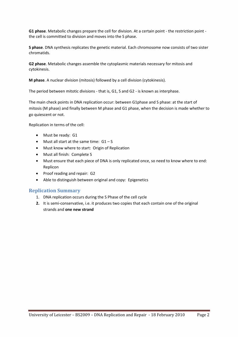
G1 phase. Metabolic changes prepare the cell for division. At a certain point - the restriction point - the cell is committed to division and moves into the S phase.
S phase. DNA synthesis replicates the genetic material. Each chromosome now consists of two sister chromatids.
G2 phase. Metabolic changes assemble the cytoplasmic materials necessary for mitosis and cytokinesis.
M phase. A nuclear division (mitosis) followed by a cell division (cytokinesis).
The period between mitotic divisions - that is, G1, S and G2 - is known as interphase.
The main check points in DNA replication occur: between G1phase and S phase: at the start of
mitosis (M phase) and finally between M phase and G1 phase, when the decision is made whether to
go quiescent or not.
Replication in terms of the cell:
Must be ready: G1
Must all start at the same time: G1 – S
Must know where to start: Origin of Replication
Must all finish: Complete S
Must ensure that each piece of DNA is only replicated once, so need to know where to end:
Replicon
Proof reading and repair: G2
Able to distinguish between original and copy: Epigenetics
Replication Summary 1. DNA replication occurs during the S Phase of the cell cycle
2. It is semi-conservative, i.e. it produces two copies that each contain one of the original
strands and one new strand

University of Leicester – BS2009 – DNA Replication and Repair - 18 February 2010 Page 3
(source: Wikipedia)

University of Leicester – BS2009 – DNA Replication and Repair - 18 February 2010 Page 4
Source: http://barleyworld.org/css430_09/lecture%207-09/notes7-09.html
3. It takes place in a 5’ to 3’ direction with a leading strand and a lagging strand (which is
discontinuous), and the use of an RNA primer
4. In bacteria, there is only a single origin of replication
5. In eukaryotes, there are multiple origins of replication
6. Replication is bi-directional
Source: http://faculty.irsc.edu/FACULTY/TFischer/images/DNA%20replication.jpg
7. In yeast, the ARS (Autonomously Replicating Sequence) element is present . Yeast can be
considered to be the eukaryotic equivalent of E. coli, and it has approximately 400 ARS
elements in 12 chromosomes.

University of Leicester – BS2009 – DNA Replication and Repair - 18 February 2010 Page 5
Replication Enzymes
DNA Polymerase: Matches the correct nucleotide and then joins adjacent nucleotides
together
Primase: Provides and RNA primer to start polymerisation
Ligase: Joins adjacent DNA strands together
Helicase: Unwinds the DNA and melts it
Single Strand Binding Proteins: Keep the DNA single stranded after it has been melted by
helicase
Gyrase: A topisomerase that relieves torsional strain in the DNA molecule
Telomerase: Finishes off the ends of the DNA strand
(Insert video here of DNA replication: http://www.youtube.com/watch?v=teV62zrm2P0
2. Eukaryotes vs Prokaryotes There is much conservation between the two systems, in as much as the enzymology, the replication
fork geometry, the basic fundamental features and the use of multi-protein machinery are all very
much the same in both.
However, there are more protein components in the Eukaryotic replication machinery.
In prokaryotes, the replication form moves 10x faster than in eukaryotes.
Prokaryotic replication
Eukaryotic replication
semiconservative replication
semiconservative replication
single origin replication (oriC)
multiple origins of replication (ARS)
primer synthesized by primase
primer synthesized by subunits of DNA polymerase α
processing enzyme: DNA polymerase III
processing enzymes: DNA polymerases α and δ
removal of primer: DNA polymerase I
removal of primer: DNA polymerase β
DNA free in cytoplasm as nucleoid
chromatin structure, chromosomes, histones
circular DNA
linear DNA: problem of replication of chromosome ends → telomerase

University of Leicester – BS2009 – DNA Replication and Repair - 18 February 2010 Page 6
3. Maintenance of DNA Sequences DNA Polymerase as a Self-Correcting Enzyme
The correct nucleotide has a greater affinity for moving polymerase than the incorrect nucleotide
has.
Exonucleolytic proofreading of DNA polymerase occurs as follows:
DNA molecules with mismatched 3’ OH ends are not effective templates because
polymerase cannot extend when 3’ OH is not base paired.
DNA polymerase has a separate catalytic site that removes unpaired residues at the
terminus
Source: Molecular biology of the cell, 4th Edition
The diagram shows the 2 catalytic sites: P where polymerisation takes place, and E, where editing
takes place
Strand Directed Mismatch Repair System DNA mismatch repair is a system which recognises and repairs erroneous insertion, deletion and
mis-incorporation of bases that can arise during DNA replication. It also repairs some forms of DNA
damage.
Mismatch repair is strand-specific. During DNA synthesis the newly synthesised (daughter) strand
will often include errors. In order to carry out the repairs, the mismatch repair machinery
distinguishes the newly synthesised strand from the template (parental).
The mismatch repair system carries out the following functions:
Removes replication errors which are not recognised by the replication machine
Detects distortions in the DNA helix
Distinguishes the newly replicated strand from the parental strand by means of methylation
of A residues in GATC in bacteria
Methylation occurs shortly after replication occurs
University of Leicester – BS2009 – DNA Replication and Repair - 18 February 2010 Page 7
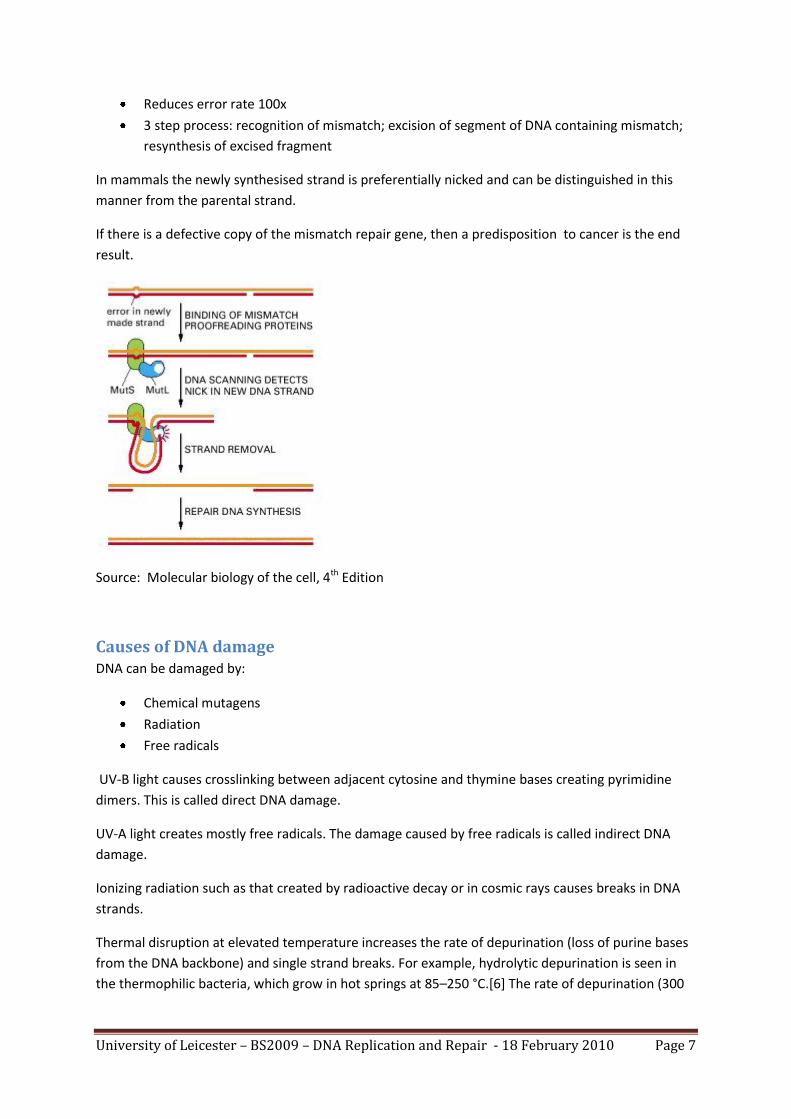
Reduces error rate 100x
3 step process: recognition of mismatch; excision of segment of DNA containing mismatch;
resynthesis of excised fragment
In mammals the newly synthesised strand is preferentially nicked and can be distinguished in this
manner from the parental strand.
If there is a defective copy of the mismatch repair gene, then a predisposition to cancer is the end
result.
Source: Molecular biology of the cell, 4th Edition
Causes of DNA damage DNA can be damaged by:
Chemical mutagens
Radiation
Free radicals
UV-B light causes crosslinking between adjacent cytosine and thymine bases creating pyrimidine
dimers. This is called direct DNA damage.
UV-A light creates mostly free radicals. The damage caused by free radicals is called indirect DNA
damage.
Ionizing radiation such as that created by radioactive decay or in cosmic rays causes breaks in DNA
strands.
Thermal disruption at elevated temperature increases the rate of depurination (loss of purine bases
from the DNA backbone) and single strand breaks. For example, hydrolytic depurination is seen in
the thermophilic bacteria, which grow in hot springs at 85–250 °C.[6] The rate of depurination (300

University of Leicester – BS2009 – DNA Replication and Repair - 18 February 2010 Page 8
purine residues per genome per generation) is too high in these species to be repaired by normal
repair machinery, hence a possibility of an adaptive response cannot be ruled out.
Industrial chemicals such as vinyl chloride and hydrogen peroxide, and environmental chemicals
such as polycyclic hydrocarbons found in smoke, soot and tar create a huge diversity of DNA
adducts- ethenobases, oxidized bases, alkylated phosphotriesters and Crosslinking of DNA just to
name a few.
The natural ageing process and respiration also causes DNA damage at the rate of around 10000
lesions/cell/day.
The main types of DNA damage that occurs are: base loss and base modification.

University of Leicester – BS2009 – DNA Replication and Repair - 18 February 2010 Page 9
* The thickness of the arrows corresponds to the relative sensitivity to alkylation.
Source: openlearn.ac.uk (creative commons)
DNA Repair Despite the 1000’s of alterations that occur in our DNA each day, very few are actually retained as
mutations and this is due to highly efficient DNA repair mechanisms. This is a very important
mechanism, and this is highlighted by the high number of genes that are devoted to DNA repair.
Also, if there is a inactivation or loss of function of the DNA repair genes, then this results in
increased mutation rates.

University of Leicester – BS2009 – DNA Replication and Repair - 18 February 2010 Page 10
Defects in the DNA repair mechanisms are associated with several disease states as can be seen in
the following table:
Disorder Frequency Defect Hereditary/non Hereditary
Fanconi’s anaemia 1/22,000 in some populations
Deficient excision repair
Non – hereditary
Hereditary nonpolyposis colon cancer
1/200 Deficient mismatch repair
Hereditary
Werner’s syndrome 3/1,000000 Deficient helicase Non-hereditary
Xeroderma pigmentosum 1/250,000 Deficient excision repair
Hereditary
DNA damage can activate the expression of whole sets of genes, including:
the Heath Shock Response
the SOS response
The SOS response is a post-replication DNA repair system that allows DNA replication to bypass
lesions or errors in the DNA. The SOS uses the RecA protein. The RecA protein, stimulated by single-
stranded DNA, is involved in the inactivation of the LexA repressor thereby inducing the response. It
is an error-prone repair system.
DNA repair and the cell cycle:
G1 phase arrests and prevents replication errors
S-phase arrests replicon initiation inhibition and prevents replication errors
G2 phase delays and protects against mitotic errors
Base Excision Repair (BER)
BER is a cellular mechanism that repairs damaged DNA throughout the cell cycle. It is primarily
responsible for removing small, non-helix distorting base lesions from the genome. The related
nucleotide excision repair pathway repairs bulky helix-distorting lesions. BER is important for
removing damaged bases that could otherwise cause mutations by mispairing or lead to breaks in
DNA during replication. BER is initiated by DNA glycosylases, which recognize and remove specific
damaged or inappropriate bases, forming AP sites. These are then cleaved by an AP endonuclease.
The resulting single-strand break can then be processed by either short-patch (where a single
nucleotide is replaced) or long-patch BER (where 2-10 new nucleotides are synthesized).

University of Leicester – BS2009 – DNA Replication and Repair - 18 February 2010 Page 11
Source: Friedberg, E.C., Walker, G.C. and Siede, W. (1995). DNA Repair and Mutagenesis. American Society
for Microbiology, Washington DC, USA, pp. 91-225.
A. DNA glycosylace recognises
damaged base
B. Removes base leaving
deoxyribose sugar
C. AP endonuclease cuts
phosphodiester backbone
D. DNA polymerase replaces
missing nucleotide
E. DNA ligase seals nick

University of Leicester – BS2009 – DNA Replication and Repair - 18 February 2010 Page 12
If there are double strand breaks in DNA, then there are 2 methods by which DNA can be repaired:
non-homologous end-joining repair:- the original DNA sequence is altered during repair (by means of
deletions or insertions)
homologous end-joining repair: - this is a general recombination mechanism where information is
transferred from the intact strand.
Source: Molecular biology of the Cell. NCBI Bookshelf
Failure of DNA repair When DNA repair fails, fewer mutations are corrected and this leads to an increase in the number of
mutations in the genome.
In most cases, the protein p53 monitors the repair of damaged DNA, however, if the damage is too
severe, then p53 promotes programmed cell death (apoptosis).
However, mutations in genes which encode the DNA repair proteins can be inherited and this leads
to an overall increase in the number of mutations as errors or damage to the DNA is no longer
repaired efficiently.

University of Leicester – BS2009 – DNA Replication and Repair - 18 February 2010 Page 13
Source: http://en.wikipedia.org/wiki/File:P53_pathways.jpg
In a normal cell p53 is inactivated by its negative regulator, mdm2. Upon DNA damage or other
stress, various pathways will lead to the dissociation of the p53 and mdm2 complex. Once activated,
p53 will either induce a cell cycle arrest to allow repair and survival of the cell or apoptosis to discard
the damage cell. How p53 makes this choice is currently unknown.
This marks the end of the lecture notes for BS2009 on DNA Replication and Repair.