DNA Repair and Genome Maintenance in Bacillus subtilis · series of DNA repair pathways dedicated...
35
DNA Repair and Genome Maintenance in Bacillus subtilis Justin S. Lenhart, Jeremy W. Schroeder, Brian W. Walsh, and Lyle A. Simmons Department of Molecular, Cellular, and Developmental Biology, University of Michigan, Ann Arbor, Michigan, USA INTRODUCTION ............................................................................................................................................530 THE SOS RESPONSE ........................................................................................................................................531 SOS Responses in other Gram-Positive Bacteria ..........................................................................................................535 HOMOLOGOUS RECOMBINATION .........................................................................................................................535 Cross-Link Repair .........................................................................................................................................535 RecN ......................................................................................................................................................535 End Processing by AddAB ................................................................................................................................537 RecQ, RecS, and RecJ .....................................................................................................................................537 RecA Recruitment, Loading, and Coupling to DNA Synthesis ............................................................................................539 RecA-Catalyzed Strand Exchange ........................................................................................................................541 Branch Migration and Holliday Junction Resolution......................................................................................................541 Primosome Assembly ....................................................................................................................................541 Potential Roles for Other SMC-Like Proteins in DSB Repair ...............................................................................................542 NONHOMOLOGOUS END JOINING ........................................................................................................................543 NUCLEOTIDE EXCISION REPAIR ............................................................................................................................543 Transcription-Coupled Repair ............................................................................................................................544 BASE EXCISION REPAIR .....................................................................................................................................544 The “GO” System .........................................................................................................................................544 Uracil Glycosylases........................................................................................................................................545 Processing of Apurinic/Apyrimidinic Sites ................................................................................................................545 ALKYLATION DAMAGE .....................................................................................................................................546 Methyl and Alkyl Glycosylases ............................................................................................................................546 Alkyltransferases and the Ada Response .................................................................................................................546 MISMATCH REPAIR .........................................................................................................................................547 B. subtilis MutL is a Latent Endonuclease .................................................................................................................547 Mismatch Repair Proteins Are Coupled to DNA Synthesis................................................................................................548 Involvement of Clamp in Mismatch Repair .............................................................................................................549 RecD2 Is a Possible Mismatch Repair Helicase in Bacillus .................................................................................................549 YshD, a MutS Paralog .....................................................................................................................................549 DNA DAMAGE TOLERANCE AND TRANSLESION SYNTHESIS..............................................................................................550 DnaE-Induced Mutagenesis ..............................................................................................................................550 DNA INTEGRITY REGULATES BACTERIAL CELL DIVISION ..................................................................................................550 RecA-Dependent Regulation of Cell Division.............................................................................................................550 RecA-Independent and DnaA-Dependent Regulation of Cell Division ...................................................................................551 GENOME INTEGRITY AND CHECKPOINT CONTROL DURING DEVELOPMENT ............................................................................552 Coupling Development to DNA Replication .............................................................................................................552 Sda .......................................................................................................................................................553 SirA .......................................................................................................................................................553 DisA ......................................................................................................................................................553 CONCLUDING REMARKS ...................................................................................................................................554 ACKNOWLEDGMENTS......................................................................................................................................554 REFERENCES ................................................................................................................................................554 INTRODUCTION A ll cells must accurately copy and maintain their DNA to en- sure faithful transmission of their genetic material to the next generation. Organisms ranging from bacteria to humans contain a series of DNA repair pathways dedicated to the specific recogni- tion and repair of the myriad of DNA damage or base-pairing errors that can occur throughout the lifetime of a cell. In mam- mals, it has been estimated that every cell is subject to 15,000 lesions per 24-h period (128, 212). Most of these lesions are hy- pothesized to arise from endogenous sources, such as reactive by- products of normal cellular metabolism (128). In higher eukary- otic systems, failures in DNA repair are often attributed to numerous disease states and/or cell death (e.g., see reference 128). In bacterial cells, DNA damage and mutation accumulation can reduce cell fitness and potentially affect viability (128, 131). Con- versely, mutagenesis also provides the material for evolution, as base pair substitutions may confer a selective advantage to bacte- rial cells vulnerable to a changing environment (e.g., see reference 481). Therefore, transient increases in mutagenesis must be bal- anced carefully with high-fidelity repair to ensure genome preser- vation while providing the opportunity for genetic diversity (for Address correspondence to Lyle A. Simmons, [email protected]. Copyright © 2012, American Society for Microbiology. All Rights Reserved. doi:10.1128/MMBR.05020-11 530 mmbr.asm.org Microbiology and Molecular Biology Reviews p. 530 –564 September 2012 Volume 76 Number 3 on May 18, 2019 by guest http://mmbr.asm.org/ Downloaded from
Transcript of DNA Repair and Genome Maintenance in Bacillus subtilis · series of DNA repair pathways dedicated...
DNA Repair and Genome Maintenance in Bacillus subtilis
Justin S. Lenhart, Jeremy W. Schroeder, Brian W. Walsh, and Lyle A. Simmons
Department of Molecular, Cellular, and Developmental Biology, University of Michigan, Ann Arbor, Michigan, USA
INTRODUCTION . . . . . . . . . . . . . . . . . . . . . . . . . . . . . . . . . . . . . . . . . . . . . . . . . . . . . . . . . . . . . . . . . . . . . . . . . . . . . . . . . . . . . . . . . . . . . . . . . . . . . . . . . . . . . . . . . . . . . . . . . . . . . . . . . . . . . . . . . . . .530THE SOS RESPONSE . . . . . . . . . . . . . . . . . . . . . . . . . . . . . . . . . . . . . . . . . . . . . . . . . . . . . . . . . . . . . . . . . . . . . . . . . . . . . . . . . . . . . . . . . . . . . . . . . . . . . . . . . . . . . . . . . . . . . . . . . . . . . . . . . . . . . . . .531
SOS Responses in other Gram-Positive Bacteria . . . . . . . . . . . . . . . . . . . . . . . . . . . . . . . . . . . . . . . . . . . . . . . . . . . . . . . . . . . . . . . . . . . . . . . . . . . . . . . . . . . . . . . . . . . . . . . . . . . . . . . . . .535HOMOLOGOUS RECOMBINATION . . . . . . . . . . . . . . . . . . . . . . . . . . . . . . . . . . . . . . . . . . . . . . . . . . . . . . . . . . . . . . . . . . . . . . . . . . . . . . . . . . . . . . . . . . . . . . . . . . . . . . . . . . . . . . . . . . . . . . . . .535
Cross-Link Repair . . . . . . . . . . . . . . . . . . . . . . . . . . . . . . . . . . . . . . . . . . . . . . . . . . . . . . . . . . . . . . . . . . . . . . . . . . . . . . . . . . . . . . . . . . . . . . . . . . . . . . . . . . . . . . . . . . . . . . . . . . . . . . . . . . . . . . . . .535RecN. . . . . . . . . . . . . . . . . . . . . . . . . . . . . . . . . . . . . . . . . . . . . . . . . . . . . . . . . . . . . . . . . . . . . . . . . . . . . . . . . . . . . . . . . . . . . . . . . . . . . . . . . . . . . . . . . . . . . . . . . . . . . . . . . . . . . . . . . . . . . . . . . . . . . .535End Processing by AddAB . . . . . . . . . . . . . . . . . . . . . . . . . . . . . . . . . . . . . . . . . . . . . . . . . . . . . . . . . . . . . . . . . . . . . . . . . . . . . . . . . . . . . . . . . . . . . . . . . . . . . . . . . . . . . . . . . . . . . . . . . . . . . . . .537RecQ, RecS, and RecJ . . . . . . . . . . . . . . . . . . . . . . . . . . . . . . . . . . . . . . . . . . . . . . . . . . . . . . . . . . . . . . . . . . . . . . . . . . . . . . . . . . . . . . . . . . . . . . . . . . . . . . . . . . . . . . . . . . . . . . . . . . . . . . . . . . . . .537RecA Recruitment, Loading, and Coupling to DNA Synthesis . . . . . . . . . . . . . . . . . . . . . . . . . . . . . . . . . . . . . . . . . . . . . . . . . . . . . . . . . . . . . . . . . . . . . . . . . . . . . . . . . . . . . . . . . . . .539RecA-Catalyzed Strand Exchange . . . . . . . . . . . . . . . . . . . . . . . . . . . . . . . . . . . . . . . . . . . . . . . . . . . . . . . . . . . . . . . . . . . . . . . . . . . . . . . . . . . . . . . . . . . . . . . . . . . . . . . . . . . . . . . . . . . . . . . .541Branch Migration and Holliday Junction Resolution. . . . . . . . . . . . . . . . . . . . . . . . . . . . . . . . . . . . . . . . . . . . . . . . . . . . . . . . . . . . . . . . . . . . . . . . . . . . . . . . . . . . . . . . . . . . . . . . . . . . . .541Primosome Assembly . . . . . . . . . . . . . . . . . . . . . . . . . . . . . . . . . . . . . . . . . . . . . . . . . . . . . . . . . . . . . . . . . . . . . . . . . . . . . . . . . . . . . . . . . . . . . . . . . . . . . . . . . . . . . . . . . . . . . . . . . . . . . . . . . . . .541Potential Roles for Other SMC-Like Proteins in DSB Repair . . . . . . . . . . . . . . . . . . . . . . . . . . . . . . . . . . . . . . . . . . . . . . . . . . . . . . . . . . . . . . . . . . . . . . . . . . . . . . . . . . . . . . . . . . . . . . .542
NONHOMOLOGOUS END JOINING . . . . . . . . . . . . . . . . . . . . . . . . . . . . . . . . . . . . . . . . . . . . . . . . . . . . . . . . . . . . . . . . . . . . . . . . . . . . . . . . . . . . . . . . . . . . . . . . . . . . . . . . . . . . . . . . . . . . . . . .543NUCLEOTIDE EXCISION REPAIR . . . . . . . . . . . . . . . . . . . . . . . . . . . . . . . . . . . . . . . . . . . . . . . . . . . . . . . . . . . . . . . . . . . . . . . . . . . . . . . . . . . . . . . . . . . . . . . . . . . . . . . . . . . . . . . . . . . . . . . . . . . .543
Transcription-Coupled Repair . . . . . . . . . . . . . . . . . . . . . . . . . . . . . . . . . . . . . . . . . . . . . . . . . . . . . . . . . . . . . . . . . . . . . . . . . . . . . . . . . . . . . . . . . . . . . . . . . . . . . . . . . . . . . . . . . . . . . . . . . . . .544BASE EXCISION REPAIR . . . . . . . . . . . . . . . . . . . . . . . . . . . . . . . . . . . . . . . . . . . . . . . . . . . . . . . . . . . . . . . . . . . . . . . . . . . . . . . . . . . . . . . . . . . . . . . . . . . . . . . . . . . . . . . . . . . . . . . . . . . . . . . . . . . . .544
The “GO” System . . . . . . . . . . . . . . . . . . . . . . . . . . . . . . . . . . . . . . . . . . . . . . . . . . . . . . . . . . . . . . . . . . . . . . . . . . . . . . . . . . . . . . . . . . . . . . . . . . . . . . . . . . . . . . . . . . . . . . . . . . . . . . . . . . . . . . . . .544Uracil Glycosylases. . . . . . . . . . . . . . . . . . . . . . . . . . . . . . . . . . . . . . . . . . . . . . . . . . . . . . . . . . . . . . . . . . . . . . . . . . . . . . . . . . . . . . . . . . . . . . . . . . . . . . . . . . . . . . . . . . . . . . . . . . . . . . . . . . . . . . . .545Processing of Apurinic/Apyrimidinic Sites . . . . . . . . . . . . . . . . . . . . . . . . . . . . . . . . . . . . . . . . . . . . . . . . . . . . . . . . . . . . . . . . . . . . . . . . . . . . . . . . . . . . . . . . . . . . . . . . . . . . . . . . . . . . . . . .545
ALKYLATION DAMAGE . . . . . . . . . . . . . . . . . . . . . . . . . . . . . . . . . . . . . . . . . . . . . . . . . . . . . . . . . . . . . . . . . . . . . . . . . . . . . . . . . . . . . . . . . . . . . . . . . . . . . . . . . . . . . . . . . . . . . . . . . . . . . . . . . . . . .546Methyl and Alkyl Glycosylases . . . . . . . . . . . . . . . . . . . . . . . . . . . . . . . . . . . . . . . . . . . . . . . . . . . . . . . . . . . . . . . . . . . . . . . . . . . . . . . . . . . . . . . . . . . . . . . . . . . . . . . . . . . . . . . . . . . . . . . . . . . .546Alkyltransferases and the Ada Response . . . . . . . . . . . . . . . . . . . . . . . . . . . . . . . . . . . . . . . . . . . . . . . . . . . . . . . . . . . . . . . . . . . . . . . . . . . . . . . . . . . . . . . . . . . . . . . . . . . . . . . . . . . . . . . . .546
MISMATCH REPAIR . . . . . . . . . . . . . . . . . . . . . . . . . . . . . . . . . . . . . . . . . . . . . . . . . . . . . . . . . . . . . . . . . . . . . . . . . . . . . . . . . . . . . . . . . . . . . . . . . . . . . . . . . . . . . . . . . . . . . . . . . . . . . . . . . . . . . . . . .547B. subtilis MutL is a Latent Endonuclease . . . . . . . . . . . . . . . . . . . . . . . . . . . . . . . . . . . . . . . . . . . . . . . . . . . . . . . . . . . . . . . . . . . . . . . . . . . . . . . . . . . . . . . . . . . . . . . . . . . . . . . . . . . . . . . . .547Mismatch Repair Proteins Are Coupled to DNA Synthesis. . . . . . . . . . . . . . . . . . . . . . . . . . . . . . . . . . . . . . . . . . . . . . . . . . . . . . . . . . . . . . . . . . . . . . . . . . . . . . . . . . . . . . . . . . . . . . . .548Involvement of � Clamp in Mismatch Repair. . . . . . . . . . . . . . . . . . . . . . . . . . . . . . . . . . . . . . . . . . . . . . . . . . . . . . . . . . . . . . . . . . . . . . . . . . . . . . . . . . . . . . . . . . . . . . . . . . . . . . . . . . . . .549RecD2 Is a Possible Mismatch Repair Helicase in Bacillus . . . . . . . . . . . . . . . . . . . . . . . . . . . . . . . . . . . . . . . . . . . . . . . . . . . . . . . . . . . . . . . . . . . . . . . . . . . . . . . . . . . . . . . . . . . . . . . . .549YshD, a MutS Paralog . . . . . . . . . . . . . . . . . . . . . . . . . . . . . . . . . . . . . . . . . . . . . . . . . . . . . . . . . . . . . . . . . . . . . . . . . . . . . . . . . . . . . . . . . . . . . . . . . . . . . . . . . . . . . . . . . . . . . . . . . . . . . . . . . . . . .549
DNA DAMAGE TOLERANCE AND TRANSLESION SYNTHESIS. . . . . . . . . . . . . . . . . . . . . . . . . . . . . . . . . . . . . . . . . . . . . . . . . . . . . . . . . . . . . . . . . . . . . . . . . . . . . . . . . . . . . . . . . . . . . .550DnaE-Induced Mutagenesis . . . . . . . . . . . . . . . . . . . . . . . . . . . . . . . . . . . . . . . . . . . . . . . . . . . . . . . . . . . . . . . . . . . . . . . . . . . . . . . . . . . . . . . . . . . . . . . . . . . . . . . . . . . . . . . . . . . . . . . . . . . . . .550
DNA INTEGRITY REGULATES BACTERIAL CELL DIVISION . . . . . . . . . . . . . . . . . . . . . . . . . . . . . . . . . . . . . . . . . . . . . . . . . . . . . . . . . . . . . . . . . . . . . . . . . . . . . . . . . . . . . . . . . . . . . . . . . .550RecA-Dependent Regulation of Cell Division. . . . . . . . . . . . . . . . . . . . . . . . . . . . . . . . . . . . . . . . . . . . . . . . . . . . . . . . . . . . . . . . . . . . . . . . . . . . . . . . . . . . . . . . . . . . . . . . . . . . . . . . . . . . .550RecA-Independent and DnaA-Dependent Regulation of Cell Division. . . . . . . . . . . . . . . . . . . . . . . . . . . . . . . . . . . . . . . . . . . . . . . . . . . . . . . . . . . . . . . . . . . . . . . . . . . . . . . . . . .551
GENOME INTEGRITY AND CHECKPOINT CONTROL DURING DEVELOPMENT . . . . . . . . . . . . . . . . . . . . . . . . . . . . . . . . . . . . . . . . . . . . . . . . . . . . . . . . . . . . . . . . . . . . . . . . . . . .552Coupling Development to DNA Replication . . . . . . . . . . . . . . . . . . . . . . . . . . . . . . . . . . . . . . . . . . . . . . . . . . . . . . . . . . . . . . . . . . . . . . . . . . . . . . . . . . . . . . . . . . . . . . . . . . . . . . . . . . . . .552Sda . . . . . . . . . . . . . . . . . . . . . . . . . . . . . . . . . . . . . . . . . . . . . . . . . . . . . . . . . . . . . . . . . . . . . . . . . . . . . . . . . . . . . . . . . . . . . . . . . . . . . . . . . . . . . . . . . . . . . . . . . . . . . . . . . . . . . . . . . . . . . . . . . . . . . . .553SirA . . . . . . . . . . . . . . . . . . . . . . . . . . . . . . . . . . . . . . . . . . . . . . . . . . . . . . . . . . . . . . . . . . . . . . . . . . . . . . . . . . . . . . . . . . . . . . . . . . . . . . . . . . . . . . . . . . . . . . . . . . . . . . . . . . . . . . . . . . . . . . . . . . . . . . .553DisA . . . . . . . . . . . . . . . . . . . . . . . . . . . . . . . . . . . . . . . . . . . . . . . . . . . . . . . . . . . . . . . . . . . . . . . . . . . . . . . . . . . . . . . . . . . . . . . . . . . . . . . . . . . . . . . . . . . . . . . . . . . . . . . . . . . . . . . . . . . . . . . . . . . . . .553
CONCLUDING REMARKS . . . . . . . . . . . . . . . . . . . . . . . . . . . . . . . . . . . . . . . . . . . . . . . . . . . . . . . . . . . . . . . . . . . . . . . . . . . . . . . . . . . . . . . . . . . . . . . . . . . . . . . . . . . . . . . . . . . . . . . . . . . . . . . . . . .554ACKNOWLEDGMENTS. . . . . . . . . . . . . . . . . . . . . . . . . . . . . . . . . . . . . . . . . . . . . . . . . . . . . . . . . . . . . . . . . . . . . . . . . . . . . . . . . . . . . . . . . . . . . . . . . . . . . . . . . . . . . . . . . . . . . . . . . . . . . . . . . . . . . .554REFERENCES . . . . . . . . . . . . . . . . . . . . . . . . . . . . . . . . . . . . . . . . . . . . . . . . . . . . . . . . . . . . . . . . . . . . . . . . . . . . . . . . . . . . . . . . . . . . . . . . . . . . . . . . . . . . . . . . . . . . . . . . . . . . . . . . . . . . . . . . . . . . . . . .554
INTRODUCTION
All cells must accurately copy and maintain their DNA to en-sure faithful transmission of their genetic material to the next
generation. Organisms ranging from bacteria to humans contain aseries of DNA repair pathways dedicated to the specific recogni-tion and repair of the myriad of DNA damage or base-pairingerrors that can occur throughout the lifetime of a cell. In mam-mals, it has been estimated that every cell is subject to �15,000lesions per 24-h period (128, 212). Most of these lesions are hy-pothesized to arise from endogenous sources, such as reactive by-products of normal cellular metabolism (128). In higher eukary-otic systems, failures in DNA repair are often attributed tonumerous disease states and/or cell death (e.g., see reference 128).
In bacterial cells, DNA damage and mutation accumulation canreduce cell fitness and potentially affect viability (128, 131). Con-versely, mutagenesis also provides the material for evolution, asbase pair substitutions may confer a selective advantage to bacte-rial cells vulnerable to a changing environment (e.g., see reference481). Therefore, transient increases in mutagenesis must be bal-anced carefully with high-fidelity repair to ensure genome preser-vation while providing the opportunity for genetic diversity (for
Address correspondence to Lyle A. Simmons, [email protected].
Copyright © 2012, American Society for Microbiology. All Rights Reserved.
doi:10.1128/MMBR.05020-11
530 mmbr.asm.org Microbiology and Molecular Biology Reviews p. 530–564 September 2012 Volume 76 Number 3
on May 18, 2019 by guest
http://mm
br.asm.org/
Dow
nloaded from
reviews, see references 434 to 438). The conservation of DNA re-pair pathways from bacteria to humans is often very impressive;such conservation has allowed for experimentally tractable bacte-ria to provide important mechanistic insights into processes crit-ical for genome maintenance in more complex systems (for a re-view, see reference 128). To date, the DNA repair and mutagenesispathways in Escherichia coli are the best understood for a bacterialsystem, and this information has led to the identification of severalfounding members of DNA repair and damage tolerance super-families that show exquisite conservation across biology (e.g., seereferences 8, 78, 118, 119, 128, 231, and 296).
Efforts in genome sequencing and evolution have estimatedthat Gram-positive and Gram-negative bacteria diverged over abillion years ago (e.g., see references 67, 298, and 303). Such a longseparation has allowed for many DNA repair processes in Gram-negative and Gram-positive bacteria to diverge, evolving substan-tial differences in both their molecular mechanisms and modes ofregulation. Over the last decade, it has become increasingly clearthat the DNA repair pathways of many Gram-positive bacteria canbe different from those described for E. coli. In some cases, entirepathways exist in Gram-positive bacteria that are completely ab-sent from the prototypical Gram-negative bacterium E. coli. Be-low, we review and discuss several pathways that are critical forgenome maintenance in the Gram-positive bacterium Bacillussubtilis. We discuss many DNA repair, DNA damage tolerance,and DNA damage checkpoints that maintain genome integrityduring vegetative growth and during the developmental programof sporulation. We also review the similarities of DNA repair andDNA replication pathways in several other bacteria, and we relatethese findings to what is known for better-characterized bacterialsystems.
THE SOS RESPONSE
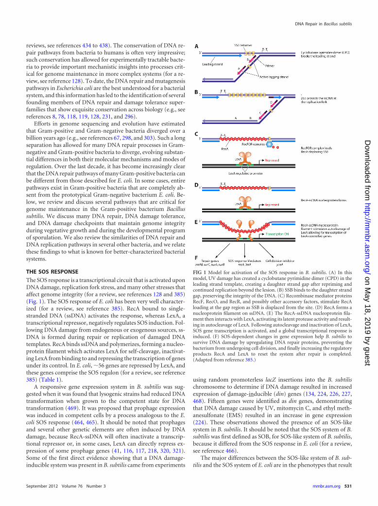
The SOS response is a transcriptional circuit that is activated uponDNA damage, replication fork stress, and many other stresses thataffect genome integrity (for a review, see references 128 and 385)(Fig. 1). The SOS response of E. coli has been very well character-ized (for a review, see reference 385). RecA bound to single-stranded DNA (ssDNA) activates the response, whereas LexA, atranscriptional repressor, negatively regulates SOS induction. Fol-lowing DNA damage from endogenous or exogenous sources, ss-DNA is formed during repair or replication of damaged DNAtemplates. RecA binds ssDNA and polymerizes, forming a nucleo-protein filament which activates LexA for self-cleavage, inactivat-ing LexA from binding to and repressing the transcription of genesunder its control. In E. coli, �56 genes are repressed by LexA, andthese genes comprise the SOS regulon (for a review, see reference385) (Table 1).
A responsive gene expression system in B. subtilis was sug-gested when it was found that lysogenic strains had reduced DNAtransformation when grown to the competent state for DNAtransformation (469). It was proposed that prophage expressionwas induced in competent cells by a process analogous to the E.coli SOS response (464, 465). It should be noted that prophagesand several other genetic elements are often induced by DNAdamage, because RecA-ssDNA will often inactivate a transcrip-tional repressor or, in some cases, LexA can directly repress ex-pression of some prophage genes (41, 116, 117, 218, 320, 321).Some of the first direct evidence showing that a DNA damage-inducible system was present in B. subtilis came from experiments
using random promoterless lacZ insertions into the B. subtilischromosome to determine if DNA damage resulted in increasedexpression of damage-inducible (din) genes (134, 224, 226, 227,468). Fifteen genes were identified as din genes, demonstratingthat DNA damage caused by UV, mitomycin C, and ethyl meth-anesulfonate (EMS) resulted in an increase in gene expression(224). These observations showed the presence of an SOS-likesystem in B. subtilis. It should be noted that the SOS system of B.subtilis was first defined as SOB, for SOS-like system of B. subtilis,because it differed from the SOS response in E. coli (for a review,see reference 466).
The major differences between the SOS-like system of B. sub-tilis and the SOS system of E. coli are in the phenotypes that result
FIG 1 Model for activation of the SOS response in B. subtilis. (A) In thismodel, UV damage has created a cyclobutane pyrimidine dimer (CPD) in theleading strand template, creating a daughter strand gap after repriming andcontinued replication beyond the lesion. (B) SSB binds to the daughter strandgap, preserving the integrity of the DNA. (C) Recombinase mediator proteinsRecF, RecO, and RecR, and possibly other accessory factors, stimulate RecAloading at the gap region as SSB is displaced from the site. (D) RecA forms anucleoprotein filament on ssDNA. (E) The RecA-ssDNA nucleoprotein fila-ment then interacts with LexA, activating its latent protease activity and result-ing in autocleavage of LexA. Following autocleavage and inactivation of LexA,SOS gene transcription is activated, and a global transcriptional response isinduced. (F) SOS-dependent changes in gene expression help B. subtilis tosurvive DNA damage by upregulating DNA repair proteins, preventing thebacterium from undergoing cell division, and finally increasing the regulatoryproducts RecA and LexA to reset the system after repair is completed.(Adapted from reference 385.)
DNA Repair in Bacillus subtilis
September 2012 Volume 76 Number 3 mmbr.asm.org 531
on May 18, 2019 by guest
http://mm
br.asm.org/
Dow
nloaded from
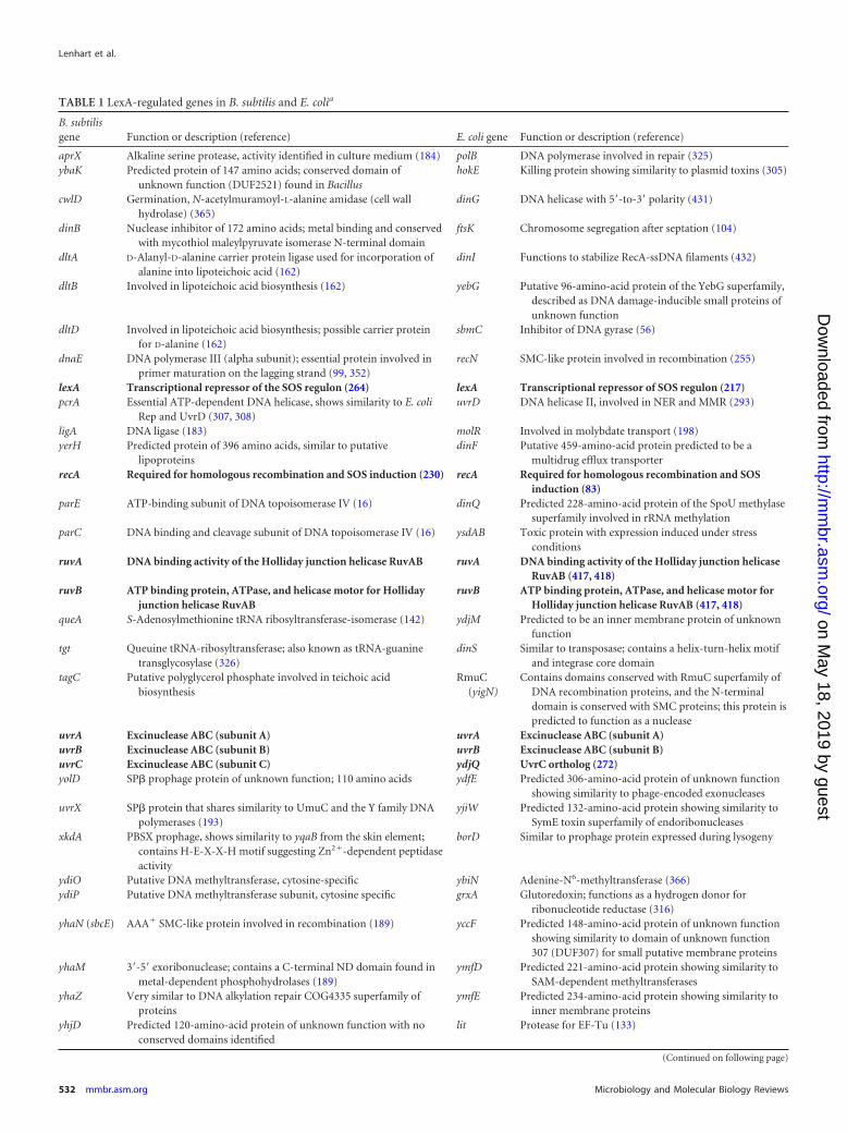
TABLE 1 LexA-regulated genes in B. subtilis and E. colia
B. subtilisgene Function or description (reference) E. coli gene Function or description (reference)
aprX Alkaline serine protease, activity identified in culture medium (184) polB DNA polymerase involved in repair (325)ybaK Predicted protein of 147 amino acids; conserved domain of
unknown function (DUF2521) found in BacillushokE Killing protein showing similarity to plasmid toxins (305)
cwlD Germination, N-acetylmuramoyl-L-alanine amidase (cell wallhydrolase) (365)
dinG DNA helicase with 5=-to-3= polarity (431)
dinB Nuclease inhibitor of 172 amino acids; metal binding and conservedwith mycothiol maleylpyruvate isomerase N-terminal domain
ftsK Chromosome segregation after septation (104)
dltA D-Alanyl-D-alanine carrier protein ligase used for incorporation ofalanine into lipoteichoic acid (162)
dinI Functions to stabilize RecA-ssDNA filaments (432)
dltB Involved in lipoteichoic acid biosynthesis (162) yebG Putative 96-amino-acid protein of the YebG superfamily,described as DNA damage-inducible small proteins ofunknown function
dltD Involved in lipoteichoic acid biosynthesis; possible carrier proteinfor D-alanine (162)
sbmC Inhibitor of DNA gyrase (56)
dnaE DNA polymerase III (alpha subunit); essential protein involved inprimer maturation on the lagging strand (99, 352)
recN SMC-like protein involved in recombination (255)
lexA Transcriptional repressor of the SOS regulon (264) lexA Transcriptional repressor of SOS regulon (217)pcrA Essential ATP-dependent DNA helicase, shows similarity to E. coli
Rep and UvrD (307, 308)uvrD DNA helicase II, involved in NER and MMR (293)
ligA DNA ligase (183) molR Involved in molybdate transport (198)yerH Predicted protein of 396 amino acids, similar to putative
lipoproteinsdinF Putative 459-amino-acid protein predicted to be a
multidrug efflux transporterrecA Required for homologous recombination and SOS induction (230) recA Required for homologous recombination and SOS
induction (83)parE ATP-binding subunit of DNA topoisomerase IV (16) dinQ Predicted 228-amino-acid protein of the SpoU methylase
superfamily involved in rRNA methylationparC DNA binding and cleavage subunit of DNA topoisomerase IV (16) ysdAB Toxic protein with expression induced under stress
conditionsruvA DNA binding activity of the Holliday junction helicase RuvAB ruvA DNA binding activity of the Holliday junction helicase
RuvAB (417, 418)ruvB ATP binding protein, ATPase, and helicase motor for Holliday
junction helicase RuvABruvB ATP binding protein, ATPase, and helicase motor for
Holliday junction helicase RuvAB (417, 418)queA S-Adenosylmethionine tRNA ribosyltransferase-isomerase (142) ydjM Predicted to be an inner membrane protein of unknown
functiontgt Queuine tRNA-ribosyltransferase; also known as tRNA-guanine
transglycosylase (326)dinS Similar to transposase; contains a helix-turn-helix motif
and integrase core domaintagC Putative polyglycerol phosphate involved in teichoic acid
biosynthesisRmuC
(yigN)Contains domains conserved with RmuC superfamily of
DNA recombination proteins, and the N-terminaldomain is conserved with SMC proteins; this protein ispredicted to function as a nuclease
uvrA Excinuclease ABC (subunit A) uvrA Excinuclease ABC (subunit A)uvrB Excinuclease ABC (subunit B) uvrB Excinuclease ABC (subunit B)uvrC Excinuclease ABC (subunit C) ydjQ UvrC ortholog (272)yolD SP� prophage protein of unknown function; 110 amino acids ydfE Predicted 306-amino-acid protein of unknown function
showing similarity to phage-encoded exonucleasesuvrX SP� protein that shares similarity to UmuC and the Y family DNA
polymerases (193)yjiW Predicted 132-amino-acid protein showing similarity to
SymE toxin superfamily of endoribonucleasesxkdA PBSX prophage, shows similarity to yqaB from the skin element;
contains H-E-X-X-H motif suggesting Zn2�-dependent peptidaseactivity
borD Similar to prophage protein expressed during lysogeny
ydiO Putative DNA methyltransferase, cytosine-specific ybiN Adenine-N6-methyltransferase (366)ydiP Putative DNA methyltransferase subunit, cytosine specific grxA Glutoredoxin; functions as a hydrogen donor for
ribonucleotide reductase (316)yhaN (sbcE) AAA� SMC-like protein involved in recombination (189) yccF Predicted 148-amino-acid protein of unknown function
showing similarity to domain of unknown function307 (DUF307) for small putative membrane proteins
yhaM 3=-5= exoribonuclease; contains a C-terminal ND domain found inmetal-dependent phosphohydrolases (189)
ymfD Predicted 221-amino-acid protein showing similarity toSAM-dependent methyltransferases
yhaZ Very similar to DNA alkylation repair COG4335 superfamily ofproteins
ymfE Predicted 234-amino-acid protein showing similarity toinner membrane proteins
yhjD Predicted 120-amino-acid protein of unknown function with noconserved domains identified
lit Protease for EF-Tu (133)
(Continued on following page)
Lenhart et al.
532 mmbr.asm.org Microbiology and Molecular Biology Reviews
on May 18, 2019 by guest
http://mm
br.asm.org/
Dow
nloaded from
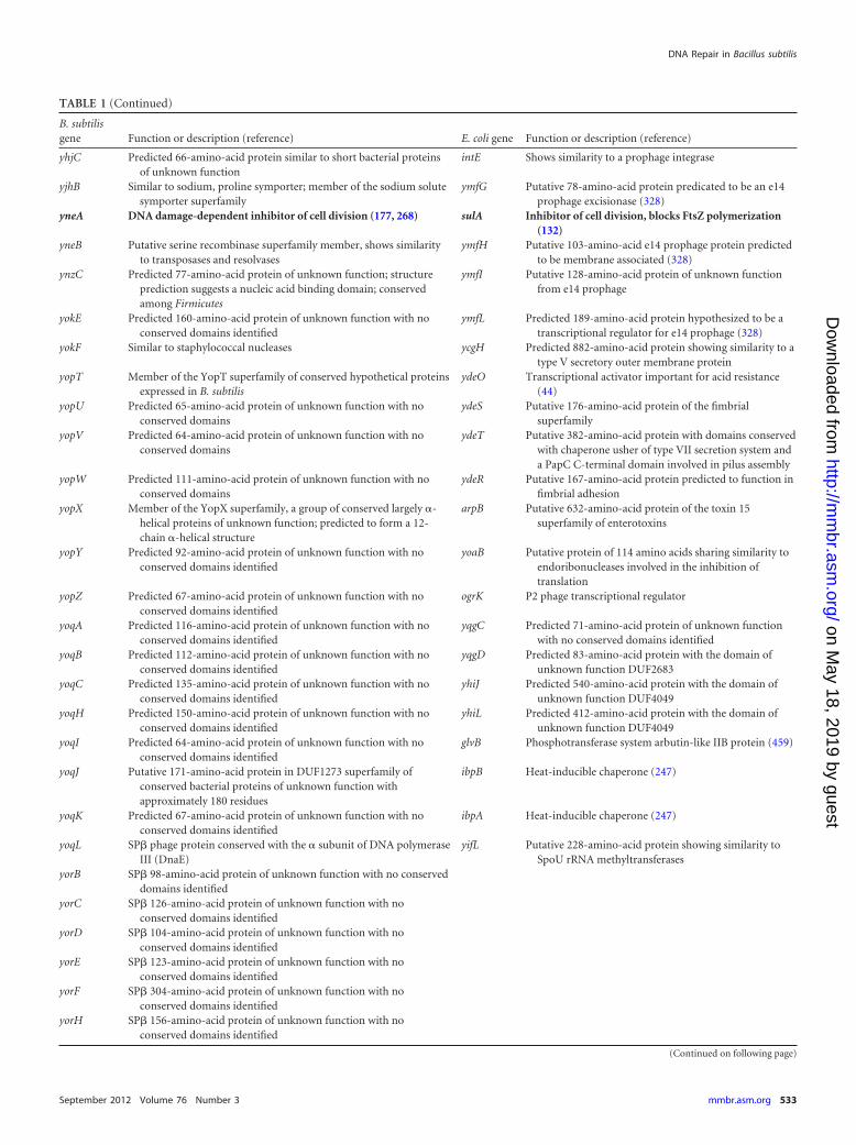
TABLE 1 (Continued)
B. subtilisgene Function or description (reference) E. coli gene Function or description (reference)
yhjC Predicted 66-amino-acid protein similar to short bacterial proteinsof unknown function
intE Shows similarity to a prophage integrase
yjhB Similar to sodium, proline symporter; member of the sodium solutesymporter superfamily
ymfG Putative 78-amino-acid protein predicated to be an e14prophage excisionase (328)
yneA DNA damage-dependent inhibitor of cell division (177, 268) sulA Inhibitor of cell division, blocks FtsZ polymerization(132)
yneB Putative serine recombinase superfamily member, shows similarityto transposases and resolvases
ymfH Putative 103-amino-acid e14 prophage protein predictedto be membrane associated (328)
ynzC Predicted 77-amino-acid protein of unknown function; structureprediction suggests a nucleic acid binding domain; conservedamong Firmicutes
ymfI Putative 128-amino-acid protein of unknown functionfrom e14 prophage
yokE Predicted 160-amino-acid protein of unknown function with noconserved domains identified
ymfL Predicted 189-amino-acid protein hypothesized to be atranscriptional regulator for e14 prophage (328)
yokF Similar to staphylococcal nucleases ycgH Predicted 882-amino-acid protein showing similarity to atype V secretory outer membrane protein
yopT Member of the YopT superfamily of conserved hypothetical proteinsexpressed in B. subtilis
ydeO Transcriptional activator important for acid resistance(44)
yopU Predicted 65-amino-acid protein of unknown function with noconserved domains
ydeS Putative 176-amino-acid protein of the fimbrialsuperfamily
yopV Predicted 64-amino-acid protein of unknown function with noconserved domains
ydeT Putative 382-amino-acid protein with domains conservedwith chaperone usher of type VII secretion system anda PapC C-terminal domain involved in pilus assembly
yopW Predicted 111-amino-acid protein of unknown function with noconserved domains
ydeR Putative 167-amino-acid protein predicted to function infimbrial adhesion
yopX Member of the YopX superfamily, a group of conserved largely �-helical proteins of unknown function; predicted to form a 12-chain �-helical structure
arpB Putative 632-amino-acid protein of the toxin 15superfamily of enterotoxins
yopY Predicted 92-amino-acid protein of unknown function with noconserved domains identified
yoaB Putative protein of 114 amino acids sharing similarity toendoribonucleases involved in the inhibition oftranslation
yopZ Predicted 67-amino-acid protein of unknown function with noconserved domains identified
ogrK P2 phage transcriptional regulator
yoqA Predicted 116-amino-acid protein of unknown function with noconserved domains identified
yqgC Predicted 71-amino-acid protein of unknown functionwith no conserved domains identified
yoqB Predicted 112-amino-acid protein of unknown function with noconserved domains identified
yqgD Predicted 83-amino-acid protein with the domain ofunknown function DUF2683
yoqC Predicted 135-amino-acid protein of unknown function with noconserved domains identified
yhiJ Predicted 540-amino-acid protein with the domain ofunknown function DUF4049
yoqH Predicted 150-amino-acid protein of unknown function with noconserved domains identified
yhiL Predicted 412-amino-acid protein with the domain ofunknown function DUF4049
yoqI Predicted 64-amino-acid protein of unknown function with noconserved domains identified
glvB Phosphotransferase system arbutin-like IIB protein (459)
yoqJ Putative 171-amino-acid protein in DUF1273 superfamily ofconserved bacterial proteins of unknown function withapproximately 180 residues
ibpB Heat-inducible chaperone (247)
yoqK Predicted 67-amino-acid protein of unknown function with noconserved domains identified
ibpA Heat-inducible chaperone (247)
yoqL SP� phage protein conserved with the � subunit of DNA polymeraseIII (DnaE)
yifL Putative 228-amino-acid protein showing similarity toSpoU rRNA methyltransferases
yorB SP� 98-amino-acid protein of unknown function with no conserveddomains identified
yorC SP� 126-amino-acid protein of unknown function with noconserved domains identified
yorD SP� 104-amino-acid protein of unknown function with noconserved domains identified
yorE SP� 123-amino-acid protein of unknown function with noconserved domains identified
yorF SP� 304-amino-acid protein of unknown function with noconserved domains identified
yorH SP� 156-amino-acid protein of unknown function with noconserved domains identified
(Continued on following page)
DNA Repair in Bacillus subtilis
September 2012 Volume 76 Number 3 mmbr.asm.org 533
on May 18, 2019 by guest
http://mm
br.asm.org/
Dow
nloaded from
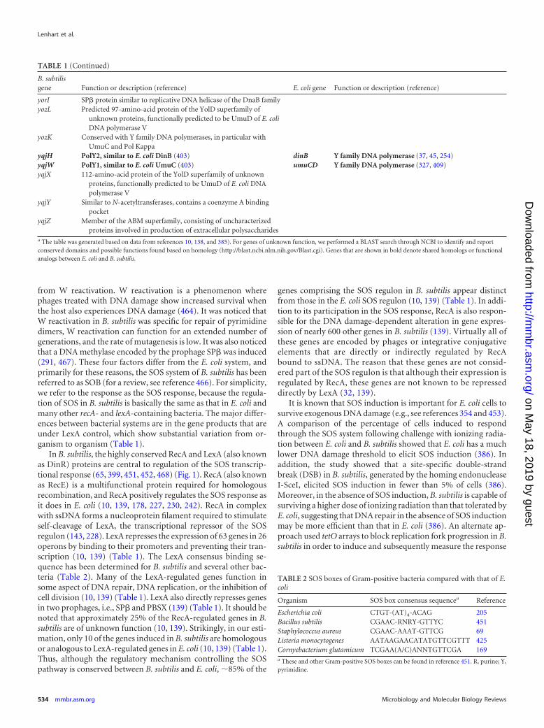
from W reactivation. W reactivation is a phenomenon wherephages treated with DNA damage show increased survival whenthe host also experiences DNA damage (464). It was noticed thatW reactivation in B. subtilis was specific for repair of pyrimidinedimers, W reactivation can function for an extended number ofgenerations, and the rate of mutagenesis is low. It was also noticedthat a DNA methylase encoded by the prophage SP� was induced(291, 467). These four factors differ from the E. coli system, andprimarily for these reasons, the SOS system of B. subtilis has beenreferred to as SOB (for a review, see reference 466). For simplicity,we refer to the response as the SOS response, because the regula-tion of SOS in B. subtilis is basically the same as that in E. coli andmany other recA- and lexA-containing bacteria. The major differ-ences between bacterial systems are in the gene products that areunder LexA control, which show substantial variation from or-ganism to organism (Table 1).
In B. subtilis, the highly conserved RecA and LexA (also knownas DinR) proteins are central to regulation of the SOS transcrip-tional response (65, 399, 451, 452, 468) (Fig. 1). RecA (also knownas RecE) is a multifunctional protein required for homologousrecombination, and RecA positively regulates the SOS response asit does in E. coli (10, 139, 178, 227, 230, 242). RecA in complexwith ssDNA forms a nucleoprotein filament required to stimulateself-cleavage of LexA, the transcriptional repressor of the SOSregulon (143, 228). LexA represses the expression of 63 genes in 26operons by binding to their promoters and preventing their tran-scription (10, 139) (Table 1). The LexA consensus binding se-quence has been determined for B. subtilis and several other bac-teria (Table 2). Many of the LexA-regulated genes function insome aspect of DNA repair, DNA replication, or the inhibition ofcell division (10, 139) (Table 1). LexA also directly represses genesin two prophages, i.e., SP� and PBSX (139) (Table 1). It should benoted that approximately 25% of the RecA-regulated genes in B.subtilis are of unknown function (10, 139). Strikingly, in our esti-mation, only 10 of the genes induced in B. subtilis are homologousor analogous to LexA-regulated genes in E. coli (10, 139) (Table 1).Thus, although the regulatory mechanism controlling the SOSpathway is conserved between B. subtilis and E. coli, �85% of the
genes comprising the SOS regulon in B. subtilis appear distinctfrom those in the E. coli SOS regulon (10, 139) (Table 1). In addi-tion to its participation in the SOS response, RecA is also respon-sible for the DNA damage-dependent alteration in gene expres-sion of nearly 600 other genes in B. subtilis (139). Virtually all ofthese genes are encoded by phages or integrative conjugativeelements that are directly or indirectly regulated by RecAbound to ssDNA. The reason that these genes are not consid-ered part of the SOS regulon is that although their expression isregulated by RecA, these genes are not known to be represseddirectly by LexA (32, 139).
It is known that SOS induction is important for E. coli cells tosurvive exogenous DNA damage (e.g., see references 354 and 453).A comparison of the percentage of cells induced to respondthrough the SOS system following challenge with ionizing radia-tion between E. coli and B. subtilis showed that E. coli has a muchlower DNA damage threshold to elicit SOS induction (386). Inaddition, the study showed that a site-specific double-strandbreak (DSB) in B. subtilis, generated by the homing endonucleaseI-SceI, elicited SOS induction in fewer than 5% of cells (386).Moreover, in the absence of SOS induction, B. subtilis is capable ofsurviving a higher dose of ionizing radiation than that tolerated byE. coli, suggesting that DNA repair in the absence of SOS inductionmay be more efficient than that in E. coli (386). An alternate ap-proach used tetO arrays to block replication fork progression in B.subtilis in order to induce and subsequently measure the response
TABLE 1 (Continued)
B. subtilisgene Function or description (reference) E. coli gene Function or description (reference)
yorI SP� protein similar to replicative DNA helicase of the DnaB familyyozL Predicted 97-amino-acid protein of the YolD superfamily of
unknown proteins, functionally predicted to be UmuD of E. coliDNA polymerase V
yozK Conserved with Y family DNA polymerases, in particular withUmuC and Pol Kappa
yqjH PolY2, similar to E. coli DinB (403) dinB Y family DNA polymerase (37, 45, 254)yqjW PolY1, similar to E. coli UmuC (403) umuCD Y family DNA polymerase (327, 409)yqjX 112-amino-acid protein of the YolD superfamily of unknown
proteins, functionally predicted to be UmuD of E. coli DNApolymerase V
yqjY Similar to N-acetyltransferases, contains a coenzyme A bindingpocket
yqjZ Member of the ABM superfamily, consisting of uncharacterizedproteins involved in production of extracellular polysaccharides
a The table was generated based on data from references 10, 138, and 385). For genes of unknown function, we performed a BLAST search through NCBI to identify and reportconserved domains and possible functions found based on homology (http://blast.ncbi.nlm.nih.gov/Blast.cgi). Genes that are shown in bold denote shared homologs or functionalanalogs between E. coli and B. subtilis.
TABLE 2 SOS boxes of Gram-positive bacteria compared with that of E.coli
Organism SOS box consensus sequencea Reference
Escherichia coli CTGT-(AT)4-ACAG 205Bacillus subtilis CGAAC-RNRY-GTTYC 451Staphylococcus aureus CGAAC-AAAT-GTTCG 69Listeria monocytogenes AATAAGAACATATGTTCGTTT 425Cornyebacterium glutamicum TCGAA(A/C)ANNTGTTCGA 169a These and other Gram-positive SOS boxes can be found in reference 451. R, purine; Y,pyrimidine.
Lenhart et al.
534 mmbr.asm.org Microbiology and Molecular Biology Reviews
on May 18, 2019 by guest
http://mm
br.asm.org/
Dow
nloaded from
to replication fork arrest (23). That study found that the SOSresponse in B. subtilis was not readily induced by a protein block toreplication fork progression (23). Taking both of these studiesinto consideration, it seems that B. subtilis can efficiently repairDNA damage or tolerate perturbations to replication forks in away that does not readily induce the SOS transcriptional response.In support of this idea, cells incapable of SOS induction due tointegration of a noncleavable lexA variant were shown to survive aconsiderable amount of DNA damage, suggesting efficient repairin the absence of triggering the SOS response (386).
SOS Responses in other Gram-Positive Bacteria
The SOS response has been investigated in several pathogenic andnonpathogenic Gram-positive organisms. The opportunistic hu-man pathogen Staphylococcus aureus contains the lexA and recAgenes (18, 27, 163). As expected, antibiotics that damage DNA,such as fluoroquinolones (inhibitors of DNA gyrase), induced theSOS response when administered at subinhibitory concentrations(259). The genome-wide SOS response of S. aureus to ciprofloxa-cin, a fluoroquinolone which induces DSBs and stalls replicationforks, was determined using microarrays (69). In that study, theresponses of wild-type and noncleavable lexA-bearing S. aureusstrains to ciprofloxacin were compared (69). Sixteen genes wereidentified as under LexA control (69). This number is small rela-tive to the number of genes under SOS control in B. subtilis (10,139). The genes that were identified as upregulated included recAand lexA, genes involved in nucleotide excision repair (NER)(uvrA and uvrB), topoisomerase IV genes (parE and parC), andnuclease genes (sbcC and sbcD) (69). The binding of S. aureusLexA to the promoter of recA has been demonstrated (27), andthis result is consistent with the mode of recA regulation in othersystems (for a review, see reference 385). Interestingly, fibronectinbinding proteins produced by S. aureus to aid in its attachment tothe extracellular matrix and the plasma membrane are induced byfluoroquinolones (27), and the promoter for the fibronectin bind-ing protein B gene (fnbB) is bound by LexA. This suggests thatDNA damage may affect the ability of S. aureus to form clumps orattach to surfaces, a feature important during infection (27).
Listeria monocytogenes also contains the lexA and recA genes(113, 425). Challenge of Listeria with the DNA damaging agentmitomycin C resulted in the identification of 29 genes inducedfrom 16 operons (425). Of these genes, most are involved in DNArepair, regulation of cytokinesis, and translesion synthesis (425).In addition to these studies, the SOS regulon has also been inves-tigated in other Gram-positive bacteria. The SOS responses inmany other Gram-positive bacteria are also regulated by RecA andLexA, as expected (169, 288). Overall, the number of genes andfunctions of genes controlled by this response differ considerablyfrom organism to organism. With that stated, the recA gene, thelexA gene, and a gene product important for inhibiting cell divi-sion are consistently found to be under LexA control.
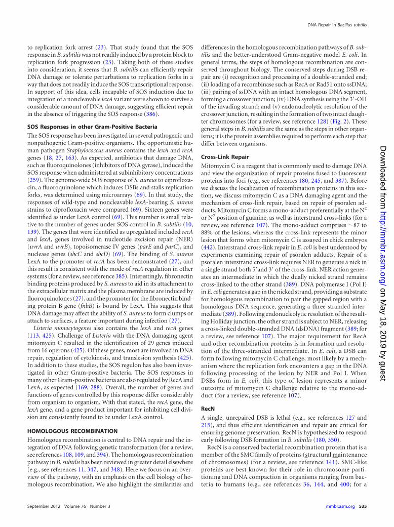
HOMOLOGOUS RECOMBINATION
Homologous recombination is central to DNA repair and the in-tegration of DNA following genetic transformation (for a review,see references 108, 109, and 394). The homologous recombinationpathway in B. subtilis has been reviewed in greater detail elsewhere(e.g., see references 11, 347, and 348). Here we focus on an over-view of the pathway, with an emphasis on the cell biology of ho-mologous recombination. We also highlight the similarities and
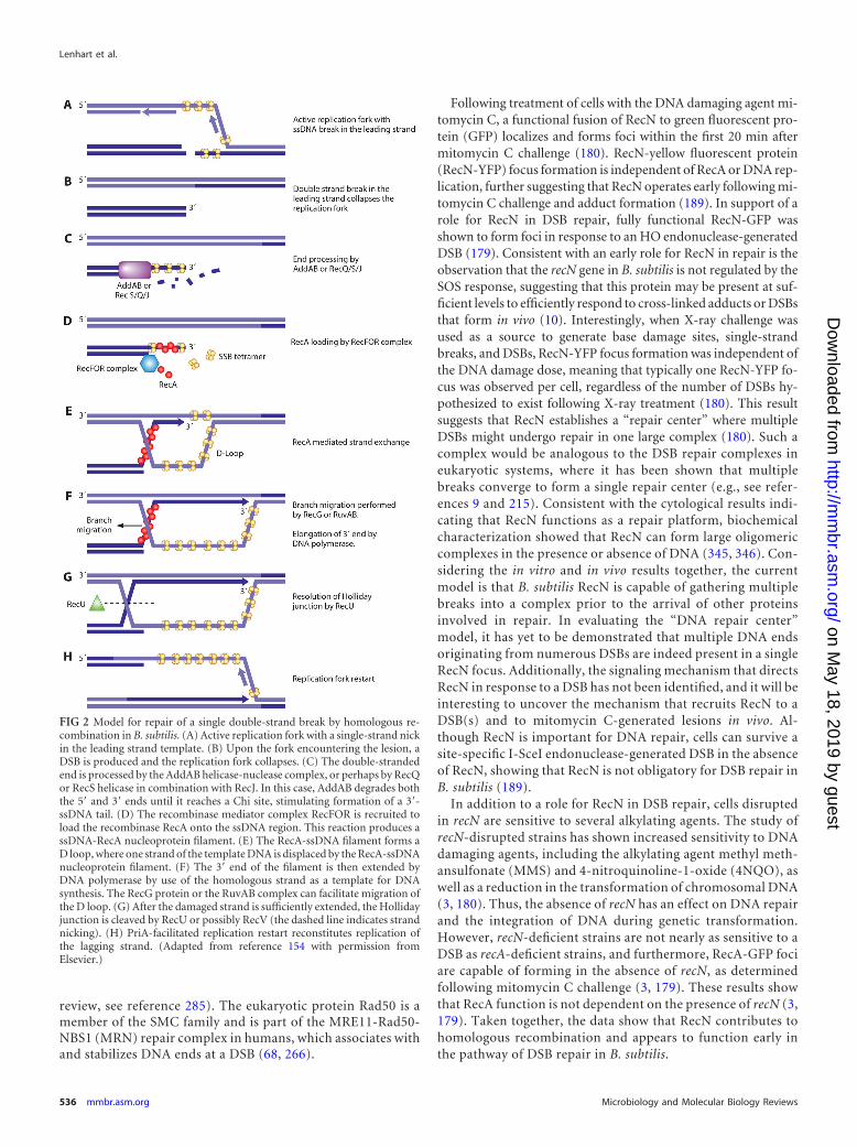
differences in the homologous recombination pathways of B. sub-tilis and the better-understood Gram-negative model E. coli. Ingeneral terms, the steps of homologous recombination are con-served throughout biology. The conserved steps during DSB re-pair are (i) recognition and processing of a double-stranded end;(ii) loading of a recombinase such as RecA or Rad51 onto ssDNA;(iii) pairing of ssDNA with an intact homologous DNA segment,forming a crossover junction; (iv) DNA synthesis using the 3=-OHof the invading strand; and (v) endonucleolytic resolution of thecrossover junction, resulting in the formation of two intact daugh-ter chromosomes (for a review, see reference 128) (Fig. 2). Thesegeneral steps in B. subtilis are the same as the steps in other organ-isms; it is the protein assemblies required to perform each step thatdiffer between organisms.
Cross-Link Repair
Mitomycin C is a reagent that is commonly used to damage DNAand view the organization of repair proteins fused to fluorescentproteins into foci (e.g., see references 180, 245, and 387). Beforewe discuss the localization of recombination proteins in this sec-tion, we discuss mitomycin C as a DNA damaging agent and themechanism of cross-link repair, based on repair of psoralen ad-ducts. Mitomycin C forms a mono-adduct preferentially at the N2
or N7 position of guanine, as well as interstrand cross-links (for areview, see reference 107). The mono-adduct comprises �87 to88% of the lesions, whereas the cross-link represents the minorlesion that forms when mitomycin C is assayed in chick embryos(442). Interstrand cross-link repair in E. coli is best understood byexperiments examining repair of psoralen adducts. Repair of apsoralen interstrand cross-link requires NER to generate a nick ina single strand both 5=and 3= of the cross-link. NER action gener-ates an intermediate in which the dually nicked strand remainscross-linked to the other strand (389). DNA polymerase I (Pol I)in E. coli generates a gap in the nicked strand, providing a substratefor homologous recombination to pair the gapped region with ahomologous DNA sequence, generating a three-stranded inter-mediate (389). Following endonucleolytic resolution of the result-ing Holliday junction, the other strand is subject to NER, releasinga cross-linked double-stranded DNA (dsDNA) fragment (389; fora review, see reference 107). The major requirement for RecAand other recombination proteins is in formation and resolu-tion of the three-stranded intermediate. In E. coli, a DSB canform following mitomycin C challenge, most likely by a mech-anism where the replication fork encounters a gap in the DNAfollowing processing of the lesion by NER and Pol I. WhenDSBs form in E. coli, this type of lesion represents a minoroutcome of mitomycin C challenge relative to the mono-ad-duct (for a review, see reference 107).
RecN
A single, unrepaired DSB is lethal (e.g., see references 127 and215), and thus efficient identification and repair are critical forensuring genome preservation. RecN is hypothesized to respondearly following DSB formation in B. subtilis (180, 350).
RecN is a conserved bacterial recombination protein that is amember of the SMC family of proteins (structural maintenanceof chromosomes) (for a review, see reference 141). SMC-likeproteins are best known for their role in chromosome parti-tioning and DNA compaction in organisms ranging from bac-teria to humans (e.g., see references 36, 144, and 400; for a
DNA Repair in Bacillus subtilis
September 2012 Volume 76 Number 3 mmbr.asm.org 535
on May 18, 2019 by guest
http://mm
br.asm.org/
Dow
nloaded from
review, see reference 285). The eukaryotic protein Rad50 is amember of the SMC family and is part of the MRE11-Rad50-NBS1 (MRN) repair complex in humans, which associates withand stabilizes DNA ends at a DSB (68, 266).
Following treatment of cells with the DNA damaging agent mi-tomycin C, a functional fusion of RecN to green fluorescent pro-tein (GFP) localizes and forms foci within the first 20 min aftermitomycin C challenge (180). RecN-yellow fluorescent protein(RecN-YFP) focus formation is independent of RecA or DNA rep-lication, further suggesting that RecN operates early following mi-tomycin C challenge and adduct formation (189). In support of arole for RecN in DSB repair, fully functional RecN-GFP wasshown to form foci in response to an HO endonuclease-generatedDSB (179). Consistent with an early role for RecN in repair is theobservation that the recN gene in B. subtilis is not regulated by theSOS response, suggesting that this protein may be present at suf-ficient levels to efficiently respond to cross-linked adducts or DSBsthat form in vivo (10). Interestingly, when X-ray challenge wasused as a source to generate base damage sites, single-strandbreaks, and DSBs, RecN-YFP focus formation was independent ofthe DNA damage dose, meaning that typically one RecN-YFP fo-cus was observed per cell, regardless of the number of DSBs hy-pothesized to exist following X-ray treatment (180). This resultsuggests that RecN establishes a “repair center” where multipleDSBs might undergo repair in one large complex (180). Such acomplex would be analogous to the DSB repair complexes ineukaryotic systems, where it has been shown that multiplebreaks converge to form a single repair center (e.g., see refer-ences 9 and 215). Consistent with the cytological results indi-cating that RecN functions as a repair platform, biochemicalcharacterization showed that RecN can form large oligomericcomplexes in the presence or absence of DNA (345, 346). Con-sidering the in vitro and in vivo results together, the currentmodel is that B. subtilis RecN is capable of gathering multiplebreaks into a complex prior to the arrival of other proteinsinvolved in repair. In evaluating the “DNA repair center”model, it has yet to be demonstrated that multiple DNA endsoriginating from numerous DSBs are indeed present in a singleRecN focus. Additionally, the signaling mechanism that directsRecN in response to a DSB has not been identified, and it will beinteresting to uncover the mechanism that recruits RecN to aDSB(s) and to mitomycin C-generated lesions in vivo. Al-though RecN is important for DNA repair, cells can survive asite-specific I-SceI endonuclease-generated DSB in the absenceof RecN, showing that RecN is not obligatory for DSB repair inB. subtilis (189).
In addition to a role for RecN in DSB repair, cells disruptedin recN are sensitive to several alkylating agents. The study ofrecN-disrupted strains has shown increased sensitivity to DNAdamaging agents, including the alkylating agent methyl meth-ansulfonate (MMS) and 4-nitroquinoline-1-oxide (4NQO), aswell as a reduction in the transformation of chromosomal DNA(3, 180). Thus, the absence of recN has an effect on DNA repairand the integration of DNA during genetic transformation.However, recN-deficient strains are not nearly as sensitive to aDSB as recA-deficient strains, and furthermore, RecA-GFP fociare capable of forming in the absence of recN, as determinedfollowing mitomycin C challenge (3, 179). These results showthat RecA function is not dependent on the presence of recN (3,179). Taken together, the data show that RecN contributes tohomologous recombination and appears to function early inthe pathway of DSB repair in B. subtilis.
FIG 2 Model for repair of a single double-strand break by homologous re-combination in B. subtilis. (A) Active replication fork with a single-strand nickin the leading strand template. (B) Upon the fork encountering the lesion, aDSB is produced and the replication fork collapses. (C) The double-strandedend is processed by the AddAB helicase-nuclease complex, or perhaps by RecQor RecS helicase in combination with RecJ. In this case, AddAB degrades boththe 5= and 3= ends until it reaches a Chi site, stimulating formation of a 3=-ssDNA tail. (D) The recombinase mediator complex RecFOR is recruited toload the recombinase RecA onto the ssDNA region. This reaction produces assDNA-RecA nucleoprotein filament. (E) The RecA-ssDNA filament forms aD loop, where one strand of the template DNA is displaced by the RecA-ssDNAnucleoprotein filament. (F) The 3= end of the filament is then extended byDNA polymerase by use of the homologous strand as a template for DNAsynthesis. The RecG protein or the RuvAB complex can facilitate migration ofthe D loop. (G) After the damaged strand is sufficiently extended, the Hollidayjunction is cleaved by RecU or possibly RecV (the dashed line indicates strandnicking). (H) PriA-facilitated replication restart reconstitutes replication ofthe lagging strand. (Adapted from reference 154 with permission fromElsevier.)
Lenhart et al.
536 mmbr.asm.org Microbiology and Molecular Biology Reviews
on May 18, 2019 by guest
http://mm
br.asm.org/
Dow
nloaded from
End Processing by AddAB
After a DSB is identified, the ends are processed, marking them forrepair. In bacterial systems, the two enzyme classes responsible forthis task are the helicase-nuclease complexes RecBCD and AddAB(for a review, see reference 471). During repair of a double-stranded end, the DNA is unwound and simultaneously digestedby a nuclease, eventually generating a 3=-ssDNA overhang, whichonce loaded with RecA forms a nucleoprotein filament capable ofundergoing strand exchange in the next step of homologous re-combination (for a review, see reference 81) (Fig. 2). In E. coli, themajor complex required for double-stranded end processing is theRecBCD helicase-nuclease complex (for a review, see reference100). Briefly, RecBCD contains two helicase motors, RecB andRecD, with opposite polarities (29, 101, 410). The unwound ss-DNA segments are then cleaved by the single RecB nuclease (388,474, 475). The RecB nuclease is required for cleavage of bothstrands (388). Cleavage of the 3= strand is more processive becausethe 3= strand is channeled closer to the active site of RecB. Cleavageof the 5= strand is less frequent, as movement of the 5= strand isfurther away from the active site, making cleavage of the 5= strandless efficient (388). The RecC protein contains a “pin” functioningto efficiently separate the duplex DNA (388). Cleavage of the 3=strand is attenuated when the Chi sequence (crossover hot spotinstigator) (5=-GCTGGTGG-3=) is encountered and bound byRecC, while cleavage of the 5= strand continues. This overallmechanism allows for degradation of both strands until a Chisequence is encountered, generating a 3= overhang, a substrateappropriate for RecA binding.
B. subtilis lacks RecBCD, so the analogous functioning complexis AddAB, which performs the same overall reaction, although themechanisms and organization of the protein complex are different(for a review, see reference 471). The AddAB complex engages inDSB end processing and is highly conserved among the Firmicutes(84, 148–151, 185, 257). Deletion or inactivation of the addA oraddB gene causes substantial defects in DSB repair and increasesthe sensitivity of B. subtilis to a wide spectrum of DNA damagingagents (3).
AddA is both a helicase and an endonuclease. The N terminus ofAddA is an SF1A family helicase, and the C-terminal domain is aRecB-type nuclease, which cuts the 3=-5= strand (319, 338, 470).AddB does not have helicase activity; the C terminus of AddBforms a RecB-like nuclease domain which cleaves the 5=-3= strand(319, 470). An Fe-S cluster is present in AddB, and this region hasbeen shown to bind DNA in the crystal structure and to stabilizethe protein structure (338). AddAB initiates end processing bybinding to a double-stranded end, followed by processive un-winding and cleavage of both DNA strands (58; for a review, seereference 471). Degradation of both strands continues until a Chisite is encountered (58–61). Chi sites in B. subtilis have the shortsequence 5=-AGCGG-3= and are enriched in the chromosome(61). Chi sites are also found to generally coorient with the leadingstrand of replication (61). When AddAB encounters Chi, muchlike E. coli RecBCD, its nuclease activity is altered, allowing forcontinued unwinding and degradation of the 5= strand down-stream of Chi while interrupting the degradation of the 3= stranddownstream of Chi (59). The attenuation in nuclease activity onthe 3= strand produces a 3=-ssDNA segment that is appropriate forRecA binding (45, 46). As mentioned above, AddB is a nuclease,and the N-terminal portion of the protein shows similarity to
DNA helicases, although AddB lacks the motifs required for heli-case activity (338). Recent structural work has shown that the Chisequence binds to the Chi recognition site in AddB. This bindingevent prevents degradation of the 3= strand by AddAB as it ischanneled through the AddAB complex (338).
Stoichiometric analysis of the AddAB complex shows thatAddAB is active in vitro as a 1:1 heterodimer (472). The het-erodimer has numerous activities, including the ability to bind adsDNA end and to catalyze unwinding of duplex DNA (471).AddAB also has two genetically separable nuclease activities (470).As mentioned above, the AddAB helicase activity is conferred bythe AddA subunit and has 3=-to-5= polarity (472, 473). Inactiva-tion of the ATP binding site (Walker A motif) in AddB has verylittle effect on the helicase activity of the AddAB complex, furtherdemonstrating that AddA powers the helicase activity of AddAB(149–151). Unlike RecBCD, which contains two helicase activitiesand a single nuclease, AddAB has two nucleases that reside sepa-rately in its individual subunits, as well as a single helicase (for areview, see reference 471). In B. subtilis and many Gram-positivebacteria, the AddAB complex is responsible primarily for end pro-cessing during DSB repair.
RecQ, RecS, and RecJ
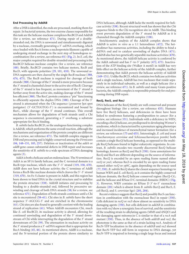
DNA helicases of the RecQ family are well conserved and presentthroughout biology (for a review, see reference 455). Humanshave five RecQ helicases, and deficiencies in three have beenlinked to syndromes featuring a predisposition to cancer (for areview, see reference 331). Individuals with a deficiency in WRN,encoding a RecQ helicase, are predisposed to developing Werner’ssyndrome, which is characterized by a premature aging phenotypeand increased incidence of mesenchymal tumor formation (for areview, see references 175 and 455). Interestingly, E. coli and yeast(Saccharomyces cerevisiae and Schizosaccharomyces pombe) bothcontain a single RecQ helicase homolog, as opposed to the multi-ple RecQ helicases found in higher eukaryotic organisms. In con-trast, B. subtilis encodes two recently discovered RecQ helicasehomologs, known as RecQ and RecS (350). Gene annotations forRecQ and RecS are different depending on the source of informa-tion. RecQ is encoded by an open reading frame named eitherrecQ or yocI, whereas RecS is encoded by an open reading framenamed either recQ or ypbC, again depending on the source used(77, 350). B. subtilis RecQ shares the closest sequence homology tohuman WRN and E. coli RecQ, as it contains the highly conservedhelicase domain, the RecQ helicase conserved region (RecQ-Ct),and the helicase and RNase D C-terminal domains (HRDC) (Fig.3). However, WRN contains an RNase D 3=-to-5= exonucleasedomain (281) which is absent from B. subtilis RecQ and RecS, E.coli RecQ, and S. cerevisiae Sgs1 (281, 284).
Recent evidence suggests that RecQ and possibly RecS can func-tion in combination with the exonuclease RecJ in repair (350).Cells deficient in recQ or recS show almost no sensitivity to DNAdamaging agents (350), but cells deficient in addAB in combina-tion with recJ show a synergistic loss of survival, and the sensitivityof the resulting strain to alkylating agents (MMS and 4NQO) andthe damaging agent mitomycin C is similar to that of a recA nullmutant (350). Thus, in the absence of both addAB and recJ, thephenotype is the same as that of a strain lacking recA. Analysis ofRecN-YFP localization in the absence of addAB and recJ showedthat RecN-YFP foci still form in response to DNA damage, yetRecN-YFP is impaired in forming a single large focus and instead
DNA Repair in Bacillus subtilis
September 2012 Volume 76 Number 3 mmbr.asm.org 537
on May 18, 2019 by guest
http://mm
br.asm.org/
Dow
nloaded from
forms multiple small foci (350). These results have been used toconclude that end processing is required for the establishment of asingle large RecN-YFP complex, with the caveat that alkylatingagents were used and a DSB was not directly tested (350). To-gether, these data also support the model that RecN complexesform in vivo prior to action by AddAB or RecQ-RecS-RecJ, furthersupporting a role for RecN early in DNA repair.
Recent work showed that the SSB protein targets several pro-teins, including RecQ, RecS, and RecJ, to the replication fork (77).Proteomic analysis of the interactome of SSB revealed numerousDNA repair proteins that bind SSB in vivo, including but not lim-ited to RecQ, RecS, and RecJ (77) (see Table 3 for a complete list).When YFP-RecQ was expressed ectopically with the native recQgene intact, YFP-RecQ was shown to form foci that colocalized
with the replisome (DnaX-cyan fluorescent protein [DnaX-CFP])in untreated cells (197). This suggests that RecQ is constitutivelyassociated with the DNA replication machinery and that repli-some association is mediated by interaction with SSB. Strikingly, aC-terminal truncation of ssb (ssb�35) which reduces RecQ bind-ing in vitro blocks GFP-RecQ focus formation in vivo, indicatingthat the C terminus of SSB may recruit RecQ to replication forks inB. subtilis (197). This observation is supported by results showingthat the SSB-RecQ interaction is conserved in E. coli and that theSSB C terminus is required for their interaction (374).
Ectopic expression of GFP-RecJ with the native recJ locus intactalso showed that GFP-RecJ formed foci in vivo (77). Focus forma-tion by GFP-RecJ under these conditions occurred in the absenceof exogenous DNA damage, suggesting that like RecQ, RecJ may
FIG 3 Schematic representation of the domain structure of B. subtilis DNA helicases RecQ and RecS in comparison with human WRN. The N-terminal regioncontains the helicase motifs (light blue); the RecQ helicase conserved region (RecQ-Ct) (dark blue/purple) and the helicase and RNase D C-terminal domains(HRDC) (red) are also shown. The human protein contains the RNase D domain N-terminal region, which contains a 3=-to-5= exonuclease domain. (Adaptedfrom references 24 with permission of Oxford University Press, 281 with permission from Elsevier, and 284 with permission from Macmillan Publishers Ltd.)
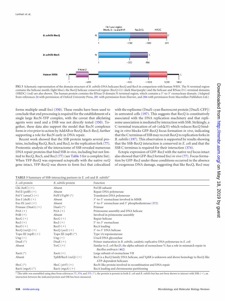
TABLE 3 Summary of SSB-interacting partners in E. coli and B. subtilisa
E. coli protein B. subtilis protein Function
Chi (holC) (�) Absent Pol III subunitPol II (polB) (�) Absent Repair DNA polymerasePol V (umuC) (�) PolY1/YqjW (*) Translesion DNA polymeraseExo I (sbcB) (�) Absent 3=-to-5= exonuclease involved in MMRExo IX (xni) (�) Absent 3=-to-5= exonuclease and 3= phosphodiesterase (372)Primase (DnaG) (�) DnaG (*) PrimasePriA (�) PriA (�) Primosome assembly and DNA helicasePriB (�) Absent Involved in primosome assemblyRecG (�) RecG (�) Repair helicaseRecJ (�) RecJ (�) 5=-to-3= exonucleaseRecO (�) RecO (�) RecA loadingRecQ (recQ) (�) RecQ (yocI) (�) 3=-to-5= DNA helicaseTopo III (topB) (�) Topo III (topB) (*) Type 1A topoisomeraseUng (�) Ung (�) Uracil DNA glycosylaseDnaE (*) DnaE (�) Primer maturation in B. subtilis, catalytic replicative DNA polymerase in E. coliAbsent YrrC (�) Similar to E. coli RecD, the alpha subunit of exonuclease V; has a role in mismatch repair in
Bacillus anthracis (462)XseA (*) XseA (�) Large subunit of exonuclease VIIAbsent YpbB/RecS (recQ) (�) RecS is a RecQ family DNA helicase, and YpbB is unknown and shows homology to RecQ-like
ATP-dependent helicasesSbcC (*) SbcC (yirY) (�) RecN-like protein involved in recombination and DNA repairRarA (mgsA) (*) RarA (mgsA) (�) RecA loading and chromosome partitioninga This table was assembled using data from references 77, 374, and 375. (*), the protein is present in both E. coli and B. subtilis but has not been shown to interact with SSB; (�), aninteraction between the indicated protein and SSB has been measured.
Lenhart et al.
538 mmbr.asm.org Microbiology and Molecular Biology Reviews
on May 18, 2019 by guest
http://mm
br.asm.org/
Dow
nloaded from

also routinely be positioned at active replication forks in B. subti-lis. GFP-RecJ formed one or two foci per nucleoid, at a subcellularposition similar to where the replisome would be expected to lo-calize (77). It is worth noting that when visualized in vivo, func-tional AddA-GFP and AddB-YFP localized diffusely throughoutB. subtilis cells and failed to organize into discrete foci in cells thatwere exposed to DNA damaging agents (245). Together, theseresults suggest that RecQ-GFP and RecJ-GFP could be positionedat the replisome, whereas AddAB does not appear to be located atthe replisome as judged by fluorescence microscopy. These resultssuggest that the RecQ/RecJ functions could be localized to the siteof DNA replication in B. subtilis through interaction with SSB(77).
RecS was also shown to bind B. subtilis SSB in vitro, and SSBbearing a tandem affinity purification (TAP) tag was purified fromextracts with RecS associated (77). In the reciprocal experiment,TAP tag purification of RecS showed interaction with SSB but alsowith an unannotated protein, YpbB (77). Interestingly, RecS iscotranslated with YpbB, as the two genes slightly overlap (77).When gfp-ypbB-recS was expressed ectopically, the complexformed foci, but only if the SSB C terminus was intact (77). WhenGFP-YpbB or GFP-RecS was imaged, foci were not observed, sug-gesting that RecS and YpbB function together. Thus, the C termi-nus of SSB in B. subtilis is critical for DNA repair and recruitmentof RecQ, RecS, and RecJ to the replication fork in vivo. SSB isgaining considerable attention as a protein that facilitates traffick-ing of replication and repair proteins to the replication fork andother ssDNA substrates in B. subtilis and E. coli (77, 197, 374, 375).The proteins that bind SSB in B. subtilis and E. coli show someoverlap. However, many SSB binding partners are not shared be-tween the two organisms (Table 2).
RecA Recruitment, Loading, and Coupling to DNA Synthesis
Processing of DNA ends, predominantly by AddAB and perhapsby RecQ-RecJ or RecS-RecJ, will result in a 3=-ssDNA suitable forRecA binding (Fig. 2 and 4). As mentioned above, B. subtilis alsocontains SSB (also termed SsbA), which is essential for DNA rep-lication and critical for repair processes during exponential-phasegrowth (183). Unlike E. coli, B. subtilis contains a second SSBparalog, designated SsbB, encoded by the ywpH gene (213). YwpHis upregulated during the development of genetic competence andis critical for DNA transformation (213). SSB-coated ssDNA in-hibits RecA filament formation (31, 187, 195, 423). At the sametime, SSB can promote RecA binding by removing secondarystructure from the DNA, ultimately providing a more suitablesubstrate for RecA filament formation (187). Even so, RecA mustreplace SSB on ssDNA in order to form the RecA-ssDNA nucleo-protein filament that mediates strand exchange (422). The mech-anisms for RecA loading are well established for E. coli yet poorlyunderstood for many other bacteria. In E. coli, RecBCD and theRecFOR pathways can each function in RecA loading (153, 201,412, 422, 423). Once E. coli RecBCD produces a 3=-ssDNA end,RecBCD actively begins to load RecA onto ssDNA in the 5=-3=direction, while displacing SSB (83). The RecFOR pathway func-tions primarily in the repair of daughter strand gaps, as well as inprotection of the nascent strand following replication fork arrestin response to UV damage (66).
In B. subtilis, AddAB is not known to load RecA, whereas theRecFOR complex, specifically RecO, does have a RecA loadingfunction (for a review, see references 11, 347, and 348). Mutations
in recF, recO, or recR strongly sensitize B. subtilis to DNA damag-ing agents (MMS, EMS, and 4NQO), which primarily formdaughter strand gaps (3, 4, 121). Mutations in recF, recO, and recRalso decrease the transformation of B. subtilis with chromosomalDNA, providing more direct evidence that these proteins functionin recombination of ssDNA entering the cell (3, 4, 121). It is notentirely clear if B. subtilis RecFOR functions in repair of a double-stranded end (DSE), which would be formed by ionizing radiationor through an I-SceI-induced break in the chromosome. If it does,one possibility is that RecFOR may help to load RecA onto the3=-ssDNA tail generated following end processing by AddAB.
A critical actor in the RecFOR complex is the RecO protein. Thedomain organization and structure of the E. coli and Deinococcusradiodurans RecO proteins are very similar (201, 232, 337). Basedon homology, B. subtilis RecO has a similar overall domain orga-nization, particularly to that of D. radiodurans RecO (201, 232,337). The N-terminal domain is an oligonucleotide/oligosaccha-ride binding fold (OB fold) characteristic of proteins that bindssDNA and/or dsDNA. RecO contains a C-terminal domain com-posed of six alpha helices forming the core and a zinc bindingdomain (201, 232). For D. radiodurans, zinc binding is coordi-nated by four conserved cysteine residues, which are conserved inthe B. subtilis protein (201, 232). The E. coli protein has one of thefour cysteine residues, and the crystal structure of E. coli RecOlacks zinc (337). The overall fold of the “zinc binding domain” inE. coli RecO is very similar to that of the D. radiodurans protein.Thus, although sequence conservation between the E. coli and D.radiodurans RecO proteins is low (�21% identical), the overallstructures are very similar (201, 232).
In addition to a role in RecA loading, RecO generally containstwo conserved biochemical activities: it can anneal complemen-tary single strands, and it helps to load and facilitate strand ex-change by RecA (236, 237). For the B. subtilis proteins, it has beenshown in vitro that RecO will help to load RecA onto SSB-boundssDNA, although the mechanism of action is not clear (236, 237).A major difference in RecO function between B. subtilis and E. coliis that B. subtilis RecO alone is sufficient to nucleate RecA filamentformation on SSB-coated ssDNA in vitro, while in E. coli, RecOand RecR are necessary, because RecO will not overcome SSBinhibition alone (422, 423).
A possible mechanism is that RecO physically binds and loadsRecA onto ssDNA or that the strand annealing activity of RecOindirectly helps to stimulate RecA loading. Experiments have beenperformed to test for a direct interaction between B. subtilis RecAand RecO, but so far an interaction between these proteins has notbeen shown (236). However, in E. coli, a very weak interaction wasdetected between RecO and RecA by surface plasmon resonance(423). Thus, there is some evidence suggesting that RecO bindsdirectly to RecA, although the binding appears to be very weak(423). It is also not clear whether RecF and RecR function in theloading of RecA onto ssDNA in B. subtilis (179). In total, the RecAloading mechanism in B. subtilis is unclear and will require furtherstudy in order to understand the concerted steps that result inRecA-ssDNA filaments in vitro and in vivo.
One of the requirements for homologous recombination is thepresence of two chromosome copies in order to provide an iden-tical template for repair of a DSB. By coupling homologous re-combination with DNA replication status, a cell may ensure thatthis requirement is met. A study using a partially functional recA-gfp fusion allele integrated at the native recA locus as the only
DNA Repair in Bacillus subtilis
September 2012 Volume 76 Number 3 mmbr.asm.org 539
on May 18, 2019 by guest
http://mm
br.asm.org/
Dow
nloaded from

source of RecA activity in the cell showed that ongoing DNA rep-lication was necessary for RecA-GFP to form foci in response tosingle-strand gaps or an I-SceI-induced DSB in vivo (387). In thisstudy, a DSB was generated and RecA-GFP failed to organize intoa focus when DNA replication initiation was blocked (387). It isalso worth noting that DNA replication has previously beenshown to be necessary for SOS induction in E. coli (357). In thatwork, the LexA cleavage and degradation following UV irradia-tion were shown to be dependent on active DNA replication (357).
The dependence on DNA replication may be due to the produc-tion of a significant amount of ssDNA at collapsed replicationforks or perhaps to the presence of recombination proteins at thereplisome. As discussed above, the SSB C-terminal tail is able torecruit proteins that might stimulate RecA loading at stalled orcollapsed replication forks in B. subtilis, providing a possible plat-form for coupling between DNA replication and recombination(77). For example, the RecA loading protein RecO fused to GFPdoes not localize in cells deleted for the C-terminal 35 amino acids
FIG 4 Model for double Holliday junction formation during homologous recombination and repair of DSBs in B. subtilis. (A) Ionizing radiation or an I-SceIendonuclease creates a DSB in the B. subtilis chromosome. (B) The ends of the DSB are processed by the AddAB helicase-nuclease complex, or perhaps by RecQor RecS in combination with RecJ. AddAB degrades both the 5= and 3= ends until it encounters a Chi site (5=-AGCGG-3=), where 3=-5= degradation is attenuated,whereas degradation of the 5=-3= strand continues. This produces a 3=-ssDNA strand on both sides of the DSB, which is bound by SSB. (C) The recombinasemediator complex RecFOR is recruited and functions to load RecA, generating a 3=-ssDNA–RecA nucleoprotein filament. (D) One of the filaments undergoesa homology search and pairs with a template. This produces a displacement loop (D loop) where one strand of the template DNA is displaced by the RecA filamentduring pairing. One advantage of D loop formation is that the displaced strand can anneal to the other processed DNA, providing a template for its replication.(E) The 3= ends of both invading strands are then extended by DNA polymerase, using the homologous strand as a template for DNA synthesis. The RecG proteinor the RuvAB complex facilitates migration of the D loop, extending the degree of strand exchange. (G) Endonuclease resolution of the double Holliday junctionsis facilitated by RecU or RecV, and depending on the location of the cut site, different exchanges between the two strands will be generated. (H) If the Hollidayjunctions are cleaved at the black dashed line, a gene conversion results in which the flanking sequences are the same as before. (I) If the Holliday junctions arecleaved at the blue dashed line, the downstream sequence flanking the site of damage is exchanged between the two strands. (Adapted from reference 154 withpermission from Elsevier.)
Lenhart et al.
540 mmbr.asm.org Microbiology and Molecular Biology Reviews
on May 18, 2019 by guest
http://mm
br.asm.org/
Dow
nloaded from

of SSB (77). Furthermore, RecO colocalizes predominantly withthe replisome, suggesting that RecO is staged at the replicationforks through SSB (77, 197). Thus, RecA-GFP focus formation isdependent on ongoing DNA synthesis. Interestingly, this featureis conserved in S. cerevisiae. Rad52, the S. cerevisiae analog of bac-terial RecO, was found to form foci only during S phase (216).These data show a distinct coupling of Rad52 to DNA replicationstatus in eukaryotes (216).
RecA-Catalyzed Strand Exchange
After RecA has been recruited into a complex with ssDNA, it mustthen search for an intact homologous sequence and catalyzestrand exchange. In vitro, it has been shown that B. subtilis RecAbound to ssDNA is sufficient to catalyze strand exchange, formingrecombinational intermediates (49, 54, 230, 397). The RecA/Rad51/DCM1/RadA superfamily is comprised of RecA-like re-combinases that bind ATP and contain the conservedGXXXXGKT ATP binding motif (e.g., see reference 457). RecAbinds to and hydrolyzes ATP and dATP (54, 230). Once the RecA-ssDNA nucleoprotein filament is formed, RecA-dATP hydrolysisin the presence of SSB stimulates strand exchange (54). RecA com-plexed with ATP can still catalyze strand exchange, but the level ofRecA-ATP required to achieve this reaction is 10-fold higher thanthe necessary level of RecA-dATP (54). It has been proposed thatthe formation of the RecA nucleoprotein filament is regulated bydATP and through interaction with SSB (54). In Gram-negativebacteria, many proteins have been identified to modulate RecAactivity, and most of these proteins are not present in B. subtilis(for a review, see reference 82). However, in B. subtilis, the Holli-day junction endonuclease RecU modulates RecA activity (12, 46,47, 50). RecU shows some stimulation of RecA binding to ssDNA,yet it inhibits its ssDNA-dependent dATPase activity (49). In sup-port of this idea, RecU mutants have been isolated that are nolonger capable of modulating RecA activities yet remain capable ofHolliday junction cleavage (46, 47, 50). Cells with a recU deletionare sensitive to DNA damage and have a substantially reducedcapability for transformation with plasmid and chromosomalDNAs (51, 52). In vitro cross-linking results show a weak but di-rect interaction between RecA and RecU in the absence of DNA(46). These results suggest that RecA and RecU interact in a steppreceding Holliday junction cleavage. Taken together, the datashow that RecA and RecU bind in a defined system and that RecUhas the ability to regulate the role of RecA in homologous recom-bination.
Branch Migration and Holliday Junction Resolution
In E. coli, the RuvABC complex is involved in branch migrationand Holliday junction cleavage (for a review, see reference 446).RuvA binds Holliday junctions and recruits the helicase RuvB toundertake branch migration (for a review, see reference 446).RuvC is an endonuclease that ultimately cleaves the Holliday junc-tion. B. subtilis contains homologs of RuvA and RuvB but notRuvC. It is hypothesized that RecU provides the function of E. coliRuvC in B. subtilis and many other Gram-positive bacteria, asRecU appears to be absent from most or all Gram-negative bacte-ria (250). As mentioned above, RecU can cleave four-way junc-tions, and recU-deficient cells are sensitive to DNA damagingagents and have a reduced capability for transformation of B. sub-tilis with chromosomal DNA (122, 250) (Fig. 4). The crystal struc-ture of RecU shows that RecU is similar to type II restriction en-
donucleases and a Holliday junction endonuclease (Hjc) from thearchaeal organism Sulfolobus solfataricus (30, 250, 289). The over-all RecU fold has been described as a “mushroom” with a cap anda stalk (250). The cap contains the catalytic residues important forHolliday junction cleavage (250) (Fig. 5). The stalk region is im-portant for Holliday junction binding and for inhibiting the dAT-Pase activity of RecA (47). Cytological experiments with a func-tional RecU-GFP fusion showed that RecU forms discrete foci,though in a small percentage of damaged cells and very late in theprocess of repair (351). Cells deficient for ruvAB do not supportRecU-GFP focus formation, suggesting that RuvAB may recruitRecU in vivo (351).
For B. subtilis, RecV has also been proposed to cleave Hollidayjunctions (349). The RecV protein is largely uncharacterized;however, it contains motifs that are similar to those of knownHolliday junction endonucleases (349), and the recV41 allele re-sults in a similar level of sensitivity to DNA damage to that for astrain deleted for recU (349). Because deletion of ruvAB does notcompletely disable homologous recombination, it is possible thatRecG catalyzes branch migration along with RecU or RecV tocleave and thus resolve recombinational intermediates. The cur-rent data argue against RecV working together with RecG, as adouble mutant combining the recV41 and �recG alleles is muchmore sensitive to DNA damage caused by alkylating agents thanthe corresponding single mutants (349). Very little is knownabout RecV, and more experiments are necessary to understandthe physiological and biochemical roles of this protein in homol-ogous recombination in Gram-positive bacteria.
Primosome Assembly
The primosome has been well characterized for E. coli, and wedirect readers to recent reviews on this subject (e.g., see references238 to 240). The E. coli primosome is composed of PriA, PriB,PriC, DnaT, DnaB, DnaC, and DnaG (for a review, see reference241). Briefly, PriA is an SF2 helicase with 3=-to-5= polarity (199).During replication fork restart, PriA recognizes D loops and canreestablish replication forks at a D loop or free 3=-OH, where it canfacilitate the loading and reassembly of the replisome (e.g., seereferences 219, 220, 249, and 292). In E. coli, PriA, PriB, and DnaT
FIG 5 Crystal structure of Holliday junction resolvase RecU of B. subtilis.(Adapted from reference 250 with permission from Elsevier.) RecU hasbeen described as a “mushroom”-like protein with a “stalk” and “cap” asindicated in the figure. The catalytic residues critical for Holliday junctioncleavage are located in the cap, whereas the stalk interacts with RecA andmodulates RecA activity. The Protein Data Bank accession number for theRecU structure is 1ZP7. This image was generated using Pymol(www.pymol.org/).
DNA Repair in Bacillus subtilis
September 2012 Volume 76 Number 3 mmbr.asm.org 541
on May 18, 2019 by guest
http://mm
br.asm.org/
Dow
nloaded from
are dedicated to the primosome and assist with replication restart,yet they do not function in replication initiation from oriC. In B.subtilis, the primosome has the same overall function as in E. coliwith respect to replication fork restart, but the composition of theprimosome is different. B. subtilis contains the highly conservedPriA protein but lacks orthologs for PriB, PriC, and DnaT (193).Instead, B. subtilis uses components that normally function inreplication initiation from oriC to fulfill the role of PriB, PriC, andDnaT in primosome assembly and replication fork restart (38,243, 315). DnaD, DnaB (helicase loader in B. subtilis), and DnaIfunction in the loading of the B. subtilis replicative helicase DnaCat recombinational intermediates and oriC (38, 243, 315, 393). InB. subtilis, priA-deficient strains have very slow growth and areextremely sensitive to DNA damage, and their cells filament andshow aberrant nucleoid morphology, demonstrating a criticalneed for PriA during vegetative growth (38). Mutations in thehelicase loading protein of B. subtilis (DnaB) rapidly accumulateto suppress the growth phenotype resulting from deprivation ofpriA (38). This observation is very similar to the case for E. coli, asa priA deficiency is suppressed by mutations in the helicase loaderencoded by dnaC (353).
Analysis of DnaD and DnaB has shown that these two pro-teins are related even though their sequence similarity is low(244). Both proteins contain a conserved YXXXIXXXW motifcritical for DNA binding (244), and both proteins are capableof binding ssDNA and dsDNA. DnaD is a scaffold protein thatremodels DNA, resulting in untwisting of a plasmid substrate.In contrast, DnaB remodels DNA by compaction, and thus thetwo proteins have opposing effects (48, 361, 477). For DnaD,the N-terminal domain forms oligomers, and structural anal-ysis has shown a winged helix domain important for dimer andtetramer formation (361). The N-terminal domain of DnaDforms oligomers in the absence of DNA, while the C-terminaldomain harbors DNA binding activity and a DNA-dependentoligomerization activity (48). Atomic force microscopy showsthat DnaB forms a tetramer with a central hole (476). DnaBbinds and condenses DNA, suggesting a more global role inDNA organization in vivo.
Ectopically expressed GFP-PriA shows constitutive associationwith the replisome, suggesting that it is poised to aid in reestab-lishment of replication forks that collapse during vegetativegrowth (197). Consistent with this hypothesis, PriA interacts withthe C terminus of SSB. Thus, SSB probably functions to help re-cruit PriA to sites of replication fork arrest (197). Experiments invivo show that the primosome proteins DnaD and DnaB associatewith genomic regions experiencing high transcriptional activity,causing conflicts with DNA replication (258). To minimize suchconflicts, transcription and DNA replication are cooriented in B.subtilis in vivo, but conflicts arise nevertheless (396). In particular,DnaD and DnaB associate with rRNA genes or the promoter of anintegrative conjugative element, causing codirectional or head-oncollisions with DNA replication, respectively (258). Association ofDnaD and DnaB with rRNA genes requires priA and is indepen-dent of the replication initiation protein DnaA (258). Taken to-gether, the data show that the composition of the primosome in B.subtilis is different from that in E. coli. It is not clear how theprimosome functions at the biochemical level; however, it isknown that it functions to reestablish collapsed replication forksand that the pathway is critical for B. subtilis during vegetativegrowth.
Potential Roles for Other SMC-Like Proteins in DSB Repair
In eukaryotic organisms, the SMC family of proteins has manyfunctions in genome maintenance: Smc1 and Smc3 function insister chromatid cohesion, while Smc5 and Smc6 are involved inreplication fork stabilization, the DNA damage checkpoint re-sponse, and homologous recombination (6, 76, 97, 155, 156, 398).In addition, Smc2 and Smc4 are involved in X chromosome genedosage compensation and mitotic chromosome condensation(e.g., see references 85 to 87). Chromosome organization and seg-regation in B. subtilis are accomplished by the SMC complex,which includes SMC, ScpA (kleisin), and ScpB (35, 36, 144, 214,246, 395, 400). In addition to SMC, B. subtilis contains three otherSMC-like proteins (RecN, SbcC, and YhaN [now known as SbcE])(for a review, see reference 141). A yeast two-hybrid analysisshowed that ScpA interacts with AddB, a nuclease subunit of theend processing helicase-nuclease AddAB complex and a criticalcomponent of DSB repair in Gram-positive bacteria (discussedabove) (98). In support of a role for ScpA in homologous recom-bination, point mutations in ScpA were identified that renderedcells sensitive to mitomycin C challenge (98). Overexpression ofaddBA within these mutant backgrounds suppressed this pheno-type, suggesting that ScpA interacts with AddB during some stepof the repair process (74). Deletion of scpAB or smc also renderedcells sensitive to mitomycin C, although the sensitivity was notsuppressed by increased expression of addBA, showing that sup-pression of scpA was specific (98). The sensitivity of scpA- andsmc-deleted cells to mitomycin C does not implicate the corre-sponding proteins in a direct role for DSB repair in vivo; however,the observations that ScpA binds AddB and that addBA overex-pression suppresses the sensitivity of mutant alleles of scpA toDNA damage do suggest a role for at least ScpA in DSB repair. Ithas yet to be determined if the role of ScpA in DNA repair takesplace while ScpA is in complex with SMC and ScpB or if ScpA hasa role in repair on its own. Because eukaryotic Smc5 and Smc6have established roles in DSB repair (6, 76, 97, 130, 200), it istempting to speculate that the SMC complex in B. subtilis may alsohave a role in DSB repair. One possibility is that ScpA in complexwith SMC and ScpB functions to direct the end processing reac-tion of AddAB to break sites in vivo.
In S. cerevisiae, Rad50 is an SMC-like protein that forms a com-plex with Mre11 and Xrs2, forming the MRX complex (for a re-view, see reference 265). MRX with Sae2 functions to catalyze thefirst step in end processing of DSBs by catalyzing DNA end resec-tion, a step that facilitates repair by homologous recombinationwhile inhibiting nonhomologous end joining (NHEJ) (266, 480).In bacterial systems, the closest orthologs of Rad50 and Mre11 arethe SMC-like protein SbcC and the endonuclease SbcD (373). InE. coli, SbcCD proteins are able to cleave certain DNA structures,such as hairpins (75). In B. subtilis, cells disrupted for sbcC showonly a slight decrease in survival to DNA damage by ionizing ra-diation and mitomycin C challenge, yet a fully functional SbcC-YFP fusion forms faint foci in vivo during exponential-phasegrowth, an effect exacerbated following challenge with mitomycinC (245). Colocalization experiments showed that SbcC-YFP fociwere coincident with the replisome, as detected with the DnaX-CFP fusion protein (245). Consistent with this finding, SbcC wasshown to interact with SSB in B. subtilis as part of the SSB inter-actome (77). Furthermore, SbcC and the SbcD homolog YhaOwere shown to interact with the B. subtilis primase (DnaG) in a
Lenhart et al.
542 mmbr.asm.org Microbiology and Molecular Biology Reviews
on May 18, 2019 by guest
http://mm
br.asm.org/
Dow
nloaded from

yeast two-hybrid analysis (245). Based on these results, it seemsthat the SbcCD complex may act at the site of DNA synthesis,perhaps through coupling to primase. This could aid in vivo in therepair of DNA damage such as interstrand cross-links at replica-tion forks, as already suggested. However, the overall roles of theseproteins in repair and genome maintenance are still unclear (387).
SbcE (YhaN) was recently identified as an SMC family proteinin B. subtilis which has a role in DNA repair (189). Cells disruptedfor sbcE show �30% survival following introduction of an I-SceI-induced DSB (189). The gene downstream of sbcE shows homol-ogy to sbcD and has been named sbcF (yhaO) (189). SbcF and SbcEhave been shown to interact by yeast two-hybrid analysis (290).Fully functional SbcE-YFP forms foci during vegetative growth,with slightly more foci in cells exposed to DNA damage (189). Likethe case for RecN, SbcE-YFP foci form in cells experiencing DNAdamage induced by mitomycin C in the absence of DNA replica-tion. SbcE has also been shown to function during competence.Cells deficient for sbcE have a reduced capability for transforma-tion, maintaining a transformation efficiency of only �5% rela-tive to the wild-type level (189). Epistasis analysis places SbcE in apathway parallel to that of RecN, because sbcE and recN doublemutants are more sensitive than the single mutants (189, 290).Taken together, the data indicate that SbcE is an SMC-like proteinthat contributes to DSB repair and transformation of B. subtiliscells.
NONHOMOLOGOUS END JOINING
NHEJ is a low-fidelity DSB repair pathway that catalyzes end join-ing of two broken DNA ends, using minimal or no sequence ho-mology (for a review, see references 34, 89, 206, 207, and 380).Although it is mutagenic, NHEJ can repair DSBs when a homol-ogous chromosome is not available. NHEJ was identified andstudied in eukaryotic systems based on studies of viral integration(450). It was later found that NHEJ is important for the repair ofDSBs (for a review, see references 34, 89, 206, 207, and 380). NHEJis commonly used in mammals to repair DSBs, while the pathwayis much more restricted in S. cerevisiae (174). In eukaryotes, theKu70/80 heterodimer is responsible for binding broken DNAends (439), followed by recruitment of other proteins responsiblefor repair (e.g., see reference 188). In 2001, it became apparentthat some prokaryotes contain Ku-like proteins and several ATP-dependent ligases that are necessary to perform NHEJ (7, 102, 444,447). Currently, our understanding of the bacterial NHEJ path-way is based largely on biochemical analysis of the proteins fromMycobacterium tuberculosis (for a review, see references 34 and380).
Unlike eukaryotic NHEJ, bacterial NHEJ is a two-componentDNA repair pathway consisting of a Ku homolog and an ATP-dependent ligase (96, 137, 445). Bacterial Ku functions as a ho-modimer that preferentially binds double-stranded DNA ends,both protecting them from nuclease attack and facilitating repair(445). Ku recruits the ATP-dependent ligase to the DSB, where itfacilitates end processing, polymerization, and ligation in order torepair the broken segment (137, 445). Using Mycobacterium smeg-matis as a genetic system, it was shown that ATP-dependent LigDis required to ligate a blunt-ended or 5=-cohesive-ended plasmidin vivo (137). Sequencing of the repair junctions showed thatNHEJ is error prone, demonstrating that repair of the DNA ends isaccompanied by nucleotide insertions or deletions (137). In theabsence of ligD, NHEJ is dependent on the ligase LigC (137). LigD
is a multidomain protein harboring an N-terminal polymerasedomain, a central domain with 3=-5= exonuclease activity, and aC-terminal domain with ATP-dependent ligase activity (313). ThePol domain belongs to the X family of DNA polymerases, whosemembers include Pol � and Pol �, which function during NHEJ inmammalian cells (34). In all, LigD has consolidated three process-ing activities into one protein, allowing for NHEJ to repair a widerange of DNA breaks without the need for other proteins.
B. subtilis also utilizes an NHEJ pathway that has been charac-terized almost exclusively in vivo. The B. subtilis Ku and LigD-likegene products are encoded in an operon, by the ykoV and ykoUgenes, respectively (103). Deletion of ykoV or ykoU sensitizes B.subtilis strains to ionizing radiation in stationary phase (445) andsensitizes spores to X-ray and high-energy charged particle chal-lenge (270). These results are consistent with a role of Ku and LigDin DSB repair. The B. subtilis proteins have not been examinedbiochemically, because both YkoV and YkoU have been reportedto be insoluble (445).
What is the function of such a specialized DSB repair pathwayin B. subtilis? Results defining the forespore-specific G regulon ofB. subtilis have provided a clue (440). The sigma factor G directstranscription of genes in the forespore during development (401).This observation is striking because asymmetric septation duringsporulation would physically separate each chromosome from itshomolog by a membrane barrier, preventing homologous recom-bination (for a review, see reference 335). The physical compart-mentalization and separation of the two chromosomes during thisgrowth phase would be ideal for NHEJ to repair DSBs that mightoccur once a chromosome has been partitioned into the develop-ing forespore. In addition, NHEJ has been associated with pro-karyotes that undergo extended periods with one genome copy aspart of their life-style, whether during sporulation or during ex-tended periods in stationary phase, in the case of M. tuberculosis(34, 96, 137, 444, 445). These observations support the idea thatNHEJ in B. subtilis contributes to genome maintenance duringstationary phase and during germination of B. subtilis spores,when B. subtilis has predominantly 1C chromosomal DNA con-tent per cell (270, 440, 445).
NUCLEOTIDE EXCISION REPAIR
NER is an important DNA repair pathway that allows for thehigh-fidelity repair of a variety of drug- and UV-induced lesions(for a review, see reference 128). The NER pathway is conservedfrom prokaryotes to eukaryotes, and defects in NER in humans areassociated with many diseases, including xeroderma pigmento-sum, which contributes to a high risk for developing skin cancer(71, 72). The highly conserved UvrABC excinuclease complex isrequired for recognition and excision of the damaged base (for areview, see reference 128). B. subtilis contains homologs of the Uvrsystem, including UvrA, UvrB, and UvrC (193). The uvrBA genesare in the same operon, and their expression is regulated by theSOS response (10, 65). Disruption of uvrA renders cells highlysensitive to a variety of DNA damaging agents, including UV,4NQO, and mitomycin C (129). The uvrC gene is located in adifferent position on the chromosome from uvrBA and shows verylow levels of SOS induction (10, 193). In E. coli, the UvrA2B com-plex is responsible for identifying the damaged base (159, 248; fora review, see references 342 to 344). UvrA dissociates, leavingUvrB to recruit UvrC to the site of damage (300–302). UvrC incooperation with UvrB removes approximately 10 to 15 nucleo-
DNA Repair in Bacillus subtilis
September 2012 Volume 76 Number 3 mmbr.asm.org 543
on May 18, 2019 by guest
http://mm
br.asm.org/
Dow
nloaded from

tides surrounding and encompassing the noncoding base (208,209, 301). The dually nicked strand containing the noncodingbase is then removed by DNA helicase II (UvrD), followed by gapfilling with DNA polymerase I for the E. coli pathway (128). Theresulting nick is then sealed by DNA ligase to finish the repairprocess (342, 381). Most of the biochemical experiments on NERhave been performed with the E. coli proteins. It has been shownthat B. subtilis uvrC will complement an E. coli uvrC deletion andthat purified UvrC protein from B. subtilis will replace E. coli UvrCin a repair reaction reconstituted with purified components (210).Localization experiments using a functional UvrA-GFP fusionshowed that UvrA-GFP was coincident with the nucleoid and thatUvrA-GFP fluorescence was enriched on the nucleoid followingUV damage (390). It has been proposed that UvrA may scan thechromosome to identify damaged bases, and the subcellular local-ization of UvrA-GFP is consistent with this hypothesis (390).
Transcription-Coupled Repair
The mutation frequency decline (Mfd) protein, also known astranscription repair coupling factor (TRCF), is a 133-kDa proteinthat dislodges stalled RNA polymerase from the transcribedstrand when a lesion is encountered, targeting repair to the tran-scribed strand (123, 124). Deletion of mfd causes an increase inmutagenesis during vegetative growth, and interestingly, an mfddeficiency substantially reduces “adaptive” or “stationary-phase”mutagenesis in B. subtilis (330). Stationary-phase mutagenesis re-fers to the formation of mutations under nonlethal selection whencells are not growing, including conditions of nutrient limitation(402, 403). The effect of Mfd on DNA damage-inducible mutationfrequency requires the presence of a functional excision repairpathway (124). B. subtilis Mfd has been purified and shown todisplace RNA polymerase stalled at a cross-linked lesion (13). Inaddition, Mfd-deficient strains have a reduced capacity for sup-porting homologous recombination as measured by plasmid andchromosomal DNA transformation (13). These data show that anmfd deficiency results in 2- and 2.6-fold reductions in transforma-tion with plasmid and chromosomal DNAs, respectively (13).When an mfd deficiency is combined with other gene defects inhomologous recombination, transformation is further reduced(13). A synergistic decrease in plasmid and chromosomal DNAtransformation was measured when an mfd disruption was com-bined with deficiencies in recH, recP, recB, and recG with respect tochromosomal DNA transformation (13). The mechanism under-lying this observation is unknown. Because the functions of recHand recP are not clear and deficiencies in these genes cause themost striking effects with loss of mfd, elucidating the role of mfd inhomologous recombination has been difficult (13). It has beensuggested that Mfd may bind a recombination protein such asRecG, recruiting it to sites of homologous recombination (13).Mfd has not been studied in great detail in Gram-positive systems,and thus more work is necessary to understand the mechanismand possible role of Mfd in transcription-coupled repair and ho-mologous recombination.
BASE EXCISION REPAIR
Base excision repair (BER) is a pathway that specializes in therepair of nonbulky lesions in DNA. This is in contrast to NER,which functions on the nucleotide/oligonucleotide level to repairbulky lesions (for a review, see reference 90). Nonbulky lesions arecaused by numerous chemical assaults, including alkylation, oxi-
dation, depurination/depyrimidination, deamination, and dUTPincorporation during DNA replication (for a review, see refer-ences 90 and 128). With many sources of potential damage, BER isconsidered the most frequently used DNA repair pathway in vivo(for a review, see references 28 and 191). The general process be-gins with detection of the lesion by a glycosylase, which hydrolyzesthe N-glycosidic bond, removing the damaged base. This pro-duces an apurinic/apyrimidinic or abasic site (referred to as an APsite henceforth). These sites are highly mutagenic and have thepotential to cause single-stranded DNA breaks (for a review, seereferences 17, 90, and 128). AP endonucleases and AP lyases nicksites 5= and 3= of the AP site, respectively, which allows subsequentprocessing of the AP site by an exonuclease or a deoxyribophos-phodiesterase (dRpase). The small gap is then closed by a repairpolymerase, such as Pol I, and ligated, restoring the site to itsundamaged form (for a review, see references 17, 90, and 128).
The “GO” System
The “GO” system is part of the BER pathway dedicated to therepair of oxidized guanines, such as 7,8-dihydro-8-oxoguanine(8-oxo-G), as well as opened guanine imidazole rings, referred toas formamidopyrimidines (FaPy-G) (176, 411). 8-oxo-G is theresult of the oxidation of guanine at carbon 8, while FaPy-G iscaused by ionizing radiation, methylation of N7, or oxidativedamage (128). DNA damage caused by oxidation can originatefrom exogenous sources; however, most damage is due to endog-enous reactive oxygen species (ROS) originating from cellular me-tabolism (176). 8-oxo-G is the most prevalent form of oxidativedamage in DNA, and if left unrepaired, it can be mutagenic be-cause replicative polymerases can pair dATP opposite the dam-aged base during DNA synthesis (160, 233, 377). This produces an8-oxo-G–A mismatch. Failure to repair this mismatch before thenext replication cycle will create a GC ¡ TA transversion (128,260–263). In E. coli, the glycosylases MutM (fpg) and MutY reducethe mutagenic potential of 8-oxo-G in DNA by removing 8-oxo-G(MutM) or the mismatched adenine (MutY) from the 8-oxo-G–Amispair. Specifically, MutM selectively removes 8-oxo-G, in addi-tion to producing a nick 3= of the AP site with its AP lyase activity(e.g., see references 317 and 318). This occurs before an 8-oxo-G–A mismatch is produced during the next replication cycle. Toprovide an additional layer of protection from G ¡ T transver-sions, MutY recognizes the 8-oxo-G–A mismatches and selec-tively removes the adenine base. MutY also has 3= AP lyase activity,allowing for MutY to nick the DNA 3= of the AP site, followed byrepair synthesis with Pol I (415, 416). MutY therefore provides thecell with another replication cycle for MutM to remove 8-oxo-Gbefore a mutation can occur (190, 448, 449).
Detection and excision of 8-oxo-G lesions and mismatches in B.subtilis are performed by homologs of E. coli MutM and MutY. InB. subtilis, a mutM-deficient strain has a slight increase in sponta-neous mutagenesis (�5-fold), whereas mutY knockout confers amuch larger increase in frequency of mutant occurrence (�100-fold). Characteristic of the GO pathway, a double knockout(mutM mutY) which prevents removal of both 8-oxo-G lesionsand 8-oxo-G–A mismatches is synergistic, with a mutation fre-quency of �1,000-fold (355).
Another target of oxidation is the nucleotide pool. For example(d)GTP can be oxidized to form 8-oxo-(d)GTP (26, 80). Oxidizednucleotides remain competent for incorporation into DNA dur-ing replication, with a high mutagenic potential (233). When
Lenhart et al.
544 mmbr.asm.org Microbiology and Molecular Biology Reviews
on May 18, 2019 by guest
http://mm
br.asm.org/
Dow
nloaded from

8-oxo-dGTP is incorporated into DNA, it can pair with adenine,producing an AT ¡ CG transversion (233). Thus, the GO systemhas a third component responsible for cleansing nucleotide poolsand preventing 8-oxo-dGTP incorporation into DNA. In E. coli,MutT provides this function (e.g., see references 26 and 80). Cellsdeficient for mutT confer a large increase in spontaneous muta-tion frequency (up to 10,000-fold) (79), with a mutation spectrumstrongly biased toward AT ¡ CG transversions (358). MutT is anucleoside triphosphatase that sanitizes the nucleotide pools byselectively hydrolyzing 8-oxo-dGTP to 8-oxo-dGMP, with releaseof pyrophosphate, producing a nucleotide that cannot be incor-porated into DNA during replication (26).
A clear functional analog for MutT in B. subtilis has not beenidentified. Analysis of the sequenced B. subtilis genome revealed aputative mutT gene (with 27.4% amino acid sequence homology)as well as two other possible orthologs (yvcI and yjhB) (193). Thesethree genes, along with two more (ytkD and nudF), carry a con-served array of amino acids called the Nudix box (nucleosidediphosphates linked to some other moiety X), to which MutT’sability to hydrolyze 8-oxo-(d)GTP is attributed (25). Cells defi-cient for mutT, yvcI, or yjhB showed virtually no change in muta-tion frequency relative to a wild-type strain; a similar result oc-curred when all three genes were disrupted (355, 356). There isevidence suggesting that the ytkD gene may encode an activity thatis similar to that of E. coli MutT (55, 324, 428). It was shown thatYtkD can specifically hydrolyze 8-oxo-(d)GTP, as well as comple-ment a mutT deficiency in E. coli. It was also shown that cellsdeficient for ytkD are more susceptible to oxidative DNA damage(55, 324, 428). However, others have presented evidence thatYtkD fails to hydrolyze 8-oxo-dGTP selectively over dGTP (458).These results, in addition to the low frequency of occurrence ofmutants observed with a ytkD knockout (�4-fold higher than thatof the wild type), suggest that YtkD does not provide a role anal-ogous to that of E. coli MutT but may function as a general nucle-otide hydrolase turning over the nucleotide pool, indirectly reduc-ing the amount of oxidized nucleotides within the pool. Anotherpossibility is that with the lack of a clear B. subtilis ortholog of E.coli mutT, removal of oxidized nucleotides from the deoxynucleo-side triphosphate (dNTP) pools is due to ribo- and deoxyribo-nucleoside tri- and diphosphatases that remain unidentified.
Uracil Glycosylases
Base excision repair is also responsible for reducing mutagenicpotential from the introduction of UMP into the chromosome(111). Uracil is integrated into dsDNA through incorporation ofdUMP opposite dAMP by the replicative DNA polymerase (419).dUMP incorporation can lead to GC ¡ AT transition mutations(407, 408, 420). dUMP incorporation is rare because the dUTPpool is relatively small compared to that of dTTP; however, thekinetics of incorporation are similar to those in E. coli, and anyincrease in the dUTP/dTTP ratio will increase the likelihood ofdUMP incorporation into DNA (379, 421). Another mechanismfor dUMP incorporation into chromosomal DNA is throughdeamination of dCMP to dUMP. This process can occur sponta-neously but may also be exacerbated by the presence of hydroxylradicals or other DNA damaging agents (128). To counteractdUMP incorporation, B. subtilis possesses a uracil-DNA glycosy-lase (UDG; encoded by ung) which specifically removes uracilfrom DNA (74).
Interestingly, the PBS1 and PBS2 phages of B. subtilis have
dUMP substituted for dTMP in their genomes (406). Thesephages contain Ung or UDG inhibitors (Ugi) that prevent Ungfrom removing dUMP from their DNA (20). Overexpression ofUgi causes an increase in mutagenesis in E. coli, demonstrating theeffect of this protein as a Ung/UDG inhibitor in vitro and in vivo(441).
Processing of Apurinic/Apyrimidinic Sites
After removal of the damaged nitrogenous base by the appropriateglycosylase, a noncoding 2-deoxyribose-phosphate or AP site re-mains present within the DNA (211). AP sites also arise sponta-neously following breakage of the N-glycosidic bond under phys-iological conditions (90). AP sites can damage DNA throughmutagenesis or stalling of DNA replication and transcriptioncomplexes and can lead to chromosomal breakage (145, 191). Inaddition, when RNA polymerase encounters either ssDNA breaksor AP sites during transcription, it may incorporate ATP by de-fault as a mechanism to ensure continued transcription, poten-tially producing base substitutions in the RNA transcript (70,479). Therefore, it is important for the cell to process and correctlyrepair AP sites in order to prevent phenotypic consequences(211).
Both glycosylase and spontaneously produced AP sites are pro-cessed by AP endonucleases, which yield a 3=-OH group (128). B.subtilis contains three AP endonuclease genes: yqfS, exoA, andyshC (14, 193). All three AP endonucleases nick the phosphatesugar backbone 5= of the AP site, in a metal-dependent manner,producing a replication-competent 3=-OH and a 5= deoxyribosephosphate terminus (5=-dRp) (339, 378). Differences arise be-tween the three AP endonucleases in terms of their spatial local-ization, temporal expression, and enzymatic activities. YqfS is amember of the Nfo family (endonuclease IV) of related AP endo-nucleases (339). Members of this family possess both AP endonu-clease and 3=-phosphatase activities yet lack 3=-5= exonucleasefunction and 5=-phosphatase activities (90). Importantly, whenexpressed from a plasmid, B. subtilis yqfS can complement an E.coli strain lacking its 2 major AP endonuclease genes (nfo andxthA) (339). In B. subtilis, yqfS expression is under the control ofthe G transcription factor and is therefore expressed late duringsporulation (339). This provides the forespore with its own APendonuclease to repair AP sites formed in the forespore chromo-some during development. In addition, His6-YqfS protein is de-tected in mature spores, suggesting that this protein could repairAP sites during spore germination (339). In contrast to E. coli nfo,yqfS is not induced by the presence of superoxide radicals, whichare generated following challenge of cells with paraquat and hy-drogen peroxide (424). B. subtilis also has an AP endonucleasegene of the exonuclease III class, called exoA, which is conservedthroughout biology, with members including E. coli xthA and hu-man APEX1 and APEX2. Northern blot analysis indicates thatexoA is expressed during vegetative growth in B. subtilis (378).ExoA, much like E. coli XthA, possesses AP endonuclease, 3=-5=exonuclease, and 3=-phospho-monoesterase activities (378). Sur-prisingly, an exoA yqfS double mutant fails to display a reductionin tolerance to AP sites, and its mutation frequency does not in-crease, suggesting that another AP endonuclease may be present inB. subtilis (424).
B. subtilis contains a homolog of E. coli Nth (endonuclease III)(73). Interestingly, the nth gene is located directly downstream ofthe dnaD gene. As discussed above, DnaD is involved in loading of
DNA Repair in Bacillus subtilis
September 2012 Volume 76 Number 3 mmbr.asm.org 545
on May 18, 2019 by guest
http://mm
br.asm.org/
Dow
nloaded from
the replicative DNA helicase at oriC and at sites of replication forkrestart (73). In addition, it has been proposed that DnaD providesa general role in nucleoid organization, as it untwists DNA and hasDNA remodeling activities (48). Nth has received very little atten-tion in B. subtilis; however, it has been shown to nick substrateswith AP sites (73). DnaD, YonN, and HBsu showed stimulation ofNth nicking of AP site-containing substrates in vitro. A directphysical interaction between DnaD and Nth has not been shown,suggesting that DnaD, YonN, and HBsu are capable of remodelingDNA in a way that allows for efficient access of Nth to AP sites(73). Activation of the nth gene in B. subtilis confers sensitivity tohydrogen peroxide, consistent with the idea that Nth recognizesoxidized derivatives, including thymine glycol (443).
A newly discovered AP endonuclease activity was found for theB. subtilis DNA polymerase X (yshC) (14). PolX is a DNA poly-merase that possesses low processivity and slow polymerization.PolX also acts preferentially on gaps of one to a few nucleotides inlength (14). Polymerase activity is increased by the presence of aphosphate group on the 5= terminus 1 to 5 nucleotides down-stream of a 3=-OH (14). In addition, it was shown that PolX is ableto nick a DNA substrate containing a nucleotide analog that mim-ics an AP site (15). The combination of AP endonuclease activityand the preference of its polymerase activity for small gapped sub-strates strongly suggests that PolX may be the primary polymerasethat repairs small lesions, such as those produced by BER. Fur-thermore, PolX possesses a 3=-5= exonuclease activity which wasshown to process AP sites that were cleaved on the 3= side by an APlyase (15). Lyase cleavage produces a 3=-phospho-�,�-unsatu-rated aldehyde (3=-PUA), a group that blocks replication unless itis processed further (15). Therefore, PolX 3=-5= exonuclease activ-ity will process 3=-PUA by hydrolyzing the phosphodiester bond,leaving a 3=-OH from which PolX can extend (15). Interestingly,the exonuclease and AP endonuclease activities could not be sep-arated by mutational analysis, and the presence of an AP site ap-pears to inhibit the exonuclease activity of PolX, thereby favoringthe AP endonuclease activity when an AP site is present (15).
PolX has been well characterized in vitro; however, the in vivoeffects of deleting PolX are unknown. Because an exoA deficiencyin vivo has very little effect on the repair of AP sites, it is hypoth-esized that PolX, jointly with ExoA, may provide an overlappingstrategy for B. subtilis to repair AP sites during vegetative growth(15).
ALKYLATION DAMAGE
Bacteria occupy a variety of niches, from aqueous environmentsto mammalian and plant hosts. DNA damage by alkylating andmethylating agents is common, and therefore the repair of alkyla-tion and methylation damage is critical for survival of many bac-terial species. Soil bacteria, including B. subtilis, are frequentlyexposed to environmental alkylating agents, including methylchloride and methyl bromide as well as many naturally producedantibiotics (for a review, see reference 364). Alkylating agents reactwith many sites on DNA bases, causing numerous forms of DNAdamage. Some DNA modifications are benign, whereas others canbe toxic (for a review, see reference 90). These agents are dividedinto two categories based on their modes of substitution: SN1 andSN2 (for a review, see references 105 and 363). The SN2 class ofalkylating agents, such as the methyl halides, react selectively withnitrogen within the base ring not bound to hydrogen (for a review,see references 105 and 363). The main targets (and products) of
SN2 alkylation are the 7th nitrogen of both the guanine (7-meG)and adenine (7-meA) rings as well as the 3rd nitrogen of adenine(3-meA) (for a review, see references 105 and 363). SN1-type al-kylating agents, such as the lab reagent N-methyl-N=-nitro-N-ni-trosoguanidine (MNNG), react with the same nitrogen moietiesas SN2 alkylating agents, while also reacting with the exocyclicoxygen sites primarily of guanine and thymine, yielding O6-meGand O4-meT (for a review, see references 105 and 363). 3-meA and3-meG lesions can block the cellular replicase, leading to a loss ofviability. O6-meG and O4-meT lesions have the capacity to causemispairing and the formation of GC ¡ AT transitions in vivo (fora review, see references 105 and 363).
Methyl and Alkyl Glycosylases
Bacteria such as those from the genus Bacillus have developedboth constitutive and inducible pathways for repair of lesions re-sulting from DNA alkylation (146, 274–276, 278–280). The AdaApathway is inducible and relies on a combination of methyltrans-ferases and 3-methyl glycosylases to repair alkylation damage toDNA. Part of the constitutive pathway relies on the presence ofnumerous methyl and alkyl glycosylases encoded within the Ba-cillus genome (193). We focus most of our attention on the induc-ible Ada response. The AdaA response includes induction of a3-methyl glycosylase called AlkA, encoded by the alkA gene (for areview, see reference 90). AlkA functions predominantly in theremoval of lesions formed by alkylating agents that target nitrogenmoieties, specifically 3-meA, 3-meG, and 7-meG (277). In addi-tion to AlkA, the B. subtilis genome encodes at least four otherglycosylases, which have redundant substrate repair activities aswell as the ability to recognize other alkylation-based lesions (274–280). B. subtilis Aag (yxlJ) is a member of the mammalian AAGfamily, which is composed of 3-methyladenine DNA glycosylases(1). B. subtilis Aag is able to excise 3-meA and 3-meG but lacks theability to excise 7-meG in vitro (1). In addition, B. subtilis containstwo putative alkyl glycosylases (encoded by yfjP and yhaZ), al-though their functions and substrate recognition are unknown(193).
The bacterium Bacillus cereus also possesses three known3-meA DNA glycosylases: AlkC, AlkD, and AlkE (5). The alkEgene is predicted to produce a protein that shares 26% sequenceidentity with E. coli AlkA, and it has been shown to complement anE. coli strain deficient in both alkA and tag, encoding the twoglycosylases able to remove 3-meA (5). Therefore, AlkE is proba-bly the AlkA ortholog in B. cereus. Conversely, AlkC and AlkD ofB. cereus may represent a novel protein superfamily of 3-meAglycosylases (5). Both AlkC and AlkD have been shown to removeSN2 lesions, whereas AlkD has also been shown to remove bulkylesions in vitro (5, 334). Recently, it was shown that AlkD bendsalkylated dsDNA without intercalating between the bases. Strik-ingly, AlkD causes hydrolysis of the N-glycosidic bond withoutdirect chemical attack from the enzyme itself, thus distinguishingits mechanism of action from the base-flipping mechanism usedby most other glycosylases (334).
Alkyltransferases and the Ada Response
Upon alkylation of DNA, enzymes called alkyltransferases catalyzethe transfer of the alkyl group from the DNA onto a cysteineresidue (for a review, see reference 128). This process repairs theDNA by accepting the alkyl group while consuming its enzymaticactivity in the reaction (128). The expensive activity of removing
Lenhart et al.
546 mmbr.asm.org Microbiology and Molecular Biology Reviews
on May 18, 2019 by guest
http://mm
br.asm.org/
Dow
nloaded from

alkylation damage via a suicidal action is a highly conserved pro-cess, and unlike glycosylases, this process does not produce muta-genic AP sites (for a review, see reference 306). The B. subtilisgenome carries at least five separate alkyltransferase genes: adaA,adaB, dat, ydiO, and ydiP (193).
The Ada response is a semiconserved response to alkylationagents designed to increase defenses against further alkylation(167, 168, 359). For a complete and comprehensive review of theE. coli Ada response, we direct readers to previously publishedreviews (128, 376, 430). The Ada response occurs in many Gram-negative bacteria as well as some Gram-positive bacteria, includ-ing but not limited to B. subtilis, Bacillus thuringiensis, M. tuber-culosis, and Micrococcus luteus (340, 341, 463). The Ada response isa transcriptional response that was originally referred to as “theadaptive response to alkylation damage,” because pretreatment ofE. coli cells with small doses of alkylating agents guarded cellsagainst the effects of higher doses (128, 167, 168, 359, 376, 430).Nonadapted (i.e., nonpretreated) B. subtilis cells displayed highermutation frequencies and lower viability upon exposure to alky-lating agents (274–280). The B. subtilis adaAB operon encodes twoseparate alkyltransferases with different substrate specificities.The AdaA protein catalyzes the selective removal of the S stereo-isomer of methylphosphotriesters from the sugar-phosphatebackbone of DNA (274). This irreversible transfer of the methylgroup from the methylphosphotriester to AdaA elicits a confor-mational change which activates a latent transcriptional activatoractivity inducing the Ada response (274–280). Thus, repair ofmethylphosphotriesters is used to upregulate AdaA target genes,including the adaAB operon and alkA. The second gene in theoperon, adaB, is hypothesized to encode a protein catalyzing theremoval of O6-methylguanine (274–280). In doing so, AdaB pro-tects the chromosome from the deleterious effects of alkylationagents.
MISMATCH REPAIR
Mismatch repair (MMR) is a process used to correct replicationerrors introduced during DNA synthesis (for a review, see refer-ences 192 and 362). In E. coli, MutS binds a mismatch, followed byrecruitment of MutL (for a review, see references 192 and 362).MutL recruits and activates the MutH endonuclease, causingMutH to nick the unmethylated and thus nascent strand bearingthe mismatch (for a review, see references 192 and 362). The he-licase UvrD is then loaded at the site of the nick, where it unwindsthe DNA strand containing the error (for a review, see references192 and 362). The error-containing strand is then degraded by oneof many exonucleases, depending on the polarity of the excisedstrand (429). The resulting gap is synthesized by Pol III, and the
nick is sealed by DNA ligase (for a review, see references 192 and362). The MMR pathway in E. coli requires MutS, MutL, MutH,UvrD, and Dam methylase to methylate adenine in the d(GATC)sequence, allowing for identification of the newly replicated, mis-match-bearing strand (Table 4).
B. subtilis contains the highly conserved MutS and MutL pro-teins (135); however, it lacks MutH, Dam, and a clear UvrD or-tholog known to function in mismatch repair (88, 118, 119). MutSis the “sensor” that recognizes the mismatch (384), whereas MutLis the “linker,” which functions to link the remaining proteins inthe pathway together to allow for efficient repair of replicationerrors in B. subtilis (for a review, see references 192 and 362).Because neither dam nor mutH is present within the B. subtilisgenome, it has been hypothesized that B. subtilis uses a methyla-tion-independent mismatch repair pathway, unlike the mismatchrepair pathway characterized for E. coli. Consistent with this idea,d(GATC) sequences in B. subtilis and S. aureus are not methylated,suggesting that a functional analog of Dam is not present in theseorganisms (106). It is also important that the methylation-di-rected mismatch repair pathway characterized for E. coli is absentfrom most bacteria, and it is hypothesized that most bacteria uti-lize a methylation-independent mismatch repair system (310),just as in eukaryotic systems it is hypothesized that identifying thenewly replicated strand in B. subtilis and other organisms that lacka methylation-directed signal relies on strand discontinuities lo-cated at or near DNA replication forks in vivo (for a review, seereference 194).
B. subtilis MutL is a Latent Endonuclease
One of the major differences between E. coli and B. subtilis is thatB. subtilis MutL is an endonuclease and E. coli MutL is not (171,310). In the E. coli methylation-directed mismatch repair system,MutH endonuclease activity is required for mismatch correction(2, 152). The endonuclease active site in B. subtilis MutL is iden-tical to the active site in human and S. cerevisiae MutL�, suggest-ing a strong conservation in mechanism between B. subtilis andeukaryotic organisms (171). Recently, the crystal structure of theendonuclease domain of B. subtilis MutL was solved (310). Twocritical observations came from this work. First, MutL contains azinc-binding loop, and mutations that abolish zinc binding inac-tivate mismatch repair in vivo (310). The zinc-binding loop ishypothesized to play a structural role in allowing for the properpositioning of DNA into the active site for subsequent cleavage(310). Second, a � clamp-binding site with the sequence487QEMIVP492 was identified in the C-terminal domain of MutL(310), and this site was shown to indeed bind the � clamp (311).One favored mode of action is that the � clamp binds to the C
TABLE 4 Comparison of mismatch repair proteins between E. coli and B. subtilis
E. coli protein(s) Function B. subtilis protein
MutS Mismatch recognition MutSMutL Protein “matchmaker” MutL (endonuclease activity)MutH Methylation-directed endonuclease AbsentDam Methylates adenine in d(GATC) sequences AbsentExonucleases I, X, and VII, RecJ Exonucleases for degradation of the mismatch-bearing strand Yet to be established in mismatch repaira
UvrD DNA helicase YrrCb
a Exonucleases I and X are not present in B. subtilis; Exo VII and RecJ are present in B. subtilis, but it is not known if these proteins function in mismatch repair.b YrrC may function as a mismatch repair helicase in Gram-positive systems. YrrC is a RecD2 ortholog with 5=-to-3= DNA helicase polarity, based on studies of the D. radioduransRecD2 protein (462).
DNA Repair in Bacillus subtilis
September 2012 Volume 76 Number 3 mmbr.asm.org 547
on May 18, 2019 by guest
http://mm
br.asm.org/
Dow
nloaded from
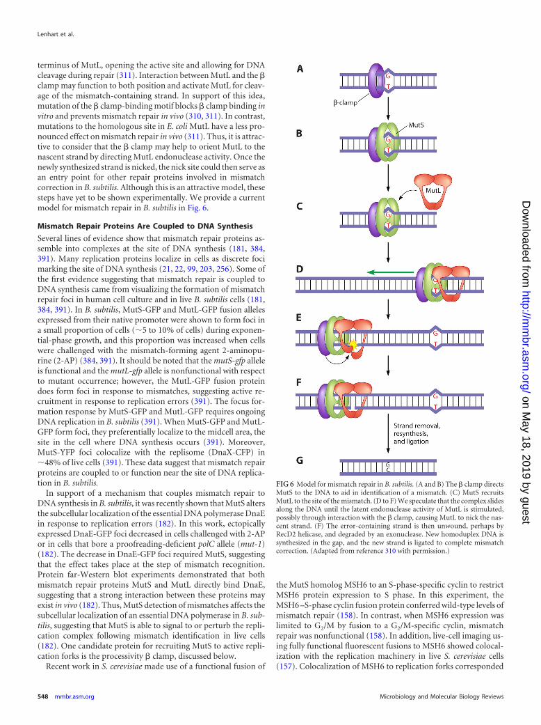
terminus of MutL, opening the active site and allowing for DNAcleavage during repair (311). Interaction between MutL and the �clamp may function to both position and activate MutL for cleav-age of the mismatch-containing strand. In support of this idea,mutation of the � clamp-binding motif blocks � clamp binding invitro and prevents mismatch repair in vivo (310, 311). In contrast,mutations to the homologous site in E. coli MutL have a less pro-nounced effect on mismatch repair in vivo (311). Thus, it is attrac-tive to consider that the � clamp may help to orient MutL to thenascent strand by directing MutL endonuclease activity. Once thenewly synthesized strand is nicked, the nick site could then serve asan entry point for other repair proteins involved in mismatchcorrection in B. subtilis. Although this is an attractive model, thesesteps have yet to be shown experimentally. We provide a currentmodel for mismatch repair in B. subtilis in Fig. 6.
Mismatch Repair Proteins Are Coupled to DNA Synthesis
Several lines of evidence show that mismatch repair proteins as-semble into complexes at the site of DNA synthesis (181, 384,391). Many replication proteins localize in cells as discrete focimarking the site of DNA synthesis (21, 22, 99, 203, 256). Some ofthe first evidence suggesting that mismatch repair is coupled toDNA synthesis came from visualizing the formation of mismatchrepair foci in human cell culture and in live B. subtilis cells (181,384, 391). In B. subtilis, MutS-GFP and MutL-GFP fusion allelesexpressed from their native promoter were shown to form foci ina small proportion of cells (�5 to 10% of cells) during exponen-tial-phase growth, and this proportion was increased when cellswere challenged with the mismatch-forming agent 2-aminopu-rine (2-AP) (384, 391). It should be noted that the mutS-gfp alleleis functional and the mutL-gfp allele is nonfunctional with respectto mutant occurrence; however, the MutL-GFP fusion proteindoes form foci in response to mismatches, suggesting active re-cruitment in response to replication errors (391). The focus for-mation response by MutS-GFP and MutL-GFP requires ongoingDNA replication in B. subtilis (391). When MutS-GFP and MutL-GFP form foci, they preferentially localize to the midcell area, thesite in the cell where DNA synthesis occurs (391). Moreover,MutS-YFP foci colocalize with the replisome (DnaX-CFP) in�48% of live cells (391). These data suggest that mismatch repairproteins are coupled to or function near the site of DNA replica-tion in B. subtilis.
In support of a mechanism that couples mismatch repair toDNA synthesis in B. subtilis, it was recently shown that MutS altersthe subcellular localization of the essential DNA polymerase DnaEin response to replication errors (182). In this work, ectopicallyexpressed DnaE-GFP foci decreased in cells challenged with 2-APor in cells that bore a proofreading-deficient polC allele (mut-1)(182). The decrease in DnaE-GFP foci required MutS, suggestingthat the effect takes place at the step of mismatch recognition.Protein far-Western blot experiments demonstrated that bothmismatch repair proteins MutS and MutL directly bind DnaE,suggesting that a strong interaction between these proteins mayexist in vivo (182). Thus, MutS detection of mismatches affects thesubcellular localization of an essential DNA polymerase in B. sub-tilis, suggesting that MutS is able to signal to or perturb the repli-cation complex following mismatch identification in live cells(182). One candidate protein for recruiting MutS to active repli-cation forks is the processivity � clamp, discussed below.
Recent work in S. cerevisiae made use of a functional fusion of
the MutS homolog MSH6 to an S-phase-specific cyclin to restrictMSH6 protein expression to S phase. In this experiment, theMSH6 –S-phase cyclin fusion protein conferred wild-type levels ofmismatch repair (158). In contrast, when MSH6 expression waslimited to G2/M by fusion to a G2/M-specific cyclin, mismatchrepair was nonfunctional (158). In addition, live-cell imaging us-ing fully functional fluorescent fusions to MSH6 showed colocal-ization with the replication machinery in live S. cerevisiae cells(157). Colocalization of MSH6 to replication forks corresponded
FIG 6 Model for mismatch repair in B. subtilis. (A and B) The � clamp directsMutS to the DNA to aid in identification of a mismatch. (C) MutS recruitsMutL to the site of the mismatch. (D to F) We speculate that the complex slidesalong the DNA until the latent endonuclease activity of MutL is stimulated,possibly through interaction with the � clamp, causing MutL to nick the nas-cent strand. (F) The error-containing strand is then unwound, perhaps byRecD2 helicase, and degraded by an exonuclease. New homoduplex DNA issynthesized in the gap, and the new strand is ligated to complete mismatchcorrection. (Adapted from reference 310 with permission.)
Lenhart et al.
548 mmbr.asm.org Microbiology and Molecular Biology Reviews
on May 18, 2019 by guest
http://mm
br.asm.org/
Dow
nloaded from

to �15% of MMR in S. cerevisiae, and interestingly, it was re-quired for functional mismatch repair in the absence of ExoI(157). This work shows that mismatch repair is coupled to DNAreplication in S. cerevisiae, demonstrating that the coupling ofmismatch repair to DNA replication is conserved in higher organ-isms and is not unique to Bacillus (157, 158).
Involvement of � Clamp in Mismatch Repair
The DNA replication processivity factor � clamp is important forlinking mismatch repair foci to the DNA replication status (112,384). B. subtilis MutS contains a five-amino-acid motif originallyidentified through bioinformatics analysis as a putative � clamp-binding site (91). Residues 810QLSFF814 provide a mostly hydro-phobic plug that fits into a hydrophobic cleft on the � clamp (42,91). Replacement of this motif with five alanine residues caused an�40-fold increase in measurements of mutant frequency, and amutant form of MutS fused to GFP and bearing a replacement ofQLSFF with five alanine residues was reduced �3-fold for focusformation in response to 2-AP challenge (384). Biochemical anal-ysis shows that the � clamp binds to peptides bearing the QLSFFresidues (384). Therefore, the MutS QLSFF motif is critical forinteraction between MutS and the � clamp for efficient mismatchrepair in B. subtilis (384).
Although mutation of the � clamp-binding motif in MutS in-creased the frequency of mutants of live cells, deletion of the 58-residue unstructured C-terminal region bearing this motif (result-ing in the MutS800 protein) nearly abolished the interactionbetween B. subtilis MutS and the � clamp in vitro (384). Impor-tantly, purified MutS800 protein bound to a mismatch at the samelevel as wild-type MutS in vitro, suggesting that removal of theC-terminal 58 amino acids does not diminish mismatch binding(384). Although it was expressed at wild-type levels, the mutS800allele conferred a spontaneous mutant frequency close to that fora strain disrupted in the mutS gene. MutS800 fused with GFPfailed to localize as a focus in response to 2-AP, and YFP fused toonly the C-terminal 58 amino acids of MutS bearing the � clamp-binding motif was sufficient to target YFP for localization whenthe fusion was overexpressed (384). These data support the hy-pothesis that the � clamp aids in the formation of MutS-GFP fociin response to mismatches in vivo (384). This work also found thata conditional allele of dnaN (coding for the � clamp) which causeda partial defect in mismatch repair had a reduced capacity forsupporting MutS-GFP focus formation in vivo (384). In addition,intragenic suppressors of the temperature-sensitive phenotypecaused by this � clamp allele (dnaN5) maintained defects in mis-match repair while rescuing the DNA replication defect conferredby this allele. These data further support a role for the � clamp inmismatch repair in B. subtilis (112).
An important role for the � clamp (DnaN) in mismatch repairis strongly supported by studies of Bacillus anthracis. Interestingly,B. anthracis contains two dnaN genes: dnaN1 and dnaN2 (460).Both dnaN-encoded clamps support growth in vivo, yet deletionof dnaN1 confers a rate of spontaneous mutagenesis that is iden-tical to that of B. anthracis cells disrupted for mismatch repair(460). These results show that both dnaN genes allow for properDNA synthesis; however, only dnaN1 is capable of functioning inmismatch repair (460). These data further establish a role for the �clamp in correction of DNA replication errors in the genus Bacil-lus (460). The data from B. subtilis and B. anthracis show thatMutS interaction with the � clamp is crucial for mismatch repair
in some Gram-positive organisms, and it may be found to beimportant in other organisms that lack a dam-directed repairpathway, although this remains to be established. The involve-ment of the � clamp in mismatch repair in dam-directed systemssuch as that of E. coli is unclear and requires further study (221–223, 314).
RecD2 Is a Possible Mismatch Repair Helicase in Bacillus
UvrD (DNA helicase II) is a 3=-5= DNA helicase that functionsduring methylation-directed mismatch repair in E. coli (for a re-view, see references 192 and 362). B. subtilis contains two proteinsshowing homology to UvrD, namely, PcrA and YjcD (53, 307).PcrA is an essential helicase in B. subtilis, and the pcrA gene is ableto complement the UV sensitivity conferred on E. coli lackinguvrD (307). It has been reported that a strain deficient in yjcDconfers a wild-type level of mutagenesis (307). Thus, it is possiblethat PcrA functions in mismatch repair. Another possibility is thatB. subtilis does not have a helicase that participates in mismatchrepair and that mismatch correction is more similar to the processin eukaryotes, where an exonuclease (Exo I) is likely to function inthe absence of a DNA helicase (for a review, see references 192 and362). Recently, a screen for transposon insertions in B. anthracisthat result in colony papillation revealed several genes that in-crease the rate of mutagenesis (461). Of these, a recD2 orthologwas identified, and this work showed that a recD2 deficiency in B.anthracis conferred a spontaneous mutation frequency and a mu-tation spectrum similar to those seen with a mismatch repair de-fect (461). Epistasis analysis of the recD2-deficient allele in com-bination with a mutS-defective allele showed a spontaneousmutation frequency that was similar to that of a strain bearingonly a mutS deficiency, suggesting a role for RecD2 in mismatchrepair in B. anthracis (462). RecD2 is similar to RecD1, a compo-nent of the RecBCD complex involved in end processing duringDSB repair (e.g., see reference 462). RecD2 homologs are con-served in the Firmicutes and in Gram-positive bacteria that lackthe RecBCD recombination complex (462). Based on these data, itis tempting to conclude that RecD2 helicases function in mis-match repair in many Gram-positive bacteria.
YshD, a MutS Paralog
In rare fashion, B. subtilis encodes MutS and a paralog, MutS2(yshD). The mutS2 gene is transcribed during normal growth, andtranscription continues until cells reach stationary phase (332).Disruption of mutS causes the substantial increase in mutagenesisexpected for a gene product involved in mismatch repair (112,135, 384). Disruption of mutS2 (yshD) has no observable effect onmutant frequency (332); however, analysis of the mutation spec-trum in the rpoB genes of mutS2-deficient cells showed an unusualincrease in transversion mutations (332). It has been suggestedthat such an increase could be attributed to inefficient repair ofoxidative DNA damage; however, it is not known if mutS2-defi-cient strains are sensitive to oxidative damage (14). Analysis of theMutS2 protein in the Gram-negative organism Helicobacter pylorishowed that MutS2 is an endonuclease involved in homologousrecombination (312). Although the role for MutS2 in B. subtilis isunknown, based on conservation and analogy with other systems,B. subtilis MutS2 is probably an endonuclease involved in someunknown aspect of genome maintenance.
DNA Repair in Bacillus subtilis
September 2012 Volume 76 Number 3 mmbr.asm.org 549
on May 18, 2019 by guest
http://mm
br.asm.org/
Dow
nloaded from

DNA DAMAGE TOLERANCE AND TRANSLESION SYNTHESIS
Translesion DNA polymerases are so named because they are ca-pable of replicative bypass over noncoding bases that would nor-mally block progression of a replicative DNA polymerase (for areview, see references 404 and 405). B. subtilis and many otherGram-positive bacteria contain two Y family DNA polymerases,termed PolY1 (YqjW) and PolY2 (YqjH) (403). PolY1 shows thehighest sequence identity to E. coli UmuC, and PolY2 most closelyresembles E. coli DinB (Pol IV) (403). In E. coli, UmuC cooperateswith a posttranslationally modified version of UmuD calledUmuD= (for a review, see reference 297). This protein lacks theN-terminal 24 amino acids of UmuD, and two UmuD= monomersdimerize and associate with UmuC to form DNA polymerase V(UmuD=2C) (327, 409). UmuD is not well conserved outside bac-teria closely related to E. coli (119). Thus, a protein analogous to E.coli UmuD has not been identified in B. subtilis, though not with-out attempts (110). Therefore, we hypothesize that PolY1 of B.subtilis acts in the absence of an accessory factor analogous to E.coli UmuD. Both PolY1 and PolY2 in B. subtilis were originallyidentified through sequence comparisons to the E. coli Y familyDNA polymerases and later in a genome-wide yeast two-hybridassay aimed at identifying B. subtilis proteins that interact with thereplication processivity � clamp (DnaN) (290, 403). Like the casewith their E. coli counterparts, expression of PolY1 or PolY2 froman ectopic locus induces mutation frequency on undamagedDNA, but not to the same extent (110, 403).
It should be noted that E. coli cells deficient in dinB were orig-inally identified as causing a minor decrease in untargeted UVmutagenesis of � phage and later shown to have a more strikingrole in adaptive mutagenesis (37, 45, 254). B. subtilis PolY2 alsohas a role in adaptive or stationary-phase mutagenesis, as polY2(yqjH)-deficient cells show a significant decease in stationary-phase his� reversion (403). Thus, both E. coli DinB and B. subtilisPolY2 have roles in adaptive mutagenesis.
DnaE-Induced Mutagenesis
Translesion synthesis is not limited to DNA polymerases of the Yfamily, because C family DNA polymerases can also have roles inlesion bypass and mutagenesis in several Gram-positive bacteria.DnaE is the replicative polymerase in E. coli and other Gram-negative organisms (for a review, see references 170, 251, and253). B. subtilis and many low-G�C Gram-positive bacteria con-tain two C family DNA polymerases, i.e., PolC and DnaE (186,478). The composition of the replicative DNA polymerase in low-G�C Gram-positive bacteria is different from that of the Pol IIIcharacterized for E. coli. For readers interested in these differences,we suggest a series of reviews and research articles that have re-cently been published (40, 98, 186, 196, 251–253, 352, 392, 427,478). In the case of B. subtilis, Streptomyces coelicolor, Streptococcuspyogenes, S. aureus, and Enterococcus faecalis, the dnaE gene isessential (99, 125, 126, 164). The essential role of DnaE in B. sub-tilis is due to the requirement for utilization of RNA primers onthe lagging strand during replication (352). DnaE can efficientlyextend from an RNA primer, and PolC cannot (352). DnaE inGram-negative organisms has an associated proofreading ε sub-unit (DnaQ) which provides the 3=-5= exonuclease activity, allow-ing for proofreading and thus high-fidelity DNA replication (115,360). Searches of B. subtilis coding sequences have failed to iden-tify an ε subunit that would be available to associate with DnaE
(data not shown). Extensive biochemical characterization of S.pyogenes DnaE showed that this protein is error prone, can easilybypass abasic sites, and can efficiently extend from a mismatchedprimer terminus (39). All of this evidence points to DnaE of S.pyogenes providing a role in mutagenesis and translesion synthesis(39).
As mentioned above, the dnaE gene product in B. subtilis isessential and required for maturation of the lagging strand in vitro(352). DnaE protein levels in B. subtilis have been shown to in-crease following DNA damage, and DnaE protein levels are ele-vated in cells deficient for lexA (196). In addition, the dnaE genehas a LexA binding site located upstream, and dnaE transcripts areinduced �11-fold in response to DNA damage (10). These datasupport the hypothesis that DnaE could have a role in DNA repairor lesion bypass, in addition to its essential role in chromosomalDNA replication. Consistent with a role for DnaE in lesion bypass,B. subtilis DnaE has been shown to bypass N2-acetylaminofluo-rene guanine and AP sites with low efficiency, although DnaE wasunable to bypass benzo-(a)-pyrene adducts or 6-4 TT photoprod-ucts (196). It was also recently shown that the percentage of cellswith DnaE-GFP foci was elevated in cells challenged with the DNAdamaging agent mitomycin C, further suggesting a role for DnaEin repair or translesion synthesis across from mitomycin C ad-ducts (182). Together, the data indicate that dnaE encodes anSOS-inducible DNA polymerase capable of lesion bypass in vitroand that DnaE is essential for chromosomal replication in vivo.
In some Gram-positive bacteria, typified by M. tuberculosis,PolC is absent, and M. tuberculosis instead contains two dnaEgenes (dnaE1 and dnaE2) (33). The dnaE1 gene is essential, anddeletion of dnaE2 abolishes induced mutagenesis (33). It seemsthat bacteria containing two DnaE-type DNA polymerases use thednaE2 gene product for lesion bypass in vivo.
DNA INTEGRITY REGULATES BACTERIAL CELL DIVISION
When bacterial cells experience DNA damage or replication forkstress during vegetative growth, a response is activated to preventcell division (57, 161, 177, 269, 294). A block to cell division, eithertransient or prolonged, allows for repair of damaged DNA, ensur-ing that a broken or otherwise damaged chromosome is not seg-regated into a new cell. Cells experiencing a sustained block to celldivision show filamentation, meaning that cells continue to growin length without a cell division event. For example, in E. coli,inhibition of the cell cycle is accomplished through the SOS-in-duced expression of SulA (sfiA or sulA). SulA prevents cell divisionby inhibiting polymerization of FtsZ, the tubulin ortholog respon-sible for cytokinesis (for a review, see references 147 and 283).After the SOS response is turned off, SulA is rapidly degraded byLon protease, allowing for cell division to resume, and cell cycleprogression continues (267). In B. subtilis, the yneA gene is SOSregulated, and expression of yneA inhibits cell division. We discussYneA in greater detail below.
RecA-Dependent Regulation of Cell Division
When B. subtilis is challenged with DNA damage, cells filament ina recA-dependent manner (177, 225). The yneA gene is transcribeddivergently from the lexA gene, and the yneA promoter regioncontains three LexA binding sites (10, 177). Inactivation of theyneA gene prevents DNA damage-inducible cell filamentation(177). YneA is a small protein secreted by the Sec pathway (232). Ithas a single transmembrane segment and a C-terminal pepti-
Lenhart et al.
550 mmbr.asm.org Microbiology and Molecular Biology Reviews
on May 18, 2019 by guest
http://mm
br.asm.org/
Dow
nloaded from

doglycan-binding LysM domain separated by a predicted proteasecleavage site (268). YneA shares no sequence similarity to E. coliSulA (177). If yneA is under the control of an inducible promoter,expression of yneA is sufficient to prevent cell division, with cellsgrowing to a mean length of about 5.5 �m (177). Upon subse-quent depletion of inducer and repression of yneA expression,filamenting cells rapidly engage in cell division, and the averagecell length returns to �3 �m in as little as 20 min (268). These datashow that cell division resumes efficiently after yneA expression isblocked, indicating that the YneA protein is rapidly cleared (268).Mutational analysis indicates that full-length YneA is necessary toinhibit cell division. Upon signal peptide cleavage, the LysM do-main of YneA is released into the culture medium, inactivatingYneA, which is followed by rapid degradation by extracellular pro-teases (268). It should be noted that overexpression of yneA alsoappears to impair separation of daughter cells following cell divi-sion, suggesting an additional role of YneA in cell wall remodeling(268). Furthermore, yneA is upregulated by the competence tran-scription factor ComK, suggesting a potential in vivo role indaughter cell separation following entry into stationary phase(295).
Currently, the mechanism that YneA uses to inhibit cell divisionin B. subtilis is unknown. Ectopic expression of yneA or expressionof yneA through disruption of lexA caused a reduction or slightloss of FtsZ ring formation (177, 268). Yeast two-hybrid analysisfailed to detect an interaction between YneA and FtsZ (177). It hasbeen proposed that YneA may interact with a component of thecell division apparatus, probably through its transmembrane do-main (268). Consistent with this idea, mutations in the transmem-brane domain of YneA inactivate its ability to block cytokinesis(268). Furthermore, these mutations cluster to a single face of thetransmembrane helix, adding evidence that the transmembranedomain may facilitate checkpoint activation through interactionwith another protein, or perhaps through direct contact and inhi-bition of the cell division apparatus (268). Transmembrane do-mains have previously been shown to facilitate formation ofhetero- and homo-oligomers during complex formation (202,414). Interestingly, the newly discovered Caulobacter crescentusSOS-induced inhibitor of cell division A (SidA) was shown toprevent cell division by blocking the final stages of cytokinesisthrough inhibiting formation of the FtsWIN subcomplex (269).The interaction between SidA and FtsW occurs partly through thetransmembrane domain, and mutations localized to the trans-membrane domain abolish SidA-dependent blocks to cell division(269).
Blocking of cytokinesis is widely conserved among bacterialcheckpoint proteins, as exemplified by YneA gene orthologs pres-ent in other Gram-positive bacteria, including L. monocytogenes(yneA), M. tuberculosis (Rv2719c), and Corynebacterium glutami-cum (divS) (57, 294). Like B. subtilis yneA, these genes all maintainLexA-dependent repression, have a conserved genomic positionadjacent to lexA, and are sufficient to prevent cell division whenexpressed (57, 294). Challenge of M. tuberculosis with cell wallantibiotics or the DNA damaging agent mitomycin C induces ex-pression of Rv2719c and results in a block to cell division resultingin cell filamentation (57). Deletion of Rv2719c renders cells highlysensitive to DNA damaging agents, indicative of a checkpoint-likefunction (57). The DivS protein of C. glutamicum does not showsequence similarity to YneA or M. tuberculosis Rv2719c, but thisprotein does appear to have a single-pass transmembrane domain
(294). It also appears that peptidoglycan synthesis at midcell and,to some extent, FtsZ ring assembly are altered upon divS inductionfollowing mitomycin C challenge (294). It is not known whetherDivS, YneA, and Rv2719c directly inhibit Z ring assembly, likeSulA, or whether they do so indirectly, regulating a late stage in celldivision, like Caulobacter SidA. It should be noted that all knownfunctional analogs of B. subtilis YneA aside from E. coli SulA,which is cytoplasmic, possess a single transmembrane domain.YneA may therefore represent a more widely conserved mode ofinhibiting cell division than the SulA-type mechanism present inE. coli (57, 268, 269, 294).
RecA-Independent and DnaA-Dependent Regulation of CellDivision
B. subtilis regulates DNA replication initiation through regulatingthe activity of the initiation protein DnaA at the origin, oriC (for areview, see reference 273). The formation of a nucleoprotein com-plex between DnaA and oriC marks the start of replication initia-tion, a process that ultimately facilitates local chromosome un-winding and the assembly of the replication apparatus at theorigin (e.g., see references 172, 173, and 282). DnaA is essentialand is required for replication initiation in virtually all free-livingbacteria (for a review, see reference 172). Although the essentialfunction of DnaA is in DNA replication initiation, we focus onother roles for DnaA here and refer readers to several comprehen-sive reviews of DnaA and its role in chromosomal DNA replica-tion initiation (e.g., see references 172, 173, and 282).
In addition to the well-defined roles for DnaA in replicationinitiation, DnaA has also been shown to function as a transcrip-tion factor that activates or represses transcription of a variety ofgenes, including its own (138). Results from gene profiling studiesperformed with B. subtilis show that DnaA controls a reguloncomprising 56 genes in 20 operons (138). Among these DnaA-regulated genes, putative DnaA binding sites were identified up-stream of 19 of the 20 operons (138). Furthermore, DnaA has beenshown to bind a number of these sites in vivo, as determined bychromatin immunoprecipitation (138).
A striking finding was that DnaA is capable of sensing DNAreplication fork perturbations, resulting in alterations to the ex-pression of DnaA-regulated genes (138). Upon closer analysis ofthe DnaA regulon, two genes of particular interest were found: sdaand ftsL (Fig. 7). Suppressor of dnaA (Sda) is a sporulation check-point protein and is described in detail below; however, we focushere on ftsL and the regulation of cell division. In contrast to thecase for yneA, which is SOS regulated, ftsL regulation of cell divi-sion is recA independent (138). Upon encountering DNA replica-tion stress, DnaA binds to the promoter region of the yllB-ylxA-ftsL-pbpB operon, downregulating transcription 2- to 4-fold andultimately preventing additional rounds of cell division (138).
The penicillin-binding protein 2B gene (pbpB) and ftsL are es-sential genes required for cell division (183). FtsL has an unknownfunction but is known to form an oligomeric complex with DivIBand DivIC preceding cell division, and this recruitment is depen-dent on FtsZ ring formation (94, 382, 383). PbpB synthesizes pep-tidoglycan that eventually constitutes the new poles of daughtercells (120, 136). PbpB is a stable septal protein, whereas FtsL hasbeen shown to be unstable, making ftsL an ideal target for regulat-ing a cell division checkpoint (92, 138). Depletion of FtsL inde-pendent of pbpB results in substantial cell filamentation, as celldivision is arrested at a step preceding septum formation and
DNA Repair in Bacillus subtilis
September 2012 Volume 76 Number 3 mmbr.asm.org 551
on May 18, 2019 by guest
http://mm
br.asm.org/
Dow
nloaded from
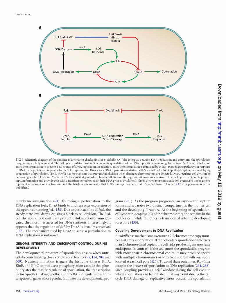
membrane invagination (93). Following a perturbation to theDNA replication fork, DnaA binds to and represses expression ofthe operon containing ftsL (138). Due to the instability of FtsL, thesteady-state level drops, causing a block to cell division. The FtsLcell division checkpoint may prevent cytokinesis over unsegre-gated chromosomes arrested for DNA synthesis. Interestingly, itappears that the regulation of ftsL by DnaA is broadly conserved(138). The mechanism used by DnaA to sense a perturbation toDNA replication is unknown.
GENOME INTEGRITY AND CHECKPOINT CONTROL DURINGDEVELOPMENT
The developmental program of sporulation ensues when nutri-ents become limiting (for a review, see references 95, 114, 304, and309). Nutrient limitation triggers the histidine kinases KinA,KinB, and KinC to produce a phosphorylation cascade that phos-phorylates the master regulator of sporulation, the transcriptionfactor Spo0A (making Spo0A�P). Spo0A�P regulates the tran-scription of genes whose products initiate the developmental pro-
gram (271). As the program progresses, an asymmetric septumforms and separates two distinct compartments: the mother celland the developing forespore. At the beginning of sporulation,cells contain 2 copies (2C) of the chromosome; one remains in themother cell, while the other is translocated into the developingforespore (456).
Coupling Development to DNA Replication
B. subtilis has mechanisms to ensure a 2C chromosome copy num-ber as it enters sporulation. If the cell enters sporulation with fewerthan 2 chromosomal copies, the cell risks producing an anucleateendospore. In contrast, if the cell enters the sporulation programwith more than 2 chromosomal copies, it may produce sporeswith multiple chromosomes or with twin spores, with one sporelocated at each cell pole (426). To avoid these outcomes, B. subtiliscouples the process of sporulation to DNA replication (234, 235).Such coupling provides a brief window during the cell cycle inwhich sporulation can be initiated. If at any point during the cellcycle DNA damage or replicative stress occurs, the sporulation
FIG 7 Schematic diagram of the genome maintenance checkpoints in B. subtilis. (A) The interplay between DNA replication and entry into the sporulationprogram is carefully regulated. The cell cycle regulator protein Sda prevents sporulation when DNA replication is ongoing. In contrast, SirA is activated uponentry into sporulation to prevent new rounds of DNA replication. In addition, entry into sporulation is regulated by at least two separate pathways in responseto DNA damage. Sda is upregulated by the SOS response, and DisA senses DNA repair intermediates. Both Sda and DisA inhibit Spo0A phosphorylation, delayingprogression of sporulation. (B) B. subtilis has mechanisms that prevent cell division when damaged chromosomes are detected. DnaA regulates cell division bydecreasing levels of FtsL, and YneA is an SOS-regulated gene which blocks cell division through an unknown mechanism. These cell cycle checkpoints preventseptum formation and provide cells with a transient period to repair their DNA prior to cytokinesis. Green arrows represent activation events, red line segmentsrepresent repression or inactivation, and the black arrow indicates that DNA damage has occurred. (Adapted from reference 433 with permission of thepublisher.)
Lenhart et al.
552 mmbr.asm.org Microbiology and Molecular Biology Reviews
on May 18, 2019 by guest
http://mm
br.asm.org/
Dow
nloaded from
program will be inhibited to allow for repair or continued DNAreplication. By inhibiting DNA replication elongation with thePolC-specific drug HPURa [6-(p-hydroxyphenylazo)uracil], itwas shown that inhibition of sporulation was recA dependent(166). Experiments using alleles that are temperature sensitive forreplication initiation (dnaA1, dnaB19, and dnaD23) showed thatsporulation was inhibited when replication initiation was inhib-ited (165, 204). This pathway used a recA-independent mecha-nism to delay sporulation. In both the recA-dependent and recA-independent pathways, sporulation is halted by inhibitingformation of Spo0A�P. An allele of spo0A (rvtA11) that permitsits activation directly by KinC bypassed the replication inhibitiondefect (165, 166, 204). These data indicate that the DNA replica-tion status during sporulation is sensed and that this informationis used to regulate formation of Spo0A�P.
Sda
The source of coupling between the B. subtilis replication cycleand its entry into sporulation is sda. Mutations in the sda locusallowed B. subtilis to sporulate in a dnaA1 temperature-sensitivemutant at the restrictive temperature in the absence of DNA rep-lication initiation (43). Sda inhibits KinA and KinB, such thatphosphorylation of Spo0A no longer efficiently occurs when levelsof Sda are elevated upon inhibition of DNA replication (34). Inthis way, Sda inhibits sporulation by limiting Spo0A�P levels(34). In vitro studies have confirmed that Sda inhibits KinA bybinding its autokinase domain, preventing autophosphorylationand subsequent activation (333).
Sda protein levels are correlated directly with stages of the cellcycle and rely on cell cycle-dependent gene transcription, as wellas the instability of Sda, to transiently prevent entry into sporula-tion (426). The sda promoter contains 5 dnaA boxes that allowregulation of sda transcription at the beginning of each cell cyclethrough binding of DnaA (43, 138, 426). Reductions in Sda levelscorrelate with the end of DNA replication, ensuring that a 2CDNA content is present before sporulation to help ensure viablespores. However, to enter into the sporulation program, the cellmust reduce the amount of Sda in the cell to allow for Spo0Aactivation at the appropriate time. This process is performed byClpXP-mediated proteolysis. In vivo, Sda maintains a half-life onthe order of minutes (336, 426) due to residues at its C terminusthat target Sda for degradation (V-S-S-COOH) (140, 336).
In addition to dnaA-dependent regulation of the sda locus, theSOS response can also mediate an increase in Sda levels in re-sponse to DNA damage or stalled replication forks (10, 336). Thesda promoter contains a putative LexA binding site that has beenshown to be responsive to UV damage and HPURa treatments(10, 166, 336). DNA damage-dependent upregulation of sda pre-vents entry into sporulation when chromosomal DNA is dam-aged.
SirA
As an additional measure to maintain the proper chromosomenumber during sporulation, B. subtilis can inhibit DNA replica-tion initiation through the action of the checkpoint protein SirA(sporulation inhibitor of replication A) (yneE) (322, 323, 433).Using this checkpoint, the cell ensures that the 2C DNA content ismaintained (322, 323, 433). The sirA gene was identified in theSpo0A regulon and found to inhibit DNA replication initiation(322, 323, 433). In addition, it was shown that artificial expression
of sirA during vegetative growth caused a block in DNA replica-tion initiation (322, 433). Conversely, sporulating cells deficient insirA overreplicate, as assayed by observing genome equivalents orthe origin copy number during sporulation (322, 433).
SirA prevents DNA replication initiation by binding DnaA andsequestering it from oriC (322, 433). In addition, cells that lackoriC and initiate replication through an ectopically integrated or-igin (oriN) that does not require DnaA for replication initiationare refractory to the inhibitory action of SirA (433). Depletion ofDnaA blocks GFP-SirA focus formation, and expression of SirAcauses DnaA-GFP foci to disperse (433). It was further shown thatSirA binds to a group of amino acids (N44, F46, and V47) residingin domain 1 of DnaA, presumably preventing DnaA from estab-lishing an active initiation complex at oriC (323). Mutation ofthese DnaA residues causes the cell to be unaffected by the inhib-itory action of SirA (323).
Together, Sda and SirA maintain the 2C DNA content as cellsenter sporulation. Sda prevents sporulation from occurring tooearly in the replication cycle, guarding against 1C bacteria under-going development. Conversely, SirA expression is activated bySpo0A�P upon entrance of B. subtilis into sporulation, thus pre-venting overreplication during development and preventing anincrease in chromosomal copy number, which can reduce sporeviability (426) (Fig. 7).
DisA
In addition to the importance of entering sporulation with twocopies of the chromosome, B. subtilis must also ensure that ge-nome integrity is maintained. One approach is recA-dependenttranscription of sda as discussed above, and the other involves disA(19). DisA (DNA integrity scanning protein A) is a checkpointprotein that mediates a delay in sporulation following detection ofDNA damage (19). In the absence of disA, the checkpoint fails andcells with damaged chromosomes progress into sporulation, pro-ducing nonviable spores (19). The disA-dependent checkpointappears to attenuate phosphorylation of the master regulatorSpo0A, preventing cells from entering sporulation following thedetection of DNA damage (19).
How does DisA monitor genome integrity? DisA movesthroughout the cell, scanning the chromosome for intermediatesof DNA repair. DisA fused to GFP forms a dynamic complex thatactively moves throughout the cell and pauses at sites of DNAdamage (19). DisA movement requires that the cell make ATP andis independent of the cytoskeletal proteins MreB and Mbl (19).DisA-GFP also forms a mobile complex in the absence of DNA,suggesting that its movement is DNA independent (19).
Structural determination of a DisA homolog from Thermotogamaritima found that DisA has diadenylate cyclase activity (454).Specifically, this activity allows DisA to synthesize the second mes-senger c-di-AMP [bis-(3=,5=)-cyclic dimeric AMP] (454), and dia-denylate cyclase activity is important for DisA-GFP focus move-ment in vivo (299). Interestingly, DisA diadenylate cyclase activityis regulated by different DNA substrates (454). DisA is unaffectedby a ssDNA oligonucleotide or a blunt dsDNA substrate (454).However, branched DNA substrates that mimic intermediates ofrecombination, including 5=- and 3=-flapped structures or three-and four-way junctions, modulate diadenylate cyclase activity ofDisA (454). These substrates reduce the rate of c-di-AMP produc-tion, which suggests that DisA does not scan for DNA damage perse but instead identifies intermediates of DNA repair, such as Hol-
DNA Repair in Bacillus subtilis
September 2012 Volume 76 Number 3 mmbr.asm.org 553
on May 18, 2019 by guest
http://mm
br.asm.org/
Dow
nloaded from
liday junctions. This model was also supported in vivo by moni-toring a disA-dependent increase in endogenous levels of c-di-AMP (299). Following challenge of cells with DNA damagingagents, c-di-AMP levels dropped, supporting the idea that DisAsignals that genome integrity is compromised through a decreasein c-di-AMP production (299). To further support this model, theB. subtilis protein YybT was shown to ensure a constant turnoverof c-di-AMP in vivo by degrading the signaling molecule and al-lowing for a quick and efficient decrease following the DNA dam-age-dependent attenuation of c-di-AMP production by DisA(299).
The current model invokes a mechanism where DisA scans theB. subtilis chromosome for intermediates of DNA repair. Uponidentifying a Holliday junction or other DNA repair intermediate,DisA diadenylate cyclase activity is attenuated. The reduction inc-di-AMP may act as a signal to pause entry into sporulation bypreventing an increase in Spo0A�P levels in vivo (Fig. 7).
CONCLUDING REMARKS
DNA replication, repair, and mutagenesis have been studied in B.subtilis and other Gram-positive bacteria for over 40 years. Duringthis time, we have begun to appreciate that the mechanisms allow-ing for genome replication and repair are often different from thepathways described for the bacterial prototype, E. coli. During thelast 10 years, researchers in the field have discovered new proteinsand pathways with intricate mechanisms of regulation, and wehave witnessed a spectacular increase in our understanding of ge-nome maintenance in B. subtilis and other Gram-positive bacte-ria. This has allowed for a greater appreciation of the differences inthe pathways of different bacteria and has provided a platform forunderstanding the molecular details of genome maintenance. Welook forward to learning the mechanistic details of these pathwaysin years to come.
There are several other areas under active investigation thathave contributed significantly to our knowledge of genome stabil-ity in B. subtilis and other Gram-positive bacteria. These includebut are not limited to stationary-phase mutagenesis, natural ge-netic transformation, and the mechanisms that allow for spores tobe resistant to genotoxic stress. We chose not to discuss thesetopics because several excellent review articles have been pub-lished on these subjects, and we direct readers interested in thesesubjects to those comprehensive reviews (62–64, 108, 109, 286,287, 304, 329, 367–371, 413).
ACKNOWLEDGMENTS
We thank the many labs that founded the fields of DNA replication, re-pair, and genome maintenance in Gram-positive bacteria. We regret notbeing able to cite all of the work that has shaped these fields over the lastseveral decades. We thank members of the Simmons lab, Eileen Brandes,Nick Bolz, and Gabriella Szewczyk for comments and discussions on themanuscript. We also thank three anonymous referees for their helpfulcomments, which improved the manuscript.
This work was supported by grant MCB1050948 from the NationalScience Foundation, by a grant from the Wendy Will Case Cancer Fund,and by start-up funds from the University of Michigan and the Depart-ment of Molecular, Cellular, and Developmental Biology to L.A.S. TheNIH Genetics Training Grant at the University of Michigan supportedJ.W.S. during this work (T32 GM007544).
REFERENCES1. Aamodt RM, Falnes PO, Johansen RF, Seeberg E, Bjoras M. 2004. The
Bacillus subtilis counterpart of the mammalian 3-methyladenine DNA
glycosylase has hypoxanthine and 1,N6-ethenoadenine as preferred sub-strates. J. Biol. Chem. 279:13601–13606.
2. Ahrends R, et al. 2006. Identifying an interaction site between MutHand the C-terminal domain of MutL by crosslinking, affinity purifica-tion, chemical coding and mass spectrometry. Nucleic Acids Res. 34:3169 –3180.
3. Alonso JC, Stiege AC, Luder G. 1993. Genetic recombination in Bacillussubtilis 168: effect of recN, recF, recH and addAB mutations on DNArepair and recombination. Mol. Gen. Genet. 239:129 –136.
4. Alonso JC, Tailor RH, Luder G. 1988. Characterization of recombina-tion-deficient mutants of Bacillus subtilis. J. Bacteriol. 170:3001–3007.
5. Alseth I, et al. 2006. A new protein superfamily includes two novel3-methyladenine DNA glycosylases from Bacillus cereus, AlkC and AlkD.Mol. Microbiol. 59:1602–1609.
6. Ampatzidou E, Irmisch A, O’Connell MJ, Murray JM. 2006. Smc5/6 isrequired for repair at collapsed replication forks. Mol. Cell. Biol. 26:9387–9401.
7. Aravind L, Koonin EV. 2001. Prokaryotic homologs of the eukaryoticDNA-end-binding protein Ku, novel domains in the Ku protein andprediction of a prokaryotic double-strand break repair system. GenomeRes. 11:1365–1374.
8. Aravind L, Walker DR, Koonin EV. 1999. Conserved domains in DNArepair proteins and evolution of repair systems. Nucleic Acids Res. 27:1223–1242.
9. Aten JA, et al. 2004. Dynamics of DNA double-strand breaks revealed byclustering of damaged chromosome domains. Science 303:92–95.
10. Au N, et al. 2005. Genetic composition of the Bacillus subtilis SOSsystem. J. Bacteriol. 187:7655–7666.
11. Ayora S, et al. 2011. Double-strand break repair in bacteria: a view fromBacillus subtilis. FEMS Microbiol. Rev. 35:1055–1081.
12. Ayora S, Carrasco B, Doncel-Perez E, Lurz R, Alonso JC. 2004. Bacillussubtilis RecU protein cleaves Holliday junctions and anneals single-stranded DNA. Proc. Natl. Acad. Sci. U. S. A. 101:452– 457.
13. Ayora S, Rojo F, Ogasawara N, Nakai S, Alonso JC. 1996. The Mfdprotein of Bacillus subtilis 168 is involved in both transcription-coupledDNA repair and DNA recombination. J. Mol. Biol. 256:301–318.
14. Banos B, Lazaro JM, Villar L, Salas M, de Vega M. 2008. Character-ization of a Bacillus subtilis 64-kDa DNA polymerase X potentially in-volved in DNA repair. J. Mol. Biol. 384:1019 –1028.
15. Banos B, Villar L, Salas M, de Vega M. 2010. Intrinsic apurinic/apyrimidinic (AP) endonuclease activity enables Bacillus subtilis DNApolymerase X to recognize, incise, and further repair abasic sites. Proc.Natl. Acad. Sci. U. S. A. 107:19219 –19224.
16. Barnes MH, LaMarr WA, Foster KA. 2003. DNA gyrase and DNAtopoisomerase of Bacillus subtilis: expression and characterization of re-combinant enzymes encoded by the gyrA, gyrB and parC, parE genes.Protein Expr. Purif. 29:259 –264.
17. Baute J, Depicker A. 2008. Base excision repair and its role in maintain-ing genome stability. Crit. Rev. Biochem. Mol. Biol. 43:239 –276.
18. Bayles KW, et al. 1994. A genetic and molecular characterization of therecA gene from Staphylococcus aureus. Gene 147:13–20.
19. Bejerano-Sagie M, et al. 2006. A checkpoint protein that scans thechromosome for damage at the start of sporulation in Bacillus subtilis.Cell 125:679 – 690.
20. Bennett SE, Mosbaugh DW. 1992. Characterization of the Escherichiacoli uracil-DNA glycosylase.inhibitor protein complex. J. Biol. Chem.267:22512–22521.
21. Berkmen MB, Grossman AD. 2006. Spatial and temporal organizationof the Bacillus subtilis replication cycle. Mol. Microbiol. 62:57–71.
22. Berkmen MB, Grossman AD. 2007. Subcellular positioning of the originregion of the Bacillus subtilis chromosome is independent of sequenceswithin oriC, the site of replication initiation, and the replication initiatorDnaA. Mol. Microbiol. 63:150 –165.
23. Bernard R, Marquis KA, Rudner DZ. 2010. Nucleoid occlusion pre-vents cell division during replication fork arrest in Bacillus subtilis. Mol.Microbiol. 78:866 – 882.
24. Bernstein DA, Keck JL. 2003. Domain mapping of Escherichia coli RecQdefines the roles of conserved N- and C-terminal regions in the RecQfamily. Nucleic Acids Res. 31:2778 –2785.
25. Bessman MJ, Frick DN, O’Handley SF. 1996. The MutT proteins or“Nudix” hydrolases, a family of versatile, widely distributed, “house-cleaning” enzymes. J. Biol. Chem. 271:25059 –25062.
26. Bhatnagar SK, Bessman MJ. 1988. Studies on the mutator gene, mutT of
Lenhart et al.
554 mmbr.asm.org Microbiology and Molecular Biology Reviews
on May 18, 2019 by guest
http://mm
br.asm.org/
Dow
nloaded from

Escherichia coli. Molecular cloning of the gene, purification of the geneproduct, and identification of a novel nucleoside triphosphatase. J. Biol.Chem. 263:8953– 8957.
27. Bisognano C, et al. 2004. A recA-lexA-dependent pathway mediatesciprofloxacin-induced fibronectin binding in Staphylococcus aureus. J.Biol. Chem. 279:9064 –9071.
28. Bjelland S, Seeberg E. 2003. Mutagenicity, toxicity and repair of DNAbase damage induced by oxidation. Mutat. Res. 531:37– 80.
29. Boehmer PE, Emmerson PT. 1992. The RecB subunit of the Escherichiacoli RecBCD enzyme couples ATP hydrolysis to DNA unwinding. J. Biol.Chem. 267:4981– 4987.
30. Bond CS, Kvaratskhelia M, Richard D, White MF, Hunter WN. 2001.Structure of Hjc, a Holliday junction resolvase, from Sulfolobus solfatari-cus. Proc. Natl. Acad. Sci. U. S. A. 98:5509 –5514.
31. Bork JM, Cox MM, Inman RB. 2001. The RecOR proteins modulateRecA protein function at 5= ends of single-stranded DNA. EMBO J. 20:7313–7322.
32. Bose B, Auchtung JM, Lee CA, Grossman AD. 2008. A conservedanti-repressor controls horizontal gene transfer by proteolysis. Mol. Mi-crobiol. 70:570 –582.
33. Boshoff HI, Reed MB, Barry CE, 3rd, Mizrahi V. 2003. DnaE2 poly-merase contributes to in vivo survival and the emergence of drug resis-tance in Mycobacterium tuberculosis. Cell 113:183–193.
34. Bowater R, Doherty AJ. 2006. Making ends meet: repairing breaks inbacterial DNA by non-homologous end-joining. PLoS Genet. 2:e8. doi:10.1371/journal.pgen.0020008.
35. Britton RA, Grossman AD. 1999. Synthetic lethal phenotypes caused bymutations affecting chromosome partitioning in Bacillus subtilis. J. Bac-teriol. 181:5860 –5864.
36. Britton RA, Lin DC, Grossman AD. 1998. Characterization of a pro-karyotic SMC protein involved in chromosome partitioning. Genes Dev.12:1254 –1259.
37. Brotcorne-Lannoye A, Maenhaut-Michel G. 1986. Role of RecA proteinin untargeted UV mutagenesis of bacteriophage lambda: evidence for therequirement for the dinB gene. Proc. Natl. Acad. Sci. U. S. A. 83:3904 –3908.
38. Bruand C, Farache M, McGovern S, Ehrlich SD, Polard P. 2001. DnaB,DnaD and DnaI proteins are components of the Bacillus subtilis replica-tion restart primosome. Mol. Microbiol. 42:245–255.
39. Bruck I, Goodman MF, O’Donnell M. 2003. The essential C familyDnaE polymerase is error-prone and efficient at lesion bypass. J. Biol.Chem. 278:44361– 44368.
40. Bruck I, O’Donnell M. 2000. The DNA replication machine of a gram-positive organism. J. Biol. Chem. 275:28971–28983.
41. Bunny K, Liu J, Roth J. 2002. Phenotypes of lexA mutations in Salmo-nella enterica: evidence for a lethal lexA null phenotype due to the Fels-2prophage. J. Bacteriol. 184:6235– 6249.
42. Bunting KA, Roe SM, Pearl LH. 2003. Structural basis for recruitmentof translesion DNA polymerase Pol IV/DinB to the beta-clamp. EMBO J.22:5883–5892.
43. Burkholder WF, Kurtser I, Grossman AD. 2001. Replication initiationproteins regulate a developmental checkpoint in Bacillus subtilis. Cell104:269 –279.
44. Burton NA, Johnson MD, Antczak P, Robinson A, Lund PA. 2010.Novel aspects of the acid response network of E. coli K-12 are revealed bya study of transcriptional dynamics. J. Mol. Biol. 401:726 –742.
45. Cairns J, Foster PL. 1991. Adaptive reversion of a frameshift mutation inEscherichia coli. Genetics 128:695–701.
46. Canas C, Carrasco B, Ayora S, Alonso JC. 2008. The RecU Hollidayjunction resolvase acts at early stages of homologous recombination.Nucleic Acids Res. 36:5242–5249.
47. Canas C, et al. 2011. The stalk region of the RecU resolvase is essen-tial for Holliday junction recognition and distortion. J. Mol. Biol.410:39 – 49.
48. Carneiro MJ, et al. 2006. The DNA-remodelling activity of DnaD is thesum of oligomerization and DNA-binding activities on separate do-mains. Mol. Microbiol. 60:917–924.
49. Carrasco B, Ayora S, Lurz R, Alonso JC. 2005. Bacillus subtilis RecUHolliday-junction resolvase modulates RecA activities. Nucleic AcidsRes. 33:3942–3952.
50. Carrasco B, Canas C, Sharples GJ, Alonso JC, Ayora S. 2009. TheN-terminal region of the RecU holliday junction resolvase is essential forhomologous recombination. J. Mol. Biol. 390:1–9.
51. Carrasco B, Cozar MC, Lurz R, Alonso JC, Ayora S. 2004. Geneticrecombination in Bacillus subtilis 168: contribution of Holliday junctionprocessing functions in chromosome segregation. J. Bacteriol. 186:5557–5566.
52. Carrasco B, Fernandez S, Asai K, Ogasawara N, Alonso JC. 2002. Effectof the recU suppressors sms and subA on DNA repair and homologousrecombination in Bacillus subtilis. Mol. Genet. Genomics 266:899 –906.
53. Carrasco B, Fernandez S, Petit MA, Alonso JC. 2001. Genetic recom-bination in Bacillus subtilis 168: effect of DeltahelD on DNA repair andhomologous recombination. J. Bacteriol. 183:5772–5777.
54. Carrasco B, Manfredi C, Ayora S, Alonso JC. 2008. Bacillus subtilisSsbA and dATP regulate RecA nucleation onto single-stranded DNA.DNA Repair (Amsterdam) 7:990 –996.
55. Castellanos-Juarez FX, et al. 2006. YtkD and MutT protect vegetativecells but not spores of Bacillus subtilis from oxidative stress. J. Bacteriol.188:2285–2289.
56. Chatterji M, Sengupta S, Nagaraja V. 2003. Chromosomally encodedgyrase inhibitor GyrI protects Escherichia coli against DNA-damagingagents. Arch. Microbiol. 180:339 –346.
57. Chauhan A, et al. 2006. Interference of Mycobacterium tuberculosis celldivision by Rv2719c, a cell wall hydrolase. Mol. Microbiol. 62:132–147.
58. Chedin F, Ehrlich SD, Kowalczykowski SC. 2000. The Bacillus subtilisAddAB helicase/nuclease is regulated by its cognate Chi sequence invitro. J. Mol. Biol. 298:7–20.
59. Chedin F, Handa N, Dillingham MS, Kowalczykowski SC. 2006. TheAddAB helicase/nuclease forms a stable complex with its cognate Chisequence during translocation. J. Biol. Chem. 281:18610 –18617.
60. Chedin F, Kowalczykowski SC. 2002. A novel family of regulated heli-cases/nucleases from Gram-positive bacteria: insights into the initiationof DNA recombination. Mol. Microbiol. 43:823– 834.
61. Chedin F, Noirot P, Biaudet V, Ehrlich SD. 1998. A five-nucleotidesequence protects DNA from exonucleolytic degradation by AddAB, theRecBCD analogue of Bacillus subtilis. Mol. Microbiol. 29:1369 –1377.
62. Chen I, Christie PJ, Dubnau D. 2005. The ins and outs of DNA transferin bacteria. Science 310:1456 –1460.
63. Chen I, Dubnau D. 2003. DNA transport during transformation. Front.Biosci. 8:s544 –s556.
64. Chen I, Dubnau D. 2004. DNA uptake during bacterial transformation.Nat. Rev. Microbiol. 2:241–249.
65. Cheo D, Bayles K, Yasbin R. 1991. Cloning and characterization ofDNA damage-inducible promoter regions from Bacillus subtilis. J. Bac-teriol. 173:1696 –1703.
66. Chow KH, Courcelle J. 2004. RecO acts with RecF and RecR to protectand maintain replication forks blocked by UV-induced DNA damage inEscherichia coli. J. Biol. Chem. 279:3492–3496.
67. Ciccarelli FD, et al. 2006. Toward automatic reconstruction of a highlyresolved tree of life. Science 311:1283–1287.
68. Ciccia A, Elledge SJ. 2010. The DNA damage response: making it safe toplay with knives. Mol. Cell 40:179 –204.
69. Cirz RT, et al. 2007. Complete and SOS-mediated response of Staphy-lococcus aureus to the antibiotic ciprofloxacin. J. Bacteriol. 189:531–539.
70. Clauson CL, Saxowsky TT, Doetsch PW. 2010. Dynamic flexibility ofDNA repair pathways in growth arrested Escherichia coli. DNA Repair(Amsterdam) 9:842– 847.
71. Cleaver JE. 1968. Defective repair replication of DNA in xerodermapigmentosum. Nature 218:652– 656.
72. Cleaver JE. 1969. Xeroderma pigmentosum: a human disease in whichan initial stage of DNA repair is defective. Proc. Natl. Acad. Sci. U. S. A.63:428 – 435.
73. Collier C, Machon C, Briggs GS, Smits WK, Soultanas P. 2012.Untwisting of the DNA helix stimulates the endonuclease activity of Ba-cillus subtilis Nth at AP sites. Nucleic Acids Res. 40:739 –750.
74. Cone R, Duncan J, Hamilton L, Friedberg EC. 1977. Partial purifica-tion and characterization of a uracil DNA N-glycosidase from Bacillussubtilis. Biochemistry 16:3194 –3201.
75. Connelly JC, de Leau ES, Leach DR. 1999. DNA cleavage and degrada-tion by the SbcCD protein complex from Escherichia coli. Nucleic AcidsRes. 27:1039 –1046.
76. Cost GJ, Cozzarelli NR. 2006. Smc5p promotes faithful chromosometransmission and DNA repair in Saccharomyces cerevisiae. Genetics 172:2185–2200.
77. Costes A, Lecointe F, McGovern S, Quevillon-Cheruel S, Polard P.2010. The C-terminal domain of the bacterial SSB protein acts as a DNA
DNA Repair in Bacillus subtilis
September 2012 Volume 76 Number 3 mmbr.asm.org 555
on May 18, 2019 by guest
http://mm
br.asm.org/
Dow
nloaded from

maintenance hub at active chromosome replication forks. PLoS Genet.6:e1001238. doi:10.1371/journal.pgen.1001238.
78. Cox EC, Degnen GE, Scheppe ML. 1972. Mutator gene studies inEscherichia coli: the mutS gene. Genetics 72:551–567.
79. Cox EC, Yanofsky C. 1967. Altered base ratios in the DNA of an Esche-richia coli mutator strain. Proc. Natl. Acad. Sci. U. S. A. 58:1895–1902.
80. Cox EC, Yanofsky C. 1969. Mutator gene studies in Escherichia coli. J.Bacteriol. 100:390 –397.
81. Cox MM. 1999. Recombinational DNA repair in bacteria and the RecAprotein. Prog. Nucleic Acid Res. Mol. Biol. 63:311–366.
82. Cox MM. 2007. Regulation of bacterial RecA protein function. Crit. Rev.Biochem. Mol. Biol. 42:41– 63.
83. Cox MM, Lehman IR. 1982. recA protein-promoted DNA strand ex-change. Stable complexes of RecA protein and single-stranded DNAformed in the presence of ATP and single-stranded DNA binding pro-tein. J. Biol. Chem. 257:8523– 8532.
84. Cromie GA. 2009. Phylogenetic ubiquity and shuffling of the bacterialRecBCD and AddAB recombination complexes. J. Bacteriol. 191:5076 –5084.
85. Csankovszki G. 2009. Condensin function in dosage compensation. Epi-genetics 4:212–215.
86. Csankovszki G, McDonel P, Meyer BJ. 2004. Recruitment and spread-ing of the C. elegans dosage compensation complex along X chromo-somes. Science 303:1182–1185.
87. Csankovszki G, Petty EL, Collette KS. 2009. The worm solution: achromosome-full of condensin helps gene expression go down. Chro-mosome Res. 17:621– 635.
88. Culligan KM, Meyer-Gauen G, Lyons-Weiler J, Hays JB. 2000. Evolu-tionary origin, diversification and specialization of eukaryotic MutS ho-molog mismatch repair proteins. Nucleic Acids Res. 28:463– 471.
89. Daley JM, Palmbos PL, Wu D, Wilson TE. 2005. Nonhomologous endjoining in yeast. Annu. Rev. Genet. 39:431– 451.
90. Dalhus B, Laerdahl JK, Backe PH, Bjoras M. 2009. DNA base repair—recognition and initiation of catalysis. FEMS Microbiol. Rev. 33:1044 –1078.
91. Dalrymple BP, Kongsuwan K, Wijffels G, Dixon NE, Jennings PA.2001. A universal protein-protein interaction motif in the eubacterialDNA replication and repair systems. Proc. Natl. Acad. Sci. U. S. A. 98:11627–11632.
92. Daniel RA, Errington J. 2000. Intrinsic instability of the essential celldivision protein FtsL of Bacillus subtilis and a role for DivIB protein inFtsL turnover. Mol. Microbiol. 36:278 –289.
93. Daniel RA, Harry EJ, Katis VL, Wake RG, Errington J. 1998. Charac-terization of the essential cell division gene ftsL(yIID) of Bacillus subtilisand its role in the assembly of the division apparatus. Mol. Microbiol.29:593– 604.
94. Daniel RA, Noirot-Gros MF, Noirot P, Errington J. 2006. Multipleinteractions between the transmembrane division proteins of Bacillussubtilis and the role of FtsL instability in divisome assembly. J. Bacteriol.188:7396 –7404.
95. de Hoon MJ, Eichenberger P, Vitkup D. 2010. Hierarchical evolution ofthe bacterial sporulation network. Curr. Biol. 20:R735–R745.
96. Della M, et al. 2004. Mycobacterial Ku and ligase proteins constitute atwo-component NHEJ repair machine. Science 306:683– 685.
97. De Piccoli G, et al. 2006. Smc5-Smc6 mediate DNA double-strand-break repair by promoting sister-chromatid recombination. Nat. CellBiol. 8:1032–1034.
98. Dervyn E, et al. 2004. The bacterial condensin/cohesin-like proteincomplex acts in DNA repair and regulation of gene expression. Mol.Microbiol. 51:1629 –1640.
99. Dervyn E, et al. 2001. Two essential DNA polymerases at the bacterialreplication fork. Science 294:1716 –1719.
100. Dillingham MS, Kowalczykowski SC. 2008. RecBCD enzyme and therepair of double-stranded DNA breaks. Microbiol. Mol. Biol. Rev. 72:642– 671.
101. Dillingham MS, Spies M, Kowalczykowski SC. 2003. RecBCD enzymeis a bipolar DNA helicase. Nature 423:893– 897.
102. Doherty AJ, Jackson SP. 2001. DNA repair: how Ku makes ends meet.Curr. Biol. 11:R920 –R924.
103. Doherty AJ, Jackson SP, Weller GR. 2001. Identification of bacterialhomologues of the Ku DNA repair proteins. FEBS Lett. 500:186 –188.
104. Dorazi R, Dewar SJ. 2000. The SOS promoter dinH is essential for ftsKtranscription during cell division. Microbiology 146:2891–2899.
105. Drablos F, et al. 2004. Alkylation damage in DNA and RNA—repairmechanisms and medical significance. DNA Repair (Amsterdam)3:1389 –1407.
106. Dreiseikelmann B, Wackernagel W. 1981. Absence in Bacillus subtilisand Staphylococcus aureus of the sequence-specific deoxyribonucleic acidmethylation that is conferred in Escherichia coli K-12 by the Dam andDcm enzymes. J. Bacteriol. 147:259 –261.
107. Dronkert ML, Kanaar R. 2001. Repair of DNA interstrand cross-links.Mutat. Res. 486:217–247.
108. Dubnau D. 1999. DNA uptake in bacteria. Annu. Rev. Microbiol. 53:217–244.
109. Dubnau D. 1991. Genetic competence in Bacillus subtilis. Microbiol.Rev. 55:395– 424.
110. Duigou S, Ehrlich SD, Noirot P, Noirot-Gros MF. 2004. Distinctivegenetic features exhibited by the Y-family DNA polymerases in Bacillussubtilis. Mol. Microbiol. 54:439 – 451.
111. Duncan BK, Rockstroh PA, Warner HR. 1978. Escherichia coli K-12mutants deficient in uracil-DNA glycosylase. J. Bacteriol. 134:1039 –1045.
112. Dupes NM, et al. 2010. Mutations in the Bacillus subtilis beta clamp thatseparate its roles in DNA replication from mismatch repair. J. Bacteriol.192:3452–3463.
113. Duwat P, Ehrlich SD, Gruss A. 1992. A general method for cloning recAgenes of gram-positive bacteria by polymerase chain reaction. J. Bacte-riol. 174:5171–5175.
114. Dworkin J, Losick R. 2001. Linking nutritional status to gene activationand development. Genes Dev. 15:1051–1054.
115. Echols H, Lu C, Burgers PM. 1983. Mutator strains of Escherichia coli,mutD and dnaQ, with defective exonucleolytic editing by DNA polymer-ase III holoenzyme. Proc. Natl. Acad. Sci. U. S. A. 80:2189 –2192.
116. Eguchi Y, Ogawa T, Ogawa H. 1988. Cleavage of bacteriophage f80 cIrepressor by RecA protein. J. Mol. Biol. 202:565–573.
117. Eguchi Y, Ogawa T, Ogawa H. 1988. Stimulation of RecA-mediatedcleavage of phage f80 cI repressor by deoxydinucleotides. J. Mol. Biol.204:69 –77.
118. Eisen JA. 1998. A phylogenomic study of the MutS family of proteins.Nucleic Acids Res. 26:4291– 4300.
119. Eisen JA, Hanawalt PC. 1999. A phylogenomic study of DNA repairgenes, proteins, and processes. Mutat. Res. 435:171–213.
120. Errington J, Daniel RA, Scheffers DJ. 2003. Cytokinesis in bacteria.Microbiol. Mol. Biol. Rev. 67:52– 65.
121. Fernandez S, Kobayashi Y, Ogasawara N, Alonso JC. 1999. Analysis ofthe Bacillus subtilis recO gene: RecO forms part of the RecFLOR function.Mol. Gen. Genet. 261:567–573.
122. Fernandez S, Sorokin A, Alonso JC. 1998. Genetic recombination inBacillus subtilis 168: effects of recU and recS mutations on DNA repairand homologous recombination. J. Bacteriol. 180:3405–3409.
123. Filippov VD, El-Halfawy KA. 1981. UV-induced mutation fixation inBacillus subtilis. Mutat. Res. 82:251–261.
124. Filippov VD, Zagoruiko EE. 1978. Study of MFD in Bacillus subtilis.Mutat. Res. 52:49 –56.
125. Flett F, et al. 1999. A ‘gram-negative-type’ DNA polymerase III is essen-tial for replication of the linear chromosome of Streptomyces coelicolorA3(2). Mol. Microbiol. 31:949 –958.
126. Foster KA, et al. 2003. DNA polymerase III of Enterococcus faecalis:expression and characterization of recombinant enzymes encoded by thepolC and dnaE genes. Protein Expr. Purif. 27:90 –97.
127. Frankenber-Schwager M, Frankenberg D, Harbich R. 1985. Potentiallylethal damage, sublethal damage and DNA double strand breaks. Radiat.Prot. Dosimetry 13:171–174.
128. Friedberg EC, et al. 2006. DNA repair and mutagenesis, 2nd ed. Amer-ican Society for Microbiology, Washington, DC.
129. Friedman BM, Yasbin RE. 1983. The genetics and specificity of theconstitutive excision repair system of Bacillus subtilis. Mol. Gen. Genet.190:481– 486.
130. Fujioka Y, Kimata Y, Nomaguchi K, Watanabe K, Kohno K. 2002.Identification of a novel non-structural maintenance of chromosomes(SMC) component of the SMC5-SMC6 complex involved in DNA re-pair. J. Biol. Chem. 277:21585–21591.
131. Funchain P, et al. 2000. The consequences of growth of a mutator strainof Escherichia coli as measured by loss of function among multiple genetargets and loss of fitness. Genetics 154:959 –970.
132. George J, Castellazzi M, Buttin G. 1975. Prophage induction and cell
Lenhart et al.
556 mmbr.asm.org Microbiology and Molecular Biology Reviews
on May 18, 2019 by guest
http://mm
br.asm.org/
Dow
nloaded from

division in E. coli. III. Mutations sfiA and sfiB restore division in tif andlon strains and permit the expression of mutator properties of tif. Mol.Gen. Genet. 140:309 –332.
133. Georgiou T, et al. 1998. Specific peptide-activated proteolytic cleavageof Escherichia coli elongation factor Tu. Proc. Natl. Acad. Sci. U. S. A.95:2891–2895.
134. Gillespie K, Yasbin RE. 1987. Chromosomal locations of three Bacillussubtilis din genes. J. Bacteriol. 169:3372–3374.
135. Ginetti F, Perego M, Albertini AM, Galizzi A. 1996. Bacillus subtilismutS mutL operon: identification, nucleotide sequence and mutagenesis.Microbiology 142:2021–2029.
136. Goffin C, Ghuysen JM. 1998. Multimodular penicillin-binding pro-teins: an enigmatic family of orthologs and paralogs. Microbiol. Mol.Biol. Rev. 62:1079 –1093.
137. Gong C, et al. 2005. Mechanism of nonhomologous end-joining inmycobacteria: a low-fidelity repair system driven by Ku, ligase D andligase C. Nat. Struct. Mol. Biol. 12:304 –312.
138. Goranov AI, Katz L, Breier AM, Burge CB, Grossman AD. 2005. Atranscriptional response to replication status mediated by the conservedbacterial replication protein DnaA. Proc. Natl. Acad. Sci. U. S. A. 102:12932–12937.
139. Goranov AI, Kuester-Schoeck E, Wang JD, Grossman AD. 2006.Characterization of the global transcriptional responses to different typesof DNA damage and disruption of replication in Bacillus subtilis. J. Bac-teriol. 188:5595–5605.
140. Gottesman S. 2003. Proteolysis in bacterial regulatory circuits. Annu.Rev. Cell Dev. Biol. 19:565–587.
141. Graumann PL, Knust T. 2009. Dynamics of the bacterial SMC complexand SMC-like proteins involved in DNA repair. Chromosome Res. 17:265–275.
142. Grimm C, et al. 2006. Crystal structure of Bacillus subtilis S-adeno-sylmethionine:tRNA ribosyltransferase-isomerase. Biochem. Bio-phys. Res. Commun. 351:695–701.
143. Groban ES, et al. 2005. Binding of the Bacillus subtilis LexA protein tothe SOS operator. Nucleic Acids Res. 33:6287– 6295.
144. Gruber S, Errington J. 2009. Recruitment of condensin to replicationorigin regions by ParB/SpoOJ promotes chromosome segregation in B.subtilis. Cell 137:685– 696.
145. Guillet M, Boiteux S. 2003. Origin of endogenous DNA abasic sites inSaccharomyces cerevisiae. Mol. Cell. Biol. 23:8386 – 8394.
146. Hadden CT, Foote RS, Mitra S. 1983. Adaptive response of Bacillussubtilis to N-methyl-N=-nitro-N-nitrosoguanidine. J. Bacteriol. 153:756 –762.
147. Haeusser DP, Levin PA. 2008. The great divide: coordinating cell cycleevents during bacterial growth and division. Curr. Opin. Microbiol. 11:94 –99.
148. Haijema BJ, Hamoen LW, Kooistra J, Venema G, van Sinderen D.1995. Expression of the ATP-dependent deoxyribonuclease of Bacillussubtilis is under competence-mediated control. Mol. Microbiol. 15:203–211.
149. Haijema BJ, Meima R, Kooistra J, Venema G. 1996. Effects of lysine-to-glycine mutations in the ATP-binding consensus sequences in theAddA and AddB subunits on the Bacillus subtilis AddAB enzyme activi-ties. J. Bacteriol. 178:5130 –5137.
150. Haijema BJ, et al. 1996. Replacement of the lysine residue in the con-sensus ATP-binding sequence of the AddA subunit of AddAB drasticallyaffects chromosomal recombination in transformation and transductionof Bacillus subtilis. Mol. Microbiol. 21:989 –999.
151. Haijema BJ, Venema G, Kooistra J. 1996. The C terminus of the AddAsubunit of the Bacillus subtilis ATP-dependent DNase is required for theATP-dependent exonuclease activity but not for the helicase activity. J.Bacteriol. 178:5086 –5091.
152. Hall MC, Matson SW. 1999. The Escherichia coli MutL protein physi-cally interacts with MutH and stimulates the MutH-associated endonu-clease activity. J. Biol. Chem. 274:1306 –1312.
153. Hegde SP, et al. 1996. Interactions of RecF protein with RecO, RecR, andsingle-stranded DNA binding proteins reveal roles for the RecF-RecO-RecR complex in DNA repair and recombination. Proc. Natl. Acad. Sci.U. S. A. 93:14468 –14473.
154. Helleday T, Lo J, van Gent DC, Engelward BP. 2007. DNA double-strand break repair: from mechanistic understanding to cancer treat-ment. DNA Repair (Amsterdam) 6:923–935.
155. Hirano M, Hirano T. 1998. ATP-dependent aggregation of single-stranded DNA by a bacterial SMC homodimer. EMBO J. 17:7139 –7148.
156. Hirano T. 2006. At the heart of the chromosome: SMC proteins inaction. Nat. Rev. Mol. Cell. Biol. 7:311–322.
157. Hombauer H, Campbell CS, Smith CE, Desai A, Kolodner RD. 2011.Visualization of eukaryotic DNA mismatch repair reveals distinct recog-nition and repair intermediates. Cell 147:1040 –1053.
158. Hombauer H, Srivatsan A, Putnam CD, Kolodner RD. 2011. Mismatchrepair, but not heteroduplex rejection, is temporally coupled to DNAreplication. Science 334:1713–1716.
159. Hsu DS, Kim ST, Sun Q, Sancar A. 1995. Structure and function of theUvrB protein. J. Biol. Chem. 270:8319 – 8327.
160. Hsu GW, Ober M, Carell T, Beese LS. 2004. Error-prone replication ofoxidatively damaged DNA by a high-fidelity DNA polymerase. Nature431:217–221.
161. Huisman GW, Siegele D, Almiron M, Tormo A, Kolter R. 1996.Morphological and physiological changes during stationary phase, p1672–1682. In Neidhardt F, et al (ed), Escherichia coli and Salmonella:cellular and molecular biology, 2nd ed. American Society for Microbiol-ogy, Washington, DC.
162. Hyyrylainen HL, et al. 2000. D-Alanine substitution of teichoic acids asa modulator of protein folding and stability at the cytoplasmic mem-brane/cell wall interface of Bacillus subtilis. J. Biol. Chem. 275:26696 –26703.
163. Inoue M, Oshima H, Okubo T, Mitsuhashi S. 1972. Isolation of the recmutants in Staphylococcus aureus. J. Bacteriol. 112:1169 –1176.
164. Inoue R, et al. 2001. Genetic identification of two distinct DNA poly-merases, DnaE and PolC, that are essential for chromosomal DNA rep-lication in Staphylococcus aureus. Mol. Genet. Genomics 266:564 –571.
165. Ireton K, Grossman AD. 1994. A developmental checkpoint couples theinitiation of sporulation to DNA replication in Bacillus subtilis. EMBO J.13:1566 –1573.
166. Ireton K, Grossman AD. 1992. Coupling between gene expression andDNA synthesis early during development in Bacillus subtilis. Proc. Natl.Acad. Sci. U. S. A. 89:8808 – 8812.
167. Jeggo P. 1979. Isolation and characterization of Escherichia coli K-12mutants unable to induce the adaptive response to simple alkylatingagents. J. Bacteriol. 139:783–791.
168. Jeggo P, Defais M, Samson L, Schendel P. 1978. The adaptive responseof E. coli to low levels of alkylating agent: the role of polA in killingadaptation. Mol. Gen. Genet. 162:299 –305.
169. Jochmann N, et al. 2009. Genetic makeup of the Corynebacteriumglutamicum LexA regulon deduced from comparative transcriptomicsand in vitro DNA band shift assays. Microbiology 155:1459 –1477.
170. Johnson A, O’Donnell M. 2005. Cellular DNA replicases: componentsand dynamics at the replication fork. Annu. Rev. Biochem. 74:283–315.
171. Kadyrov FA, Dzantiev L, Constantin N, Modrich P. 2006. Endonu-cleolytic function of MutLalpha in human mismatch repair. Cell 126:297–308.
172. Kaguni JM. 2006. DnaA: controlling the initiation of bacterial DNAreplication and more. Annu. Rev. Microbiol. 60:351–375.
173. Kaguni JM. 2011. Replication initiation at the Escherichia coli chromo-somal origin. Curr. Opin. Chem. Biol. 15:606 – 613.
174. Karathanasis E, Wilson TE. 2002. Enhancement of Saccharomycescerevisiae end-joining efficiency by cell growth stage but not by impair-ment of recombination. Genetics 161:1015–1027.
175. Karow JK, Wu L, Hickson ID. 2000. RecQ family helicases: roles incancer and aging. Curr. Opin. Genet. Dev. 10:32–38.
176. Kasai H, et al. 1991. 8-Hydroxyguanine, a DNA adduct formed byoxygen radicals: its implication on oxygen radical-involved mutagenesis/carcinogenesis. J. Toxicol. Sci. 16(Suppl 1):95–105.
177. Kawai Y, Moriya S, Ogasawara N. 2003. Identification of a protein,YneA, responsible for cell division suppression during the SOS responsein Bacillus subtilis. Mol. Microbiol. 47:1113–1122.
178. Keggins KM, Duvall EJ, Lovett PS. 1978. Recombination betweencompatible plasmids containing homologous segments requires the Ba-cillus subtilis recE gene product. J. Bacteriol. 134:514 –520.
179. Kidane D, Graumann PL. 2005. Dynamic formation of RecA filamentsat DNA double strand break repair centers in live cells. J. Cell Biol. 170:357–366.
180. Kidane D, Sanchez H, Alonso JC, Graumann PL. 2004. Visualization ofDNA double-strand break repair in live bacteria reveals dynamic recruit-
DNA Repair in Bacillus subtilis
September 2012 Volume 76 Number 3 mmbr.asm.org 557
on May 18, 2019 by guest
http://mm
br.asm.org/
Dow
nloaded from
ment of Bacillus subtilis RecF, RecO and RecN proteins to distinct sites onthe nucleoids. Mol. Microbiol. 52:1627–1639.
181. Kleczkowska HE, Marra G, Lettieri T, Jiricny J. 2001. hMSH3 andhMSH6 interact with PCNA and colocalize with it to replication foci.Genes Dev. 15:724 –736.
182. Klocko AD, et al. 2011. Mismatch repair causes the dynamic release ofan essential DNA polymerase from the replication fork. Mol. Microbiol.82:648 – 663.
183. Kobayashi K, et al. 2003. Essential Bacillus subtilis genes. Proc. Natl.Acad. Sci. U. S. A. 100:4678 – 4683.
184. Kodama T, et al. 2007. Bacillus subtilis AprX involved in degradation ofa heterologous protein during the late stationary growth phase. J. Biosci.Bioeng. 104:135–143.
185. Kooistra J, Haijema BJ, Hesseling-Meinders A, Venema G. 1997. Aconserved helicase motif of the AddA subunit of the Bacillus subtilis ATP-dependent nuclease (AddAB) is essential for DNA repair and recombi-nation. Mol. Microbiol. 23:137–149.
186. Koonin EV, Bork P. 1996. Ancient duplication of DNA polymeraseinferred from analysis of complete bacterial genomes. Trends Biochem.Sci. 21:128 –129.
187. Kowalczykowski SC, Krupp RA. 1987. Effects of Escherichia coli SSBprotein on the single-stranded DNA-dependent ATPase activity of Esch-erichia coli RecA protein. Evidence that SSB protein facilitates the bind-ing of RecA protein to regions of secondary structure within single-stranded DNA. J. Mol. Biol. 193:97–113.
188. Krejci L, Chen L, Van Komen S, Sung P, Tomkinson A. 2003. Mendingthe break: two DNA double-strand break repair machines in eukaryotes.Prog. Nucleic Acid Res. Mol. Biol. 74:159 –201.
189. Krishnamurthy M, Tadesse S, Rothmaier K, Graumann PL. 2010. Anovel SMC-like protein, SbcE (YhaN), is involved in DNA double-strandbreak repair and competence in Bacillus subtilis. Nucleic Acids Res. 38:455– 466.
190. Krokan HE, Standal R, Slupphaug G. 1997. DNA glycosylases in thebase excision repair of DNA. Biochem. J. 325:1–16.
191. Krwawicz J, Arczewska KD, Speina E, Maciejewska A, Grzesiuk E.2007. Bacterial DNA repair genes and their eukaryotic homologues. 1.Mutations in genes involved in base excision repair (BER) and DNA-endprocessors and their implication in mutagenesis and human disease. ActaBiochim. Pol. 54:413– 434.
192. Kunkel TA, Erie DA. 2005. DNA mismatch repair. Annu. Rev. Biochem.74:681–710.
193. Kunst F, et al. 1997. The complete genome sequence of the gram-positive bacterium Bacillus subtilis. Nature 390:249 –256.
194. Larrea AA, Lujan SA, Kunkel TA. 2010. SnapShot: DNA mismatchrepair. Cell 141:730.e1. doi:10.1016/j.cell.2010.05.002.
195. Lavery PE, Kowalczykowski SC. 1990. Properties of recA441 protein-catalyzed DNA strand exchange can be attributed to an enhanced abilityto compete with SSB protein. J. Biol. Chem. 265:4004 – 4010.
196. Le Chatelier E, et al. 2004. Involvement of DnaE, the second replicativeDNA polymerase from Bacillus subtilis, in DNA mutagenesis. J. Biol.Chem. 279:1757–1767.
197. Lecointe F, et al. 2007. Anticipating chromosomal replication fork ar-rest: SSB targets repair DNA helicases to active forks. EMBO J. 26:4239 –4251.
198. Lee JH, Wendt JC, Shanmugam KT. 1990. Identification of a new gene,molR, essential for utilization of molybdate by Escherichia coli. J. Bacte-riol. 172:2079 –2087.
199. Lee MS, Marians KJ. 1987. Escherichia coli replication factor Y, a com-ponent of the primosome, can act as a DNA helicase. Proc. Natl. Acad.Sci. U. S. A. 84:8345– 8349.
200. Lehmann AR, et al. 1995. The rad18 gene of Schizosaccharomyces pombedefines a new subgroup of the SMC superfamily involved in DNA repair.Mol. Cell. Biol. 15:7067–7080.
201. Leiros I, Timmins J, Hall DR, McSweeney S. 2005. Crystal structureand DNA-binding analysis of RecO from Deinococcus radiodurans.EMBO J. 24:906 –918.
202. Lemmon MA, Engelman DM. 1994. Specificity and promiscuity inmembrane helix interactions. FEBS Lett. 346:17–20.
203. Lemon KP, Grossman AD. 2000. Movement of replicating DNAthrough a stationary replisome. Mol. Cell 6:1321–1330.
204. Lemon KP, Kurtser I, Wu J, Grossman AD. 2000. Control of initiationof sporulation by replication initiation genes in Bacillus subtilis. J. Bacte-riol. 182:2989 –2991.
205. Lewis LK, Harlow GR, Gregg-Jolly LA, Mount DW. 1994. Identifica-tion of high affinity binding sites for LexA which define new DNA dam-age-inducible genes in Escherichia coli. J. Mol. Biol. 241:507–523.
206. Lieber MR, Gu J, Lu H, Shimazaki N, Tsai AG. 2010. NonhomologousDNA end joining (NHEJ) and chromosomal translocations in humans.Subcell. Biochem. 50:279 –296.
207. Lieber MR, Wilson TE. 2010. SnapShot: nonhomologous DNA endjoining (NHEJ). Cell 142:496.e1. doi:10.1016/j.cell.2010.07.035.
208. Lin JJ, Phillips AM, Hearst JE, Sancar A. 1992. Active site of (A)BCexcinuclease. II. Binding, bending, and catalysis mutants of UvrB reveal adirect role in 3= and an indirect role in 5= incision. J. Biol. Chem. 267:17693–17700.
209. Lin JJ, Sancar A. 1992. Active site of (A)BC excinuclease. I. Evidence for5= incision by UvrC through a catalytic site involving Asp399, Asp438,Asp466, and His538 residues. J. Biol. Chem. 267:17688 –17692.
210. Lin JJ, Sancar A. 1990. Reconstitution of nucleotide excision nucleasewith UvrA and UvrB proteins from Escherichia coli and UvrC proteinfrom Bacillus subtilis. J. Biol. Chem. 265:21337–21341.
211. Lindahl T. 1993. Instability and decay of the primary structure of DNA.Nature 362:709 –715.
212. Lindahl T, Barnes DE. 2000. Repair of endogenous DNA damage. ColdSpring Harb. Symp. Quant. Biol. 65:127–133.
213. Lindner C, et al. 2004. Differential expression of two paralogous genes ofBacillus subtilis encoding single-stranded DNA binding protein. J. Bac-teriol. 186:1097–1105.
214. Lindow JC, Britton RA, Grossman AD. 2002. Structural maintenance ofchromosomes protein of Bacillus subtilis affects supercoiling in vivo. J.Bacteriol. 184:5317–5322.
215. Lisby M, Mortensen UH, Rothstein R. 2003. Colocalization of multipleDNA double-strand breaks at a single Rad52 repair centre. Nat. Cell Biol.5:572–577.
216. Lisby M, Rothstein R, Mortensen UH. 2001. Rad52 forms DNA repairand recombination centers during S phase. Proc. Natl. Acad. Sci. U. S. A.98:8276 – 8282.
217. Little J. 1991. Mechanism of specific LexA cleavage: autodigestion andthe role of RecA coprotease. Biochimie 73:411– 422.
218. Little JW. 1993. LexA cleavage and other self-processing reactions. J.Bacteriol. 175:4943– 4950.
219. Liu J, Marians KJ. 1999. PriA-directed assembly of a primosome on Dloop DNA. J. Biol. Chem. 274:25033–25041.
220. Liu J, Xu L, Sandler SJ, Marians KJ. 1999. Replication fork assembly atrecombination intermediates is required for bacterial growth. Proc. Natl.Acad. Sci. U. S. A. 96:3552–3555.
221. Lopez de Saro FJ, Georgescu RE, O’Donnell M. 2003. A peptide switchregulates DNA polymerase processivity. Proc. Natl. Acad. Sci. U. S. A.100:14689 –14694.
222. Lopez de Saro FJ, Marinus MG, Modrich P, O’Donnell M. 2006. Thebeta sliding clamp binds to multiple sites within MutL and MutS. J. Biol.Chem. 281:14340 –14349.
223. Lopez de Saro FJ, O’Donnell M. 2001. Interaction of the beta slidingclamp with MutS, ligase, and DNA polymerase I. Proc. Natl. Acad. Sci.U. S. A. 98:8376 – 8380.
224. Love PE, Lyle MJ, Yasbin RE. 1985. DNA-damage-inducible (din) lociare transcriptionally activated in competent Bacillus subtilis. Proc. Natl.Acad. Sci. U. S. A. 82:6201– 6205.
225. Love PE, Yasbin RE. 1984. Genetic characterization of the inducibleSOS-like system of Bacillus subtilis. J. Bacteriol. 160:910 –920.
226. Love PE, Yasbin RE. 1986. Induction of the Bacillus subtilis SOS-likeresponse by Escherichia coli RecA protein. Proc. Natl. Acad. Sci. U. S. A.83:5204 –5208.
227. Lovett CM, Jr, Love PE, Yasbin RE, Roberts JW. 1988. SOS-likeinduction in Bacillus subtilis: induction of the RecA protein analog and adamage-inducible operon by DNA damage in Rec� and DNA repair-deficient strains. J. Bacteriol. 170:1467–1474.
228. Lovett CM, Jr, Cho KC, O’Gara TM. 1993. Purification of an SOSrepressor from Bacillus subtilis. J. Bacteriol. 175:6842– 6849.
229. Reference deleted.230. Lovett CM, Jr, Roberts JW. 1985. Purification of a RecA analogue from
Bacillus subtilis. J. Biol. Chem. 260:3305–3313.231. Lusetti SL, Cox MM. 2002. The bacterial RecA protein and the recom-
binational DNA repair of stalled replication forks. Annu. Rev. Biochem.71:71–100.
232. Makharashvili N, Koroleva O, Bera S, Grandgenett DP, Korolev S.
Lenhart et al.
558 mmbr.asm.org Microbiology and Molecular Biology Reviews
on May 18, 2019 by guest
http://mm
br.asm.org/
Dow
nloaded from
2004. A novel structure of DNA repair protein RecO from Deinococcusradiodurans. Structure 12:1881–1889.
233. Maki H, Sekiguchi M. 1992. MutT protein specifically hydrolyses apotent mutagenic substrate for DNA synthesis. Nature 355:273–275.
234. Mandelstam J, Higgs SA. 1974. Induction of sporulation during syn-chronized chromosome replication in Bacillus subtilis. J. Bacteriol. 120:38 – 42.
235. Mandelstam J, Sterlini JM, Kay D. 1971. Sporulation in Bacillus subtilis.Effect of medium on the form of chromosome replication and on initi-ation to sporulation in Bacillus subtilis. Biochem. J. 125:635– 641.
236. Manfredi C, Carrasco B, Ayora S, Alonso JC. 2008. Bacillus subtilisRecO nucleates RecA onto SsbA-coated single-stranded DNA. J. Biol.Chem. 283:24837–24847.
237. Manfredi C, Suzuki Y, Yadav T, Takeyasu K, Alonso JC. 2010. RecO-mediated DNA homology search and annealing is facilitated by SsbA.Nucleic Acids Res. 38:6920 – 6929.
238. Marians KJ. 2004. Mechanisms of replication fork restart in Escherichiacoli. Philos. Trans. R. Soc. Lond. B Biol. Sci. 359:71–77.
239. Marians KJ. 2000. PriA-directed replication fork restart in Escherichiacoli. Trends Biochem. Sci. 25:185–189.
240. Marians KJ. 1999. PriA: at the crossroads of DNA replication and re-combination. Prog. Nucleic Acid Res. Mol. Biol. 63:39 – 67.
241. Marians KJ. 1992. Prokaryotic DNA replication. Annu. Rev. Biochem.61:673–719.
242. Marrero R, Yasbin RE. 1988. Cloning of the Bacillus subtilis recE� geneand functional expression of recE� in B. subtilis. J. Bacteriol. 170:335–344.
243. Marsin S, McGovern S, Ehrlich SD, Bruand C, Polard P. 2001. Earlysteps of Bacillus subtilis primosome assembly. J. Biol. Chem. 276:45818 –45825.
244. Marston FY, et al. 2010. When simple sequence comparison fails: thecryptic case of the shared domains of the bacterial replication initiationproteins DnaB and DnaD. Nucleic Acids Res. 38:6930 – 6942.
245. Mascarenhas J, et al. 2006. Bacillus subtilis SbcC protein plays an im-portant role in DNA inter-strand cross-link repair. BMC Mol. Biol. 7:20.doi:10.1186/1471-2199-7-20.
246. Mascarenhas J, Soppa J, Strunnikov AV, Graumann PL. 2002. Cellcycle-dependent localization of two novel prokaryotic chromosome seg-regation and condensation proteins in Bacillus subtilis that interact withSMC protein. EMBO J. 21:3108 –3118.
247. Matuszewska M, Kuczynska-Wisnik D, Laskowska E, Liberek K. 2005.The small heat shock protein IbpA of Escherichia coli cooperates withIbpB in stabilization of thermally aggregated proteins in a disaggregationcompetent state. J. Biol. Chem. 280:12292–12298.
248. Mazur SJ, Grossman L. 1991. Dimerization of Escherichia coli UvrA andits binding to undamaged and ultraviolet light damaged DNA. Biochem-istry 30:4432– 4443.
249. McGlynn P, Al-Deib AA, Liu J, Marians KJ, Lloyd RG. 1997. The DNAreplication protein PriA and the recombination protein RecG bind D-loops. J. Mol. Biol. 270:212–221.
250. McGregor N, et al. 2005. The structure of Bacillus subtilis RecU Hollidayjunction resolvase and its role in substrate selection and sequence-specific cleavage. Structure 13:1341–1351.
251. McHenry CS. 2011. Breaking the rules: bacteria that use several DNApolymerase IIIs. EMBO Rep. 12:408 – 414.
252. McHenry CS. 2003. Chromosomal replicases as asymmetric dimers:studies of subunit arrangement and functional consequences. Mol. Mi-crobiol. 49:1157–1165.
253. McHenry CS. 2011. DNA replicases from a bacterial perspective. Annu.Rev. Biochem. 80:403– 436.
254. McKenzie GJ, Lee PL, Lombardo MJ, Hastings PJ, Rosenberg SM.2001. SOS mutator DNA polymerase IV functions in adaptive mutationand not adaptive amplification. Mol. Cell 7:571–579.
255. Meddows TR, Savory AP, Grove JI, Moore T, Lloyd RG. 2005. RecNprotein and transcription factor DksA combine to promote faithful re-combinational repair of DNA double-strand breaks. Mol. Microbiol. 57:97–110.
256. Meile JC, Wu LJ, Ehrlich SD, Errington J, Noirot P. 2006. Systematiclocalisation of proteins fused to the green fluorescent protein in Bacillussubtilis: identification of new proteins at the DNA replication factory.Proteomics 6:2135–2146.
257. Meima R, Haijema BJ, Venema G, Bron S. 1995. Overproduction of the
ATP-dependent nuclease AddAB improves the structural stability of amodel plasmid system in Bacillus subtilis. Mol. Gen. Genet. 248:391–398.
258. Merrikh H, Machon C, Grainger WH, Grossman AD, Soultanas P.2011. Co-directional replication-transcription conflicts lead to replica-tion restart. Nature 470:554 –557.
259. Mesak LR, Miao V, Davies J. 2008. Effects of subinhibitory concentra-tions of antibiotics on SOS and DNA repair gene expression in Staphy-lococcus aureus. Antimicrob. Agents Chemother. 52:3394 –3397.
260. Michaels ML, Cruz C, Grollman AP, Miller JH. 1992. Evidence thatMutY and MutM combine to prevent mutations by an oxidatively dam-aged form of guanine in DNA. Proc. Natl. Acad. Sci. U. S. A. 89:7022–7025.
261. Michaels ML, Pham L, Cruz C, Miller JH. 1991. MutM, a protein thatprevents GC ¡ TA transversions, is formamidopyrimidine-DNA glyco-sylase. Nucleic Acids Res. 19:3629 –3632.
262. Michaels ML, Pham L, Nghiem Y, Cruz C, Miller JH. 1990. MutY, anadenine glycosylase active on G-A mispairs, has homology to endonu-clease III. Nucleic Acids Res. 18:3841–3845.
263. Michaels ML, Tchou J, Grollman AP, Miller JH. 1992. A repair systemfor 8-oxo-7,8-dihydrodeoxyguanine. Biochemistry 31:10964 –10968.
264. Miller MC, Resnick JB, Smith BT, Lovett CM, Jr. 1996. The Bacillussubtilis dinR gene codes for the analogue of Escherichia coli LexA. Purifi-cation and characterization of the DinR protein. J. Biol. Chem. 271:33502–33508.
265. Mimitou EP, Symington LS. 2011. DNA end resection— unraveling thetail. DNA Repair (Amsterdam) 10:344 –348.
266. Mimitou EP, Symington LS. 2008. Sae2, Exo1 and Sgs1 collaborate inDNA double-strand break processing. Nature 455:770 –774.
267. Mizusawa S, Gottesman S. 1983. Protein degradation in Escherichia coli:the lon gene controls the stability of sulA protein. Proc. Natl. Acad. Sci.U. S. A. 80:358 –362.
268. Mo AH, Burkholder WF. 2010. YneA, an SOS-induced inhibitor of celldivision in Bacillus subtilis, is regulated posttranslationally and requiresthe transmembrane region for activity. J. Bacteriol. 192:3159 –3173.
269. Modell JW, Hopkins AC, Laub MT. 2011. A DNA damage checkpointin Caulobacter crescentus inhibits cell division through a direct interac-tion with FtsW. Genes Dev. 25:1328 –1343.
270. Moeller R, et al. 2008. Roles of the major, small, acid-soluble sporeproteins and spore-specific and universal DNA repair mechanisms inresistance of Bacillus subtilis spores to ionizing radiation from X rays andhigh-energy charged-particle bombardment. J. Bacteriol. 190:1134 –1140.
271. Molle V, et al. 2003. The Spo0A regulon of Bacillus subtilis. Mol. Micro-biol. 50:1683–1701.
272. Moolenaar GF, van Rossum-Fikkert S, van Kesteren M, Goosen N.2002. Cho, a second endonuclease involved in Escherichia coli nucleotideexcision repair. Proc. Natl. Acad. Sci. U. S. A. 99:1467–1472.
273. Moriya S, Imai Y, Hassan AK, Ogasawara N. 1999. Regulation ofinitiation of Bacillus subtilis chromosome replication. Plasmid 41:17–29.
274. Morohoshi F, Hayashi K, Munakata N. 1990. Bacillus subtilis adaoperon encodes two DNA alkyltransferases. Nucleic Acids Res. 18:5473–5480.
275. Morohoshi F, Hayashi K, Munakata N. 1989. Bacillus subtilis genecoding for constitutive O6-methylguanine-DNA alkyltransferase. Nu-cleic Acids Res. 17:6531– 6543.
276. Morohoshi F, Hayashi K, Munakata N. 1991. Molecular analysis ofBacillus subtilis ada mutants deficient in the adaptive response to simplealkylating agents. J. Bacteriol. 173:7834 –7840.
277. Morohoshi F, Hayashi K, Munkata N. 1993. Bacillus subtilis alkA geneencoding inducible 3-methyladenine DNA glycosylase is adjacent to theada operon. J. Bacteriol. 175:6010 – 6017.
278. Morohoshi F, Munakata N. 1990. Isolation of a Bacillus subtilis mutantdefective in constitutive O6-alkylguanine-DNA alkyltransferase. Mutat.Res. 235:15–23.
279. Morohoshi F, Munakata N. 1987. Multiple species of Bacillus subtilisDNA alkyltransferase involved in the adaptive response to simple alky-lating agents. J. Bacteriol. 169:587–592.
280. Morohoshi F, Munakata N. 1986. Two classes of Bacillus subtilis mu-tants deficient in the adaptive response to simple alkylating agents. Mol.Gen. Genet. 202:200 –206.
281. Morozov V, Mushegian AR, Koonin EV, Bork P. 1997. A putativenucleic acid-binding domain in Bloom’s and Werner’s syndrome heli-cases. Trends Biochem. Sci. 22:417– 418.
DNA Repair in Bacillus subtilis
September 2012 Volume 76 Number 3 mmbr.asm.org 559
on May 18, 2019 by guest
http://mm
br.asm.org/
Dow
nloaded from
282. Mott ML, Berger JM. 2007. DNA replication initiation: mechanisms andregulation in bacteria. Nat. Rev. Microbiol. 5:343–354.
283. Mukherjee A, Cao C, Lutkenhaus J. 1998. Inhibition of FtsZ polymer-ization by SulA, an inhibitor of septation in Escherichia coli. Proc. Natl.Acad. Sci. U. S. A. 95:2885–2890.
284. Nakayama H. 2002. RecQ family helicases: roles as tumor suppressorproteins. Oncogene 21:9008 –9021.
285. Nasmyth K, Haering CH. 2005. The structure and function of SMC andkleisin complexes. Annu. Rev. Biochem. 74:595– 648.
286. Nicholson WL, Munakata N, Horneck G, Melosh HJ, Setlow P. 2000.Resistance of Bacillus endospores to extreme terrestrial and extraterres-trial environments. Microbiol. Mol. Biol. Rev. 64:548 –572.
287. Nicholson WL, Schuerger AC, Setlow P. 2005. The solar UV environ-ment and bacterial spore UV resistance: considerations for Earth-to-Mars transport by natural processes and human spaceflight. Mutat. Res.571:249 –264.
288. Nishimura T, Teramoto H, Inui M, Yukawa H. 2011. Gene expressionprofiling of Corynebacterium glutamicum during anaerobic nitrate respi-ration: induction of the SOS response for cell survival. J. Bacteriol. 193:1327–1333.
289. Nishino T, Komori K, Tsuchiya D, Ishino Y, Morikawa K. 2001.Crystal structure of the archaeal Holliday junction resolvase Hjc andimplications for DNA recognition. Structure 9:197–204.
290. Noirot-Gros MF, et al. 2002. An expanded view of bacterial DNA rep-lication. Proc. Natl. Acad. Sci. U. S. A. 99:8342– 8347.
291. Noyer-Weidner M, Jentsch S, Pawlek B, Gunthert U, Trautner TA.1983. Restriction and modification in Bacillus subtilis: DNA methylationpotential of the related bacteriophages Z, SPR, SP beta, phi 3T, and rho11. J. Virol. 46:446 – 453.
292. Nurse P, Liu J, Marians KJ. 1999. Two modes of PriA binding to DNA.J. Biol. Chem. 274:25026 –25032.
293. Oeda K, Horiuchi T, Sekiguchi M. 1982. The uvrD gene of E. coliencodes a DNA-dependent ATPase. Nature 298:98 –100.
294. Ogino H, Teramoto H, Inui M, Yukawa H. 2008. DivS, a novel SOS-inducible cell-division suppressor in Corynebacterium glutamicum. Mol.Microbiol. 67:597– 608.
295. Ogura M, et al. 2002. Whole-genome analysis of genes regulated by theBacillus subtilis competence transcription factor ComK. J. Bacteriol. 184:2344 –2351.
296. Ohmori H, et al. 2001. The Y-family of DNA polymerases. Mol. Cell8:7– 8.
297. Ollivierre JN, Fang J, Beuning PJ. 2010. The roles of UmuD in regulat-ing mutagenesis. J. Nucleic Acids 2010:947680. doi:10.4061/2010/947680.
298. Olsen GJ, Woese CR, Overbeek R. 1994. The winds of (evolutionary)change: breathing new life into microbiology. J. Bacteriol. 176:1– 6.
299. Oppenheimer-Shaanan Y, Wexselblatt E, Katzhendler J, Yavin E,Ben-Yehuda S. 2011. c-di-AMP reports DNA integrity during sporula-tion in Bacillus subtilis. EMBO Rep. 12:594 – 601.
300. Orren DK, Sancar A. 1990. Formation and enzymatic properties of theUvrB.DNA complex. J. Biol. Chem. 265:15796 –15803.
301. Orren DK, Sancar A. 1989. The (A)BC excinuclease of Escherichia colihas only the UvrB and UvrC subunits in the incision complex. Proc. Natl.Acad. Sci. U. S. A. 86:5237–5241.
302. Orren DK, Selby CP, Hearst JE, Sancar A. 1992. Post-incision steps ofnucleotide excision repair in Escherichia coli. Disassembly of the UvrBC-DNA complex by helicase II and DNA polymerase I. J. Biol. Chem. 267:780 –788.
303. Pace NR, Olsen GJ, Woese CR. 1986. Ribosomal RNA phylogeny andthe primary lines of evolutionary descent. Cell 45:325–326.
304. Paredes-Sabja D, Setlow P, Sarker MR. 2011. Germination of spores ofBacillales and Clostridiales species: mechanisms and proteins involved.Trends Microbiol. 19:85–94.
305. Pedersen K, Gerdes K. 1999. Multiple hok genes on the chromosome ofEscherichia coli. Mol. Microbiol. 32:1090 –1102.
306. Pegg AE, Byers TL. 1992. Repair of DNA containing O6-alkylguanine.FASEB J. 6:2302–2310.
307. Petit MA, et al. 1998. PcrA is an essential DNA helicase of Bacillus subtilisfulfilling functions both in repair and rolling-circle replication. Mol. Mi-crobiol. 29:261–273.
308. Petit MA, Ehrlich D. 2002. Essential bacterial helicases that counteractthe toxicity of recombination proteins. EMBO J. 21:3137–3147.
309. Piggot PJ, Hilbert DW. 2004. Sporulation of Bacillus subtilis. Curr.Opin. Microbiol. 7:579 –586.
310. Pillon MC, et al. 2010. Structure of the endonuclease domain of MutL:unlicensed to cut. Mol. Cell 39:145–151.
311. Pillon MC, Miller JH, Guarne A. 2011. The endonuclease domain ofMutL interacts with the beta sliding clamp. DNA Repair (Amsterdam)10:87–93.
312. Pinto AV, et al. 2005. Suppression of homologous and homeologousrecombination by the bacterial MutS2 protein. Mol. Cell 17:113–120.
313. Pitcher RS, Tonkin LM, Green AJ, Doherty AJ. 2005. Domain structureof a NHEJ DNA repair ligase from Mycobacterium tuberculosis. J. Mol.Biol. 351:531–544.
314. Pluciennik A, Burdett V, Lukianova O, O’Donnell M, Modrich P.2009. Involvement of the beta clamp in methyl-directed mismatch repairin vitro. J. Biol. Chem. 284:32782–32791.
315. Polard P, et al. 2002. Restart of DNA replication in Gram-positivebacteria: functional characterisation of the Bacillus subtilis PriA initiator.Nucleic Acids Res. 30:1593–1605.
316. Prieto-Alamo MJ, et al. 2000. Transcriptional regulation of glutare-doxin and thioredoxin pathways and related enzymes in response tooxidative stress. J. Biol. Chem. 275:13398 –13405.
317. Qi Y, et al. 2009. Encounter and extrusion of an intrahelical lesion by aDNA repair enzyme. Nature 462:762–766.
318. Qi Y, Spong MC, Nam K, Karplus M, Verdine GL. 2010. Entrapmentand structure of an extrahelical guanine attempting to enter the activesite of a bacterial DNA glycosylase, MutM. J. Biol. Chem. 285:1468 –1478.
319. Quiberoni A, et al. 2001. In vivo evidence for two active nuclease motifsin the double-strand break repair enzyme RexAB of Lactococcus lactis. J.Bacteriol. 183:4071– 4078.
320. Quinones M, Kimsey HH, Ross W, Gourse RL, Waldor MK. 2006.LexA represses CTXphi transcription by blocking access of the alphaC-terminal domain of RNA polymerase to promoter DNA. J. Biol.Chem. 281:39407–39412.
321. Quinones M, Kimsey HH, Waldor MK. 2005. LexA cleavage is requiredfor CTX prophage induction. Mol. Cell 17:291–300.
322. Rahn-Lee L, Gorbatyuk B, Skovgaard O, Losick R. 2009. The conservedsporulation protein YneE inhibits DNA replication in Bacillus subtilis. J.Bacteriol. 191:3736 –3739.
323. Rahn-Lee L, Merrikh H, Grossman AD, Losick R. 2011. The sporula-tion protein SirA inhibits the binding of DnaA to the origin of replicationby contacting a patch of clustered amino acids. J. Bacteriol. 193:1302–1307.
324. Ramirez MI, Castellanos-Juarez FX, Yasbin RE, Pedraza-Reyes M.2004. The ytkD (mutTA) gene of Bacillus subtilis encodes a functionalantimutator 8-oxo-(dGTP/GTP)ase and is under dual control of sigma Aand sigma F RNA polymerases. J. Bacteriol. 186:1050 –1059.
325. Rangarajan S, Woodgate R, Goodman MF. 1999. A phenotype forenigmatic DNA polymerase II: a pivotal role for pol II in replicationrestart in UV-irradiated Escherichia coli. Proc. Natl. Acad. Sci. U. S. A.96:9224 –9229.
326. Reader JS, Metzgar D, Schimmel P, de Crecy-Lagard V. 2004. Identi-fication of four genes necessary for biosynthesis of the modified nucleo-side queuosine. J. Biol. Chem. 279:6280 – 6285.
327. Reuven NB, Arad G, Maor-Shoshani A, Livneh Z. 1999. The mutagen-esis protein UmuC is a DNA polymerase activated by UmuD=, RecA, andSSB and specialized for translesion synthesis. J. Biol. Chem. 274:31763–31766.
328. Riley M, et al. 2006. Escherichia coli K-12: a cooperatively developedannotation snapshot—2005. Nucleic Acids Res. 34:1–9.
329. Robleto EA, Yasbin R, Ross C, Pedraza-Reyes M. 2007. Stationaryphase mutagenesis in B. subtilis: a paradigm to study genetic diversityprograms in cells under stress. Crit. Rev. Biochem. Mol. Biol. 42:327–339.
330. Ross C, et al. 2006. Novel role of mfd: effects on stationary-phase mu-tagenesis in Bacillus subtilis. J. Bacteriol. 188:7512–7520.
331. Rossi ML, Ghosh AK, Bohr VA. 2010. Roles of Werner syndromeprotein in protection of genome integrity. DNA Repair (Amsterdam)9:796 – 804.
332. Rossolillo P, Albertini AM. 2001. Functional analysis of the Bacillussubtilis yshD gene, a mutS paralogue. Mol. Gen. Genet. 264:809 – 818.
333. Rowland SL, et al. 2004. Structure and mechanism of action of Sda, an
Lenhart et al.
560 mmbr.asm.org Microbiology and Molecular Biology Reviews
on May 18, 2019 by guest
http://mm
br.asm.org/
Dow
nloaded from
inhibitor of the histidine kinases that regulate initiation of sporulation inBacillus subtilis. Mol. Cell 13:689 –701.
334. Rubinson EH, Gowda AS, Spratt TE, Gold B, Eichman BF. 2010. Anunprecedented nucleic acid capture mechanism for excision of DNAdamage. Nature 468:406 – 411.
335. Rudner DZ, Losick R. 2001. Morphological coupling in development:lessons from prokaryotes. Dev. Cell 1:733–742.
336. Ruvolo MV, Mach KE, Burkholder WF. 2006. Proteolysis of the repli-cation checkpoint protein Sda is necessary for the efficient initiation ofsporulation after transient replication stress in Bacillus subtilis. Mol. Mi-crobiol. 60:1490 –1508.
337. Ryzhikov M, Koroleva O, Postnov D, Tran A, Korolev S. 2011.Mechanism of RecO recruitment to DNA by single-stranded DNA bind-ing protein. Nucleic Acids Res. 39:6305– 6314.
338. Saikrishnan K, et al. 2012. Insights into Chi recognition from the struc-ture of an AddAB-type helicase-nuclease complex. EMBO J. 31:1568 –1578.
339. Salas-Pacheco JM, Urtiz-Estrada N, Martinez-Cadena G, Yasbin RE,Pedraza-Reyes M. 2003. YqfS from Bacillus subtilis is a spore protein anda new functional member of the type IV apurinic/apyrimidinic-endonuclease family. J. Bacteriol. 185:5380 –5390.
340. Samson L. 1992. The suicidal DNA repair methyltransferases of mi-crobes. Mol. Microbiol. 6:825– 831.
341. Samson LD. 1992. The repair of DNA alkylation damage by methyltrans-ferases and glycosylases. Essays Biochem. 27:69 –78.
342. Sancar A. 1996. DNA excision repair. Annu. Rev. Biochem. 65:43– 81.343. Sancar A. 1994. Mechanisms of DNA excision repair. Science 266:1954 –
1956.344. Sancar A, Sancar GB. 1988. DNA repair enzymes. Annu. Rev. Biochem.
57:29 – 67.345. Sanchez H, Alonso JC. 2005. Bacillus subtilis RecN binds and protects
3=-single-stranded DNA extensions in the presence of ATP. Nucleic Ac-ids Res. 33:2343–2350.
346. Sanchez H, Cardenas PP, Yoshimura SH, Takeyasu K, Alonso JC.2008. Dynamic structures of Bacillus subtilis RecN-DNA complexes. Nu-cleic Acids Res. 36:110 –120.
347. Sanchez H, Carrasco B, Ayora S, Alonso JC. 2007. Dynamics of DNAdouble-strand break repair in Bacillus subtilis. Caister Academic Press,Norfolk, United Kingdom.
348. Sanchez H, Carrasco B, Ayora S, Alonso JC. 2006. Homologous re-combination in low dC�dG Gram-positive bacteria. Springer, Berlin,Germany.
349. Sanchez H, Carrasco B, Cozar MC, Alonso JC. 2007. Bacillus subtilisRecG branch migration translocase is required for DNA repair and chro-mosomal segregation. Mol. Microbiol. 65:920 –935.
350. Sanchez H, Kidane D, Castillo Cozar M, Graumann PL, Alonso JC.2006. Recruitment of Bacillus subtilis RecN to DNA double-strand breaksin the absence of DNA end processing. J. Bacteriol. 188:353–360.
351. Sanchez H, et al. 2005. The RuvAB branch migration translocase andRecU Holliday junction resolvase are required for double-stranded DNAbreak repair in Bacillus subtilis. Genetics 171:873– 883.
352. Sanders GM, Dallmann HG, McHenry CS. 2010. Reconstitution of theB. subtilis replisome with 13 proteins including two distinct replicases.Mol. Cell 37:273–281.
353. Sandler SJ, Samra HS, Clark AJ. 1996. Differential suppression ofpriA2::kan phenotypes in Escherichia coli K-12 by mutations in priA, lexA,and dnaC. Genetics 143:5–13.
354. Sargentini NJ, Smith KC. 1986. Quantitation of the involvement of therecA, recB, recC, recF, recJ, recN, lexA, radA, radB, uvrD, and umuC genesin the repair of X-ray-induced DNA double-strand breaks in Escherichiacoli. Radiat. Res. 107:58 –72.
355. Sasaki M, Kurusu Y. 2004. Analysis of spontaneous base substitutionsgenerated in mutator strains of Bacillus subtilis. FEMS Microbiol. Lett.234:37– 42.
356. Sasaki M, Yonemura Y, Kurusu Y. 2000. Genetic analysis of Bacillussubtilis mutator genes. J. Gen. Appl. Microbiol. 46:183–187.
357. Sassanfar M, Roberts JW. 1990. Nature of the SOS-inducing signal inEscherichia coli: the involvement of DNA replication. J. Mol. Biol. 212:79 –96.
358. Schaaper RM, Bond BI, Fowler RG. 1989. AT ¡ CG transversions andtheir prevention by the Escherichia coli mutT and mutHLS pathways.Mol. Gen. Genet. 219:256 –262.
359. Schendel PF, Defais M, Jeggo P, Samson L, Cairns J. 1978. Pathways of
mutagenesis and repair in Escherichia coli exposed to low levels of simplealkylating agents. J. Bacteriol. 135:466 – 475.
360. Scheuermann RH, Echols H. 1984. A separate editing exonuclease forDNA replication: the epsilon subunit of Escherichia coli DNA polymeraseIII holoenzyme. Proc. Natl. Acad. Sci. U. S. A. 81:7747–7751.
361. Schneider S, Zhang W, Soultanas P, Paoli M. 2008. Structure of theN-terminal oligomerization domain of DnaD reveals a unique tetramer-ization motif and provides insights into scaffold formation. J. Mol. Biol.376:1237–1250.
362. Schofield MJ, Hsieh P. 2003. DNA mismatch repair: molecular mecha-nisms and biological function. Annu. Rev. Microbiol. 57:579 – 608.
363. Sedgwick B. 2004. Repairing DNA-methylation damage. Nat. Rev. Mol.Cell. Biol. 5:148 –157.
364. Sedgwick B, Lindahl T. 2002. Recent progress on the Ada response forinducible repair of DNA alkylation damage. Oncogene 21:8886 – 8894.
365. Sekiguchi J, et al. 1995. Nucleotide sequence and regulation of a newputative cell wall hydrolase gene, cwlD, which affects germination inBacillus subtilis. J. Bacteriol. 177:5582–5589.
366. Sergiev PV, Serebryakova MV, Bogdanov AA, Dontsova OA. 2008. TheybiN gene of Escherichia coli encodes adenine-N6 methyltransferase spe-cific for modification of A1618 of 23S ribosomal RNA, a methylatedresidue located close to the ribosomal exit tunnel. J. Mol. Biol. 375:291–300.
367. Setlow P. 2007. I will survive: DNA protection in bacterial spores. TrendsMicrobiol. 15:172–180.
368. Setlow P. 1992. I will survive: protecting and repairing spore DNA. J.Bacteriol. 174:2737–2741.
369. Setlow P. 1995. Mechanisms for the prevention of damage to DNA inspores of Bacillus species. Annu. Rev. Microbiol. 49:29 –54.
370. Setlow P. 1994. Mechanisms which contribute to the long-term survivalof spores of Bacillus species. Soc. Appl. Bacteriol. Symp. Ser. 23:49S– 60S.
371. Setlow P. 2006. Spores of Bacillus subtilis: their resistance to and killingby radiation, heat and chemicals. J. Appl. Microbiol. 101:514 –525.
372. Shafritz KM, Sandigursky M, Franklin WA. 1998. Exonuclease IX ofEscherichia coli. Nucleic Acids Res. 26:2593–2597.
373. Sharples GJ, Leach DR. 1995. Structural and functional similaritiesbetween the SbcCD proteins of Escherichia coli and the RAD50 andMRE11 (RAD32) recombination and repair proteins of yeast. Mol. Mi-crobiol. 17:1215–1217.
374. Shereda RD, Bernstein DA, Keck JL. 2007. A central role for SSB inEscherichia coli RecQ DNA helicase function. J. Biol. Chem. 282:19247–19258.
375. Shereda RD, Kozlov AG, Lohman TM, Cox MM, Keck JL. 2008. SSB asan organizer/mobilizer of genome maintenance complexes. Crit. Rev.Biochem. Mol. Biol. 43:289 –318.
376. Shevell DE, Friedman BM, Walker GC. 1990. Resistance to alkylationdamage in Escherichia coli: role of the Ada protein in induction of theadaptive response. Mutat. Res. 233:53–72.
377. Shibutani S, Takeshita M, Grollman AP. 1991. Insertion of specificbases during DNA synthesis past the oxidation-damaged base 8-oxodG.Nature 349:431– 434.
378. Shida T, Kaneda K, Ogawa T, Sekiguchi J. 1999. Abasic site recognitionmechanism by the Escherichia coli exonuclease III. Nucleic Acids Symp.Ser. 1999:195–196.
379. Shlomai J, Kornberg A. 1978. Deoxyuridine triphosphatase of Esche-richia coli. Purification, properties, and use as a reagent to reduce uracilincorporation into DNA. J. Biol. Chem. 253:3305–3312.
380. Shuman S, Glickman MS. 2007. Bacterial DNA repair by non-homologous end joining. Nat. Rev. Microbiol. 5:852– 861.
381. Sibghat-Ullah, Sancar A, Hearst JE. 1990. The repair patch of E. coli(A)BC excinuclease. Nucleic Acids Res. 18:5051–5053.
382. Sievers J, Errington J. 2000. Analysis of the essential cell division geneftsL of Bacillus subtilis by mutagenesis and heterologous complementa-tion. J. Bacteriol. 182:5572–5579.
383. Sievers J, Errington J. 2000. The Bacillus subtilis cell division proteinFtsL localizes to sites of septation and interacts with DivIC. Mol. Micro-biol. 36:846 – 855.
384. Simmons LA, Davies BW, Grossman AD, Walker GC. 2008. Betaclamp directs localization of mismatch repair in Bacillus subtilis. Mol.Cell 29:291–301.
385. Simmons LA, Foti JJ, Cohen SE, Walker GC. 25 July 2008, posting date.Chapter 5.4.3, The SOS regulatory network. In Böck A, et al (ed), Eco-
DNA Repair in Bacillus subtilis
September 2012 Volume 76 Number 3 mmbr.asm.org 561
on May 18, 2019 by guest
http://mm
br.asm.org/
Dow
nloaded from

Sal—Escherichia coli and Salmonella: cellular and molecular biology.ASM Press, Washington, DC. doi:10.1128/ecosal.4.7.4.
386. Simmons LA, et al. 2009. Comparison of responses to double-strandbreaks between Escherichia coli and Bacillus subtilis reveals different re-quirements for SOS induction. J. Bacteriol. 191:1152–1161.
387. Simmons LA, Grossman AD, Walker GC. 2007. Replication is requiredfor the RecA localization response to DNA damage in Bacillus subtilis.Proc. Natl. Acad. Sci. U. S. A. 104:1360 –1365.
388. Singleton MR, Dillingham MS, Gaudier M, Kowalczykowski SC, Wig-ley DB. 2004. Crystal structure of RecBCD enzyme reveals a machine forprocessing DNA breaks. Nature 432:187–193.
389. Sladek FM, Munn MM, Rupp WD, Howard-Flanders P. 1989. In vitrorepair of psoralen-DNA cross-links by RecA, UvrABC, and the 5=-exonuclease of DNA polymerase I. J. Biol. Chem. 264:6755– 6765.
390. Smith BT, Grossman AD, Walker GC. 2002. Localization of UvrA andeffect of DNA damage on the chromosome of Bacillus subtilis. J. Bacte-riol. 184:488 – 493.
391. Smith BT, Grossman AD, Walker GC. 2001. Visualization of mismatchrepair in bacterial cells. Mol. Cell 8:1197–1206.
392. Smits WK, Goranov AI, Grossman AD. 2010. Ordered association ofhelicase loader proteins with the Bacillus subtilis origin of replication invivo. Mol. Microbiol. 75:452– 461.
393. Smits WK, Merrikh H, Bonilla CY, Grossman AD. 2011. Primosomalproteins DnaD and DnaB are recruited to chromosomal regions boundby DnaA in Bacillus subtilis. J. Bacteriol. 193:640 – 648.
394. Sonenshein AL, Hoch JA, Losick R. 1993. Bacillus subtilis and othergram-positive bacteria: biochemistry, physiology, and molecular genet-ics. American Society for Microbiology, Washington DC.
395. Soppa J, et al. 2002. Discovery of two novel families of proteins that areproposed to interact with prokaryotic SMC proteins, and characteriza-tion of the Bacillus subtilis family members ScpA and ScpB. Mol. Micro-biol. 45:59 –71.
396. Srivatsan A, Tehranchi A, MacAlpine DM, Wang JD. 2010. Co-orientation of replication and transcription preserves genome integrity.PLoS Genet. 6:e1000810. doi:10.1371/journal.pgen.1000810.
397. Steffen SE, Bryant FR. 1999. Reevaluation of the nucleotide cofactorspecificity of the RecA protein from Bacillus subtilis. J. Biol. Chem. 274:25990 –25994.
398. Stephan AK, Kliszczak M, Dodson H, Cooley C, Morrison CG. 2011.Roles of vertebrate Smc5 in sister chromatid cohesion and homologousrecombinational repair. Mol. Cell. Biol. 31:1369 –1381.
399. Stranathan MC, Bayles KW, Yasbin RE. 1990. The nucleotide sequenceof the recE� gene of Bacillus subtilis. Nucleic Acids Res. 18:4249.
400. Sullivan NL, Marquis KA, Rudner DZ. 2009. Recruitment of SMC byParB-parS organizes the origin region and promotes efficient chromo-some segregation. Cell 137:697–707.
401. Sun DX, Stragier P, Setlow P. 1989. Identification of a new sigma-factorinvolved in compartmentalized gene expression during sporulation ofBacillus subtilis. Genes Dev. 3:141–149.
402. Sung HM, Yasbin RE. 2002. Adaptive, or stationary-phase, mutagenesis,a component of bacterial differentiation in Bacillus subtilis. J. Bacteriol.184:5641–5653.
403. Sung HM, Yeamans G, Ross CA, Yasbin RE. 2003. Roles of YqjH andYqjW, homologs of the Escherichia coli UmuC/DinB or Y superfamily ofDNA polymerases, in stationary-phase mutagenesis and UV-inducedmutagenesis of Bacillus subtilis. J. Bacteriol. 185:2153–2160.
404. Sutton MD. 2010. Coordinating DNA polymerase traffic during highand low fidelity synthesis. Biochim. Biophys. Acta 1804:1167–1179.
405. Sutton MD, Walker GC. 2001. Managing DNA polymerases: coordinat-ing DNA replication, DNA repair, and DNA recombination. Proc. Natl.Acad. Sci. U. S. A. 98:8342– 8349.
406. Takahashi I, Marmur J. 1963. Replacement of thymidylic acid by de-oxyuridylic acid in the deoxyribonucleic acid of a transducing phage forBacillus subtilis. Nature 197:794 –795.
407. Tamanoi F, Machida Y, Okazaki T. 1979. Uracil incorporation intonascent DNA of Bacillus subtilis and Escherichia coli. Cold Spring Harb.Symp. Quant. Biol. 43:239 –242.
408. Tamanoi F, Okazaki T. 1978. Uracil incorporation into nascent DNA ofthymine-requiring mutant of Bacillus subtilis 168. Proc. Natl. Acad. Sci.U. S. A. 75:2195–2199.
409. Tang M, et al. 1999. UmuD=2C is an error-prone DNA polymerase,Escherichia coli pol V. Proc. Natl. Acad. Sci. U. S. A. 96:8919 – 8924.
410. Taylor AF, Smith GR. 2003. RecBCD enzyme is a DNA helicase with fastand slow motors of opposite polarity. Nature 423:889 – 893.
411. Tchou J, et al. 1991. 8-Oxoguanine (8-hydroxyguanine) DNA glycosy-lase and its substrate specificity. Proc. Natl. Acad. Sci. U. S. A. 88:4690 –4694.
412. Timmins J, Leiros I, McSweeney S. 2007. Crystal structure and muta-tional study of RecOR provide insight into its mode of DNA binding.EMBO J. 26:3260 –3271.
413. Tortosa P, Dubnau D. 1999. Competence for transformation: a matterof taste. Curr. Opin. Microbiol. 2:588 –592.
414. Toutain CM, et al. 2003. The transmembrane domain of the DnaJ-likeprotein DjlA is a dimerisation domain. Mol. Genet. Genomics 268:761–770.
415. Tsai-Wu JJ, Liu HF, Lu AL. 1992. Escherichia coli MutY protein has bothN-glycosylase and apurinic/apyrimidinic endonuclease activities on A.Cand A.G mispairs. Proc. Natl. Acad. Sci. U. S. A. 89:8779 – 8783.
416. Tsai-Wu JJ, Lu AL. 1994. Escherichia coli mutY-dependent mismatchrepair involves DNA polymerase I and a short repair tract. Mol. Gen.Genet. 244:444 – 450.
417. Tsaneva IR, Illing G, Lloyd RG, West SC. 1992. Purification andproperties of the RuvA and RuvB proteins of Escherichia coli. Mol. Gen.Genet. 235:1–10.
418. Tsaneva IR, Muller B, West SC. 1992. ATP-dependent branch migra-tion of Holliday junctions promoted by the RuvA and RuvB proteins ofE. coli. Cell 69:1171–1180.
419. Tye BK, Chien J, Lehman IR, Duncan BK, Warner HR. 1978. Uracilincorporation: a source of pulse-labeled DNA fragments in the replica-tion of the Escherichia coli chromosome. Proc. Natl. Acad. Sci. U. S. A.75:233–237.
420. Tye BK, Lehman IR. 1977. Excision repair of uracil incorporated inDNA as a result of a defect in dUTPase. J. Mol. Biol. 117:293–306.
421. Tye BK, Nyman PO, Lehman IR. 1978. Excision repair of uracil duringreplication of phiX174 DNA in vitro. Biochem. Biophys. Res. Commun.82:434 – 441.
422. Umezu K, Chi N-W, Kolodner RD. 1993. Biochemical interactions ofthe Escherichia coli RecF, RecO, and RecR proteins with RecA protein andsingle-stranded binding protein. Proc. Natl. Acad. Sci. U. S. A. 90:3875–3879.
423. Umezu K, Kolodner RD. 1994. Protein interactions in genetic recom-bination in Escherichia coli. Interactions involving RecO and RecR over-come the inhibition of RecA by single-stranded DNA-binding protein. J.Biol. Chem. 269:30005–30013.
424. Urtiz-Estrada N, Salas-Pacheco JM, Yasbin RE, Pedraza-Reyes M.2003. Forespore-specific expression of Bacillus subtilis yqfS, which en-codes type IV apurinic/apyrimidinic endonuclease, a component of thebase excision repair pathway. J. Bacteriol. 185:340 –348.
425. van der Veen S, et al. 2010. The SOS response of Listeria monocytogenesis involved in stress resistance and mutagenesis. Microbiology 156:374 –384.
426. Veening JW, Murray H, Errington J. 2009. A mechanism for cell cycleregulation of sporulation initiation in Bacillus subtilis. Genes Dev. 23:1959 –1970.
427. Velten M, et al. 2003. A two-protein strategy for the functional loadingof a cellular replicative DNA helicase. Mol. Cell 11:1009 –1020.
428. Vidales LE, Cardenas LC, Robleto E, Yasbin RE, Pedraza-Reyes M.2009. Defects in the error prevention oxidized guanine system potentiatestationary-phase mutagenesis in Bacillus subtilis. J. Bacteriol. 191:506 –513.
429. Viswanathan M, Burdett V, Baitinger C, Modrich P, Lovett ST. 2001.Redundant exonuclease involvement in Escherichia coli methyl-directedmismatch repair. J. Biol. Chem. 276:31053–31058.
430. Volkert MR. 1988. Adaptive response of Escherichia coli to alkylationdamage. Environ. Mol. Mutagen. 11:241–255.
431. Voloshin ON, Camerini-Otero RD. 2007. The DinG protein from Esch-erichia coli is a structure-specific helicase. J. Biol. Chem. 282:18437–18447.
432. Voloshin ON, Ramirez BE, Bax A, Camerini-Otero RD. 2001. A modelfor the abrogation of the SOS response by an SOS protein: a negativelycharged helix in DinI mimics DNA in its interaction with RecA. GenesDev. 15:415– 427.
433. Wagner JK, Marquis KA, Rudner DZ. 2009. SirA enforces diploidy byinhibiting the replication initiator DnaA during spore formation in Ba-cillus subtilis. Mol. Microbiol. 73:963–974.
Lenhart et al.
562 mmbr.asm.org Microbiology and Molecular Biology Reviews
on May 18, 2019 by guest
http://mm
br.asm.org/
Dow
nloaded from
434. Walker GC. 1984. Mutagenesis and inducible responses to deoxyribo-nucleic acid damage in Escherichia coli. Microbiol. Rev. 48:60 –93.
435. Walker GC. 1995. SOS-regulated proteins in translesion synthesis andmutagenesis. Trends Biochem. Sci. 10:416 – 420.
436. Walker GC. 1987. The SOS response of Escherichia coli, p 1346 –1357. InNeidhardt F, et al (ed), Escherichia coli and Salmonella: cellular and mo-lecular biology. American Society for Microbiology, Washington, DC.
437. Walker GC, Kenyon CJ, Bagg A, Langer PJ, Shanabruch WG. 1982.Mutagenesis and cellular responses to DNA damage. J. Natl. Cancer Inst.Monogr. 60:257–267.
438. Walker GC, Smith BT, Sutton MD. 2000. The SOS response to DNAdamage, p 131–144. In Storz G, Hengge-Aronis R (ed), Bacterial stressresponses. The American Society for Microbiology, Washington DC.
439. Walker JR, Corpina RA, Goldberg J. 2001. Structure of the Ku het-erodimer bound to DNA and its implications for double-strand breakrepair. Nature 412:607– 614.
440. Wang ST, et al. 2006. The forespore line of gene expression in Bacillussubtilis. J. Mol. Biol. 358:16 –37.
441. Wang ZG, Smith DG, Mosbaugh DW. 1991. Overproduction andcharacterization of the uracil-DNA glycosylase inhibitor of bacterio-phage PBS2. Gene 99:31–37.
442. Warren AJ, Maccubbin AE, Hamilton JW. 1998. Detection of mitomy-cin C-DNA adducts in vivo by 32P-postlabeling: time course for forma-tion and removal of adducts and biochemical modulation. Cancer Res.58:453– 461.
443. Weiss B, Cunningham RP. 1985. Genetic mapping of nth, a gene affect-ing endonuclease III (thymine glycol-DNA glycosylase) in Escherichiacoli K-12. J. Bacteriol. 162:607– 610.
444. Weller GR, Doherty AJ. 2001. A family of DNA repair ligases in bacteria?FEBS Lett. 505:340 –342.
445. Weller GR, et al. 2002. Identification of a DNA nonhomologous end-joining complex in bacteria. Science 297:1686 –1689.
446. West SC. 1997. Processing of recombination intermediates by the Ruv-ABC proteins. Annu. Rev. Genet. 31:213–244.
447. Wilkinson A, Day J, Bowater R. 2001. Bacterial DNA ligases. Mol.Microbiol. 40:1241–1248.
448. Williams SD, David SS. 2000. A single engineered point mutation in theadenine glycosylase MutY confers bifunctional glycosylase/AP lyase ac-tivity. Biochemistry 39:10098 –10109.
449. Williams SD, David SS. 1999. Formation of a Schiff base intermediate isnot required for the adenine glycosylase activity of Escherichia coli MutY.Biochemistry 38:15417–15424.
450. Wilson JH, Berget PB, Pipas JM. 1982. Somatic cells efficiently joinunrelated DNA segments end-to-end. Mol. Cell. Biol. 2:1258 –1269.
451. Winterling KW, et al. 1998. The Bacillus subtilis DinR binding site:redefinition of the consensus sequence. J. Bacteriol. 180:2201–2211.
452. Winterling KW, Levine AS, Yasbin RE, Woodgate R. 1997. Character-ization of DinR, the Bacillus subtilis SOS repressor. J. Bacteriol. 179:1698 –1703.
453. Witkin EM, Roegner-Maniscalco V, Sweasy JB, McCall JO. 1987.Recovery from ultraviolet light-induced inhibition of DNA synthesis re-quires umuDC gene products in recA718 mutant strains but not in recA�
strains of Escherichia coli. Proc. Natl. Acad. Sci. U. S. A. 84:6805– 6809.454. Witte G, Hartung S, Buttner K, Hopfner KP. 2008. Structural biochem-
istry of a bacterial checkpoint protein reveals diadenylate cyclase activityregulated by DNA recombination intermediates. Mol. Cell 30:167–178.
455. Wu L, Hickson ID. 2006. DNA helicases required for homologous re-combination and repair of damaged replication forks. Annu. Rev. Genet.40:279 –306.
456. Wu LJ, Errington J. 1994. Bacillus subtilis spoIIIE protein required forDNA segregation during asymmetric cell division. Science 264:572–575.
457. Wu Y, He Y, Moya IA, Qian X, Luo Y. 2004. Crystal structure ofarchaeal recombinase RADA: a snapshot of its extended conformation.Mol. Cell 15:423– 435.
458. Xu W, Jones CR, Dunn CA, Bessman MJ. 2004. Gene ytkD of Bacillussubtilis encodes an atypical nucleoside triphosphatase member of theNudix hydrolase superfamily. J. Bacteriol. 186:8380 – 8384.
459. Yamamoto H, Uchiyama S, Fajar AN, Ogasawara N, Sekiguchi J. 1996.Determination of a 12 kb nucleotide sequence around the 76 degreesregion of the Bacillus subtilis chromosome. Microbiology 142:1417–1421.
460. Yang H, Miller JH. 2008. Deletion of dnaN1 generates a mutator phe-notype in Bacillus anthracis. DNA Repair (Amsterdam) 7:507–514.
461. Yang H, et al. 2011. Papillation in Bacillus anthracis colonies: a tool forfinding new mutators. Mol. Microbiol. 79:1276 –1293.
462. Yang H, Yung M, Sikavi C, Miller JH. 2011. The role of Bacillusanthracis RecD2 helicase in DNA mismatch repair. DNA Repair (Am-sterdam) 10:1121–1130.
463. Yang M, et al. 2011. The ada operon of Mycobacterium tuberculosisencodes two DNA methyltransferases for inducible repair of DNA alky-lation damage. DNA Repair (Amsterdam) 10:595– 602.
464. Yasbin RE. 1977. DNA repair in Bacillus subtilis. I. The presence of aninducible system. Mol. Gen. Genet. 153:211–218.
465. Yasbin RE. 1977. DNA repair in Bacillus subtilis. II. Activation of theinducible system in competent bacteria. Mol. Gen. Genet. 153:219 –225.
466. Yasbin RE, Cheo D, Bayles KW. 1991. The SOB system of Bacillussubtilis: a global regulon involved in DNA repair and differentiation. Res.Microbiol. 142:885– 892.
467. Yasbin RE, Fields PI, Andersen BJ. 1980. Properties of Bacillus subtilis168 derivatives freed of their natural prophages. Gene 12:155–159.
468. Yasbin RE, Stranathan M, Bayles KW. 1991. The recE(A)� gene of B.subtilis and its gene product: further characterization of this universalprotein. Biochimie 73:245–250.
469. Yasbin RE, Wilson GA, Young FE. 1975. Transformation and transfec-tion in lysogenic strains of Bacillus subtilis: evidence for selective induc-tion of prophage in competent cells. J. Bacteriol. 121:296 –304.
470. Yeeles JT, Dillingham MS. 2007. A dual-nuclease mechanism for DNAbreak processing by AddAB-type helicase-nucleases. J. Mol. Biol. 371:66 –78.
471. Yeeles JT, Dillingham MS. 2010. The processing of double-strandedDNA breaks for recombinational repair by helicase-nuclease complexes.DNA Repair (Amsterdam) 9:276 –285.
472. Yeeles JT, Gwynn EJ, Webb MR, Dillingham MS. 2011. The AddABhelicase-nuclease catalyses rapid and processive DNA unwinding using asingle superfamily 1A motor domain. Nucleic Acids Res. 39:2271–2285.
473. Yeeles JT, van Aelst K, Dillingham MS, Moreno-Herrero F. 2011.Recombination hotspots and single-stranded DNA binding proteinscouple DNA translocation to DNA unwinding by the AddAB helicase-nuclease. Mol. Cell 42:806 – 816.
474. Yu M, Souaya J, Julin DA. 1998. Identification of the nuclease active sitein the multifunctional RecBCD enzyme by creation of a chimeric en-zyme. J. Mol. Biol. 283:797– 808.
475. Yu M, Souaya J, Julin DA. 1998. The 30-kDa C-terminal domain of theRecB protein is critical for the nuclease activity, but not the helicaseactivity, of the RecBCD enzyme from Escherichia coli. Proc. Natl. Acad.Sci. U. S. A. 95:981–986.
476. Zhang W, et al. 2005. The Bacillus subtilis DnaD and DnaB proteinsexhibit different DNA remodelling activities. J. Mol. Biol. 351:66 –75.
477. Zhang W, et al. 2008. Single-molecule atomic force spectroscopy revealsthat DnaD forms scaffolds and enhances duplex melting. J. Mol. Biol.377:706 –714.
478. Zhao XQ, Hu JF, Yu J. 2006. Comparative analysis of eubacterial DNApolymerase III alpha subunits. Genomics Proteomics Bioinformatics4:203–211.
479. Zhou W, Doetsch PW. 1993. Effects of abasic sites and DNA single-strand breaks on prokaryotic RNA polymerases. Proc. Natl. Acad. Sci.U. S. A. 90:6601– 6605.
480. Zhu Z, Chung WH, Shim EY, Lee SE, Ira G. 2008. Sgs1 helicase and twonucleases Dna2 and Exo1 resect DNA double-strand break ends. Cell134:981–994.
481. Zinser ER, Kolter R. 1999. Mutations enhancing amino acid catabolismconfer a growth advantage in stationary phase. J. Bacteriol.181:5800 –5807.
Continued next page
DNA Repair in Bacillus subtilis
September 2012 Volume 76 Number 3 mmbr.asm.org 563
on May 18, 2019 by guest
http://mm
br.asm.org/
Dow
nloaded from
Justin S. Lenhart is a graduate student in the Department of Molecular, Cel-lular, and Developmental Biology at the University of Michigan. His doctoralstudies are on mismatch repair in the Simmons lab. Justin received his B.S. inCellular and Molecular Biology from the University of Michigan in 2007.
Jeremy W. Schroeder is a graduate student at the University of Michigan in theDepartment of Molecular, Cellular, and Developmental Biology. He is a mem-ber of the Simmons lab, where he studies mismatch repair. Jeremy received hisB.S. in Biology from Ohio Northern University.
Brian W. Walsh is a graduate student at the University of Michigan in theDepartment of Molecular, Cellular, and Developmental Biology. He is a mem-ber of the Simmons lab and is studying helicases. He received his B.S. in Mi-crobiology and Molecular Genetics from Michigan State University in 2006.
Lyle A. Simmons is an Assistant Professor in the Department of Molecular,Cellular, and Developmental Biology at the University of Michigan in AnnArbor. He received his B.S. in Biology from Alma College and his Ph.D. inBiochemistry and Molecular Biology from Michigan State University, wherehe studied the initiation of DNA replication in E. coli. Dr. Simmons was apostdoctoral fellow in the Department of Biology at the Massachusetts Insti-tute of Technology, where he studied DNA repair and genome maintenance inB. subtilis. Since establishing his lab at the University of Michigan, Dr. Sim-mons has continued to study DNA mismatch repair, and he teaches introduc-tory microbiology and an advanced undergraduate course covering genomestability.
Lenhart et al.
564 mmbr.asm.org Microbiology and Molecular Biology Reviews
on May 18, 2019 by guest
http://mm
br.asm.org/
Dow
nloaded from