Diving and Hyperbaric Medicine · Kathleen Pye Chantrey, Hillside Road Stromness, Orkney KW16 3HR,...
68
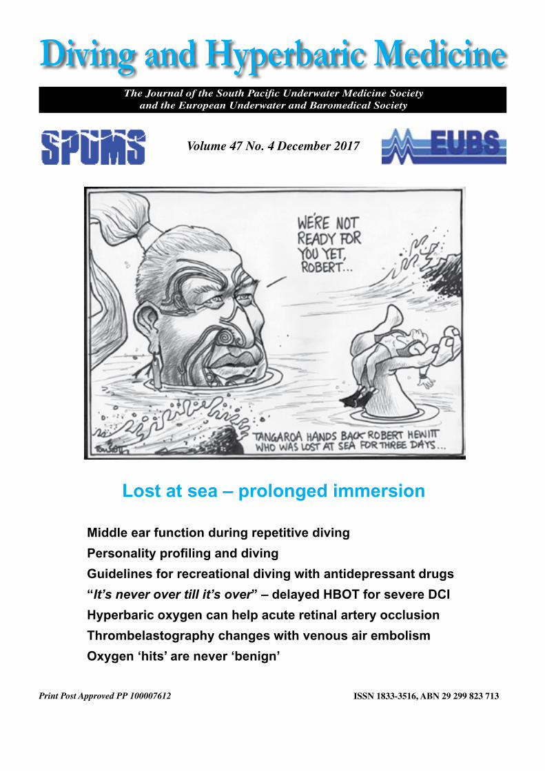
Print Post Approved PP 100007612 Volume 47 No. 4 December 2017 Diving and Hyperbaric Medicine The Journal of the South Pacific Underwater Medicine Society and the European Underwater and Baromedical Society ISSN 1833-3516, ABN 29 299 823 713 Lost at sea – prolonged immersion Middle ear function during repetitive diving Personality profiling and diving Guidelines for recreational diving with antidepressant drugs “It’s never over till it’s over” – delayed HBOT for severe DCI Hyperbaric oxygen can help acute retinal artery occlusion Thrombelastography changes with venous air embolism Oxygen ‘hits’ are never ‘benign’
Transcript of Diving and Hyperbaric Medicine · Kathleen Pye Chantrey, Hillside Road Stromness, Orkney KW16 3HR,...
Print Post Approved PP 100007612
Volume 47 No. 4 December 2017
Diving and Hyperbaric MedicineThe Journal of the South Pacific Underwater Medicine Society
and the European Underwater and Baromedical Society
ISSN 1833-3516, ABN 29 299 823 713
Lost at sea – prolonged immersion
Middle ear function during repetitive diving
Personality profiling and diving
Guidelines for recreational diving with antidepressant drugs
“It’s never over till it’s over” – delayed HBOT for severe DCI
Hyperbaric oxygen can help acute retinal artery occlusion
Thrombelastography changes with venous air embolism
Oxygen ‘hits’ are never ‘benign’

SOUTH PACIFIC UNDERWATERMEDICINE SOCIETY
OFFICE HOLDERSPresident
David Smart <[email protected]>Past President
Michael Bennett <[email protected]>Secretary
Douglas Falconer <[email protected]>Treasurer
Sarah Lockley <[email protected]>Education Officer
David Wilkinson <[email protected]>Chairman ANZHMG
Neil Banham <[email protected]> Committee Members
Jen Coleman Tamara Ford Ian Gawthrope Cathy Meehan Peter Smith
WebmasterJoel Hissink <[email protected]>
ADMINISTRATIONMembership
Steve Goble <[email protected]>MEMBERSHIP
For further information on SPUMS and to register to become a member, go to the Society’s website: <www.spums.org.au> The official address for SPUMS is:
c/o Australian and New Zealand College of Anaesthetists, 630 St Kilda Road, Melbourne, Victoria 3004, AustraliaSPUMS is incorporated in Victoria A0020660B
EUROPEAN UNDERWATER ANDBAROMEDICAL SOCIETY
Diving and Hyperbaric Medicine Volume 47 No. 3 September 2017
PURPOSES OF THE SOCIETIESTo promote and facilitate the study of all aspects of underwater and hyperbaric medicine
To provide information on underwater and hyperbaric medicineTo publish a journal and to convene members of each Society annually at a scientific conference
OFFICE HOLDERSPresident Jacek Kot <[email protected]>Vice President Ole Hyldegaard <[email protected]>Immediate Past President Costantino Balestra <[email protected]>Past President Peter Germonpré <[email protected]>Honorary Secretary
Peter Germonpré <[email protected]>Member-at-Large 2017
Rodrigue Pignel <[email protected]>Member-at-Large 2016 Bengusu Oroglu <[email protected]> Member-at-Large 2015 Karin Hasmiller <[email protected]>Liaison Officer
Phil Bryson <[email protected]>
ADMINISTRATIONHonorary Treasurer and Membership Secretary
Kathleen Pye <[email protected]>MEMBERSHIP
For further information on EUBS and to complete a membership application, go to the Society’s website: <www.eubs.org>The official address for EUBS is:
Kathleen Pye Chantrey, Hillside Road
Stromness, Orkney KW16 3HR, United KingdomEUBS is a UK Registered Charity No. 264970
EditorMichael Davis <[email protected]>PO Box 35, Tai Tapu 7645New ZealandPhone: +64-(0)3-329-6857
European (Deputy) EditorLesley Blogg <[email protected]>
Editorial AssistantNicky Telles <[email protected]>
Journal distributionSteve Goble <[email protected]>
Journal submissions: <https://www.manuscriptmanager.net/dhm>
Editorial BoardMichael Bennett, AustraliaDavid Doolette, USAChristopher Edge, United KingdomIngrid Eftedal, NorwayPeter Germonpré, BelgiumJacek Kot, PolandSimon Mitchell, New ZealandClaus-Martin Muth, GermanyNeal Pollock, CanadaMonica Rocco, ItalyMartin Sayer, United KingdomErika Schagatay, SwedenDavid Smart, AustraliaRobert van Hulst, The Netherlands
DIVING AND HYPERBARIC MEDICINE<www.dhmjournal.com>
Diving and Hyperbaric Medicine is published jointly by the South Pacific Underwater Medicine Society and the European Underwater and Baromedical Society (ISSN 1833-3516, ABN 29 299 823 713)

Diving and Hyperbaric Medicine Volume 47 No. 4 December 2017 211
The Editor's offering
Front page cartoon: Tangaroa hands back Robert Hewitt who was lost at sea for three days. “We’re not ready for you yet, Robert...” 10 February 2006 by Tom Scott [Digital cartoon published in the Dominion Post]. Ref: DCDL-0000754. Alexander Turnbull Library, Wellington, New Zealand. /records/23153470; with the kind permission of the artist and the Turnbull Library.
The three review articles in this issue focus on aspects of psychology – survival in an extreme environment, the relationship between personality and the behaviour of divers and guidelines, promulgated by the Dutch Association for Diving Medicine, on the use by divers of antidepressant drugs.1−3 Personality inventories have been used quite widely in the selection of military personnel for diver training with mixed results and little impact on any understanding of how divers would actually perform in an operational setting. As a university student in the mid-1960s, I recall a friend who had joined the Royal Navy officer training scheme describing the thrill of jumping off the deck of an aircraft carrier in full clearance diver kit; apparently hesitating did not make for a good candidate! There is a dearth of data on the interaction between psychoactive medication and the diving environment, most advice being largely based on theoretical considerations and hearsay. Robert Hewitt’s extraordinary survival at sea completely captured the New Zealand nation, indeed, it was international news. I and my fellow authors were criticised by the reviewers (I recused myself from any involvement in the peer review process) because the article was neither a simple case report nor a full review paper of prolonged immersion in cold water. However, we felt strongly that this story carried many messages that would contribute to readers’ understanding of such extreme situations and this argument was accepted by Lesley Blogg, our European Editor.
The diver in the report by Perez et al. received seven US Navy Treatment Table 6s (USN TT6) to achieve a good functional recovery from a life-threatening injury.4 Given this and the week’s delay to definitive hyperbaric treatment, one could say that the end justified the means. However, might the same outcome have been achieved with multiple shorter, shallower HBOT which would have incurred less risk of decompression sickness (DCS) for nurse attendants? By coincidence, the health and safety of chamber attendants vis-à-vis DCS risk was recently reviewed by Dick Clarke, in which he documents several fatalities or career-ending injuries amongst attendants.5 Treatment pressures of 284 kPa or higher are associated with an increased DCS incidence. Means of mitigating DCS risk include a variety of approaches, not only the use of oxygen by attendants.
John Lippmann of Divers Alert Network – Asia Pacific (DAN AP) very recently asked the question “how many USN TT6 is enough?” of the participants of the Australian and New Zealand Hyperbaric Medicine Group chat line, triggered by another case from the Pacific region that DAN AP had been involved with in which a diver with spinal DCS received four USN TT6 and made a good recovery. The answers he received from this group of clinicians were varied, but the essence of all was simply that there is no single or easy answer. Lippmann summarised the feedback as “there is no doubt that continuing treatment until there is no significant
improvement is appropriate but the best regimen used to get there remains unresolved but should involve a careful risk benefit assessment of the patient and attendants.”
The future for Diving and Hyperbaric Medicine
This issue celebrates a decade of publication cooperation between SPUMS and EUBS. The path has not always been smooth between the two societies, but everyone can be proud of how DHM has developed into an international journal over that time. Major changes will happen to Diving and Hyperbaric Medicine (DHM) over the coming year.• As mentioned by both Presidents, DHM becomes an
electronic publication as of the March 2018 issue. Aprint version will no longer be produced by this editorial office, though both societies are considering how theymight provide one in the future for those who wish topay extra. I am strongly opposed to this, especiallyas the original directive I was given as Editor was toproduce an e-journal only. DHM needs to move withthe times – the future is in electronic publication! Please remember that articles published in DHM are subjectto a one-year embargo – we do not want to see DHMon social media platforms straight after publication, ashappened earlier this year on a Swiss platform with ascanned-in version that could only have come from asociety member! A friendly user interface for a varietyof platforms (e.g., smart phones, iPad) will be provided.
• This task has proved more challenging than expected,especially in relation to preserving MedLine citation.This has resulted in a great deal of additional work andexpense, including a complete rebuild and enhancementbeing necessary of what is currently a very basic journal website. Because of this, a crowd-funding activity bySPUMS is in place to help raise the required capital,about which you will hear soon. Please give generously to support your society’s journal into the future.
• All articles from 2017 on will now have a digital objectidentifier (DOI) number attributed to them, and we will gradually add these to back articles to when SPUMSand EUBS joined forces on DHM in 2008. DHM isnow a member of Crossref, which will expand furtherthe exposure of articles published in DHM.
• Revised Instructions to Authors will be releasedearly in 2018. Many authors have been rather poor atfollowing our instructions correctly but this will nolonger be tolerated – it adds greatly to our work – andall incorrectly submitted papers will automatically berejected for correct resubmission. It is worth noting
Diving and Hyperbaric Medicine Volume 47 No. 4 December 2017212
The Presidents' pagesDavid Smart, President SPUMS
This issue represents the end of an era for Diving and Hyperbaric Medicine (DHM). It is the last purely printed version of the Journal. From March 2018, the publishing societies (SPUMS and EUBS) are moving to an electronic journal. It is the way of the future and will open up many opportunities and capabilities for developing DHM and will position us at the forefront of diving medicine this century. Over the past five years, under the editorship of Mike Davis, with assistance from the European Editor, Lesley Blogg, DHM has become the premier publication in its field. In 2016, the journal’s impact factor reached 1.2, an important milestone for a small, society-published specialty journal. With publishing societies on either side of the globe, this is an amazing result, given the huge exercise in logistics to co-ordinate DHM as a publication.
In addition, it has become apparent that the cost of continuing DHM print circulation has accelerated far faster than the cost of living. Simply put, current SPUMS membership subscriptions are insufficient to cover the costs of the print version of the journal and the effective running of the Society. SPUMS, together with EUBS decided to move to the e-journal from the first edition (March) in 2018. SPUMS subscriptions for 2018 will include the electronic journal on a quarterly basis. It is our plan to have available a print quality PDF version, for a small additional fee. This will permit members to print their own journal locally, should they wish. I will be writing to all SPUMS members in coming weeks to brief them of our future plans for the journal.
There is an enormous amount of work in the transition from
print to electronic, particularly for the Editorial Office. The efforts of the Office, including Editorial Assistant, Nicky Telles, are greatly appreciated. It is expected that, once implemented, the workload should reduce for the office, and opportunities for exposure of our speciality on a global basis will be greater. Linkages to the National Library of Medicine by joining PubMedCentral will have huge advantages in this electronic era. In addition, during 2018 there will be a transition and handover of the Editor-in-Chief role from Mike Davis, to Professor Simon Mitchell, who will officially take the reins from the December 2018 issue. It is indeed an exciting time for our speciality.
I am delighted to report that David Wilkinson will continue as Education Officer for a further three-year term, and that Joel Hissink has withdrawn his resignation and will continue as Webmaster. Neil Banham (Western Australia) has been elected chair of the Australia and New Zealand Hyperbaric Medicine Group, a subcommittee of SPUMS.
Key wordsMedical society; General interest
The
website is at<www.spums.org.au>
Members are encouraged to log in
here that about a third of all manuscripts submitted to DHM are eventually declined for publication, mainly because of poor science. DHM is determined to maintain a high standard of peer review so that both researchers and readers have something worthwhile adding to our understanding and satisfying to read.
• Manuscript Manager (MM), the DHM submissionsplatform, has just launched a new, upgraded, user-friendly version for authors and reviewers to use.Training videos linked to the website will be availablefrom the MM software developers early next year.
• A new Editor-in-Chief takes over from me in late 2018. Whilst his appointment is still under final negotiation, I am very excited about the calibre of my likely successor, whom I am sure will take DHM to a new level.
References
1 Massey H, Leach J, Davis FM, Vertongan V. Lost at sea: the medical, physiological and psychological factors of
prolonged immersion. Diving Hyperb Med. 2017;47:239-47. doi10.28920/dhm47.4.239-247.
2 van Wijk CH. Personality and behavioural outcomes in diving: current status and recommendations for future research. Diving Hyperb Med. 2017;47:248-52. doi10.28920/dhm47.4.248-252.
3 Querido AL. Diving and antidepressants. Diving Hyperb Medicine. 2017;47:253-56. doi10.28920/dhm47.4.253-256.
4 Perez MFM, Ongkeko-Perez J, Serrano AR, Andal MP, Aldover MCC. Delayed hyperbaric intervention in life-threatening decompression illness. Diving Hyperb Med. 2017; 47:257-9. doi10.28920/dhm47.4.257-259.
5 Clarke R. Health care worker decompression sickness: incidence, risk and mitigation. Undersea Hyperb Med. 2017;44:509-19.
Key wordsPsychology; Decompression illness; Treatment; Hyperbaric oxygen therapy; Writing – medical; Editorial
Michael Davis

Diving and Hyperbaric Medicine Volume 47 No. 4 December 2017 213
Jacek Kot, President EUBS
The 43rd Annual Scientific Meeting of the EUBS was held in September in the beautiful, historical city of Ravenna, Italy. The scientific committee, consisting of Paolo Pelaia (Chairman), Costantino Balestra, Zeljko Dujic, Paquale Longobardi, Monica Roco and myself, were able to prepare a diverse programme of lectures, reviews and technical reports, with a good balance between diving and hyperbaric medicine. In total, there were 40 oral presentations and 68 poster presentations. The conference was well attended with 270 full participants. It has become a tradition that the Conference starts with the Master Class for Young Investigators, and this year it was devoted to statistical aspects in medical sciences. In comparison to previous meetings, there was a large number of sessions organised in cooperation with other organisations. These included a DMAC Workshop on nutrition and hydration for saturation divers and medical aspects of hyperbaric evacuation; a SINSEC session on multidisciplinary approach to right-left shunt, an international session presenting UHMS clinical practice guidelines and EUBS HBOT in Europe, an EBAss session on application of standards for hyperbaric technology, and an ECHM Workshop on the role of HBO on mitochondrial functions, oxidative stress, cell signalling and chemokines. Indeed, there was something for everyone!
There were also two unforgettable social events. The first was the string quartet concert that was performed in the Basilica of San Vitale (consecrated in 547 AD) in the old centre of Ravenna. I always thought that I was resistant to the beauty of sound, preferring to measure it rather than to listen to it, but when I heard the first notes in the magnificent interior of the church I had goose bumps! The other was the conference dinner at Lido di Savio close to Ravenna. This will be remembered not only for the good food and company, but mostly for the sudden, heavy storm that flooded the beach part of the restaurant and nearly carried away the large marquee in which we were dining! Fortunately, the storm passed as quickly as it came leaving no real damage, but long-lasting memories.
On behalf of the EUBS, I would like to express our sincere gratitude to Pasquale Longobardi and all his team, including Paolo Pelaia and Monica Rocco, for organising the Ravenna meeting. The conference was an astounding success thanks to all their hard work.
On the EUBS Executive Committee, Rob van Hulst has now completed his term as member at large and is succeeded by Rodrigue Pignel from Switzerland. On the DHM Governance Committee, Peter Müller and Joerg Schmutz have been succeeded by Philip Bryson from the UK and Karin Hasmiller from Germany. We thank Rob, Peter and Joerg for their contributions, the latter two having been European Editor of DHM and EUBS Secretary respectively in the past.
The next EUBS ASM will be the Second Tricontinental Conference organised jointly with SPUMS and SAUHMA in Durban, South Africa in September 2018. I am sure that there is no need to convince those who remember the first joint tri-continental meeting in Reunion in 2014 to attend. Reserve your flights soon to get the best prices. More information can be found further in the issue. The 2019 EUBS ASM will take place in Israel − more information soon. Interestingly, after several years of having few applications from volunteers to organise the EUBS annual ASM, the ExCom was pleased to receive formal requests from Denmark (Copenhagen), France (Brest) and Croatia (Rijeka). Specific decisions have not yet been taken, but you should apply now for a frequent flyer programme!
Two final points must be mentioned. One is that the end of the year is fast approaching and this surprises me every year. This gives me the delightful opportunity to pass on my best personal greetings, wishing you and your loved ones peace, health, happiness and prosperity in the coming New Year − “Merry Christmas”.
The other point is that we are close to switching to an electronic platform for our Journal. Our Society members should say goodbye to the standard printed version from the beginning of 2018. While I will still keep my office printer switched on and ready to print anything that I need to, this transition must be perceived as an upgrade of the Journal. Nevertheless, stay tuned to our website <www.eubs.org> for all society news.
List of abbreviations:DHM – Diving and Hyperbaric MedicineDMAC – Diving Medical Advisory CommitteeSINSEC – Societa Italiana di NeuroSonologia ed Emodinamica CerebraleUHMS – Undersea and Hyperbaric Medical SocietyEBAss – European Baromedical Association for nurses, operators and techniciansECHM – European Committee for Hyperbaric MedicineSPUMS – South Pacific Underwater Medicine SocietySAUHMA – Southern African Undersea and Hyperbaric Medical Association
Key wordsMedical society; General interest
The
website is at <www.eubs.org>
Members are encouraged to log in and keep their personal details up to date
Diving and Hyperbaric Medicine Volume 47 No. 4 December 2017214
Editorial
September 14, 2017 marked a potential watershed for diver training in Australia, when The Underwater Centre Tasmania (TUCT) was forced into liquidation and closed its doors. TUCT was the only Australian Diver Accreditation Scheme (ADAS)-accredited and International Marine Contractors Association (IMCA)-recognised level 4 Diver Training Centre in the southern hemisphere and had been operational for over 20 years. The centre was also the only civilian (not military or police) level 3 diving training centre in Australia (Level 3 training is available in New Zealand). TUCT has trained hundreds of commercial divers in accordance with AS/NZS 2815. The closure of the centre will be a huge blow for diver training in Australia and the future ramifications are quite concerning, given current trends in deregulating industry diver training. Essentially it means that Australian divers will not be able to access internationally recognised off-shore training in this country.
As with other high-risk industries, an appropriately trained workforce is a key foundation for maintaining a safe workplace. A key aspect of diver training at TUCT was that it fully complied with ADAS which was set up in the 1990s to reflect international best practice in occupational diver training and certification. The AS/NZS 2815 series of occupational diving training standards reflected this. ADAS courses are also nationally aligned with the Australian Skills Quality Authority and are Vocational Education and Training (VET) -accredited. ADAS accredited training establishments are required to meet stringent entry conditions and maintain
compliance with a number of standards in operational training assessment and administration. TUCT maintained ADAS accreditation for the whole of its operational existence. This has enabled their graduates to work globally in the diving industry. An additional advantage of international standard training is the sharing of information regarding safety on a global basis, and the systems of safe command and control of dive operations.
Well-trained dive teams can identify risks before they occur, manage the risks in real time to improve safety, and are empowered to challenge borderline safety practices if they occur. If divers possess the necessary skills to competently and efficiently perform tasks using appropriate equipment and safety procedures, productivity and safety are improved. High-quality training processes also improve operational safety in diving because there is broader understanding of standards, maintenance and safety procedures, dive schedules, appropriate use of the correct equipment and international best practice. It is unfortunate that when Australia’s Model Work Health and Safety Legislation was passed in 2012, it failed to recognise many of the existing Australian Standards that relate to occupational diving. In addition, the legislation artificially separated high-risk “construction divers” (subject to AS/NZS 2299.1), from “general divers”.1,2 Inadvertently, this has paved the way for deregulation of diver training in Australia. Any diving other than construction diving was effectively declared “low risk”, without consideration of fundamental contributors to
Back to the future: occupational diver training in AustraliaDavid Smart
Professor David Smart, Department of Diving and Hyperbaric Medicine, Royal Hobart Hospital, PO Box 1061, Hobart, TAS 7001, [email protected]
Key wordsDiving industry; Diving at work; Standards; Education; Safety; Diving incidents
Abstract
(Smart D. Back to the future: diver training in Australia. Diving and Hyperbaric Medicine. 2017 December;47(4):214-215. doi10.28920/dhm47.4.214-215. doi10.28920/dhm47.4.214-215.)The Australian Diver Accreditation Scheme (ADAS) had its genesis in the 1990s in response to a need to produce occupational divers who were trained to international standards with the necessary skills to safely undertake complex work in high-risk environments. Well-trained dive teams who are ‘fit-for-purpose’ can be regarded as the highest level of risk control in preventing accidents and workplace morbidity. Without such training, work site risks are not detected, with potentially disastrous consequences. In September 2017, the only civilian ADAS level 3 and 4 training facility in Australia, The Underwater Centre Tasmania (TUCT), closed its doors. The reasons for TUCT closure were multifactorial. However, the loss of higher level training capability in this country and its benefits to industry will have a future adverse impact. As industry pushes for more complex diving to improve productivity, Australian occupational diver training processes are becoming ‘streamlined’ and are losing parity with international benchmarks. This is a potentially fatal combination.

Diving and Hyperbaric Medicine Volume 47 No. 4 December 2017 215
risk such as the diver’s activity and tasks, the equipment they use and the conditions/environment in which they dive.
In the past I have documented the adverse impacts on safety when dive teams lacked the necessary skills and training to perform their tasks, or when they lack the knowledge to challenge unsafe shortcuts or practices being requested by their employers.3 Tasmania’s aquaculture industry provides a model to demonstrate how safety was improved by high standards of training and operational practice within the industry.4 In the 1980s rates of diver morbidity in Tasmania were over 50 times current levels, owing to lack of training, poor safety procedures, inadequate equipment maintenance and inappropriate use of dive schedules and profiles. Among other factors, major improvements in safety occurred when divers were trained to a universal benchmark certified by ADAS.
However as a potential downside for employers, ADAS training also allowed divers to become geographically mobile, because their qualifications allowed them multiple employment options. After providing financial support for the diver to train, the employer would rightfully expect a degree of loyalty, which was not always forthcoming. A solution to this issue has been to create industry-specific training programmes that meet basic requirements of WHS legislation, but are not recognised outside the industry. This permits a reduced cost structure with shorter courses and lower levels of compliance than are required for international courses. Without international or national certification, divers potentially remain captive within the industry.
With economic pressures on employers, a different kind of pressure is also exerted on dive teams – to increase productivity at lower cost. This is common to all industries; however, for occupational divers, changes in operational practice and cutting corners will eventually adversely impact on safety. Lessons from the early years of occupational diving sit indelibly etched in history, waiting to surface [sic] when the guard is dropped. Regulators appear to have short memories. Australia’s Model WHS Legislation still has serious deficiencies in relation to diving, and it is my opinion that these deficiencies will unintentionally allow ‘dumbing down’ of diver training standards. I have represented SPUMS on reviews of the legislation, which unfortunately were very narrow in focus and did not address the key deficiencies SPUMS identified in our submissions. I would assert that divers who lack the higher level knowledge to identify impacts of changes in practice on safety are more likely to sustain injuries.
The future challenge will be to maintain the quality of training at a level that matches the rapid development of technology in industry. Although some of this technology (e.g., remote operating vehicles) has improved safety for divers, other technology has increased diver risk. An example of this is the necessary move by industry to develop offshore
marine farming.5 This requires industrial structures, with major plant and equipment. Divers are diving to 30 m depth, performing tasks that fall completely within the purvey of construction diving.2 Diving in these settings would be far better achieved using a diving workforce and supervisors trained to ADAS standards. Santayana stated “those who cannot remember the past are condemned to repeat it”.6 For diver training in Australia it is back to the future. I hope I am proven wrong in relation to the adverse impact this will have on safety.
References
1 Model Work Health and Safety Regulations. 28 November 2016. Part 4.8. Diving Work. p. 130-9. [cited 2017 October 10]. Available from: https://www.safeworkaustralia.gov.au/system/files/documents/1703/model-whs-regulations-28nov2016.pdf.
2 Standards Australia/Standards New Zealand. Australian/New Zealand Standard Occupational diving operations. AS/NZS 2299 Part 1. Standard operational practice. December 2015.
3 Smart DR, McCartney P. High risk diving; Tasmania’s aquaculture industry. SPUMS Journal 1990;20:159-65.
4 Smart DR, Van den Broek C, Nishi R, Cooper PD, Eastman D. Field validation of Tasmania’s aquaculture industry bounce diving schedules using Doppler analysis of decompression stress. Diving Hyperb Med. 2014;44:124-36.
5 Gurra B. Salmon farms look for new pastures to allow sustainable growth. ABC News on-line 28th September 2016. [cited 2017 October10]. Available at: http://www.abc.net.au/news/2016-0928/salmon-farms-set-for-offshore-development/7885898.
6 Wikiquote. George Santayana. [cited 2017 October 10]. Available at: https://en.wikiquote.org/wiki/George_Santayana.
Conflict of interest
The author provided paid teaching services and medical advice to TUCT, and has run a not-for-profit DMAC Level 2 Medical Support of Offshore and Saturation Diving Course with assistance of TUCT.
Submitted: 27 October 2017Accepted: 01 November 2017
Copyright: This article is the copyright of the author who grants Diving and Hyperbaric Medicine a non-exclusive licence to publish the article in printed and other forms.

Diving and Hyperbaric Medicine Volume 47 No. 4 December 2017216
Original articlesInfluence of repetitive diving in saltwater on pressure equalization and Eustachian tube function in recreational scuba diversMoritz F Meyer1, Manuela Boor1, Stefanie Jansen1, Eberhard D Pracht2, Moritz Felsch3,Heinz D Klünter1, Karl-Bernd Hüttenbrink1, Dirk Beutner1, Maria Grosheva1
1 Department of Otorhinolaryngology, Head and Neck Surgery, University of Cologne, Germany2 German Centre for Neurodegenerative Diseases (DZNE), Bonn, Germany 3 Institute of Medical Statistics, Informatics and Epidemiology, University of Cologne, Germany
Corresponding author: Professor Moritz F Meyer, Department of Otorhinolaryngology, Head and Neck Surgery, Faculty of Medicine, University of Cologne, Kerpener Straße 62, 50937 Cologne, [email protected]
Key wordsTympanometry; Middle ear; Ear barotrauma; Recreational diving
Abstract(Meyer MF, Boor M, Jansen S, Pracht ED, Felsch M, Klünter HD, Hüttenbrink K-B, Beutner D, Grosheva M. Influence of repetitive diving in saltwater on pressure equalization and Eustachian tube function in recreational scuba divers. Diving and Hyperbaric Medicine. 2017 December;47(4):216-222. doi10.28920/dhm47.4.216-222.)Introduction: We investigated in a prospective, observational trial the feasibility of using the Eustachian tube function test (ETFT) to measure the effect of repetitive pressure exposure during open seawater dives on Eustachian tube function.Methods: The study included 28 adult divers during six consecutive days of diving in the Red Sea. Participants underwent otoscopy and ETFT before the first dive, between each dive and after the last dive. ETFT included regular tympanometry (R-tymp), tympanometry after Valsalva (V-tymp) and after swallowing (S-tymp). The R-tymp was obtained as ‘baseline’ peak pressure. After a Valsalva, the peak pressure should shift (positively), revealing a positive shift of the tympanic membrane. This pressure shift is defined here as R-V
dP. The changes in compliance and peak pressure were recorded and correlated
with otoscopic findings and diving experience. Middle ear barotrauma was scored using the Edmonds modified TEED scale.Results: The 28 participants performed 437 dives. Positive shift of pressure in the middle ear was evident with significant changes from day one to day three (P < 0.0001). Divers with barotrauma showed significantly lower values of R-tymp peak pressure and significantly higher negative R-V
dP, compared to divers with normal otoscopic findings (P < 0.05). Diving
experience significantly correlated with R-tymp peak pressure and prevalence of middle ear barotrauma.Conclusion: Significant changes in middle ear pressure and pressure equalization from repeated pressure exposure in saltwater were seen using ETFT. Repetitive, multi-day diving led to significantly decreased compliance and increased R-tymp peak pressure (overpressure) in the middle ear. Most profound changes were observed in less and intermediate experienced divers.
Introduction
Recreational scuba diving requires reliable and adequate pressure equalization in the middle ear. Rapidly increasing and decreasing the surrounding pressure during diving induces stress on the tympanic membrane (TM) and middle ear. The Eustachian tube (ET) is essential for drainage, protection and pressure equalization of the middle ear.1–3 Pressure equalization occurs in both directions;4–6 active pressure equalization is usually required during descent, whilst equalization is passive on ascent. Active equalization is supported by well-known musculature-involving manoeuvres such as swallowing, Valsalva or Toynbee.4–6 Inadequate pressure equalization leads to consequent trauma of the middle ear mucosa, and TM and may impair ET function.7,8 Corresponding pathological otoscopic findings such as hyperaemia, oedema, haemotympanum or even
TM rupture characterize painful middle ear barotrauma (MEBt).9,10
Despite the rising popularity of recreational scuba diving,7 the influence of repeated pressure changes on the middle ear and ET during scuba diving remains unclear. Challenging study conditions and the lack of reliable diagnostic methods might partly explain the low number of prospective, open-water studies. Whilst tympanometry remains one of the most commonly used diagnostic tools for assessing middle ear function in clinical otology and in basic research,11 the Eustachian tube function test (ETFT) (based on the Williams inflation/deflation procedure12–14) takes this a step further, using a series of three tympanometric measurements.
The primary objective of this trial was to evaluate changes in ET function and pressure equalization of the middle ear
Diving and Hyperbaric Medicine Volume 47 No. 4 December 2017 217
after repetitive saltwater dives. The secondary objective was to confirm the feasibility of using the ETFT prospectively in recreational scuba diving to study these changes.
Methods
ETHICAL CONSIDERATIONS AND INCLUSION CRITERIA
The Ethics Committee of the University Hospital of Cologne, Germany approved this observational, prospective cohort study. The trial was registered by the German Clinical Trial Register(No. DRKS00008968). Written informed consent was obtained from each participant before their inclusion. The study-related examinations took place during their vacation. All participants were certified divers over 18 years old, with a valid medical certificate and who had not been diving for 24 hours prior to the study. The divers were classified as ‘inexperienced’ (< 50 dives), ‘intermediate’ (50–200 dives), ‘experienced’ (201–499 dives) and ‘professional’(≥ 500 dives). The experience levels were set according to recommendations of the diving instructors who participated in the study.
DIVING
Diving was conducted in the Sharm el Sheikh region of Egypt over six consecutive days. Water temperature in the Red Sea was stable at 23–24ºC, and the salinity was 4.2%.15 On the first day of diving, all participants performed shore dives; from days two to six the majority of dives were boat dives. Participants were allocated to two boats for the whole week. Start time, duration and depth of every dive were assessed for each participant.
STUDY-RELATED INTERVENTIONS
Certification level, diving experience, number of dives and date of the last dive were recorded. The otoscopic appearances of the TM, the external ear canal and the ability to perform a Valsalva manoeuvre were evaluated (Heine, Herrsching, Germany). Presence of TM movement during a Valsalva manoeuvre was documented as ‘present’ or ‘absent’. Nasopharyngeal pathologies were excluded using a rigid 0º endoscope (Storz, Tuttlingen, Germany). All parameters were documented separately for the left and right ears. During the week of diving, participants underwent otoscopy and the ETFT before the first dive, between each dive and after the last dive. ETFT was performed an average of 43 ± 22 min after the dive (range: 1–116 min). Two consecutive dives within 12 h of each other were defined as repetitive dives. TM changes were evaluated using the TEED classification for MEBt as modified by Edmonds.16,17 Final examination took place at least 12 hours after the last dive.
EUSTACHIAN TUBE FUNCTION TEST
ETFT was carried out according to the guidelines of
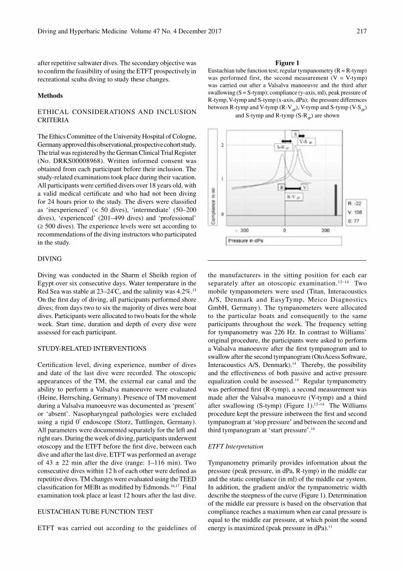
the manufacturers in the sitting position for each ear separately after an otoscopic examination.12−14 Two mobile tympanometers were used (Titan, Interacoustics A/S, Denmark and EasyTymp, Meico Diagnostics GmbH, Germany). The tympanometers were allocated to the particular boats and consequently to the same participants throughout the week. The frequency setting for tympanometry was 226 Hz. In contrast to Williams’ original procedure, the participants were asked to perform a Valsalva manoeuvre after the first tympanogram and to swallow after the second tympanogram (OtoAcess Software, Interacoustics A/S, Denmark).14 Thereby, the possibility and the effectiveness of both passive and active pressure equalization could be assessed.14 Regular tympanometry was performed first (R-tymp), a second measurement was made after the Valsalva manoeuvre (V-tymp) and a third after swallowing (S-tymp) (Figure 1).12−14 The Williams procedure kept the pressure inbetween the first and second tympanogram at ‘stop pressure’ and between the second and third tympanogram at ‘start pressure’.14
ETFT Interpretation
Tympanometry primarily provides information about the pressure (peak pressure, in dPa, R-tymp) in the middle ear and the static compliance (in ml) of the middle ear system. In addition, the gradient and/or the tympanometric width describe the steepness of the curve (Figure 1). Determination of the middle ear pressure is based on the observation that compliance reaches a maximum when ear canal pressure is equal to the middle ear pressure, at which point the sound energy is maximized (peak pressure in dPa).11
Figure 1Eustachian tube function test; regular tympanometry (R = R-tymp) was performed first, the second measurement (V = V-tymp) was carried out after a Valsalva manoeuvre and the third after swallowing (S = S-tymp); compliance (y-axis, ml), peak pressure of R-tymp, V-tymp and S-tymp (x-axis, dPa); the pressure differences between R-tymp and V-tymp (R-V
dP), V-tymp and S-tymp (V-S
dP)
and S-tymp and R-tymp (S-RdP
) are shown
Diving and Hyperbaric Medicine Volume 47 No. 4 December 2017218
A positive shift of the R-tymp peak pressure mirrors the increasing middle ear pressure (‘overpressure’ or positive pressure). Accordingly, a negative shift of the peak pressure occurs due to negative middle ear pressure. The R-tymp was obtained as ‘baseline’ peak pressure. After a Valsalva, the peak pressure should shift (positively), revealing a positive shift of the TM. This pressure shift is defined in this paper as R-V
dP. The extent of the peak pressure shifts
(i.e., R-VdP
, V-SdP
and S-RdP
) provides information about the effectiveness of pressure equalization during the Valsalva manoeuvre and swallowing, respectively. Higher R-V
dP
might indicate better ET function. Figure 1 gives an example of normal ETFT data.
Compliance depends on the state of the TM and the pressure in the middle ear. The condition of the middle ear mucosa, fluid in the middle ear, chronic and acute structural TM changes influence the compliance variables.
STATISTICAL ANALYSIS
All data were de-identified. The findings of the left and right ears were evaluated separately, but analysed together using SPSS software (version 23.0.0.0, IBM Corporation, USA). Quantitative variables are presented as mean ± standard deviation, or 95% confidence intervals (95% CI), or median and range, or interquartile range (IQR) and qualitative variables as absolute number and percentage. We applied mixed model analysis of variance with repeated measures for analysis of quantitative data and Chi-square test for analysis of qualitative data. The mixed model analysis allows for inclusion of multiple time points per subject, while accounting for unbalanced data structure of irregular time intervals between ETFT measurements and unequal numbers of ETFT analyses per subject. The respective ETFT parameters (R-tymp, R-V
dP, V-S
dP, and R-tymp compliance)
were included as dependent variable and the dive number, diving day, TEED grade and diving experience groups as covariates. Univariate analysis (F-test) was based on analysis of linear independent pairwise comparisons. A P-value < 0.05 was considered statistical significant. All reported P-values are two-sided. To correct for multiple testing, P-values were adjusted using the Bonferroni correction. A corrected P-value of < 0.05 was considered to be significant for all tests.
Results
PARTICIPANTS
Of the 28 participants, 19 were male and nine female, mean age 38 ± 10.0 years. Median number of dives before the study was 53 (range: 4−2,550). The last dive was performed a median of 2.5 months (range: 27 days to 186 months) prior to the study. The participants performed an average of 15
dives (range: 9−19) during the six days; on average three per day (range: 1−4). Average diving duration was 51 ± 7 minutes; average depth was 25 ± 7 metres’ seawater. We defined 13 divers as inexperienced, seven as intermediate, three as experienced and five as professional.
CLINICAL EXAMINATIONS BEFORE THE FIRST DIVE
Endoscopy of the ear, nose and epipharynx was normal in 26 participants. No local anaesthetic or lubricant was used for the endoscopic evaluation. Two participants (both experienced divers) showed exostoses of the external ear canal in both ears. The appearances of the TMs were normal bilaterally in all the divers and they were able to perform a Valsalva manoeuvre successfully, with a type-A tympanogram.18
OTOSCOPY DURING THE DIVING
Valsalva manoeuvres were effective in 99.7% (n = 437) of dives. No cases of external ear canal inflammation were seen. As reported elsewhere, MEBt (TEED 1−3) was observed in 42.2% (n = 490) of the 1,161 otoscopic examations.19 In total, TEED 0 was observed in 57.8% (n = 671), TEED 1 in 34.1% (n = 396), TEED 2 in 7.5% (n = 87) and TEED 3 in 0.6% (n = 7). No TM perforations (TEED 4) occurred.19
COMPLIANCE
No significant differences in compliance were seen between the right and left ears (F-test for left/right ear, P = 0.717). Mean compliance values also did not differ significantly between the R-, V- and S-tymp measurements: 1.4 ± 1.3 ml for all measurements; median 1.0 ml (IQR 0.7−1.5 ml), 1.0 ml (IQR 0.7−1.5 ml) and 1.1 ml (IQR 0.7−1.6 ml), respectively. For this reason, we performed the following analyses using only the R-tymp compliance. There was a significant increase in compliance after the first dive (F-test for dive number, P < 0.0001). During the six diving days, the mean values for compliance slightly decreased after day 1 (1.9 ml on day 1, 1.5 ml on day 2) but thereafter remained stable (F-test for diving day, all P > 0.474). Professional divers had higher compliance values compared to divers with less experience (F-test for experience groups,P < 0.0001; data not shown*). Furthermore, professional divers also showed a significantly greater difference in compliance after the first, second and third dive in a day compared to less experienced divers (pair-by-pair comparison, all P < 0.05; data not shown*).
PEAK PRESSURE
All participants were able to equalize pressure in the middle ear before diving. Analysing the R-tymp values during the six consecutive days of diving, significant positive shift of
* Footnote: Additional summary data tables not shown here are available from the authors at <[email protected]>
Diving and Hyperbaric Medicine Volume 47 No. 4 December 2017 219
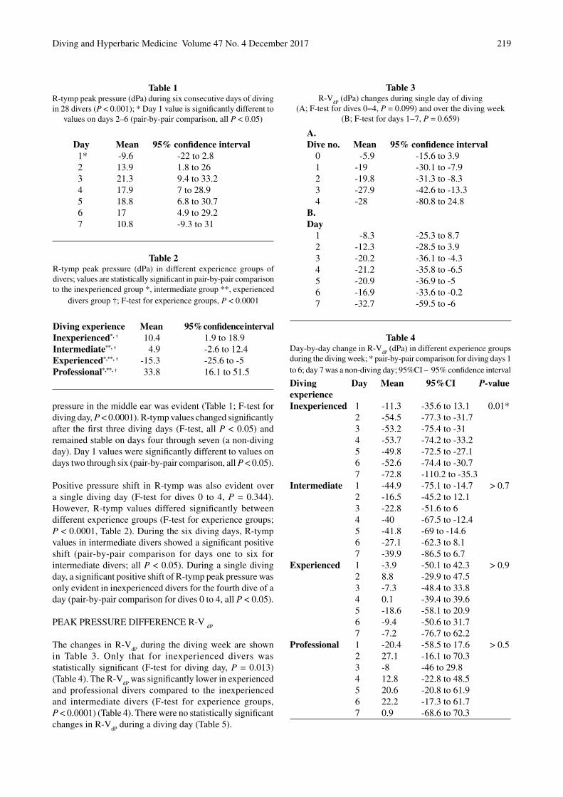
pressure in the middle ear was evident (Table 1; F-test for diving day, P < 0.0001). R-tymp values changed significantly after the first three diving days (F-test, all P < 0.05) and remained stable on days four through seven (a non-diving day). Day 1 values were significantly different to values on days two through six (pair-by-pair comparison, all P < 0.05).
Positive pressure shift in R-tymp was also evident over a single diving day (F-test for dives 0 to 4, P = 0.344). However, R-tymp values differed significantly between different experience groups (F-test for experience groups; P < 0.0001, Table 2). During the six diving days, R-tymp values in intermediate divers showed a significant positive shift (pair-by-pair comparison for days one to six for intermediate divers; all P < 0.05). During a single diving day, a significant positive shift of R-tymp peak pressure was only evident in inexperienced divers for the fourth dive of a day (pair-by-pair comparison for dives 0 to 4, all P < 0.05).
PEAK PRESSURE DIFFERENCE R-V dP
The changes in R-VdP
during the diving week are shown in Table 3. Only that for inexperienced divers was statistically significant (F-test for diving day, P = 0.013) (Table 4). The R-V
dP was significantly lower in experienced
and professional divers compared to the inexperienced and intermediate divers (F-test for experience groups,P < 0.0001) (Table 4). There were no statistically significant changes in R-V
dP during a diving day (Table 5).
Day Mean 95% confidence interval1* -9.6 -22 to 2.82 13.9 1.8 to 263 21.3 9.4 to 33.24 17.9 7 to 28.95 18.8 6.8 to 30.76 17 4.9 to 29.27 10.8 -9.3 to 31
Table 2R-tymp peak pressure (dPa) in different experience groups of divers; values are statistically significant in pair-by-pair comparison to the inexperienced group *, intermediate group **, experienced
divers group †; F-test for experience groups, P < 0.0001
Table 1R-tymp peak pressure (dPa) during six consecutive days of diving in 28 divers (P < 0.001); * Day 1 value is significantly different to
values on days 2–6 (pair-by-pair comparison, all P < 0.05)
Diving experience Mean 95% confidence intervalInexperienced*, † 10.4 1.9 to 18.9Intermediate**, † 4.9 -2.6 to 12.4Experienced*,**, † -15.3 -25.6 to -5Professional*,**, † 33.8 16.1 to 51.5
Table 4Day-by-day change in R-V
dP (dPa) in different experience groups
during the diving week; * pair-by-pair comparison for diving days 1 to 6; day 7 was a non-diving day; 95%CI – 95% confidence interval
Table 3R-V
dP (dPa) changes during single day of diving
(A; F-test for dives 0−4, P = 0.099) and over the diving week(B; F-test for days 1−7, P = 0.659)
Diving Day Mean 95%CI P-valueexperienceInexperienced 1 -11.3 -35.6 to 13.1 0.01* 2 -54.5 -77.3 to -31.7 3 -53.2 -75.4 to -31 4 -53.7 -74.2 to -33.2 5 -49.8 -72.5 to -27.1 6 -52.6 -74.4 to -30.7 7 -72.8 -110.2 to -35.3Intermediate 1 -44.9 -75.1 to -14.7 > 0.7 2 -16.5 -45.2 to 12.1 3 -22.8 -51.6 to 6 4 -40 -67.5 to -12.4 5 -41.8 -69 to -14.6 6 -27.1 -62.3 to 8.1 7 -39.9 -86.5 to 6.7Experienced 1 -3.9 -50.1 to 42.3 > 0.9 2 8.8 -29.9 to 47.5 3 -7.3 -48.4 to 33.8 4 0.1 -39.4 to 39.6 5 -18.6 -58.1 to 20.9 6 -9.4 -50.6 to 31.7 7 -7.2 -76.7 to 62.2Professional 1 -20.4 -58.5 to 17.6 > 0.5 2 27.1 -16.1 to 70.3 3 -8 -46 to 29.8 4 12.8 -22.8 to 48.5 5 20.6 -20.8 to 61.9 6 22.2 -17.3 to 61.7 7 0.9 -68.6 to 70.3
A.Dive no. Mean 95% confidence interval
0 -5.9 -15.6 to 3.91 -19 -30.1 to -7.92 -19.8 -31.3 to -8.33 -27.9 -42.6 to -13.34 -28 -80.8 to 24.8
B.Day
1 -8.3 -25.3 to 8.72 -12.3 -28.5 to 3.93 -20.2 -36.1 to -4.34 -21.2 -35.8 to -6.55 -20.9 -36.9 to -56 -16.9 -33.6 to -0.27 -32.7 -59.5 to -6

Diving and Hyperbaric Medicine Volume 47 No. 4 December 2017220
Similarly changes in V-S dP
and S-R dP
were not statistically significant (F-test for diving day and dive number, P > 0.05 respectively; data not shown*). However, inexperienced divers showed significantly higher V-S
dP compared to other
experience groups (F-test for experience groups, P < 0.0001; data not shown*).
CORRELATION OF PEAK PRESSURE AND MEBt
The clinical findings of MEBt have been described elsewhere.19 Divers with signs of barotrauma(TEED > 0) showed lower values of R-tymp peak pressure (mean 5.8 dPa, 95% CI -1.7 to 13.3) than divers with regular otoscopic findings (mean 13.8 dPa, 95% CI 6.8 to 20.9; F-test for TEED 0 vs. > 0, P = 0.029). Furthermore, theR-V
dP displayed significantly higher negative pressure
values in divers with barotrauma (mean -31.9, 95% CI-43.5 to -20.3) compared to those without barotrauma (mean 9.3, 95% CI -20.2 to 1.6; F-test for TEED 0 vs. > 0,P < 0.0001). R-V
dP increased from TEED 0 to TEED 1 (pair-
by-pair comparison, P = 0.002) and then increased further for TEED 2 and 3 (P = 0.012 and P = 0.52 respectively; data not shown*). Divers with the greatest level of barotrauma (TEED 3) displayed high negative R-tymp peak pressure in the middle ear (mean -55.8 daPa, 95% CI -96.4 to -15.2).
Discussion
Despite the enormous popularity of recreational scuba diving, only few aspects are known about ET and middle ear function after repetitive diving. The feasibility of test procedures, small study groups and different diving environments are some of the parameters which limit scientific understanding.
INTERPRETATION OF RESULTS
We included 28 participants in this prospective cohort study. Continuous supervision of the participants during the week’s diving allowed for accurate clinical follow-up. Overall, we observed on-going changes in middle ear pressures from day to day, which could be correlated to the repeated pressure exposure. Cumulative pressure exposure resulted in significantly decreasing compliance, which was obvious from the first dive, mostly affecting less experienced divers. The ‘professional’ divers showed significantly higher initial compliance before diving than inexperienced divers and significantly higher differences in compliance between the dives. They also had less barotrauma. Thus, the decreasing compliance in inexperienced divers may be owing to more pronounced MEBt arising from inadequate pressure equalization techniques. Hence, repeated exposure to pressure alterations over a long time period may influence TM mobility and ET function in a positive manner, resulting in increased compliance without decreasing TM stability. However, a long-term follow-up of professional divers is required to confirm this assumption.
We defined R-VdP
as the main indicator for successful pressure equalization in the middle ear. The R-V
dP
measurements suggest that inexperienced divers who were unused to repetitive pressure exposure seemed to apply more pressure during a Valsalva manoeuvre than did more experienced divers. However, the steadily increasing R-V
dP
in this group after repetitive dives might also indicate easier equalization as the week progressed.
Divers with signs of barotrauma (TEED > 0) had lower values of R-tymp peak pressure (‘underpressure’), as well as a greater negative R-V
dP. These results correlated with
peak pressure changes in the inexperienced and intermediate divers. Thus, increasing negative pressure in the middle ear during diving seems to be a crucial factor for the development of barotrauma.
RESULTS IN CONTEXT OF PREVIOUS PUBLICATIONS
Previous studies have described visible alterations of the TM from exposure to varying pressure levels.16,17,20 The TEED-classification nowadays is applied worldwide for the classification of MEBt.16 However, the influence of repetitive pressure changes on ET function is insufficiently investigated by simple otoscopy, whilst other diagnostic
Diving experience Dive # Mean 95%CI P-valueInexperienced 0 -39.7 -52.9 to -26.5 > 0.6 1 -42.7 -57.8 to -27.6 2 -48.3 -63.8 to -32.9 3 -55.4 -75.4 to -35.3 4 -67.4 -125.4 to -9.4Intermediate 0 -11.9 -29.5 to 5.6 > 0.5 1 -37 -56.5 to -17.6 2 -34.9 -55.1 to -14.8 3 -54.3 -83.8 to -24.7 4 – –Experienced 0 12.2 -14.2 to 38.6 > 0.4 1 -16.2 -44.1 to 11.8 2 -2.1 -33.2 to 29 3 -9.1 -43.8 to 25.7 4 – –Professional 0 17.9 -6.8 to 42.5 > 0.9 1 9.9 -18 to 37.8 2 2.1 -26.2 to 30.5 3 2.5 -34.9 to 39.9 4 33.7 -83.5 to -83.5
Table 5Dive-by-dive change in R-V
dP (dPa) in different experience groups
of divers over a day’s diving; pair-by-pair comparison for dives 0 to 4 not significant; 95%CI – 95% confidence interval
Diving and Hyperbaric Medicine Volume 47 No. 4 December 2017 221
techniques, such as pure tone tympanometry etc., allow drawing only indirect conclusions about ET function.11
The Valsalva manoeuvre during tympanometry is the most widely-used diagnostic test for evaluating middle ear ventilation and ET function.11 Whereas the Valsalva manoeuvre only allows categorical differentiation of pressure equalization, pure tone tympanometry displays compliance, impedance and peak pressure objectively and thus provides information about TM mobility and pressure conditions in the middle ear.11 Because repeatedly performed tympanometry only shows minor fluctuations of middle ear pressure,11 tests based on repeated measurement of tympanic impedance were developed (e.g., Williams inflation/deflation test and the nine-step inflation/deflation test).12,13,21−23 However, the reliable dynamic evaluation of ET function is only possible in a pressure chamber;4–6 its application in routine clinical practice has been limited by time and cost.4
There are several studies evaluating ET function in scuba divers.19−21,24,25 In 62 healthy participants in a Navy diving programme, the original Williams inflation/deflation test correlated with subjective performance and otoscopic findings after a single dive in a pressure chamber and after a single 12 m deep dive in water. There was no relationship between ET test results, otoscopy or subjective complaints by the participants. Consequently, the authors concluded that this test was of little value for screening divers.24
In contrast, a high predictive value for the nine-step inflation/deflation test for symptomatic MEBt was reported in 31 divers after repeated pressure exposure during 774 dives.20 Our results confirm these findings. Decreasing compliance, lower values of R-tymp peak pressure and negative R-V
dP in
our study correlated significantly with a higher prevalence of barotrauma. Previous analysis of the MEBt prevalence in the same participants revealed that four of the 28 divers missed at least one dive because of problems during pressure equalization.19 However, the question whether a diver omitted a dive was not asked until the last day of the trial and could not be correlated retrospectively to a specific ETFT measurement. If our results can be reproduced in further prospective trials, ETFT may serve as a valuable monitoring test for adequate pressure equalization in divers to prevent further MEBt development. The study has shown clearly the feasibility of using the ETFT during diving activities.
In the only long-term follow-up, tympanometry, otoscopic findings and subjective symptoms were studied over a 31-day study period. Despite the limited sample size (two divers) and asymptomatic participants throughout the study period, tympanometry revealed a strong association of decreased middle ear pressure with repetitive pressure exposure (more than two dives daily).25 Furthermore, using the Valsalva manoeuvre led to restoration of middle ear pressure in both participants. These observations confirm other reports describing a significant recovery of the middle
ear after a surface interval of more than 11 h.21 We detected neither complete recovery of middle ear pressure, nor of the otoscopic findings in the present trial.19 However, higher numbers of less experienced divers might explain this discrepancy.
LIMITATIONS OF THE STUDY
There is no standardized procedure for quantitative measurement of successful pressure equalization in the middle ear during diving or flying. Furthermore, routine tympanometry only provides categorical classification of middle ear and ET function.11 In this trial, we assumed that active equalization methods (especially the Valsalva manoeuvre) could differentiate within individuals based on the length and intensity of the action.6 This aspect may have biased the measurements. In addition, since our analysis does not contain normal non-diving participants, who underwent sham-dives (i.e., in a pressure chamber), the present results should be seen as a reference point for further controlled studies.
Conclusions
Using ETFT, we observed significant changes in middle ear pressure and pressure equalization in divers due to repeated pressure exposure in saltwater. Repetitive diving over six consecutive days led to significantly decreased compliance and increased R-tymp peak pressure (‘overpressure’) in the middle ear. The most profound changes were observed in less experienced divers. Besides greater stiffness of the TM, significantly negative values of R-tymp peak pressure, as well as significantly higher negative R-V
dP were associated
with a higher prevalence of MEBt in this cohort of divers. An accompanying paper investigates these changes in another cohort of divers in colder freshwater.26
References
1 Bluestone CD. Impact of evolution on the Eustachian tube. Laryngoscope. 2008;118:522-7.
2 Feldmann H. [Physiology and physiopathology of middle ear ventilation. I. Middle ear volume and its gas content. Physiology of the Eustachian tube.] Z Laryngol Rhinol Otol. 1973;52:471-85. German.
3 Feldmann H. [Physiology and pathophysiology of the ventilation of the middle ear. 2. Methods of examination of the Eustachian tube. Pathophysiology of typical ventilation disorders of the middle ear.] Z Laryngol Rhinol Otol. 1973;52:555-72. German.
4 Meyer MF, Mikolajczak S, Korthäuer C, Jumah MD, Hahn M, Grosheva M, et al. Impact of xylomethazoline on Eustachian tube function in healthy participants. Otol Neurotol. 2015;36:769-75.
5 Meyer MF, Mikolajczak S, Luers JC, Lotfipour S, Beutner D, Jumah MD. [Characterizing the passive opening of the Eustachian tube in a hypo-/hyperbaric pressure chamber.] Laryngorhinootologie. 2013;92:600-6. German.
6 Mikolajczak S, Meyer MF, Hahn M, Korthäuer C, Jumah MD,
Diving and Hyperbaric Medicine Volume 47 No. 4 December 2017222
Hüttenbrink KB, et al. Characterizing the active opening of the Eustachian tube in a hypobaric/hyperbaric pressure chamber. Otol Neurotol. 2015;36:70-5.
7 Klingmann C, Praetorius M, Baumann I, Plinkert PK. Otorhinolaryngologic disorders and diving accidents: an analysis of 306 divers. Eur Arch Otorhinolaryngol. 2007;264:1243-51.
8 Azizi MH. Ear disorders in scuba divers. Int J Occup Environ Med. 2011;2:20-6.
9 Molvaer OI, Natrud E. Ear damage due to diving. Acta Otolaryngol Suppl. 1979;360:187-9.
10 Shupak A, Doweck I, Greenberg E, Gordon CR, Spitzer O, Melamed Y, et al. Diving-related inner ear injuries. Laryngoscope. 1991;101:173-9.
11 Therkildsen AG, Gaihede M. Accuracy of tympanometric middle ear pressure determination. The role of direction and rate of pressure change with a fast modern tympanometer. Otol Neurotol. 2005;26:252-6.
12 Williams PS. A tympanometric pressure swallow test for assessment of Eustachian tube function. Ann Otol Rhinol Laryngol. 1975;84:339-43.
13 Spreitzer JB, Newman CW. Reliability of a measure of Eustachian tube function in normal subjects. Ann Otol Rhinol Laryngol. 1984;93:48-51.
14 Titan tympanometers manual. Interacoustics, www.interacoustics.com. Interacoustics A/S. Audiometer Allé 1, 5500 Middelfart.
15 Turekian K. Oceans. Englewood Cliffs, NJ: Prentice Hall; 1968.16 Teed RW. Factors producing obstruction of the auditory tube
in submarine personnel. United States Naval Medical Bulletin. 1944;42:293-306.
17 Edmonds C. Otological aspects of diving. Sydney: Australasian Medical Publishing Company; 1973.
18 Jerger, J. Clinical experience with impedance audiometry. Arch Otolaryngol. 1970;92:311-24.
19 Jansen S, Meyer MF, Boor M, Felsch M, Klünter HD, Pracht ED, et al. Prevalence and risk factors of barotrauma in recreational scuba divers after repetitive dives in salt water. Otol Neurotol. 2016;37:1325-31.
20 Ramos CC, Rapoport PB, Brito Neto RV. Clinical and tympanometric findings in repeated recreational scuba diving. Travel Med Infect Dis. 2005;3:19-25.
21 Uzun C. Evaluation of predive parameters related to Eustachian tube dysfunction for symptomatic middle ear barotrauma in divers. Otol Neurotol. 2005;26:59-64.
22 Bluestone CD. Assessment of Eustachian tube function. In: Jerger J, editor. Handbook of clinical impedance audiometry. New York: American Electromedics; 1975. p. 127-48.
23 Hussein A, Abousetta A. Use of the nine-step inflation/deflation test and resting middle-ear pressure range as predictors of middle-ear barotrauma in aircrew members. J Laryngol Otol. 2014;128:612-7.
24 Schuchman G, Joachims HZ. Tympanometric assessment of Eustachian tube functions of divers. Ear and Hearing. 1985;6:325-8.
25 Green SM, Rothrock SG, Green EA. Tympanometric evaluation of middle ear barotrauma during recreational scuba diving. Int J Sports Med. 1993;14:411-5.
26 Jansen S, Boor M, Meyer MF, Pracht ED, Volland R, Kluenter HD, et al. Repetitive diving in freshwater alters Eustachian tube function measured by Eustachian tube function test in recreational scuba divers. Diving Hyperb Med. 2017;47:222-6.
Acknowledgments
The authors would like to thank the team of Actionsport Würzbung, especially Georg Seufert, the team of Sinai Divers (Naama Bay) and the enthusiastic, voluntary contribution of all the divers who participated in this trial.
Conflicts of interest: nil
Funding
Interaucoustics A/S kindly loaned the tympanometry equipment.
Submitted: 23 January 2017; revised 04 July and 09 September 2017Accepted: 11 September 2017
Copyright: This article is the copyright of the authors who grant Diving and Hyperbaric Medicine a non-exclusive licence to publish the article in printed and other forms.
Present address for Dirk Beutner: Department of Otorhinolaryngology, Head and Neck Surgery, University Medical Center Göttingen, Göttingen, Germany

Diving and Hyperbaric Medicine Volume 47 No. 4 December 2017 223
Influence of repetitive diving in freshwater on pressure equalization and Eustachian tube function in recreational scuba diversStefanie Jansen1, Manuela Boor1, Moritz F Meyer1, Eberhard D Pracht2, Ruth Volland3, Heinz D Kluenter1, Karl-Bernd Huettenbrink1, Dirk Beutner1 , Maria Grosheva1
1 Department of Otorhinolaryngology, Head and Neck Surgery, University of Cologne, Medical Faculty, Germany2 German Center for Neurodegenerative Diseases (DZNE), Bonn, Germany3 Department of Paediatric Oncology and Haematology, University Children’s Hospital of Cologne, Cologne, Germany
Corresponding author: Professor Moritz F Meyer, Department of Otorhinolaryngology, Head and Neck Surgery, Faculty of Medicine, University of Cologne, Kerpener Straße 62, 50937 Cologne, [email protected]
Key wordsTympanometry; Middle ear; Ear barotrauma; Recreational diving
Abstract(Jansen S, Boor M, Meyer MF, Pracht ED, Volland R, Kluenter HD, Huettenbrink K-B, Beutner D, Grosheva M. Repetitive diving in freshwater alters Eustachian tube function measured by Eustachian tube function test in recreational scuba divers. Diving and Hyperbaric Medicine. 2017 December;47(4):223-227. doi10.28920/dhm47.4.223-227.)Introduction: We investigated the effect of repetitive pressure exposure during freshwater dives on Eustachian tube function and the middle ear, assessed by the Eustachian tube function test (ETFT).Methods: This prospective observational cohort study included 23 divers over three consecutive days of diving in freshwater lakes in Nordhausen, Germany. Participants underwent otoscopy and ETFT before the first dive, between each dive and after the last dive. ETFT included regular tympanometry (R-tymp), tympanometry after Valsalva (V-tymp) and after swallowing (S-tymp). The peak pressure difference between the R-tymp and the V-tymp (R-V
dP) defined effectiveness of
pressure equalization after Valsalva manoeuvres. We evaluated the change in compliance and peak pressure and correlated the results to the otoscopic findings and diving experience.Results: Twenty-three divers performed 144 dives. Middle ear barotrauma was assessed using the Edmonds modification of the TEED scoring system. In the ETFT, the R-tymp peak pressure displayed a negative shift from day one to three(P = 0.001) and differed significantly between the experience groups (P = 0.01). R-V
dP did not change significantly on any
of the three days of diving (all P > 0.05). Participants without MEBt showed significantly lower R-tymp values than did those with barotrauma (P = 0.019).Conclusion: Repetitive pressure exposure during three consecutive days of freshwater diving led to a negative shift of the peak pressure in the middle ear. Less experienced divers showed significantly higher middle ear peak pressure and higher pressure differences after equalization manoeuvres. Higher middle ear peak pressure was also associated with a higher prevalence of barotrauma.
Introduction
Our knowledge about the effect of rapid pressure changes during diving on middle ear and Eustachian tube (ET) function is mostly based on research on professional divers, navy divers or on case reports and retrospective questionnaires.1–4 None of these studies were conducted in freshwater. However, diving in freshwater differs from diving in the sea. Besides the characteristics of the water itself, e.g., density, temperature, salinity, etc., diving conditions in a freshwater lake, such as visibility, temperature and depth, may influence the ability to effectively equalize pressure in the middle ear. Using the Eustachian tube function test (ETFT),5, 6 we prospectively evaluated the changes in middle ear pressures and evidence of middle ear barotrauma (MEBt) after repetitive freshwater dives. The findings were also compared to those in the accompanying study on a cohort of divers in the Red Sea.7
Methods
The Ethics Committee of the University Hospital of Cologne, Germany, approved this observational prospective cohort study. The trial was registered prior to all study-related interventions by the German Clinical Trials Register (No. DRKS00008968; URL: http://apps.who.int/trialsearch/). Written informed consent was obtained from each participant before their inclusion. All participants presented a valid medical certificate prior to all study-related interventions. Three participants were under age (all 16-years-old) and, therefore, presented with written consent from a parent or legal guardian before their inclusion. None of the divers had been diving for 24 hours prior to the study.
The study was conducted in two freshwater lakes in Nordhausen, Thuringia, Germany in August 2015 over three consecutive days. The lakes are a maximum depth of 31

Diving and Hyperbaric Medicine Volume 47 No. 4 December 2017224
metres. Surface water temperature was 22–23ºC, 18–19ºC below the first thermocline at a depth of 8–10 metres and 7–8ºC below the second thermocline at 14–15 metres. Dive time, maximum depth of each dive and surface intervals were recorded for each diver.
STUDY-RELATED INTERVENTIONS AND THE ETFT
Before the first dive, all participants were questioned about their diving and ENT-related medical history and diving experience. In addition, otoscopy and endoscopy of the nose and epipharynx were performed to exclude any pathology. No topical anesthetic or lubricant was used for nasal endoscopy. Otoscopic changes of the tympanic membrane (TM) were evaluated according to the TEED classification for middle ear barotrauma (MEBt) as modified by Edmonds.8,9 A Valsalva manoeuvre was assessed by otoscopy in all participants. Tympanometry was performed at 226 Hz before the first dive and immediately after every dive according to the ETFT using a mobile tympanometer (Titan®, Interacoustics A/S, Denmark) as previously described.7 R-tymp measurement represented the peak pressure in the middle ear at rest, V-tymp the peak pressure after a Valsalva manoeuvre and S-tymp after swallowing. R-V
dP represented the difference in peak pressure between
the R-tymp and V-tymp and V-SdP
the difference between the V-tymp and the S-tymp. A more detailed description of the tympanometry methodology is provided in the accompanying paper.7
PARTICIPANTS
We included 23 participants (46 ears), seven female and 16 male, with a mean age of 34.5 ± 11.5 years. The median number of dives completed before the study was 40 (range 1–1,100). The last dive was performed a median of 1.9 months (range 0.9–11.9 months) prior to the study. We defined 13 divers as ‘inexperienced’ (< 50 dives), five divers as ‘intermediate’ (50–200 dives) and five divers as ‘professional’ (≥ 500 dives). The categorization of diving experience was developed during our saltwater study and was applied in the present trial to allow comparison of the results.7 No one matched the criteria of an ‘experienced’ diver (201–499 dives) as defined in that study.
STATISTICAL EVALUATION
All data were de-identified. The findings of the left and right ears were evaluated separately, but analyzed together using SPSS (version 23.0.0.0, IBM Corporation, USA). Quantitative data are presented as mean ± standard deviation (SD), or 95% confidence interval (95% CI), or as median and range, or interquartile range (IQR) and qualitative variables as absolute number and percentage. We applied mixed model analysis of variance with repeated measures for analysis of quantitative data for the ETFT values. To correct for multiple testing, P-values were adjusted using the Bonferroni method. The mixed model analysis allows for inclusion of multiple
time points per subject, while accounting for unbalanced data structure of irregular time intervals between ETFT measurements and unequal numbers of ETFT analyses per subject. The respective ETFT parameters − R-tymp peak pressure, R-V
dP, V-S
dP and R-tymp compliance − were
included as dependent variable; and the dive number, diving day, TEED and diving experience groups as covariates. The univariate analysis (F-test) was based on analysis of linear independent pairwise comparisons. Chi-square test was applied for analysis of TEED distribution per diving day. Kruskall-Wallis test was applied to test the influence of diving depth on TEED distribution. Spearman’s correlation was applied to test an association of diving depth and R-tymp peak pressure. A corrected P-value of < 0.05 was considered to be significant for all tests. All reported P-values are two-sided.
Results
DIVING
During the three days, the participants completed 144 dives. Seven participants used a drysuit and 16 a wetsuit. The average number of dives during the three days was six (median 5, range 4–10). The average depth and duration of the dives were 13 ± 4.1 metres’ freshwater and 35 ± 10.6 min, respectively. There was a significant difference in the diving depths between the different experience groups (Figure 1; Kruskal-Wallis test, P < 0.0001). The mean duration of the surface intervals between the dives was 127 ± 67 min.
Figure 1Maximum diving depth in different experience groups; (median test, P = 0.002; Kruskal-Wallis test, P < 0.0001); 13 divers were defined as ‘inexperienced’ (< 50 dives) (IE), five divers as ‘intermediate’ (50-200 dives) (IM) and five divers as ‘professional’
(≥ 500 dives) (PF)

Diving and Hyperbaric Medicine Volume 47 No. 4 December 2017 225
CLINICAL EXAMINATION
Before the first dive, endoscopy of the nose and the epipharynx was normal in all divers as were the otoscopy findings in 45 ears. One diver had a hyperaemic TM (TEED 1). However, all divers were able to perform a Valsalva manoeuvre successfully before diving. Tympanometry showed a normal type A pattern in all 46 ears.10
During the three days’ diving, MEBt (TEED 1−3) was observed in 105 ears (26%). TEED 1 was present in 86 (21%), TEED 2 in 14 (3%) and TEED 3 in five (1%). No TM perforations (TEED 4) occurred. Increasing number of dives per day was associated with a significant increase of pathologic otoscopic findings (TEED > 0; Chi-squared test, P < 0.0001). Furthermore, the maximum diving depth significantly influenced the MEBt prevalence (Kruskal-Wallis test, P = 0.035), shallower dives being associated with more signs of MEBt. The number of ears with signs of MEBt (TEED > 0) was equally distributed between the three experience groups (Fisher´s test, P = 0.623); however, higher TEED levels (TEED ≥ 2) were present only in the inexperienced and intermediate divers.
COMPLIANCE
The mean values of the R-tymp compliance on days one to three did not differ significantly: mean 1.2 ml (IQR 0.7−1.7 ml); mean 1.3 ml (IQR 0.7−1.8 ml) and mean 1.2 ml (IQR 0.5−1.8), for days one, two and three respectively (F-test,
P = 0.947). However, a significant increase in compliance was evident after the first dive (F-test, P = 0.041; Table 1). Professional divers had higher compliance values compared to the intermediate or inexperienced divers (F-test,P = 0.004; Table 2). Professional and intermediate divers also showed significantly higher change in compliance after the first dive compared to the inexperienced divers (pair-by-pair comparison, all P < 0.05, data not shown*).
PEAK PRESSURE
The mean values of the R-tymp peak pressure differed significantly from day one to day three and revealed a negative pressure shift (F-test for diving day, P = 0.001; Table 3). The peak pressure in the middle ear varied significantly in different experience groups (F-test, P = 0.01; Table 2). Pair-by-pair comparison revealed significantly decreased R-tymp peak pressure during the three days of diving especially in groups of intermediate and professional divers, compared to inexperienced divers (data not shown*).
PEAK PRESSURE DIFFERENCE (R-VdP
)
R-VdP
did not change significantly on any of the three days of diving: mean 19.2 dPa (95%CI -27 to 65.4); mean 18.2 dPa (95%CI 24.5 to 60.9) and mean 8.3 dPa (95%CI -40.9 to 57.5) for days one, two and three respectively; F-test,P = 0.818). In addition there were no statistically significant changes in R-V
dP during a diving day (F-test, P = 0.522).
However, when comparing the experience groups, a
Table 3R-tymp peak pressure (dPa) during three consecutive days of diving in all participants; mean values differed significantly from days one
through three (P = 0.001)
Table 1R-tymp compliance (ml) in dives 0 to 6 in all participants; increase of compliance after the first dive (F-test for dive number P < 0.041)
Dive number Mean 95% confidence interval1 1 0.7 to 1.32 1.4 1.1 to 1.73 1.3 1 to 1.54 1.2 0.8 to 1.65 1.1 0.6 to 1.56 1.3 0.3 to 2.0
Table 2R-tymp compliance (ml), R-tymp peak pressure (dPa) and peak pressure difference after Valsalva manoeuvre (R-V
dP) (dPa) in different
experience groups of divers; ‘professional’ divers had higher compliance values (P = 0.004) and lower peak pressures (P = 0.01) compared to divers with lower experience; R-V
dP was greatest in inexperienced divers (P = 0.02); 95% CI – 95% confidence interval
R-tymp compliance (ml) R-tymp peak pressure (dPa) R-VdP (dPa)Diving experience Mean (95%CI) Mean (95%CI) Mean (95%CI)Inexperienced (n = 13) 0.9 (0.5 to 1.4) 9.5 (-17 to 36) 39.3 (-3.7 to 82.2)Intermediate (n = 5) 0.7 (0.1 to 1.3) -13.8 (-42.3 to 14.7) 13.4 (-33 to 59.8)Professional (n = 5) 1 (0.3 to 2.6) -23.1 (-53 to 6.7) -6.9 (-55.7 to 41.9)
Day Mean 95% confidence interval1 -10.4 -39.2 to 18.52 15.2 -10.6 to 40.93 -33.8 -65 to -2.5
* Footnote: Summary tables for data not shown here are available from the authors at <[email protected]>

Diving and Hyperbaric Medicine Volume 47 No. 4 December 2017226
significantly higher R-VdP
was evident in the inexperienced group (F-test, P = 0.02; Table 2). Similarly changes inV-S
dP and S-R
dP were not statistically significant (F-test
for diving day and dive number, P > 0.05 respectively; data not shown*).
CORRELATION OF PEAK PRESSURE AND MEBt
Altogether, an increase of cumulative pathologic otoscopic findings (TEED > 0) was evident over the three days (Pearson’s Chi-squared test, P < 0.0001). Participants without barotrauma showed significantly lower values of the R-tymp peak pressure (mean -21.2 dPa, 95%CI -47.5 to 5.1) than did those with barotrauma (mean 2.9 dPa, 95%CI -23.8 to 29.5; F-test 1 through 3, P = 0.019). This difference was evident on each diving day (data not shown*). Diving depth also significantly influenced the distribution of MEBt (Kruskal-Wallis test, P = 0.035); however, only a small association between diving depth and R-tymp peak pressure was evident (Spearman’s Rho -0.185, -0.093 and +0.239 on days one, two and three respectively).
Discussion
INTERPRETATION OF THE RESULTS
Summarizing all the changes in the ETFT measurements, the negative shift in pressures was likely the result of ET dysfunction and/or increasing tissue damage because of slowly failing equalization. Because of the short (three-day) evaluation period, we were unable to analyze these pressure changes further. The more experienced divers had significantly lower peak pressures, including following a Valsalva and higher compliance values and a greater difference in compliance after the first dive, compared to less experienced divers. The higher compliance may be the result of chronic elasticity loss due to cumulative pressure exposure during diving. However, the increasing compliance did not seem to influence TM stability and implies efficient equalization techniques. Although the distribution of TEED classification between the groups did not show significant alterations, otoscopic findings with TEED 2 and 3 occurred only in the less experienced divers.
A variety of factors influence middle ear and ET function. Besides divers' pre-existing medical history and diving experience, the depth profile, number of repetitive dives per day, water and air temperature, salinity and density may influence the stress on the ET and the middle ear. The dives performed in this freshwater study were shorter and shallower than those in our saltwater (Red Sea) study.7 Shallower dives require more frequent active and passive pressure equalization (Boyle’s Law) and are probably associated with an increased risk of barotrauma.11,12 This could be exacerbated by divers experiencing more buoyancy problems in waters with poor visibility, resulting in inadequate and/or delayed pressure equalization and consequent MEBt.
We observed a significant inverse correlation between the prevalence of barotrauma and diving depth, but this may be because less experienced divers performed shallower dives than those with more experience. The short evaluation time does not allow us to draw reliable conclusions regarding this correlation. However, peak pressure alteration in the ETFT may reflect more sensitively the immediate effect of the previous dive, while longer pressure exposure may be needed for manifestation of otoscopic evidence of barotrauma.
RESULTS IN CONTEXT TO PREVIOUS RESEARCH
It is assumed that active pressure equalization during descent and passive pressure equalization during ascent does not differ between saltwater and freshwater. There is a possible decongestant effect of saltwater that might improve ET function and make pressure equalization easier. However, this effect would have to occur very rapidly to have any impact on equalization during the initial descent.
As reported in the accompanying saltwater paper, we observed that pressure equalization became easier during repetitive diving.7 Alternatively divers might have performed a Valsalva manoeuvre more often on a dive as the week progressed. In the present study, such an effect could be observed only on the first day, remaining unchanged thereafter, which might suggest less efficient pressure equalization in the middle ear during repetitive freshwater dives compared to saltwater.
The same procedures and the same tympanometer were used during the ETFT in both trials. Repetitive saltwater dives resulted in a steady increase in R-tymp peak pressure, whereas in the present study, the middle ear peak pressure did the opposite, moving to ‘underpressure’ over the three days. These conflicting observations might be caused by the two diving cohorts utilizing different equalization techniques. Nevertheless, in both conditions, inexperienced divers had, on average, the highest peak pressures and higher R-V
dP. In
this trial, divers with barotrauma also showed significantly higher R-tymp peak pressure. Consequently, increasing R-V
dP probably mirrors higher equalization pressure in
the inexperienced divers. We previously hypothesized that increasing underpressure in the middle ear might lead to acute tissue damage and to MEBt.7 Comparing the results of the salt- and fresh-water investigations, the pressure difference during pressure equalization appears to be crucial for development of barotrauma.
Ear problems are the commonest issue that forces a diver to stop a dive.11,12 There has not been a good screening test available to evaluate MEBt and ET dysfunction on an on-going basis during diving activities. The poor reliability of the Valsalva manoeuvre and the poor correlation of subjective complaints to the degree of barotrauma has been of concern for many.1–4 In the seawater study, over 70% of the divers with otoscopic evidence of MEBt remained asymptomatic.13 Furthermore, all participants in both studies
Diving and Hyperbaric Medicine Volume 47 No. 4 December 2017 227
were able to perform the Valsalva manoeuvre effectively. The use of the ETFT has shown that changes in middle ear pressure occur before MEBt is otoscopically or clinically obvious. The ETFT has proven to be a useful investigative tool during diving activities.
LIMITATIONS OF THE STUDY
The analysis of ET function outside of a clinical facility poses a challenge. The ETFT offers the advantages of being easy to operate and provides a series of measurements during pressure equalization manoeuvres. However, no standardized values exist for this test. The lack of a control group, such as subjects undergoing dry dives in a pressure chamber, only allowed us to compare the data between the measurements. Furthermore, the possibility of intra-individual variation of intensity and length of a pressure equalization manoeuvre14 cannot be excluded and might also bias the results. An extended evaluation time is needed to verify our findings. Furthermore, additional influencing factors like water temperature and the exact diving depth profile could not be considered in this analysis.
Conclusions
Using the ETFT, we were able to detect early changes in ET function in divers after repetitive dives in freshwater. Repetitive pressure exposure led to negative shift of the peak pressure in the middle ear. Less experienced divers showed significantly higher middle ear peak pressure and higher pressure differences after equalization manoeuvres. Higher middle ear peak pressure was also associated with higher barotrauma prevalence. The ETFT has proven to be a useful investigative tool during diving activities.
References
1 Klingmann C, Praetorius M, Baumann I, Plinkert PK. Otorhinolaryngologic disorders and diving accidents: an analysis of 306 divers. Eur Arch Otorhinolaryngol. 2007;264:1243-51.
2 Kitajima N, Kitajima S. Altered Eustachian tube function in SCUBA divers with alternobaric vertigo. Otol Neurotol. 2014;35:850-6.
3 Uzun C. Evaluation of predive parameters related to Eustachian tube dysfunction for symptomatic middle ear barotrauma in divers. Otol Neurotol. 2005;26:59-64.
4 Ramos CC, Rapoport PB, Brito Neto RV. Clinical and tympanometric findings in repeated recreational scuba diving. Travel Med Infect Dis. 2005;3:19-25.
5 Williams PS. A tympanometric pressure swallow test for assessment of Eustachian tube function. Ann Otol Rhinol Laryngol. 1975;84(3 Pt1):339-43.
6 Spreitzer JB, Newman CW. Reliability of a measure of Eustachian tube function in normal subjects. Ann Otol Rhinol Laryngol. 1984;93:48-51.
7 Meyer MF, Boor M, Jansen S, Pracht ED, Felsch M, Klünter HD, et al. Influence of repetitive diving in saltwater on pressure equalization and Eustachian tube function in recreational scuba divers. Diving Hyperb Med. 2017;47:215-21.
8 Teed RW. Factors producing obstruction of the auditory tube in submarine personnel. United States Naval Medical Bulletin. 1944;42:293-306.
9 Edmonds C. Otological aspects of diving. Sydney: Australasian Medical Publishing Company; 1973.
10 Jerger, J. Clinical experience with impedance audiometry. Arch Otolaryngol. 1970;92:311-24.
11 Azizi MH. Ear disorders in scuba divers. Int J Occup Environ Med. 2011;2:20-2.
12 Molvaer OI, Natrud E. Ear damage due to diving. Acta Otolaryngol. 1979;360(Suppl):187-9.
13 Jansen S, Meyer MF, Boor M, Felsch M, Klünter HD, Pracht ED, et al. Prevalence and risk factors of barotrauma in recreational scuba divers after repetitive dives in salt water. Otol Neurotol. 2016;37:1325-31.
14 Mikolajczak S, Meyer MF, Hahn M, Korthäuer C, Jumah MD, Hüttenbrink KB, et al. Characterizing the active opening of the Eustachian tube in a hypobaric/hyperbaric pressure chamber. Otol Neurotol. 2015;36:70-5.
Acknowledgments
We thank the team at Actionsport Würzbung, especially Georg Seufert and Bernd Blümmert, and the OASIS Dive Centre (Nordhausen, Germany) for their support and the enthusiastic volunteer divers in this trial.
Conflicts of interest: nil
Funding
Interacoustics A/S kindly loaned the Titan tympanometer for the study.
Submitted: 25 February 2017; revised 04 July and 05 September 2017Accepted: 11 September 2017
Copyright: This article is the copyright of the authors who grant Diving and Hyperbaric Medicine a non-exclusive licence to publish the article in printed and other forms.
Present address for Dirk Beutner: Department of Otorhinolaryngology, Head and Neck Surgery, University Medical Center Göttingen, Göttingen, Germany
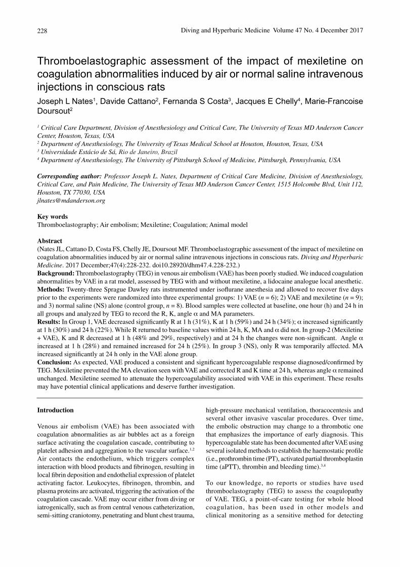
Diving and Hyperbaric Medicine Volume 47 No. 4 December 2017228
Thromboelastographic assessment of the impact of mexiletine on coagulation abnormalities induced by air or normal saline intravenous injections in conscious ratsJoseph L Nates1, Davide Cattano2, Fernanda S Costa3, Jacques E Chelly4, Marie-Francoise Doursout2
1 Critical Care Department, Division of Anesthesiology and Critical Care, The University of Texas MD Anderson Cancer Center, Houston, Texas, USA2 Department of Anesthesiology, The University of Texas Medical School at Houston, Houston, Texas, USA3 Universidade Estácio de Sá, Rio de Janeiro, Brazil4 Department of Anesthesiology, The University of Pittsburgh School of Medicine, Pittsburgh, Pennsylvania, USA
Corresponding author: Professor Joseph L. Nates, Department of Critical Care Medicine, Division of Anesthesiology, Critical Care, and Pain Medicine, The University of Texas MD Anderson Cancer Center, 1515 Holcombe Blvd, Unit 112, Houston, TX 77030, [email protected]
Key wordsThromboelastography; Air embolism; Mexiletine; Coagulation; Animal model
Abstract(Nates JL, Cattano D, Costa FS, Chelly JE, Doursout MF. Thromboelastographic assessment of the impact of mexiletine on coagulation abnormalities induced by air or normal saline intravenous injections in conscious rats. Diving and Hyperbaric Medicine. 2017 December;47(4):228-232. doi10.28920/dhm47.4.228-232.)Background: Thromboelastography (TEG) in venous air embolism (VAE) has been poorly studied. We induced coagulation abnormalities by VAE in a rat model, assessed by TEG with and without mexiletine, a lidocaine analogue local anesthetic.Methods: Twenty-three Sprague Dawley rats instrumented under isoflurane anesthesia and allowed to recover five days prior to the experiments were randomized into three experimental groups: 1) VAE (n = 6); 2) VAE and mexiletine (n = 9); and 3) normal saline (NS) alone (control group, n = 8). Blood samples were collected at baseline, one hour (h) and 24 h in all groups and analyzed by TEG to record the R, K, angle α and MA parameters. Results: In Group 1, VAE decreased significantly R at 1 h (31%), K at 1 h (59%) and 24 h (34%); α increased significantly at 1 h (30%) and 24 h (22%). While R returned to baseline values within 24 h, K, MA and α did not. In group-2 (Mexiletine + VAE), K and R decreased at 1 h (48% and 29%, respectively) and at 24 h the changes were non-significant. Angle α increased at 1 h (28%) and remained increased for 24 h (25%). In group 3 (NS), only R was temporarily affected. MA increased significantly at 24 h only in the VAE alone group.Conclusion: As expected, VAE produced a consistent and significant hypercoagulable response diagnosed/confirmed by TEG. Mexiletine prevented the MA elevation seen with VAE and corrected R and K time at 24 h, whereas angle α remained unchanged. Mexiletine seemed to attenuate the hypercoagulability associated with VAE in this experiment. These results may have potential clinical applications and deserve further investigation.
Introduction
Venous air embolism (VAE) has been associated with coagulation abnormalities as air bubbles act as a foreign surface activating the coagulation cascade, contributing to platelet adhesion and aggregation to the vascular surface.1,2 Air contacts the endothelium, which triggers complex interaction with blood products and fibrinogen, resulting in local fibrin deposition and endothelial expression of platelet activating factor. Leukocytes, fibrinogen, thrombin, and plasma proteins are activated, triggering the activation of the coagulation cascade. VAE may occur either from diving or iatrogenically, such as from central venous catheterization, semi-sitting craniotomy, penetrating and blunt chest trauma,
high-pressure mechanical ventilation, thoracocentesis and several other invasive vascular procedures. Over time, the embolic obstruction may change to a thrombotic one that emphasizes the importance of early diagnosis. This hypercoagulable state has been documented after VAE using several isolated methods to establish the haemostatic profile (i.e., prothrombin time (PT), activated partial thromboplastin time (aPTT), thrombin and bleeding time).3,4
To our knowledge, no reports or studies have used thromboelastography (TEG) to assess the coagulopathy of VAE. TEG, a point-of-care testing for whole blood coagulation, has been used in other models and clinical monitoring as a sensitive method for detecting
Diving and Hyperbaric Medicine Volume 47 No. 4 December 2017 229
hypercoagulable states.6,7 Presently, TEG is widely used to assess global haemostatic function from a sample of whole blood, e,g., in trauma, obstetrics, cardiovascular surgery, early sepsis, liver transplantation and others.2,7 Several studies have reported that TEG parameters are interrelated and reflect the initiation of the coagulation cascade from the initial platelet-fibrin interaction through platelet aggregation, clot strengthening, and fibrin cross linkage.8,9
There have been preclinical studies showing that various medications, e.g., lidocaine, ketamine, and magnesium sulphate can act as antiplatelet agents or affect coagulation.10 For instance, lidocaine, a local anesthetic and intravenous antiarrhythmic agent, has been suggested to be of benefit in patients with decompression illness (DCI), mainly for cerebral arterial gas embolism (CAGE),11−13 possibly by affecting membrane stability.10 Mexiletine is a Class I-b local anesthetic and lidocaine analog.14 It exerts similar antiarrhythmic properties and could have some beneficial effects during the treatment of VAE, possibly diminishing the hypercoagulable state.10,11,13 In contrast to lidocaine, it can be ingested by mouth, increasing its clinical applicability.
This study aimed to evaluate TEG changes in rats subjected to consistent and reproducible VAE, and to assess hypercoagulability changes in the presence and absence of mexiletine, an analogue of lidocaine. The null hypothesis tested was that mexiletine would not reduce the impact in the coagulation system produced by intravascular injection of air in rats. Whether TEG findings following VAE could be applied to humans in the diagnosis and treatment of VAE associated with DCI or surgical procedures is discussed. No similar study has been described in the literature.
Methods
After approval of the experimental protocol by the University of Texas Animal Welfare Committee, 23 Sprague-Dawley rats were included in the study. To avoid damage to the implanted catheters, animals were housed in individual cages in an air-conditioned (22 ± 1°C), light-controlled room (12-hrs light, 12-hrs dark) and were allowed to mobilize freely. Animals had free access to food and water. Their behaviour, posture and appearance were monitored daily.
INSTRUMENTATION
The rats were anesthetized with isoflurane 5%, intubated with a 16-gauge intravenous catheter and mechanically ventilated using a mixture of 30% oxygen, room air and 1.5 % isoflurane. Under sterile conditions, a Tygon® catheter was inserted into the femoral vein for drug administration and tunneled subcutaneously to the dorsum of the neck and secured in place after closure of the incisions. The animals were then allowed five days to recover prior to the start of the experiments and were individually housed and given free access to food and water.
EXPERIMENTAL DESIGN
To minimize the difference among groups, animals were randomized into three groups by a blinded investigator. Group 1 (n = 7, VAE); animals received air (0.5 ml) infused over two minutes (min) via the femoral vein catheter. The catheter was connected to a syringe driver (Medfusion, Medex, Inc; Duluth, GA) with PE tubing and used for drug administration. Group 2 (n = 9; Mexiletine and VAE). Mexiletine was administered in a dose of 10 mg∙kg-1 IV over two min.15 Thirty min following mexiletine administration air was infused as described in Group 1. Group 3 (n = 8); to test the adequacy of the TEG responses, animals in this group received NS only and were used as the control group.When the study was initiated, Group 1 included seven animals; Groups 2 and 3 had eight animals each. However, one animal died following surgery (during catheterization) for an unknown reason before the experiment began, resulting in six animals allocated to the VAE group. We added one animal to Group 2 due to the combination of treatments. To prevent artifacts from onset of coagulation to analysis, whole blood samples (with no activator) from all groups were drawn for immediate analysis using TEG (Haemoscope Corp., Skokie, IL) prior to VAE, mexiletine, saline and 1 hour (h) and 24 h thereafter, respectively.
THE SAMPLING PROCEDURES
TEG was performed with blood collected from the previously implanted femoral catheter. Citrate anticoagulation was achieved by collecting 900 µl of blood in 100 μl of 4% sodium citrate (1:10 dilution). Blood samples were gently inverted five times, and were placed on their sides for 30 minutes to allow adequate equilibration of the citrate throughout the sample. At this point, 340 μl of the blood was pipetted gently into a disposable plastic TEG cup containing 20 μl of 0.2M calcium chloride, being careful to avoid mixing. The cup was then transferred to a TEG 5000 thrombelastograph haemostasis analyzer (Haemoscope, Skokie, IL) for assay at 37ºC.
MEASUREMENTS
All TEG parameters were recorded from standard tracings: split point (SP, min), reaction time (R, min), coagulation time (K, min), angle (α, degrees), maximum amplitude (MA, mm), clot strength (G, dynes∙cm-2), and lysis at 30 min (LY30, %).7 The SP is a measure of the time to initial clot formation, interpreted from the earliest resistance detected by the TEG analyzer causing the tracing to split; this is the terminus of all other platelet-poor plasma clotting assays (e.g., PT and aPTT). The R value, the time elapsed from start of the test until the developing clot provides enough resistance to produce a 2 mm amplitude reading on the TEG tracing, represents the initiation phase of enzymatic clotting factors. K measures the time from clotting factor initiation (R) until clot formation reaches amplitude of 20 mm. The
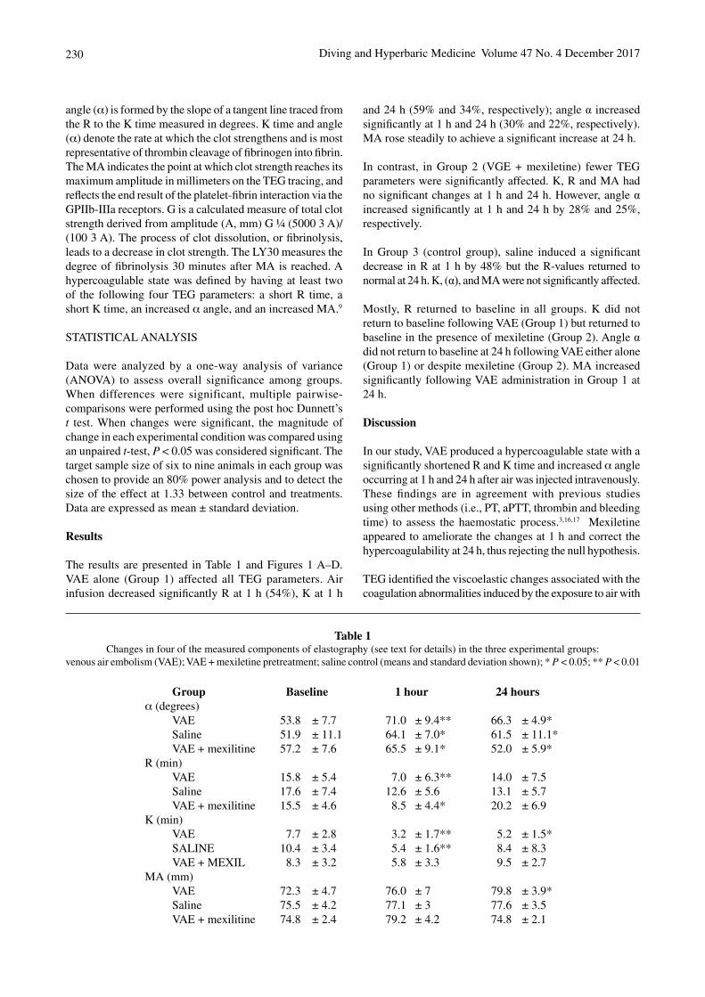
Diving and Hyperbaric Medicine Volume 47 No. 4 December 2017230
angle (α) is formed by the slope of a tangent line traced from the R to the K time measured in degrees. K time and angle (α) denote the rate at which the clot strengthens and is most representative of thrombin cleavage of fibrinogen into fibrin. The MA indicates the point at which clot strength reaches its maximum amplitude in millimeters on the TEG tracing, and reflects the end result of the platelet-fibrin interaction via the GPIIb-IIIa receptors. G is a calculated measure of total clot strength derived from amplitude (A, mm) G ¼ (5000 3 A)/(100 3 A). The process of clot dissolution, or fibrinolysis, leads to a decrease in clot strength. The LY30 measures the degree of fibrinolysis 30 minutes after MA is reached. A hypercoagulable state was defined by having at least two of the following four TEG parameters: a short R time, a short K time, an increased α angle, and an increased MA.9
STATISTICAL ANALYSIS
Data were analyzed by a one-way analysis of variance (ANOVA) to assess overall significance among groups. When differences were significant, multiple pairwise-comparisons were performed using the post hoc Dunnett’s t test. When changes were significant, the magnitude of change in each experimental condition was compared using an unpaired t-test, P < 0.05 was considered significant. The target sample size of six to nine animals in each group was chosen to provide an 80% power analysis and to detect the size of the effect at 1.33 between control and treatments. Data are expressed as mean ± standard deviation.
Results
The results are presented in Table 1 and Figures 1 A–D. VAE alone (Group 1) affected all TEG parameters. Air infusion decreased significantly R at 1 h (54%), K at 1 h
Table 1Changes in four of the measured components of elastography (see text for details) in the three experimental groups:
venous air embolism (VAE); VAE + mexiletine pretreatment; saline control (means and standard deviation shown); * P < 0.05; ** P < 0.01
Group Baseline 1 hour 24 hoursα (degrees) VAE 53.8 ± 7.7 71.0 ± 9.4** 66.3 ± 4.9* Saline 51.9 ± 11.1 64.1 ± 7.0* 61.5 ± 11.1* VAE + mexilitine 57.2 ± 7.6 65.5 ± 9.1* 52.0 ± 5.9*R (min) VAE 15.8 ± 5.4 7.0 ± 6.3** 14.0 ± 7.5 Saline 17.6 ± 7.4 12.6 ± 5.6 13.1 ± 5.7 VAE + mexilitine 15.5 ± 4.6 8.5 ± 4.4* 20.2 ± 6.9K (min) VAE 7.7 ± 2.8 3.2 ± 1.7** 5.2 ± 1.5* SALINE 10.4 ± 3.4 5.4 ± 1.6** 8.4 ± 8.3 VAE + MEXIL 8.3 ± 3.2 5.8 ± 3.3 9.5 ± 2.7MA (mm) VAE 72.3 ± 4.7 76.0 ± 7 79.8 ± 3.9* Saline 75.5 ± 4.2 77.1 ± 3 77.6 ± 3.5 VAE + mexilitine 74.8 ± 2.4 79.2 ± 4.2 74.8 ± 2.1
and 24 h (59% and 34%, respectively); angle α increased significantly at 1 h and 24 h (30% and 22%, respectively). MA rose steadily to achieve a significant increase at 24 h.
In contrast, in Group 2 (VGE + mexiletine) fewer TEG parameters were significantly affected. K, R and MA had no significant changes at 1 h and 24 h. However, angle α increased significantly at 1 h and 24 h by 28% and 25%, respectively.
In Group 3 (control group), saline induced a significant decrease in R at 1 h by 48% but the R-values returned to normal at 24 h. K, (α), and MA were not significantly affected.
Mostly, R returned to baseline in all groups. K did not return to baseline following VAE (Group 1) but returned to baseline in the presence of mexiletine (Group 2). Angle α did not return to baseline at 24 h following VAE either alone (Group 1) or despite mexiletine (Group 2). MA increased significantly following VAE administration in Group 1 at 24 h.
Discussion
In our study, VAE produced a hypercoagulable state with a significantly shortened R and K time and increased α angle occurring at 1 h and 24 h after air was injected intravenously. These findings are in agreement with previous studies using other methods (i.e., PT, aPTT, thrombin and bleeding time) to assess the haemostatic process.3,16,17 Mexiletine appeared to ameliorate the changes at 1 h and correct the hypercoagulability at 24 h, thus rejecting the null hypothesis.
TEG identified the viscoelastic changes associated with the coagulation abnormalities induced by the exposure to air with
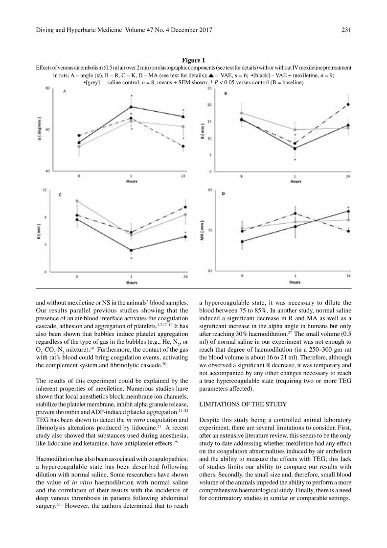
Diving and Hyperbaric Medicine Volume 47 No. 4 December 2017 231
and without mexiletine or NS in the animals’ blood samples. Our results parallel previous studies showing that the presence of an air-blood interface activates the coagulation cascade, adhesion and aggregation of platelets.1,2,17–19 It has also been shown that bubbles induce platelet aggregation regardless of the type of gas in the bubbles (e.g., He, N
2, or
O2-CO
2-N
2 mixture).19 Furthermore, the contact of the gas
with rat’s blood could bring coagulation events, activating the complement system and fibrinolytic cascade.20
The results of this experiment could be explained by the inherent properties of mexiletine. Numerous studies have shown that local anesthetics block membrane ion channels, stabilize the platelet membrane, inhibit alpha granule release, prevent thrombin and ADP-induced platelet aggregation.21−24 TEG has been shown to detect the in vitro coagulation and fibrinolysis alterations produced by lidocaine.21 A recent study also showed that substances used during anesthesia, like lidocaine and ketamine, have antiplatelet effects.25
Haemodilution has also been associated with coagulopathies; a hypercoagulable state has been described following dilution with normal saline. Some researchers have shown the value of in vitro haemodilution with normal saline and the correlation of their results with the incidence of deep venous thrombosis in patients following abdominal surgery.26 However, the authors determined that to reach
a hypercoagulable state, it was necessary to dilute the blood between 75 to 85%. In another study, normal saline induced a significant decrease in R and MA as well as a significant increase in the alpha angle in humans but only after reaching 30% haemodilution.27 The small volume (0.5 ml) of normal saline in our experiment was not enough to reach that degree of haemodilution (in a 250–300 gm rat the blood volume is about 16 to 21 ml). Therefore, although we observed a significant R decrease, it was temporary and not accompanied by any other changes necessary to reach a true hypercoagulable state (requiring two or more TEG parameters affected).
LIMITATIONS OF THE STUDY
Despite this study being a controlled animal laboratory experiment, there are several limitations to consider. First, after an extensive literature review, this seems to be the only study to date addressing whether mexiletine had any effect on the coagulation abnormalities induced by air embolism and the ability to measure the effects with TEG; this lack of studies limits our ability to compare our results with others. Secondly, the small size and, therefore, small blood volume of the animals impeded the ability to perform a more comprehensive haematological study. Finally, there is a need for confirmatory studies in similar or comparable settings.
Figure 1Effects of venous air embolism (0.5 ml air over 2 min) on elastographic components (see text for details) with or without IV mexiletine pretreatment
in rats; A – angle (α), B – R, C – K, D – MA (see text for details); – VAE, n = 6; •[black] – VAE + mexiletine, n = 9;•[grey] – saline control, n = 8; means ± SEM shown; * P < 0.05 versus control (B = baseline)
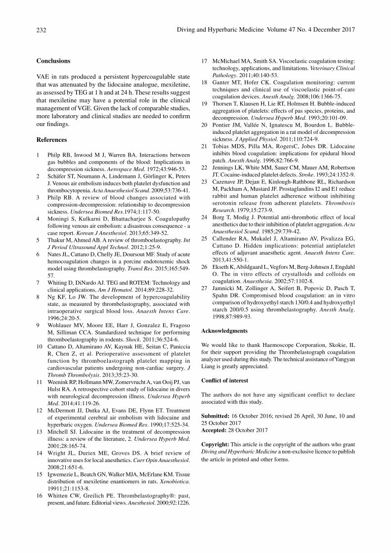
Diving and Hyperbaric Medicine Volume 47 No. 4 December 2017232
Conclusions
VAE in rats produced a persistent hypercoagulable state that was attenuated by the lidocaine analogue, mexiletine, as assessed by TEG at 1 h and at 24 h. These results suggest that mexiletine may have a potential role in the clinical management of VGE. Given the lack of comparable studies, more laboratory and clinical studies are needed to confirm our findings.
References
1 Philp RB, Inwood M J, Warren BA. Interactions between gas bubbles and components of the blood: Implications in decompression sickness. Aerospace Med. 1972;43:946-53.
2 Schäfer ST, Neumann A, Lindemann J, Görlinger K, Peters J. Venous air embolism induces both platelet dysfunction and thrombocytopenia. Acta Anaesthesiol Scand. 2009;53:736-41.
3 Philp RB. A review of blood changes associated with compression-decompression: relationship to decompression sickness. Undersea Biomed Res.1974;1:117-50.
4 Moningi S, Kulkarni D, Bhattacharjee S. Coagulopathy following venous air embolism: a disastrous consequence - a case report. Korean J Anesthesiol. 2013;65:349-52.
5 Thakur M, Ahmed AB. A review of thromboelastography. Int J Period Ultrasound Appl Technol. 2012;1:25-9.
6 Nates JL, Cattano D, Chelly JE, Doursout MF. Study of acute hemocoagulation changes in a porcine endotoxemic shock model using thrombelastography. Transl Res. 2015;165:549-57.
7 Whiting D, DiNardo AJ. TEG and ROTEM: Technology and clinical applications, Am J Hematol. 2014;89:228-32.
8 Ng KF, Lo JW. The development of hypercoagulability state, as measured by thrombelastography, associated with intraoperative surgical blood loss. Anaesth Intens Care. 1996;24:20-5.
9 Wohlauer MV, Moore EE, Harr J, Gonzalez E, Fragoso M, Silliman CCA. Standardized technique for performing thromboelastography in rodents. Shock. 2011;36:524-6.
10 Cattano D, Altamirano AV, Kaynak HE, Seitan C, Paniccia R, Chen Z, et al. Perioperative assessment of platelet function by thromboelastograph platelet mapping in cardiovascular patients undergoing non-cardiac surgery. J Thromb Thrombolysis. 2013;35:23-30.
11 Weenink RP, Hollmann MW, Zomervrucht A, van Ooij PJ, van Hulst RA. A retrospective cohort study of lidocaine in divers with neurological decompression illness. Undersea Hyperb Med. 2014;41:119-26.
12 McDermott JJ, Dutka AJ, Evans DE, Flynn ET. Treatment of experimental cerebral air embolism with lidocaine and hyperbaric oxygen. Undersea Biomed Res. 1990;17:525-34.
13 Mitchell SJ. Lidocaine in the treatment of decompression illness: a review of the literature, 2. Undersea Hyperb Med. 2001;28:165-74.
14 Wright JL, Duriex ME, Groves DS. A brief review of innovative uses for local anesthetics. Curr Opin Anaesthesiol. 2008;21:651-6.
15 Igwemezie L, Beatch GN, Walker MJA, McErlane KM. Tissue distribution of mexiletine enantiomers in rats. Xenobiotica. 19911;21:1153-8.
16 Whitten CW, Greilich PE. Thrombelastography®: past, present, and future. Editorial views. Anesthesiol. 2000;92:1226.
17 McMichael MA, Smith SA. Viscoelastic coagulation testing: technology, applications, and limitations. Veterinary Clinical Pathology. 2011;40:140-53.
18 Ganter MT, Hofer CK. Coagulation monitoring: current techniques and clinical use of viscoelastic point-of-care coagulation devices. Anesth Analg. 2008;106:1366-75.
19 Thorsen T, Klausen H, Lie RT, Holmsen H. Bubble-induced aggregation of platelets: effects of pas species, proteins, and decompression. Undersea Hyperb Med. 1993;20:101-09.
20 Pontier JM, Vallée N, Ignatescu M, Bourdon L. Bubble-induced platelet aggregation in a rat model of decompression sickness. J Applied Physiol. 2011;110:724-9.
21 Tobias MDS, Pilla MA, RogersC, Jobes DR. Lidocaine inhibits blood coagulation: implications for epidural blood patch. Anesth Analg. 1996;82:766-9.
22 Jennings LK, White MM, Sauer CM, Mauer AM, Robertson JT. Cocaine-induced platelet defects. Stroke. 1993;24:1352-9.
23 Cazenave JP, Dejan E, Kinlough-Rathbone RL, Richardson M, Packham A, Mustard JF. Prostaglandins I2 and E1 reduce rabbit and human platelet adherence without inhibiting serotonin release from adherent platelets. Thrombosis Research. 1979;15:273-9.
24 Borg T, Modig J. Potential anti-thrombotic effect of local anesthetics due to their inhibition of platelet aggregation. Acta Anaesthesiol Scand. 1985;29:739-42.
25 Callender RA, Mukalel J, Altamirano AV, Pivalizza EG, Cattano D. Hidden implications: potential antiplatelet effects of adjuvant anaesthetic agent. Anaesth Intens Care. 2013,41:550-1.
26 Ekseth K, Abildgaard L, Vegfors M, Berg-Johnsen J, Engdahl O. The in vitro effects of crystalloids and colloids on coagulation. Anaesthesia. 2002;57:1102-8.
27 Jamnicki M, Zollinger A, Seifert B, Popovic D, Pasch T, Spahn DR. Compromised blood coagulation: an in vitro comparison of hydroxyethyl starch 130/0.4 and hydroxyethyl starch 200/0.5 using thrombelastography. Anesth Analg. 1998;87:989-93.
Acknowledgments
We would like to thank Haemoscope Corporation, Skokie, IL for their support providing the Thrombelastograph coagulation analyzer used during this study. The technical assistance of Yangyan Liang is greatly appreciated.
Conflict of interest
The authors do not have any significant conflict to declare associated with this study.
Submitted: 16 October 2016; revised 26 April, 30 June, 10 and 25 October 2017Accepted: 28 October 2017
Copyright: This article is the copyright of the authors who grant Diving and Hyperbaric Medicine a non-exclusive licence to publish the article in printed and other forms.
Diving and Hyperbaric Medicine Volume 47 No. 4 December 2017 233
Hyperbaric oxygen in the treatment of acute retinal artery occlusionMark J Elder1,2, John A Rawstron1, Michael Davis2,3
1 Ophthalmology Department, Christchurch Hospital, Christchurch, New Zealand2 University of Otago, Christchurch3 formerly Hyperbaric Medicine Unit, Christchurch Hospital
Corresponding author: Professor Mark Elder, Ophthalmology Department, Christchurch Hospital, Private Bag 4710, Christchurch 8140, New [email protected]
Key wordsVision disorders; Visual acuity; Outcome; Retrospective studies; Clinical audit
Abstract(Elder MJ, Rawstron JA, Davis M. Hyperbaric oxygen in the treatment of acute retinal artery occlusion. Diving and Hyperbaric Medicine. 2017 December;47(4):233-238. doi10.28920/dhm47.4.233-238.)Introduction: Acute retinal artery occlusion (ARAO) is a major cause of sudden, painless visual loss, often leaving no useful vision in the affected eye. Its incidence is cited at 0.85 per 100,000 persons per year but may be higher because of under-reporting. The natural history is difficult to study, but a spontaneous resolution rate of < 1–8% for acute, non-arteritic ARAO has been cited. Occurrence in an only eye is devastating for the patient. There is currently no consensus regarding management of ARAO and little evidence to support any treatment modality. Despite only limited case series, hyperbaric oxygen treatment (HBOT) is recommended for ARAO by the Undersea and Hyperbaric Medical Society (UHMS) and by the European Committee for Hyperbaric Medicine.Methods: Between early 2003 and December 2012, all ARAO patients presenting to Christchurch Hospital were referred for consideration of HBOT. These 31 consecutive patients’ medical records were reviewed retrospectively. The time delay from onset of visual loss to commencing HBOT; the presenting visual acuity; various demographic data; the HBOT administered and the outcome visual acuity were documented.Results: All 31 patients underwent at least one HBOT (median 4, range 1–7) at a pressure of 203–284 kPa for 1.5 to 2.0 h. One patient’s treatment was terminated after 60 min at their request; another declined further HBOT and one suffered middle ear barotrauma. Thirteen patients also received anticoagulants at the discretion of the referring ophthalmologist. Twenty-three patients had temporarily improved vision with the first HBOT. Seven patients had permanent, good visual recovery (6/18 or better; Snellen chart); and two only modest improvement (6/60). All nine patients who improved permanently were treated within 10 hours of symptom onset.Conclusions: Where available, HBOT is indicated for ARAO. Our protocol may not have been aggressive enough and the UHMS protocol is recommended. A multi-centre, randomised controlled trial is feasible, but would be logistically difficult and expensive and may be ethically unsupportable given the lack of alternative, effective treatments.
Introduction
Acute retinal artery occlusion (ARAO), either central or branch occlusion, is a major cause of sudden, painless visual loss, often leaving no useful vision in the affected eye if a central occlusion. Its incidence has been estimated as 0.85 per 100,000 persons per year, but may in fact be significantly higher due to under-reporting.1 The natural history is difficult to study, but a spontaneous resolution rate of less than 1 to 8% for acute non-arteritic ARAO has been cited.1,2 Incidence in an only eye is catastrophic for the patient’s quality of life (QALY). There is debate regarding which, if any, treatment options are useful in the acute setting.
Dramatic improvement in visual acuity in a few ARAO patients undergoing hyperbaric oxygen treatment (HBOT) in our institution in the early 2000s3 gained the interest of
ophthalmologists and hyperbaric specialists, and we have continued to use HBOT in ARAO. HBOT is available in most of the main centres in Australasia, and is utilised by some of these centres for treatment of ophthalmic vascular occlusive events. The only controlled trial showed HBOT improved visual acuity by three lines or more in 38% compared to 18% in a group without HBOT, but this difference was not statistically significant.2,4 ARAO first appeared as an indication for emergency HBOT in the 2009 committee report of the Undersea and Hyperbaric Medical Society (UHMS) in the USA.5 Recently this has been supported by the European Committee for Hyperbaric Medicine.6
We conducted a retrospective clinical audit of a consecutive series of patients with ARAO referred over a decade at our institution for consideration of HBOT.
Diving and Hyperbaric Medicine Volume 47 No. 4 December 2017234
Methods
Between early 2003 and December 2012, all ARAO patients presenting to Christchurch Hospital were referred for consideration of HBOT. Thirty-one consecutive patients’ medical records were reviewed retrospectively. This was an anonymous quality assurance review. The study was discussed formally with the Ethics Committee, Office of the Chief Medical Officer, Clinical Leadership, Protection and Regulation, Ministry of Health, New Zealand and they designated the study as an audit.
The diagnosis of ARAO was made by an ophthalmology specialist or registrar in training and was based on the history of sudden visual loss and the clinical signs of a cherry red spot, cessation of retinal artery flow and possible embolus. The time delay from onset of visual loss to commencing HBOT; the presenting visual acuity; various demographic data; the HBOT administered, other treatments used and the outcome visual acuity were documented. Time to presentation was not easy to elucidate with precision in some cases, particularly with those patients who stated that they noticed the problem on waking in the morning. In these cases, we have recorded the time to presentation as less than or equal to the approximate duration since they last were known to have unaffected vision (e.g., the previous evening). Visual acuity was measured as lines of vision on an ETDRS/Snellen-type visual chart. If letters could not be seen, then the categories of counting fingers, hand movements, perception of light or no perception of light were used.7
Hyperbaric treatments, with patients breathing 100% oxygen from a head hood, were given at a pressure of 203 kPa (2.0 ATA, equivalent to the pressure at 10 metres’ depth of seawater) or 243 kPa for 90 minutes (with a 10-minute air-breathing break after 45 min) or 284 kPa for 60 minutes, followed by a 30-minute decompression to ambient room pressure. Treatment plans were determined on a case-by-case basis at the discretion of the on-call hyperbaric physician.
Given the small population of patients, formal statistical analysis used a 2X2 contingency table, mid-P exact test P (2-tail) (http://www.openepi.com/TwobyTwo/TwobyTwo.htm).
Results
DEMOGRAPHIC AND CLINICAL DATA
Case records of 31 patients (21 male and 10 female; mean age 70 years, range 37 to 88 years) with a diagnosis of non-arteritic ARAO were identified in the ophthalmological department records between January 2003 and December 2012 (Table 1). Three patients were affected in an ‘only eye’, with the fellow eye having hand movements or worse vision. Time from symptom onset to presentation ranged from three to 25.5 hours (h). Nineteen patients had hypertension, on medication, and three had diabetes mellitus. The erythrocyte sedimentation rate, measured in 19 patients, was within the
normal range. Other investigations, including intra-ocular pressures (IOP), which was performed in all patients, were unremarkable. Follow-up ranged from one to 79 months.
Carotid artery ultrasounds were performed in 18/31 and, of these, two cases proceeded to acute endarterectomy, one other had an 80% stenosis and the rest were regarded as within normal limits.
CO-THERAPY
The co-treatments used in conjunction with HBOT varied widely. Intraocular pressure was attempted to be reduced with oral acetazolamide in three cases, ocular massage was used in six cases and anterior ocular chamber paracentesis in nine. Ten patients were already on aspirin and one of those was also on dipryidamole. An additional nine patients were started on aspirin as therapy. Low dose heparin was started in four cases and full-dose heparin was used in another four cases. Four patients were on warfarin and another four were started on warfarin (these four were the patients that were initially put on full-dose heparin). One patient had an ARAO during a coronary angiogram and subsequently was put on clopidogrel. Two patients were started on steroids by their general practitioner before an accurate diagnosis was determined by the admitting ophthalmologist but both were stopped when the temporal artery biopsy was reported as negative for arteritis.
HYPERBARIC OXYGEN TREATMENT
All 31 patients underwent at least one HBOT (median 4, range 1–7). Three patients withdrew from further HBOT after the first treatment, one because of an acute upper respiratory tract infection, another because of claustrophobia and one suffered middle ear barotrauma (modified TEED grade 3). The initial HBOT was at a pressure of 203 kPa in eight patients, 243 kPa in 19 and 284 kPa in four. All subsequent HBOT was administered at a pressure of 243 kPa.
POST-HBOT VISUAL RECOVERY
Twenty-three of the 31 patients reported some temporary return of vision during or immediately following their first HBOT. In nine patients vision was restored permanently: to 6/18 or better in seven and to 6/60 in two (Table 1). The remaining 14 did not maintain the initial improvement. The other eight patients showed no improvement with the first HBOT. Two patients had further deterioration in vision despite treatment, whilst two patients who showed no improvement with the first HBOT had slight overall improvement in VA at discharge. Follow-up was a minimum of one month (range 1–79 months).
All nine patients who improved had a delay from onset of visual loss to HBOT of less than 10 h, out of a total of 22 patients presenting in that time frame; whereas one of the remaining nine patients with a longer delay improved. Using
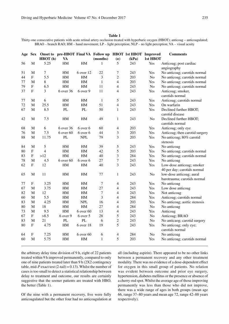
Diving and Hyperbaric Medicine Volume 47 No. 4 December 2017 235
Age Sex Onset to pre-HBOT Final VA Follow up HBOT 1st HBOT Improved Comments HBOT (h) VA (months) (n) (kPa) 1st HBOT56 M 5.25 HM HM 1 5 243 Yes Anticoag; post cardiac angiography51 M 7 HM 6 over 12 22 7 243 Yes No anticoag; carotids normal44 F 5.5 HM HM 3 2 203 No No anticoag; carotids normal77 M 8 HM HM 1 4 203 Yes No anticoag; carotids normal79 F 6.5 HM HM 11 4 243 No No anticoag; carotids normal37 F 3 6 over 36 6 over 9 11 4 243 Yes Anticoag; smoker, carotids normal77 M 6 HM HM 1 5 243 Yes Anticoag; carotids normal72 M 25.5 HM HM 51 4 243 Yes On warfarin67 M 8.5 PL PL 50 1 243 Yes Declined further HBOT; carotid disease42 M 7.5 HM HM 49 1 243 No Declined further HBOT; carotids normal68 M 6 6 over 36 6 over 6 60 4 203 Yes Anticoag; only eye76 M 7.5 6 over 60 6 over 6 44 3 203 Yes Anticoag; then carotid surgery88 M 11.75 PL NPL 79 3 203 Yes No anticoag; 80% carotid stenosis84 M 5 HM HM 39 5 243 Yes No anticoag80 F 4 HM HM 42 5 203 Yes No anticoag; carotids normal83 F >12 HM HM 40 3 284 Yes No anticoag; carotids normal78 M 4.5 6 over 60 6 over 6 27 7 243 Yes No anticoag62 F 12 HM HM 40 3 243 Yes Low-dose anticoag; smoker 40 per day; carotids normal65 M 11 HM HM 77 1 243 No low-dose anticoag; aural barotrauma; carotids normal77 F 3.25 HM HM 7 4 243 Yes No anticoag67 M 3.75 HM HM 27 4 243 Yes Low dose anticoag82 M 12 HM HM 7 4 243 Yes Not anticoag60 M 5.5 HM HM 3 4 284 Yes Anticoag; carotids normal83 M 4.25 HM NPL 16 4 203 Yes No anticoag; aortic stenosis80 M 18 HM HM 27 4 284 No No anticoag73 M 9.5 HM 6 over 60 13 4 243 Yes Anticoag67 F >8.5 6 over 9 6 over 5 28 5 243 No Anticoag; BRAO83 M 21 PL PL 6 2 243 No No anticaog; carotid surgery80 F 4.75 HM 6 over 18 19 5 243 Yes No anticoag; only eye; carotids normal64 F 7.25 HM 6 over 60 6 4 284 No No anticoag60 M 5.75 HM HM 1 5 203 Yes No anticoag; carotids normal
Table 1Thirty-one consecutive patients with acute retinal artery occlusion treated with hyperbaric oxygen (HBOT); anticoag – anticoagulated;
BRAO – branch RAO; HM – hand movement; LP – light perception; NLP – no light perception; VA – visual acuity
the arbitrary delay time division of 9 h, eight of 22 patients treated within 9 h improved permanently, compared to only one of nine patients treated later than 9 h (2X2 contingency table, mid-P exact test (2-tail) = 0.13). Whilst the number of cases is too small to detect a statistical relationship between delay to treatment and outcome, our results are certainly suggestive that the sooner patients are treated with HBO, the better (Table 1).
Of the nine with a permanent recovery, five were fully anticoagulated but the other four had no anticoagulation at
all (including aspirin). There appeared to be no other links between a permanent recovery and any other treatment modality. There was no evidence of a dose-dependent effect for oxygen in this small group of patients. No relation was evident between outcome and prior eye surgery, hypertension, diabetes mellitus or the presence or absence of a cherry-red spot. Whilst the average age of those improving permanently was less than those who did not improve, there was a wide range of ages in both groups (mean age 66, range 37–80 years and mean age 72, range 42–88 years respectively).

Diving and Hyperbaric Medicine Volume 47 No. 4 December 2017236
Discussion
HYPERBARIC OXYGEN THERAPY – MECHANISM OF ACTION
The inspired partial pressure of oxygen during a hyperbaric treatment at 203 kPa is almost ten times that when breathing air at normal atmospheric pressure. It is postulated that oxygen at higher pressures diffuses from the choroidal circulation or other patent retinal vessels to reach the ischaemic retina. This restarts cellular metabolism and keeps the retina alive, allowing time for emboli to break up or move on. This may explain the anecdotal phenomenon of visual return reported in the majority of this patient cohort during the first HBOT, with reduction of oedema in the retina allowing better acuity. It also suggests that HBOT would, at most, allow a few extra hours in which circulation may be restored to the retina. It will not help in situations where the retina is already infarcted and, therefore, is only of use in patients who present within a limited time of arterial occlusion. Since there appears to be an increased rate of improvement in vision in our patients treated within 10 h, it would seem sensible to provide HBOT to any patient with ARAO presenting within this time period.
The majority of non-arteritic central retinal artery occlusions are thought to be due to emboli. There are however, several different types of emboli. Cholesterol, platelet, or red cell emboli would each respond differently to any specific medical or mechanical attempts to dislodge or dissolve them. For example heparin may have little effect on a cholesterol thrombus. Successful treatment of an embolus would depend on the type of embolus involved. It seems sensible to combine HBOT with treatments designed to remove emboli in one way or another, such as anticoagulation, though there is no good evidence to substantiate this.
Whilst IOP was recorded as normal in all patients, IOP has no known relationship to CRAO. The perfusion pressure of the eye is mean ophthalmic arterial pressure (62 mmHg if BP is 120/80) minus IOP, i.e., a small increase in IOP does not alter perfusion pressure unless it reaches 50 mmHg or more. There is a relationship between IOP and central retinal vein occlusion, which makes sense as retinal venous pressure is about 20 mmHg and, when IOP approaches or is above this, the vein can be seen opening and collapsing at the disk. The relationship between IOP and HBOT has not been investigated.
COST
The average cost per HBOT for these patients in Christchurch Hospital was approximately NZ $500 (€325) during that period and, therefore, a series of five treatments totals approximately NZ $2,500. This compared to the cost of a cataract operation to the New Zealand Government at that time of approximately NZ$4,000. It has been suggested that
cataract surgery is one of the most cost effective medical interventions for QALY whereas treatment of ARAO before the advent of HBOT was one of the least cost-effective interventions.8 With HBOT, if our percentage recovery of useful vision (nine of 31 patients, 29%) is compared with the high-end Cochrane analysis (8%, thus a conservative estimate), then the number needed to treat with HBOT for useful visual recovery is approximately five patients, making HBOT a cost-effective intervention.
PUBLISHED EVIDENCE ABOUT HBOT FOR ARAO
There is only one controlled, non-randomised trial of HBOT in ARAO.2,4 This compared HBOT and haemodilution with haemodilution alone as the control. Fifty-one patients received HBOT and haemodilution and 29 patients haemodilution alone. In the HBOT group, mean VA improvement was three lines (P < 0.0001) versus one line in the haemodilution alone group (n.s.). However, there was no significant difference between the two groups at discharge or at follow-up. In an extensive review of the clinical evidence for HBOT in ARAO carried out for the UHMS, it was concluded that there was clear evidence of clinical benefit over and above other treatment modalities.4 In a literature review, 25 case series of HBOT for ARAO totalling 476 patients reported ‘improvement’ in 303 (64%).5 The quality of reported improvement and its relationship to delay to treatment were often poorly documented, and most reports showed obvious potential selection bias. Several of the better retrospective studies follow as illustrative of the generally weak quality of the existing literature.
Comparing eight patients undergoing HBOT for ARAO, to eight who refused HBOT or had contraindications, no significant difference in outcome was noted.9 In a comparison of 35 patients treated with HBOT no later than 8 hours after the beginning of their visual symptoms to 37 patients from a different centre not treated with HBOT, 29 patients in the HBOT group showed improvement compared to 10 in the non-HBOT group.10 This improvement in outcome, by three Snellen lines, for those patients treated with HBOT was statistically significant.
Finally, data from 11 patients with ARAO treated with HBOT,11 of whom eight achieved improved visual acuity, were combined with that from two other case series in which the clinical data had been recorded in a comparable manner12,13 to give a total of 51 eyes with 27 patients showing improvement of two or more lines with HBOT on a modified Snellen value. Analysis of the combined case series suggested that improvement in VA may be more likely if HBOT was given within less than 24 h, but the data are not particularly convincing.
A detailed management protocol was proposed by the UHMS.5 Reviewing this protocol, we believe that our management during that time was not sufficiently aggressive

Diving and Hyperbaric Medicine Volume 47 No. 4 December 2017 237
and that perhaps more of the patients who showed temporary improvement with the first HBOT could have benefitted from longer and/or more frequent hyperbaric exposures based on on-going close monitoring of vision and prompt re-treatment if deterioration was noted, as recommended in the UHMS protocol. The UHMS protocol has been slightly modified for use in our hospital in recent years (Table 2). In particular, it was not felt justified to automatically move to a US Navy Treatment Table 6 if improvement was not seen at 283 kPa, but that this would be at the discretion of the treating medical officer.
PUBLISHED EVIDENCE REGARDING ALL OTHER TREATMENTS FOR ARAO
We could find no published consensus on best practice or previous studies documenting current practice for ARAO management. The scientific evidence on this topic is weak. There do not appear to be any prospective, randomized controlled trials or cohort studies. A retrospective comparison of case series at Wills Eye Hospital from 1995 compares 40 patients treated with both carbogen and anterior compartment paracentesis, with 49 patients treated with neither.14 They found no significant difference in outcome. Case series of ARAO patients treated with local intra-arterial thrombolysis are relatively numerous; however, these are balanced by reports of stroke and intracerebral haemorrhage caused by both local and systemic thrombolysis, the conclusion being that the risks may outweigh any benefits.15,16
A survey of New Zealand ophthalmologists was conducted in 2003 with a 78% (76 from 97) response rate.3 Eight
respondents indicated that they would not actively treat ARAO. Of those who would treat, only four followed a written protocol for management. A wide range of treatments was chosen, somewhat dependent on the time delay to presentation, including ocular massage, anterior compartment paracentesis, aspirin, oral acetazolamide and intra-ocular pressure lowering drops. Only five respondents chose HBOT, reflecting the fact that HBOT is not available to a large proportion of the New Zealand population. When asked if they would offer HBOT if it were available, a quarter indicated that they would refer for HBOT.
Conclusions
Hyperbaric oxygen treatment, where available, is a safe, relatively low-cost and moderately effective treatment option for patients with ARAO compared to the natural history of the condition. A multi-centre, randomized controlled trial of HBOT is feasible, but would be logistically difficult and expensive and may be ethically unsupportable given the lack of alternative, effective treatments.
References
1 Rumelt S, Dorenboim Y, Rehany U. Aggressive systematic treatment for central retinal artery occlusion. Am J Ophthalmol. 1999;128:733-8.
2 Fraser SG, Adams W. Interventions for acute non-arteritic central retinal artery occlusion. Cochrane Database of Systematic Reviews 2009, Issue 1. Art. No.: CD001989. doi: 10.1002/14651858.CD001989.pub2.
3 Rawstron JA, Davis FM, Elder MJ. Hyperbaric oxygen for central retinal artery occlusion. Proceedings of the 2004 Royal
Table 2Christchurch Hospital management protocol for acute retinal artery occlusion (modified from the UHMS-recommended protocol;5
see text for explanation)
• All patients with sudden painless loss of vision in one eye should be treated urgently with high flow oxygen, 15 L∙min-1 or greater via a non-rebreather mask.
• If there is no response to normobaric oxygen in patients with a diagnosis of central or major branch retinal arterial of less than 24 hours duration, they should be referred to the Hyperbaric Medicine Unit for assessment for emergency HBOT. If the vision in the other eye has been compromised in the past, consider referring even with a delay longer than 24 hours.
• Patients will be seen in the Emergency Department by the on call Hyperbaric Senior Medical Officer (SMO) to exclude contraindications to HBO before initial acute treatment.
• The HBOT Table will be 18.60.30, 14.90.30 or 10.90.30 based on the depth at which vision improves and at the Hyperbaric SMOs discretion.
• If vision does not improve at 18 m (284 kPa), a US Navy Treatment Table 6 may be considered.• Ideally two HBOT treatments should be within the first 24 hours.• Visual acuity should be monitored following treatment. Should visual loss recur, high flow normobaric oxygen 15mins
every hour should be administered on the ward until repeat HBOT can be arranged. If the first treatment was undertaken after hours, this is likely to be the next day.
• Treatment continues until clinical plateau reached (or angiogram confirms recanalization).• Other treatments are at the Department of Ophthalmology’s discretion.• Patients should be admitted for at least the first night.• Liaise closely with the Department of Ophthalmology.
Diving and Hyperbaric Medicine Volume 47 No. 4 December 2017238
Australian and New Zealand College of Ophthalmologists New Zealand Conference, Napier, New Zealand, May 2004.
4 Menzel-Severing J, Siekmann U, Weinberger A, Gernot R, Walter P, Mazinani B. Early hyperbaric oxygen treatment for nonarteritic central retinal artery obstruction. Am J Ophthalmol 2012;153:454-9.
5 Murphy-Lavoie H, Butler F, Hagan C. Central retinal artery occlusion treated with oxygen: A literature review and treatment algorithm. Undersea Hyperb Med. 2012;39:943-53.
6 Mathieu D, Marroni A, Kot J. Tenth European Consensus Conference on hyperbaric medicine: recommendations for accepted and non-accepted clinical indications and practice of hyperbaric oxygen treatment. Diving Hyperb Med. 2017;47:24-32.
7 Early Treatment Diabetic Retinopathy Study Research Group. Early treatment diabetic retinopathy study design and baseline patient characteristics: ETDRS Report Number 7. Ophthalmology. 1991;98:741-56.
8 Busbee BG, Brown GC, Brown MM. Cost-effectiveness of ocular interventions. Current Opinion in Ophthalmology. 2003;14:132-8.
9 Aisenbrey S, Krott R, Heller R, Krauss D, Rössler G, Heimann K. [Hyperbaric oxygen therapy in retinal artery occlusion]. Ophthalmologie. 2000;97:461-67. German.
10 Beiran I, Goldenberg I, Adir Y, Tamir A, Shupak A, Miller B. Early hyperbaric oxygen therapy for retinal artery occlusion. Eur J Ophthalmol. 2001;11:345-50.
11 Cope A, Eggert JV, O’Brien E. Retinal artery occlusion: visual outcome after treatment with hyperbaric oxygen. Diving Hyperb Med. 2011;41:135-8.
12 Hertog LM, Meyer GW, Carson S, Strauss MB, Hart GB.
Central retinal artery occlusion treated with hyperbaric oxygen. Journal of Hyperbaric Medicine. 1992;7(1):33-42.
13 Weinberger AW, SiekmannUP, Wolf S, Rossaint R, Kirchhof B, Schrage NF. [Treatment of acute central retinal artery occlusion (ARAO) by hyperbaric therapy (HBO) – Pilot study with 21 patients]. Klin Monatsbl Augenheilkd. 2002;219:728-34. German.
14 Atebara NH, Brown GC, Cater J. Efficacy of anterior chamber paracentesis and carbogen in treating acute nonarteritic central retinal artery occlusion. Ophthalmology. 1995;102:2029-34.
15 Butz B, Strotzer M, Manke C, Roider J, Link J, Lenhart M. Selective intraarterial fibrinolysis of acute central retina artery occlusion. Acta Radiologica. 2003;44:680-4.
16 Barth H, Stein H, Fasse A, Mehdorn HM. [Intracerebral hemorrhage after systemic thrombolytic therapy in patients with central retinal artery occlusion. Report of two cases]. Ophthalmologe. 1996;93:739-44. German.
Acknowledgements
We wish to acknowledge the statistical advice of Elizabeth Wells PhD and of Joan Weenink RN for assisting with data collection.
Conflicts of interest and funding: nil
Submitted: 01 June 2017; revised 02 September 2017Accepted: 21 September 2017
Copyright: This article is the copyright of the authors who grant Diving and Hyperbaric Medicine a non-exclusive licence to publish the article in printed and other forms.
The database of randomised controlled trials in diving and hyperbaric medicine maintained by Michael Bennett and his colleagues at the Prince of Wales Hospital
Diving and Hyperbaric Medicine Unit, Sydney is at: <http://hboevidence.unsw.wikispaces.net/>
Assistance from interested physicians in preparing critical appraisals (CATs) is welcomed, indeed needed, as there is a considerable backlog.
Guidance on completing a CAT is provided.Contact Professor Michael Bennett: <[email protected]>
Diving and Hyperbaric Medicine Volume 47 No. 4 December 2017 239
Review articlesLost at sea: the medicine, physiology and psychology of prolonged immersionHeather Massey1, John Leach1, Michael Davis2, Vicki Vertongen3
1 Department of Sport and Exercise Science, University of Portsmouth, Portsmouth, United Kingdom 2 Formerly Medical Director, Hyperbaric Medicine Unit, Christchurch Hospital, Christchurch, New Zealand 3 Emergency Medicine Department, Wellington Hospital, Wellington, New Zealand
Corresponding author: Dr Heather Massey, Department of Sport and Exercise Science, University of Portsmouth, Spinnaker Building, Cambridge Road, Portsmouth PO1 2ER, [email protected]
Key wordsImmersion; Hypothermia; Physiology; Psychology; Environment; Diving incidents; Review article
Abstract
(Massey H, Leach J, Davis FM, Vertongan V. Lost at sea: the medical, physiological and psychological factors of prolonged immersion. Diving and Hyperbaric Medicine. 2017 December;47(4):239-247. doi10.28920/dhm47.4.239-247.)In most countries, immersion represents the second most common cause of accidental death in children and the third in adults. Between 2010 and 2013, 561 deaths worldwide involving recreational divers were recorded by the Divers Alert Network. Consequently, there is no room for complacency when diving. Being lost at sea is a diver’s worst nightmare. In 2006, a diver was lost at sea off the coast of New Zealand for 75 hours. It is unprecedented that, after such a long time immersed in temperate (16−17°C) waters, he was found and survived. His case is presented and utilised to illustrate the many physiological and psychological factors involved in prolonged immersion and what might determine survival under such circumstances. We also briefly review options for enhancing diver location at sea and a few issues related to search and rescue operations are discussed.
Introduction
In most countries, immersion represents the second most common cause of accidental death in children and the third in adults.1 World Health Organization (WHO) statistics report almost 375,000 immersion deaths each year; the actual figure worldwide is probably four or five times as high.2 Of the water-related deaths, deaths of divers are uncommon; however, the Divers Alert Network (DAN) recorded 561 deaths worldwide involving recreational divers between 2010 and 2013.3 Consequently, there is no room for complacency when diving. The harsh environment that divers expose themselves to can only be temporary, it is technology which allows humans to explore underwater environments for longer than a breath hold and enter such potentially hazardous surroundings.
Following immersion in cold water (there is no definitive definition of cold, in this context we include any water temperature below thermoneutrality (35°C water temperature); the rate of cooling will depend on many factors). The surface interval between dives allows not only for de-gassing but also for rewarming and fluid and energy balance. It is this return to homeostasis which ensures survival following immersion. There is a preoccupation that
many in-water deaths are ascribed to hypothermia. However, various factors contribute to the rate of cooling and death as a result of cold water immersion including cold shock, physical incapacitation leading to swimming cessation, drowning, hypothermia and dehydration.
A number of media reports have described incidents when divers have become separated from their boats and have endured prolonged immersions, some successfully rescued, others not.4−8 Perhaps the most poignant of these was the death of six divers who went missing off Peleliu in the Republic of Palau; three bodies were recovered.9 Over two days, one diver recorded a series of notes on her waterproof slate, which told of multiple sightings of vessels and planes – “we can see you searching, but you can’t see us.”9 The chances of finding the person alive diminish with each passing hour and a successful rescue relies upon the physiology and psychology of the diver, good use of available equipment, the expertise and bravery of search and rescue services and sometimes luck.
In February 2006, a diver was lost at sea off the coast of New Zealand for 75 hours. It is unprecedented that after such a long time immersed in temperate waters he was found and survived. It is also of great value that he wrote a book about
Diving and Hyperbaric Medicine Volume 47 No. 4 December 2017240
his experience,10 and allows such candour to learn from errors made and highlight good decision-making or survival strategies used to hopefully prevent further accidents. His case exemplifies and illustrates many aspects of prolonged immersion that ‘bought him enough time to be rescued’. In this review, we discuss these under two main headings – physiological and psychological – as well as summarising some issues related to search and rescue (SAR) of lost divers. We are grateful to Robert Hewitt for permission to access his medical records and to identify him in this publication − me te mihi nui mo o manaakitanga (“with deepest thanks for generously allowing his experience to help others”).
Case report
On the afternoon of 09 February 2006, the media reported a missing diver had been found. What made this remarkable was that he had been missing at sea for over three days in 16−17°C water. The patient was an experienced navy diving instructor of Maori descent (of the Ngati Kahungunu ‘iwi’ [tribe]) participating in a recreational boat dive at Mana Island, off the southern end of the North Island of New Zealand, close to Cook Strait. Rob wore a bespoke, well-fitting, 5 mm ‘Farmer John’-style neoprene wetsuit, hood and gloves. When he surfaced he was several hundred metres away from the boat which had relocated to pick up other divers as he drifted away in a strong current. He remained floating on the surface for the next 75 hours. He had been diving for crayfish which he ate along with several ‘kina’ (sea urchin, the roe of which is considered a delicacy by Maori) over this time. Other than the water content of this food (approx. 80%), he had nothing to drink except occasional raindrops collected in his mask and wetsuit dive jacket. He had attempted to use his dive jacket to protect himself from the sun; however, an hour or so before he was discovered, his jacket had been washed away. On that final day, he described increasing confusion and disorientation, but said he was still passing urine twice a day until found. He was discovered floating not far from where he was last seen, dressed in his wet suit trousers, hallucinating but alive. During the intervening three days, he had been caught in the strong tides that sweep unpredictably through Cook Strait.11 It was estimated that he had drifted back and forth for approximately 60 km. During that time, an intensive air, land and sea search had been fruitless and it was only by chance that he was spotted by Navy diving friends.
He was recovered, conscious, at 1602 h into a rigid inflatable boat and then transferred to a larger vessel where he was hosed down with fresh water to get rid of the sea lice on his body. He was then wrapped in a space blanket and woollen blankets and given 1.5 L of warm fluids orally without nausea or adverse effects. Initial vital signs recorded by the ambulance team at 1719 h were Glasgow Coma Scale 15, pulse 90 beats per min, blood pressure (BP) 136/48 mmHg, respiratory rate 18 breaths per min (bpm), aural temperature 35.7°C, oxygen saturation 100% and blood glucose 5.1 mmol∙L-1.
He was evacuated by helicopter to the Emergency Department of Wellington Public Hospital, a tertiary centre. On admission, he was conscious, fully orientated, and in very good spirits. His main complaint was of discomfort behind his knees for which he received paracetamol 1 g and codeine phosphate 60 mg orally. He was very sunburnt to the face and lips, with skin markedly erythematous but not blistered, and lips swollen and peeling. His temperature at 1814 h was now 36.6°C, heart rate (HR) 97 beats per minute, supine BP 123/36 mmHg, jugular venous pressure -1 cm and respiratory rate 22 bpm. He had mild abrasions behind both knees and in his groins. His initial examination was otherwise unremarkable. An electrocardiogram (ECG) was normal except for mild sinus tachycardia. He received one litre of normal saline (0.9S) over one hour, with repeat vital signs of HR 103 per min, and BP 139/61. A further 5 L 0.9S were infused over the next 14 h.
Initial investigations were haemoglobin 152 g∙L-1; white blood cell count 17.0 (x109∙L-1, normal range (n.r.) 4–11); neutrophils 14.2 (x109∙L-1, n.r. 2–7.5); platelets 242 (x109∙L-1, n.r. 150–500). All biochemical parameters, including glucose, creatinine, liver function tests and acid-base balance were within the normal range except for a mildly raised urea and magnesium. Urinalysis was positive for ketones, and a trace of lysed red cells. An initial creatinine kinase(CK)-MB isoenzyme (looking for evidence of rhabdomyosis) was elevated at 18.8 iu∙L-1 (normal range: 0−6.6 iu∙L-1), whilst a repeat total CK the next morning was 942 iu∙L-1 (normal range: 60−174 iu∙L-1), evidence of rhabdomyosis.
He was given free oral food and fluids and was mobilizing independently the next morning, although he felt slightly unsteady on his feet. His blood pressure remained stable, and heart rate had normalized to 80 beats per min. Repeat electrolytes were within the normal range. His heels had developed some erythema, thought to be early cellulitis, so he was commenced on oral flucloxacillin 500 mg orally four times daily and discharged into the care of relatives.
He later presented to his general practitioner with skin breakdown over the Achilles tendons bilaterally, with positive culture of E Coli, Group B heamolytic Streptococcus and a Gram-negative bacillus. He was referred to the plastic surgical department at Hutt Hospital with a 1.5 x 0.5 cm area of sloughed skin over the left Achilles tendon region, and a 1.5 x 5 cm wound with a necrotic base over the right Achilles tendon. Thirteen days post rescue he underwent bilateral debridement of his ankles and a split skin graft was applied over the right Achilles region, from which he made an uneventful recovery and was discharged three days post surgery. On outpatient review, all wounds were healing well.
The patient went on to make a full recovery, retired from the Navy, wrote a book on his experience10 and became an ambassador for water safety promotion, especially amongst the Maori community in New Zealand.
Diving and Hyperbaric Medicine Volume 47 No. 4 December 2017 241
Physiological responses to prolonged immersion
There are four phases of cold water immersion, from the initial responses known as ‘cold shock’, through to peripheral muscle cooling, progressive deep body cooling and finally the circum-rescue phase.12 Each of these stages of immersion in cold water can be a precursor to or a cause of death given the right combination of factors and are discussed below in terms of Rob Hewitt’s survival.
COLD SHOCK
Cooling occurs sequentially through the body tissues: firstly, the skin and cutaneous cold receptors respond to the superficial cooling upon initial immersion, which results in the ‘cold shock response’, consisting of an inspiratory gasp, uncontrollable hyperventilation, hypertension and increased cardiac workload.13 In those not able to control their breathing, they may inhale a lethal dose of water to drown,14 whilst in those with pre-existing cardiac conditions, the increased cardiac workload could result in cardiac arrhythmias possibly leading to ventricular fibrillation.12 For those unaccustomed to cold water immersion, these responses peak in the first 30 s and gradually diminish within the first few minutes of immersion.13 In the wet-suited diver, the initial impact of immersion in cold water is mitigated and these physiological responses are also reduced in those who are aerobically fit15 and repeatedly exposed (habituated) to cold water;16 repeated exposure (Rob had completed over 1,000 dives in a range of water temperatures) would have habituated him to cold water.
PERIPHERAL MUSCLE COOLING
After cooling the skin, the next body tissues to cool are peripheral muscles and superficial nerves (second phase of immersion). Cooling of these tissues can impair physical performance, reducing dexterity and consequently the ability to perform manual tasks crucial for survival, such as tightening straps or equipment or even coordinate swimming. Swim failure can occur without central hypothermia, and has been attributed to peripheral muscle cooling.17 It has been found that maximum muscle power falls by 3% for every degree reduction in muscle temperature.18 Rob made several attempts to self-rescue by finning towards the boat or land; these ended in either fatigue or apparent loss of consciousness (he writes that he passed out several times from the effort of swimming10). He initially considered removing his buoyancy compensator (BCD) to allow him to fin at a faster pace; had he done so, once unconscious or too fatigued to swim, he may have drowned. Instead, the BCD maintained his airway clear of the water which allowed him to rest and recover from the exhaustive exercise without the need to swim. At night he blew it up fully to cradle his head and neck and to raise himself as high as possible in the water. Keeping his BCD extended his survival so that a successful rescue could be made at a later point.
DEEP BODY COOLING
Deep body temperature regulation is a balancing act between inputs that cool the body (for example cold water, wind and waves19) and those that can increase heat production (exercise and shivering) or maintain heat storage (insulation from clothing, unperfused muscle and body fat). The temperature and duration of immersion endured in 17°C waters is likely to result in a fall in deep body temperature (from approximately 37°C) and may result in hypothermia (a deep body temperature below 35°C, immersion phase three12). Hypothermia affects cellular metabolism, neural activity and blood flow. The early signs and symptoms of hypothermia, confusion, introversion and disorientation occur with a deep body temperature of about 35°C.20 Rob describes periods which demonstrate his confused and disorientated state, particularly during the third night and into the morning of the fourth day.10 This may be indicative of deep body cooling and had cooling continued, loss of consciousness would occur at a temperature of approximately 30–32°C and death at a temperature below 28°C, especially if a severe dysrhythmia develops.20
His first recorded body temperature was 35.7°C, by which time he was wrapped in warm coverings and a space blanket and had received warm oral fluids. This suggests that he was able to generate and store sufficient heat to defend his deep body temperature above the level considered to be hypothermic. The initial temperatures taken during rescue were measured using an infrared tympanic thermometer (ITT), similar to those used to indicate fever in paediatric patients. In conditions where the auditory canal and tympanic membrane may have been exposed to cold water, these devices provide deep body temperature measurements that are on average 1°C lower than more invasive, but accurate deep body temperature measurements, e.g., rectal or oesophageal. Consequently, his deep body temperature may have been warmer than recorded. In light of basic research, ITT should be considered unreliable in recording accurate deep body temperatures in casualties immersed in cold water, and should not be used for clinical decision-making.21,22
Survival time and search duration
Advice about search duration is based upon the estimation of median survival time. For lightly clothed swimmers in water temperatures of 15°C, similar to that experienced by Rob, the 50% survival time would be 4.8−7.7 hours, depending on the model used.23 At these water temperatures, individual differences and protective clothing are a great source of variation in cooling rates and, thus, survival during cold water immersion. Consequently, if the SARs are aware of the victims’ characteristics, level of protective clothing or provisions, these predicted survival times may be extended by up to ten times in the hope of finding survivors, unless conditions deteriorate.23 In this case, they were aware of Rob’s physical characteristics and his clothing and
Diving and Hyperbaric Medicine Volume 47 No. 4 December 2017242
equipment; this knowledge extends the predicted survival time24 and consequently the duration of the search.
Physical characteristics
Rob is a large muscular male, 1.8 m tall, weighing 100 kg at the time of the incident. These factors combine to make an ideal body type for heat conservation, a large mass and smaller surface area to mass ratio, thus his deep body would cool more slowly than lighter, leaner people with a larger surface area to mass ratio.24 In addition, for much of the time Rob adopted the foetal posture to minimise heat loss, this is sometimes called the heat escape lessening posture (HELP) and has been shown to reduce the rate of cooling and extend survival time in cold water.25 It is also estimated that deep body temperature falls in men by approximately 0.1°C∙h-1 less with each 1% increase in body fat.26 Consequently, a person such as Rob with a proportion of subcutaneous fat has a smaller change in deep body temperature than a lean person. Furthermore when not exercising, peripheral muscles would be poorly perfused, acting to further insulate the deep body from a fall in temperature.27 When exercising, the insulative contribution of muscle is reduced, due to increased blood flow to the muscles, facilitating heat transfer away from the deep body tissues to the superficial tissues and then into the water.27 His high level of aerobic fitness would enable him to generate heat for prolonged periods of time (by exercising and shivering) and his body fat and large amount of non-perfused muscle would act to insulate the body from cooling and retain the heat produced, extending his survival in cold water.
Protective clothing
Rob’s well-fitting 5 mm Farmer John-style neoprene wetsuit, hood and gloves would give additional buoyancy and act as an additional insulative layer. The wetsuit traps a boundary layer of water next to the skin which could be warmed,28 thus reducing the thermal gradients for heat loss between the deep body, skin and water. Consequently, exercising whilst wearing a wetsuit reduces deep body cooling compared to non-wet-suited immersions, slowing the decline into hypothermia.29 Black wetsuits may help to absorb heat radiated from the sun and may slow deep body cooling (Rob reported a sense of rewarming during sunny spells10). Wearing a wetsuit also helps to protect covered areas from skin damage caused by prolonged exposure to the sun; unprotected areas such as the face can become severely blistered and burned.
HYDRATION AND NUTRITION
As well as appropriate physical characteristics and protective clothing to limit cooling, long-term survival is also dependent upon nutrition and hydration. Sea water has a high salt concentration, approximately 3.5% sodium chloride in solution.12 If sea water is ingested, it will add to cellular
dehydration through osmosis, drawing intracellular water into the stomach and intestine, this can result in diarrhoea and increases the volume of urine produced to remove the excess salt; thus hastening dehydration.12 During rough seas, Rob accidentally ingested sea water and subsequently vomited. Hydrostatic squeeze, peripheral vasoconstriction due to immersion in cold30 and wetsuit squeeze31 can also contribute to dehydration by increasing central blood volume which leads to increased diuresis; he reported that he continued to urinate despite his dehydrated state.
It is recommend that potable water should be conserved for the first day as most drunk in this period will be wasteful and lost as urine.12 The delay in drinking stimulates hormone-mediated body water conservation pathways32 and, thereafter, a greater quantity of fluid intake is retained. In addition, unless supplies of potable water are plentiful, they should be conserved and intake restricted to 500 ml a day.12 Rob reported that, during periods of rainfall, he was able to collect rainwater in his mask and wetsuit jacket but this probably did not amount to even the minimum volume required. This combination of body water loss and lack of potable water made dehydration life threatening.
Unlike fluid consumption, starvation is far less of an imminent threat. Humans rely on energy to enable a wide variety of physiological and psychological functions. Once energy stores are depleted, fatigue, psychological impairment and body cooling set in when immersed in cold water. Consuming food increases metabolism and heat production, but requires an abundance of fresh water to excrete urea from protein metabolism.33 Rob preserved his food supply for the first day before eating it. In hindsight, although he had planned to eke out what little he had, he admits this did not happen. The large bolus of protein whilst giving energy would hasten dehydration if potable water was not freely available. Limiting food consumption especially protein, whilst not a long-term survival plan, can assist with water conservation.12
CIRCUM-RESCUE PHASE
Rob’s fight for survival did not end once he had been found. The circum-rescue phase, the fourth phase of water immersion, can result in collapse immediately before, during and after rescue.34 Anticipation of imminent rescue can cause a reduction in sympathetic stimulation, reducing blood pressure and coronary perfusion resulting in cardiovascular instability.12 Collapse during rescue can also occur after prolonged immersion (and hypovolaemia) whereby vertical lifting, sudden removal of the hydrostatic pressure, the reimposition of gravity on the body and impairment of baroreceptor reflexes (due to hypothermia) result in a blunted venous return, subsequently cardiac output and cerebral circulation collapse and a rapid loss of consciousness occurs.34 The rescue services now routinely bring casualties on board in a horizontal posture, positioning
Diving and Hyperbaric Medicine Volume 47 No. 4 December 2017 243
the casualty to maintain cerebral blood flow and then give verbal encouragement to ‘keep fighting for their lives’ to prevent the reduction in sympathetic stimulation and its cascade.12 These procedures were carefully followed by the rescue team. They were well aware that successful recovery from the water was not the end of this survival story, and that careful transportation and monitoring was required.
A further reason for careful treatment during rescue is to prevent skin damage from worsening. Percutaneous absorption occurs with prolonged water immersion.35 This additional absorption of water by the skin does not improve systemic hydration status, but does lead to the breakdown of the skin and may occur more rapidly in sea water owing to its abrasive nature. Skin damage from salt water dermatitis and destruction of the stratum corneum results in breakdown of the ‘waxy surface barrier’ leading to maceration of the upper layers of the dermis and epidermis.36 In Rob’s case, the friction caused by his wetsuit and fins accelerated the destruction of the skin and, when found, his body was covered with sea lice feeding on his macerated skin, both of which would increase the opportunity for skin infections.
MEDICAL FOLLOW UP
Although his initial recovery was uneventful, high CK-MB levels were reported upon arrival in hospital and the following day, which were considered indicative of rhabdomyosis. Abnormally high CK-MB levels may be cardiac-related or provoked by prolonged bouts of exhaustive exercise;37 in this case, exhaustive finning and shivering for prolonged periods of time. Troponin T assay was available and could have been used to diagnose pathological cardiac muscle damage even in the presence of significant skeletal muscle breakdown, but this was not considered to be indicated in this case.38
Psychological and behavioural responses
Rob’s story is not only one of being bought time by his physiology and equipment, but also the psychological battles played out before separation from the boat, during his immersion and during recovery. His psychological responses during the incident match closely the psychodynamic model of core survivor behaviours observed during five specific phases of a life-threatening event: pre-impact, impact, recoil, rescue and post-trauma.39,40
PRE-IMPACT
The pre-impact phase incorporates the knowledge, training and relevant experience an individual possesses to support an adaptive survival response, e.g., emergency evacuation practice from a burning building, helicopter underwater escape training, etc. This knowledge, training and skill set establish a psychological state of preparedness for an emergency. The pre-impact phase can be subdivided into the
threat and warning stages. In the threat stage the hazard is known but the risks are not compelling. During the warning stage, the danger is perceived although its full implications are not always appreciated.
Rob was a well-trained, professional and experienced Navy diver possessing a degree of underwater expertise exceeding that of the average sport diver. However, whilst acknowledging the inherent hazard in diving he perceived his personal risk to be lower than the actual risk, which led to his decision to dive alone rather than aborting his dive, joining another team or using a surface marker buoy. As indicated by the police search team leader, “in some ways Rob almost contributed to his own demise. He took some short cuts”.10
IMPACT AND RECOIL
The impact phase occurs when a person realises that their life is under threat. The recoil phase begins once the immediate dangers have subsided and the survivor starts to show a gradual return of awareness and cognitive function although not always a full understanding of the predicament. The impact phase is usually sudden, violent and outside of the victim’s control, although in some instances, such as in this case, it unfolds slowly and blurs psychologically into the recoil phase. During these impact and recoil phases, Rob evinced psychological coping behaviours common to those who find themselves in peril (e.g., prayer, thoughts of family and friends, planning, routines) although later he also demonstrated behaviours counter-indicated for survival, such as despair and suicidal ideation.
People and prayer
At various times Rob prayed to God and to those personified elements of the sea (Tangaroa), wind and weather (Tawhiri), etc. consistent with his Maori culture. He reports that he prayed every prayer that he could remember and to have repeatedly recited the Lord’s Prayer. Later he would take out his frustration by swearing at God. People turn to prayer as a coping response in times of high stress41 and recent research has shown that religious belief can compensate for a lack of control over a situation,42 alleviate anxiety and stress,43,44 strengthen self-control45 and evoke feelings of inner strength and rest.46
He also reports verbalising the names of his partner and children. The extended family “whanau” is an important element of Maori society. This recalling of family members, reciting their names as a litany and even talking to them individually has been identified as a coping behaviour under stress by various shipwreck survivors. In one study, some of the most frequently heard phrases were, “I remembered my wife and children and this seemed to give me strength”; “thoughts of my home kept me going”; “had I been single I’d not have survived”.47 In one instance, a shipwreck survivor would recite the names of his children one after the other

Diving and Hyperbaric Medicine Volume 47 No. 4 December 2017244
whilst paddling his life-raft, like a litany.48 Reciting prayers and names of family members as a litany or mantra serves to increase the hope of surviving and to reduce anxiety through both physiological and psychological mechanisms.49,50
Physiologically, reciting prayers and mantras enhances and synchronizes the inherent cardiovascular rhythms, slowing breathing to approximately six breaths per minute, which coincides closely with the timing of the endogenous circulatory rhythms.49 Psychologically, the processing efficiency theory proposes that, under stress, working memory is taken up with worry, anxiety and intrusive thoughts that consume limited working memory capacity and deny resources for processing important task-relevant information.51 Correspondingly, prayer and recitation compete for these same cognitive resources enabling the suppression of worrying thoughts and a reduction in anxiety.
Planning and prioritising
Survival requires goal-directed planning and action; such planning also keeps the brain engaged, implies hope in a future, prevents ‘brain inertia’ leading to apathy and is a coping technique frequently used by long-term survivors. Conversely, an empty mind (that is, one with a complete absence of spontaneous mental activity and goal-related thought content) is a characteristic of the clinical demotivational states of aboulia and psychic akinesia.52 Rob reports drifting into a comparable state of demotivation having, “no thought […] - just nothing […] I actually thought about nothing […] there was nothing going on in my mind”.10
Conversely Rob created a ‘wish list’ of objects that he wanted in life and things he wanted to do. Later he would revisit his wish list prioritising his objects and activities and further refining his list to more abstract desires (e.g., personal love, harmony). This planning and mental listing is commonly reported amongst survivors. Hostages will mentally plan to travel, often to sail single-handed, around the world and will work out lists of provisions, books, equipment and everything else needed for their voyage.53
Routine
It was found amongst prisoners-of-war that those close to giving up and relinquishing life could be recovered if they were made to do something, no matter how trivial.54 A survivor requires two types of routine: one to break up the day and the other to fill up the day. A routine also serves to increase the amount of spare capacity in working memory for planning and decision making. Here Rob set himself the simple but important task of repeatedly and systematically checking all his gear. This action also provides a task with meaning that supports the goal-directed behaviour necessary for survival.
Despair and suicidal ideation
Day three of a survival ordeal is often when the victim reaches a psychological low in their struggle to survive. As one shipwreck survivor, who spent 14 days in a life-raft reported, “the second day was the longest, the third day was the hardest. After that, those who succeeded in making a mental and physical readjustment to life on the raft remained hopeful”.55 Rob writes that on the third day, “I was probably at my lowest point and I thought, just give up, you’re not going to make it. I had tried to swim and get to the island so I felt like a failure. With that in mind I felt like giving up life itself. That’s when I look a big breath, rolled over and put my face in the water.”10 This response is mirrored in the account of a deckhand who accidentally went overboard. After many hours in the water and various ships passing him by, he became dispirited and repeatedly tried to drown himself by letting himself sink and gulping down seawater.39
Rescue
Although little research has been conducted into the psychological aspects of rescue it is known that survivors in the rescue phase have a need for contact, comfort and a compelling need to talk about their experience, often becoming garrulous. This was noticed amongst survivors of a sinking, who were reported to have a “… compulsive need to tell the story again and again, with identical detail and emphasis”.56 Similarly with Rob who “… began to talk and talk”. Finally, one of the officers said “man, you talk a lot!”.10
Post-trauma
It is stated that Rob took several months to recover to a state of normal physical and psychological functioning. This follows the usual progress of post-traumatic recuperation. His wife reported that since the event he had changed, becoming more self-reliant and confident, less dependent on others and more appreciative and understanding of his family. Undergoing a life-threatening experience can have psychological repercussions that may be pathogenic, often producing some degree of psychological debility, or they may be salutogenic, producing health-enhancing effects and positive outcomes for the individual.57 Not everyone is severely affected by experiencing extremis and different people can react differently to the same life-threatening situation; consequently, it is not the situation per se but the meaning that people attach to their experience in that situation that is the determining factor. Salutogenic effects usually result through the successful application by the survivor of strategies to cope with their adversity.58 This appears to be the case with Rob’s psychological coping responses to his survival situation, the meaning he interpreted from his experience and the subsequent salutogenic effects of his ordeal.
Diving and Hyperbaric Medicine Volume 47 No. 4 December 2017 245
Diver location and search and rescue operations (SAR)
As pointed out in the introduction, a successful rescue depends on many factors. Divers have relied for decades on physical devices to make them more visible on the surface. These include fluorescent head hoods; whistles on BCDs (almost always unheard over the noise of the sea and boat engines) and especially the ‘safety sausage’ – a brightly coloured (usually red or orange) inflatable plastic tube two to three metres in length. Many dive centres and resorts insist on divers carrying one of these, but their range of visibility from a large vessel is only about half a nautical mile and far less from a small dive tender or when, as is often the case, flattened against the water surface by the wind. A bright yellow flag on an extendable pole was shown in a Health and Safety Executive (UK) study to be more visible than the safety sausage.59 A surface marker buoy (SMB) attached to the diver is in common use in some countries such as the UK. In the mid-1960s, one author (FMD) was involved in trials of dye markers and flairs, but these have never gained popularity. At night, dive torches and flashing strobes may help, but only a minority of divers carry a fully charged torch on every dive whatever the time of day. The most effective systems to use may be location and activity specific. However, it is recommended diver location planning is undertaken prior to the dive, that divers ‘carry’ a range of location devices (visual, auditory and technology based) on all dives and are conversant with their operation.
Modern technology to enhance diver safety is slowly catching on in the recreational diving industry. Personal locator beacons (PLBs) in a watertight case are now marketed, but there is some concern that their power may be insufficient to act reliably as an Emergency Position Indicating Radio Beacon (EPIRB). Other options utilize GPS positioning and distress messages to nearby vessels equipped with the Automatic Identification System and there is also a German-manufactured system designed for diving operations. Both, however, require the dive boat operator to invest in the system. None of these have been adopted widely for recreational diving activities. Rob’s use of his available equipment ensured his immediate survival (drowning prevention due to floatation and insulation to slow the rate of deep body cooling). However, what was not included on the dive boat or with his personal dive equipment was any of the above means of identifying his location. At the time that the incident happened, diving SMBs were rarely used and PLBs specifically for diving were prohibitively expensive; this is not the case today.
In New Zealand in 2011, a diver deliberately went missing off the Otago coast, generating a massive air and sea search. Two days later, he turned up at a police station, hundreds of kilometres away. The authorities charged him NZ $50,000 for wasting police time.60 The costs of SAR operations are huge (one example from Costa Rica is quoted as in excess ofUS $3 million61) and SAR personnel, many of whom
worldwide are volunteers, are potentially at risk of injury or death in any operation. Whilst the lead organisation responsible for SAR may differ (e.g., Her Majesty’s Coastguard in the UK; Police in New Zealand; the Australian Maritime Safety Authority), many nations now have excellent cooperative plans involving various authorities and organisations, including volunteers. In some jurisdictions, delivery of SAR operations is free to the victim, whilst in others the individual is charged for at least a proportion of the cost. Personal insurance schemes for divers are recommended and readily available, providing worldwide cover for such eventualities.
Concluding remarks
Prolonged immersion in cold water, strong currents and rough seas, exposure to the sun, wind, minimal food and dehydration were some of the conditions that Robert Hewitt had to contend with during the 75 hours he was immersed off the West coast of North Island, New Zealand. Hewitt’s incredible account highlights the extreme nature of the environment, the challenges he faced and how a combination of his experience, physical characteristics, equipment, actions, psychology and luck helped him to survive. This report highlights a number of crucial lessons which can be learned to prevent future accidents from occurring. Perhaps the final comment should be “expect the unexpected and plan accordingly”.
References
1 Bierens JJLM, Knape JTA, Gelissen HPMM . Drowning. Curr Opin Crit Care. 2002;8:578-86.
2 Bierens JJLM, Lunetta P, Tipton M, Warner DS. Physiology of drowning: a review. Physiology. 2016;31:147-66.
3 Buzzacott P, Trout BM, Caruso JL, Nelson C, Denoble PJ, Nord DA. DAN Annual Diving Report, 2012–2015 ed. Durham NC: Divers Alert Network; 2015.
4 British Sub-Aqua Club. BSAC annual diving incident reports. [cited 2017 March 12]. Available from: http://www.bsac.com/page.asp?section=1038§ionTitle=Annual+Diving+Incident+Report
5 Richardson VA lost-at-sea diver tells his tale: a 14 hour swim from Gordo Banks to the mainland. Undercurrent. 2013;28(3):7-9. [cited 2017 March 11]. Available from: https://www.undercurrent.org/UCnow/dive_magazine/2013/LostSeaDiver201303.html.
6 McKenize M. Lost at sea: a whale’s tale. Coast Guard Compass. Posted February 2016. [cited 2017 March 11]. Available from: http://coastguard.dodlive.mil/2016/02/lost-at-sea-a-whales-tale/.
7 Lodge J, Cansdale D. Scuba diver rescued after six hours at sea unfazed by ordeal, planning return to the water. ABC news. [cited 2016 November 02]. Available from: http://www.abc.net.au/news/2016-07-06/scuba-diver-rescued-six-hours-sea-unfazed-by-ordeal/7574496.
8 Anonymous. Two groups of divers lost within a week. Undercurrent. 2016;10:9-14. [cited 2017 March 11]. Available from: https://www.undercurrent.org/UCnow/dive_magazine/2016/DiversLost201610.html.

Diving and Hyperbaric Medicine Volume 47 No. 4 December 2017246
9 Lost at sea. Learning from the Palau tragedy. Undercurrent. 1994;4:9-12. [cited 2017 March 11]. Available from: https://www.undercurrent.org/UCnow/issues/y1994/ID0494/PalauTragedy0494i.pdf.
10 Hewitt R, Smale A. Treading water: Robert Hewitt’s survival story. Wellington: Huia Publishers; 2007.
11 Walters RA, Gillibrand PA, Bell RG, Lane EM. A study of tides and currents in Cook Strait, New Zealand. Ocean Dyn. 2010;60:1559-80.
12 Golden FSC, Tipton M. Essentials of sea survival. Champaign, IL: Human Kinetics; 2002.
13 Tipton MJ. The initial responses to cold-water immersion in man. Clin Sci. 1989;77:581-8.
14 Modell JH, Moya F. Effect of volume of aspirated fluid during chlorinated fresh water drowning. Anaesthesiology. 1966;27:662-72.
15 Golden F, Tipton MJ. Human thermal responses during leg-only exercise in cold water. J Physiol. 1987;391:399-405.
16 Golden F, Tipton MJ. Human adaptation to repeated cold immersions. J Physiol. 1988;396:349-63.
17 Tipton M, Eglin CM, Gennser M, Golden FSC. Immersion deaths and deterioration in swimming performance in cold water. Lancet. 1999;354:626-9.
18 Vincent MJ, Tipton MJ. The effects of cold immersion and hand protection on grip strength. Aviation Space Environ Med. 1988;59:738-41.
19 Power J, Ré A, Barwood M, Tikuisis P, Tipton M. Reduction in predicted survival times in cold water due to wind and waves. Applied Ergonomics, 2015;49:18-24.
20 Golden F. Recognition and treatment of immersion hypothermia. Proc R Soc Med. 1973;66:1058-61.
21 Ducharme MB, Frim J, Bourdon L, Giesbrecht GG. Evaluation of infrared tympanic thermometers during normothermia and hypothermia in humans. Ann N Y Acad Sci. 1997;813:225-9.
22 Muth CM, Shank E, Hauser B, Radermacher P, Groger M, Ehrmann U. Infrared ear thermometry in water-related accidents − not a good choice. J Emerg Med. 2010;38:417-21.
23 McCormack E. Building a search and rescue (SAR) victim empirical survival model. MPhil thesis. Portsmouth: University of Portsmouth; 2009.
24 Tarlochan F, Ramesh S. Heat transfer model for predicting survival time in cold water immersion. Biomed Eng Appl Basis Commun. 2005;17:159-66.
25 McArdle WD, Magel JR, Gergley TJ, Spina RJ, Toner MM. Thermal adjustment to cold-water exposure in resting men and women. J Appl Physiol. 1984;56:1565-71.
26 Ducharme MB, Tikuisis P. In vivo thermal conductivity of the human forearm tissues. J Appl Physiol. 1991;70:2682-90.
27 Park YS, Pendergast DR, Rennie D. Decrease in body insulation with exercise in cool water. Undersea Biomed Res. 1984;11:159-68.
28 Trappe T, Pease D, Trappe S, Troup J, Burke E. Physiological responses to swimming while wearing a wetsuit. Int J Sports Med. 1996;17:111-4.
29 Shiraki K, Sagawa S, Konda N, Park YS, Komatsu T, Hong SK. Energetics of wet-suit diving in Japanese male breath-hold divers. J Appl Physiol. 1986;61:1475-80.
30 Martineau L, Jacobs I. Muscle glycogen utilization during shivering thermogenesis in humans. J Appl Physiol. 1988;65:2046-50.
31 Castagna O, Blatteau J, Vallee N, Schmid B, Regnard J. The underestimated compression effect of neoprene wetsuit on divers hydromineral homeostasis. Int J Sports Med. 2013;34:1043-50.
32 Andreoli T, Reeves B, Bichet D. Endocrine control of water balance. In: Fray J, Goodman H, editors. Handbook of physiology. Oxford: Oxford University Press; 2000. p. 530-69.
33 Robertson WG, Heyburn PJ, Peacock M, Hanes FA, Swaminathan R. The effect of high animal protein intake on the risk of calcium stone formation in the urinary tract. Clin Sci. 1979;57:285-8.
34 Golden FSC, Hervey G, Tipton M. Circum-rescue collapse: collapse, sometimes fatal, associated with rescue of immersion victims. J R Nav Med Serv. 1991;77:139-49.
35 Willis I. The effects of prolonged water exposure on human skin. J Invest Dermatol. 1973;60:166-71.
36 Angelini G. Occupational aquatic dermatology. In: Kanerva L, editor. Handbook of occupational dermatology. Berlin, Heidelberg: Springer; 2000.
37 Thompson PD, Apple FS, Wu A. Marathoner’s heart? Circulation. 2006;114:2306-8.
38 Shave R, Baggish A, George K, Wood M, Scharhag J, Whyte G, et al. Exercise-induced cardiac troponin elevation: evidence, mechanisms, and implications. J Am Coll Cardiol. 2010;56:169-76.
39 Leach J. Survival psychology. Basingstoke: Macmillan press; 1994.
40 Leach J. Maladaptive behaviour in survivors: dysexecutive survivor syndrome. Aviat Space Environ Med. 2012;83:1152-61.
41 McCullough M, Larson D. Prayer. In: W. R. Miller editor. Integrating spirituality into treatment: resources for practitioners. Washington, DC: American Psychological Association; 1999. p. 85-110.
42 Kay AC, Whitson JA, Gaucher D, Galinsky A. Compensatory control: achieving order through the mind, our institutions, and the heavens. Curr Dir Psychol Sci. 2009;18:264-8.
43 Inzlicht M, Tullett AM, Good M. The need to belief: a neuroscience account of religion as a motivated process. Relig Brain Behav. 2011;1:192-212.
44 Ano G, Vasconcelles E. Religious coping and psychological adjustment to stress: a meta-analysis. J Clin Psychol. 2005;61:461-80.
45 Friese M, Wänke M. Personal prayer buffers self-control depletion. J Exp Soc Psychol. 2014;51:56-9.
46 Bänziger S, van Uden M, Janssen J. Praying and coping: The relation between varieties of praying and religious coping styles. Ment Health Relig Cult. 2008;11:101-18.
47 Critchley M. Shipwreck survivors: a medical study. London: J & A Churchill; 1943.
48 Henderson S, Bostock T. Coping after shipwreck. Br J Psychiatry. 1977;131:15-20.
49 Bernardi L, Sleight P, Bandinelli G, Cencetti S, Fattorini L, Wdowczyc-Szulc, Lagi A. Effect of rosary prayer and yoga mantras on autonomic cardiovascular rhythms: comparative study. Br Med J. 2001;323:1446-9.
50 Sik HH, Wu WYB, Leung HK, Skouras S, Gao J. Religious chanting improves psychological resilience to stress-provoking events. 38th STAR Conference: stress, anxiety and resilience: Challenges of the 21st Century; Hong Kong, 5–7 July 2017. The HKU Scholars Club; 2017. [cited 2017 September 20]. Available from: http://hdl.handle.net/10722/243970.
51 Eysenck M, Calvo M. Anxiety and performance: The processing efficiency theory. Cogn Emot. 1992;6:409-34.
52 Marin, RS, Wilkosz PA. Disorders of diminished motivation. J Head Trauma Rehabil. 2005;20:377-88.
53 Leach J. Coping in captivity: a cognitive perspective. Adv Psychol Res. 2010;66:213-31.
54 Strassman H, Thaler M, Schein E. A prisoner of war syndrome:
Diving and Hyperbaric Medicine Volume 47 No. 4 December 2017 247
apathy as a reaction to severe stress. Am J Psychiatry. 1956;112:998-1003.
55 Llano G. Airmen against the sea: an analysis of sea survival experiences. Hawaii: University Press of the Pacific; 2013.
56 Friedman P, Linn L. Some psychiatric notes on the Andrea Doria disaster. Am J Psychiatry. 1957;114:426-32.
57 Steel G. Whole lot of parts: stress in extreme environments. Aviat Space Environ Med. 2005;76(Suppl):B67-73.
58 Leach J. Psychological factors in exceptional, extreme and torturous environments. Extrem Physiol Med. 2016:5:7-21. doi:10.1186/s13728-016-0048-y.
59 Health and Safety Executive (1999). Diver emergency surface location devices. Offshore Technology Report: HSE Report OTO 1999 057. [Report unavailable from HSE site]. Available from: http://www.jeanelaine.co.uk/diveraids/contents.htm. [cited 2017 March 11].
60 Diver tells elaborate tale to explain disappearance. NZHerald. 2011 March 24 [cited 2017 March 11]. Available from: https://www.nzherald.co.nz/nz/news/article.cfm?c_id=1&objectid=10714676.
61 Gilliam B, Davison B. The cost of search and rescue missions.
Undercurrent. 2011;26(3):7-10. [cited 2017 March 11]. Available from: https://www.undercurrent.org/UCnow/dive_magazine/2011/SearchAndRescue201103.html.
Acknowledgements
Once again, the authors wish to thank Robert Hewitt for his generosity in allowing the release of his medical records and for permission to name him in this article. We believed that it was important that the personal nature of his experiences should colour this review. We would also like to thank Professor Mike Tipton for his comments on the manuscript.
Funding and conflicts of interest: nil
Submitted: 07 July; revised 22 September 2017Accepted: 04 October 2017
Copyright: This article is the copyright of the authors who grant Diving and Hyperbaric Medicine a non-exclusive licence to publish the article in printed and other forms.

Diving and Hyperbaric Medicine Volume 47 No. 4 December 2017248
Personality and behavioural outcomes in diving: current status and recommendations for future researchCharles H Van Wijk
Charles H Van Wijk, PO Box 494, Simon’s Town 7995, South [email protected]
Key wordsPsychology; Performance; Military diving; Recreational diving; Review article
Abstract
(Van Wijk CH. Personality and behavioural outcomes in diving: current status and recommendations for future research. Diving and Hyperbaric Medicine. 2017 December;47(4):248-252. doi10.28920/dhm47.4.248-252.)This paper provides a brief overview of the shift from studies describing the personality profiles of divers to studies exploring associations between personality variables and diving performance in terms of behavioural outcomes. The personality associations that were investigated include performance during training, panic proneness, diving injuries, susceptibility to inert gas narcosis, and the behaviour of tourist divers. The paper concludes with a number of suggested directions for further research on personality and diving that may provide tangible benefits in terms of both enhanced safety and improved performance underwater.
Introduction
This paper aims to provide an overview of the role of personality in diving research. It briefly reviews historical work in descriptive personality profiling of divers, as well as profiling for selection purposes. It continues by reviewing more recent research into the associations of personality and a range of behavioural outcomes in diving, and concludes by positing themes to focus future research.
Personality profiling in diving – a brief overview
Historically, a large portion of psychological research in diving was directed to the personality profiling of divers. This is not surprising, as diving in its earlier days was considered a particularly unusual and extreme activity, with much interest in the type of person who would participate in such an activity. One motivation of such profiling appeared to be an intellectual curiosity that focussed on either general personality profiling, or the description of very specific personality factors or traits.1–13 A second theme was the profiling of mental health, almost exclusively done with military divers, with the likely aim to inform understanding of operational readiness.1,14–19 A third motivation was profiling for the purpose of selection, stimulated by the expense of training and the costs of high drop-out rates from training programmes.20,21
Various samples were profiled over time. Military divers provided a captive target group and allowed for good scientific control. Sport diver samples often comprised college students, possibly because of their availability to the researchers. Literature on divers in the commercial sector is harder to find, sometimes buried in technical reports22 rather
than found in more readily available academic publications.
DESCRIPTIVE PERSONALITY PROFILING
A number of comprehensive personality profiles for military1,7,9,12 and recreational2,13 divers have been described. Such studies usually compared qualified divers to general population norms or to comparable control groups. Some of these profiles were highly context specific, and often used different measures to describe personality, which in combination restricted the generalizability of findings. While some similarities between results could be interpreted, there were limits to their comparability due to the different measures used.10
Other studies described specific individual personality factors or traits, including socialisation,5,6 risk taking or sensation seeking,1,4–6,8,11 locus of control (also referred to as internality-externality),4–6,12 masculinity4 and trait anxiety (also referred to as dispositional anxiety).3–6 These studies often used comparable measures, and divers in general could thus be described as conforming to a profile of more internal locus of control, higher sensation seeking (with specific sensation-seeking profiles), greater masculine orientation (both women and men), and low trait anxiety, when compared to general population norms. In particular, two traits are regularly and consistently described across contexts, namely an adventurous or sensation-seeking propensity and lower trait anxiety.
PERSONALITY PROFILING IN THE MENTAL HEALTH CONTEXT
Published studies profiling personality functioning of navy

Diving and Hyperbaric Medicine Volume 47 No. 4 December 2017 249
diver samples from a mental health perspective used the Minnesota Multiphasic Personality Inventory (MMPI).1,15–19 The use of a standardised measure allows for comparisons across samples from different navies. The reports suggest that diver profiles could be meaningfully interpreted against the context of the psychological demands of their specialised environment.19 Studies further reported positive mental health profiles,15−19 which may partly be due to the comprehensive medical screening during entry into navy diving.
PERSONALITY PROFILING FOR SELECTION
The use of psychological assessment for selection purposes has a long history, reviewed elsewhere.21 Selection was usually understood as ‘selection-for-training’, and not necessarily as selection for eventual success in post training diving operations; success was generally operationalised in terms of course pass or fail. In spite of numerous studies on the roles of other psychological variables in selection, including aptitude23,24 and attitude,25 very few true “personality” variables have been found to meaningfully predict successful completion of training. Most of the earlier personality studies (reviewed above) were retrospective descriptions, often interpreted by authors as adaptive for that environment and, thus, suggestive of traits that would be desirable for selection. However, the paucity of reports on studies that actually tested the value of prospective personality measures in determining training success suggests a lack of positive findings in this field, leading to the non-reporting of such studies. In support, earlier reviews concluded that standard personality measures were only marginally useful for the selection of divers,20,26 and currently there appears little robust evidence that general personality measurements have significant value in predicting success during diving training.
DISCUSSION
A number of limitations on the practical value of descriptive personality profiling have been highlighted:• descriptions are often context specific (navy divers,
underwater demolitions, college students, tourist divers, etc.), which does not lend itself to easy generalisation;
• profiling contributes very little to selection for training success;
• descriptions are not always helpful in improving safety or performance;
• the sharp increase in the number of certified divers resulted in such a wide spectrum of people being involved, either professionally or recreationally, that personality profiling now seems less productive.
In response, research published in the last 15 years has moved from descriptions of personality to exploring associations between personality variables and a range of behavioural outcomes in diving, which are outlined below.
Personality variables and behavioural outcomes in diving
GENERAL PERFORMANCE DURING NAVY DIVING TRAINING
A recent study from the Spanish Navy extended the prediction of training outcome beyond a dichotomous pass or fail, and found significant associations between personality traits and general scuba training performance (operationalised as ‘underwater adaptation’).27 Better adaptation to underwater performance was associated with high scores on scales of emotional stability, self-control, and facilitating anxiety, and low scores on sensitivity, apprehension and tension.27–29 Another study associated personality traits with risk of injury during naval diving training.30 Students who sustained injuries reported higher pre-course trait anxiety scores (higher than population means and mean scores of qualified divers) and lower sensation-seeking scores than non-injured students.30
PANIC PRONENESS
Trait anxiety has received much interest as a possible marker of panic proneness among divers. For example, it was recognised as a possible predictor of panic in beginner diving students,31 and pre-course trait anxiety mean scores effectively predicted underwater panic behaviour for beginner sport divers during training.32 In a separate study, trait anxiety scores only predicted panic among experienced scuba divers when adding one standard deviation to the mean.33 An earlier review of the research concluded that trait anxiety was a reliable predictor of panic proneness while using scuba.34 In particular, individuals with trait anxiety scores equal to or higher than the general population had an increased risk of panic behaviour during recreational scuba training.34
DIVING INJURIES
Outside of the diving environment, measures of personality have shown a relationship between risk-taking behaviour and personal injury during activities like extreme sports, such as skiing and skydiving. Applying this to the diving context, in a comparison of divers with a history of decompression illness (DCI) to a control group of divers without a history of DCI using several personality measures, personality did not appear to predict DCI.35 Although the DCI group reported more internal locus of control, lower levels of experience seeking, and had more driver’s licence endorsements, it was concluded that there was no clear relationship between risk-taking and personal injury among this group of sport divers.
INERT GAS NARCOSIS (IGN)
In spite of anecdotal accounts regarding the role of personality in the susceptibility to IGN, very little is known. A number of studies associated situational (or ‘state’) anxiety
Diving and Hyperbaric Medicine Volume 47 No. 4 December 2017250
with performance during IGN.36–39 However, state anxiety is not generally considered a personality trait, due to its association with situational cognitive appraisals, rather than enduring behavioural patterns. There was suggestion that low trait anxiety is associated with better memory performance after dives under conditions of IGN.40 Conversely, a recent study found no association between measures of personality and susceptibility to IGN,41 although the small sample size and the use of navy divers with homogenous profiles may have limited the applicability of the results.
DIVING BEHAVIOUR IN TOURISM CONTEXTS
Responsible diving behaviour in the tourism context was investigated using the Five Factor Model of personality.42 Tourist divers high in neuroticism were more likely to be irresponsible, for example, in damaging coral reefs and marine ecosystems,43 while high scores for agreeableness, extroversion, and openness to new experiences were associated with more responsible behaviour underwater.43,44
Recommendations for future research
Many of the studies reported above used small samples with highly specific (either intentionally, or self-selected) participant groups, and it is not always clear whether the results can be generalised to other diver populations. However, they provide suggestions for enhancing both safety and performance during underwater activities. Thus, given the enduring interest in personality, a number of directions to focus further research are suggested.
PRACTICAL TESTS FOR SELECTION
Although non-personality psychological constructs, like technical aptitude or general mental ability, have shown some positive predictive value for training success, there is little robust evidence for the value of personality measures in selection. However, the financial and human resource implications of high failure rates during commercial and military diving courses continue to stimulate interest in personality and related psychological constructs. With little support to recommend continued personality testing, it may be worth considering a blend of psychometric assessment and practical tasks to elicit markers of personal psychological performance. Two practical tasks that have been used in a number of navies may provide opportunity for fruitful research:• Platform jump: an earlier unpublished study with Royal
Navy divers (Leach J, personal communication, August 2017) used a test in which each recruit had to walk to the edge of a ship’s platform 12 meters above the harbour and step off into the water. The study found a highly significant negative correlation between the length of time a person hesitated at the edge before stepping off and their performance on the dive course. A similar experience was observed for South African Navy
divers using an 8-meter platform (Waters A, personal communication, August 2017).
• Night swim: another test popular in many navies to determine whether divers have the ‘right stuff’ is to drop recruits into deep and/or cold water at night (with the instruction to swim to a specific location), and observe how they perform.
Both these practical tests are already in use, and with formal measuring protocols, could be useful in considering selection guidelines. Many other tasks may also already be in use in diver selection, and considering their use as expression of personality may prove fruitful.
PERSONALITY AND SUSCEPTIBILITY TO IGN
There are significant safety-critical risks associated with narcosis, as well as risks of general performance impairment during deep dives. Understanding the role of personality may be of particular interest in a commercial deep diving context, where the ability to predict may assist to prepare individual divers better for deep excursions. If the behavioural effects of IGN are understood through the Slowed Information Processing model,45 susceptibility to IGN may be influenced by central nervous system inhibitory and excitatory factors, which in turn may share a psycho-biological basis with expressions of personality. In spite of the current lack of evidence associating personality and susceptibility to IGN, further investigation into this topic, possibly following recent recommendations,41 is encouraged.
PERSONALITY AND DIVING-ASSOCIATED INJURY
The sustained surge of interest in recreational diving46 and the associated increase in both diving tourism and technical diving has changed the risk profile of diving. The original studies on personality and hyperbaric injury35 could be replicated with larger numbers and possibly expanding both the measurement of personality and the definitions of injury, and differentiated across contexts (e.g., local recreational diving, tourist diving, technical diving and so forth).
PERSONALITY AND PANIC EXPERIENCES
From a psychological perspective, the greatest risk during underwater activity is panic. Panic is a leading cause of rapid ascents, which in turn may result in severe hyperbaric injuries.47 It is further estimated to contribute to 40–60% of all scuba diving deaths.26,47–50 Given the reportedly high occurrence of panic experiences among divers,48,50 supplementary studies are needed to explore other possible personality correlates. Panic experiences have implications for safety-critical diving behaviours, and understanding the role of personality may assist in improved training and preparation of at-risk divers. A number of avenues of research could be considered, including exploring the most effective ways to measure trait anxiety, exploring
Diving and Hyperbaric Medicine Volume 47 No. 4 December 2017 251
other personality correlates and, although strictly not in the domain of personality studies, continue to explore the best interventions to counter the effects of high trait anxiety on decision making and behaviour.31,51,52
RISK TAKING/SENSATION SEEKING AND TRAIT ANXIETY IN BEHAVIOURAL OUTCOMES DURING DIVING
These two personality markers have consistently been identified among divers1,3–5,8,11,32 and both are associated with tolerance to physiological excitation. Both are considered to have significant safety implications, for example, the relationship of trait anxiety to injury may be mediated through its association with panic proneness and, in the case of sensation seeking, through greater risk-taking behaviour. Elucidating the relationships between physical activation, personality descriptors, and behavioural outcomes during diving may contribute considerably to the broader understanding of many manifestations of diver conduct.
PERSONALITY AND UNDERWATER BEHAVIOUR IN DIVING TOURISM
Diving tourism has become a multi-million dollar business, and is associated with pressure of human activity on popular reefs. Tourist divers often have little experience,44 and may be at greater risk for non-intentional damage to marine ecosystems. Clarifying the relationship between personality and underwater behaviour may be important when considering ways to enhance diving experiences without compromising the sustainability of popular reef systems.
PERSONALITY PROFILES AND PSYCHOLOGICAL RESILIENCE
Previous attempts endeavoured to link personality traits of navy divers to measures of resilience, with the rationale that personality profiles associated with resilience may be desirable profiles for military divers. Two small studies suggested that the typical navy diver profile12 might not be the most resilient profile (Bester PC, personal communication, June 2017). Researching resilient personality characteristics, specific to different contexts, may support positive long-term mental health outcomes in fields such as military deployments or civilian underwater search and recovery operations.
Conclusions
This paper highlights the shifting focus from describing personality profiles to exploring associations between personality variables and diving performance in terms of behavioural outcomes. A number of interesting relationships have been studied, and a number of directions for further research are suggested that may provide tangible benefits in terms of both enhanced safety and improved performance.
References
1 Biersner RJ, Cameron BJ. Betting preferences and personality characteristics of navy divers. Aerosp Med. 1970;41:1289-91.
2 Martin WS, Myrick FL. Personality and leisure time activities. Research Quarterly: American Alliance for Health, Physical Education and Recreation. 1976;47:246-53.
3 Griffiths TJ, Steel DH, Vaccaro P. Anxiety levels of beginning scuba students. Percept Mot Skills. 1978;47:312-4.
4 Heyman SR, Rose KG. Psychological variables affecting SCUBA performance. In: Nadeau CA, Halliwell WR, Newell KM, Robers GC, editors. Psychology of motor behaviour and sport. Champaign, III.: Human Kinetics; 1980.
5 Biersner RJ, La Rocco JM. Personality characteristics of US Navy Divers. J Occup Organ Psychol. 1983;56:329-34.
6 Biersner RJ, LaRocco JM. Personality and demographic variables related to individual responsiveness to diving stress. Undersea Biomed Res. 1987;14:67-73.
7 Beckman TJ, Johnson WB, Lall R. Salient personality characteristics among navy divers. Mil Med. 1996;161:717-9.
8 Taylor DMcD, O’Toole KS, Auble TE, Ryan CM, Sherman DR. Sensation seeking personality traits of recreational divers. SPUMS Journal. 2001;31:25-8.
9 Van Wijk C, Waters AH. Personality characteristics of South African Navy divers. Undersea Hyperb Med. 2001;28:25-30.
10 Beckman TJ. Personality characteristics of South African Navy divers. Letter. Undersea Hyperb Med. 2001;28:233-4.
11 Van Wijk, CH. Sensation-seeking personality traits of navy divers. Diving Hyperb Med. 2007;37:10-15.
12 Van Wijk CH. The resilience of naval specialists: Their sense of coherence and its relationship with measures of personality. S Afr J Psychol. 2008;38:737-51.
13 Coetzee N. Personality profiles of recreational scuba divers. Afr J Phys Health Educ Recreat Dance. 2010;16:568-79.
14 Biersner RJ, Ryman DH. Psychiatric incidence among military divers. Mil Med. 1974;139:633-5.
15 Tansy WA. The longitudinal health study: a multiphasic medical surveillance program for US Navy submarines and diving personnel. Naval Submarine Medical Research Laboratory, Groton, CT; 1974. Report No. 786. Available from: http://archive.rubicon-foundation.org/8814. [cited 2017 September 16].
16 Weybrew BB. Psychological screening of divers as subjects for long duration saturation experimentation. Naval Submarine Medical Research Laboratory, Groton, Connecticut; 1974. Report no. 776. Available from: http://archive.rubicon-foundation.org/8804. [cited 2017 September 16].
17 Dembert ML, Mooney LW, Ostfeld AM, Lacroix PG. Multiphasic health profiles of navy divers. Undersea Biomed Res. 1983;10:45-60.
18 El Sheshai A, Rashed S, Sadek M. Psychiatric and psychometric study among divers. Egypt J Psychiatry. 1994;17:87-93.
19 Van Wijk CH, Meintjes WAJ. Mental health and personality functioning of naval specialists working in extreme environments. Mil Psychol. Forthcoming 2017. doi: 10.1037/mil0000185
20 Bachrach AJ, Miller JW, Joiner J, Parks R, Stewart J, Ginzburg H. Psychological factors involved in undersea-hyperbaric exposures: selection and training of professional divers. In: Shilling CW, Beckett MW, editors. National plan for the safety and health of divers in their quest for subsea energy. Bethesda, MD: Undersea Medical Society; 1976. p. 5.i–5.43.
21 Nevo B, Breitstein S. Psychological and behavioural aspects
Diving and Hyperbaric Medicine Volume 47 No. 4 December 2017252
of diving. Flagstaff, AZ: Best Publishing Company; 1999.22 Baddeley AD, Godden D, Moray NP, Ross HE, Synodinos NE.
Selection of diving trainees: Final Report. London: Training Services Agency; 1978.
23 Berghage TE. The use of Standard Navy Classification Test scores for the selection of Diver First Class candidates. Navy Experimental Diving Unit, Panama City, FL; 1972. Report NEDU-RR-20-72. Available from: http://archive.rubicon-foundation.org/4098 [cited 2017 September 16].
24 Wise DA. Aptitude selection standards for the U.S. Navy’s first class diving course. Navy Experimental Diving Unit, Panama City, FL; 1963. Report NEDU-3-63. Available from: http://archive.rubicon-foundation.org/3859 [cited 20107 September 16].
25 Biersner RJ, Ryman DR. Prediction of scuba training performance. J Appl Psychol. 1974;59:519-21.
26 Edmonds C, Lowry C, Pennefather J, Walker R, editors. Diving and subaquatic medicine, 4th ed. London: Hodder Arnold; 2002.
27 Colodro-Plaza J, Garcés-de-Los-Fayos EJ, López-García JJ, Colodro-Conde L. Prediction of human adaptation and performance in underwater environments. Psicothema. 2014;26:336-42.
28 Colodro J, Garcés de los Fayos-Ruiz E J, López-García JJ, Colodro-Conde L. Incremental validity of personality measures in predicting underwater performance and adaptation. Span J Psychol. 2015;18:E15.
29 Colodro-Plaza J, Garcés de los Fayos-Ruiz E J, López-García JJ, Colodro-Conde L. Individual differences in diving: Intelligence, personality, and underwater adaptation. Mil Psychol. 2015;27:129-41. doi:10.1037/mil0000073.
30 Van Wijk CH, Fourie M. Using psychological markers of sport injuries for navy diving training. Int J Sport Exerc Psychol. 2015. doi: 10.1080/1612197X.2015.1056903.
31 Griffiths TJ, Steel DH, Vaccaro P, Allen R, Karpman M. The effects of relaxation and cognitive rehearsal on the anxiety levels and performance of scuba students. Int J Sport Psychol. 1985; 16:113-9.
32 Morgan WP, Raglin JS, O Connor PJ. Trait anxiety predicts panic behaviour in beginning scuba students. Int J Sports Med. 2004;25:314-22.
33 Colvard DF. Identifying anxiety and panic risk in divers. DAN-SA diver stress and panic prevention Workshop; 2007 September 27; Johannesburg, South Africa. [cited 2017 September 16]. Available from: http://www.divepsych.com/articles-by-dr-colvard.
34 Raglin S, Stegner J. Psychobiological aspects of panic in SCBA and SCUBA. International Journal of Sport and Exercise Psychology. 2005;4:446-54.
35 Harding SA, Gee P. Personality as a predisposing factor for DCI: a pilot study. Diving Hyperb Med. 2008;38:134-8.
36 Baddeley A, Idzikowski C. Anxiety, manual dexterity and diver performance. Ergonomics. 1985;28:1475-82.
37 Hobbs M, Kneller W. Anxiety and psychomotor performance
in divers on the surface and underwater at 40m. Aviat Space Environ Med. 2011;82:20-5.
38 Kneller W, Higham P, Hobbs M. Measuring manual dexterity and anxiety in divers using a novel task at 35–41m. Aviat Space Environ Med. 2012;83:54-7.
39 Mears JD, Cleary PJ. Anxiety as a factor in underwater performance. Ergonomics. 1980;23:549-57.
40 Van Wijk CH, Meintjes WAJ. Nitrogen narcosis and tactile shape memory in low visibility. Undersea Hyperb Med. 2014;41:371-7.
41 Van Wijk CH, Martin JH, Meintjes WAJ. Diving under the influence: issues in researching personality and inert gas narcosis. Int Marit Health. 2017;68:52-9.
42 Goldberg LR. The structure of phenotypic personality traits. Am Psychol. 1993;48:26-34. doi:10.1037/0003-066X.48.1.26.
43 Musa G, Seng WT, Thirumoorthi T, Abessi M. The influence of scuba divers’ personality, experience, and demographic profile on their underwater behaviour. Tourism in Marine Environments. 2011;7:1-14.
44 Ong TF, Musa G. Examining the influences of experience, personality and attitude on SCUBA divers’ underwater behaviour: a structural equation model. Tourism Management. 2012;33:1521-34.
45 Fowler B, Ackles KN, Porlier G. Effects of inert gas narcosis on behaviour: a critical review. Undersea Biomed Res. 1985;12:369-402.
46 PADI (2016). Worldwide Corporate Statistics. [cited 2017 September 16]. Available from: https://www.padi.com/sites/default/files/documents/about-padi/statistics/PADI_2016_WW_Statistics.pdf.
47 Morgan WP. Anxiety and panic in recreational scuba divers. Sports Med. 1995;20:398-421.
48 Bachrach AJ, Egstrom GH, editors. Stress and performance in diving. San Pedro, Ca: Best Publishing Company; 1987.
49 Colvard DF, Colvard LY. A study of panic in recreational scuba divers. The Undersea Journal. 2003;Q1:40-4.
50 Ladd G. When panic strikes. Hangline. 1997:2(1). Available from: http://www.psychodiver.com [cited 2017 September 16].
51 Griffiths TJ, Steel DH, Vaccaro P, Karpman MB. The effects of relaxation techniques on anxiety and underwater performance. Int J Sport Psychol. 1981;12:176-82.
52 Terry PC, Mayer JL, Howe, BL. Effectiveness of a mental training program for novice scuba divers. J Appl Sport Psychol. 1978;10:251-67.
Conflicts of interest and funding: nil
Submitted: 06 June 2017; revised 10 September 2017Accepted: 21 October 2017
Copyright: This article is the copyright of the author who grants Diving and Hyperbaric Medicine a non-exclusive licence to publish the article in printed and other forms.
Diving and Hyperbaric Medicine Volume 47 No. 4 December 2017 253
Diving and antidepressantsAbraham L Querido
Abraham L Querido, Praktijk Querido, Larenseweg 14, Hilversum, The [email protected]
Key wordsMedications; Fitness to dive; Diving medicine; Recreational diving; Side effects; Review article
Abstract
(Querido AL. Diving and antidepressants. Diving and Hyperbaric Medicine. 2017 December;47(4):253-256. doi10.28920/dhm47.4.253-256.)Psychoactive drugs pose a risk to both the diver and his or her buddy. Little is known about the safety of diving with antidepressants. Amongst the potential interactions with the diving environment are: somnolence; convulsions; a bleeding tendency (potentially worsening decompression illness, DCI), alterations to glucose metabolism and psychiatric side effects. Fluoxetine may potentially reduce the inflammatory process associated with DCI. This article presents guidelines for recreational diving in combination with antidepressants. These guidelines were endorsed at a meeting of the Dutch Association for Diving Medicine in 2015 and are solely based on ‘expert’ opinion.
Introduction
Scuba diving is a relatively safe sport, as long as the rules of recreational diving are adhered to. A preventative diving medical examination is aimed at detecting any medical risks and then issue an appropriate recommendation accordingly. Psychiatric issues and psychopharmacologia pose a risk to both the diver and his or her buddy. Not much is known about what exactly happens with medicines in hyperbaric conditions. Rat studies show that the blood-brain barrier becomes more permeable for medications.1 Also, there are indications that antidepressants increase susceptibility to nitrogen narcosis; however, there is no scientific evidence for this claim.
The Divers Alert Network Europe in discussing what little is known about the interaction between psychiatric disease and pharmacological agents used in its treatment, make the general comment that “in terms of danger to divers, [antipsychotic] medications usually play a secondary role to the condition for which the medication is prescribed. Plainly a powerful drug, a mood-altering medication, should be used with care by divers. Drugs that carry warnings indicating they are dangerous for use while driving or when operating hazardous equipment should also be considered risky for divers; if they’re dangerous for drivers, they’re risky for divers. It is important to consider the possibility of additive effect of nitrogen narcosis on the actual effects of the medication.”2
The following guidelines, endorsed by the Dutch Association for Diving Medicine in 2015, formulate conditions for recreational diving in combination with antidepressants. Depression itself will not be considered here but will be discussed in future guidelines. No research is available about the effect of hyperbaric situations on psychotropic
medication or psychiatric disorders. This means that these guidelines cannot be substantiated beyond the level of ‘expert opinion’ and are not rigid. If justified, individual cases may warrant a deviation from these guidelines which are intended to support rational clinical practice.
Method
PubMed was searched for articles using the following terms: “antidepressants and diving”. Twenty-one articles were identified of which only two research articles were used, Nineteen articles did not fully match the subject. Furthermore, there are many posts to be found on scuba message boards, only one recent guideline and one recent article on the Divers Alert Network (DAN) Europe website. PubMed was also searched for literature reviews using the following terms: “antidepressants and seizures”, “antidepressants and bleeding” and "antidepressants and hypoglycemia/hyperglycemia". The combined search resulted in identification of five literature reviews, one cohort and one follow-up study to be reviewed.
Applying these guidelines
• This document relates to recreational sports diving. The recommendations given are not aimed at professional divers, including dive masters and instructors.
• Under Dutch law (ARBO-besluit, H6, Afd 5, Art 6.14) an instructor must be “physically and mentally capable of recognizing and, if possible, preventing hazards”. Divemasters and recreational diving instructors with psychiatric issues are in principle unsuited to carrying out diving instruction, but may be deemed suitable for personal recreational sports diving activities based on the current guidelines.
• These guidelines are not set in stone. The scientific
Diving and Hyperbaric Medicine Volume 47 No. 4 December 2017254
rationale has its limitations and deviation from the recommendations may be justifiable in individual cases.
• The key principle is that the individual diver is primarily responsible for his/her health and safety, as well as the health and safety of his/her buddy.
• The existence of other medical contraindications should be checked for each diver, including the indication for the use of the relevant substances.
Classification of antidepressants
• Traditional: amitriptyline, clomipramine, dosulepin, doxepin, imipramine, maprotiline, nortriptyline.
• Modern:selective serotonin reuptake inhibitors (SSRIs) (also known as serotonin-specific reuptake inhibitors); citalopram, escitalopram, fluoxetine, fluvoxamine, paroxetine, sertraline;serotonin and norepinephrine reuptake inhibitors (SNRIs); duloxetine, venlafaxine.
• Other:trazodone, bupropion, mianserin, mirtazapine, moclobemide, agomelatine;monoamine oxidase inhibitors (MAOIs) (non-selective); phenelzine, tranylcypromine.
Side effects of antidepressants
Antidepressants have several side effects that may pose risks when diving with compressed air. These include: drowsiness and sleepiness; convulsions; increased bleeding tendency; hypoglycaemia; mania, dry mouth and blurred vision.
DROWSINESS AND SLEEPINESS
Hypnosedation often occurs with antidepressants that have a strong antihistaminic or noradrenergic action (the traditional medicines; of the modern medicines, trazodone, mianserin and mirtazapine).3 SSRIs and SNRIs can also cause hypnosedation, especially in higher doses.
SEIZURES
An antidepressant overdose may induce seizures. At a normal dose of clomipramine, and more so with bupropion, there is an increased risk of the occurrence of convulsions.4 One review of the newer antidepressants considered the risk generally to not be very different from the incidence of first seizure in the general population, whereas the risk with tricyclic antidepressants at effective therapeutic doses is relatively high.5 Ten years later a follow-up study of 151,005 depressed patients showed that the current use of SSRIs or SNRIs is associated with a twofold increased risk of first-time seizure compared with non-use, while current use of tricyclic antidepressants (mostly low dose) is not associated with seizures.6 Treatment initiation in SSRI and SNRI users is associated with a higher risk of seizures than longer-
term treatment.6 However an analysis of 238,963 British patients diagnosed with depression led to the conclusion that risk of seizures is significantly increased for all classes of antidepressants.7 There are no data on whether these agents increase risks of or sensitivity to central nervous system oxygen toxicity.
INCREASED BLEEDING TENDENCY
Case reports and observational studies show that the SSRIs may cause bleeding complications.8,9 An increased bleeding tendency has a theoretical risk of making any neurological decompression sickness (DCS) worse than it might otherwise have been without antidepressants and also making bleeding associated with barotrauma worse. The mechanism behind this increased risk is probably the inhibition of serotonin reuptake in the thrombocyte, which affects primary haemostasis. Serotonin is a potent vasoconstrictor and increases blood platelet aggregation. The use of antidepressants with high serotonin affinity (such as fluoxetine, sertraline, clomipramine and paroxetine) slightly increases the risk of high gastrointestinal bleeds.10 This increased risk may well be only clinically relevant for risk patients: those using warfarin11 and non-steroidal anti-inflammatories (NSAIDs),12 users with a history of bleeding with surgical procedures, and in the elderly. Concurrent use of NSAIDs or antiplatelet drugs increases the risk for upper gastrointestinal bleeding, but this risk might be reduced significantly by concomitant use of acid-suppressing drugs.13 Antidepressants appear not to increase the risk of a haemorrhagic cerebrovascular accident.10 However, clinical studies show that antidepressant use may be associated with an increased risk of new subclinical cerebral microbleeds.14 Data are very limited on other types of bleeding. There are risks attached to the combination of an antidepressant with a NSAID or warfarin. In such circumstances, the NSAID could be replaced with a selective COX-2 inhibitor.
HYPERGLYCAEMIA AND HYPOGLYCAEMIA
Between April 1995 and February 2010, the Dutch Pharmacovigilance Centre Lareb received ten reports of hypoglycaemia with the use of a SSRI by patients with known type 1 diabetes mellitus.15 Evidence about the effects of antidepressants on glucose metabolism comes mainly from case reports, animal studies and studies involving relatively small groups of patients.16 In short, the result is that the various types of antidepressants may have paradoxical effects on the glucose system, as they can increase the risk of both hyperglycaemia and hypoglycaemia depending on the specific medication used.17 Moreover, antidepressants may have either a positive or a negative effect on other glycaemic and metabolic parameters, such as glycated haemoglobin (HbA1C), serum insulin, insulin sensitivity and body weight. Diabetes patients who are treated with antidepressants must remain watchful as their diabetes may become unbalanced. Symptoms of hypoglycaemia or hyperglycaemia occurred from four days to five months
Diving and Hyperbaric Medicine Volume 47 No. 4 December 2017 255
after starting the antidepressant treatment, with more than two-thirds occurring within a month.17
DRY MOUTH AND BLURRED VISION
Dry mouth, blurred vision, perspiring and difficulty urinating often occur with the older (traditional) medicines and are due to unwanted blocking of cholinergic receptors. A dry mouth can be particularly annoying, as the air in the diving cylinder is dry and must be moistened. A considerable amount of fluid loss may occur during a dive; immersion and a lower water temperature cause peripheral vasoconstriction and a central shift of blood volume which leads to a diuresis.
PSYCHIATRIC SIDE EFFECTS
The chances of developing mania when taking antidepressants for bipolar depression approaches 14% and for unipolar depression 6%.18 Antidepressants do not very likely lead to an increase of suicidal thoughts or suicide in adults over 25 years of age.19 A meta-analysis of 4,582 patients in 24 placebo-controlled trials showed that the use of antidepressant drugs in paediatric patients is associated with a modest increased risk of suicidality.20
Reduction and discontinuation of antidepressants
Antidepressants are not addictive and do not lead to dependency. However, non-specific withdrawal symptoms may occur during reduction and after discontinuation. The symptoms are usually mild, start two to five days after discontinuation and last between one to three weeks. Fluoxetine has such a long half-life, that there are few complaints following its withdrawal. The most commonly reported complaints are flu-like and gastrointestinal symptoms, problems sleeping and psychotic symptoms, but cognitive dysfunction, neurological symptoms and movement disorders may also occur. It is important to distinguish between a relapse of the depression and withdrawal symptoms. There is a clinically significant relapse rate of mood disorders and anxiety disorders after stopping antidepressants.21,22
Fluoxetine
Increased levels of proinflammatory circulating cytokines have been detected in animal models of DCS, which supports the theory that severe DCS is a systemic process characterized by an inflammatory response syndrome.23 Fluoxetine has anti-inflammatory properties and suppresses pro-inflammatory cytokine production, e.g., circulating interleukin-6, resulting in improvement of depressive symptoms.24,25 An interesting finding is that fluoxetine dramatically reduces the incidence of DCS and promotes motor recovery in mice, possibly by reducing inflammatory processes resulting from DCS.26,27 Further studies are needed to better elucidate these mechanisms and their relevance to clinical DCS.
Recommendations for diving whilst on antidepressants
• Only modern antidepressants: SSRIs, SNRIs and others, such as bupropion and agomelatine, are tolerated better than the traditional medicines that, above all, cause more sleepiness and drowsiness.
• Only a single psychotropic medicine: more than one psychotropic medicine will increase the risk for potentially dangerous side effects whilst diving and susceptibility to nitrogen narcosis.
• Well-adjusted medicine for a minimum of three months: this will allow for resolution of side effects, e.g., heightened arousal and anxiety.28
• No significant side effects;• Compliant with medication and other therapy;• No diving during reduction of medication: there is a
chance of (non-specific) withdrawal symptoms during reduction and after discontinuation and there is a clinically significant relapse rate of mood and anxiety disorders after stopping antidepressants.
• The condition for which drugs were prescribed should have resolved and treatment should be in the maintenance phase. This means that the diver should have returned to work and normal daily life.28
• Maximum diving depth of 18−20 metres (60−65 feet) is advised to minimise the risk of DCI and the slight theoretical risk of worsening inert gas narcosis.
• No combination with NSAIDs: because of an increased bleeding tendency with modern antidepressants, their combination with NSAIDs should be avoided in diving.
• No combination with anticoagulants: because of an increased bleeding tendency with modern antidepressants, their combination with anticoagulants should be avoided in diving.
• No combination with epilepsy: the combination of diving with antidepressants and epilepsy is absolutely contraindicated because the incidence of convulsions is slightly higher with all antidepressants.
• In diabetes mellitus: diabetes may become unbalanced in patients treated with antidepressants; the advice is for an evaluation by an internist/sports diving physician.
Who should carry out the examination?
The recommendation to the Dutch Association for Diving Medicine is that with the use of the guidelines, certified sports diving physicians should be able to arrive at a well-considered opinion on whether a diver should be deemed suitable or unsuitable for diving with compressed air. In case of doubt or a complicated underlying condition, a psychiatrist/sports diving physician may be asked to carry out a specialist examination.
Conclusions
There are indications that antidepressants increase nitrogen narcosis. Modern antidepressants, especially SSRIs, SNRIs as well as bupropion and agomelatine are tolerated better
Diving and Hyperbaric Medicine Volume 47 No. 4 December 2017256
than the traditional medicines which, above all, cause more sleepiness and drowsiness. There is a slight chance of the development of convulsions with antidepressants, especially with bupropion and clomipramine. SSRIs may cause bleeding complications, which may only be clinically relevant for users of warfarin and NSAIDs. SSRIs seem not to cause orthostatic hypotension, but there are indications that their use by patients with type 1 diabetes mellitus could unbalance glucose metabolism. The possible shift to mania during reduction or after discontinuation of the medication could pose a danger to the diver and his/her buddy.
References
1 Cevik NG, Orhan N, Yilmaz CU, Arican N, Ahishali B, Kucuk M, et al. The effects of hyperbaric air and hyperbaric oxygen on blood-brain barrier integrity in rats. Brain Res. 2013;19;1531:113-21.
2 Divers Alert Network Europe. Medical conditions. [cited 2017 August 29]. Available from: http://www.daneurope.org/web/guest/medical-questions.
3 Moleman P. Practische Farmacologie [Practical Pharmacology]. Houten: Prelum uitgevers; 2015. Dutch.
4 Alper K, Schwartz KA, Kolts RL, Khan A. Seizure incidence in psychopharmacological clinical trials: an analysis of food and drug administration (FDA) summary basis of approval reports. Biol Psychiatry. 2007;62:345-54.
5 Montgomery SA Antidepressants and seizures: emphasis on newer agents and clinical implications. Int J Clin Pract. 2005;59:1435-40.
6 Bloechliger M, Alessandro C, Rüegg S, Kupferschmidt H, Kraehenbuehl S, Jick SS, et al. Risk of seizures associated with antidepressant use in patients with depressive disorder: follow-up study with a nested case-control analysis using the Clinical Practice Research Datalink. Drug Safety. 2016;39:307-21.
7 Hill T, Coupland C, Morriss R, Arthur A, Moore M, Hippisley-Cox J. Antidepressant use and risk of epilepsy and seizures in people aged 20 to 64 years: cohort study using a primary care database. BMC Psychiatry. 2015;15:315.
8 de Abajo FJ, Rodríguez LA, Montero D. Association between selective serotonin reuptake inhibitors and upper gastrointestinal bleeding: population based case-control study. BMJ. 1999;319:1106-9.
9 de Abajo FJ, García Rodríguez A, Montero D. Antidepressant drugs: a potential new drug cause of upper gastrointestinal bleeding. Dig Liver Dis. 2000;32:455-7.
10 Küçükaycan M, Eede F van den, Moreels T, Sabbe BGC. Antidepressiva en risico op bloedingen: een literatuuroverzicht [Antidepressants and the risk of bleeding: a literature review]. Tijdschrift voor psychiatrie. 2012;54:225-34. Dutch.
11 Cochran KA, Cavallari LH, Shapiro NL, Bishop JR. Bleeding incidence with concomitant use of antidepressants and warfarin. Ther Drug Monit. 2011;33:433-8.
12 Anglin R, Yuan Y, Moayyedi P, Tse F, Armstrong D, Leontiadis GI. Risk of upper gastrointestinal bleeding with selective serotonin reuptake inhibitors with or without concurrent nonsteroidal anti- inflammatory use: a systematic review and meta-analysis. Am J Gastroenterol. 2014;109:811-9.
13 Jiang HY, Chen HZ, Hu XJ, Yu ZH, Yang W, Deng M, et al. Use of selective serotonin reuptake inhibitors and risk of upper gastrointestinal bleeding: a systematic review and meta-analysis. Clin Gastroenterol Hepatol. 2015;13:42-50.
14 Akoudad S, Aarts N, Noordam R, Ikram MA, Tiemeier
H, Hofman A, et al. Antidepressant use is associated with an increased risk of developing microbleeds. Stroke. 2016;47:251-4.
15 Dutch Pharmacovigilance Centre Lareb. Selectieve serotonine-heropnameremmers en hypoglykemie bij patiënten met diabetes mellitus. [Selective serotonin reuptake inhibitors and hypoglycaemia in patients with diabetes mellitus]. Gebu 2010;6:67-8. Dutch.
16 D e r i j k s H J . [Influence of antidepressants on glucose homeostasis: effects and mechanisms]. Thesis, Utrecht University; 2009. Dutch.
17 Khoza S, Barner JC. Glucose dysregulation associated with antidepressant agents: an analysis of 17 published case reports. Int J Clin Pharm. 2011;33:484-92.
18 Baldessarini RJ, Faedda GL, Offidani E, Vázquez GH, Marangoni C, Serra G, et al. Antidepressant-associated mood-switching and transition from unipolar major depression to bipolar disorder: a review. J Affect Disord. 2013;148:129-35.
19 Stone M, Laughren T, Jones ML, Levenson M, Holland PC, Hughes A, et al. Risk of suicidality in clinical trials of antidepressants in adults: analysis of proprietary data submitted to US Food and Drug Administration. BMJ. 2009;339:b2880.
20 Hammad TA, Laughren TP, Racoosin JA. Suicidality in pediatric patients treated with antidepressant drugs. Arch Gen Psychiatry. 2006;63:332-9.
21 Berwian IM,Walter H, Seifritz HE, Huys QJM. Predicting relapse after antidepressant withdrawal – a systematic review Psychol Med. 2017;47:426-37.
22 Choy Y, Peselow ED, Case BG, Pressman MA, Luff JA, Laje G, et al. Three-year medication prophylaxis in panic disorder: to continue or discontinue? A naturalistic study. Compr Psychiatry. 2007;48:419-25.
23 Ersson A, Linder C, Ohlsson K, Ekholm A. Cytokine response after acute hyperbaric exposure in the rat. Undersea Hyperb Med. 1998;25:217-21.
24 Kenis G, Maes M. Effects of antidepressants on the production of cytokines. Int J Neuropsychopharmacol. 2002;5:401-12.
25 Kubera M, Kenis G, Bosmans E, Kajta M, Basta-Kaim A, Scharpe S, et al. Stimulatory effect of antidepressants on the production of IL-6. Int Immunopharmacol. 2004;4:185-92.
26 Blatteau JE, Barre S, Pascual A, Castagna O, Abraini JH, Risso JJ, et al. Protective effects of fluoxetine on decompression sickness in mice. Plos One. https://doi.org/10.1371/journal.pone.0049069.
27 Blatteau JE, Maistre S de, Lambrechts K, Abraini J, Risso JJ, Vallée N. Fluoxetine stimulates anti-inflammatory IL-10 cytokine production and attenuates sensory deficits in a rat model of decompression sickness J Applied Physiol. 2015; 119:1393-9.
28 UK Diving Medical Committee guidelines. [cited 2017, January 13]. Available from: http://ukdmc.org/depression/.
Conflicts of interest and funding: nil
Submitted: 31 January 2017; revised 03 August and 12 September 2017Accepted: 13 September 2017
Copyright: This article is the copyright of the author who grants Diving and Hyperbaric Medicine a non-exclusive licence to publish the article in printed and other forms.

Diving and Hyperbaric Medicine Volume 47 No. 4 December 2017 257
Case reportsDelayed hyperbaric intervention in life-threatening decompression illnessMichael FM Perez, Janet V Ongkeko-Perez, April R Serrano, Maravic P Andal, Maria CC Aldover
Saint Patrick’s Hospital Medical Centre, SPHMC-HH, Philippines
Corresponding author: Dr Michael FM Perez, Saint Patrick’s Hospital Medical Centre, SPHMC-HH, Lopez Jaena Street, Batangas City, [email protected]
Key wordsCerebral arterial gas embolism (CAGE); Rebreathers/closed circuit; Hyperbaric oxygen therapy; Intensive care medicine; Persistent (patent) foramen ovale; Case report
Abstract
(Perez MFM, Ongkeko-Perez JV, Serrano AR, Andal MP, Aldover MCC. Delayed hyperbaric intervention in life-threatening decompression illness. Diving and Hyperbaric Medicine. 2017 December; 47(4):257-259. doi10.28920/dhm47.4.257-259.)Arterial gas embolism is a catastrophic event. Bubbles in the arterial circulation may lodge in the brain and cause infarction in the affected area and/or in a coronary vessel causing acute myocardial ischaemia. There is no well-defined window of time beyond which a response to hyperbaric oxygen is not expected. Major improvement may occur if the patient is treated as soon as possible, but is less likely in divers with severe decompression illness who have delayed intervention. We report on a 51-year-old, male rebreather diver who suffered loss of consciousness and cardiovascular collapse within minutes of a 30-metre deep dive at a remote Micronesian dive site. Recompression treatment did not start for six days for reasons to be presented, during which time he remained deeply comatose, cardiovascularly unstable and intubated on ventilator support. Despite this, following aggressive hyperbaric treatment over many days he made a functional recovery. At one year post injury, he is leading a functional life but has not returned to his previous occupation as a diver and suffers from moderately severe tinnitus and impaired right ear hearing and occasional mild speech problems. He is undertaking a number of on-line courses with a view to re-employment.
Introduction
Decompression illness (DCI) is classified into decompression sickness (DCS) and arterial gas embolism (AGE).1 Gas embolism occurs when gas bubbles enter arteries or veins. AGE was classically described during submarine escape training, in which pulmonary barotrauma occurred during free ascent after breathing compressed gas at depth.3 The treatment of choice for DCI is recompression on oxygen (HBOT), with minimal delay. Most dive physicians believe that less therapeutic effect can be expected the longer the delay to hyperbaric treatment. In practice, the expected clinical benefit of recompression treatment administered more than a week following symptom onset is likely to be insignificant.3,4 However, no matter the delay to treatment, HBOT is still indicated and remains the treatment of choice. We report a technical diver who suffered life-threatening DCI for which HBOT was delayed for many days.
Case report
A 51-year-old, male technical diver was diving at Chuuk Lagoon using a REVO closed-circuit rebreather (CCR). He had more than 4,000 dives, two years on CCR, and was
certified on the REVO to 40 metres’ depth on air diluent. He had completed four dives over two days of his trip. His first dive on the second day was to a maximum depth of 30 metres’ sea water (msw) for 120 minutes, with a 5-hour surface interval. The second dive was to a maximum depth of 30 msw for an unknown duration with 23 minutes of decompression at 3 msw. According to the diver’s wife, after boarding the small dive boat, he removed and stowed his gear and sat down before falling backwards unconscious about 10 to 15 minutes after the dive. He was breathing irregularly.
He was given oxygen using a scuba regulator and taken to the local hospital emergency room where he was delirious, and unresponsive to commands, afebrile and in mild respiratory distress. This was accompanied by excessive movements of all extremities. On auscultation, crepitations were heard in both lung fields and an electrocardiogram (ECG) showed sinus tachycardia (200 beats per min). Two intravenous (IV) lines were inserted and lactated ringers and normal saline solutions commenced to a total of seven litres, with heart rate now 100–110 bpm and BP 120/80 mmHg. The following medications were started: valium 5 mg every 6 hours (h), metoprolol 5 mg every 4 h, nitroglycerin transdermal patch, cefoxitin sodium 1 g every 12 h, cefazolin sodium 1 g every
Diving and Hyperbaric Medicine Volume 47 No. 4 December 2017258
6 h, furosemide 20 mg every 8 h, sodium bicarbonate (8.4%) 10 ml every 8 h and a stat IV dose of 200 mg hydrocortisone.
Two hours after admission, the patient became more restless and had progressive difficulty in breathing. The decision was made to intubate and he was admitted to the intensive care unit (ICU). On the second day at Chuuk Hospital, the patient was unresponsive and semi-comatose. The working diagnosis was DCS with severe brain injury. Although there is a stand-alone recompression chamber on Chuuk, he was judged to be too ill to transfer safely for hyperbaric treatment. Divers Alert Network – Asia Pacific (DAN-AP) was approached to procure air retrieval from Chuuk. After multiple attempts throughout the Asia-Pacific region proved unsuccessful, the US Coast Guard agreed to air evacuate him (unpressurized) to Guam for further management to include recompression treatment.
In Guam two days post-injury, computer tomography (CT) of the head was reported as showing “multiple acute infarcts in anterior/posterior circulation” and a CT of the chest “pleural effusion; anasarca; ascites; pericardial effusion; atelectasis”. Quantitative Troponin-I, CK-MB, random blood glucose, creatinine, urea, C-reactive protein and B-natriuretic peptide levels were all elevated, electrolyte levels were fluctuating and the albumin level was below normal. The working diagnoses in Guam were DCI, multiple acute cerebrovascular accidents, ventilator-dependent acute respiratory failure, acute renal insufficiency, aspiration pneumonia and atrial fibrillation with rapid ventricular response. Only supportive care and medical management was given, as the hyperbaric facility in Guam was unable to accept the patient whilst ventilated. DAN-AP called several centres including Australia and Singapore in an effort to transfer him elsewhere for hyperbaric treatment without success, availability of suitable aircraft also being a problem.
On the sixth day post injury, DAN came into contact with the SPH-HH hyperbaric facility in Saint Patrick’s Hospital Medical Centre (SPH) in the Philippines, who agreed to accept him. On arrival 127 hours post incident, he was admitted to the ICU still intubated and comatose, with a Glasgow coma scale (GCS) of 4/15 (E2V1M1), eye opening to deep pain only. Anasarca and crepitations in both lungs were present and his vital signs were stable. CXR on admission showed “considerable pulmonary congestion and/or edema, bilaterally”, ECG showed “poor R-wave progression” and he had a metabolic alkalosis. Parenteral nutrition was started since the patient had not been fed since the initial event six days previously.
Bilateral tube myringotomies were done and he underwent a US Navy Treatment Table 6 (USN TT6) accompanied by two hyperbaric-trained nurses as attendants. Management during the treatment was physically demanding for the inside attendants. After the initial hyperbaric treatment, there was no significant change in the patient’s status. On the second (now day 7) day, the patient had a GCS of 6/15 (E2V1M3).
Vital signs were normal. A brain CT scan with contrast was reported as showing “minute acute right cerebral peduncular infarct may indicate compromise to the tip of the basilar artery”. He underwent a second USN TT6. At 284 kPa pressure, he raised his eyebrows and moved the fingers of his left hand spontaneously.
The next day, his GCS was 11/15 (E4V1M6), with spontaneous eye opening and spontaneous movement of his right foot. He was able to follow simple instructions such as moving his arms or legs and raising his eyebrows. Left-sided weakness was noted. Weaning from the respirator was started as was deep vein thrombosis prophylaxis. He underwent a third USN TT6. On the fourth day at SPH (day 9 post injury), he was successfully extubated prior to his fourth USN TT6. Amiodarone was commenced pre-HBOT because of an episode of tachycardia. After this treatment he was awake, with spontaneous movement of the right extremities, predominantly the right leg, and a strong grip. He had sensory awareness over all four limbs and was able to stick out his tongue (deviated to the left) on command.
On day 10 post injury, his GCS was 12/15 with the right side of his body stronger than the left. CXR was now essentially normal. However, he had redeveloped atrial fibrillation with a rapid ventricular response so beta blockers, calcium channel blockers and digoxin were given intravenously to control the rapid rate. In the meantime, hyperbaric treatment was deferred. On day 11 post injury, the patient had an episode of supraventricular tachycardia (200 bpm) for which verapamil IV was given. Once this was controlled, he underwent a fifth USN TT6 during which his condition remained stable. He was now able to nod or shake his head when asked.
Two further daily USN TT6 were given and he was then transferred out of the ICU on the fourteenth post injury day. During an eighth USN TT6 the following day, he became restless at depth and immediately after the hyperbaric treatment appeared exhausted and would not cooperate with a post-treatment assessment, simply falling asleep. He was now moving all his extremities spontaneously, though the right side remained stronger than the left.
On day 15, a cranial CT angiogram (performed under sedation because the patient was restless and agitated) was within normal limits. A planned ninth USN TT6 was converted to a USN TT5 because he became combative and uncooperative in the chamber to a degree that it was feared he might harm himself or the two inside attendants. The following day the patient refused to undergo further hyperbaric treatment and his wife signed a waiver to that effect. That day, he started having difficulty swallowing and a nasogastric tube was reinserted and enteral feeding restarted. Digoxin and amiodarone IV medications were changed to oral administration.
After the last HBOT, his condition slowly but steadily
Diving and Hyperbaric Medicine Volume 47 No. 4 December 2017 259
improved, with increasing strength and his voice and speech were clearer. By day 21 post injury, he was able to tolerate clear liquids and a soft diet, was starting to mobilise and the nasogastric tube, IV lines and urinary catheter were removed. By day 31 post injury, he was able to walk on his own and had no problems with micturition and defecation. He was repatriated home. Discharge diagnoses were hypoxic encephalopathy and coma secondary to DCI and ECG evidence of an anteroseptal wall myocardial infarct (old).
One year after the accident, a 9−11 mm diameter PFO was closed. He describes his degree of recovery is “about 90%” and he is on no medications. His occupation was diving but he has not returned to work since the incident. He was diagnosed with Eustachian tube dysfunction and experiences moderately severe tinnitus and a 75% right-sided hearing loss. He is currently working on some on-line courses for potential re-employment. He has no problems walking and doing mechanical tasks, but has occasional speech problems − “I can still get easily tongue tied if I try to talk too fast”.
Discussion
The manifestations of CAGE usually begin during ascent or immediately after surfacing. When coma is the dominant manifestation, symptoms generally develop within 30 seconds to one minute of surfacing. A time lapse of more than 10 minutes between surfacing and the onset of symptoms is generally regarded as inconsistent with the diagnosis of CAGE, although there may be exceptions.5 At the time of admission to SPH-HH, no information as to his previous medical history was available in order to provide us with a better grasp of his health status, particularly evidence of coronary artery disease, before the accident, as the transfer notes from Guam were of limited value. We considered that the patient may have had a cerebrovascular accident (CVA) either during the ascent from the dive or when he passed out on the boat. However, since the patient had been diving, the case was managed as CAGE even though the CVA issue was always at the back of our minds. A subsequent CT angiogram did not support this diagnosis and the initial CT scan showed multiple cortical lesions suggestive of CAGE.
Whilst USN TT6A was traditionally recommended for treating CAGE, clinical experience has suggested that it will respond to USN TT6.2–4 An alternative option is the use of helium-oxygen tables such as the Comex 30 table.6 However, SPH-HH does not have this capability. An additional management decision is how long to continue with HBOT and which treatment tables to use.7 Given the severity of this diver’s presentation, we opted for rather aggressive therapy with eight USN TT6 and one USN TT5. Indeed, were it not for the patient’s refusal of further treatment, we would have planned to continue his hyperbaric course until he reached a plateau in terms of symptoms and signs.
Since there was nothing really irregular about the patient’s dives, he was advised to be screened for a persistent foramen
ovale (PFO), since PFO appears to be associated with an increased risk of cerebral DCS.3 This proved to be the case, and the PFO was successfully closed.
The delay after which no benefit from hyperbaric treatments can be obtained may be many hours or even days. Whilst a proportionally less therapeutic effect can be expected the longer the delay, any patient with a diagnosis of DCS or CAGE should be considered for recompression treatment, especially in severe cases. Coma is associated with a high mortality and severer morbidity rate.8 It is always better to give the patient a fighting chance. It is difficult to understand why other hyperbaric centres that were approached would or could not accept this diver for treatment. Certainly, his care challenged our resources and personnel to the utmost. We had not expected to obtain such a remarkable recovery in this diver given the long delay to recompression and the severity of his presentation. In this era of advanced life support and intensive care medicine, there is no substitute for good patient care by skilled and dedicated professionals.
References
1 Edmonds C, Bennett M, Lippman J, Mitchell SJ, editors. Diving and subaquatic medicine, 4th ed. New York: Arnold; 2002.
2 Moon R. Hyperbaric oxygen treatment for air or gas embolism. Undersea Hyperb Med. 2014;41:159-62.
3 Moon R, Gorman DF. Treatment of the decompression disorders. In: Brubakk AO, Neuman TS, editors. Bennett and Elliot’s physiology and medicine of diving, 5th ed. London: Saunders; 2003. p. 600-50.
4 Bove AA. Medical supervision of diving operations. In: Bove AA, editor. Bove and Davis’s diving medicine, 4th ed. Philadelphia, PA: Saunders; 2004.
5 Francis TJR, Pearson RR, Robertson AG, Hodgson M, Dutka AJ, Flynn ET. Central nervous system decompression sickness: latency of 1070 human cases. Undersea Biomed Res. 1988;15:403-17.
6 Bennett MH, Mitchell SJ, Young D, King D. The use of deep tables in the treatment of decompression illness. Diving Hyperb Med. 2012;24:171-80.
7 Tan VH, Chin K, Kumar AA, Chng J, Soh CR. Treatment preferences for decompression illness amongst Singapore dive physicians. Diving Hyperb Med. 2017;47:118-22.
8 Neuman TS. Arterial gas embolism and pulmonary barotrauma. In: Brubakk AO, Neuman TS, editors. Bennett and Elliott’s physiology and medicine of diving, 5th ed. Edinburgh: Saunders; 2003. p. 557-77.
Acknowledgement
We greatly appreciate the patient’s permission to report his case and for providing us with longer term follow-up.
Submitted: 20 June 2017; revised 05 October 2017Accepted: 07 October 2017
Copyright: This article is the copyright of the authors who grant Diving and Hyperbaric Medicine a non-exclusive licence to publish the article in printed and other forms.

Diving and Hyperbaric Medicine Volume 47 No. 4 December 2017260
Hyperbaric oxygen-associated seizure leading to strokeJordan M Warchol1, Jeffrey S Cooper1, Thomas S Diesing2
1 Department of Emergency Medicine, University of Nebraska Medical Centre, Omaha, Nebraska, USA2 Department of Neurology, University of Nebraska Medical Centre, Omaha
Corresponding author: Dr Jeffrey S Cooper, 981150 NMC, University of Nebraska Medical Centre, Omaha, NE 68198-1150, [email protected]
Key wordsHyperoxia; Central nervous system; Toxicity; Case reports
Abstract
(Warchol JM, Cooper JS, Diesing TS. Hyperbaric oxygen-associated seizure leading to stroke. Diving and Hyperbaric Medicine. 2017 December;47(4):260-262. doi10.28920/dhm47.4.260-262.)Oxygen toxicity seizures are a well-known complication of hyperbaric oxygen treatment (HBOT). Until now, there have not been any reported cases of an acute ischaemic event (stroke) as the result of a HBOT-associated oxygen toxicity seizure. We report an event in which a seizure and stroke occurred together and consider that the stroke may have been caused by seizure-induced demand ischaemia. This challenges the generally held view that oxygen toxicity seizures in the clinical hyperbaric setting are benign. A discussion of the literature on the subject of seizure-induced brain injury is included. Risk factors for cerebrovascular disease should be taken into consideration in determining treatment pressures for HBOT, as reducing pressure reduces seizure risk.
Introduction
Oxygen toxicity seizures are a well-known complication of hyperbaric oxygen treatment (HBOT).1 These seizures are generally regarded as benign. Until now, there have not been any reported cases of acute ischaemic events (stroke) as the result of an HBOT-induced oxygen toxicity seizure. Herein, we report the possibility of such an event, challenging the generally held view that oxygen toxicity seizures in the clinical hyperbaric setting are benign and raising questions as to the current acceptable risk threshold for HBOT in patients with multiple risk factors for cerebrovascular accident (CVA). Reducing treatment pressure for at risk individuals may be advisable.2
Case report
An 80-year-old male with a past medical history of a CVA without residual deficit, mild chronic obstructive pulmonary disease (COPD), coronary artery disease, stage IV chronic kidney disease and peripheral vascular disease underwent HBOT for a non-healing arterial-insufficiency ulcer of the right second toe. Although he had significant proximal vascular disease, revascularization was felt to be an unacceptable risk given his advanced renal disease, particularly because surgery might not rectify the flow to the affected toe, secondary to his small vessel occlusive disease. HBOT was chosen for treatment in an attempt to save the patient’s leg while avoiding the surgery which would likely lead to permanent dialysis.
The patient underwent his first HBOT (90 minutes at 243 kPa, with air breaks) in a monoplace chamber without incident. He presented the next day for a second treatment, which he tolerated well until the time of ascent (depressurization). At the start of the ascent, the patient appropriately acknowledged the nurse informing him of that. Nursing staff noted at the very end of the depressurization that he had altered mentation and did not respond to them. The patient could not be taken off oxygen, being in a monoplace chamber pressurized with oxygen.
On opening the chamber door, the supervising physician noted expressive and likely receptive aphasia, inability to follow commands in his left upper extremity, spastic, clumsy movements of the right hand and rhythmic lip smacking movements. Within one to two minutes, the patient began to have what appeared to be a tonic-clonic seizure lasting approximately 90 seconds. He was noted to be incontinent of urine during this event. His blood sugar was measured to be 4.94 mmol∙L-1 and there was no ectopy seen on his cardiac monitor. Following cessation of the seizure, the patient seemed to be post-ictal. He was taken directly to the emergency department and the stroke team was activated.
In the emergency department, neurological examination showed somnolence but with response to loud voices, mumbling and garbled speech, intact vestibulo-ocular reflex and an inability to follow commands. Physical examination was otherwise unremarkable. The patient’s head CT showed age-indeterminate lacunar infarcts, global parenchymal volume loss, sequelae of mild to moderate small vessel
Diving and Hyperbaric Medicine Volume 47 No. 4 December 2017 261
ischaemic disease but no evidence of haemorrhage, gas or mass effect. His electrocardiogram (ECG) revealed normal sinus rhythm with 1st degree atrio-ventricular block. The patient returned to his baseline function and mental status during this examination time. He was admitted to the hospital for observation and further work-up.
Five hours after this event, he underwent a brain MRI which showed acute to sub-acute infarcts of his right putamen and right frontoparietal cortex. The diffusion weighted, ADC, and T2 weighted sequences indicate that the ischaemia had to have occurred on that day or, less likely, in the several days prior to the event. MR angiography was not indicated as this pattern of ischaemia is not suggestive of a single large artery distribution. There was no reason to do angiography. Carotid Doppler ultrasound revealed mild (1–49%) stenosis of his right carotid artery and moderate (50–69%) stenosis of his left carotid artery.
Chest X-ray showed only postoperative changes in the right lung base and low lung volumes with mild bibasilar atelectasis. There was no evidence of pneumothorax, blebs or other COPD findings. Echocardiography demonstrated no evidence of shunting across the inter-atrial septum. There was aortic valve sclerosis and mitral calcification but no sign of thrombus or other potential source of emboli. Electroencephalography (EEG) was consistent with the presence of moderate diffuse cerebral dysfunction; there were no epileptiform discharges or seizures recorded. The patient was believed to have suffered a seizure as a result of CNS oxygen toxicity, a known complication of HBOT. This seizure was thought to have provoked his stroke and therefore further HBOT was deemed to comprise more risk than benefit to him and his treatment was discontinued.
Discussion
Seizures during HBOT are a well-known complication of such therapy and occur with a 0.03% incidence.1 Incidence increases with treatment pressure, and may be almost non-existent at 202 kPa (2.0 ATA) or lower.2 These seizures “may occur suddenly without warning or they may be preceded by an aura or sequence of premonitory sensations”.1 Oxygen toxicity seizures generally present with tonic-clonic convulsions; however, they may also start as focal seizures.1,3 In this case, the patient had both lip twitching and tonic-clonic seizure, both of which are characteristic signs of oxygen toxicity.1 The pathophysiology of oxygen toxicity seizures is only partially understood, but it is known that cerebral blood flow is increasingly reduced at higher partial pressures of oxygen.4 This is postulated to be owing to the increased arterial oxygen tension, which causes slight hyperventilation, decreased cerebral blood flow and arterial hypocapnia. However, hyperoxia has also been shown to cause an independent cerebral vasoconstrictive effect.
Regarding the aetiology of this patient’s seizure, it is unusual in that the patient did not go on to full-blown
tonic-clonic activity until he was out of the chamber. This raises a question about whether the seizure was indeed due to oxygen toxicity and presents an interesting differential diagnosis conundrum. Other possibilities for this event include stroke-induced seizure (perhaps from an embolic event), coincidental seizure from prior infarct or disorder, presyncope (cerebral hypoperfusion) and cerebral gas embolism. Other than prior stroke, the patient had no other seizure history or risk factors. The only MRI evidence of his old stroke was “scattered foci of T2 hyperintensity throughout the hemispheric white matter, likely sequelae of mild to moderate small vessel ischaemic disease”. These lesions are incidental age-related changes. It is unlikely that such a lesion could have been an epileptogenic focus as such subcortical structures are not thought to be epileptogenic.
EEG failed to reveal an underlying epileptiform pattern. Given the chronology of the event, the hyperoxygen exposure may have triggered a heretofore potential seizure focus from prior ischaemic disease. Thus, the seizure would still be considered an oxygen toxicity event and not a coincidental, first-time seizure secondary to old stroke or a seizure due to coincidental new stroke (discussed as follows).
In this case, the stroke was thought to have been potentiated by the seizure due to the patient’s presenting symptoms and the location of the stroke. The patient initially had aphasia and right upper extremity motor symptoms, which would have originated from the posterior inferior left frontal cortex. However, the infarct was found to be located in the right putamen and frontoparietal cortex. The seizure would initially have to have begun in the left frontal cortex before generalizing to the entire brain to present in the way in which it did. The stroke involving the right putamen and frontoparietal cortex would also not have been responsible for the aphasia and right arm issues seen: stroke in these areas would not result in a seizure with initial left hemisphere prodrome. Additionally, the putamen is not a seizure focus.5 It would be exceedingly unlikely that two distinct (right putamen and right frontal cortex) incidental asymptomatic strokes would happen at the same time. We conclude that this largely asymptomatic stroke was the result of the seizure.
The possibility of this event being due to presyncopal myoclonus seems unlikely. The patient was on a cardiac monitor and no dysrhythmia was noted. Although blood pressure measurements at the time of the event are not available, he had documented hypertension of 179/84 shortly after the event and 144/50 the day prior. No cause or indication exists for transient hypotension. He had what appeared to be oral automatisms of rhythmic lip and mouth movements which are highly suggestive of temporal lobe or complex partial seizures, before proceeding to a generalized tonic-clonic seizure. Hypoperfusion or presyncope can give some shaking movements, as in the case of ‘limb-shaking’ carotid TIAs, but not complex oral movements or automatisms. Further, this mechanism fails to explain the sub-acute infarcts noted on MRI.
Diving and Hyperbaric Medicine Volume 47 No. 4 December 2017262
Other possible causes of this seizure and stroke presentation would include cerebral arterial gas embolism (CAGE) and a cardio-embolic stroke. Due to the onset of symptoms during decompression and the history of COPD, air trapping leading to pulmonary barotrauma and subsequently to cerebral arterial gas embolism (CAGE) could be considered as a differential diagnosis. Still, the areas infarcted did not correlate with the early signs prior to tonic-clonic activity and there was no evidence of pulmonary barotrauma (or visible COPD changes for that matter), making this mechanism less likely (although possible, as embolic transient ischaemia could account for signs that would not necessarily correlate with infarct areas). A cardio-embolic stroke is unlikely given no evidence of atrial fibrillation or other arrhythmia, and no right to left shunt in the heart or embolic source on the ECG. Additionally, seizure activity at onset is not common nor is altered mentation or the other signs noted prior to tonic-clonic activity.
It is unclear what the effect was of having the patient removed from the hyperbaric chamber. It is possible that the drop in oxygen partial pressure occurring with the cessation of HBOT
combined with ongoing HBOT-induced
vasoconstriction and small vessel atherosclerotic disease provided a situation in which demand ischaemia might occur. We posit that the stroke was in fact the result of demand ischaemia in the face of the seizure. First, the initial left-sided symptoms appeared to be due to focal seizure activity which then generalized globally. However, the right-sided stroke findings appear clinically silent. Although the patient had greater left- than right-sided carotid disease, the stroke areas are not proximal carotid strokes, but occurred much farther downstream in locations where the patient’s known small vessel ischaemic disease would have come into play.
There have been animal studies which have demonstrated chemical changes and apoptosis in the brain following induced oxygen toxicity seizures, suggestive of non-ischaemic injury. 6–9
Reversible imaging changes in both functional and anatomic exams during the peri-ictal period have been recognized in human subjects.10,11 These changes occur both local to the area of maximal EEG signal or remotely. Such changes may persist even weeks after an event. Some may progress to a permanent change. What is not yet understood is the mechanism of these changes and their reversal, although many hypotheses have been suggested.11
It is possible that this man had such high metabolic demand during his seizure that, when coupled with his known vascular insufficiency as demonstrated by his peripheral vascular disease and prior stroke, the result was an area of ischaemia. Once the seizure concluded, the metabolic demand returned to baseline and the ischaemic area was again supplied with sufficient oxygen.
Conclusions
Although brain injury secondary to seizure has been described in the literature, ours is the first report of brain injury potentially due to hyperbaric oxygen-induced seizure. Other reports have raised the possibility that hyperbaric oxygen can induce strokes. Hyperbaric oxygen-induced seizures are dose (pressure) related. Reducing treatment pressure can markedly ameliorate, if not eliminate, the risk of an oxygen toxicity seizure. This case serves as a reminder that oxygen toxicity seizures may not always be benign. Cerebrovascular disease may predispose to demand ischaemic insult in the face of oxygen toxicity seizures. Pre-existing cerebral structural lesions may leave one more prone to oxygen-induced focally triggered seizure activity. These risk factors should be taken into consideration when determining treatment pressures for HBOT.
References
1 Neuman T, Thom S, editors. Physiology and medicine of hyperbaric oxygen therapy. Philadelphia: Saunders, Elsevier; 2008.
2 Heyboer M, Jennings S, Grant W, Ojevwe C, Byrne J, Wojcik S. Seizure incidence by treatment pressure in patients undergoing hyperbaric oxygen therapy. Undersea Hyperb Med. 2014;41:379-85.
3 Seckin M, Gurgor N, Beckmann Y, Ulukok M, Suzen A, Basoqlu M. Focal status epilepticus induced by hyperbaric oxygen therapy. Neurologist. 2011;17:31-3.
4 Demchenko I, Boso A, Bennett P, Whorton A, Piantadosi C. Hyperbaric oxygen reduces cerebral blood flow by inactivating nitric oxide. Nitric Oxide. 2000;4:597-608.
5 Rektor I, Kuba R, Brázdil M. Interictal and ictal EEG activity in the basal ganglia: an SEEG study in patients with temporal lobe epilepsy. Epilepsia. 2002;43:253-62.
6 Domachevsky L, Pick C, Arieli Y, Krinsky N, Abramovich A, Eynan M. Do hyperbaric oxygen-induced seizures cause brain damage? Epilepsy Res. 2012;100:37-41.
7 Chavko M, Harabin A. Regional lipid peroxidation and protein oxidation in rat brain after hyperbaric oxygen exposure. Free Radic Biol Med. 1996;20:973-8.
8 Noda Y, McGeer P, McGeer E. Lipid peroxide distribution in brain and the effect of hyperbaric oxygen. J Neurochem. 1983;40:1329-32.
9 Domachevsky L, Pick C, Peled N, Gomori J, Abramovich A, Tempel-Brami C. MRI findings after hyperbaric oxygen-induced seizures. Epilepsy Res. 2013;105:62-8.
10 Briellmann R, Wellard R, Jackson G. Seizure-associated abnormalities in Epilepsy: Evidence from MR Imaging. Epilepsia. 2005;46:760-6.
11 Cole A. Status epilepticus and periictal imaging. Epilepsia. 2004;45:72-7.
Conflicts of interest and funding: nil
Submitted: 26 May 2017; revised 07 SeptemberAccepted: 14 September 2017
Copyright: This article is the copyright of the authors who grant Diving and Hyperbaric Medicine a non-exclusive licence to publish the article in printed and other forms.
Diving and Hyperbaric Medicine Volume 47 No. 4 December 2017 263
Book reviews
Hyperbaric Medicine PracticeHarry T Whelan, Eric P Kindwall
Hard cover, 4th edition, 2017, 1175 pagesISBN 978-1-947239-00-5Best Publishing CompanyNorth Palm Beach, FL 33408, USAE-mail: <[email protected]>Available from: http://www.bestpub.comPrice: RRP USD 178.00
Since the review of the third edition of this tome in Diving and Hyperbaric Medicine in 2009, the esteemed previous lead author, Eric Kindwall died some years ago, leaving his co-author Harry Whelan to review and update this work
Oxygen First Aid for DiversJohn Lippmann
Softcover, 5th edition, 80 pagesISBN 9780958645287JL Publications, Submariner Publications Pty LtdP O Box 387, AshburtonVictoria 3147, AustraliaE-mail: [email protected] from: http://www.submarinerpublications.comPrice: AUD 20.00 plus shipping
The on-scene management of emergencies by providing supplemental oxygen is an important area of knowledge for all first responders; especially those assisting injured divers. The 5th edition of Oxygen First Aid for Divers is the latest revision of John Lippmann’s well-known reference handbook. It has been written to be used in conjunction with courses such as the DAN Oxygen First Aid in Dive Accidents course.
Previous editions were titled simply “Oxygen first aid” and were released in a number of regional variants (UK, USA, Australasian). While primarily targeted at divers, previous editions had twice the number of pages of this edition due to a detailed bibliography, appendices on practical resuscitation topics and specific chapters on the recognition of circulatory shock and resuscitation. The fifth edition has abbreviated the review of resuscitation information into a four-page summary, updated in line with the 2015 International Liaison Committee on Resuscitation (ILCOR) guidelines.1 While it is always difficult to know what to trim from an established title covering topics in resuscitation, the resulting text remains informative, readable and practical, especially now that the ILCOR guidelines are under continuous rather than episodic review and are available online.
Chapter three, “Benefits of breathing supplementary oxygen” has been rewritten to focus exclusively on conditions seen in divers, including immersion pulmonary oedema. Chapters four through seven retain their practical tone and content, covering storage and handling of oxygen, oxygen delivery systems (including the DAN-branded units) and care and maintenance of oxygen equipment. A strength of this handbook is the wealth of practical information which is so often missing from reference texts. Photographs of the different types of oxygen cylinder fittings makes identifying them as a beginner much easier. A detailed explanation of why careful oxygen cylinder valve management is important in minimising the risk of a fire upon pressurising a regulator is another example.
Because this book has been targeted at divers, the author says that there is another title in the works which will focus on oxygen provision in non-diving illness and injury. This 5th edition remains an important and useful reference for the intended audience – divers and first responders.
Reference
1 Monsieurs KG, Nolan JP, Bossaert LL, Greif R, Maconochie IK, Nikolaou NI, et al. European Resuscitation Council guidelines for resuscitation 2015 Section 1. Executive summary. Resuscitation. 205;95:1-80. Available from: https://cprguidelines.eu/. Multiple language versions. [cited 2017 October 23].
Greg van der HulstMedical Director, Christchurch Hyperbaric Medicine Unit, Christchurch, New [email protected]
Key wordsDiving incidents; Book reviews
which is widely used in the hyperbaric community. The book has grown by a 100 pages but is in fact smaller, due to use of thinner paper.
Many of the contributors from previous editions remain; however, there is some ‘fresh blood’, including a brand new chapter on off-label indications for hyperbaric oxygen by our own Michael H Bennett and Simon J Mitchell (did I say fresh blood?!). Giants in the field remain well represented, with Lindell Weaver writing on the management of critically ill patients in the monoplace hyperbaric chamber, Richard Moon on gas embolism and Robert Marx on radiation injury to tissue, to name but a few.
Some chapters have been re-written, some deleted: a history of hyperbaric medicine; physics of diving and hyperbaric

Diving and Hyperbaric Medicine Volume 47 No. 4 December 2017264
medicine; chapters on sternal wounds, payment, ileus and acute myocardial infarction, whilst some new indications have been added: idiopathic sudden sensorineural hearing loss; central retinal artery occlusion. The book begins with a new chapter by Tom Workman on hyperbaric facility accreditation, predominantly relevant to the USA market, but noting that there have been three international facilities listed in the 252 programmes accredited. This may become more relevant to Australasia (and possibly Europe?) in years to come.
An entirely new section has been created on diving, submarine rescue and living in the sea, with chapters on the emergency management of stricken divers in remote areas (essentially a chapter on in-water recompression); submarine rescue and rebreather diving. The chapter on living in the sea is a fascinating review of subsea habitation projects, as well as the costs and benefits of space and deep ocean exploration. Submarine rescue is comprehensive, well-illustrated and informative - very useful to me for when our hyperbaric unit is involved in the annual Black Carillion submarine escape exercise.
This book previously lacked any diving medicine aside from the treatment of decompression sickness and gas embolism. The excellent chapter on decompression illness by Kindwall in the third edition has been inexplicably removed, with less than a page devoted to the subject in the new diving medicine chapter. This is perhaps the major deficiency in this new edition − and a reason not to discard the previous edition!
The diving medicine chapter is an excellent but brief synopsis, and includes the expected subjects of basic physiology, gas toxicities, barotrauma and free diving, as well as medications and diving, pregnancy and diving, altitude and flying after diving. Hopefully this excellent chapter will be expanded further in future editions.
The treatment of traumatic brain injury with hyperbaric oxygen is well covered, with new studies included, both in the stand-alone chapter as well as in the excellent chapter on off-label indications, which reviews the current evidence as well as exploring the alternative interpretation of the evidence to that held by enthusiastic proponents. The off-label chapter also reviews the evidence for efficacy (or lack
thereof) in acute ischaemic stroke. Readers of Diving and Hyperbaric Medicine will be familiar with some of the debate regarding off-label indications for HBOT.1
This book, as stated in the 2009 review of the third edition, remains targeted primarily to the American audience, with few Australasian or European references included. Indeed, some of the references are not the most current. The references remain in the unfamiliar alphabetical order format rather than numerically in the order in which the reference appears in the text, which is the format one is used to, based on the recommendations of the International Committee of Medical Journal Editors.2 The only benefit of an alphabetical format that I could think of is that the reader could rapidly ascertain whether they had been cited (I was not…)!
Future editions could perhaps consider the inclusion of tunnelling-related hyperbaric medicine, which is a growing area of activity worldwide.
This latest edition of Hyperbaric Medicine Practice is a noticeable improvement on its predecessor (aside from the omission of the decompression illness chapter) and, as such, warrants a place on the bookshelf of every hyperbaric unit and of anyone with an interest in the field.
References
1 Mitchell SJ, Bennett MH. Unestablished indications for hyperbaric oxygen therapy. Diving Hyperb Med. 2014;44:228-34.
2 International Committee of Medical Journal Editors. Recommendations for the Conduct, Reporting, Editing, and Publication of Scholarly work in Medical Journals. [cited 2017 October 10]. Available from: http://www.icmje.org/recommendations/.
Neil BanhamDirector of Hyperbaric Medicine, Fiona Stanley Hospital, Freemantle, WA, [email protected]
Key wordsHyperbaric oxygen therapy; Diving medicine; Textbook; Book reviews
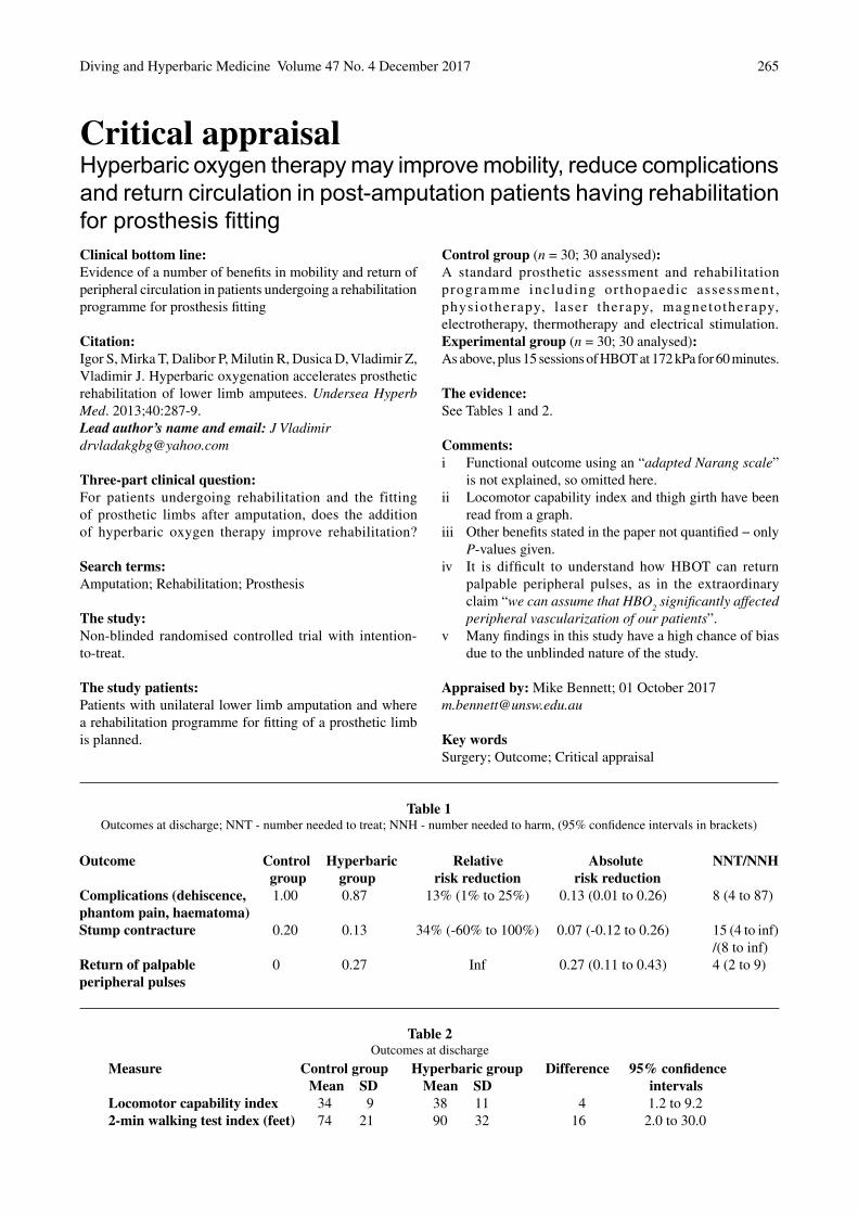
Diving and Hyperbaric Medicine Volume 47 No. 4 December 2017 265
Critical appraisal
Clinical bottom line:Evidence of a number of benefits in mobility and return of peripheral circulation in patients undergoing a rehabilitation programme for prosthesis fitting
Citation:Igor S, Mirka T, Dalibor P, Milutin R, Dusica D, Vladimir Z, Vladimir J. Hyperbaric oxygenation accelerates prosthetic rehabilitation of lower limb amputees. Undersea Hyperb Med. 2013;40:287-9.Lead author’s name and email: J [email protected] Three-part clinical question:For patients undergoing rehabilitation and the fitting of prosthetic limbs after amputation, does the addition of hyperbaric oxygen therapy improve rehabilitation? Search terms:Amputation; Rehabilitation; Prosthesis The study:Non-blinded randomised controlled trial with intention-to-treat. The study patients:Patients with unilateral lower limb amputation and where a rehabilitation programme for fitting of a prosthetic limbis planned.
Control group (n = 30; 30 analysed):A standard prosthetic assessment and rehabilitation programme including or thopaedic assessment , physiotherapy, laser therapy, magnetotherapy, electrotherapy, thermotherapy and electrical stimulation. Experimental group (n = 30; 30 analysed):As above, plus 15 sessions of HBOT at 172 kPa for 60 minutes. The evidence:See Tables 1 and 2.
Comments:i Functional outcome using an “adapted Narang scale”
is not explained, so omitted here.ii Locomotor capability index and thigh girth have been
read from a graph.iii Other benefits stated in the paper not quantified − only
P-values given.iv It is difficult to understand how HBOT can return
palpable peripheral pulses, as in the extraordinary claim “we can assume that HBO
2 significantly affected
peripheral vascularization of our patients”.v Many findings in this study have a high chance of bias
due to the unblinded nature of the study.
Appraised by: Mike Bennett; 01 October [email protected]
Key wordsSurgery; Outcome; Critical appraisal
Hyperbaric oxygen therapy may improve mobility, reduce complications and return circulation in post-amputation patients having rehabilitation for prosthesis fitting
Table 1Outcomes at discharge; NNT - number needed to treat; NNH - number needed to harm, (95% confidence intervals in brackets)
Outcome Control Hyperbaric Relative Absolute NNT/NNH group group risk reduction risk reductionComplications (dehiscence, 1.00 0.87 13% (1% to 25%) 0.13 (0.01 to 0.26) 8 (4 to 87)phantom pain, haematoma)Stump contracture 0.20 0.13 34% (-60% to 100%) 0.07 (-0.12 to 0.26) 15 (4 to inf) /(8 to inf)Return of palpable 0 0.27 Inf 0.27 (0.11 to 0.43) 4 (2 to 9)peripheral pulses
Table 2Outcomes at discharge
Measure Control group Hyperbaric group Difference 95% confidence Mean SD Mean SD intervalsLocomotor capability index 34 9 38 11 4 1.2 to 9.22-min walking test index (feet) 74 21 90 32 16 2.0 to 30.0

Diving and Hyperbaric Medicine Volume 47 No. 4 December 2017266
Obituary
as a leader in the development and implementation of diving schools, safety requirements for commercial diver education and training programmes for law-enforcement officers, researchers, diver medics, and scientific, military and commercial divers. To support these, he and his wife, Susan, founded the Best Publishing Company, which grew to become the world’s largest publisher of books, textbooks, and manuals related to all aspects of diving, diving and hyperbaric medicine and wound-care management. He proudly edited and published the NOAA Diving Manual, the US Navy Diving Manual and the premier editions of Hyperbaric Medicine Practice and Wound Care Practice.
Born in Long Beach, California, in 1935, Jim received his degree in zoology and chemistry from the University of California Los Angeles. Afterwards, he attended the UCLA School of Medicine, where he specialized in microbiology and immunology. He served as a consultant to the State of California and developed the Marine Technology Training Center at the California Institute for Men. He also served as an international consultant, devising feasibility studies and proposals for diving facilities and diver training programmes in a host of international countries.
Among the many organizations that were recipients of his 40-plus years of volunteer leadership were the Undersea and Hyperbaric Medical Society (Board of Directors); Association of Diving Contractors International (Chairperson, education committee); Association of Commercial Diving Educators(founding President); National Association of DivingMedical Technicians (founder); International Diving Schools Association (cofounder); coauthor for Occupational Safetyand Health Administration (OSHA) and US Coast Guarddiving standards and the US Government’s study for theNational Plan for Safety and Health of Divers.
Jim received numerous awards, was inducted into the Commercial Diving Hall of Fame and was formally recognized with a US Congressional Award for Outstanding Service and Contributions to Safety of Diving (99th Congress). “Jim always made whoever he spoke with feel special,” said Renée Duncan, Managing Editor of the Undersea and Hyperbaric Medicine Journal and Pressure; “he was the special one”.
Nothing was more important to Jim than his family, and he will always be remembered for his generosity, humility, patience and wonderful sense of humour. He is survived by his wife of 46 years, Susan and a large extended family.
Many members of SPUMS and EUBS have benefitted directly or indirectly from Jim’s influence on the diving and hyperbaric medical community, most especially for the superb support given to us by Best Publishing. The societies extend their thoughts to Susan and her family in their loss.
Any personal reflections or memories of Jim should be sent to Susan at <[email protected]>.
James “Jim” T JoinerJames “Jim” T Joiner, founder and former President and CEO of the Best Publishing Company, passed away on27 August 2017, with his family by his side. His passion for diving and medicine were combined into his life’s careers. During his tenure as President and CEO of the College of Oceaneering, Jim was recognized worldwide
TheDiving and Hyperbaric Medicine Journal
website is at
<www.dhmjournal.com>
Articles for immediate release into the public domain, information about submitting to the Journal, profiles of the Editorial Board and contents of the most recent and previous
issues are to be found on the site.A new site is being built for launch in 2018 to coincide with
DHM becoming an electronic publication.
Diving and Hyperbaric Medicine Volume 47 No. 4 December 2017 267
Gavin Anthony, UKTino Balestra, BelgiumNeil Banham, AustraliaMartin Barwood, UKRoger Beer, UKMike Bennett, Australia*John-Paul Bingham, USADenise Blake, AustraliaJean-Eric Blatteau, FranceLesley Blogg, UK*Phillipe Cauchie, BelgiumJames Caruso, USADick Clarke, USASue Coleshaw, UKMike Davis, New Zealand*Yvonne Denny, New ZealandDavid Doolette, USA*Chris Edge, UK*Ingrid Eftedal, Norway*Mark Elder, New ZealandJoel Edney, UKJohn Fitz-Clarke, USAJames Francis, UKPeter Germonpré, Belgium*Steve Goble, AustraliaRichard Harris, AustraliaJane Heyworth, Australia*Edmond Kay, USATom Kertesz, AustraliaAndreas Koch, GermanyJacek Kot, Poland*Chris Lawrence, Australia
John Leach, UKRichard Lee, USAGeoff Loveman, UKIan Millar, AustraliaSimon Mitchell, New Zealand*Otto Molvaer, NorwayHeather Murphy-Lavoie, USACluas-Martin Muth, Germany*Mirjam Nolting, GermanyChristine Penny, UKGeorge Perdrizet, USANeal Pollock, Canada*Andrew Proctor, UKJaques Regnard, FranceMonica Rocco, Italy*Marguerite St Leger Dowse, UKDavid Smart, Australia*Gary Smerdon, UKQuentin Summers, AustraliaMichael Taplin, AustraliaKay Tetzlaff, GermanySigrid Theunissen, SpainMark Turner, UKRob van Hulst, The Netherlands*Pieter van Ooij, The NetherlandsJose Vega, USAMatthew White, UKDavid Wilkinson, AustraliaPeter Wilmshurst, UKColin Wilson, UKWeigang Xu, ChinaPiers Yates, Australia
As Editor of DHM, and on behalf of all our submitting authors, both successful and unsuccessful, I would like to sincerely thank all the reviewers who assessed submissions to DHM in 2017, both Editorial Board (EB) members (* EB members) and external reviewers. If I have missed out anyone in the list below, please let me know. Sadly, we say goodbye to Professor Jane Heyworth, University of Western Australia, who is resigning from the EB owing to her other commitments. Jane, as a highly regarded epidemiologist, has contributed strongly to the peer review of epidemiology projects submitted to DHM.
DHM reviewers in 2017
Another chamber explosion
In mid-2014, a man in his 60s was undergoing hyperbaric treatment in Nanxiong China People’s Hospital for a head injury when he lit a cigarette he had smuggled in with him. The ensuing inferno blew up the chamber, destroying it and incinerating the patient. The hospital has admitted responsibility in not warning the patient that smoking or taking a lighter into the chamber could be dangerous and were in negotiations with his family over compensation. Available from: http://www.dailymail.co.uk/news/article-2711646/Patient-killed-blowing-hospital-ward-decided-smoke-cigarette-undergoing-treatment-high-pressure-oxygen-chamber.html#ixzz4rkwj8zGT
Depth-record bids end in tragedy
In September, a Bulgarian female technical diving instructor-trainer died in Greece while attempting to set a new depth record for women by diving to 231 metres, whilst her husband who had accompanied her is in hospital in critical condition. Only days earlier, a Polish technical diver failed to surface from an attempt to 275 m in a lake in Italy.News item in Undercurrent. 2017;32;11:12.
Diving and Hyperbaric Medicine Volume 47 No. 4 December 2017268
Second Tricontinental Scientific Conference onDiving and Hyperbaric Medicine
http://www.tricon2018.org
Dates: 23−29 September 2018Venue: Durban, South Africa
After a very successful first edition in 2013 on Reunion Island, our next Tricontinental Scientific Conference will take place in the coastal city of Durban, KwaZulu Natal, South Africa. Tricon2018 replaces the usual EUBS meeting for 2018.
We are once again organising a full week with scientific days interspersed between diving workshops and social events. The academic programme will include oral and poster presentations, workshops, discussion sessions and special topic conferences.
Start planning your submission now for the ZetterstrÖm or Musimu Award.
A Joint Organising Committee from EUBS, SPUMS, SAUHMA and the Scott Haldane Foundation will work together with local Durban Hyperbaric Centre staff and a South Africa Event Management Bureau to make sure everything runs smoothly.
An excellent social calendar is planned, including opportunities to dive the nearby Aliwal Shoal, visit wildlife game parks, take in a local rugby match and explore Zulu culture. A combination of easy access, friendly people, rich culture, nature at its most spectacular and affordable prices makes this an opportunity not to be missed. The weather in September is ideal with temperatures in the low 20s on land and in the sea and little chance of rain. Why not plan an extra week before or after the conference to travel the area and experience more of South Africa's amazing diversity, hospitality and wildlife.
Bring your family too – there are lots of child-friendly activities nearby!
Visit the dedicated website: <http://www.tricon2018.org> for all information and to start planning your trip.We very much look forward to seeing you there.
Diving and Hyperbaric Medicine Volume 47 No. 4 December 2017 269
Notices and news
EUBS notices and news and all other society information is now to be found mainly on thesociety's website: <www.eubs.org>
EUBS Annual General Meeting 2017
The EUBS AGM was held in Ravenna, Italy on Saturday 16 September 2017, immediately after the closing ceremony of the 43rd EUBS Annual Scientific Meeting. The AGM presentations and financial information are on the EUBS website in the members’ section.
EUBS 2017 Annual Scientific Meeting Awards
The Arne Zetterström Award
At the EUBS Annual General Assembly on 16 September 2017 in Ravenna, the Zetterström Committee, composed of M Rocco, B Mirasoglu and C Balestra, awarded the Arne Zetterström Award for best poster presentation to:
Portier W, Van Molle B, Neirynck Y, Germonpré P. Incidence of oxygen toxicity during hyperbaric oxygen therapy: influence of air breaks and treatment comfort. Retrospective analysis of 69,406 treatments in a single hyperbaric centre.
Arne Zetterström (1917–1945) is best known for his research with the breathing mixture ‘hydrox’ for the Swedish Navy. Zetterström first described the use of hydrogen as a breathing gas in 1943. From 1943 to 1944, a total of six ocean dives were made utilising this mixture with the deepest to 160 metres (96% hydrogen, 4% oxygen). On 07 August 1945, Zetterström experienced technical problems diving from HSwMS Belos. His support divers misread his signals and this was followed by a rapid ascent that resulted in severe decompression sickness and hypoxia, resulting in his untimely death.
The Patrick Musimu Award
The Patrick Musimu Award for best contribution to an ASM, either oral or poster presentation, in the area of breathhold diving, was instituted in 2011 by the Belgian Society for Diving and Hyperbaric Medicine. Patrick (1970–2011) was a Belgian free diver, business man and physiotherapist. On 30 June 2005, he free dove to a depth of 209 metres, beating the previous “no limits” world record by almost 40 metres. At his request, this dive was done without the supervision of the International Association for the Development of Apnea, from which Musimu had dissociated himself in 2002. According to him, extreme deep freediving should not be considered as a sport but as an adventure.
Patrick began freediving in 1999 at the age of 28. His secret
lay in years of training and preparation, but special attention should be given to his ear clearing technique: instead of equalizing his ears by the regular manoeuvres, he flooded his air spaces (sinuses and middle ears) with seawater before reaching the depth where ordinary equalization would become hard. On 21 July 2011, Patrick died whilst pool training alone at home.
At the Ravenna ASM, the jury decided not to award this prize. It was felt that more research should be conducted in this field and the jury, speaking on behalf of the Belgian Society for Diving and Hyperbaric Medicine, would like to encourage this further.
EUBS Executive Committee
Rodrigue Pignel, from Geneva, Switzerland, has been elected Member-at-Large to replace Rob van Hulst (The Netherlands) after serving his three-year term. The Executive Committee wish to express their gratitude for Rob’s contributions to its activities. Many members will know Rodrigue as co-organiser of EUBS 2016. He is the Head of the Hyperbaric Centre of the Geneva University Hospital and also very active in the field of hyperbaric and diving medicine education.
EUBS Website
Please visit the EUBS Website for the latest news and updates. Specifically, a new “EUBS History” section has been added under the Menu item “The Society”. There is still some information missing in the list of EUBS Meetings, Presidents and Members-at-Large – please dig into your memories and help us complete this list.
By popular demand, EUBS Members can now also download the complete Abstract Book of previous EUBS Meetings from the Members’ area.
TRICON2018
Favorable airfares are dependent on early booking! Early registration for TRICON2018 ends on 31 March 2018. When accessing the website <http://www.tricon2018.org>, please remember to type in the full web address, including the “http://” else you may receive a security warning when using certain web browsers. You can also access the TRICON2018 site by visiting the EUBS website.
N.B. TRICON2018 replaces the EUBS meeting for 2018.
Diving and Hyperbaric Medicine Volume 47 No. 4 December 2017270
Notices and news
All SPUMS society information and news is to be found mainly on thesociety website: <www.spums.org.au>
Australian and New Zealand College of Anaesthetists news
The new Diploma of Advanced Diving and Hyperbaric Medicine was launched on 31 July 2017. Those interested in applying for training are directed to the Australian and New Zealand College of Anaesthetists website: <http://www.anzca.edu.au/training/diving-and-hyperbaric-medicine>
The curriculum and handbook for training can be found there, as well as documents for units wishing to apply for accreditation.
The process for application for transition credits for those transitioning from the Certificate in DHM to the Diploma closed on 28 August 2017. Those who did not apply within that timeframe can apply for recognition of prior learning. All queries should be directed to <[email protected].>
Suzy SzekelyChair, ANZCA Diving and Hyperbaric Medicine Special Interest [email protected]
Publications database of the German Diving and Hyperbaric Medical Society (GTUeM)
EUBS and SPUMS members are able to access the German Society’s very large database of publications in diving and hyperbaric medicine. This will enhance anyone’s literature search. EUBS members have had this access for many years and it is now available for SPUMS members. SPUMS members should log onto the SPUMS website with their user name and password, click on “Resources” then on “GTUeM database” in the pull-down menu. This opens a new window; click on the link provided and enter the user name and password listed on the page that appears, which will then access the database.
For EUBS members, click on “Members area”, scroll down to “Access the GTUEM Literature Database” and click on this link to enter the database.
Much of the SPUMS Journal and all of Diving and Hyperbaric Medicine up till late 2016 is now on this database as individual, searchable and downloadable articles.
Divers Emergency Service/DAN AP Telemedicine Scholarship 2018
Purpose: Award of AUD 4,000 to support attendance of a trainee to the next SPUMS ASM
Criteria: The successful applicant will:• Be a medical practitioner at registrar or fellow level;• Have undertaken a formal attachment to a diving and
hyperbaric medicine facility in Australiafor a minimum of three months duration in the year prior to the date of the award;
• Use the scholarship to attend the next SPUMS ASM (2018 is Durban);
• Be a registered trainee for the SPUMS Diploma (or completed);
• Give a talk at the SPUMS ASM on a topic of their choosing (not mandatory).
Application: Trainees should apply in writing, including:• A cover letter stating they are applying for the
scholarship;• Full contact details;• Whether it is intended to present at the ASM;• Current curriculum vitae;• A letter of support from their supervisor or Head of Unit
confirming their attachment to the unit and their ability to attend the SPUMS ASM.
Applications open: 01 February 2018Applications close: 02 April 2018Scholarship announced: 30 April 2018Applications and enquiries to:<[email protected]>
SPUMS HTNA Award 2017
The SPUMS Award for the best presentation at the Hyperbaric Technicians and Nurses Association (HTNA) Annual Scientific Meeting in August 2017 was given to Corry Van der Broek, Facility Manager at the Royal Hobart Hospital Hyperbaric Unit for his presentation:“The flammability potential of dressing products and their use in monoplace chambers”
He will receive a copy of the book Diving and subaquatic medicine by Carl Edmonds et al. and a certificate. The SPUMS Executive congratulates Corry.

Diving and Hyperbaric Medicine Volume 47 No. 4 December 2017 271
SPUMS Diploma in Diving and Hyperbaric Medicine
Requirements for candidates (May 2014)
In order for the Diploma of Diving and Hyperbaric Medicine to be awarded by the Society, the candidate must comply with the following conditions:1 (S)he must be medically qualified, and remain a current
financial member of the Society at least until they have completed all requirements of the Diploma.
2 (S)he must supply evidence of satisfactory completion of an examined two -week full- time course in diving and hyperbaric medicine at an approved facility. The list of such approved facilities may be found on the SPUMS website.
3 (S)he must have completed the equivalent (as determined by the Education Officer) of at least six months’ full- time clinical training in an approved Hyperbaric Medicine Unit.
4 (S)he must submit a written proposal for research in a relevant area of underwater or hyperbaric medicine, in a standard format, for approval before commencing the research project.
5 (S)he must produce, to the satisfaction of the Academic Board, a written report on the approved research project, in the form of a scientific paper suitable for publication. Accompanying this report should be a request to be considered for the SPUMS Diploma and supporting documentation for 1–4 above.
In the absence of other documentation, it will be assumed that the paper is to be submitted for publication in Diving and Hyperbaric Medicine. As such, the structure of the paper needs to broadly comply with the ‘Instructions to authors’ available on the SPUMS website <www.spums.org.au> or at <www.dhmjournal.com>.
The paper may be submitted to journals other than Diving and Hyperbaric Medicine; however, even if published in another journal, the completed paper must be submitted to the Education Officer (EO) for assessment as a diploma paper. If the paper has been accepted for publication or published in another journal, then evidence of this should be provided.
The diploma paper will be assessed, and changes may be requested, before it is regarded to be of the standard required for award of the Diploma. Once completed to the reviewers’ satisfaction, papers not already submitted to, or accepted by, other journals should be forwarded to the Editor of Diving and Hyperbaric Medicine for consideration. At this point the Diploma will be awarded, provided all other requirements are satisfied. Diploma projects submitted to Diving and Hyperbaric Medicine for consideration of publication will be subject to the Journal’s own peer review process.
Additional information – prospective approval of projects is required
The candidate must contact the EO in writing (or e mail) to advise of their intended candidacy and to discuss the proposed topic of their research. A written research proposal must be submitted before commencement of the research project.
All research reports must clearly test a hypothesis. Original basic and clinical research are acceptable. Case series reports may be acceptable if thoroughly documented, subject to quantitative analysis and if the subject is extensively researched in detail. Reports of a single case are insufficient. Review articles may be acceptable if the world literature is thoroughly analysed and
discussed and the subject has not recently been similarly reviewed. Previously published material will not be considered. It is expected that the research project and the written report will be primarily the work of the candidate, and that the candidate is the first author where there are more than one.
It is expected that all research will be conducted in accordance with the joint NHMRC/AVCC statement and guidelines on research practice, available at: <www.nhmrc.gov.au/_files_nhmrc/publications/attachments/r39.pdf>, or the equivalent requirement of the country in which the research is conducted. All research involving humans, including case series, or animals must be accompanied by documentary evidence of approval by an appropriate research ethics committee. Human studies must comply with the Declaration of Helsinki (1975, revised 2013). Clinical trials commenced after 2011 must have been registered at a recognised trial registry site such as the Australia and New Zealand Clinical Trials Registry <http://www.anzctr.org.au/> and details of the registration provided in the accompanying letter. Studies using animals must comply with National Health and Medical Research Council Guidelines or their equivalent in the country in which the work was conducted.
The SPUMS Diploma will not be awarded until all requirements are completed. The individual components do not necessarily need to be completed in the order outlined above. However, it is mandatory that the research proposal is approved prior to commencing research.
Projects will be deemed to have lapsed if:• the project is inactive for a period of three years, or• the candidate fails to renew SPUMS Membership in any year
after their Diploma project is registered (but not completed).
For unforeseen delays where the project will exceed three years, candidates must explain to the EO by email why they wish their diploma project to remain active, and a three-year extension may be approved. If there are extenuating circumstances why a candidate is unable to maintain financial membership, then these must be advised by email to the EO for consideration by the SPUMS Executive. If a project has lapsed, and the candidate wishes to continue with their DipDHM, then they must submit a new application as per these guidelines.
The Academic Board reserves the right to modify any of these requirements from time to time. As of January 2016, the SPUMS Academic Board consists of:
Dr David Wilkinson, Education Officer, Adelaide;Professor Simon Mitchell, Auckand;Dr Denise Blake, Townsville.
All enquiries and applications should be addressed to:David [email protected]
Key wordsQualifications; Underwater medicine; Hyperbaric oxygen; Research; Medical society

Diving and Hyperbaric Medicine Volume 47 No. 4 December 2017272
The South Pacific Underwater Medicine Society is proud to confer Life Membership on Associate Professor Michael Davis. Mike is a passionate diver, a highly respected clinician and an influential academic; all the characteristics that define a quintessential diving physician.
Mike has been a diver for some 54 years. He began diving in the UK in the early 1960s and involved himself in a spectrum of activities which extended well beyond recreational diving for simple pleasure. In particular, he contributed to the activities of teams undertaking scientific diving and diving medical research. This early passion has continued unabated to the present day, and many of us have found his unbridled enthusiasm and ‘watermanship’ in the face of advancing age to be quite inspirational; not to mention his apparent imperviousness to cold.
At an early stage, Mike recognised that whatever his diving medicine aspirations, his medical career would be best served by training first in a more conventional specialty. For that he chose anaesthesia and intensive care, and he forged a career as a highly respected anaesthetist. His anaesthetic practice included the challenging subspecialty of cardiac anaesthesia. As time went on, he incorporated diving and
hyperbaric medicine into his clinical practice, and provided superlative service to the New Zealand diving community in doing so. From 1979 to 1996 he, along with a small group of others, voluntarily manned the Christchurch Hyperbaric Medicine Unit. From 1996 to 2009 he served as Clinical Director of the service after it was formally established. This, on its own, is an extraordinary record of service to the specialty.
From an early stage in his career Mike stamped his mark as a serious contributor to the academic milieu of both anaesthesia and diving medicine. During the 1980s he completed an MD, and over his career he has published more than 60 scientific journal articles (the majority as first author) and many letters. He was appointed Associate Professor at the University of Auckland in 2004, and established a level 700 distance-learning course in diving and hyperbaric medicine which saw a number of graduates at Diploma and Masters levels. While all of this is impressive enough, the endeavour for which the society (and indeed the wider diving medicine community) will forever remain in debt to Mike is his stewardship of transitioning the SPUMS Journal from a relatively casual publication into a high quality, peer-reviewed, Medline-listed academic journal. Diving and Hyperbaric Medicine as it is now known, has become a rigorous yet informative and readable journal under Mike as Editor. He has presided over the process of having its academic rigour recognised by the National Library of Medicine in the United States, and of amalgamating its governance between SPUMS and the European Underwater and Baromedical Society. The journal, above all other things, is the Society’s footprint in the wider world, and Mike has ensured that it is a large and highly influential one.
Mike is a truly deserving Life Member.
Presented by the Executive Committee at the 2017 SPUMS Annual General Meeting and adopted unanimously by those present.
Note: The Purposes and Rules of SPUMS allow for eight Life Members at any one time. Other current Life Members are Carl Edmonds, David Elliott, John Knight, Chris Lourey, John Pennefather and Douglas Walker,
Life membership of SPUMS awarded to Michael Davis
SPUMS Website News
The website continues to evolve under the diligent attention of the Web Assistant, Nicky Telles. Hopefully members are finding the new website is meeting their expectations and allowing them to easily access information that they require. Please email the Webmaster or Web Assistant with suggestions for improvement. The site has been moved to new a host server based in Australia to reduce costs. Apart from a short period of downtime there should have been no noticeable change to accessing the site. Your current webmaster, Joel Hissink, has agreed to continue in this
role for now. The webmaster oversees the development and operation of the website, acts as a conduit between the Executive Committee and the Web Assistant and also sits on the ExCom participating in all ExCom decisions.
SPUMS Facebook page
Remember to ‘like’ SPUMS at:<http://www.facebook.com/pages/SPUMS-South-Pacific-Underwater-Medicine-Society/221855494509119>
Diving and Hyperbaric Medicine Volume 47 No. 4 December 2017 273
Royal Adelaide Hospital Medical Officers’ Course in Diving and Hyperbaric Medicine
2018Dates: TBAVenue: The Royal Adelaide Hospital, AdelaideCost: AUD2,500.00 (inclusive of GST)
Course Conveners: David Wilkinson and Suzy SzekelyInvited faculty includes: Professors Michael Bennett andSimon MitchellThe course content includes:• Physics and physiology of diving• Recreational fitness-to-dive• Occupational fitness-to-dive• Decompression illness and non-dysbaric injuries• Medical management and return to diving• Technical and professional diving• Marine envenomation• Introduction to hyperbaric medicine
Contact for information:Ms Lorna Mirabelli, Course AdministratorPhone: +61-(0)8-8222-5116E-mail: <[email protected]>
Scott Haldane Foundation
As an institute dedicated to education in diving medicine, the Scott Haldane Foundation has organized more than 240 courses over the past 20 years. SHF is targeting more and more on an international audience with coursesworldwide.
The courses Medical Examiner of Diver (part I and II) and SHF in-depth courses, as modules of the level 2d Diving Medicine Physician course, fully comply with the ECHM/EDTC curriculum for Level 1 and 2d respectively and are accredited by the European College of Baromedicine (ECB).
SHF Course Calendar 1st half 2018February: Refresher course Organization Diving medical NL23−24 March: Basic course part 1, Zeist, NL7, 13−14 April: Basic course part 2, Amsterdam, NL12–19 May: Basic course part 2, Bonaire1st half 2018: HBOT and decompression; tbc
On request: Internship different types of diving (DMP), NLOn request: Internship hyperbaric medicine (DMP certification), NL/Belgium
The course calendar will be supplemented regularly.For the latest information: <www.scotthaldane.org>
Undersea & Hyperbaric Medical SocietyAnnual Scientific Meeting 2018
Dates: 28–30 JuneVenue: Disney's Coronado Springs Resort Lake Buena Vista, Florida
Pre-course: 27 June Topic: How to prepare for accreditationProgramme chair: Tom Workman
Call for abstracts: not yet openRegistration: <https://www.uhms.org/asm-new.html>For further information: <[email protected]>
ANZ Hyperbaric Medicine GroupIntroductory Course in Diving and Hyperbaric
Medicine 2018Dates: 12–23 FebruaryVenue: Esplanade Hotel, Fremantle, Western AustraliaCost: AUD2,500 (inclusive of GST) for the two weeks
This highly popular course has moved its roots from Sydney to sunny Western Australia. It is for medical graduates with an interest in diving and hyperbaric medicine; designed for both those wishing to pursue a career in this specialised field and those whose primary interest lies in related areas. Excursions include the Fiona Stanley Hyperbaric Medicine Unit, HMAS Stirling and the local Royal Flying Doctor base. The course is accredited for the SPUMS Diploma and the Australian and New Zealand College of Anaesthetists (ANZCA) Diploma of Advanced Diving and Hyperbaric Medicine. It is also approved by the Royal Australian College of General Practitioners as an Active Learning Module.
Course Conveners: Ian Gawthrope and Neil BanhamThe Course content includes:• History of diving medicine and hyperbaric oxygen• Physics and physiology of diving and compressed gases• Presentation, diagnosis and management of diving injuries• Assessment of fitness to dive• Visit to RFDS base for flying and diving workshop• Accepted indications for hyperbaric oxygen treatment• Hyperbaric oxygen evidence-based medicine• Wound management and transcutaneous oximetry• In water rescue and management of a seriously ill diver• Visit to HMAS Stirling• Practical workshops• Marine Envenomation
Contact for information:Sue Conlon, Course AdministratorPhone: +61-(0)8-6152-5222E-mail: <[email protected]>
Diving and Hyperbaric Medicine Volume 47 No. 4 December 2017274
Capita Selecta Diving Medicine Academic Medical Centre,
University of Amsterdam, The Netherlands
Spring Symposium21st Century decompression theory and DCI treatment; from Haldane to BVM, endothelium response and helioxDate: Saturday 17 March 2018Venue: Academic Medical Centre, Amsterdam
Autumn SymposiumThe ageing diverInformation: <[email protected]>Website: <www.capitaselectaduikgeneeskunde.nl>
DAN Europe
DAN Europe has a fresh, multilingual selection of recent news, articles and events featuring DAN and its staff.
Go to the website: <http://www.daneurope.org/web/guest/>
German Society for Diving andHyperbaric Medicine (GTÜeM)
An overview of basic and refresher courses in diving and hyperbaric medicine, accredited by GTÜeM according to EDTC/ECHM curricula, can be found on the website:<http://www.gtuem.org/212/Kurse_/_Termine/Kurse.html>
Hyperbaric Oxygen, Karolinska
Welcome to: <http://www.hyperbaricoxygen.se/>This site, supported by the Karolinska University Hospital, Stockholm, Sweden, offers publications and high -quality lectures from leading investigators in hyperbaric medicine. Please register to obtain a password via e- mail. Once registered, watch on line, or download to your iPhone, iPad or computer for later viewing.For further information contact:E- mail: <[email protected]>
The Science of Diving
Support EUBS by buying the PHYPODE book “The science of diving”. Written for anyone with an interest in the latest research in diving physiology and pathology. The royalties from this book are being donated to the EUBS.Need more reason to buy? We don’t think so!Available from: Morebooks <https://www.morebooks.de/store/gb/book/the-science-of-diving/isbn/978-3-659-66233-1>
Instructions for authors
A downloadable pdf of the ‘Instructions for authors’ (revised December 2017) and other guidance for p repar ing a submiss ion can be found on the Diving and Hyperbaric Medicine (DHM) website:<www.dhmjournal.com> and on the Manuscript Manager platform. Authors must read and follow these instructions.
All submissions to DHM should be made using the portal at <http://www.manuscriptmanager.net/dhm>. Before submitting, please view the video on how to prepare a submission at: <https://www.youtube.com/watch?v=gpMsPAX4pWA&t=41s>.
In case of difficulty, please contact the Editorial Assistant by e-mail at: <[email protected]>.
Copyright
All articles in Diving and Hyperbaric Medicine are published under licence from the authors. Copyright to these articles remains with these authors. Any distribution, apart from for limited educational purposes, is in breach of copyright.
Advertising in Diving and Hyperbaric Medicine
Companies and organisations within the diving, hyperbaric medicine and wound-care communities wishing to advertise their goods and services in Diving and Hyperbaric Medicine are welcome. The advertising policy of the parent societies appears on the journal website: <www.dhmjournal.com>
Details of advertising rates and formatting requirements are available on request from:E-mail: <[email protected]>
DIVING HISTORICAL SOCIETY
AUSTRALIA, SE ASIA
P O Box 347, Dingley Village Victoria, 3172, AustraliaE-mail: <[email protected]>Website: <www.classicdiver.org>
20th International Congress on Hyperbaric Medicine 2020
Dates: 13−16 September 2020Venue: Rio de Janeiro, BrazilFor preliminary information contact:Dr Mariza D’Agostino DiasEmail: <[email protected]>
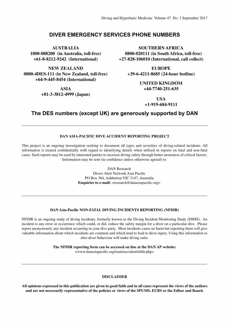
Diving and Hyperbaric Medicine Volume 47 No. 3 September 2017
DAN Asia-Pacific NON-FATAL DIVING INCIDENTS REPORTING (NFDIR)
NFDIR is an ongoing study of diving incidents, formerly known as the Diving Incident Monitoring Study (DIMS). An incident is any error or occurrence which could, or did, reduce the safety margin for a diver on a particular dive. Please report anonymously any incident occurring in your dive party. Most incidents cause no harm but reporting them will give valuable information about which incidents are common and which tend to lead to diver injury. Using this information to
alter diver behaviour will make diving safer.
The NFDIR reporting form can be accessed on line at the DAN AP website:<www.danasiapacific.org/main/accident/nfdir.php>
DAN ASIA-PACIFIC DIVE ACCIDENT REPORTING PROJECT
This project is an ongoing investigation seeking to document all types and severities of diving-related incidents. All information is treated confidentially with regard to identifying details when utilised in reports on fatal and non-fatal cases. Such reports may be used by interested parties to increase diving safety through better awareness of critical factors.
Information may be sent (in confidence unless otherwise agreed) to:
DAN ResearchDivers Alert Network Asia Pacific
PO Box 384, Ashburton VIC 3147, AustraliaEnquiries to e-mail: <[email protected]>
DIVER EMERGENCY SERVICES PHONE NUMBERS
DISCLAIMER
All opinions expressed in this publication are given in good faith and in all cases represent the views of the authors and are not necessarily representative of the policies or views of the SPUMS, EUBS or the Editor and Board.
The DES numbers (except UK) are generously supported by DAN
AUSTRALIA1800-088200 (in Australia, toll-free)
+61-8-8212-9242 (International)
NEW ZEALAND0800-4DES-111 (in New Zealand, toll-free)
+64-9-445-8454 (International)
ASIA+81-3-3812-4999 (Japan)
SOUTHERN AFRICA 0800-020111 (in South Africa, toll-free)
+27-828-106010 (International, call collect)
EUROPE+39-6-4211-8685 (24-hour hotline)
UNITED KINGDOM+44-7740-251-635
USA+1-919-684-9111

Printed by Snap Printing, 166 Burwood Road, Hawthorn, Victoria 3122, <[email protected]>
CONTENTS
Diving and Hyperbaric Medicine is indexed on MEDLINE, SciSearch® and Embase/Scopus
Diving and Hyperbaric Medicine Volume 47 No. 4 December 2017
Original articles
SPUMS notices and news
273 Courses and meetings
216 Influence of repetitive diving in saltwater on pressure equalization and Eustachian tube function in recreational scuba diversMoritz F Meyer, Manuela Boor, Stefanie Jansen, Eberhard D Pracht, Moritz Felsch, Heinz D Klünter, Karl-Bernd Hüttenbrink, Dirk Beutner, Maria Grosheva
223 Influence of repetitive diving in freshwater on pressure equalization and Eustachian tube function in recreational scuba diversStefanie Jansen, Manuela Boor, Moritz F Meyer, Eberhard D Pracht, Ruth Volland, Heinz D Kluenter, Karl-Bernd Huettenbrink, Dirk Beutner, Maria Grosheva
228 Thromboelastographic assessment of the impact of mexiletine on coagulation abnormalities induced by air or normal saline intravenous injections in conscious ratsJoseph L Nates, Davide Cattano, Fernanda S Costa, Jacques E Chelly, Marie-Francoise Doursout
233 Hyperbaric oxygen in the treatment of acute retinal artery occlusionMark J Elder, John A Rawstron, Michael Davis
270 Australian and New Zealand College of Anaesthetists news
270 Publications database of the German Diving and Hyperbaric Medical Society (GTÜeM)
271 SPUMS Diploma in Diving and Hyperbaric Medicine
272 Life membership of SPUMS awarded to Michael Davis
269 EUBS Annual General Meeting 2017
269 EUBS 2017 Annual Scientific Meeting Awards
269 EUBS Executive Committee
239 Lost at sea: the medicine, physiology and psychology of prolonged immersionHeather Massey, John Leach, Michael Davis, Vicki Vertongan
248 Personality and behavioural outcomes in diving: current status and recommendations for future researchCharles H van Wijk
253 Diving and antidepressantsAbraham L Querido
Review articles
Book reviews263 Hyperbaric medicine practice
Neil Banham
264 Oxygen first aid for diversGreg van der Hulst
Case reports257 Delayed hyperbaric intervention in life-threatening
decompression illnessMichael FM Perez, April R Serrano, Maravic P Andal, Maria CC Aldover
260 Hyperbaric oxygen-associated seizure leading to strokeJordan M Warchol, Jeffrey S Cooper, Thomas S Diesing
268 Tricon2018, 23–29 September, Durban, South Africa
Editorials
211 The Editor’s offering212 The Presidents’ pages214 Back to the future: occupational diver training in Australia
David Smart
Critical appraisal
265 Hyperbaric oxygen therapy may improve mobility, reduce complicat ions and return circulation in post-amputation patients have rehabilitation for prosthesis fittingMike Bennett
Obituary
266 James “Jim” T Joiner
EUBS notices and news
267 DHM reviewers in 2017