Diversity of Life - Dr. Michael Belanich · PDF fileProkaryotes are fundamentally different...
64
Diversity of Life Bacteria Archae Protista Fungi
Transcript of Diversity of Life - Dr. Michael Belanich · PDF fileProkaryotes are fundamentally different...
Diversity of Life
Bacteria
Archae
Protista
Fungi

Bacteria on the point of a pin

Archaebacteria & Eubacteria
• prokaryotic cells
• Abundant
– Ex: in the human body- for every one human
cell there are 10 prokaryotic cells
• important decomposers and symbionts

Prokaryotes are fundamentally
different than Eukaryotes• Unicellular
– Can form colonies (masses of single species) or biofilms (complex
community of different species)
• Usually very small
– ten times smaller than typical eukaryotic cells
– Typically 1-10 microns long
Prokaryotes are fundamentally
different than Eukaryotes• Nucleoid region
– Have a single circular chromosome made up of DNA and histone-
like proteins in a nucleoid region of the cell
– May have small circular DNA’s called plasmids outside of
nucleoid region
• Cell division
– Binary fission (no spindle, no phases of mitosis)
Prokaryotes are fundamentally
different than Eukaryotes• Genetic recombination:
– Horizontal gene transfer
• Conjugation – cell to cell transfer of plasmids
• Transduction – viral transfer of DNA
• Transformation – uptake of pieces of DNA not associated with cells or virus
– No sexual cycle (no meiosis)
• Internal compartmentalization
– Cytoplasm does not have extensive internal compartments and no
membrane-bounded organelles

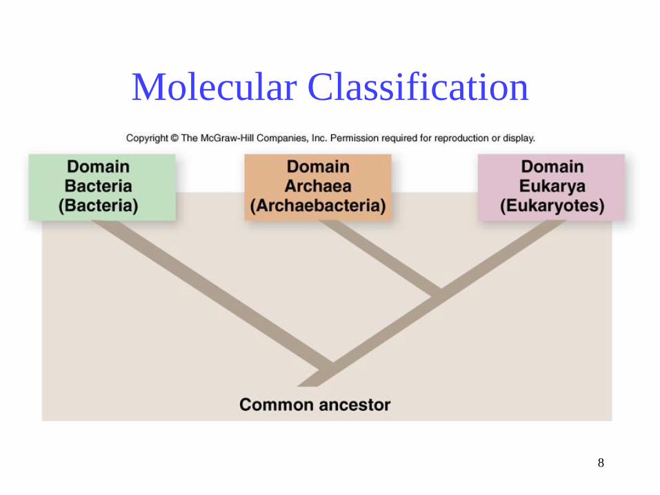
Prokaryotic Evolution
• Kingdom Monera is NOT monophyletic
• Two main branches
– Archaebacteria = extreme environments
– Eubacteria or Bacteria
8
Molecular Classification

Bacteria and Archae differ
fundamentally
• Plasma Membranes– Archaean membrane lipids are composed of glycerol linked to
hydrocarbon chains by ether linkages, not the ester linkages seen in
bacteria and eukaryotes

Bacteria and Archae differ
fundamentally
• Cell Wall– Bacterial cell walls are made of peptidoglycan, Archae are not
• DNA replication– Both have single origin but Archaeal initiation of DNA replication
is more similar to that of eukaryotes
• Gene expression– Archaea may have more than one RNA polymerase (Transcription:
reads DNA to make RNA), and these enzymes more closely
resemble the eukaryotic RNA polymerases than they do the single
bacterial RNA polymerase


“Heat-loving” prokaryotes

Extreme halophiles
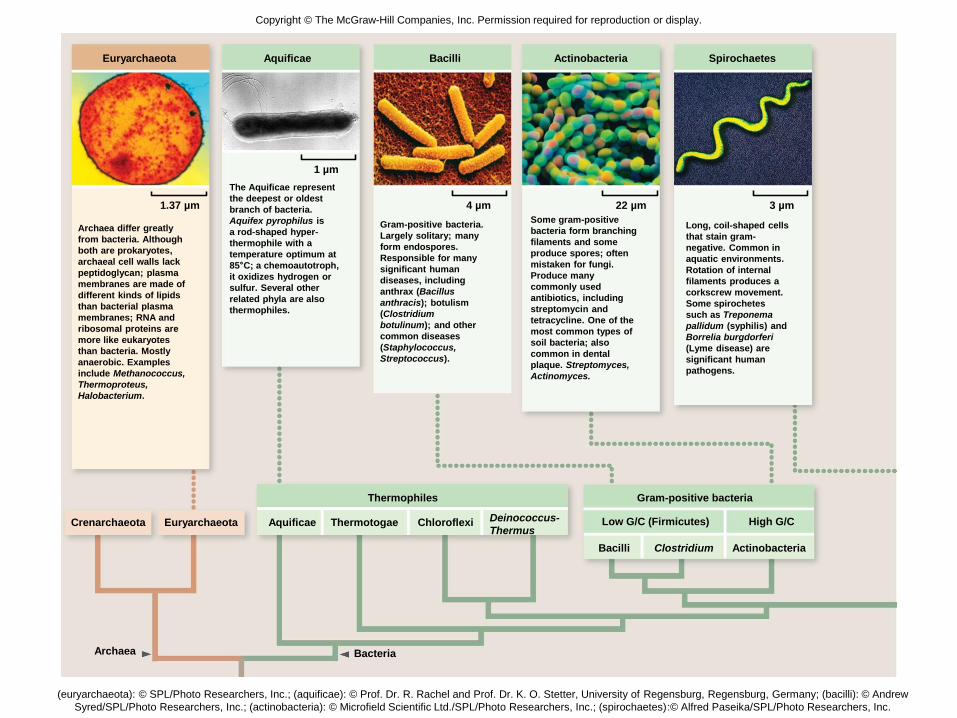
Fig. 28.6left
Crenarchaeota Euryarchaeota Aquificae Thermotogae Chloroflexi
Thermophiles
BacteriaArchaea
Bacilli Clostridium Actinobacteria
Gram-positive bacteria
High G/CLow G/C (Firmicutes)
Euryarchaeota Aquificae Bacilli Actinobacteria Spirochaetes
3 µm
Archaea differ greatly
from bacteria. Although
both are prokaryotes,
archaeal cell walls lack
peptidoglycan; plasma
membranes are made of
different kinds of lipids
than bacterial plasma
membranes; RNA and
ribosomal proteins are
more like eukaryotes
than bacteria. Mostly
anaerobic. Examples
include Methanococcus,
Thermoproteus,
Halobacterium.
The Aquificae represent
the deepest or oldest
branch of bacteria.
Aquifex pyrophilus is
a rod-shaped hyper-
thermophile with a
temperature optimum at
85°C; a chemoautotroph,
it oxidizes hydrogen or
sulfur. Several other
related phyla are also
thermophiles.
Gram-positive bacteria.
Largely solitary; many
form endospores.
Responsible for many
significant human
diseases, including
anthrax (Bacillus
anthracis); botulism
(Clostridium
botulinum); and other
common diseases
(Staphylococcus,
Streptococcus).
Some gram-positive
bacteria form branching
filaments and some
produce spores; often
mistaken for fungi.
Produce many
commonly used
antibiotics, including
streptomycin and
tetracycline. One of the
most common types of
soil bacteria; also
common in dental
plaque. Streptomyces,
Actinomyces.
Long, coil-shaped cells
that stain gram-
negative. Common in
aquatic environments.
Rotation of internal
filaments produces a
corkscrew movement.
Some spirochetes
such as Treponema
pallidum (syphilis) and
Borrelia burgdorferi
(Lyme disease) are
significant human
pathogens.
1.37 µm
1 µm
4 µm 22 µm
Deinococcus-
Thermus
Copyright © The McGraw-Hill Companies, Inc. Permission required for reproduction or display.
(euryarchaeota): © SPL/Photo Researchers, Inc.; (aquificae): © Prof. Dr. R. Rachel and Prof. Dr. K. O. Stetter, University of Regensburg, Regensburg, Germany; (bacilli): © Andrew
Syred/SPL/Photo Researchers, Inc.; (actinobacteria): © Microfield Scientific Ltd./SPL/Photo Researchers, Inc.; (spirochaetes):© Alfred Paseika/SPL/Photo Researchers, Inc.

Fig. 28.6right
Copyright © The McGraw-Hill Companies, Inc. Permission required for reproduction or display.
Cyanobacteria Chlorobi Beta Gamma Delta
ProteobacteriaPhotosynthetic
Spirochaetes
Cyanobacteria Beta Gamma Delta
25 µm
10 µm 750 µmCyanobacteria are a
form of photosynthetic
bacterium common in
both marine and
freshwater environ-
ments. Deeply
pigmented; often
responsible for “blooms”
in polluted waters. Both
colonial and solitary
forms are common.
Some filamentous
forms have cells
specialized for nitrogen
fixation.
A nutritionally diverse
group that includes
soil bacteria like the
Lithotroph Nitrosomonas
that recycle nitrogen
within ecosystems by
oxidizing the
ammonium ion (NH4+).
Other members are
heterotrophs and
photoheterotrophs.
Gammas are a diverse
group including
photosynthetic sulfur
bacteria, pathogens,
like Legionella, and the
enteric bacteria that
inhabit animal
intestines. Enterics
include Escherichia
coli, Salmonella (food
poisoning), and Vibrio
cholerae (cholera).
Pseudomonas
are a common form
of soil bacteria,
responsible for many
plant diseases, and are
important opportunistic
pathogens.
The cells of
myxobacteria exhibit
gliding motility by
secreting slimy
polysaccharides over
which masses of cells
glide; when the soil
dries out, cells
aggregate to form
upright multicellular
colonies called fruiting
bodies. Other delta
bacteria are solitary
predators that attack
other bacteria
(Bdellovibrio) and
bacteria used in
bioremediation
(Geobacter).
Epsilon–
Helicobacter
Alpha–
Rickettsia
(cyanobacteria): © Dr. Robert Calentine/Visuals Unlimited; (beta): © Science VU/S. Watson/Visuals Unlimited; (gamma): © Dennis Kunkel Microscopy, Inc.;
(delta): © Prof. Dr. Hans Reichenbach, Helmholtz Centre for Infection Research, Braunschweig
0.5 µm

16
Most prokaryotes have one of 3 basic shapes
-Bacillus = Rod-shaped
-Coccus = Spherical
-Spirillum = Helical-shaped
Prokaryotic Shapes

Prokaryotic Cell Surfaces• Cell surfaces
– Plasma membrane
– Cell walls of peptidoglycan in Eubacteria• Polysaccharides cross-linked with peptides
• Archaea do not possess peptidoglycan
– may have S-layer (Archae always do)• Function is diverse and variable but often involve adhesion to
surfaces or protection
– may have capsule • An additional gelatinous layer- interfering with recognition by
phagocytic cells- ability to cause disease
– may have Pili for adhesion and/or flagella for
movement

18

Pili

Prokaryotic flagella (Bacillus)

Copyright © The McGraw-Hill Companies, Inc. Permission required for reproduction or display.
Gram-positive Gram-negative Gram-positive Gram-negative Gram-positive Gram-negative Gram-positive Gram-negative
a.
b.10 µm
Crystal violet–iodine complex
formed inside cells.
All one color.
Alcohol
dehydrates thick
PG layer trapping
dye complex.
Alcohol
has minimal
effect on thin
PG layer.
Dark Purple
masks the
red dye.
Red dye
stains the
colorless cell.Both cell walls affix the dye.
1. Crystal violet
is applied.
2. Gram’s iodine
is applied.
3. Alcohol wash
is applied.
4. Safranin (red dye)
is applied.
b: © Jack Bostrack/Visuals Unlimited
Gram-positive bacteria have a thicker
peptidoglycan wall and stain a purple color,
whereas the more common gram-negative
bacteria contain less peptidoglycan and do
not retain the purple-colored dye.

Prokaryotic Genome– in the nucleoid region
– major chromosome
• one doubled stranded DNA molecule forms a ring
– Plasmids
• Small circular pieces of DNA
• Not required for normal cell function
• Exchanged in conjugation

23
Eukaryotic Origins
The nucleus and
endoplasmic reticulum
arose from infoldings of
prokaryotic cell
membrane

Endosymbiotic theory
• Eukaryotic organelles evolved from a
consortium of symbiotic prokaryotes
– mitochondria were aerobic heterotrophic
prokaryotes
– chloroplasts were photosynthetic prokaryotes

25

Kingdom Protista
• Eukaryotic
• Most are unicellular (there are some simple
multicellular ones)

Protista Taxonomy
• Originally consisted of all unicellular eukaryotes
• was paraphyletic
• The 17 major protist phyla are grouped into six
major monophyletic groups

Copyright © The McGraw-Hill Companies, Inc. Permission required for reproduction or display.
Cilia
tes
Ap
ico
mp
lex
an
s
ArchaeaEubacteria Chromalveolata Rhizaria Archaeplastida Excavata Amoeboza Opisthokonta
Pa
rab
as
ali
d
Din
ofl
ag
ell
ate
s
Alveolata Stramenopila Rhodophyta Chlorophytes Diplomonads Euglenozoa Choanoflagellates
An
ima
ls
Fu
ng
i
Am
oe
bo
zo
a
La
nd
pla
nts
Ch
aro
ph
yte
s
Cerc
ozo
a
Fo
ram
inif
era
Rad
iola
ra
Bro
wn
alg
ae
Dia
tom
s
Oo
myc
ete
s
Fig. 29.5

Characteristics Used to Classify
Protists
• Mode of locomotion
• mode of nutrition
• overall body form
• pigments
• & others…

An
ima
ls
Ch
oa
no
fla
ge
llid
a
Fu
ng
i
Am
oe
bo
zo
a
Eu
gle
no
zo
a
Pa
rab
as
ali
ds
Dip
lom
on
ad
s
Ch
loro
ph
yte
s
Ch
aro
ph
yte
s
Rh
od
op
hyta
Lan
d p
lan
ts
Cil
iate
s
Ap
ico
mp
lex
an
sChromalveolata Rhizaria Archaeplastida Excavata Amoeboza Opisthokonta
Din
ofl
ag
ellate
s
Alveolata Stramenopila
Ce
rco
zo
a
Fo
ram
inif
era
Ra
dio
lara
Bro
wn
alg
ae
Dia
tom
s
Oo
myc
ete
s
Copyright © The McGraw-Hill Companies, Inc. Permission required for reproduction or display.
Copyright © The McGraw-Hill Companies, Inc. Permission required for reproduction or display.
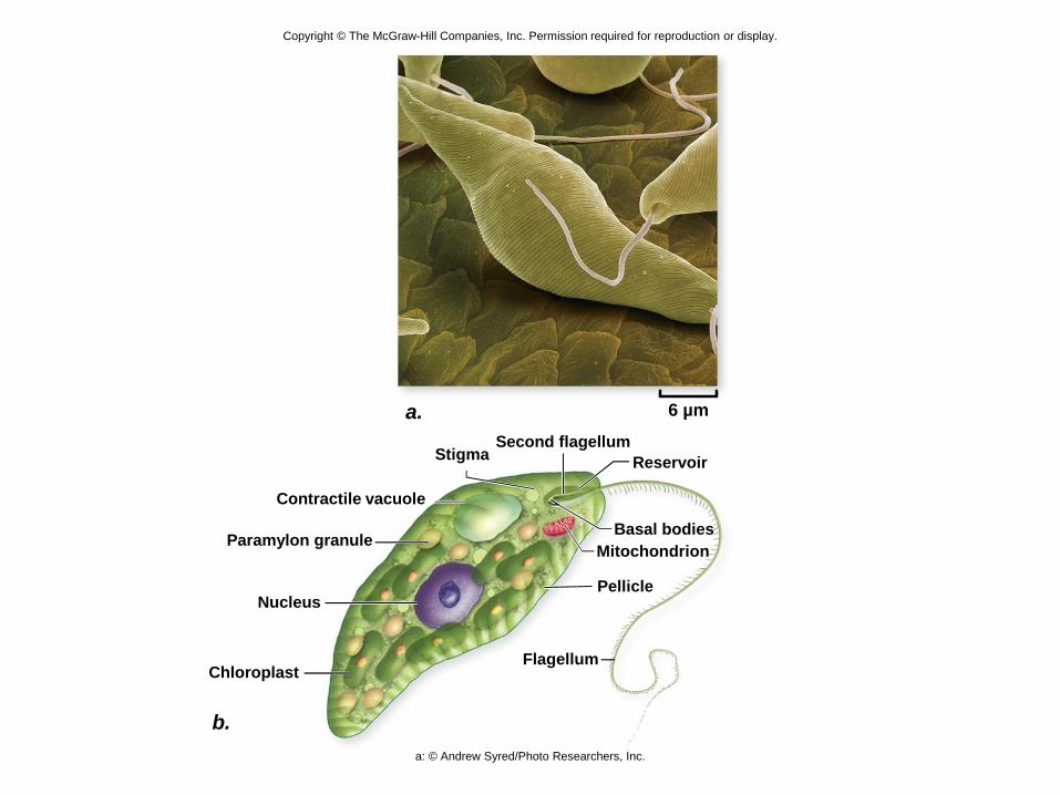
Second flagellumStigma
Contractile vacuole
Paramylon granule
Nucleus
ChloroplastFlagellum
Pellicle
Mitochondrion
Basal bodies
Reservoir
b.
a. 6 µm
a: © Andrew Syred/Photo Researchers, Inc.
Copyright © The McGraw-Hill Companies, Inc. Permission required for reproduction or display.
An
ima
ls
Ch
oa
no
fla
ge
llid
a
Fu
ng
i
Am
oe
bo
zo
a
Eu
gle
no
zo
a
Pa
rab
as
ali
ds
Dip
lom
on
ad
s
Ch
loro
ph
yte
s
Ch
aro
ph
yte
s
Rh
od
op
hyta
La
nd
pla
nts
Cil
iate
s
Ap
ico
mp
lex
an
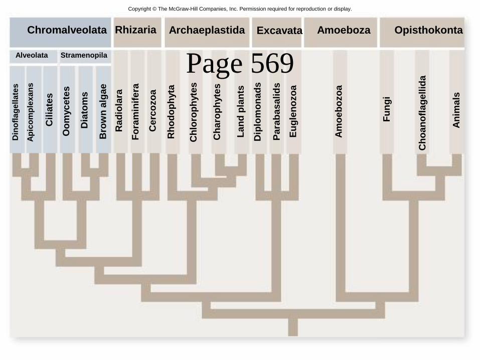
sChromalveolata Rhizaria Archaeplastida Excavata Amoeboza Opisthokonta
Din
ofl
ag
ell
ate
s
Alveolata Stramenopila
Ce
rco
zo
a
Fo
ram
inif
era
Ra
dio
lara
Bro
wn
alg
ae
Dia
tom
s
Oo
myc
ete
sPage 569

A ciliated protozoan

Too diverse for one kingdom: a diatom, a unicellular "alga"

Too diverse for one kingdom: Australian bull kelp (Durvillea potatorum)
Copyright © The McGraw-Hill Companies, Inc. Permission required for reproduction or display.
An
ima
ls
Ch
oa
no
fla
ge
llid
a
Fu
ng
i
Am
oe
bo
zo
a
Eu
gle
no
zo
a
Pa
rab
asa
lid
s
Dip
lom
on
ad
s
Ch
loro
ph
yte
s
Ch
aro
ph
yte
s
Rh
od
op
hyta
La
nd
pla
nts
Cilia
tes
Ap
ico
mp
lex
an
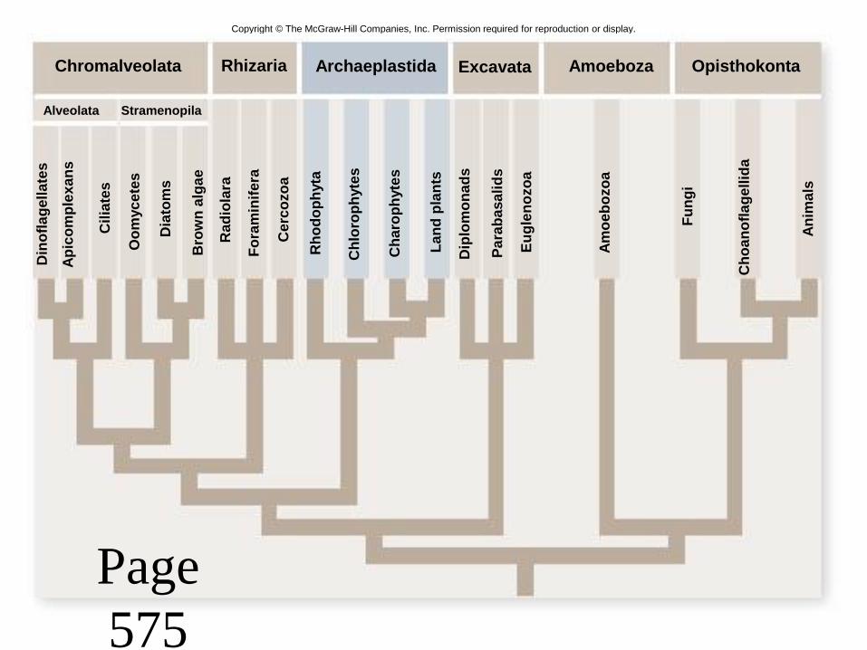
sChromalveolata Rhizaria Archaeplastida Excavata Amoeboza Opisthokonta
Din
ofl
ag
ell
ate
s
Alveolata Stramenopila
Cerc
ozo
a
Fo
ram
inif
era
Rad
iola
ra
Bro
wn
alg
ae
Dia
tom
s
Oo
myc
ete
s
Page
575

Fig. 29.24

Copyright © The McGraw-Hill Companies, Inc. Permission required for reproduction or display.
+
Gametangia
Gametophyte (n)
Gametes
+Gametangia
+Gametophyte (n)
Spores
Sporangia
Sporophyte (2n)
Germinating
zygote
n
2n
Zygote
© Dr. Diane S. Littler

Copyright © The McGraw-Hill Companies, Inc. Permission required for reproduction or display.
An
ima
ls
Ch
oa
no
fla
ge
llid
a
Fu
ng
i
Am
oe
bo
zo
a
Eu
gle
no
zo
a
Para
basalid
s
Dip
lom
on
ad
s
Ch
loro
ph
yte
s
Ch
aro
ph
yte
s
Rh
od
op
hyta
La
nd
pla
nts
Cil
iate
s
Ap
ico
mp
lex
an
sChromalveolata Rhizaria Archaeplastida Excavata Amoeboza Opisthokonta
Din
ofl
ag
ell
ate
s
Alveolata Stramenopila
Ce
rco
zo
a
Fo
ram
inif
era
Ra
dio
lara
Bro
wn
alg
ae
Dia
tom
s
Oo
myc
ete
s
Page 578

Copyright © The McGraw-Hill Companies, Inc. Permission required for reproduction or display.
© Wim van Egmond/Visuals Unlimited
10x

Copyright © The McGraw-Hill Companies, Inc. Permission required for reproduction or display.
© KAGE-Mikrofotografie 8 µm

Copyright © The McGraw-Hill Companies, Inc. Permission required for reproduction or display.
An
ima
ls
Ch
oa
no
fla
ge
llid
a
Fu
ng
i
Am
oe
bo
zo
a
Eu
gle
no
zo
a
Pa
rab
as
ali
ds
Dip
lom
on
ad
s
Ch
loro
ph
yte
s
Ch
aro
ph
yte
s
Rh
od
op
hyta
La
nd
pla
nts
Cil
iate
s
Ap
ico
mp
lex
an
sChromalveolata Rhizaria Archaeplastida Excavata Amoeboza Opisthokonta
Din
ofl
ag
ell
ate
s
Alveolata Stramenopila
Ce
rco
zo
a
Fo
ram
inif
era
Ra
dio
lara
Bro
wn
alg
ae
Dia
tom
s
Oo
myc
ete
s

Too diverse for one kingdom: Amoeba proteus, a unicellular "protozoan"

Too diverse for one kingdom: a slime mold (Physarum polychalum)

Kingdom Fungi
• Eukaryotes, mostly multicellular,
heterotrophic, have cell walls (chitin)
• decomposers, food, some cause disease
• Acquire nutrients through absorption

Fig. 32.1Copyright © The McGraw-Hill Companies, Inc. Permission required for reproduction or display.
c.
Microsporidia Blastocladiomycota Zygomycota Neocallimastigomycota Chytridiomycota Glomeromycota Basidiomycota Ascomycota
Dikarya
Fungi
a. 10 μm b. h.g.300 μm200 μm300 μm e. f.d.300 μm500 μm
a: © Dr. Ronny Larsson; b: Contributed by Don Barr, Mycological Society of America; c: © Carolina Biological Supply Company/Phototake;
d: Contributed by Don Barr, Mycological Society of America; e: © Dr. Yuuji Tsukii; f: © Yolande Dalpe, Agriculture and Agri-Food Canada;
g: © inga spence/Alamy; h: © Michael & Patricia Fogden

Table 32.1

48
Defining FungiMycologists believe there may be as many as 1.5
million fungal species
Fungi are classified into 5 major phyla based on mode of reproduction
-Chytrids (aquatic, flagellated, ancestral)
-Zygomycetes (bread molds)
-Glomeromycetes (mycorrhizae)
-Ascomycetes (bread yeast, truffles)
-Basidiomycetes (mushrooms)
-

49
Defining Fungi

50
General Biology of the Fungi
Multicellular fungi consist of long, slender
filaments called hyphae
-Some hyphae
are continuous
-Others are
divided by
septa
51
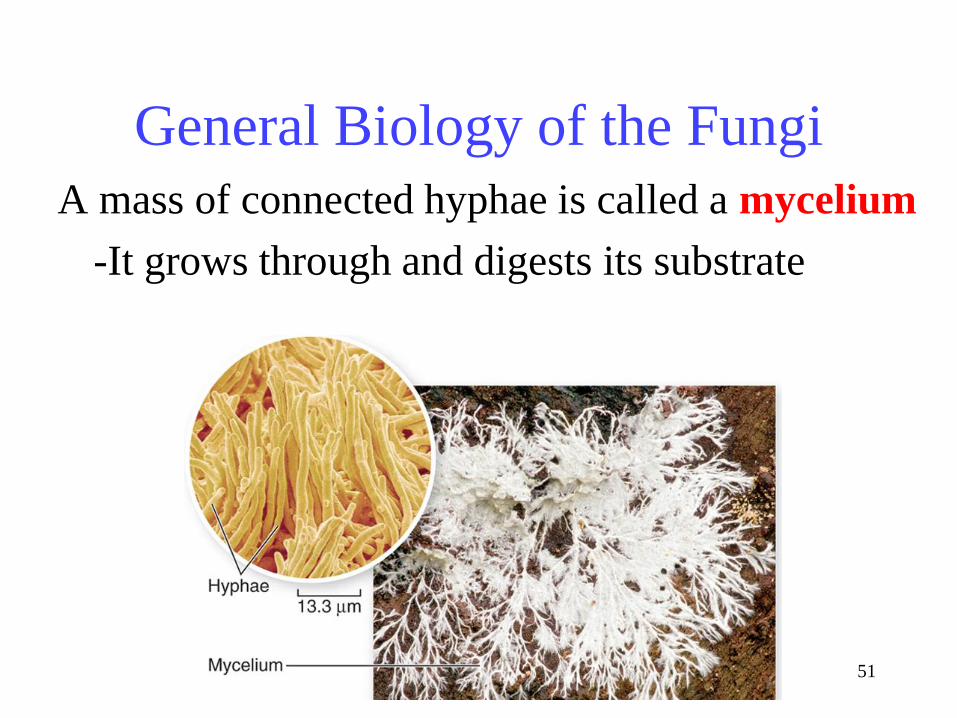
General Biology of the Fungi
A mass of connected hyphae is called a mycelium
-It grows through and digests its substrate
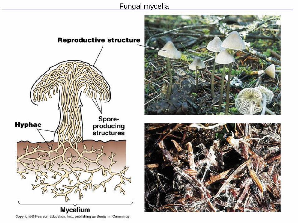
Fungal mycelia


54
Fungal Parasites and Pathogens
Largest Organism?
Armillaria –a pathogenic fungus – 8 hectares
Fungi Reproduction• spores are produced either sexually or asexually
• hyphae and spore nuclei are haploid
– except for a brief diploid stage that occurs during
sexual reproduction

Figure 31.3 Generalized life cycle of fungi (Layer 1)

Figure 31.3 Generalized life cycle of fungi (Layer 2)

Figure 31.3 Generalized life cycle of fungi (Layer 3)
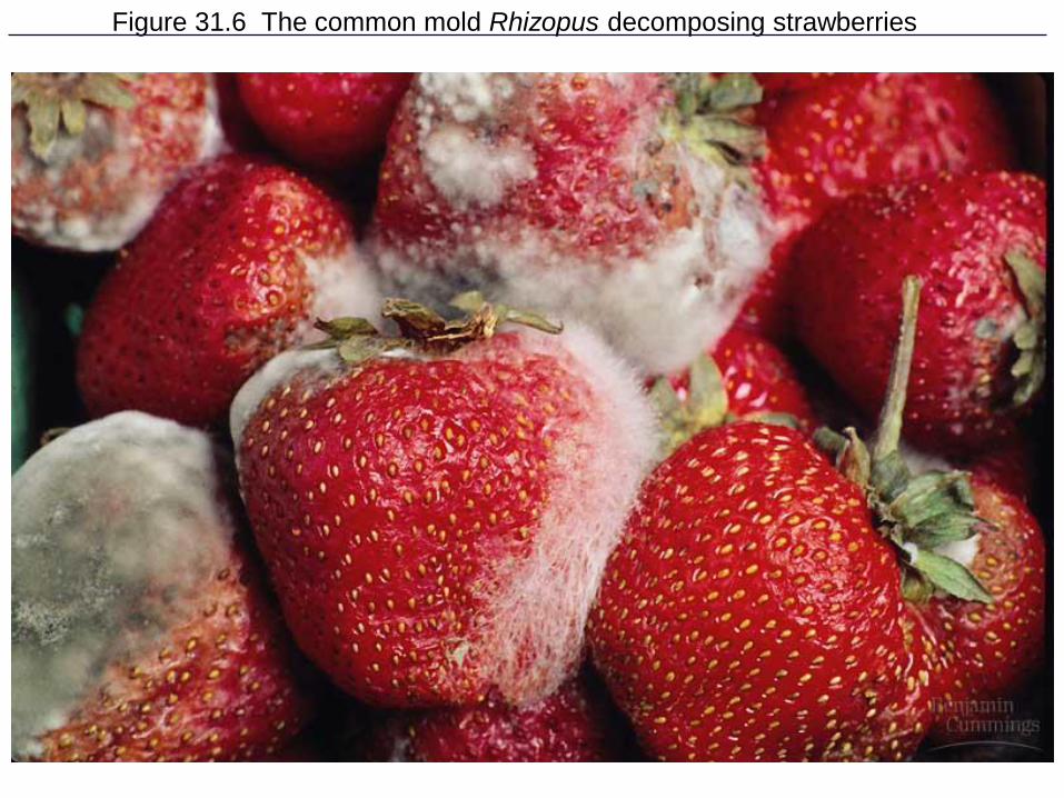
Figure 31.6 The common mold Rhizopus decomposing strawberries

60
Zygomycetes

Lichens
• Mutualism between fungi and algae or
cyanobacteria
• Sensitive to pollution due to absorption
capabilitues

Fig. 32.15
Copyright © The McGraw-Hill Companies, Inc. Permission required for reproduction or display.
a. b. c.
Fruticose Lichen Crustose LichenFoliose Lichen
a: © Ken Wagner/Phototake; b: © Robert & Jean Pollock/Visuals Unlimited; c: © Robert Lee/Photo Researchers, Inc.

Fig. 32.16
Copyright © The McGraw-Hill Companies, Inc. Permission required for reproduction or display.
Algal
cells
Fungal
hyphae
40 μm© Ed Reschke

Mycorrhizae
• Mutualism between
fungi and the roots of
90% of all vascular
plants
• Increases absorption
of phosphorous, zinc
& other nutrients