
Distribution of neuropeptide S receptor mRNA and ...usdbiology.com/cliff/Courses/Advanced Seminars...
19
Distribution of Neuropeptide S Receptor mRNA and Neurochemical Characteristics of Neuropeptide S- Expressing Neurons in the Rat Brain YAN-LING XU, 1 CHRISTINE M. GALL, 2 VALERIE R. JACKSON, 1,3 OLIVIER CIVELLI, 1,3 AND RAINER K. REINSCHEID 1,4 * 1 Department of Pharmacology, University of California Irvine, Irvine, California 92697 2 Department of Anatomy and Neurobiology, University of California Irvine, Irvine, California 92697 3 Department of Developmental and Cell Biology, University of California Irvine, Irvine, California 92697 4 Program in Pharmaceutical Sciences, University of California Irvine, Irvine, California 92697 ABSTRACT Neuropeptide S (NPS) and its receptor (NPSR) constitute a novel neuropeptide system that is involved in regulating arousal and anxiety. The NPS precursor mRNA is highly expressed in a previously undescribed group of neurons located between the locus coeruleus (LC) and Bar- rington’s nucleus. We report here that the majority of NPS-expressing neurons in the LC area and the principal sensory trigeminal nucleus are glutamatergic neurons, whereas many NPS- positive neurons in the lateral parabrachial nucleus coexpress corticotropin-releasing factor (CRF). In addition, we describe a comprehensive map of NPSR mRNA expression in the rat brain. High levels of expression are found in areas involved in olfactory processing, including the anterior olfactory nucleus, the endopiriform nucleus, and the piriform cortex. NPSR mRNA is expressed in several regions mediating anxiety responses, including the amygdaloid complex and the paraventricular hypothalamic nucleus. NPSR mRNA is also found in multiple key regions of sleep neurocircuitries, such as the thalamus, the hypothalamus, and the preoptic region. In addition, NPSR mRNA is strongly expressed in major output and input regions of hippocampus, including the parahippocampal regions, the lateral entorhinal cortex, and the retrosplenial agranular cortex. Multiple hypothalamic nuclei, including the dorsomedial and the ventromedial hypothalamic nucleus and the posterior arcuate nucleus, express high levels of NPSR mRNA, indicating that NPS may regulate energy homeostasis. These data suggest that the NPS system may play a key role in modulating a variety of physiological functions, especially arousal, anxiety, learning and memory, and energy balance. J. Comp. Neurol. 500:84 –102, 2007. © 2006 Wiley-Liss, Inc. Indexing terms: G protein-coupled receptor; in situ hybridization; expression pattern; locus coeruleus; glutamate; corticotropin-releasing factor; acetylcholine; GABA Neuropeptide S (NPS) and its receptor (NPSR) form a newly discovered neuropeptide system. In the rat, NPS is encoded by a precursor protein of 89 amino acids, contain- ing a typical amino-terminal signal peptide. The imma- ture NPS peptide sequence is preceded by a pair of basic amino acids (Lys-Arg) that are presumably cleaved by prohormone convertases to release the 20-amino-acid-long peptide (primary structure of rNPS: SFRNGVGS- GVKKTSFRRAKQ). Pharmacologically, NPS activates NPSR at low nanomolar concentration, induces mobiliza- tion of intracellular Ca 2 , increases intracellular levels of cAMP, and stimulates phosphorylation of mitogen- Grant sponsor: National Institutes of Health; Grant number: MH-71313 (to R.K.R.); Grant number: MH-60231 (to O.C.); Grant number: DK-63001 (to O.C.); Grant number: DK-70619 (to O.C.); Grant sponsor: National Alliance for Research on Schizophrenia and Depression (to R.K.R.). *Correspondence to: Rainer K. Reinscheid, Program in Pharmaceutical Sciences, University of California Irvine, 360 Med Surge II, Irvine, CA 92697-4625. E-mail: [email protected] Received 7 April 2006; Revised 16 June 2006; Accepted 31 July 2006 DOI 10.1002/cne.21159 Published online in Wiley InterScience (www.interscience.wiley.com). THE JOURNAL OF COMPARATIVE NEUROLOGY 500:84 –102 (2007) © 2006 WILEY-LISS, INC.
Transcript of Distribution of neuropeptide S receptor mRNA and ...usdbiology.com/cliff/Courses/Advanced Seminars...

Distribution of Neuropeptide S ReceptormRNA and Neurochemical
Characteristics of Neuropeptide S-Expressing Neurons in the Rat Brain
YAN-LING XU,1 CHRISTINE M. GALL,2 VALERIE R. JACKSON,1,3
OLIVIER CIVELLI,1,3AND RAINER K. REINSCHEID1,4*
1Department of Pharmacology, University of California Irvine, Irvine, California 926972Department of Anatomy and Neurobiology, University of California Irvine,
Irvine, California 926973Department of Developmental and Cell Biology, University of California Irvine,
Irvine, California 926974Program in Pharmaceutical Sciences, University of California Irvine,
Irvine, California 92697
ABSTRACTNeuropeptide S (NPS) and its receptor (NPSR) constitute a novel neuropeptide system that
is involved in regulating arousal and anxiety. The NPS precursor mRNA is highly expressed ina previously undescribed group of neurons located between the locus coeruleus (LC) and Bar-rington’s nucleus. We report here that the majority of NPS-expressing neurons in the LC areaand the principal sensory trigeminal nucleus are glutamatergic neurons, whereas many NPS-positive neurons in the lateral parabrachial nucleus coexpress corticotropin-releasing factor(CRF). In addition, we describe a comprehensive map of NPSR mRNA expression in the rat brain.High levels of expression are found in areas involved in olfactory processing, including theanterior olfactory nucleus, the endopiriform nucleus, and the piriform cortex. NPSR mRNA isexpressed in several regions mediating anxiety responses, including the amygdaloid complex andthe paraventricular hypothalamic nucleus. NPSR mRNA is also found in multiple key regions ofsleep neurocircuitries, such as the thalamus, the hypothalamus, and the preoptic region. Inaddition, NPSR mRNA is strongly expressed in major output and input regions of hippocampus,including the parahippocampal regions, the lateral entorhinal cortex, and the retrosplenialagranular cortex. Multiple hypothalamic nuclei, including the dorsomedial and the ventromedialhypothalamic nucleus and the posterior arcuate nucleus, express high levels of NPSR mRNA,indicating that NPS may regulate energy homeostasis. These data suggest that the NPS systemmay play a key role in modulating a variety of physiological functions, especially arousal, anxiety,learning and memory, and energy balance. J. Comp. Neurol. 500:84–102, 2007.© 2006 Wiley-Liss, Inc.
Indexing terms: G protein-coupled receptor; in situ hybridization; expression pattern; locus
coeruleus; glutamate; corticotropin-releasing factor; acetylcholine; GABA
Neuropeptide S (NPS) and its receptor (NPSR) form anewly discovered neuropeptide system. In the rat, NPS isencoded by a precursor protein of 89 amino acids, contain-ing a typical amino-terminal signal peptide. The imma-ture NPS peptide sequence is preceded by a pair of basicamino acids (Lys-Arg) that are presumably cleaved byprohormone convertases to release the 20-amino-acid-longpeptide (primary structure of rNPS: SFRNGVGS-GVKKTSFRRAKQ). Pharmacologically, NPS activatesNPSR at low nanomolar concentration, induces mobiliza-tion of intracellular Ca2�, increases intracellular levels ofcAMP, and stimulates phosphorylation of mitogen-
Grant sponsor: National Institutes of Health; Grant number: MH-71313(to R.K.R.); Grant number: MH-60231 (to O.C.); Grant number: DK-63001(to O.C.); Grant number: DK-70619 (to O.C.); Grant sponsor: NationalAlliance for Research on Schizophrenia and Depression (to R.K.R.).
*Correspondence to: Rainer K. Reinscheid, Program in PharmaceuticalSciences, University of California Irvine, 360 Med Surge II, Irvine, CA92697-4625. E-mail: [email protected]
Received 7 April 2006; Revised 16 June 2006; Accepted 31 July 2006DOI 10.1002/cne.21159Published online in Wiley InterScience (www.interscience.wiley.com).
THE JOURNAL OF COMPARATIVE NEUROLOGY 500:84–102 (2007)
© 2006 WILEY-LISS, INC.

activated protein kinase (Xu et al., 2004b; Reinscheid etal., 2005). Physiologically, central administration of NPSin rodents induces hyperlocomotion, increases arousal andwakefulness, and suppresses all stages of sleep. NPS ad-ministration also reduces anxiety-like behaviors in a bat-tery of four different tests that measure innate fear orresponses to novelty in rodents (Xu et al., 2004b). Anatom-ically, we have shown that NPS precursor mRNA is ex-pressed only in several discrete regions in the rat brain.The dorsomedial hypothalamus and amygdala containscattered NPS precursor mRNA signals. Strong expres-sion of NPS precursor mRNA was observed in three brain-stem regions, including the principle sensory trigeminalnucleus, lateral parabrachial nucleus, and locus coeruleus(LC) area. The NPS-expressing neurons in the LC area arelocalized ventromedial to the LC and caudolateral to Bar-rington’s nucleus, a neighboring nucleus of LC that isoften referred to as the pontine micturition center. Theunique anatomical pattern of this group of NPS-expressing neurons reveals a previously unmapped popu-lation of cells between Barrington’s nucleus and the LC. Inour previous study, we have demonstrated that neithernoradrenaline (NA), the major neurotransmitter synthe-sized in LC neurons, nor corticotropin-releasing factor(CRF), the major peptidergic transmitter in Barrington’snucleus, is expressed in the NPS-synthesizing cell cluster
in the pontine brainstem (Xu et al., 2004b). Thus, theidentity of any classical neurotransmitters in this novelgroup of neurons in the LC area is still unknown. It is wellestablished that neuropeptides usually do not exist aloneas neurotransmitters but colocalize with classical neuro-transmitters or with other neuropeptides and may playimportant roles in modulating the activities of theseneurotransmitters/neuromodulators (Hokfelt et al., 2000).
It has been reported that �-aminobutyric acid (GABA)-ergic neurons (Aston-Jones et al., 2004), cholinergic neu-rons (Sutin and Jacobowitz, 1988; Rizvi et al., 1994), andglutamatergic neurons (Stornetta et al., 2002; Varoqui etal., 2002; Moriyama and Yamamoto, 2004) are present inLC and the surrounding peri-LC area. In this study, wetherefore determined the chemical neuroanatomy of NPS-expressing cells in the rat brainstem, by using glutamateacid decarboxylase 67 (GAD67; Lauterborn et al., 1995),choline acetyltransferase (ChAT; Lauterborn et al., 1993),and vesicular glutamate transporters (VGLUTs; Smith etal., 2001; McCullumsmith and Meador-Woodruff, 2003) asmarkers for GABAergic, cholinergic, and glutamatergicneurons, respectively. This study will reveal for the firsttime the neurochemical profiles of NPS-producing neu-rons and the neurotransmitters/neuromodulators thatmay be coreleased with NPS peptide. Identification ofcoexisting transmitters in NPS-producing neurons will
Abbreviations
3V third ventricle4V fourth ventricleac anterior commissureaca anterior commissure, anterior partACo anterior cortical amygdaloid nucleusAH anterior hypothalamic areaAMV anteromedial thalamic nucleus, ventral partAON anterior olfactory nucleusApir amygdalopiriform transition areaaq aqueductArc P arcuate hypothalamic nucleus, posterior partCA1 CA1 region of the hippocampusCb cerebellumcc corpus callosumCg1 cingulate cortex, area 1Cl claustrumCLi caudal linear nucleus of rapheCM central medial thalamic nucleuscp cerebral peduncleDEn dorsal endopiriform nucleusDG dentate gyrusDMH dorsomedial hypothalamic nucleusDpG deep gray layer of superior colliculusDR dorsal rapheDTM dorsal tuberomammillary nucleusEn endopiriform nucleusf fornixHDB nucleus of the horizontal limb of the diagonal bandHpx hippocampusI intercalated nuclei of amygdalaIAM interanteromedial thalamic nucleusIPR interpeduncular nucleus, rostral partLC locus coeruleusLENt lateral entorhinal cortexLH lateral hypothalamic areaLPB lateral parabrachial nucleusLPO lateral preoptic areaLV lateral ventricleM2 motor cortex 2MeAD medial amygdala, anterodorsal partMePD/V medial amygdala, posterodorsal/ventralMnR median raphe nucleus
MPT medial pretectal nucleusOPT olivary pretectal nucleusopt optical tractox optic chiasmPAG periaqueductal grayPaMP medial parvicellular paraventricular hypothalamic nu-
cleusPaPo paraventricular hypothalamic nucleus, posterior partPaS parasubiculumPaV paraventricular hypothalamic nucleus, ventral partpc posterior commissurePeF perifornical nucleusPH posterior hypothalamic nucleusPir piriform cortexPLi posterior limitans thalamic nucleusPMCo posteromedial cortical amygdalaPMD premammillary nucleus, dorsal partPMV premammillary nucleus, ventral partPost postsubiculumPr5 principle sensory 5 nucleusPrC precommissural nucleusPrS presubiculumPT paratenial thalamic nucleusPtA parietal association cortexPVP paraventricular thalamic nucleus, posteriorRe reuniens thalamic nucleusRh rhomboid thalamic nucleusRSA retrosplenial agranular cortexS subiculumSCO subcommissural organscp superior cerebellar peduncleSG suprageniculate thalamic nucleusSNCD substantia nigra, compact part, dorsal tierSuG superficial layer of superior colliculusTg/PnO tegmental area/pontine reticular nucleus, oral partVLPO ventrolateral preoptic nucleusVMH ventromedial hypothalamic nucleusVMHDM ventromedial hypothalamic nucleus, dorsomedial partVTA ventral tegmental areaVTM ventral tuberomammillary nucleusXi xiphoid thalamic nucleus
The Journal of Comparative Neurology. DOI 10.1002/cne
85ANATOMICAL CHARACTERIZATION OF RAT NPS SYSTEM

help to develop new hypotheses about anatomical connec-tions of NPS neurons and possible regulatory roles of theNPS system.
Our previous study has demonstrated several represen-tative regions with high levels of NPS receptor mRNAexpression, such as anterior olfactory nucleus, motor cor-tex 2, amygdala, and hypothalamic regions (Xu et al.,2004b). However, the complete expression pattern of NPSreceptor mRNA in the rat brain has not been described indetail. To explore the distribution of NPS receptor mRNAin the rat central nervous system, we describe in thispaper a comprehensive anatomical mapping of NPSRmRNA in rat brain. Insofar as central administration ofNPS potently regulates anxiety and arousal, it is impor-tant to map the anatomical substrates of these effects andinvestigate interactions of the NPS system with otherneurotransmitter systems that are important in modulat-ing vigilance and anxiety. Furthermore, these studies willalso provide an anatomical basis for hypotheses aboutother physiological functions of the NPS system.
MATERIALS AND METHODS
Animals
Brain tissues from adult male Sprague-Dawley rats(250–300 g) were used for in situ hybridizations. Theanimals were housed under a 12-hour light-dark cycle,with lights on at 6:00 AM. Food and water were availablead libitum. The experimental procedures were approvedby the Institutional Animal Care and Use Committee andare consistent with federal regulations and guidelines forexperimentation on animals.
Tissue preparation
Briefly, animals were killed by decapitation, and theirbrains were quickly removed and frozen in methyl butanecooled to –20°C for 1 minute. Brains were stored at –80°Cuntil sectioning. Brains were sectioned coronally into20-�m slices with a cryostat set at –19°C and thaw-mounted on Vectabond-coated glass slides, followed byfixation in 4% paraformaldehyde, 0.1 M phosphate-buffered saline (PBS), pH 7.4, for 1 hour. Slides wererinsed briefly in 0.1 M phosphate buffer, followed by dis-tilled water, then dried at room temperature and stored at–80°C.
Probe synthesis
A 184-bp BamHI-NotI fragment of the rat NPS precur-sor cDNA (corresponding to nucleotides 92–276) was gen-erated by PCR and subcloned into pBluescript SK (Strat-agene, La Jolla, CA). Antisense or sense riboprobes weregenerated by using T7 or T3 RNA polymerase, respec-tively. A 326-bp NotI-BamHI fragment of the rat NPSreceptor cDNA (corresponding to nt 408–734) was clonedinto the same vector. Antisense or sense riboprobes of NPSreceptor were generated by using T3 or T7 RNA polymer-ase, respectively. Radiolabeled probes were prepared bytranscription in the presence of 35S-UTP (Amersham, Ar-lington Heights, IL). The labeled probes were separatedfrom unincorporated nucleotides on Sephadex G-50 col-umns (Roche Applied Science, Indianapolis, IN).
For double in situ hybridization, antisense and senseriboprobes of ChAT, GAD67, CRF, VGLUT1, VGLUT2, orVGLUT3 were labeled with digoxigenin (DIG) using a DIG
RNA labeling kit (Roche Applied Science, Indianapolis,IN). A 432-bp fragment of rat ChAT cDNA (correspondingto nucleotides 268–699) was cloned into pBluescript SK.Antisense or sense DIG-labeled ChAT cRNAs were syn-thesized with T7 or T3 RNA polymerase after HindIII orPvuII digestion of the pBSChAT vector, respectively (Lau-terborn et al., 1993). A fragment corresponding to nucle-otides 1,324–1,683 of cat glutamic acid decarboxylase(GAD)-67 cDNA was subcloned into pBluescript SK. An-tisense or sense DIG-labeled GAD67 cRNAs were synthe-sized with T7 or T3 RNA polymerase after BamHI orPvuII digestion of pBSGAD, respectively (Lauterborn etal., 1995). CRF riboprobes were synthesized as describedpreviously (Xu et al., 2004b). A 529-bp fragment corre-sponding to nucleotides 953–1,481 of human VGLUT1cDNA was subcloned into pCR-Blunt II TOPO (Invitrogen,Carlsbad, CA). Antisense or sense riboprobes of DIG-labeled VGLUT1 were synthesized with SP6 or T7 RNApolymerase after XhoI or HindIII digestion of the vector,respectively (Smith et al., 2001; McCullumsmith andMeador-Woodruff, 2003). A fragment corresponding to nu-cleotides 211–800 of rat VGLUT2 cDNA was subclonedinto pBluescript SK. Antisense or sense riboprobes ofVGLUT2 were synthesized with T7 RNA polymerase or T3RNA polymerase after BamHI or EcoRI digestion of thevector, respectively (Smith et al., 2001; McCullumsmithand Meador-Woodruff, 2003). A fragment correspondingto nucleotides 826–1,673 of human VGLUT3 cDNA wassubcloned into pCR-Blunt II TOPO. Antisense or senseDIG-labeled VGLUT3 riboprobes were synthesized withSP6 or T7 RNA polymerase after XhoI or SpeI digestion ofthe vector, respectively (McCullumsmith and Meador-Woodruff, 2003). Vectors containing VGLUT1, VGLUT2,and VGLUT3 were generously provided by Dr. James H.Meador-Woodruff (University of Michigan).
In situ hybridization
Sections for in situ hybridization were processed aspreviously described (Clark et al., 2001). First, sectionswere prehybridized, followed by proteinase K (0.75 �g/ml)treatment for 10 minutes; acetylated; and dehydratedthrough increasing concentrations of ethanol and then airdried. Next, hybridization buffer [50% formamide, 10%dextran sulfate, 0.3 M NaCl, 0.02% polyvinylpyrolidone,0.02% bovine serum albumin, 0.02% Ficoll, 10 mM dithio-threitol (DTT), 10 mM Tris, pH 8.0, 1 mM EDTA, pH 8.0,and 500 �g/ml tRNA] including 35S-labeled antisense orsense riboprobe of NPS precursor or NPSR (1 � 107 cpm/ml) was applied to the slides and incubated overnight at60°C. Sections were then washed four times with 4� SSC;treated with 20 �g/ml RNaseA for 30 minutes at 37°C; andwashed with 2� SSC/1 mM DTT, 1� SSC/1 mM DTT,0.5� SSC/1 mM DTT for 5–10 minutes each, followed by a30 minutes wash in 0.1� SSC/1 mM DTT at 68°C. Slideswere dehydrated by ethanol and air dried. The processedslides were then exposed to �-Max film (Kodak, Rochester,NY) for 6 days to check for positive hybridization signals.Finally, sections were dipped with NTB-2 emulsion(Kodak) and stored at 4°C for 6 weeks. Slides were thendeveloped with Kodak D-19, followed by rapid fixative.Cresyl violet was used to counterstain the sections. Slideswere then coverslipped and analyzed under a microscope.
The Journal of Comparative Neurology. DOI 10.1002/cne
86 Y.-L. XU ET AL.

Double in situ hybridization
Double in situ hybridization was carried out as de-scribed previously (Clark et al., 2001). Briefly, stock con-centrations of DIG-labeled ChAT, GAD, or VGLUT ribo-probes were determined by using dot blot hybridization,followed by optical density measurements. To determinethe optimal working concentrations for the various ribo-probes, each riboprobe was diluted serially with hybrid-ization buffer and hybridized to test sections first. Foreach probe, one concentration giving the strongest signalat minimal background was chosen for the experimentalsections. For experimental sections, a combination of iso-topic and colorimetric in situ hybridization was used.First, sections were prehybridized as described above.Then, DIG-labeled riboprobes of ChAT (0.4 mg/ml) orGAD67 (0.4 mg/ml) or a combination of riboprobes for allthree VGLUTs (each at 0.15 mg/ml) were mixed with35S-labeled NPS precursor riboprobe (1–2 � 107 cpm/ml)in hybridization buffer and incubated with sections at60°C overnight. After hybridization, sections underwentthe same posthybridization process as described above.Final stringent washes were for 30 minutes in 0.1 � SSCat 68°C for GAD67 and ChAT riboprobes or 0.3 � SSC at58°C for VGLUTs. After incubation with 0.1� SSC or 0.3�SSC at room temperature, sections were rinsed in GeniusBuffer 1 (GB1; 100 mM Tris-HCl, 150 mM NaCl, pH 7.5)for 1 minute and then incubated with blocking buffercontaining GB1, 0.25% Triton X-100, 5% nonfat dry milkfor 1 hour at room temperature. Alkaline phosphatase-conjugated sheep anti-DIG antibody (Roche Diagnostics,Indianapolis, IN) was diluted 1:5,000 in blocking solution,and 1 ml was applied to each slide, followed by incubationat 37°C for 3 hours. Tissues were then washed and incu-bated with color substrate nitroblue tetrazolium salt (0.5mg/ml) and 5-bromo-4-chloro-3-indolyl phosphate (0.187mg/ml) in GB3 (100 mM Tris-HCl, 100 mM NaCl, 50 mMMgCl2, pH 9.5) in the dark for 12–16 hours at RT. Sectionswere washed again and air dried overnight before beingexposed to film as described above. After the films weredeveloped, slides were dipped in 0.3% parlodin dissolvedin isoamylacetate for 5 seconds and dried overnight. Fi-nally, slides were dipped in photoemulsion and processedas described above.
Reference sections
A series of cryostat sections of rat brain tissue adjacentto the sections used for in situ hybridizations was collectedand mounted on coated microscope slides as describedabove. Sections were stained with cresyl violet, rinsed,and then coverslipped.
Data analysis
Sections were qualitatively analyzed with a microscopewith both light- and darkfield condensers (Axioskop 40;Carl Zeiss, Goettingen, Germany). Images were capturedwith Spot camera software version 2.2.2 (Diagnostic In-struments, Sterling Heights, MI), and the digital imageswere optimized for image resolution in Adobe Photoshop7.0. Anatomical structures were identified according toadult rat brain atlases (Paxinos, 1986; Swanson, 1999).The level of expression was scored as follows: ����, verystrong expression; ���, strong expression; ��, moderateexpression; �, widely scattered to low expression; –/�,
scattered expression slightly above background; –, no ex-pression.
RESULTS
Neurochemical characteristics of NPS-expressing neurons in the brainstem
Double-label in situ hybridization demonstrated thatthe NPS-expressing neurons in the LC area do not expressGAD67 mRNA (Fig. 1A,B), suggesting that these neuronsdo not produce GABA. Very small numbers of NPS mRNA-positive neurons in the lateral part of this cluster arecoexpressing ChAT mRNA (Fig. 1C), indicating the pres-ence of acetylcholine. The vast majority of NPS-expressingneurons in the locus coeruleus area coexpress VGLUTsmRNA (Fig. 1D), suggesting that these cells are glutama-tergic neurons. Small numbers of NPS mRNA neurons inthe LC area do not express tyrosine hydroxylase, GAD67,ChAT, or VGLUTs mRNA, indicating that neurotransmit-ters other than noradrenaline, GABA, acetylcholine, andglutamate may be present (Fig. 1B–D). Taken together,our data indicate that NPS-expressing neurons in the LCarea are predominantly glutamatergic, with a few cholin-ergic neurons found in the lateral part of this brainstemstructure. Another brainstem area with strong expressionof NPS precursor mRNA is the principle sensory trigemi-nal nucleus, which is known to contain a high density ofglutamatergic neurons. As shown in Figure 1E,F, manyNPS-expressing neurons of the principle sensory nucleusalso coexpress VGLUTs mRNA, indicating that these neu-rons use glutamate as neurotransmitter. The third brain-stem structure with a high number of NPS precursor-expressing cells is the lateral parabrachial nucleus. Byusing double in situ hybridization for NPS precursor andCRF mRNA, we found that many of the NPS-positiveneurons in the lateral parabrachial nucleus also coexpressCRF (Fig. 1G,H). Control hybridizations with NPS precur-sor sense probes showed no signals in any sections ana-lyzed (data not shown), indicating sufficient stringency ofhybridization conditions and specificity of the signals ob-tained with the antisense probes.
NPS receptor mRNA distribution
Regional expression of NPSR mRNA was studied by insitu hybridization with 35S-labeled riboprobes. No detect-able signal was found in sections hybridized with thesense probe. In contrast to NPS precursor mRNA, NPSRmRNA is found widely expressed in many brain regions. Asummary of NPSR mRNA distribution in various rat brainregions is shown in Table 1.
Telencephalon. High levels of NPSR mRNA expres-sion were found in the anterior olfactory nucleus, includ-ing medial, ventral, and posterior parts (Fig. 2A,B).Strong expression of NPSR mRNA was observed in en-dopiriform nucleus and deep layers of the piriform cortexin olfactory regions, and the dense labeling extended cau-dally throughout the entire cortical distribution of thesetwo regions (Figs. 2A–D, 3E, 5A–F). There was no detect-able signal in the main olfactory bulb (Table 1).
Strong NPSR mRNA expression was found in severaldistinct areas of the cerebral cortex. Densely labeled cellsexpressing NPSR mRNA were observed in motor cortex 2and parts of the cingulate cortex 1, but signals were ab-sent from cingulate cortex 2 (Fig. 3A,B). Retrosplenial
The Journal of Comparative Neurology. DOI 10.1002/cne
87ANATOMICAL CHARACTERIZATION OF RAT NPS SYSTEM

Figure 1
The Journal of Comparative Neurology. DOI 10.1002/cne

agranular cortex contained neurons strongly expressingNPSR mRNA, and the labeling extended into the adjacentparietal association cortex (Fig. 3C,D). Neurons in deeplayers of the amygdalopiriform transition area also ex-press high levels of NPSR mRNA (Fig. 3E,F). Scatteredcells with moderate to sparse labeling were observed inother cortical areas, including ventral and lateral orbitalcortex; motor cortex 1; insular cortex; parts of cingulatecortex 1 in the parietal cortical region; somatosensorycortex, including the barrel fields; temporal associationcortex; and auditory cortex (Table 1).
In the hippocampal formation and parahippocampal re-gions, prominent expression of NPSR mRNA was observedin the dorsal subiculum (Fig. 4A–D). Densely labeled cellswere also observed in both superficial and deep layers ofpostsubiculum (Fig. 4C,D). Presubiculum and parasubicu-lum also display substantial signals (Fig. 4E,F). However,no hybridization signals were found in hippocampusproper, including CA1–3 regions and the dentate gyrus(Fig. 4A–D, Table 1). Deep layers of the lateral entorhinalcortex also express moderate to high levels of NPSRmRNA (Fig. 4E,F).
TABLE 1. Summary of Regional Expression of NPSR mRNA in the Rat Brain
Region NPSR mRNA Region NPSR mRNA
Olfactory ThalamusMain bulb – Anteroventral/dorsal thalamic n. –Anterior olfactory n. ���� Interanteromedial thalamic n. ��Olfactory tubercle �/�� Paraventricular thalamic n. �/��Endopiriform n. ���� Reuniens thalamic n. ��Piriform cx ���/���� Rhomboid thalamic n. ��Cerebral cortex Precommissural n. ��/���Orbital cx � Subcommissural organ ����Claustrum ��/��� Paraventricular thalamic n. posterior ��/���Frontal cx –/� Central medial thalamic n. ��Insular cx � Central lateral thalamic n. �/–Motor cx 1 � Ventral anterior/lateral thalamic n. –Motor cx 2 ��� Ventral posteromedial/lateral thalamic n. –Cingulate cx 1 �� Posterior limitans thalamic n. ��Cingulate cx 2 – Suprageniculate thalamic n. ���Somatosensory cx �/�� Medial geniculate body �/��Parietal cx �� Lateral geniculate body �/–Occipital cx –/� HypothalamusTemporal cx � Medial preoptic n. –/�Retrosplenial agranular cx ��� Lateral preoptic n. ��Retrosplenial granular cx – Ventrolateral preoptic n. �Lateral entorhinal cx ��/��� Anterior hypothalamic area ���Amygdala and extended amygdala Paraventricular hypothalamic n. ��/���Anterior cortical amygdaloid n. ��� Posterior hypothalamic area ���Posteromedial cortical amygdala n. ��/��� Lateral hypothalamus area ��/���Posterolateral cortical amygdala n. –/� Perifornical n. ��Medial amygdala n. ��/��� Dorsomedial hypothalamic n. ���Intercalated n. of amygdala ���/���� Ventromedial hypothalamic n. ���/����Basolateral amygdala n. –/� Posterior arcuate hypothalamic n. ���Basomedial amygdala n. –/� Dorsal premammillary n. ���Lateral amygdala n. –/� Ventral premammillary n. ���Central amygdala n. – Ventral tuberomammillary n. ��/���Amygdalopiriform trans. area ��/��� Dorsal tuberomammillary n. ���Amygdalohippocampal area �/�� BrainstemBed n. stria terminalis � Medial pretectal n. ���Hippocampal formation Olivary pretectal n. ���CA1 – Superficial/deep gray layer superior colliculus ��CA2 – Inferior colliculus �/–CA3 – Periaqueductal gray ��/���Dentate gyrus – Dorsal raphe n. �Septal hippocampal – Caudal linear raphe n. ��Subiculum ���� Median/pontine raphe n. �/��Presubiculum ���� Ventral tegmental area ��Postsubiculum ���� Substantia nigra pars compacta ��Parasubiculum ���� Paranigral n. �/��Basal ganglia and septum Interpeduncular n. ��N.accumbens shell/core – Pontine reticular n, oral part ��Caudate putamen – Locus coeruleus –Globus pallidus –/� Barrington’s n. –Lateral/medial septal n. –/� Lateral parabrachial n. �N. horiz limb diagonal band, medial part �� Intermediate reticular n. �Magnocellular preoptic n. –/� Lateral reticular n. �
Cerebellum –
Fig. 1. Neurochemical profiles of NPS-expressing neurons in therat brainstem. A–D: NPS-expressing cell cluster in the locus coeruleus(LC) area. A: Representative section showing hybridization with an-tisense riboprobes for GAD67 (purple) and NPS precursor (blackgrains) mRNAs in the LC area. Locations of the fourth ventricle (4V),cerebellum (Cb), and superior cerebellar peduncle (scp) are indicated.B: Higher magnification of the cluster of NPS-expressing neurons inA, showing no colocalization of the hybridization signals. C: Adjacentsection of the LC area hybridized with antisense riboprobes for NPSprecursor (black grains) and ChAT (purple) mRNA. A single cellcoexpressing NPS precursor and ChAT mRNA is indicated by anarrow. D: Section hybridized with antisense riboprobes for VGLUTs1–3 (purple) and NPS precursor (black grains) mRNA. Arrows indi-cate cells coexpressing VGLUTs and NPS precursor mRNAs. E,F:Coexpression of VGLUTs (purple) and NPS precursor (black grains)mRNAs in neurons of the principle sensory 5 nucleus (Pr5). F is anadjacent section of the same area, and arrows indicated double-labeled cells. G,H: Coexpression of CRF (dark brown) and NPS pre-cursor (black grains) mRNAs in cells of the lateral parabrachial nu-cleus (LPB). Arrows in H indicate double-labeled cells, and thelocation of the superior cerebellar peduncle (scp) is given as a land-mark. Sections are oriented with medial areas on the right and lateralareas on the left. Scale bars � 250 �m in A,E,G; 100 �m in B–D,F,H.
The Journal of Comparative Neurology. DOI 10.1002/cne
89ANATOMICAL CHARACTERIZATION OF RAT NPS SYSTEM

The amygdaloidal complex shows strong to moderatesignals of NPSR mRNA expression in several divisions.High levels of expression are evident in neurons withinthe medial (Fig. 5A–F), anterior cortical (Fig. 5A–D), pos-teromedial cortical (Fig. 5E,F) and intercalated (Fig.5A–D) nuclei of the amygdala. No significant expressionwas observed in basolateral or lateral amygdala, exceptfor a few scattered neurons (Fig. 5A–F, Table 1).
Septal and basal forebrain regions generally expresslow levels of NPSR mRNA. The magnocellular preopticnucleus shows only weakly labeled cells. Scattered neu-rons in the bed nucleus of the stria terminalis containlow levels of NPSR mRNA. Only scant signals werefound in lateral and medial septum (Table 1). Basalganglia, including caudate, putamen, and globus palli-dus, show little hybridization signal. No NPSR mRNAexpression was observed in the nucleus accumbens (Ta-ble 1).
Diencephalon. In the thalamus, moderate to high lev-els of NPSR mRNA expression were found in various
areas. Multiple midline thalamic nuclei show significantlevels of expression, such as interanteromedial (Fig.6A,B), rhomboid (Figs. 6A,B, 8A,B), reuniens (Fig. 6A,B),anteromedial (Fig. 8A,B), and xiphoid (Fig. 8A,B) tha-lamic nuclei. Substantial hybridization signals were ob-served in the precommissural nucleus, subcommissuralorgan, and paraventricular thalamic nucleus (Fig. 6C–F).A moderate level of expression was also found in theposterior limitans thalamic nucleus (Fig. 6G,H). Substan-tial NPSR mRNA expression was detected in scatteredneurons of the medial geniculate body, with labeling ex-tending to a cluster of cells in the suprageniculate tha-lamic nucleus (Fig. 9C,D). Dispersed, moderately labeledcells are observed in the intralaminar nuclei (Fig. 6G,H).The major sensory relay nuclei of the thalamus, such asventral anterior, ventral lateral and ventroposteromedial/lateral thalamic nuclei, do not express NPSR mRNA (Ta-ble 1).
In preoptic regions, NPSR mRNA is expressed mainlyin the lateral preoptic area. Moderate labeling was as-
Fig. 2. Distribution of NPSR mRNA in olfactory regions. A,C: Darkfield images of sections hybridizedwith antisense 35S-labeled riboprobes for NPSR mRNA. B,D: Brightfield images of adjoining sectionsillustrated in A,C counterstained with cresyl violet, respectively. For abbreviations see list. Scale bar �500 �m.
The Journal of Comparative Neurology. DOI 10.1002/cne
90 Y.-L. XU ET AL.

Fig. 3. Expression of NPSR mRNA in cortical regions. A,C,E: Darkfield images of sections hybridizedwith antisense 35S-labeled riboprobe for NPSR mRNA. B,D,F: Brightfield images of adjoining sectionsshown in A,C,E counterstained with cresyl violet. For abbreviations see list. Scale bar � 500 �m.
The Journal of Comparative Neurology. DOI 10.1002/cne
91ANATOMICAL CHARACTERIZATION OF RAT NPS SYSTEM

Fig. 4. Expression of NPSR mRNA in hippocampal formation and parahippocampus. A,C,E: Dark-field images of NPSR mRNA expression. B,D,F: Brightfield images of adjacent sections shown in A,C,Ecounterstained with cresyl violet. For abbreviations see list. Scale bar � 500 �m.
The Journal of Comparative Neurology. DOI 10.1002/cne
92 Y.-L. XU ET AL.

sociated with scattered neurons of the lateral preopticarea and a cluster of neurons at the medial end of thenucleus of the horizontal limb of the diagonal band
(HDB; Fig. 7A,B), whereas, more caudally, this clusteris found at the interface of the ventrolateral preopticnucleus and the medial part of HDB (Fig. 7C,D).
Fig. 5. NPSR mRNA is expressed in multiple nuclei of the amygdala complex. A,C,E: Darkfieldimages of NPSR mRNA expression in amygdala complex. B,D,F: Brightfield images of adjoining sectionsshown in A,C,E counterstained with cresyl violet. For abbreviations see list. Scale bar � 500 �m.
The Journal of Comparative Neurology. DOI 10.1002/cne
93ANATOMICAL CHARACTERIZATION OF RAT NPS SYSTEM

Fig. 6. Expression of NPSR mRNA in thalamus and pretectal area. A,C,E,G: Darkfield images ofsections hybridized with antisense 35S-labeled riboprobe for NPSR mRNA. B,D,F,H: Brightfield imagesof adjacent sections shown in A,C,E,G counterstained with cresyl violet. For abbreviations see list. Scalebar � 500 �m.

NPSR mRNA signals were localized within multiplehypothalamic areas and nuclei. Moderate to strong levelsof expression were found in many neurons of the dorsalhypothalamus, including the paraventricular hypotha-lamic nucleus (Fig. 8A–D). Abundant signals were ob-served in the anterior (Fig. 8A,B), posterior, and lateralhypothalamic areas, including the perifornical nucleus(Figs. 6E,F, 8A,B,E,F). Dorsomedial and ventromedial hy-pothalamic nuclei express high levels of NPSR mRNA(Fig. 8C–F). The tuberomammillary area contains signif-icant amounts of NPSR mRNA, and many neurons in theposterior arcuate hypothalamic nucleus were well labeled(Fig. 8G,H). High levels of expression were found in boththe dorsal and the ventral premammillary nucleus and inthe dorsal tuberomammillary nucleus (Fig. 8G,H). Scattedneurons in the ventral tuberomammillary nucleus alsocontain significant levels of NPSR mRNA (Fig. 8G,H). Nohybridization signal was found in medial and lateralmammillary nuclei or in the supramammillary nucleus(Table 1).
Brainstem. NPSR mRNA was abundant in the medialpretectal nucleus and olivary pretectal nucleus in pretec-
tum regions (Fig. 6E–H). Moderate hybridization signalsalso labeled neurons in superficial and deep layers of thesuperior colliculus (Fig. 9A,D). The inferior colliculus con-tained only weak NPSR expression. Many neurons in theperiaqueductal gray (PAG), especially the medial PAG,were labeled with moderate intensity (Fig. 9A,B,E,F).
Scattered neurons in the caudal linear nuclei of raphecontained moderate NPSR mRNA signals (Fig. 10A,B).The dorsal raphe nucleus displays only weak hybridiza-tion signals (Fig. 9E,F). Weak to moderate levels of ex-pression were also found in scattered neurons of the me-dian (Fig. 10E,F) and pontine raphe (Table 1). A fewpositive neurons with moderate intensity of signals wereidentified in the ventral tegmental area and substantianigra pars compacta (Fig. 10C,D). Pontine tegmental re-gions in general contain scatted NPSR mRNA expression.The paranigral and interpeduncular nuclei of the ponsshow moderate to weak NPSR signals (Fig. 10A,B). Mod-erate signals were also observed in a few neurons in theoral part of the pontine reticular nucleus (Fig. 10E,F).Weak expression of NPSR mRNA was found in the lateralparabrachial nucleus as well as in the ventral, external,
Fig. 7. Expression of NPSR mRNA in preoptic area. A,C: Darkfield images of sections hybridized withantisense 35S-labeled riboprobes for NPSR mRNA; B,D: Brightfield images of adjacent sections shown inA,C counterstained with cresyl violet. For abbreviations see list. Scale bar � 500 �m.
The Journal of Comparative Neurology. DOI 10.1002/cne
95ANATOMICAL CHARACTERIZATION OF RAT NPS SYSTEM
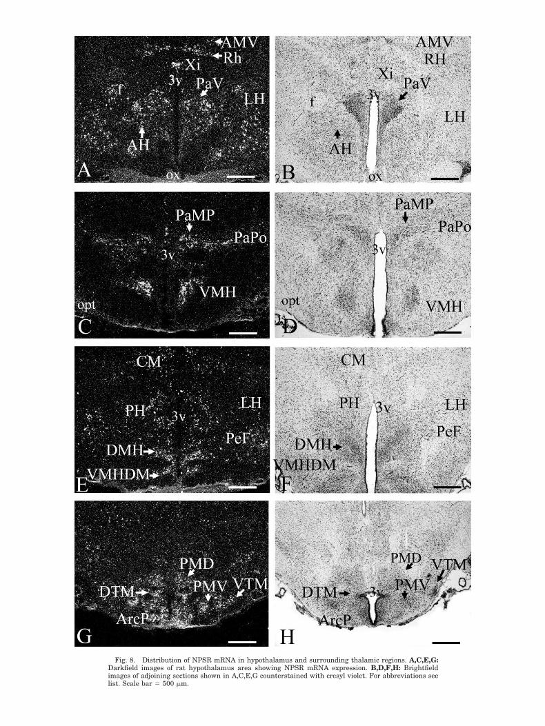
Fig. 8. Distribution of NPSR mRNA in hypothalamus and surrounding thalamic regions. A,C,E,G:Darkfield images of rat hypothalamus area showing NPSR mRNA expression. B,D,F,H: Brightfieldimages of adjoining sections shown in A,C,E,G counterstained with cresyl violet. For abbreviations seelist. Scale bar � 500 �m.

and dorsal part of medial parabrachial nuclei (Table 1). Afew neurons in facial nucleus express weak to moderatesignals (Table 1). Scattered neurons in intermediate and
lateral reticular nuclei show weak NPSR mRNA signals.Neurons of LC proper and surrounding pericoerulear ar-eas did not show measurable hybridization signals of
Fig. 9. Expression of NPSR mRNA in midbrain regions. A,C,E,F: Darkfield images of rat midbrainsections showing NPSR mRNA expression. B,D: Brightfield images of adjacent sections shown in A,Ccounterstained with cresyl violet. For abbreviations see list. Scale bar � 500 �m.
The Journal of Comparative Neurology. DOI 10.1002/cne
97ANATOMICAL CHARACTERIZATION OF RAT NPS SYSTEM

Fig. 10. Distribution of NPSR mRNA in tegmental area of the midbrain and pontine regions of thebrainstem. A,C,E: Darkfield images of sections displaying NPSR mRNA expression. B,D,F: Brightfieldimages of adjoining sections shown in A,C,E counterstained with cresyl violet. For abbreviations see list.Scale bar � 500 �m.
The Journal of Comparative Neurology. DOI 10.1002/cne
98 Y.-L. XU ET AL.

NPSR mRNA expression. No detectable hybridization wasobserved in cerebellum.
DISCUSSION
Here we describe the neurochemical profiles of NPS-synthesizing neurons in the rat pontine brainstem and adetailed map of NPSR expression throughout the adult ratbrain. Insofar as NPS is a very recently identified neu-ropeptide system, these data will provide a useful basis fornew hypotheses about physiological functions of NPS andits interactions with other neurotransmitter systems inthe brain.
Our results show that NPS precursor is coexpressedwith excitatory or stimulatory transmitters, such as glu-tamate, acetylcholine, and CRF. These observations pro-vide a neurochemical link to our initial findings showingthat central NPS administration promotes arousal andwakefulness (Xu et al., 2004b). They are also in line withour observation that NPS receptors produce stimulatoryintracellular signals, such as mobilization of Ca2� andincrease of cAMP (Reinscheid et al., 2005). Most of theNPS-expressing cells in the LC area and the trigeminalprinciple sensory nucleus are glutamatergic neurons, so itis likely that NPS may be coreleased together with gluta-mate from these neurons. Therefore, NPS may provideadditional excitatory input to the postsynaptic targets ofthese cells. It is also possible that functional interactionsbetween NPS receptors and glutamate receptors inpostsynaptic neurons might lead to enhanced glutamater-gic neurotransmission. Interactions between peptideG-protein-coupled receptors and glutamate receptors havebeen described for dopamine D1 receptors and N-methyl-D-aspartate (NMDA)-type glutamate receptors (Lee et al.,2002) and for prolactin-releasing peptide receptors andGluR2 or GluR3 AMPA receptors (Lin et al., 2001, 2002).Further studies investigating the interaction betweenNPS receptors and glutamate receptors will be necessaryto elucidate the role of the NPS system in excitatory neu-rotransmission. Similarly, the corelease of NPS and CRFfrom lateral parabrachial nucleus neurons might have afunctional correlate that warrants further studies.
The presence of glutamatergic and cholinergic neuronsin the area ventromedial to LC and dorsolateral to Bar-rington’s nucleus is consistent with previously publishedliterature. It has been reported that a few ChAT-positiveneurons were found in the area between Barrington’s nu-cleus and LC (Sutin and Jacobowitz, 1988; Rizvi et al.,1994). In situ hybridization studies with VGLUT2 probeshave shown that glutamatergic neurons are also localizedin this region (Stornetta et al., 2002). So far, the projec-tions of these NPS-containing glutamatergic neurons areunknown. Tracing experiments or immunohistochemicalanalysis of NPS peptide localization will be needed to mapthe projections of these neurons. The specific responsesthat these neurons may regulate are unknown at present.No significant signals of NPSR mRNA were observed inthis area. Therefore, it is unlikely that NPS coexpressed inthese glutamatergic neurons could modulate glutamaterelease via a presynaptic mechanism. We have shown thatcentral NPS administration promotes arousal and reducesanxiety. The LC and its major neurotransmitter noradren-aline (NA) are well known to regulate arousal and stressresponses. It has been reported that tonic discharges of LCneurons are virtually silent during REM sleep, low during
slow wave sleep, and highest during wakefulness (Hobsonet al., 1975; Foote et al., 1980). Also, the NA system playsan important role in mediating stress responses (Berridgeand Waterhouse, 2003). The absence of NPSR mRNA inLC neurons, however, indicates that NPS may not modu-late NA release from these cells via direct action. It istherefore possible that the LC area might contain twoneurochemically distinct systems, namely, noradrenalineand NPS containing, that are both involved in regulatingarousal and anxiety.
The glutamatergic neurons of the trigeminal principalsensory nucleus are known to transmit sensory informa-tion from orofacial fields to thalamocortical relays (Kil-lackey et al., 1995). The expression of NPS mRNA in theseneurons suggests that NPS neurotransmission might beinvolved in the regulation of sensory input into the tri-geminothalamic pathway.
The lateral parabrachial nucleus plays an importantrole in regulating autonomic functions and nociceptiveprocessing, in that it relays visceral afferent input fromthe nucleus of the solitary tract (Herbert and Saper, 1990)and pain stimuli from the spinal cord (Gauriau and Ber-nard, 2002) to the forebrain. It has been reported thatCRF-positive neurons are identified in the lateral para-brachial nucleus (Swanson et al., 1983; Herbert andSaper, 1990), which is consistent with our findings. Neu-rons of the lateral parabrachial nucleus have been re-ported to project to many brain regions, such as ventro-medial and paraventricular hypothalamic nuclei,amygdala, and periaqueductal gray matter (Gauriau andBernard, 2002), where NPSR mRNA are highly expressed.This indicates that the NPS system might also play im-portant roles in integrating and regulating behavioral oremotional responses to autonomic or nociceptive inputs.
Olfaction
Abundant NPSR mRNA was found in many regions thatare involved in olfaction. A high level of expression ofNPSR mRNA was found in anterior olfactory nucleus,endopiriform nucleus, and piriform cortex. Strong expres-sion is also evident in several nuclei of the amygdalacomplex that are functionally associated with the olfactorysystem, including medial, anterior cortical, and postero-medial cortical amygdala (Swanson and Petrovich, 1998).Scattered NPSR mRNA signals were also found in neu-rons of claustrum and insular cortex, where olfactory af-ferent pathways terminate (Shipley and Geinisman,1984). The high expression levels of NPSR mRNA in theseregions indicate that NPS may be involved in regulation ofperception and/or integration of olfactory or pheromonalinformation.
Modulation of anxiety
We have shown before that central administration ofNPS produces anxiolytic-like behaviors in mice using fourdifferent paradigms that measure behavioral responses tostress or fear, including open field, light-dark box, ele-vated plus maze, and marble burying tests. NPSR mRNAis expressed in various stress-related brain regions, suchas the amygdala, hypothalamus, raphe nuclei, and ventraltegmental area (Xu et al., 2004b).
The amygdala plays a major role in regulating stressresponses and processing of emotional memory (Swansonand Petrovich, 1998; McGaugh, 2004). NPSR mRNA isdensely expressed in the medial, anterior cortical, postero-
The Journal of Comparative Neurology. DOI 10.1002/cne
99ANATOMICAL CHARACTERIZATION OF RAT NPS SYSTEM
medial cortical, and intercalated nuclei of the amygdala.Neurons in these subdivisions project to either the centralor the basolateral amygdala, two amygdaloid regions as-sociated with integration of stress responses. The NPSR-expressing amygdala regions also project directly tostress-related regions outside the amygdaloid complex,including, most notably, the hypothalamus (Sah et al.,2003). NPSR mRNA is also expressed at low levels in thebed nucleus of the stria terminals, a region considered aspart of the extended amygdala that is strongly implicatedin modulating stress responses. Moderate to high levels ofNPSR expression were observed in several nuclei of thehypothalamus, including the paraventricular hypotha-lamic nucleus, which is one of the main centers of CRFsynthesis and an integrator of stress responses via thehypothalamus-pituitary-adrenal axis (Koob and Hein-richs, 1999; Heinrichs and Koob, 2004).
Aminergic neurons in the brain have been shown to playimportant roles in regulation of anxiety. Serotonergic (5-HT) neurons in raphe nuclei project widely to amygdala,frontal cortex, hippocampus, and hypothalamus and areinvolved in modulating anxious states (Millan, 2003).NPSR mRNA is expressed at moderate levels in raphenuclei, including the caudal linear nucleus, median raphe,and pontine raphe. NPSR mRNA is also expressed in theventral tegmental area, which is the origin of mesolimbicand mesocortical dopaminergic pathways. The dopaminer-gic pathways send input to the amygdala and play impor-tant roles in control of stress responses and other emo-tional processes (Millan, 2003). In summary, theexpression of NPSR mRNA in anxiety-associated neuro-circuits such as corticolimbic, hypothalamic, and impor-tant aminergic centers indicates that the NPS system mayinteract with other neurotransmitter systems, such asCRF, 5-HT, and dopaminergic systems in the modulationof anxiety and stress responses.
Arousal regulation
Our previous studies have shown that central adminis-tration of NPS potently suppressed paradoxical (REM)sleep and slow wave sleep 1 and 2 in rats. Conversely,NPS significantly increased the percentage of time spentin wakefulness during the first hour after injection. Inaddition, NPS induces hyperlocomotion in mice when in-jected centrally (Xu et al., 2004b). These data suggest thatNPS might be part of the brain arousal systems (Jones,2003). Indeed, significant expression of NPSR mRNA wasfound in several activating/arousal pathways. In the thal-amus, NPSR mRNA expression was detected in multiplemidline thalamic nuclei, such as interanteromedial, rhom-boid, and diffuse interlaminar group of thalamic nuclei,which relay extensive input from the brainstem reticularformation to cortical regions and are thus important inregulation of arousal (Van der Werf et al., 2002). In thehypothalamus, significant NPSR expression was found inthe perifornical region of the lateral and posteriorhypothalamic area, where another important arousal-promoting neuropeptide, hypocretin/orexin, is produced(Sutcliffe and de Lecea, 2002). Significant NPSR mRNAhybridization was localized in the ventral tuberomammil-lary nucleus, which produces histamine and is well knownto regulate vigilance states (Jones, 2003). In the brain-stem, NPSR is expressed in the substantia nigra and theVTA. These brain regions are important centers of dopa-mine synthesis, and activation of dopamine systems has
been shown to induce behavioral arousal and hyperloco-motion (Jones, 2003). Moderate NPSR mRNA signalswere also found in scattered neurons of the pontine retic-ular nucleus, a brainstem region known to activate wide-spread regions of the cortex (Jones, 2003).
In addition to these activating systems, NPSR mRNA isalso expressed in brain centers that promote sleep. NPSRmRNA was found in many neurons of the lateral preopticarea and in a cluster of neurons at the interface of theventrolateral preoptic nucleus (VLPO) and the medialpart of the nucleus of the HDB. It has been shown thatneurons in these regions use GABA as their neurotrans-mitter and are active during sleep and relatively inactiveduring wakefulness (Saper et al., 2001). Lesions of HDBwere found to suppress sleep, whereas electrical stimula-tion of HDB promotes non-REM sleep (Szymusiak andMcGinty, 1989). Many HDB neurons are thermosensitive,and local warming of the area facilitates non-REM sleep(Hays et al, 1997). The expression of NPSR mRNA in allthese regions suggests possible interaction of the NPSsystem with various activating as well as sleep-promotingsystems in the brain, with the overall result of increasingvigilance. Activation of NPS receptors may stimulatearousal-promoting systems such as glutamatergic, hista-minergic, dopaminergic, and hypocretin/orexin systemsand/or inhibit GABAergic systems in sleep centers such asthe VLPO and HDB. Further double in situ hybridizationexperiments are necessary to determine whether NPS recep-tor mRNA in these regions is coexpressed with these neuro-transmitters that are involved in sleep/wake regulation.
Learning and memory
NPSR mRNA is expressed at striking levels in majoroutput areas of the hippocampal formation, such as sub-iculum and posterior parahippocampal regions, includingpresubiculum and parasubiculum. It is also expressed bymajor afferents to the hippocampal formation, such aslateral entorhinal and endopiriform cortex. All of theseregions are critical in regulating learning and memory, inparticular, spatial learning. The subiculum sends out ma-jor hippocampal efferents to a wide range of cortical andsubcortical targets (Swanson and Cowan, 1977). Neuronsin the subiculum are stimulated specifically according toboth location and directional information (Sharp andGreen, 1994; Laxmi et al., 1999; Sharp et al., 2001). Sev-eral studies have shown that selective subiculum lesionscan impair spatial learning in rats (Morris et al., 1990;Laxmi et al., 1999). The posterior parahippocampus, in-cluding post-, para-, and presubiculum, receives inputfrom various brain regions that are involved in the asso-ciation of memory with visuospatial information, includ-ing CA1, subiculum, thalamus, basolateral amygdala, andretrosplenial cortex (Kohler, 1985; Van Groen and Wyss,1990, 1992, 1995). Projections of pre- and parasubiculummake up one of the major output pathways for memoryinformation processed in the hippocampus to the entorhi-nal cortex (Swanson and Cowan, 1977; Van Groen andWyss, 1990). So-called head direction cells representingthe actual position and angle of the head are found in thepresubiculum. Selective lesions of parahippocampal re-gions disrupt object recognition and spatial memory, es-pecially maintenance of spatial information over pro-longed periods of time, place memory, and object–placeassociations (Taube et al., 1990, 1992; Liu et al., 2004).NPSR mRNA is also expressed in deep layers of the lateral
The Journal of Comparative Neurology. DOI 10.1002/cne
100 Y.-L. XU ET AL.

entorhinal cortex, where outputs from the hippocampusprimarily reach the entorhinal cortex. Superficial layers ofthe entorhinal cortex are also known to integrate multi-modal sensory information from various cortical fields andto project back to hippocampal structures, such as thedentate gyrus region, via the perforant pathway (Amaraland Witter, 1995). Strong NPSR mRNA expression wasalso found in several cortical regions such as endopiriformcortex, piriform cortex, and retrosplenial agranular cor-tex, which project densely to the entorhinal cortex and arepresumed to relay multimodal sensory information fromother cortical regions (Amaral and Witter, 1995). Thestrong expression of NPSR in these areas indicates thatthe NPS system may modulate conveyance of informationin and out of the hippocampus and thereby might be a keyplayer in memory formation or consolidation, especiallyregarding spatial memory.
Energy balance
The hypothalamus is the predominant brain center reg-ulating energy homeostasis. NPS precursor and receptormRNAs are expressed in various nuclei in the hypothala-mus that are involved in feeding. Both NPS precursor andNPSR mRNA are expressed in the dorsomedial hypotha-lamic nucleus (DMH). The ventromedial hypothalamic nu-cleus (VMH) also contains strong NPSR mRNA hybridiza-tion signals. Lesion studies have shown that destruction ofDMH (Bernardis and Bellinger, 1998) or VMH (Shimizu etal., 1987) leads to hyperphagia, demonstrating that theseregions may act as “satiety” centers in regulating energybalance. In addition, significant amounts of NPSR mRNAare expressed in the posterior arcuate hypothalamic nu-cleus, where important orexigenic systems, such as neu-ropeptide Y (NPY) or agouti gene-related protein (AGRP),and anorexigenic systems, such as proopiomelanocortin(POMC)- or cocaine- and amphetamine-regulated tran-script (CART)-expressing neurons, are found (Flier andMaratos-Flier, 1998; Elias et al., 2001). Moderately denseNPSR mRNA labeling was also found in lateral hypotha-lamic areas and posterior hypothalamus, especially in theperifornical area, where two other neuropeptides,hypocretin/orexin (de Lecea et al., 1998) and melanin-concentrating hormone (MCH; Bittencourt and Elias,1998), are synthesized. Extensive studies have shown thatMCH acts as an orexigenic signal to induce food intakeand decrease energy expenditure (Rossi et al., 1997; forreview see Xu et al., 2004a). The hypocretin/orexin systemcan also modulate food intake, and it has been reportedthat central administration of hypocretin 1 can stimulatefood intake (Sakurai, 1999; Yamanaka et al., 2003). Theanatomical distribution of NPSR mRNA in the hypothal-amus indicates that the NPS system could modulate otherorexigenic and anorexigenic signals and play a role inregulating food intake and/or energy expenditure. Re-cently, it has been reported that acute central administra-tion of NPS (1 �g) decreased food intake in fasted ratsduring the first hour postinjection (Beck et al., 2005).Cumulative food intake during the first 3 hours was sig-nificantly reduced in rats injected with NPS at low doses(1 �g) and during 6 hours in rats injected with a high dose(10 �g) of NPS. NPS injection also significantly inhibitedspontaneous intake of high-energy diet in satiated ratsduring the first hour after injection. This study indicatesthat NPS could have a potent effect on inhibiting foodintake. However, the arousal-enhancing effect of NPS hasto be considered when effects of NPS on food intake are
evaluated. We have shown that central administration ofNPS induces wakefulness in rats and hyperlocomotion inmice during the first hour after injection. NPS-inducedinhibition of food intake was also observed, mainly duringthe first hour postinjection, and was followed by a reboundlasting for up to 3 hours postinjection in fasted rats. It istherefore possible that reduced food intake observed afteracute administration of NPS may be partially secondaryto the increased exploratory activity or vigilance inducedby NPS and not a direct effect on food-seeking behavior.
CONCLUSIONS
In summary, this study is the first report to demon-strate that the majority of NPS-expressing neurons lo-cated between LC and Barrington’s nucleus are glutama-tergic neurons. The present study also provides acomprehensive anatomical description of NPSR mRNAexpression in the rat brain. NPSR mRNA is expressed inmany brain regions, notably in olfactory regions, amyg-dala complex, and limbic regions, along with multiplearousal-activating systems and sleep-promoting regions,which supports a role of the NPS system in anxiety andvigilance regulation. NPS receptor mRNA is also ex-pressed significantly in the input and output regions of thehippocampal formation amd multiple hypothalamic re-gions, such as arcuate nucleus, VMH, and DMH, suggest-ing possible roles of NPS in modulating learning andmemory as well as effects on energy homeostasis. Thewidespread distribution of NPS receptor mRNA in thebrain indicates that the NPS system may be important inregulating a variety of physiological functions.
ACKNOWLEDGMENTS
We thank Steven H. Lin, Steward D. Clark, Katie Mai,Ethan Brenner, and Cynthia Huang for their technical sup-port. We also thank Dr. Meador-Woodruff and Dr. McCul-lumsmith for generously providing cDNA constructs forvGLUTs.
LITERATURE CITED
Amaral DG, Witter MP. 1995. Hippocampal formation. In: Paxinos G,editor. The rat nervous system. San Diego: Academic Press. p 443–493.
Aston-Jones G, Zhu Y, Card JP. 2004. Numerous GABAergic afferents tolocus ceruleus in the pericerulear dendritic zone: possible interneuro-nal pool. J Neurosci 24:2313–2321.
Beck B, Fernette B, Stricker-Krongrad A. 2005. Peptide S is a novel potentinhibitor of voluntary and fast-induced food intake in rats. BiochemBiophys Res Commun 332:859–865.
Bernardis LL, Bellinger LL. 1998. The dorsomedial hypothalamic nucleusrevisited: 1998 update. Proc Soc Exp Biol Med 218:284–306.
Berridge CW, Waterhouse BD. 2003. The locus coeruleus-noradrenergicsystem: modulation of behavioral state and state-dependent cognitiveprocesses. Brain Res Brain Res Rev 42:33–84.
Bittencourt JC, Elias CF. 1998. Melanin-concentrating hormone and neu-ropeptide EI projections from the lateral hypothalamic area and zonaincerta to the medial septal nucleus and spinal cord: a study usingmultiple neuronal tracers. Brain Res 805:1–19.
Clark SD, Nothacker HP, Wang Z, Saito Y, Leslie FM, Civelli O. 2001. Theurotensin II receptor is expressed in the cholinergic mesopontine teg-mentum of the rat. Brain Res 923:120–127.
de Lecea L, Kilduff TS, Peyron C, Gao X, Foye PE, Danielson PE, FukuharaC, Battenberg EL, Gautvik VT, Bartlett FS 2nd, Frankel WN, van denPol AN, Bloom FE, Gautvik KM, Sutcliffe JG. 1998. The hypocretins:hypothalamus-specific peptides with neuroexcitatory activity. ProcNatl Acad Sci U S A 95:322–327.
Elias CF, Lee CE, Kelly JF, Ahima RS, Kuhar M, Saper CB, Elmquist JK.
The Journal of Comparative Neurology. DOI 10.1002/cne
101ANATOMICAL CHARACTERIZATION OF RAT NPS SYSTEM

2001. Characterization of CART neurons in the rat and human hypo-thalamus. J Comp Neurol 432:1–19.
Flier JS, Maratos-Flier E. 1998. Obesity and the hypothalamus: novelpeptides for new pathways. Cell 92:437–440.
Foote SL, Aston-Jones G, Bloom FE. 1980. Impulse activity of locus coer-uleus neurons in awake rats and monkeys is a function of sensorystimulation and arousal. Proc Natl Acad Sci U S A 77:3033–3037.
Gauriau C, Bernard JF. 2002. Pain pathways and parabrachial circuits inthe rat. Exp Physiol 87:251–258.
Hays T, Morairty S, Szymusiak R, McGinty D. 1997. The effects of tem-perature and synaptic blockade in vitro on neurons of the horizontallimb of the diagonal band of Broca in the rat basal forebrain. Brain Res746:52–58.
Heinrichs SC, Koob GF. 2004. Corticotropin-releasing factor in brain: arole in activation, arousal, and affect regulation. J Pharmacol Exp Ther311:427–440.
Herbert H, Saper CB. 1990. Cholecystokinin-, galanin-, and corticotropin-releasing factor-like immunoreactive projections from the nucleus ofthe solitary tract to the parabrachial nucleus in the rat. J Comp Neurol293:581–598.
Hobson JA, McCarley RW, Wyzinski PW. 1975. Sleep cycle oscillation:reciprocal discharge by two brainstem neuronal groups. Science 189:55–58.
Hokfelt T, Broberger C, Xu ZQ, Sergeyev V, Ubink R, Diez M. 2000.Neuropeptides—an overview. Neuropharmacology 39:1337–1356.
Jones BE. 2003. Arousal systems. Front Biosci 8:s438–s451.Killackey HP, Rhoades RW, Bennett-Clarke CA. 1995. The formation of a
cortical somatotopic map. Trends Neurosci 18:402–407.Kohler C. 1985. Intrinsic projections of the retrohippocampal region in the
rat brain. I. The subicular complex. J Comp Neurol 236:504–522.Koob GF, Heinrichs SC. 1999. A role for corticotropin releasing factor and
urocortin in behavioral responses to stressors. Brain Res 848:141–152.Lauterborn JC, Isackson PJ, Montalvo R, Gall CM. 1993. In situ hybrid-
ization localization of choline acetyltransferase mRNA in adult ratbrain and spinal cord. Brain Res Mol Brain Res 17:59–69.
Lauterborn JC, Bizon JL, Tran TM, Gall CM. 1995. NGF mRNA is ex-pressed by GABAergic but not cholinergic neurons in rat basal fore-brain. J Comp Neurol 360:454–462.
Laxmi TR, Bindu PN, Raju TR, Meti BL. 1999. Spatial memory impair-ment in ventral subicular lesioned rats. Brain Res 816:245–248.
Lee FJ, Xue S, Pei L, Vukusic B, Chery N, Wang Y, Wang YT, Niznik HB,Yu XM, Liu F. 2002. Dual regulation of NMDA receptor functions bydirect protein–protein interactions with the dopamine D1 receptor. Cell111:219–230.
Lin SH, Arai AC, Wang Z, Nothacker HP, Civelli O. 2001. The carboxylterminus of the prolactin-releasing peptide receptor interacts with PDZdomain proteins involved in alpha-amino-3-hydroxy-5-methylisoxazole-4-propionic acid receptor clustering. Mol Pharmacol 60:916–923.
Lin SH, Arai AC, Espana RA, Berridge CW, Leslie FM, Huguenard JR,Vergnes M, Civelli O. 2002. Prolactin-releasing peptide (PrRP) promotesawakening and suppresses absence seizures. Neuroscience 114:229–238.
Liu P, Jarrard LE, Bilkey DK. 2004. Excitotoxic lesions of the pre- andparasubiculum disrupt the place fields of hippocampal pyramidal cells.Hippocampus 14:107–116.
McCullumsmith RE, Meador-Woodruff JH. 2003. Expression of transcriptsfor the vesicular glutamate transporters in the human medial temporallobe. Ann N Y Acad Sci 1003:438–442.
McGaugh JL. 2004. The amygdala modulates the consolidation of memo-ries of emotionally arousing experiences. Annu Rev Neurosci 27:1–28.
Millan MJ. 2003. The neurobiology and control of anxious states. ProgNeurobiol 70:83–244.
Moriyama Y, Yamamoto A. 2004. Glutamatergic chemical transmission:look! Here, there, and anywhere. J Biochem (Tokyo) 135:155–163.
Morris RG, Schenk F, Tweedie F, Jarrard LE. 1990. Ibotenate lesions ofhippocampus and/or subiculum: dissociating components of allocentricspatial learning. Eur J Neurosci 2:1016–1028.
Paxinos G. 1986. The rat brain in stereotaxic coordinates. New York:Academic Press.
Reinscheid RK, Xu YL, Okamura N, Zeng J, Chung S, Pai R, Wang Z, CivelliO. 2005. Pharmacological characterization of human and murine neu-ropeptide S receptor variants. J Pharmacol Exp Ther 315:1338–1345.
Rizvi TA, Ennis M, Aston-Jones G, Jiang M, Liu WL, Behbehani MM,Shipley MT. 1994. Preoptic projections to Barrington’s nucleus and thepericoerulear region: architecture and terminal organization. J CompNeurol 347:1–24.
Rossi M, Choi SJ, O’Shea D, Miyoshi T, Ghatei MA, Bloom SR. 1997.Melanin-concentrating hormone acutely stimulates feeding, butchronic administration has no effect on body weight. Endocrinology138:351–355.
Sah P, Faber ES, Lopez De Armentia M, Power J. 2003. The amygdaloidcomplex: anatomy and physiology. Physiol Rev 83:803–834.
Sakurai T. 1999. Orexins and orexin receptors: implication in feedingbehavior. Regul Pept 85:25–30.
Saper CB, Chou TC, Scammell TE. 2001. The sleep switch: hypothalamiccontrol of sleep and wakefulness. Trends Neurosci 24:726–731.
Sharp PE, Green C. 1994. Spatial correlates of firing patterns of single cellsin the subiculum of the freely moving rat. J Neurosci 14:2339–2356.
Sharp PE, Blair HT, Cho J. 2001. The anatomical and computational basisof the rat head-direction cell signal. Trends Neurosci 24:289–294.
Shimizu N, Oomura Y, Plata-Salaman CR, Morimoto M. 1987. Hyperpha-gia and obesity in rats with bilateral ibotenic acid-induced lesions ofthe ventromedial hypothalamic nucleus. Brain Res 416:153–156.
Shipley MT, Geinisman Y. 1984. Anatomical evidence for convergence ofolfactory, gustatory, and visceral afferent pathways in mouse cerebralcortex. Brain Res Bull 12:221–226.
Smith RE, Haroutunian V, Davis KL, Meador-Woodruff JH. 2001. Vesic-ular glutamate transporter transcript expression in the thalamus inschizophrenia. Neuroreport 12:2885–2887.
Stornetta RL, Sevigny CP, Guyenet PG. 2002. Vesicular glutamate trans-porter DNPI/VGLUT2 mRNA is present in C1 and several other groupsof brainstem catecholaminergic neurons. J Comp Neurol 444:191–206.
Sutcliffe JG, de Lecea L. 2002. The hypocretins: setting the arousal thresh-old. Nat Rev Neurosci 3:339–349.
Sutin EL, Jacobowitz DM. 1988. Immunocytochemical localization of pep-tides and other neurochemicals in the rat laterodorsal tegmental nu-cleus and adjacent area. J Comp Neurol 270:243–270.
Swanson LW. 1999. Brain maps: structures of the rat brain. Amsterdam:Elsevier.
Swanson LW, Cowan WM. 1977. An autoradiographic study of the organi-zation of the efferent connections of the hippocampal formation in therat. J Comp Neurol 172:49–84.
Swanson LW, Petrovich GD. 1998. What is the amygdala? Trends Neurosci21:323–331.
Swanson LW, Sawchenko PE, Rivier J, Vale WW. 1983. Organization ofovine corticotropin-releasing factor immunoreactive cells and fibers inthe rat brain: an immunohistochemical study. Neuroendocrinology 36:165–186.
Szymusiak R, McGinty D. 1989. Sleep suppression following kainic acid-induced lesions of the basal forebrain. Exp Neurol 94:598–614.
Taube JS, Muller RU, Ranck JB Jr. 1990. Head-direction cells recordedfrom the postsubiculum in freely moving rats. II. Effects of environ-mental manipulations. J Neurosci 10:436–447.
Taube JS, Kesslak JP, Cotman CW. 1992. Lesions of the rat postsubiculumimpair performance on spatial tasks. Behav Neural Biol 57:131–143.
Van der Werf YD, Witter MP, Groenewegen HJ. 2002. The intralaminarand midline nuclei of the thalamus. Anatomical and functional evi-dence for participation in processes of arousal and awareness. BrainRes Brain Res Rev 39:107–140.
Van Groen T, Wyss JM. 1990. The connections of presubiculum and para-subiculum in the rat. Brain Res 518:227–243.
Van Groen T, Wyss JM. 1992. Connections of the retrosplenial dysgranularcortex in the rat. J Comp Neurol 315:200–216.
Van Groen T, Wyss JM. 1995. Projections from the anterodorsal andanteroventral nucleus of the thalamus to the limbic cortex in the rat.J Comp Neurol 358:584–604.
Varoqui H, Schafer MK, Zhu H, Weihe E, Erickson JD. 2002. Identificationof the differentiation-associated Na�/PI transporter as a novel vesicu-lar glutamate transporter expressed in a distinct set of glutamatergicsynapses. J Neurosci 22:142–155.
Xu YL, Jackson VR, Civelli O. 2004a. Orphan G protein-coupled receptorsand obesity. Eur J Pharmacol 500:243–253.
Xu YL, Reinscheid RK, Huitron-Resendiz S, Clark SD, Wang Z, Lin SH,Brucher FA, Zeng J, Ly NK, Henriksen SJ, de Lecea L, Civelli O.2004b. Neuropeptide S: a neuropeptide promoting arousal andanxiolytic-like effects. Neuron 43:487–497.
Yamanaka A, Beuckmann CT, Willie JT, Hara J, Tsujino N, Mieda M,Tominaga M, Yagami K, Sugiyama F, Goto K, Yanagisawa M, SakuraiT. 2003. Hypothalamic orexin neurons regulate arousal according toenergy balance in mice. Neuron 38:701–713.
The Journal of Comparative Neurology. DOI 10.1002/cne
102 Y.-L. XU ET AL.


