Erythropoietin administration partially prevents adipose tissue loss ...
Distinct roles of erythropoietin, insulin-like growth factor I, andstem cell factor in the development of erythroid progenitor cells.
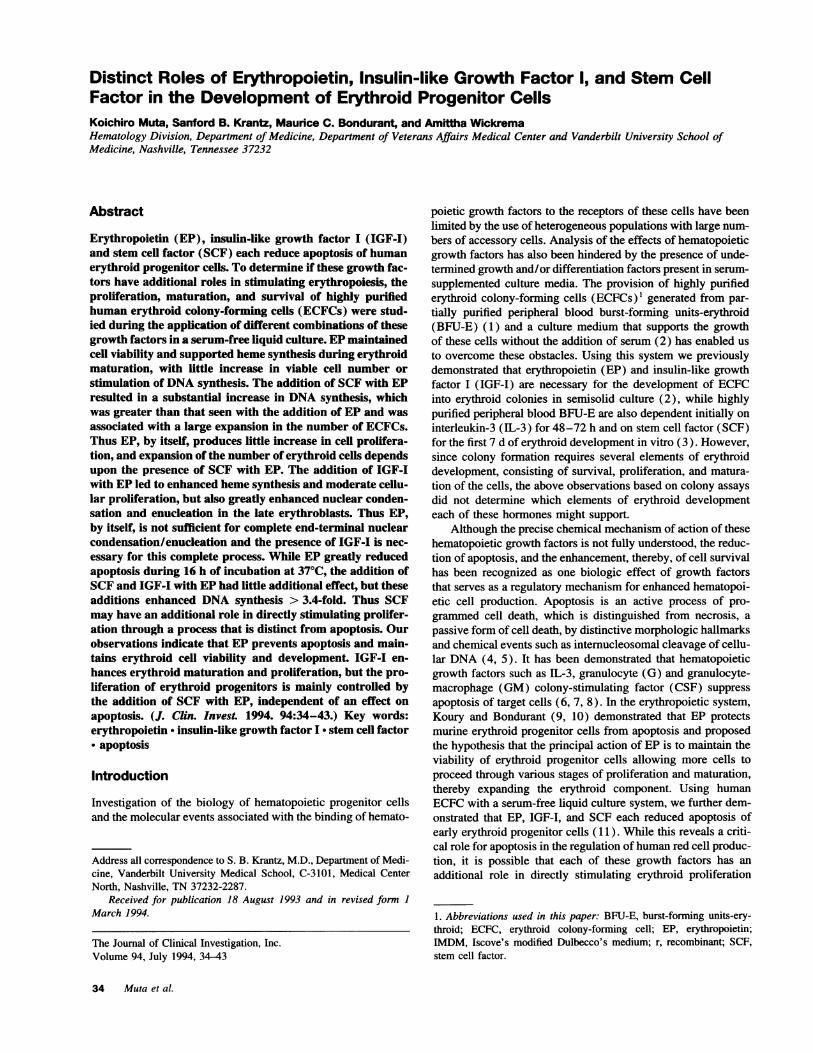
K Muta, … , M C Bondurant, A Wickrema
J Clin Invest. 1994;94(1):34-43. https://doi.org/10.1172/JCI117327.
Erythropoietin (EP), insulin-like growth factor I (IGF-I) and stem cell factor (SCF) each reduce apoptosis of humanerythroid progenitor cells. To determine if these growth factors have additional roles in stimulating erythropoiesis, theproliferation, maturation, and survival of highly purified human erythroid colony-forming cells (ECFCs) were studied duringthe application of different combinations of these growth factors in a serum-free liquid culture. EP maintained cell viabilityand supported heme synthesis during erythroid maturation, with little increase in viable cell number or stimulation of DNAsynthesis. The addition of SCF with EP resulted in a substantial increase in DNA synthesis, which was greater than thatseen with the addition of EP and was associated with a large expansion in the number of ECFCs. Thus EP, by itself,produces little increase in cell proliferation, and expansion of the number of erythroid cells depends upon the presence ofSCF with EP. The addition of IGF-I with EP led to enhanced heme synthesis and moderate cellular proliferation, but alsogreatly enhanced nuclear condensation and enucleation in the late erythroblasts. Thus EP, by itself, is not sufficient forcomplete end-terminal nuclear condensation/enucleation and the presence of IGF-I is necessary for this completeprocess. While EP greatly reduced apoptosis during 16 h of incubation at 37 degrees C, the addition of SCF and IGF-Iwith EP […]
Research Article
Find the latest version:
https://jci.me/117327/pdf
Distinct Roles of Erythropoietin, Insulin-like Growth Factor 1, and Stem CellFactor in the Development of Erythroid Progenitor CellsKoichiro Muta, Sanford B. Krantz, Maurice C. Bondurant, and Amittha WickremaHematology Division, Department of Medicine, Department of Veterans Affairs Medical Center and Vanderbilt University School ofMedicine, Nashville, Tennessee 37232
Abstract
Erythropoietin (EP), insulin-like growth factor I (IGF-I)and stem cell factor (SCF) each reduce apoptosis of humanerythroid progenitor cells. To determine if these growth fac-tors have additional roles in stimulating erythropoiesis, theproliferation, maturation, and survival of highly purifiedhuman erythroid colony-forming cells (ECFCs) were stud-ied during the application of different combinations of thesegrowth factors in a serum-free liquid culture. EPmaintainedcell viability and supported heme synthesis during erythroidmaturation, with little increase in viable cell number orstimulation of DNAsynthesis. The addition of SCFwith EPresulted in a substantial increase in DNAsynthesis, whichwas greater than that seen with the addition of EP and wasassociated with a large expansion in the number of ECFCs.Thus EP, by itself, produces little increase in cell prolifera-tion, and expansion of the number of erythroid cells dependsupon the presence of SCF with EP. The addition of IGF-Iwith EPled to enhanced heme synthesis and moderate cellu-lar proliferation, but also greatly enhanced nuclear conden-sation and enucleation in the late erythroblasts. Thus EP,by itself, is not sufficient for complete end-terminal nuclearcondensation/enucleation and the presence of IGF-I is nec-essary for this complete process. While EP greatly reducedapoptosis during 16 h of incubation at 370C, the addition ofSCFand IGF-I with EPhad little additional effect, but theseadditions enhanced DNA synthesis > 3.4-fold. Thus SCFmay have an additional role in directly stimulating prolifer-ation through a process that is distinct from apoptosis. Ourobservations indicate that EP prevents apoptosis and main-tains erythroid cell viability and development. IGF-I en-hances erythroid maturation and proliferation, but the pro-liferation of erythroid progenitors is mainly controlled bythe addition of SCF with EP, independent of an effect onapoptosis. (J. Clin. Invest. 1994. 94:34-43.) Key words:erythropoietin - insulin-like growth factor I * stem cell factor* apoptosis
Introduction
Investigation of the biology of hematopoietic progenitor cellsand the molecular events associated with the binding of hemato-
Address all correspondence to S. B. Krantz, M.D., Department of Medi-cine, Vanderbilt University Medical School, C-3101, Medical CenterNorth, Nashville, TN 37232-2287.
Received for publication 18 August 1993 and in revised form IMarch 1994.
The Journal of Clinical Investigation, Inc.Volume 94, July 1994, 34-43
poietic growth factors to the receptors of these cells have beenlimited by the use of heterogeneous populations with large num-bers of accessory cells. Analysis of the effects of hematopoieticgrowth factors has also been hindered by the presence of unde-termined growth and/or differentiation factors present in serum-supplemented culture media. The provision of highly purifiederythroid colony-forming cells (ECFCs)' generated from par-tially purified peripheral blood burst-forming units-erythroid(BFU-E) (1) and a culture medium that supports the growthof these cells without the addition of serum (2) has enabled usto overcome these obstacles. Using this system we previouslydemonstrated that erythropoietin (EP) and insulin-like growthfactor I (IGF-I) are necessary for the development of ECFCinto erythroid colonies in semisolid culture (2), while highlypurified peripheral blood BFU-E are also dependent initially oninterleukin-3 (IL-3) for 48-72 h and on stem cell factor (SCF)for the first 7 d of erythroid development in vitro (3). However,since colony formation requires several elements of erythroiddevelopment, consisting of survival, proliferation, and matura-tion of the cells, the above observations based on colony assaysdid not determine which elements of erythroid developmenteach of these hormones might support.
Although the precise chemical mechanism of action of thesehematopoietic growth factors is not fully understood, the reduc-tion of apoptosis, and the enhancement, thereby, of cell survivalhas been recognized as one biologic effect of growth factorsthat serves as a regulatory mechanism for enhanced hematopoi-etic cell production. Apoptosis is an active process of pro-grammed cell death, which is distinguished from necrosis, apassive form of cell death, by distinctive morphologic hallmarksand chemical events such as internucleosomal cleavage of cellu-lar DNA (4, 5). It has been demonstrated that hematopoieticgrowth factors such as IL-3, granulocyte (G) and granulocyte-macrophage (GM) colony-stimulating factor (CSF) suppressapoptosis of target cells (6, 7, 8). In the erythropoietic system,Koury and Bondurant (9, 10) demonstrated that EP protectsmurine erythroid progenitor cells from apoptosis and proposedthe hypothesis that the principal action of EP is to maintain theviability of erythroid progenitor cells allowing more cells toproceed through various stages of proliferation and maturation,thereby expanding the erythroid component. Using humanECFCwith a serum-free liquid culture system, we further dem-onstrated that EP, IGF-I, and SCF each reduced apoptosis ofearly erythroid progenitor cells ( 11 ). While this reveals a criti-cal role for apoptosis in the regulation of human red cell produc-tion, it is possible that each of these growth factors has anadditional role in directly stimulating erythroid proliferation
1. Abbreviations used in this paper: BFU-E, burst-forming units-ery-throid; ECFC, erythroid colony-forming cell; EP, erythropoietin;IMDM, Iscove's modified Dulbecco's medium; r, recombinant; SCF,stem cell factor.
34 Muta et al.
and/or erythroid maturation which has not been recognized( 12). To address this question we have made a detailed investi-gation of the effect of EP, IGF-I, and SCF on proliferation,maturation, and survival of ECFC in serum-free liquid suspen-sion culture. The results indicate that while EP mainly preventsapoptosis and supports orderly erythroid maturation, IGF-I en-hances terminal erythroid maturation plus proliferation and SCFsubstantially stimulates proliferation of erythroid progenitorcells without significant additional effects on apoptosis.
Methods
Blood. Blood was obtained from normal volunteers after informed con-sent. These studies were approved by the Vanderbilt University andNashville Department of Veterans Affairs Medical Center InstitutionalReview Boards. Approximately 400 ml of peripheral blood was col-lected in sodium heparin (Upjohn Co., Kalamazoo, MI) at a final con-centration of 20 U/ml.
Generation of ECFCs. ECFCs were prepared by a modified methoddescribed in detail by Sawada et al. ( 1). Light density mononuclear cellswere obtained by density centrifugation using Ficoll-Hypaque ( 1.077 g/cm3). Platelet depletion was accomplished by cell centrifugationthrough phosphate-buffered saline (PBS) containing 10% bovine serumalbumin (BSA; Intergen Co., Purchase, NY). This was followed by T-lymphocyte depletion using sheep erythrocyte rosetting. Adherent celldepletion was then performed by overnight incubation in polystyrenetissue culture flasks at 370C, followed by negative panning with CD1 lb/OKM* 1, CD2/OKT* 1 1, CD45/MY I 1, and CD16/MY23 antibodies topurify BFU-E to - 0.4%. The remaining cells were then cultured for5-6 d (day 6-7 cells) to generate ECFCs in 0.9% methylcellulosecontaining 30% fetal calf serum, 0.5% deionized BSA, recombinanthuman EP (rEP; 2 U/ml; Amgen Inc., Thousand Oaks, CA), recombi-nant human IL-3 (50 U/ml; Amgen Inc.) l0-4 M 2-mercaptoethanol,penicillin 500 U/ml, streptomycin 40 pg/ml, and Iscove's ModifiedDulbecco's Medium (IMDM; Sigma Chemical Co., St. Louis, MO) at37°C, in a high humidity 5% C02/95% air incubator.
The cells were then collected and ECFCs were further enriched bycentrifugation through 10% BSA and then over Ficoll-Hypaque, fol-lowed by incubation at 37°C for 1 h to remove adherent cells. Thispreparation generally resulted in 94% viable cells of which 30-90%,referred to here as ECFCs, gave rise to colonies of 2-500 hemoglo-binized cells in the plasma clot system. In some experiments, ECFCswere further incubated at 2 x 10⁵ cells/ml in liquid medium consistingof IMDM, 15% FCS, 15% human AB serum, 0.5% human serum albu-min (HSA), 2 U/ml rEP, penicillin, and streptomycin to obtain moremature erythroid cells. At the indicated times (days 7-12), the cellswere collected by centrifugation at 1,000 g for 5 min and were washedwith IMDM. The cells were then resuspended in IMDM containing0. 1%BSAand incubated at 37°C for 1 h. After centrifugation over PBScontaining 10% BSA, the cells were then washed with IMDMand usedfor further experiments.
Cell culture. Purified ECFCs were incubated at 37°C in serum-free liquid medium consisting of 0.5% deionized, delipidated, dialyzedcrystalline BSA(C-BSA-3D), iron-saturated transferrin (Sigma Chemi-cal Co.; 300 ,ug/ml), lipid suspension (oleic acid, 2.8 ,g/ml; L-a-phosphatidylcholine, 4.0 pg/ml; cholesterol, 3.9 ,ug/ml; Sigma Chemi-cal Co.), penicillin, streptomycin, and 50% IMDM/50% F-12 (HAM)(Sigma Chemical Co.), which were prepared as previously described(2). When the rate of 59Fe incorporation into heme was measured, theconcentration of transferrin was reduced to 100 yig/ml. In some cultures,rEP, recombinant human IGF-I (rIGF-I; Intergen Co.), and/or recombi-nant human SCF (rSCF; Amgen Inc.) were added. When ECFCs werecultured further than day 11, the cells were collected by centrifugationat 1,000 g and recultured in fresh media every 3-4 d. The cell concentra-tion was reduced to no more than 106/ml at each medium change.
The number of ECFCs after 40 h of incubation in serum-free liquidculture was determined by adding 200-800 cells to 0.2 ml of serum
medium for plasma clot assay after 7 d of incubation as previouslydescribed (1, 2). Colonies of two or more hemoglobinized cells werescored as ECFC. The number of erythroid colonies per microliter at theonset of the 40-h liquid culture was designated as 100%. The erythroidcolony-forming capacity of day 14 cells was similarly determined andwas expressed as the percentage of erythroid colonies formed per cellnumber plated. The purity of the ECFCs in each experiment was deter-mined by inoculating 200 cells in plasma clots prepared in the same
manner and counting four replicates.Determination of cell viability, benzidine positivity, and morphol-
ogy. The viable cell number was determined by trypan blue exclusionusing a hemacytometer. Aliquots of the cells were removed for cytocen-trifugation onto glass slides at the indicated times on days 7-14. Theslides were stained with 3,3' dimethoxybenzidine and hematoxylin, andwere examined by light microscopy. All the mononuclear cells thatstained with benzidine were identified using a magnification of 1,000,and the percentage of benzidine positive cells was determined by count-
ing 400 cells. The net benzidine positive cell number was calculatedfrom the total cell number and the percentage of benzidine positivecells.
Immunofluorescence. To determine the expression of spectrin as a
marker for the erythroid lineage, immunofluorescence using anti-ery-throid spectrin antibody (Sigma Chemical Co.) was performed. The day14 cells were washed with IMDM and allowed to adhere to an alcianblue-coated cover slip for 15 min at 370C. The coverslips were dippedin 2.0% formaldehyde in PBS containing 0.1% DMSOfor 8 min fol-lowed by washing in PBS for 1 h and the cells were then permeabilizedby dipping in 0.5% Triton X-100 in PBS for 5 min. Incubation withrabbit anti-human erythrocyte spectrin (Sigma Chemical Co.) was per-formed at room temperature for 45 min, followed by incubation with a
goat anti-rabbit IgG secondary antibody conjugated to FITC (SigmaChemical Co.). Photomicrographs were taken using an Olympus BH-2microscope equipped with epifluorescence optics. For a negative control,peripheral blood BFU-E were purified as previously described (3) andwere cultured in serum-free media containing rIL-3 (100 U/ml), rSCF(50 ng/ml), and rEP (lU/ml) for 2 d to generate day 3 BFU-E.
59Fe incorporation into heme. The effect of growth factors on hemesynthesis were assessed by measuring 59Fe incorporation into heme as
previously described (13). The ECFCs (3 x 10⁵ day 6 or 7 cells) wereincubated in 0.5 ml of serum-free media with various combinations ofgrowth factors at 37°C. At the indicated times, 50 ,l of a solution ofhuman transferrin (2.4 mg/ml; Calbiochem-Boehring, La Jolla, CA)containing 0.5 pCi of 59FeCl3 (54 mCi/mg; NewEngland Nuclear Corp.,Boston, MA) was added directly to the culture. After 4 h the cells werecollected by centrifugation at 1,000 g for 5 min and were washed withIMDM. The cells were lysed in Drabkin's solution which was thenacidified with HCI, and this was followed by extraction of heme usingcyclohexanone. The radioactivity of 59Fe in the cyclohexanone fractionwas measured in a gammacounter and expressed as cpm/ml culture oras a percentage of radioactivity in the cultures with rEP alone.
To determine the growth factor requirements for the late stagesof erythroid maturation, day 9-12 cells were harvested from serum-supplemented liquid cultures and 3 x 10⁵ cells were further incubatedwith 0.125 pCi of 59Fe-transferrin in 0.5 ml of serum-free liquid mediumwith various combinations of growth factors for 24 h at 37°C. Measure-ments of accumulated 59Fe-heme were performed as described above.
Measurement of DNAsynthesis. The effect of various growth factorson proliferation of ECFCswas determined by measuring DNAsynthesisusing [3H] thymidine as previously described (9). Briefly, 0.5 ml serum-free cultures with 105 ECFCs were established with various combina-tions of growth factors and were incubated with 1.25 ,uCi of [3H]-thymidine (6.7 Ci/mmol; New England Nuclear Corp.) for 1 h at theindicated times. The cells were then collected by centrifugation at 1,000g for S min, washed with IMDM, and placed in 10% ice-cold trichloro-acetic acid (TCA). The cell precipitates were collected on glass filterdiscs (type GF/A; Whatman, Maidstone, England), washed with 5%ice-cold TCA, followed by 95% ethanol, air dried, placed in a scintillantand counted in a scintillation counter. [3H]I thymidine incorporation was
Relative Effects of EP, IGF-I, and SCFon Erythroid Progenitor Cells 35

I00)
-
cx
-J
w
0'U
I.-
0A.
zaNz'U0o
E0
(0-J-JLa
LU
o0C.Lu
NzLU0o
3
2
7 a 9 10 11
DAYS IN CULTURE
9
3
2
7 a 9 10 11
DAYS IN CULTURE
expressed as cpm/ml of culture or as a percentage of cellular radioactiv-ity in the cultures with rEP, rIGF-I, and rSCF.
Analysis of ECFCapoptosis. To quantitate growth factor protectionof ECFCs from DNA fragmentation, the amount of uncleaved DNAwas measured as described by Koury and Bondurant (10). The ECFCswere preincubated in IMDM containing 30% FCS and 0.5% HSA, at370C, for 30 min. [3H]Thymidine (0.5 IsCi/ml) was added to 106 cells/ml and incubation was carried out for a further 30 min. The cells werecollected, centrifuged through PBS containing 10% BSA at 1,000 g,40C, for 5 min, and washed with IMDM. Replicate [3H]thymidine-labeled cells (5 x 10⁵) were then incubated at 370C in 1 ml of serum-free liquid medium containing thymidine and deoxycytidine (20 1iM;Sigma Chemical Co.) with various combinations of growth factors. Afterincubation for 16 h, the cells were collected and the DNAwas extractedand analyzed by electrophoresis on alkaline 0.6% agarose gels as pre-viously described (10, 11). Each lane was cut into 16 5-mm fractionsand the radioactivity of each fraction was determined. The sum of theradioactivity in fractions 1-4 was considered as the amount of uncleavedDNAand was expressed as a percentage of the total radioactivity ( 11).Data are presented as mean±SD and significance was calculated usingthe t test.
Results
Effect of rEP, rIGF-I, and rSCF on growth of ECFCs in serum-free liquid culture. Fig. 1 A shows the effect of rEP, rIGF-I,
Figure 1. Effect of rEP, rIGF-I, and rSCF on growthof ECFCs. ECFCs (day 7 cells) were incubated in
A// a serum-free liquid medium with rEP (1 U/ml), rIGFT/ a{/ , (100 ng/ml), and/or rSCF (100 ng/ml) (A and B),
or with rEP plus rIGF-I and/or rSCF (C and D)..__________,_____,____.___ Viable cells (A and C) and benzidine positive cells7 a 9 10 11 (B and D) were enumerated. The data are the mean
(±+SD) of four replicates from two experiments. TheDAYS IN CULTURE purity of the ECFCwas 74+10%, and 53±10%.
and rSCF on the number of viable cells when day 7 ECFCswere incubated in serum-free liquid culture. The majority ofthe cells cultured with rEP alone remained viable through day11, but no increase in the number of viable cells was evident.rIGF-I, and/or rSCF, increased the number of viable cells abovethat seen in the cultures without rEP for 2 d (P < 0.05), butbeyond that time, rEP alone maintained cell viability signifi-cantly more than rIGF-I and/or rSCF (P < 0.01). When thenumber of benzidine positive cells was determined (Fig. 1 B),an increasing number of ECFCincubated with rEP alone turnedpositive due to the accumulation of hemoglobin during erythroidmaturation, even though the number of viable cells did notincrease. By day 11, the number of benzidine positive cells wasapproximately equal to the number of viable cells. Increases inthe number of benzidine positive cells were also seen with theaddition of rIGF-I (P < 0.05 on days 8-11 ), rSCF (P < 0.05on day 9), or rIGF-I plus rSCF (P < 0.05 on days 8-11). Onday 9 the number of benzidine positive cells seen in the cultureswith rEP was similar to those seen in the cultures with rIGF-Iand/or rSCF, but the former increased after day 9, and wassignificantly greater (P < 0.01), while the latter declined.
Fig. 1 further shows the number of viable cells (C) andbenzidine positive cells (D) in cultures incubated with rIGF-Iand/or rSCF together with rEP. While rEP alone promoted little
5
4
3
2
EI0
(0co
-J-JLU0Lu0i
B --> None*-* rEP&-- rlGF-I
--a rSCFv-@ rlGF-I + rSCF
A O--@ NonerEP- rlGF-I
* rSCF* O rlGF-I + rSCF
.z*~~~~~~T*o.I . . .
0
4
7 a 9 10 11
DAYS IN CULTURE
31EI10
-
-JuJ
LU0
0-* rEF-- rEP + rlQF-I
w*- rEP + rSCFv - rEP + rlGF-I + rSCF
A
21
0
36 Muta et al.
l
I
A
; a
C
II
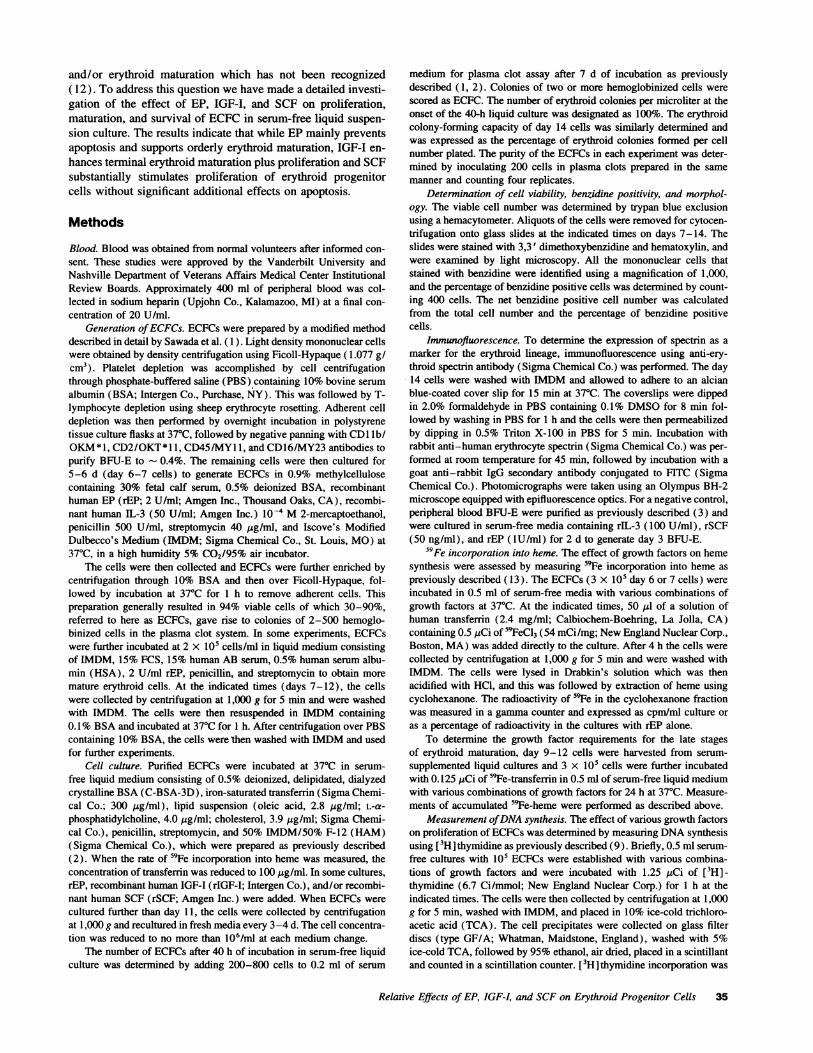
Figure 2. Morphology of ECFCon day 14 of serum-free liquid culture. ECFC(day 7 cells) were incubated with rEP (A), rEP plus rIGF-I (B),rEP plus rSCF (C), or rEP plus rIGF-I and rSCF (D) for 7 d, followed by staining with benzidine and hematoxylin. The purity of the ECFCwas56+5%. Bar, 10 Itm.
cellular proliferation, the addition of rIGF-I (P < 0.01 ) or rSCF(P < 0.01) with rEP each resulted in a significantly greaternumber of viable cells compared to the cultures with rEP alone(Fig. 1 C). Analysis of the viable cell number in the cultureswith rEP plus rIGF-I from six experiments revealed a 4.3-foldincrease on day 11 (P < 0.01) and an 8.0-fold increase on day14 (P < 0.01) compared to the viable cell number on day 7.The addition of rSCF plus rEP resulted in a much larger numberof viable cells than that of rIGF-I plus rEP (P < 0.05), andthe addition of both factors together with rEP produced an evengreater number of viable cells than that seen when each factoralone was added to rEP (P < 0.05; days 9-11). The increasein the number of benzidine positive cells was parallel to theincrease in viable cell number during the later days of culture(Fig. 1 D) and the addition of rIGF-I and/or rSCF plus rEPresulted in a greater number of benzidine positive cells thanthat with rEP alone (P < 0.05 on day 11).
After reculture in fresh medium on day 11, the cells werefurther incubated through day 14 with rEP alone or with rEPplus rIGF-I and/or rSCF for examination of morphology (Fig.2, A-D). At the beginning of the cultures on day 7, ECFCshowed the characteristic morphology of immature erythroidcells as previously described (1). While the majority of thecells cultured with rEP plus rIGF-I were hemoglobinized (86%;
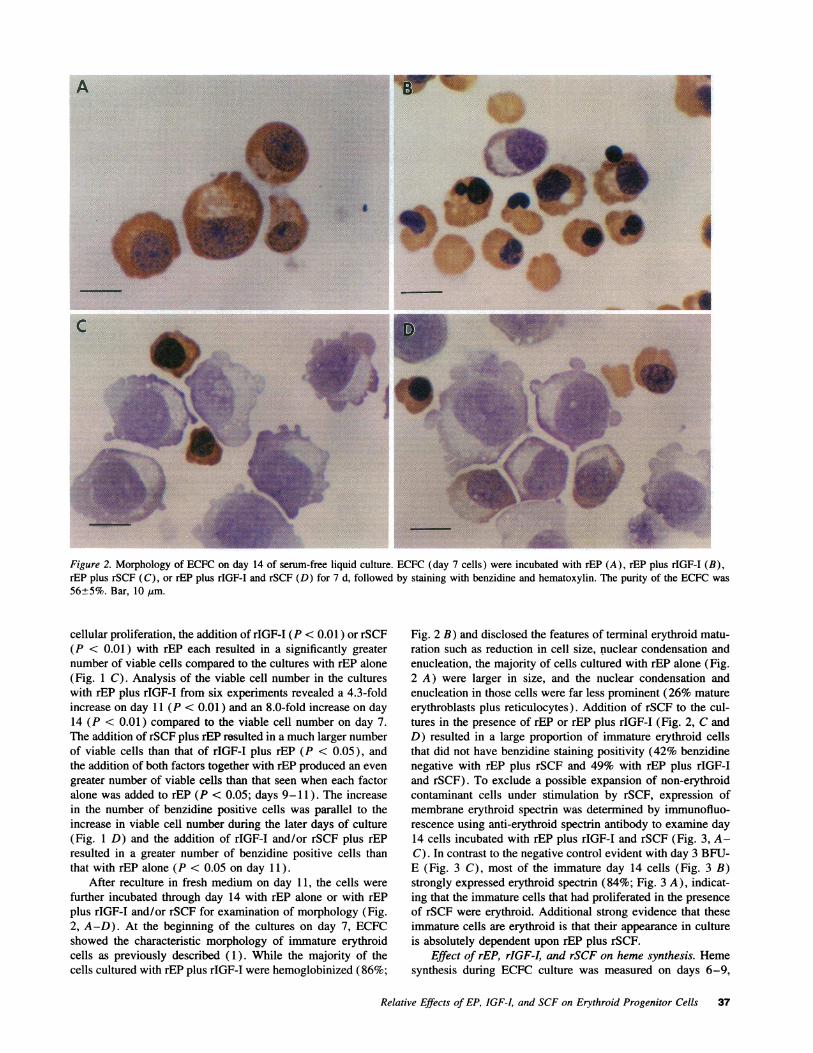
Fig. 2 B) and disclosed the features of terminal erythroid matu-ration such as reduction in cell size, nuclear condensation andenucleation, the majority of cells cultured with rEP alone (Fig.2 A) were larger in size, and the nuclear condensation andenucleation in those cells were far less prominent (26% matureerythroblasts plus reticulocytes). Addition of rSCF to the cul-tures in the presence of rEP or rEP plus rIGF-I (Fig. 2, C andD) resulted in a large proportion of immature erythroid cellsthat did not have benzidine staining positivity (42% benzidinenegative with rEP plus rSCF and 49% with rEP plus rIGF-Iand rSCF). To exclude a possible expansion of non-erythroidcontaminant cells under stimulation by rSCF, expression ofmembrane erythroid spectrin was determined by immunofluo-rescence using anti-erythroid spectrin antibody to examine day14 cells incubated with rEP plus rIGF-I and rSCF (Fig. 3, A-C). In contrast to the negative control evident with day 3 BFU-E (Fig. 3 C), most of the immature day 14 cells (Fig. 3 B)strongly expressed erythroid spectrin (84%; Fig. 3 A), indicat-ing that the immature cells that had proliferated in the presenceof rSCF were erythroid. Additional strong evidence that theseimmature cells are erythroid is that their appearance in cultureis absolutely dependent upon rEP plus rSCF.
Effect of rEP, rIGF-I, and rSCF on heme synthesis. Hemesynthesis during ECFC culture was measured on days 6-9,
Relative Effects of EP, IGF-I, and SCFon Erythroid Progenitor Cells 37
%.,::
aFigure 3. Immunolocaliza-tion of spectrin. Expressionof erythroid spectrin in day14 cells incubated withrEP, rIGF-I, and rSCF wasdetermined by immuno-fluorescence using anti-er-ythroid spectrin antibody.(A) Immunofluorescence.(B) Differential interfer-ence contrast microscopy
U- _, _ _111of the same immu-nofluorescence field dem-onstrating that most of
the cells reacted with the spectrin antibody. (C) Spectrin negative cells observed with day 3 BFU-E preparation (arrows). The purity of theECFCwas 56±5%.
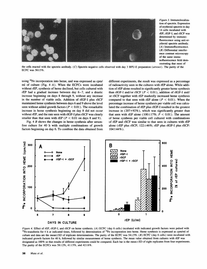
using 59Fe incorporation into heme, and was expressed as cpm/ml of culture (Fig. 4 A). When the ECFCs were incubatedwithout rEP, synthesis of heme declined, but cells cultured withrEP had a gradual increase between day 6-7, and a drasticincrease beginning on days 8 through 9, without any increasein the number of viable cells. Addition of rIGF-I plus rSCFmaintained heme synthesis between days 6 and 9 above the levelseen without added growth factors (P < 0.01). The remarkableincrease in heme synthesis beginning on day 8 did not occurwithout rEP, and the rate seen with rIGF-I plus rSCF was clearlysmaller than that seen with rEP (P < 0.01 on days 8 and 9).
Fig. 4 B shows the changes in heme synthesis after serum-free culture for 40 h with multiple combinations of growthfactors beginning on day 6. To combine the data obtained from
E 300Ea.U
w
wI0 200
z
z0
cc100a.
0z
0
AO-O None* * rEP.-m rlGF-I + rSCF
6 7 8 9
DAYS IN CULTURE
different experiments, the result was expressed as a percentageof radioactivity seen in the cultures with rEP alone. While addi-tion of rEP alone resulted in significantly greater heme synthesisthan rIGF-I and/or rSCF (P < 0.01), addition of rIGF-I and/or rSCF together with rEP markedly increased heme synthesiscompared to that seen with rEP alone (P < 0.01). When thepercentage increase of heme synthesis per viable cell was calcu-lated the combination of rEP plus rIGF-I resulted in the greatestincrease in (207+92%), which was significantly greater thanthat seen with rEP alone (100±17%; P < 0.01). The amountof heme synthesis per viable cell cultured with combinationsof rEP and rSCF was similar to that seen in cultures with rEPalone (rEP plus rSCF; 122+46%; rEP plus rIGF-I plus rSCF:104+44%).
B 600B~~~~~~~NonerIGF-I w
CZX1 rSCF |rIGF-I + rSCF
400 Z
0~
200C.)0z
U.
00 1
rEP (U/ml)
Figure 4. Effect of rEP, rIGF-I, and rSCF on heme synthesis. (A) ECFC (day 6 cells) incubated with indicated growth factors were pulsed with59Fe-transferrin for 4 h at indicated times, followed by determination of s9Fe incorporation into heme. Heme synthesis is expressed as cpm/ml ofculture and data are the mean±SDof triplicate determinations. The purity of the ECFCwas 54±5%. (B) ECFC(day 6 cells) were incubated withindicated growth factors for 40 h, followed by similar measurement of heme synthesis. The mean value obtained from cultures with rEP was
designated as 100% so that results of different experiments could be compared. Each bar is the mean±SDof eight replicates from four experiments.The purity of the ECFCs was 58±2%, 41±5%, and 42+6%.
38 Muta et al.

0
E
Z.6
z0
0
a-
0FIr0z
2L.01 0
DAY 9 DAY 10 DAY 11 DAY 12
DAYS IN CULTURE
Figure 5. The requirement for IGF-I during late erythroid maturation.Day 7 ECFCwere recultured in serum-supplemented liquid cultureswith 2 U/ml rEP and were harvested on days 9-12 for 24 h incubationswith indicated growth factors and 59Fe-transferrin in serum-free medium.Hemesynthesis was determined and each bar is the mean±SDof tripli-cate determinations. The purity of the ECFCs was 57±5%.
The requirement for IGF-I during the later stages of ery-throid development was determined by measuring the accumu-lated amount of 59Fe incorporation into heme over successive24 h periods (Fig. 5). On days 9-11, the cells incubated withrIGF-I disclosed a significantly larger amount of heme synthesisthan that seen in the cells cultured without the addition ofgrowth factors (P < 0.01 ) and the effect of rIGF-I on accumula-tion of heme was equivalent to that of rEP throughout the cultureperiod. The addition of rEP and rIGF-I together significantlyincreased the amount of heme synthesis beyond that seen witheach factor alone (P < 0.05 on day 9; P < 0.01 on day 10 andday 11). The effect of rSCF was also examined in a similarexperiment and showed little effect on days 9-11 (data notshown).
To determine the effect of rSCF on heme synthesis, liquidcultures in a serum-free medium were started with day 7 cellsand incubated through day 16 in the presence or absence ofrSCF (Table I). On days 10 and 14, the culture medium wasreplaced with fresh medium and the concentration of the cellswas reduced to no more than 106/ml. Accordingly, the amountof heme synthesis was adjusted to reflect the dilution factor. Theaddition of rSCF to the cultures with rEP plus rIGF-I produced aremarkable increase in heme synthesis and resulted in a 21-foldgreater total accumulation on day 16, compared with the peakvalue seen on day 12 with only rEP plus rIGF-I, reflecting amarked expansion of the erythroid mass as the cells stimulatedby rSCF eventually matured.
Effect of rEP, IGF-I, and rSCF on DNA synthesis. Theproliferative response of the ECFCto rEP, rIGF-I, and rSCF wasdetermined by measuring DNAsynthesis using [3H] thymidineincorporation (Fig. 6 A). rEP alone maintained the [3H] -thymidine uptake, and the amount was similar to the initialamount throughout most of the culture period. The addition ofrIGF-I plus rSCF, in the absence of rEP, resulted in slightlygreater DNAsynthesis compared to the cultures with rEP alone(P < 0.01 on days 7 and 8). After rEP, rIGF-I and rSCF wereadded together, a very large increase in DNAsynthesis occurredwith a markedly enhanced proliferative response. When theamount of DNAsynthesis per viable cell was calculated, addi-
Table I. Effect of rSCF on heme synthesis by ECFCs
Culture additionDays ofculture rEP + rIGF-I rEP + rIGF-I + rSCF
8 181+12* 125+27*10 2155+57 1763+16912 2322±69 5570±72714 1644±39 32700±+169916 318+25 49350±2229
ECFCs (day 7 cells) were incubated with indicated growth factors inliquid serum-free medium and pulsed with 59Fe-transferrin over 4 h atindicated times. Heme 59 Fe was measured after extraction using cyclo-hexanone to determine the amount of heme synthesis. The data are themean+SDof triplicate determinations. The purity of the ECFCs was40+5%. * Heme '9Fe (cpm/ml).
tion of rIGF-I and rSCF to rEP resulted in a 3.6- (day 7), 3.8-(day 8), and 2.4- (day 9) fold increase, compared to the valuesseen with rEP alone.
To further determine the interaction of rEP, rIGF-I, andrSCF in regulating the proliferation of ECFC, various combina-tions of these growth factors were added to the serum-free liquidcultures on day 6, followed by a 1-h assessment of [3H]-thymidine incorporation after 40 h of incubation (Fig. 6 B).The addition of rEP, rIGF-I and/or rSCF significantly increasedDNAsynthesis compared to that seen without rEP (P < 0.01),but, as a single addition, rSCF had the greatest effect comparedwith rEP or rIGF-I (P < 0.01). The addition of rIGF-I andrSCF together resulted in a higher rate of DNAsynthesis thaneach factor alone (P < 0.01). While rEP had little effect onDNAsynthesis when added alone, the addition of rEP togetherwith rIGF-I and/or rSCF resulted in a much larger increasecompared to that seen with each factor alone (P < 0.01). Addi-
E
06
E
5-z
*F
7 a
DAYS IN CULTURE rEP (U/mi)
Figure 6. Effect of rEP, rIGF-I, and rSCF on DNAsynthesis. (A) ECFC(day 6 cells) incubated with indicated growth factors in liquid serum-free cultures, were pulsed with [3H]thymidine for 1 h at indicated times,followed by TCAprecipitation to determine the amount of DNAsynthe-sis, which is expressed as cpm per ml of culture. The data are themean±SD. (B) ECFCs (day 6 cells) were incubated with indicatedgrowth factors for 40 h, followed by measurement of DNAsynthesiswith [3H]thymidine for 1 h. Each bar is the mean±SDof six replicatesfrom two experiments. The purity of the ECFCs was 32±2%, and41±2%.
Relative Effects of EP, IGF-I, and SCFon Erythroid Progenitor Cells 39
i_ r Before cultureo Non.x ^ rm rEP
2 rEP + rIQF-l + rC
.2
z
0
DAY S DAY 7 DAY S DAY 9
DAYS IN CULTURE
Figure 7. ECFCs (day 6 cells) were removed from methylcellulose andwere recultured in liquid serum-supplemented cultures for harvest onsucceeding days followed by incubation in serum-free medium with theindicated growth factors. The incubations were continued for 24 h, andDNAsynthesis was determined before and after each 24 h of culture.The data are mean±SD of triplicate determinations. The purity of theECFCs was 51±7%.
tion of rEP to the cultures with rIGF-I, rSCF, or rIGF and rSCFresulted in 3.5-, 3.5-, and 4.1-fold greater increases in DNAsynthesis, respectively, compared to the sum of the increasesobserved with rIGF-I and/or rSCF in the absence of rEP plus theincrease observed with rEP alone. Since these large increasesdepended on the presence of rEP, it is very likely that theyrepresented the erythroid cells and not the contaminant cells.In the presence of rEP, addition of rSCF produced a greaterincrease than that of rIGF-I (P < 0.01), and the addition ofrIGF-I and rSCF together to the cultures resulted in a greaterincrease than that of each factor alone (P < 0.01 ) or the sumof these factors. WhenDNAsynthesis per viable cell was calcu-lated in each incubation, addition of rEP plus rIGF-I and rSCF(3,578 cpm/ I05 viable cells) increased 2.2-fold compared withthe sum of the cultures with rIGF-I and rSCF (1,176 cpm/ 105viable cells) plus those with rEP alone (463 cpm/ 105 viablecells).
To understand the kinetics of growth factor action on DNAsynthesis by the ECFC, day 6 cells were recultured in a serum-supplemented liquid medium, and were studied after serial har-vests on succeeding days. DNAsynthesis was measured beforeand after 24-h incubations with the indicated growth factorsin serum-free liquid medium (Fig. 7). Throughout the cultureperiod, DNA synthesis in the cultures with rEP alone wasgreater than in those without rEP, but similar or less than thatobserved before the 24 h of incubation. The marked effect ofthe addition of three factors together was noted in the earlierday 6 and day 7 cells, and declined by day 8.
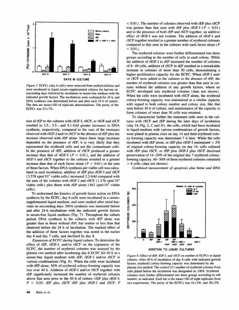
Expansion of ECFCduring liquid culture. To determine theeffect of rEP, rIGF-I, and/or rSCF on the expansion of theECFC, the number of erythroid colonies was assayed by theplasma clot method after incubating day 6 ECFCfor 40 h in aserum-free liquid medium with rEP, rIGF-I and/or rSCF invarious combinations (Fig. 8). When the cells were incubatedwith rEP alone, 36% of erythroid colony-forming capacity waslost over 40 h. Addition of rIGF-I and/or rSCF together withrEP significantly increased the number of erythroid coloniesabove that seen prior to the 40 h of culture (rEP plus rIGF-I:P < 0.05; rEP plus rSCF rEP plus rIGF-I and rSCF: P
< 0.01). The number of colonies observed with rEP plus rSCFwas greater than that seen with rEP plus rIGF-I (P < 0.01)and in the presence of both rEP and rSCF together, an additiveeffect of rIGF-I was not evident. The addition of rIGF-I andrSCF together resulted in a greater number of erythroid coloniescompared to that seen in the cultures with each factor alone (P< 0.01).
The erythroid colonies were further differentiated into threegroups according to the number of cells in each colony. Whilethe addition of rIGF-I to rEP increased the number of coloniesof 8-49 cells, addition of rSCF to rEP resulted in a remarkableincrease in colonies of more than 50 cells, demonstrating ahigher proliferative capacity for the ECFC. When rIGF-I and/or rSCF were added to the cultures in the absence of rEP, thenumber of erythroid colonies was greater than that seen in cul-tures without the addition of any growth factors, where noECFC developed into erythroid colonies (data not shown).When the cells were incubated with rSCF alone, the erythroidcolony-forming capacity was maintained at a similar capacitywith regard to both colony number and colony size, like thatseen before 40 h of culture, and maintenance of the capacity toform colonies of more than 50 cells was retained.
To characterize further the immature cells seen in the cul-tures with rSCF and rEP during the later days of incubation(day 14; Fig. 2, C and D), the cells, which had been incubatedin liquid medium with various combinations of growth factors,were plated in plasma clots on day 14 and their erythroid colo-ny-forming capacity was determined 7 d later. While the cellsincubated with rEP alone, or rEP plus rIGF-I maintained < 2%of original colony-forming capacity on day 14, cells culturedwith rEP plus rSCF, or rEP plus rIGF-I plus rSCF disclosedpreservation of 14-20% of the original day 7 erythroid colony-forming capacity. 40-50% of these erythroid colonies contained> 8 cells (data not shown).
Combined measurement of apoptosis plus heme and DNA
Nmber of cob hi colonis600 2-7
.-49
Ct "9E r E
0
0400
~-200
Lu
0C rEP rEP rEP rEP
rlQF-I r1QF-I rlGF-1 rIQF-lrSCF rSCF rSCF rSCF
ADDITION TO LIQUID CULTURES
Figure 8. Effect of rEP, IGF-I, and rSCF on number of ECFCs in liquidcultures. After 40 h of incubation of day 6 cells with indicated growthfactors, erythroid colony-forming capacity was determined by theplasma clot method. The control (C) number of erythroid colonies fromcells plated before the incubation was designated as 100%. Erythroidcolonies were further differentiated into three groups according to cellnumber, as indicated. Each bar is the mean±SDof eight replicates fromtwo experiments. The purity of the ECFCs was 41±2%, and 30±2%.
40 Muta et al.
- A
Bcj
I so~
vnone rEP rlGF-I
rSCF
ADDITION TO LIQUID CUL
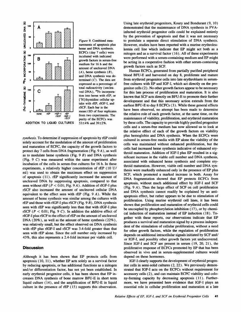
f Figure 9. Combined mea-surements of apoptosis plusheme and DNAsynthesis.ECFCs (day 7 cells) wereincubated with indicatedgrowth factors in serum-freemedium for 16 h and theamount of uncleaved DNA
.0i (A), heme synthesis (B),and DNAsynthesis was de-termined (C). The data areexpressed as a percentage of
_ - total radioactivity (unclea-ved DNA), 59Fe incorpora-tion into heme with rEP, or[3H]thymidine cellular up-take with rEP, rIGF-I, andrSCF. Each bar is themean±SD of four replicates
rEPrIGF-I from two experiments. TherSCF purity of the ECFCs was
LTURES 53±4% and 39±2%.
synthesis. To determine if suppression of apoptosis by rEP couldsolely account for the modulation of the amount of proliferationand maturation of ECFC, the capacity of the growth factors toprotect day 7 cells from DNAfragmentation (Fig. 9 A), as wellas to modulate heme synthesis (Fig. 9 B) and DNAsynthesis(Fig. 9 C) was measured within the same experiment afterincubation of the cells in serum-free cultures for 16 h. In theseexperiments, a relatively higher concentration of rEP (10 U/ml) was used to obtain the maximum effect on suppressionof apoptosis (11). rEP significantly increased the amount ofuncleaved DNA by suppressing apoptosis, compared to thatseen without rEP (P < 0.01; Fig. 9 A). Addition of rIGF-I plusrSCF also increased the amount of uncleaved cellular DNAequivalent to the effect seen with rEP (Fig. 9 A). While theamount of heme synthesis was similar among the cultures withrEP and those with rIGF-I plus rSCF (Fig. 9 B), DNAsynthesisseen with rEP was significantly less than that with rIGF-I plusrSCF (P < 0.01; Fig. 9 C). In addition the additive effect ofrIGF-I plus rSCF to the effect of rEP on the amount of uncleavedDNA(20%), as well as the amount of heme synthesis (22%),was relatively small, but the effect observed on DNAsynthesiswith rEP plus rIGF-I and rSCF was 3.4-fold greater than thatseen with rEP alone. Since the cell number only increased by43%, this also represents a 2.4-fold increase per viable cell.
Discussion
Although it has been shown that EP protects cells fromapoptosis (10, 11), whether EP acts solely as a survival factorby reducing apoptosis, or has additional functions as a mitogenand/or differentiation factor, has not yet been established. Inearly erythroid progenitor cells, it has been shown that EP in-creases DNA synthesis of bone marrow BFU-E in short termliquid culture (14), and the amplification of BFU-E in liquidculture in the presence of rEP (15) supports this observation.
Using late erythroid progenitors, Koury and Bondurant (9, 10)demonstrated that the maintenance of DNAsynthesis in FVA-infected erythroid progenitor cells could be explained entirelyby the prevention of apoptosis and that it was not necessaryto postulate a separate direct stimulation of DNA synthesis.However, studies have been reported with a murine erythroleu-kemia cell line which indicate that EP might act both as a
mitogen and as a survival factor (16). All of these experimentswere performed with a serum-containing medium and EPmightbe acting in a cooperative fashion with other serum-containinggrowth factors such as SCF.
HumanECFCs, generated from partially purified peripheralblood BFU-E and harvested on day 8, proliferate and maturefrom erythroid progenitor cells into late erythroblasts in serum-
free cultures with EP and IGF-I, which act directly on the pro-genitor cells (2). No other growth factors appear to be necessaryfor this late process of proliferation and maturation. It is alsoknown that SCFacts directly on BFU-E to promote their further
development and that this necessary action extends from theearliest BFU-E to day 8 ECFCs (3). While these general effectshave been observed, no attempt has been made to determinethe relative role of each growth factor, at the same time, on themaintenance of viability, proliferation, and erythroid maturationby these cells. The capacity to provide highly purified progenitorcells and a serum-free medium has now allowed us to look atthe relative effect of each of the growth factors on viabilityplus hemoglobin and DNAsynthesis. When the ECFCs were
cultured in serum-free media with EP alone the viability of thecells was maintained without enhanced proliferation, but thecells had increased heme synthesis indicative of enhanced ery-throid maturation. Addition of IGF-I plus EP resulted in a sig-nificant increase in the viable cell number and DNAsynthesis,associated with enhanced heme synthesis and complete ery-throid maturation. However, viable cell number and DNAsyn-thesis were markedly enhanced only in the presence of EP plusSCF, which promoted a marked increase in both. Assay forDNA fragmentation showed that EP protects ECFCs fromapoptosis, without much additional effect by IGF-I and SCF(Fig. 9 A). Thus the large effect of SCF on cell proliferationand DNA synthesis cannot readily be explained by an anti-apoptosis effect, but rather appears to be due to stimulation ofproliferation. Using murine erythroid cell lines, it has beenshown that proliferation and maturation of erythroid cells couldbe uncoupled by phosphorylation inhibition (17), or by chemi-cal induction of maturation instead of EP induction (18). To-gether with these reports, our observations indicate that EPproduces a survival and maturation signal by a process indepen-dent of the stimulation of cellular proliferation, without a needfor other growth factors, while the regulation of proliferationdepends on additional intracellular signals initiated by SCFand/or IGF-I, and possibly other growth factors yet undiscovered.Since IGF-I and SCF are present in serum (19, 20, 21), theproliferative response of ECFCspromoted by EP that has beenobserved in vivo and in serum-supplemented cultures woulddepend on these hormones.
IGF-I clearly supports the development of erythroid progen-itor cells in semi-solid cultures (2, 22). Wepreviously demon-strated that IGF-I acts on the ECFCs without requirement foraccessory cells (2), and can maintain ECFCviability and colo-ny-forming capacity by decreasing apoptosis (11). Further-more, we have presented here evidence that IGF-I plays an
essential role in cellular proliferation and maturation at a late
Relative Effects of EP, IGF-I, and SCFon Erythroid Progenitor Cells 41
R
<100za0LU
LU-j
z S 0
0
<1000.0 50
zID 0
z5>- 50xI-
stage of erythroid development. In the presence of EP, IGF-Imarkedly enhanced heme synthesis (Fig. 4 B). In addition, amarkedly defective morphology of the cells cultured with EPalone (Fig. 2 A), compared with that of normal erythroblastsseen in the cultures with EP plus IGF-I (Fig. 2 B), supportsthis observation. Thus a reduction of apoptosis by EP is notadequate for complete erythroid maturation which requires di-rect stimulation by IGF-I. Using murine purified CFU-E, it wasreported that IGF-I enhances erythroid maturation only whenEP levels are low (10 mU/ml) (23). In our experimental sys-tem, even in the presence of high concentrations of EP (1 U/ml), the stimulating effect of IGF-I on heme synthesis wasprominent. Difference in the cell system, cell maturity, and/orculture conditions, such as BSA preparation, might account forthis discrepancy.
The proto-oncogene c-kit encodes a transmembrane tyrosinekinase receptor, and its ligand, SCF (24), stimulates colonyformation by hematopoietic progenitor cells of diverse lineages(25, 26). Wepreviously demonstrated that SCF, as well as EPand IGF-I, reduces apoptosis of ECFCbetween days 7-8 ofculture ( 11). While the effect of SCF on suppression ofapoptosis was less prominent than EP ( 11), the evidence pre-sented here indicates that SCFstimulates proliferation of ECFCssubstantially. In contrast to the cellular proliferation associatedwith erythroid maturation seen in the cultures with EP plus IGF-I, addition of SCF together with EP resulted in an expansion ofcolony-forming cells with high proliferative capacity (Fig. 8)and an increase in immature erythroid cells that were evidentduring later days of culture (Fig. 2, C and D). While it hasbeen reported that SCFstimulates maturation of mast cells (27),this work was performed in a mixed cellular system, with thepresence of serum, and several reports have indicated that SCFpreferentially enhances proliferation, but not differentiation ofa variety of precursor cells (28, 29, 30). In our experimentsSCFenhanced ECFCproliferation by itself, although the effectwas substantially greater in the presence of EP. SCF also en-hanced maturation itself during days 8-9 of culture, but theeffect was limited to the very early phase of heme synthesis,after which EP was required as the principal maturation factor.SCFand EP, with, or without, IGF-I produced a marked prolif-eration of erythroid cells through day 14, which were benzidinenegative and this was coupled with a maintenance of colony-forming capacity. This shows a difference in the maturationstage of the cells generated by SCF from those generated with-out SCF. There remain the following two possible mechanismsfor this: (a) SCF greatly enhanced the proliferation of a rela-tively immature subpopulation of the erythroid progenitor cellswithin the ECFCs; or (b) the maturation process of many ofthe ECFCswas delayed during the marked cellular proliferationstimulated by SCF. Experiments to clarify this issue are nowunderway in our laboratory.
It could be maintained that SCFmight allow enhanced pro-liferation by preventing apoptosis of progenitor cells that are inan earlier stage of development and were more prone to cellproliferation rather than hemoglobin synthesis. However, day 7cells, which are responsive to both EP and SCF, had a markedsuppression of apoptosis by EP with a stimulation of DNAsynthesis that was only '/3 of the maximum response (Fig. 9).While the suppression of apoptosis by EP was only minimallyaffected by the addition of SCF, DNAsynthesis was markedlyenhanced. This discrepancy between the large increase in DNAsynthesis and minimal effect on the amount of uncleaved (i.e.,
protected) DNA, suggests that the proliferation of ECFCis notonly "maintained" by suppressing apoptosis, but also stimu-lated by SCF in the presence of EP.
In conclusion, it appears that EP prevents apoptosis andsupports erythroid viability and maturation. IGF-I enhances ery-throid maturation and supports limited proliferation, while SCFstimulates extensive cellular proliferation to expand the numberof erythroid progenitor cells. These findings indicate that prolif-eration and maturation of erythroid cells are regulated in aseparate manner by these hormones. While the intracellular mo-lecular mechanisms which mediate these actions are still un-known and need to be clarified, further molecular work willhave to be performed under conditions that segregate the effectsof these growth factors.
Acknowledgments
The authors are grateful for the generous gifts of rIL-3 and rSCF fromAmgen Inc. Wethank Sarah Coode, Millie Clancey, Kathleen Kollar,and Amanda Hodges for their excellent technical help and Pat Hofmannfor her kind assistance with typing.
This work was supported by Veterans Health Administration MeritReview Grants (S. B. Krantz and M. C. Bandurant), by grants DK-15555 and 2 T32-DKO7186 from the National Institutes of Health(S. B. Krantz), and by the Joe C. Davis Hematology Research Fund.Dr. Koichiro Muta is an Ortho Biotech Hematology Fellow.
References
1. Sawada, K., S. B. Krantz, J. S. Kans, E. N. Dessypris, S. Sawyer, A. D.Glick, and C. I. Civin. 1987. Purification of human erythroid colony-formingunits and demonstration of specific binding of erythropoietin. J. Clin. Invest.80:357-366.
2. Sawada, K., S. B. Krantz, E. N. Dessypris, S. T. Koury, and S. T. Sawyer.1989. Human colony-forming units-erythroid do not require accessory cells, butdo require direct interaction with insulin-like growth factor I and/or insulin forerythroid development. J. Clin. Invest. 83:1701-1709.
3. Dai, C-H., S. B. Krantz, and K. M. Zsebo. 1991. Human burst-formingunits-erythroid need direct interaction with stem cell factor for further develop-ment. Blood. 78:2493-2497.
4. Kerr, J. F. R., A. H. Wyllie, and A. R. Currie. 1972. Apoptosis: a basicbiological phenomenon with wide ranging implications in tissue kinetics. Br. J.Cancer. 26:239-257.
5. Wyllie, A. H., J. F. R. Kerr, and A. R. Currie. 1980. Cell death: thesignificance of apoptosis. Int. Rev. Cytol. 68:251-306.
6. Williams, G. T., C. A. Smith, E. Spooncer, T. M. Dexter, and D. R.Taylor. 1990. Haemopoietic colony stimulating factors promote cell survival bysuppressing apoptosis. Nature (Lond.). 343:76-79.
7. Her, E., J. Frazer, K. F. Austin, and W. F. Owen, Jr. 1991. Eosinophilhematopoietins antagonize the programmed cell death of eosinophils. Cytokineand glucocorticoid effects on eosinophils maintained by endothelial cell-condi-tioned medium. J. Clin. Invest. 88:1982-1987.
8. Yu, H., B. Bauer, G. K. Lipke, R. L. Phillips, and G. Van Zant. 1993.Apoptosis and hematopoiesis in murine fetal liver. Blood. 81:373-384.
9. Koury, M. J., and M. C. Bondurant. 1988. Maintenance by erythropoietinof viability and maturation of murine erythroid precursor cells. J. Cell. Physiol.137:65-74.
10. Koury, M. J., and M. C. Bondurant. 1990. Erythropoietin retards DNAbreakdown and prevents programmed death in erythroid progenitor cells. Science(Wash. DC). 248:378-381.
11. Muta, K., and S. B. Krantz. 1993. Apoptosis of erythroid colony-formingcells is decreased by stem cell factor and insulin-like growth factor I as well aserythropoietin. J. Cell. Physiol. 156:264-271.
12. Krantz, S. B. 1991. Erythropoietin. Blood. 77:419-434.13. Wickrema, A., S. B. Krantz, J. C. Winkelmann, and M. C. Bondurant.
1992. Differentiation and erythropoietin receptor gene expression in human ery-throid progenitor cells. Blood. 80:1940-1949.
14. E. N. Dessypris, and S. B. Krantz. 1984. Effect of pure erythropoietin onDNA-synthesis by human marrow day 15 erythroid burst forming units in shortterm liquid culture. Br. J. Haematol. 56:295-306.
15. Umemura, T., T. Papayannopoulou, and G. Stamatoyannopoulos. 1989.The mechanism of expansion of late erythroid progenitors during erythroid regen-
42 Muta et al.
eration: target cells and effects of erythropoietin and interleukin-3. Blood.73:1993-1998.
16. Spivak, J. L., T. Pham, M. Isaacs, and W. D. Hankins. 1991. Erythropoietinis both a mitogen and a survival factor. Blood. 77:1228-1233.
17. Noguchi, T., H. Fukumoto, Y. Mishina, and M. Obinata. 1988. Differentia-tion of erythroid progenitor (CFU-E) cells from mouse fetal liver cells and murineerythroleukemia (TSA8) cells without proliferation. Mol. Cell. Biol. 8:2604-2609.
18. Busfield, S. J., and S. P. Klinken. 1992. Erythropoietin-induced stimulationof differentiation and proliferation in J2E cells is not mimicked by chemicalinduction. Blood. 80:412-419.
19. Kurtz, A., W. Hdrtl, W. Jelkmann, J. Zapf, and C. Bauer. 1985. Activityin fetal bovine serum that stimulates erythroid colony formation is insulin likegrowth factor I. J. Clin. Invest. 76:1643-1648.
20. Papayannopoulou, T., M. Brice, V. C. Broudy, and K. M. Zsebo. 1991.Isolation of c-kit receptor-expressing cells from bone marrow, peripheral blood,and fetal liver: functional properties and composite antigenic profile. Blood.78:1403-1412.
21. Langley, K. E., L. G. Bennett, J. Wypych, S. A. Yancik, S-D. Liu, K. R.Westcott, D. G. Chang, K. A. Smith, and K. M. Zsebo. 1993. Soluble stem cellfactor in human serum. Blood. 81:656-660.
22. Correa, P. N., and A. A. Axelrad. 1991. Production of erythropoietic burstsby progenitor cells from adult human peripheral blood in an improved serum-freemedium: role of insulin-like growth factor-I. Blood. 78:2823-2833.
23. Boyer, S. H., T. R. Bishop, 0. C. Rogers, A. N. Noyes, L. P. Frelin,and S. Hobbs. 1992. Roles of erythropoietin, insulin-like growth factor 1. andunidentified serum factors in promoting maturation of purified murine erythroidcolony-forming units. Blood. 80:2503-2512.
24. Zsebo, K. M., D. A. Williams, E. N. Geissler, V. C. Broudy, F. H. Martin,H. L. Atkins, R-Y. Hsu, N. C. Birkett, K. H. Okino, D. C. Murdock, F. W.Jacobsen, K. E. Langley, K. A. Smith, T. Takeishi, B. M. Cattanach, S. J. Galli,and S. V. Suggs. 1990. Stem cell factor is encoded at SI locus of the mouse andis the ligand for the c-kit tyrosine kinase receptor. Cell. 63:213-224.
25. McNiece, I. K., K. E. Langley, and K. M. Zsebo. 1991. Recombinanthuman stem cell factor synergises with GM-CSF, G-CSF, IL-3 and Epo to stimu-late human progenitor cells of the myeloid and erythroid lineages. Exp. Hematol.(NY). 19:226-231.
26. Migliaccio, G., A. R. Migliaccio, M. L. Druzin, P-J. V. Giardina, K. M.Zsebo, and J. W. Adamson. 1991. Effect of recombinant human stem cell factor(SCF) on the growth of human progenitor cells in vitro. J. Cell. Physiol. 148:503-509.
27. A-M. A. Irani, G. Nilsson, U. Miettinen, S. S. Craig, L. K. Ashman, T.Ishizaka, K. M. Zsebo, and L. B. Schwartz. 1992. Recombinant human stem cellfactor stimulates differentiation of mast cells from dispersed human fetal livercells. Blood 80:3009-3021.
28. Tanaka, R., K. Koike, T. Imai, M. Shiohara, T. Kubo, Y. Amano, A.Komiyama, and T. Nakahaata. 1992. Stem cell factor enhances proliferation, butnot maturation of murine megakaryocytic progenitors in serum-free culture. Blood.80:1743-1749.
29. Murphy, M., K. Reid, D. E. Williams, S. D. Lyman, and P. F. Bartlett.1992. Steel factor is required for maintenance, but not differentiation of melano-cyte precursors in the neural crest. Dev. Biol. 153:396-401.
30. Miura, N., S. Okada, K. M. Zsebo, Y. Miura, and T. Suda. 1993. Rat stemcell factor and IL-6 preferentially support the proliferation of c-kit-positive murinehemopoietic cells rather than their differentiation. Exp. Hematol. 21:143-149.
Relative Effects of EP, IGF-I, and SCFon Erythroid Progenitor Cells 43