DISSERTATION ON ANAL SPHINCTER COMPLEX A …
103
DISSERTATION ON ANAL SPHINCTER COMPLEX A COMPARATIVE STUDY OF VAGINAL DELIVERY WITH EPISIOTOMY AND WITH PERINEAL LACERATIONS IN PRIMIPAROUS WOMEN - TRANSVAGINAL ULTRASONOGRAPHIC EVALUATION Submitted to THE TAMILNADU DR.M.G.R. MEDICAL UNIVERSITY CHENNAI – 600 032 With fulfillment of the Regulations For the Award of the Degree of M.D. OBSTETRICS AND GYNAECOLOGY (BRANCH-II) DEPARTMENT OF OBSTETRICS AND GYANECOLOGY KILPAUK MEDICAL COLLEGE CHENNAI – 600 010 MARCH – 2009
Transcript of DISSERTATION ON ANAL SPHINCTER COMPLEX A …

DISSERTATION ON
ANAL SPHINCTER COMPLEX
A COMPARATIVE STUDY OF VAGINAL DELIVERY WITH
EPISIOTOMY AND WITH PERINEAL LACERATIONS
IN PRIMIPAROUS WOMEN -
TRANSVAGINAL ULTRASONOGRAPHIC EVALUATION
Submitted to
THE TAMILNADUDRMGR MEDICAL UNIVERSITY
CHENNAI ndash 600 032
With fulfillment of the RegulationsFor the Award of the Degree of
MD OBSTETRICS AND GYNAECOLOGY(BRANCH-II)
DEPARTMENT OF OBSTETRICS AND GYANECOLOGYKILPAUK MEDICAL COLLEGE
CHENNAI ndash 600 010
MARCH ndash 2009
CERTIFICATE
This is to certify that the dissertation work titled ldquoANAL
SPHINCTER COMPLEX - A COMPARATIVE STUDY OF VAGINAL
DELIVERY WITH EPISIOTOMY AND WITH PERINEAL
LACERATIONS IN PRIMIPAROUS WOMEN -TRANSVAGINAL
ULTRASONOGRAPHIC EVALUATIONrdquo is a bonafide research work
of DRNBANUREKHA Enrolment Nohelliphelliphelliphelliphelliphelliphelliphelliphellip Submitted
in partial fulfillment of the requirements for the award of Degree of MD
OBSTETRICS amp GYNAECOLOGY (BRANCH-II) in THE TAMIL
NADU DRMGR MEDICAL UNIVERSITY CHENNAI- 600 032
Signature of HOD Signature of Dean
2
ACKNOWLEDGEMENTS
I thank our Dean Prof Dr M DHANAPAL Kilpauk Medical
College Chennai ndash 10 for permitting me to conduct this study in Kilpauk
Medical College and Hospital
I gratefully thank my Prof Dr MMUTHULAKSHMI MD
DGO Head of Department Government Kilpauk Medical College and
Hospital under whom I have the honour to work as a post graduate student
I express my sincere thanks to Prof Dr TASRIDEVI MD
DGO for her support and guidance through this endeavour
I am deeply indebted to her valuable guidance inspiration and
suggestions without which this work would not have been completed
My heartful thanks to our Professors Dr HKFATHIMA
Dr FAMIDHA Dr R PREMALATHA and Dr PMEENALOCHANI
and assistant Professors for their suggestions
I thank Mr PADMANABAN (Research Officer) for his guidance in
statistical analysis
I thank Precision Diagnostics Pvt Ltd lsquos Dr Bharathi Y Dhala Dr
Sathyabama Chandrasekaran Dr K Rajan DrBobji Kettay for sharing the
expertise necessary to conduct this study
3
I wholeheartedly thank my patients without their cooperation this
study could not have been completed
I thank my Father Mother and Brother for making me who I am
today
I thank my Husband for having stood by me at all times
I thank God for inspiring me to believe in Him
4
CONTENTS
CHAPTER TITLE PAGE NO
1 INTRODUCTION 1
2 REVIEW OF LITERATURE 15
3 AIM OF THE STUDY 26
4 MATERIALS AND METHODS 27
5 RESULTS 29
6 SUMMARY 61
7 DISCUSSION 64
8 CONCLUSION 66
9 ANNEXURE
a BIBLIOGRAPHY
b PROFORMA
c ABBREVIATIONS
d MASTER CHART
5
INTRODUCTION
6
INTRODUCTION
Episiotomy is a widely performed intervention in childbirth
Episiotomy has traditionally been one of the most frequently performed
obstetric procedures and has been one of the more controversial Defined as
a surgical incision in the perineum to enlarge the vaginal opening for birth
episiotomy is the incision of the pudenda whereas perineotomy is the
incision of the perineum
It is the only surgical procedure in obstetrics to be performed without
the patientrsquos specific consent
History of episiotomy
First description of an episiotomy was given by Sir Fielding Ould
(1710-1789) a Dublin midwife in 1742(1) in his A treatise on Midwifery in
three parts
lsquoIt sometimes happenshelliphellip that the head of the child hellipcannot however
come forward by reason of the extraordinary constriction of the external
orifice of the vagina helliphellipwherefore it must be dilated if possible by the
fingershellip if this cannot be accomplished there must be an incision made
toward the anus with a pair of crooked probe scissors introducing one blade
between the head the vaginahelliphelliphellip as for as shall be thought necessary
for the present purpose and the business is done at one pinch by which the
whole body will easily come forth(2)
Our predecessors were accomplished at trying to avoid perineal
lacerations
7
In 1776 Harrie discussed the importance of lubricating the perineum
and vagina with fresh hogrsquos lard ironing out the perineum and controlling
delivery of the vertex(3)(38)
Puzos recommended support of the perineum to prevent lacerations
(4) Before the development of suturing perineal lacerations were managed
with prolonged bed rest and sometimes binding the legs tightly together
Pare is said to have been the first to suture the perineum whereas
Mauriceau is credited for the first perineorraphy(5)In 1799 Michelis first
recommended midline incisions in the perineum(6) In 1847 Dubois first
suggested making an oblique incision in the perineum known today as the
mediolateral episiotomy In 1851 The Stethoscope and Virginia Medical
Gazette reported that Taliaferro from Virginia unaware that the operation
had ever been done before performed the first episiotomy in the United
States(6)(38)
Perineotomy is the incision of the perineum whereas the term
episiotomy coined by Carl Braun in 1857 is the incision of the pudenda or
external genitalia These terms gradually became synonymous(7)
Episiotomy was not widely used until the 1920s when Pomery and De
Lee changed the climate of opinion regarding the entire birth process In his
The Prophylactic Forceps Operation De Lee claimed that episiotomy would
preserve the integrity of the pelvic floor and the introitus(8)
In addition to the strong advocacy for the use of episiotomy by the
obstetricians of the day changes in maternity practices also affected the use
of episiotomy The shift from home birth to hospital deliveries contributed
to a shift in the conceptualization of the nature of childbirth This shift
8
provided aseptic operating conditions and the necessary technology to carry
out episiotomy safely This change in delivery sites unleashed a cascade of
interventions that were not based on scientific evidence With the increase
in the number of hospital deliveries physicians attending deliveries believed
policies of liberal episiotomy use shortened labour allowing the physician
to complete a birth rapidly
As more women delivered in hospital increasing numbers of them
received anesthesia which in turn interfered with the natural expulsive
efforts of the mothers increasing the need for forceps Forceps delivery
required better access to the birth canal and more room for vaginal
manipulations and the dorsal lithotomy position provided just such access
During the 1970s and 1980s as the naturalist movement flourished
the routine use of episiotomy began to be questioned Thacker and Banta
found that there is no clearly defined evidence for its efficacy particularly
for routine use and considerable evidence of risks associated with
episiotomy including pain edema infection and ultimately dyspareunia
(6)
Incidence of episiotomy
Thacker and Banta estimated that episiotomy is performed on 50 to
90 percent of all nulliparas (6)Thorpe et al reported that episiotomy was
performed on 62 of vaginal deliveries in the United States Further
breakdown of these numbers indicate that the procedure was executed in
80 of nulliparas patients and in 20 of parous patients (9)
Types of Episiotomy
9
Multiple techniques can be used to effect an episiotomy The incision
may be made with scissors or a knife and may be made in the midline
(midline episiotomy) or begun in the midline and extended laterally
(mediolateral episiotomy) (10)
Midline episiotomy - two fingers are placed in the vagina between
the fetal head and the perineum and using straight scissors the incision is
made from fourchette through the perineal body up to but not including the
external anal sphincter Advantages of the midline episiotomy are that it
does not cut through the belly of the muscle the two sides of the incised
area are anatomically balanced making surgical repair easier blood loss is
less than with mediolateral episiotomy A major drawback is the propensity
for extension through the external anal sphincter and in to the rectum For
this reason may practitioners avoid the midline technique
Mediolateral episiotomy ndash the incision is made starting at the midline
of the posterior fourchette and aimed towards the ischial tuberosity to avoid
injury to the anal sphincter The incision is usually about 4cm long In
addition to the skin and subcutaneous tissues the bulbocavernosus
transverse perineii and puborectalis muscles are cut Whether the incision is
to the right or left depends on operator preference(1)
10
ROLE OF EPISIOTOMY IN MODERN OBSTETRICS
The traditional teaching that episiotomy was protective against more
severe perineal lacerations has not been substantiated Thus the liberal use
of prophylactic episiotomy is no longer recommended However there are
still valid reasons for the performance of an episiotomy
1 To shorten the second stage of labour in cases of fetal distress
2 In selected cases of assisted vaginal delivery with forceps and less
frequently for vacuum assisted delivery
3 To obtain more room for obstetrical manoeuvres such as those with
shoulder dystocia assisted breech delivery and delivery of second
twin and in occiputo posterior(1)
Consequences of an episiotomy
Immediate consequences of an episiotomy would include blood loss
pain edema infection and hematoma Definitive disadvantages would
include extension to a third degree or fouth degree laceration the
complications of which would include loss of rectal tone rectovaginal
fistula and abscess formation(9) (23)More long term complications include
the formation of scar tissue wound infection and dyspareunia
Routine use of episiotomies has not been shown to improve outcomes
for patients and can actually be harmful Routine or liberal use of
episiotomies increases the risk that the patient will have a surgical incision
that is larger than if she experiences a spontaneous laceration at birth (11)
11
Lacerations of the vagina and perineum are classified as first second third
or fourth degree (12)
Classification of perineal
injury
INJURY
DEFINITION
First degree Injury confined to vaginal mucosa
Second degreeInjury of vaginal mucosa and perineal muscles but
not the anal sphincter
Third degreeInjury to the perineum involving the anal sphincter
complex (external and internal)
3a emspensplt50 of external sphincter thickness is torn
3b emspenspgt50 of external sphincter thickness is torn
3c Internal sphincter is tornemspensp
Fourth degreeInjury to anal sphincter complex (external and
internal sphincter )and rectal mucosa
One of the major justifications for the use of episiotomy is the
suggested protective role of episiotomy in the prevention of severe tears
The use of elective episiotomy is believed to prevent the occurrence of third
12
and fourth degree perineal lacerations A comparison of the incidence of
third and fourth degree perineal lacerations in patients who did not undergo
episiotomy women who underwent midline episiotomy and women who
underwent mediolateral episiotomy is important These injuries range from
0 to 23 in women with intact perineii02 to 9 in women with a
mediolateral episiotomy to 3 to 24 in women who underwent midline
episiotomy(22)
The association between episiotomy and perineal trauma (third degree
laceration) is complex On one hand mediolateral episiotomy is associated
with a lower risk of anal sphincter rupture at delivery (13)(14)On the other
hand it has also been shown that the number of mediolateral episiotomies
may be reduced without an increase in perineal trauma(15) Then the
protective effect of mediolateral episiotomy may be limited to situations in
which its use is inevitable while its routine performance may increase the
risk of anal incontinence It is possible that performing an episiotomy when
anal sphincter is not in danger increases the risk of direct scissor injuries to
the sphincter (Fritel x et al)(16) Unfortunately we do not know what
episiotomy rate offers the balance between benefits and risks for the anal
sphincter
It seems reasonable to conclude that episiotomy should not be
performed routinely (Eason Feldman 2000)(17) The procedure should be
applied selectively for appropriate indications The final rule is that there is
no substitute for surgical judgement and common sense (18)
ANAL INCONTINENCE
13
Anal incontinence is much more prevalent than once thought Anal
incontinence is an embarrassing condition that is largely underreported
Obstetric anal sphincter injuries are the major etiological factor
Recognition of risk factors may minimize the development of sphincter
injuries Many women do not discuss this problem with anyone because it is
socially embarrassing Anal incontinence affects approximately 10 of
women (19)
Less than 50 of woman with fecal incontinence report the problem
to their physician It can be a devastating problem for women and
obstetricians must be careful first to try and avoid a third or fourth degree
tear and then to repair it meticulously to give a woman the best opportunity
for a good functional repair
Risk factors for third- and fourth-degree tears have been identified
mainly in retrospective studies Taking an overall risk of 1 of vaginal
deliveries the following factors are associated with an increased risk of a
third- and fourth-degree tear (12)
14
Major risk factors for obstetric anal sphincter injury
RISK FACTOR
ODDS RATIO
Nulliparity (primigravidity) 3ndash4
Inherent predisposition
Short perineal bodyemspenspemspensp 8
Instrumental delivery overall 3
Forceps-assisted deliveryemspenspemspensp 3ndash7
Vacuum-assisted deliveryemspenspemspensp 3
Forceps vs vacuumemspenspemspensp 288
Forcepsemspenspemspensp with midline episiotomy 25
Prolonged second stage of labor (gt1 hour) 15ndash4
Epidural analgesia 15ndash3
Intrapartum infant factors
Birthweight over 4 kgemspenspemspensp 2
Persistent occipitoposterior positionemspenspemspensp 2ndash3
Episiotomy mediolateral 14
Episiotomy midline 3ndash5
Previous anal sphincter tear 4
15
POSTNATAL ASSESSMENT
Postnatal continence assessment should include direct questioning
using a standardized bowel function questionnaire so that a reliable
assessment of continence can be done(19) Continence of flatus liquid and
solid feces should be documented together with inquiry concerning fecal
urgency inability to defer defecation for longer than 5 minutes a socially
debilitating symptom The complaint of urgency incontinence may reflect
external anal sphincter dysfunction
A standardized bowel function questionnaire would include in
addition to the above questions an inquiry assessing the need to wear a pad
because of anal symptoms any extra anal leakage or any leakage of material
other than stool (21)
Digital examination of the anal canal may provide an approximate of
the integrity of the sphincter and perineal body but is not otherwise
diagnostically reliable
16
INVESTIGATIONS FOR ANAL INCONTINENCE
It is important to bear in mind that continence depends on multiple
physiologic mechanisms and therefore no single diagnostic test yields
positive results in all patients
Endoanal ultrasound has significantly changed the evaluation of fecal
incontinence over the last decade In women with fecal incontinence in
whom obstetric injury is suspected 90 have sonographic evidence of
injury to one or both sphincters The ultrasound images show 2 discrete
rings of tissue The inner hypoechoic ring represents the internal anal
sphincter and the outer hyperechoic ring represents the external anal
sphincter Disrupted continuity of these rings is consistent with
structural damage to the sphincter
It can serve as a screening tool to detect occult sphincter injury
following vaginal delivery thus identifying women at high risk for
future incontinence Women with transient stool or gas incontinence
following vaginal delivery may be candidates for screening endoanal
ultrasound for further evaluation
Transperineal and transvaginal ultrasound are reasonable alternatives
which are now advocated for use in centres where the equipment or
expertise necessary for endoanal ultrasound are not readily available
Anorectal manometry can detect functional weakness of sphincters
that are anatomically intact by measuring sensation resting and
squeeze pressures Decreased resting pressure suggests isolated internal
anal sphincter injury decreased squeeze pressure suggests external
anal sphincter injury
17
Rectal sensory testing is assessed by inflating a balloon in the rectum
and recording the smallest volume of rectal distention for first
detectable sensation (rectal sensory threshold) sensory urgency and
pain (maximum tolerable volume)
Electromyography of the external anal sphincter and the pelvic
floor musclesmdashtraditionally performed using needle electrodes or
surface electrodesmdashis helpful in delineating areas of sphincter injury
by mapping the sphincter However much of this information is now
obtained accurately by endoanal ultrasound which has replaced
needle EMG for most clinicians
Defecography involves imaging the rectum after filling it with
contrast material and then observing the process rate and
completeness of rectal evacuation using fluoroscopy Its usefulness in
the evaluation of fecal incontinence is limited to cases of rectal
prolapse and for the diagnosis of rectocele and enterocele
Pudendal nerve testing This test uses an electrode to measure
pudendal nerve conduction time known as pudendal nerve terminal
motor latency (PNTML)mdashthus allowing further investigation for
nerve injury
bull Normal PNTML value is 22 milliseconds
bull A value between 22 and 26 milliseconds indicates probable
nerve damage
bull A value of 26 milliseconds or greater confirms nerve damage
Clinical use of PNTML is controversial it helps diagnose nerve injury
but is currently reserved for investigational purposes
18
Anatomy of the anal sphincter
The pelvic floor is a multilayer structure consisting of muscles and
ligaments that support the pelvic organs and play an important role in pelvic
organ function The major muscle of the pelvic floor is the levator ani a
muscular sheet arising from the pubis anteriorly and the arcus tendineus
levator ani and ischial spines on the pelvic sidewalls and inserting into the
midline area of the pelvic floor
The anal canal is the distal part of the rectum 3 to 4 cm in length
lying between the anorectal junction proximally and the anal verge distally
A dentate line found 15 cm from the anal verge demarks the distal
squamous (sensory) epithelium and the proximal columnar epithelium The
muscular component or anal sphincter consists of the internal sphincter (IS)
and external sphincter (ES)
The internal and external anal sphincter anatomy has been studied in
cadavers as it relates to midline obstetric lacerations by Delancey et al The
internal sphincter is a rubbery layer that lies between the external anal
sphincter and anal canal It is a condensation of circular smooth muscle
fibers of the rectum It extends approximately 1 to 15cm cephalic to the
external sphincter The internal sphincter is a smooth muscle that provides
75 of the resting anal canal tone
The External sphincter (ES) is a circular layer of striated muscle
fibers The ES consists of 3 components the subcutaneous superficial and
deep portions The external sphincter increases the anal canal closing
pressure in times of increasing need The puborectalis muscle part of the
levator anii muscle pulls the rectum anteriorly toward the pubic bone
which creates a kinking effect in the rectum and makes the anorectic angle
It thus forms a functional unit with the ES
19
Schematic cross-sectional view of the female pelvis (25)
Schematic diagram of the anal sphincter complex DL
indicates dentate line LAM levator ani muscle and 1 2 and 3
subcutaneous superficial and deep portions of the ES
respectively (25)
20
REVIEW OF
LITERATURE
21
REVIEW OF LITERATURE
In 1993 Sultan et al from London reported on anal sphincter
disruption during vaginal delivery Anal incontinence may be caused by
injury of the anal sphincter complex its innervations or both In a
prospective study of women before and after delivery using anal
endosonography of the internal and external anal sphincter and anorectic
neurophysiologic testing to find out the incidence of mechanical and
neurologic trauma during childbirth they reported that forty percent of the
multiparous women studied demonstrated some evidence of prior sphincter
disruption on anal endosonography Most of these occult injuries were only
detected by anal endosonography They also reported that 35 of
nulliparous and 4 of parous women sustained an occult sphincter injury at
delivery The authors concluded that the risk of sphincter damage is greatest
during the first vaginal delivery A posterolateral episiotomy did not appear
to protect the patient against the development of sphincter defects There
was a definite relationship between presence of sphincter defects anal
pressure and bowel symptoms (26)
In 1997 Nygaard et al (36) from Iowa city reported in a retrospective
cohort study of women who were approximately 30 years postpartum all of
whom sustained an anal sphincter disruption at delivery They matched the
women with a group who had had an episiotomy but with no extension and
with a group who had delivered by caesarean section Bothersome
incontinence of flatus was reported in 586 of the anal sphincter disruption
group303 of the episiotomy with no extension group and 152 of the
cesarean section group (P=0001) Bothersome fecal incontinence was
reported in 276 of the sphincter disruption group 258 of the
episiotomy with no extension group and 152 of the cesarean section
22
group (not statistically signification) Regardless of the type of delivery
anal incontinence occurs in a surprisingly large number of middle aged
women
Carroli G and Belizan J in 1999 conducted a Cochrane review on
randomised trials comparing restrictive use of episiotomy with routine use
of episiotomy restrictive use of mediolateral episiotomy versus routine
mediolateral episiotomy restrictive use of midline episiotomy versusroutine
midline episiotomy and use of midline episiotomy versus mediolateral
episiotomy Restrictive episiotomy policies appear to have a number of
benefits compared to routine episiotomy policies There is less posterior
perineal trauma less suturing and fewer complications no difference for
most pain measures and severe vaginal or perineal trauma but there was an
increased risk of anterior perineal trauma with restrictive episiotomy (27)
Andrews V et al in 2006 conducted a prospective study on the risk
factorsfor obstetric anal sphincter injury The objective of this study was to
identify risk factors for sphincter injuries and measure dimensions of
mediolateral episiotomies The authors concluded that mediolateral
episiotomy is an independent risk factor for anal sphincter injuries
Although a liberal policy of mediolateral episiotomy does not appear to
reduce the risk of such injuries it may be related to inappropriate technique
A concerted approach to educate trainees in appropriate episiotomy
technique and identification of sphincter injuries is imperative to enable
reexamination of the true merits or disadvantages of mediolateral
episiotomy (28)
Andrews V et al in 2005 onducted a study investigated potential
differences in the cutting of mediolateral episiotomy between doctors and
23
midwives Doctors performed episiotomies that were significantly deeper
longer and more obtuse than those by midwives No midwife and only 13
(22) doctors performed truly mediolateral episiotomies It appears that the
majority of episiotomies are not truly mediolateral but closer to the midline
More focused training in mediolateral episiotomy technique is required (29)
In 2000 Signorello et al (30) from Boston reported a retrospective
cohort study of midline episiotomies and anal incontinence They compared
patients who had no episiotomy but sustained spontaneous second degree
lacerations with patients who had episiotomies and no extensions A non
extended episiotomy tripled the risk of faecal inontinenece at 3 months
postpartum compared with a spontaneous second degree laceration an
episiotomy may allow the head or shoulder to apply more force closer to the
sphincter which leads to occult disruption
In 2000 Samuelsson et al evaluated the intrapartum risk factors for
anal sphincter disruption and found out that perineal edemadeficient
perineal protection during delivery protracted final phase of the second
stage parity and high infant weight all constitute independent obstetric risk
factors for anal sphincter tear Such information is essential in order to
reduce perineal trauma during childbirth (31)
In 2008 Mous m et al from Netherlands conducted a retrospective
case control study in 171 women operated for anal sphincter rupture and
171 age an parity matched controls and found that obstetric anal sphincter
rupture is an important risk factor for sexual complaints and for fecal
incontinence increasing with age irrespective of menopausal state for more
than 2 decades after delivery For fecal incontinence this association is even
stronger than 15 years after delivery (32)
24
In 2007 Fritel x et al (16) conducted a quasi randomized comparative
study on pelvic floor disorders 4 years after first delivery in two hospitals
with contrasting policies for episiotomy one using episiotomy restrictively
and the second routinely
A questionnaire was mailed 4 years after delivery to 774 nulliparous
women who delivered of a singleton cephalic fetus at term to measure
outcomes such as urinary incontinence anal incontinence perineal pain and
pain during intercourse
Of the 627 responses received 320 were from women delivered
under restrictive policy and 307 from women delivered under routine
policy
The trivariate comparison between the two institutions showed no
differences of urinary disorders perineal pain and pain during intercourse
Flatus incontinence on the other hand was more frequent in women who
gave with at the maternity ward with a routine episiotomy policy In
multivariate analyses the episiotomy policy did not affect the risk of urinary
incontinence four years after the first delivery on the other hand a routine
episiotomy policy nearly doubled the risk of anal incontinence So they
concluded that there were no benefits to routine mediolateral episiotomy
during first deliveries
In 2008 Rodriguez et al from Colombia performed a prospective
randomized clinical trial to compare selective vs routine midline episiotomy
for the prevention of third or fourth degree lacerations in nulliparous
woman They found that routine episiotomy was associated with twice as
25
many severe perineal lacerations as selective episiotomy This difference
cannot be attributed to variables such as fetal weight gestational age or
head circumference given the similarity between our 2 study groups most
of the third and fourth degree tears in the selective episiotomy group
occurred in women who had undergone episiotomy(33)
The West Berkshire perineal management trial focused on the late
consequences of mediolateral episiotomy It found no differences in urinary
incontinence perineal pain or dyspareunia 3 years after delivery in the two
groups randomized to restrictive or liberal use of mediolateral
episiotomy (34)
The clinical relationship between mediolateral episiotomies and third
degree perineal tears has been investigated Harrison et al randomized 181
women to receive either a routine or indicated episiotomy A lower
incidence of tears occurred in the restricted use policy (0) compared to the
liberal episiotomy group policy(56)The investigators questioned the
value of routine episiotomy in primigravid patients but leave the ultimate
decision to the birth attendant They support the association of an increase
in perineal damage when mediolateral episiotomy is liberal and they
advocate restricting the use of episiotomy as a mechanism to decrease
perineal trauma in labour (24)
The Argentine Collaborative trial involved both nulliparous and
parous women delivered at eight Argentine hospitals who were randomized
to mediolateral episiotomy or no intervention unless indicated by fetal
status28 fewer women in the restrictive episiotomy group required
perineal repairs The authors concluded that there was no evidence to show
that routine episiotomy use decreased the perineal trauma (35)
26
The Childbirth and Pelvic floor dysfunction study conducted in the
University of Michigan by Divya et al reported that the laceration of the
external anal sphincter during vaginal delivery is a risk factor for
incontinence of flatus or feces The coexistence of unrecognized injury to
the internal anal sphincter may explain the reason that upto one half of
parturients subsequently experience fecal incontinence even after repair of a
recognized sphincter laceration (37)
A prospective single blind study of the exoanal ultrasound of the anal
sphincter normal anatomy and sphincter defects was done in Michigan
USA by Ursula et al A convex scanner was placed on the perineum of
women in lithotomy position Images were taken at three levels of the anal
sphincter canal The internal anal sphincter is visible as a hypoechoic circle
the external anal sphincter show a hyperechoic pattern Proximally the sling
of the puborectalis muscle is visible Sphincter defects were detected in 20
women They concluded that exoanal ultrasound provides information on
normal anatomy and on defects of the anal sphincter (39)
In 2005 Timor et al conducted a simple ultrasound evaluation of the
anal sphincter in female patients using a trans vaginal transducer they used a
trans vaginal probe with the footprint placed in the transverse and then in a
median (sagittal) plane If seen the combined internal and external anal
sphincter thickness at the 12orsquoclock location was measured In patients with
third or fourth degree lacerations there was thinning or discontinuous
sphincter anatomy at the 12orsquo clock position All patients symptomatic for
fecal incontinences showed abnormal sphincter anatomy and the anal
mucosa on the transverse section was deformed (40)
27
In a prospective study of 106 women from Childbirth and Pelvic
symptoms imaging supplementary study who had third or fourth degree
lacerations at delivery and endoanal ultrasound 6-12 months postpartum
Bradley et al 2007 concluded that fourth degree tears and episiotomy are
associated with more frequent sonographic IAS gaps(41)
A literature review conducted by DanValsky and Simcha Yagel
(25)from Israel in March 2007 concluded that a 5- to 9-MHz 3D vaginal
probe placed in the area of the fourchette is the most effective for
examination of the anorectum by the transperineal approach A 4- to 8-MHz
3D abdominal probe is also suitable An empty rectum improves evaluation
In the transverse plane the internal sphincter appears as a hypoechoic
ring and the external sphincter appears as a ring of mixed echogenicity
The puborectalis muscle is visualized as a U-shaped echogenic area
surrounding the ES posteriorly near the anorectal junction forming the
anorectal angle The mucous folds are visible as structures of mixed
echogenicity with a characteristic radiation from the central area or star
sign This star appearance of the mucous folds is most clearly seen in the
area of the anal columns (above the dentate line) 1 to 15 cm from the anal
verge
Transperineal ultrasound in males and transvaginal ultrasound in
females have been used by the surgeons to evaluate the anal sphincter in
patients with perianal inflammatory disease and anal fistulae as endo anal
ultrasonography in these patients is limited by pain
28
Sharon Maslovitz in 2007 conducted a study to evaluate the clinical
significance of postpartum anal sphincter damage by transperineal
ultrasonography (TPUS) performed on the day of delivery Continence
questionnaires were handed out and TPUS was performed on 154
consecutive primiparous women 6 ndash 24 hours after vaginal delivery At 2
and 6 months later complaints and sphincteric and sonographic appearance
were reassessed The puerperal womenrsquos clinical status and sonographic
findings in the immediate and late postpartum period were evaluatedTPUS
findings on the day of delivery are related to long term anorectal
complaintssupporting a potential role for TPUS as a screening aid for anal
sphincter tears (42)
Gregory et al used the fact that significant nerve injury to a muscle
can be associated with muscle atrophy and volume loss and analysed muscle
volume using three-dimensional (3D) ultrasound They measured the length
of the external anal sphincter (EAS) from a mid sagittal image and the
width of the EAS and internal anal sphincter (IAS) from axial images at mid
anal canal The EAS volume was calculated by repetitively outlining only
the EAS in each sequential axial viewThey concluded that quantitative 3D
ultrasound of the anal sphincter is moderately reproducible (43)
In 1994 Sultan et al conducted a study to evauate vaginal
endosonography Although anal endosonography provides clear images of
anal sphincters the probe in the anal canal may distort epithelial structures
and sphincter muscles may be compressed producing inaccurate muscle
thickness measurements The aim of this study was to describe an approach
using vaginal endosonography to image the anal canal undistorted The
undisturbed anorectum submucosa anal cushions and anal sphincter
muscles were clearly visualized by vaginal endosonography and anatomy
was described They concluded that vaginal endosonography is a technique
29
that enables accurate imaging of anal sphincters and epithelial structures at
rest
In 1997 Lori and Stephanie conducted a study to validate the use of
transvaginal sonography for anal sphincter evaluationand to compare this
technique with the more commonly used transanal technique The study
population consisted of 50 women of whom 44 prospectively underwent
transanal and transvaginal sonography The six remaining patients with
surgical confirmation underwent only transvaginal sonography defects in
the external and internal anal sphincters the status of the perineal body and
any perianal collections or fistulas were documented The authors concluded
that transvaginal sonography is a reliable method for evaluating the anal
sphincter with an accuracy equivalent to that of the transanal technique
Alexander AA et al evaluated transvaginal ultrasonography (US) as
an alternative to transanal US for determining the anatomic cause of fetal
incontinence in women Transvaginal US of the anal canal was performed in
28 women (aged 27-74 years) with fecal incontinence The internal anal
sphincter (IAS) and external anal sphincter muscles were imaged as
independent bands in all 28 patients The calculated mean thickness of the
IAS in patients aged younger than 55 years was not significantly different
from that in patients aged older than 55 years (P=31) All muscle
disruptions fistulas and abscesses were surgically confirmed They
concluded that transvaginal US enables determination of the anatomic cause
of fecal incontinence allowing the surgeon to select patients who would
benefit form surgical repair
30
Ramirez in 2005 reported that endoanal ultrasound scans of the high
and medium anterior anal canal in women cannot obtain good images
mainly due to normal variants of the female anatomy Vaginal
endosonography allows to image the anal canal with no disturbance
Endovaginal ultrasound should be considered an option for women when
doubts exist about the integrity of the anterior anal canal
31
Normal endoanal ultrasound (above)
Normal endovaginal ultrasound (below) It is possible to identify the
external (black arrow) as well as the internal (white arrow) anal sphincter
Normal transverse scanning planes of the anal sphincter complex (a)
at the anal verge and the ES (b) at the midpoint of the anal canal where
measurements are usually taken M indicates mucous folds (c) at the most
proximal area to the anorectal junction
32
AIM OF THE
STUDY
33
AIM OF THE STUDY
To evaluate the anal sphincter complex in primiparous women
prenatally and postnatally using transperineal ultrasound using a
transvaginal transducer and compare the same between the mothers who had
an episiotomy at delivery and those who did not have an episiotomy at
delivery
34
MATERIALS
METHODS
35
MATERIALS AND METHODS
Primiparous women at term before delivery were evaluated for anal
sphincter thickness both external and internal and for sphincter integrity
Among the primiparous women 46445 underwent a primary
cesarean section Of the 53555 of primiparous women who delivered
vaginally 8797 had an episiotomy during delivery
Those women who delivered vaginally were evaluated again
immediately after delivery Postnatally these women were divided to two
groups
Group I consisted of mothers who delivered vaginally with an
episiotomy The episiotomy is always mediolateral This is the practice in
our institution
Group II consisted of women who delivered vaginally without an
episiotomy These women may or may not have perineal lacerations
These women were then asked to come for follow up 4 weeks later
for a postpartum evaluation Forty women in each group were evaluated
postpartum
The ultrasound was done using a transvaginal transducer available in
the Department of Obstetrics and Gynaecology in Kilpauk Medical College
and Hospital
It was a 65 MHz Probe made by Larsen ndash Toubro Limited
The External anal sphincter thickness and internal anal sphincter
thickness were measured at 12 orsquo clock 3 orsquo clock 6 orsquo clock and 9 orsquo clock
36
positions in the transverse plane The average external and internal anal
sphincter thickness was then calculated The combined anal sphincter
thickness was derived
This was then compared between the various groups
37
PATIENT INCLUSION CRITERIA
1 Primiparous women
2 Age group -15-35
3 Mode of delivery ndash vaginal
4 With or without episiotomy
5 With or without perineal lacerations
PATIENT EXCLUSION CRITERIA
1 Multiparous woman with previous vaginal delivery ndash because each
vaginal delivery is associated with sphincter damage- to avoid the
confounding factor of prior sphincter damage
2 Instrumental deliveries- as instrumental deliveries are known to cause
sphincter damage
3 History of diabetes
4 History of preexisting neurological dysfunction
5 History of back injury
6 History of surgical treatment of back pain
7 History of abdominopelvic surgery like hemorroidectomy
8 History of radiation
38
RESULTS
39
Of all the primiparous women who had a vaginal delivery
8797 had an episiotomy
1203 did not have an episiotomy
40
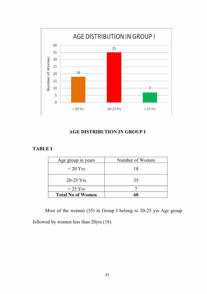
AGE DISTRIBUTION IN GROUP I
TABLE I
Age group in years Number of Women
lt 20 Yrs 18
20-25 Yrs 35
gt 25 Yrs 7Total No of Women 60
Most of the women (35) in Group I belong to 20-25 yrs Age group
followed by women less than 20yrs (18)
41
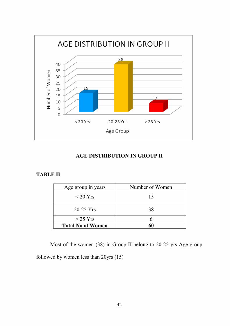
AGE DISTRIBUTION IN GROUP II
TABLE II
Age group in years Number of Women
lt 20 Yrs 15
20-25 Yrs 38
gt 25 Yrs 6Total No of Women 60
Most of the women (38) in Group II belong to 20-25 yrs Age group
followed by women less than 20yrs (15)
42
MEAN AGE IN YEARS
TABLE III
Labour Natural with
Episiotomy
Labour Natural without
Episiotomy
Mean age in years 220667 2275
p = 0198
There is no significant difference in the mean of the ages between the two groups
43
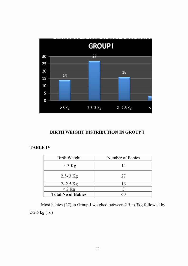
BIRTH WEIGHT DISTRIBUTION IN GROUP I
TABLE IV
Birth Weight Number of Babies
gt 3 Kg 14
25- 3 Kg 27
2- 25 Kg 16lt 2 Kg 3
Total No of Babies 60
Most babies (27) in Group I weighed between 25 to 3kg followed by
2-25 kg (16)
44
BIRTH WEIGHT DISTRIBUTION IN GROUP II
TABLE V
Birth Weight Number of Babies
gt 3 Kg 11
25- 3 Kg 28
2- 25 Kg 14lt 2 Kg 7
Total No of Babies 60
Most babies (28) in Group II weighed between 25 to 3kg followed by
2-25 kg (14)
45
MEAN BIRTH WEIGHT IN EACH GROUP
TABLE VI
Labour Natural
with Episiotomy
Labour Natural
without EpisiotomyMean Birth Weight
in Kg 26896 25577
p = 0095
There is no significant difference in the mean of the birth weights
between the two groups
46
MEAN APGAR AT ONE MINUTE
TABLE VII
Labour Natural with
Episiotomy
Labour Natural
without EpisiotomyMean APGAR at
one minute63333 63833
p = 0748
There is no significant difference in the mean of the APGAR at one
minute between the two groups
47
MEAN APGAR AT FIVE MINUTES
TABLE VIII
Labour Natural with
Episiotomy
Labour Natural
without EpisiotomyMean APGAR at
Five Minutes77167 775
p = 0801
There is no significant difference in the mean of the APGAR at five
minutes between the two groups
48
DURATION OF SECOND STAGE OF LABOUR
TABLE IX
Labour Natural with
Episiotomy
Labour Natural
without EpisiotomyMean Duration of Second Stage of
Labour in Minutes114167 113833
p = 0949
There is no significant difference in the mean of the duration of second
stage of labour in minutes between the two groups
49
50
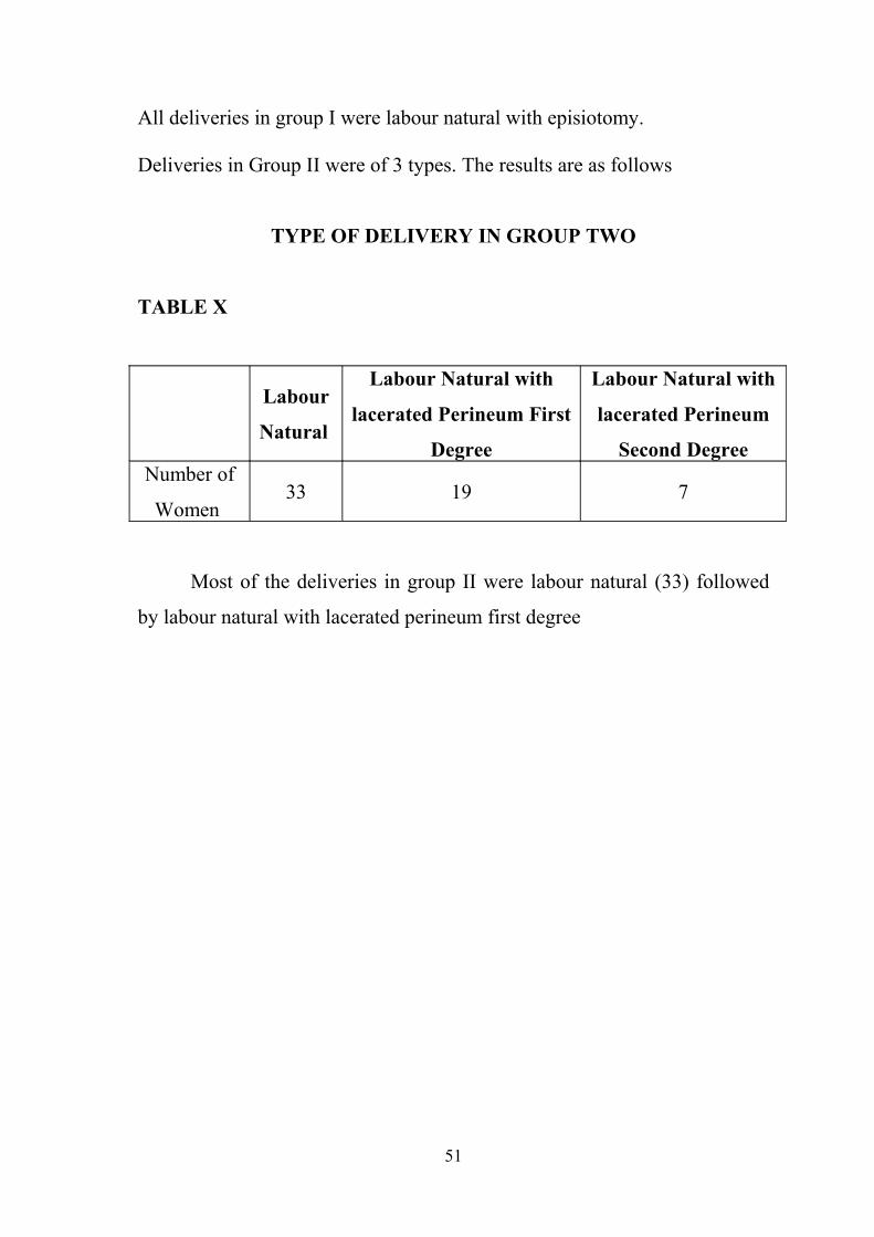
All deliveries in group I were labour natural with episiotomy
Deliveries in Group II were of 3 types The results are as follows
TYPE OF DELIVERY IN GROUP TWO
TABLE X
Labour
Natural
Labour Natural with
lacerated Perineum First
Degree
Labour Natural with
lacerated Perineum
Second DegreeNumber of
Women 33 19 7
Most of the deliveries in group II were labour natural (33) followed
by labour natural with lacerated perineum first degree
51
52
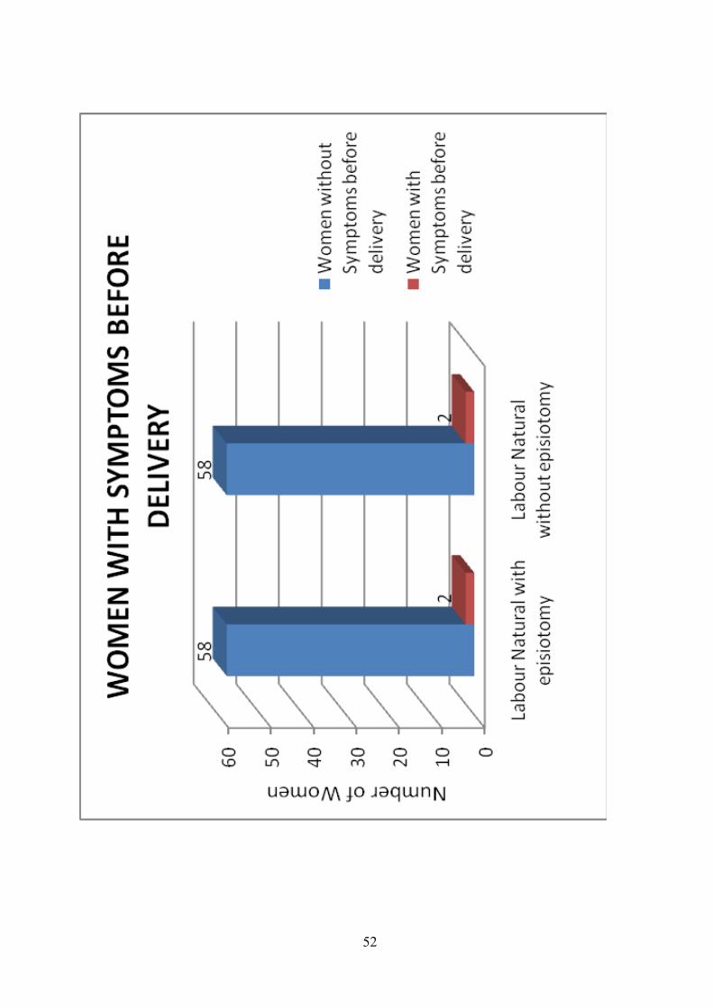
WOMEN WITH SYMPTOMS BEFORE DELIVERY
All the women in both groups were evaluated for the following
symptoms before delivery and for new symptoms after delivery and post
partum
1 Passing flatus when undesirable
2 Any incontinence of liquid stool
3 Any need to wear a pad because of anal symptoms
4 Any fecal urgency
5 Any extra anal leakage
6 Any leakage of material other than stool
TABLE XI
Labour Natural with
Episiotomy
Labour Natural without
EpisiotomyWomen without
Symptoms before delivery
58 58
Women with Symptoms before
delivery2 2
Total Number of Women
60 60
Two patients had symptoms before delivery in group I both of them were passing flatus when undesirable
Two patients had symptoms before delivery in group II Both of them were passing flatus when undesirable
Thus the most common symptom before delivery was passing flatus
when undesirable
53
SIGNS BEFORE DELIVERY
All the women in both groups were evaluated for the following signs
before delivery and for new signs after delivery and post partum
1 Perianal soiling
2 Absence of cutaneous anal reflex
3 Patulous anus
4 Local scarring
None of the women in both the groups had any signs before delivery
54
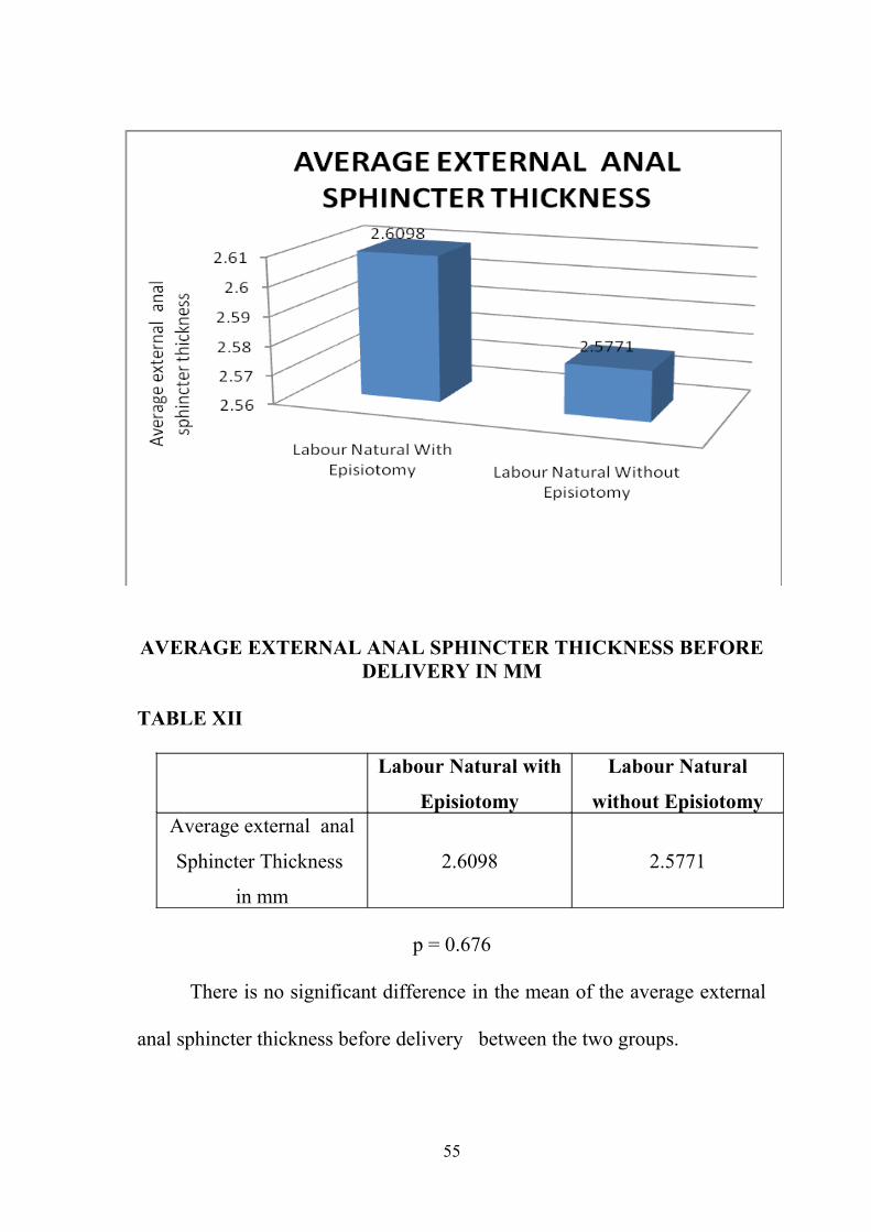
AVERAGE EXTERNAL ANAL SPHINCTER THICKNESS BEFORE DELIVERY IN MM
TABLE XII
Labour Natural with
Episiotomy
Labour Natural
without EpisiotomyAverage external anal
Sphincter Thickness
in mm
26098 25771
p = 0676
There is no significant difference in the mean of the average external
anal sphincter thickness before delivery between the two groups
55
AVERAGE INTERNAL ANAL SPHINCTER THICKNESS BEFORE DELIVERY IN MM
TABLE XIII
Labour Natural with
Episiotomy
Labour Natural
without EpisiotomyAverage Internal Anal
Sphincter Thickness
in mm
23984 23288
p = 02
There is no significant difference in the mean of the average internal
anal sphincter thickness before delivery between the two groups
56
57
AVERAGE COMBINED EXTERNAL amp INTERNAL ANAL SPHINCTER THICKNESS BEFORE DELIVERY IN MM
TABLE XIV
Labour Natural
with Episiotomy
Labour Natural
without EpisiotomyAverage Combined External
amp Internal Anal Sphincter
Thickness
In mm
25038 24529
p = 0384
There is no significant difference in the mean of the average combined
external and internal anal sphincter thickness before delivery between the
two groups
58
SPHINCTER INTEGRITY BEFORE DELIVERY
All women had intact sphincters before delivery in both groups
WOMEN WITH NEW SYMPTOMS AFTER DELIVERY
Women in both groups were evaluated for new symptoms after
delivery
None of the women in the two groups developed any new symptoms
after delivery
59
60
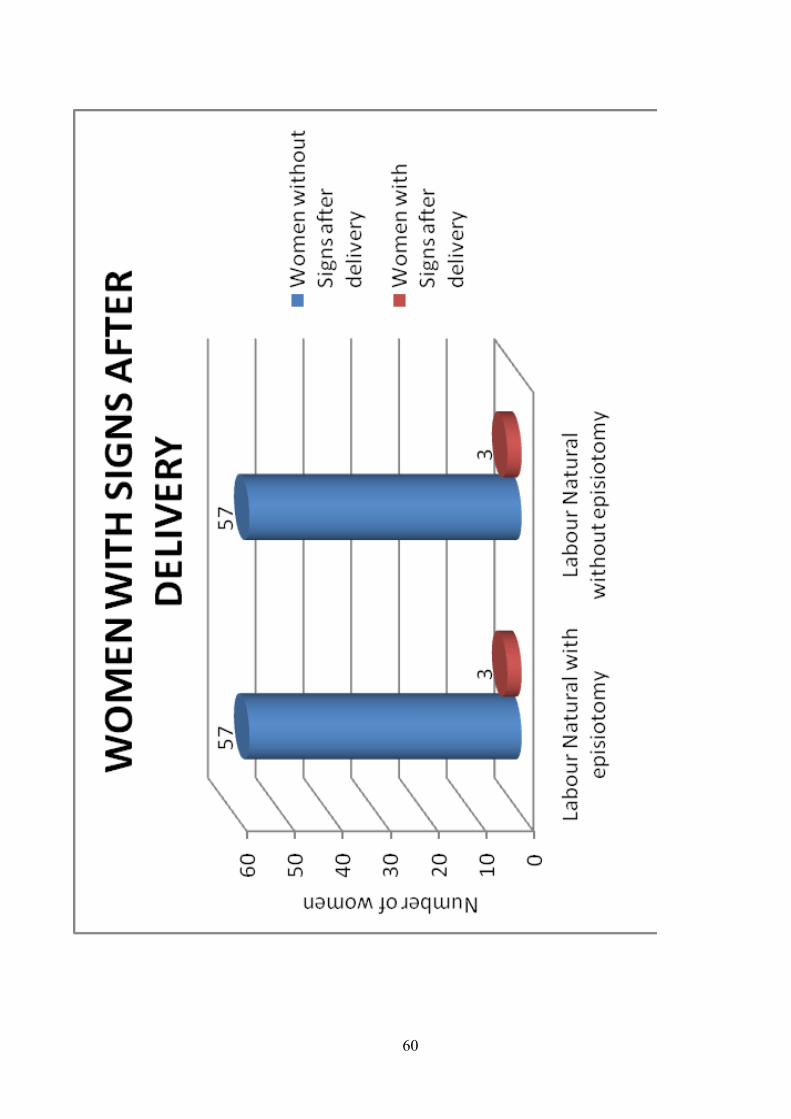
WOMEN WITH SIGNS AFTER DELIVERY
TABLE XV
Labour Natural with
Episiotomy
Labour Natural without
EpisiotomyWomen without
Signs after delivery57 57
Women with Signs
after delivery3 3
Total Number of
Women 60 60
Three women in each group had signs of sphincter damage after
delivery
In the group of women who had labour natural with episiotomy one
woman had patulous anus and two had perianal soiling
In the group of women who had labour natural without episiotomy
one woman had absence of cutaneous anal reflex one woman had patulous
anus and one had perianal soiling
61
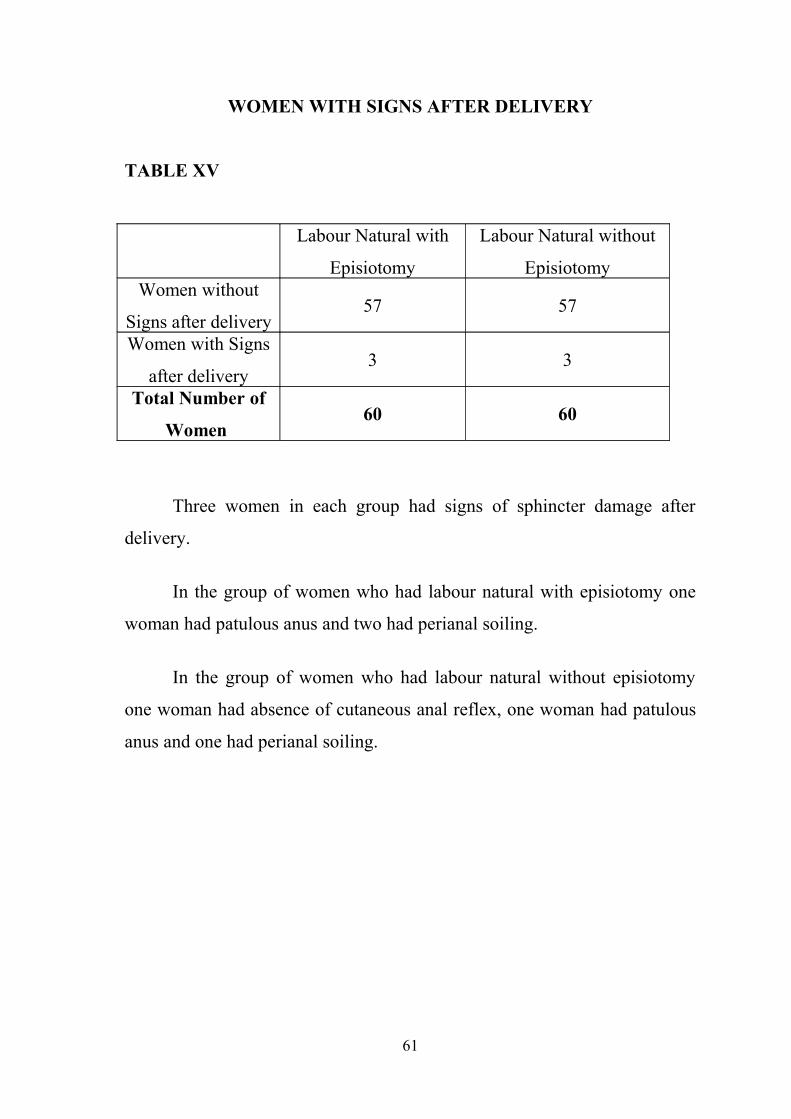
AVERAGE EXTERNAL ANAL SPHINCTER THICKNESS AFTER DELIVERY IN MM
TABLE XVI
Labour Natural with
Episiotomy
Labour Natural
without EpisiotomyAverage External Anal
Sphincter Thickness
after Delivery
In mm
25683 25250
p = 0581
There is no significant difference in the mean of the average external
anal sphincter thickness after delivery between the two groups
62
AVERAGE INTERNAL ANAL SPHINCTER THICKNESS AFTER DELIVERY IN MM
TABLE XVII
Labour Natural with
Episiotomy
Labour Natural
without EpisiotomyAverage Internal Anal
Sphincter Thickness
after Delivery
In mm
23704 228
p = 0113
There is no significant difference in the mean of the average internal
anal sphincter thickness after delivery between the two groups
63
64
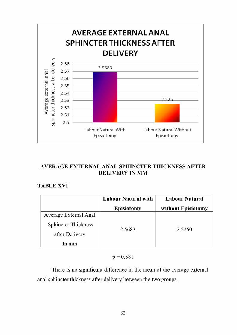
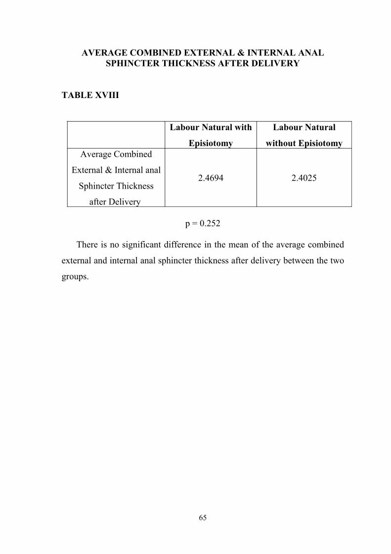
AVERAGE COMBINED EXTERNAL amp INTERNAL ANAL SPHINCTER THICKNESS AFTER DELIVERY
TABLE XVIII
Labour Natural with
Episiotomy
Labour Natural
without EpisiotomyAverage Combined
External amp Internal anal
Sphincter Thickness
after Delivery
24694 24025
p = 0252
There is no significant difference in the mean of the average combined
external and internal anal sphincter thickness after delivery between the two
groups
65
66
SPHINCTER INTEGRITY AFTER DELIVERY
TABLE XIX
Labour Natural with
Episiotomy
Labour Natural without
EpisiotomyWomen with intact
sphincter58 60
Women without
intact sphincter 2 0
Total Number of
Women 60 60
In the group of women who had Labour Natural with Episiotomy 58 women
had an intact sphincter after delivery
Two women had sphincter disruption immediately after delivery
In the group of women who had labour Natural without episiotomy all of
them had an intact sphincter after delivery
On a Pearson Chi Square test (p= 0154) there was no significant difference
between the two groups for sphincter integrity
67
AVERAGE POSTPARTUM EXTERNAL ANAL SPHINCTER
THICKNESS IN MM
TABLE XX
Labour Natural with
Episiotomy
Labour Natural without
EpisiotomyAverage Postpartum
External Anal Sphincter
Thickness
In mm
2573125 25375
p = 0718401
There is no significant difference in the mean of the average post
partum external anal sphincter thickness between the two groups
68
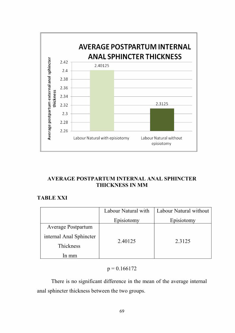
AVERAGE POSTPARTUM INTERNAL ANAL SPHINCTER THICKNESS IN MM
TABLE XXI
Labour Natural with
Episiotomy
Labour Natural without
EpisiotomyAverage Postpartum
internal Anal Sphincter
Thickness
In mm
240125 23125
p = 0166172
There is no significant difference in the mean of the average internal
anal sphincter thickness between the two groups
69
70
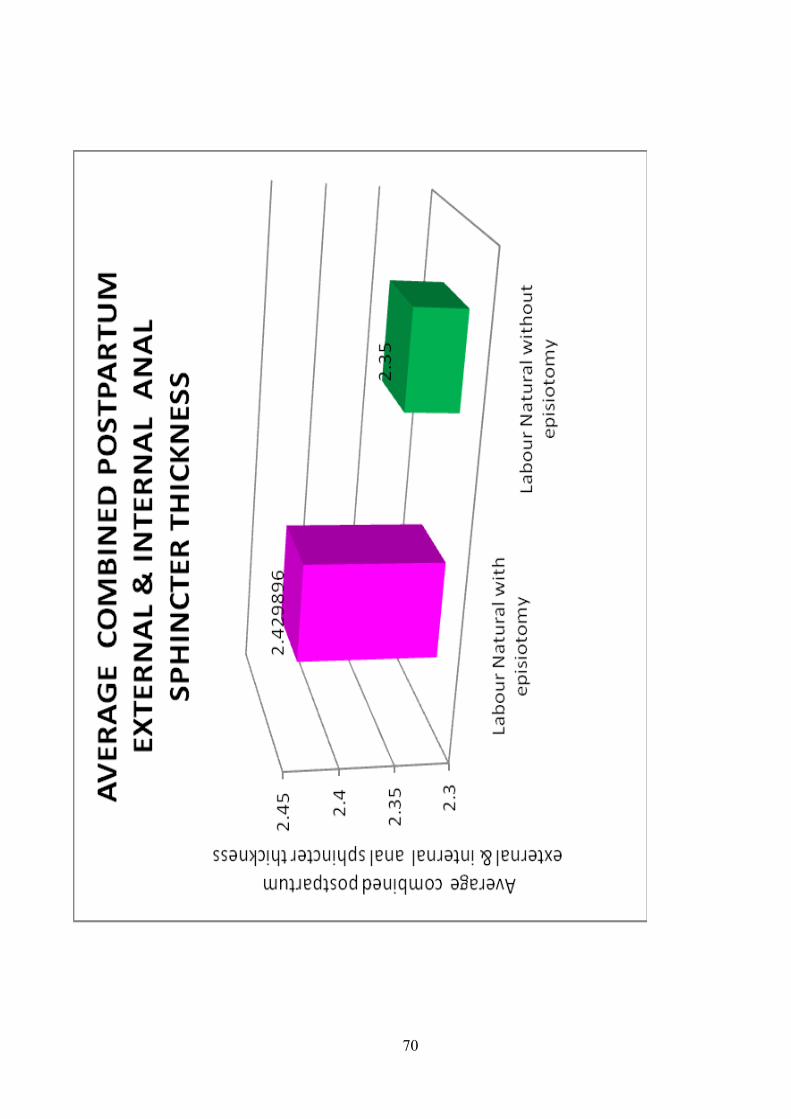
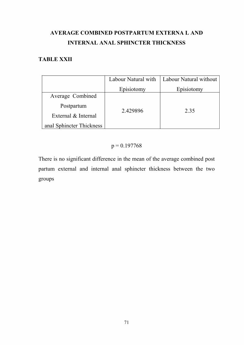
AVERAGE COMBINED POSTPARTUM EXTERNA L AND
INTERNAL ANAL SPHINCTER THICKNESS
TABLE XXII
Labour Natural with
Episiotomy
Labour Natural without
EpisiotomyAverage Combined
Postpartum
External amp Internal
anal Sphincter Thickness
2429896 235
p = 0197768
There is no significant difference in the mean of the average combined post
partum external and internal anal sphincter thickness between the two
groups
71
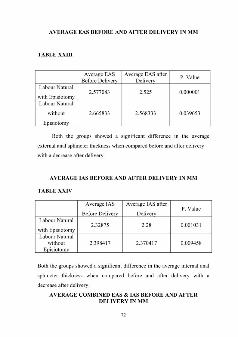
AVERAGE EAS BEFORE AND AFTER DELIVERY IN MM
TABLE XXIII
Average EAS Before Delivery
Average EAS after Delivery
P Value
Labour Natural
with Episiotomy 2577083 2525 0000001
Labour Natural
without
Episiotomy
2665833 2568333 0039653
Both the groups showed a significant difference in the average
external anal sphincter thickness when compared before and after delivery
with a decrease after delivery
AVERAGE IAS BEFORE AND AFTER DELIVERY IN MM
TABLE XXIV
Average IAS
Before Delivery
Average IAS after
DeliveryP Value
Labour Natural
with Episiotomy 232875 228 0001031
Labour Natural without
Episiotomy2398417 2370417 0009458
Both the groups showed a significant difference in the average internal anal
sphincter thickness when compared before and after delivery with a
decrease after delivery
AVERAGE COMBINED EAS amp IAS BEFORE AND AFTER DELIVERY IN MM
72
TABLE XXV
Average EAS amp IAS
Before Delivery
Average EAS amp
IAS after DeliveryP Value
Labour Natural
with Episiotomy 2452971 24025 0000001
Labour Natural without
Episiotomy2532125 2469375 0009005
Both the groups showed a significant difference in the average combined
External and Internal anal sphincter thickness when compared before and
after delivery with a decrease after delivery
AVERAGE EAS AFTER DELIVERY AND POSTPARTUM IN MM
TABLE XXVI
Average EAS After
Delivery
Average EAS
Postpartum P Value
Labour Natural
with Episiotomy 259 2573125 0956862
Labour Natural without
Episiotomy2538125 25375 0945631
Both the groups did not show a significant difference in the average
External anal sphincter thickness when compared after delivery and
Postpartum
AVERAGE IAS AFTER DELIVERY AND POSTPARTUM IN MM
TABLE XXVII
73
Average IAS After
Delivery
Average IAS
Postpartum P Value
Labour Natural
with Episiotomy 2423125 240125 0222267
Labour Natural without
Episiotomy234125 23125 0009021
The first group did not show a significant difference in the average
Internal anal sphincter thickness when compared after delivery and
Postpartum
The Second group showed a significant difference in the average
Internal anal sphincter thickness when compared after delivery and
Postpartum
AVERAGE COMBINED EAS amp IAS AFTER DELIVERY AND
POSTPARTUM IN MM
74
TABLE XXVIII
Average EAS amp IAS
After Delivery
Average EAS amp
IAS Postpartum P Value
Labour Natural
with Episiotomy 2450938 2429896 0187099
Labour Natural without
Episiotomy2374063 235 0010330
The first group did not show a significant difference in the average
combined External and Internal anal sphincter thickness when compared
after delivery and postpartum
The Second group showed a significant difference in the average
combined External and Internal anal sphincter thickness when compared
after delivery and postpartum
75
SUMMARY OF THE COMBINED SPHINCTER THICKNESS
LEVELS
TABLE XXIX
Labour Natural with
Episiotomy
Labour Natural
without Episiotomy Before Delivery and
After Delivery Significant Significant
After Delivery and Postpartum
Not Significant Significant
76
Image of a normal anal sphincter as visualized by TVS
Normal anal sphincter with thick internal anal sphincter
77
Image of a sphincter tear at 11 0rsquoclock
Image of a sphincter tear between 11 and 12 0rsquo clock
78
SUMMARY
79
SUMMARY
1 Of the total number of women who delivered in this hospital 4978 were primiparous
2 Among the primiparous women 46445 underwent a primary cesarean section
3 Of the 53555 of primiparous women who delivered vaginally 8797 had an episiotomy during delivery
4 Most of the women in both the groups belonged to the age group 20 -25 years
5 There is no significant difference between the two groups with respect to age (p = 0198) and hence the two groups are comparable
6 Most of the babies born to women in both groups weighed between 25 to 3 kg
7 There is no significant difference between the means of the birth weights of babies born to women in both the groups (p = 0095)
8 The mean APGAR at one minute and at five minutes are not significantly different between the two groups
9 There is no significant difference in the mean of the duration of second stage of labour in minutes between the two groups
10 The most common symptom of incontinence before delivery was passing flatus when undesirable
80
11 None of the women in both the groups had any signs of anal incontinence before delivery
Before Delivery
12 There is no significant difference in the mean of the average external anal sphincter thickness before delivery between the two groups (P=0676)
13 There is no significant difference in the mean of the average internal anal sphincter thickness before delivery between the two groups p = 02
14 There is no significant difference in the mean of the average combined external and internal anal sphincter thickness before delivery between the two groups p=0384
15 None of the women in the two groups developed any new symptoms after delivery
After Delivery16 There is no significant difference in the mean of the average external
anal sphincter thickness after delivery between the two groups p=0581
17 There is no significant difference in the mean of the average internal anal sphincter thickness after delivery between the two groups p=0113
18 There is no significant difference in the mean of the average combined external and internal anal sphincter thickness after delivery between the two groups p=0252
19 Two women had sphincter disruption immediately after delivery in the group of women who had Labour Natural with Episiotomy On
81
comparison with the group that did not have an episiotomy this was not statistically significant
Post partum20 There is no significant difference in the mean of the average post
partum external anal sphincter thickness between the two groups p=0718401
21 There is no significant difference in the mean of the average internal anal sphincter thickness between the two groups p=0166172
22 There is no significant difference in the mean of the average combined post partum external and internal anal sphincter thickness between the two groups p=0197768
23 The Average EAS and IAS and their combined thickness showed a significant difference in both the groups when compared before and after delivery with a decrease in thickness after delivery
24 When compared after delivery and postpartum the EAS did not show a significant difference in both the groups
In the women who had an episiotomy there was no significant difference in the IAS thickness after delivery and postpartum
In the women who did not have an episiotomy there was a significant difference in the IAS thickness after delivery and postpartum
25 In the women who had an episiotomy there was no significant difference in the combined thickness after delivery and postpartum
In the women who did not have an episiotomy there was a significant difference in the combined thickness after delivery and postpartum
82
DISCUSSION
83
DISCUSSION
Thacker and Banta estimated that episiotomy was performed on 50
to 90 of all nulliparas (6)
Thorpe et al reported that episiotomy was performed on 62 of
vaginal deliveries and further breakdown revealed that this procedure was
performed in 80 of nulliparas(9)
In this study the episiotomy rate among primiparous women is
8797
Larrson et al compared an episiotomy group with a lacerated group
and a non traumatic birth group and found no significant differences in
APGAR scores among the two groups (49)
Thranov et al studied one minute APGAR scores and found no
differences among groups with low medium and high episiotomy rates (48)
Borgatta et al have also found no difference in APGAR scores
between episiotomy and no episiotomy groups (50)
In this study there was no significant difference in the APGAR scores
between the two groups
Borgatta et al and Harrison et al have found no significant difference
in the duration of second stage of labour between the episiotomy group and
no episiotomy group
In this study there was no significant difference in the duration of
second stage of labour in minutes between the two groups
84
There is no difference in the individual and combined averages of the
sphincter thickness before delivery after delivery and postpartum between
the two groups in this study
The is a significant decrease in the individual and combined sphincter
thickness immediately after delivery when compared to before delivery
irrespective of whether an episiotomy was given or not
The is a significant decrease in the internal and combined anal
sphincter thickness in postpartum period when compared to immediately
after delivery in the labour natural without episiotomy group
85
CONCLUSION
86
CONCLUSION
Episiotomy is the most commonly performed surgical procedure in
Obstetrics There will always be circumstances in which prudent clinical
judgement may dictate the necessity for an episiotomy fetal distress
shoulder dystocia breech delivery persistent occipito posterior vacuum or
forceps operation and maternal exhaustion
Until clear guidelines emerge for practitioners based on prospective
randomized control trials obstetricians should determine the need for
episiotomy on a case by case basis
The use of transvaginal ultrasound is a simple method to evaluate the
anal sphincter that can be used in centres where more advanced methods
like endoanal ultrasound pudendal nerve terminal motor latency and anal
manometry are not readily available
Increasing awareness of the frequency and extent of pelvic floor
lesions has led to the increased use of new diagnostic and treatment
modalities Our understanding of normal pelvic floor anatomy and its
ultrasonographic appearance has improved as imaging options have
expanded The next step is practical application of these findings to
characterizing pelvic floor function This can be done through well-
designed and sufficiently powered clinical studies These studies will
establish the association between the clinical presentations of dysfunction
and the ultrasonographic findings
87
Annexure
88
BIBLIOGRAPHY
BIBLIOGRAPHY
1 Munro Kerrrsquos Operative obstetrics Eleventh edition Centenary edition
Centenary edition Chapter 21 Pg 253 Elsevier Saunders ndash Thomas
F Baskett Andrew A Calder Sabaratnam Arulkumaran
89
2 Fielding Ould A treatise on Midwifery in three parts Dublin Nelson and
Connor 1742 p 145
3 Harrie J Practical directions showing a method of preserving the
perineum in childbirth London Wilson D Nicol 6 1767
4 Goodell WA critical inquiry into the management of the perineum
during labour AmJMed Sci 1871 61 53-79
5 Quilligan EJ Zuspan F Management of delivery trauma In Douglas
Stromme editor Operative obstetrics 4th edn New York Appleton
Century crofts 1982 697-742
6 Thacker SB Banta HD Benefits and risks of episiotomy an interpretive
review of the English language literature 1860-1980 Obst Gynecol
Survey 1983 38(6) 322-338
7 Nugent F the primiparous perineum after forceps delivery Am J obstet
Gynecol 193530 249
8 DeLee JB The prophylactic forceps operationAm J Obstet Gynecol
134 1920
9 Thorpe H Bowes W Brame R et al Selected use of midline
episiotomy Effect on perineal traumaObstet Gynecol 79 945 ndash
9491992
10 Arthur TEvans Manual of Obstetrics 7th edition Lippincott Williams
Wilhem Wolters Kluwer Health Ch-2 Normal labour pg41
11Vishwanathan M Hartmann K Palmieri R et al the use of episiotomy in
obstetrical care a systematic review evidence report Technology
assessment no112 (prepared by the RTI ndashUNC evidence based practice
90
center under contract no290-02-0016) AHRQ Publication no05-E
009-2 Rockville MD Agency for Healthcare Research and Quality
2005
12Royal college of Obstetricians and Gynecologists Management of third
and fourth degree perineal tears following vaginal delivery Guideline
No 29 London RCOG press 2001
13Anthony S Buitendijk SE Zondervan KT Van Rijssel EJC Verkerk PH
episiotomies and the occurrence of severe perineal lacerations Br J obstet
Gynecol 1994 1011064-7
14Poen AC Felt Bersma RJF Dekker GA Deville W Cuesta MA
Meuwissen SGM Third degree obstetric perineal tears risk factors and
the preventive role of mediolateral episiotomy Br J Obstet Gynecol
1997 104 563-6
15Henriksen TB Bek KM Hedegaard M Secher NJ Mehods and
consequences of changes in use of episiotomy BMJ 1994309 1255-8
16Fritel X Schaal J Fauconnier A Bertrand V Levet C Pigne A Pelvic
floor disorders 4 years after first delivery a comparative study of
restrictive versus systematic episiotomy BJOG 2008 115 247-252
17Eason E Feldman P Much ado about a little cut is episiotomy
worthwhile Obstet Gynecol 956162000
18 Williams textbook of obstetrics Cunningham FG Leveno KJ Bloom SL
Hauth JC Gilstrap LC Wenstrom KD 22nd Edition Stamford
Appleton amp Lange Ch-17 Normal labor and Delivery Page ndash 435 ndash 438
19Abramowitz L Sobhani I GanansiaR et al Are sphincter defects the
cause of anal incontinence after vaginal delivery Results of a
prospective study Dis colon rectum 2000 43590-8
20Jorge JM Wexner SD Aetiology and management of fecal incontinence
Dis Colon Rectum 199336 77-79
91
21 High Risk Pregnancy Management Options Third Edition D K James
P J Steer CP Weiner B Gonik Saunders (WB) Co Ltd Elsevier
Ch ndash 73 Perineal Repair and Pelvic floor injury Page ndash 1505 ndash 1506
22Delancey J Episiotomy In Hankins G Clarke S Cunningham FG
et al Operative Obstetrics Norwalk CTAppleton and Lange1995
p108 ndash 109
23Myers Helfgot MG etal Routine use of episiotomy in modern
Obstetrics Should it be performed
North Am J Obs Gyn 1999p 305 ndash 325
24Harrison R Brennan M North P et al Is routine episiotomy necessary
BMJ 288 1971 ndash 1975 1984
25 Dan V Valsky MD Simcha Yagel MD Three-Dimensional
Transperineal Ultrasonography of the Pelvic Floor Improving
Visualization for New Clinical Applications and Better Functional
Assessment J Ultrasound Med 2007 261373ndash1387
26Sultan AH Kamm MA Hudson CN et al Anal sphincter disruption
during vaginal delivery N Eng J Med 19933291905 ndash 11
27 Carroli G Belizan J Episiotomy for vaginal birth Cochrane Database of
Systematic Reviews 1999 Issue 3 Art No CD000081
28 Andrews V Sultan AH Thakar R Jones PW Risk factors for obstetric
anal sphincter injury a prospective study Birth 200633(2)117-22
29 Vasanth Andrews Ranee Thakar Abdul H Sultan Peter W Jones
Are mediolateral episiotomies actually mediolateral BJOG An
International Journal of Obstetrics amp Gynaecology Volume 112 Issue
8 Pages 1156 ndash 1158 Mar 2005
30Signorello LB et al ndash Midline episiotomy and anal incontinence
Retrospective cohort study BMJ 2000320 86- 90
92
31Samuelsson E et al ndash Anal sphincter tears prospective study of obstetric
risk factors BJOG July 2000 Vol 107 pp 926 ndash 931
32Mous M Muller SAde Leeuw JW ndash Long term effects of anal sphincter
rupture during vaginal delivery Faecal incontinence and sexual
complaintsBJOG 2008115 234- 238
33Rodriguez A Arenas EA Osorio AL et al Selective vs routine midline
episiotomy for the prevention of third or fourth degree lacerations in
nulliparous womenAm J Obstet and Gynecol 2008198285e1- e4
34Sleep J et al ndash West Berkshire Perineal management trial BMJ 1984
35Belizan J ndash Argentine Collaborative trial Routine Vs Selective episiotomy ndash An RCT - Lancet 1993
36Nygaard et al Anal incontinence after anal sphincter disruption 30yrs retrospective cohort study Obs Gyn 1997
37Divya et al ndash Childbirth and pelvic floor dysfunction An epidemiologic
approach to the assessment of prevention oppurtunities at delivery-
AJOG 2006 Vol 19523-8
38John R Scott MD ndash Episiotomy and Vaginal Trauma ndash Obstetrics and
Gynecology Clinics of North America -32(2005) 307- 321
39Ursula M Peschers John O L De Lancey Gabriel N Schner Bernhard
Schuessler ndash Exoanal ultrasound of the anal sphincter normal anatomy
and sphincter defects- BJOG 1997 Vol 104 pp 999 ndash 1003
40 Timor-Tritsch IE Monteagudo A Smilen SW Porges RF Avizova E
Simple ultrasound evaluation of the anal sphincter in female patients
93
using a transvaginal transducer J Ultrasound Obstet Gynecol 2005
Feb25(2)177-83
41 Bradley et al - Risk factors for sonographic internal anal sphincter gaps
6-12 months after delivery complicated by anal sphincter tear American
Journal of Obstetrics amp Gynecology 197(3)310e1-310e5 September
2007
42 Sharon Maslovitz Ariel Jaffa Ishai Levin Benjamin Almog Joseph B
Lessing Igal Wolman The clinical significance of postpartum
transperineal ultrasound of the anal sphincterEuropean Journal of
Obstetrics Gynecology and Reproductive Biology Vol 134 issue 1
115-119 Sept 2007
43 Gregory w Thomas Boyles Sarah Hamilton Simmons Kimberly
Corcoran Amy Clark Amanda lAmerican journal of obstetrics and
gynecology 2006 vol 194 5 pp 1243-1248
44Sultan AH Loder PB Bartram CI Kamm MA Hudson CN Vaginal
Endosonography new technique to image the undisturbed anal sphincter
Diseases of the Colon amp Rectum 1994371296-1299
45 Lori K Stewart Stephanie A Wilson- Transvaginal Sonography of the
Anal Sphincter Reliable or Not AJR 1999173179-185
46 Alexander AA Liu JB Merton DA Nagle DA Fecal incontinence
transvaginal US evaluation of anatomic causes Radiology 1996
May199(2)529-32
47 J M Ramiacuterez V Aguilella M Martiacutenez and J A Gracia The utility of
endovaginal sonography in the evaluation of fecal incontinence Rev esp
enferm dig 2005 97(5) 317-322
94
48 Thranov I Kringelbach AM Melchior E Postpartum symptoms
Episiotomy or tear at vaginal delivery Acta Obstet Gynecol Scand
6911 ndash 15 1990
49Larsson P Platz ndash Christensen J Bergam B et al Advantage or
disadvantage of episiotomy compared with spontaneous perineal
laceration Gynecol Obstet Invest 31 213- 216 1991
50 Borgatta L Piening S Cohen WR Association of episiotomy and
delivery position with deep perineal lacerations during spontaneous
vaginal delivery in nulliparous women Am J Obstet Gynecol 160
294-297 1989
95
PROFORMA
PROFORMA FOR EVALUATION
Name Age IP No
Date of Admission Date of Delivery
Obstetric score
Symptoms
Passage of any flatus when socially undesirable
96
Any incontinence of liquid stool
Any need to wear a pad because of anal symptoms
Any incontinence of solid stool
Any fecal urgency (inability to defer defecation for more than 5 minutes)
Past medical history
History of diabetes preexisting neurological dysfunction
Past Surgical history
History of back injury
History of surgical treatment of back pain
History of abdominopelvic surgery like hemorroidectomy
History of radiation
Present Obstetric history
Birth Weight of Baby
Duration of second stage of labour APGAR 1 APGAR 5
Mode of delivery
With or without episiotomy
Episiotomy median mediolateral
Perineal laceration degree of perineal laceration
Without perineal laceration
General Examination
Spine
Evidence of neurological deficit
97
Before delivery
PA
Local examination
Posterior vaginal wall
PR to rule out mass impaction
Perineal body
Levator anii tone
External sphincter tone on voluntary contraction
Signs
perianal soiling
absence of the cutaneous anal reflex
patulous anus
local scarring
Ultrasonographic criteria
Before delivery Sphincter thickness
EAS ndash External Anal Sphincter IAS ndash Internal Anal Sphincter
EAS 12 orsquo clock IAS 12 orsquo clock
EAS 3 orsquo clock IAS 3 orsquo clock
EAS 6 orsquo clock IAS 6 orsquo clock
EAS 9 orsquo clock IAS 9 orsquo clock
Average EAS Average IAS
After delivery
98
Any new symptoms of incontinence
Local examination
Posterior vaginal wall
Perineal body
Levator anii tone
External sphincter tone on voluntary contraction
Signs
perianal soiling
absence of the cutaneous anal reflex
patulous anus
local scarring
After delivery Sphincter thickness
EAS 12 orsquo clock IAS 12 orsquo clock
EAS 3 orsquo clock IAS 3 orsquo clock
EAS 6 orsquo clock IAS 6 orsquo clock
EAS 9 orsquo clock IAS 9 orsquo clock
Average EAS Average IAS
Postpartum
Any new symptoms of incontinence
99
Local examination
Posterior vaginal wall
Perineal body
Levator anii tone
External sphincter tone on voluntary contraction
Signs
perianal soiling
absence of the cutaneous anal reflex
patulous anus
local scarring
Postpartum Sphincter thickness
EAS 12 orsquo clock IAS 12 orsquo clock
EAS 3 orsquo clock IAS 3 orsquo clock
EAS 6 orsquo clock IAS 6 orsquo clock
EAS 9 orsquo clock IAS 9 orsquo clock
Average EAS Average IAS
100
Abbreviations
s
ABBREVIATIONS
ES EAS ndash External Anal Sphincter
IS IAS ndash Internal Anal Sphincter
PP ndash Postpartum
Master chart
BEFORE 12 EAS ndash External Anal Sphincter thickness at 12 orsquoclock
position
before delivery
101
BEFORE 12 IAS - Internal Anal Sphincter thickness at 12 orsquoclock position
before
delivery
AVG EAS ndash Average External Anal Sphincter thickness before delivery
AVG IAS - ndash Average Internal Anal Sphincter thickness before delivery
AFTER 12 EAS - External Anal Sphincter thickness at 12 orsquoclock position
after
delivery
AFTER 12 IAS- Internal Anal Sphincter thickness at 12 orsquoclock position
after
delivery
AVG AFTER EAS - Average External Anal Sphincter thickness after
delivery
AVG AFTER IAS - Average External Anal Sphincter thickness after
delivery
PP 12 EAS - External Anal Sphincter thickness at 12 orsquoclock position
postpartum
PP 12 IAS- Internal Anal Sphincter thickness at 12 orsquoclock position
postpartum
AVG PP EAS - Average Internal Anal Sphincter thickness postpartum
102
MASTERCHART
103
CERTIFICATE
This is to certify that the dissertation work titled ldquoANAL
SPHINCTER COMPLEX - A COMPARATIVE STUDY OF VAGINAL
DELIVERY WITH EPISIOTOMY AND WITH PERINEAL
LACERATIONS IN PRIMIPAROUS WOMEN -TRANSVAGINAL
ULTRASONOGRAPHIC EVALUATIONrdquo is a bonafide research work
of DRNBANUREKHA Enrolment Nohelliphelliphelliphelliphelliphelliphelliphelliphellip Submitted
in partial fulfillment of the requirements for the award of Degree of MD
OBSTETRICS amp GYNAECOLOGY (BRANCH-II) in THE TAMIL
NADU DRMGR MEDICAL UNIVERSITY CHENNAI- 600 032
Signature of HOD Signature of Dean
2
ACKNOWLEDGEMENTS
I thank our Dean Prof Dr M DHANAPAL Kilpauk Medical
College Chennai ndash 10 for permitting me to conduct this study in Kilpauk
Medical College and Hospital
I gratefully thank my Prof Dr MMUTHULAKSHMI MD
DGO Head of Department Government Kilpauk Medical College and
Hospital under whom I have the honour to work as a post graduate student
I express my sincere thanks to Prof Dr TASRIDEVI MD
DGO for her support and guidance through this endeavour
I am deeply indebted to her valuable guidance inspiration and
suggestions without which this work would not have been completed
My heartful thanks to our Professors Dr HKFATHIMA
Dr FAMIDHA Dr R PREMALATHA and Dr PMEENALOCHANI
and assistant Professors for their suggestions
I thank Mr PADMANABAN (Research Officer) for his guidance in
statistical analysis
I thank Precision Diagnostics Pvt Ltd lsquos Dr Bharathi Y Dhala Dr
Sathyabama Chandrasekaran Dr K Rajan DrBobji Kettay for sharing the
expertise necessary to conduct this study
3
I wholeheartedly thank my patients without their cooperation this
study could not have been completed
I thank my Father Mother and Brother for making me who I am
today
I thank my Husband for having stood by me at all times
I thank God for inspiring me to believe in Him
4
CONTENTS
CHAPTER TITLE PAGE NO
1 INTRODUCTION 1
2 REVIEW OF LITERATURE 15
3 AIM OF THE STUDY 26
4 MATERIALS AND METHODS 27
5 RESULTS 29
6 SUMMARY 61
7 DISCUSSION 64
8 CONCLUSION 66
9 ANNEXURE
a BIBLIOGRAPHY
b PROFORMA
c ABBREVIATIONS
d MASTER CHART
5
INTRODUCTION
6
INTRODUCTION
Episiotomy is a widely performed intervention in childbirth
Episiotomy has traditionally been one of the most frequently performed
obstetric procedures and has been one of the more controversial Defined as
a surgical incision in the perineum to enlarge the vaginal opening for birth
episiotomy is the incision of the pudenda whereas perineotomy is the
incision of the perineum
It is the only surgical procedure in obstetrics to be performed without
the patientrsquos specific consent
History of episiotomy
First description of an episiotomy was given by Sir Fielding Ould
(1710-1789) a Dublin midwife in 1742(1) in his A treatise on Midwifery in
three parts
lsquoIt sometimes happenshelliphellip that the head of the child hellipcannot however
come forward by reason of the extraordinary constriction of the external
orifice of the vagina helliphellipwherefore it must be dilated if possible by the
fingershellip if this cannot be accomplished there must be an incision made
toward the anus with a pair of crooked probe scissors introducing one blade
between the head the vaginahelliphelliphellip as for as shall be thought necessary
for the present purpose and the business is done at one pinch by which the
whole body will easily come forth(2)
Our predecessors were accomplished at trying to avoid perineal
lacerations
7
In 1776 Harrie discussed the importance of lubricating the perineum
and vagina with fresh hogrsquos lard ironing out the perineum and controlling
delivery of the vertex(3)(38)
Puzos recommended support of the perineum to prevent lacerations
(4) Before the development of suturing perineal lacerations were managed
with prolonged bed rest and sometimes binding the legs tightly together
Pare is said to have been the first to suture the perineum whereas
Mauriceau is credited for the first perineorraphy(5)In 1799 Michelis first
recommended midline incisions in the perineum(6) In 1847 Dubois first
suggested making an oblique incision in the perineum known today as the
mediolateral episiotomy In 1851 The Stethoscope and Virginia Medical
Gazette reported that Taliaferro from Virginia unaware that the operation
had ever been done before performed the first episiotomy in the United
States(6)(38)
Perineotomy is the incision of the perineum whereas the term
episiotomy coined by Carl Braun in 1857 is the incision of the pudenda or
external genitalia These terms gradually became synonymous(7)
Episiotomy was not widely used until the 1920s when Pomery and De
Lee changed the climate of opinion regarding the entire birth process In his
The Prophylactic Forceps Operation De Lee claimed that episiotomy would
preserve the integrity of the pelvic floor and the introitus(8)
In addition to the strong advocacy for the use of episiotomy by the
obstetricians of the day changes in maternity practices also affected the use
of episiotomy The shift from home birth to hospital deliveries contributed
to a shift in the conceptualization of the nature of childbirth This shift
8
provided aseptic operating conditions and the necessary technology to carry
out episiotomy safely This change in delivery sites unleashed a cascade of
interventions that were not based on scientific evidence With the increase
in the number of hospital deliveries physicians attending deliveries believed
policies of liberal episiotomy use shortened labour allowing the physician
to complete a birth rapidly
As more women delivered in hospital increasing numbers of them
received anesthesia which in turn interfered with the natural expulsive
efforts of the mothers increasing the need for forceps Forceps delivery
required better access to the birth canal and more room for vaginal
manipulations and the dorsal lithotomy position provided just such access
During the 1970s and 1980s as the naturalist movement flourished
the routine use of episiotomy began to be questioned Thacker and Banta
found that there is no clearly defined evidence for its efficacy particularly
for routine use and considerable evidence of risks associated with
episiotomy including pain edema infection and ultimately dyspareunia
(6)
Incidence of episiotomy
Thacker and Banta estimated that episiotomy is performed on 50 to
90 percent of all nulliparas (6)Thorpe et al reported that episiotomy was
performed on 62 of vaginal deliveries in the United States Further
breakdown of these numbers indicate that the procedure was executed in
80 of nulliparas patients and in 20 of parous patients (9)
Types of Episiotomy
9
Multiple techniques can be used to effect an episiotomy The incision
may be made with scissors or a knife and may be made in the midline
(midline episiotomy) or begun in the midline and extended laterally
(mediolateral episiotomy) (10)
Midline episiotomy - two fingers are placed in the vagina between
the fetal head and the perineum and using straight scissors the incision is
made from fourchette through the perineal body up to but not including the
external anal sphincter Advantages of the midline episiotomy are that it
does not cut through the belly of the muscle the two sides of the incised
area are anatomically balanced making surgical repair easier blood loss is
less than with mediolateral episiotomy A major drawback is the propensity
for extension through the external anal sphincter and in to the rectum For
this reason may practitioners avoid the midline technique
Mediolateral episiotomy ndash the incision is made starting at the midline
of the posterior fourchette and aimed towards the ischial tuberosity to avoid
injury to the anal sphincter The incision is usually about 4cm long In
addition to the skin and subcutaneous tissues the bulbocavernosus
transverse perineii and puborectalis muscles are cut Whether the incision is
to the right or left depends on operator preference(1)
10
ROLE OF EPISIOTOMY IN MODERN OBSTETRICS
The traditional teaching that episiotomy was protective against more
severe perineal lacerations has not been substantiated Thus the liberal use
of prophylactic episiotomy is no longer recommended However there are
still valid reasons for the performance of an episiotomy
1 To shorten the second stage of labour in cases of fetal distress
2 In selected cases of assisted vaginal delivery with forceps and less
frequently for vacuum assisted delivery
3 To obtain more room for obstetrical manoeuvres such as those with
shoulder dystocia assisted breech delivery and delivery of second
twin and in occiputo posterior(1)
Consequences of an episiotomy
Immediate consequences of an episiotomy would include blood loss
pain edema infection and hematoma Definitive disadvantages would
include extension to a third degree or fouth degree laceration the
complications of which would include loss of rectal tone rectovaginal
fistula and abscess formation(9) (23)More long term complications include
the formation of scar tissue wound infection and dyspareunia
Routine use of episiotomies has not been shown to improve outcomes
for patients and can actually be harmful Routine or liberal use of
episiotomies increases the risk that the patient will have a surgical incision
that is larger than if she experiences a spontaneous laceration at birth (11)
11
Lacerations of the vagina and perineum are classified as first second third
or fourth degree (12)
Classification of perineal
injury
INJURY
DEFINITION
First degree Injury confined to vaginal mucosa
Second degreeInjury of vaginal mucosa and perineal muscles but
not the anal sphincter
Third degreeInjury to the perineum involving the anal sphincter
complex (external and internal)
3a emspensplt50 of external sphincter thickness is torn
3b emspenspgt50 of external sphincter thickness is torn
3c Internal sphincter is tornemspensp
Fourth degreeInjury to anal sphincter complex (external and
internal sphincter )and rectal mucosa
One of the major justifications for the use of episiotomy is the
suggested protective role of episiotomy in the prevention of severe tears
The use of elective episiotomy is believed to prevent the occurrence of third
12
and fourth degree perineal lacerations A comparison of the incidence of
third and fourth degree perineal lacerations in patients who did not undergo
episiotomy women who underwent midline episiotomy and women who
underwent mediolateral episiotomy is important These injuries range from
0 to 23 in women with intact perineii02 to 9 in women with a
mediolateral episiotomy to 3 to 24 in women who underwent midline
episiotomy(22)
The association between episiotomy and perineal trauma (third degree
laceration) is complex On one hand mediolateral episiotomy is associated
with a lower risk of anal sphincter rupture at delivery (13)(14)On the other
hand it has also been shown that the number of mediolateral episiotomies
may be reduced without an increase in perineal trauma(15) Then the
protective effect of mediolateral episiotomy may be limited to situations in
which its use is inevitable while its routine performance may increase the
risk of anal incontinence It is possible that performing an episiotomy when
anal sphincter is not in danger increases the risk of direct scissor injuries to
the sphincter (Fritel x et al)(16) Unfortunately we do not know what
episiotomy rate offers the balance between benefits and risks for the anal
sphincter
It seems reasonable to conclude that episiotomy should not be
performed routinely (Eason Feldman 2000)(17) The procedure should be
applied selectively for appropriate indications The final rule is that there is
no substitute for surgical judgement and common sense (18)
ANAL INCONTINENCE
13
Anal incontinence is much more prevalent than once thought Anal
incontinence is an embarrassing condition that is largely underreported
Obstetric anal sphincter injuries are the major etiological factor
Recognition of risk factors may minimize the development of sphincter
injuries Many women do not discuss this problem with anyone because it is
socially embarrassing Anal incontinence affects approximately 10 of
women (19)
Less than 50 of woman with fecal incontinence report the problem
to their physician It can be a devastating problem for women and
obstetricians must be careful first to try and avoid a third or fourth degree
tear and then to repair it meticulously to give a woman the best opportunity
for a good functional repair
Risk factors for third- and fourth-degree tears have been identified
mainly in retrospective studies Taking an overall risk of 1 of vaginal
deliveries the following factors are associated with an increased risk of a
third- and fourth-degree tear (12)
14
Major risk factors for obstetric anal sphincter injury
RISK FACTOR
ODDS RATIO
Nulliparity (primigravidity) 3ndash4
Inherent predisposition
Short perineal bodyemspenspemspensp 8
Instrumental delivery overall 3
Forceps-assisted deliveryemspenspemspensp 3ndash7
Vacuum-assisted deliveryemspenspemspensp 3
Forceps vs vacuumemspenspemspensp 288
Forcepsemspenspemspensp with midline episiotomy 25
Prolonged second stage of labor (gt1 hour) 15ndash4
Epidural analgesia 15ndash3
Intrapartum infant factors
Birthweight over 4 kgemspenspemspensp 2
Persistent occipitoposterior positionemspenspemspensp 2ndash3
Episiotomy mediolateral 14
Episiotomy midline 3ndash5
Previous anal sphincter tear 4
15
POSTNATAL ASSESSMENT
Postnatal continence assessment should include direct questioning
using a standardized bowel function questionnaire so that a reliable
assessment of continence can be done(19) Continence of flatus liquid and
solid feces should be documented together with inquiry concerning fecal
urgency inability to defer defecation for longer than 5 minutes a socially
debilitating symptom The complaint of urgency incontinence may reflect
external anal sphincter dysfunction
A standardized bowel function questionnaire would include in
addition to the above questions an inquiry assessing the need to wear a pad
because of anal symptoms any extra anal leakage or any leakage of material
other than stool (21)
Digital examination of the anal canal may provide an approximate of
the integrity of the sphincter and perineal body but is not otherwise
diagnostically reliable
16
INVESTIGATIONS FOR ANAL INCONTINENCE
It is important to bear in mind that continence depends on multiple
physiologic mechanisms and therefore no single diagnostic test yields
positive results in all patients
Endoanal ultrasound has significantly changed the evaluation of fecal
incontinence over the last decade In women with fecal incontinence in
whom obstetric injury is suspected 90 have sonographic evidence of
injury to one or both sphincters The ultrasound images show 2 discrete
rings of tissue The inner hypoechoic ring represents the internal anal
sphincter and the outer hyperechoic ring represents the external anal
sphincter Disrupted continuity of these rings is consistent with
structural damage to the sphincter
It can serve as a screening tool to detect occult sphincter injury
following vaginal delivery thus identifying women at high risk for
future incontinence Women with transient stool or gas incontinence
following vaginal delivery may be candidates for screening endoanal
ultrasound for further evaluation
Transperineal and transvaginal ultrasound are reasonable alternatives
which are now advocated for use in centres where the equipment or
expertise necessary for endoanal ultrasound are not readily available
Anorectal manometry can detect functional weakness of sphincters
that are anatomically intact by measuring sensation resting and
squeeze pressures Decreased resting pressure suggests isolated internal
anal sphincter injury decreased squeeze pressure suggests external
anal sphincter injury
17
Rectal sensory testing is assessed by inflating a balloon in the rectum
and recording the smallest volume of rectal distention for first
detectable sensation (rectal sensory threshold) sensory urgency and
pain (maximum tolerable volume)
Electromyography of the external anal sphincter and the pelvic
floor musclesmdashtraditionally performed using needle electrodes or
surface electrodesmdashis helpful in delineating areas of sphincter injury
by mapping the sphincter However much of this information is now
obtained accurately by endoanal ultrasound which has replaced
needle EMG for most clinicians
Defecography involves imaging the rectum after filling it with
contrast material and then observing the process rate and
completeness of rectal evacuation using fluoroscopy Its usefulness in
the evaluation of fecal incontinence is limited to cases of rectal
prolapse and for the diagnosis of rectocele and enterocele
Pudendal nerve testing This test uses an electrode to measure
pudendal nerve conduction time known as pudendal nerve terminal
motor latency (PNTML)mdashthus allowing further investigation for
nerve injury
bull Normal PNTML value is 22 milliseconds
bull A value between 22 and 26 milliseconds indicates probable
nerve damage
bull A value of 26 milliseconds or greater confirms nerve damage
Clinical use of PNTML is controversial it helps diagnose nerve injury
but is currently reserved for investigational purposes
18
Anatomy of the anal sphincter
The pelvic floor is a multilayer structure consisting of muscles and
ligaments that support the pelvic organs and play an important role in pelvic
organ function The major muscle of the pelvic floor is the levator ani a
muscular sheet arising from the pubis anteriorly and the arcus tendineus
levator ani and ischial spines on the pelvic sidewalls and inserting into the
midline area of the pelvic floor
The anal canal is the distal part of the rectum 3 to 4 cm in length
lying between the anorectal junction proximally and the anal verge distally
A dentate line found 15 cm from the anal verge demarks the distal
squamous (sensory) epithelium and the proximal columnar epithelium The
muscular component or anal sphincter consists of the internal sphincter (IS)
and external sphincter (ES)
The internal and external anal sphincter anatomy has been studied in
cadavers as it relates to midline obstetric lacerations by Delancey et al The
internal sphincter is a rubbery layer that lies between the external anal
sphincter and anal canal It is a condensation of circular smooth muscle
fibers of the rectum It extends approximately 1 to 15cm cephalic to the
external sphincter The internal sphincter is a smooth muscle that provides
75 of the resting anal canal tone
The External sphincter (ES) is a circular layer of striated muscle
fibers The ES consists of 3 components the subcutaneous superficial and
deep portions The external sphincter increases the anal canal closing
pressure in times of increasing need The puborectalis muscle part of the
levator anii muscle pulls the rectum anteriorly toward the pubic bone
which creates a kinking effect in the rectum and makes the anorectic angle
It thus forms a functional unit with the ES
19
Schematic cross-sectional view of the female pelvis (25)
Schematic diagram of the anal sphincter complex DL
indicates dentate line LAM levator ani muscle and 1 2 and 3
subcutaneous superficial and deep portions of the ES
respectively (25)
20
REVIEW OF
LITERATURE
21
REVIEW OF LITERATURE
In 1993 Sultan et al from London reported on anal sphincter
disruption during vaginal delivery Anal incontinence may be caused by
injury of the anal sphincter complex its innervations or both In a
prospective study of women before and after delivery using anal
endosonography of the internal and external anal sphincter and anorectic
neurophysiologic testing to find out the incidence of mechanical and
neurologic trauma during childbirth they reported that forty percent of the
multiparous women studied demonstrated some evidence of prior sphincter
disruption on anal endosonography Most of these occult injuries were only
detected by anal endosonography They also reported that 35 of
nulliparous and 4 of parous women sustained an occult sphincter injury at
delivery The authors concluded that the risk of sphincter damage is greatest
during the first vaginal delivery A posterolateral episiotomy did not appear
to protect the patient against the development of sphincter defects There
was a definite relationship between presence of sphincter defects anal
pressure and bowel symptoms (26)
In 1997 Nygaard et al (36) from Iowa city reported in a retrospective
cohort study of women who were approximately 30 years postpartum all of
whom sustained an anal sphincter disruption at delivery They matched the
women with a group who had had an episiotomy but with no extension and
with a group who had delivered by caesarean section Bothersome
incontinence of flatus was reported in 586 of the anal sphincter disruption
group303 of the episiotomy with no extension group and 152 of the
cesarean section group (P=0001) Bothersome fecal incontinence was
reported in 276 of the sphincter disruption group 258 of the
episiotomy with no extension group and 152 of the cesarean section
22
group (not statistically signification) Regardless of the type of delivery
anal incontinence occurs in a surprisingly large number of middle aged
women
Carroli G and Belizan J in 1999 conducted a Cochrane review on
randomised trials comparing restrictive use of episiotomy with routine use
of episiotomy restrictive use of mediolateral episiotomy versus routine
mediolateral episiotomy restrictive use of midline episiotomy versusroutine
midline episiotomy and use of midline episiotomy versus mediolateral
episiotomy Restrictive episiotomy policies appear to have a number of
benefits compared to routine episiotomy policies There is less posterior
perineal trauma less suturing and fewer complications no difference for
most pain measures and severe vaginal or perineal trauma but there was an
increased risk of anterior perineal trauma with restrictive episiotomy (27)
Andrews V et al in 2006 conducted a prospective study on the risk
factorsfor obstetric anal sphincter injury The objective of this study was to
identify risk factors for sphincter injuries and measure dimensions of
mediolateral episiotomies The authors concluded that mediolateral
episiotomy is an independent risk factor for anal sphincter injuries
Although a liberal policy of mediolateral episiotomy does not appear to
reduce the risk of such injuries it may be related to inappropriate technique
A concerted approach to educate trainees in appropriate episiotomy
technique and identification of sphincter injuries is imperative to enable
reexamination of the true merits or disadvantages of mediolateral
episiotomy (28)
Andrews V et al in 2005 onducted a study investigated potential
differences in the cutting of mediolateral episiotomy between doctors and
23
midwives Doctors performed episiotomies that were significantly deeper
longer and more obtuse than those by midwives No midwife and only 13
(22) doctors performed truly mediolateral episiotomies It appears that the
majority of episiotomies are not truly mediolateral but closer to the midline
More focused training in mediolateral episiotomy technique is required (29)
In 2000 Signorello et al (30) from Boston reported a retrospective
cohort study of midline episiotomies and anal incontinence They compared
patients who had no episiotomy but sustained spontaneous second degree
lacerations with patients who had episiotomies and no extensions A non
extended episiotomy tripled the risk of faecal inontinenece at 3 months
postpartum compared with a spontaneous second degree laceration an
episiotomy may allow the head or shoulder to apply more force closer to the
sphincter which leads to occult disruption
In 2000 Samuelsson et al evaluated the intrapartum risk factors for
anal sphincter disruption and found out that perineal edemadeficient
perineal protection during delivery protracted final phase of the second
stage parity and high infant weight all constitute independent obstetric risk
factors for anal sphincter tear Such information is essential in order to
reduce perineal trauma during childbirth (31)
In 2008 Mous m et al from Netherlands conducted a retrospective
case control study in 171 women operated for anal sphincter rupture and
171 age an parity matched controls and found that obstetric anal sphincter
rupture is an important risk factor for sexual complaints and for fecal
incontinence increasing with age irrespective of menopausal state for more
than 2 decades after delivery For fecal incontinence this association is even
stronger than 15 years after delivery (32)
24
In 2007 Fritel x et al (16) conducted a quasi randomized comparative
study on pelvic floor disorders 4 years after first delivery in two hospitals
with contrasting policies for episiotomy one using episiotomy restrictively
and the second routinely
A questionnaire was mailed 4 years after delivery to 774 nulliparous
women who delivered of a singleton cephalic fetus at term to measure
outcomes such as urinary incontinence anal incontinence perineal pain and
pain during intercourse
Of the 627 responses received 320 were from women delivered
under restrictive policy and 307 from women delivered under routine
policy
The trivariate comparison between the two institutions showed no
differences of urinary disorders perineal pain and pain during intercourse
Flatus incontinence on the other hand was more frequent in women who
gave with at the maternity ward with a routine episiotomy policy In
multivariate analyses the episiotomy policy did not affect the risk of urinary
incontinence four years after the first delivery on the other hand a routine
episiotomy policy nearly doubled the risk of anal incontinence So they
concluded that there were no benefits to routine mediolateral episiotomy
during first deliveries
In 2008 Rodriguez et al from Colombia performed a prospective
randomized clinical trial to compare selective vs routine midline episiotomy
for the prevention of third or fourth degree lacerations in nulliparous
woman They found that routine episiotomy was associated with twice as
25
many severe perineal lacerations as selective episiotomy This difference
cannot be attributed to variables such as fetal weight gestational age or
head circumference given the similarity between our 2 study groups most
of the third and fourth degree tears in the selective episiotomy group
occurred in women who had undergone episiotomy(33)
The West Berkshire perineal management trial focused on the late
consequences of mediolateral episiotomy It found no differences in urinary
incontinence perineal pain or dyspareunia 3 years after delivery in the two
groups randomized to restrictive or liberal use of mediolateral
episiotomy (34)
The clinical relationship between mediolateral episiotomies and third
degree perineal tears has been investigated Harrison et al randomized 181
women to receive either a routine or indicated episiotomy A lower
incidence of tears occurred in the restricted use policy (0) compared to the
liberal episiotomy group policy(56)The investigators questioned the
value of routine episiotomy in primigravid patients but leave the ultimate
decision to the birth attendant They support the association of an increase
in perineal damage when mediolateral episiotomy is liberal and they
advocate restricting the use of episiotomy as a mechanism to decrease
perineal trauma in labour (24)
The Argentine Collaborative trial involved both nulliparous and
parous women delivered at eight Argentine hospitals who were randomized
to mediolateral episiotomy or no intervention unless indicated by fetal
status28 fewer women in the restrictive episiotomy group required
perineal repairs The authors concluded that there was no evidence to show
that routine episiotomy use decreased the perineal trauma (35)
26
The Childbirth and Pelvic floor dysfunction study conducted in the
University of Michigan by Divya et al reported that the laceration of the
external anal sphincter during vaginal delivery is a risk factor for
incontinence of flatus or feces The coexistence of unrecognized injury to
the internal anal sphincter may explain the reason that upto one half of
parturients subsequently experience fecal incontinence even after repair of a
recognized sphincter laceration (37)
A prospective single blind study of the exoanal ultrasound of the anal
sphincter normal anatomy and sphincter defects was done in Michigan
USA by Ursula et al A convex scanner was placed on the perineum of
women in lithotomy position Images were taken at three levels of the anal
sphincter canal The internal anal sphincter is visible as a hypoechoic circle
the external anal sphincter show a hyperechoic pattern Proximally the sling
of the puborectalis muscle is visible Sphincter defects were detected in 20
women They concluded that exoanal ultrasound provides information on
normal anatomy and on defects of the anal sphincter (39)
In 2005 Timor et al conducted a simple ultrasound evaluation of the
anal sphincter in female patients using a trans vaginal transducer they used a
trans vaginal probe with the footprint placed in the transverse and then in a
median (sagittal) plane If seen the combined internal and external anal
sphincter thickness at the 12orsquoclock location was measured In patients with
third or fourth degree lacerations there was thinning or discontinuous
sphincter anatomy at the 12orsquo clock position All patients symptomatic for
fecal incontinences showed abnormal sphincter anatomy and the anal
mucosa on the transverse section was deformed (40)
27
In a prospective study of 106 women from Childbirth and Pelvic
symptoms imaging supplementary study who had third or fourth degree
lacerations at delivery and endoanal ultrasound 6-12 months postpartum
Bradley et al 2007 concluded that fourth degree tears and episiotomy are
associated with more frequent sonographic IAS gaps(41)
A literature review conducted by DanValsky and Simcha Yagel
(25)from Israel in March 2007 concluded that a 5- to 9-MHz 3D vaginal
probe placed in the area of the fourchette is the most effective for
examination of the anorectum by the transperineal approach A 4- to 8-MHz
3D abdominal probe is also suitable An empty rectum improves evaluation
In the transverse plane the internal sphincter appears as a hypoechoic
ring and the external sphincter appears as a ring of mixed echogenicity
The puborectalis muscle is visualized as a U-shaped echogenic area
surrounding the ES posteriorly near the anorectal junction forming the
anorectal angle The mucous folds are visible as structures of mixed
echogenicity with a characteristic radiation from the central area or star
sign This star appearance of the mucous folds is most clearly seen in the
area of the anal columns (above the dentate line) 1 to 15 cm from the anal
verge
Transperineal ultrasound in males and transvaginal ultrasound in
females have been used by the surgeons to evaluate the anal sphincter in
patients with perianal inflammatory disease and anal fistulae as endo anal
ultrasonography in these patients is limited by pain
28
Sharon Maslovitz in 2007 conducted a study to evaluate the clinical
significance of postpartum anal sphincter damage by transperineal
ultrasonography (TPUS) performed on the day of delivery Continence
questionnaires were handed out and TPUS was performed on 154
consecutive primiparous women 6 ndash 24 hours after vaginal delivery At 2
and 6 months later complaints and sphincteric and sonographic appearance
were reassessed The puerperal womenrsquos clinical status and sonographic
findings in the immediate and late postpartum period were evaluatedTPUS
findings on the day of delivery are related to long term anorectal
complaintssupporting a potential role for TPUS as a screening aid for anal
sphincter tears (42)
Gregory et al used the fact that significant nerve injury to a muscle
can be associated with muscle atrophy and volume loss and analysed muscle
volume using three-dimensional (3D) ultrasound They measured the length
of the external anal sphincter (EAS) from a mid sagittal image and the
width of the EAS and internal anal sphincter (IAS) from axial images at mid
anal canal The EAS volume was calculated by repetitively outlining only
the EAS in each sequential axial viewThey concluded that quantitative 3D
ultrasound of the anal sphincter is moderately reproducible (43)
In 1994 Sultan et al conducted a study to evauate vaginal
endosonography Although anal endosonography provides clear images of
anal sphincters the probe in the anal canal may distort epithelial structures
and sphincter muscles may be compressed producing inaccurate muscle
thickness measurements The aim of this study was to describe an approach
using vaginal endosonography to image the anal canal undistorted The
undisturbed anorectum submucosa anal cushions and anal sphincter
muscles were clearly visualized by vaginal endosonography and anatomy
was described They concluded that vaginal endosonography is a technique
29
that enables accurate imaging of anal sphincters and epithelial structures at
rest
In 1997 Lori and Stephanie conducted a study to validate the use of
transvaginal sonography for anal sphincter evaluationand to compare this
technique with the more commonly used transanal technique The study
population consisted of 50 women of whom 44 prospectively underwent
transanal and transvaginal sonography The six remaining patients with
surgical confirmation underwent only transvaginal sonography defects in
the external and internal anal sphincters the status of the perineal body and
any perianal collections or fistulas were documented The authors concluded
that transvaginal sonography is a reliable method for evaluating the anal
sphincter with an accuracy equivalent to that of the transanal technique
Alexander AA et al evaluated transvaginal ultrasonography (US) as
an alternative to transanal US for determining the anatomic cause of fetal
incontinence in women Transvaginal US of the anal canal was performed in
28 women (aged 27-74 years) with fecal incontinence The internal anal
sphincter (IAS) and external anal sphincter muscles were imaged as
independent bands in all 28 patients The calculated mean thickness of the
IAS in patients aged younger than 55 years was not significantly different
from that in patients aged older than 55 years (P=31) All muscle
disruptions fistulas and abscesses were surgically confirmed They
concluded that transvaginal US enables determination of the anatomic cause
of fecal incontinence allowing the surgeon to select patients who would
benefit form surgical repair
30
Ramirez in 2005 reported that endoanal ultrasound scans of the high
and medium anterior anal canal in women cannot obtain good images
mainly due to normal variants of the female anatomy Vaginal
endosonography allows to image the anal canal with no disturbance
Endovaginal ultrasound should be considered an option for women when
doubts exist about the integrity of the anterior anal canal
31
Normal endoanal ultrasound (above)
Normal endovaginal ultrasound (below) It is possible to identify the
external (black arrow) as well as the internal (white arrow) anal sphincter
Normal transverse scanning planes of the anal sphincter complex (a)
at the anal verge and the ES (b) at the midpoint of the anal canal where
measurements are usually taken M indicates mucous folds (c) at the most
proximal area to the anorectal junction
32
AIM OF THE
STUDY
33
AIM OF THE STUDY
To evaluate the anal sphincter complex in primiparous women
prenatally and postnatally using transperineal ultrasound using a
transvaginal transducer and compare the same between the mothers who had
an episiotomy at delivery and those who did not have an episiotomy at
delivery
34
MATERIALS
METHODS
35
MATERIALS AND METHODS
Primiparous women at term before delivery were evaluated for anal
sphincter thickness both external and internal and for sphincter integrity
Among the primiparous women 46445 underwent a primary
cesarean section Of the 53555 of primiparous women who delivered
vaginally 8797 had an episiotomy during delivery
Those women who delivered vaginally were evaluated again
immediately after delivery Postnatally these women were divided to two
groups
Group I consisted of mothers who delivered vaginally with an
episiotomy The episiotomy is always mediolateral This is the practice in
our institution
Group II consisted of women who delivered vaginally without an
episiotomy These women may or may not have perineal lacerations
These women were then asked to come for follow up 4 weeks later
for a postpartum evaluation Forty women in each group were evaluated
postpartum
The ultrasound was done using a transvaginal transducer available in
the Department of Obstetrics and Gynaecology in Kilpauk Medical College
and Hospital
It was a 65 MHz Probe made by Larsen ndash Toubro Limited
The External anal sphincter thickness and internal anal sphincter
thickness were measured at 12 orsquo clock 3 orsquo clock 6 orsquo clock and 9 orsquo clock
36
positions in the transverse plane The average external and internal anal
sphincter thickness was then calculated The combined anal sphincter
thickness was derived
This was then compared between the various groups
37
PATIENT INCLUSION CRITERIA
1 Primiparous women
2 Age group -15-35
3 Mode of delivery ndash vaginal
4 With or without episiotomy
5 With or without perineal lacerations
PATIENT EXCLUSION CRITERIA
1 Multiparous woman with previous vaginal delivery ndash because each
vaginal delivery is associated with sphincter damage- to avoid the
confounding factor of prior sphincter damage
2 Instrumental deliveries- as instrumental deliveries are known to cause
sphincter damage
3 History of diabetes
4 History of preexisting neurological dysfunction
5 History of back injury
6 History of surgical treatment of back pain
7 History of abdominopelvic surgery like hemorroidectomy
8 History of radiation
38
RESULTS
39
Of all the primiparous women who had a vaginal delivery
8797 had an episiotomy
1203 did not have an episiotomy
40
AGE DISTRIBUTION IN GROUP I
TABLE I
Age group in years Number of Women
lt 20 Yrs 18
20-25 Yrs 35
gt 25 Yrs 7Total No of Women 60
Most of the women (35) in Group I belong to 20-25 yrs Age group
followed by women less than 20yrs (18)
41
AGE DISTRIBUTION IN GROUP II
TABLE II
Age group in years Number of Women
lt 20 Yrs 15
20-25 Yrs 38
gt 25 Yrs 6Total No of Women 60
Most of the women (38) in Group II belong to 20-25 yrs Age group
followed by women less than 20yrs (15)
42
MEAN AGE IN YEARS
TABLE III
Labour Natural with
Episiotomy
Labour Natural without
Episiotomy
Mean age in years 220667 2275
p = 0198
There is no significant difference in the mean of the ages between the two groups
43
BIRTH WEIGHT DISTRIBUTION IN GROUP I
TABLE IV
Birth Weight Number of Babies
gt 3 Kg 14
25- 3 Kg 27
2- 25 Kg 16lt 2 Kg 3
Total No of Babies 60
Most babies (27) in Group I weighed between 25 to 3kg followed by
2-25 kg (16)
44
BIRTH WEIGHT DISTRIBUTION IN GROUP II
TABLE V
Birth Weight Number of Babies
gt 3 Kg 11
25- 3 Kg 28
2- 25 Kg 14lt 2 Kg 7
Total No of Babies 60
Most babies (28) in Group II weighed between 25 to 3kg followed by
2-25 kg (14)
45
MEAN BIRTH WEIGHT IN EACH GROUP
TABLE VI
Labour Natural
with Episiotomy
Labour Natural
without EpisiotomyMean Birth Weight
in Kg 26896 25577
p = 0095
There is no significant difference in the mean of the birth weights
between the two groups
46
MEAN APGAR AT ONE MINUTE
TABLE VII
Labour Natural with
Episiotomy
Labour Natural
without EpisiotomyMean APGAR at
one minute63333 63833
p = 0748
There is no significant difference in the mean of the APGAR at one
minute between the two groups
47
MEAN APGAR AT FIVE MINUTES
TABLE VIII
Labour Natural with
Episiotomy
Labour Natural
without EpisiotomyMean APGAR at
Five Minutes77167 775
p = 0801
There is no significant difference in the mean of the APGAR at five
minutes between the two groups
48
DURATION OF SECOND STAGE OF LABOUR
TABLE IX
Labour Natural with
Episiotomy
Labour Natural
without EpisiotomyMean Duration of Second Stage of
Labour in Minutes114167 113833
p = 0949
There is no significant difference in the mean of the duration of second
stage of labour in minutes between the two groups
49
50
All deliveries in group I were labour natural with episiotomy
Deliveries in Group II were of 3 types The results are as follows
TYPE OF DELIVERY IN GROUP TWO
TABLE X
Labour
Natural
Labour Natural with
lacerated Perineum First
Degree
Labour Natural with
lacerated Perineum
Second DegreeNumber of
Women 33 19 7
Most of the deliveries in group II were labour natural (33) followed
by labour natural with lacerated perineum first degree
51
52
WOMEN WITH SYMPTOMS BEFORE DELIVERY
All the women in both groups were evaluated for the following
symptoms before delivery and for new symptoms after delivery and post
partum
1 Passing flatus when undesirable
2 Any incontinence of liquid stool
3 Any need to wear a pad because of anal symptoms
4 Any fecal urgency
5 Any extra anal leakage
6 Any leakage of material other than stool
TABLE XI
Labour Natural with
Episiotomy
Labour Natural without
EpisiotomyWomen without
Symptoms before delivery
58 58
Women with Symptoms before
delivery2 2
Total Number of Women
60 60
Two patients had symptoms before delivery in group I both of them were passing flatus when undesirable
Two patients had symptoms before delivery in group II Both of them were passing flatus when undesirable
Thus the most common symptom before delivery was passing flatus
when undesirable
53
SIGNS BEFORE DELIVERY
All the women in both groups were evaluated for the following signs
before delivery and for new signs after delivery and post partum
1 Perianal soiling
2 Absence of cutaneous anal reflex
3 Patulous anus
4 Local scarring
None of the women in both the groups had any signs before delivery
54
AVERAGE EXTERNAL ANAL SPHINCTER THICKNESS BEFORE DELIVERY IN MM
TABLE XII
Labour Natural with
Episiotomy
Labour Natural
without EpisiotomyAverage external anal
Sphincter Thickness
in mm
26098 25771
p = 0676
There is no significant difference in the mean of the average external
anal sphincter thickness before delivery between the two groups
55
AVERAGE INTERNAL ANAL SPHINCTER THICKNESS BEFORE DELIVERY IN MM
TABLE XIII
Labour Natural with
Episiotomy
Labour Natural
without EpisiotomyAverage Internal Anal
Sphincter Thickness
in mm
23984 23288
p = 02
There is no significant difference in the mean of the average internal
anal sphincter thickness before delivery between the two groups
56
57
AVERAGE COMBINED EXTERNAL amp INTERNAL ANAL SPHINCTER THICKNESS BEFORE DELIVERY IN MM
TABLE XIV
Labour Natural
with Episiotomy
Labour Natural
without EpisiotomyAverage Combined External
amp Internal Anal Sphincter
Thickness
In mm
25038 24529
p = 0384
There is no significant difference in the mean of the average combined
external and internal anal sphincter thickness before delivery between the
two groups
58
SPHINCTER INTEGRITY BEFORE DELIVERY
All women had intact sphincters before delivery in both groups
WOMEN WITH NEW SYMPTOMS AFTER DELIVERY
Women in both groups were evaluated for new symptoms after
delivery
None of the women in the two groups developed any new symptoms
after delivery
59
60
WOMEN WITH SIGNS AFTER DELIVERY
TABLE XV
Labour Natural with
Episiotomy
Labour Natural without
EpisiotomyWomen without
Signs after delivery57 57
Women with Signs
after delivery3 3
Total Number of
Women 60 60
Three women in each group had signs of sphincter damage after
delivery
In the group of women who had labour natural with episiotomy one
woman had patulous anus and two had perianal soiling
In the group of women who had labour natural without episiotomy
one woman had absence of cutaneous anal reflex one woman had patulous
anus and one had perianal soiling
61
AVERAGE EXTERNAL ANAL SPHINCTER THICKNESS AFTER DELIVERY IN MM
TABLE XVI
Labour Natural with
Episiotomy
Labour Natural
without EpisiotomyAverage External Anal
Sphincter Thickness
after Delivery
In mm
25683 25250
p = 0581
There is no significant difference in the mean of the average external
anal sphincter thickness after delivery between the two groups
62
AVERAGE INTERNAL ANAL SPHINCTER THICKNESS AFTER DELIVERY IN MM
TABLE XVII
Labour Natural with
Episiotomy
Labour Natural
without EpisiotomyAverage Internal Anal
Sphincter Thickness
after Delivery
In mm
23704 228
p = 0113
There is no significant difference in the mean of the average internal
anal sphincter thickness after delivery between the two groups
63
64
AVERAGE COMBINED EXTERNAL amp INTERNAL ANAL SPHINCTER THICKNESS AFTER DELIVERY
TABLE XVIII
Labour Natural with
Episiotomy
Labour Natural
without EpisiotomyAverage Combined
External amp Internal anal
Sphincter Thickness
after Delivery
24694 24025
p = 0252
There is no significant difference in the mean of the average combined
external and internal anal sphincter thickness after delivery between the two
groups
65
66
SPHINCTER INTEGRITY AFTER DELIVERY
TABLE XIX
Labour Natural with
Episiotomy
Labour Natural without
EpisiotomyWomen with intact
sphincter58 60
Women without
intact sphincter 2 0
Total Number of
Women 60 60
In the group of women who had Labour Natural with Episiotomy 58 women
had an intact sphincter after delivery
Two women had sphincter disruption immediately after delivery
In the group of women who had labour Natural without episiotomy all of
them had an intact sphincter after delivery
On a Pearson Chi Square test (p= 0154) there was no significant difference
between the two groups for sphincter integrity
67
AVERAGE POSTPARTUM EXTERNAL ANAL SPHINCTER
THICKNESS IN MM
TABLE XX
Labour Natural with
Episiotomy
Labour Natural without
EpisiotomyAverage Postpartum
External Anal Sphincter
Thickness
In mm
2573125 25375
p = 0718401
There is no significant difference in the mean of the average post
partum external anal sphincter thickness between the two groups
68
AVERAGE POSTPARTUM INTERNAL ANAL SPHINCTER THICKNESS IN MM
TABLE XXI
Labour Natural with
Episiotomy
Labour Natural without
EpisiotomyAverage Postpartum
internal Anal Sphincter
Thickness
In mm
240125 23125
p = 0166172
There is no significant difference in the mean of the average internal
anal sphincter thickness between the two groups
69
70
AVERAGE COMBINED POSTPARTUM EXTERNA L AND
INTERNAL ANAL SPHINCTER THICKNESS
TABLE XXII
Labour Natural with
Episiotomy
Labour Natural without
EpisiotomyAverage Combined
Postpartum
External amp Internal
anal Sphincter Thickness
2429896 235
p = 0197768
There is no significant difference in the mean of the average combined post
partum external and internal anal sphincter thickness between the two
groups
71
AVERAGE EAS BEFORE AND AFTER DELIVERY IN MM
TABLE XXIII
Average EAS Before Delivery
Average EAS after Delivery
P Value
Labour Natural
with Episiotomy 2577083 2525 0000001
Labour Natural
without
Episiotomy
2665833 2568333 0039653
Both the groups showed a significant difference in the average
external anal sphincter thickness when compared before and after delivery
with a decrease after delivery
AVERAGE IAS BEFORE AND AFTER DELIVERY IN MM
TABLE XXIV
Average IAS
Before Delivery
Average IAS after
DeliveryP Value
Labour Natural
with Episiotomy 232875 228 0001031
Labour Natural without
Episiotomy2398417 2370417 0009458
Both the groups showed a significant difference in the average internal anal
sphincter thickness when compared before and after delivery with a
decrease after delivery
AVERAGE COMBINED EAS amp IAS BEFORE AND AFTER DELIVERY IN MM
72
TABLE XXV
Average EAS amp IAS
Before Delivery
Average EAS amp
IAS after DeliveryP Value
Labour Natural
with Episiotomy 2452971 24025 0000001
Labour Natural without
Episiotomy2532125 2469375 0009005
Both the groups showed a significant difference in the average combined
External and Internal anal sphincter thickness when compared before and
after delivery with a decrease after delivery
AVERAGE EAS AFTER DELIVERY AND POSTPARTUM IN MM
TABLE XXVI
Average EAS After
Delivery
Average EAS
Postpartum P Value
Labour Natural
with Episiotomy 259 2573125 0956862
Labour Natural without
Episiotomy2538125 25375 0945631
Both the groups did not show a significant difference in the average
External anal sphincter thickness when compared after delivery and
Postpartum
AVERAGE IAS AFTER DELIVERY AND POSTPARTUM IN MM
TABLE XXVII
73
Average IAS After
Delivery
Average IAS
Postpartum P Value
Labour Natural
with Episiotomy 2423125 240125 0222267
Labour Natural without
Episiotomy234125 23125 0009021
The first group did not show a significant difference in the average
Internal anal sphincter thickness when compared after delivery and
Postpartum
The Second group showed a significant difference in the average
Internal anal sphincter thickness when compared after delivery and
Postpartum
AVERAGE COMBINED EAS amp IAS AFTER DELIVERY AND
POSTPARTUM IN MM
74
TABLE XXVIII
Average EAS amp IAS
After Delivery
Average EAS amp
IAS Postpartum P Value
Labour Natural
with Episiotomy 2450938 2429896 0187099
Labour Natural without
Episiotomy2374063 235 0010330
The first group did not show a significant difference in the average
combined External and Internal anal sphincter thickness when compared
after delivery and postpartum
The Second group showed a significant difference in the average
combined External and Internal anal sphincter thickness when compared
after delivery and postpartum
75
SUMMARY OF THE COMBINED SPHINCTER THICKNESS
LEVELS
TABLE XXIX
Labour Natural with
Episiotomy
Labour Natural
without Episiotomy Before Delivery and
After Delivery Significant Significant
After Delivery and Postpartum
Not Significant Significant
76
Image of a normal anal sphincter as visualized by TVS
Normal anal sphincter with thick internal anal sphincter
77
Image of a sphincter tear at 11 0rsquoclock
Image of a sphincter tear between 11 and 12 0rsquo clock
78
SUMMARY
79
SUMMARY
1 Of the total number of women who delivered in this hospital 4978 were primiparous
2 Among the primiparous women 46445 underwent a primary cesarean section
3 Of the 53555 of primiparous women who delivered vaginally 8797 had an episiotomy during delivery
4 Most of the women in both the groups belonged to the age group 20 -25 years
5 There is no significant difference between the two groups with respect to age (p = 0198) and hence the two groups are comparable
6 Most of the babies born to women in both groups weighed between 25 to 3 kg
7 There is no significant difference between the means of the birth weights of babies born to women in both the groups (p = 0095)
8 The mean APGAR at one minute and at five minutes are not significantly different between the two groups
9 There is no significant difference in the mean of the duration of second stage of labour in minutes between the two groups
10 The most common symptom of incontinence before delivery was passing flatus when undesirable
80
11 None of the women in both the groups had any signs of anal incontinence before delivery
Before Delivery
12 There is no significant difference in the mean of the average external anal sphincter thickness before delivery between the two groups (P=0676)
13 There is no significant difference in the mean of the average internal anal sphincter thickness before delivery between the two groups p = 02
14 There is no significant difference in the mean of the average combined external and internal anal sphincter thickness before delivery between the two groups p=0384
15 None of the women in the two groups developed any new symptoms after delivery
After Delivery16 There is no significant difference in the mean of the average external
anal sphincter thickness after delivery between the two groups p=0581
17 There is no significant difference in the mean of the average internal anal sphincter thickness after delivery between the two groups p=0113
18 There is no significant difference in the mean of the average combined external and internal anal sphincter thickness after delivery between the two groups p=0252
19 Two women had sphincter disruption immediately after delivery in the group of women who had Labour Natural with Episiotomy On
81
comparison with the group that did not have an episiotomy this was not statistically significant
Post partum20 There is no significant difference in the mean of the average post
partum external anal sphincter thickness between the two groups p=0718401
21 There is no significant difference in the mean of the average internal anal sphincter thickness between the two groups p=0166172
22 There is no significant difference in the mean of the average combined post partum external and internal anal sphincter thickness between the two groups p=0197768
23 The Average EAS and IAS and their combined thickness showed a significant difference in both the groups when compared before and after delivery with a decrease in thickness after delivery
24 When compared after delivery and postpartum the EAS did not show a significant difference in both the groups
In the women who had an episiotomy there was no significant difference in the IAS thickness after delivery and postpartum
In the women who did not have an episiotomy there was a significant difference in the IAS thickness after delivery and postpartum
25 In the women who had an episiotomy there was no significant difference in the combined thickness after delivery and postpartum
In the women who did not have an episiotomy there was a significant difference in the combined thickness after delivery and postpartum
82
DISCUSSION
83
DISCUSSION
Thacker and Banta estimated that episiotomy was performed on 50
to 90 of all nulliparas (6)
Thorpe et al reported that episiotomy was performed on 62 of
vaginal deliveries and further breakdown revealed that this procedure was
performed in 80 of nulliparas(9)
In this study the episiotomy rate among primiparous women is
8797
Larrson et al compared an episiotomy group with a lacerated group
and a non traumatic birth group and found no significant differences in
APGAR scores among the two groups (49)
Thranov et al studied one minute APGAR scores and found no
differences among groups with low medium and high episiotomy rates (48)
Borgatta et al have also found no difference in APGAR scores
between episiotomy and no episiotomy groups (50)
In this study there was no significant difference in the APGAR scores
between the two groups
Borgatta et al and Harrison et al have found no significant difference
in the duration of second stage of labour between the episiotomy group and
no episiotomy group
In this study there was no significant difference in the duration of
second stage of labour in minutes between the two groups
84
There is no difference in the individual and combined averages of the
sphincter thickness before delivery after delivery and postpartum between
the two groups in this study
The is a significant decrease in the individual and combined sphincter
thickness immediately after delivery when compared to before delivery
irrespective of whether an episiotomy was given or not
The is a significant decrease in the internal and combined anal
sphincter thickness in postpartum period when compared to immediately
after delivery in the labour natural without episiotomy group
85
CONCLUSION
86
CONCLUSION
Episiotomy is the most commonly performed surgical procedure in
Obstetrics There will always be circumstances in which prudent clinical
judgement may dictate the necessity for an episiotomy fetal distress
shoulder dystocia breech delivery persistent occipito posterior vacuum or
forceps operation and maternal exhaustion
Until clear guidelines emerge for practitioners based on prospective
randomized control trials obstetricians should determine the need for
episiotomy on a case by case basis
The use of transvaginal ultrasound is a simple method to evaluate the
anal sphincter that can be used in centres where more advanced methods
like endoanal ultrasound pudendal nerve terminal motor latency and anal
manometry are not readily available
Increasing awareness of the frequency and extent of pelvic floor
lesions has led to the increased use of new diagnostic and treatment
modalities Our understanding of normal pelvic floor anatomy and its
ultrasonographic appearance has improved as imaging options have
expanded The next step is practical application of these findings to
characterizing pelvic floor function This can be done through well-
designed and sufficiently powered clinical studies These studies will
establish the association between the clinical presentations of dysfunction
and the ultrasonographic findings
87
Annexure
88
BIBLIOGRAPHY
BIBLIOGRAPHY
1 Munro Kerrrsquos Operative obstetrics Eleventh edition Centenary edition
Centenary edition Chapter 21 Pg 253 Elsevier Saunders ndash Thomas
F Baskett Andrew A Calder Sabaratnam Arulkumaran
89
2 Fielding Ould A treatise on Midwifery in three parts Dublin Nelson and
Connor 1742 p 145
3 Harrie J Practical directions showing a method of preserving the
perineum in childbirth London Wilson D Nicol 6 1767
4 Goodell WA critical inquiry into the management of the perineum
during labour AmJMed Sci 1871 61 53-79
5 Quilligan EJ Zuspan F Management of delivery trauma In Douglas
Stromme editor Operative obstetrics 4th edn New York Appleton
Century crofts 1982 697-742
6 Thacker SB Banta HD Benefits and risks of episiotomy an interpretive
review of the English language literature 1860-1980 Obst Gynecol
Survey 1983 38(6) 322-338
7 Nugent F the primiparous perineum after forceps delivery Am J obstet
Gynecol 193530 249
8 DeLee JB The prophylactic forceps operationAm J Obstet Gynecol
134 1920
9 Thorpe H Bowes W Brame R et al Selected use of midline
episiotomy Effect on perineal traumaObstet Gynecol 79 945 ndash
9491992
10 Arthur TEvans Manual of Obstetrics 7th edition Lippincott Williams
Wilhem Wolters Kluwer Health Ch-2 Normal labour pg41
11Vishwanathan M Hartmann K Palmieri R et al the use of episiotomy in
obstetrical care a systematic review evidence report Technology
assessment no112 (prepared by the RTI ndashUNC evidence based practice
90
center under contract no290-02-0016) AHRQ Publication no05-E
009-2 Rockville MD Agency for Healthcare Research and Quality
2005
12Royal college of Obstetricians and Gynecologists Management of third
and fourth degree perineal tears following vaginal delivery Guideline
No 29 London RCOG press 2001
13Anthony S Buitendijk SE Zondervan KT Van Rijssel EJC Verkerk PH
episiotomies and the occurrence of severe perineal lacerations Br J obstet
Gynecol 1994 1011064-7
14Poen AC Felt Bersma RJF Dekker GA Deville W Cuesta MA
Meuwissen SGM Third degree obstetric perineal tears risk factors and
the preventive role of mediolateral episiotomy Br J Obstet Gynecol
1997 104 563-6
15Henriksen TB Bek KM Hedegaard M Secher NJ Mehods and
consequences of changes in use of episiotomy BMJ 1994309 1255-8
16Fritel X Schaal J Fauconnier A Bertrand V Levet C Pigne A Pelvic
floor disorders 4 years after first delivery a comparative study of
restrictive versus systematic episiotomy BJOG 2008 115 247-252
17Eason E Feldman P Much ado about a little cut is episiotomy
worthwhile Obstet Gynecol 956162000
18 Williams textbook of obstetrics Cunningham FG Leveno KJ Bloom SL
Hauth JC Gilstrap LC Wenstrom KD 22nd Edition Stamford
Appleton amp Lange Ch-17 Normal labor and Delivery Page ndash 435 ndash 438
19Abramowitz L Sobhani I GanansiaR et al Are sphincter defects the
cause of anal incontinence after vaginal delivery Results of a
prospective study Dis colon rectum 2000 43590-8
20Jorge JM Wexner SD Aetiology and management of fecal incontinence
Dis Colon Rectum 199336 77-79
91
21 High Risk Pregnancy Management Options Third Edition D K James
P J Steer CP Weiner B Gonik Saunders (WB) Co Ltd Elsevier
Ch ndash 73 Perineal Repair and Pelvic floor injury Page ndash 1505 ndash 1506
22Delancey J Episiotomy In Hankins G Clarke S Cunningham FG
et al Operative Obstetrics Norwalk CTAppleton and Lange1995
p108 ndash 109
23Myers Helfgot MG etal Routine use of episiotomy in modern
Obstetrics Should it be performed
North Am J Obs Gyn 1999p 305 ndash 325
24Harrison R Brennan M North P et al Is routine episiotomy necessary
BMJ 288 1971 ndash 1975 1984
25 Dan V Valsky MD Simcha Yagel MD Three-Dimensional
Transperineal Ultrasonography of the Pelvic Floor Improving
Visualization for New Clinical Applications and Better Functional
Assessment J Ultrasound Med 2007 261373ndash1387
26Sultan AH Kamm MA Hudson CN et al Anal sphincter disruption
during vaginal delivery N Eng J Med 19933291905 ndash 11
27 Carroli G Belizan J Episiotomy for vaginal birth Cochrane Database of
Systematic Reviews 1999 Issue 3 Art No CD000081
28 Andrews V Sultan AH Thakar R Jones PW Risk factors for obstetric
anal sphincter injury a prospective study Birth 200633(2)117-22
29 Vasanth Andrews Ranee Thakar Abdul H Sultan Peter W Jones
Are mediolateral episiotomies actually mediolateral BJOG An
International Journal of Obstetrics amp Gynaecology Volume 112 Issue
8 Pages 1156 ndash 1158 Mar 2005
30Signorello LB et al ndash Midline episiotomy and anal incontinence
Retrospective cohort study BMJ 2000320 86- 90
92
31Samuelsson E et al ndash Anal sphincter tears prospective study of obstetric
risk factors BJOG July 2000 Vol 107 pp 926 ndash 931
32Mous M Muller SAde Leeuw JW ndash Long term effects of anal sphincter
rupture during vaginal delivery Faecal incontinence and sexual
complaintsBJOG 2008115 234- 238
33Rodriguez A Arenas EA Osorio AL et al Selective vs routine midline
episiotomy for the prevention of third or fourth degree lacerations in
nulliparous womenAm J Obstet and Gynecol 2008198285e1- e4
34Sleep J et al ndash West Berkshire Perineal management trial BMJ 1984
35Belizan J ndash Argentine Collaborative trial Routine Vs Selective episiotomy ndash An RCT - Lancet 1993
36Nygaard et al Anal incontinence after anal sphincter disruption 30yrs retrospective cohort study Obs Gyn 1997
37Divya et al ndash Childbirth and pelvic floor dysfunction An epidemiologic
approach to the assessment of prevention oppurtunities at delivery-
AJOG 2006 Vol 19523-8
38John R Scott MD ndash Episiotomy and Vaginal Trauma ndash Obstetrics and
Gynecology Clinics of North America -32(2005) 307- 321
39Ursula M Peschers John O L De Lancey Gabriel N Schner Bernhard
Schuessler ndash Exoanal ultrasound of the anal sphincter normal anatomy
and sphincter defects- BJOG 1997 Vol 104 pp 999 ndash 1003
40 Timor-Tritsch IE Monteagudo A Smilen SW Porges RF Avizova E
Simple ultrasound evaluation of the anal sphincter in female patients
93
using a transvaginal transducer J Ultrasound Obstet Gynecol 2005
Feb25(2)177-83
41 Bradley et al - Risk factors for sonographic internal anal sphincter gaps
6-12 months after delivery complicated by anal sphincter tear American
Journal of Obstetrics amp Gynecology 197(3)310e1-310e5 September
2007
42 Sharon Maslovitz Ariel Jaffa Ishai Levin Benjamin Almog Joseph B
Lessing Igal Wolman The clinical significance of postpartum
transperineal ultrasound of the anal sphincterEuropean Journal of
Obstetrics Gynecology and Reproductive Biology Vol 134 issue 1
115-119 Sept 2007
43 Gregory w Thomas Boyles Sarah Hamilton Simmons Kimberly
Corcoran Amy Clark Amanda lAmerican journal of obstetrics and
gynecology 2006 vol 194 5 pp 1243-1248
44Sultan AH Loder PB Bartram CI Kamm MA Hudson CN Vaginal
Endosonography new technique to image the undisturbed anal sphincter
Diseases of the Colon amp Rectum 1994371296-1299
45 Lori K Stewart Stephanie A Wilson- Transvaginal Sonography of the
Anal Sphincter Reliable or Not AJR 1999173179-185
46 Alexander AA Liu JB Merton DA Nagle DA Fecal incontinence
transvaginal US evaluation of anatomic causes Radiology 1996
May199(2)529-32
47 J M Ramiacuterez V Aguilella M Martiacutenez and J A Gracia The utility of
endovaginal sonography in the evaluation of fecal incontinence Rev esp
enferm dig 2005 97(5) 317-322
94
48 Thranov I Kringelbach AM Melchior E Postpartum symptoms
Episiotomy or tear at vaginal delivery Acta Obstet Gynecol Scand
6911 ndash 15 1990
49Larsson P Platz ndash Christensen J Bergam B et al Advantage or
disadvantage of episiotomy compared with spontaneous perineal
laceration Gynecol Obstet Invest 31 213- 216 1991
50 Borgatta L Piening S Cohen WR Association of episiotomy and
delivery position with deep perineal lacerations during spontaneous
vaginal delivery in nulliparous women Am J Obstet Gynecol 160
294-297 1989
95
PROFORMA
PROFORMA FOR EVALUATION
Name Age IP No
Date of Admission Date of Delivery
Obstetric score
Symptoms
Passage of any flatus when socially undesirable
96
Any incontinence of liquid stool
Any need to wear a pad because of anal symptoms
Any incontinence of solid stool
Any fecal urgency (inability to defer defecation for more than 5 minutes)
Past medical history
History of diabetes preexisting neurological dysfunction
Past Surgical history
History of back injury
History of surgical treatment of back pain
History of abdominopelvic surgery like hemorroidectomy
History of radiation
Present Obstetric history
Birth Weight of Baby
Duration of second stage of labour APGAR 1 APGAR 5
Mode of delivery
With or without episiotomy
Episiotomy median mediolateral
Perineal laceration degree of perineal laceration
Without perineal laceration
General Examination
Spine
Evidence of neurological deficit
97
Before delivery
PA
Local examination
Posterior vaginal wall
PR to rule out mass impaction
Perineal body
Levator anii tone
External sphincter tone on voluntary contraction
Signs
perianal soiling
absence of the cutaneous anal reflex
patulous anus
local scarring
Ultrasonographic criteria
Before delivery Sphincter thickness
EAS ndash External Anal Sphincter IAS ndash Internal Anal Sphincter
EAS 12 orsquo clock IAS 12 orsquo clock
EAS 3 orsquo clock IAS 3 orsquo clock
EAS 6 orsquo clock IAS 6 orsquo clock
EAS 9 orsquo clock IAS 9 orsquo clock
Average EAS Average IAS
After delivery
98
Any new symptoms of incontinence
Local examination
Posterior vaginal wall
Perineal body
Levator anii tone
External sphincter tone on voluntary contraction
Signs
perianal soiling
absence of the cutaneous anal reflex
patulous anus
local scarring
After delivery Sphincter thickness
EAS 12 orsquo clock IAS 12 orsquo clock
EAS 3 orsquo clock IAS 3 orsquo clock
EAS 6 orsquo clock IAS 6 orsquo clock
EAS 9 orsquo clock IAS 9 orsquo clock
Average EAS Average IAS
Postpartum
Any new symptoms of incontinence
99
Local examination
Posterior vaginal wall
Perineal body
Levator anii tone
External sphincter tone on voluntary contraction
Signs
perianal soiling
absence of the cutaneous anal reflex
patulous anus
local scarring
Postpartum Sphincter thickness
EAS 12 orsquo clock IAS 12 orsquo clock
EAS 3 orsquo clock IAS 3 orsquo clock
EAS 6 orsquo clock IAS 6 orsquo clock
EAS 9 orsquo clock IAS 9 orsquo clock
Average EAS Average IAS
100
Abbreviations
s
ABBREVIATIONS
ES EAS ndash External Anal Sphincter
IS IAS ndash Internal Anal Sphincter
PP ndash Postpartum
Master chart
BEFORE 12 EAS ndash External Anal Sphincter thickness at 12 orsquoclock
position
before delivery
101
BEFORE 12 IAS - Internal Anal Sphincter thickness at 12 orsquoclock position
before
delivery
AVG EAS ndash Average External Anal Sphincter thickness before delivery
AVG IAS - ndash Average Internal Anal Sphincter thickness before delivery
AFTER 12 EAS - External Anal Sphincter thickness at 12 orsquoclock position
after
delivery
AFTER 12 IAS- Internal Anal Sphincter thickness at 12 orsquoclock position
after
delivery
AVG AFTER EAS - Average External Anal Sphincter thickness after
delivery
AVG AFTER IAS - Average External Anal Sphincter thickness after
delivery
PP 12 EAS - External Anal Sphincter thickness at 12 orsquoclock position
postpartum
PP 12 IAS- Internal Anal Sphincter thickness at 12 orsquoclock position
postpartum
AVG PP EAS - Average Internal Anal Sphincter thickness postpartum
102
MASTERCHART
103

INTRODUCTION
Episiotomy is a widely performed intervention in childbirth
Episiotomy has traditionally been one of the most frequently performed
obstetric procedures and has been one of the more controversial Defined as
a surgical incision in the perineum to enlarge the vaginal opening for birth
episiotomy is the incision of the pudenda whereas perineotomy is the
incision of the perineum
It is the only surgical procedure in obstetrics to be performed without
the patientrsquos specific consent
History of episiotomy
First description of an episiotomy was given by Sir Fielding Ould
(1710-1789) a Dublin midwife in 1742(1) in his A treatise on Midwifery in
three parts
lsquoIt sometimes happenshelliphellip that the head of the child hellipcannot however
come forward by reason of the extraordinary constriction of the external
orifice of the vagina helliphellipwherefore it must be dilated if possible by the
fingershellip if this cannot be accomplished there must be an incision made
toward the anus with a pair of crooked probe scissors introducing one blade
between the head the vaginahelliphelliphellip as for as shall be thought necessary
for the present purpose and the business is done at one pinch by which the
whole body will easily come forth(2)
Our predecessors were accomplished at trying to avoid perineal
lacerations
7
In 1776 Harrie discussed the importance of lubricating the perineum
and vagina with fresh hogrsquos lard ironing out the perineum and controlling
delivery of the vertex(3)(38)
Puzos recommended support of the perineum to prevent lacerations
(4) Before the development of suturing perineal lacerations were managed
with prolonged bed rest and sometimes binding the legs tightly together
Pare is said to have been the first to suture the perineum whereas
Mauriceau is credited for the first perineorraphy(5)In 1799 Michelis first
recommended midline incisions in the perineum(6) In 1847 Dubois first
suggested making an oblique incision in the perineum known today as the
mediolateral episiotomy In 1851 The Stethoscope and Virginia Medical
Gazette reported that Taliaferro from Virginia unaware that the operation
had ever been done before performed the first episiotomy in the United
States(6)(38)
Perineotomy is the incision of the perineum whereas the term
episiotomy coined by Carl Braun in 1857 is the incision of the pudenda or
external genitalia These terms gradually became synonymous(7)
Episiotomy was not widely used until the 1920s when Pomery and De
Lee changed the climate of opinion regarding the entire birth process In his
The Prophylactic Forceps Operation De Lee claimed that episiotomy would
preserve the integrity of the pelvic floor and the introitus(8)
In addition to the strong advocacy for the use of episiotomy by the
obstetricians of the day changes in maternity practices also affected the use
of episiotomy The shift from home birth to hospital deliveries contributed
to a shift in the conceptualization of the nature of childbirth This shift
8
provided aseptic operating conditions and the necessary technology to carry
out episiotomy safely This change in delivery sites unleashed a cascade of
interventions that were not based on scientific evidence With the increase
in the number of hospital deliveries physicians attending deliveries believed
policies of liberal episiotomy use shortened labour allowing the physician
to complete a birth rapidly
As more women delivered in hospital increasing numbers of them
received anesthesia which in turn interfered with the natural expulsive
efforts of the mothers increasing the need for forceps Forceps delivery
required better access to the birth canal and more room for vaginal
manipulations and the dorsal lithotomy position provided just such access
During the 1970s and 1980s as the naturalist movement flourished
the routine use of episiotomy began to be questioned Thacker and Banta
found that there is no clearly defined evidence for its efficacy particularly
for routine use and considerable evidence of risks associated with
episiotomy including pain edema infection and ultimately dyspareunia
(6)
Incidence of episiotomy
Thacker and Banta estimated that episiotomy is performed on 50 to
90 percent of all nulliparas (6)Thorpe et al reported that episiotomy was
performed on 62 of vaginal deliveries in the United States Further
breakdown of these numbers indicate that the procedure was executed in
80 of nulliparas patients and in 20 of parous patients (9)
Types of Episiotomy
9
Multiple techniques can be used to effect an episiotomy The incision
may be made with scissors or a knife and may be made in the midline
(midline episiotomy) or begun in the midline and extended laterally
(mediolateral episiotomy) (10)
Midline episiotomy - two fingers are placed in the vagina between
the fetal head and the perineum and using straight scissors the incision is
made from fourchette through the perineal body up to but not including the
external anal sphincter Advantages of the midline episiotomy are that it
does not cut through the belly of the muscle the two sides of the incised
area are anatomically balanced making surgical repair easier blood loss is
less than with mediolateral episiotomy A major drawback is the propensity
for extension through the external anal sphincter and in to the rectum For
this reason may practitioners avoid the midline technique
Mediolateral episiotomy ndash the incision is made starting at the midline
of the posterior fourchette and aimed towards the ischial tuberosity to avoid
injury to the anal sphincter The incision is usually about 4cm long In
addition to the skin and subcutaneous tissues the bulbocavernosus
transverse perineii and puborectalis muscles are cut Whether the incision is
to the right or left depends on operator preference(1)
10
ROLE OF EPISIOTOMY IN MODERN OBSTETRICS
The traditional teaching that episiotomy was protective against more
severe perineal lacerations has not been substantiated Thus the liberal use
of prophylactic episiotomy is no longer recommended However there are
still valid reasons for the performance of an episiotomy
1 To shorten the second stage of labour in cases of fetal distress
2 In selected cases of assisted vaginal delivery with forceps and less
frequently for vacuum assisted delivery
3 To obtain more room for obstetrical manoeuvres such as those with
shoulder dystocia assisted breech delivery and delivery of second
twin and in occiputo posterior(1)
Consequences of an episiotomy
Immediate consequences of an episiotomy would include blood loss
pain edema infection and hematoma Definitive disadvantages would
include extension to a third degree or fouth degree laceration the
complications of which would include loss of rectal tone rectovaginal
fistula and abscess formation(9) (23)More long term complications include
the formation of scar tissue wound infection and dyspareunia
Routine use of episiotomies has not been shown to improve outcomes
for patients and can actually be harmful Routine or liberal use of
episiotomies increases the risk that the patient will have a surgical incision
that is larger than if she experiences a spontaneous laceration at birth (11)
11
Lacerations of the vagina and perineum are classified as first second third
or fourth degree (12)
Classification of perineal
injury
INJURY
DEFINITION
First degree Injury confined to vaginal mucosa
Second degreeInjury of vaginal mucosa and perineal muscles but
not the anal sphincter
Third degreeInjury to the perineum involving the anal sphincter
complex (external and internal)
3a emspensplt50 of external sphincter thickness is torn
3b emspenspgt50 of external sphincter thickness is torn
3c Internal sphincter is tornemspensp
Fourth degreeInjury to anal sphincter complex (external and
internal sphincter )and rectal mucosa
One of the major justifications for the use of episiotomy is the
suggested protective role of episiotomy in the prevention of severe tears
The use of elective episiotomy is believed to prevent the occurrence of third
12
and fourth degree perineal lacerations A comparison of the incidence of
third and fourth degree perineal lacerations in patients who did not undergo
episiotomy women who underwent midline episiotomy and women who
underwent mediolateral episiotomy is important These injuries range from
0 to 23 in women with intact perineii02 to 9 in women with a
mediolateral episiotomy to 3 to 24 in women who underwent midline
episiotomy(22)
The association between episiotomy and perineal trauma (third degree
laceration) is complex On one hand mediolateral episiotomy is associated
with a lower risk of anal sphincter rupture at delivery (13)(14)On the other
hand it has also been shown that the number of mediolateral episiotomies
may be reduced without an increase in perineal trauma(15) Then the
protective effect of mediolateral episiotomy may be limited to situations in
which its use is inevitable while its routine performance may increase the
risk of anal incontinence It is possible that performing an episiotomy when
anal sphincter is not in danger increases the risk of direct scissor injuries to
the sphincter (Fritel x et al)(16) Unfortunately we do not know what
episiotomy rate offers the balance between benefits and risks for the anal
sphincter
It seems reasonable to conclude that episiotomy should not be
performed routinely (Eason Feldman 2000)(17) The procedure should be
applied selectively for appropriate indications The final rule is that there is
no substitute for surgical judgement and common sense (18)
ANAL INCONTINENCE
13
Anal incontinence is much more prevalent than once thought Anal
incontinence is an embarrassing condition that is largely underreported
Obstetric anal sphincter injuries are the major etiological factor
Recognition of risk factors may minimize the development of sphincter
injuries Many women do not discuss this problem with anyone because it is
socially embarrassing Anal incontinence affects approximately 10 of
women (19)
Less than 50 of woman with fecal incontinence report the problem
to their physician It can be a devastating problem for women and
obstetricians must be careful first to try and avoid a third or fourth degree
tear and then to repair it meticulously to give a woman the best opportunity
for a good functional repair
Risk factors for third- and fourth-degree tears have been identified
mainly in retrospective studies Taking an overall risk of 1 of vaginal
deliveries the following factors are associated with an increased risk of a
third- and fourth-degree tear (12)
14
Major risk factors for obstetric anal sphincter injury
RISK FACTOR
ODDS RATIO
Nulliparity (primigravidity) 3ndash4
Inherent predisposition
Short perineal bodyemspenspemspensp 8
Instrumental delivery overall 3
Forceps-assisted deliveryemspenspemspensp 3ndash7
Vacuum-assisted deliveryemspenspemspensp 3
Forceps vs vacuumemspenspemspensp 288
Forcepsemspenspemspensp with midline episiotomy 25
Prolonged second stage of labor (gt1 hour) 15ndash4
Epidural analgesia 15ndash3
Intrapartum infant factors
Birthweight over 4 kgemspenspemspensp 2
Persistent occipitoposterior positionemspenspemspensp 2ndash3
Episiotomy mediolateral 14
Episiotomy midline 3ndash5
Previous anal sphincter tear 4
15
POSTNATAL ASSESSMENT
Postnatal continence assessment should include direct questioning
using a standardized bowel function questionnaire so that a reliable
assessment of continence can be done(19) Continence of flatus liquid and
solid feces should be documented together with inquiry concerning fecal
urgency inability to defer defecation for longer than 5 minutes a socially
debilitating symptom The complaint of urgency incontinence may reflect
external anal sphincter dysfunction
A standardized bowel function questionnaire would include in
addition to the above questions an inquiry assessing the need to wear a pad
because of anal symptoms any extra anal leakage or any leakage of material
other than stool (21)
Digital examination of the anal canal may provide an approximate of
the integrity of the sphincter and perineal body but is not otherwise
diagnostically reliable
16
INVESTIGATIONS FOR ANAL INCONTINENCE
It is important to bear in mind that continence depends on multiple
physiologic mechanisms and therefore no single diagnostic test yields
positive results in all patients
Endoanal ultrasound has significantly changed the evaluation of fecal
incontinence over the last decade In women with fecal incontinence in
whom obstetric injury is suspected 90 have sonographic evidence of
injury to one or both sphincters The ultrasound images show 2 discrete
rings of tissue The inner hypoechoic ring represents the internal anal
sphincter and the outer hyperechoic ring represents the external anal
sphincter Disrupted continuity of these rings is consistent with
structural damage to the sphincter
It can serve as a screening tool to detect occult sphincter injury
following vaginal delivery thus identifying women at high risk for
future incontinence Women with transient stool or gas incontinence
following vaginal delivery may be candidates for screening endoanal
ultrasound for further evaluation
Transperineal and transvaginal ultrasound are reasonable alternatives
which are now advocated for use in centres where the equipment or
expertise necessary for endoanal ultrasound are not readily available
Anorectal manometry can detect functional weakness of sphincters
that are anatomically intact by measuring sensation resting and
squeeze pressures Decreased resting pressure suggests isolated internal
anal sphincter injury decreased squeeze pressure suggests external
anal sphincter injury
17
Rectal sensory testing is assessed by inflating a balloon in the rectum
and recording the smallest volume of rectal distention for first
detectable sensation (rectal sensory threshold) sensory urgency and
pain (maximum tolerable volume)
Electromyography of the external anal sphincter and the pelvic
floor musclesmdashtraditionally performed using needle electrodes or
surface electrodesmdashis helpful in delineating areas of sphincter injury
by mapping the sphincter However much of this information is now
obtained accurately by endoanal ultrasound which has replaced
needle EMG for most clinicians
Defecography involves imaging the rectum after filling it with
contrast material and then observing the process rate and
completeness of rectal evacuation using fluoroscopy Its usefulness in
the evaluation of fecal incontinence is limited to cases of rectal
prolapse and for the diagnosis of rectocele and enterocele
Pudendal nerve testing This test uses an electrode to measure
pudendal nerve conduction time known as pudendal nerve terminal
motor latency (PNTML)mdashthus allowing further investigation for
nerve injury
bull Normal PNTML value is 22 milliseconds
bull A value between 22 and 26 milliseconds indicates probable
nerve damage
bull A value of 26 milliseconds or greater confirms nerve damage
Clinical use of PNTML is controversial it helps diagnose nerve injury
but is currently reserved for investigational purposes
18
Anatomy of the anal sphincter
The pelvic floor is a multilayer structure consisting of muscles and
ligaments that support the pelvic organs and play an important role in pelvic
organ function The major muscle of the pelvic floor is the levator ani a
muscular sheet arising from the pubis anteriorly and the arcus tendineus
levator ani and ischial spines on the pelvic sidewalls and inserting into the
midline area of the pelvic floor
The anal canal is the distal part of the rectum 3 to 4 cm in length
lying between the anorectal junction proximally and the anal verge distally
A dentate line found 15 cm from the anal verge demarks the distal
squamous (sensory) epithelium and the proximal columnar epithelium The
muscular component or anal sphincter consists of the internal sphincter (IS)
and external sphincter (ES)
The internal and external anal sphincter anatomy has been studied in
cadavers as it relates to midline obstetric lacerations by Delancey et al The
internal sphincter is a rubbery layer that lies between the external anal
sphincter and anal canal It is a condensation of circular smooth muscle
fibers of the rectum It extends approximately 1 to 15cm cephalic to the
external sphincter The internal sphincter is a smooth muscle that provides
75 of the resting anal canal tone
The External sphincter (ES) is a circular layer of striated muscle
fibers The ES consists of 3 components the subcutaneous superficial and
deep portions The external sphincter increases the anal canal closing
pressure in times of increasing need The puborectalis muscle part of the
levator anii muscle pulls the rectum anteriorly toward the pubic bone
which creates a kinking effect in the rectum and makes the anorectic angle
It thus forms a functional unit with the ES
19
Schematic cross-sectional view of the female pelvis (25)
Schematic diagram of the anal sphincter complex DL
indicates dentate line LAM levator ani muscle and 1 2 and 3
subcutaneous superficial and deep portions of the ES
respectively (25)
20
REVIEW OF
LITERATURE
21
REVIEW OF LITERATURE
In 1993 Sultan et al from London reported on anal sphincter
disruption during vaginal delivery Anal incontinence may be caused by
injury of the anal sphincter complex its innervations or both In a
prospective study of women before and after delivery using anal
endosonography of the internal and external anal sphincter and anorectic
neurophysiologic testing to find out the incidence of mechanical and
neurologic trauma during childbirth they reported that forty percent of the
multiparous women studied demonstrated some evidence of prior sphincter
disruption on anal endosonography Most of these occult injuries were only
detected by anal endosonography They also reported that 35 of
nulliparous and 4 of parous women sustained an occult sphincter injury at
delivery The authors concluded that the risk of sphincter damage is greatest
during the first vaginal delivery A posterolateral episiotomy did not appear
to protect the patient against the development of sphincter defects There
was a definite relationship between presence of sphincter defects anal
pressure and bowel symptoms (26)
In 1997 Nygaard et al (36) from Iowa city reported in a retrospective
cohort study of women who were approximately 30 years postpartum all of
whom sustained an anal sphincter disruption at delivery They matched the
women with a group who had had an episiotomy but with no extension and
with a group who had delivered by caesarean section Bothersome
incontinence of flatus was reported in 586 of the anal sphincter disruption
group303 of the episiotomy with no extension group and 152 of the
cesarean section group (P=0001) Bothersome fecal incontinence was
reported in 276 of the sphincter disruption group 258 of the
episiotomy with no extension group and 152 of the cesarean section
22
group (not statistically signification) Regardless of the type of delivery
anal incontinence occurs in a surprisingly large number of middle aged
women
Carroli G and Belizan J in 1999 conducted a Cochrane review on
randomised trials comparing restrictive use of episiotomy with routine use
of episiotomy restrictive use of mediolateral episiotomy versus routine
mediolateral episiotomy restrictive use of midline episiotomy versusroutine
midline episiotomy and use of midline episiotomy versus mediolateral
episiotomy Restrictive episiotomy policies appear to have a number of
benefits compared to routine episiotomy policies There is less posterior
perineal trauma less suturing and fewer complications no difference for
most pain measures and severe vaginal or perineal trauma but there was an
increased risk of anterior perineal trauma with restrictive episiotomy (27)
Andrews V et al in 2006 conducted a prospective study on the risk
factorsfor obstetric anal sphincter injury The objective of this study was to
identify risk factors for sphincter injuries and measure dimensions of
mediolateral episiotomies The authors concluded that mediolateral
episiotomy is an independent risk factor for anal sphincter injuries
Although a liberal policy of mediolateral episiotomy does not appear to
reduce the risk of such injuries it may be related to inappropriate technique
A concerted approach to educate trainees in appropriate episiotomy
technique and identification of sphincter injuries is imperative to enable
reexamination of the true merits or disadvantages of mediolateral
episiotomy (28)
Andrews V et al in 2005 onducted a study investigated potential
differences in the cutting of mediolateral episiotomy between doctors and
23
midwives Doctors performed episiotomies that were significantly deeper
longer and more obtuse than those by midwives No midwife and only 13
(22) doctors performed truly mediolateral episiotomies It appears that the
majority of episiotomies are not truly mediolateral but closer to the midline
More focused training in mediolateral episiotomy technique is required (29)
In 2000 Signorello et al (30) from Boston reported a retrospective
cohort study of midline episiotomies and anal incontinence They compared
patients who had no episiotomy but sustained spontaneous second degree
lacerations with patients who had episiotomies and no extensions A non
extended episiotomy tripled the risk of faecal inontinenece at 3 months
postpartum compared with a spontaneous second degree laceration an
episiotomy may allow the head or shoulder to apply more force closer to the
sphincter which leads to occult disruption
In 2000 Samuelsson et al evaluated the intrapartum risk factors for
anal sphincter disruption and found out that perineal edemadeficient
perineal protection during delivery protracted final phase of the second
stage parity and high infant weight all constitute independent obstetric risk
factors for anal sphincter tear Such information is essential in order to
reduce perineal trauma during childbirth (31)
In 2008 Mous m et al from Netherlands conducted a retrospective
case control study in 171 women operated for anal sphincter rupture and
171 age an parity matched controls and found that obstetric anal sphincter
rupture is an important risk factor for sexual complaints and for fecal
incontinence increasing with age irrespective of menopausal state for more
than 2 decades after delivery For fecal incontinence this association is even
stronger than 15 years after delivery (32)
24
In 2007 Fritel x et al (16) conducted a quasi randomized comparative
study on pelvic floor disorders 4 years after first delivery in two hospitals
with contrasting policies for episiotomy one using episiotomy restrictively
and the second routinely
A questionnaire was mailed 4 years after delivery to 774 nulliparous
women who delivered of a singleton cephalic fetus at term to measure
outcomes such as urinary incontinence anal incontinence perineal pain and
pain during intercourse
Of the 627 responses received 320 were from women delivered
under restrictive policy and 307 from women delivered under routine
policy
The trivariate comparison between the two institutions showed no
differences of urinary disorders perineal pain and pain during intercourse
Flatus incontinence on the other hand was more frequent in women who
gave with at the maternity ward with a routine episiotomy policy In
multivariate analyses the episiotomy policy did not affect the risk of urinary
incontinence four years after the first delivery on the other hand a routine
episiotomy policy nearly doubled the risk of anal incontinence So they
concluded that there were no benefits to routine mediolateral episiotomy
during first deliveries
In 2008 Rodriguez et al from Colombia performed a prospective
randomized clinical trial to compare selective vs routine midline episiotomy
for the prevention of third or fourth degree lacerations in nulliparous
woman They found that routine episiotomy was associated with twice as
25
many severe perineal lacerations as selective episiotomy This difference
cannot be attributed to variables such as fetal weight gestational age or
head circumference given the similarity between our 2 study groups most
of the third and fourth degree tears in the selective episiotomy group
occurred in women who had undergone episiotomy(33)
The West Berkshire perineal management trial focused on the late
consequences of mediolateral episiotomy It found no differences in urinary
incontinence perineal pain or dyspareunia 3 years after delivery in the two
groups randomized to restrictive or liberal use of mediolateral
episiotomy (34)
The clinical relationship between mediolateral episiotomies and third
degree perineal tears has been investigated Harrison et al randomized 181
women to receive either a routine or indicated episiotomy A lower
incidence of tears occurred in the restricted use policy (0) compared to the
liberal episiotomy group policy(56)The investigators questioned the
value of routine episiotomy in primigravid patients but leave the ultimate
decision to the birth attendant They support the association of an increase
in perineal damage when mediolateral episiotomy is liberal and they
advocate restricting the use of episiotomy as a mechanism to decrease
perineal trauma in labour (24)
The Argentine Collaborative trial involved both nulliparous and
parous women delivered at eight Argentine hospitals who were randomized
to mediolateral episiotomy or no intervention unless indicated by fetal
status28 fewer women in the restrictive episiotomy group required
perineal repairs The authors concluded that there was no evidence to show
that routine episiotomy use decreased the perineal trauma (35)
26
The Childbirth and Pelvic floor dysfunction study conducted in the
University of Michigan by Divya et al reported that the laceration of the
external anal sphincter during vaginal delivery is a risk factor for
incontinence of flatus or feces The coexistence of unrecognized injury to
the internal anal sphincter may explain the reason that upto one half of
parturients subsequently experience fecal incontinence even after repair of a
recognized sphincter laceration (37)
A prospective single blind study of the exoanal ultrasound of the anal
sphincter normal anatomy and sphincter defects was done in Michigan
USA by Ursula et al A convex scanner was placed on the perineum of
women in lithotomy position Images were taken at three levels of the anal
sphincter canal The internal anal sphincter is visible as a hypoechoic circle
the external anal sphincter show a hyperechoic pattern Proximally the sling
of the puborectalis muscle is visible Sphincter defects were detected in 20
women They concluded that exoanal ultrasound provides information on
normal anatomy and on defects of the anal sphincter (39)
In 2005 Timor et al conducted a simple ultrasound evaluation of the
anal sphincter in female patients using a trans vaginal transducer they used a
trans vaginal probe with the footprint placed in the transverse and then in a
median (sagittal) plane If seen the combined internal and external anal
sphincter thickness at the 12orsquoclock location was measured In patients with
third or fourth degree lacerations there was thinning or discontinuous
sphincter anatomy at the 12orsquo clock position All patients symptomatic for
fecal incontinences showed abnormal sphincter anatomy and the anal
mucosa on the transverse section was deformed (40)
27
In a prospective study of 106 women from Childbirth and Pelvic
symptoms imaging supplementary study who had third or fourth degree
lacerations at delivery and endoanal ultrasound 6-12 months postpartum
Bradley et al 2007 concluded that fourth degree tears and episiotomy are
associated with more frequent sonographic IAS gaps(41)
A literature review conducted by DanValsky and Simcha Yagel
(25)from Israel in March 2007 concluded that a 5- to 9-MHz 3D vaginal
probe placed in the area of the fourchette is the most effective for
examination of the anorectum by the transperineal approach A 4- to 8-MHz
3D abdominal probe is also suitable An empty rectum improves evaluation
In the transverse plane the internal sphincter appears as a hypoechoic
ring and the external sphincter appears as a ring of mixed echogenicity
The puborectalis muscle is visualized as a U-shaped echogenic area
surrounding the ES posteriorly near the anorectal junction forming the
anorectal angle The mucous folds are visible as structures of mixed
echogenicity with a characteristic radiation from the central area or star
sign This star appearance of the mucous folds is most clearly seen in the
area of the anal columns (above the dentate line) 1 to 15 cm from the anal
verge
Transperineal ultrasound in males and transvaginal ultrasound in
females have been used by the surgeons to evaluate the anal sphincter in
patients with perianal inflammatory disease and anal fistulae as endo anal
ultrasonography in these patients is limited by pain
28
Sharon Maslovitz in 2007 conducted a study to evaluate the clinical
significance of postpartum anal sphincter damage by transperineal
ultrasonography (TPUS) performed on the day of delivery Continence
questionnaires were handed out and TPUS was performed on 154
consecutive primiparous women 6 ndash 24 hours after vaginal delivery At 2
and 6 months later complaints and sphincteric and sonographic appearance
were reassessed The puerperal womenrsquos clinical status and sonographic
findings in the immediate and late postpartum period were evaluatedTPUS
findings on the day of delivery are related to long term anorectal
complaintssupporting a potential role for TPUS as a screening aid for anal
sphincter tears (42)
Gregory et al used the fact that significant nerve injury to a muscle
can be associated with muscle atrophy and volume loss and analysed muscle
volume using three-dimensional (3D) ultrasound They measured the length
of the external anal sphincter (EAS) from a mid sagittal image and the
width of the EAS and internal anal sphincter (IAS) from axial images at mid
anal canal The EAS volume was calculated by repetitively outlining only
the EAS in each sequential axial viewThey concluded that quantitative 3D
ultrasound of the anal sphincter is moderately reproducible (43)
In 1994 Sultan et al conducted a study to evauate vaginal
endosonography Although anal endosonography provides clear images of
anal sphincters the probe in the anal canal may distort epithelial structures
and sphincter muscles may be compressed producing inaccurate muscle
thickness measurements The aim of this study was to describe an approach
using vaginal endosonography to image the anal canal undistorted The
undisturbed anorectum submucosa anal cushions and anal sphincter
muscles were clearly visualized by vaginal endosonography and anatomy
was described They concluded that vaginal endosonography is a technique
29
that enables accurate imaging of anal sphincters and epithelial structures at
rest
In 1997 Lori and Stephanie conducted a study to validate the use of
transvaginal sonography for anal sphincter evaluationand to compare this
technique with the more commonly used transanal technique The study
population consisted of 50 women of whom 44 prospectively underwent
transanal and transvaginal sonography The six remaining patients with
surgical confirmation underwent only transvaginal sonography defects in
the external and internal anal sphincters the status of the perineal body and
any perianal collections or fistulas were documented The authors concluded
that transvaginal sonography is a reliable method for evaluating the anal
sphincter with an accuracy equivalent to that of the transanal technique
Alexander AA et al evaluated transvaginal ultrasonography (US) as
an alternative to transanal US for determining the anatomic cause of fetal
incontinence in women Transvaginal US of the anal canal was performed in
28 women (aged 27-74 years) with fecal incontinence The internal anal
sphincter (IAS) and external anal sphincter muscles were imaged as
independent bands in all 28 patients The calculated mean thickness of the
IAS in patients aged younger than 55 years was not significantly different
from that in patients aged older than 55 years (P=31) All muscle
disruptions fistulas and abscesses were surgically confirmed They
concluded that transvaginal US enables determination of the anatomic cause
of fecal incontinence allowing the surgeon to select patients who would
benefit form surgical repair
30
Ramirez in 2005 reported that endoanal ultrasound scans of the high
and medium anterior anal canal in women cannot obtain good images
mainly due to normal variants of the female anatomy Vaginal
endosonography allows to image the anal canal with no disturbance
Endovaginal ultrasound should be considered an option for women when
doubts exist about the integrity of the anterior anal canal
31
Normal endoanal ultrasound (above)
Normal endovaginal ultrasound (below) It is possible to identify the
external (black arrow) as well as the internal (white arrow) anal sphincter
Normal transverse scanning planes of the anal sphincter complex (a)
at the anal verge and the ES (b) at the midpoint of the anal canal where
measurements are usually taken M indicates mucous folds (c) at the most
proximal area to the anorectal junction
32
AIM OF THE
STUDY
33
AIM OF THE STUDY
To evaluate the anal sphincter complex in primiparous women
prenatally and postnatally using transperineal ultrasound using a
transvaginal transducer and compare the same between the mothers who had
an episiotomy at delivery and those who did not have an episiotomy at
delivery
34
MATERIALS
METHODS
35
MATERIALS AND METHODS
Primiparous women at term before delivery were evaluated for anal
sphincter thickness both external and internal and for sphincter integrity
Among the primiparous women 46445 underwent a primary
cesarean section Of the 53555 of primiparous women who delivered
vaginally 8797 had an episiotomy during delivery
Those women who delivered vaginally were evaluated again
immediately after delivery Postnatally these women were divided to two
groups
Group I consisted of mothers who delivered vaginally with an
episiotomy The episiotomy is always mediolateral This is the practice in
our institution
Group II consisted of women who delivered vaginally without an
episiotomy These women may or may not have perineal lacerations
These women were then asked to come for follow up 4 weeks later
for a postpartum evaluation Forty women in each group were evaluated
postpartum
The ultrasound was done using a transvaginal transducer available in
the Department of Obstetrics and Gynaecology in Kilpauk Medical College
and Hospital
It was a 65 MHz Probe made by Larsen ndash Toubro Limited
The External anal sphincter thickness and internal anal sphincter
thickness were measured at 12 orsquo clock 3 orsquo clock 6 orsquo clock and 9 orsquo clock
36
positions in the transverse plane The average external and internal anal
sphincter thickness was then calculated The combined anal sphincter
thickness was derived
This was then compared between the various groups
37
PATIENT INCLUSION CRITERIA
1 Primiparous women
2 Age group -15-35
3 Mode of delivery ndash vaginal
4 With or without episiotomy
5 With or without perineal lacerations
PATIENT EXCLUSION CRITERIA
1 Multiparous woman with previous vaginal delivery ndash because each
vaginal delivery is associated with sphincter damage- to avoid the
confounding factor of prior sphincter damage
2 Instrumental deliveries- as instrumental deliveries are known to cause
sphincter damage
3 History of diabetes
4 History of preexisting neurological dysfunction
5 History of back injury
6 History of surgical treatment of back pain
7 History of abdominopelvic surgery like hemorroidectomy
8 History of radiation
38
RESULTS
39
Of all the primiparous women who had a vaginal delivery
8797 had an episiotomy
1203 did not have an episiotomy
40
AGE DISTRIBUTION IN GROUP I
TABLE I
Age group in years Number of Women
lt 20 Yrs 18
20-25 Yrs 35
gt 25 Yrs 7Total No of Women 60
Most of the women (35) in Group I belong to 20-25 yrs Age group
followed by women less than 20yrs (18)
41
AGE DISTRIBUTION IN GROUP II
TABLE II
Age group in years Number of Women
lt 20 Yrs 15
20-25 Yrs 38
gt 25 Yrs 6Total No of Women 60
Most of the women (38) in Group II belong to 20-25 yrs Age group
followed by women less than 20yrs (15)
42
MEAN AGE IN YEARS
TABLE III
Labour Natural with
Episiotomy
Labour Natural without
Episiotomy
Mean age in years 220667 2275
p = 0198
There is no significant difference in the mean of the ages between the two groups
43
BIRTH WEIGHT DISTRIBUTION IN GROUP I
TABLE IV
Birth Weight Number of Babies
gt 3 Kg 14
25- 3 Kg 27
2- 25 Kg 16lt 2 Kg 3
Total No of Babies 60
Most babies (27) in Group I weighed between 25 to 3kg followed by
2-25 kg (16)
44
BIRTH WEIGHT DISTRIBUTION IN GROUP II
TABLE V
Birth Weight Number of Babies
gt 3 Kg 11
25- 3 Kg 28
2- 25 Kg 14lt 2 Kg 7
Total No of Babies 60
Most babies (28) in Group II weighed between 25 to 3kg followed by
2-25 kg (14)
45
MEAN BIRTH WEIGHT IN EACH GROUP
TABLE VI
Labour Natural
with Episiotomy
Labour Natural
without EpisiotomyMean Birth Weight
in Kg 26896 25577
p = 0095
There is no significant difference in the mean of the birth weights
between the two groups
46
MEAN APGAR AT ONE MINUTE
TABLE VII
Labour Natural with
Episiotomy
Labour Natural
without EpisiotomyMean APGAR at
one minute63333 63833
p = 0748
There is no significant difference in the mean of the APGAR at one
minute between the two groups
47
MEAN APGAR AT FIVE MINUTES
TABLE VIII
Labour Natural with
Episiotomy
Labour Natural
without EpisiotomyMean APGAR at
Five Minutes77167 775
p = 0801
There is no significant difference in the mean of the APGAR at five
minutes between the two groups
48
DURATION OF SECOND STAGE OF LABOUR
TABLE IX
Labour Natural with
Episiotomy
Labour Natural
without EpisiotomyMean Duration of Second Stage of
Labour in Minutes114167 113833
p = 0949
There is no significant difference in the mean of the duration of second
stage of labour in minutes between the two groups
49
50
All deliveries in group I were labour natural with episiotomy
Deliveries in Group II were of 3 types The results are as follows
TYPE OF DELIVERY IN GROUP TWO
TABLE X
Labour
Natural
Labour Natural with
lacerated Perineum First
Degree
Labour Natural with
lacerated Perineum
Second DegreeNumber of
Women 33 19 7
Most of the deliveries in group II were labour natural (33) followed
by labour natural with lacerated perineum first degree
51
52
WOMEN WITH SYMPTOMS BEFORE DELIVERY
All the women in both groups were evaluated for the following
symptoms before delivery and for new symptoms after delivery and post
partum
1 Passing flatus when undesirable
2 Any incontinence of liquid stool
3 Any need to wear a pad because of anal symptoms
4 Any fecal urgency
5 Any extra anal leakage
6 Any leakage of material other than stool
TABLE XI
Labour Natural with
Episiotomy
Labour Natural without
EpisiotomyWomen without
Symptoms before delivery
58 58
Women with Symptoms before
delivery2 2
Total Number of Women
60 60
Two patients had symptoms before delivery in group I both of them were passing flatus when undesirable
Two patients had symptoms before delivery in group II Both of them were passing flatus when undesirable
Thus the most common symptom before delivery was passing flatus
when undesirable
53
SIGNS BEFORE DELIVERY
All the women in both groups were evaluated for the following signs
before delivery and for new signs after delivery and post partum
1 Perianal soiling
2 Absence of cutaneous anal reflex
3 Patulous anus
4 Local scarring
None of the women in both the groups had any signs before delivery
54
AVERAGE EXTERNAL ANAL SPHINCTER THICKNESS BEFORE DELIVERY IN MM
TABLE XII
Labour Natural with
Episiotomy
Labour Natural
without EpisiotomyAverage external anal
Sphincter Thickness
in mm
26098 25771
p = 0676
There is no significant difference in the mean of the average external
anal sphincter thickness before delivery between the two groups
55
AVERAGE INTERNAL ANAL SPHINCTER THICKNESS BEFORE DELIVERY IN MM
TABLE XIII
Labour Natural with
Episiotomy
Labour Natural
without EpisiotomyAverage Internal Anal
Sphincter Thickness
in mm
23984 23288
p = 02
There is no significant difference in the mean of the average internal
anal sphincter thickness before delivery between the two groups
56
57
AVERAGE COMBINED EXTERNAL amp INTERNAL ANAL SPHINCTER THICKNESS BEFORE DELIVERY IN MM
TABLE XIV
Labour Natural
with Episiotomy
Labour Natural
without EpisiotomyAverage Combined External
amp Internal Anal Sphincter
Thickness
In mm
25038 24529
p = 0384
There is no significant difference in the mean of the average combined
external and internal anal sphincter thickness before delivery between the
two groups
58
SPHINCTER INTEGRITY BEFORE DELIVERY
All women had intact sphincters before delivery in both groups
WOMEN WITH NEW SYMPTOMS AFTER DELIVERY
Women in both groups were evaluated for new symptoms after
delivery
None of the women in the two groups developed any new symptoms
after delivery
59
60
WOMEN WITH SIGNS AFTER DELIVERY
TABLE XV
Labour Natural with
Episiotomy
Labour Natural without
EpisiotomyWomen without
Signs after delivery57 57
Women with Signs
after delivery3 3
Total Number of
Women 60 60
Three women in each group had signs of sphincter damage after
delivery
In the group of women who had labour natural with episiotomy one
woman had patulous anus and two had perianal soiling
In the group of women who had labour natural without episiotomy
one woman had absence of cutaneous anal reflex one woman had patulous
anus and one had perianal soiling
61
AVERAGE EXTERNAL ANAL SPHINCTER THICKNESS AFTER DELIVERY IN MM
TABLE XVI
Labour Natural with
Episiotomy
Labour Natural
without EpisiotomyAverage External Anal
Sphincter Thickness
after Delivery
In mm
25683 25250
p = 0581
There is no significant difference in the mean of the average external
anal sphincter thickness after delivery between the two groups
62
AVERAGE INTERNAL ANAL SPHINCTER THICKNESS AFTER DELIVERY IN MM
TABLE XVII
Labour Natural with
Episiotomy
Labour Natural
without EpisiotomyAverage Internal Anal
Sphincter Thickness
after Delivery
In mm
23704 228
p = 0113
There is no significant difference in the mean of the average internal
anal sphincter thickness after delivery between the two groups
63
64
AVERAGE COMBINED EXTERNAL amp INTERNAL ANAL SPHINCTER THICKNESS AFTER DELIVERY
TABLE XVIII
Labour Natural with
Episiotomy
Labour Natural
without EpisiotomyAverage Combined
External amp Internal anal
Sphincter Thickness
after Delivery
24694 24025
p = 0252
There is no significant difference in the mean of the average combined
external and internal anal sphincter thickness after delivery between the two
groups
65
66
SPHINCTER INTEGRITY AFTER DELIVERY
TABLE XIX
Labour Natural with
Episiotomy
Labour Natural without
EpisiotomyWomen with intact
sphincter58 60
Women without
intact sphincter 2 0
Total Number of
Women 60 60
In the group of women who had Labour Natural with Episiotomy 58 women
had an intact sphincter after delivery
Two women had sphincter disruption immediately after delivery
In the group of women who had labour Natural without episiotomy all of
them had an intact sphincter after delivery
On a Pearson Chi Square test (p= 0154) there was no significant difference
between the two groups for sphincter integrity
67
AVERAGE POSTPARTUM EXTERNAL ANAL SPHINCTER
THICKNESS IN MM
TABLE XX
Labour Natural with
Episiotomy
Labour Natural without
EpisiotomyAverage Postpartum
External Anal Sphincter
Thickness
In mm
2573125 25375
p = 0718401
There is no significant difference in the mean of the average post
partum external anal sphincter thickness between the two groups
68
AVERAGE POSTPARTUM INTERNAL ANAL SPHINCTER THICKNESS IN MM
TABLE XXI
Labour Natural with
Episiotomy
Labour Natural without
EpisiotomyAverage Postpartum
internal Anal Sphincter
Thickness
In mm
240125 23125
p = 0166172
There is no significant difference in the mean of the average internal
anal sphincter thickness between the two groups
69
70
AVERAGE COMBINED POSTPARTUM EXTERNA L AND
INTERNAL ANAL SPHINCTER THICKNESS
TABLE XXII
Labour Natural with
Episiotomy
Labour Natural without
EpisiotomyAverage Combined
Postpartum
External amp Internal
anal Sphincter Thickness
2429896 235
p = 0197768
There is no significant difference in the mean of the average combined post
partum external and internal anal sphincter thickness between the two
groups
71
AVERAGE EAS BEFORE AND AFTER DELIVERY IN MM
TABLE XXIII
Average EAS Before Delivery
Average EAS after Delivery
P Value
Labour Natural
with Episiotomy 2577083 2525 0000001
Labour Natural
without
Episiotomy
2665833 2568333 0039653
Both the groups showed a significant difference in the average
external anal sphincter thickness when compared before and after delivery
with a decrease after delivery
AVERAGE IAS BEFORE AND AFTER DELIVERY IN MM
TABLE XXIV
Average IAS
Before Delivery
Average IAS after
DeliveryP Value
Labour Natural
with Episiotomy 232875 228 0001031
Labour Natural without
Episiotomy2398417 2370417 0009458
Both the groups showed a significant difference in the average internal anal
sphincter thickness when compared before and after delivery with a
decrease after delivery
AVERAGE COMBINED EAS amp IAS BEFORE AND AFTER DELIVERY IN MM
72
TABLE XXV
Average EAS amp IAS
Before Delivery
Average EAS amp
IAS after DeliveryP Value
Labour Natural
with Episiotomy 2452971 24025 0000001
Labour Natural without
Episiotomy2532125 2469375 0009005
Both the groups showed a significant difference in the average combined
External and Internal anal sphincter thickness when compared before and
after delivery with a decrease after delivery
AVERAGE EAS AFTER DELIVERY AND POSTPARTUM IN MM
TABLE XXVI
Average EAS After
Delivery
Average EAS
Postpartum P Value
Labour Natural
with Episiotomy 259 2573125 0956862
Labour Natural without
Episiotomy2538125 25375 0945631
Both the groups did not show a significant difference in the average
External anal sphincter thickness when compared after delivery and
Postpartum
AVERAGE IAS AFTER DELIVERY AND POSTPARTUM IN MM
TABLE XXVII
73
Average IAS After
Delivery
Average IAS
Postpartum P Value
Labour Natural
with Episiotomy 2423125 240125 0222267
Labour Natural without
Episiotomy234125 23125 0009021
The first group did not show a significant difference in the average
Internal anal sphincter thickness when compared after delivery and
Postpartum
The Second group showed a significant difference in the average
Internal anal sphincter thickness when compared after delivery and
Postpartum
AVERAGE COMBINED EAS amp IAS AFTER DELIVERY AND
POSTPARTUM IN MM
74
TABLE XXVIII
Average EAS amp IAS
After Delivery
Average EAS amp
IAS Postpartum P Value
Labour Natural
with Episiotomy 2450938 2429896 0187099
Labour Natural without
Episiotomy2374063 235 0010330
The first group did not show a significant difference in the average
combined External and Internal anal sphincter thickness when compared
after delivery and postpartum
The Second group showed a significant difference in the average
combined External and Internal anal sphincter thickness when compared
after delivery and postpartum
75
SUMMARY OF THE COMBINED SPHINCTER THICKNESS
LEVELS
TABLE XXIX
Labour Natural with
Episiotomy
Labour Natural
without Episiotomy Before Delivery and
After Delivery Significant Significant
After Delivery and Postpartum
Not Significant Significant
76
Image of a normal anal sphincter as visualized by TVS
Normal anal sphincter with thick internal anal sphincter
77
Image of a sphincter tear at 11 0rsquoclock
Image of a sphincter tear between 11 and 12 0rsquo clock
78
SUMMARY
79
SUMMARY
1 Of the total number of women who delivered in this hospital 4978 were primiparous
2 Among the primiparous women 46445 underwent a primary cesarean section
3 Of the 53555 of primiparous women who delivered vaginally 8797 had an episiotomy during delivery
4 Most of the women in both the groups belonged to the age group 20 -25 years
5 There is no significant difference between the two groups with respect to age (p = 0198) and hence the two groups are comparable
6 Most of the babies born to women in both groups weighed between 25 to 3 kg
7 There is no significant difference between the means of the birth weights of babies born to women in both the groups (p = 0095)
8 The mean APGAR at one minute and at five minutes are not significantly different between the two groups
9 There is no significant difference in the mean of the duration of second stage of labour in minutes between the two groups
10 The most common symptom of incontinence before delivery was passing flatus when undesirable
80
11 None of the women in both the groups had any signs of anal incontinence before delivery
Before Delivery
12 There is no significant difference in the mean of the average external anal sphincter thickness before delivery between the two groups (P=0676)
13 There is no significant difference in the mean of the average internal anal sphincter thickness before delivery between the two groups p = 02
14 There is no significant difference in the mean of the average combined external and internal anal sphincter thickness before delivery between the two groups p=0384
15 None of the women in the two groups developed any new symptoms after delivery
After Delivery16 There is no significant difference in the mean of the average external
anal sphincter thickness after delivery between the two groups p=0581
17 There is no significant difference in the mean of the average internal anal sphincter thickness after delivery between the two groups p=0113
18 There is no significant difference in the mean of the average combined external and internal anal sphincter thickness after delivery between the two groups p=0252
19 Two women had sphincter disruption immediately after delivery in the group of women who had Labour Natural with Episiotomy On
81
comparison with the group that did not have an episiotomy this was not statistically significant
Post partum20 There is no significant difference in the mean of the average post
partum external anal sphincter thickness between the two groups p=0718401
21 There is no significant difference in the mean of the average internal anal sphincter thickness between the two groups p=0166172
22 There is no significant difference in the mean of the average combined post partum external and internal anal sphincter thickness between the two groups p=0197768
23 The Average EAS and IAS and their combined thickness showed a significant difference in both the groups when compared before and after delivery with a decrease in thickness after delivery
24 When compared after delivery and postpartum the EAS did not show a significant difference in both the groups
In the women who had an episiotomy there was no significant difference in the IAS thickness after delivery and postpartum
In the women who did not have an episiotomy there was a significant difference in the IAS thickness after delivery and postpartum
25 In the women who had an episiotomy there was no significant difference in the combined thickness after delivery and postpartum
In the women who did not have an episiotomy there was a significant difference in the combined thickness after delivery and postpartum
82
DISCUSSION
83
DISCUSSION
Thacker and Banta estimated that episiotomy was performed on 50
to 90 of all nulliparas (6)
Thorpe et al reported that episiotomy was performed on 62 of
vaginal deliveries and further breakdown revealed that this procedure was
performed in 80 of nulliparas(9)
In this study the episiotomy rate among primiparous women is
8797
Larrson et al compared an episiotomy group with a lacerated group
and a non traumatic birth group and found no significant differences in
APGAR scores among the two groups (49)
Thranov et al studied one minute APGAR scores and found no
differences among groups with low medium and high episiotomy rates (48)
Borgatta et al have also found no difference in APGAR scores
between episiotomy and no episiotomy groups (50)
In this study there was no significant difference in the APGAR scores
between the two groups
Borgatta et al and Harrison et al have found no significant difference
in the duration of second stage of labour between the episiotomy group and
no episiotomy group
In this study there was no significant difference in the duration of
second stage of labour in minutes between the two groups
84
There is no difference in the individual and combined averages of the
sphincter thickness before delivery after delivery and postpartum between
the two groups in this study
The is a significant decrease in the individual and combined sphincter
thickness immediately after delivery when compared to before delivery
irrespective of whether an episiotomy was given or not
The is a significant decrease in the internal and combined anal
sphincter thickness in postpartum period when compared to immediately
after delivery in the labour natural without episiotomy group
85
CONCLUSION
86
CONCLUSION
Episiotomy is the most commonly performed surgical procedure in
Obstetrics There will always be circumstances in which prudent clinical
judgement may dictate the necessity for an episiotomy fetal distress
shoulder dystocia breech delivery persistent occipito posterior vacuum or
forceps operation and maternal exhaustion
Until clear guidelines emerge for practitioners based on prospective
randomized control trials obstetricians should determine the need for
episiotomy on a case by case basis
The use of transvaginal ultrasound is a simple method to evaluate the
anal sphincter that can be used in centres where more advanced methods
like endoanal ultrasound pudendal nerve terminal motor latency and anal
manometry are not readily available
Increasing awareness of the frequency and extent of pelvic floor
lesions has led to the increased use of new diagnostic and treatment
modalities Our understanding of normal pelvic floor anatomy and its
ultrasonographic appearance has improved as imaging options have
expanded The next step is practical application of these findings to
characterizing pelvic floor function This can be done through well-
designed and sufficiently powered clinical studies These studies will
establish the association between the clinical presentations of dysfunction
and the ultrasonographic findings
87
Annexure
88
BIBLIOGRAPHY
BIBLIOGRAPHY
1 Munro Kerrrsquos Operative obstetrics Eleventh edition Centenary edition
Centenary edition Chapter 21 Pg 253 Elsevier Saunders ndash Thomas
F Baskett Andrew A Calder Sabaratnam Arulkumaran
89
2 Fielding Ould A treatise on Midwifery in three parts Dublin Nelson and
Connor 1742 p 145
3 Harrie J Practical directions showing a method of preserving the
perineum in childbirth London Wilson D Nicol 6 1767
4 Goodell WA critical inquiry into the management of the perineum
during labour AmJMed Sci 1871 61 53-79
5 Quilligan EJ Zuspan F Management of delivery trauma In Douglas
Stromme editor Operative obstetrics 4th edn New York Appleton
Century crofts 1982 697-742
6 Thacker SB Banta HD Benefits and risks of episiotomy an interpretive
review of the English language literature 1860-1980 Obst Gynecol
Survey 1983 38(6) 322-338
7 Nugent F the primiparous perineum after forceps delivery Am J obstet
Gynecol 193530 249
8 DeLee JB The prophylactic forceps operationAm J Obstet Gynecol
134 1920
9 Thorpe H Bowes W Brame R et al Selected use of midline
episiotomy Effect on perineal traumaObstet Gynecol 79 945 ndash
9491992
10 Arthur TEvans Manual of Obstetrics 7th edition Lippincott Williams
Wilhem Wolters Kluwer Health Ch-2 Normal labour pg41
11Vishwanathan M Hartmann K Palmieri R et al the use of episiotomy in
obstetrical care a systematic review evidence report Technology
assessment no112 (prepared by the RTI ndashUNC evidence based practice
90
center under contract no290-02-0016) AHRQ Publication no05-E
009-2 Rockville MD Agency for Healthcare Research and Quality
2005
12Royal college of Obstetricians and Gynecologists Management of third
and fourth degree perineal tears following vaginal delivery Guideline
No 29 London RCOG press 2001
13Anthony S Buitendijk SE Zondervan KT Van Rijssel EJC Verkerk PH
episiotomies and the occurrence of severe perineal lacerations Br J obstet
Gynecol 1994 1011064-7
14Poen AC Felt Bersma RJF Dekker GA Deville W Cuesta MA
Meuwissen SGM Third degree obstetric perineal tears risk factors and
the preventive role of mediolateral episiotomy Br J Obstet Gynecol
1997 104 563-6
15Henriksen TB Bek KM Hedegaard M Secher NJ Mehods and
consequences of changes in use of episiotomy BMJ 1994309 1255-8
16Fritel X Schaal J Fauconnier A Bertrand V Levet C Pigne A Pelvic
floor disorders 4 years after first delivery a comparative study of
restrictive versus systematic episiotomy BJOG 2008 115 247-252
17Eason E Feldman P Much ado about a little cut is episiotomy
worthwhile Obstet Gynecol 956162000
18 Williams textbook of obstetrics Cunningham FG Leveno KJ Bloom SL
Hauth JC Gilstrap LC Wenstrom KD 22nd Edition Stamford
Appleton amp Lange Ch-17 Normal labor and Delivery Page ndash 435 ndash 438
19Abramowitz L Sobhani I GanansiaR et al Are sphincter defects the
cause of anal incontinence after vaginal delivery Results of a
prospective study Dis colon rectum 2000 43590-8
20Jorge JM Wexner SD Aetiology and management of fecal incontinence
Dis Colon Rectum 199336 77-79
91
21 High Risk Pregnancy Management Options Third Edition D K James
P J Steer CP Weiner B Gonik Saunders (WB) Co Ltd Elsevier
Ch ndash 73 Perineal Repair and Pelvic floor injury Page ndash 1505 ndash 1506
22Delancey J Episiotomy In Hankins G Clarke S Cunningham FG
et al Operative Obstetrics Norwalk CTAppleton and Lange1995
p108 ndash 109
23Myers Helfgot MG etal Routine use of episiotomy in modern
Obstetrics Should it be performed
North Am J Obs Gyn 1999p 305 ndash 325
24Harrison R Brennan M North P et al Is routine episiotomy necessary
BMJ 288 1971 ndash 1975 1984
25 Dan V Valsky MD Simcha Yagel MD Three-Dimensional
Transperineal Ultrasonography of the Pelvic Floor Improving
Visualization for New Clinical Applications and Better Functional
Assessment J Ultrasound Med 2007 261373ndash1387
26Sultan AH Kamm MA Hudson CN et al Anal sphincter disruption
during vaginal delivery N Eng J Med 19933291905 ndash 11
27 Carroli G Belizan J Episiotomy for vaginal birth Cochrane Database of
Systematic Reviews 1999 Issue 3 Art No CD000081
28 Andrews V Sultan AH Thakar R Jones PW Risk factors for obstetric
anal sphincter injury a prospective study Birth 200633(2)117-22
29 Vasanth Andrews Ranee Thakar Abdul H Sultan Peter W Jones
Are mediolateral episiotomies actually mediolateral BJOG An
International Journal of Obstetrics amp Gynaecology Volume 112 Issue
8 Pages 1156 ndash 1158 Mar 2005
30Signorello LB et al ndash Midline episiotomy and anal incontinence
Retrospective cohort study BMJ 2000320 86- 90
92
31Samuelsson E et al ndash Anal sphincter tears prospective study of obstetric
risk factors BJOG July 2000 Vol 107 pp 926 ndash 931
32Mous M Muller SAde Leeuw JW ndash Long term effects of anal sphincter
rupture during vaginal delivery Faecal incontinence and sexual
complaintsBJOG 2008115 234- 238
33Rodriguez A Arenas EA Osorio AL et al Selective vs routine midline
episiotomy for the prevention of third or fourth degree lacerations in
nulliparous womenAm J Obstet and Gynecol 2008198285e1- e4
34Sleep J et al ndash West Berkshire Perineal management trial BMJ 1984
35Belizan J ndash Argentine Collaborative trial Routine Vs Selective episiotomy ndash An RCT - Lancet 1993
36Nygaard et al Anal incontinence after anal sphincter disruption 30yrs retrospective cohort study Obs Gyn 1997
37Divya et al ndash Childbirth and pelvic floor dysfunction An epidemiologic
approach to the assessment of prevention oppurtunities at delivery-
AJOG 2006 Vol 19523-8
38John R Scott MD ndash Episiotomy and Vaginal Trauma ndash Obstetrics and
Gynecology Clinics of North America -32(2005) 307- 321
39Ursula M Peschers John O L De Lancey Gabriel N Schner Bernhard
Schuessler ndash Exoanal ultrasound of the anal sphincter normal anatomy
and sphincter defects- BJOG 1997 Vol 104 pp 999 ndash 1003
40 Timor-Tritsch IE Monteagudo A Smilen SW Porges RF Avizova E
Simple ultrasound evaluation of the anal sphincter in female patients
93
using a transvaginal transducer J Ultrasound Obstet Gynecol 2005
Feb25(2)177-83
41 Bradley et al - Risk factors for sonographic internal anal sphincter gaps
6-12 months after delivery complicated by anal sphincter tear American
Journal of Obstetrics amp Gynecology 197(3)310e1-310e5 September
2007
42 Sharon Maslovitz Ariel Jaffa Ishai Levin Benjamin Almog Joseph B
Lessing Igal Wolman The clinical significance of postpartum
transperineal ultrasound of the anal sphincterEuropean Journal of
Obstetrics Gynecology and Reproductive Biology Vol 134 issue 1
115-119 Sept 2007
43 Gregory w Thomas Boyles Sarah Hamilton Simmons Kimberly
Corcoran Amy Clark Amanda lAmerican journal of obstetrics and
gynecology 2006 vol 194 5 pp 1243-1248
44Sultan AH Loder PB Bartram CI Kamm MA Hudson CN Vaginal
Endosonography new technique to image the undisturbed anal sphincter
Diseases of the Colon amp Rectum 1994371296-1299
45 Lori K Stewart Stephanie A Wilson- Transvaginal Sonography of the
Anal Sphincter Reliable or Not AJR 1999173179-185
46 Alexander AA Liu JB Merton DA Nagle DA Fecal incontinence
transvaginal US evaluation of anatomic causes Radiology 1996
May199(2)529-32
47 J M Ramiacuterez V Aguilella M Martiacutenez and J A Gracia The utility of
endovaginal sonography in the evaluation of fecal incontinence Rev esp
enferm dig 2005 97(5) 317-322
94
48 Thranov I Kringelbach AM Melchior E Postpartum symptoms
Episiotomy or tear at vaginal delivery Acta Obstet Gynecol Scand
6911 ndash 15 1990
49Larsson P Platz ndash Christensen J Bergam B et al Advantage or
disadvantage of episiotomy compared with spontaneous perineal
laceration Gynecol Obstet Invest 31 213- 216 1991
50 Borgatta L Piening S Cohen WR Association of episiotomy and
delivery position with deep perineal lacerations during spontaneous
vaginal delivery in nulliparous women Am J Obstet Gynecol 160
294-297 1989
95
PROFORMA
PROFORMA FOR EVALUATION
Name Age IP No
Date of Admission Date of Delivery
Obstetric score
Symptoms
Passage of any flatus when socially undesirable
96
Any incontinence of liquid stool
Any need to wear a pad because of anal symptoms
Any incontinence of solid stool
Any fecal urgency (inability to defer defecation for more than 5 minutes)
Past medical history
History of diabetes preexisting neurological dysfunction
Past Surgical history
History of back injury
History of surgical treatment of back pain
History of abdominopelvic surgery like hemorroidectomy
History of radiation
Present Obstetric history
Birth Weight of Baby
Duration of second stage of labour APGAR 1 APGAR 5
Mode of delivery
With or without episiotomy
Episiotomy median mediolateral
Perineal laceration degree of perineal laceration
Without perineal laceration
General Examination
Spine
Evidence of neurological deficit
97
Before delivery
PA
Local examination
Posterior vaginal wall
PR to rule out mass impaction
Perineal body
Levator anii tone
External sphincter tone on voluntary contraction
Signs
perianal soiling
absence of the cutaneous anal reflex
patulous anus
local scarring
Ultrasonographic criteria
Before delivery Sphincter thickness
EAS ndash External Anal Sphincter IAS ndash Internal Anal Sphincter
EAS 12 orsquo clock IAS 12 orsquo clock
EAS 3 orsquo clock IAS 3 orsquo clock
EAS 6 orsquo clock IAS 6 orsquo clock
EAS 9 orsquo clock IAS 9 orsquo clock
Average EAS Average IAS
After delivery
98
Any new symptoms of incontinence
Local examination
Posterior vaginal wall
Perineal body
Levator anii tone
External sphincter tone on voluntary contraction
Signs
perianal soiling
absence of the cutaneous anal reflex
patulous anus
local scarring
After delivery Sphincter thickness
EAS 12 orsquo clock IAS 12 orsquo clock
EAS 3 orsquo clock IAS 3 orsquo clock
EAS 6 orsquo clock IAS 6 orsquo clock
EAS 9 orsquo clock IAS 9 orsquo clock
Average EAS Average IAS
Postpartum
Any new symptoms of incontinence
99
Local examination
Posterior vaginal wall
Perineal body
Levator anii tone
External sphincter tone on voluntary contraction
Signs
perianal soiling
absence of the cutaneous anal reflex
patulous anus
local scarring
Postpartum Sphincter thickness
EAS 12 orsquo clock IAS 12 orsquo clock
EAS 3 orsquo clock IAS 3 orsquo clock
EAS 6 orsquo clock IAS 6 orsquo clock
EAS 9 orsquo clock IAS 9 orsquo clock
Average EAS Average IAS
100
Abbreviations
s
ABBREVIATIONS
ES EAS ndash External Anal Sphincter
IS IAS ndash Internal Anal Sphincter
PP ndash Postpartum
Master chart
BEFORE 12 EAS ndash External Anal Sphincter thickness at 12 orsquoclock
position
before delivery
101
BEFORE 12 IAS - Internal Anal Sphincter thickness at 12 orsquoclock position
before
delivery
AVG EAS ndash Average External Anal Sphincter thickness before delivery
AVG IAS - ndash Average Internal Anal Sphincter thickness before delivery
AFTER 12 EAS - External Anal Sphincter thickness at 12 orsquoclock position
after
delivery
AFTER 12 IAS- Internal Anal Sphincter thickness at 12 orsquoclock position
after
delivery
AVG AFTER EAS - Average External Anal Sphincter thickness after
delivery
AVG AFTER IAS - Average External Anal Sphincter thickness after
delivery
PP 12 EAS - External Anal Sphincter thickness at 12 orsquoclock position
postpartum
PP 12 IAS- Internal Anal Sphincter thickness at 12 orsquoclock position
postpartum
AVG PP EAS - Average Internal Anal Sphincter thickness postpartum
102
MASTERCHART
103
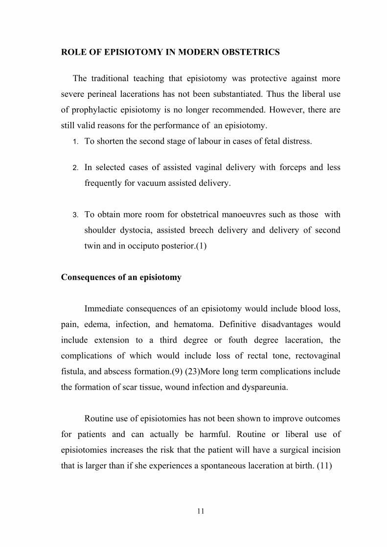
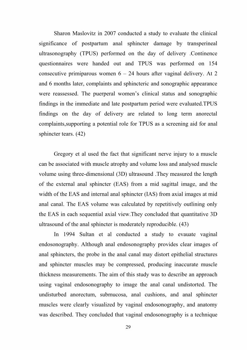
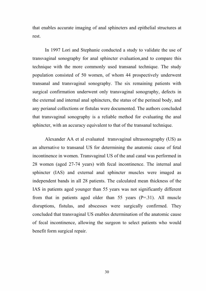