
Disorders of the temporomandibular joint · Disorders of the temporomandibular joint are summarized...
8
© Copyright 2013 Elsevier, Ltd. All rights reserved. Disorders of the temporomandibular joint CHAPTER CONTENTS Disorders of the inert structures e209 Internal derangement . . . . . . . . . . . . . . . . e209 Arthrosis . . . . . . . . . . . . . . . . . . . . . . . e212 Luxation of the condyle . . . . . . . . . . . . . . . e213 Synovial disorders . . . . . . . . . . . . . . . . . . e214 Disorders of the contractile structures e214 Myalgia . . . . . . . . . . . . . . . . . . . . . . . e214 Muscular trismus . . . . . . . . . . . . . . . . . . e215 Abscess in the pterygoid muscle . . . . . . . . . . e215 A great number of people (60–70%) have some problem with the temporomandibular joint (TMJ) but most are asymptomatic. About 25% of this group has symptoms that can be attributed to the TMJ. 1,2 Of this group only 5% will get treatment and these patients are mostly women – they outnumber male patients by at least 4 : 1. 3 Temporoman- dibular disorders occur at any age but most often in young adults. 4,5 Pain the temporomandibular area is the most common complaint and is easily related to a lesion of the TMJ. 6,7 However, the exact cause of TMJ problems is often difficult to determine because several disorders may be present at the same time and combined muscular and inert tissue disorders are not uncommon. As a consequence, it may be necessary to treat both joint and muscle. Conservative treatment is still the most effective management for more than 80% of patients. 8 There are several major causes of TMJ problems: 9 • A single major injury, even if it has been only a light blow to the mandible or extreme stretching of the TMJ, can permanently injure the joint and ligaments. 10 • Repetitive microtraumas, such as clenching, grinding or atypical chewing for a prolonged period of time, may damage the joint and then lead to muscular problems. • Monoarticular arthritis and inflammatory polyarthritis, such as rheumatoid arthritis, but seldom psoriatic arthritis, may affect the joint. 11 • Abnormal biomechanical loading as a result of structural changes of the teeth may also be harmful. However, this should not be overestimated as a cause of TMJ problems. 12 Disorders of the temporomandibular joint are summarized in Table 1. Disorders of the inert structures The most common disorders of the inert structures are internal derangement and arthritis. The former may be present in hypermobile joints or may affect a normal joint, so leading to hypomobility. Internal derangement Internal derangement is usually the result of lack of coordina- tion between the meniscus and the condyle, in which the meniscus displaces anteriorly and the condyle posterosuperi- orly. Acute or chronic repetitive injury may be the cause. Acute injury, such as whiplash, traumatic tooth extraction or intuba- tion during anaesthesia, may displace the condyle posteriorly, so stretching the posterior attachments of the meniscus. Repetitive microtraumas occurring over a longer period, as a result of loss of posterior teeth or interference with the inci- sors, may provoke a posteriorly directed force on the jaw, which again displaces the mandibular head posteriorly. Initially, this process is characterized by reciprocal clicking, later by temporary or continuous locking. Finally, osteoarthro- sis may result. Each stage of internal derangement may give rise to painful chronic irritation of the synovium, with resulting arthritis.
Transcript of Disorders of the temporomandibular joint · Disorders of the temporomandibular joint are summarized...
© Copyright 2013 Elsevier, Ltd. All rights reserved.
Disorders of the temporomandibular joint
CHAPTER CONTENTS
Disorders of the inert structures e209
Internal derangement . . . . . . . . . . . . . . . . e209Arthrosis . . . . . . . . . . . . . . . . . . . . . . . e212Luxation of the condyle . . . . . . . . . . . . . . . e213Synovial disorders . . . . . . . . . . . . . . . . . . e214
Disorders of the contractile structures e214
Myalgia . . . . . . . . . . . . . . . . . . . . . . . e214Muscular trismus . . . . . . . . . . . . . . . . . . e215Abscess in the pterygoid muscle . . . . . . . . . . e215
A great number of people (60–70%) have some problem with the temporomandibular joint (TMJ) but most are asymptomatic. About 25% of this group has symptoms that can be attributed to the TMJ.1,2 Of this group only 5% will get treatment and these patients are mostly women – they outnumber male patients by at least 4 : 1.3 Temporoman-dibular disorders occur at any age but most often in young adults.4,5
Pain the temporomandibular area is the most common complaint and is easily related to a lesion of the TMJ.6,7 However, the exact cause of TMJ problems is often difficult to determine because several disorders may be present at the same time and combined muscular and inert tissue disorders are not uncommon. As a consequence, it may be necessary to treat both joint and muscle. Conservative treatment is still the most effective management for more than 80% of patients.8
There are several major causes of TMJ problems:9
• A single major injury, even if it has been only a light blow to the mandible or extreme stretching of the TMJ, can permanently injure the joint and ligaments.10
• Repetitive microtraumas, such as clenching, grinding or atypical chewing for a prolonged period of time, may damage the joint and then lead to muscular problems.
• Monoarticular arthritis and inflammatory polyarthritis, such as rheumatoid arthritis, but seldom psoriatic arthritis, may affect the joint.11
• Abnormal biomechanical loading as a result of structural changes of the teeth may also be harmful. However, this should not be overestimated as a cause of TMJ problems.12
Disorders of the temporomandibular joint are summarized in Table 1.
Disorders of the inert structures
The most common disorders of the inert structures are internal derangement and arthritis. The former may be present in hypermobile joints or may affect a normal joint, so leading to hypomobility.
Internal derangement
Internal derangement is usually the result of lack of coordina-tion between the meniscus and the condyle, in which the meniscus displaces anteriorly and the condyle posterosuperi-orly. Acute or chronic repetitive injury may be the cause. Acute injury, such as whiplash, traumatic tooth extraction or intuba-tion during anaesthesia, may displace the condyle posteriorly, so stretching the posterior attachments of the meniscus. Repetitive microtraumas occurring over a longer period, as a result of loss of posterior teeth or interference with the inci-sors, may provoke a posteriorly directed force on the jaw, which again displaces the mandibular head posteriorly.
Initially, this process is characterized by reciprocal clicking, later by temporary or continuous locking. Finally, osteoarthro-sis may result.
Each stage of internal derangement may give rise to painful chronic irritation of the synovium, with resulting arthritis.
The Temporomandibular Joint
e210© Copyright 2013 Elsevier, Ltd. All rights reserved.
Table 1 Summary of disorders of the temporomandibular joint
Disorder Symptoms Signs Treatment
Disorders of the inert structures
Internal derangement Reciprocal clicking Mainly on opening
Temporary lockingStiff feeling
Opening slightly diminishedChin deviation to ipsilateral side
Stretching capsule and ligamentsRepositioning splint
Fixed dislocation of meniscus
Sudden locking Opening strongly limitedChin deviates to ipsilateral sidePain on deviation to contralateral side
Manipulation
Arthrosis Grinding sound Pain at all tests and during the whole of each movement
Spontaneous cureStop clenching
Crepitus CrepitusOpening limitedContralateral deviation limitedOn opening: chin deviates to ipsilateral
sideBack of condyle tender on palpation
Dental correctionFriction to joint capsuleIntra-articular injectionForced stretchingArthroplasty
Luxation of condyle Cannot close mouth Closing of mouth limitedElastic recoil on passive closing
Manipulative reduction
Synovial disorders Monoarticular
steroid-sensitive arthritis
CrepitationPainStiffness
Opening painful and limitedIpsilateral deviation on openingContralateral deviation when closedResisted opening: pain
Intra-articular injection
Inflammatory polyarthritic diseases
As in monoarticular steroid-sensitive arthritis
As in monoarticular steroid-sensitive arthritis
General treatment of underlying disorder
Infections Pain Severely illOpening painfully limited and ipsilateral
deviation
HospitalizationAspirationAntibiotics
Sympathetic arthritis Pain Opening slightly diminishedPain on all movementsTenderness on palpationAbscess
Spontaneous cureAntibiotics
Arthritis from loss of molar teeth
Continuous pain Opening slightly decreasedPain at extreme of all movements
Dental correction
Disorders of the contractile structures
Myalgia Dull aching, continuous pain worse in cold weather and at end of day
Pain on resisted closing and on maximal active opening
Painful bands
Stabilization applianceStop bruxism or tooth clenching
Muscular trismus Vague pain Tremor of jawIncoordination on movementDiminished opening
Solve underlying problem
Abscess in pterygoid muscles
Pain on chewing Opening diminishedPain on clenchingResisted contralateral deviation painfulFever
AntibioticsDrainage

Disorders of the temporomandibular joint
e211© Copyright 2013 Elsevier, Ltd. All rights reserved.
The other thumb, protected by a rubber pad, is placed on the molar teeth and the fingers pass round the mandibular body. Downward pressure is applied to the molar teeth, which dis-tracts the joint surfaces, allowing repositioning of the meniscus on the condyle.
Technique: distraction with anterior glideIn the same position, the tip of the index finger of the manipulating hand is hooked around the mandibular angle. Downward pressure and anterior pull are applied. This restores the anterior translatory glide which has been lost because of capsular retraction or because of the anteriorly displaced meniscus.
Technique: combined distraction, anterior glide and lateral stretchThe mandible is additionally pulled to the side, towards the therapist, thus realigning the fibres in all three planes of space.
Technique: lateral glide without distractionThe position and fixation of the patient is the same as for previous techniques. The hand grasps the mandible around the angle and pulls it towards the therapist, restoring lateral joint movement.
Home exercise programmeThe patient is shown how to perform self-mobilizing tech-niques on opening, on forward movement and on lateral excur-sions of the mandible.
The muscles of mastication should be gently stretched to their full length. Coordination can be increased by practising hinge-type movement of the jaw and then making specific lateral movements without protrusion. Jaw movements must be limited to the click-free range and chewing on the side that provokes any noise must be avoided.
If significant painful clicking is associated with a possible meniscus displacement, a repositioning occlusal splint can be useful. It is used only if a small change in the position of the mandible stops the click on opening and closing of the mouth. It should initially be worn 24 hours a day for 8–10 weeks, except while eating. Once the clinical symptoms have dimin-ished, use of the appliance is progressively decreased, although a part-time repositioning appliance is needed.9,14
Resisted muscular exercises on opening, forward movement and lateral deviation may be used in association.15
Fixed dislocation of the meniscus
Progression of the disorder may finally lead, via momentary self-reducing dislocations, to a permanent luxation of the meniscus. From time to time the joint becomes suddenly locked, with the meniscus lodged anterior to the condyle.
With a dislocated meniscus, clinical examination shows a reduced range of opening of the mouth. Because of loss of anterior translatory glide, only rotation is possible. In such a condition the patient can always open the mouth by at least 1 cm but often not any wider. Full closure remains possible and is painless. The chin deviates to the ipsilateral side and deviation to the contralateral side is painful.
Reciprocal clicking
Clinically there is a ‘click’, usually most noticeable on opening the mouth and less so on closing. A click implies that the TMJ, which is normally a friction-free joint, is functioning abnor-mally. With the mouth closed, the meniscus lies too far ante-riorly in relation to the condyle (Fig. 1). On opening, the click occurs when the condyle moves anteriorly and snaps beneath the meniscus and its posterior attachments to fall into its normal position. The opening click implies a reduction of the meniscus.
Clicks may occur early, intermediate or late in relation to the range of opening of the mouth. Early clicks indicate a small degree of anterior displacement; clicking more towards full opening indicates that the displacement is greater, as a result of progressive stretching of the posterior attachments. A closing click does not occur unless there is a preceding opening one. The click is present just before the mouth is closed and is the result of the condyle sliding posteriorly to the posterior band of the meniscus so that the meniscus is displaced anteriorly.
In addition to repetitive clicking, there is often a stiff feeling and a short period of pain after the joint has been immobile for a while.
On clinical examination, opening of the mouth is slightly decreased and provokes a small deviation of the chin to the affected side because the condyle cannot move fully anteriorly.
TreatmentTreatment aims to fully restore the normal range of motion, mainly the lost anterior translatory glide. The technique used is called ‘joint liberation’13 and consists of passive movements applied in all directions in order to realign the shortened fibres of the capsule. All the techniques can be performed under steady pull or, if pain is excessive, with small vibrations. Each manœuvre is performed over about 20–30 seconds and repeated several times. The whole procedure takes about 15 minutes.
Technique: distractionThe patient sits on a chair with the therapist facing the painless side and stabilizing the patient’s head with the ipsilateral hand.
Fig 1 • Anterior displacement of the disc.

The Temporomandibular Joint
e212© Copyright 2013 Elsevier, Ltd. All rights reserved.
molar teeth. In primary osteoarthrosis, no apparent cause is present.17
The outcome of progressive change in internal derangement is shortening and fibrosis of the meniscus–condyle attach-ments, and occasionally perforation or total rupture of the meniscus. Paradoxically if the latter occurs, there is only a small limitation or even complete freedom of movement, so that subjectively the patient feels better although radiography clearly shows the degenerative changes. The click changes to a more grinding noise.
Patients with arthrosis are usually over 40 years of age and it remains unilateral.18 Usually, it does not give rise to signifi-cant inflammation or swelling, or to severe pain or limitation of movement. Only when the capsule is inflamed does it cause pain, in which case it is often present in any part of the range of movement. Crepitus is frequently found; limitation of opening of the mouth and of contralateral deviation of the chin may be present. On opening, the chin deviates to the affected side. Palpation of the back of the condyle is usually tender. Pain and stiffness increase during the day.
A radiograph shows flattening of the anterior slope of the condyle and the posterior slope of the articular tubercle, with loss of joint space and formation of osteophytes.
The complaints usually diminish progressively over 2–3 years with little painless residual disability. However, about one in five patients still has pain after 2 years.19 Ankylosis is uncommon.
TreatmentTreatment should be directed towards the cause: dental cor-rection is performed and clenching and grinding is stopped. Symptoms may be relieved by deep friction to the joint capsule followed by forced movements.15
An intra-articular injection with steroid should be given if stretching alone is insufficient. The results are better than forced movements and friction alone.
If intra-articular injection is without effect, or when ankylosis appears imminent, capsular stretching should be performed.
If either intra-articular injections or capsular stretching have failed, arthroplasty may be indicated. Because open surgery can be followed by formation of fibrotic adhesions in the joint, so limiting the normal range of movement, or cause malocclusion, surgical treatment must only exceptionally be recommended.
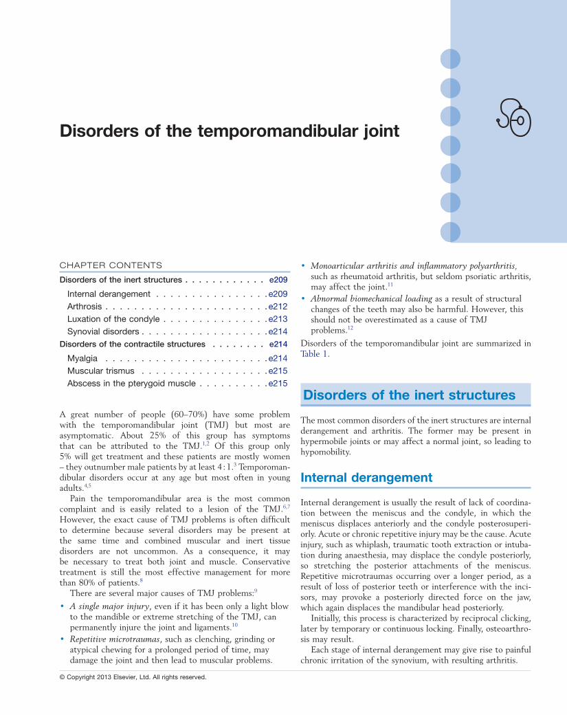
Technique: deep friction (Fig. 3)For friction the patient lies on a couch, painful side up. The therapist sits behind the patient and places the index of the ipsilateral hand, reinforced by the middle finger, in front of the joint line. This is easily palpable, just cranial to the mandibular head, on opening and closing the mouth. The other hand stabilizes the head on the skull. The index finger is now pulled backwards while pressure is applied.
Friction is given for about 20 minutes, three times a week.
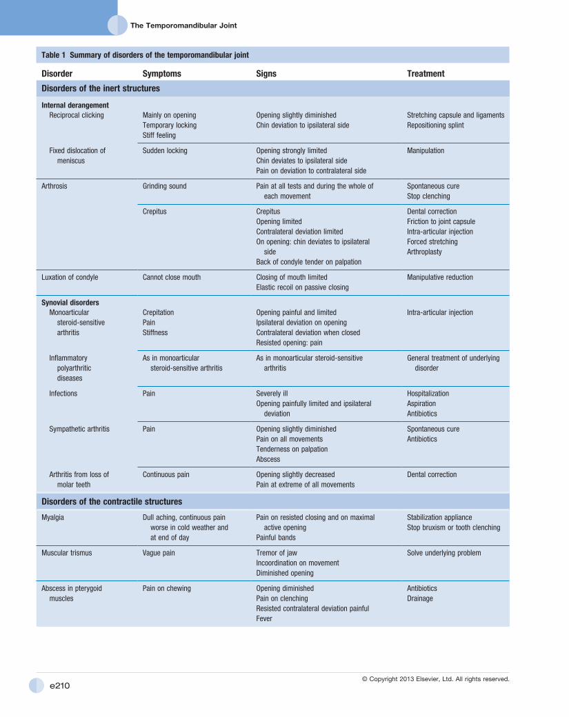
Technique: intra-articular injection (Fig. 4)16
The patient lies with the affected side up. The posterior aspect of the mandibular condyle is palpated, anterior to the tragus and below the zygomatic bone. The space to be injected lies
Treatment
Manipulative reductionManipulative treatment aims to restore the normal relationship between the meniscus and the joint surfaces.
Technique (Fig. 2)The patient lies on a high couch. The manipulator stands at the patient’s opposite side and puts one thumb, protected by a thick pad, on the molar teeth of the affected side. The other hand is put around the patient’s head and holds it steady. Caudal pressure is now applied to the molar teeth and swift translatory movements of the patient’s mandible are per-formed, three or four times. During the manœuvre, the reduc-tion click is felt.16
Manipulative reduction usually succeeds in one session.
Problems arising because of hypermobility
Hypermobile joints may also give rise to reciprocal clicking, subluxations and dislocations because of excessive translatory glide. On clinical examination, excessive movements are found together with clicking. Treatment consists mainly of stabilizing the joint and avoiding excessive anterior translatory gliding of the condyle by controlling the rotation in the joint. To do this, the patient is instructed to put and to keep the distal third of the tongue flat against the palate during opening, which limits opening to rotation movement only and reduces the tendency to anterior sliding. This method of opening should also be performed when chewing and protects the joint from further wear and tear.
Arthrosis
Arthrosis of the TMJ may be the final stage of internal derangement or is sometimes due to bruxism or missing
Fig 2 • Manipulative reduction of a dislocated disc.
Disorders of the temporomandibular joint
e213© Copyright 2013 Elsevier, Ltd. All rights reserved.
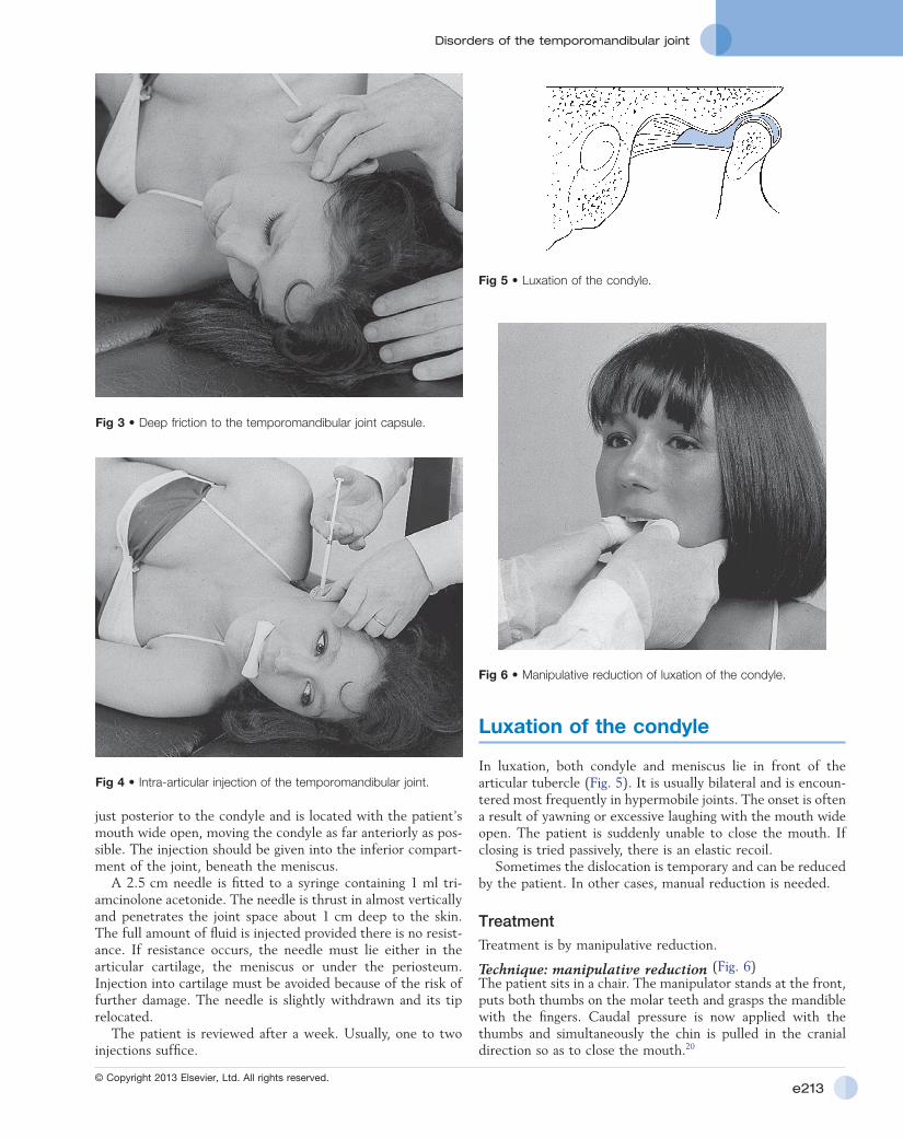
Luxation of the condyle
In luxation, both condyle and meniscus lie in front of the articular tubercle (Fig. 5). It is usually bilateral and is encoun-tered most frequently in hypermobile joints. The onset is often a result of yawning or excessive laughing with the mouth wide open. The patient is suddenly unable to close the mouth. If closing is tried passively, there is an elastic recoil.
Sometimes the dislocation is temporary and can be reduced by the patient. In other cases, manual reduction is needed.
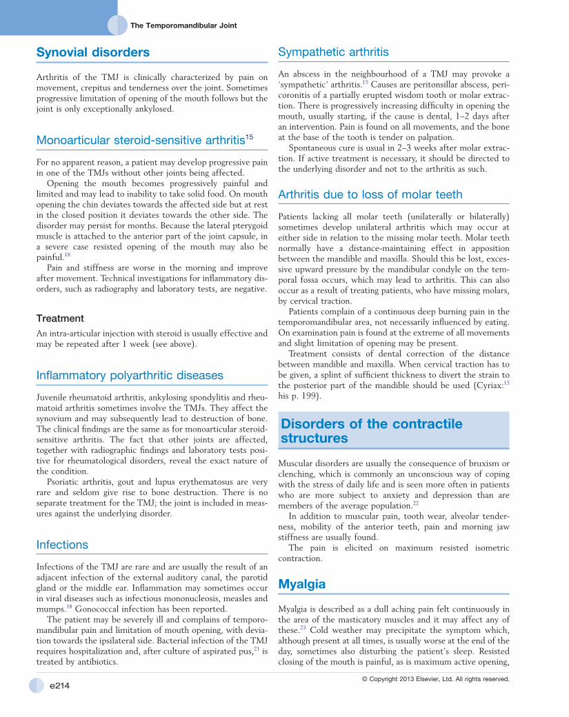
TreatmentTreatment is by manipulative reduction.
Technique: manipulative reduction (Fig. 6)The patient sits in a chair. The manipulator stands at the front, puts both thumbs on the molar teeth and grasps the mandible with the fingers. Caudal pressure is now applied with the thumbs and simultaneously the chin is pulled in the cranial direction so as to close the mouth.20
just posterior to the condyle and is located with the patient’s mouth wide open, moving the condyle as far anteriorly as pos-sible. The injection should be given into the inferior compart-ment of the joint, beneath the meniscus.
A 2.5 cm needle is fitted to a syringe containing 1 ml tri-amcinolone acetonide. The needle is thrust in almost vertically and penetrates the joint space about 1 cm deep to the skin. The full amount of fluid is injected provided there is no resist-ance. If resistance occurs, the needle must lie either in the articular cartilage, the meniscus or under the periosteum. Injection into cartilage must be avoided because of the risk of further damage. The needle is slightly withdrawn and its tip relocated.
The patient is reviewed after a week. Usually, one to two injections suffice.
Fig 3 • Deep friction to the temporomandibular joint capsule.
Fig 4 • Intra-articular injection of the temporomandibular joint.
Fig 5 • Luxation of the condyle.
Fig 6 • Manipulative reduction of luxation of the condyle.
The Temporomandibular Joint
e214© Copyright 2013 Elsevier, Ltd. All rights reserved.
Sympathetic arthritis
An abscess in the neighbourhood of a TMJ may provoke a ‘sympathetic’ arthritis.15 Causes are peritonsillar abscess, peri-coronitis of a partially erupted wisdom tooth or molar extrac-tion. There is progressively increasing difficulty in opening the mouth, usually starting, if the cause is dental, 1–2 days after an intervention. Pain is found on all movements, and the bone at the base of the tooth is tender on palpation.
Spontaneous cure is usual in 2–3 weeks after molar extrac-tion. If active treatment is necessary, it should be directed to the underlying disorder and not to the arthritis as such.
Arthritis due to loss of molar teeth
Patients lacking all molar teeth (unilaterally or bilaterally) sometimes develop unilateral arthritis which may occur at either side in relation to the missing molar teeth. Molar teeth normally have a distance-maintaining effect in apposition between the mandible and maxilla. Should this be lost, exces-sive upward pressure by the mandibular condyle on the tem-poral fossa occurs, which may lead to arthritis. This can also occur as a result of treating patients, who have missing molars, by cervical traction.
Patients complain of a continuous deep burning pain in the temporomandibular area, not necessarily influenced by eating. On examination pain is found at the extreme of all movements and slight limitation of opening may be present.
Treatment consists of dental correction of the distance between mandible and maxilla. When cervical traction has to be given, a splint of sufficient thickness to divert the strain to the posterior part of the mandible should be used (Cyriax:15 his p. 199).
Disorders of the contractile structures
Muscular disorders are usually the consequence of bruxism or clenching, which is commonly an unconscious way of coping with the stress of daily life and is seen more often in patients who are more subject to anxiety and depression than are members of the average population.22
In addition to muscular pain, tooth wear, alveolar tender-ness, mobility of the anterior teeth, pain and morning jaw stiffness are usually found.
The pain is elicited on maximum resisted isometric contraction.
Myalgia
Myalgia is described as a dull aching pain felt continuously in the area of the masticatory muscles and it may affect any of these.23 Cold weather may precipitate the symptom which, although present at all times, is usually worse at the end of the day, sometimes also disturbing the patient’s sleep. Resisted closing of the mouth is painful, as is maximum active opening,
Synovial disorders
Arthritis of the TMJ is clinically characterized by pain on movement, crepitus and tenderness over the joint. Sometimes progressive limitation of opening of the mouth follows but the joint is only exceptionally ankylosed.
Monoarticular steroid-sensitive arthritis15
For no apparent reason, a patient may develop progressive pain in one of the TMJs without other joints being affected.
Opening the mouth becomes progressively painful and limited and may lead to inability to take solid food. On mouth opening the chin deviates towards the affected side but at rest in the closed position it deviates towards the other side. The disorder may persist for months. Because the lateral pterygoid muscle is attached to the anterior part of the joint capsule, in a severe case resisted opening of the mouth may also be painful.18
Pain and stiffness are worse in the morning and improve after movement. Technical investigations for inflammatory dis-orders, such as radiography and laboratory tests, are negative.
TreatmentAn intra-articular injection with steroid is usually effective and may be repeated after 1 week (see above).
Inflammatory polyarthritic diseases
Juvenile rheumatoid arthritis, ankylosing spondylitis and rheu-matoid arthritis sometimes involve the TMJs. They affect the synovium and may subsequently lead to destruction of bone. The clinical findings are the same as for monoarticular steroid-sensitive arthritis. The fact that other joints are affected, together with radiographic findings and laboratory tests posi-tive for rheumatological disorders, reveal the exact nature of the condition.
Psoriatic arthritis, gout and lupus erythematosus are very rare and seldom give rise to bone destruction. There is no separate treatment for the TMJ; the joint is included in meas-ures against the underlying disorder.
Infections
Infections of the TMJ are rare and are usually the result of an adjacent infection of the external auditory canal, the parotid gland or the middle ear. Inflammation may sometimes occur in viral diseases such as infectious mononucleosis, measles and mumps.18 Gonococcal infection has been reported.
The patient may be severely ill and complains of temporo-mandibular pain and limitation of mouth opening, with devia-tion towards the ipsilateral side. Bacterial infection of the TMJ requires hospitalization and, after culture of aspirated pus,21 is treated by antibiotics.

Disorders of the temporomandibular joint
e215© Copyright 2013 Elsevier, Ltd. All rights reserved.
When movement is attempted, there is often tremor of the jaw and incoordination. Treatment consists of solving the underlying problem.
Hysterical trismus is characterized by a total loss of ability to open the mouth. A vague pain, which extends over the whole of the head and face, is usually described. Very often these patients also hold the neck extremely still. Because it is very difficult to examine the TMJ in an unwilling patient, it is better to proceed with caution: multiple positive findings and inconsistencies should warn the examiner of a severe disorder or hysteria.
which stretches the muscles. The muscles are usually very tender, and firm palpable bands within them are often present.
The differential diagnosis includes temporal arteritis, which can also lead to myalgic pain. Redness and swelling over the artery are characteristic of arteritis.
TreatmentPrimary therapy is a full-arch occlusal stabilization appliance. This involves the construction of a flat occlusal surface that is adjusted to have multiple tooth contact in a habitual comfort-able jaw closure position.9,24 Initially it is worn continuously, except on eating, for 6–8 weeks and is adjusted several times to establish a comfortable jaw position. As the symptoms decrease, the amount of daytime wear of the appliance is pro-gressively reduced.
In musculoskeletal pain from bruxism or tooth clenching the patient must break the habit. Advice is given to reduce all physical and mentally stressful activities during treatment. Muscle relaxants or anti-anxiety drugs can be useful. Hard or chewy foods are avoided. Application of moist heat to the temporal and masseter muscles for 20 minutes, three or more times a day, can be helpful.
Muscular trismus
Pathological muscular stimulation or inhibition may limit the opening of the mouth but only very rarely affect closing. Limi-tation is often the result of attempting to avoid pain. Trismus can be the outcome of a chronic pain problem or of a recent local event (injury or surgery).
Warning
When trismus is present, tetanus should not be forgotten. Painless stiffness coming on abruptly at the TMJ, and causing complete inability to open the mouth, may be an early sign of the condition. In this event, both joints are usually affected.
Abscess in the pterygoid muscle
An anaesthetic injection for dental work may be followed some days later by an abscess. The cheek begins to hurt and feels stiff. The patient complains of problems on opening the mouth and has pain on chewing.
Clinical examination reveals a loss of range of mouth opening, together with pain on clenching. Resisted deviation towards the painless side also hurts.
Signs of infection including pyrexia should be sought. Treat-ment is with antibiotics and drainage.
References
1. Salonen L, Hellden L. Prevalence of signs and symptoms of dysfunction in the masticatory system: an epidemiological study in an adult Swedish population. J Craniomandib Disord (J Orofac Pain) 1990;4:241–50.
2. McNeill C, editor. Temporomandibular Disorders – Guidelines for Classification, Assessment and Management. 2nd ed. Chicago: Quintessence Books; 1993.
3. Dworkin SF, Huggins KH, Le Resche LR, et al. Epidemiology of signs and symptoms in temporomandibular disorders. I. Clinical signs in cases and controls. J Am Dent Assoc 1990;120:273–81.
4. Greene CS, Marbach JJ. Epidemiologic studies of mandibular dysfunction: a critical review. J Prosthet Dent 1982;48:184–90.
5. Dimitroulis G. Temporomandibular disorders: a clinical update. BMJ 1998;317:190–4.
6. Ohrbach R, Gale EN. Pressure pain thresholds, clinical assessment and differential diagnosis: reliability and validity in patients with myogenic pain. Pain 1989;39:157–69.
7. Dworkin S, Le Resche L, De Rouen T, Von Korff M. Assessing clinical signs of temporomandibular disorders: reliability of clinical examiners. J Prosthet Dent 1990;63(5):574–9.
8. Dimitroulis G, Gremillion HA, Dolwick MF, Walter JH. Temporomandibular disorders. 2. Non-surgical treatment. Aust Dent J 1995;40:372–6.
9. Clark G. Diagnosis and treatment of painful temporomandibular disorders. Dent Clin North Am 1987;31(4):645–74.
10. Ryan DE. Temporomandibular disorders. Curr Opin 1993;5:209–18.
11. Veys E, Mielants H, Verbruggen G. Reumatologie. Ghent: Omega Editions; 1985. p. 169.
12. Greene C, Marbach J. Epidemiologic studies of mandibular dysfunction: a critical review. J Prosthet Dent 1982;48(2).
13. Rocabado M. Arthrokinematics of the temporomandibular joint. Dent Clin North Am 1983;27(3):573–94.
14. De Laat A. Functionele behandeling van kaakgewrichtsklachten. Tijd Geneeskd 1986;42(11):753–7.
15. Cyriax JH. Textbook of Orthopaedic Medicine, vol I, Diagnosis of Soft Tissue Lesions. 8th ed. London: Baillière Tindall; 1982. p. 117.
16. Cyriax JH. Textbook of Orthopaedic Medicine, vol II, Treatment by Manipulation, Massage and Injection. 11th ed. London: Baillière Tindall; 1984.
17. De Bont L, Stegenga G, Boering G. Kaakgewrichtsstoornissen. Deel I, Gedachtenontwikkeling en classificatie. Ned Tijd Tandheelkd 1989;96:496–500.
18. Hodges JM. Managing temporomandibular joint syndrome. Laryngoscope 1990;100:60–6.
19. Toller P. Osteoarthrosis of the mandibular condyle. Br Dent J 1973;20:223–31.
20. Steenks H. Welke niet-chirurgische behandelingsstrategie kan worden gekozen bij een ‘closed lock’ van het kaakgewricht. Respons 1990;3:1–2.
21. Friedman M, Weisber J, Agus B. Diagnosis and treatment of inflammation of the temporomandibular joint. Sem Arthritis Rheum 1982;12(1):44–51.

The Temporomandibular Joint
e216© Copyright 2013 Elsevier, Ltd. All rights reserved.
22. Thomas L, Tiber N, Schireson S. The effects of anxiety and frustration on muscular tension related to the temporomandibular joint syndrome. Oral Surg 1973;Nov:763–8.
23. Ernest E, Martinez M, Rydzewski D, Salter E. Photomicrographic evidence of insertion tendinosis: the etiologic factor in pain for temporal tendinitis. J Prosthet Dent 1991;65(1):127–31.
24. Clark GT, Adler RC. A critical evaluation of occlusal therapy. Occlusal adjustment procedures. J Am Dent Assoc 1985;110:743–50.


