Disorders Of The Hallux
107
DR. PRUTHVIRAJ NISTANE Deptt. Of Orthopaedics,Unit II Govt. Medical College and Rajindra Hospital, Patiala
-
Upload
pruthviraj-nistane -
Category
Documents
-
view
25 -
download
7
description
MTP joint is a major wt bearing joint of the body. so it s disorders have very importance clinically
Transcript of Disorders Of The Hallux

DR. PRUTHVIRAJ NISTANE
Deptt. Of Orthopaedics,Unit II Govt. Medical College and Rajindra Hospital, Patiala

A large proportion of clinical complaints of the foot center on the first metatarsophalangeal (MTP) joint.
This articulation alone bears one-third of the weight of the forefoot and helps stabilize the longitudinal arch


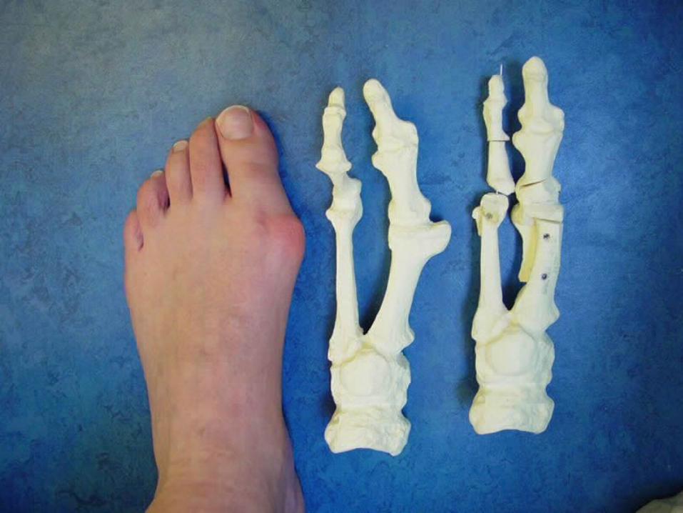
=Lateral deviation of the proximal phalanx on the 1st metatarsal head
• Complex deformity of the 1st
ray that frequently is accompanied by deformity & symptoms in lesser toes
• metatarsus primus varus• hallux valgus• hallux valgus interphalangeus


EtiologyEtiology
•Female/male = 2:1 to 15:1• Heredity: + FH ~63%
•Essential extrinsic factor = shoe

EtiologyEtiology
•Intrinsic ANOTOMICAL cause• Pes planus• Metatarsus primus varus: juvenile form• First metatarsal length• Hypermobility of first ray• Pronated flatfeet• Abnormal insertion of the posterior tibial
tendon

• Amputation of 2nd toe• Cystic degeneration of medial capsule
MTPJ• Achilles tendon contracture• Joint hyperelasticity = Ehlers-Danlos

AnatomyAnatomy


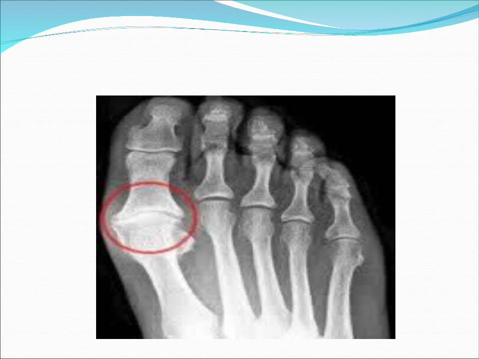
PathoanatomyPathoanatomy•Most commonly LATERAL DEVIATION OF GREAT TOE is primary deformity•Valgus angle of the first metatarsophalangeal joint exceeds 30 to 35 degrees•Increase in angle between first and second metatarsal (metatarsus primus varus) at MTMC joint•Pronation of the great toe•Subluxation/dislocation of the first metatarsophalangeal joint•Excessive valgus tilt of the articular surface of
the first metatarsal head and proximal phalangeal articular surface

PathoanatomyPathoanatomy

Pathogenesis • the abductor hallucis moves plantar ward• Only restraining medial structure is the medial
capsular ligament • Dorsiflexion of MTP joint• The adductor hallucis, which is unopposed by the
abductor hallucis, pulls the great toe further into valgus
• The flexor hallucis brevis, flexor hallucis longus and extensor hallucis increases the valgus moment, further deforming the first ray.
• the metatarsal head to drift medially from the sesamoids.

PathophysiologyPathophysiology• Valgus deviation of hallux• Attenuatedmedial structure • Varus metatarsal head deviation • Sesamoidsubluxation • Hallux pronation• Lateral
contracture
• the sesamoid ridge on the plantar surface of the first metatarsal head (the crista) flattens
• With this restraint lost, the fibular sesamoid displaces partially or completely into the first space
• Fibular sesamoid, when pulled proximally by the lateral head of the flexor hallucis brevis, pulls the flexor hallucis longus laterally through the sesamoid apparatus and contributes to recurrent hallux
valgus.(so, when the deformity is
severe-excision of the fibular
sesamoid is added to the
procedure)• patient is bearing less weight
on the first ray and more on the
lesser metatarsal heads causing
transfer metatarsalgia, callosities, and stress fractures

first variant, the articular surface of the metatarsal head is offset,
resembling a scoop of ice cream sitting at an angle on a cone This has been described as the distal
metatarsal articular angle
Second variant the articular angle of the base of the proximal phalanx
in relation to its longitudinal axis is offset. This has been described as the phalangeal articular angle

Consequencesa hammer toe–like deformity of the second
toe the splaying of the forefoot corns often developbursal hypertrophy over the medial eminence
of the first metatarsal head (bunion)Osteoarthritis Callositymetatarsalgia.
The entire forefoot must be evaluated for these multiple components of hallux valgus


History• Chief complaint: oPain over medial eminence ~70%, at the metatarsophalangeal joint or beneath the lesser metatarsal headsoKeratosis• Associated problems• Age & level of activity• Occupation• Athletic inclinations• Shoe wear• Reasons for surgery
Patient evaluation

Physical examination
• Vascular / neurologic status• ROM of MTP joint• Pronation of hallux• Callosities under lesser MTHs• Hammer / claw toes• MTC joint stability• Assess hind foot
Patient evaluation

X-ray
Standard preoperative radiographs should include
1.Standing dorsoplantar views
2.Standing Lateral views
3.Nonstanding lateral oblique view
4.Axial sesamoid views

Standing dorsoplantar view
Non-standing lateral oblique view
Standing lateral view Axial sesamoid view

Evaluation of x-rays
•IMA (normal 8-9) •HVA (normal 15-20) •DMAA (normal 10-15) •PAA (normal 7-10)•OA changes•Position of sesamoids•Incongruent or subluxated joint
Hallux valgus angle
Intermetatarsal angle
Distal metatarsal articular
angle


Mild Moderate Severe
Hallux Valgus Angle <20 20-40 >40Intermetatarsal Angle<11 11-16 >16Sesamoid Subluxation <50% 50-75% >75%
Hallux valgus classificationHallux valgus classification

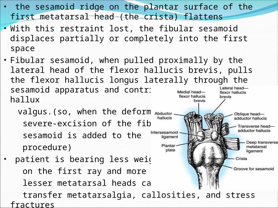
Give initial trial
Shoes with wide toe box
Orthotics• medial arch support• hallux valgus splint
Achilles tendon stretchingExercisesActivity adjustments
Non-operative treatmentNon-operative treatment


•Painful joint ROM•Deformity of the joint complex•Pain or difficulty with footwear•Inhibition of activity or lifestyle
for cosmetic reasons alone is seldom indicated except in an adolescent with a significant progressive deformity. Even the mildest symptoms in an adolescent often worsen
Indications for surgeryIndications for surgery

Associated foot disorders
- Neuritis/nerve entrapment - Overlapping/underlapping 2nd digit - Hammer digits - First metatarsocuneiform joint exostosis - Sesamoiditis - Ulceration - Inflammatory conditions (bursitis, tendinitis)
of 1st metatarsal head
Indications for surgeryIndications for surgery

Extensive peripheral vascular disease Active infection Active osteoarthropathy Septic arthritis Lack of pain or deformity Advanced age Lack of compliance Co-morbidities
Contraindications

Relieve pain
Correct deformity
Preserve MTP joint motion
Surgical Goals
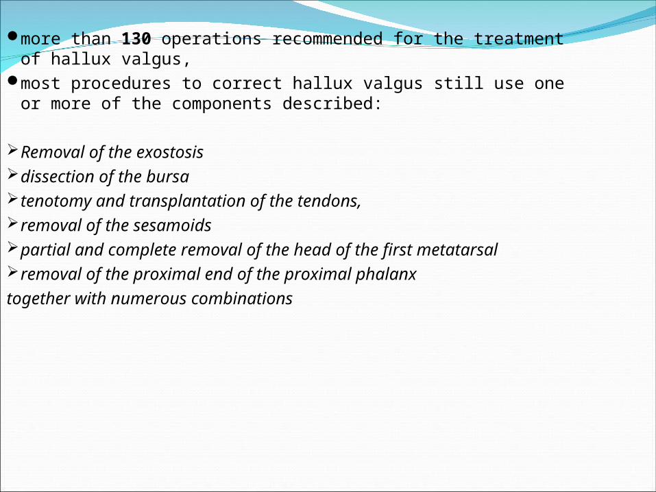
more than 130 operations recommended for the treatment of hallux valgus,
most procedures to correct hallux valgus still use one or more of the components described:
Removal of the exostosisdissection of the bursatenotomy and transplantation of the tendons, removal of the sesamoidspartial and complete removal of the head of the first metatarsalremoval of the proximal end of the proximal phalanx
together with numerous combinations

1. Valgus deviation of the great toe
2. Varus deviation of the 1st metatarsal
3. Pronation of hallux and/or 1st
metatarsal
4. Hallux valgus interphalangeus
5. Arthritis and limitation of motion of the
1st metatarsophalangeal joint
6. Length of the 1st metatarsal relative to
lesser metatarsals
Preoperative evaluation
7. Excessive mobility or obliquity of the 1st
metatarsomedial cuneiform joint
8. The medial eminence (bunion)
9. The location of the sesamoid apparatus
10. Intrinsic and extrinsic muscle-tendon
balance and synchrony
Preoperative evaluationPreoperative evaluation

IndicationsStress view radiographs - a firm forefoot wrap
reduces the intermetatarsal angle to a normal value and decreases the hallux valgus angle
Middle agedMild to moderatea valgus angle at the metatarsophalangeal joint of
15 to 25 degreesan intermetatarsal angle of less than 13 degrees, valgus of the interphalangeal joint of less than 15
degreesno degenerative changes at the
metatarsophalangeal jointa history of conservative management failure

Modified McBride Bunionectomy
L-SHAPED MEDIAL CAPSULAR INCISION
MEDIAL EMINENCE REMOVAL ADDUCTOR TENDON AND LATERAL CAPSULAR
RELEASE and reattach to 1st MT head between heads of 1st and 2nd MT
MEDIAL CAPSULAR IMBRICATION

FIBULAR (LATERAL) SESAMOIDECTOMY - the adductor hallucis and lateral head of the flexor hallucis brevis are released reducing the valgus.
In addition, the pull of the fibular sesamoid on the flexor hallucis longus through its tendon sheath and pulley system is prevented, reducing another important valgus-producing force on the hallux at the metatarsophalangeal joint
CLOSURE OF THE INVERTED-L CAPSULOTOMY

Modified McBride bunionectomy
DuVries & Mann

Distal Soft tissue handling includes
Medial eminence removal Adductor tendon and lateral capsular releaseMedial capsular imbricationReduction of MTP joint and sesamoids
The decision to perform an osteotomy should be made at the time of surgery by passively reducing the intermetatarsal angle. If the first metatarsal does not move laterally, or if it springs back quickly into varus after the laterally directed pressure is released then an osteotomy should be done
A) KELLER RESECTION ARTHROPLASTY
INDICATIONS
moderate-to-severe hallux valgus (30 to 45 degrees)
mild-to-moderate metatarsus primus varus(intermetatarsal
angles of 13 degrees or less)
pain over the medial eminence
An incongruous first metatarsophalangeal joint caused by lateral
subluxation of the phalanx on the metatarsal head
severe lateral displacement of the sesamoids,
Any evidence of degenerative cartilage changes

Resection hemiarthroplasty of the first metatarsophalangeal joint- resect 1/3 of proximal phalanx- mobilizes the hallux, allowing marked correction of valgus
removal of the medial eminence of the first metatarsal
fibular sesamoidectomy Adductor tenotomy lateral displacement of the first metatarsal complete lateral dislocation of the sesamoids,
marked degenerative changes, and severe pronation of the hallux may benefit
Complications are more

Resection arthroplastyResection arthroplasty

B) DISTAL METATARSAL B) DISTAL METATARSAL OSTEOTOMYOSTEOTOMY
Some studies suggest that VARUS OF THE FIRST METATARSAL WAS THE PRIME OR INITIAL DEFORMITY, and that valgus deviation of the hallux only followed it
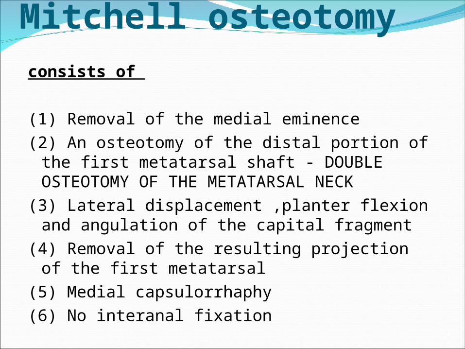
Mitchell osteotomy
consists of
(1) Removal of the medial eminence
(2) An osteotomy of the distal portion of the first metatarsal shaft - DOUBLE OSTEOTOMY OF THE METATARSAL NECK
(3) Lateral displacement ,planter flexion and angulation of the capital fragment
(4) Removal of the resulting projection of the first metatarsal
(5) Medial capsulorrhaphy
(6) No interanal fixation

Mitchell osteotomy

CHEVRON INTRACAPSULAR OSTEOTOMY
Indications younger patients (adolescence through the 30s) hallux valgus angle of 30 degrees or less an intermetatarsal angle of less than 13 degrees.
ADVANTAGESmade through cancellous boneshortens the metatarsal lessinherently stableFixation of the osteotomy with one or two Kirschner wires, a
cortical screw, or a biodegradable pin adds stability to the osteotomy

Consists of
(1) medial eminence removal
(2) a V-shaped intracapsular through the first metatarsal
head in trasverse plane
(3) lateral displacement of the capital fragment
(4) removal of the resulting projection of the first metatarsal
(5) medial capsulorrhaphy

Modified Chevron Osteotomysimply a more proximal placement of the apex of the
osteotomy in the metatarsal head. can be used for more severe deformities (up to 35 degrees
of hallux valgus and up to 15 degrees of first to second intermetatarsal diversion)
Johnson Modified Chevron Osteotomychanging the length and position of the limbs of the
osteotomy in the metatarsal head - short dorsal arm and long plantar arm
extended the indications for the osteotomy to severe deformities with intermetatarsal angles of 15 or 16 degrees
a 2.7-mm screw is used for internal fixation

Modified Chevron osteotomy

Johnson modified Chevron osteotomy

C) PROXIMAL FIRST METATARSAL OSTEOTOMYvarus of the first metatarsal, whether primary or
secondary, contributes to the hallux valgus complex
correction near the origin of the deformity is reasonable,
combined with a soft-tissue procedure at the first
metatarsophalangeal joint to correct the valgus of the
hallux
a few degrees' shift of the metatarsal at its base causes
marked improvement at the distal end of the metatarsal

Advantages1. Cancellous bone and broad contact surfaces
2. Small changes in position at the osteotomy produce excellent correction
at the distal end of the metatarsal where the symptoms are located
3. The metatarsal is shortened minimally
4. Large angles between the first and second metatarsals can be corrected
5. Slightly tilting the distal fragment plantarward reduces load bearing by
the second metatarsal, decreasing the chance of transfer
metatarsalgia.

Disadvantages
1. Extensive soft-tissue dissection is required
2. The distal fragment tends to displace dorsally or
medially
3. The second ray may be overloaded if the
fragment displaces or migrates
4. Three incisions are required
5. more difficult
6. more pain, swelling, and immobility
7. Cast immobilization is more frequently needed

Indications
A patient without significant degenerative arthritis in the
first metatarsophalangeal joint
hallux valgus of more than 35 degrees
an intermetatarsal angle of more than 10 degrees
Severe deformities

Types
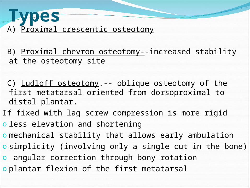
Types A) Proximal crescentic osteotomy
B) Proximal chevron osteotomy--increased stability at the osteotomy site
C) Ludloff osteotomy.-- oblique osteotomy of the first metatarsal oriented from dorsoproximal to distal plantar.
If fixed with lag screw compression is more rigido less elevation and shorteningo mechanical stability that allows early ambulationo simplicity (involving only a single cut in the bone)o angular correction through bony rotationo plantar flexion of the first metatarsal
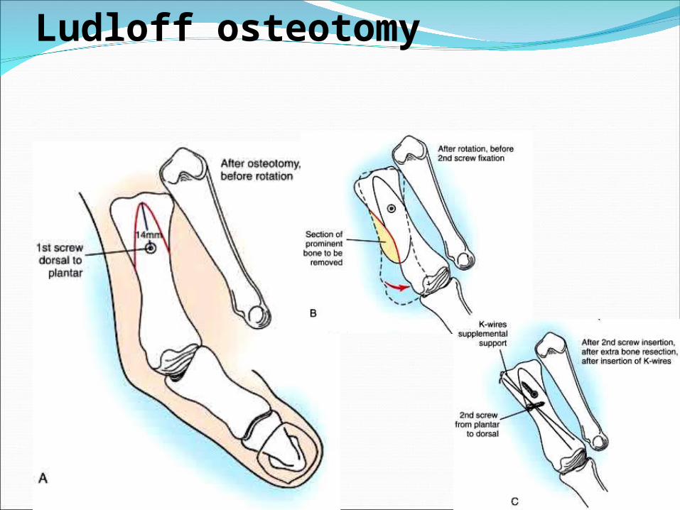
Ludloff osteotomy

D.Scarf osteotomy--horizontally directed displacement Z-osteotomy made at the diaphyseal level
o “scarf” refers to a joint made by notching, grooving, or otherwise cutting the ends of two pieces and fastening them together
versatility: o lateral displacement of the plantar bone fragment to reduce
the intermetatarsal angleo medial displacement of the capital fragment to correct hallux
varuso plantar displacement to increase the load of the first rayo elongation or shortening of the first metatarsal. o The stability of the osteotomy allows early weight bearing

Scarf osteotomy

Identify ???

D) MEDIAL CUNEIFORM OSTEOTOMY
Indications
in adolescents with open proximal metatarsal physes
especially patients with an abnormally wide
intermetatarsal angle

Medial Cuneiform OsteotomyMedial Cuneiform Osteotomy
Riedl & Coughlin

E) PROXIMAL PHALANGEAL OSTEOTOMY (AKIN’S)a medially based closing wedge osteotomy at the base of the
proximal phalanx, combined with medial eminence removal
mostly as an adjunctive procedure to the primary bunion repair
alone rarely is indicated
limited value if the sesamoid apparatus is subluxed
does not correct the principal deforming forces of the adductor
hallucis and the varus of the first metatarsal, so, is indicated
primarily in combination with other procedures , but after which
slight residual valgus deformity remains

Indications
1. Patient older than 55 years
2. Excessive hallux valgus interphalangeus (in
patient of any age)
3. Hallux valgus of no more than 25 degrees
4. Intermetatarsal angle of less than 13 degrees
5. Good metatarsophalangeal joint motion

Contraindications
1. Rheumatoid arthritis
2. osteoarthritis at the metatarsophalangeal joint
3. Intermetatarsal angle more than 13 degrees
4. Hallux valgus angle more than 30 degrees
5. Subluxation laterally of the tibial sesamoid more
than 50% of its width
6. Open physis of the proximal phalanx (can be
performed at neck instead of base)

Proximal Phalangeal OsteotomyProximal Phalangeal OsteotomyAkin procedure

Chevron-Akin Double Osteotomycombination of the chevron and Akin osteotomies to
gain greater correction of mild-to-moderate hallux valgus deformities.

F) ARTHRODESIS OF THE FIRST METATARSOPHALANGEAL JOINT
Various fixation methods have been described.
one-quarter tubular plate with one oblique
interfragmentary screw
one-third tubular plate
two ⅛-inch Steinmann pins placed through the
hallux into the first metatarsal

Indication 1. Severe deformity (an intermetatarsal angle >20 to 22
degrees, a hallux valgus angle >45 degrees, and severe pronation of the hallux)
2. with Degenerative arthritis / rheumatoid arthritis
3. motion of the metatarsophalangeal joint is limited and painful
4. Recurrent hallux valgus
5. Hallux valgus caused by muscle imbalance in patients with neuromuscular disorders, such as cerebral palsy, to prevent recurrence
6. Posttraumatic hallux valgus with severe disruption of all medial capsular structures that cannot be adequately reconstructed.


Why to differentiate ???an increased distal metatarsal articular angle may be the
defining characteristic of juvenile hallux valgus
1. Pain, either at the metatarsophalangeal joint or
beneath the lesser metatarsal heads, may not be the
primary complaint in many instances
2.Osteotomy of the first metatarsal is almost always
necessary
3. Varus of the first metatarsal with a widened
intermetatarsal angle is almost always present

4. Hypermobile flatfoot with pronation of the foot during
weight bearing frequently is associated with the deformity
5. Recurrence of the deformity is more frequent
6. Hallux valgus interphalangeus and deformity in
articular angles may be prominent
7. The family history frequently
8. Soft-tissue procedures alone are unlikely to result
in permanent correction.

Indicaion for surgery
Any adolescent 12 to 18 years old
with cosmetically unattractive hallux valgus deformity
report to be progressive
family history is positive for hallux valgus is
Pain and shoe-fitting problems

Types of surgerylesser deformities Adductor tenotomy, lateral
capsulotomy, medial eminence removal, and medial capsulorrhaphy
Moderate to severeo metatarsal physis is fully open a distal medial opening wedge
osteotomyo metatarsal physis is closed/ near closureproximal
crescentic osteotomy is recommended
Severe Peterson and Newman double first metatarsal osteotomies, an opening wedge proximally and a closing wedge distally to correct the abnormal distal metatarsal articular angle and the abnormal intermetatarsal angle

Distal osteotomyDistal osteotomyMitchell: double cut, step
Chevron: V shape cut
Diaphyseal osteotomyDiaphyseal osteotomyScarf osteotomy: Z shape, step cut, translation
Ludloff: Rotation
Basal osteotomBasal osteotomCrescentic
Basal chevron
AVN of 1AVN of 1stst MT head ! MT head !
Avoid shorteningAvoid shorteningMore stable then basalMore stable then basal
Extensive exposureExtensive exposure
High corrective powerHigh corrective power
Mild degreeMild degree
Unstable Unstable


Hallux Valgus <25Hallux Valgus <25Congruent Joint Soft tissue procedures Chevron osteotomy Mitchell osteotomy
Incongruent Joint (subluxation) Distal soft-tissue realignment +
Chevron osteotomy Mitchell osteotomy
Treatment of Hallux ValgusTreatment of Hallux Valgus

Hallux Valgus 25Hallux Valgus 25-40-40
Congruent Joint Chevron osteotomy + Akin procedure Mitchell osteotomy
Incongruent Joint Distal soft-tissue realignment +
proximal osteotomy
Treatment of Hallux ValgusTreatment of Hallux Valgus

Severe Hallux Valgus >40Severe Hallux Valgus >40
Congruent Joint
Double osteotomy
Akin + 1st metatarsal osteotomy Akin + 1st cuneiform opening wedge osteotomy
Treatment of Hallux ValgusTreatment of Hallux Valgus

Severe Hallux Valgus >40Severe Hallux Valgus >40Incongruent Joint Distal soft-tissue realignment +
Proximal osteotomy First cuneiform opening wedge osteotomy
Treatment of Hallux ValgusTreatment of Hallux Valgus

Hypermobile 1Hypermobile 1stst MTC Joint MTC Joint
Distal soft-tissue realignment + fusion 1st metatarsocuneiform joint
Degenerative joint diseaseDegenerative joint disease
Fusion or Keller procedure or prosthesis
Treatment of Hallux ValgusTreatment of Hallux Valgus

Post-operative managementPost-operative management
Immobilization ~2 weeks Weight bearing as tolerated or NWB

Post-operative managementPost-operative management
HV night splint to be worn for 6-8 wks after dressing changes are completed

Complications of surgeryEven experience, detailed physical and radiographic
evaluations, excellent surgical technique, and careful postoperative care do not guarantee that a complication will not occur
nonunion
recurrence of the deformity
The most troublesome has been metatarsalgia,
attributable to dorsiflexion malunion of the distal fragment
(use of a Kirschner wire for fixation (instead of sutures)
prevented malunion)
excessive shortening of the metatarsal,

medial eminence pain
clawed hallux
transfer keratotic lesions
development of the opposite deformity, hallux
varus


complication of hallux valgus surgeryBECAUSE
(1) complete release of the lateral structures of the metatarsophalangeal joint combined with excessive plication of the medial capsule, which pulls the sesamoids too far medially;
(2) excessive resection of the medial eminence, leading to loss of medial bony buttress for the proximal phalanx;
(3) excision of the fibular sesamoid;
(4) release of the lateral head of the flexor hallucis brevis at its insertion into the fibular sesamoid
(5) closure of the intermetatarsal angle to neutral or a negative value.

Two typesstatic (supple)
Uniplanar, and passively correctableusually is asymptomatic and mainly is a cosmetic
complication
dynamic (fixed)A multiplanar deformity that is fixed, symptomatic, and
difficult to correct surgically .The term that best describes the deformity is intrinsic minus
deformity of the hallux with a varus component. This is a true intrinsic-extrinsic muscle imbalance. The first metatarsophalangeal joint is hyperextended interphalangeal joint is acutely flexed

CORRECTION OF UNIPLANAR (STATIC) HALLUX VARUSNot all patients with acquired hallux varus require
operative treatmentA conservative program of modified shoe wear and taping
of the hallux should be attempted
A medial capsulotomy, placing the sesamoids in their proper location if subluxed medially, and holding the hallux in 10 to 15 degrees of valgus with a K- wire
Transfer of Extensor Hallucis Longus with Arthrodesis of the Interphalangeal Joint of the Hallux

CORRECTION OF DYNAMIC (MULTIPLANAR) HALLUX VARUSmost often either resection arthroplasty (resecting the
proximal third of the phalanx)
or arthrodesis of the metatarsophalangeal joint
along with an arthrodesis of the interphalangeal joint or a plantar
plate release at the interphalangeal joint with pin fixationIf all components in the all planes are correctable and
passive motion at the metatarsophalangeal joint approaches normal in flexion and extension, soft-tissue repair of the deformity may be successful


adult hallux rigidus most often is caused by degenerative arthritis of the first metatarsophalangeal joint
in adolescents, hallux rigidus usually results from localized cartilage damage to the first metatarsal head.
Earliest lesion in the articular cartilage of the first metatarsal head without any detached subchondral bone,
Earliest radiographic finding was a small depression in the dome of the metatarsal head
Late limited extension. As the disease worsens, an osteophyte at the dorsal articular margin of the metatarsal head presents a mechanical abutment to extension
limitation of motion, and pain.

limitation of motion of the metatarsophalangeal joint of the great toe.
the pathogenesis of hallux rigidus is still not clearly defined,
its unrelenting destructive course is well appreciated. Cartilage damage is believed to initiate the synovitis, which
leads to further cartilage destruction, osteophyte proliferation, and subchondral bone destruction.
may begin in adolescence when a single traumatic event at the metatarsophalangeal joint damages the dorsal articular surface of the metatarsal head.
Repeated microtrauma also may cause articular cartilage damage.
Other causes include osteochondritis dissecans of the first metatarsal head secondary to an osteochondral fracture over the dorsal convexity of the joint surface

Non-operative Treatment In most patients, operative correction is required to relieve
pain and improve function
activity modification, shoe adjustments ensuring adequate
room for the metatarsophalangeal joint, and stiffening the
shoe by inserting either an orthotic device
NSAIDs

Operative Treatment
Cheilectomy The goal of this procedure is to remove the
proliferative bone from around the metatarsal head so as to
remove the buttress preventing dorsiflexion of the proximal
phalanx on the metatarsal head
Arthrodesis of the First Metatarsophalangeal Joint
Resection Arthroplasty (Keller Procedure)
Extension Osteotomy of the Proximal Phalanx

Scenario #1Older PatientSevere deformity (HV angle > 40)Inflammatory diseaseDegenerative Changes
FUSION ? Keller’s ? Prosthetic arthroplasty

Scenario #2Young Patient (congenital Hallux Valgus)Congruent, Increased DMAA, Increased IMA
All Extra ArticularProximal Chevron / Medial closing wedge distallyAkinNO lat release / NO medial tightening
Scenario #3Middle aged patient / wide forefootIncongruent, Increased IMA, Normal DMAA
Proximal osteotomyLateral release / Medial tightening (Modified
McBride)+/- Akin

Scenario #4The most common oneMiddle aged femaleNot severe, Normal IMA, Slightly incongruent
Chevron, medial capsular tightening+/- Akin