DISINFECTION OF AIRBORNE ORGANISMS BY ULTRAVIOLET-C ...
34
DISINFECTION OF AIRBORNE ORGANISMS BY ULTRAVIOLET-C RADIATION AND SUNLIGHT ECBC-TR-1011 Jana Kesavan Jose-Luis Sagripanti RESEARCH AND TECHNOLOGY DIRECTORATE July 2012 Approved for public release; distribution is unlimited.
Transcript of DISINFECTION OF AIRBORNE ORGANISMS BY ULTRAVIOLET-C ...
DISINFECTION OF AIRBORNE ORGANISMS
BY ULTRAVIOLET-C RADIATION AND SUNLIGHT
ECBC-TR-1011
Jana Kesavan
Jose-Luis Sagripanti
RESEARCH AND TECHNOLOGY DIRECTORATE
July 2012
Approved for public release; distribution is unlimited.
Disclaimer
The findings in this report are not to be construed as an official Department of the Army
position unless so designated by other authorizing documents.
REPORT DOCUMENTATION PAGE Form Approved
OMB No. 0704-0188 Public reporting burden for this collection of information is estimated to average 1 hour per response, including the time for reviewing instructions, searching existing data sources, gathering and maintaining the data needed, and completing and reviewing this collection of information. Send comments regarding this burden estimate or any other aspect of this collection of information, including suggestions for reducing this burden to Department of Defense, Washington Headquarters Services, Directorate for Information Operations and Reports (0704-0188), 1215 Jefferson Davis Highway, Suite 1204, Arlington, VA 22202-4302. Respondents should be aware that notwithstanding any other provision of law, no person shall be subject to any penalty for failing to comply with a collection of information if it does not display a currently valid OMB control number. PLEASE DO NOT RETURN YOUR FORM TO THE ABOVE ADDRESS.
1. REPORT DATE (DD-MM-YYYY)
XX-07-2012 2. REPORT TYPE
Final 3. DATES COVERED (From - To)
Sep 2011 - Jun 2012
4. TITLE AND SUBTITLE
Disinfection of Airborne Organisms by Ultraviolet-C Radiation and Sunlight
5a. CONTRACT NUMBER
5b. GRANT NUMBER
5c. PROGRAM ELEMENT NUMBER
6. AUTHOR(S)
Kesavan, Jana; and Sagripanti, Jose-Luis
5d. PROJECT NUMBER
5e. TASK NUMBER
5f. WORK UNIT NUMBER
7. PERFORMING ORGANIZATION NAME(S) AND ADDRESS(ES)
Director, ECBC, ATTN: RDCB-DRI-A\\RDCB-DR, APG, MD 21010-5424 8. PERFORMING ORGANIZATION REPORT NUMBER
ECBC-TR-1011
9. SPONSORING / MONITORING AGENCY NAME(S) AND ADDRESS(ES)
10. SPONSOR/MONITOR’S ACRONYM(S)
11. SPONSOR/MONITOR’S REPORT NUMBER(S)
12. DISTRIBUTION / AVAILABILITY STATEMENT
Approved for public release; distribution is unlimited.
13. SUPPLEMENTARY NOTES
14. ABSTRACT-LIMIT 200 WORDS
This report provides background information on UV radiation, selected organisms of potential interest to aerosol
researchers, and an overview of the effects of UV light on aerosolized organisms. Aerosol generation methodology and
equipment, as well as test methodology used at U.S. Army Edgewood Chemical Biological Center (ECBC), are provided
as examples for researchers to compare to their own methodology or as guidance for those newer to this field.
15. SUBJECT TERMS
UV radiation Sunlight Bio-organisms Decontamination Kill curves
16. SECURITY CLASSIFICATION OF:
17. LIMITATION OF ABSTRACT
18. NUMBER OF PAGES
19a. NAME OF RESPONSIBLE PERSON
Renu B. Rastogi a. REPORT
U
b. ABSTRACT
U
c. THIS PAGE
U
UU 34
19b. TELEPHONE NUMBER (include area
code)
(410) 436-7545 Standard Form 298 (Rev. 8-98)
Prescribed by ANSI Std. Z39.18

ii
Blank

iii
PREFACE
The work described in this report was started in September 2011 and completed in
June 2012.
The use of either trade or manufacturers’ names in this report does not constitute
an official endorsement of any commercial products. This report may not be cited for purposes of
advertisement.
This report has been approved for public release.

iv
Blank
v
CONTENTS
1. INTRODUCTION ...................................................................................................1
2. UV RADIATION.....................................................................................................1
3. SUNLIGHT..............................................................................................................3
4. BACTERIAL ENDOSPORES ................................................................................4
5. VEGETATIVE BACTERIA ...................................................................................6
6. VIRUSES .................................................................................................................6
7. CELL DAMAGE DUE TO UV RADIATION .......................................................7
8. PHOTOREPAIR ......................................................................................................8
9. TYPICAL SURVIVAL CURVE FOR UV EXPOSURE ........................................9
10. THE UV RATE CONSTANT ...............................................................................11
11. RH AND TEMPERATURE EFFECTS ................................................................12
12. BACTERIA CLUSTERS ......................................................................................13
13. DISINFECTION OF ROOMS USING UV-C RADIATION ...............................13
14. SUNLIGHT EXPOSURE STUDIES ....................................................................14
15. TESTING CONSIDERATIONS ...........................................................................14
16. TEST METHODOLOGY IN OUR LABORATORY ...........................................16
17. EQUIPMENT USED DURING TESTING ...........................................................16
18. COLLISON NEBULIZER.....................................................................................16
19. SONO-TEK AEROSOL GENERATOR ...............................................................17
20. AEROSOL EXPOSURE TEST METHODOLOGY .............................................18
21. DISCUSSION ........................................................................................................18
REFERENCES ......................................................................................................21

vi
FIGURES
1. Release of UV radiation ...........................................................................................2
2. Spore structure .........................................................................................................5
3. Example of disinfection or decay curve that shows first-order decay .....................9
4. Example of disinfection curve that shows the first- and second-stage decays ......10
5. Example of disinfection or decay curve that shows the shoulder and
first-stage decay regions ........................................................................................10
6. Single (left) and approximately 4 µm clustered (right) bacterial spores ...............16
7. A Collison nebulizer ..............................................................................................17
8. Sono-Tek aerosol generators..................................................................................17
9. Aerosol exposure chamber .....................................................................................18
TABLE
Wavelength Ranges of UV Regions ........................................................................3
1
DISINFECTION OF AIRBORNE ORGANISMS
BY ULTRAVIOLET-C RADIATION AND SUNLIGHT
1. INTRODUCTION
Bioaerosols are a serious concern to human health because of their potential role
in transmission of infectious diseases during natural epidemics or after intentional release of
biological agents through acts of terrorism or warfare. Extensive research has been conducted
toward understanding the disinfection of organisms deposited on surfaces, suspended in water,
and contaminating food, all of which have been discussed elsewhere (Block, 2001). In contrast,
the disinfection of microorganisms in aerosols has received much less attention, mainly because
of the difficulties inherent in conducting the experiments. Airborne organisms are difficult to
reach with the liquid disinfectants that are commonly used to treat medical devices, foods, and
drinking water. Therefore, the primary means for organism inactivation in aerosols is ultraviolet
(UV) radiation.
Radiation from the sunlight is used as a disinfectant to kill organisms. Radiation
of <290 nm wavelength is absorbed by atmospheric ozone before sunlight reaches the earth’s
surface; therefore, the longer UV wavelengths in sunlight provide the main germicidal agent in
the environment (Jagger, 1985; Giese, 1976; Lytle and Sagripanti, 2005). Germicidal UV lights
are relatively easy to use indoors; hence, light with 254 nm wavelength is frequently used to
disinfect the air and surfaces in healthcare and biomedical industrial settings.
Organisms appear to be more susceptible to UV radiation when they are
suspended in air (King et al., 2011). For this reason, conclusions derived from studies of
organisms on liquids or surfaces are generally not transferable to aerosolized organisms. This
increased susceptibility is likely a result of comparatively easier mixing in air combined with
lower rates of UV absorption by air. In addition, the aerosolization process itself may weaken or
damage the organisms, thereby rendering them more susceptible to UV irradiation. The effects of
dehydration and oxygenation may also contribute to the increased vulnerability of airborne
microbes.
This report provides background and overview information on UV radiation,
selected organisms of potential interest to aerosol scientists and engineers, and the effects of UV
light on aerosolized organisms. The aerosol generation methodology and equipment, as well as
the test methodology used at the U.S. Army Edgewood Chemical Biological Center (ECBC), are
provided as examples for researchers to compare to their own experimental conditions or as
guidance to those newer to this field.
2. UV RADIATION
Electromagnetic radiation is a fluctuation of electric and magnetic fields in space
and is classified according to the frequency of these fluctuations as radio waves; microwaves;
infrared, visible, and UV waves; X-rays; and γ rays. The basic unit of electromagnetic radiation
is the photon, which has no mass or electric charge and travels at the speed of light in a wavelike
pattern. UV radiation is nonionizing radiation that is emitted by atoms when their electrons

2
descend from higher to lower energy states to yield photons of specific wavelengths and energy
ranges. The emission of UV radiation from a mercury gas-filled light is illustrated in Figure 1. In
this system, two high-voltage electrodes are placed in a mercury-filled chamber. The arc between
the electrodes provides the energy for the electrons of the mercury atoms to enter a higher energy
state, and photons are released as these electrons fall back to their basal low-energy states.
Figure 1. Release of UV radiation.
The UV wavelength region is between the visible and X-ray regions. The lower
wavelength of the UV region is 10 nm, and the upper wavelength limit is 380 nm (or 400 nm,
depending on the author), which is the beginning of the range that is visible to human eyes. As
shown in the Table, the spectrum of UV light is divided into four regions: far UV or vacuum UV,
UV-A, UV-B, and UV-C. These regions of the UV spectrum have markedly different effects on
microorganisms. Far UV (<190 nm wavelength) radiation from the sun is absorbed by the
atmosphere and does not reach the earth. For this reason, far UV radiation has little relevance to
inactivation of organisms within the environment. Far UV wavelengths are highly deleterious to
all life forms, but they are difficult to produce in the laboratory.
The precise boundaries between UV-A, UV-B, and UV-C often vary among
authors and depend on whether the division is based on health effects or on the physical
principles of the illumination sources. In general, from a health perspective, UV-B and UV-C
wavelengths are defined to be germicidal, and UV-A radiation is considered to be nongermicidal.
UV-A radiation includes the wavelengths between 320 and 380 (or 400) nm and is used in
tanning lamps. UV-B radiation spans the wavelengths between 290 and 320 nm, and UV-C
radiation contains the wavelengths between 190 and 290 nm. An alternate definition by the
International Commission on Illumination (Vienna, Austria) defines UV-B radiation as having
wavelengths between 280 and 315 nm.
The availability of low- and medium-pressure mercury lamps makes UV radiation
relatively easy and inexpensive to obtain and use. Low-pressure mercury lamps are referred to as
“monochromatic” in that they produce a narrow band of UV-C radiation at a wavelength of
about 254 nm. Medium-pressure mercury lamps are described as “polychromatic” and produce a
broader but flatter spectrum that extends from about 200 to 400 nm.

3
Table. Wavelength Ranges of UV Regions
Region Wavelength,
nm
Far or Vacuum UV 10–190
UV-C 190–290
UV-B 290–320
UV-A 320–380
Organisms are affected by UV exposure intensity (flux) and exposure time, and
the multiplication of these is called the UV exposure (fluence). UV meters measure the flux (in
watts per square meter), and this is multiplied by exposure time (seconds) to obtain the fluence
(in joules per square meter). In addition to direct radiation from the sun or lamps, reflection from
surfaces and particles in the environment is also important. Highly reflective environments such
as white sand and fresh snow can significantly increase the UV irradiance in areas shaded from
direct illumination and still disinfect organisms effectively (Ben-David and Sagripanti, 2010).
3. SUNLIGHT
Increasing attention to epidemiology, biodefense, and public health has led
increasing numbers of scientists to become interested in evaluating organism decontamination
via sunlight exposure (Coohill and Sagripanti, 2009). The germicidal effect of solar radiation has
been known for many years and was described as early as 1878 (Black and Veatch, 2010). The
role of sunlight in human epidemics was demonstrated by an increase in hospital admissions for
influenza during the burning season in Brazil, when smoke blocked the UV irradiation and
reduced the inactivation of viruses in the air (Mims, 2005). Sunlight is known to kill organisms;
however, conducting germicidal experiments using direct sunlight is challenging. Sunlight is
difficult to control, and the risk of inadvertent contamination of samples is especially high
outdoors.
Many factors affect the amount of UV radiation that reaches the earth’s surface,
including latitude, altitude, solar zenith angle (the angle between the sun and zenith during the
day), the day of the year, and weather conditions (Lytle and Sagripanti, 2005). Environmental
parameters such as total column ozone; the presence of clouds, pollutants, dusts, and aerosols;
and reflection from the ground also affect radiation levels (Ben-David and Sagripanti, 2010). It is
important to note that the atmospheric ozone layer absorbs radiation of wavelengths <290 nm,
thus completely eliminating germicidal UV-C radiation and decreasing the amount of UV-B
radiation that reaches the ground. For example, in regions with less atmospheric ozone, more
UV-B radiation reaches the earth (Puskeppeleit et al., 1992; Lubin and Jensen, 1995).
Additionally, the amount of UV-B radiation that reaches the earth is greater at high altitudes and
low latitudes and during summer months, and it peaks at solar noon.
Researchers have used many methods to remove portions of the sun’s spectrum to
allow evaluation of the effects of UV-A and UV-B radiation separately and combined. For
example, a single layer of Saran wrap (Dow Chemical Company, Midland, MI) allows
4
penetration of UV-A and UV-B radiation while protecting samples from environmental
contaminants during exposure (Riesenman and Nicholson, 2000). In the same study, a 1.25 cm–
thick glass plate was used to block UV-B and allow UV-A radiation to reach the samples. In
addition to natural solar radiation, artificial lights that simulate sunlight are also used in
experiments. Exposure to artificial lights is easier to control during experiments conducted inside
the laboratory. Furthermore, medium-pressure mercury arc lamps that emit a UV spectrum in the
range of 280 to 320 nm (peak at 302 nm) wavelengths can be used with filters that block
wavelengths below 290 nm to yield the UV-B wavelengths (Riesenman and Nicholson, 2000).
Many researchers have measured the UV radiation doses received at different
geographical locations. Heisler et al. (2004) measured the UV-B dose in Baltimore, MD for a
14 month period. When those measurements were compared to measurements from two other
sites in Maryland, the results indicated that the UV radiation doses were similar at the three sites.
The UV-B radiation was greatest during the June–July time period (when the solar elevation
angle was also the highest) and depended on the annual stratospheric O3 cycle. Results of this
study indicated that the yearly average daily UV dose was 35,600 J/m2. The maximum daily dose
of 70,000 J/m2 was observed in summer, and the minimum daily dose of 10,000 J/m
2 was
observed in winter. On clear days, the maximum total UV-B irradiance in Baltimore ranged from
about 3 W/m2 (in June) to 0.6 W/m
2 (in December).
Hicke et al. (2008) studied radiation amounts in different parts of the country. The
UV-B radiation-measuring instruments used in their study were designed to replicate the
erythemal action spectrum. An erythemal action spectrum is a parameter that describes the
relative effectiveness of different wavelengths of energy to produce a skin response. An action
spectrum is used as a “weighting factor” for the UV spectrum to identify the actual biologically
effective dose for a given result. Reports from this study indicated that over an eight-year period,
the mean annual irradiance for Maine was 0.15 W/m2 and for New Mexico was 0.35 W/m
2. The
mean monthly irradiance for New Mexico was 0.5 W/m2 in June and 0.18 W/m
2 in January, and
for Maine was 0.25 W/m2 in June and 0.05 W/m
2 in January. This shows that the amount of UV
radiation varies throughout the United States, and it changes throughout the year.
For a given date, the flux does not change, on average, from year to year;
therefore, previously published data can be reanalyzed using newly obtained flux measurements.
For many sites in the United States, full daily UV-A and UV-B fluence measurements are
obtained using broadband sensors or sensors tuned to certain wavelengths. This research is
conducted by the UV-B Monitoring and Research Program (UVMRP), a data collection and
research program of the U.S. Department of Agriculture headquartered at Colorado State
University (Fort Collins, CO). The UVMRP website (http://UVB.nrel.colostate.edu) includes
information about local UV-B and UV-A radiation that has been obtained since 1993.
4. BACTERIAL ENDOSPORES
Organisms that form spores are among the life forms most resistant to disinfection
and inactivation (Block, 2001). Endospores are highly resistant to a variety of stresses, such as
toxic chemicals, desiccation, temperature and pressure extremes, and high doses of ionizing or
UV radiation.

5
The high level of resistance of spores is related to their architecture. An
illustration of the typical bacterial spore layers, including the exosporium, coats, outer
membrane, cortex, germ cell wall, inner membrane, and core is shown in Figure 2. The
outermost layer is the exosporium, which is a large, loose-fitting structure composed of proteins,
including glycoproteins that are found only in some Bacillus species. The amount of exosporium
varies with the species; for example, Bacillus subtilis has a thin layer of exosporium, if any.
Inside the exosporium are the spore coats, which are complex structures formed by many
proteins in multiple layers. The outer membrane, which is necessary for spore formation, lies
under the spore coat. Located within the outer membrane, the cortex is essential for reduction of
the water content in the spore core and formation of a dormant spore. The cortex is degraded in
spore germination, and this degradation is required for spore core expansion and subsequent
outgrowth to occur. The germ cell wall (composed of peptidoglycan) under the cortex becomes
the cell wall of the outgrowing spore. The inner spore membrane serves as a strong permeability
barrier to chemicals that damage spore DNA. The spore core contains DNA, enzymes,
ribosomes, and transfer RNA.
Figure 2. Spore structure. (Data from Setlow, 2003.)
Microorganisms use several mechanisms to protect themselves from
environmental stresses such as UV radiation. These include:
1. Nucleocapsids and cytoplasm contain UV radiation-absorbing proteins.
2. The majority of bacterial spores identified at high altitudes contain pigments
such as carotenoids and melanins that scavenge the free radicals produced by
oxidative damage caused by UV radiation (Moeller et al., 2005).
3. Organisms can contain a biochemical pathway for repairing the DNA damage
caused by UV radiation.
4. Organisms are protected by thick spore protein coats that consist of inner and
outer coat layers.
5. The core’s low water content keeps DNA in a specific conformation (the
A form) that is more resistant to UV radiation (Setlow, 2006).

6
Bacillus endospores are the best characterized species among spore-forming
bacteria and are used as a model system for studying the resistance of bacterial endospores to
environmental extremes. Studies have shown that B. anthracis Sterne spores and B. subtilis
spores have identical UV inactivation kinetics; therefore, B. subtilis strains could reliably be used
as a biodosimetry model for the UV inactivation of B. anthracis spores (Nicholson and Galeano,
2003; Menetrez et. al., 2006). However, various researchers identified different inactivation
kinetics for the same organisms. This may be due to a lack of consistent exposure techniques
between laboratories, varying spore concentrations in the liquid samples (mixed or not mixed),
the quality and state of the spores that were used in the studies, and the proportion of vegetative
bacteria in the samples. The differences identified between some species pertained to plasmid
content, proteins, and the presence and thickness of spore coats.
5. VEGETATIVE BACTERIA
Vegetative bacteria continuously grow and reproduce. Unlike endospores,
vegetative bacteria lack spore coats and an exosporium; however, they have a cell wall that is
composed of peptidoglycan (protein and sugar). Vegetative bacteria can be classified as either
Gram positive or Gram negative, depending on whether the cells retain crystal violet dye during
the Gram-staining protocol. Gram-positive bacteria have a thick peptidoglycan layer within their
walls, which also contain modified alcohols called teichoic acids (Kratz, 2005). Some bacteria,
such as Mycobacterium tuberculosis, have a waxy outer layer that is resistant to disinfectants.
Gram-negative bacteria have a thin peptidoglycan layer within their walls and an additional
outside layer called the outer membrane. The outer membrane is composed of phospholipids,
lipopolysaccharides, lipoproteins, and channel proteins. Some bacteria do not have Gram-
positive or Gram-negative cell walls. The nuclear material of vegetative bacteria is the target site
for damage by UV radiation. The nuclear material is located inside the fluid-filled cytoplasm,
which is within the plasma membrane.
6. VIRUSES
Viruses, which are among the smallest infectious agents, exist in the boundary
between living organisms and chemical entities. Viruses are composed of nucleic acids
surrounded by a protein coat called the capsid. Different viruses can infect and then replicate
within a variety of host cells. Some viruses have an additional layer surrounding the capsid,
called the envelope, that is made of phospholipids, proteins, and carbohydrates. The envelope is
taken from the host as the virus exits the host cell. The nucleic acids in a virus can be DNA or
RNA and may be double- or single-stranded, and some viruses contain both DNA and RNA.
Many viruses (more than 90) can cause disease in humans (Fields Virology, 1990,
2001). Because of their apparent fragility and sensitivity to salts, solvents, and pressure, viruses
might be expected to be more sensitive than bacteria to other environmental effects. However,
recent observations and experimental measurements indicate that viruses can reach further into
the environment than previously expected. In this regard, an information leak from the former
Soviet Union reported an accidental infection of naval personnel 11 miles offshore from a
smallpox testing site (Tucker and Zilinskas, 2003). This indicated that viruses can survive for
long time periods and over great distances outdoors and are not as vulnerable as previously
thought.

7
Lytle and Sagripanti (2005) reviewed published data on virus sensitivity to UV
light and devised an approach for estimating survival of various viruses after their release at any
location and at any time of the year. Ben-David and Sagripanti (2010) modeled the use of solar
UV radiation to inactivate viruses aerosolized in the atmosphere. They estimated that a full day
of sun exposure would decrease the infectivity of UV-sensitive viruses on average by 3 log10.
Similar calculations were used by public agencies to estimate viral persistence in the
environment during recent influenza epidemics (Sagripanti and Lytle, 2007).
Although mammalian viruses are the causative agents of human epidemics, few
have been used in UV experiments and other survival studies because of safety concerns.
Bacteriophages are often studied as surrogates to infectious viruses; however, phages differ
greatly in morphology, composition, and UV resistance when compared with mammalian viruses
of interest in public health and biodefense (Lytle and Sagripanti, 2005). For these reasons,
bacteriophages are not useful surrogates for studying the sensitivity of mammalian viruses to UV
light.
7. CELL DAMAGE DUE TO UV RADIATION
DNA is the most sensitive target of an organism, and UV light chemically
modifies the genetic material of microorganisms (Lytle and Sagripanti, 2005). Unsaturated bonds
present in biological molecules such as coenzymes, hormones, and electron carriers may also be
susceptible to UV damage.
Nucleic acids have absorption peaks at 200 and 265 nm. The 200 nm radiation is
absorbed by molecules of ribose and phosphate in the DNA backbone, and the 265 nm radiation
is absorbed by the nucleotide bases thymine, adenine, cytosine, and guanine (Kowalski, 2009).
Thus, among the UV radiation regions, UV-C radiation (254 nm, close to the maximal
absorption of DNA) inactivates organisms most effectively, UV-A radiation (320–380 nm)
inactivates organisms least effectively, and the effectiveness of UV-B radiation (290–320 nm)
falls in the middle. Sensitivity to UV light correlates inversely with the size (or molecular
weight) of the genome, such that smaller DNA molecules are more severely affected by UV
radiation (Lytle and Sagripanti, 2005).
UV radiation can cause crosslinks to form between two adjacent thymine bases.
The result is the formation of stable thymine dimers that inhibit replication and inactivate
bacteria and DNA viruses (Masschelein and Rice, 2002). In addition, thymine forms crosslinks
with thymine in adjacent DNA strands and proteins; this inactivates the DNA and negatively
affects reproduction. Crosslinks can also occur between cytosine and guanine, but more energy is
required due to the three hydrogen bonds between them. Other dimers formed after UV exposure
include cytosine dimers, cytosine-thymine dimers, uracil dimers, uracil-thymine dimers, and
uracil-cytosine dimers. Dimer formation occurs mainly after UV-A exposure, whereas lesions of
other types occur due to UV-B and UV-C exposure. Fewer dimers are formed in the hydrated
state, because it is less compact and the nucleotides are relatively far apart (Schreier et al., 2007).
Uracil takes the place of thymine in RNA. Uracil dimers are formed after UV
exposure of RNA, but not to the extent that thymine dimers are formed in DNA. RNA also
covalently links with proteins in response to UV irradiation. The pyrimidine-pyrimidone (6-4)
series of lesions (called 6-4 photoproducts) also occurs due to UV exposure.

8
Because it has more structural stability, double-strand RNA is more stable after
UV exposure than single-strand RNA (Rauth, 1965; Becker and Wang, 1989). Parallel findings
in DNA viruses indicate that double-strand DNA viruses are also more resistant than single-
strand DNA viruses (Tseng and Li, 2007). In addition, DNA viruses are more resistant than RNA
viruses with the same number of strands (double or single).
Similar to nucleotide crosslinks, nucleotides can also bind with water in response
to UV exposure in a process called photohydration. Photohydration reactions cause cytosine and
uracil to bond with water molecules; however, this does not occur with thymine. Unlike DNA
and RNA damage, photohydration is independent of wavelength size (Cerutti et al., 1969;
Remsen et al., 1970).
8. PHOTOREPAIR
Many environmental bacteria live in balance between UV damage and repair. The
repair mechanisms start immediately after sunlight irradiation and continue into the night, with
the organisms needing to be fully repaired the next morning. The repair that occurs during the
night is called dark repair. The use of light energy for organism repair after UV damage is called
photorepair, photorecovery, or photoreactivation (Peccia and Hernandez, 2001; Xue and
Nicholson, 1996). Photorepair activities show an initial peak in the UV-A region (320–380 nm).
In addition, visible light (380–750 nm) is also used by cells undergoing photorepair. The degree
of photoreactivation of vegetative bacteria depends on the particular species and strain.
Photorepair does not occur while bacteria that can do so are in the spore stage, but it does occur
during and after spore germination.
Three primary repair processes are used by organisms in response to UV
radiation. In the first process, called the excision repair process or the cut-and-patch mechanism,
a series of cellular enzymes replaces thymidine dimers with undimerized thymine. The second
process occurs in some spores and vegetative bacteria that can use photons in visible and UV
light to reverse DNA damage by monomerizing dimers. The third repair mechanism rescues cells
from severe DNA damage: polymerization around DNA damage is followed by excision of the
damaged areas and normal DNA synthesis. However, this process may lead to mutation due to
the addition of extra bases (Coohill and Sagripanti, 2008, Hanawalt, 1989). Photorepair
processes are optimized at different temperatures, depending on the enzymes used by particular
cells. Humidity also affects the photorepair process; exposure to high relative humidity (RH)
environments generally supports photoreactivation by facilitating overall cell function.
Repair intensity depends on the amount of damage sustained and the biological
organization of the microorganism. The repair mechanism involves the production of enzyme-
substrate complexes at the DNA lesion site, where light energy is absorbed for the repair.
Photorepair fixes the damages that result from narrow-band UV-C radiation (254 nm) exposure.
Radiation produced by broad-spectrum UV lamps damages the enzymes and other microbial
constituents, and photorepair processes are unable to fix these damages.

9
9. TYPICAL SURVIVAL CURVE FOR UV EXPOSURE
The kill effect of UV radiation can be quantified by several parameters, such as
the doses required to kill 90 and 99% of the present organisms (D90 and D99, respectively) and
the dose required for 37% survival of the organisms (D37 or e–1
). D90 and D99 are often indicated
as F–1log and F–2log, respectively (King et al., 2011). D37 corresponds to the amount of radiation
required to produce, on average, one lethal hit per organism (Lytle and Sagripanti, 2005).
A curve describing the survival of organisms after UV exposure has been
modeled to incorporate different complexities, such as how the organism is damaged and how it
repairs the damage. The disinfection curve shown in Figure 3 reflects a simple exponential eq 1.
This is a first-order decay model that provides relatively accurate values for D90 and D99. Higher
values for rate constants (or slope) indicate fast disinfection, and lower values indicate slow
disinfection:
(1)
where S is the survival fraction (the number of organisms surviving radiation divided by the
number of organisms before irradiation), k is the UV rate constant (generally in m2/J ), and D is
the dose or fluence (generally in J/m2).
Figure 3. Example of a disinfection or decay curve that shows first-order decay.
Often, a tiny fraction (sometimes <1%, but as high as 40–50% when protected by media
or pigments) of the organisms exhibits a higher level of resistance and a significantly lower rate
constant. In these cases, a fast decay is observed, which is attributable to the susceptible
population; this is followed by a slower decay that corresponds to the apparently resistant
population. If the resistant population is on the order of 1% or less, it can be identified
graphically after the D99 value has been reached. In this two-stage decay curve (shown in
Figure 4), survival is described by
(2)
where f is the resistant fraction, k1 is the first-stage rate constant, and k2 is the second-stage rate
constant.

10
Figure 4. Example of disinfection curve that shows the first- and second-stage decays.
The early part of the curve could also have a shoulder region, where there is no
permanent damage. The bacteria can repair the damage caused by low levels of UV exposure.
This has been experimentally shown by a lack of change in culturable bacteria concentrations
with increasing exposure to certain levels of UV radiation (reviewed in Coohill and Sagripanti,
2008). The length of the shoulder region reflects the ability of the organisms to withstand and
repair damage, and it is dependent upon the particular organism and the assay sensitivity. The
shoulder region is followed by the first-stage linear region on a log10-linear graph, as shown in
Figure 5 (Coohill and Sagripanti, 2008). The multihit model (Kowalski, 2009) shows the survival
curve that includes the shoulder and linear portion as
(3)
where k is the rate constant (the slope of the linear portion of the curve), and n is the shoulder
constant that is equal to the y-axis intercept. Equations used to describe microbial decay that
include a shoulder followed by two (fast and slow) rate decays also exist, but are not shown here.
Figure 5. Example of disinfection or decay curve that shows the shoulder and first-stage decay
regions.
11
As shown in Figure 4, the decay curves can end in a tail that indicates the
presence of a resistant population. There are also other reasons for the appearance of tailing. For
example, tailing often occurs in the decay curves from experiments conducted with bacteria on
agar surfaces. Agar surfaces have microscopic pits and fissures. Bacteria inside these
microcrevices are exposed to less or no UV radiation, and therefore appear to be resistant. If one
bacterium in a thousand was shielded, then the linear portion of the decay curve would end at the
–3 log10 level, and tailing would be evident after that point. For bacteria exposed on filter paper,
the resulting decay curves are determined by the filter paper structure and moisture content. To
reduce these surface effects, some researchers have irradiated the bacteria while stirring them in
dilute solutions in an attempt to expose all of the organisms to comparable levels of UV radiation
(Moeller, et al., 2005). For these experiments, the medium must be transparent, and cell density
must be sufficiently low such that no shadow effects occur between cells in the center of the
container and those closer to the container walls.
10. THE UV RATE CONSTANT
When organisms are exposed to UV light, they decay as a function of dose
(fluence), where the slope of the logarithmic decay curve is defined as the rate constant, k. The
rate constant is affected by RH, temperature, DNA conformation (A or B), irradiance level,
photoreactivation, type of UV lamp used, dose range used in the test, culture media and method,
and specific strain of organism used. As discussed in the previous section, for various organisms,
the resulting decay curves have different shapes, with the linear portions resulting in different
rate constants. It is difficult to make comparisons between organisms that have different shapes
of decay. Therefore, the D90 value is frequently used to compare different organisms, and a rate
constant (known as the UV susceptibility, with units of square meters per joule) is calculated as
(4)
This equation does not describe the actual shape of the decay curve that may have
a shoulder and one or two linear portions; therefore, D90 is used more often to compare between
studies or organisms. The dose needed to reduce the microbial population to 10% of the original
amount is indicated as T1, and the fluence that reduces the microbial load by 4 log10 is indicated
as T4. Alternatively, a parameter derived from quantum mechanics and statistical damage has
been used by scientists to characterize microbial sensitivity to UV radiation. The D37 value is
equal to the reciprocal of the slope and corresponds to the UV fluence that produces, on average,
one lethal hit to the organisms, to result in a survival rate of 37%. D37 can be calculated by
dividing the fluence that inactivates a 1 log10 virus load (as obtained from the linear portion of
the graphs) by 2.3 (the natural logarithmic base). A lower value for the D37 value indicates a
higher sensitivity to inactivation by UV radiation.
12
11. RH AND TEMPERATURE EFFECTS
The effect of RH levels on microbial susceptibility to UV radiation has been
studied for many years; however, a clear consensus has not been achieved. As mentioned in the
previous section, DNA exists in A and B conformations. The conformations change in response
to varying RH levels. The A conformation occurs during low RH conditions, when water
molecules are desorbed from DNA due to the hydrophobic nature of the tighter internal channels.
During high RH conditions, the DNA hydrates into the B conformation. Most bacteria tend to
exhibit decreased susceptibility when exposed to high RH levels, but some bacteria respond with
the opposite effect or no effect at all, and viruses respond with mixed effects. In studies of
Escherichia coli, Mycobacterium parafortuitum, and Staphylococcus epidermis, low
susceptibility was identified during exposure to high RH conditions (Kowalski, 2009). Xu et al.
(2005) reported similar results in experiments in which M. parafortuitum cells were aerosolized
to determine the effectiveness of upper-room air UV germicidal irradiation. McDevitt et al.
(2007) showed that the susceptibility of vaccinia virus increased with decreasing RH. In
addition, Tseng and Li (2007) evaluated the survival fraction of four viruses at 55 and 85% RH
levels, and the results indicated that susceptibility was increased at lower RH levels. Similar
effects were observed in a study by Lin and Li (2002), in which microorganism susceptibility at
80% RH was lower than at 50% RH. The organisms used in this test were E. coli cells, B. subtilis
spores, Candida famata var. flareri cells, and Penicillium citrinum spores. A few studies
indicated that susceptibility was high for organisms in high RH environments. For example, for
some strains of Serratia marcescens, disinfection was increased at higher RH levels (Kowalski,
2009).
Kowalski (2009) reported that the D90 value for inactivating organisms in high-
RH air was fivefold higher than for organisms in low-RH air. In that same report, the D90 value
for inactivating organisms in water was twofold higher than for organisms in high-RH air.
In any case, the apparently contradictory reports regarding changes attributable to
RH describe only modest effects on the susceptibility of organisms to UV radiation. For
example, normal environmental RH changes (50–70%) only affected survival of the influenza
virus by 9%, and extreme RH changes (15–90%) affected the viruses by 12.5-fold (Hemmes et
al., 1960). An infected patient’s nasal secretions can contain 107 viral particles per milliliter
(Couch, 1995), and an increase in RH may reduce the viable particle concentration to
106 particles per milliliter. This reduction is not significant when considered in the context of
preventing disease transmission. In comparison, the effects of solar radiation or indoor UV lamps
on the inactivation of microorganisms outdoors and indoors are substantial.
Temperature also influences the effects of UV radiation. Higher temperatures can
cause structural changes in cell membrane phospholipids, proteins, and DNA. Ko et al. (2002)
reported that UV effectiveness was significantly increased when temperature was increased from
4 to 25 °C. UV radiation is likely to cause mutagenic effects at high temperatures, where DNA
becomes more flexible due to a more active metabolism.

13
12. BACTERIA CLUSTERS
The majority of the previous studies intended to establish the effects of germicidal
UV lights or sunlight were conducted using single spores on surfaces and in air (King et al.,
2011; Foarde et al., 2006; reviewed in Coohill and Sagripanti, 2009). However, the outer layers
of spores in a cluster could protect the organisms in the center. The killing effect of UV light on
all the organisms in the cluster may be underestimated by data obtained from single spore
dispersion tests.
Clumping also shields bacteria in the center from UV effects and can therefore
affect survival curves. On the basis of previous calculations for irradiation with 254 nm
wavelength, 61% of the incident light will be transmitted through one organism (Coohill, 1986).
Similarly, two organisms shielding a third organism will only allow 37% of the incident UV light
to reach the third organism. Therefore, clumping can protect the bacteria in the center from UV
light and thereby cause the tailing observed in the decay curves from some experiments.
13. DISINFECTION OF ROOMS USING UV-C RADIATION
UV lamps generating radiation of 254 nm wavelength (UV-C) have been
proposed as a supplemental germicidal control in healthcare facilities (TB Infection-Control
Guidelines Work Group, 1994) because of the associated low costs, ease of application, and
potential efficiency. UV-C lamps can be used in enclosed mechanical ventilation system ducts
and locally recirculating units as well as in open configurations in rooms.
In several studies (Rutala et al., 2010; Xu et al., 2003), a reduction in bacterial
spore and vegetative cell numbers occurred in response to upper-room air UV irradiation. As
expected, UV-C lights installed to irradiate upper-room air were reported to be more effective at
inactivating vegetative bacteria than bacterial spores (Miller and Macher, 2000). In one of the
first studies to demonstrate the benefits of germicidal UV lights, the number of influenza
infections in a veterans’ hospital was shown to be reduced when compared to infections in
nearby, nonirradiated hospital rooms (McLean, 1956). The U.S. Center for Disease Control and
Prevention (CDC) recommends that hospital isolation rooms be ventilated with at least 12 clean,
outside air changes each hour (Siegel et al., 2007). In addition, the general rule for the number of
UV lightbulbs in a room is that one 30 W fixture should be used for every 18 m2 of floor area or
for every seven people in the room. In a study by Xu et al. (2005) that included fourfold higher
UV wattage than is recommended by CDC guidelines, it was shown that UV irradiation that is
uniformly distributed throughout a room is more effective than UV radiation of nonuniform
distribution.
In a study conducted by Menzies et al. (2003), it was shown that UV-C lights
installed in office ventilation systems reduced microbial and endotoxin concentrations on
irradiated surfaces within the ventilation systems by 99% (2 log10) and also reduced respiratory
and musculoskeletal symptoms in office personnel. Ko et al. (2002) showed that UV germicidal
irradiation in an upper room significantly reduced culturable bacteria concentrations in a lower
room. A study conducted in a test chamber (King et al., 2011) showed that bacteria culturability
was reduced in response to UV-C radiation exposure. These findings indicate that UV-C
irradiation can contribute to the control of contagious disease.
14
14. SUNLIGHT EXPOSURE STUDIES
In previous studies, the sun as well as artificial light sources designed to simulate
the sunlight spectrum (Munakata, 1999; Munakata and Kazadzis et al., 2000; Munakata and
Makita et al., 2000) were used to expose organisms to direct and indirect radiation. The effects of
direct and indirect radiation must be considered to completely explain the effects of UV radiation
on organisms.
Analysis of sunlight-exposure studies indicated that the UV sensitivity of B.
anthracis Sterne spores was similar to that of B. subtilis spores (strain SN624, ATCC 6633),
which suggests that B. subtilis can be used as a surrogate for B. anthracis Sterne in UV
decontamination studies (Nicholson and Galeano, 2003). Also a carotene-like pigment found in
Bacillus atrophaeus DSM 675 spores at high altitudes affects the resistance of the spores to
environmental UV radiation (Moeller et al., 2005).
UV doses measured by the U.S. Department of Agriculture were used to
determine virus kill rates from sunlight exposure (Sagripanti and Lytle, 2007; Lytle and
Sagripanti, 2005). The flux at each wavelength in the UV-B region was multiplied by an
appropriate normalized action spectrum value that was based on the 254 nm kill effect. It was
determined that the germicidal activity of sunlight peaks at 300–305 nm, with the solar flux
between these wavelengths accounting for 75% of the total effective germicidal solar flux
(Sagripanti and Lytle, 2007). Thus, a UV dosimeter that is sensitive between 295 and 305 nm
provides a usable correlation between solar light dose and its germicidal effect (Coohill and
Sagripanti, 2009).
For reasons of convenience, artificial sunlight is preferred for testing
environmental solar effects (U.S. Environmental Protection Agency, 2010). Beebe et al. (1962)
exposed B. anthracis Ames to 600 µW/cm2 (1 W/m
2) of artificial sunlight using General Electric
(Schenectady, NY) type RS sunlamps with wavelengths <305 nm removed. Mice were exposed
to nonirradiated and irradiated B. anthracis spores to determine the effect of simulated solar UV
on virulence. In addition to experiments, mathematical models to predict virus inactivation by
solar radiation have been developed by Lytle and Sagripanti (2005) and Ben-David and
Sagripanti (2010). These authors have predicted the inactivation of smallpox virus as well as
Ebola, Marburg, Crimean-Congo, Junin, and other hemorrhagic viruses and Venezuelan equine
encephalitis in different parts of the world. Using calculations allows researchers to predict the
survival of organisms in outdoor settings while avoiding the dangers of working with infectious
agents.
15. TESTING CONSIDERATIONS
In many of the previous studies on the effects of UV radiation, not all variables
were controlled, nor was all the information necessary for duplicating the experiments and
confirming the findings reported. Variables that should be reported in UV-C and solar exposure
studies have been reviewed (Coohill and Sagripanti, 2009) and include temperature, RH,
moisture state of the particle (wet or dry), surface conditions (if microbes are deposited on
surfaces), dosimetry, environmental conditions (date, time of day, altitude, latitude), organism
stage (exponential or stationary phase), amount of debris present, and ratio of vegetative-to-spore
15
bacteria, as well as any other relevant chemical or biological conditions. Biological parameters
such as the ability of the organism to repair, its life cycle stage, sample temperature, and the UV
spectrum of the radiation are among the most critical variables to be specified if reproducibility
is intended.
Previous studies have shown that the type of microbial damage that occurs after
solar UV irradiation depends on wavelength. For example, the biocidal effectiveness of the
300 nm wavelength is 100-fold higher than that of the 320 nm wavelength. In laboratory-based
experiments, a relatively narrow range of wavelengths is generally used to mimic solar radiation.
A disadvantage of using the narrow-range exposure method is that the solar spectrum has been
reported to produce biological effects that can differ by an order of magnitude from those of
laboratory-based, monochromatic radiation studies. In addition, UV-A and visible radiation can
support photorepair of the damage caused by other wavelengths, and this effect would not be
observed in the exposure of microbes to a narrow range of wavelengths.
Sunlight exposure time and duration are important parameters for predicting
radiation wavelength and dose to organisms. The greatest solar damage occurs at solar noon, and
less damage occurs on either side of noon due to the decrease in UV-B flux. Therefore, sun
exposure time must be recorded, and dosimetry must be measured accurately.
Unexplained differences between the effects of artificial sources and sunlight on
microbial inactivation have been suggested. Formation of different photoproducts were reported
in response to exposure to UV-A, UV-B, and UV-C radiation (Nicholson et al., 2002). Therefore,
extreme care must be exercised when selecting exposure wavelengths and matching the proper
dosimeter with a wavelength or spectrum response that accurately reflects the dose received by
organisms during testing.
Three distinct methods are used by researchers to determine the effects of UV
radiation on microorganisms on surfaces, in liquids, or in aerosols. In general, the disinfection of
organisms on surfaces is studied by exposing organisms on agar plates. The agar plate method
only allows organisms to be exposed to UV light on one side. Alternatively, organisms in liquid
or air suspensions are exposed to UV light from all directions. When organisms are exposed to
an identical radiation source under any of the three physical conditions (surface, liquid, or
aerosol) different fluencies are required to attain comparable damage.
The sensitivity to UV of bacterial spores is the aim of many studies. The quality
and state of the spores in a sample affect their response to UV irradiation. The ratio of vegetative
bacteria to spores in a bacterial preparation strongly affects the response of that preparation to
UV irradiation because spores are 5- to 10-fold more resistant to UV radiation than are
vegetative bacteria (Coohill and Sagripanti, 2008). The physical condition of the sample also
affects inactivation because organisms in liquid or on agar plates have layers of water around
them, which may offer a level of protection that is not experienced by organisms on dry surfaces
or suspended in air.
During solar exposure, spore inactivation is independent of temperature; however,
vegetative bacteria are more resistant at higher temperatures because vegetative cells, as opposed
to spores, can undergo photoreactivation to repair the suffered damage. When it is desirable to
reduce or eliminate the photoreactivation, vegetative bacteria are exposed to sunlight at or near

16
0 °C. Furthermore, spores can be in the dormant state for many weeks after exposure, but
vegetative bacteria must be cultured immediately after exposure. Repair-deficient mutants of
vegetative bacteria are used in some studies to determine inactivation kinetics, but these tests
occur rapidly and require shorter exposure times that are difficult to measure accurately.
16. TEST METHODOLOGY IN OUR LABORATORY
We have developed a methodology for determining the disinfection efficiencies of
UV-C radiation and simulated sunlight on organisms suspended in air. We have tested single
spores and spore clusters of different sizes. A similar methodology was used to study the effect
of UV light on vegetative bacteria. Organisms deposited on membrane filters and agar surfaces
were used to determine the decontamination effectiveness against organisms on surfaces.
Chambers filled with bioaerosols were used for aerosol testing.
17. EQUIPMENT USED DURING TESTING
For the aerosol disinfection studies, a Collison nebulizer (BGI, Inc., Waltham,
MA) was used to generate particles containing single organisms. For aerosol testing, a Sono-Tek
aerosol generator (Milton, NY) was used to produce bioaerosol clusters. Each instrument and test
chamber is briefly described in the following sections. Examples of single spores and clusters
generated during testing are shown in Figure 6.
Figure 6. Single (left) and approximately 4 µm clustered (right) bacterial spores.
18. COLLISON NEBULIZER
Collison nebulizers (Figure 7) with 4, 6, 24, and 36 jets were used at ECBC to
generate single spore particles for aerosol testing. Test organisms were added to deionized water,
and this solution was used in the nebulizer. To minimize the possibility of doublet and triplet
organisms occurring in each aerosol particle, relatively low organism concentrations were used.
The Collison nebulizer was connected to a compressed air source. The air exited at high velocity
from small holes inside the nebulizer. The low pressure created in the exit region caused the
water-organism solution to be drawn from the bottom of the nebulizer through a second tube (in
accordance with the Bernoulli effect). The liquid exited the tube as a thin filament. As it
accelerated in the airstream, the filament stretched until it broke into droplets. The spray stream
was directed onto a wall. Larger droplets impacted the wall and were removed from the air.

17
Figure 7. A Collison nebulizer.
19. SONO-TEK AEROSOL GENERATOR
Sono-Tek ultrasonic atomizing nozzles, like the one shown in Figure 8, were used
to produce droplets of the liquid and material mixtures suspended in liquids that were then dried
to yield a dry aerosol of residual material particles. In biological experiments, the aerosolized
mixture was composed of organisms suspended in water. Final particle size was determined from
the material concentration in the feed liquid and the characteristic size of the Sono-Tek droplets.
The rate of aerosol generation was dependent upon the rate at which the fluid mixture was fed to
the Sono-Tek nozzle. Five models of Sono-Tek aerosol generators are available to produce
particles with mean diameters ranging from 23 to 70 m (at 120–25 kHz operating frequencies)
and flow rates from a few microliters per second to about 6 gallons per hour. We used the
25 kHz model, fed by a peristaltic pump, and the 120 kHz microbore model, fed by a syringe
pump. These were used to create controllable aerosol concentrations from about 500 to
50,000 particles per liter of air inside a chamber.
Figure 8. Sono-Tek aerosol generators.
18
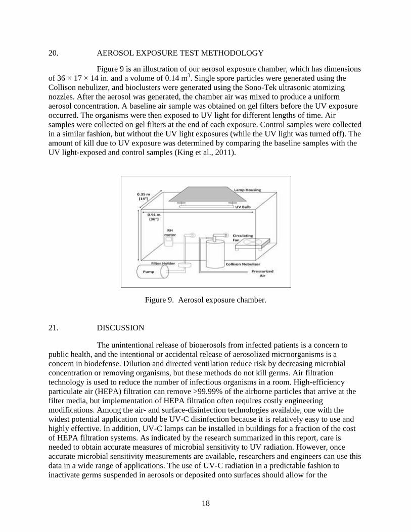
20. AEROSOL EXPOSURE TEST METHODOLOGY
Figure 9 is an illustration of our aerosol exposure chamber, which has dimensions
of 36 × 17 × 14 in. and a volume of 0.14 m3. Single spore particles were generated using the
Collison nebulizer, and bioclusters were generated using the Sono-Tek ultrasonic atomizing
nozzles. After the aerosol was generated, the chamber air was mixed to produce a uniform
aerosol concentration. A baseline air sample was obtained on gel filters before the UV exposure
occurred. The organisms were then exposed to UV light for different lengths of time. Air
samples were collected on gel filters at the end of each exposure. Control samples were collected
in a similar fashion, but without the UV light exposures (while the UV light was turned off). The
amount of kill due to UV exposure was determined by comparing the baseline samples with the
UV light-exposed and control samples (King et al., 2011).
Figure 9. Aerosol exposure chamber.
21. DISCUSSION
The unintentional release of bioaerosols from infected patients is a concern to
public health, and the intentional or accidental release of aerosolized microorganisms is a
concern in biodefense. Dilution and directed ventilation reduce risk by decreasing microbial
concentration or removing organisms, but these methods do not kill germs. Air filtration
technology is used to reduce the number of infectious organisms in a room. High-efficiency
particulate air (HEPA) filtration can remove >99.99% of the airborne particles that arrive at the
filter media, but implementation of HEPA filtration often requires costly engineering
modifications. Among the air- and surface-disinfection technologies available, one with the
widest potential application could be UV-C disinfection because it is relatively easy to use and
highly effective. In addition, UV-C lamps can be installed in buildings for a fraction of the cost
of HEPA filtration systems. As indicated by the research summarized in this report, care is
needed to obtain accurate measures of microbial sensitivity to UV radiation. However, once
accurate microbial sensitivity measurements are available, researchers and engineers can use this
data in a wide range of applications. The use of UV-C radiation in a predictable fashion to
inactivate germs suspended in aerosols or deposited onto surfaces should allow for the

19
development of precise and effective engineering solutions for protecting health and life.
Understanding the effect of natural sunlight on airborne germs should assist in understanding and
thus predicting microbial dissemination and its role in natural or manmade epidemics.

20
Blank

21
REFERENCES
Becker, M.M.; Wang, Z. B–A. Transitions within a 5 S Ribosomal RNA Gene are Highly
Sequence-Specific. J. Biol. Chem. 1989, 264(7), 4163–4167.
Beebe, J.M.; Dorsey, E.L.; Guse, D.G.; Hunt, G.R. Stability and Virulence Relationships of
Air-Borne Bacillus anthracis Spores Under Stress of Light and Humidity. Technical
Memorandum 18; U.S. Army Biological Laboratories: Fort Detrick, MD, 1962.
Ben-David, A.; Sagripanti, J.-L. A Model for Inactivation of Microbes Suspended in the
Atmosphere by Solar Ultraviolet Radiation. Photochem. Photobiol. 2010, 86, 895–908.
Black and Veatch Corporation. White’s Handbook of Chlorination and Alternative Disinfectants,
5th ed.; John Wiley & Sons: Hoboken, NJ, 2010.
Block, S.S. Disinfection, Sterilization, and Preservation, 5th ed.; Lippincott Williams & Wilkins:
Philadelphia, PA, 2001.
Cerutti, P.A.; Miller, N.; Pleiss, M.G.; Remsen, J.F.; Ramsay, W.J. Photohydration of Uridine in
the RNA of Coliphage R17. I. Reductive Assay for Uridine Photohydration. Biochemistry
1969, 64, 731–738.
Coohill, T.P. Virus Cell Interactions as Probes for Vacuum Ultraviolet Radiation Damage and
Repair. Photochem. Photobiol. 1986, 44, 359–363.
Coohill, T.P.; Sagripanti J.-L. Overview of the Inactivation by 254 nm Ultraviolet Radiation of
Bacteria with Particular Relevance of Biodefense. Photochem. Photobiol. 2008, 84,1084–
1090.
Coohill, T.P;. Sagripanti, J.-L. Review: Bacterial Inactivation by Solar Ultraviolet Radiation
Compared with Sensitivity to 254 nm Radiation. Photochem. Photobiol. 2009, 85, 1043–
1052.
Couch, R.B. Orthomyxoviruses. In: Medical Microbiology, 4th ed. University of Texas Medical
Branch: Galveston, TX, 1995, pp 1–22.
Fields Virology; Fields, B.N.; Knipe, D.M., Eds.; 2nd ed.; Raven Press: New York, 1990.
Fields Virology; Knipe, D.M.; Howley, P.M., Eds.; 4th ed.; Lippincott Williams and Wilkins:
Philadelphia, PA, 2001.
Foarde, K.; Franke, D.; Webber, T.; Hanley, J.; Owen, K. Biological Inactivation Efficiency by
HVAC In-Duct Ultraviolet Light Systems. Technology Evaluation Report. U.S.
Environmental Protection Agency, Office of Research and Development, National
Homeland Security Research Center, 2006.
Giese, A.C. Living with Our Sun’s Ultraviolet Rays. Plenum Press: New York, 1976.
Hanawalt, P.C. Concepts and Models for DNA Repair: From Escherichia coli to Mammalian
Cells. Environ. Mol. Mutagen. 1989, 14(16), 90–98.
Heisler, G.M.; Grant, R.H.; Gao, W.; Slusser, J.R. Solar Ultraviolet-B Radiation in Urban
Environments: The Case of Baltimore, Maryland. Photochem. Photobiol. 2004, 80(3),
422–428.

22
Hemmes, J.H.; Winkler, K.C.; Kool, S.M. Virus Survival as a Seasonal Factor in Influenza and
Poliomyelitis. Nature 1960, 188, 430–431.
Hicke, J.A.; Slusser, J.; Lantz, K.; Pascual, F.G. Trends and Interannual Variability in Surface
UVB Radiation Over 8 to 11 Years Observed Across the United States. J.Geophys. Res.
2008, 113, D21302.
Jagger, J. Solar-UV Actions on Living Cells. Praeger: New York, 1985.
King, B.; Kesavan, J.; Sagripanti, J.-L. Germicidal UV Sensitivity of Bacteria in Aerosols and on
Contaminated Surfaces. Aerosol Sci. Technol. 2011, 45, 1–9.
Ko, G.; Melvin, F.W.; Burge, H.A. The Characterization of Upper-Room Ultraviolet Germicidal
Irradiation in Inactivating Airborne Microorganisms. Environ. Health Perspect. 2002,
110(1), 95–101.
Kowalski, W. Ultraviolet Germicidal Irradiation Handbook: UVGI for Air and Surface
Disinfection. Springer: New York, 2009.
Kratz, R.F. Barron’s E-Z Microbiology. Barron’s Educational Series: New York, 2005.
Lin, C.; Li, C. Control Effectiveness of Ultraviolet Germicidal Irradiation on Bioaerosols.
Aerosol Sci. Technol. 2002, 36(4), 474–478.
Lubin, D; Jensen, E.H. Effects of Clouds and Stratospheric Ozone Depletion on Ultraviolet
Radiation Trends. Nature 1995, 377, 710–713.
Lytle, D.C.; Sagripanti, J.-L. Predicted Inactivation of Viruses of Relevance to Biodefense by
Solar Radiation. J. Virol. 2005, 79(22), 14244–14252.
Masschelein, W.J.; Rice, R. Ultraviolet Light in Water and Wastewater Sanitation. CRC Press:
Boca Raton, FL, 2002.
McDevitt, J.J.; Lai, K.M.; Rudnick, S.N.; Houseman, E; Andres, M.W.; Milton, D.K.
Characterization of UVC Light Sensitivity of Vaccinia Virus. Appl. Environ. Microbiol.
2007, 73(18), 5760–5766.
McLean, R.L. Comments on Reducing Influenza Epidemics Among Hospitalized Veterans by
UV Irradiation of Droplets in the Air. Amer. Rev. Respir. Dis. 1956, 83(Suppl.), 36–38.
Menetrez, M.Y.; Foarde, K.K.; Webber, T.D.; Dean, T.R.; Betancourt, D.A. Efficiency of UV
Irradiation on Eight Species of Bacillus. J. Environ. Eng. Sci. 2006, 5, 329–334.
Menzies, D.; Popa, J.; Hanley, J.A.; Rand, T; Milton, D.K. Effect of Ultraviolet Germicidal
Lights Installed in Office Ventilation Systems on Workers’ Health and Wellbeing:
Double-Blind Multiple Crossover Trial. Lancet 2003, 362, 1785–1791.
Miller S.L.; Macher, J.M. Evaluation of a Methodology for Quantifying the Effect of Room Air
Ultraviolet Germicidal Irradiation on Airborne Bacteria. Aerosol Sci. Technol.2000, 33,
274–295.
Mims, F.M. Avian Influenza and UV-B Blocked by Biomass Smoke. Environ. Health Perspect.
2005, 113(12), A806–A807.
23
Moeller, R.; Horneck, G.; Facius, R.; Stackebrandt, E. Role of Pigmentation in Protecting
Bacillus sp. Endospores Against Environmental UV Radiation. FEMS Microbiol. Ecol.
2005, 51, 231–236.
Munakata, N. Comparative Measurements of Solar UV Radiation with Spore Dosimetry at Three
European and Two Japanese Sites. J. Photochem. Photobiol B. 1999, 53, 7–11.
Munakata, N.; Kazadzis, S.; Bais, A.F.; Hieda, K.; Ronto, G.; Rettberg, P.; Horneck, G.
Comparisons of Spore Dosimetry and Spectral Photometry of Solar-UV Radiation at
Four Sites in Japan and Europe. Photochem. Photobiol. 2000, 72(6), 739–745.
Munakata, N.; Makita, K.; Bolsee, D.; Gillotay, D.; Horneck, G. Spore Dosimetry of Solar UV
Radiation: Applications to Monitoring of Daily Irradiance and Personal Exposure. Adv.
Space Res. 2000, 26(12), 1995–2003.
Nicholson, W.L.; Galeano, B. UV Resistance of Bacillus anthracis Spores Revisited: Validation
of Bacillus subtilis Spores as UV Surrogates for Spores of B. anthracis Sterne. Appl.
Environ. Microbiol. 2003, 69(2),1327–1330.
Nicholson, W.L.; Setlow, B.; Setlow, P. UV Photochemistry of DNA in vitro and in Bacillus
subtilis Spores at Earth Ambient and Low Atmospheric Pressure: Implications for Spore
Survival on Other Planets or Moons in the Solar System. Astrobiology 2002, 2, 417–425.
Peccia, J.; Hernandez, M. Photoreactivation in Airborne Mycobacterium parafortuitum. Appl.
Environ. Microbiol. 2001, 67(9), 4225–4232.
Puskeppeleit, M.; Quintern, L.E.; Naggar, S.E.; Schott, J.-U.; Eschweiler, U.; Horneck, G. Long-
Term Dosimetry of Solar UV Radiation in Antarctica with Spores of Bacillus subtilis.
Appl. Environ. Microbiol. 1992, 58(8), 2355–2359.
Rauth, A.M. The Physical State of Viral Nucleic Acid and the Sensitivity of Viruses to
Ultraviolet Light. Biophys. J. 1965, 5, 257–273.
Remsen, J.F.; Miller, N.; Cerutti, P.A. Photohydration of Uridine in the RNA of Coliphage R17,
II. The Relationship Between Ultraviolet Inactivation and Uridine Photohydration. Proc.
Natl. Acad. Sci. USA 1970, 65(2), 460–466.
Riesenman P.J.; Nicholson, W.L. Role of the Spore Coat Layers in Bacillus subtilis Spore
Resistance of Hydrogen Peroxide, Artificial UV-C, UV-B, and Solar UV Radiation. Appl.
Environ. Microbiol. 2000, 66(2), 620–626.
Rutala, W.A.; Gergen, M.F.; Weber, D.J. Room Decontamination with UV Radiation. Infect.
Control Hosp. Epidemiol. 2010, 31(10), 1025–1029.
Sagripanti, J.-L.; Lytle, C.D. Inactivation of Influenza Virus by Solar Radiation. Photochem.
Photobiol. 2007, 83, 1278–1282.
Schreier, W.J.; Schrader, T.E.; Koller, F.O.; Gilch, P.; Crespo-Hernandez, C.E.; Swaminathan,
V.N.; Carell, T.; Zinth, W.; Kohler, B. Thymine Dimerization in DNA is an Ultrafast
Photoreaction. Science 2007, 315(5812), 625–629.
Setlow, P. Spore Germination. Curr. Opin. Microbiol. 2003, 6, 550–556.
Setlow, P. Spores of Bacillus subtilis: Their Resistance to and Killing by Radiation, Heat, and
Chemicals. J. Appl. Microbiol. 2006, 101, 514–525.
24
Siegal, J.D.; Rhinehart, E.; Jacckson, M.; Chiarello, L., and the Healthcare Infection Control
Practices Advisory Committee. Guideline for Isolation Precautions: Preventing
Transmission of Infectious Agents in Healthcare Settings. 2007,
http://www.cdc.gov/ncidod/dhqp/pdf/isolation2007. pdf (accessed June 2012).
TB Infection-Control Guidelines Work Group. Guidelines for Preventing the Transmission of
Mycobacterium Tuberculosis in Health-Care Facilities. MMWR Morb. Mortal Wkly. Rep.
1994, 43(RR-13), 1–132.
Tseng, C.; Li, C. Inactivation of Viruses on Surfaces by Ultraviolet Germicidal Irradiation. J.
Occup. Environ. Hyg. 2007, 4, 400–405.
Tucker, J.B.; Zilinskas R.A. The 1971 Smallpox Outbreak in the Soviet City of Aralsk:
Implications for Variola Virus as a Bioterrorist Threat. Crit. Rev. Microbiol. 2003, 29,
81–95.
U.S. Environmental Protection Agency. Investigation of Simulated Sunlight in the Inactivation of
B. anthracis and B. subtilis on Outdoor Materials. Report EPA/600/R-10/048. Office of
Research and Development, National Homeland Security Research Center: Washington,
DC, 2010.
UV-B Monitoring and Research Program website, http://UVB.nrel.colostate.edu (accessed
June 2012).
Xu, P.; Kujundzic, E.; Peccia, J.; Schafer, M.P.; Moss, G.; Hernandez, M.; Miller, S.L. Impact of
Environmental Factors on Efficacy of Upper-Room Air Ultraviolet Germicidal
Irradiation for Inactivation Airborne Mycobacteria. Environ. Sci. Technol. 2005, 39,
9656–9664.
Xu, P.; Peccia, J.; Fabian, P.; Martyny, J.W.; Fennelly, K.P.; Hernandez, M.; Miller S.L.
Efficacy of Ultraviolet Germicidal Irradiation of Upper-Room Air in Inactivating
Airborne Bacterial Spores and Mycobacteria in Full-Scale Studies. Atmos. Environ. 2003,
37, 405–419.
Xue, Y.; Nicholson, W.L. The Two Major Spore DNA Repair Pathways, Nucleotide Excision
Repair and Spore Photoproduct Lyase, are Sufficient for the Resistance of Bacillus
subtilis Spores to Artificial UV-C and UV-B But not to Solar Radiation. Appl. Environ.
Microbiol. 1996, 62(7), 2221–2227.