ANDdm5migu4zj3pb.cloudfront.net/manuscripts/100000/100405/JCI32100405.pdf · diseases in which a...
14
STUDIES OF CALCIUM AND PHOSPHORUS METABOLISM XV. IN VARIOUS METABOLIC AND BONE DISEASES By J. C. AUB AND R. F. FARQUHARSON (From the Medical Clinic of the Massachusetts General Hospital, and the Peter Bent Brigham Hospital, Boston) (Received for publication August 3, 1931) The bones may well be looked upon as a storehouse for calcium and phosphorus, readily available for the body needs for fixed base. The demands upon this supply can readily be determined if a carefully con- trolled diet inadequate in calcium is administered. Under such condi- tions the normal excretion of calcium remains fairly constant, and a com- parison with disease states may readily be undertaken. For several years this laboratory has systematically studied the cal- cium and phosphorus exchange in numerous pathological conditions. In the course of these studies various diseases have been investigated and are to be published (1, 2, 3). We have also had the opportunity to investigate many interesting diseases in which a disturbance of bone metabolism might well be expected. Some of these isolated cases are here brought together and this paper, therefore, demonstrates the metabolic need of calcium and phosphorus in various bone abnormalities as well as in gout and chronic hepatitis with jaundice. EXPERIMENTAL METHODS The patients were, with one exception, studied in the metabolism ward at the Massachusetts General Hospital. The careful management and routine used there for the preparation of accurate diets and for the collec- tions of urine and feces has already been fully described (4). The periods used here were of three days' duration, and the feces were divided by the appearance of carmine. The methods of analysis for calcium and phos- phorus were those of Fiske (5) and the nitrogen determinations were made by Kjeldahl method. In order to determine the endogenous need for calcium the patients received our usual neutral diet, which is inadequate only in calcium. In nine normal control subjects on similar neutral diets, the excretion of calcium averaged 186 mgm. in the urine and 386 mgm. in the feces per period. A. DISEASES AFFECTING THE SKELETON Secondary carcinoma involving bone In the course of studies on the effect of lead therapy upon cancer growth, observations were also made upon the calcium and phosphorus 235
Transcript of ANDdm5migu4zj3pb.cloudfront.net/manuscripts/100000/100405/JCI32100405.pdf · diseases in which a...

STUDIES OF CALCIUMANDPHOSPHORUSMETABOLISMXV. IN VARIOUS METABOLIC AND BONEDISEASES
By J. C. AUB AND R. F. FARQUHARSON(From the Medical Clinic of the Massachusetts General Hospital, and the
Peter Bent Brigham Hospital, Boston)
(Received for publication August 3, 1931)
The bones may well be looked upon as a storehouse for calcium andphosphorus, readily available for the body needs for fixed base. Thedemands upon this supply can readily be determined if a carefully con-trolled diet inadequate in calcium is administered. Under such condi-tions the normal excretion of calcium remains fairly constant, and a com-parison with disease states may readily be undertaken.
For several years this laboratory has systematically studied the cal-cium and phosphorus exchange in numerous pathological conditions. Inthe course of these studies various diseases have been investigated andare to be published (1, 2, 3).
Wehave also had the opportunity to investigate many interestingdiseases in which a disturbance of bone metabolism might well beexpected. Some of these isolated cases are here brought together andthis paper, therefore, demonstrates the metabolic need of calcium andphosphorus in various bone abnormalities as well as in gout and chronichepatitis with jaundice.
EXPERIMENTALMETHODS
The patients were, with one exception, studied in the metabolism wardat the Massachusetts General Hospital. The careful management androutine used there for the preparation of accurate diets and for the collec-tions of urine and feces has already been fully described (4). The periodsused here were of three days' duration, and the feces were divided by theappearance of carmine. The methods of analysis for calcium and phos-phorus were those of Fiske (5) and the nitrogen determinations were madeby Kjeldahl method. In order to determine the endogenous need forcalcium the patients received our usual neutral diet, which is inadequateonly in calcium. In nine normal control subjects on similar neutral diets,the excretion of calcium averaged 186 mgm. in the urine and 386 mgm.in the feces per period.
A. DISEASES AFFECTING THE SKELETONSecondary carcinoma involving bone
In the course of studies on the effect of lead therapy upon cancergrowth, observations were also made upon the calcium and phosphorus
235

CALCIUM AND PHOSPHORUSIN BONE DISEASE
metabolism of patients with secondary breast carcinoma involving thespine, pelvis, and other bones. The calcium excretion of most of thesepatients fell within the normal range. In one patient, however, whoselesion in the pelvis was progressing, there was a very high urinary calciumexcretion.
Case I, Mrs. B., age 47, weighing 48 kilos, had the left breast removedin 1925, and the right in June, 1927, both breasts being infiltrated withcarcinoma. For approximately six months before her admission to thehospital, she had suffered from stiffness and soreness in the back andright shoulder and, for some weeks immediately preceding admission, withmore severe pain low in the back. She was fairly comfortable when lyingstill but was afraid to move. X-ray examinations revealed metastases inthe spine, ribs, and pelvis, with pathological fracture of a vertebra, andsome weeks later showed that the process in the pelvis had rapidly ad-vanced. The values for calcium determinations are presented in Table I.
TABLE I
Secondary carcinoma involving bone(Intake and output in 3-day periods)
Calcium Phosphorus Serum
Subject and diet PeriodUrine Feces In- Balance Urine In- Ca p Pro-
take take tein
mgm. mgm. grams
(3-day) grams grams grams grams grants grams per per per100 100 100CC. CC. CC.
Subject B. M.-Case I. 1 .92 .58 .25 -1.25 1.02 1.50 10.7 5.1 6.8Low Ca diet. 2 1.11 .66 .25 -1.52 1.31 1.50
3 1.33 .29 .26 -1.36 1.55 1.504* .37 .26 1.62 1.505* 1.19 .25 .28 -1.16 1.59 1.506* .92 .40 .35 - .97 1.61 1.477* .48 .39 .32 - .55 1.09 1.398 .62 .22 .38 - .46 .95 1.47
High Ca diet. 9* .77 1.06 2.83 +1.00 1.43 3.4310* .69 1.05 3.18 +1.44 1.38 3.4311 .49 1.15 3.19 +1.55 1.59 10.5 3.9
Subject L. N.-Case II. 1 .33 .27 .34 - .26 2.04 9.3 3.6 7.3Low Ca diet. 2 .35 .36 .33 - .38
3* .A1 .29 .31 - .394* .59 .26 .32 - .53
* 60 mgm. ofphosphate.
lead lnjected intravenously in the torm ot colloicaal leau
In the initial periods on a constant, potentially neutral diet low incalcium, the urinary excretion of calcium was very high, the calcium ofthe feces being within normal limits. There was a large negative calcium
236
I

J. C. AUB AND R. F. FARQUHARSON
balance. While she remained on the same diet the urinary calcium excre-tion fell to a level within the normal range. Then she was given a highcalcium diet on which the calcium of the urine remained at the relativelylow level while the fecal calcium increased greatly and there was a positivebalance. The serum calcium did not change.
It seems reasonable to associate the excretion of large amounts ofcalcium in the urine with the abnormal liberation of calcium about theenlarging, destructive lesion in the pelvis. The periodicity of growth ofmalignant disease in bone is generally recognized. As the activity de-creases temporarily the flow of calcium from the bone into the excretorychannels would naturally diminish and the calcium of the urine fall towardnormal amounts. It is interesting that in this case the subsequent in-gestion of a diet fairly high in calcium, which gave rise to a large positivecalcium balance, had little effect on the amount of calcium in the urine oron the serum calcium level. The same thing has been found in othercases.
Wedo not attribute the change in calcium excretion to the effect ofinjected colloidal lead, as we have had no evidence in other cases toindicate that lead given under the conditions of our work had anyspecific effect on the tumor growth or on calcium metabolism.
Case II, Mrs. L. N., age 33, weighing 62 kilos, suffered from extensivemetastatic carcinoma involving the skull, spine, pelvis, and humerus. Thedata for calcium and phosphorus excretion in her case are also given inTable I. In spite of the extensive malignant disease the calcium excretionduring the period of observation was within the normal range. A verymarked clinical improvement after lead and x-ray therapy with recalcifi-cation of tumor masses is interesting though of doubtful significance.
MyelomaIt was interesting to compare the calcium metabolism in a patient
suffering from multiple myeloma with that of cases of breast carcinomawith metastases in the bones. Case III (Mrs. M), age 27, weighing 44kilos, had suffered for approximately two years with darting painsin various bones, which followed sudden movements. She was comfort-able when resting, but weak and pale. X-ray examinations demonstrateda diffuse and destructive process involving the spine, ribs, scapulae,humeri, pelvis, and heads of the femora. Someof the areas of destructionwere quite large with sharply defined margins. The 9th dorsal vertebrawas mushroomed. The appearance suggested to Dr. George Holmeseither myeloma or metastatic carcinoma. Large amounts of Bence Jonesprotein were found in the urine. The pathological diagnosis of myelomawas made from sections of bone removed at biopsy. Results are givenin Table II.
237

CALCIUM AND PHOSPHORUSIN BONE DISEASE
0
0
babo0
z
Qtu-Q
8 00
C) Ca- 0E - O,t: t-
C 0-4t-
0cU)
UI)6
- V
-4
VIf)
Vo 00 0
1- -4
- U) 0C'"m C"-rl r. r-r" --
0 .'6 4 CN
I +
a to ) 00 t- 0000r-I'll Cd4 C-0 000
.0 - o - -0'
o~~~.0t*' I-00
0Cd
qdC000
t*, *4 I
eI C
I
C' C
14 VAf 00
C) e C-C1' ItU 'N-4 " -T
Ix,~~~~~~~~~~~~~~~~~
N. .0o0
*0-..t° C)t _ > vl ] <> + C cs
.)
0
10IC
0r
I.
:~
ItI)
C')-
,0
-4
-C)
Cc-4 0)00cI" -i
cC'-'%C)E
238
6cs
c-
*t~Ic-
;'It
E- |
-I0 IC1
s
* !.2(l
Q
I~~~~~~~~~~~~~~~~~~
(1 CN
\6 11
r.

J. C. AUB AND R. F. FARQUHARSON
The excretion of calcium and phosphorus in both urine and feces waswithin the normal range for adult patients on a low calcium diet (6). Itis notable that in spite of wide-spread involvement of bone there was noincreased excretion of calcium. Due to a very low urinary calcium ex-cretion, the negative balance was actually less than that of the averagenormal adult. From the clinical point of view also the disease appearedto be relatively stationary.
Focal osteitis fibrosaCase IV I (Miss R.), aged 34, weight 53 kilos, had been well up to three
years ago, when a fracture of the right femur occurred at the site of a largecyst. At open operation the cyst wall was curetted, following whichfirm bony union occurred. The report of histological exafination of thecurettings described "a cell rich fibrous tissue containing bone trabeculaewith areas of hemorrhage and blood pigment. The histological appear-ances are those of osteitis fibrosa." In 1928 she began to suffer severeshooting pain in the right ankle. X-ray examination revealed a largecyst in the lower end of the right tibia and another in the right pubis;but the rest of the skeleton appeared to be normal. The cyst in the tibiawas opened and its wall curetted. The pathological examination re-vealed a similar picture to that previously reported.
Before operation she was given a low calcium diet for nine days. InTable II are presented results of the calcium and phosphorus determina-tions.
The excretion of calcium and phosphorus was within normal limitsand the blood serum values were normal. On a higher calcium intakepart of this calcium was retained as in normal individuals. The normalcalcium metabolism in this case of focal osteitis fibrosa is in striking con-trast with that of cases of generalized "osteitis fibrosa cystica" (hyper-parathyroidism) (1) in which the serum calcium is high and calcium ex-cretion greatly increased. There is an equally great contrast in the clin-ical picture of the two conditions.
Fragilitas ossiumCase V (May T.), age 14, weighing 25 kilos, had a typical history of
fragilitas ossium. One femur had been broken at birth and x-ray exam-ination the next day also showed healed fractures of both femora. Therehad been repeated fractures from the slightest trauma in early years.Whenshe was 4 years old attention was drawn to the fact that her trunkwas unusually short and this has become progressively more marked sincethat time. Her diet, as described, had been adequate in both calcium andvitamin D. On admission she presented a very unusual appearance
1 After this article had gone to press, similar findings were reported byDonald Hunter, Hyperparathyroidism: Generalized Osteitis Fibrosa. Brit.J. Surg., 1931-32, xix, 203.
239

CALCIUM AND PHOSPHORUSIN BONE DISEASE
with great deformity due to the shortness of the trunk, which exhibited abroad kyphotic curve and a gross scoliosis. The head was large and theupper extremities appeared normal, but there was extreme deformity ofthe lower limbs which had suffered repeated fractures. Apart from theskeletal changes no abnormalities were found.
On x-ray examination all the bones showed increased radiability withcoarse irregular trabeculation and thin cortex. The findings, accordingto Dr. George Holmes, were those of osteomalacia.
The calcium excretion on a low calcium diet was within the limits ofnormal, although the urinary calcium was probably a little higher thanin most children of her age. Serum calcium was normal and serum phos-phorus slightly above the usual level for children. More than three yearssince this observation her bones remained unchanged, as judged by x-rayphotographs, in spite of ample calcium and vitamines in her diet.
Osteoscierosis (marble bone disease)Case VI (Mrs. H. B.), P. B. B. H. No. 67748, age 41, weighing 63.7
kilos, complained that for eight months she had dull pain in the back ofthe neck and shoulders with limitation of motion. Her previous historywas interesting. A very severe diabetes started ten years ago with dia-betic coma relieved by insulin eight years ago. The removal of anadenoma of the thyroid four years ago greatly improved the diabetesand also relieved attacks of cardiac palpitation. X-rays of nearly thewhole skeleton disclosed a marked osteosclerosis which had not obviouslyincreased in the past four years. All the bones except the ribs were un-usually dense and rather structureless in appearance but with very thickcortices. There were no areas of decreased density as seen in Paget'sdisease. Four other members of the immediate family had similar find-ings by x-ray examination.
TABLE III
Osteosclerosis. Case VI. (Mrs. H. B.)(Intake and output in 3-day periods)
Calcium
Period ExcretionIntake Balance
Urine Feces Total
(S days) grams grams grams grams grams
2 .19 .06 .25 .20 -.053 .20 .13 .33 .20 -.134 .20 .19 .39 .20 -.195 .15 .18 .33 .20 -.13
Blood serum valuesCalcium-8 mgm. per 100 cc., 10 mgm. per 100 cc.; Phosphorus-3.2 mgm. per 100
cc., 3.2 mgm. per 100 cc.; Total protein-8.0 grams per 100 cc.; Albumin-5.6 grams per100 cc.; Globulin-2.4 grams per 100 cc.
240

J. C. AUB AND R. F. FARQUHARSON
Extensive laboratory studies disclosed no variation from the normal,except a slight elevation of sugar in the blood. A study of the calciumexchange is shown in Table III. The findings are not abnormal exceptpossibly a diminution in fecal calcium excretion. This is of less signifi-cance considering her basal metabolic rate of - 19 per cent. Therefore,further studies of phosphorus and nitrogen balance were not made in thiscase.
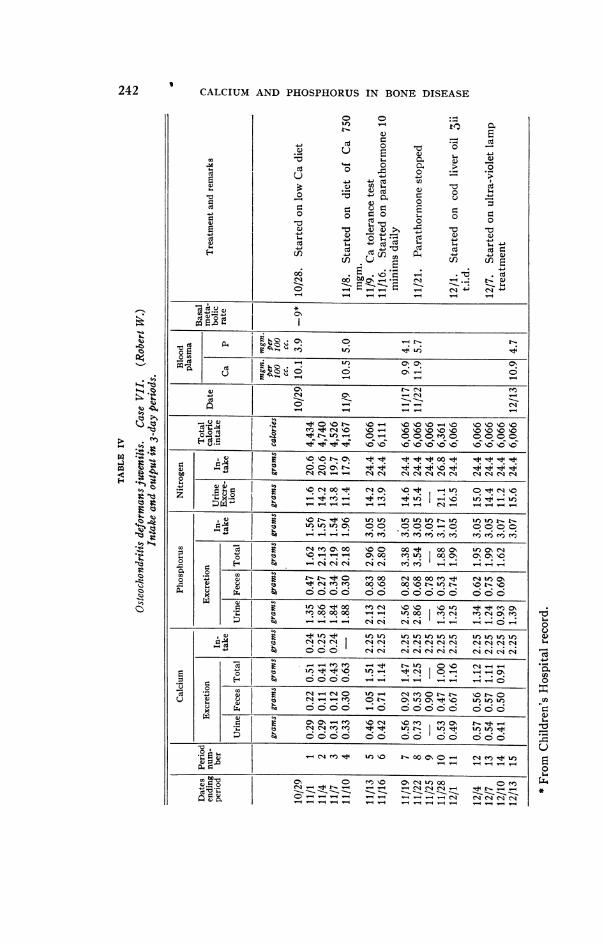
Osteochondritis deformans juvenilis (Legg's disease)Case VII (Robert W.2), M. G. H. No. 279602, age 10, weighing 25.5
kilos, was an adopted child. Birth was two months premature. Hischildhood had been characterized by a limping gait and an avoidance ofsuch activity as walking upstairs. These signs had become progressivelymore marked. He had lived upon a vegetarian diet all his life. Physicalexamination disclosed a short, stocky, well nourished boy with nothingabnormal to be found except in his skeleton. There was normal, freemovement of all his joints. The right leg was shorter and smaller thanthe left with slight atrophy of the muscles. The x-rays, taken at theBoston Children's Hospital, included all the bones in the body. Theyshowed a wide-spread disturbance practically limited to the epiphyses.This disturbance was characterized by delayed development, mushroom-ing of the weight-bearing epiphyses, and increased density of the bonealong the epiphyseal margins. In some areas, particularly the spine andpelvis, this had resulted in absence of parts of the bone. The diagnosisof generalized osteochondritis deformans was made by all the manyphysicians who saw him.
Prolonged metabolic studies were undertaken with both a low and ahigh calcium diet, with the addition of parathormone and cod liver oil.These results are difficult to interpret because there are practically nodata of the normal excretion of calcium at this age period. When com-pared to other growing children the urinary calcium excretion is slightlyelevated. This is particularly true, for his diet was essentially neutralthroughout the whole observation. In spite of the abnormality of mostof his epiphyses he had no difficulty in storing calcium at a rapid rate.
The abnormality of this boy's metabolism was in the phosphorus ex-cretion. This was distinctly above the theoretical value to be derivedfrom the calcium and nitrogen balances (see Paper IV (7)) for the firstnine periods. Thereafter, more phosphorus was retained than could betheoretically explained. This change appeared before cod liver oil wasgiven and followed the distinct elevation in plasma calcium produced byparathormone.
The nitrogen excretion and the blood findings are not abnormal fora growing boy. The results of these studies are given in Table IV andFigures 1 and 2.
2 Referred by Dr. Robert Osgood.
w
241
CALCIUM AND PHOSPHORUSIN BONE DISEASE
cC
10
CC
co'
0e 0'j.-0 > ~~~~~~0)r ~ ~ , ,.
4- 4- Ct cn
uz cn ECSoo~ ~ ~~. ~
0~~~~~~
_0 C 4
0) c__4- s(
06 E~~~~~~~~~~~~~~- C4-i
0
- E -C- ,1-
d cd.u Q *az En d
o qv o o00
ou-
fi ~ ~~~ If) 0'Cr0%0
n o~~0 -~ -04 0 -4 -
_.w Q .X IC\ 0 N0\0% \0
o\0 \0nn \ £_0'0 %0%0%0\\0 0%0%OVC C CN4O
. t-
44 Ne00-d4 CN_ON o0.t Cr )C0 \
- U. w_
co m Cs4.0 \-O%0 1O-4 000 'fn a,%0J "I$
00ve oi oo oO Odq ° Ch 0\ CKo W 0 e ' 1 ? 0' 0 % %
.00
__xo~~~~~~ C 4 cl eN cl cl cl el 14 _4 1414
0 * +oo6 6 oo Ioo oo6 666Nmu Cl m ene 00 ~o 00 ~o u: In r- t-
n'IOOOO'0 O O O O O O ONX~~~~- 14 V4+ clic cli C-i v4
1-dI.. Ilo; e Cf'oo oo oof- 'W oC I0~ --e0% sb4oe O-O O N C4C'i C' C'CN C 'i CN e
c_\t0_tC
Q~~~~~~% '-' %0 %Of e#0 £'- . b° WoF£* 0
co -(N "4 ON CO O) O O O O t- OO-X $ O _ C\ in CsIt\ O ot
i^ e N Cs n n + + m WWmW+~tn v.
>~~~~~CM0-0 0O000-040 I0 WI)0
bC- Ne+moXo
C o O 0o ON C9 U) 00 O
oC --_ _ _ _4 _ C4 C1 N4 Ct C_- _1 _ _I 4_ _ 1 4
C-0
CL)C-4
0.al)
~0
-W
(U
10*
80w
4a
242
C-E).:C.

J. C. AUB AND R. F. FARQUHARSON
Period No. 2 4 6 8 10 I1 14
Q PS r.CodLOil OE10L 100F
+-14- +4--4-4- 4- Ca. Intake0
0 ~~~~~~~~~00~~~~~~~~~~~~~
E -
10 2.0 % >
fi~~~~~~~~~~~~~~TtL C X
e
-0 -
1.5
8
7 0
Period No. 2 4 6 8 10 12 14FIG. 1. CALCIUM METABOLISMIN CASE VII
243

CALCIUM AND PHOSPHORUSIN BONE DISEASE
Period No. 2 4 6 8 10 12 14
Para C, Liver Oil
arE
5
O - Iool
toE °o @
sC >J
O )0C)
o_C
b Blood P
+--+ -4--I---+--++ P Intake
4F
3 F
2
0
o.j-a
L~
Urinary P
NL Balance,
-Period No. 2 4 6 8 10 12 14
FIG. 2. PHOSPHORUSMETABOLISMIN CASE VII
a.0.0
i0totoE0'76
5
3
2
0c
d
z-5
0
+5
+-1-0
244
4

J. C. AUB AND R. F. FARQUHARSON
B. OTHERMETABOLIC DISEASES
GoutCase VIII (H. H.), age 22, weighing 49 kilos, had suffered from re-
curring attacks of gout for five years. Many joints were involved andfinally he became a complete invalid. There were numerous tophi in ears,fingers, and toes. Both olecranon bursae were involved. The meta-carpophalangeal and interphalangeal joints of both hands were swollenand the interossei muscles were atrophic. There was great thickening ofthe common tendon sheath of the right palm. The prepatellar bursa wassimilarly affected. There was very marked swelling of the joints of theankles and feet on both sides with preternatural mobility of the great toes.From an open sinus on the right great toe a chalky material made up ofcrystals of sodium biurate could be easily expressed. X-ray platesshowed complete destruction of the articular surfaces of both first meta-tarsophalangeal joints with extensive destruction of the adjoining bones.There was a varying degree of destruction of the joints of the hands.The blood uric acid was 10.3 mgm. on one occasion and 12.4 mgm. onanother. Results of studies of calcium and phosphorus metabolism aregiven in Table V.
TABLE V
Gout. Case VIII. (H. H.)(Intake and output in 3-day periods)
Calcium Phosphorus Nitrogen SerumPeriod
Urine Feces Intake Balance Urine Feces Intake Balance Urine Intake Ca Protein
grams grams grams grams grams grams grams grams grams grams 100 cc. ccgrmCC.
1 .09 .34 .29 - .14 1.21 .52 1.31 -.42 22.4 25.0 9.7 7.432 .16 .56 .29 -.43 1.23 .71 1.82 -.12 19.6 25.03 .06 .37 .17 -.26 1.10 .44 .97 -.57 16.0 12.6
4 .10 1.03 1.54 +.41 .98 1.14 2.03 -.09 17.8 18.95 .05 1.15 1.63 +.43 .98 1.43 2.68 +.27 13.3 29.66 .09 .85 1.66 +.73 1.10 1.07 2.45 .28 19.0 29.8
On a low calcium diet the calcium metabolism was essentially normal.When the calcium intake was increased five-fold there was, as in Cases Iand IX, no increase in the urinary calcium, although the fecal outputincreased considerably. On this diet, calcium was retained in thebody.
Chronic hepatitis with jaundiceIt has been shown that in experimental obstructive jaundice the serum
calcium may be low (8). In animals with a bile fistula Whipple (9) notedthat the bones became thin and spontaneous fractures occurred. It
245

CALCIUM AND PHOSPHORUSIN BONEDISEASE
seemed worth while to study the calcium and phosphorus metabolismof a case of chronic hepatitis with jaundice.
Case IX (P. N.), a patient of Dr. Chester Jones, age 35, weighing 51kilos, had suffered from chronic hepatitis with jaundice (biliary cirrhosis)for 1 1/2 years. The jaundice ran an undulant course, gradually becom-ing worse. She was moderately jaundiced, thin, and looked weak and ill.The liver was grossly enlarged, extending as far as the umbilicus, and thespleen was readily palpable. There was no ascites or edema. Serumbilirubin was 7 to 8 mgm. per 100 cc., bile pigments were present in theurine, and serum protein and blood nonprotein nitrogen were normal.Results are presented in Table VI.
TABLE VI
Chronic hepatitis with jaundice. Case IX. (P. N.)(Intake and outpu in 3-day periods)
Calcium Phosphorus SerumPeriod
Urine Feces Intake Balance Urine Feces Intake Balance Ca P
(3 days) grams grams grams grams grams grams grams grams mgm. per mgm. prc100 cc. 100 cc.
1 .17 .51 .39 -.29 1.07 .57 1.79 .15 8.5 3.32 .10 .66 .37 -.39 1.20 .54 1.88 .14 9.8 3.53 .12 3.95 3.41 -.66 1.19 1.01 2.93 .734 .14 2.42 3.15 +.59 1.52 1.17 3.07 .38 8.9 3.6
The serum calcium on two of three occasions was slightly below nor-mal. Excretion in stool and urine was within normal limits. The changeto a high calcium diet, as in Case I, had no appreciable effect on the urinecalcium but was associated with an increase in the fecal calcium andphosphorus.
OsteomalaciaSince this paper was written, a further type of bone abnormality has been
studied-osteomalacia due to a dietary deficiency in calciuim.Mrs. L. H., P.B.B.H. No. 39821 (Medical), aged 64, weighing 63.5 kilos,
had largely avoided milk and green vegetables in her diet. Six years ago shehad symptoms suggesting arthritis of the spine. Five years ago she had aslight fall and fractured two thoracic vertebrae. Convalescence has been veryslow and she is still wearing a supporting cast. Except for her bones, herphysical examination was not abnormal for a woman of her age. X-rays ofher whole skeleton disclosed marked generalized decalcification. The thoracicand lumbar spine also showed collapse of the central portions of the bodies andapparent marked expansion of the intervertebral discs. Several of the vertebraein the mid-thoracic region showed marked collapse suggesting spontaneousfractures.
Laboratory data: The Wassermann was negative and the urine, feces, basalmetabolic rate, blood urea nitrogen, blood sugar, and blood morphology were allwithin normal limits. There was no excess of fat or fatty acids in the feces.The blood serum calcium level was 9.8 mgm. and 10.6 mgm. per 100 cc. Theserum phosphorus was found to be 4.0 and 4.8 mgm. per 100 cc.
246

J. C. AUB AND R. F. FARQUHARSON
Mrs. L. H., aged 64, white, female.(Intake and output per 3-day period)
Calcium
ExcretionPeriod Intake Balance
Urine Feces Total
grams grams grams grams grams
1 .29 .50 .79 .26 - .532 .40 .40 .80 .28 - .52
3 1.11 2.16 3.27 5.56 2.294 1.03 2.55 3.58 6.00 2.42
These data disclose normal blood values and a normal calcium excretion.When a diet with a moderate amount of calcium was given to her, a largeproportion of this added calcium was retained. It is interesting that theorganism can maintain normal blood levels and normal calcium excretion evenwith such marked decalcification of the bones.
DISCUSSIONThe changes in deposition or removal of bone salts probably occur
so slowly that the variation from normal cannot be observed by ourmethods in relatively short observations. The observations here re-ported do show, however, that there is nothing grossly abnormal in theblood level or in the excretion of calcium or phosphorus. The reasonfor this may well lie in the large amount of calcium readily available forutilization in times of need, such as occurs on our diet inadequate in cal-cium. This storehouse in the trabeculae, described by Bauer, Aub andAlbright (10), apparently exists in all the diseases here studied, and isavailable for the liberation or storage of calcium. The method determin-ing calcium exchange, therefore, does not necessarily disclose abnormal-ities which might be occurring in one part of the skeleton, as there is thiscompensatory mechanism still present in other bones. Calcium andphosphorus may well be liberated from one part and deposited in anotherportion of bone. It is, therefore, not surprising that in this series ofpatients suffering from various types of bone diseases or chronic jaundicethe variations from normal in calcium and phosphorus excretions arerelatively slight.
BIBLIOGRAPHY
1. Bauer, W., Albright, F., and Aub, J. C., J. Clin. Invest., 1930, viii, 229.A Case of Osteitis Fibrosa Cystica (Osteomalacia?) with Evidence ofHyperactivity of the Parathyroid Bodies. Metabolic Study II.
2. Bauer, W., and Aub, J. C., Studies of Calcium and Phosphorus Metabolism.XVI. Calcium Metabolism in Osteitis Fibrosa (Paget's). To be pub-lished.
247

CALCIUM AND PHOSPHORUSIN BONEDISEASE
3. Bauer, W., and Aub, J. C., Studies of Calcium and Phosphorus Metabolism.XVII. Calcium Metabolism in Acromegaly. To be published.
4. Bauer, W., and Aub, J., J. Am. Dietet. Assoc., 1927, iii, 106. Studies ofInorganic Salt Metabolism. I. The Ward Routine and Methods.
5. Fiske, C. H., and Subbarow, Y., J. Biol. Chem., 1925, lxvi, 375. TheCalorimetric Determination of Phosphorus.
Fiske, C. H., and Logan, M. A., J. Biol. Chem., 1931, xciii, 211, The De-termination of Calcium by Alkalimetric Titration. II. The Precipita-tion of Calcium in the Presence of Magnesium, Phosphate, and Sul-phate, with Applications to the Analysis of Urine.
6. Bauer, W., Albright, F., and Aub, J. C., J. Clin. Invest., 1929, vii, 75.Studies of Calcium and Phosphorus Metabolism. II. The CalciumExcretion of Normal Individuals on a Low Calcium Diet, also Data ona Case of Pregnancy.
7. Albright, F., Bauer, W., Ropes, M., and Aub, J. C., J. Clin. Invest., 1929,vii, 139. Studies of Calcium and Phosphorus Metabolism. IV. TheEffect of the Parathyroid Hormone.
8. Buchbinder, W. C., and Kern, R., Am. J. Physiol., 1927, lxxx, 273. BloodCalcium Deficiency in Experimental Obstructive Jaundice.
9. Whipple, G. H., Arch. Int. Med., 1922, xxix, 711. Pigment Metabolismand Regeneration of Hemoglobin in the Body.
10. Bauer, W., Aub, J. C., and Albright, F., J. Exp. Med., 1929, xlix, 145.Studies of Calcium and Phosphorus Metabolism. V. A Study of theBone Trabeculae as a Readily Available Reserve Supply of Calcium.
248


![[XLS]reports.mca.gov.inreports.mca.gov.in/Reports/MasterDataExcels/company... · Web view500000 500000 500000 500000 100000 100000 100000 100000 100000 100000 100000 100000 100000](https://static.fdocuments.net/doc/165x107/5b2c80367f8b9a3d348b8549/xls-web-view500000-500000-500000-500000-100000-100000-100000-100000-100000.jpg)