Directing stem cells and progenitors towards neuronal ... · Anke Brederlau Directing stem cells...
85
Anke Brederlau Directing stem cells and progenitors towards neuronal differentiation Directing stem cells and progenitors towards neuronal differentiation – implications for experimental therapies of Parkinson’s disease Anke Brederlau Institute of Biomedicine The Sahlgrenska Academy University of Gothenburg 2008 ISBN 978-91-628-7463-6
Transcript of Directing stem cells and progenitors towards neuronal ... · Anke Brederlau Directing stem cells...

Anke B
rederlau
D
irecting stem
cells and p
rog
enitors tow
ards neuro
nal differentiatio
n
Directing stem cells and progenitors towards neuronal differentiation
– implications for experimental therapies of Parkinson’s disease
Anke Brederlau
Institute of Biomedicine The Sahlgrenska Academy
University of Gothenburg
2008
ISBN 978-91-628-7463-6

Directing stem cells and progenitors towards
neuronal differentiation
Implications for experimental therapies for Parkinson’s disease
Anke Brederlau
Institute of Biomedicine Department of Medical Chemistry and Cell Biology
2008

Cover picture: A neuronal colony derived from human embryonic stem cells, cocultured with the stromal cell line PA6 as described in Paper IV. Cells stain positive for the neuronal marker �-tubulinIII (green) and for the dopaminergic marker tyrosine hydroxylase (red). Nuclei are counterstained with Hoechst 33258 (blue). ISBN 978-91-628-7463-6 © Anke Brederlau, March 2008 Institute of Biomedicine Department of Medical Chemistry and Cell Biology Sahlgrenska Academy at Gothenburg University Printed by Intellecta Docusys AB, Gothenburg, Sweden
ABSTRACT
Directing stem cells and progenitors towards neuronal differentiation – implications for experimental therapies for Parkinson’s disease
Anke Brederlau
Institute of Biomedicine, Department of Medical Chemistry and Cell Biology, Sahlgrenska Academy at Gothenburg University, Sweden.
The insight that stem- and progenitor-cells contribute to replacement of nerve cells in the adult central nervous system is the basis of modern therapies for structural brain repair. Their goal is to protect, support and stimulate endogenous stem cells in areas affected by disease and to replace lost cells by transplanting in vitro generated, tailored nerve cells.
In the present thesis growth factors and signaling molecules were investigated for their potential to direct stem- and neural progenitor- cells towards neuronal cell fate. Involved signaling pathways were characterized and candidate molecules identified that might be beneficial for cell-based therapies in Parkinson’s disease.
Results show that Bone Morphogenetic Proteins (BMPs) and Growth Differentiation Factors increase astroglial differentiation while inhibiting oligodendrocyte maturation in rat embryonic mesencephalic culture. None of the factors protect dopaminergic neurons against oxidative radicals in vitro. BMP5, 6 and 7, however, promote dopaminergic differentiation by directly targeting the neuronal cell population.
In cultures of adult rat hippocampus-derived progenitors (AHPs), endogenous BMPs were found to increase undesired astroglial differentiation via the BMP type I receptors ALK6 and ALK2. By viral transduction of dominant negative ALK2 or ALK6, respectively, BMP signaling was blocked in order to inhibit astroglial cell differentiation. Indeed, the expression of glial fibrillary acidic protein (GFAP), a marker for astrocytes, decreased. The number of oligodendrocytes increased and neurons were not affected. However, the strategy proved impractical since it induced cell death. RT-PCR results indicate that only the ALK6, but not the ALK2 receptor, is dynamically regulated in these cultures, suggesting that ALK6 is mainly responsible for glial differentiation and survival of AHPs.
Apoptosis signal-regulating kinase-1 is a ubiquitously expressed enzyme involved in apoptosis. Overexpression of its constitutively active form induced neuronal differentiation in AHP culture. At the same time GFAP expression was inhibited. The effect is mediated via p38 mitogen-activated protein kinase and via inhibition of GFAP promoter activity.
In order to generate transplantable dopaminergic neurons, human embryonic stem cells (hESCs) were cocultured with the stromal cell line PA6, known to instruct mouse and primate ESCs towards dopaminergic cell fate. About 11% of hESCs developed into tyrosine hydroxy-lase-positive (TH-pos) neurons with CNS identity. The hESC-derived neurons displayed action potential in vitro. However, they did not induce behavioral recovery after trans-plantation to the 6-hy-droxydopamine -lesioned rat striatum. Extended differentiation time on PA6 in vitro decreased the risk for teratoma formation after transplantation, but did not elevate the low number of TH-pos neurons in the graft.
In conclusion, certain BMPs as well as ASK1 have been identified as molecules that increase neuronal differentiation. Their putative role in experimental CNS cell therapies is discussed. At the moment, however, the gap between experimental systems and biological reality is difficult to overcome. Further investigations that are necessary to reduce safety concerns in cell-based treatment strategies are outlined.

POPULÄRVETENSKAPLIG SAMMANFATTNING
I Sverige finns cirka 15 000 människor med Parkinsons sjukdom. De flesta är över 50 år och har långsamt tilltagande symtom i form av stelhet och skakningar som beror på att signalsubstansen dopamin saknas i hjärnan. Dopamin produceras av nervceller i mellanhjärnan vilka långsamt bryts ned vid Parkinsons sjukdom. Moderna terapier syftar till att skydda, stödja eller ersätta dessa celler. De första celltransplantationerna utfördes för nästan 20 år sedan. Alla av de idag transplanterade patienterna har erhållit dopaminproducerande celler tagna från mellanhjärnan av embryoner. Möjliga alternativa cellkällor är embryonala stemceller eller omogna hjärnceller, så kallade progenitorer. Till skillnad från mellanhjärnsceller kan de två sistnämnda förökas genom odling. Med hjälp av tillväxtfaktorer eller signalproteiner måste de dock först utvecklas till nervceller innan de kan transplanteras. Syftet med avhandlingens olika studier var att hitta substanser som kan användas i denna process och att förstå deras mekanismer.
Celler tagna från mellanhjärnan hos råttfoster odlades och behandlades med olika tillväxtfaktorer. Faktorerna ”bone morphogenetic protein (BMP) 5, 6 och 7” ökade antalet dopaminproducerande celler genom att omvandla omogna nervceller till mogna. Ingen av faktorerna kunde dock skydda cellerna mot det toxiska ämnet 6-hydroxydopamin som tycks bidra till utvecklingen av nervcelldöd vid Parkinsons sjukdom.
Hippocampus är ett av de två områden i den vuxna hjärnan där progenitorer finns och en ständig nybildning av nervceller pågår. I odlade hippocampala progenitorceller tagna från vuxna råttor orsakar BMP-faktorerna en oönskad ökning av stödjeceller (astrocyter), vilket kan ha en negativ inverkan på nervcellernas funktion efter transplantationen. För att förhindra utvecklingen av stödjeceller blockerades BMP:s signalering genom att introducera icke-fungerande BMP-receptorer i dessa omogna cellerna. Receptorerna binder BMPs men kan inte vidarebefordra signalen till cellerna. Som förväntat avtog antalet stödjeceller medan nervcellerna förblev opåverkade. Eftersom blockeringen av BMP också orsakade celldöd kommer denna strategi inte att kunna användas för att minska stödjeceller . Studien visade att bara en av de tre undersökta BMP-receptorerna, ALK6, regleras dynamiskt av cellerna, vilket tyder på att det är ALK6 som ansvarar för stödjecellbildning och dess överlevnad i odlingen av hippocampala progenitorer.
Genom att införa ett annat protein ”apoptosis signalling kinase 1 (ASK1)” i liknande cellkultur ökades antalet av nybildade nervceller samtidigt som astrocyter minskades och utan att celldöd orsakades. Det kunde påvisas att detta skedde via aktivering av ett enzym, ”p38 mitogen-aktiverad protein kinas”. Enzymet bidrar till att ett viktigt strukturprotein, gliafibrillärt surt protein (GFAP), inte kan bildas i stödjecellerna.
Dessutom har dopaminproducerande celler framställts från humana embryonala stamceller och transplanterats i råtthjärnor till det område där dopamin saknas. Efter transplantationen minskade dock antalet dopaminproducerande celler i transplantatet. Råttornas rörlighet förbättrades således inte. En möjlig risk med denna terapimetod är att dessa celler kan bilda tumörer. Risken minskades genom att, före transplantationen, öka cellernas mognadsgrad i kultur.
Sammanfattningsvis påvisades att BMP5, 6 och 7 så väl som ASK1 har en gynnsam effekt på utvecklingen av nervceller i olika stadier och kan få betydelse för experimentella terapiformer mot Parkinsons sjukdom. Idag är riskerna med cellterapier dock inte tillräckligt studerade och den eventuella kliniska användningen av dessa substanser ligger därför långt fram i tiden. I diskussionen föreslås några fortsatta studier som skulle kunna bidra till vidare utveckling av cellterapi och dess säkerhet.

LIST OF PUBLICATIONS
This thesis is based on the following articles, which will be referred to in the text by their Roman numerals I. Brederlau A, Faigle R, Kaplan P, Odin P, Funa K. Bone morphogenetic proteins but
not growth differentiation factors induce dopaminergic differentiation in mesencephalic precursors. Mol Cell Neurosci 2002, 21(3):367-378.
II. Brederlau A, Faigle R, Elmi M, Zarebski A, Sjoberg S, Fujii M, Miyazono K, Funa K.
The bone morphogenetic protein type Ib receptor is a major mediator of glial differentiation and cell survival in adult hippocampal progenitor cell culture. Mol Biol Cell 2004, 15(8):3863-3875.
III. Faigle R, Brederlau A, Elmi M, Arvidsson Y, Hamazaki TS, Uramoto H, Funa K.
ASK1 inhibits astroglial development via p38 mitogen-activated protein kinase and promotes neuronal differentiation in adult hippocampus-derived progenitor cells. Mol Cell Biol 2004, 24(1):280-293
IV. Brederlau A*, Correia AS*, Anisimov SV, Elmi M, Paul G, Roybon L, Morizane A,
Bergquist F, Riebe I, Nannmark U, Carta M, Hanse E, Takahashi J, Sasai Y, Funa K, Brundin P, Eriksson PS, Li JY. Transplantation of human embryonic stem cell-derived cells to a rat model of Parkinson's disease: effect of in vitro differentiation on graft survival and teratoma formation. Stem Cells 2006, 24(6):1433-1440.
* joint first authors


___________________________________________________________________________________________________________________________________________________________________________________________________________________________________
CONTENTS
ABBREVIATIONS.................................................................................................................... 9
INTRODUCTION.................................................................................................................... 11
Parkinson’s Disease (PD)................................................................................................ 11 Pathophysiology of PD........................................................................................... 11 Models for PD ........................................................................................................ 13
Trophic factors ................................................................................................................ 14 Glial Cell Line-Derived Neurotrophic Factor (GDNF).......................................... 14 Bone Morphogenetic Proteins (BMPs) .................................................................. 15
Apoptosis Signal-Regulating Kinase 1 (ASK1).............................................................. 20 ASK1 signaling ...................................................................................................... 20 Biological functions ............................................................................................... 21
Stem- and progenitor cells............................................................................................... 22 Definitions.............................................................................................................. 22 Cell genesis in the developing brain ...................................................................... 24 Cell genesis in the adult brain ................................................................................ 24
Molecular mechanisms in neural cell genesis ................................................................. 26 Transcription factors .............................................................................................. 26 Epigenetic mechanisms .......................................................................................... 28
BMPs’ changing role in neural cell genesis .................................................................... 28 Molecular cues in the development of DAergic neurons................................................ 29
DAergic cell types.................................................................................................. 29 Molecular patterning in the mesencephalon........................................................... 30
Embryonic stem cells ...................................................................................................... 30 Propagation of ESCs .............................................................................................. 31 Neuronal differentiation of ESCs........................................................................... 31 Increasing the numbers of A9 DAergic cells ......................................................... 32 Tumor formation .................................................................................................... 32
Cell transplantation in PD ............................................................................................... 33 Fetal ventral mesencephalic cells ........................................................................... 33 Adult-derived neural progenitors ........................................................................... 34 Embryonic stem cells ............................................................................................. 34
OBJECTIVES .......................................................................................................................... 36
Overall aim...................................................................................................................... 36 Specific aims ................................................................................................................... 36
METHODOLOGICAL ASPECTS .......................................................................................... 37
Ethical approval............................................................................................................... 37 Cell culture ...................................................................................................................... 37
E14 mesencephalic primary culture ....................................................................... 37 Adult-derived hippocampal progenitors................................................................. 37 Human embryonic stem cell culture....................................................................... 39
Assays to characterize neuronal maturity........................................................................ 39
___________________________________________________________________________________________________________________________________________________________________________________________________________________________________
Transgene expression systems ........................................................................................ 40 Gene delivery by chemical agents.......................................................................... 40 Gene delivery by adenoviral vectors...................................................................... 40
Evaluation of proliferation and viability ......................................................................... 41
[3H]thymidine incorporation ................................................................................. 41 MTT assay.............................................................................................................. 41 Lactate Dehydrogenase (LDH) assay..................................................................... 42 Cell cycle analysis by fluorescence-activated cell sorting (FACS) ....................... 42 Immunohistochemical methods.............................................................................. 42
Methods for protein detection ......................................................................................... 43 Immunohistochemistry (IHC) ................................................................................ 43 Gel electrophoresis and Western blot (WB) .......................................................... 45 Immunoprecipitation .............................................................................................. 46
In vitro kinase assay ........................................................................................................ 46 Real-time polymerase chain reaction (RT-PCR) ............................................................ 46 Analysis of promoter activity by luciferase assay........................................................... 47 Chemical inhibitors ......................................................................................................... 48 In vivo experiments......................................................................................................... 48
6-OHDA lesion and rotational testing.................................................................... 48 Transplantation....................................................................................................... 48
Microscopical analysis and cell quantification ............................................................... 48 Statistics .......................................................................................................................... 50
RESULTS................................................................................................................................. 51
BMPs induce dopaminergic differentiation in mesencephalic precursors (Paper I) ....... 51 The BMPR type Ib is a major mediator of glial differentiation in AHPs (Paper II) ....... 52 ASK1 promotes neuronal differentiation in AHPs (Paper III)........................................ 53 Transplantation of hESC-derived cells to a rat model of PD (Paper IV)........................ 54
DISCUSSION .......................................................................................................................... 55
The limitations of in vitro systems.................................................................................. 55 GFAP as a marker for astrocytes..................................................................................... 57 Will BMPs have a role in future therapies for PD?......................................................... 57 Is ASK1 a putative therapeutic molecule? ...................................................................... 59 Can AHPs serve as a source for experimental cell therapies in PD? .............................. 59 SDIA-induced differentiation of hESCs ......................................................................... 60 Implications for future therapies of PD? ......................................................................... 61 Future aspects .................................................................................................................. 62
CONCLUSIONS...................................................................................................................... 64
ACKNOWLEDGEMENTS ..................................................................................................... 65
REFERENCES......................................................................................................................... 67
APPENDIX Papers I-IV
ABBREVIATIONS ___________________________________________________________________________________________________________________________________________________________________________________________________________________________________
9
ABBREVIATIONS Ad-ASK1-�N adenoviral vector encoding the constitutively active form of ASK1 Ad-ASK1-KM adenoviral vector encoding the kinase mutant form of ASK1 AdLacZ adenoviral vector encoding the �-galactosidase reporter gene AHPs Adult-derived rat hippocampal progenitors ALK activin receptor-like kinase AP anterior-posterior ASK1 apoptosis signal-regulating kinase 1 bFGF basic fibroblast growth factor bHLH basic helix-loop-helix �-gal �-galactosidase �-tubIII �-tubulin III BMP, BMPR bone morphogenetic protein, bone morphogenetic protein receptor BrdU bromodeoyuridine ca constitutively active CBP CREB binding protein cDNA Complementary DNA Co-Smad common-partner Smad CNS central nervous system CREB cyclic AMP-responsive transcriptional enhancer binding protein DA dopamine DAB 3,3’-diaminobenzidine DAergic dopaminergic DIV days in vitro dn dominant negative DNA deoxyribonucleic acid DV dorsal-ventral E12 embryonic day 12 EB embryonic body EGF epidermal growth factor En engrailed ERK extracellular signaling kinase ESCs embryonic stem cells FACS fluorescence-activated cell sorting FBS fetal bovine serum GABA gamma-aminobutyric acid GalC galactocerebroside GDF growth differentiation factor GDNF glial cell line-derived neurotrophic factor GFAP glial fibrillary acidic protein Girk2 G protein inwardly rectifying potassium channel 2 HA hemagglutinin Hes Hairy/Enhancer-of-split hESCs human embryonic stem cells HNu human nuclei HPLC high-performance liquid chromatography H2O2 hydrogen peroxide ICC immunocytochemistry Id Inhibitor-of-differentiation IHC immunohistochemistry I-Smad inhibitory Smad JNK Jun-kinase LDH lactate dehydrogenase LIF leukaemia inhibitory factor
10 ABBREVIATIONS ___________________________________________________________________________________________________________________________________________________________________________________________________________________________________
Lmx1a LIM homeobox transcription factor 1 alpha MAO monoamine oxidase Map2 microtubule-associated protein-2 MAPK mitogen-activated protein kinase Mash1 mammalian archaete-scute homologue 1 MEFs mouse embryonic fibroblasts mESCs mouse embryonic stem cells MPTP 1-methyl 4-phenyl 1,2,3,6-tetrahydropyridine mRNA messenger deoxyribonucleic acid miRNA microRNA moi multiplicity of infection mTOR/FRAP mammalian target of rapamycin/FKBP12-rapamycin associated protein MTT 3-(4,5-dimethylthiazol-2-yl)-2,5-diphenyl-tetrazolium bromide NeuN neuronal nuclei Ngn neurogenin 6-OHDA 6-hydroxydopamine Olig oligodendrocyte transcription factor gene OLPs oligodendrocyte precursors P0 postnatal day 0 PD Parkinson’s disease PDGF�R Platelet-derived growth factor-� receptor PINK1 serine/threonine-protein kinase PI3K phosphatidylinositol 3-kinase PORN polyornithine p-Smad Phosphorylated Smad ROS reactive oxygen species R-Smad receptor activated Smad RT-PCR Real time polymerase chain reaction SDIA stromal cell-derived inducing activity Shh Sonic hedgehog SN, SNpc substantia nigra, substantia nigra pars compacta Sox Sry-related high-mobility-group box STAT signal transducer and activator of transcription SVZ subventricular zone TAK1 TGF-� activated kinase 1 TF transcription factor TGF-�� transforming growth factor-��TH tyrosine hydroxylase TNF-� tumor necrosis factor-� TRAF2 tumor necrosis factor receptor associated factor 2 Trx thioredoxin UPS ubiquitin-proteasome system VTA ventral tegmental area WB western blot XIAP X-chromosome-linked inhibitor of apoptosis protein

INTRODUCTION __________________________________________________________________________________________________________________________________________________________________________________________________________________________________ 11
INTRODUCTION
Parkinson’s Disease (PD)
PD is a neurodegenerative movement disorder that affects about 1% of individuals over the age of 60 (reviewed by de Lau and Breteler 2006). The characteristic motor symptoms like tremor, rigidity and bradykinesia, arise from the death of dopaminergic (DAergic) neurons in the nigrostriatal pathway. The consecutive reduction in striatal dopamine (DA) content leads to imbalance in the neuronal network that controls movement. However, the spectrum of clinical features is broad and the neurodegenerative process is not restricted to DAergic neurons.
It also affects progenitor cells (Hoglinger et al. 2004), noradrenergic, serotoninergic and cholinergic neurons of different brain stem nuclei, neurons of the cerebral cortex, of the olfactory bulb and of the autonomic nervous system (Braak et al. 2002). Brains from patients with idiopathic PD usually show reactive gliosis and fibrillar aggregations, called Lewy bodies, in surviving neurons (Lewy 1912). �
Pathophysiology of PD The cause of onset of PD still remains obscure. The relative contribution of genetics is debated (Siderowf and Stern 2003, Klein and Schlossmacher 2007). In the largest twin study of PD approximately 20.000 white male twins were screened (Tanner et al. 1999). The results suggest that genetics do not play a major role in PD with typical age onset but may be important in cases with earlier age onset. There are to date at least 11 distinct genetic loci associated with PD (reviewed by Klein and Schlossmacher 2007). Currently, only 2-3% of idiopathic PD are known to be caused by a single genetic event (Klein and Schlossmacher 2007). However, it is not excluded that even patients that develop sporadic forms of PD may carry certain genes that render them more susceptible to environmental toxins such as heavy metals and pesticides (Benmoyal-Segal and Soreq 2006). Regarding the pathophysiology of sporadic and familial forms of PD, two major hypotheses are under discussion: DAergic cell death could be caused by misfolding and aggregation of proteins or by mitochondrial dysfunction and oxidative stress, respectively (Jenner and Olanow 2006, McNaught and Olanow 2006, Olanow 2007). These pathogenetic factors are not mutually exclusive but interact to amplify each other. One of the major aims in PD research today is to elucidate the sequence in which they participate in the process of degeneration.
Oxidative stress The maintenance of redox potential within a cell is imperative for neuronal survival. “Oxidative stress” develops when the generation of reactive oxygen species (ROS) (such as hydrogen peroxide (H2O2), superoxide radicals, hydroxyl radicals, nitric oxide or peroxinitrite) exceeds the rate at which endogenous antioxidant defenses (like the antioxidative enzymes superoxide dismutases and glutathione peroxidases) can scavenge

INTRODUCTION __________________________________________________________________________________________________________________________________________________________________________________________________________________________________ 12
oxidants. Lipids, deoxyribonucleic acid (DNA), proteins and other macromolecules are targets for oxidative modifications. The consequences are deterioration of cellular structural architecture, impairments in signaling and finally cell death. Due to the metabolism of DA and a high content of iron and neuromelanin, levels of basal oxidative stress are generally high in the substantia nigra (SN) (reviewed by Jenner 2003). The existence of an imbalance in oxidative cell metabolism in PD is supported by post-mortem studies that show increased iron levels in parkinsonian brains (Sofic et al. 1988, Dexter et al. 1989, Oakley et al. 2007), a decreased amount of antioxidative enzymes (Sofic et al. 1992, Sian et al. 1994) and reduced activity of mitochondrial complex I (Schapira et al. 1989, Dexter et al. 1994, reviewed by Sian et al. 1994, Abou-Sleiman et al. 2006). The latter is important in energy generation and ROS detoxification.
Several proteins affected by PD-related gene mutations have been linked to oxidative stress metabolism. Thus, Parkin (Palacino et al. 2004) and Serine/threonine-protein kinase (PINK1) (Valente et al. 2004) are important for mitochondrial function. DJ-1 seems to function as an intracellular sensor for oxidative stress (Canet-Aviles et al. 2004). However, neither DJ-1 nor Parkin-deficiency alone were sufficient to induce the loss of DAergic neurons in brains from mice carrying the respective mutation (Kim et al. 2005, Perez and Palmiter 2005).
Proteolytic stress The discovery that the mutations in any of the first three PD genes identified, i.e. alpha-synuclein (Polymeropoulos et al. 1997), parkin (Kitada et al. 1998) or ubiquitin C-terminal hydrolase L1 (Maraganore et al. 2004) all interfere with normal protein degradation, aroused interest in the concept of proteolytic stress (reviewed by Gasser 2001, McNaught and Olanow 2006). This implies that intracellular levels of non-functional and potentially toxic proteins exceed the cell’s capacity for clearance. This can either be due to increased generation of abnormal proteins or to insufficient clearing function of the ubiquitin-proteasome system (UPS).
Alpha-synuclein is an abundantly expressed cytosolic and lipid-binding protein in the vertebrate nervous system. It participates in synaptic transport, DAergic neurotransmission and the refolding of abnormal proteins (reviewed by Uversky 2007). However, disruption of the normal function of alpha-synuclein does not cause neurodegeneration (Abeliovich et al. 2000). Instead, DAergic cell death is most likely due to insoluble fibrils that are formed by the mutated form of alpha-synuclein and that resist UPS clearance (reviewed by Goedert 2001). Interestingly, even patients with sporadic PD show impairments in proteosomal degradation (McNaught et al. 2003). This could be due to environmental toxins such as polyphenols that are potent inhibitors of proteosomal function (Nam et al. 2001).
Oxidative stress may also contribute to the impairment of protein degradation. For example, the alpha subunits of the 26/20S proteasomes are particularly vulnerable to free radical-induced damage (Bulteau et al. 2001). On the other hand, a primary impairment of proteosomal degradation causes an accumulation of damaged proteins which can in turn, induce oxidative stress. Thus, different key components of cell degeneration can form a circle that results in similar biochemical changes. For a successful therapeutic concept it would be advantageous to know where the process starts.
INTRODUCTION __________________________________________________________________________________________________________________________________________________________________________________________________________________________________ 13
Cell death in PD
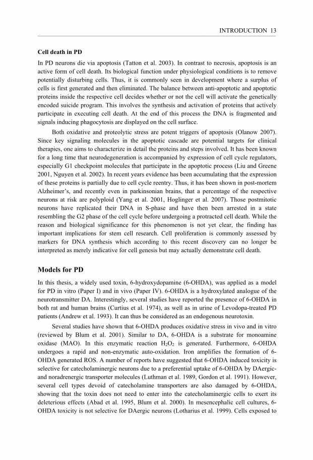
In PD neurons die via apoptosis (Tatton et al. 2003). In contrast to necrosis, apoptosis is an active form of cell death. Its biological function under physiological conditions is to remove potentially disturbing cells. Thus, it is commonly seen in development where a surplus of cells is first generated and then eliminated. The balance between anti-apoptotic and apoptotic proteins inside the respective cell decides whether or not the cell will activate the genetically encoded suicide program. This involves the synthesis and activation of proteins that actively participate in executing cell death. At the end of this process the DNA is fragmented and signals inducing phagocytosis are displayed on the cell surface.
Both oxidative and proteolytic stress are potent triggers of apoptosis (Olanow 2007). Since key signaling molecules in the apoptotic cascade are potential targets for clinical therapies, one aims to characterize in detail the proteins and steps involved. It has been known for a long time that neurodegeneration is accompanied by expression of cell cycle regulators, especially G1 checkpoint molecules that participate in the apoptotic process (Liu and Greene 2001, Nguyen et al. 2002). In recent years evidence has been accumulating that the expression of these proteins is partially due to cell cycle reentry. Thus, it has been shown in post-mortem Alzheimer’s, and recently even in parkinsonian brains, that a percentage of the respective neurons at risk are polyploid (Yang et al. 2001, Hoglinger et al. 2007). Those postmitotic neurons have replicated their DNA in S-phase and have then been arrested in a state resembling the G2 phase of the cell cycle before undergoing a protracted cell death. While the reason and biological significance for this phenomenon is not yet clear, the finding has important implications for stem cell research. Cell proliferation is commonly assessed by markers for DNA synthesis which according to this recent discovery can no longer be interpreted as merely indicative for cell genesis but may actually demonstrate cell death.
Models for PD
In this thesis, a widely used toxin, 6-hydroxydopamine (6-OHDA), was applied as a model for PD in vitro (Paper I) and in vivo (Paper IV). 6-OHDA is a hydroxylated analogue of the neurotransmitter DA. Interestingly, several studies have reported the presence of 6-OHDA in both rat and human brains (Curtius et al. 1974), as well as in urine of Levodopa-treated PD patients (Andrew et al. 1993). It can thus be considered as an endogenous neurotoxin.
Several studies have shown that 6-OHDA produces oxidative stress in vivo and in vitro (reviewed by Blum et al. 2001). Similar to DA, 6-OHDA is a substrate for monoamine oxidase (MAO). In this enzymatic reaction H2O2 is generated. Furthermore, 6-OHDA undergoes a rapid and non-enzymatic auto-oxidation. Iron amplifies the formation of 6-OHDA generated ROS. A number of reports have suggested that 6-OHDA induced toxicity is selective for catecholaminergic neurons due to a preferential uptake of 6-OHDA by DAergic- and noradrenergic transporter molecules (Luthman et al. 1989, Gordon et al. 1991). However, several cell types devoid of catecholamine transporters are also damaged by 6-OHDA, showing that the toxin does not need to enter into the catecholaminergic cells to exert its deleterious effects (Abad et al. 1995, Blum et al. 2000). In mesencephalic cell cultures, 6-OHDA toxicity is not selective for DAergic neurons (Lotharius et al. 1999). Cells exposed to

INTRODUCTION __________________________________________________________________________________________________________________________________________________________________________________________________________________________________ 14
6-OHDA die via an apoptotic mechanism (reviewed by Blum et al. 2001). Recently, the increased expression of cell cycle related proteins was also demonstrated in the 6-OHDA animal model (El-Khodor et al. 2003). Thus, several studies suggest that some of the pathophysiological hallmarks of PD are replicated by this model.
The other important parkinsonism-inducing toxin is MPTP (1-methyl 4-phenyl 1,2,3,6-tetrahydropyridine). It was discovered in the early eighties when a group of young drug addicts developed irreversible parkinsonism after abuse of a synthetic fentanyl derivate which contained around 3 % MPTP (1-methyl 4-phenyl 1,2,3,6-tetrahydropyridine) (Langston et al. 1983). Post-mortem brain investigation confirmed the SN lesion (Davis et al. 1979). Today, MPTP is frequently used in animal models for PD (reviewed by Blum et al. 2001, Smeyne and Jackson-Lewis 2005). The active metabolite of MPTP, MPP+, inhibits mitochondrial complex I, leading to a drop in cellular ATP levels and subsequent cell death. Furthermore, MPP+ induces mitochondria dependent and -independent ROS generation. Rats are, however, not susceptible to MPTP toxicity and it was therefore not applied in our studies.
Other existing models are based on known genetic defects or the induction of proteolytic stress. Their pros and cons have recently been reviewed (Manning-Bog and Langston 2007).
Trophic factors
Glial Cell Line-Derived Neurotrophic Factor (GDNF) According to the historical concept, neurotrophic factors are secreted peptides that support growth and survival of neuronal cells (Manning-Bog and Langston 2007). Their withdrawal induces neuronal apoptosis and it was therefore speculated that a decrease in expression of trophic factors contributes to the pathophysiology of PD (Siegel and Chauhan 2000). To date there is no consistent evidence for this theory. On the contrary, models as well as post-mortem analysis of parkinsonian brains rather show an upregulation of growth factors in the affected areas (Mogi et al. 1995, Yurek and Fletcher-Turner 2001). GDNF (Lin et al. 1993), for example, the factor most extensively studied in the context of PD, is upregulated in the striatum of parkinsonian brains, while the expression of its receptors is unchanged in the striatonigral system (Backman et al. 2006). This can be interpreted as an endogenous mechanism for protection against oxidative or proteolytic stress.
GDNF signals increase the synthesis of antiapoptotic proteins via a tyrosine kinase receptor that activates intracellular signaling cascades. The phosphatidylinositol 3-kinase (PI3K)-Akt signaling cascade or the mitogen-activated protein kinase (MAPK) – extracellular signaling kinase (ERK) pathway are typical examples (Brunet et al. 1999, Willaime-Morawek et al. 2005). In fact, the latter is involved in GDNF’s protective effect against 6-OHDA in nigral DAergic neurons (Zigmond 2006).
Apart from providing protection for DAergic neurons in vivo and in vitro (reviewed by Gash et al. 1998), GDNF has several other potentially beneficial effects for PD patients: it increases the expression of tyrosine hydroxylase (TH) which is the rate limiting enzyme in the process of DA production. Furthermore it induces local sprouting and outgrowth of DAergic

INTRODUCTION __________________________________________________________________________________________________________________________________________________________________________________________________________________________________ 15
fibers, an important prerequisite for restoration and transplant integration (Kirik et al. 2000). Behavioral recovery after GDNF delivery has been documented in numerous animal models of PD (Kirik et al. 2000, Kordower et al. 2000, Grondin et al. 2002). Even one of the first clinical trials, a phase I study with 5 patients receiving continuous infusion of GDNF into the putamen, showed improvement of motility rating scores (Patel et al. 2005). However, the outcome in a following placebo controlled phase II trial was not significantly different between the groups after 6 months of infusion (Lang et al. 2006). Cerebral lesions in GDNF treated monkeys, and the appearance of neutralizing GDNF antibodies in patients, raised safety concerns and contributed to the decision to remove the drug from the market in 2004. The concept of trophic factors as merely benefiting neuron supporting molecules has undergone tremendous changes since the discovery of the first neurotrophic factor, NGF ( neurotrophic gowth factor) (Levi-Montalcini 1964). To date, these molecules are acknowledged to be multifunctional and contextually acting proteins, affecting but not necessarily supporting growth, differentiation and function of principally all known cell types of the developing and mature nervous system. Their potentially widespread effects need to be carefully investigated before therapeutic application can be considered.
Bone Morphogenetic Proteins (BMPs)
BMP ligands The trophic factors investigated in this thesis, BMPs, are distantly related to GDNF. Both BMPs and GDNF belong to the transforming growth factor-� (TGF-�) superfamily, which also includes TGF-�, activins/inhibins, Nodal, myostatin and anti-Mullerian hormone (Kingsley 1994).
More than 20 BMP-related proteins have been identified, of which the majority are encoded by the human genome (Feng and Derynck 2005, Sullivan and O'Keeffe 2005, Katoh and Katoh 2006). Based on their structure and function the BMP family is divided in different subgroups (Fig 1) (Kawabata et al. 1998, Sullivan and O'Keeffe 2005).
BMPs were originally isolated as components of bone extracts that induced ectopic cartilage and bone formation when implanted in muscle (Wozney et al. 1988). However, they are also highly expressed in the embryonic and adult nervous system (reviewed by Harvey et al. 2005). Here they contribute to the specification of cell fate (Mehler et al. 2000, Liu and Niswander 2005) and neuronal cell survival (Chen and Panchision 2007). They have, for example, been shown to protect DAergic neurons in vitro from 6-OHDA induced oxidative stress (Espejo et al. 1999), and to increase survival and DA uptake in rat embryonic mesencephalic cultures via unknown mechanisms (Jordan et al. 1997). BMPs are thus potentially therapeutic molecules for nigral neurons in PD. However, they also have unwanted effects. For example, they induce glial instead of neuronal differentiation in transplanted stem cells (Lim et al. 2000). In order to be able to specifically augment or inhibit BMP signaling, respectively, the function of key components in their signaling pathways has to be understood.

BMP 8aBMP 8b
BMP 7BMP 5BMP 6
BMP 4BMP 2
BMP 10BMP 9GDF 2
GDF 6GDF 5GDF 7
BMP 15GDF 9GDF 15
GDF 3GDF 1
BMP 3GDF 10
GDF 8GDF 11
Activins
TGF-β
Inhibin-αGDNF
Fig. 1Simplified phylogeny tree showing evolutionary relationship between human TGF-β family members. Proteins depicted in italics were used in Paper I

Fig. 2Schematic illustration of theBMP signaling cascade

INTRODUCTION __________________________________________________________________________________________________________________________________________________________________________________________________________________________________ 18
(Panchision et al. 2001). It is not known how Smad proteins mediate specific BMP receptor subtype signals. Smad 1, 5 and 8 are structurally very similar, and even if studies suggest that the BMP type I receptors have differential affinities towards Smad proteins (Aoki et al. 2001), those have not been characterized in detail (Miyazono et al. 2005).
Other signaling cascades activated by BMPs Smad proteins are considered to be the main transducers of BMP signaling. However, MAPK- and STAT-signaling cascades are also directly targeted by BMPs, and numerous other pathways closely interact with BMP effector molecules.
MAPK signaling MAPKs are central regulators of neuronal survival and differentiation. The classic MAPK cascade consists of three sequential steps depicted in Fig. 3. The typical growth factor-mediated activation of a MAPK kinase kinase (MAPKKK) leads to phosphorylation of a MAPK kinase (MAPKK) that, in turn, activates a MAPK. Four major families of MAPK have been characterized, namely extracellular signal-regulated protein kinases1/2 (ERK1/2) (Boulton et al. 1991), p38 (Han et al. 1994, Lee et al. 1994), Jun kinase (JNK) (Derijard et al. 1994, Kyriakis et al. 1994) and big mitogen-activated protein kinase /ERK 5 (Lee et al. 1995, Zhou et al. 1995, reviewed in Junttila et al. 2007).
MAPKs have been shown to phosphorylate Smads. Unlike BMP receptors that target the C-terminal site of Smads, MAPKs phosphorylate the Smad linker region. They thereby inhibit Smads from nuclear translocation and BMPs from exerting any effects. This was first reported for the MAPK ERK, that is activated by the epidermal growth factor (EGF) receptor (Kretzschmar et al. 1997a). Linker region phosphorylation also increases proteosomal degradation of nuclear phosphorylated Smad1 (p-Smad1) via polyubiquitinylation (Zhu et al. 1999, Sapkota et al. 2007).
TAK1 (TGF-� activated kinase 1) is a MAPKKK, activated by BMPs. Interestingly, even TAK1-phosphorylated Smads are retained in the cytoplasm (Hoffmann et al. 2005). The initial receptor mediated steps in this process are unclear. It has been shown that an adaptor protein, designated (XIAP), links TAK1 to ALK3 but not to other type I receptors (Yamaguchi et al. 1999, Qi et al. 2004). This might partly explain why the activation of different type I receptors results in different biological outcomes.
Signal Transducer and Activator of Transcription (STAT) signaling STAT proteins are transcription factors that are typically phosphorylated and activated by cytokine receptors. BMP4 has recently been shown to Smad-independently activate STAT3 via the serine-threonine kinase mammalian target of rapamycin/FKBP12-rapamycin associated protein (mTOR/FRAP) (Rajan et al. 2003). However, maximal STAT activation is first observed 8 hours after BMP receptor binding, suggesting some intermediate signaling steps that have not yet been characterized.
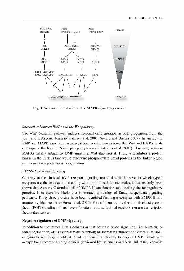
EGF, bFGF,mitogens
stress cytokines
stimulus
Raf, MEKK1
ASK1, TAK1, MEKK4
MEK1,MEK2
MEK3,MEK6
MEK4,MEK7
ERK1 (p44MAPK)ERK2 (p42MAPK) p38 isoforms JNK1/2/3 ERK5
MEK5
MEKK2,MEKK3
,
Ras
stressgrowth factorsBMPs
targetstranscription factors

INTRODUCTION __________________________________________________________________________________________________________________________________________________________________________________________________________________________________ 20
2005, Gazzerro and Canalis 2006). Recently, BMP3 has been identified as a BMP antagonist. It sequesters the BMPRII into a non-signaling complex (Gamer et al. 2005). Thus, the maximal possible BMP signaling is not only determined by the number of functional receptors or ligands. Broadly overlapping binding patterns, multiple co-signaling molecules and antagonists, crosstalk with other pathways as well as complex feedback mechanisms, contribute to a branched network in which the outcome of interventions is diffi-cult to predict.
Apoptosis Signal-Regulating Kinase 1 (ASK1)
ASK1 signaling ASK1 is an ubiquitously expressed enzyme that has the function of a MAPKKK and activates both the p38 and JNK pathways (Ichijo et al. 1997) (Fig. 3). ASK1 has been shown to be involved in the apoptotic reaction towards various forms of cytotoxic insults. It is activated in cells treated with death receptor ligands, such as tumor necrosis factor-� (TNF-�� and Fas ligand (Chang et al. 1998, Nishitoh et al. 1998), and in response to oxidative as well as endoplasmic reticulum stress (reviewed by Matsukawa et al. 2004).
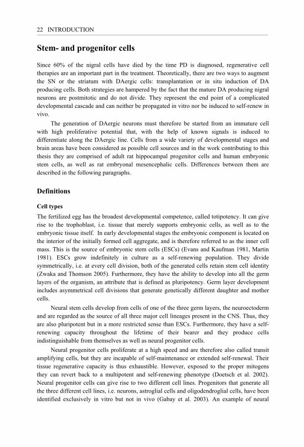
The important step in ASK1 is depicted in Fig. 4. Under physiological conditions the redox-regulatory protein thioredoxin (Trx) is bound to ASK1 and represses its activity (Saitoh et al. 1998). Upon oxidization, Trx dissociates from ASK1 and reciprocally TRAF2 (tumor necrosis factor receptor associated factor 2) and TRAF 6 are recruited into a higher mass complex. ASK1 becomes active by autophosphorylation of Thr845, a threonine residue within the activation loop in one of the monomers. The phosphorylated protein complex is known to activate MKKs (Tobiume et al. 2002), that in turn activate p38 and JNK MAPK.
P
P
HS S
H
HS S
H
S-S
S-S
ROS
Trx
inactive ASK1 active ASK1
TRAF2/6
Fig. 4Schematic illustration of ASK1 activation

INTRODUCTION __________________________________________________________________________________________________________________________________________________________________________________________________________________________________ 21
Apart from Trx, several other molecules are known to control the activation state of ASK1. For example, the 14-3-3 proteins suppress ASK1 activation by binding to phosphorylated Ser-967 in the 14-3-3- motif of ASK1 (Zhang et al. 1999). Protein phosphatase 5 (PP5) inhibits ASK1 by dephosphorylating the activation loop (Morita et al. 2001). Recently, the type I insulin-like growth factor receptor kinase, which is important for proliferation and survival in many different cell types, has also been shown to inhibit ASK1 activation (Galvan et al. 2003).
Biological functions Given its important role in apoptosis evoked by oxidative or endoplasmatic reticulum stress, ASK1 might be partly responsible for cell death in PD. Thus, at first sight it seems absurd to investigate beneficial effects of this molecule in the context of PD or stem cell therapy. However, the idea becomes interesting when one considers the fact that ASK1 participates in the same MAPK signalling network that is also used by many trophic factors. The MAPK p38 and JNK have been shown to be involved in both apoptosis and cell differentiation (reviewed by Mielke and Herdegen 2000, Takeda et al. 2000, Matsuzawa et al. 2002). Furthermore, several molecules participating in the apoptotic signaling cascade are also expressed and activated under physiological conditions in the central nervous system (CNS), and play an important role in stress adaptation, survival and differentiation (reviewed by Garrido and Kroemer 2004). For example, caspases are executors of apoptotic cell death. Caspase-3- Knock-out animals do, however, display excess perinatal brain tissue lethality (Kuida et al. 1996), indicating the vital role of this enzyme and its involvement in the normal differentiation process of neurogenesis. The bipartite nature of this response is not completely understood.
In fact, ASK1 did induce neurite outgrowth, neuronal differentiation and survival in PC12 cells via the activation of p38 and JNK MAPK (Takeda et al. 2000). Furthermore, it was found that calcium signaling molecules regulate the ASK1-p38 MAPK cascade in primary neurons (Takeda et al. 2004), suggesting that ASK1 activation is involved in synaptic plasticity. The C.elegans homolog of ASK1, NSY-1, is required for specification of olfactory neurons, hinting at a role for ASK1 in neuronal differentiation (Sagasti et al. 2001, Wes and Bargmann 2001). Again, it is not totally clear how the decision between life and death is made. Differences in kinase expression with respect to quantity and time are known to trigger different outcomes (Takeda et al. 2000, Hazzalin and Mahadevan 2002). The signaling result is furthermore, determined by the intracellular balance of anti- and proapoptotic programs (Matsuzawa and Ichijo 2008). Understanding a mechanism that governs apoptosis versus cell differentiation in more detail is clearly an important goal in the development of therapies against neurodegenerative diseases.
INTRODUCTION __________________________________________________________________________________________________________________________________________________________________________________________________________________________________ 22
Stem- and progenitor cells
Since 60% of the nigral cells have died by the time PD is diagnosed, regenerative cell therapies are an important part in the treatment. Theoretically, there are two ways to augment the SN or the striatum with DAergic cells: transplantation or in situ induction of DA producing cells. Both strategies are hampered by the fact that the mature DA producing nigral neurons are postmitotic and do not divide. They represent the end point of a complicated developmental cascade and can neither be propagated in vitro nor be induced to self-renew in vivo.
The generation of DAergic neurons must therefore be started from an immature cell with high proliferative potential that, with the help of known signals is induced to differentiate along the DAergic line. Cells from a wide variety of developmental stages and brain areas have been considered as possible cell sources and in the work contributing to this thesis they are comprised of adult rat hippocampal progenitor cells and human embryonic stem cells, as well as rat embryonal mesencephalic cells. Differences between them are described in the following paragraphs.
Definitions
Cell types The fertilized egg has the broadest developmental competence, called totipotency. It can give rise to the trophoblast, i.e. tissue that merely supports embryonic cells, as well as to the embryonic tissue itself. In early developmental stages the embryonic component is located on the interior of the initially formed cell aggregate, and is therefore referred to as the inner cell mass. This is the source of embryonic stem cells (ESCs) (Evans and Kaufman 1981, Martin 1981). ESCs grow indefinitely in culture as a self-renewing population. They divide symmetrically, i.e. at every cell division, both of the generated cells retain stem cell identity (Zwaka and Thomson 2005). Furthermore, they have the ability to develop into all the germ layers of the organism, an attribute that is defined as pluripotency. Germ layer development includes asymmetrical cell divisions that generate genetically different daughter and mother cells.
Neural stem cells develop from cells of one of the three germ layers, the neuroectoderm and are regarded as the source of all three major cell lineages present in the CNS. Thus, they are also pluripotent but in a more restricted sense than ESCs. Furthermore, they have a self-renewing capacity throughout the lifetime of their bearer and they produce cells indistinguishable from themselves as well as neural progenitor cells.
Neural progenitor cells proliferate at a high speed and are therefore also called transit amplifying cells, but they are incapable of self-maintenance or extended self-renewal. Their tissue regenerative capacity is thus exhaustible. However, exposed to the proper mitogens they can revert back to a multipotent and self-renewing phenotype (Doetsch et al. 2002). Neural progenitor cells can give rise to two different cell lines. Progenitors that generate all the three different cell lines, i.e. neurons, astroglial cells and oligodendroglial cells, have been identified exclusively in vitro but not in vivo (Gabay et al. 2003). An example of neural

INTRODUCTION __________________________________________________________________________________________________________________________________________________________________________________________________________________________________ 23
progenitor cells are the adult rat hippocampus-derived progenitors (AHPs) (Palmer et al. 1997) used in Papers II and III.
Lineage-specific progenitors are cells restricted to one distinct lineage. The term precursor is often used for a not well defined proliferating population of cells (Weiss et al. 1996).
A committed cell no longer changes its cell fate in response to signals from the environment. It undergoes terminal differentiation that is characterized by increased expression of genes and proteins important for the fully functional cell. Growth of axons, dendrites, synthesis of neurotransmitters and synapse formation indicate maturation.
Lineages According to the historical model, the lineage tree was considered to have two separate branches representing the development of the glial and neuronal lineage (Alvarez-Buylla et al. 2001). The potential to adopt alternate cell fates was thought to be more and more restricted the further the cells had proceeded along the respective branches. Thus, neuronal progenitor cells would generate neurons while glial progenitors would undergo progressive maturation towards postmitotic astro-and oligodendroglial cells, respectively. More recently, it has become clear that these developmental routes only apply to some cells in vitro (Gabay et al. 2003). In vivo, oligodendrocyte and astrocyte precursors do not necessarily develop from a common glial progenitor but rather through distinct routes (Rowitch 2004). In fact, studies in embryonic spinal chord and forebrain provide evidence for the existence of a common precursor for neurons and oligodendroglial cells (He et al. 2001, Jackson et al. 2006, reviewed by Rowitch 2004).
For the correct description of lineage development it is important that one can clearly define the different cell types. Historically, cells were classified by their function, morphology and expression of only a few different proteins. This is too simple and can be misleading, exemplified by cells expressing glial fibrillary acidic protein (GFAP). For a long time GFAP has been regarded as a specific marker for astrocytes (Bignami and Dahl 1974). However, not every astrocyte stains positive for GFAP, and not every GFAP-positive (GFAP-pos) cell is an astrocyte in the classical sense. The degree of correlation between GFAP expression and astrocyte-type morphology depends on the brain region. It is now established that many postnatal and adult NSCs also express GFAP both in vivo (Doetsch et al. 1999a, Doetsch et al. 1999b, Seri et al. 2001, Garcia et al. 2004) and in vitro (Laywell et al. 2000, Imura et al. 2003, Morshead et al. 2003). Since some stem cells ultrastructurally resemble astrocytes, some authors use the terms “neurogenic” and “nonneurogenic astrocyte”, respectively, to distinguish between GFAP-expressing stem cells and the classical astrocytes. It is obvious that even these two categories comprise a huge number of subgroups of cells which could be further defined by different expression profiles of transcription factors, cell membrane proteins, structural proteins etc. In an attempt to describe the similarities and differences between different cell types in more detail, Cahoy et al. recently compared expression levels of more than 20.000 genes of neural cell types (Cahoy et al. 2008). Interestingly, this study revealed that astrocytes and oligodendrocytes are as dissimilar to each other as they are to neurons. This finding causes the authors to question the concept of a glial cell class. Today, we are far from understanding the implications of these differences. To

INTRODUCTION __________________________________________________________________________________________________________________________________________________________________________________________________________________________________ 24
what extent can we, for example, simplify the characterization of neurons generated for cell replacement therapy?
Cell genesis in the developing brain In the early embryonic period the CNS develops from an epithelium composed of rapidly proliferating progenitors. This epithelium undergoes morphogenic movements and thereby transforms from a neural plate to a neural tube (reviewed by Colas and Schoenwolf 2001, Copp et al. 2003) that surrounds the ventricular space. The earliest neural stem cells are neuroepithelial cells. They develop from ectodermal cells when the neural plate is formed (Kageyama et al. 2005) and give rise to radial glia that constitute the major embryonic neural stem cell pool in the ventricular zone (reviewed by Malatesta et al. 2003, Pinto and Gotz 2007) and even participate in neural development in the adult brain (Alvarez-Buylla et al. 2001, Bonfanti and Peretto 2007). In late embryonic development the ventricular zone loses its role as a primary germinal center. Instead, this task is taken over by the subventricular zone (SVZ).
Neurogenesis begins at embryonic day 12 (E12) in the rat, peaks at E14 and recedes by E17 (Parnavelas 1999, Gates et al. 2006). DAergic midbrain neurons belong to one of the first neuronal populations generated in the CNS (Sechrist and Bronner-Fraser 1991). Altman and Bayer have demonstrated that DAergic neurons become postmitotic in rats at around E12-E15 (Altman and Bayer 1981). More recently, it was reported that the vast majority of DAergic neurons in the SN make their final division on E12. At E13 they begin to extend processes, and by E14 many axons have reached their target regions, i.e. the lateral ganglionic eminence and the striatal anlagen (Riddle and Pollock 2003, Gates et al. 2006). Between postnatal day 0 (P0) and P21 a change in synaptical structure in the target area is associated with two waves of DAergic cell death, peaking at P2 and P14. Cell death is thought to be a result of a failure of these neurons to receive trophic factors from the target region (Oo and Burke 1997, Oo et al. 2003).
Distinct subsets of astro-and oligodendroglial progenitors become specified already at the earliest stages of neurogenesis. They take however, a long time to develop mature progeny (Pinto and Gotz 2007). Thus in the rat, the peak of astrocyte differentiation occurs between P0 and P2 and oligodendrocytes mature between P4 and P20 (Levison et al. 1993, Zerlin et al. 1995, Parnavelas 1999).
Cell genesis in the adult brain In the adult mammalian brain neurogenesis is much more restricted than in the embryonic brain. It has convincingly been described in two distinct areas of the adult rodent, primate and human brain, namely the subgranular zone of the hippocampal dentate gyrus and the SVZ of the lateral’s ventricle lateral wall (reviewed by Gould 2007, Alvarez-Buylla and Lim 2004, Ming and Song 2005).

INTRODUCTION __________________________________________________________________________________________________________________________________________________________________________________________________________________________________ 25
Subventricular zone In the SVZ, the multipotent stem cells, designated type B cells (Doetsch et al. 1997), are slowly proliferating cells expressing GFAP (Doetsch et al. 1999a, Tramontin et al. 2003) and the PDGF�R (Platelet-derived growth factor-� receptor), a marker previously used mainly to detect oligodendrocyte precursor cells (Jackson et al. 2006). Type B cells are formed by the transformation of embryonic radial glia (Merkle et al. 2004). They give rise to type C cells which are rapidly dividing GFAP neg immature progenitors. Those, in turn, generate neuroblasts, type A cells, that migrate through the rostral migratory stream to the olfactory bulb where they differentiate into functional neurons (Curtis et al. 2007). SVZ type B cells also generate oligodendrocytes, that in contrast to neuroblasts not only move to the olfactory bulb but even to the corpus callosum, striatum and fimbria fornix (Jackson et al. 2006, Menn et al. 2006).
Hippocampus The currently favored model of cell genesis in the hippocampus, suggests that the putative stem cell is a radial-glia-like GFAP expressing cell that has the electrophysiological properties of astrocytes (Seri et al. 2001, Filippov et al. 2003, Fukuda et al. 2003, Kempermann et al. 2004). It rarely divides but gives rise to three faster proliferating cell types. Their differential but partly overlapping expression of glial and neuronal markers suggests that they all belong to the same developing neuronal lineage that supplies the granule cell layer with postmitotic neurons (Kempermann et al. 2004). However, proof of a developmental continuum between the three cell types does not exist. Furthermore, it is unresolved which precursor the postmitotic hippocampal astrocytes have. Thus, evidence is lacking that the putative “stem cell” is multipotent, and the second criterion for stem cells (lifelong self-renewal) cannot be proven in vivo. Experiments addressing this question in vitro gave controversial results. Palmer et al. demonstrated multipotent self-renewing stem cells in cell cultures generated from gross dissection of the adult rat hippocampus (Palmer et al. 1997). When the hippocampus is further subdivided however, stem cells are found in the subependyma of the lateral ventricle adjacent to the hippocampus. The hippocampus only contains multipotent progenitors (Seaberg and van der Kooy 2002, Bull and Bartlett 2005). Migration of cells from those adjacent areas into the hippocampus has never been observed in the adult brain. Seaberg et al. suggest that large numbers of progenitors enter the hippocampus already perinatally. Since this pool of progenitors decreases over time, neurogenesis declines with age in this area.
Non-neurogenic regions Whether or not neurogenesis also exists in other parts of the adult normal brain is highly controversial (reviewed by Gould 2007). Cycling self-renewing cells are found dispersed throughout the brain parenchyma (Reynolds and Weiss 1992, Dawson et al. 2003, Nunes et al. 2003). It has been suggested that these cells are multipotent neural stem cells that are biased by their environment towards glial production, but that they are potentially neurogenic when not hampered by suppressing factors in the surrounding tissue (Reynolds and Weiss 1992, Nunes et al. 2003, reviewed by Lie et al. 2004). However, some authors report in vivo neurogenesis in other areas such as the adult neocortex or striatum, albeit at about ten times

INTRODUCTION __________________________________________________________________________________________________________________________________________________________________________________________________________________________________ 26
lower frequency than in the neurogenic areas described above (Gould et al. 2001, Dayer et al. 2005, Cameron and Dayer 2008).
Substantia nigra Zhao et al. suggested that 20 nigral neurons are born each day in the SN of adult mice, a number sufficiently high to completely replace the SN during the adult lifetime of a mouse. The DAergic neurons were thought to be derived from stem cells lining the ventricular system. Importantly, a partial MPTP-induced lesion of the nigral cell population was found to increase the number of bromodeoyuridine (BrdU) incorporating DAergic cells two-fold (Zhao et al. 2003). Similar results were presented by Shan et al. (2006). Furthermore, chronic DA receptor stimulation was recently reported to increase the number of proliferating TH pos cells in a 6-OHDA lesion model (Van Kampen and Eckman 2006). However, the three studies base their conclusion on BrdU incorporation into TH pos cells, which according to recent findings might also represent apoptosis related aberrant re-entry into the cell cycle (Yang et al. 2001, Hoglinger et al. 2007). Höglinder et al. found BrdU /TH-double-pos cells only shortly after a MPTP lesion, but not 70 days later, which argues against neurogenesis. In the same study they also demonstrated that cell-cycle markers were only expressed in neurons that had established connections to the striatum and therefore could not be newborn .
Thus, the existence of in vivo neurogenesis in the adult SN is still highly controversial. In contrast, nigral progenitors with neurogenic potential in vitro have been demonstrated convincingly (Lie et al. 2002).
Molecular mechanisms in neural cell genesis
Previous paragraphs described that cell fate decisions are influenced by external factors in the respective environments. The responsiveness of a given cell at a given time to those environmental factors is, however, limited by cell intrinsic programs. Transcription factors (TFs) are key molecules in this process. Their expression pattern determines proliferation, cell differentiation and cell type specification. Based on their main effect on cell fates, they can be divided into subgroups, which are presented below. However, it must be mentioned that the same TF can promote different cell fates depending on the cellular context.
Transcription factors
TFs promoting proliferation TFs, which are important for proliferation, are highly expressed by neural stem cells. They keep the stem cell pool undifferentiated until late developmental stages so that cells of full diversity can be generated in correct numbers. For example, TFs belonging to the Inhibitor-of-differentiation (Id) family, inhibit (as their name suggests) the differentiation of progenitors into neurons and oligodendrocytes. In mice lacking both Id1 and Id3, cortical progenitors exit the cell cycle prematurely and undergo accelerated neuronal differentiation (Lyden et al. 1999).

INTRODUCTION __________________________________________________________________________________________________________________________________________________________________________________________________________________________________ 27
Hairy/Enhancer-of-split (Hes) proteins also maintain proliferation by antagonizing proneural TFs (Sasai et al. 1992). Their importance is demonstrated by Hes1 (-/-) Hes5 (-/-) neurospheres that do not expand properly even in the presence of the otherwise potent mitogenic growth factors bFGF or EGF (Ohtsuka et al. 2001). Hes1 and 5 are also targeted by signals from the transmembrane protein Notch (Ohtsuka et al. 1999) that mediates lateral inhibition. This term describes the phenomenon that a differentiating cell inhibits its neighbor from doing likewise. Thus, differentiating neurons express Delta that activates Notch to induce Hes expression (Kunisch et al. 1994). The lack of lateral inhibition is thought to be the reason for increased differentiation in sparsely cultured cells.
In neurogenic regions of the adult CNS, Sry-related high-mobility-group box (Sox) genes of the B1 group, i.e. Sox1, Sox2 and Sox3, keep the stem cell pool undifferentiated (reviewed by Pevny and Placzek 2005). Interestingly, mutations of mouse Sox2 cause neurodegeneration and impair adult neurogenesis (Ferri et al. 2004).
TFs promoting neuronal cell fate The transition from proliferation to neuronal differentiation is governed by a decrease in Hes and Id activity, expression of Sox21 that blocks the activity of Sox1-3 (Sandberg et al. 2005) and an increase in proneural bHLH TF activity.
For example, the proneural basic helix-loop-helix (bHLH) genes Mash1 and Neurogenin1 (Ngn1) initiate neuronal differentiation by inducing expression of genes like NeuroD and Math3 (Cau et al. 1997, Ma et al. 1998, Cau et al. 2002) that cause exit from the cell cycle and mediate terminal neuronal differentiation (reviewed by Kageyama and Nakanishi 1997)). Mice deficient in NeuroD completely lack the dentate gyrus granule cell layer (Miyata et al. 1999). The differential expression of proneural bHLH factors determines which subtype of neuron will be formed (Fode et al. 2000, Wilson and Rubenstein 2000).
TFs promoting astroglial cell fate The developmental neuronal-glial switch can partially be explained by decreasing amounts of the proneural TF Ngn1. Ngn1 competes with the glial cell fate inducer STAT for the common binding partner p300/CBP that can support transcription from neuronal as well as glial genes, depending on the binding partner. The intracellular balance between Ngn1 and STAT then determines transcriptional outcome (Sun et al. 2001). However, loss of proneural genes does not seem to be enough to cause glial commitment (Nieto et al. 2001). The transcriptional programme activated in the gliogenic phase comprises a complex set of transcriptional co-modulators. Notch signaling, for example, promotes astrocyte differentiation in a number of cell types, among them the adult hippocampal progenitors (Tanigaki et al. 2001). Mechanisms involved include the direct activation of the GFAP promoter by the intracellular domain of Notch, but also the suppression of neuronal cell fate via Hes1 and Hes5 (reviewed by Guillemot 2007).
TFs promoting oligodendroglial cell fate The specification of oligodendrocytes is regulated by the bHLH genes Olig1 and Olig2 (Lu et al. 2000, Takebayashi et al. 2000, Zhou et al. 2000). In Olig1-/-Olig2-/- double mutants there is a total failure of oligodendrocyte formation (Zhou and Anderson 2002). Furthermore, oligodendrocyte precursors (OLPs) are transformed into GFAP-pos astrocytes in these
INTRODUCTION __________________________________________________________________________________________________________________________________________________________________________________________________________________________________ 28
animals. To suppress GFAP expression Olig2 binds to p300/CBP-co-activator and thereby inhibits complex formation between p300/CBP and STAT, a mechanism reminding of Ngn1 induced GFAP inhibition (Fukuda et al. 2004). Mash1, previously mentioned in the context of neuronal differentiation, also promotes oligodendrocyte development. It is extensively co-expressed with Olig2 in the embryonic brain, in oligodendrocyte precursors in culture and in the white matter of the postnatal brain (Kondo and Raff 2000, Gokhan et al. 2005). Its functional importance for oligodendroglial cell fate was shown in Mash1 mutant mice where a reduced number of OLPs were found in the olfactory bulb (Parras et al. 2004).
Id TFs act as dominant negative binding partners for Olig1/2 (Wang et al. 2001, Samanta and Kessler 2004) and thereby suppress oligodendrocyte development.
Epigenetic mechanisms Epigenetic mechanisms interfere with transcriptional and translational processes by altering the accessibility of genes and RNA, respectively. In that way they control the activity of TFs and have an important impact on cell fate development. Due to space limitations only a few examples can be given here. More detailed information is provided in recent review articles (Kondo 2006, Kosik 2006).
Genes can be silenced when the DNA is methylated at CpG sites. It has been shown that this is an important regulatory mechanism for the timing of gliogenesis. For example, around the onset of astrocyte differentiation at E14, a CpG site in the STAT3 binding element in GFAP genes is demethylated allowing for STAT3 to bind (Takizawa et al. 2001, Namihira et al. 2004). Furthermore, astroglial differentiation is accompanied by histone modifications in the GFAP promoter (Song and Ghosh 2004). In neurogenesis the chromatin remodeling complex Brg1 has been shown to play a critical role by activating the two proneural genes NeuroD and Ngn (Seo et al. 2005). Micro RNAs (miRNAs) guide the cleavage of target mRNAs and/or inhibit their translation (reviewed by Harfe 2005). It has been suggested that they can modulate more than a third of the coding mRNA in multicellular organisms (Lewis et al. 2005). In a recent study evidence was provided that miRNAs are essential for maintaining DAergic neurons in the brain (Kim et al. 2007).
BMPs’ changing role in neural cell genesis
At very early embryonic stages cells are known to differentiate into neurons by default. BMPs prevent this process (Wilson and Hemmati-Brivanlou 1995) by inducing Id TFs (Ying et al. 2003). They thereby insure the development of other tissue types in the embryo. Neural cells only develop around the organizer, a region that secretes BMP inhibitors. At later stages BMPs participate in specifying neural cell fate (Gross et al. 1996, Li et al. 1998, Mehler et al. 2000). They increase neuronal differentiation in mid-gestational and astroglial differentiation in late gestational neural progenitors, respectively. This changing potential is due to the co-activator function of p-Smad that binds the previously mentioned p300/CBP molecule at a site distinct from that for Ngn1 and STAT (Sun et al. 2001). Since
INTRODUCTION __________________________________________________________________________________________________________________________________________________________________________________________________________________________________ 29
the concentration of Ngn1 decreases in progenitor cells during development, BMPs’ astroglial inducing effect prevails in progenitor cells from later stages and in adulthood. BMPs can also increase GFAP expression by phosphorylating STAT (Rajan et al. 2003) or by inducing Hes expression (Nakashima et al. 2001).
The neurogenic regions of the adult brain contain high amounts of BMP antagonists (Lim et al. 2000, Ueki et al. 2003) suggesting that neutralization of the gliogenic BMP signal is an important mechanism for neuronal development in those areas. It was proposed that the BMP antagonist, noggin, being expressed in ependymal cells of the SVZ, inhibits BMP signaling in neighboring B cells and thereby permits neuroblast development (Lim et al. 2000). However, nuclear translocation of p-Smad in B and C cells was recently shown in situ (Colak et al. 2008), clearly indicating functional BMP signaling in those cells. Complete inhibition of BMP signaling by deletion of the common BMP signal mediator Smad4 resulted in increased development of oligodendroglia at the expense of neurogenesis. Thus, Colak et al. suggest that BMPs are important for neuroblast development. The authors also imply that noggin can actively adjust BMP signaling levels in the SVZ thereby allowing a low degree of oligodendrogliogenesis to occur (Colak et al. 2008). Concentration dependant induction of differential cell fate is in line with BMPs’ definition as a morphogen and has previously been shown in neural stem cell cultures (Chang et al. 2003).
BMP induced inhibition of oligodendrocyte development and differentiation does not only occur in the SVZ. It has been described in multiple brain regions (Gomes et al. 2003) and in cultures from postnatal cortical progenitors (Mabie et al. 1997, Samanta et al. 2007) as well as adult oligodendrocyte precursor cells (Cheng et al. 2007). The effect is mediated via Id TFs (Samanta and Kessler 2004, Cheng et al. 2007). In cultures of maturing oligodendrocytes BMPs decrease the expression of three myelin proteins, PLP (proteolipid protein), MBP (myelin basic protein) and CNP (cyclic nucleotide phosphodiesterase) (See et al. 2004).
In contrast, BMPs promote the differentiation of postmitotic neurons. Thus, they regulate axonal pathfinding (Charron and Tessier-Lavigne 2005) and dendritic growth in neurons from cortical (Esquenazi et al. 2002), hippocampal (Withers et al. 2000) and striatal neurons (Gratacos et al. 2001).
Molecular cues in the development of DAergic neurons
DAergic cell types The final goal of cell replacement therapy in PD is the production of DAergic neurons that truly represent SN neurons. Cells with a DAergic phenotype can be found in several different neuronal nuclei in the brain. In the mesencephalon they are divided into three different groups, i.e. the lateral group of the retrorubral field (A8), cells in the SN pars compacta (pc) (A9) and in the medially located ventral tegmental area (VTA) (A10). Compared to all cells with a DAergic phenotype, the DAergic cells in the SN and VTA exhibit the most complete set of molecules involved in DA synthesis, neurotransmission and metabolism (Vernier et al.

INTRODUCTION __________________________________________________________________________________________________________________________________________________________________________________________________________________________________ 30
2004). They can be distinguished by their molecular pattern. For example, only the meso-diencephalic DAergic neurons express the homeodomain gene Pitx3 (Smidt et al. 1997, Maxwell et al. 2005) and the G protein inwardly rectifying potassium channel 2 (Girk2) has been used to identify A9 DAergic neurons in mesencephalic grafts (Schein et al. 1998, Thompson et al. 2005).
The current challenge is to integrate and apply the detailed knowledge of molecular cues in nigral DAergic fate decisions to produce transplantable neurons of the A9 subtype.
Molecular patterning in the mesencephalon Mesencephalic DAergic progenitors develop at anatomical locations that are exposed to a unique combination of permissive and inductive cell signals. These are derived from four different signaling centers that lie on opposite locations of the dorsal-ventral (DV) and the anterior- posterior (AP) axis of the neural tube, respectively. Thereby, a Cartesian grid is defined in which every cell will receive signals depending on its position in the AP and DV coordinates (Rubenstein et al. 1994). The floor plate is the most ventral signaling center. It secretes Sonic Hedgehoc (Shh) that has a pivotal role in specifying ventral cell populations (Echelard et al. 1993). Together with FGF8 from the isthmus (Crossley et al. 1996, Lee et al. 1997), which represents the anterior signaling center, Shh can induce DA cell fate in cells expressing the TFs Pax2, Pax5 and Otx2 (reviewed by Hynes and Rosenthal 1999). Permissive factors, e.g. Wnt1, ensure that the aforementioned TFs are expressed and that others, which would prepare the cell for alternative cell fates, are suppressed (Prakash et al. 2006).
The recently discovered homeodomain TF Lmx1a is the only one known so far that not only permits but even induces DA neuronal cell fate (Andersson et al. 2006b). Lmx1a induces the expression of Ngn2 in the mesencephalic midline. In the absence of Ngn2, the DA neuronal population is initially reduced to 20%. Postnatally, DAergic neurons recover to 50% of the cell number seen in Ngn2 expressing animals, supposedly due to the activity of Mash1 (Andersson et al. 2006a, Kele et al. 2006). After the last cell division a number of TFs important for further differentiation are expressed. The first one is Lmx1b (Smidt et al. 2000) which ensures survival. For the acquisition of a mature phenotype, Nurr1 has to be present. In its absence, postmitotic DA precursors are initially born but disappear later (reviewed by Perlmann and Wallen-Mackenzie 2004). Equally important for proper differentiation is the TF Pitx3 (Maxwell et al. 2005) which is specifically expressed in a subset of more mature midbrain TH-pos neurons (van den Munckhof et al. 2003). Maintenance of the neuronal population in later stages is ensured by the homeobox TFs engrailed1 (En1) and En2 that are induced by FGF8, which then control continued FGF8 expression (Liu and Joyner 2001, Simon et al. 2001).
Embryonic stem cells
The intrinsic propensity of ESCs for extended proliferation and their neuronal differentiation potential is a favorable attribute for the production of large numbers of DAergic cells.

INTRODUCTION __________________________________________________________________________________________________________________________________________________________________________________________________________________________________ 31
Considering the high number of regulatory events in DAergic cell development outlined in the previous section, it is surprising that one has been successful in generating DAergic neurons from these primitive pluripotent cells.
Methods have been primarily developed for mouse ESCs (mESCs). Transferring the gained experiences to human ESCs (hESC) culture is not totally straightforward. An analysis of the transcriptional profile of mouse versus hESCs revealed a similar core molecular program but also many differences between the species (Sato et al. 2003). It is currently debated whether these inconsistencies arise from different generation time points. Thus, it seems that mESCs represent a slightly earlier developmental stage than hESCs (Brons et al. 2007, Tesar et al. 2007).
Propagation of ESCs Mouse embryonic fibroblasts (MEFs) are frequently used as feeder cells for both mouse and hESC propagation (Williams et al. 1988). They have the dual function of serving as a matrix as well as providing some of the extrinsic signals needed for self-renewal. When ESCs are cultured under feeder free conditions Matrigel and fibroblast conditioned medium are often applied to replace the feeder layer (Xu et al. 2001). Since the use of animal derived components does not meet safety requirements for clinical application, the ultimate goal is to culture cells with material from exclusively recombinant or purified human sources. In fact, the successful derivation and maintenance of two hESC lines under these conditions has been reported (Ludwig et al. 2006). However, these high standard culture conditions are expensive and impractical for experimental work and therefore not yet routinely used.
Neuronal differentiation of ESCs Neuronal differentiation from ESCs in vitro is easily achieved by depriving the cells of mitogens applied for propagation. Two conceptually different approaches exist:
The first is based on the spontaneous formation of embryonic bodies (EBs) in suspension culture (Okabe et al. 1996). This process resembles the first steps in embryogenesis. In the three dimensional cell aggregates, cells belonging to all three germ layers develop (Itskovitz-Eldor et al. 2000). The neural inducing signals are provided by the developing cell structures. Lee et al. were the first to demonstrate the generation of A9 midbrain DAergic neuronal populations from mESCs via EB formation (Lee et al. 2000). This method is now generally referred to as “the 5 step protocol” since it includes the expansion of undifferentiated ESCs, the formation of EBs, a selection of neural progenitors via plating of EBs on tissue surface, the consecutive expansion of neural progenitors with bFGF (and laminin) and finally induction of differentiation.
In the second approach hESCs are dissociated and placed into adherent culture. Neuroepithelial cells have been described to develop 14 days after plating on MEF in the presence of noggin. They can then be propagated with mitogens. After mitogen-withdrawal about 30% of cells express the neuronal marker �-tubulin III (�-tubIII), but only 0.5% co-express TH (Reubinoff et al. 2001, Ben-Hur et al. 2004).
Empirically it has been noticed that co-culture with stromal cells such as HepG2 (Rathjen et al. 1999), PA6 (Mizuseki et al. 2003, Buytaert-Hoefen et al. 2004) and MS5

INTRODUCTION __________________________________________________________________________________________________________________________________________________________________________________________________________________________________ 32
(Perrier et al. 2004) promotes neuroepithelial differentiation. Expression of neuronal markers such as �-tubIII and NCAM can be observed already after 2 days in mESCs (Kawasaki et al. 2000) and after about 6 days in primate ESC when they are co-cultured with PA6 cells (Kawasaki et al. 2002). This cell line was originally derived from mouse skull bone marrow (Kodama et al. 1982) and interestingly, it favors the differentiation of neural progenitors with mid/hindbrain characters. The instructive signals of this stromal cell-derived inducing activity (SDIA) are exerted at or prior to Sox1 expression, i.e. at a very early stage in neural progenitor specification (Parmar and Li 2007). The TF Wnt5a has recently been shown to play an important role in this process (Hayashi et al. 2008) However, the exact molecular basis of SDIA activity remains unclear. Both a soluble factor and a direct contact between ESCs and stromal cells may be required since even fixed PA6 cells retain neural inducing activity (Kawasaki et al. 2000).
Increasing the numbers of A9 DAergic cells Distinct regional identity can also be established by supplementing the culture medium with developmental key morphogens or by overexpressing key fate-determining TFs. For example, the addition of FGF8 and Shh induce TH and mid/hindbrain related markers in hESC-derived neural progenitors (Perrier et al. 2004). Similar to mouse in vivo development, the generated cells sequentially express Pax2 and Pax5 and, upon exiting the cell cycle, become positive for En1 and TH. In accordance with this Yan et al. showed that Shh and FGF8 exposure of hESCs during the process of neuroepithelial specification (i.e. before they express Sox1) increases the amount of TH pos cells from 3 to 30% of differentiated cells. However, a 2 week delay in the factor addition resulted in the development of TH-pos neurons that have the characteristics of forebrain olfactory bulb neurons (Yan et al. 2005). This example shows that the cells’ intrinsic program rapidly changes over time and induces different responses to the same stimuli. Thus, it is easy to misdirect the developing cells by doing the right procedure at the wrong time. This is probably why the midbrain specific marker ptx3 is only rarely found in DAergic neurons generated from hESCs by similar protocols. In contrast, overexpression of the TF Lmx1a in mESCs induces the whole set of midbrain-typical genes in more than 95% of the developing DAergic neurons (ie Lmx1b, En1, Nurr1, ptcx3) (Andersson et al. 2006b). Genetic manipulation is, however, not acceptable for clinical application. Therefore, a method reported by Roy et al. appears more relevant for practical use. This group exposed hESCs to human nigral astrocyte conditioned medium. After 5 weeks about 25% of all cells in that study expressed TH and about 70% of these cells were A9 Girk2-pos DAergic neurons (Roy et al. 2006). In comparison, DAergic differentiation from hESCs on PA6 cells is about twice as fast but the efficiency is highly variable between the cell lines (Buytaert-Hoefen et al. 2004, Zeng et al. 2004, Park et al. 2005). A modified version of the PA6 protocol has been reported to induce DAergic cell fate in 12% of hESCs at the most, and the exact regional specification of the TH-pos cells in this study is unclear (Park et al. 2005).
Tumor formation Every available protocol for neuronal ESC differentiation generates cell populations that are diverse in terms of both specification and developmental stage. In an ectopic environment the

INTRODUCTION __________________________________________________________________________________________________________________________________________________________________________________________________________________________________ 33
proliferation of undifferentiated ESCs can become deregulated and result in the formation of teratomas. These contain a disorganized mixture of differentiated cell types from all three germ layers. They can be benign or malignant and are in that case called teratocarcinomas (Pierce 1967). It is not totally clear whether genetic alterations are obligatory for ESCs’ tumorigenic potential. This is, however likely since many embryonic carcinoma cell lines exhibit chromosomal abnormalities, as for example the trisomy of chromosome 6. Interestingly, the pluripotency factor Nanog is located on this chromosome (reviewed by (Chambers and Smith 2004)). In summary, DAergic neurons with the true midbrain phenotype can be generated from hESCs in amounts sufficient for transplantation. However, even the most efficient protocols result in mixed, partially undefined cell populations that raise safety concerns.
Cell transplantation in PD
In PD cells are transplanted ectopically in the easily accessible striatum where DA is needed. This is a gliogenic area that does not easily support neural development. The transplanted cells should therefore be committed to DAergic cell fate at the time of grafting. Since survival in transplants is low (Freed 2002), cells should be available in high numbers in order to allow for at least 100000 DAergic neurons to survive in each human putamen (Hagell and Brundin 2001). In contrast, in rats, as few as 500 surviving grafted TH-pos neurons have been found to ameliorate amphetamine-induced circling (Nakao et al. 1994).
In order to achieve clinical improvement after grafting, neurons should release DA in a regulated manner, establish a dense network throughout the striatum, integrate functionally and resemble nigral DAergic neurons in their special molecular as well as electrophysiological properties (Isacson et al. 2003, Lindvall et al. 2004).
Fetal ventral mesencephalic cells DAergic neurons in dissociated tissue from fetal mesencephalon, fulfill many of the mentioned criteria and are the cells used in clinical transplantation. They are committed at their site of origin and will, after grafting, simply complete their maturation process into functioning DA providing cells. The dissociated tissue contains neurons from the VTA and SN regions. It has recently been shown that only the latter establish connections with striatal neurons after transplantation (Thompson et al. 2005) which might partly explain the variable outcome in transplanted patients. Cell transplantation for the treatment of PD has become clinical practice since the early 1990s (reviewed by Isacson 2003). By now, 400 patients have been transplanted worldwide. In these patients the grafted neurons survive and are able to normalize striatal DA release. Some patients have experienced significant lasting benefits, up to 14 years, that have been validated by fluorine-18-labeled dopa PET scanning and functional MRI (Kordower et al. 1995, Piccini et al. 1999, Piccini et al. 2000, Isacson 2003). However, the outcome of double blind trials was disappointing, showing only modest

INTRODUCTION __________________________________________________________________________________________________________________________________________________________________________________________________________________________________ 34
improvement (Freed et al. 2001, Olanow et al. 2003). These two studies indicated that present cell replacement procedures are not optimal. Progress in the field of transplantation has been hampered by the limited availability of transplantable cells. More than eight early gestation human fetuses per patient are required for clinical improvement. Ethical concerns related to the use of material from aborted fetuses and the small amount of available cells raised the interest in alternative cell sources that provide expandable cells with neurogenic potential.
Adult-derived neural progenitors Neural progenitors in the adult brain might be a source for allogenic transplants. If cells can be derived from the recipient himself immunosuppression will no longer be necessary.
AHPs have the potential to differentiate into TH-pos cells as shown by transplantation experiments to the olfactory bulb. In contrast, transplantation into the gliogenic striatum converted them into glial cells (Suhonen et al. 1996). Thus, in order to be able to apply AHPs in cell therapies for PD, their pattern of response to the striatal environment has to be modified. Theoretically, this could be achieved by blocking glial-inducing pathways or by inducing neuronal fate determination before transplantation.
It is not clear, if it is possible to convert AHPs into TH-pos neurons with a mesencephalic phenotype. Proliferating AHPs express transcripts of the FGF- and Shh receptors (Sakurada et al. 1999) that are important for DAergic differentiation. However, only about 1% of the neuronal cell population has been found to be TH-pos in culture (Sakurada et al. 1999, Takahashi et al. 1999). Overexpression of Nurr1 in AHPs increases TH expression without affecting neuronal differentiation or the expression of other genes characteristic for DAergic neurons (Sakurada et al. 1999).
In contrast, in progenitors from the SVZ and white matter of the adult rat brain, overexpression of Nurr1 resulted in acquisition of the DAergic neurotransmitter phenotype and differentiation towards mature DAergic neurons that displayed functionality in vitro and survived, integrated, and differentiated into DAergic neurons in vivo. They reversed behavioral deficits of parkinsonian rats (Shim et al. 2007). This is so far the only report on successful generation of significant amounts of functional DAergic neurons from adult-derived neural progenitors.
Embryonic stem cells ESCs are considered to be the most promising alternative cell source. For example, mESC-derived neural progenitors have a 100-fold higher expansion potential than neural progenitors from mouse E12.5 mesencephalon. Furthermore, ESC-derived neural progenitors, but not mesencephalon-derived progenitors, maintain their potential to generate functional DAergic neurons (Chung et al. 2006). Recently, stable expansion of hESC-derived neural progenitors over a period of 12 weeks has been reported. Furthermore, these cells kept their differentiation potential throughout long-term storage over 2 years and multiple freeze-thaw cycles (Hong et al. 2008). Interestingly, very efficient DAergic differentiation can be observed when undifferentiated mESCs are transplanted into the striatum of 6-OHDA lesioned rats. On an average about 2000 surviving TH-pos cells were detected at 12-14 weeks after 2000-4000 mESCs had been grafted (Bjorklund et al. 2002). The cells displayed

INTRODUCTION __________________________________________________________________________________________________________________________________________________________________________________________________________________________________ 35
midbrain markers (calbindin, calretinin, DA, DA-transporter) and induced behavioral recovery. However, teratoma formation was observed in 5 of 19 rats, arguing for in vitro differentiation prior to implantation. Even in vitro pre-differentiated DAergic neurons from mouse and monkey ESCs survive and integrate well in the host brains (Kim et al. 2002, Takagi et al. 2005). For example, transplantation of monkey ESC-derived DAergic neurons into the monkey striatum relieved motor symptoms, and approximately 17% of transplanted cells had differentiated into TH-pos neurons 14 weeks post-implantation (Takagi et al. 2005). In contrast, the numbers of TH-pos neurons in striatal transplants of human ESC-derived neural progenitors are commonly low (Ben-Hur et al. 2004, Zeng et al. 2004, Park et al. 2005). Surprisingly, significant survival was achieved in the aforementioned study using nigral astrocyte conditioned medium for hESC differentiation (Roy et al. 2006). Twenty-one percent of cells expressed TH, and the implanted mice displayed significant behavioral improvement. Six percent of the transplanted cells still proliferated 70 d after grafting but no tumor formation was observed.

OBJECTIVES ___________________________________________________________________________________________________________________________________________________________________________________________________________________________________ 36
OBJECTIVES
Overall aim
To evaluate growth factors and signaling molecules with regard to their effects on differentiation of CNS stem cells and progenitors, to understand their mechanism of action and to determine their putative role in experimental therapies for PD.
Specific aims
1. To assess the effects of BMPs and GDFs on differentiation and survival of rat embryonic mesencephalic cells in vitro.
2. To decrease the responsiveness of AHPs to gliogenic cues by blocking BMP signaling
via dominant negativ BMP type Ib receptors and to evaluate the effects on cell fate and receptor regulation.
3. To increase neuronal differentiation in AHPs by manipulating MAPK signaling
pathways via ASK1 and to define the involved signaling pathways.
4. To characterize the potential of hESCs to generate transplantable TH-pos neurons when co-cultured with the mesenchymal cell line PA6.

METHODOLOGICAL ASPECTS ___________________________________________________________________________________________________________________________________________________________________________________________________________________________________
37
METHODOLOGICAL ASPECTS
Ethical approval The experimental designs in Papers I and IV were approved by the local ethical committee in Gothenburg and Lund, respectively. Experimental procedures and euthanasia were in accordance with published guidelines on the use of animals in research (European Communities Council Directive 86/609/ECC).
Cell culture
E14 mesencephalic primary culture (Paper I) Time-mated Sprague-Dawley rats were purchased. Due to the breeder’s routine of placing breeding pairs together over night the actual age of the embryos could vary up to 12 hours. Given the short developmental time window for DAergic cells this was a significant source of variability in terms of cell survival and culture composition.
The embryonic midbrain was dissected according to Shimoda 1992 (Shimoda et al. 1992). At this stage of development it is not possible to separate VTA and SN. Thus the generated cultures contain neurons and glial precursors from both of these regions and, depending on the accuracy of dissection, possibly even cells from adjacent developing areas. Contamination of meningeal cells that inhibit neuronal growth was reduced by frequent changes of dissection buffers and instruments. Tissue pieces were processed through successive steps of enzymatic dissociation (0.1% trypsin) and careful mechanical trituration with fire-polished Pasteur pipettes in neuronal medium (see Table 1). Finally, larger undissociated cell aggregates were removed to obtain a single cell suspension.
In order to assure cell attachment after plating, 5% fetal bovine serum (FBS) was added to the neuronal medium. Exposure to FBS was kept as short as possible by changing to serum-free medium at day 1 in vitro (DIV1) (day of plating assigned DIV0). On the consecutive days complete medium changes were avoided because they interfered with cell survival. Instead, fresh medium was added every 3rd day.
To achieve results more relevant for mature neurons (see discussion) cells were allowed to differentiate in culture for 6 d before they were exposed to growth factors or 6-OHDA.
Adult-derived hippocampal progenitors (Papers II and III) These cells were kindly provided by Dr. F. Gage, La Jolla, USA. Cells have been generated by gross dissection of the adult rat hippocampus (Palmer et al. 1995, Palmer et al. 1997). As stated in the introduction their exclusively hippocampal origin is controversial. They have been clonally derived from primary cultures and shown to be able to differentiate into all three major cell types of the CNS. Sister clones established from the same culture retained a multipotent phenotype through multiple rounds of division and a normal diploid karyotype for up to 35 population doublings which corresponds to approximately 15 passages.

METHODOLOGICAL ASPECTS ___________________________________________________________________________________________________________________________________________________________________________________________________________________________________ 38
Culture Cell density
Coating Name Basal culture medium
Supplements (final concentration)
E14 mesence -phalic primary culture
1 x 10⁵ cells/cm2
PORN plating medium
1:1 mixture Dulbecco’s modified Eagle`s medium (DMEM) : Ham`s F12 (Sigma)
N1 supplements (Sigma), 2.5 �g/ml insulin from bovine pancreas, 2.5 �g/ml human transferrin (partially iron-saturated), 2.5 ng/ml sodium selenite, 8 �g/ml putrescine, 3.65 ng/ml progesterone.
0.125% bovine serum albumin (Sigma), 2 mM L-glutamine 100 U/ml penicillin 50 �g/ml streptomycin 1 mM sodium pyruvate 5% FBS
E14 mesence -phalic primary culture
1 x 10⁵ cells/cm2
PORN neuronal medium
1:1 mixture DMEM : Ham`s F12 (Sigma)
N1 supplements (Sigma) 5 �g/ml insulin from bovine pancreas, 5 �g/ml human transferrin (partially iron-saturated), 5 ng/ml sodium selenite, 16 �g/ml putrescine, 7.3 ng/ml progesterone.
0.25% bovine serum albumin (Sigma), 2 mM L-glutamine 100 U/ml penicillin 50 �g/ml streptomycin 1mM sodium pyruvate
adult rat hippo -campal progenitor culture
0.5 x 10⁴ cells/cm2
PORN proliferation medium
1:1 mixture DMEM : Ham`s F12 (Sigma)
N2 supplements (Life Technology) 5 �g/ml recombinant insulin, 100�g/ml human transferrin, 5.22 ng/ml Selenit,16 �g/ml Putrescine, 6.3 ng/ml Progesterone
2 mM L-glutamine 20 ng/ml human bFGF
adult rat hippo - campal progenitor culture
0.8–2.7 x 10⁴ cells/cm2
PORN/ laminin
Differen-tiation medium
1:1 mixture DMEM : Ham`s F12 (Sigma)
N2 supplements (Life Technology) as above
2 mM L-glutamine
human embryonic stem cell culture
not specified
MEF hESC medium
knockout D-MEM (Vitrolife)
20% knockout serum replacement (KSR) 5 mM L-glutamine 1% non-essential amino acids 0.1 mM 2-mercaptoethanol 100 U/ml penicillin G 4 ng/ml bFGF
human embryonic stem cell culture
2 x 10³ cells/cm2
PA6 or PORN/ laminin
hESC differen -tiation medium
Glasgow minimum essential medium G-MEM (Gibco)
8% KSR 1 mM pyruvate 0.1 mM non-essential amino acids 0.1 mM 2-mercaptoetanol Comment: L-glutamine is contained in G-MEM
Table 1: cell culture conditions

METHODOLOGICAL ASPECTS ___________________________________________________________________________________________________________________________________________________________________________________________________________________________________
39
After expansion in mitogenic concentrations of bFGF these cultures contain a mixed population of self-renewing stem cells as well as uni- and multipotent progenitors. A majority of cells (>80%) express the intermediate filament nestin. They have a population doubling time of 36-48 h in the presence of bFGF.
AHPs were used between passage 10 and 20 in order to minimize the risk of contamination with aneuploid cells. Cells were maintained in bFGF containing medium on polyornithine (PORN) coated plastic surfaces (see Table 1). Since confluency in the presence of bFGF induces oligodendroglial differentiation, cells were regularly passaged before reaching subconfluency. In experimental cultures bFGF was omitted. Medium was added every 3rd day. At plating, cell densities had to be adjusted depending on the expected time in vitro (ranging from 2-10 DIV) in order to avoid extremely dense or sparse cultures at fixation, respectively. To exclude that variations in cell density influenced cell fate, representative cultures for the respective densities were evaluated at DIV6. No differences in the percentage of microtubule-associated protein-2- (MAP2ab), GFAP-, or galactocerebroside- (GalC) pos cells were detected.
Human embryonic stem cell culture (Paper IV) hESCs (line SA002.5, Cellartis AB, Gothenburg, Sweden) were grown on mitomycin C-treated early passage MEFs in hESC medium (see Tab 1). Colonies were mechanically passaged every 4-5 days. For this purpose undifferentiated colonies were selected under the microscope and cut into small pieces, approximately 200 x 200 μm.
For differentiation experiments colonies were dissected as described above, incubated in type IV collagenase and then carefully dissociated by 3 strokes with a 1 ml pipette. Clusters of 2-30 cells were plated on PA6 feeders in hESC differentiation medium. Cell density and efficient removal of serum-containing PA6 cell medium was a key component in efficiently generating neurons from ESCs. After plating hESCs were left undisturbed for 5 days. Half medium changes were done from DIV6 onwards every 3rd day. After 14 days whole colonies were separated enzymatically from the PA6 cells using papain. In this way the feeder cell layer remained intact. The colonies were washed off and plated on PORN/Laminin coated 8-well-glass-chamberslides (4-5 colonies/ well).
In contrast, for transplantation experiments, colonies were dissociated into single cells using Accutase cell detachment solution. Viability of the cells before transplantation was assessed by trypan blue staining. To see whether neuronal cells had survived the dissociation procedure, small aliquots of the cell suspension used for transplantation were plated on chamberslides in hESC differentiation medium, allowed to attach and fixed 24 hours later.
Assays to characterize neuronal maturity (Paper IV)
Dopamine analysis The DA content of hESC was electrochemically detected by high-performance liquid chromatography (HPLC). Cell lysates were collected from hESCs co-cultured with PA6 for 16, 20 and 23 days, respectively, and after filtration, run through a reverse phase C18 column.
METHODOLOGICAL ASPECTS ___________________________________________________________________________________________________________________________________________________________________________________________________________________________________ 40
Patch clamp recording For assessment of function, hESCs had been grown on PA6 for 2 weeks and thereafter for 1-2 weeks on PORN/laminin coated chamberslides. Neuronal colonies were identified using infrared-differential interference contrast video microscopy. Whole-cell patch-clamp recordings were performed on 61 hESCs from 6 different cultures. Cells were chosen by their typical neuronal morphology and their accessibility for the patch-clamp electrodes. Cells with a more mature neuronal phenotype were often surrounded by a matrix-like sheath which prevented the contact between the patch-clamp electrode and the cell membrane. Therefore, mature neurons are probably underrepresented in our recordings. For the same reason it was not possible to investigate cells that had been differentiated in vitro for more than 4 weeks. Electron microscopy Electron microscopy was performed on cells from the same culture and time points as the patch clamp experiments to obtain information about the ultrastructure of the neurons.
Transgene expression systems
Gene delivery by chemical agents (Paper III) The commercially available lipid-based transfection agent FuGENE6 (Roche) was used to introduce reporter plasmids and expression vectors into AHPs via endocytosis. The ratio between transfection reagent and DNA was 1.7. The transfection efficiency was evaluated by transfecting the cells with a reporter plasmid carrying the gene for �-galactosidase (�-gal) and subsequent staining for �-gal. Expression of �-gal was detected in 10-20% of AHP cells.
Gene delivery by adenoviral vectors (Papers II and III) All viral vectors used are constructed from adenovirus typ 5. They belong to the first generation of adenoviral vectors and are replication defective by deletion of the viral E1 region (Saito et al. 1985, Miyake et al. 1996). They were propagated on E1 complementing 293 cells to produce high titer stocks.
The recombinant Ad encoding Lac-Z (Ad-Lac-Z), the hemagglutinine (HA)-tagged kinase mutant ASK1 (Ad-ASK1-KM) and the constitutively active ASK1 (Ad-ASK1-�N) were constructed as described (Saitoh et al. 1998). The ASK1-�N construct generates a constitutively active ASK1 since the binding domain for its repressor Trx is deleted. In the ASK1-KM construct the kinase domain is mutated and defective while the Trx-binding domain is unaltered.
Adenovirus encoding the HA-tagged constitutively active (ca) or dominant negative (dn) ALK2, ALK3 and ALK6 receptor has been described (Zou et al. 1997, Fujii et al. 1999). Ca BMP type I receptors have increased kinase activity and phosphorylate Smad1/5/8 in the absence of a ligand or BMPRII. They were obtained by substitution of a single amino acid within the activation domain. In dn BMP type I receptors a single amino acid is substituted in the ATP-binding site so that Smad-phosphorylation is not possible. Importantly, the extracellular part is intact and can bind a ligand as efficiently as a native receptor.

METHODOLOGICAL ASPECTS ___________________________________________________________________________________________________________________________________________________________________________________________________________________________________
41
Compared to other viral delivery systems adenoviral vectors have the advantage that the viral DNA translocates into the intact nucleus and the transgene therefore can be expressed in both dividing and non-dividing cells. However, the viral DNA does not integrate into the somatic DNA leading only to transient gene expression. Adenovirus carrying the �-gal reporter gene (AdLacZ) was used to evaluate infection efficiency. Staining for �-gal revealed that up to 95% of cells were infected at a low multiplicity of infection (moi) of 10. More intense staining at higher mois indicated a higher virus load per cell. In all following experiments AdLacZ was included as a negative control. We tracked transgene expression by the HA-tag coupled to all constructs. As early as 60 minutes after infection the adenovirus begins to transcribe its genome in the host cell. Expression peaked within the first 24-48 hours after infection and decreased continuously over the following days.
Evaluation of proliferation and viability
[3H]thymidine incorporation (Papers I and III) [3H]thymidine incorporation is a common method to assess proliferation in cell culture. It is based on the incorporation of radioactively labeled thymidine into DNA during the S-phase of the cell cycle. The radioactivity detected by a �-counter is proportional to the amount of dividing cells. Values are, however, not related to total cell numbers. High amounts of radioactivity in comparison to control, reflect proliferation only if the assay is carried out shortly after the addition of the experimental compound. If the assay is delayed it might reflect increased survival, proliferation or both. If the experimental compound induces cell cycle stop and thus a decrease in radioactivity, neither the early nor the late measurement of radioactivity can distinguish between cell death and differentiation. It is therefore important to combine [3H]thymidine incorporation assays with other tests for viability and cell death.
In Paper I [3H]thymidine incorporation was carried out 32 hours after the addition of BMP ligands to the cultures. The cells were pulsed with 0.5 �Ci of [methyl-3H]thymidine per ml medium for 4 hours, washed and precipitated with 5% trichloracetate on ice. Precipitates were dissolved in 1M NaOH, neutralized with 1 M HCl and transferred to vials containing szintillation fluid where the radioactivity was measured. Samples were analyzed in quadruplicate. Immunocytochemistry was performed in sister cultures to distinguish between cell death and differentiation.
In Paper III [3H]thymidine incorporation was combined with the MTT assay (see below).
MTT assay (Paper III) The MTT assay is a method to assess viability. It reflects the ability of mitochondria in viable cells to reduce 2-(4,5-dimethyldiazol-2-yl)-2,5-diphenyltetrazolium bromide (MTT), a light yellow compound, to a dark blue product, formazan MTT, that can be quantified spectrophotometrically. Even this assay is preferentially carried out soon after exposure to the experimental compound because in analogy to [3H]thymidine incorporation signal strength is influenced by changes in total cell numbers. Thus, cultures in Paper III were evaluated on the

METHODOLOGICAL ASPECTS ___________________________________________________________________________________________________________________________________________________________________________________________________________________________________ 42
2nd day after viral infection, considering that maximum protein expression from the viral genome was achieved at 1 day after infection. Cells were cultured for 4 hours in the presence of MTT-solution. Solubilization buffer was added for another 30 minutes to dissolve the formazan crystals before absorbance was measured at 570 nm. Cells from 3-4 different culture wells per group were analyzed.
Since mitochondria from apoptotic cells retain functional enzymes during the apoptotic process, it was important to rule out that cells infected with low mois of Ad-ASK1-�N undergo secondary necrosis at a later stage. The latter was excluded by using the LDH-assay.
Lactate Dehydrogenase (LDH) assay (Papers II and III) In order to monitor cell death we used a colorimetric method for measuring LDH, a stable cytosolic enzyme that is released into culture medium upon cell lysis. In analogy to the MTT assay the amount of LDH in culture supernatants is assessed with a coupled enzymatic assay. LDH induces the conversion of a tetrazolium salt (INT) into a red formazan which exhibits a specific measurable absorption maximum. In contrast to the [3H]thymidine or MTT assay, LDH release can be compared between samples with different cell densities since it is related to the total amount of LDH in each sample, determined by measuring LDH release after cell lysis. Thus, we could carried out this assay on day 4 after viral infection, a time point when the effect of secondary necrosis would have been visible. All photometric measurements lay in the linear part of the absorbance curve and samples with “medium only” were included as background control. The detection limit of the LDH assay was approximately 2000 cells corresponding to 3.5% of cells on a 24 well plate at day of plating. Three wells per experimental group were evaluated.
Cell cycle analysis by fluorescence-activated cell sorting (FACS) (Paper III) DNA content in the virus infected cell cultures was analyzed 2 d after infection. For that purpose nuclear chromatine was purified using the Cycle TEST PLUS DNA kit (Becton Dickinson). Thus, the cell membrane was dissolved with a nonionic detergent, cell skeleton and nuclear proteins were eliminated with trypsin, the cellular RNA was enzymatically digested, and nuclear chromatine was stabilized with spermine before staining with fluorescent DNA binding propidium-iodide. Samples were analyzed using a FACS Calibur flow cytometer. For correct results it is essential that cell nuclei are passed one by one through a channel where the amount of fluorescence (that is proportional to the DNA content) is measured. In order to reduce cell doublets that could falsely be counted as one cell in G2 phase, samples were filtered before analysis. Doublets that remained in the samples were identified by their slightly longer passage time through the laser beam. By plotting pulse width against signal intensity it was possibility to exclude doublets from the analysis by gating. At least 10.000 cells were counted per sample and 3 samples per group.
Immunohistochemical methods (Papers I and IV) Immunohistochemical staining for BrdU allows the detection of cell proliferation on a single cell level. BrdU is a thymidine analogue that, similar to the radioactively labeled [3H]thymidine, is incorporated into the DNA during S-phase. In Paper I 10 �M BrdU was

METHODOLOGICAL ASPECTS ___________________________________________________________________________________________________________________________________________________________________________________________________________________________________
43
added to the culture at various time points 20 hours before fixation. In Paper IV rats were injected intraperitoneally with 50 mg/kg BrdU 3 times at 8-hour intervals 24 hours prior to sacrifice. Fixation and staining shortly after BrdU exposure reflect cell proliferation. If this procedure is delayed BrdU-pos cells might represent progenies that have inherited the marker from their parent cell but are not proliferative at the time of fixation. Potential sources of artefacts are dying cells that enter into an abortive cell cycle. Therefore, BrdU staining should be combined with methods visualizing apoptosis (Cooper-Kuhn and Kuhn 2002, Kuan et al. 2004, Hoglinger et al. 2007).
Hoechst staining (Papers I, II and III) is a simple method to visualize chromatin by the intercalating dye Hoechst 33258. Condensed nuclei typical for apoptotic cells or mitotic figures seen in dividing cells can then be counted under the microscope and reflect cell death and cell division, respectively, at the time of fixation.
Methods for protein detection
Immunohistochemistry (IHC) (Papers I-IV) Immunohisto- or cytochemistry (ICC) applies primary antibodies specific to the protein or antigen of interest, which then react with secondary antibodies coupled to a detection system. This method visualizes the location of the antigen. It is therefore especially suitable for investigation of cell development in which cell morphology changes over time.
Two different detection systems were applied: in DAB-staining a peroxidase is coupled to the antigen-antibody complex. It catalyzes a color-producing reaction with 3,3’-diaminobenzidine (DAB) and produces a brown precipitate at the location of the protein. Since the staining is not subject to fading this method is convenient when the total number of cells of only one cell population is to be evaluated. When relative cell numbers or two different proteins were analyzed a fluorescent detection system was used combining two different fluorophores with non-overlapping fluorescent spectra. Cell nuclei were labeled with Hoechst 33258. In both detection procedures unspecific binding of the secondary antibody was blocked with bovine serum albumin and with serum of the animal species, in which the secondary antibody was raised.
In order to test the specificity of the primary antibody the latter was replaced by the respective serum, resulting in negative staining. Furthermore, specificity was judged from the staining pattern that showed typical morphology and distribution of the protein. A third control consisted of immunoblots showing that the antibody recognizes a protein of the appropriate molecular weight. Since staining intensity can vary between staining sessions, all cultures intended for comparative evaluation were processed at the same time. Fixation and buffer exchanges during the staining procedure were carefully performed so as not to risk bias in cell counts by detachment of less adherent cells.
A list of primary antibodies used is given in Table 2
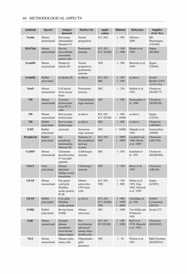
METHODOLOGICAL ASPECTS ___________________________________________________________________________________________________________________________________________________________________________________________________________________________________ 44
Antibody Species Antigen detected
Marker for Appli-cation
Dilution Reference Supplier (Cat. No.)
Nestin Mouse monoclonal
Rat nestin, intermediate filament VI
Neural progenitors
ICC (IF) 1 : 500
Gilyarov 2008
BD Pharmingen (556309)
MAP2ab Mouse monoclonal
Bovine microtubule-associated protein 2ab
Postmitotic neurons
ICC (IF) ICC (DAB)
1 : 100 1 : 1000
Binder et al. 1986
Sigma (M1406)
�-tubIII Mouse monoclonal
Human �-tubulin III
Neural progenitors, postmitotic neurons
WB 1 : 500 Banerjee et al. 1990
Sigma (T8660)
�-tubIII Rabbit polyclonal
�-tubulin III as above ICC (IF) IHC
1 : 500 1 : 500
as above Nordic BioSite (PRB -435P-100)
NeuN Mouse monoclonal
Cell nuclei from mouse brain
Postmitotic neurons
IHC 1 : 250 Mullen et al. 1992
Chemicon (MAB377)
TH Mouse monoclonal
Tyrosine hydroxylase from PC12 cells
Catecholaminergic neurons
IHC 1 : 500 Semenenko et al. 1986
Chemicon (MAB358)
TH Mouse monoclonal
Rat tyrosine hydroxylase
as above ICC (IF) ICC (DAB)
1 : 500 1 : 10000
as above Sigma (T2928)
TH Rabbit polyclonal
Rat tyrosine hydroxylase
as above IHC 1 : 400 as above Chemicon (AB5986)
5-HT Rabbit polyclonal
serotonin Serotonin-ergic neurons
IHC 1 : 10000 Dabadie et al. 1992
ImmunoStar (20080)
Peripherin Rabbit polyclonal
Rat peripherin, intermediate filament III
Neurons of the peripheral nervous system
ICC (IF) IHC
1 : 2000 1 : 2000
Leonard et al. 1988, Brody et al. 1989
Chemicon (AB1530)
GAD67 Mouse monoclonal
Glutamate decarbosylase 67 (recomb peptide)
GABAergic neurons
IHC 1 : 500 Kaufman et al. 1991
Chemicon (MAB5406)
ChAT Goat polyclonal
Human placental choline acetyl transferase
Cholinergic neurons
IHC 1 : 100 Bruce et al. 1985
Chemicon (AB144)
GFAP Mouse monoclonal
Pig spinal cord glial fibrillary acidic protein, IF III
Mature astrocytes, CNS stem cells
ICC (IF) WB
1 : 100 1 : 800
Debus et al. 1983, Eng 1985, Doetsch et al. 1999
Sigma (G3893)
GFAP Rabbit polyclonal
Cow glial fibrillary acidic protein
as above ICC (IF) ICC (DAB) WB
1 : 400 1 : 1000 1 : 2000
Castellano et al. 1991�
DAKO Cytomation (Z0334)
S100�� Rabbit polyclonal
Bovine brain S100��
Mature astrocytes
IHC 1 : 1000 Van Eldik and Watterson 1981
Swant (37)
GalC Mouse monoclonal
Synaptic plasma membrane from bovine hippocampus
Pre-myelinating and myeli-nating oligo-dendrocytes
ICC (IF) ICC(DAB)
1 : 100 1 : 1000
Raff et al. 1978, Ranscht et al. 1982
Chemicon (MAB342)
NG2 Mouse monoclonal
Human mela-noma cells
Oligodendro-glial precursor
IHC 1 : 50 Wilson et al. 1981
R&D Systems (MAB2585)
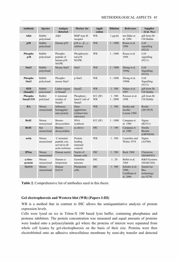
METHODOLOGICAL ASPECTS ___________________________________________________________________________________________________________________________________________________________________________________________________________________________________
45
Table 2. Comprehensive list of antibodies used in this thesis
Gel electrophoresis and Western blot (WB) (Papers I-III)
WB is a method that in contrast to IHC allows the semiquantitative analysis of protein expression levels. Cells were lysed on ice in Triton-X 100 based lysis buffer, containing phosphatase and protease inhibitors. The protein concentration was measured and equal amounts of proteins were loaded onto a polyacrylamide gel where the proteins of interest were separated from whole cell lysates by gel-electrophoresis on the basis of their size. Proteins were then electroblotted onto an adhesive nitrocellulose membrane by semi-dry transfer and detected
Antibody Species Antigen detected
Marker for Appli-cation
Dilution Reference Supplier (Cat. No.)
Alk6 Rabbit polyclonal
Alk6 BMP type Ib receptor
WB 1 �g/ml ten Dijke et al. 1994
gift from Dr CH Heldin
p38 Rabbit polyclonal
Human p38 p38 �-, �-, -subform
WB 1 : 1000 Rouse et al. 1994
Cell signalling (9212)
Phospho p38
Rabbit polyclonal
Phospho-peptide of human p38 MAPK
Phosphoryla-ted p38 MAPK
WB 1 : 1000 Rouse et al. 1994
Cell signalling (9211)
Stat3 Rabbit polyclonal
Mouse Stat3 Stat3 WB 1 : 1000 Zhong et al. 1994a
Cell Signalling (9132)
Phospho Stat3
Rabbit polyclonal
Phospho-mouse Stat3
p-Stat3 WB 1 : 1000 Zhong et al. 1994b
Cell Signalling (9131)
SED (Smad2)
Rabbit polyclonal
Linker region of Smad2
Smad2 WB 1 : 500 Nakao et al. 1997
gift from Dr CH Heldin
Phospho Smad1/5/8
Rabbit polclonal
C-terminal residue of Smad1
Phosphory-lated C-tail of Smad1
ICC (IF) WB
1 : 500 1 : 2000
Persson et al. 1998
gift from Dr CH Heldin
HA Mouse monoclonal
Influenza hemaggluti-nine protein
Hem-agglutinine (Adenovirus infection)
WB 1 : 500 Surdej and Jacobs-Lorena 1994
Roche (2013819)
BrdU Mouse monoclonal
Bromo-deoxyuridine
DNA synthesis
ICC (IF) 1 : 1000 Campana et al. 1988
Sigma (B2531)
BrdU Rat monoclonal
Bromo-deoxyuridine
as above IHC 1 : 100 Campana et al. 1988
Nordic Biosite (OBT0030)
actin Mouse monoclonal
C-terminal peptide con-served in all actin isoforms
Protein loading (internal control)
WB 1 : 500 Lazarides and Weber 1974
Sigma (A4700)
HNuc Mouse monoclonal
Human nuclei Nuclei of human cells
IHC 1 : 200 Beck 1968 Chemicon (MAB4383)
�-feto-protein
Mouse monoclonal
Human �-fetoprotein
Germline tumours
IHC 1 : 20 Bellet et al. 1984
R&D Systems (MAB1368)
Oct3/4 Mouse monoclonal
Human Oct3/4
Pluripotent cells
IHC 1 : 500 Scholer et al. 1990, Cauffman et al. 2006
SantaCruz Bio-technology (sc-5279)

METHODOLOGICAL ASPECTS ___________________________________________________________________________________________________________________________________________________________________________________________________________________________________ 46
with antibodies, shown as bands of a specific molecular weight. In order to be able to compare the amount of protein expression between different samples �-actin staining was performed. The expression level of this protein is expected to be stable in different experimental settings. Whether this is true for all conditions has been extensively discussed (reviewed by Ruan and Lai 2007, Tanic et al. 2007). Using Image Gauge software (Fujifilm) staining intensity of protein bands was measured and related to the �-actin band of the respective sample.
Immunoprecipitation In immunoprecipitation the protein of interest is first separated from the cell lysate by a specific antibody directly or indirectly coupled to agarose beads that allow precipitation of the antigen-antibody complex. The precipitate is then loaded onto a gel, transferred to a membrane and visualized by antibodies as described above. This method is more sensitive than WB and it was used in Paper II to detect Smad2 and its phosphorylation, respectively.
In vitro kinase assay (Paper II) In vitro kinase assays provide information about the functionality or enzymatic activity of kinases. In analogy to the procedure described above kinases are enriched from whole cell lysates by immunoprecipitation. We were interested in the kinase activity of dn and ca BMP receptors. Since all of these receptor constructs contained an HA-tag we used an anti-HA antibody coupled to sepharose beads for receptor separation. The immunoprecipitates were then incubated with radiolabeled ATP. After gel-electrophoresis, phosphorylation was detected by a plate reader (FLA2000, Fuji). The positive control in this assay was the ca receptor. The negative controls consisted of kinase precipitates of Ad-dnALK3 buffer treated samples, as well as solely antibody-sepharose complexes (“LacZ”).
Real-time polymerase chain reaction (RT-PCR) (Papers II and III) For the detection of subtle dynamic changes in gene expression RT-PCR is the method of choice due to its high sensitivity. Total RNA was isolated from cells in culture by the guanidinium thiocyanate method of Chomczynski (Chomczynski and Sacchi 1987) and 1 �g of RNA was reversely transcribed into complementary DNA (cDNA). Specific fragments of this cDNA were then amplified by cyclic repetition of DNA denaturation, primer annealing to the resulting single strand DNA molecules and extension of the DNA sequence. The resulting products were detected as bands of a specific size on an ethidium bromide stained agarose gel. The method allows the semiquantitative comparison of mRNA amounts in different samples. This is, however, only possible if the PCR reaction is stopped while fragment amplification is still exponential in all samples and the resulting fragment number high enough to be detected by the relatively insensitive ethidium bromide staining. In order to find these conditions cDNA amounts in the initial reaction mixture were carefully titrated for each primer pair and the number of extension cycles was adjusted. To assure that samples initially contained equal

METHODOLOGICAL ASPECTS ___________________________________________________________________________________________________________________________________________________________________________________________________________________________________
47
amounts of cDNA and that reaction efficiency and loading of the gel were comparable between samples, the cDNA for �-actin was amplified and its band used as a reference. For discussion of its reliability see section VI.2. and the following references (Ruan and Lai 2007, Tanic et al. 2007)
The sequences of the respective primer pairs are depicted in table 3. Resulting PCR products in Paper II were sequenced to confirm specificity of the primers. In every reaction we included one sample that did not contain any cDNA in order to control for unspecific reactions and purity of the reaction mixture. No product was detected in these samples.
Table 3. List of nucleotide primers used in the present thesis
Analysis of promoter activity by luciferase assay (Paper III) Luciferase assays are based on the transfection of expression vector plasmids that contain the promoter of interest coupled to the gene for luciferase. Luciferase is an enzyme that catalyzes a light producing reaction when luciferin substrate is added. After cell lysis the emitted light can be quantified by photometric measurement and correlates to promoter activity.
To assess the activity of the p38-, Mash1- or GFAP-promoter, respectively, cells were transfected with the respective expression vector constructs 4 hours prior to infection with adenovirus. The negative control consisted of an empty vector containing luciferase but no promoter. Luciferin substrate was added 36 hours after infection and photometric measurement carried out immediately afterwards. In some cultures growth factors were added at the peak of transgene expression, i.e. 24 hours after infection and light emission was analyzed 12 hours later. In order to correct for differences in transfection efficiency between culture wells, samples were transfected with AdLacZ. Photometric measurements were then
Gene Primer sequence Paper
Actin 5’-AAG ATG ACC CAG ATC ATG TTT GAG 5’-AGG AGG AGC AAT GAT CTT GAT CTT II-IV
ActRII 5’-CTG GAT ATC TAG CGA CAA C 5’-ACG ACA CTC CCG TTA TAC AC II
ActRIIb 5’-GAG ACT ATG TCA CGA GGC C 5’-CTC GGT AGT TGA AGG TCT CT II
Alk2 5’-TCT GTG CTA ATG ATG GCT CTC C 5’-TTC TGC GAT CCA GGG AAG GAT TTC II
Alk3 5’-GGA GGA ATC GTG GAG GAA TAT 5’-CAT ACG CAA AGA ACA GCA TGTC II
Alk6 5’-CGG CTG AAT CAC AAC CAT TTG G 5’-CTA GAC ATC CAG AGG TGA CAA CAG II
BMP 2/4 5’-TAT GTG GAC TTC AGT GAT GTG G 5’-CAG AGT CTG CAC TAT GGC ATG GTT II
BMP 5/6/7 5’-TGG CAG GAC TGG ATC ATT GCA C 5’-ATG GCG TGG TTG GTG GCA TTC AT II
BMPRII 5’-GGA GAA ATC AAA AGG GGA C 5’-CCT ATG TCT TAC AAC TGT CC II
Mash1 5’-GGA ACT GAT GCG CTG CAA AC 5’-CCT GCT TCC AAA GTC CAT TCC III

METHODOLOGICAL ASPECTS ___________________________________________________________________________________________________________________________________________________________________________________________________________________________________ 48
standardized to �-gal staining. To correct for differences in cell density between samples, results were related to the protein content in each well. The samples were analyzed in triplicate or quadruplicate.
Chemical inhibitors (Paper IV) SB 203580 and SB 202190 were purchased from Calbiochem and used at a final concentration of 10 �M. They were added to the cell culture at the timepoint of infection and in long-term experiments every 2nd day. They are specific inhibitors of the p38 MAPK. According to the supplier SB 203580 does not inhibit JNK or p42MAPK even at 100��M. This is in line with results published by Davies et al. (Davies et al. 2000). However, this group described unspecific inhibition of LCK, GSK3� and PKB� kinases by SB 203580 at 10��M. To confirm the results obtained by the inhibitors we inhibited p38 MAPK by p38DN (dominant negative) expression vectors in parallel experiments.
In vivo experiments (Paper IV)
6-OHDA lesion and rotational testing Adult female Sprague-Dawley rats received an injection of 6-OHDA into the right median forebrain bundle causing DAergic deinnervation of the striatum. Coordinates were set according to Paxinos and Watson (Paxinos et al. 1985). Three to four weeks after the toxin injection the extent of the lesion was assessed by rotational testing. Intraperitoneal injections of amphetamine have previously been shown to induce stable ipsilateral turning behavior in rats with a more than 90% DA depleted striatum (Thomas et al. 1994). Rats that displayed stable rotational scores of >5 ipsilateral net turns/min were selected for transplantation. Behavioral recovery from graft released DA is expected to be seen first several weeks after grafting. Rotational testing was therefore repeated 2, 4, 8 and even 13 weeks after transplantation.
Transplantation In order to prevent graft rejection, rats received daily intraperitoneal injections of the immunosuppressant cyclosporin A starting one day before transplantation. For transplantation 2 �l of a solution containing DNAse (to avoid cell aggregation) and 100.000 viable single cells were injected into the DA-depleted striatum 5-6 weeks after the 6-OHDA lesion. Cells had been harvested immediately before transplantation and were kept on ice until grafting.
Microscopical analysis and cell quantification In all experiments including microscopic evaluations the investigator was blinded to experimental design. Cell numbers were assessed in at least three repeated experiments.

METHODOLOGICAL ASPECTS ___________________________________________________________________________________________________________________________________________________________________________________________________________________________________
49
Light microscopy (Papers I and III) To evaluate the effect of BMPs on different cell populations in Paper I (staining for TH, Galc and GFAP in time course and dose-response experiments) absolute cell numbers were counted. In that way we avoided the possibility that proliferation or death of another cell type might suggest declining or increasing cell numbers in the population of interest, respectively. These counts were carried out under an inverted light microscope on DAB stained monolayer cell cultures in 6 different wells on 96 well plates at a 100 x magnification.
In Paper III neurite outgrowth was assessed in DAB stained cultures on chamber slides under a standard light microscope. For this purpose all MAP2ab-pos cells with a process longer than one cell diameter were counted and related to the total number of MAP2ab-pos cells. Three thousand cells were counted in 3 different wells.
Fluorescent microscopy (Papers I-IV) BMP responsiveness was evaluated by counting the percentage of TH- or MAP2ab-pos cells that displayed nuclear translocation of p-Smad. For this purpose cultures were labeled with fluorescent antibodies and analyzed under a Nikon diaphot fluorescent microscope. Pictures were taken with a digital camera. Exposure to fluorescent light was restricted to durations insensitive for background fluorescence. In order to detect colocalization between p-Smad and cell nuclei, the overlap of Hoechst-pos nuclei and p-Smad staining was assessed in merged views on the screen. In at least three different culture wells a total number of 500 MAP2ab- and 200 TH-pos cells were counted in 15-20 randomly chosen fields of view under a 400 x magnification.
In Papers II and III the number of GFAP-, galC,-and MAP2ab-pos cells were evaluated in monolayer cultures on chamber slides by fluorescent microscopy (400 x magnification). Their numbers were related to total cell numbers by counting a total number of 9000 Hoechst-pos cells in 3 different culture wells in at least 5 randomly chosen fields of view.
In contrast to cell counts in Papers I-III that were done in monolayer culture, cell counts for in vitro differentiation experiments in Paper IV had to be done in whole colonies since a dissociation and replating procedure would have negatively selected for more immature non-process-bearing cells. However, cell counts in whole colony also result in a bias. Using a conventional fluorescent microscope one predominantly assesses cells on the surface of the colonies while cells in the centre of the colonies are not visible or accessible. Assuming that a cell maturity gradient exists with the most mature cells on the outside of the colonies, differentiated cells might be over-represented in our cell counts.
Confocal microscopy (Paper IV) Rat brain sections were analyzed under a Leica confocal microscope that makes it possible to focus on structures deep down in thick sections and to reconstruct three dimensional images. In that way one can visualize cells lying on top of each other that in two-dimensional microscopy might have appeared as one cell.
To assess cell numbers of human nuclei- (HNu), BrdU-, TH- or neuronal nuclei (NeuN)-pos cells in the transplants graft areas in each consecutive section were delineated. Using an Olympus CAST- Grid system, cells for the respective staining were counted in 1% of the

METHODOLOGICAL ASPECTS ___________________________________________________________________________________________________________________________________________________________________________________________________________________________________ 50
delineated area in fields of view randomly chosen by the automatically moving microscope stage.
Statistics
Data from cell cultures in Papers I-IV were considered to be parametric and thus significance was determined by submitting data to one-way analysis of variance (ANOVA) with cell number as the dependent variable. Significance of between-group differences was determined by the Scheffé post-hoc test.
For transplantation data in Paper IV the non-parametric Kruskal-Wallis test was applied. For post hoc analysis between two groups at the same time point and between two time points within the same group, we used the Mann-Whitney U test.
RESULTS ___________________________________________________________________________________________________________________________________________________________________________________________________________________________________ 51
RESULTS
Bone Morphogenetic Proteins but not Growth Differentiation Factors induce dopaminergic differentiation in mesencephalic precursors (Paper I) Here we characterized the effect of BMPs on different cell types in E14 rat mesencephalic culture. According to the ICC performed on DIV8 the dominant cell type in native cultures is MAP2ab-pos neurons. They make up about 65% of total cells. Five to 6% of total cells stain positive for TH, 9% for nestin, 5% for GalC and only about 0.5% of cells are positive for GFAP.
Apart from BMP3, which did not display any detectable effect on the culture, the factors tested induced comparable responses in glial cells. Thus, when BMP5, 6, 7 and GDF5 and 6 were added to the culture medium at DIV6 all of these proteins significantly increased the number of GFAP-pos astrocytes (30-50 fold) already at the low concentration of 10 ng/ml (Fig. 5A). At the same concentration a significant 30-50 % decrease in the number of GalC-pos oligodendrocytes was seen. The latter was even more pronounced at 100 ng/ml (Fig. 5C).
Judging from [3H]thymidine incorporation assays all the factors had a biphasic effect on cell cycle phases, i.e. they were increasing the rate of cell proliferation at low concentrations (around 3 ng/ml) while inducing differentiation at high concentration (100 ng/ml) (Fig. 4).
Surprisingly, only factors from the 60A subgroup, i.e. BMP5, 6 and 7, increased the number of TH-pos cells dose-dependently. The increase became statistically significant at a concentration of 100 ng/ml for BMP5 and 6 and at 30 ng/ml for BMP7 (Fig. 1).
Another striking difference was the amount of p-Smad translocation. Whereas only about 2.5% of the MAP2ab-pos cells stained positive for p-Smad after stimulation with 100 ng/ml GDF5, the same concentration of BMP5 and BMP 7 induced positive staining in 37 and 47% of MAP2ab-pos cells, respectively (Fig. 6). Smad phosphorylation was also observed in 34 and 50% of TH-pos cells after treatment with BMP5 and 7, respectively (Fig. 7). Importantly, nuclear Smad translocation was detectable already 10 minutes after exposure of the cells to the factors, indicating a direct effect of BMPs on neuronal and DAergic cell populations.
However, the increase in TH-pos cell numbers could not be detected until 32 hours after BMP5, 6 or 7 had been added. Therefore, the underlying mechanism was unlikely to be a simple increase in TH expression which one would expect to occur earlier. Considering the fact that TH-pos cell numbers increased under differentiating conditions it was highly likely that BMPs promoted the maturation of DAergic precursor cells. An alternative explanation was that BMPs supported the survival of cells that die in control cultures. However, monitoring cell numbers in control cultures over time revealed that TH-pos cell numbers were not declining but increasing until DIV9. Thereafter, TH-pos cells in both controls and BMP treated cultures decreased (Fig. 3), arguing against a survival promoting effect. In order to investigate this more closely cell cultures were exposed to the toxin 6-OHDA for 24 hours. This resulted in a 40-60% decrease in TH-pos cells that could not be prevented by any of the

RESULTS __________________________________________________________________________________________________________________________________________________________________________________________________________________________________ 52
factors tested (Fig. 9), arguing against both a survival promoting and antioxidative effect of BMPs and GDFs.
The Bone Morphogenetic Protein type Ib receptor is a major mediator of glial differentiation and cell survival in adult hippocampal progenitor cell culture (Paper II) The initial purpose of the experiments in this Paper was to reduce glial differentiation and to increase the number of neurons in AHP culture by blocking BMP signaling. We chose this approach because a previous study had demonstrated that antagonizing BMP signaling via noggin, generated a higher number of neurons from SVZ cells (Lim et al. 2000). Furthermore, ALK6 had been suggested to be the BMP type I receptor that is responsible for differentiation (Panchision et al. 2001) in mesencephalic and cortical embryonic culture. Therefore, we were interested in whether the overexpression of dnALK6 would result in different effects compared with overexpression of dnALK3 in AHPs.
Overexpression of both caALK3 and caALK6 dramatically increased the number of GFAP-pos cells that morphologically resembled type I astrocytes (Fig. 3). Furthermore, both receptors were able to increase cell survival in AHP cultures depleted of BMPs (Fig. 11).
In dominant negative receptors the intracellular receptor kinase domain is non-functional. The extracellular part is unaltered and therefore traps BMPs and depletes them from the medium. Since ligand binding patterns between receptors are overlapping, the overexpression of one dominant negative receptor will always deprive other BMP receptors of their ligands. Therefore, this method does not allow for an exact definition of each receptor’s function but rather shows its role in the cellular context. The latter is determined by the amount and type of BMPs in the medium as well as the BMP receptor expression.
When non-functional dnALK6 was overexpressed at different concentrations and BMP6 was added to the culture, no Smad phosphorylation could be detected at high mois, suggesting that all BMP6 ligands were trapped by the dnALK6 receptor. In contrast, Smad phosphorylation after BMP2 addition was not completely abolished even at the highest dnALK6 expression levels, suggesting a predominant affinity for BMP6 towards the ALK6 receptor and a higher affinity of BMP2 towards the other type I receptors (Fig. 6B-D). Surprisingly, the overexpression of dnALK3 did not decrease but rather enhanced Smad phosphorylation. Thus, we could detect a weak but clearly present p-Smad band in dnALK3 infected cultures even in the absence of BMP stimulation (Fig. 6A). RT-PCR revealed that dnALK3 overexpression induced an increase in ALK6 mRNA (Fig. 5D) suggesting that the Smad signal is generated by endogenous BMPs that even under overexpression of the dnALK3 receptor have a stronger preference for the ALK6 receptor. The expression of mRNA for the ALK3 receptor was unaffected by overexpression of dnALK6 (Fig. 5A). mRNA expression levels for BMPs were not altered by overexpression of any of the dn receptors, suggesting that the increase in ALK6 mRNA was not secondary to a higher concentration of endogenous ligands.

RESULTS ___________________________________________________________________________________________________________________________________________________________________________________________________________________________________ 53
Regarding cell differentiation we found that blocking BMP signaling by overexpression of high amounts of dnALK6 did not affect neuronal differentiation as judged by neuronal markers. WB as well as ICC showed that GFAP expression was clearly reduced in these cultures (Fig. 8) while the number of mature GalC-pos oligodendrocytes increased (Fig. 10). Furthermore, cell survival assessed by nuclear morphology and LDH release was impaired. In contrast, in dnALK3 infected cultures containing higher amounts of active ALK6 receptors than AdLacZ infected controls, cell survival and GFAP expression were increased (Fig. 11).
Overexpression of dnALK2 showed that BMP ligand affinity was most similar between ALK2 and ALK6 and the biological effect resembled dnALK6 overexpression. Similar to observations in dnALK3 infected cultures, overexpression of dnALK2, induced increased levels of endogenous ALK6 receptor mRNA. However, this lacked any functional effect since potential ligands for this receptor were trapped in the non-functional ALK2 receptor complex (Fig. 12).
ASK1 inhibits astroglial development via p38 MAP kinase and promotes neuronal differentiation in adult hippocampus-derived progenitor cells (Paper III) In this Paper we assessed the effect of ASK1 on survival and differentiation in AHP culture by overexpressing a ca form of ASKI (ASK1-�N).
We found that low expression levels of ASK1-�N decreased proliferation. In a fraction of cells ASK1-�N induced growth arrest in the G2/M phase of the cell cycle. Cell viability assessed by the amount of LDH in the culture medium as well as by the MTT assay was affected only at higher expression levels of ASK1-�N (Fig. 2).
Cultures overexpressing moderate amounts of ASK1-�N showed increased neuronal differentiation that was apparent 4 days and significantly 9 days after infection of AHPs with the expression vector. Thus, the number of cells staining positive for MAP2ab increased as well as the percentage of neurite-bearing MAP2ab-pos cells. Furthermore, the activity of the promoter for the proneural gene Mash1 was significantly elevated in cultures overexpressing ASK1-�N (Fig. 3). Conversely, the number of GFAP-pos cells as well as the amount of GFAP protein assessed by WB was found to be decreased in these cultures (Fig. 5). Consistent with this, ASK1-�N depressed the activity of the GFAP promoter in a concentration dependent manner (Fig. 6). GFAP expression levels could not be restored by the addition of BMP or LIF, growth factors that are known to instruct neural progenitors to adopt a glial cell fate (Fig. 7). No effect on the number of GalC-pos oligodendrocytes was found.
In order to investigate signaling mechanisms responsible for the observed effects, cultures were transfected with a GFAP promoter construct that contained a mutated and therefore inactive STAT binding site. Interestingly, ASK1-�N was able to repress the activity of the STAT mutated promoter with the same efficiency as the wild type GFAP promoter suggesting that STAT signaling is not involved in the ASK1-�N mediated GFAP suppression

RESULTS __________________________________________________________________________________________________________________________________________________________________________________________________________________________________ 54
in AHP culture (Fig. 8). Blocking p38 kinase signaling by treating the cultures with its inhibitor SB203580 inhibited ASK1-�N from both increasing neuronal differentiation and decreasing GFAP expression. This was further confirmed by transfection of AHP cultures with a construct for dn p38 that also inhibited the repressive effect of ASK1-�N on the GFAP promoter (Fig. 9).
Transplantation of human embryonic stem cell-derived cells to a rat model of Parkinson's disease: effect of in vitro differentiation on graft survival and teratoma formation (Paper IV) In Paper IV hESCs were investigated for their potential to generate transplantable TH-pos neurons in co-culture with the mesenchymal cell line PA6. Previous studies from other groups had reported a widely varying efficiency of DAergic neuron differentiation between different human cell lines and no data existed concerning teratoma formation.
Cell counts of hESC cultures that had been grown on PA6 for 2 weeks and then transferred to PORN/laminin coated chamberslides revealed that approximately 11% of all cells expressed the DAergic marker TH at any investigated time point, i.e. 2.5, 4 and 6 weeks after plating (Fig. 1A-H). Neurons were shown to generate action potentials of both mature and immature response patterns (Fig. 1I-J) and to possess functional synapses 3-4 weeks after plating (Fig. 1K, L).
In order to avoid excessive dendrite formation, cells for transplantation experiments were not exposed to PORN/laminin but kept on PA6 throughout the culture period. Even under these conditions hESCs developed mature DAergic neuronal features. Thus, HPLC revealed the production of DA in cultures at 20 and 23DIV but not at 16DIV.
Two weeks after grafting a stereological analysis of the transplants revealed significantly more human cells in the striatum of rats that had received cells from shorter differentiation time points. In fact, in group 1 and group 2 (i.e. cells transplanted after 16 and 20 days of differentiation on PA6) cell numbers in the grafts exceeded the amount of cells initially implanted suggesting proliferation after grafting. This was not the case for transplants with cells differentiated for 23 days (Fig. 3A). Surprisingly, even those contained proliferating cells as assessed by BrdU staining (Fig. 3D) and accordingly, even in this group human cells increased on average to 5 times the number of implanted cells 13 weeks after transplantation (Fig. 3A). However, teratoma formation was not observed in group 3 while 82% and 25% of rats from group 1 and 2 respectively, developed teratoma. Moreover, teratomas in group1 were growing faster than in group 2 resulting in the earlier demise of rats in group1 compared to group 2 (Fig. 3H).
Grafts of cells that had been grown on PA6 for a longer time tended to contain lower percentages of postmitotic NeuN-pos neurons (Fig. 4A). The low number of only 10-50 TH-pos cells in transplants from any group and time point was in line with the lack of behavioral recovery (Fig. 2).

DISCUSSION ___________________________________________________________________________________________________________________________________________________________________________________________________________________________________ 55
DISCUSSION In the studies presented in this thesis, we elucidate basic mechanisms and phenomena in neural differentiation. The applied growth factors, receptors and cells are considered to have a putative role in experimental therapies in PD. In the following paragraphs I will discuss the rationale for the experimental set-ups and the significance of generated results in the broader perspective of translational research.
The limitations of in vitro systems In Paper I we intended to characterize the effect of BMP on mature mesencephalic cells, however, adult DAergic neurons do not survive culturing procedures. Postnatal cultures of the mesencephalon have been described (Bourque and Trudeau 2000) but neurons have to be grown on a layer of astrocytes. These feeder cells add undefined factors and were therefore not applied in our study. As a compromise we dissected tissue at earlier stages and allowed a maturation process to occur in vitro. However, it is unclear to which developmental stage the cells corresponded when BMPs were added at day 6 after plating. One can assume that they mimic the differentiation pattern seen in vivo. This is known for early embryonic cortical stem cells in clonal density culture, where neurons are generated first and astrocytes differentiate later (Qian et al. 2000). Even layer specific cortical neurons appear in the same order and at a similar time as in vivo (Shen et al. 2006). Thus, cortical stem cells follow a genetic program independent of extrinsic signals. No comparable studies exist for the mesencephalon. It is known that early DAergic mesencephalic cells develop their correct in vivo identity depending on their position in the cell-cell context. When the latter is disrupted at E14 the vast majority of DAergic neurons are already postmitotic and “intrinsically deter-mined” to proceed along the DAergic line. In our cultures differentiation increased over time. Thus, neurons developed an increasingly complex fiber network and the number of nestin-pos cells decreased. The astroglial marker GFAP appeared first around DIV6 and at about the same time oligodendrocytes expressed the postmitotic marker GalC. Thus, the consecutive differentiation of neuronal and glial cells was preserved. To judge from GalC staining even the timing of glial differentiation seemed to correlate with in vivo development where GalC expression is first detected around E19–20 and increases around birth (Hardy and Reynolds 1991). Thus we inferred that our cultures corresponded to the perinatal developmental stage. However, it would have been valuable to investigate TF expression and to conduct electrophysiological studies in order to characterize the degree of maturity in more detail. Given the differences between the in vitro and in vivo environment, it is most likely that there exists no exact in vivo correlate to the mixture of cells encountered in primary mesencephalic culture.
Interpretation and extrapolation of results would be easier if medium components were better defined. We may be able to specify the ingredients of our serum-free culture medium but we do not grasp the complex mixture of molecules secreted by all the different cell types during the first 6 DIV before adding BMPs. Keeping in mind the extreme context-dependency

DISCUSSION ___________________________________________________________________________________________________________________________________________________________________________________________________________________________________ 56
of the BMP family members, the undefined conditions demand extra caution when generalizing our results, however, they have been reproduced by other groups. For example, our finding that BMP family members exert a direct effect on mesencephalic TH-pos neurons has recently been supported (Sullivan and O'Keeffe 2005, Wood et al. 2005). It has also been confirmed that GDF5 is not a potent neurotrophic factor for TH-pos neurons when cultured for several days. In contrast, when added on the day of plating, its effect is comparable to that of GDNF (reviewed by Sullivan and O'Keeffe 2005). This difference might depend on GDF5’s preference for the ALK6 receptor (Nishitoh et al. 1996, Nickel et al. 2005) that is downregulated in culture over time. Interestingly, the expression of GDF in vivo is highest at E14 and lowest around birth (O'Keeffe et al. 2004). Thus, the weak response to GDF that we observed in our presumed perinatal culture might be physiological. The predictive value of this culture model for effects on adult or mature nigral cells is, however, unclear.
AHP cultures were used in order to generate transplantable cells. A relevant question in the context of translational research is how comparable are these cultures to human hippocampal progenitors. The answer is not straightforward since derivation protocols differ markedly. The AHPs were derived from macroscopically dissected adult rat hippocampus. Tissue blocks had been enzymatically digested and differentiated cells removed by serial passaging. The resulting cultures served as maternal populations for clonal cell lines. Obviously, this proce-dure contains several sources of variance: the tissue dissections, serum batches, cell types in the initial and subsequent cell suspensions, secreted molecules and concentrations of bFGF, all might affect cell population selection. It is even more difficult to standardize cell preparations from human tissue, since most of the samples are obtained from epilepsy surgery (Johansson et al. 1999, Kukekov et al. 1999, Roy et al. 2000, Suslov et al. 2002, Hermann et al. 2006) and presumably contain varying amounts of sclerosis. It is known that glial cells are activated in those areas, neurogenesis is increased and the migration pattern of progenitors altered (Cohen et al. 2003, Crespel et al. 2005, Parent 2007). Thus, cell composition might be divergent from healthy tissue and also differ between samples. One can conclude that to date there is no prototypic culture of human hippocampal progenitors that could serve as a reference for rat AHPs.
A second question is whether those cells that have been successfully programmed in vitro will keep their cell fate after transplantation in vivo. In Papers II and III our intention was to induce committed neuronal progenitors that would resist cell fate modulators in the striatum. Unless transplantation experiments are carried out we will not be able to judge whether we have succeeded. It is promising that BMP6, a strong glial inducing factor in the striatum, did not have a negative impact on the neuronal population. However, culture conditions in these experiments do not resemble the striatal environment.
How significant discrepancies between the in vitro and striatal in vivo environment can be was demonstrated by hESC cultures in Paper IV. Here, DAergic neurons were efficiently derived from hESCs in vitro. However, after transplantation, TH-pos cells either did not sur-vive or ceased to express TH.

DISCUSSION ___________________________________________________________________________________________________________________________________________________________________________________________________________________________________ 57
In summary, cell culture systems provide the possibility to characterize in detail mechanisms in cell biology. However, results depend on culture conditions and the developmental stage of the cells and are therefore difficult to generalize.
GFAP as a marker for astrocytes As described in the introduction, GFAP can no longer be regarded as a reliable marker for astrocytes since GFAP also labels stem cells. Yet, in our studies this protein has been considered to mainly represent astrocytes. It would have been more correct to use the descriptive term “GFAP-pos cells” throughout the papers. However, in Paper I the terminology is of minor importance since we studied embryonic mesencephalic cells, and stem cells in the embryonic brain are known to be GFAP-negative radial glial cells. In contrast, in Papers II and III, GFAP-pos cells were initially most likely a heterogenous population of astrocytes and stem or precursor cells. Nevertheless, medium, coating and cell density in our AHP cultures promoted differentiation. Thus, the number of stem cells would decrease over time and the increase in GFAP that we observed in control cultures can therefore most likely be ascribed to differentiation of astrocytes. In Paper II, it would have been interesting to stain with additional markers such as nestin or Sox2 for stem cells, and S100� for mature astrocytes. GFAP expression increased in dnALK3-infected cultures that expressed a higher number of functional ALK6 receptors, thus being more responsive to BMP signals. Since it is known that BMPs can dose-dependently promote proliferation or differentiation, respectively, it cannot be excluded that GFAP-pos stem or precursor cells were induced to divide. However, BMPs have so far not been shown to exert a mitogenic effect on hippocampal precursors at any concentration. Also, ALK6 has been shown to be the BMP type I receptor that is responsible for differentiation, as opposed to ALK3 that mediates proliferation in embryonic cultures. Furthermore, stem cells are known to divide only rarely, and for this reason they were unlikely to account for the significant elevation in GFAP in these cultures.
Will BMPs have a role in future therapies for PD? In experimental animal studies proteins of the BMP family have clearly been shown to have positive effects on TH-pos neurons. Rats receiving a nigral injection of GDF5 directly before a 6-OHDA induced lesion developed less functional deficits and had higher numbers of TH-pos cells in the SN compared with control animals receiving buffer (Sullivan et al. 1999). Harvey et al. showed that BMP7 decreased rotational scores and increased TH-pos striatal fiber density when injected into the SN prior to induction of a median-forebrain-bundle lesion (Harvey et al. 2004). The authors conclude that BMP7 protects DAergic neurons which would contradict our in vitro finding. Another possible interpretation of Harvey’s results is, however, that BMP7 induces TH expression in otherwise TH-negative cells. The factors were injected into only one side, and for this reason the contralateral side is, strictly speaking, not a proper control. Interestingly, when BMP7 was injected into the lateral ventricle 1 w after a 6-OHDA induced lesion, an increase in TH positivity was observed even in the contralateral non-lesioned side (Zuch et al. 2004). The latter is consistent with our results showing a BMP induced increase of TH-pos cell numbers in both 6-OHDA- treated and -untreated cultures. In

DISCUSSION ___________________________________________________________________________________________________________________________________________________________________________________________________________________________________ 58
our cultures this was due to increased differentiation. The mechanism responsible for elevated TH-pos cell numbers in vivo remains unclear (Zuch et al. 2004).
For any practical application it is important to know whether BMPs protect or differen-tiate TH-pos cells or simply increase TH transcription. In PD DAergic cell death occurs slowly. For this reason, protective agents have to be delivered continuously over several months or years. Existing in vivo studies do not cover a comparably long time period. If increased DAergic differentiation is the prevailing mechanism for increased TH-pos cell numbers, shorter BMP exposure might be effective. However, there is no clear evidence for a responsive target population - immature DAergic precursors - in the adult human SN. Fur-thermore, even if those progenitors exist, it remains unclear whether they can be functional. Would they establish connections to the striatum?
Our results in Papers I and II indicate that BMPs are pleiotrophic agents. Thus, an intracerebral delivery of BMP will most likely have side effects on other cell populations. We and others have shown that BMPs inhibit oligodendroglial differentiation. It is commonly accepted that oligodendrocyte progenitors exist dispersed throughout the adult brain. We do not know whether these progenitors contribute to myelination in mature nervous tissue. How-ever, it concerns us that chronic exposure to BMPs will impair oligodendroglial development. Furthermore, it has been suggested that cell genesis in the SVZ is dependent on a delicate balance between BMPs and their antagonists (Lim et al. 2000, Colak et al. 2008). In line with this, we found that AHPs differentiate into glial cells upon excessive BMP signaling. Thus, target delivery of BMPs would have to be carefully restricted to the SN in order not to result in adverse effects in neurogenic regions. This is a difficult task.
Another point deserving attention is BMPs’ concentration-dependent effect. How BMPs spread after intracerebral delivery is not well known. Presumably, their concentration declines the longer the distance from the injection site. We have described that low concentrations have a proliferative effect in embryonic mesencephalic culture. Whether the same is true for the adult brain in vivo is not clear. However, the readiness of BMPs to induce mitosis demands caution when any in vivo application is considered.
A significant finding was that high concentrations of BMPs induce differentiation in neuronal as well as glial precursors. p-Smad is translocated into the nucleus 10 minutes after exposure to BMP. Thus, bathing cells in BMP-containing solution shortly before transplanta-tion might improve transplant integration. A previous study has indeed demonstrated increased transplant survival of GDF5-treated embryonic mesencephalic cells (Sullivan et al. 1998). It remains to be seen whether similar effects can be achieved when hESC-generated DAergic precursors are exposed to BMPs. In this context BMPs’ putative cytostatic effect might be beneficial. Unwanted proliferation of insufficiently differentiated cells has been described in Paper IV and is a frequently encountered problem in ESC therapy. It has been shown that BMP treatment of cells from glioblastoma – an aggressive brain tumor – results in inhibition of proliferation, induction of differentiation and reduces their tumorigenic potential (Piccirillo et al. 2006). In analogy, one might be able to increase maturation in hESC-generated cell suspensions before grafting. However, more recent work has shown that glioma cells lacking functional ALK6 receptors converted otherwise cytostatic BMP signals to proliferative signals (Lee et al. 2008). Thus, in heterogeneous cell suspensions such as those

DISCUSSION ___________________________________________________________________________________________________________________________________________________________________________________________________________________________________ 59
resulting from hESC differentiation, BMP-induced responses have to be carefully monitored before application.
Is ASK1 a putative therapeutic molecule? Our results demonstrate that ASK1 mediates both neuronal differentiation and apoptosis dependent on the amount of transgene expression. We were able to reliably define non-toxic concentrations of adenoviral infection in vitro but the window to induce differentiation versus cell death was very narrow. A follow-up study from our lab showed that this range is even narrower when ASK1 is combined with other neuronal differentiation-inducing agents (Elmi et al. 2007). Thus, in a therapeutic application the amount of ASK1 expression would have to be extremely tightly controlled. It will be of interest to further analyze the downstream signaling cascade of ASK1 in order to understand how it divides into pro-differentiating and pro-apoptotic signals. ASK1 could then be replaced by those downstream effector molecules that lead to differentiation.
Our FACS data show that a significant fraction of cells in ASK1-differentiated cultures are arrested at the G2/M stage of the cell cycle. This is usually observed when DNA damage inhibits cells from undergoing mitosis. Terminal differentiation, in contrast, is characterized by cell cycle arrest at the G1 stage. Interestingly, genotoxic stress has been shown to inhibit muscle progenitors from differentiation. In those cells DNA damage activates a transcrip-tional checkpoint leading to cell cycle arrest in either the G1 or the G2 phase (Puri et al. 2002). A similar mechanism could apply for the decrease of GFAP observed in our AHP cultures. This is a slightly provocative hypothesis in need of further investigation. At this stage, we do not know if glial progenitors form a part of the G2/M arrested cell group. Analy-sis of cell cycle markers in the various subpopulations would be worthwhile. One would like to exclude that the ASK1 induced neurons are G2/M arrested due to DNA damage. To show that they develop into functional neurons will also be important.
It is too early to answer the question of whether ASK1 is a potential cell-therapeutic molecule. Judging from current results it is more likely that one of the downstream molecules will be applicable. Future studies on this topic could reveal new insights into how basic mechanisms of neural cell differentiation are coupled to cell cycle stages.
Can AHPs serve as a source for experimental cell therapies in PD? The finding that dividing cells with neurogenic potential exist in the adult human brain evoked enthusiastic ideas about novel regenerative therapies. Adult neural progenitors were considered as a source for both endogenous neurogenesis and autologous transplantation. Experiments aimed at manipulating AHPs for cell therapies have not however been entirely optimistic.
With respect to endogenous neurogenesis, SVZ cells were shown to repopulate the neighboring striatum with new neurons after ischemic lesions (Arvidsson et al. 2002). In contrast, the migration pattern of AHPs seems to be restricted to the dentate gyrus and the hilus of the hippocampus (Picard-Riera et al. 2004). Furthermore, evidence exists that new neurons in the hippocampus do not arise from stem- but rather from progenitor cells. The latter have a more restricted potential to divide and might thus easily be depleted by excessive

DISCUSSION ___________________________________________________________________________________________________________________________________________________________________________________________________________________________________ 60
mitotic stimulation. Finally, it is potentially hazardous to blindly induce new neurons in vivo, thereby interfering with the cytoarchitecture of the cortex and presumably predisposing the individual for seizures.
We have focused on transplantation approaches and aimed at increasing neuronal differentiation from AHPs. Cells in Paper II did not survive, but in Paper III we succeeded in elevating the number of differentiated and determined neurons. However, experiments were not designed to change the neuronal subtype and - not surprisingly - the generated neurons did not express TH. We have not characterized the expression pattern of TFs in ASK1-induced neurons in detail and we do not know how plastic these cells are. Given the complex regulatory mechanisms involved in nigral DAergic cell development it is however, unlikely that they could be directed towards a DAergic cell fate. In fact, no successful production of TH-pos neurons from AHPs has been described so far. Both rat and human hippocampal progenitors mainly differentiate into GABAergic and glutamatergic neurons (Palmer et al. 1997, Hermann et al. 2006). Conditions known to increase DAergic differentiation as, for example, the addition of Shh and FGF8 to the culture medium did not induce TH-pos neurons from adult human hippocampal progenitors. Co-culture of those cells with PA6 increased neuronal differentiation without any impact on transmitter subtypes (Hermann et al. 2006). Not only the restricted differentiation potential but also long population doubling times (8 –14 days for hAHPs) (Hermann et al. 2006) argue against AHPs as the preferable cell source. Furthermore, their proliferative capacity decreases with age – a not totally insignificant fact with respect to allogenic transplantation in PD where the patients are around 60 years old at the time of diagnosis. Finally, the anatomical location of the hippocampus does not make it particularly easy to obtain tissue samples for cell generation.
SDIA-induced differentiation of hESCs Compared to adult neural progenitors hESCs have a higher proliferative capacity and can be more readily differentiated into DAergic neurons. It is maybe more correct to say “ESCs readily differentiate into DAergic neurons” since this process is as yet not well controlled.
When we started to work with hESCs the SDIA method seemed advantageous since it was fast and efficient. It is not unlikely though that the fast differentiation of DAergic neurons has negative side effects. It might increase the risk of tumor formation in the graft since other developing cell types do not have the time to sufficiently differentiate in vitro. This hypothesis is supported by our experiments that show a significant decrease in tumor forma-tion and graft proliferation with increasing in vitro differentiation time. Comparable results were recently achieved by Ferrari et al. for primate ESCs (Ferrari et al. 2006). In our experi-ments all animals transplanted with cells from the longest culture period (i.e. group 3) survived without any tumors. Teratomas might, however, have developed at a later time point. Thus, it is not feasible to define a clear-cut safety threshold for in vitro differentiation time. Our rats had to be immunosuppressed to tolerate the graft. Possibly, tumor formation is less of a problem if isogenic cells are transplanted and the recipient’s immune system is functional.
With respect to the neuronal population, our results were unsatisfactory. It is a commonly faced problem that numbers of TH-pos neurons are low after grafting. It has been suggested that this is partially due to the lack of mature glial cells in hESC generated

DISCUSSION ___________________________________________________________________________________________________________________________________________________________________________________________________________________________________ 61
transplants (Zietlow et al. 2008). In the embryonic mesencephalon, glial cells differentiate later than neurons. Thus, it seemed possible that increased in vitro differentiation before trans-plantation would lead to astroglial maturation and consequently have a positive impact on the survival or TH expression of DAergic neurons. This was not the case. Judging from the more complex fiber network and DA content in the cultures, differentiation over time proceeded as expected. However, GFAP expression did not increase. In light of recent results showing that glial differentiation starts around DIV50 in hESC culture this is not surprising (Joannides et al. 2007). Considering the time differences between neuronal and glial development in hESC culture, mixed glial-neuronal grafts might be achievable only when starting from different cell preparations. It will be interesting to evaluate the expression of markers for glial progenitors in our cultures over time. Obviously, we need to gain a more detailed understanding of the sequential induction and differentiation of neural cell types in this culture system.
The exact subtype of the generated DAergic neurons in Paper IV is also unclear. RT-PCR analysis of another hESC line that had been cultured under comparable conditions but was not included in the Paper, showed expression of Nurr1 and Lmx1b TFs. Yet, it remains unclear whether the generated neurons resembled nigral DAergic A9 cells. It is thus difficult to speculate on the underlying reasons for the in vivo outcome. Did the DAergic neurons not survive? Did the TH-pos cells acquire the wrong subtype or were they not intrinsically determined to the DAergic phenotype and thus dedifferentiated in vivo? These questions will have to be addressed in the future. However, the PA6 co-culture system can only serve to elucidate basic mechanisms in hESC DAergic differentiation. Due to contamination from xenomaterial the generated cells will not be usable in clinical approaches.
Implications for future therapies of PD? Experiments in this thesis were designed to investigate basic cell biological principles of putative therapeutic molecules. The significance of the respective findings has been presented. However, as pointed out, results obtained in vitro are only indirectly applicable to in vivo conditions. Furthermore, discrepancies between the rodent and human brain are not yet defined in detail. Thus, our results can not have any direct clinical relevance at the moment. Instead, they add important details to our understanding of neural and DAergic cell dif-ferentiation. They have also generated new questions and concerns that were discussed in the previous paragraphs. Those are an important part of the findings. The rare clinical trials of stem cell therapies illustrate the gap between what is feasible on an experimental level and what is actually safe in real life. The context dependency of every signaling event makes prediction of effects in the human brain next to impossible. Given that putative cell- and growth factor therapies will induce irreversible changes in the patients’ brain, risks are excessively high at the moment. Traditional Phase I trials are not readily feasible.
When will we understand sufficiently well to judge that these therapeutic interventions are likely to be safe?
DISCUSSION ___________________________________________________________________________________________________________________________________________________________________________________________________________________________________ 62
Future aspects Possible future investigations directly extending on results presented in this thesis have been mentioned in the previous discussion. Some aspects that will bring cell therapy closer to clinical practice are described below.
Towards clinical application of ESCs Significant practical problems have to be solved before it will be possible to manufacture and apply hESCs as a drug. Cell suspensions best suited for engraftment will have to be defined in terms of cell type composition and differentiation stage. Detailed knowledge about the molecular characteristics of each step along the differentiation path will be important. To date we know very little about cell surface markers differentially expressed at different stages. Those markers might eventually enable us to specifically select the cells of interest.
The necessary removal of all xeno-material from production processes will imply cell growth in suboptimal conditions. It might impose additional stress on the cells and therefore decrease their stability. Cells will thus have to be carefully monitored for putative acquisition of genetic, epigenetic and mitochondrial changes. ESCs can grow to form benign tumors even without being transformed. It might increase safety to equip ES cells with suicide genes. The administration of a prodrug could then trigger the elimination of those cells.
An interesting line of research is the derivation of “personalized ESC-like cells” from somatic cells of the graft recipients. Different approaches to achieve this goal have recently been reviewed (Klimanskaya et al. 2008).
Novel image techniques are mandatory to track the growth of transplanted cells longitudinally and quantitatively. In this context, the discovery of a novel biomarker that allows the identification of neural progenitor cells in the human brain with magnetic resonance spectroscopy is revolutionary if validated (Manganas et al. 2007).
Growth factor therapy When GDNF was removed from the market in 2004 enthusiasm about growth factors as a putative therapy in PD decreased. However, most likely, growth factors will have to complement cell therapy to both protect the implanted cells and to increase their integration. The negative outcome of clinical trials with GDNF made researchers more aware of possible problems. Future experimental designs will have to address how to achieve adequate concen-trations of factors in specific brain regions while preventing them from spreading to non-target areas. Ideally, one would apply trophic factors with a profile highly specific for DAergic neurons. So far, no such factor is known. Another possibility is the target delivery of growth factors with viral vectors. In fact, clinical safety trials for adeno-associated virus mediated delivery of GAD (glutamic acid decarboxylase, an enzyme involved in glutamate metabolism) to the subthalamic nucleus of PD patients did not show any evidence of severe toxic or adverse effects in a 1 year follow-up (Kaplitt et al. 2007). By inserting the right promoter into these vectors one might even target specific cell types to produce the trophic factor. Regulatable vectors that make it possible to switch off transgene expression in case of severe side effects are highly sought after for clinical use but not yet available.

DISCUSSION ___________________________________________________________________________________________________________________________________________________________________________________________________________________________________ 63
Endogenous neurogenesis in PD Currently, stimulating adult neurogenesis in the SN does not seem to be a therapeutic option. The in vivo genesis of DAergic neurons in this part of the adult brain has not been unequivo-cally demonstrated. However, well established neurogenic areas like the hippocampus and the SVZ might be impaired by the pathological process in PD. Thus, impaired spatial memory (Pillon et al. 1997), depression (reviewed in Brandstadter and Oertel 2003)) and olfactory dysfunction (Ponsen et al. 2004) are common in PD patients and they often precede motor signs. The envisioned link between these symptoms and impaired neuronal development is speculative at the moment. However, there is evidence for reduced neurogenesis in the SVZ and hippocampus in post-mortem PD brains and the significance of this observation needs to be explored. Furthermore, DA receptors are differentially expressed on progenitor cells (Hoglinger et al. 2004), suggesting that DAergic drugs currently in use might provide more than symptomatic release.

CONCLUSIONS ___________________________________________________________________________________________________________________________________________________________________________________________________________________________________ 64
CONCLUSIONS 1. BMP5, 6 and 7 but not BMP3, GDF5 or 6 promote DAergic differentiation of
embryonic mesencephalic precursors via a direct mechanism on the neuronal population. BMP5, 6, 7 and GDF5, and 6 increase the differentiation of astrocytes and inhibit maturation of oligodendrocytes to a comparable extent, whereas BMP3 does not exert any detectable effect. None of the tested factors protect against 6-OHDA induced oxidative stress in vitro.
2. Endogenous BMPs promote survival and astrocyte differentiation via the ALK6 and
ALK2 receptor in AHP culture. The interruption of BMP signaling via a depletion of endogenous ligands from the medium induces cell death and decreases the number of astrocytes, while oligodendrocytes increase. No effects on neurons are observed. The ALK6 receptor is dynamically regulated and its expression increases when BMP-mediated Smad phosphorylation decreases.
3. ASK1 induces neuronal differentiation and inhibits GFAP expression in AHPs in vitro.
The effect is sustained in the presence of potent astroglial inducers. It is mediated via p38 MAPK and a STAT3-independent suppression of the GFAP promotor.
4. Co-culture with PA6 instructs about 11 % of hESCs to develop into TH-pos neurons
with CNS identity. PA6 generated neurons display action potential of both mature and immature phenotypes in vitro, suggesting functionality. However, after transplantation into the DA depleted rat striatum, TH-pos cells are rare and lesion-induced motor deficits are not reversed. Prolonged differentiation time on PA6 decreases the risk of teratoma formation after transplantation but does not increase the number of TH-pos neurons in the graft.

ACKNOWLEDGEMENTS ___________________________________________________________________________________________________________________________________________________________________________________________________________________________________
65
ACKNOWLEDGEMENTS I would like to express my sincere gratitude to everybody who has contributed to this work throughout the years. In particular I wish to thank: Keiko Funa, my supervisor, for scientific guidance, constant encouragement and for giving me the opportunity to develop and think independently. I especially thank you for your understanding during life’s ups and downs. Per Odin, at that time at the Department of Neurology, Hannover Medical School, for initiating this work, by arousing my interest in moving disorders and restorative medicine, for helping me to find financial support and for presenting me to Keiko. Roland Faigle, my co-author and friend, for outstanding collaboration and interesting in-depth discussions on scientific and daily-life topics. I will not forget our joint ventures in front of the microscope, engaged in those incredibly monotonous cell counts for nights on end. Remember rollerblading in Hästevik? I’m still laughing whenever I think about it. I truly miss having you around! Peter Eriksson, for introducing me to the world of neural stem cells, sharing the fascination for cell biology, generously providing access to all lab resources and, most of all, for being a close friend. Your talent to see opportunities combined with your genuine kindness and great sense of humor were unique and created an always inspiring atmosphere. Your sudden death is still incomprehensible. Muna Elmi, for being a very considerate room-mate and for always keeping track of all lab supplies. I greatly appreciate your devoted work on all our possible and impossible experiments. Daniel Wetterskog, for all your help with computers and instruments. Thank you for discovering the Ankesaurus and all the other jokes you played on me! Yoshiki Matsumoto, for doing a great job in organizing fridges, freezers and boxes after the lab had moved. Urban and Catrin Molander, for helping me to find my way around the lab when I first came to Gothenburg. Yvonne Arvidsson, for introducing me to the work with viral vectors and for insightful conversations. All the other previous Members of the Institute of Anatomy and Cell biology for sharing knowledge, experience and frustrations. . Karin Gabrielsson and Xiaoyang Wang, for skillful technical assistance. Ann-Marie Alborn and Maria Åberg, for forthcoming help when we started working with AHPs.

ACKNOWLEDGEMENTS ___________________________________________________________________________________________________________________________________________________________________________________________________________________________________ 66
Jia-Yi Li, Ana Sofia Correia, and my other co-authors from the Wallenberg Neuroscience Center in Lund, for fruitful collaboration on the ESC Paper. Annika Dahlström, for allowing me to use the equipment at the animal facilities. Barbro Lanner, Eva Lyche and Laila Falk, for helping me to solve all those mysterious bureaucratic problems. Judith Klecki and Shirley Fridlund, for excellent linguistic revision of the manuscript. Thank you Shirley, for “och hur mår du?” and your great flexibility. Violeta Piculescu, my closest companion during the first years at “Universus” in Gothenburg and a wonderful friend ever since. Thank you for your warmth, for always asking interesting questions and for numerous great parties! Ingeborg Niestroy, for being such a caring and reliable friend and for flying all the way from Brussels to help me survive my dissertation. My aunt, Gerda Mühlbach, for always simply being there. My parents, Ursula and Helmut Brederlau, for having given me confidence and strength. You only got to see the beginning of this work but your love has supported me all the way through. The Boehringer Ingelheim Foundation for financial support during the first three years of this work.
REFERENCES ___________________________________________________________________________________________________________________________________________________________________________________________________________________________________
67
REFERENCES Abad, F., R. Maroto, M. G. Lopez, P. Sanchez-Garcia, and A. G. Garcia. 1995. Pharmacological protection
against the cytotoxicity induced by 6-hydroxydopamine and H2O2 in chromaffin cells. Eur J Pharmacol 293: 55-64.
Abeliovich, A., Y. Schmitz, I. Farinas, D. Choi-Lundberg, W. H. Ho, P. E. Castillo, N. Shinsky, J. M. Verdugo, M. Armanini, A. Ryan, M. Hynes, H. Phillips, D. Sulzer, and A. Rosenthal. 2000. Mice lacking alpha-synuclein display functional deficits in the nigrostriatal dopamine system. Neuron 25: 239-52.
Abou-Sleiman, P. M., M. M. Muqit, and N. W. Wood. 2006. Expanding insights of mitochondrial dysfunction in Parkinson's disease. Nat Rev Neurosci 7: 207-19.
Altman, J., and S. A. Bayer. 1981. Development of the brain stem in the rat. V. Thymidine-radiographic study of the time of origin of neurons in the midbrain tegmentum. J Comp Neurol 198: 677-716.
Alvarez-Buylla, A., and D. A. Lim. 2004. For the long run: maintaining germinal niches in the adult brain. Neuron 41: 683-6.
Alvarez-Buylla, A., J. M. Garcia-Verdugo, and A. D. Tramontin. 2001. A unified hypothesis on the lineage of neural stem cells. Nat Rev Neurosci 2: 287-93.
Andersson, E., J. B. Jensen, M. Parmar, F. Guillemot, and A. Bjorklund. 2006a. Development of the mesencephalic dopaminergic neuron system is compromised in the absence of neurogenin 2. Development 133: 507-16.
Andersson, E., U. Tryggvason, Q. Deng, S. Friling, Z. Alekseenko, B. Robert, T. Perlmann, and J. Ericson. 2006b. Identification of intrinsic determinants of midbrain dopamine neurons. Cell 124: 393-405.
Andrew, R., D. G. Watson, S. A. Best, J. M. Midgley, H. Wenlong, and R. K. Petty. 1993. The determination of hydroxydopamines and other trace amines in the urine of parkinsonian patients and normal controls. Neurochem Res 18: 1175-7.
Aoki, H., M. Fujii, T. Imamura, K. Yagi, K. Takehara, M. Kato, and K. Miyazono. 2001. Synergistic effects of different bone morphogenetic protein type I receptors on alkaline phosphatase induction. J Cell Sci 114: 1483-9.
Arvidsson, A., T. Collin, D. Kirik, Z. Kokaia, and O. Lindvall. 2002. Neuronal replacement from endogenous precursors in the adult brain after stroke. Nat Med 8: 963-70.
Backman, C. M., L. Shan, Y. J. Zhang, B. J. Hoffer, S. Leonard, J. C. Troncoso, P. Vonsatel, and A. C. Tomac. 2006. Gene expression patterns for GDNF and its receptors in the human putamen affected by Parkinson's disease: a real-time PCR study. Mol Cell Endocrinol 252: 160-6.
Balemans, W., and W. Van Hul. 2002. Extracellular regulation of BMP signaling in vertebrates: a cocktail of modulators. Dev Biol 250: 231-50.
Banerjee, A., M. C. Roach, P. Trcka, and R. F. Luduena. 1990. Increased microtubule assembly in bovine brain tubulin lacking the type III isotype of beta-tubulin. J Biol Chem 265: 1794-9.
Beck, J. S. 1968. Morphological patterns of immunofluorescence staining of cell nuclei with human antinuclear sera. Arch Dermatol 98: 559-60.
Bellet, D. H., J. R. Wands, K. J. Isselbacher, and C. Bohuon. 1984. Serum alpha-fetoprotein levels in human disease: perspective from a highly specific monoclonal radioimmunoassay. Proc Natl Acad Sci U S A 81: 3869-73.
Ben-Hur, T., M. Idelson, H. Khaner, M. Pera, E. Reinhartz, A. Itzik, and B. E. Reubinoff. 2004. Transplantation of human embryonic stem cell-derived neural progenitors improves behavioral deficit in Parkinsonian rats. Stem Cells 22: 1246-55.
Benmoyal-Segal, L., and H. Soreq. 2006. Gene-environment interactions in sporadic Parkinson's disease. J Neurochem 97: 1740-55.
Bignami, A., and D. Dahl. 1974. Astrocyte-specific protein and neuroglial differentiation. An immunofluorescence study with antibodies to the glial fibrillary acidic protein. J Comp Neurol 153: 27-38.
Binder, L. I., A. Frankfurter, and L. I. Rebhun. 1986. Differential localization of MAP-2 and tau in mammalian neurons in situ. Ann N Y Acad Sci 466: 145-66.
Bjorklund, L. M., R. Sanchez-Pernaute, S. Chung, T. Andersson, I. Y. Chen, K. S. McNaught, A. L. Brownell, B. G. Jenkins, C. Wahlestedt, K. S. Kim, and O. Isacson. 2002. Embryonic stem cells develop into functional dopaminergic neurons after transplantation in a Parkinson rat model. Proc Natl Acad Sci U S A 99: 2344-9.
Blum, D., S. Torch, M. F. Nissou, A. L. Benabid, and J. M. Verna. 2000. Extracellular toxicity of 6-hydroxydopamine on PC12 cells. Neurosci Lett 283: 193-6.
REFERENCES ___________________________________________________________________________________________________________________________________________________________________________________________________________________________________ 68
Blum, D., S. Torch, N. Lambeng, M. Nissou, A. L. Benabid, R. Sadoul, and J. M. Verna. 2001. Molecular pathways involved in the neurotoxicity of 6-OHDA, dopamine and MPTP: contribution to the apoptotic theory in Parkinson's disease. Prog Neurobiol 65: 135-72.
Bonfanti, L., and P. Peretto. 2007. Radial glial origin of the adult neural stem cells in the subventricular zone. Prog Neurobiol 83: 24-36.
Boulton, T. G., S. H. Nye, D. J. Robbins, N. Y. Ip, E. Radziejewska, S. D. Morgenbesser, R. A. DePinho, N. Panayotatos, M. H. Cobb, and G. D. Yancopoulos. 1991. ERKs: a family of protein-serine/threonine kinases that are activated and tyrosine phosphorylated in response to insulin and NGF. Cell 65: 663-75.
Bourque, M. J., and L. E. Trudeau. 2000. GDNF enhances the synaptic efficacy of dopaminergic neurons in culture. Eur J Neurosci 12: 3172-80.
Braak, H., K. Del Tredici, H. Bratzke, J. Hamm-Clement, D. Sandmann-Keil, and U. Rub. 2002. Staging of the intracerebral inclusion body pathology associated with idiopathic Parkinson's disease (preclinical and clinical stages). J Neurol 249 Suppl 3: III/1-5.
Brandstadter, D., and W. H. Oertel. 2003. Depression in Parkinson's disease. Adv Neurol 91: 371-81. Brody, B. A., C. A. Ley, and L. M. Parysek. 1989. Selective distribution of the 57 kDa neural intermediate
filament protein in the rat CNS. J Neurosci 9: 2391-401. Brons, I. G., L. E. Smithers, M. W. Trotter, P. Rugg-Gunn, B. Sun, S. M. Chuva de Sousa Lopes, S. K.
Howlett, A. Clarkson, L. Ahrlund-Richter, R. A. Pedersen, and L. Vallier. 2007. Derivation of pluripotent epiblast stem cells from mammalian embryos. Nature 448: 191-5.
Bruce, G., B. H. Wainer, and L. B. Hersh. 1985. Immunoaffinity purification of human choline acetyltransferase: comparison of the brain and placental enzymes. J Neurochem 45: 611-20.
Brunet, A., A. Bonni, M. J. Zigmond, M. Z. Lin, P. Juo, L. S. Hu, M. J. Anderson, K. C. Arden, J. Blenis, and M. E. Greenberg. 1999. Akt promotes cell survival by phosphorylating and inhibiting a Forkhead transcription factor. Cell 96: 857-68.
Bull, N. D., and P. F. Bartlett. 2005. The adult mouse hippocampal progenitor is neurogenic but not a stem cell. J Neurosci 25: 10815-21.
Bulteau, A. L., K. C. Lundberg, K. M. Humphries, H. A. Sadek, P. A. Szweda, B. Friguet, and L. I. Szweda. 2001. Oxidative modification and inactivation of the proteasome during coronary occlusion/reperfusion. J Biol Chem 276: 30057-63.
Buytaert-Hoefen, K. A., E. Alvarez, and C. R. Freed. 2004. Generation of tyrosine hydroxylase positive neurons from human embryonic stem cells after coculture with cellular substrates and exposure to GDNF. Stem Cells 22: 669-74.
Cahoy, J. D., B. Emery, A. Kaushal, L. C. Foo, J. L. Zamanian, K. S. Christopherson, Y. Xing, J. L. Lubischer, P. A. Krieg, S. A. Krupenko, W. J. Thompson, and B. A. Barres. 2008. A transcriptome database for astrocytes, neurons, and oligodendrocytes: a new resource for understanding brain development and function. J Neurosci 28: 264-78.
Cameron, H. A., and A. G. Dayer. 2008. New interneurons in the adult neocortex: small, sparse, but significant? Biol Psychiatry 63: 650-5.
Campana, D., E. Coustan-Smith, and G. Janossy. 1988. Double and triple staining methods for studying the proliferative activity of human B and T lymphoid cells. J Immunol Methods 107: 79-88.
Canet-Aviles, R. M., M. A. Wilson, D. W. Miller, R. Ahmad, C. McLendon, S. Bandyopadhyay, M. J. Baptista, D. Ringe, G. A. Petsko, and M. R. Cookson. 2004. The Parkinson's disease protein DJ-1 is neuroprotective due to cysteine-sulfinic acid-driven mitochondrial localization. Proc Natl Acad Sci U S A 101: 9103-8.
Carcamo, J., F. M. Weis, F. Ventura, R. Wieser, J. L. Wrana, L. Attisano, and J. Massague. 1994. Type I receptors specify growth-inhibitory and transcriptional responses to transforming growth factor beta and activin. Mol Cell Biol 14: 3810-21.
Castellano, B., B. Gonzalez, M. B. Jensen, E. B. Pedersen, B. R. Finsen, and J. Zimmer. 1991. A double staining technique for simultaneous demonstration of astrocytes and microglia in brain sections and astroglial cell cultures. J Histochem Cytochem 39: 561-8.
Cau, E., S. Casarosa, and F. Guillemot. 2002. Mash1 and Ngn1 control distinct steps of determination and differentiation in the olfactory sensory neuron lineage. Development 129: 1871-80.
Cau, E., G. Gradwohl, C. Fode, and F. Guillemot. 1997. Mash1 activates a cascade of bHLH regulators in olfactory neuron progenitors. Development 124: 1611-21.
Cauffman, G., I. Liebaers, A. Van Steirteghem, and H. Van de Velde. 2006. POU5F1 isoforms show different expression patterns in human embryonic stem cells and preimplantation embryos. Stem Cells 24: 2685-91.

REFERENCES ___________________________________________________________________________________________________________________________________________________________________________________________________________________________________
69
Chambers, I., and A. Smith. 2004. Self-renewal of teratocarcinoma and embryonic stem cells. Oncogene 23: 7150-60.
Chang, H. Y., H. Nishitoh, X. Yang, H. Ichijo, and D. Baltimore. 1998. Activation of apoptosis signal-regulating kinase 1 (ASK1) by the adapter protein Daxx. Science 281: 1860-3.
Chang, M. Y., H. Son, Y. S. Lee, and S. H. Lee. 2003. Neurons and astrocytes secrete factors that cause stem cells to differentiate into neurons and astrocytes, respectively. Mol Cell Neurosci 23: 414-26.
Charron, F., and M. Tessier-Lavigne. 2005. Novel brain wiring functions for classical morphogens: a role as graded positional cues in axon guidance. Development 132: 2251-62.
Chen, H. L., and D. M. Panchision. 2007. Concise review: bone morphogenetic protein pleiotropism in neural stem cells and their derivatives--alternative pathways, convergent signals. Stem Cells 25: 63-8.
Cheng, X., Y. Wang, Q. He, M. Qiu, S. R. Whittemore, and Q. Cao. 2007. Bone morphogenetic protein signaling and olig1/2 interact to regulate the differentiation and maturation of adult oligodendrocyte precursor cells. Stem Cells 25: 3204-14.
Chomczynski, P., and N. Sacchi. 1987. Single-step method of RNA isolation by acid guanidinium thiocyanate-phenol-chloroform extraction. Anal Biochem 162: 156-9.
Chung, S., B. S. Shin, M. Hwang, T. Lardaro, U. J. Kang, O. Isacson, and K. S. Kim. 2006. Neural precursors derived from embryonic stem cells, but not those from fetal ventral mesencephalon, maintain the potential to differentiate into dopaminergic neurons after expansion in vitro. Stem Cells 24: 1583-93.
Cohen, I., V. Navarro, C. Le Duigou, and R. Miles. 2003. Mesial temporal lobe epilepsy: a pathological replay of developmental mechanisms? Biol Cell 95: 329-33.
Colak, D., T. Mori, M. S. Brill, A. Pfeifer, S. Falk, C. Deng, R. Monteiro, C. Mummery, L. Sommer, and M. Gotz. 2008. Adult neurogenesis requires Smad4-mediated bone morphogenic protein signaling in stem cells. J Neurosci 28: 434-46.
Colas, J. F., and G. C. Schoenwolf. 2001. Towards a cellular and molecular understanding of neurulation. Dev Dyn 221: 117-45.
Cooper-Kuhn, C. M., and H. G. Kuhn. 2002. Is it all DNA repair? Methodological considerations for detecting neurogenesis in the adult brain. Brain Res Dev Brain Res 134: 13-21.
Copp, A. J., N. D. Greene, and J. N. Murdoch. 2003. The genetic basis of mammalian neurulation. Nat Rev Genet 4: 784-93.
Crespel, A., V. Rigau, P. Coubes, M. C. Rousset, F. de Bock, H. Okano, M. Baldy-Moulinier, J. Bockaert, and M. Lerner-Natoli. 2005. Increased number of neural progenitors in human temporal lobe epilepsy. Neurobiol Dis 19: 436-50.
Crossley, P. H., S. Martinez, and G. R. Martin. 1996. Midbrain development induced by FGF8 in the chick embryo. Nature 380: 66-8.
Curtis, M. A., M. Kam, U. Nannmark, M. F. Anderson, M. Z. Axell, C. Wikkelso, S. Holtas, W. M. van Roon-Mom, T. Bjork-Eriksson, C. Nordborg, J. Frisen, M. Dragunow, R. L. Faull, and P. S. Eriksson. 2007. Human neuroblasts migrate to the olfactory bulb via a lateral ventricular extension. Science 315: 1243-9.
Curtius, H. C., M. Wolfensberger, B. Steinmann, U. Redweik, and J. Siegfried. 1974. Mass fragmentography of dopamine and 6-hydroxydopamine. Application to the determination of dopamine in human brain biopsies from the caudate nucleus. J Chromatogr 99: 529-40.
Dabadie, H., M. Geffard, M. C. Charrier, D. Locuratolo, C. Berrier, and J. C. Jacquesy. 1992. First characterization of 6-hydroxytryptamine in the rat midbrain by using specific antibodies. J Neurochem 58: 1292-9.
Davies, S. P., H. Reddy, M. Caivano, and P. Cohen. 2000. Specificity and mechanism of action of some commonly used protein kinase inhibitors. Biochem J 351: 95-105.
Davis, G. C., A. C. Williams, S. P. Markey, M. H. Ebert, E. D. Caine, C. M. Reichert, and I. J. Kopin. 1979. Chronic Parkinsonism secondary to intravenous injection of meperidine analogues. Psychiatry Res 1: 249-54.
Dawson, M. R., A. Polito, J. M. Levine, and R. Reynolds. 2003. NG2-expressing glial progenitor cells: an abundant and widespread population of cycling cells in the adult rat CNS. Mol Cell Neurosci 24: 476-88.
Dayer, A. G., K. M. Cleaver, T. Abouantoun, and H. A. Cameron. 2005. New GABAergic interneurons in the adult neocortex and striatum are generated from different precursors. J Cell Biol 168: 415-27.
de Lau, L. M., and M. M. Breteler. 2006. Epidemiology of Parkinson's disease. Lancet Neurol 5: 525-35. Debus, E., K. Weber, and M. Osborn. 1983. Monoclonal antibodies specific for glial fibrillary acidic (GFA)
protein and for each of the neurofilament triplet polypeptides. Differentiation 25: 193-203.
REFERENCES ___________________________________________________________________________________________________________________________________________________________________________________________________________________________________ 70
Derijard, B., M. Hibi, I. H. Wu, T. Barrett, B. Su, T. Deng, M. Karin, and R. J. Davis. 1994. JNK1: a protein kinase stimulated by UV light and Ha-Ras that binds and phosphorylates the c-Jun activation domain. Cell 76: 1025-37.
Dexter, D. T., F. R. Wells, A. J. Lees, F. Agid, Y. Agid, P. Jenner, and C. D. Marsden. 1989. Increased nigral iron content and alterations in other metal ions occurring in brain in Parkinson's disease. J Neurochem 52: 1830-6.
Dexter, D. T., J. Sian, S. Rose, J. G. Hindmarsh, V. M. Mann, J. M. Cooper, F. R. Wells, S. E. Daniel, A. J. Lees, A. H. Schapira, and et al. 1994. Indices of oxidative stress and mitochondrial function in individuals with incidental Lewy body disease. Ann Neurol 35: 38-44.
Doetsch, F., J. M. Garcia-Verdugo, and A. Alvarez-Buylla. 1997. Cellular composition and three-dimensional organization of the subventricular germinal zone in the adult mammalian brain. J Neurosci 17: 5046-61.
Doetsch, F., I. Caille, D. A. Lim, J. M. Garcia-Verdugo, and A. Alvarez-Buylla. 1999a. Subventricular zone astrocytes are neural stem cells in the adult mammalian brain. Cell 97: 703-16.
Doetsch, F., L. Petreanu, I. Caille, J. M. Garcia-Verdugo, and A. Alvarez-Buylla. 2002. EGF converts transit-amplifying neurogenic precursors in the adult brain into multipotent stem cells. Neuron 36: 1021-34.
Doetsch, F., J. M. Garcia-Verdugo, A. Alvarez-Buylla, F. Doetsch, I. Caille, D. A. Lim, J. M. Garcia-Verdugo, and A. Alvarez-Buylla. 1999b. Regeneration of a germinal layer in the adult mammalian brain. Proc Natl Acad Sci U S A 96: 11619-24.
Echelard, Y., D. J. Epstein, B. St-Jacques, L. Shen, J. Mohler, J. A. McMahon, and A. P. McMahon. 1993. Sonic hedgehog, a member of a family of putative signaling molecules, is implicated in the regulation of CNS polarity. Cell 75: 1417-30.
El-Khodor, B. F., T. F. Oo, N. Kholodilov, and R. E. Burke. 2003. Ectopic expression of cell cycle markers in models of induced programmed cell death in dopamine neurons of the rat substantia nigra pars compacta. Exp Neurol 179: 17-27.
Elmi, M., R. Faigle, W. Yang, Y. Matsumoto, E. Rosenqvist, and K. Funa. 2007. Mechanism of MASH1 induction by ASK1 and ATRA in adult neural progenitors. Mol Cell Neurosci 36: 248-59.
Eng, L. F. 1985. Glial fibrillary acidic protein (GFAP): the major protein of glial intermediate filaments in differentiated astrocytes. J Neuroimmunol 8: 203-14.
Espejo, M., B. Cutillas, F. Ventura, and S. Ambrosio. 1999. Exposure of foetal mesencephalic cells to bone morphogenetic protein-2 enhances the survival of dopaminergic neurones in rat striatal grafts. Neurosci Lett 275: 13-6.
Esquenazi, S., H. Monnerie, P. Kaplan, and P. Le Roux. 2002. BMP-7 and excess glutamate: opposing effects on dendrite growth from cerebral cortical neurons in vitro. Exp Neurol 176: 41-54.
Evans, M. J., and M. H. Kaufman. 1981. Establishment in culture of pluripotential cells from mouse embryos. Nature 292: 154-6.
Feng, X. H., and R. Derynck. 2005. Specificity and versatility in tgf-beta signaling through Smads. Annu Rev Cell Dev Biol 21: 659-93.
Ferrari, D., R. Sanchez-Pernaute, H. Lee, L. Studer, and O. Isacson. 2006. Transplanted dopamine neurons derived from primate ES cells preferentially innervate DARPP-32 striatal progenitors within the graft. Eur J Neurosci 24: 1885-96.
Ferri, A. L., M. Cavallaro, D. Braida, A. Di Cristofano, A. Canta, A. Vezzani, S. Ottolenghi, P. P. Pandolfi, M. Sala, S. DeBiasi, and S. K. Nicolis. 2004. Sox2 deficiency causes neurodegeneration and impaired neurogenesis in the adult mouse brain. Development 131: 3805-19.
Filippov, V., G. Kronenberg, T. Pivneva, K. Reuter, B. Steiner, L. P. Wang, M. Yamaguchi, H. Kettenmann, and G. Kempermann. 2003. Subpopulation of nestin-expressing progenitor cells in the adult murine hippocampus shows electrophysiological and morphological characteristics of astrocytes. Mol Cell Neurosci 23: 373-82.
Fode, C., Q. Ma, S. Casarosa, S. L. Ang, D. J. Anderson, and F. Guillemot. 2000. A role for neural determination genes in specifying the dorsoventral identity of telencephalic neurons. Genes Dev 14: 67-80.
Freed, C. R. 2002. Will embryonic stem cells be a useful source of dopamine neurons for transplant into patients with Parkinson's disease? Proc Natl Acad Sci U S A 99: 1755-7.
Freed, C. R., P. E. Greene, R. E. Breeze, W. Y. Tsai, W. DuMouchel, R. Kao, S. Dillon, H. Winfield, S. Culver, J. Q. Trojanowski, D. Eidelberg, and S. Fahn. 2001. Transplantation of embryonic dopamine neurons for severe Parkinson's disease. N Engl J Med 344: 710-9.
Fuentealba, L. C., E. Eivers, A. Ikeda, C. Hurtado, H. Kuroda, E. M. Pera, and E. M. De Robertis. 2007. Integrating patterning signals: Wnt/GSK3 regulates the duration of the BMP/Smad1 signal. Cell 131: 980-93.
REFERENCES ___________________________________________________________________________________________________________________________________________________________________________________________________________________________________
71
Fujii, M., K. Takeda, T. Imamura, H. Aoki, T. K. Sampath, S. Enomoto, M. Kawabata, M. Kato, H. Ichijo, and K. Miyazono. 1999. Roles of bone morphogenetic protein type I receptors and Smad proteins in osteoblast and chondroblast differentiation. Mol Biol Cell 10: 3801-13.
Fukuda, S., T. Kondo, H. Takebayashi, and T. Taga. 2004. Negative regulatory effect of an oligodendrocytic bHLH factor OLIG2 on the astrocytic differentiation pathway. Cell Death Differ 11: 196-202.
Fukuda, S., F. Kato, Y. Tozuka, M. Yamaguchi, Y. Miyamoto, and T. Hisatsune. 2003. Two distinct subpopulations of nestin-positive cells in adult mouse dentate gyrus. J Neurosci 23: 9357-66.
Gabay, L., S. Lowell, L. L. Rubin, and D. J. Anderson. 2003. Deregulation of dorsoventral patterning by FGF confers trilineage differentiation capacity on CNS stem cells in vitro. Neuron 40: 485-99.
Galvan, V., A. Logvinova, S. Sperandio, H. Ichijo, and D. E. Bredesen. 2003. Type 1 insulin-like growth factor receptor (IGF-IR) signaling inhibits apoptosis signal-regulating kinase 1 (ASK1). J Biol Chem 278: 13325-32.
Gamer, L. W., J. Nove, M. Levin, and V. Rosen. 2005. BMP-3 is a novel inhibitor of both activin and BMP-4 signaling in Xenopus embryos. Dev Biol 285: 156-68.
Garcia, A. D., N. B. Doan, T. Imura, T. G. Bush, and M. V. Sofroniew. 2004. GFAP-expressing progenitors are the principal source of constitutive neurogenesis in adult mouse forebrain. Nat Neurosci 7: 1233-41.
Garrido, C., and G. Kroemer. 2004. Life's smile, death's grin: vital functions of apoptosis-executing proteins. Curr Opin Cell Biol 16: 639-46.
Gash, D. M., Z. Zhang, and G. Gerhardt. 1998. Neuroprotective and neurorestorative properties of GDNF. Ann Neurol 44: S121-5.
Gasser, T. 2001. Genetics of Parkinson's disease. J Neurol 248: 833-40. Gates, M. A., E. M. Torres, A. White, R. A. Fricker-Gates, and S. B. Dunnett. 2006. Re-examining the
ontogeny of substantia nigra dopamine neurons. Eur J Neurosci 23: 1384-90. Gazzerro, E., and E. Canalis. 2006. Bone morphogenetic proteins and their antagonists. Rev Endocr Metab
Disord 7: 51-65. Gilyarov, A. V. 2008. Nestin in central nervous system cells. Neurosci Behav Physiol 38: 165-9. Goedert, M. 2001. Alpha-synuclein and neurodegenerative diseases. Nat Rev Neurosci 2: 492-501. Gokhan, S., M. Marin-Husstege, S. Y. Yung, D. Fontanez, P. Casaccia-Bonnefil, and M. F. Mehler. 2005.
Combinatorial profiles of oligodendrocyte-selective classes of transcriptional regulators differentially modulate myelin basic protein gene expression. J Neurosci 25: 8311-21.
Gomes, W. A., M. F. Mehler, and J. A. Kessler. 2003. Transgenic overexpression of BMP4 increases astroglial and decreases oligodendroglial lineage commitment. Dev Biol 255: 164-77.
Gordon, B. G., J. M. DeBoer, L. D. Wooldridge, and J. G. Sharp. 1991. Effect of 6-hydroxydopamine on murine hematopoietic stem cells: enhanced cytotoxicity on megakaryocyte colony forming units. Life Sci 49: 121-7.
Gould, E. 2007. How widespread is adult neurogenesis in mammals? Nat Rev Neurosci 8: 481-8. Gould, E., N. Vail, M. Wagers, and C. G. Gross. 2001. Adult-generated hippocampal and neocortical neurons
in macaques have a transient existence. Proc Natl Acad Sci U S A 98: 10910-7. Gratacos, E., N. Checa, and J. Alberch. 2001. Bone morphogenetic protein-2, but not bone morphogenetic
protein-7, promotes dendritic growth and calbindin phenotype in cultured rat striatal neurons. Neuroscience 104: 783-90.
Grondin, R., Z. Zhang, A. Yi, W. A. Cass, N. Maswood, A. H. Andersen, D. D. Elsberry, M. C. Klein, G. A. Gerhardt, and D. M. Gash. 2002. Chronic, controlled GDNF infusion promotes structural and functional recovery in advanced parkinsonian monkeys. Brain 125: 2191-201.
Gross, R. E., M. F. Mehler, P. C. Mabie, Z. Zang, L. Santschi, and J. A. Kessler. 1996. Bone morphogenetic proteins promote astroglial lineage commitment by mammalian subventricular zone progenitor cells. Neuron 17: 595-606.
Guillemot, F. 2007. Cell fate specification in the mammalian telencephalon. Prog Neurobiol 83: 37-52. Hagell, P., and P. Brundin. 2001. Cell survival and clinical outcome following intrastriatal transplantation in
Parkinson disease. J Neuropathol Exp Neurol 60: 741-52. Han, J., J. D. Lee, L. Bibbs, and R. J. Ulevitch. 1994. A MAP kinase targeted by endotoxin and
hyperosmolarity in mammalian cells. Science 265: 808-11. Hardy, R., and R. Reynolds. 1991. Proliferation and differentiation potential of rat forebrain oligodendroglial
progenitors both in vitro and in vivo. Development 111: 1061-80. Harfe, B. D. 2005. MicroRNAs in vertebrate development. Curr Opin Genet Dev 15: 410-5. Harvey, B. K., B. J. Hoffer, and Y. Wang. 2005. Stroke and TGF-beta proteins: glial cell line-derived
neurotrophic factor and bone morphogenetic protein. Pharmacol Ther 105: 113-25.

REFERENCES ___________________________________________________________________________________________________________________________________________________________________________________________________________________________________ 72
Harvey, B. K., A. Mark, J. Chou, G. J. Chen, B. J. Hoffer, and Y. Wang. 2004. Neurotrophic effects of bone morphogenetic protein-7 in a rat model of Parkinson's disease. Brain Res 1022: 88-95.
Hassel, S., A. Eichner, M. Yakymovych, U. Hellman, P. Knaus, and S. Souchelnytskyi. 2004. Proteins associated with type II bone morphogenetic protein receptor (BMPR-II) and identified by two-dimensional gel electrophoresis and mass spectrometry. Proteomics 4: 1346-58.
Hayashi, H., A. Morizane, M. Koyanagi, Y. Ono, Y. Sasai, N. Hashimoto, and J. Takahashi. 2008. Meningeal cells induce dopaminergic neurons from embryonic stem cells. Eur J Neurosci 27: 261-8.
Hazzalin, C. A., and L. C. Mahadevan. 2002. MAPK-regulated transcription: a continuously variable gene switch? Nat Rev Mol Cell Biol 3: 30-40.
He, W., C. Ingraham, L. Rising, S. Goderie, and S. Temple. 2001. Multipotent stem cells from the mouse basal forebrain contribute GABAergic neurons and oligodendrocytes to the cerebral cortex during embryogenesis. J Neurosci 21: 8854-62.
Hebert, J. M., Y. Mishina, and S. K. McConnell. 2002. BMP signaling is required locally to pattern the dorsal telencephalic midline. Neuron 35: 1029-41.
Hermann, A., M. Maisel, S. Liebau, M. Gerlach, A. Kleger, J. Schwarz, K. S. Kim, G. Antoniadis, H. Lerche, and A. Storch. 2006. Mesodermal cell types induce neurogenesis from adult human hippocampal progenitor cells. J Neurochem 98: 629-40.
Hoffmann, A., O. Preobrazhenska, C. Wodarczyk, Y. Medler, A. Winkel, S. Shahab, D. Huylebroeck, G. Gross, and K. Verschueren. 2005. Transforming growth factor-beta-activated kinase-1 (TAK1), a MAP3K, interacts with Smad proteins and interferes with osteogenesis in murine mesenchymal progenitors. J Biol Chem 280: 27271-83.
Hoglinger, G. U., P. Rizk, M. P. Muriel, C. Duyckaerts, W. H. Oertel, I. Caille, and E. C. Hirsch. 2004. Dopamine depletion impairs precursor cell proliferation in Parkinson disease. Nat Neurosci 7: 726-35.
Hoglinger, G. U., J. J. Breunig, C. Depboylu, C. Rouaux, P. P. Michel, D. Alvarez-Fischer, A. L. Boutillier, J. Degregori, W. H. Oertel, P. Rakic, E. C. Hirsch, and S. Hunot. 2007. The pRb/E2F cell-cycle pathway mediates cell death in Parkinson's disease. Proc Natl Acad Sci U S A 104: 3585-90.
Hong, S., U. J. Kang, O. Isacson, and K. S. Kim. 2008. Neural precursors derived from human embryonic stem cells maintain long-term proliferation without losing the potential to differentiate into all three neural lineages, including dopaminergic neurons. J Neurochem 104: 316-24.
Hynes, M., and A. Rosenthal. 1999. Specification of dopaminergic and serotonergic neurons in the vertebrate CNS. Curr Opin Neurobiol 9: 26-36.
Ichijo, H., E. Nishida, K. Irie, P. ten Dijke, M. Saitoh, T. Moriguchi, M. Takagi, K. Matsumoto, K. Miyazono, and Y. Gotoh. 1997. Induction of apoptosis by ASK1, a mammalian MAPKKK that activates SAPK/JNK and p38 signaling pathways. Science 275: 90-4.
Imura, T., H. I. Kornblum, and M. V. Sofroniew. 2003. The predominant neural stem cell isolated from postnatal and adult forebrain but not early embryonic forebrain expresses GFAP. J Neurosci 23: 2824-32.
Ionescu, A. M., H. Drissi, E. M. Schwarz, M. Kato, J. E. Puzas, D. J. McCance, R. N. Rosier, M. J. Zuscik, and R. J. O'Keefe. 2004. CREB Cooperates with BMP-stimulated Smad signaling to enhance transcription of the Smad6 promoter. J Cell Physiol 198: 428-40.
Isacson, O. 2003. The production and use of cells as therapeutic agents in neurodegenerative diseases. Lancet Neurol 2: 417-24.
Isacson, O., L. M. Bjorklund, and J. M. Schumacher. 2003. Toward full restoration of synaptic and terminal function of the dopaminergic system in Parkinson's disease by stem cells. Ann Neurol 53 Suppl 3: S135-46; discussion S146-8.
Itskovitz-Eldor, J., M. Schuldiner, D. Karsenti, A. Eden, O. Yanuka, M. Amit, H. Soreq, and N. Benvenisty. 2000. Differentiation of human embryonic stem cells into embryoid bodies compromising the three embryonic germ layers. Mol Med 6: 88-95.
Jackson, E. L., J. M. Garcia-Verdugo, S. Gil-Perotin, M. Roy, A. Quinones-Hinojosa, S. VandenBerg, and A. Alvarez-Buylla. 2006. PDGFR alpha-positive B cells are neural stem cells in the adult SVZ that form glioma-like growths in response to increased PDGF signaling. Neuron 51: 187-99.
Jenner, P. 2003. Oxidative stress in Parkinson's disease. Ann Neurol 53 Suppl 3: S26-36; discussion S36-8. Jenner, P., and C. W. Olanow. 2006. The pathogenesis of cell death in Parkinson's disease. Neurology 66: S24-
36. Joannides, A. J., D. J. Webber, O. Raineteau, C. Kelly, K. A. Irvine, C. Watts, A. E. Rosser, P. J. Kemp,
W. F. Blakemore, A. Compston, M. A. Caldwell, N. D. Allen, and S. Chandran. 2007. Environmental signals regulate lineage choice and temporal maturation of neural stem cells from human embryonic stem cells. Brain 130: 1263-75.
REFERENCES ___________________________________________________________________________________________________________________________________________________________________________________________________________________________________
73
Johansson, C. B., M. Svensson, L. Wallstedt, A. M. Janson, and J. Frisen. 1999. Neural stem cells in the adult human brain. Exp Cell Res 253: 733-6.
Jordan, J., M. Bottner, H. J. Schluesener, K. Unsicker, and K. Krieglstein. 1997. Bone morphogenetic proteins: neurotrophic roles for midbrain dopaminergic neurons and implications of astroglial cells. Eur J Neurosci 9: 1699-709.
Junttila, M. R., S. P. Li, and J. Westermarck. 2007. Phosphatase-mediated crosstalk between MAPK signaling pathways in the regulation of cell survival. Faseb J 26: 26.
Kageyama, R., and S. Nakanishi. 1997. Helix-loop-helix factors in growth and differentiation of the vertebrate nervous system. Curr Opin Genet Dev 7: 659-65.
Kageyama, R., T. Ohtsuka, J. Hatakeyama, and R. Ohsawa. 2005. Roles of bHLH genes in neural stem cell differentiation. Exp Cell Res 306: 343-8.
Kaplitt, M. G., A. Feigin, C. Tang, H. L. Fitzsimons, P. Mattis, P. A. Lawlor, R. J. Bland, D. Young, K. Strybing, D. Eidelberg, and M. J. During. 2007. Safety and tolerability of gene therapy with an adeno-associated virus (AAV) borne GAD gene for Parkinson's disease: an open label, phase I trial. Lancet 369: 2097-105.
Katoh, Y., and M. Katoh. 2006. Comparative integromics on BMP/GDF family. Int J Mol Med 17: 951-5. Kaufman, D. L., C. R. Houser, and A. J. Tobin. 1991. Two forms of the gamma-aminobutyric acid synthetic
enzyme glutamate decarboxylase have distinct intraneuronal distributions and cofactor interactions. J Neurochem 56: 720-3.
Kawabata, M., T. Imamura, and K. Miyazono. 1998. Signal transduction by bone morphogenetic proteins. Cytokine Growth Factor Rev 9: 49-61.
Kawasaki, H., K. Mizuseki, S. Nishikawa, S. Kaneko, Y. Kuwana, S. Nakanishi, S. I. Nishikawa, and Y. Sasai. 2000. Induction of midbrain dopaminergic neurons from ES cells by stromal cell-derived inducing activity. Neuron 28: 31-40.
Kawasaki, H., H. Suemori, K. Mizuseki, K. Watanabe, F. Urano, H. Ichinose, M. Haruta, M. Takahashi, K. Yoshikawa, S. Nishikawa, N. Nakatsuji, and Y. Sasai. 2002. Generation of dopaminergic neurons and pigmented epithelia from primate ES cells by stromal cell-derived inducing activity. Proc Natl Acad Sci U S A 99: 1580-5.
Kele, J., N. Simplicio, A. L. Ferri, H. Mira, F. Guillemot, E. Arenas, and S. L. Ang. 2006. Neurogenin 2 is required for the development of ventral midbrain dopaminergic neurons. Development 133: 495-505.
Kempermann, G., S. Jessberger, B. Steiner, and G. Kronenberg. 2004. Milestones of neuronal development in the adult hippocampus. Trends Neurosci 27: 447-52.
Kim, J., K. Inoue, J. Ishii, W. B. Vanti, S. V. Voronov, E. Murchison, G. Hannon, and A. Abeliovich. 2007. A MicroRNA feedback circuit in midbrain dopamine neurons. Science 317: 1220-4.
Kim, J. H., J. M. Auerbach, J. A. Rodriguez-Gomez, I. Velasco, D. Gavin, N. Lumelsky, S. H. Lee, J. Nguyen, R. Sanchez-Pernaute, K. Bankiewicz, and R. McKay. 2002. Dopamine neurons derived from embryonic stem cells function in an animal model of Parkinson's disease. Nature 418: 50-6.
Kim, R. H., P. D. Smith, H. Aleyasin, S. Hayley, M. P. Mount, S. Pownall, A. Wakeham, A. J. You-Ten, S. K. Kalia, P. Horne, D. Westaway, A. M. Lozano, H. Anisman, D. S. Park, and T. W. Mak. 2005. Hypersensitivity of DJ-1-deficient mice to 1-methyl-4-phenyl-1,2,3,6-tetrahydropyrindine (MPTP) and oxidative stress. Proc Natl Acad Sci U S A 102: 5215-20.
Kingsley, D. M. 1994. The TGF-beta superfamily: new members, new receptors, and new genetic tests of function in different organisms. Genes Dev 8: 133-46.
Kirik, D., C. Rosenblad, A. Bjorklund, and R. J. Mandel. 2000. Long-term rAAV-mediated gene transfer of GDNF in the rat Parkinson's model: intrastriatal but not intranigral transduction promotes functional regeneration in the lesioned nigrostriatal system. J Neurosci 20: 4686-700.
Kitada, T., S. Asakawa, N. Hattori, H. Matsumine, Y. Yamamura, S. Minoshima, M. Yokochi, Y. Mizuno, and N. Shimizu. 1998. Mutations in the parkin gene cause autosomal recessive juvenile parkinsonism. Nature 392: 605-8.
Klein, C., and M. G. Schlossmacher. 2007. Parkinson disease, 10 years after its genetic revolution: multiple clues to a complex disorder. Neurology 69: 2093-104.
Klimanskaya, I., N. Rosenthal, and R. Lanza. 2008. Derive and conquer: sourcing and differentiating stem cells for therapeutic applications. Nat Rev Drug Discov 7: 131-42.
Kodama, H. A., Y. Amagai, H. Koyama, and S. Kasai. 1982. Hormonal responsiveness of a preadipose cell line derived from newborn mouse calvaria. J Cell Physiol 112: 83-8.
Kondo, T. 2006. Epigenetic alchemy for cell fate conversion. Curr Opin Genet Dev 16: 502-7. Kondo, T., and M. Raff. 2000. Basic helix-loop-helix proteins and the timing of oligodendrocyte
differentiation. Development 127: 2989-98.

REFERENCES ___________________________________________________________________________________________________________________________________________________________________________________________________________________________________ 74
Kordower, J. H., T. B. Freeman, B. J. Snow, F. J. Vingerhoets, E. J. Mufson, P. R. Sanberg, R. A. Hauser, D. A. Smith, G. M. Nauert, D. P. Perl, and et al. 1995. Neuropathological evidence of graft survival and striatal reinnervation after the transplantation of fetal mesencephalic tissue in a patient with Parkinson's disease. N Engl J Med 332: 1118-24.
Kordower, J. H., M. E. Emborg, J. Bloch, S. Y. Ma, Y. Chu, L. Leventhal, J. McBride, E. Y. Chen, S. Palfi, B. Z. Roitberg, W. D. Brown, J. E. Holden, R. Pyzalski, M. D. Taylor, P. Carvey, Z. Ling, D. Trono, P. Hantraye, N. Deglon, and P. Aebischer. 2000. Neurodegeneration prevented by lentiviral vector delivery of GDNF in primate models of Parkinson's disease. Science 290: 767-73.
Kosik, K. S. 2006. The neuronal microRNA system. Nat Rev Neurosci 7: 911-20. Kretzschmar, M., J. Doody, and J. Massague. 1997a. Opposing BMP and EGF signalling pathways converge
on the TGF-beta family mediator Smad1. Nature 389: 618-22. Kretzschmar, M., F. Liu, A. Hata, J. Doody, and J. Massague. 1997b. The TGF-beta family mediator Smad1
is phosphorylated directly and activated functionally by the BMP receptor kinase. Genes Dev 11: 984-95. Kuan, C. Y., A. J. Schloemer, A. Lu, K. A. Burns, W. L. Weng, M. T. Williams, K. I. Strauss, C. V.
Vorhees, R. A. Flavell, R. J. Davis, F. R. Sharp, and P. Rakic. 2004. Hypoxia-ischemia induces DNA synthesis without cell proliferation in dying neurons in adult rodent brain. J Neurosci 24: 10763-72.
Kuida, K., T. S. Zheng, S. Na, C. Kuan, D. Yang, H. Karasuyama, P. Rakic, and R. A. Flavell. 1996. Decreased apoptosis in the brain and premature lethality in CPP32-deficient mice. Nature 384: 368-72.
Kukekov, V. G., E. D. Laywell, O. Suslov, K. Davies, B. Scheffler, L. B. Thomas, T. F. O'Brien, M. Kusakabe, and D. A. Steindler. 1999. Multipotent stem/progenitor cells with similar properties arise from two neurogenic regions of adult human brain. Exp Neurol 156: 333-44.
Kunisch, M., M. Haenlin, and J. A. Campos-Ortega. 1994. Lateral inhibition mediated by the Drosophila neurogenic gene delta is enhanced by proneural proteins. Proc Natl Acad Sci U S A 91: 10139-43.
Kyriakis, J. M., P. Banerjee, E. Nikolakaki, T. Dai, E. A. Rubie, M. F. Ahmad, J. Avruch, and J. R. Woodgett. 1994. The stress-activated protein kinase subfamily of c-Jun kinases. Nature 369: 156-60.
Lang, A. E., S. Gill, N. K. Patel, A. Lozano, J. G. Nutt, R. Penn, D. J. Brooks, G. Hotton, E. Moro, P. Heywood, M. A. Brodsky, K. Burchiel, P. Kelly, A. Dalvi, B. Scott, M. Stacy, D. Turner, V. G. Wooten, W. J. Elias, E. R. Laws, V. Dhawan, A. J. Stoessl, J. Matcham, R. J. Coffey, and M. Traub. 2006. Randomized controlled trial of intraputamenal glial cell line-derived neurotrophic factor infusion in Parkinson disease. Ann Neurol 59: 459-66.
Langston, J. W., P. Ballard, J. W. Tetrud, and I. Irwin. 1983. Chronic Parkinsonism in humans due to a product of meperidine-analog synthesis. Science 219: 979-80.
Laywell, E. D., P. Rakic, V. G. Kukekov, E. C. Holland, and D. A. Steindler. 2000. Identification of a multipotent astrocytic stem cell in the immature and adult mouse brain. Proc Natl Acad Sci U S A 97: 13883-8.
Lazarides, E., and K. Weber. 1974. Actin antibody: the specific visualization of actin filaments in non-muscle cells. Proc Natl Acad Sci U S A 71: 2268-72.
Lee, J., M. J. Son, K. Woolard, N. M. Donin, A. Li, C. H. Cheng, S. Kotliarova, Y. Kotliarov, J. Walling, S. Ahn, M. Kim, M. Totonchy, T. Cusack, C. Ene, H. Ma, Q. Su, J. C. Zenklusen, W. Zhang, D. Maric, and H. A. Fine. 2008. Epigenetic-mediated dysfunction of the bone morphogenetic protein pathway inhibits differentiation of glioblastoma-initiating cells. Cancer Cell 13: 69-80.
Lee, J. C., J. T. Laydon, P. C. McDonnell, T. F. Gallagher, S. Kumar, D. Green, D. McNulty, M. J. Blumenthal, J. R. Heys, S. W. Landvatter, and et al. 1994. A protein kinase involved in the regulation of inflammatory cytokine biosynthesis. Nature 372: 739-46.
Lee, J. D., R. J. Ulevitch, and J. Han. 1995. Primary structure of BMK1: a new mammalian map kinase. Biochem Biophys Res Commun 213: 715-24.
Lee, S. H., N. Lumelsky, L. Studer, J. M. Auerbach, and R. D. McKay. 2000. Efficient generation of midbrain and hindbrain neurons from mouse embryonic stem cells. Nat Biotechnol 18: 675-9.
Lee, S. M., P. S. Danielian, B. Fritzsch, and A. P. McMahon. 1997. Evidence that FGF8 signalling from the midbrain-hindbrain junction regulates growth and polarity in the developing midbrain. Development 124: 959-69.
Leonard, D. G., J. D. Gorham, P. Cole, L. A. Greene, and E. B. Ziff. 1988. A nerve growth factor-regulated messenger RNA encodes a new intermediate filament protein. J Cell Biol 106: 181-93.
Levi-Montalcini, R. 1964. The Nerve Growth Factor. Ann N Y Acad Sci 118: 149-70. Lewis, B. P., C. B. Burge, and D. P. Bartel. 2005. Conserved seed pairing, often flanked by adenosines,
indicates that thousands of human genes are microRNA targets. Cell 120: 15-20.
REFERENCES ___________________________________________________________________________________________________________________________________________________________________________________________________________________________________
75
Levison, S. W., C. Chuang, B. J. Abramson, and J. E. Goldman. 1993. The migrational patterns and developmental fates of glial precursors in the rat subventricular zone are temporally regulated. Development 119: 611-22.
Lewy, F. 1912. Paralysis agitans. I. Pathologische Anatomie, pp. 920-930. In M. Lewandowsky [ed.], Handbuch der Neurologie III. Springer, Berlin.
Li, W., C. A. Cogswell, and J. J. LoTurco. 1998. Neuronal differentiation of precursors in the neocortical ventricular zone is triggered by BMP. J Neurosci 18: 8853-62.
Lie, D. C., H. Song, S. A. Colamarino, G. L. Ming, and F. H. Gage. 2004. Neurogenesis in the adult brain: new strategies for central nervous system diseases. Annu Rev Pharmacol Toxicol 44: 399-421.
Lie, D. C., G. Dziewczapolski, A. R. Willhoite, B. K. Kaspar, C. W. Shults, and F. H. Gage. 2002. The adult substantia nigra contains progenitor cells with neurogenic potential. J Neurosci 22: 6639-49.
Lim, D. A., A. D. Tramontin, J. M. Trevejo, D. G. Herrera, J. M. Garcia-Verdugo, and A. Alvarez-Buylla. 2000. Noggin antagonizes BMP signaling to create a niche for adult neurogenesis. Neuron 28: 713-26.
Lin, L. F., D. H. Doherty, J. D. Lile, S. Bektesh, and F. Collins. 1993. GDNF: a glial cell line-derived neurotrophic factor for midbrain dopaminergic neurons. Science 260: 1130-2.
Lindvall, O., Z. Kokaia, and A. Martinez-Serrano. 2004. Stem cell therapy for human neurodegenerative disorders-how to make it work. Nat Med 10 Suppl: S42-50.
Liu, A., and A. L. Joyner. 2001. EN and GBX2 play essential roles downstream of FGF8 in patterning the mouse mid/hindbrain region. Development 128: 181-91.
Liu, A., and L. A. Niswander. 2005. Bone morphogenetic protein signalling and vertebrate nervous system development. Nat Rev Neurosci 6: 945-54.
Liu, D. X., and L. A. Greene. 2001. Neuronal apoptosis at the G1/S cell cycle checkpoint. Cell Tissue Res 305: 217-28.
Liu, F., A. Hata, J. C. Baker, J. Doody, J. Carcamo, R. M. Harland, and J. Massague. 1996. A human Mad protein acting as a BMP-regulated transcriptional activator. Nature 381: 620-3.
Lotharius, J., L. L. Dugan, and K. L. O'Malley. 1999. Distinct mechanisms underlie neurotoxin-mediated cell death in cultured dopaminergic neurons. J Neurosci 19: 1284-93.
Lu, Q. R., D. Yuk, J. A. Alberta, Z. Zhu, I. Pawlitzky, J. Chan, A. P. McMahon, C. D. Stiles, and D. H. Rowitch. 2000. Sonic hedgehog--regulated oligodendrocyte lineage genes encoding bHLH proteins in the mammalian central nervous system. Neuron 25: 317-29.
Ludwig, T. E., V. Bergendahl, M. E. Levenstein, J. Yu, M. D. Probasco, and J. A. Thomson. 2006. Feeder-independent culture of human embryonic stem cells. Nat Methods 3: 637-46.
Luthman, J., A. Fredriksson, E. Sundstrom, G. Jonsson, and T. Archer. 1989. Selective lesion of central dopamine or noradrenaline neuron systems in the neonatal rat: motor behavior and monoamine alterations at adult stage. Behav Brain Res 33: 267-77.
Lyden, D., A. Z. Young, D. Zagzag, W. Yan, W. Gerald, R. O'Reilly, B. L. Bader, R. O. Hynes, Y. Zhuang, K. Manova, and R. Benezra. 1999. Id1 and Id3 are required for neurogenesis, angiogenesis and vascularization of tumour xenografts. Nature 401: 670-7.
Ma, Q., Z. Chen, I. del Barco Barrantes, J. L. de la Pompa, and D. J. Anderson. 1998. neurogenin1 is essential for the determination of neuronal precursors for proximal cranial sensory ganglia. Neuron 20: 469-82.
Mabie, P. C., M. F. Mehler, R. Marmur, A. Papavasiliou, Q. Song, and J. A. Kessler. 1997. Bone morphogenetic proteins induce astroglial differentiation of oligodendroglial-astroglial progenitor cells. J Neurosci 17: 4112-20.
Malaterre, J., R. G. Ramsay, and T. Mantamadiotis. 2007. Wnt-Frizzled signalling and the many paths to neural development and adult brain homeostasis. Front Biosci 12: 492-506.
Malatesta, P., M. A. Hack, E. Hartfuss, H. Kettenmann, W. Klinkert, F. Kirchhoff, and M. Gotz. 2003. Neuronal or glial progeny: regional differences in radial glia fate. Neuron 37: 751-64.
Manganas, L. N., X. Zhang, Y. Li, R. D. Hazel, S. D. Smith, M. E. Wagshul, F. Henn, H. Benveniste, P. M. Djuric, G. Enikolopov, and M. Maletic-Savatic. 2007. Magnetic resonance spectroscopy identifies neural progenitor cells in the live human brain. Science 318: 980-5.
Manning-Bog, A. B., and J. W. Langston. 2007. Model fusion, the next phase in developing animal models for Parkinson's disease. Neurotox Res 11: 219-40.
Maraganore, D. M., T. G. Lesnick, A. Elbaz, M. C. Chartier-Harlin, T. Gasser, R. Kruger, N. Hattori, G. D. Mellick, A. Quattrone, J. Satoh, T. Toda, J. Wang, J. P. Ioannidis, M. de Andrade, and W. A. Rocca. 2004. UCHL1 is a Parkinson's disease susceptibility gene. Ann Neurol 55: 512-21.
Martin, G. R. 1981. Isolation of a pluripotent cell line from early mouse embryos cultured in medium conditioned by teratocarcinoma stem cells. Proc Natl Acad Sci U S A 78: 7634-8.

REFERENCES ___________________________________________________________________________________________________________________________________________________________________________________________________________________________________ 76
Massague, J. 1996. TGFbeta signaling: receptors, transducers, and Mad proteins. Cell 85: 947-50. Matsukawa, J., A. Matsuzawa, K. Takeda, and H. Ichijo. 2004. The ASK1-MAP kinase cascades in
mammalian stress response. J Biochem 136: 261-5. Matsuzawa, A., and H. Ichijo. 2008. Redox control of cell fate by MAP kinase: physiological roles of ASK1-
MAP kinase pathway in stress signaling. Biochim Biophys Acta 16: 16. Matsuzawa, A., H. Nishitoh, K. Tobiume, K. Takeda, and H. Ichijo. 2002. Physiological roles of ASK1-
mediated signal transduction in oxidative stress- and endoplasmic reticulum stress-induced apoptosis: advanced findings from ASK1 knockout mice. Antioxid Redox Signal 4: 415-25.
Maxwell, S. L., H. Y. Ho, E. Kuehner, S. Zhao, and M. Li. 2005. Pitx3 regulates tyrosine hydroxylase expression in the substantia nigra and identifies a subgroup of mesencephalic dopaminergic progenitor neurons during mouse development. Dev Biol 282: 467-79.
McNaught, K. S., and C. W. Olanow. 2006. Protein aggregation in the pathogenesis of familial and sporadic Parkinson's disease. Neurobiol Aging 27: 530-45.
McNaught, K. S., R. Belizaire, O. Isacson, P. Jenner, and C. W. Olanow. 2003. Altered proteasomal function in sporadic Parkinson's disease. Exp Neurol 179: 38-46.
Mehler, M. F., P. C. Mabie, G. Zhu, S. Gokhan, and J. A. Kessler. 2000. Developmental changes in progenitor cell responsiveness to bone morphogenetic proteins differentially modulate progressive CNS lineage fate. Dev Neurosci 22: 74-85.
Menn, B., J. M. Garcia-Verdugo, C. Yaschine, O. Gonzalez-Perez, D. Rowitch, and A. Alvarez-Buylla. 2006. Origin of oligodendrocytes in the subventricular zone of the adult brain. J Neurosci 26: 7907-18.
Merkle, F. T., A. D. Tramontin, J. M. Garcia-Verdugo, and A. Alvarez-Buylla. 2004. Radial glia give rise to adult neural stem cells in the subventricular zone. Proc Natl Acad Sci U S A 101: 17528-32.
Mielke, K., and T. Herdegen. 2000. JNK and p38 stresskinases--degenerative effectors of signal-transduction-cascades in the nervous system. Prog Neurobiol 61: 45-60.
Ming, G. L., and H. Song. 2005. Adult neurogenesis in the mammalian central nervous system. Annu Rev Neurosci 28: 223-50.
Miyake, S., M. Makimura, Y. Kanegae, S. Harada, Y. Sato, K. Takamori, C. Tokuda, and I. Saito. 1996. Efficient generation of recombinant adenoviruses using adenovirus DNA-terminal protein complex and a cosmid bearing the full-length virus genome. Proc Natl Acad Sci U S A 93: 1320-4.
Miyata, T., T. Maeda, and J. E. Lee. 1999. NeuroD is required for differentiation of the granule cells in the cerebellum and hippocampus. Genes Dev 13: 1647-52.
Miyazawa, K., M. Shinozaki, T. Hara, T. Furuya, and K. Miyazono. 2002. Two major Smad pathways in TGF-beta superfamily signalling. Genes Cells 7: 1191-204.
Miyazono, K., S. Maeda, and T. Imamura. 2005. BMP receptor signaling: transcriptional targets, regulation of signals, and signaling cross-talk. Cytokine Growth Factor Rev 16: 251-63.
Mizuseki, K., T. Sakamoto, K. Watanabe, K. Muguruma, M. Ikeya, A. Nishiyama, A. Arakawa, H. Suemori, N. Nakatsuji, H. Kawasaki, F. Murakami, and Y. Sasai. 2003. Generation of neural crest-derived peripheral neurons and floor plate cells from mouse and primate embryonic stem cells. Proc Natl Acad Sci U S A 100: 5828-33.
Mogi, M., M. Harada, T. Kondo, H. Narabayashi, P. Riederer, and T. Nagatsu. 1995. Transforming growth factor-beta 1 levels are elevated in the striatum and in ventricular cerebrospinal fluid in Parkinson's disease. Neurosci Lett 193: 129-32.
Morita, K., M. Saitoh, K. Tobiume, H. Matsuura, S. Enomoto, H. Nishitoh, and H. Ichijo. 2001. Negative feedback regulation of ASK1 by protein phosphatase 5 (PP5) in response to oxidative stress. Embo J 20: 6028-36.
Morshead, C. M., A. D. Garcia, M. V. Sofroniew, and D. van Der Kooy. 2003. The ablation of glial fibrillary acidic protein-positive cells from the adult central nervous system results in the loss of forebrain neural stem cells but not retinal stem cells. Eur J Neurosci 18: 76-84.
Mullen, R. J., C. R. Buck, and A. M. Smith. 1992. NeuN, a neuronal specific nuclear protein in vertebrates. Development 116: 201-11.
Nakao, A., T. Imamura, S. Souchelnytskyi, M. Kawabata, A. Ishisaki, E. Oeda, K. Tamaki, J. Hanai, C. H. Heldin, K. Miyazono, and P. ten Dijke. 1997. TGF-beta receptor-mediated signalling through Smad2, Smad3 and Smad4. Embo J 16: 5353-62.
Nakao, N., E. M. Frodl, W. M. Duan, H. Widner, and P. Brundin. 1994. Lazaroids improve the survival of grafted rat embryonic dopamine neurons. Proc Natl Acad Sci U S A 91: 12408-12.
Nakashima, K., T. Takizawa, W. Ochiai, M. Yanagisawa, T. Hisatsune, M. Nakafuku, K. Miyazono, T. Kishimoto, R. Kageyama, and T. Taga. 2001. BMP2-mediated alteration in the developmental pathway of fetal mouse brain cells from neurogenesis to astrocytogenesis. Proc Natl Acad Sci U S A 98: 5868-73.

REFERENCES ___________________________________________________________________________________________________________________________________________________________________________________________________________________________________
77
Nam, S., D. M. Smith, and Q. P. Dou. 2001. Ester bond-containing tea polyphenols potently inhibit proteasome activity in vitro and in vivo. J Biol Chem 276: 13322-30.
Namihira, M., K. Nakashima, and T. Taga. 2004. Developmental stage dependent regulation of DNA methylation and chromatin modification in a immature astrocyte specific gene promoter. FEBS Lett 572: 184-8.
Nguyen, M. D., W. E. Mushynski, and J. P. Julien. 2002. Cycling at the interface between neurodevelopment and neurodegeneration. Cell Death Differ 9: 1294-306.
Nickel, J., A. Kotzsch, W. Sebald, and T. D. Mueller. 2005. A single residue of GDF-5 defines binding specificity to BMP receptor IB. J Mol Biol 349: 933-47.
Nieto, M., C. Schuurmans, O. Britz, and F. Guillemot. 2001. Neural bHLH genes control the neuronal versus glial fate decision in cortical progenitors. Neuron 29: 401-13.
Nishitoh, H., M. Saitoh, Y. Mochida, K. Takeda, H. Nakano, M. Rothe, K. Miyazono, and H. Ichijo. 1998. ASK1 is essential for JNK/SAPK activation by TRAF2. Mol Cell 2: 389-95.
Nishitoh, H., H. Ichijo, M. Kimura, T. Matsumoto, F. Makishima, A. Yamaguchi, H. Yamashita, S. Enomoto, and K. Miyazono. 1996. Identification of type I and type II serine/threonine kinase receptors for growth/differentiation factor-5. J Biol Chem 271: 21345-52.
Nunes, M. C., N. S. Roy, H. M. Keyoung, R. R. Goodman, G. McKhann, 2nd, L. Jiang, J. Kang, M. Nedergaard, and S. A. Goldman. 2003. Identification and isolation of multipotential neural progenitor cells from the subcortical white matter of the adult human brain. Nat Med 9: 439-47.
O'Keeffe, G. W., P. Dockery, and A. M. Sullivan. 2004. Effects of growth/differentiation factor 5 on the survival and morphology of embryonic rat midbrain dopaminergic neurones in vitro. J Neurocytol 33: 479-88.
Oakley, A. E., J. F. Collingwood, J. Dobson, G. Love, H. R. Perrott, J. A. Edwardson, M. Elstner, and C. M. Morris. 2007. Individual dopaminergic neurons show raised iron levels in Parkinson disease. Neurology 68: 1820-5.
Ohtsuka, T., M. Sakamoto, F. Guillemot, and R. Kageyama. 2001. Roles of the basic helix-loop-helix genes Hes1 and Hes5 in expansion of neural stem cells of the developing brain. J Biol Chem 276: 30467-74.
Ohtsuka, T., M. Ishibashi, G. Gradwohl, S. Nakanishi, F. Guillemot, and R. Kageyama. 1999. Hes1 and Hes5 as notch effectors in mammalian neuronal differentiation. Embo J 18: 2196-207.
Okabe, S., K. Forsberg-Nilsson, A. C. Spiro, M. Segal, and R. D. McKay. 1996. Development of neuronal precursor cells and functional postmitotic neurons from embryonic stem cells in vitro. Mech Dev 59: 89-102.
Olanow, C. W. 2007. The pathogenesis of cell death in Parkinson's disease - 2007. Mov Disord 22: S335-S342. Olanow, C. W., C. G. Goetz, J. H. Kordower, A. J. Stoessl, V. Sossi, M. F. Brin, K. M. Shannon, G. M.
Nauert, D. P. Perl, J. Godbold, and T. B. Freeman. 2003. A double-blind controlled trial of bilateral fetal nigral transplantation in Parkinson's disease. Ann Neurol 54: 403-14.
Oo, T. F., and R. E. Burke. 1997. The time course of developmental cell death in phenotypically defined dopaminergic neurons of the substantia nigra. Brain Res Dev Brain Res 98: 191-6.
Oo, T. F., N. Kholodilov, and R. E. Burke. 2003. Regulation of natural cell death in dopaminergic neurons of the substantia nigra by striatal glial cell line-derived neurotrophic factor in vivo. J Neurosci 23: 5141-8.
Palacino, J. J., D. Sagi, M. S. Goldberg, S. Krauss, C. Motz, M. Wacker, J. Klose, and J. Shen. 2004. Mitochondrial dysfunction and oxidative damage in parkin-deficient mice. J Biol Chem 279: 18614-22.
Palmer, T. D., J. Ray, and F. H. Gage. 1995. FGF-2-responsive neuronal progenitors reside in proliferative and quiescent regions of the adult rodent brain. Mol Cell Neurosci 6: 474-86.
Palmer, T. D., J. Takahashi, and F. H. Gage. 1997. The adult rat hippocampus contains primordial neural stem cells. Mol Cell Neurosci 8: 389-404.
Panchision, D. M., J. M. Pickel, L. Studer, S. H. Lee, P. A. Turner, T. G. Hazel, and R. D. McKay. 2001. Sequential actions of BMP receptors control neural precursor cell production and fate. Genes Dev 15: 2094-110.
Parent, J. M. 2007. Adult neurogenesis in the intact and epileptic dentate gyrus. Prog Brain Res 163: 529-40. Park, C. H., Y. K. Minn, J. Y. Lee, D. H. Choi, M. Y. Chang, J. W. Shim, J. Y. Ko, H. C. Koh, M. J. Kang,
J. S. Kang, D. J. Rhie, Y. S. Lee, H. Son, S. Y. Moon, K. S. Kim, and S. H. Lee. 2005. In vitro and in vivo analyses of human embryonic stem cell-derived dopamine neurons. J Neurochem 92: 1265-76.
Park, S. H. 2005. Fine tuning and cross-talking of TGF-beta signal by inhibitory Smads. J Biochem Mol Biol 38: 9-16.
Parmar, M., and M. Li. 2007. Early specification of dopaminergic phenotype during ES cell differentiation. BMC Dev Biol 7: 86.
Parnavelas, J. G. 1999. Glial cell lineages in the rat cerebral cortex. Exp Neurol 156: 418-29.
REFERENCES ___________________________________________________________________________________________________________________________________________________________________________________________________________________________________ 78
Parras, C. M., R. Galli, O. Britz, S. Soares, C. Galichet, J. Battiste, J. E. Johnson, M. Nakafuku, A. Vescovi, and F. Guillemot. 2004. Mash1 specifies neurons and oligodendrocytes in the postnatal brain. Embo J 23: 4495-505.
Patel, N. K., M. Bunnage, P. Plaha, C. N. Svendsen, P. Heywood, and S. S. Gill. 2005. Intraputamenal infusion of glial cell line-derived neurotrophic factor in PD: a two-year outcome study. Ann Neurol 57: 298-302.
Paxinos, G., C. Watson, M. Pennisi, and A. Topple. 1985. Bregma, lambda and the interaural midpoint in stereotaxic surgery with rats of different sex, strain and weight. J Neurosci Methods 13: 139-43.
Perez, F. A., and R. D. Palmiter. 2005. Parkin-deficient mice are not a robust model of parkinsonism. Proc Natl Acad Sci U S A 102: 2174-9.
Perlmann, T., and A. Wallen-Mackenzie. 2004. Nurr1, an orphan nuclear receptor with essential functions in developing dopamine cells. Cell Tissue Res 318: 45-52.
Perrier, A. L., V. Tabar, T. Barberi, M. E. Rubio, J. Bruses, N. Topf, N. L. Harrison, and L. Studer. 2004. Derivation of midbrain dopamine neurons from human embryonic stem cells. Proc Natl Acad Sci U S A 101: 12543-8.
Persson, U., H. Izumi, S. Souchelnytskyi, S. Itoh, S. Grimsby, U. Engstrom, C. H. Heldin, K. Funa, and P. ten Dijke. 1998. The L45 loop in type I receptors for TGF-beta family members is a critical determinant in specifying Smad isoform activation. FEBS Lett 434: 83-7.
Pevny, L., and M. Placzek. 2005. SOX genes and neural progenitor identity. Curr Opin Neurobiol 15: 7-13. Picard-Riera, N., B. Nait-Oumesmar, and A. Baron-Van Evercooren. 2004. Endogenous adult neural stem
cells: limits and potential to repair the injured central nervous system. J Neurosci Res 76: 223-31. Piccini, P., D. J. Brooks, A. Bjorklund, R. N. Gunn, P. M. Grasby, O. Rimoldi, P. Brundin, P. Hagell, S.
Rehncrona, H. Widner, and O. Lindvall. 1999. Dopamine release from nigral transplants visualized in vivo in a Parkinson's patient. Nat Neurosci 2: 1137-40.
Piccini, P., O. Lindvall, A. Bjorklund, P. Brundin, P. Hagell, R. Ceravolo, W. Oertel, N. Quinn, M. Samuel, S. Rehncrona, H. Widner, and D. J. Brooks. 2000. Delayed recovery of movement-related cortical function in Parkinson's disease after striatal dopaminergic grafts. Ann Neurol 48: 689-95.
Piccirillo, S. G., B. A. Reynolds, N. Zanetti, G. Lamorte, E. Binda, G. Broggi, H. Brem, A. Olivi, F. Dimeco, and A. L. Vescovi. 2006. Bone morphogenetic proteins inhibit the tumorigenic potential of human brain tumour-initiating cells. Nature 444: 761-5.
Pierce, G. B. 1967. Teratocarcinoma: model for a developmental concept of cancer. Curr Top Dev Biol 2: 223-46.
Pillon, B., S. Ertle, B. Deweer, A. M. Bonnet, M. Vidailhet, and B. Dubois. 1997. Memory for spatial location in 'de novo' parkinsonian patients. Neuropsychologia 35: 221-8.
Pinto, L., and M. Gotz. 2007. Radial glial cell heterogeneity--the source of diverse progeny in the CNS. Prog Neurobiol 83: 2-23.
Polymeropoulos, M. H., C. Lavedan, E. Leroy, S. E. Ide, A. Dehejia, A. Dutra, B. Pike, H. Root, J. Rubenstein, R. Boyer, E. S. Stenroos, S. Chandrasekharappa, A. Athanassiadou, T. Papapetropoulos, W. G. Johnson, A. M. Lazzarini, R. C. Duvoisin, G. Di Iorio, L. I. Golbe, and R. L. Nussbaum. 1997. Mutation in the alpha-synuclein gene identified in families with Parkinson's disease. Science 276: 2045-7.
Ponsen, M. M., D. Stoffers, J. Booij, B. L. van Eck-Smit, E. Wolters, and H. W. Berendse. 2004. Idiopathic hyposmia as a preclinical sign of Parkinson's disease. Ann Neurol 56: 173-81.
Prakash, N., C. Brodski, T. Naserke, E. Puelles, R. Gogoi, A. Hall, M. Panhuysen, D. Echevarria, L. Sussel, D. M. Weisenhorn, S. Martinez, E. Arenas, A. Simeone, and W. Wurst. 2006. A Wnt1-regulated genetic network controls the identity and fate of midbrain-dopaminergic progenitors in vivo. Development 133: 89-98.
Puri, P. L., K. Bhakta, L. D. Wood, A. Costanzo, J. Zhu, and J. Y. Wang. 2002. A myogenic differentiation checkpoint activated by genotoxic stress. Nat Genet 32: 585-93.
Qi, X., T. G. Li, J. Hao, J. Hu, J. Wang, H. Simmons, S. Miura, Y. Mishina, and G. Q. Zhao. 2004. BMP4 supports self-renewal of embryonic stem cells by inhibiting mitogen-activated protein kinase pathways. Proc Natl Acad Sci U S A 101: 6027-32.
Qian, X., Q. Shen, S. K. Goderie, W. He, A. Capela, A. A. Davis, and S. Temple. 2000. Timing of CNS cell generation: a programmed sequence of neuron and glial cell production from isolated murine cortical stem cells. Neuron 28: 69-80.
Raff, M. C., R. Mirsky, K. L. Fields, R. P. Lisak, S. H. Dorfman, D. H. Silberberg, N. A. Gregson, S. Leibowitz, and M. C. Kennedy. 1978. Galactocerebroside is a specific cell-surface antigenic marker for oligodendrocytes in culture. Nature 274: 813-6.
REFERENCES ___________________________________________________________________________________________________________________________________________________________________________________________________________________________________
79
Rajan, P., D. M. Panchision, L. F. Newell, and R. D. McKay. 2003. BMPs signal alternately through a SMAD or FRAP-STAT pathway to regulate fate choice in CNS stem cells. J Cell Biol 161: 911-21.
Ranscht, B., P. A. Clapshaw, J. Price, M. Noble, and W. Seifert. 1982. Development of oligodendrocytes and Schwann cells studied with a monoclonal antibody against galactocerebroside. Proc Natl Acad Sci U S A 79: 2709-13.
Rathjen, J., J. A. Lake, M. D. Bettess, J. M. Washington, G. Chapman, and P. D. Rathjen. 1999. Formation of a primitive ectoderm like cell population, EPL cells, from ES cells in response to biologically derived factors. J Cell Sci 112 ( Pt 5): 601-12.
Reubinoff, B. E., P. Itsykson, T. Turetsky, M. F. Pera, E. Reinhartz, A. Itzik, and T. Ben-Hur. 2001. Neural progenitors from human embryonic stem cells. Nat Biotechnol 19: 1134-40.
Reynolds, B. A., and S. Weiss. 1992. Generation of neurons and astrocytes from isolated cells of the adult mammalian central nervous system. Science 255: 1707-10.
Riddle, R., and J. D. Pollock. 2003. Making connections: the development of mesencephalic dopaminergic neurons. Brain Res Dev Brain Res 147: 3-21.
Rouse, J., P. Cohen, S. Trigon, M. Morange, A. Alonso-Llamazares, D. Zamanillo, T. Hunt, and A. R. Nebreda. 1994. A novel kinase cascade triggered by stress and heat shock that stimulates MAPKAP kinase-2 and phosphorylation of the small heat shock proteins. Cell 78: 1027-37.
Rowitch, D. H. 2004. Glial specification in the vertebrate neural tube. Nat Rev Neurosci 5: 409-19. Roy, N. S., C. Cleren, S. K. Singh, L. Yang, M. F. Beal, and S. A. Goldman. 2006. Functional engraftment of
human ES cell-derived dopaminergic neurons enriched by coculture with telomerase-immortalized midbrain astrocytes. Nat Med 12: 1259-68.
Roy, N. S., S. Wang, L. Jiang, J. Kang, A. Benraiss, C. Harrison-Restelli, R. A. Fraser, W. T. Couldwell, A. Kawaguchi, H. Okano, M. Nedergaard, and S. A. Goldman. 2000. In vitro neurogenesis by progenitor cells isolated from the adult human hippocampus. Nat Med 6: 271-7.
Ruan, W., and M. Lai. 2007. Actin, a reliable marker of internal control? Clin Chim Acta 385: 1-5. Rubenstein, J. L., S. Martinez, K. Shimamura, and L. Puelles. 1994. The embryonic vertebrate forebrain: the
prosomeric model. Science 266: 578-80. Sagasti, A., N. Hisamoto, J. Hyodo, M. Tanaka-Hino, K. Matsumoto, and C. I. Bargmann. 2001. The
CaMKII UNC-43 activates the MAPKKK NSY-1 to execute a lateral signaling decision required for asymmetric olfactory neuron fates. Cell 105: 221-32.
Saito, I., Y. Oya, K. Yamamoto, T. Yuasa, and H. Shimojo. 1985. Construction of nondefective adenovirus type 5 bearing a 2.8-kilobase hepatitis B virus DNA near the right end of its genome. J Virol 54: 711-9.
Saitoh, M., H. Nishitoh, M. Fujii, K. Takeda, K. Tobiume, Y. Sawada, M. Kawabata, K. Miyazono, and H. Ichijo. 1998. Mammalian thioredoxin is a direct inhibitor of apoptosis signal-regulating kinase (ASK) 1. Embo J 17: 2596-606.
Sakurada, K., M. Ohshima-Sakurada, T. D. Palmer, and F. H. Gage. 1999. Nurr1, an orphan nuclear receptor, is a transcriptional activator of endogenous tyrosine hydroxylase in neural progenitor cells derived from the adult brain. Development 126: 4017-26.
Samad, T. A., A. Rebbapragada, E. Bell, Y. Zhang, Y. Sidis, S. J. Jeong, J. A. Campagna, S. Perusini, D. A. Fabrizio, A. L. Schneyer, H. Y. Lin, A. H. Brivanlou, L. Attisano, and C. J. Woolf. 2005. DRAGON, a bone morphogenetic protein co-receptor. J Biol Chem 280: 14122-9.
Samanta, J., and J. A. Kessler. 2004. Interactions between ID and OLIG proteins mediate the inhibitory effects of BMP4 on oligodendroglial differentiation. Development 131: 4131-42.
Samanta, J., G. M. Burke, T. McGuire, A. J. Pisarek, A. Mukhopadhyay, Y. Mishina, and J. A. Kessler. 2007. BMPR1a signaling determines numbers of oligodendrocytes and calbindin-expressing interneurons in the cortex. J Neurosci 27: 7397-407.
Sandberg, M., M. Kallstrom, and J. Muhr. 2005. Sox21 promotes the progression of vertebrate neurogenesis. Nat Neurosci 8: 995-1001.
Sapkota, G., C. Alarcon, F. M. Spagnoli, A. H. Brivanlou, and J. Massague. 2007. Balancing BMP signaling through integrated inputs into the Smad1 linker. Mol Cell 25: 441-54.
Sasai, Y., R. Kageyama, Y. Tagawa, R. Shigemoto, and S. Nakanishi. 1992. Two mammalian helix-loop-helix factors structurally related to Drosophila hairy and Enhancer of split. Genes Dev 6: 2620-34.
Sato, N., I. M. Sanjuan, M. Heke, M. Uchida, F. Naef, and A. H. Brivanlou. 2003. Molecular signature of human embryonic stem cells and its comparison with the mouse. Dev Biol 260: 404-13.
Schapira, A. H., J. M. Cooper, D. Dexter, P. Jenner, J. B. Clark, and C. D. Marsden. 1989. Mitochondrial complex I deficiency in Parkinson's disease. Lancet 1: 1269.
Schein, J. C., D. D. Hunter, and S. Roffler-Tarlov. 1998. Girk2 expression in the ventral midbrain, cerebellum, and olfactory bulb and its relationship to the murine mutation weaver. Dev Biol 204: 432-50.

REFERENCES ___________________________________________________________________________________________________________________________________________________________________________________________________________________________________ 80
Scholer, H. R., S. Ruppert, N. Suzuki, K. Chowdhury, and P. Gruss. 1990. New type of POU domain in germ line-specific protein Oct-4. Nature 344: 435-9.
Seaberg, R. M., and D. van der Kooy. 2002. Adult rodent neurogenic regions: the ventricular subependyma contains neural stem cells, but the dentate gyrus contains restricted progenitors. J Neurosci 22: 1784-93.
Sechrist, J., and M. Bronner-Fraser. 1991. Birth and differentiation of reticular neurons in the chick hindbrain: ontogeny of the first neuronal population. Neuron 7: 947-63.
See, J., X. Zhang, N. Eraydin, S. B. Mun, P. Mamontov, J. A. Golden, and J. B. Grinspan. 2004. Oligodendrocyte maturation is inhibited by bone morphogenetic protein. Mol Cell Neurosci 26: 481-92.
Semenenko, F. M., A. C. Cuello, M. Goldstein, K. Y. Lee, and E. Sidebottom. 1986. A monoclonal antibody against tyrosine hydroxylase: application in light and electron microscopy. J Histochem Cytochem 34: 817-21.
Seo, S., A. Herr, J. W. Lim, G. A. Richardson, H. Richardson, and K. L. Kroll. 2005. Geminin regulates neuronal differentiation by antagonizing Brg1 activity. Genes Dev 19: 1723-34.
Seri, B., J. M. Garcia-Verdugo, B. S. McEwen, and A. Alvarez-Buylla. 2001. Astrocytes give rise to new neurons in the adult mammalian hippocampus. J Neurosci 21: 7153-60.
Shan, X., L. Chi, M. Bishop, C. Luo, L. Lien, Z. Zhang, and R. Liu. 2006. Enhanced de novo neurogenesis and dopaminergic neurogenesis in the substantia nigra of 1-methyl-4-phenyl-1,2,3,6-tetrahydropyridine-induced Parkinson's disease-like mice. Stem Cells 24: 1280-7.
Shen, Q., Y. Wang, J. T. Dimos, C. A. Fasano, T. N. Phoenix, I. R. Lemischka, N. B. Ivanova, S. Stifani, E. E. Morrisey, and S. Temple. 2006. The timing of cortical neurogenesis is encoded within lineages of individual progenitor cells. Nat Neurosci 9: 743-51.
Shim, J. W., C. H. Park, Y. C. Bae, J. Y. Bae, S. Chung, M. Y. Chang, H. C. Koh, H. S. Lee, S. J. Hwang, K. H. Lee, Y. S. Lee, C. Y. Choi, and S. H. Lee. 2007. Generation of functional dopamine neurons from neural precursor cells isolated from the subventricular zone and white matter of the adult rat brain using Nurr1 overexpression. Stem Cells 25: 1252-62.
Shimoda, K., Y. Sauve, A. Marini, J. P. Schwartz, and J. W. Commissiong. 1992. A high percentage yield of tyrosine hydroxylase-positive cells from rat E14 mesencephalic cell culture. Brain Res 586: 319-31.
Sian, J., D. T. Dexter, A. J. Lees, S. Daniel, Y. Agid, F. Javoy-Agid, P. Jenner, and C. D. Marsden. 1994. Alterations in glutathione levels in Parkinson's disease and other neurodegenerative disorders affecting basal ganglia. Ann Neurol 36: 348-55.
Siderowf, A., and M. Stern. 2003. Update on Parkinson disease. Ann Intern Med 138: 651-8. Siegel, G. J., and N. B. Chauhan. 2000. Neurotrophic factors in Alzheimer's and Parkinson's disease brain.
Brain Res Brain Res Rev 33: 199-227. Simon, H. H., H. Saueressig, W. Wurst, M. D. Goulding, and D. D. O'Leary. 2001. Fate of midbrain
dopaminergic neurons controlled by the engrailed genes. J Neurosci 21: 3126-34. Smeyne, R. J., and V. Jackson-Lewis. 2005. The MPTP model of Parkinson's disease. Brain Res Mol Brain
Res 134: 57-66. Smidt, M. P., C. H. Asbreuk, J. J. Cox, H. Chen, R. L. Johnson, and J. P. Burbach. 2000. A second
independent pathway for development of mesencephalic dopaminergic neurons requires Lmx1b. Nat Neurosci 3: 337-41.
Smidt, M. P., H. S. van Schaick, C. Lanctot, J. J. Tremblay, J. J. Cox, A. A. van der Kleij, G. Wolterink, J. Drouin, and J. P. Burbach. 1997. A homeodomain gene Ptx3 has highly restricted brain expression in mesencephalic dopaminergic neurons. Proc Natl Acad Sci U S A 94: 13305-10.
Sofic, E., K. W. Lange, K. Jellinger, and P. Riederer. 1992. Reduced and oxidized glutathione in the substantia nigra of patients with Parkinson's disease. Neurosci Lett 142: 128-30.
Sofic, E., P. Riederer, H. Heinsen, H. Beckmann, G. P. Reynolds, G. Hebenstreit, and M. B. Youdim. 1988. Increased iron (III) and total iron content in post mortem substantia nigra of parkinsonian brain. J Neural Transm 74: 199-205.
Song, M. R., and A. Ghosh. 2004. FGF2-induced chromatin remodeling regulates CNTF-mediated gene expression and astrocyte differentiation. Nat Neurosci 7: 229-35.
Speese, S. D., and V. Budnik. 2007. Wnts: up-and-coming at the synapse. Trends Neurosci 30: 268-75. Suhonen, J. O., D. A. Peterson, J. Ray, and F. H. Gage. 1996. Differentiation of adult hippocampus-derived
progenitors into olfactory neurons in vivo. Nature 383: 624-7. Sullivan, A. M., and G. W. O'Keeffe. 2005. The role of growth/differentiation factor 5 (GDF5) in the induction
and survival of midbrain dopaminergic neurones: relevance to Parkinson's disease treatment. J Anat 207: 219-26.
REFERENCES ___________________________________________________________________________________________________________________________________________________________________________________________________________________________________
81
Sullivan, A. M., J. Pohl, and S. B. Blunt. 1998. Growth/differentiation factor 5 and glial cell line-derived neurotrophic factor enhance survival and function of dopaminergic grafts in a rat model of Parkinson's disease. Eur J Neurosci 10: 3681-8.
Sullivan, A. M., J. Opacka-Juffry, J. Pohl, and S. B. Blunt. 1999. Neuroprotective effects of growth/differentiation factor 5 depend on the site of administration. Brain Res 818: 176-9.
Sun, Y., M. Nadal-Vicens, S. Misono, M. Z. Lin, A. Zubiaga, X. Hua, G. Fan, and M. E. Greenberg. 2001. Neurogenin promotes neurogenesis and inhibits glial differentiation by independent mechanisms. Cell 104: 365-76.
Surdej, P., and M. Jacobs-Lorena. 1994. Strategy for epitope tagging the protein-coding region of any gene. Biotechniques 17: 560-5.
Suslov, O. N., V. G. Kukekov, T. N. Ignatova, and D. A. Steindler. 2002. Neural stem cell heterogeneity demonstrated by molecular phenotyping of clonal neurospheres. Proc Natl Acad Sci U S A 99: 14506-11.
Takagi, Y., J. Takahashi, H. Saiki, A. Morizane, T. Hayashi, Y. Kishi, H. Fukuda, Y. Okamoto, M. Koyanagi, M. Ideguchi, H. Hayashi, T. Imazato, H. Kawasaki, H. Suemori, S. Omachi, H. Iida, N. Itoh, N. Nakatsuji, Y. Sasai, and N. Hashimoto. 2005. Dopaminergic neurons generated from monkey embryonic stem cells function in a Parkinson primate model. J Clin Invest 115: 102-9.
Takahashi, J., T. D. Palmer, and F. H. Gage. 1999. Retinoic acid and neurotrophins collaborate to regulate neurogenesis in adult-derived neural stem cell cultures. J Neurobiol 38: 65-81.
Takebayashi, H., S. Yoshida, M. Sugimori, H. Kosako, R. Kominami, M. Nakafuku, and Y. Nabeshima. 2000. Dynamic expression of basic helix-loop-helix Olig family members: implication of Olig2 in neuron and oligodendrocyte differentiation and identification of a new member, Olig3. Mech Dev 99: 143-8.
Takeda, K., T. Hatai, T. S. Hamazaki, H. Nishitoh, M. Saitoh, and H. Ichijo. 2000. Apoptosis signal-regulating kinase 1 (ASK1) induces neuronal differentiation and survival of PC12 cells. J Biol Chem 275: 9805-13.
Takeda, K., A. Matsuzawa, H. Nishitoh, K. Tobiume, S. Kishida, J. Ninomiya-Tsuji, K. Matsumoto, and H. Ichijo. 2004. Involvement of ASK1 in Ca2+-induced p38 MAP kinase activation. EMBO Rep 5: 161-6.
Takizawa, T., K. Nakashima, M. Namihira, W. Ochiai, A. Uemura, M. Yanagisawa, N. Fujita, M. Nakao, and T. Taga. 2001. DNA methylation is a critical cell-intrinsic determinant of astrocyte differentiation in the fetal brain. Dev Cell 1: 749-58.
Tanic, N., M. Perovic, A. Mladenovic, S. Ruzdijic, and S. Kanazir. 2007. Effects of aging, dietary restriction and glucocorticoid treatment on housekeeping gene expression in rat cortex and hippocampus-evaluation by real time RT-PCR. J Mol Neurosci 32: 38-46.
Tanigaki, K., F. Nogaki, J. Takahashi, K. Tashiro, H. Kurooka, and T. Honjo. 2001. Notch1 and Notch3 instructively restrict bFGF-responsive multipotent neural progenitor cells to an astroglial fate. Neuron 29: 45-55.
Tanner, C. M., R. Ottman, S. M. Goldman, J. Ellenberg, P. Chan, R. Mayeux, and J. W. Langston. 1999. Parkinson disease in twins: an etiologic study. Jama 281: 341-6.
Tatton, W. G., R. Chalmers-Redman, D. Brown, and N. Tatton. 2003. Apoptosis in Parkinson's disease: signals for neuronal degradation. Ann Neurol 53 Suppl 3: S61-70; discussion S70-2.
ten Dijke, P., P. Franzen, H. Yamashita, H. Ichijo, C. H. Heldin, and K. Miyazono. 1994. Serine/threonine kinase receptors. Prog Growth Factor Res 5: 55-72.
Tesar, P. J., J. G. Chenoweth, F. A. Brook, T. J. Davies, E. P. Evans, D. L. Mack, R. L. Gardner, and R. D. McKay. 2007. New cell lines from mouse epiblast share defining features with human embryonic stem cells. Nature 448: 196-9.
Thomas, J., J. Wang, H. Takubo, J. Sheng, S. de Jesus, and K. S. Bankiewicz. 1994. A 6-hydroxydopamine-induced selective parkinsonian rat model: further biochemical and behavioral characterization. Exp Neurol 126: 159-67.
Thompson, L., P. Barraud, E. Andersson, D. Kirik, and A. Bjorklund. 2005. Identification of dopaminergic neurons of nigral and ventral tegmental area subtypes in grafts of fetal ventral mesencephalon based on cell morphology, protein expression, and efferent projections. J Neurosci 25: 6467-77.
Tobiume, K., M. Saitoh, and H. Ichijo. 2002. Activation of apoptosis signal-regulating kinase 1 by the stress-induced activating phosphorylation of pre-formed oligomer. J Cell Physiol 191: 95-104.
Tramontin, A. D., J. M. Garcia-Verdugo, D. A. Lim, and A. Alvarez-Buylla. 2003. Postnatal development of radial glia and the ventricular zone (VZ): a continuum of the neural stem cell compartment. Cereb Cortex 13: 580-7.
Ueki, T., M. Tanaka, K. Yamashita, S. Mikawa, Z. Qiu, N. J. Maragakis, R. F. Hevner, N. Miura, H. Sugimura, and K. Sato. 2003. A novel secretory factor, Neurogenesin-1, provides neurogenic environmental cues for neural stem cells in the adult hippocampus. J Neurosci 23: 11732-40.
REFERENCES ___________________________________________________________________________________________________________________________________________________________________________________________________________________________________ 82
Uversky, V. N. 2007. Neuropathology, biochemistry, and biophysics of alpha-synuclein aggregation. J Neurochem 103: 17-37.
Valente, E. M., P. M. Abou-Sleiman, V. Caputo, M. M. Muqit, K. Harvey, S. Gispert, Z. Ali, D. Del Turco, A. R. Bentivoglio, D. G. Healy, A. Albanese, R. Nussbaum, R. Gonzalez-Maldonado, T. Deller, S. Salvi, P. Cortelli, W. P. Gilks, D. S. Latchman, R. J. Harvey, B. Dallapiccola, G. Auburger, and N. W. Wood. 2004. Hereditary early-onset Parkinson's disease caused by mutations in PINK1. Science 304: 1158-60.
van den Munckhof, P., K. C. Luk, L. Ste-Marie, J. Montgomery, P. J. Blanchet, A. F. Sadikot, and J. Drouin. 2003. Pitx3 is required for motor activity and for survival of a subset of midbrain dopaminergic neurons. Development 130: 2535-42.
Van Eldik, L. J., and D. M. Watterson. 1981. Reproducible production of antiserum against vertebrate calmodulin and determination of the immunoreactive site. J Biol Chem 256: 4205-10.
Van Kampen, J. M., and C. B. Eckman. 2006. Dopamine D3 receptor agonist delivery to a model of Parkinson's disease restores the nigrostriatal pathway and improves locomotor behavior. J Neurosci 26: 7272-80.
Wang, S., A. Sdrulla, J. E. Johnson, Y. Yokota, and B. A. Barres. 2001. A role for the helix-loop-helix protein Id2 in the control of oligodendrocyte development. Neuron 29: 603-14.
Weiss, S., B. A. Reynolds, A. L. Vescovi, C. Morshead, C. G. Craig, and D. van der Kooy. 1996. Is there a neural stem cell in the mammalian forebrain? Trends Neurosci 19: 387-93.
Vernier, P., F. Moret, S. Callier, M. Snapyan, C. Wersinger, and A. Sidhu. 2004. The degeneration of dopamine neurons in Parkinson's disease: insights from embryology and evolution of the mesostriatocortical system. Ann N Y Acad Sci 1035: 231-49.
Wes, P. D., and C. I. Bargmann. 2001. C. elegans odour discrimination requires asymmetric diversity in olfactory neurons. Nature 410: 698-701.
Willaime-Morawek, S., N. Arbez, J. Mariani, and B. Brugg. 2005. IGF-I protects cortical neurons against ceramide-induced apoptosis via activation of the PI-3K/Akt and ERK pathways; is this protection independent of CREB and Bcl-2? Brain Res Mol Brain Res 142: 97-106.
Williams, R. L., D. J. Hilton, S. Pease, T. A. Willson, C. L. Stewart, D. P. Gearing, E. F. Wagner, D. Metcalf, N. A. Nicola, and N. M. Gough. 1988. Myeloid leukaemia inhibitory factor maintains the developmental potential of embryonic stem cells. Nature 336: 684-7.
Wilson, P. A., and A. Hemmati-Brivanlou. 1995. Induction of epidermis and inhibition of neural fate by Bmp-4. Nature 376: 331-3.
Wilson, S. S., E. E. Baetge, and W. B. Stallcup. 1981. Antisera specific for cell lines with mixed neuronal and glial properties. Dev Biol 83: 146-53.
Wilson, S. W., and J. L. Rubenstein. 2000. Induction and dorsoventral patterning of the telencephalon. Neuron 28: 641-51.
Withers, G. S., D. Higgins, M. Charette, and G. Banker. 2000. Bone morphogenetic protein-7 enhances dendritic growth and receptivity to innervation in cultured hippocampal neurons. Eur J Neurosci 12: 106-16.
Wood, T. K., K. W. McDermott, and A. M. Sullivan. 2005. Differential effects of growth/differentiation factor 5 and glial cell line-derived neurotrophic factor on dopaminergic neurons and astroglia in cultures of embryonic rat midbrain. J Neurosci Res 80: 759-66.
Wozney, J. M., V. Rosen, A. J. Celeste, L. M. Mitsock, M. J. Whitters, R. W. Kriz, R. M. Hewick, and E. A. Wang. 1988. Novel regulators of bone formation: molecular clones and activities. Science 242: 1528-34.
Xu, C., M. S. Inokuma, J. Denham, K. Golds, P. Kundu, J. D. Gold, and M. K. Carpenter. 2001. Feeder-free growth of undifferentiated human embryonic stem cells. Nat Biotechnol 19: 971-4.
Yamaguchi, K., S. Nagai, J. Ninomiya-Tsuji, M. Nishita, K. Tamai, K. Irie, N. Ueno, E. Nishida, H. Shibuya, and K. Matsumoto. 1999. XIAP, a cellular member of the inhibitor of apoptosis protein family, links the receptors to TAB1-TAK1 in the BMP signaling pathway. Embo J 18: 179-87.
Yan, Y., D. Yang, E. D. Zarnowska, Z. Du, B. Werbel, C. Valliere, R. A. Pearce, J. A. Thomson, and S. C. Zhang. 2005. Directed differentiation of dopaminergic neuronal subtypes from human embryonic stem cells. Stem Cells 23: 781-90.
Yanagita, M. 2005. BMP antagonists: their roles in development and involvement in pathophysiology. Cytokine Growth Factor Rev 16: 309-17.
Yang, Y., D. S. Geldmacher, and K. Herrup. 2001. DNA replication precedes neuronal cell death in Alzheimer's disease. J Neurosci 21: 2661-8.
Yi, S. E., A. Daluiski, R. Pederson, V. Rosen, and K. M. Lyons. 2000. The type I BMP receptor BMPRIB is required for chondrogenesis in the mouse limb. Development 127: 621-30.
REFERENCES ___________________________________________________________________________________________________________________________________________________________________________________________________________________________________
83
Ying, Q. L., J. Nichols, I. Chambers, and A. Smith. 2003. BMP induction of Id proteins suppresses differentiation and sustains embryonic stem cell self-renewal in collaboration with STAT3. Cell 115: 281-92.
Yurek, D. M., and A. Fletcher-Turner. 2001. Differential expression of GDNF, BDNF, and NT-3 in the aging nigrostriatal system following a neurotoxic lesion. Brain Res 891: 228-35.
Zeng, X., J. Cai, J. Chen, Y. Luo, Z. B. You, E. Fotter, Y. Wang, B. Harvey, T. Miura, C. Backman, G. J. Chen, M. S. Rao, and W. J. Freed. 2004. Dopaminergic differentiation of human embryonic stem cells. Stem Cells 22: 925-40.
Zerlin, M., S. W. Levison, and J. E. Goldman. 1995. Early patterns of migration, morphogenesis, and intermediate filament expression of subventricular zone cells in the postnatal rat forebrain. J Neurosci 15: 7238-49.
Zhang, L., J. Chen, and H. Fu. 1999. Suppression of apoptosis signal-regulating kinase 1-induced cell death by 14-3-3 proteins. Proc Natl Acad Sci U S A 96: 8511-5.
Zhao, M., S. Momma, K. Delfani, M. Carlen, R. M. Cassidy, C. B. Johansson, H. Brismar, O. Shupliakov, J. Frisen, and A. M. Janson. 2003. Evidence for neurogenesis in the adult mammalian substantia nigra. Proc Natl Acad Sci U S A 100: 7925-30.
Zhong, Z., Z. Wen, and J. E. Darnell, Jr. 1994a. Stat3: a STAT family member activated by tyrosine phosphorylation in response to epidermal growth factor and interleukin-6. Science 264: 95-8.
Zhong, Z., Z. Wen, and J. E. Darnell, Jr. 1994b. Stat3 and Stat4: members of the family of signal transducers and activators of transcription. Proc Natl Acad Sci U S A 91: 4806-10.
Zhou, G., Z. Q. Bao, and J. E. Dixon. 1995. Components of a new human protein kinase signal transduction pathway. J Biol Chem 270: 12665-9.
Zhou, Q., and D. J. Anderson. 2002. The bHLH transcription factors OLIG2 and OLIG1 couple neuronal and glial subtype specification. Cell 109: 61-73.
Zhou, Q., S. Wang, and D. J. Anderson. 2000. Identification of a novel family of oligodendrocyte lineage-specific basic helix-loop-helix transcription factors. Neuron 25: 331-43.
Zhu, H., P. Kavsak, S. Abdollah, J. L. Wrana, and G. H. Thomsen. 1999. A SMAD ubiquitin ligase targets the BMP pathway and affects embryonic pattern formation. Nature 400: 687-93.
Zietlow, R., E. L. Lane, S. B. Dunnett, and A. E. Rosser. 2008. Human stem cells for CNS repair. Cell Tissue Res 331: 301-22.
Zigmond, M. J. 2006. Triggering endogenous neuroprotective mechanisms in Parkinson's disease: studies with a cellular model. J Neural Transm Suppl 70: 439-42.
Zou, H., R. Wieser, J. Massague, and L. Niswander. 1997. Distinct roles of type I bone morphogenetic protein receptors in the formation and differentiation of cartilage. Genes Dev 11: 2191-203.
Zuch, C. L., D. David, L. Ujhelyi, J. L. Hudson, G. A. Gerhardt, P. L. Kaplan, and P. C. Bickford. 2004. Beneficial effects of intraventricularly administered BMP-7 following a striatal 6-hydroxydopamine lesion. Brain Res 1010: 10-6.
Zwaka, T. P., and J. A. Thomson. 2005. Differentiation of human embryonic stem cells occurs through symmetric cell division. Stem Cells 23: 146-9.