Diphtheria and Pertussis - VIMS
31
DIPHTHERIA 1
Transcript of Diphtheria and Pertussis - VIMS
DIPHTHERIA
1
INTRODUCTION
Diphtheria is a widespread severe infectious disease that has the potential for causing epidemics.
Clinical description: An illness characterized by laryngitis or pharyngitis or tonsillitis, and an adherent membrane of the tonsils, pharynx and/or nose.
2
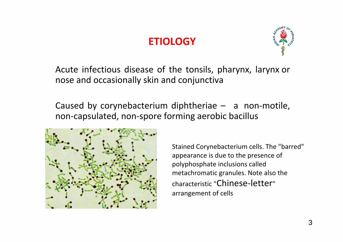
ETIOLOGY
Acute infectious disease of the tonsils, pharynx, larynx or nose and occasionally skin and conjunctiva
Caused by corynebacterium diphtheriae – a non‐motile, non‐capsulated, non‐spore forming aerobic bacillus
Stained Corynebacterium cells. The "barred" appearance is due to the presence of polyphosphate inclusions called metachromatic granules. Note also the characteristic "Chinese‐letter" arrangement of cells
3
EPIDEMIOLOGY
•Humans are the sole reservoirs•Spread by respiratory droplets and/or by contact with discharge from skin lesions•Incubation period is 2‐7 days•Although disease incidence has decreased worldwide with the immunization against diphtheria, it still remains endemic in developing countries due to less immunization coverage
4
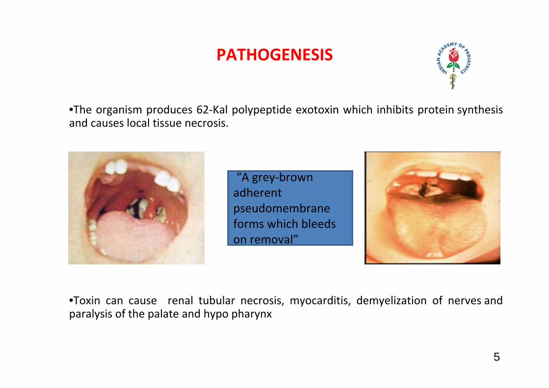
PATHOGENESIS
•The organism produces 62‐Kal polypeptide exotoxin which inhibits protein synthesis and causes local tissue necrosis.
•Toxin can cause renal tubular necrosis, myocarditis, demyelization of nerves and paralysis of the palate and hypo pharynx
“A grey‐brown adherent pseudomembrane forms which bleeds on removal”
5
CLINICAL FEATURES
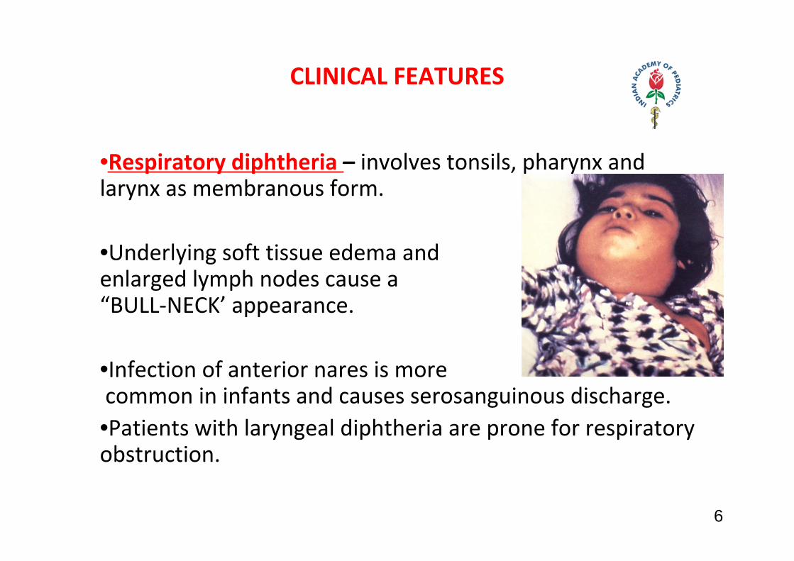
•Respiratory diphtheria – involves tonsils, pharynx and larynx as membranous form.
•Underlying soft tissue edema and enlarged lymph nodes cause a “BULL‐NECK’ appearance.
•Infection of anterior nares is more common in infants and causes serosanguinous discharge.•Patients with laryngeal diphtheria are prone for respiratory obstruction.
6
CONT…
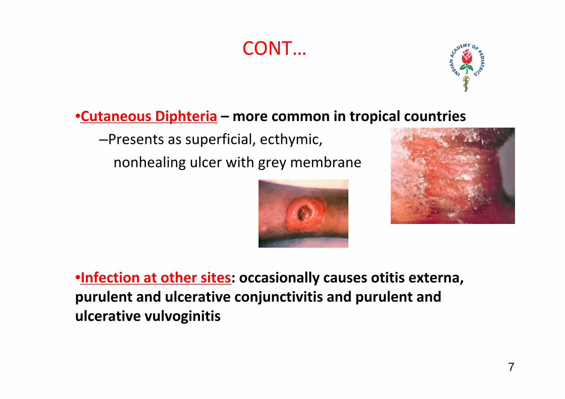
•Cutaneous Diphteria – more common in tropical countries–Presents as superficial, ecthymic, nonhealing ulcer with grey membrane
•Infection at other sites: occasionally causes otitis externa, purulent and ulcerative conjunctivitis and purulent and ulcerative vulvoginitis
7
•Toxic Cardiomyopathy – Occurs in 10‐20% of patients and is responsible for 50‐60% of deaths.
–Cardiac manifestations occurs in 2nd and 3rd week of illness but also can occur acutely in 1st week–Cardiac dysarrythmias, conduction disturbances, congestive cardiac failure can occur.
•Toxic NeuropathyParalysis of palate and hypopharynx will be
observed acutely in 2‐3 wk after the oropharayngeal implantaion
Systemic palyneuropathy may occur in 10 days 3 months after orophaysl infection
8
DIAGNOSIS
Specimens for culture should be obtained from the nose and throat and any other mucocutaneous lesions.
Special tellurite media is required
Laboratory criteria for diagnosisIsolation of Corynebacterium diphtheriae from a clinical specimen, or a fourfold or greater rise in serum antibody (but only if both serum samples are obtained before the
administration of diphtheria toxoid or antitoxin).
9
DIFFERENTIAL DIAGNOSIS
Exudative pharyngitis due to group A streptococcus , Epstein Barr virus and also Vincent angina
In case of laryngeal/ tracheal involvement ‐ to differentiate from acute epiglottis or ALTB
10
TREATMENT
•Specific antitoxin (equine antitoxin) is the mainstay of therapy and should be administered on the basis of clinical suspicion even before culture reports
Dose ranges: Pharyngeal or laryngeal disease of 48 hrs duration or less – 20,000 to 40,000 unitsNasopharyngeal lesions – 40,000 to 60,000 units Extensive disease ‐ 80,000 to 1,20,000 units
11
Antibacterial Therapy:
•Penicillin/Erythromycin given .•In severe cases PencillinG given IM/IV for 14 days•This is to stop toxin production and to eradicate C.diphtheria and to prevent transmission
12
IMMUNIZATION
•Active immunization to be undertaken during convalescent period•Universal immunization with diphtheria toxoid containing vaccine is the only effective control measure.•Three doses in the first year (6, 10, and 14), first booster at 16‐18 months, second booster at 5 years•If possible to continue Td at 10 years of age
13
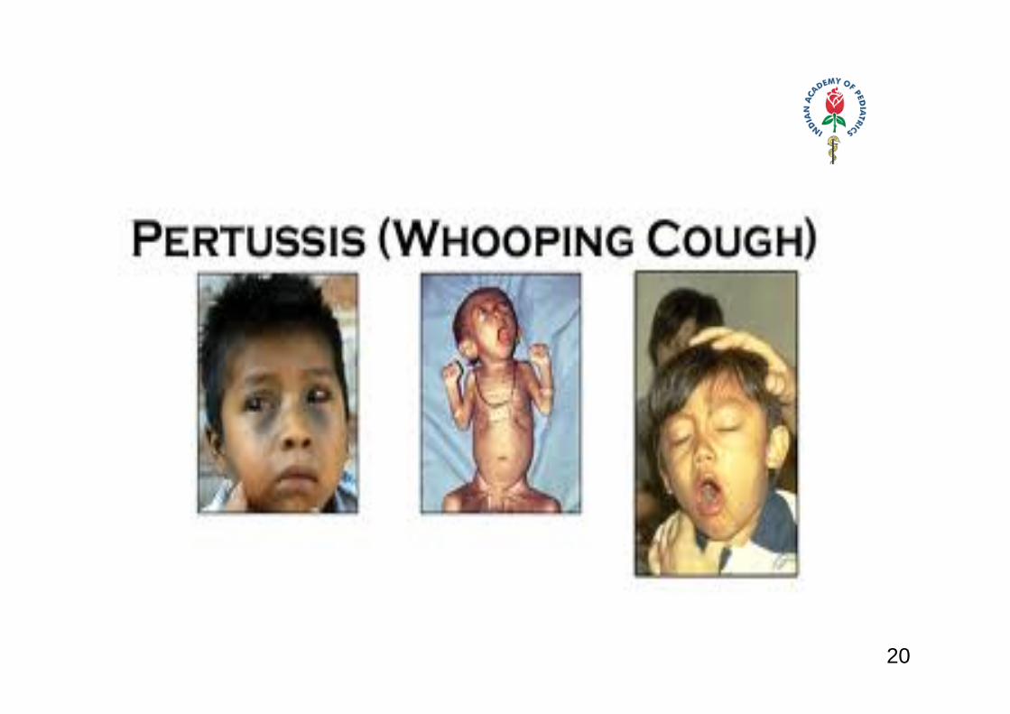
PERTUSSIS
14
ETIOLOGY & EPIDEMIOLOGY
•It is an acute respiratory infection caused by Bordetella Pertussis and occasionally B.Para pertussis characterised by Intense cough/whooping cough.•During the Pre‐vaccine era Pertussis was the leading cause of death due to communicable diseases .•Though there has been dramatic decline in the incidence with good coverage of pertussis vaccine in developed countries , in under developed countries, it is still endemic and sometimes epidemic due to low vaccine coverage .
15
Cont.
•Transmission by aerosol droplets and is highly contagious .•Organism does not survive in the environment for prolonged period.• No lifelong immunity with natural disease or vaccine .• Even with full immunization schedule , the immunity weans by 12 years.
16
B. pertussis
17
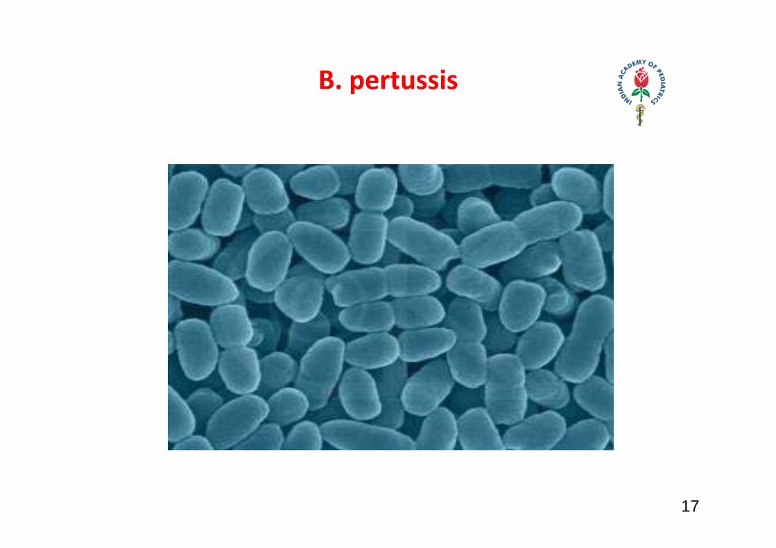
PATHOGENESIS
•B. pertussis is a tiny gram‐negative coccobacilli•Produces pertussis toxin (PT),filamentous hemagglutinin (FHA), fin type2 and 3 and pertactin (Pn),producing local epithelial damage in the respiratory system.
18
CLINICAL FEATURES
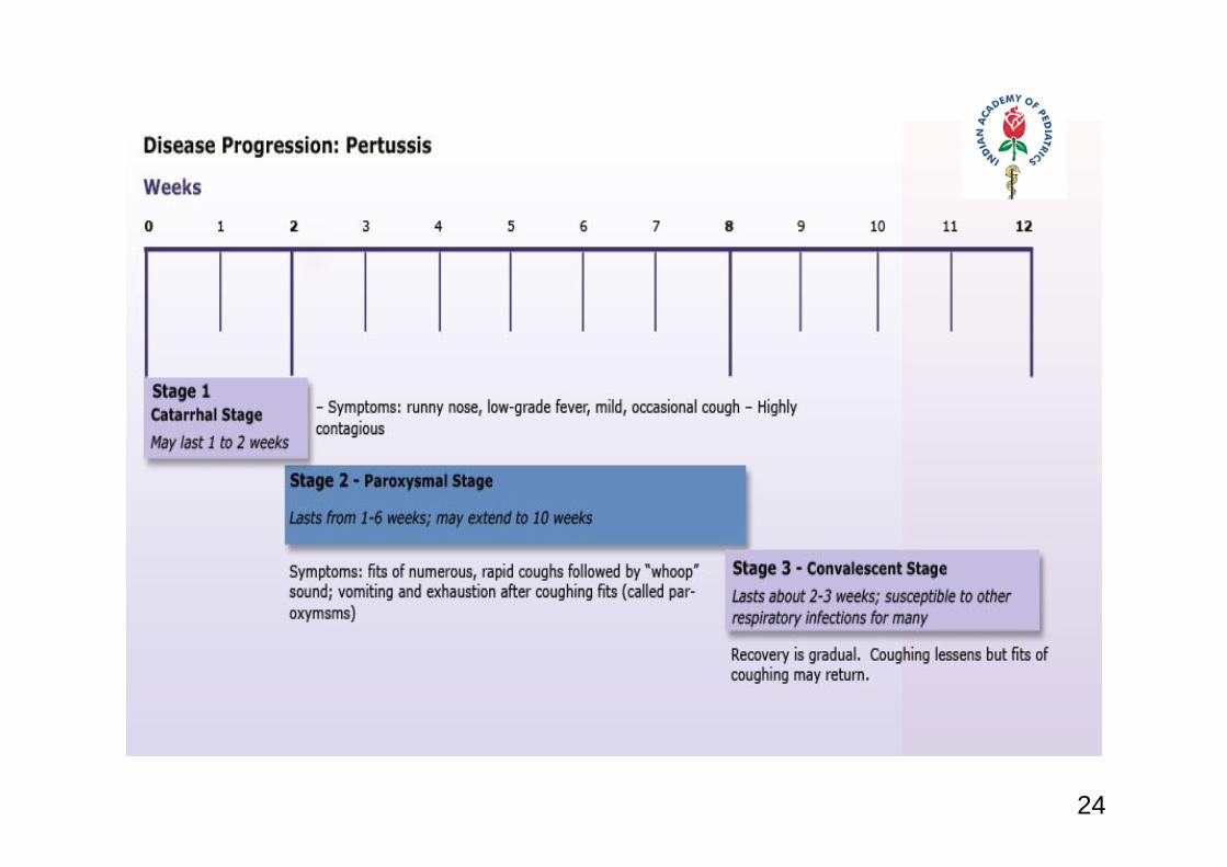
Divided into catarrhal, paroxysmal and convalescent stages
CATARRHAL STAGEcongestion, rhinorrhoea and conjunctival suffusion Begins after an incubation period of 3‐12 days.
19
20
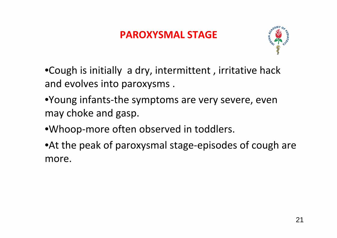
PAROXYSMAL STAGE
•Cough is initially a dry, intermittent , irritative hack and evolves into paroxysms .•Young infants‐the symptoms are very severe, even may choke and gasp.•Whoop‐more often observed in toddlers.•At the peak of paroxysmal stage‐episodes of cough are more.
21
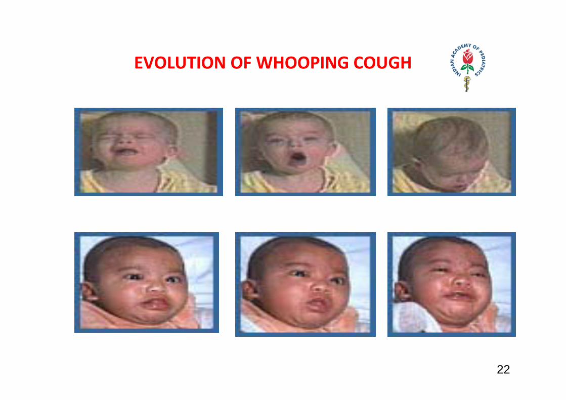
EVOLUTION OF WHOOPING COUGH
22
CONVALESCENT STAGE
Intensity and paroxyms of cough decrease gradually over 1‐4 weeksAppetite, general condition and health improves gradually
23
24
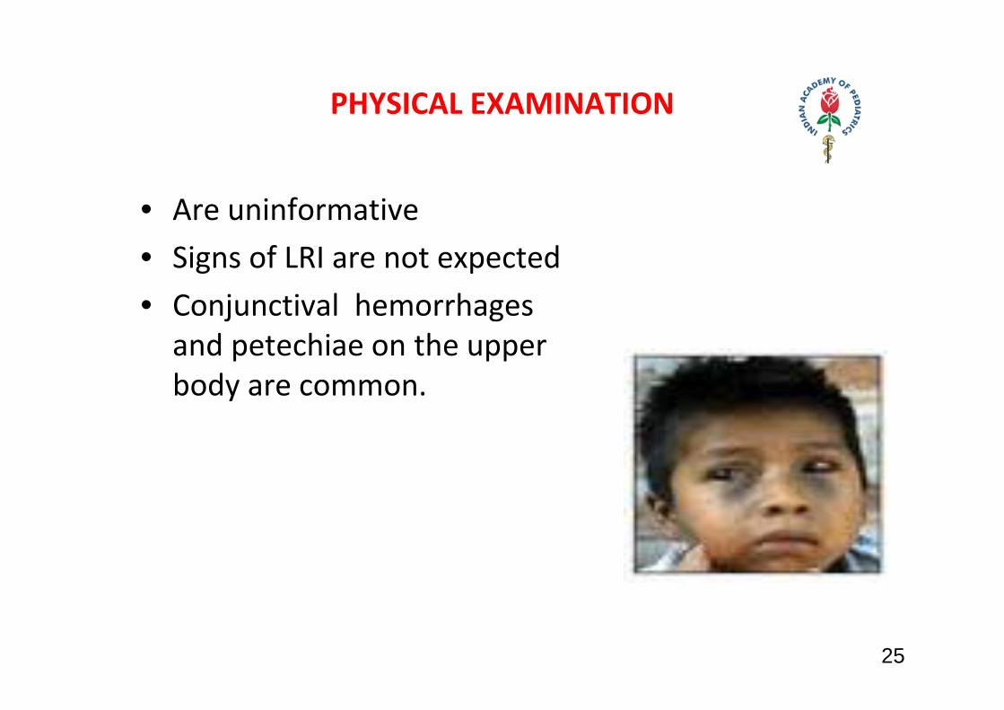
PHYSICAL EXAMINATION
• Are uninformative• Signs of LRI are not expected• Conjunctival hemorrhages and petechiae on the upper body are common.
25
DIAGNOSIS
•Suspected when intense cough WITHOUT fever, malaise,myalgia,tachypnoea,wheezes and rales •Leukocytosis (15,000‐1,00,000) due to absolute lymphocytosis is characteristic in initial stages •X‐ray chest‐mildly abnormal•Bacterial isolation from nasopharyngeal swabs or cough plate cultured on Bordet – Gengou medium
26
DIFFERENTIAL DIAGNOSIS
Other conditions which mimic protracted cough include:
Adenoviral, influenza and para influenza viral infections, mycoplasma, chlamydia, endobronchial TB, inhaled foreign body and reactive air way disease
27
COMPLICATIONS
•Infants <6 mo of age –excessive mortality and morbility •Respiratory –otitis media, pneumonia, atelectasis, emphysema, bronchiectasis, pneumothorax and pneumomediastinum•Neurological‐seizures and encephalopathy•Bleeding‐epistaxis, conjuntival hemorrhage •Rectal prolapse, inguinal hernia• Malnutrition•Flare up of tuberculosis and secondary bacterial pneumonias 28
TREATMENT
•Adequate nutrition, hydration and avoiding factors aggravating cough•Erythromycin (40‐50 mg/kg in three divided doses) for 14 days•Nebulization with salbutamol may be useful•No cough suppressants and antihistaminic agents•Younger infants may require hospitalization
29
PREVENTION
•Chemoprophylaxis with erythromycin for close family contacts
IMMUNIZATION WITH PERTUSSIS VACCINE•DTPw – 5 doses – three doses in first year (6, 10 and 14 weeks) and First booster at 16‐18 months and Second booster at 5 years•Not recommended beyond 7 years as adverse reactions are more with more doses•Contraindicated in progressive neurological diseases or with H/O severe side‐effects with previous doses
30

IAP UG Teaching slides 2015‐16
k You
31IAP UG Teaching slides 2015‐16 31