
Digital Speech Coding - Home | University of Pittsburgh
18
R E S E A R C H R E P O R T Occipital gamma activation during Vipassana meditation B. Rael Cahn • Arnaud Delorme • John Polich Received: 27 October 2009 / Accepted: 26 November 2009 Ó The Author(s) 2009. This article is published with open access at Springerlink.com Abstract Long-ter m Vipassana med itators sat in med i- tation vs. a control rest (m ind-wander ing) state for 21 min in a counterb alanced design with spontaneo us EEG reco r- ded. Meditation state dynamic s were measur ed with spec- tral decompos ition of the last 6 min of the eyes-closed silent med itation com pared to cont rol state. Medita tion was associa ted with a decrease in frontal delta (14 Hz) power, especia lly pronou nced in those partic ipants not reporting drowsi ness duri ng meditation. Relati ve increase in frontal theta (48 Hz) power was obser ved duri ng med itation, as well as signica ntly increas ed parieto-oc cipital gam ma (3545 Hz) power, bu t no othe r state eff ects wer e found for the theta (48 Hz) , alpha (812 Hz), or beta (1225 Hz) bands. Alpha powe r was sensi tive to condition order , and more experie nced meditat ors exhibited no tendency towar d enhanc ed alph a during meditat ion relative to the cont rol task. All participants tended to exhi bit decreased alph a in associa tion with reporte d drowsiness . Cross- experiment al session occi pital gam ma powe r was the greates t in med i- tators with a daily practice of 10 ? years, and the medita- tion-rel ated gamma power incr ease was similarly the stronges t in such advanced prac titioners. The nd ings sugges t that long-term Vipass ana meditation contribute s to increas ed occipital gamma powe r related to long-te rm meditat ional exper tise and enhanced senso ry awaren ess. Keywords Medita tion Á Electr oencephal ography (EEG) Á Gamm a Á Ment al sta te Á Altered state of consc iousness (ASC) Á Vipass ana Introduction The term meditat ion refers a set of diverse and specic methods of distinct attent ional engageme nt, and recent reports have begun to focus specica lly on state and trait measur es of the same. Altho ugh the gener al effects of meditat ion on neuro elect ric brain measur es are still b eing charact erized, consensus has emerge d that coher ence and/ or powe r for lower frequenc y spontaneo us elect roenceph- alograph ic (EEG ) act ivity is enhanc ed as both a trait and a state effect in man y forms of meditat ive prac tice (Cahn and Polich 2006 ). Early studies of med itators impli cated alph a (812 Hz) power incr eases as both a state and trait eff ect of Yogic, Zen, and Transcende ntal Medita tion prac tice (Anand et al. 1961a ; Kasamat su and Hirai 1966 ; Kasam atsu et al. 1957 ; Walla ce 1970 ; Wenger and Bagchi 1961 ). Later stud ies have failed to repl icate the early nding s of increas ed alpha in advanc ed practitioner s but have reporte d incr eased alpha coherence , especially in assa ys of TM practitio ners B. R. Cahn ( &) Division of Geriatric Psychiatry, Department of Psychiatry, University of California San Diego, 8950 Villa La Jolla Drive, Suite B-122, La Jolla, CA 92037, USA e-mail: rael@ucsd.edu A. Delorme Institute for Neural Computation, University of California San Diego, La Jolla, CA, USA A. Delorme CERCO, CNRS, Universite Paul Sabatier, 133 Route de Narbonne, 31062 Toulouse Cedex 9, France J. Polich Cognitive Electrophysiology Laboratory, Molecular and Integrative Neurosciences Department, The Scripps Research Institute, 10550 North Torrey Pines Road, La Jolla, CA 92037, USA e-mail: polich@scripps.edu 123 Cogn Process DOI 10.1007/s10339-009-0352-1
Transcript of Digital Speech Coding - Home | University of Pittsburgh

RESEARCH REPORT
Occipital gamma activation during Vipassana meditation
B. Rael Cahn • Arnaud Delorme • John Polich
Received: 27 October 2009 / Accepted: 26 November 2009� The Author(s) 2009. This article is published with open access at Springerlink.com
Abstract Long-term Vipassana meditators sat in medi-tation vs. a control rest (mind-wandering) state for 21 minin a counterbalanced design with spontaneous EEG recor-ded. Meditation state dynamics were measured with spec-tral decomposition of the last 6 min of the eyes-closedsilent meditation compared to control state. Meditation wasassociated with a decrease in frontal delta (1–4 Hz) power,especially pronounced in those participants not reportingdrowsiness during meditation. Relative increase in frontaltheta (4–8 Hz) power was observed during meditation, aswell as signi�cantly increased parieto-occipital gamma(35–45 Hz) power, but no other state effects were found forthe theta (4–8 Hz), alpha (8–12 Hz), or beta (12–25 Hz)bands. Alpha power was sensitive to condition order, andmore experienced meditators exhibited no tendency towardenhanced alpha during meditation relative to the control
task. All participants tended to exhibit decreased alpha inassociation with reported drowsiness. Cross-experimentalsession occipital gamma power was the greatest in medi-tators with a daily practice of 10? years, and the medita-tion-related gamma power increase was similarly thestrongest in such advanced practitioners. The �ndingssuggest that long-term Vipassana meditation contributes toincreased occipital gamma power related to long-termmeditational expertise and enhanced sensory awareness.
Keywords Meditation� Electroencephalography (EEG)�Gamma� Mental state� Altered state of consciousness(ASC) � Vipassana
Introduction
The term meditation refers a set of diverse and speci�cmethods of distinct attentional engagement, and recentreports have begun to focus speci�cally on state and traitmeasures of the same. Although the general effects ofmeditation on neuroelectric brain measures are still beingcharacterized, consensus has emerged that coherence and/or power for lower frequency spontaneous electroenceph-alographic (EEG) activity is enhanced as both a trait and astate effect in many forms of meditative practice (Cahn andPolich 2006).
Early studies of meditators implicated alpha (8–12 Hz)power increases as both a state and trait effect of Yogic,Zen, and Transcendental Meditation practice (Anand et al.1961a; Kasamatsu and Hirai1966; Kasamatsu et al.1957;Wallace 1970; Wenger and Bagchi1961). Later studieshave failed to replicate the early �ndings of increased alphain advanced practitioners but have reported increasedalpha coherence, especially in assays of TM practitioners
B. R. Cahn (&)Division of Geriatric Psychiatry, Department of Psychiatry,University of California San Diego, 8950 Villa La Jolla Drive,Suite B-122, La Jolla, CA 92037, USAe-mail: [email protected]
A. DelormeInstitute for Neural Computation,University of California San Diego,La Jolla, CA, USA
A. DelormeCERCO, CNRS, Universite Paul Sabatier,133 Route de Narbonne, 31062 Toulouse Cedex 9, France
J. PolichCognitive Electrophysiology Laboratory,Molecular and Integrative Neurosciences Department,The Scripps Research Institute,10550 North Torrey Pines Road,La Jolla, CA 92037, USAe-mail: [email protected]
123
Cogn ProcessDOI 10.1007/s10339-009-0352-1

(Gaylord et al.1989; Travis 1991; Travis and Pearson1999; Travis et al.2002), theta (4–8 Hz) power, especiallyin the assays of concentrative/focused attention practitio-ners (Aftanas and Golocheikine2001; Baijal and Sriniva-san2009; Hebert and Lehmann1977; Pan et al.1994), orgamma effects (Lehmann et al.2001; Lutz et al. 2004).Recent reports using LORETA to analyze EEG from Zen(Faber et al.2008) and Qi-Gong (Tei et al.2009) medita-tors further suggest that meditation may be associated withtrait increased frontal delta activity, possibly indexingbaseline relative inhibition of cognitive engagement andgreater detachment from ongoing daily experience. Gamma�ndings have been reported as either distinguishingbetween various meditative states in an advanced practi-tioner (Lehmann et al.2001), or as a state and trait markerfor meditation in advanced Tibetan Buddhist practitionersengaging in compassion meditation (Lutz et al.2004). Onelikely contributing factor to the inconsistency across stud-ies is the lack of standardization with respect to meditativestyle, assessment methodology, and consideration of stateeffects for beginning vs. short- vs. long-term meditators(Cahn and Polich2006). Toward this end, the present studywas designed to provide fundamental information usingEEG measures during meditation compared to control stateeffects in long-term Vipassana meditators.
Vipassana meditation and present study
Vipassana meditation is a Buddhist practice that involvesfocusing on present-moment sensory awareness with anequanimous and non-reactive mental set (Gunaratana2002;Hart 1987). This tradition has served as the foundation forcontemporary ‘‘mindfulness’’ meditation techniques such asthe widely practiced mindfulness-based stress reduction(MBSR) currently used for clinical interventions (Davidson2003; Grossman et al.2004; Kabat-Zinn 1982, 2003).Development of greater awareness of and concomitant non-reactivity to interoceptiveand exteroceptive sensory stimuliduring formal Vipassana/mindfulness meditation ishypothesized to enhance self-awareness such that selectiveadaptive responding is facilitated at the expense of auto-mated non-adaptive reactions, thereby promoting moresuccessful management of stressful life situations (Hart1987; Kabat-Zinn2003; Lutz et al.2007; Segal et al.2002).
Vipassana practitioners of the Theravadan Vipassanatradition were assayed in the present study, and themajority had been taught in the tradition of Goenka (Hart1987). This practice emphasizes attentional absorption insubtle somatosensory awareness and associated openmonitoring without mental or emotional reactivity to suchsensory experience. The speci�c Vipassana meditativetechnique involves attentional scanning of sensationsthroughout the body in an iterative and cyclic fashion,
scanning body sensations from the top of the head to thetoes and back again repeatedly, with the concomitantadoption of an attitude of detached observation and non-reactivity to any sensations and thoughts that may arise.
Systematic evaluation of meditation state in comparisonwith non-meditative thought conditions requires controlcognitive tasks, and this condition was implemented with theinstruction to let the mind wander freely through non-emo-tional thoughts and memories. This state was also chosen tomimic the aspects of the mind-wandering ‘‘default mode’’state thought to have high ecological validity to a commonmode of cognitive engagement in normal everyday life(Christoff et al.2009; Smallwood and Schooler2006).
Given that Vipassana meditation practice is thought toenhance the awareness of internal and external stimuliwhile reducing automated reactivity, it was hypothesizedthat increased frontal theta and alpha would be observedduring meditation—i.e., re�ecting increased purposefulattentional engagement and mental quiescence. Based onthe view that Vipassana practice may promote anenhancement of sensory awareness through increasedattentional engagement, we hypothesized that increasedgamma activity would also be observed in meditation rel-ative to the control condition, possibly in bilateral centro-parietal and/or frontal areas, related to enhanced processingin frontal and somatosensory cortices. Recording condi-tions were designed to capture the time period when themeditation state was deep and stable and therefore mostlikely to contrast with the control state.
Methods
Participants
A total of N = 16 Vipassana meditators (F = 5, M = 11)were assessed (M = 45.5, SD= 9.8, 24–56 years). As agroup, these individuals had been meditating for a con-siderable period of time (M = 20.0, SD= 12.1, 2.5–40 years), and all had been meditating daily (7 days/week)for at least 1 year (M = 13.0, SD= 10.7, 1–30 years),with at least 0.5? h or more each day (M = 1.3, SD= 0.7,0.5–3 h). Participants were recruited from a local Vipas-sana meditation community through word of mouth ande-mail and compensated $40 for the 3-h study.
Recording conditions
EEG data were collected using a 19-channel ECI electrodecap from the following locations: Fp1, Fp2, F3, F4, F7, F8,Fz, C3, C4, T7, T8, Cz, P3, P4, P7, P8, Pz, O1, and O2.These scalp locations were referenced to linked earlobes,with the ground at the forehead. Eye-movement (EOG)
Cogn Process
123

activity was assessed with electrodes placed at the outercanthi and above/below the left eye in line with the pupilfor horizontal and vertical EOG monitoring using bipolarreference. Impedances were kept below 10 kX. The signalswere recorded with a band pass of 0.01–70 Hz (6 dBoctave/slope) and digitization rate of 256 Hz.
Procedure
The participants were instructed to sit on cushions andmeditate within the Theravadan Vipassana meditation tra-dition or engage in the control neutral thinking state, withthe order of the tasks counterbalanced across individuals.Participants were instructed to sit in the same posture forboth the meditation and control task periods of recordingand were �tted with headphones at the outset of therecording session that they wore throughout the recording.Pilot testing indicated that some participants found it dif-�cult to refrain from engaging in their meditative practicewhen sitting in the meditative posture with eyes-closed.Participants were therefore told to think about emotionallyneutral past events if they noticed themselves slipping intomeditative practice state, otherwise to let their mind wan-der freely through non-emotional neutral thoughts. Thiscontrol cognitive engagement was chosen to emulate a‘‘mind-wandering’’ state with high ecological validity thatcan be contrasted with the purposeful attentional engage-ment of the meditation state (Christoff et al.2009; Small-wood and Schooler2006). Participants were informed thatafter 21 min of eyes-closed meditation or control thinkingthey would hear a series of tones over the headphones andthat they were to simply continue their meditation orcontrol cognitive engagement. Both passive and activepresentations of simple tone stimuli were collected, withthose data reported elsewhere (Cahn and Polich2009).
At the conclusion of the �rst recording period, the par-ticipants were given the opportunity to stand and stretchbefore taking the same posture and seating position for thesecond recording of equal length. Immediately after eachof the two recordings participants completed a short formindicating whether they experienced drowsiness or sleeponset during the experimental recording session and ratingthe depth of meditative experience on a 1–10 scale, with 1indicating the normal waking and 10 indicating the deepestmeditative absorption ever experienced.
EEG analysis
We focused the present analysis on the last 6 min of datafrom the 21 min recording period so as to assess themeditative state achieved after allowing an adequate periodof time for the participants to fully absorb themselves intothe meditation state. Given that the spontaneous brain
rhythms generated during prolonged periods of eyes-closedrest tend to �uctuate and vary with factors such as reducedarousal and drowsiness, we did not average across thewhole 21 min epoch. Future planned analysis will focus onthe temporal evolution of brain dynamics in the meditationvs. control state so as to assess the onset vs. maintenance ofmeditation state effects and the relative stability of thespectral power dynamics across the two states. The last6 min of EEG data from each of the two 21 min recordingperiods were �rst visually inspected, and transient muscle-and movement-related artifacts were removed. The datawere subsequently high-pass �ltered at 0.5 Hz usingFIR �lter (Rabiner and Gold1975). The extended ICAalgorithm was then run on the data using the runica algo-rithm implemented within EEGLAB running on Matlab(Delorme and Makeig2004; Delorme et al.2007). Theresultant independent components accounting for horizon-tal and vertical eye movements were then marked andremoved from the data (Jung et al.2000a, b) as detailed inthe next section.
After removal of eye-movement-related artifact, datawere segmented into non-overlapping 2-s artifact-freeepochs. For the meditation and control states, means of170.4± 13.8 and 164.1± 26.6 epochs were obtained. TheThomson multi-taper spectrum estimator (Matlab PMTMfunctions using time-bandwidth product of 4 and FFTlength of 512) was applied to the cleaned continuous datafor spectral decomposition (Thomson1982). The outputpower values inlV2 were then log-transformed to dB unitsusing 109 log10 (lV2) formula to normalize the powervalue distributions.
Statistical analyses of the spectral power data were �rstapplied to signals from all scalp channels using the boot-strap method (Wilcox2005) and using false discovery rate(FDR) correction for multiple comparisons (Benjamini andYekutieli 2001). Statistical assessments were conductedusing analyses of variance with the factors ofstate (med-itation vs. control) andelectrode in the a priori regions ofinterest (midline electrodes for all frequencies, occipitalelectrodes for alpha). ANOVAs also were conducted on thefrontal electrodes (F3 and F4) for delta and the occipitalelectrodes (O1 and O2) for gamma activity, as these wereregions of statistical signi�cance between states identi�edby the initial bootstrap statistical testing using FDR cor-rection for the scalp data (see Fig.2). Greenhouse-Geissercorrections were applied to the degree of freedom (df) tocorrect for violations of the sphericity assumption, andTukey post hoc means comparisons were used to decom-pose reliable interactions.
Covariate ANOVAs were conducted using subjectvariables related to the order of engaging in meditationand control states, relative meditative expertise, and self-reports of drowsiness and meditative depth during
Cogn Process
123

experimental conditions. Bootstrap statistics and scalpmap plotting were performed using the EEGLAB Matlabsoftware (Delorme and Makeig2004; Delorme et al.2007) and custom Matlab scripts. Parametric statisticswere run using the Statistica software and EEGLAB(Delorme and Makeig2004; Delorme et al.2007). Theuse of bootstrap statistics was employed as it allows formore robust statistical inference than standard parametricstatistics since no assumption is made about the proba-bility distribution at the population level (Wilcox2005).However, studying complex patterns of covariate inter-action is not yet available using bootstrap procedures incommon statistical software, so that parametric statisticswere employed.
Independent component analysis (ICA)
In addition to the statistical assessments, a parallel analysisusing independent component analysis (ICA) of threeclasses of independent components from each subject wasconducted: (1) eye-movement artifact, (2) temporal muscleartifact, and (3) occipital alpha power. For each subject,vertical eye-movement-related components were selectedbased on their characteristic scalp projection and a smoothexponentially decreasing spectrum (Delorme et al.2007;Jung et al.2000a). This approach was employed by iden-tifying the EOG artifacts by simultaneously examining theeye channels (VEOG and HEOG) for activity to verify thatthe components were active only during those time periodswhen eye channels indicated eye movement. Musclecomponents produce component topographies with focalactivities over speci�c channels, typically located tempo-rally (T7 and T8), with a characteristic spectral signaturecontaining strong spectral power over 20–30 Hz and anerratic spectrum (Jung et al.2000a). Alpha occipital com-ponents were based on the 8–12 Hz frequency peak in theoccipital areas, which was also associated with an addi-tional peak at 20 Hz. These analyses are illustrated in thefollowing paragraphs.
The spectrum for the activity of these components wasthen computed on the independent components during boththe meditation and the control periods with the sameThomson multi-taper spectrum estimator used on thechannel data (Thomson1982). Traditional t-tests werecomputed with bootstrap statistics with FDR correction formultiple comparisons, which were conducted between thepower outcomes in the meditation vs. control states foreach of the three classes of independent components at allfrequencies (including gamma). This approach helped toassess whether eye or muscle gamma activity was greaterin meditation state and whether the occipital alpha inde-pendent components replicated the occipital scalp channelgamma effect.
Results
Participants and self-report scales
The ‘‘depth of meditative state’’ from the (1–10) self-reportscale indicated that the mean meditative depth experiencedduring the rest state was 1.7± 1.4, and meditative depthexperience during the meditative state was 4.5± 1.4 (t-test, df = 15, P = 0.00004). Drowsiness was reported by7 of the 16 participants during the meditation and 10 of the16 during the control thought condition. There was noreliable correlation between the self-reported depth ofmeditative state and either the number of years of dailypractice (r = 0.24, P = 0.36) or the number of hours ofdaily practice (r = -0.02, P = 0.93). A reliable correla-tion was obtained between the order of experimental ses-sion and the self-reported experience of drowsiness duringthe control state wherein those individuals meditating �rstwere less likely to experience drowsiness during the con-trol state (r = 0.52,P = 0.041). No similar correlation wasobtained between session order and drowsiness duringmeditation (r = 0.38,P = 0.15) (see also Cahn and Polich2009).
A negative correlation between the number of years ofdaily practice and reported drowsiness during the controlstate (r = -0.60, P = 0.015) was found, but not betweennumber of years of daily practice and reported drowsinessduring meditation (r = -0.006,P = 0.98). These �ndingsimply that individuals with more years of daily meditationpractices were less likely to report drowsiness speci�callyduring the control cognitive condition. A negative corre-lation between current number of hours of daily practicereported and drowsiness during meditative state also wasobtained (r = -0.59, P = 0.016). No association wasobserved for the control state drowsiness and numberof hours of daily practice (r = 0.02, P = 0.93), implyingthat greater current number of hours of current dailypractice predicted decreased drowsiness speci�cally duringmeditation.
EEG
Figure1 illustrates the mean amplitude spectral dataaveraged across meditation and control thought states withgrand average scalp maps for each of the major bandsrepresented for delta (1–4 Hz), theta (4–8 Hz), alpha (8–12 Hz), beta (12–25 Hz), and gamma (35–45 Hz). Pre-liminary analyses across all electrodes using bootstrapstatistics and FDR correction for multiple comparisonsindicated signi�cant meditation state effects for delta andgamma log spectral power, but no such effects for theta,alpha, and beta power. Scalp maps for the two states arethus only shown separately for delta and gamma bands, and
Cogn Process
123
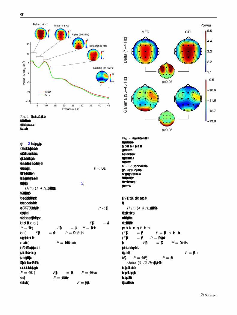
Fig. 2 shows the spectral power grand average scalp mapsfor meditation and control states separately at both fre-quencies. Also indicated is a scalp map of the statisticalsigni�cance for the comparison between the log spectralpower at each electrode across the two states (all redelectrode locations indicate signi�cance atP \ 0.05 viabootstrap with FDR correction). Delta decreases were inbilateral frontal regions, and gamma increases were inparieto-occipital areas only (see Fig.2).
Delta (1–4 Hz). As mentioned above, bootstrap analysiswith FDR corrections for multiple comparisons analysis ofthe sensor data indicated decreased delta power duringmeditation relative to control period at bilateral frontalelectrodes (F3, F4, F7, F8, C3, and C4, allP \ 0.05).Parametric repeated-measures ANOVA analysis of the datawas carried out as well, indicating that delta power atmidline electrodes yielded no state (F(1,15) = 1.84,P = 0.19), electrode (F(2,30) = 1.0, P = 0.37), or inter-action (F(2,30) = 0.01, P = 0.99) effects, althoughsomewhat greater delta power in control than meditationstate was observed (P = 0.19). Frontal delta power atelectrodes F3 and F4 was analyzed separately as deltaactivity at these electrodes was shown to be most signi�-cantly decreased in the bootstrap analysis of the scalp data.ANOVA analysis of the delta power at F3 and F4 dem-onstrated an effect for meditation state just missing theP = 0.05 cut-off (F(1,15) = 4.09, P = 0.06), with noeffect for electrode (P = 0.97), and no interaction betweenelectrode and state was observed (P = 0.67). Analyses of
FP1, F7, FP2, and F8 produced the same pattern of�ndings.
Theta (4–8 Hz). Bootstrap analysis with FDR correctionfor multiple comparisons indicated no main effect of stateat any electrodes for theta power during meditation relativeto control period. Parametric ANOVA assessment of thetapower also yielded no signi�cant effect of state,(F(1,15) = 0.037, P = 0.85) or electrode location(F(2,30) = 0.96,P = 0.39). A signi�cant state x electrodeinteraction, F(2,30) = 7.75, P = 0.002, indicated withpost hoc evaluation that theta power exhibited relativeincrease during meditation state at Fz (P = 0.006), but notat Cz (P = 0.99) or Pz (P = 0.85).
Alpha (8–12 Hz). Bootstrap analysis with FDR correc-tion for multiple comparisons indicated no main effect ofstate at any electrodes for alpha power during meditationrelative to control period. Parametric ANOVA assessmentof alpha power at midline electrodes likewise produced no
Fig. 1 The power spectrum for electrode Cz is displayed for bothmeditation and control states. Grand average scalp maps across statesare displayed for each of the major frequency bands so as to indicatescalp topography of the various frequencies
Fig. 2 The scalp maps for delta (1–4 Hz) and gamma (35–45 Hz)bands are displayed with meditation on the left and control task on theright. Both bands showed a statistically signi�cant differencecomparing meditation and control states and the scalp map indicatingstatistical signi�cance is shown below. Statistical signi�cance wasdetermined using bootstrap statistics with false detection rate (FDR)correction for multiple comparison with the threshold signi�cance setat P \ 0.05, indicating bilateral frontal decreases in delta power(signi�cant at F3, F4, F7, F8, C3, and C4) and parieto-occipitalincreases in gamma power (signi�cant at P7, P8, O1, and O2) asmeditation state effects. Similar analyses run on the theta, alpha, andbeta bands indicated no statistical differences between states at anyscalp site and are thus not shown
Cogn Process
123

meditation state effect (F(1,15) = 0.096, P = 0.76),although a main effect of electrode was found,F(2,30) =
18.8,P = 0.000005, con�rming that alpha was the greatestat more posterior sites as expected. No interaction betweenstate and electrode was obtained (F(2,30) = 1.1, P =
0.34). Post hoc testing con�rmed that alpha power wasgreater at Pz than Fz (P = 0.0001) and Cz (P = 0.004),and greater at Cz than Fz (P = 0.04).
A separate ANOVA analysis of alpha power at occipitalsites (O1 and O2) was conducted to assess alpha power atthe area of maximal power. No state (F(1,15) = 0.055,P = 0.82), electrode (F(2,30) = 1.5, P = 0.24), or inter-action was found (F(2,30) = 0.55, P = 0.47). Additionalseparate analyses of low (8–10 Hz) and high (10–12 Hz)alpha indicated the same pattern of �ndings.
Beta (12�25 Hz). Bootstrap analysis with FDR correctionfor multiple comparisons indicated no main effect of state forbeta power during meditation relative to control period.Analysis of beta power at the midline electrodes indicated nostate (F(1,15) = 0.62,P = 0.44) or interaction (F(2,30) =
1.4, P = 0.26) effects, but a main effect of electrode wasfound,F(2,30) = 4.33,P = 0.02, indicating that beta powerwas greater at more posterior sites. Post hoc assessmentfound beta power at Pz was greater than at Fz (P = 0.03), butnot signi�cantly different from Cz (P = 0.92); beta power atCz was trend-level greater than at Fz (P = 0.07). Additionalseparate analyses of low (12–15 Hz), medium (15–18), andhigh (18–25 Hz) beta indicated the same pattern of �ndingsand are not presented separately.
Gamma (35–45 Hz). Bootstrap analysis with FDR cor-rection for multiple comparisons indicated a main effect ofstate with increased gamma power during meditation rel-ative to control period at P7 and P8 (bothP \ 0.05), aswell as O1 and O2 (bothP \ 0.01). Parametric ANOVAanalysis of the gamma power at midline electrodes dem-onstrated no state (F(1,15) = 1.26, P = 0.28), electrode(F(2,30) = 2.22, P = 0.14), or interaction (F(2,30) =
1.35, P = 0.27) effects. Additional analysis of gammapower was conducted speci�cally at occipital electrodes(O1, O2) because of the greater gamma power found at thislocation in the bootstrap/FDR analysis. Analysis of occip-ital gamma power at O1 and O2 yielded a signi�cant stateeffect, F(1,15) = 9.32, P = 0.008, but no electrode(F(2,30) = 2.02,P = 0.18) or interaction (F(2,30) = 0.59,P = 0.45) effects. Additional analyses including P7 and P8in the ANOVA indicated the same pattern of �ndings, butas the effect was the greatest at O1 and O2, only these dataare presented.
Independent component analyses (ICA)
Figures3, 4, 5 illustrate the grand average scalp maps ofthe independent components of the three EOG classes or
clusters: Fig.3 illustrates those accounting for vertical andhorizontal eye movements. Figure4 illustrates thoseassociated with the left and right temporalis muscleactivity. Figure5 illustrates the occipital alpha cluster ofcomponents. Each subject yielded one to two eye-move-ment-related components and one to two occipital alphacomponents for a total of 23 components for both the alphaand eye-movement clusters. Additionally, one to six mus-cle components were identi�ed per subject for a total of 37components in the muscle cluster.
Bootstrap statistics with FDR correction across fre-quencies 1–55 indicated that no difference in spectralfrequency power was present between states for either theeye- or the muscle-independent component clusters (seeFigs.3 and 4). A statistically signi�cant increase ingamma ([25 Hz) power was found in the occipital alphaindependent components (see Fig.5, gray area at bottomindicates the area of statistical signi�cance comparingbetween states). Analysis of the speci�c 35–45 Hz
Fig. 3 Panel A indicates the grand average scalp map for theleft andright muscle-independent components, respectively.Panel B presentsthe grand average power spectra for the muscle components for bothmeditation and control states. Inpanel C, the thin, colored linesindicate difference in power across the range of frequencies for themuscle components; meditation minus control period for all subjects(when more than one component was present for a given subject, thepower spectrum for these components was averaged). Thebold, blacktrace indicates the grand average spectrum difference for the contrastmeditation minus control such that segmentsabove 0 indicate greateraverage component activity during meditation andbelow 0 indicategreater average component activity during control period
Cogn Process
123

frequency gamma band activity for each independentcomponent (IC) class comparing meditation to controlstate activity indicated that independent components dueto muscle (t-test, df = 15, P = 0.55) and eye (t-test,df = 15, P = 0.83) demonstrated no difference betweenmeditation and control states. Occipital alpha IC gammaactivity was signi�cantly greater in meditation relative tocontrol state using bootstrap statistic with FDR correctionfor multiple comparisons (bootstrap,df = 15, P =
0.0075), but only marginally different between statesusing parametric statistics (t-test, df = 15, P = 0.07), inany case mirroring the signi�cant difference observed ingamma power at the occipital scalp electrodes. Analysiswas applied to the delta power for the eye-movement ICsas this activity re�ects typical slow eye movements. Lesseye-movement activity was found for the meditationcompared to control state, with a marginal parametricdifference obtained (t(1,15) = 3.05, P = 0.10), and asigni�cant non-parametric (Wilcoxon sign test) outcomeobtained (P = 0.006).
Additional analyses
Correlations were calculated to explore the relationshipsbetween both the eye-movement-related IC delta power(eye) and scalp-recorded delta power as well as betweengamma power (muscle, eye) and scalp-recorded gammapower, so as to assess possible non-cortical sources for theobserved state effects of meditation. Speci�cally, correla-tions were computed between the gamma power in theeye- and muscle-independent components and the scalp-recorded gamma power across midline as well as occipitalelectrodes in each experimental state. To assess the pos-sibility that slow eye-movement differences betweenmeditation and control state might contribute to theoccipital gamma �ndings, correlations between eye-movement independent component delta power andgamma power at scalp channels were also computed. Asthe increased delta activity observed at fronto-laterallocations might be related to eye-movement activity not
Fig. 4 Panel A indicates the grand average scalp map for vertical andhorizontal eye-movement independent components, respectively.Panel B presents the grand average power spectra for the eye-movement components for both meditation and control states. Inpanel C, thethin, colored lines indicate the difference in power acrossthe range of frequencies for the eye-movement components betweenmeditation and control period for all subjects. Thebold, black traceindicates the grand average spectrum difference for the contrastmeditation minus control such that segmentsabove 0 indicate greateraverage component activity during meditation andbelow 0 indicategreater average component activity during control period
Fig. 5 Panel A indicates the grand average scalp map for theoccipital alpha independent components.Panel B presents the grandaverage power spectra of the occipital alpha components in medita-tion and control states. Inpanel C, the thin, colored lines indicate thedifference in power across the range of frequencies for the occipitalalpha components between meditation and control periods for allsubjects. Thebold, black trace indicates the grand average spectrumdifference for the contrast meditation minus control. Thegray bar atthe bottom of the �gure indicates frequencies over which bootstrapstatistics with FDR correction for multiple comparisons indicatedstatistically signi�cant greater values in meditation than control state(25–55 Hz)
Cogn Process
123

fully removed from the data, additional correlations wererun between delta power in the eye-movement independentcomponents and delta power in the (post eye-movementindependent component removal) midline and frontalchannel data.
There were no correlations between delta activity in theeye-movement IC’s and midline or frontolateral electrodesin either meditation or control states: meditation—Fz,r =
0.044, P = 0.87, Cz, r = 0.00, P = 1.00, Pz,r = 0.16,P = 0.56, F3, r = 0.22, P = 0.42, F4, r = 0.084,P = 0.76; control—Fz, r = -0.17, P = 0.52, Cz, r =
-0.087, P = 0.75, at Pz, r = -0.051, P = 0.85, F3,r = 0.094,P = 0.73, F4,r = 0.016,P = 0.95.
In contrast, signi�cant correlations between gammapower in the muscle IC cluster and gamma power atmidline electrodeswere obtained for both state conditions:meditation—Fz,r = 0.61, P = 0.013, Cz,r = 0.60, P =
0.014, Pz,r = 0.47, P = 0.066; control—Fz,r = 0.55,P = 0.028, at Cz,r = 0.46, P = 0.073, at Pz,r = 0.32,P = 0.22. Signi�cant correlations between gamma powerin the eye-movement IC cluster and gamma power atmidline electrodes were also obtained for the controlcondition with trends observed also at Fz in meditation:meditation—Fz,r = 0.50, P = 0.058, Cz,r = 0.41, P =
0.13, Pz, r = 0.081, P = 0.78; control—Fz, r = 0.82,P = 0.0001, Cz, r = 0.63, P = 0.012, Pz, r = 0.54,P = 0.039. Importantly, no correlations were foundbetween gamma power at occipital electrodes andgamma power in the muscle IC cluster (meditation—O1,r = 0.11,P = 0.69, O2,r = 0.17,P = 0.53; control—O1,r = -0.061,P = 0.82, O2,r = -0.093,P = 0.73) or theeye-movement IC cluster (meditation—O1,r = -0.04,P = 0.89, O2, r = 0.014, P = 0.96; control—O1,r = 0.38, P = 0.17, O2,r = 0.19, P = 0.49). The corre-lations between the occipital alpha IC gamma activity andscalp-recorded gamma power were of moderate signi�-cance at midline electrodes: meditation—Fz,r = 0.42,P = 0.104; Cz, r = 0.38, P = 0.15; Pz, r = 0.47,P = 0.064; control—Fz, r = 0.50, P = 0.048; Cz,r = 0.60, P = 0.013; Pz, r = 0.70, P = 0.003. Thesecorrelations were, however, quite signi�cant at occipitalelectrodes: (meditation—O1,r = 0.77, P = 0.0001, O2,r = 0.71,P = 0.002; control—O1,r = 0.79,P = 0.0001,O2, r = 0.75,P = 0.001.
Covariate analyses
Additional analyses were conducted using covariates tocharacterize individual differences underlying the spectralpower �ndings. The primary covariates were those relatingto order of experimental conditions, reported drowsinessduring the experimental sessions, and intensity of medita-tion practice (number of years of daily meditation practice,
current number of hours/day of meditation practice). Sig-ni�cant interactions for some of these covariates wereobtained for the delta, alpha, and gamma bands, with noreliable effects obtained for theta or beta power.
Delta
A signi�cant interaction among state, order of experi-mental sessions (meditation? control vs. control?meditation), and midline electrode location was found,F(2,28) = 4.70, P = 0.017). This outcome suggestsdecreased midline delta power during meditation relativeto rest speci�cally for those participants doing the controlperiod prior to the meditation period, but not thosemeditating �rst. Breakdown of this interaction with Tukeypost hoc testing indicated that when the control periodoccurred �rst, midline delta power was decreased in thesubsequent meditation session at Fz (P = 0.0004) and Cz(P = 0.027) but not Pz (P = 0.30). No differences amongmidline electrodes for delta power were found in partic-ipants meditating �rst (Fz,P = 1.0; Cz, P = 0.77; PzP = 0.15). A second covariate interaction was found forthe state9 reported drowsiness9 midline electrodelocation during meditation,F(2,28) = 3.39, P = 0.06),indicating that only those subjects not reporting drowsi-ness during meditation showed a tendency for decreasedmidline delta power in meditation. No signi�cant out-comes were found for state effect at lateral-frontallocations.
Alpha
The interaction among order of experimental session xstate effects x midline electrode location was signi�cant,F(1,14) = 5.31,P = 0.037. For the occipital electrodes theorder9 state interaction was reliable,F(1,14) = 4.00,P = 0.065, with both such interactions indicative thatwhichever experimental period occurred second in theexperimental order scheme tended to produce more alphapower (See Fig.6a). The interaction among state x medi-tation daily practice was trend-level signi�cant for themidline, F(1,14) = 3.89, P = 0.068, as well as occipitalelectrodes,F(1,14) = 3.99, P = 0.066. These outcomessuggested that more years of meditative practice tended tocorrelate with slight decreases in alpha power duringmeditation, whereas fewer meditation practice years cor-related with trend increases in alpha power during medi-tation (See Fig.6b). The state9 drowsiness9 electrodeinteraction was marginally signi�cant,F(2,28) = 3.06,P = 0.071, such that participants reporting drowsinessduring the control period tended to produce decreasedalpha in the control relative to meditation period, with theopposite pattern obtained for those not reporting such
Cogn Process
123

drowsiness. The same but weaker interaction was observedfor occipital alpha power (F(2,28) = 2.44,P = 0.14).
Gamma
A number of covariates produced signi�cant �ndings forboth the midline and occipital gamma power (reporteddrowsiness, number of hours of current daily meditationpractice). Given the fact that the muscle IC cluster gammapower correlated with the gamma power at midline elec-trodes and that the same pattern of signi�cant �ndingsobtained with midline gamma covariate analysis obtainedwith muscle IC gamma covariate analysis, these associations
are likely due to muscle rather than brain reactivity and arenot reported.
No reliable outcomes were obtained when the self-reportscore for meditative depth between meditation and controlsessions was used as a covariate for gamma power. Cate-gorization of participants into those with a history of10? years (n = 10, M = 19.3, SD= 8.6 years) and thosewith \10 years (n = 6, M = 2.5, SD= 1.4 years) of dailymeditation practice yielded signi�cant interactions foroccipital gamma. The long-term meditator category yiel-ded a signi�cant main effect,F(1,14) = 4.87, P = 0.044,indicating that the sub-group reporting more years of dailymeditation practice exhibited greater gamma power acrosstask conditions (See Fig.7a, b). In addition, an interactionamong state9 daily meditational practice length9 elec-trode was found,F(1,14) = 4.53, P = 0.05. Speci�cally,gamma power was increased in meditation vs. control statein long-term meditators (O1,P = 0.0002; O2,P = 0.008)but only marginally in relatively short-term meditators (O1,P = 0.76; O2,P = 0.09). The level of drowsiness9 stateinteraction was also reliable,F(1,14) = 5.41, P = 0.035.Gamma power increases were signi�cant only for thoseparticipants not reporting drowsiness during rest (Tukeypost hoc comparisonP = 0.0069) and were absent for thegroup who did report drowsiness (Tukey post hoc com-parisonP = 0.57). In contrast, the self-reported drowsinessduring meditation state did not show a signi�cant interac-tion with gamma.
Discussion
Meditation and neuroelectric measures
Vipassana meditative practice involves the adoption of amindful and receptive mental awareness, with attentionalabsorption on present-moment sensations in the body andmeta-cognitive reframing of ongoing experience asimpersonal phenomena to be observed but not reacted to(Gunaratana2002; Hart 1987; Lutz et al. 2007). EEGmeasures were obtained from experienced Vipassanameditators with conditions that contrasted the meditativestate with a control cognitive condition designed to mimic‘‘everyday thinking.’’ The pattern of meditation-inducedincrease in parieto-occipital gamma activity, concomitantdecrease in frontal delta power, and a shift to a more frontaldistribution of theta activity suggests that sensory pro-cessing and cognitive processing were altered duringmeditation relative to the control state. However, the typ-ically reported meditation state changes in the theta andalpha frequencies were not found (Cahn and Polich2006).
Delta (1–4 Hz) power is known to correspond withinhibitory functions (Babiloni et al.2006; de Jongh et al.
Fig. 6 a Experimental order and alpha power. Participants undergo-ing the control period followed by the meditation period tended toevince greater alpha power during meditation, whereas thoseundergoing the meditation period followed by the control periodtended to evince greater alpha power during the control period.b Meditators with less than 5 years of daily meditation practice(n = 6, M = 2.5, SD= 1.4) vs. meditators with 10 and greater yearsof daily meditation (n = 10, M = 19.3, SD= 8.5) practice showeddifferent tendencies with regard to alpha power across experimentalsessions, with shorter-term practitioners showing a trend towardgreater alpha during meditation and longer-term practitioners showingthe opposite pattern
Cogn Process
123

2001; Niedermeyer and Lopes da Silva1999; Penolazziet al. 2008) and has been reported to be associated withmeditation only recently, with a trait increase in frontaldelta power reported for both Zen and Qi-Gong practitio-ners (Faber et al.2008; Tei et al. 2009). Theta (4–8 Hz)power is known to correspond with meditation, althoughthe cortical sources for these effects are still not fullyunderstood. Frontal mid-line theta tends to be associatedwith concentrative attentional engagement, whereas lessspeci�c topographic theta distribution increases areobserved in periods of drowsiness (Basar et al.2001a, b;Mitchell et al. 2008). Alpha (8–12 Hz) power correspondswith cortical idling, cortical suppression, and relativedeactivation of underlying brain areas (Niedermeyer1997).Gamma (35–45 Hz) power is known to correspond withstimulus representation and feature binding, possibly
coupling tightly with perceptual awareness (Fries et al.2001, 2008) and selective attention (Fell et al.2003). Therelative impact of meditation on different frequencies ofbrain activity are still not well understood across medita-tive practices and is likely both practice-speci�c and rela-ted to differential effects early vs. late in the learningprocess.
Important to the interpretation of the present relative topast �ndings is that previous assessments often have notobtained neuroelectric measures during meditation vs.cognitive control periods of equal length. Furthermore, a‘‘resting’’ state is not likely the same for meditators com-pared to non-meditators, as meditators often report aninability for non-meditative resting. Indeed, in the presentstudy, a number of participants reported dif�culty inavoiding engagement in meditative practice with eyes-closed and the posture used during meditation even withthe instructions to keep the mind engaged with neutralmemories during the control period. Nonetheless, althougha few participants reported approximately the same medi-tation depth in both periods, the consistent rating of agreater meditative depth in the meditation period than thecontrol period likely re�ects a different subjective experi-ence of the two states. Further, both the neuroelectricmeasures and the introspective meditative depth differen-tiated between meditation and the control condition.
Delta effects
Previous assessments of meditation have not often reportedeffects on the delta frequency band, but it is unclearwhether it has been systematically analyzed. Recently, tworeports of increased trait frontal delta in long-term Zen andQi-Gong meditators suggest that there may be an importantinteraction between meditative practice and delta brainactivity (Faber et al.2008; Tei et al. 2009). Increasedfrontal delta in long-term meditators may indicate a func-tional inhibition of brain appraisal systems in line with adetachment from analysis, judgment, and expectation (Teiet al. 2009). In this study, decreased delta activity in thetemporo-parietal junction, secondary motor cortex, andsensory association cortices appeared indicative ofincreased brain activation associated with detection andintegration of internal and external sensory information,with detachment from the same as indexed by inhibitedactivity (increased delta) in prefrontal areas responsible foranalyzing, judging, and expectation (Tei et al.2009).
The present study found a signi�cant state effect in thedelta frequency band, such that the meditation state wascharacterized by a decrease in bilateral frontal delta power,indicative of an increase in frontal activation during Vip-assana meditation relative to the control condition. It isworth noting that this frontal delta decrease was signi�cant
Fig. 7 a Meditators with 10 and greater years of daily meditationpractice insolid circles (n = 10, M = 19.3, SD= 8.5), Meditatorswith less than 5 years of daily meditation practice insolid circles(n = 6, M = 2.5, SD= 1.4), smaller ‘‘?’’ indicates the meanaverage occipital gamma in control vs. meditation conditions inshorter-term meditators, larger ‘‘?’’ indicates the mean averageoccipital gamma in control vs. meditation conditions in longer-termmeditators.b Interaction between meditation expertise and occipitalgamma effect
Cogn Process
123

in the bootstrap with FDR correction for multiple com-parison statistical analysis of the scalp channel data at thefrontal electrodes, but just missed statistical signi�cance bystandard parametric ANOVA testing (P = 0.06). It hasbeen argued that bootstrap statistics may be of greatertheoretical utility in application to relatively non-Gaussianmeasures such as EEG spectral power values (Darvas et al.2004; Delorme2006), suggesting that the signi�cant �nd-ing here with bootstrap statistics is quite valid, but it shouldbe noted that the parametric testing result was trend-level. It is important to note that the delta power in theeye-movement-related independent components duringmeditation relative to control states was also decreasedre�ecting less eye movement during meditation. However,the eye-movement-related delta power did not correspondwith the scalp-recorded delta power, indicating that theeye-movement artifact rejection was effective and that thedecrease in delta power measured at the frontal scalpelectrodes was separate from meditation’s inhibitory effecton eye movements. One signi�cant covariate in the anal-ysis of midline delta activity was self-reported drowsinessduring meditation, and this covariate showed a trend sig-ni�cance (P = 0.09) when used in the ANOVA analysis oflateral frontal delta as well. Those participants reportingdrowsiness during meditation did not appreciably contrib-ute to the decrease in frontal delta power seen duringmeditation, further suggesting that this delta powerdecrease is a marker of the more highly aware state seen inmeditation relative to control state.
It is possible that the delta power decrease we reporthere as a state effect may be correlated with the increase inbaseline delta as a trait effect in previous reports, consistentwith the notion that through sustained engagement offrontal circuits during meditative practice, practitionersmay train other baseline frontal circuits associated withelaborative processing such as judging, analyzing, andexpecting downward. These hypotheses are suggestive butprovide important footholds for theoretical development ofthe relationship between meditation and EEG measures.
Theta effects
No absolute difference in theta power between the medita-tion and control states was observed. A signi�cant interac-tion between state and electrode location was found thatsuggested a more frontal distribution of theta activity waspresent during the meditation state. The implications of thisoutcome are uncertain but likely re�ect the operation ofenhanced attentional mechanisms mediated through anteriorcingulate engagement (Cahn and Polich2006). It is of notethat a number of recent studies have found strong increasesin frontal theta power during concentrative/focused atten-tion meditation states (Aftanas and Golocheikine2001,
2003; Baijal and Srinivasan2009) and that frontal thetapower is thus a key contributor to meditative neuraldynamics that likely shows differential engagement depen-dent on the speci�c technique employed.
Alpha effects
Meditation and alpha power effects in the long-term Vip-assana practitioners were absent comparing meditation andcontrol states, which supports the assertion that alphaincreases often reported in early studies of meditation wererelated to assessing beginning meditators vs. experts.While meditation state did not affect alpha power the orderwith which participants engaged in the meditation vs.control tasks was signi�cant—i.e., whichever task wasengaged later tended to have greater associated alphapower (see Fig.6a). This interaction between order andalpha power was signi�cant at midline electrodes andtrend-level at occipital electrodes. A number of earlystudies on meditation utilizing a �xed-order design for theengagement in control task (often the non-speci�cinstruction to ‘‘rest’’) followed by meditation may haveshown increases in alpha activity actually related to thepassage of time within the study rather than anythingspeci�c regarding meditation. Nonetheless, given the largenumber of studies reporting alpha state and trait effects inthe EEG literature, it is also possible that some forms ofmeditative practice may be more reliably associated withalpha state effects, and/or that there are alpha state effectstoward the beginning of regular practice that dissipate withthe development of expertise.
Supporting this latter interpretation, occipital alphapower in this study was somewhat related to meditationexpertise, as participants with 10? years daily practice(n = 10, M = 19.3) tended to demonstrate more similaralpha power levels across states, whereas those subjectsmeditating\10 years of daily practice (n = 6, M = 2.5)tended to demonstrate enhanced alpha power during med-itation (see Fig.6b). Longitudinal studies assessing theimpact of meditation over time in large samples are nec-essary to substantiate the hypothesis of increased state andtrait alpha power resulting from meditative practices atdifferent points in the learning process. The current �nd-ings support that meditative practice may enhance alphapower in the beginning stages of learning and that withexpertise a trait-level increased alpha power may developafter which meditation state effect is no longer character-ized by alpha enhancement (Cahn and Polich2006). Directsupport for this hypothesis also would require demon-strating higher trait alpha in the longer-term meditatorparticipants, which was not observed in the present sample.An association between intensity of meditative practice andalpha power was obtained; however, as participants with
Cogn Process
123

?2 h daily practice demonstrated higher baseline alphaacross states for the occipital alpha independent compo-nents. Further studies with greater numbers and multiplemeditator cohort groups (multiple techniques and experi-ence levels) are clearly needed to substantiate the alphatrait and state hypotheses.
Gamma effects
Gamma rhythms (30 Hz and above, often reported ascentered around 40 Hz (Basar-Eroglu et al.1996)) havebeen widely characterized as signi�cantly related tomomentary contents of consciousness (Sauve1999). Fur-ther, electrical activity in this high frequency range hasbeen shown to be a possible candidate for the neurophys-iologic substrate of the ‘‘binding’’ of multiple aspects ofconscious experience and perceptions into the coherentsubjective state of moment-to-moment awareness (Singer1993; Varela1995). Evidence has accumulated supportingthe notion that enhanced gamma synchrony and/or powerin appropriate cortical areas is critically associated withboth perceptual events (Gross et al.2007; Lachaux et al.2005; Meador et al.2002; Rodriguez et al.1999) andreadiness to perceive periliminal and/or ambiguous stimuli(Melloni et al. 2007). While we hypothesized �nding ameditation-related gamma power increase in frontal and/orparietal areas, re�ecting increased functional processing infrontal and/or somatosensory cortices related to body sen-sation and/or executive control, instead we found increasesin occipital areas. The increase in occipital gamma syn-chronization found in our current sample may indicatethat this open-awareness meditative state involves a moresensitive and perceptually clear awareness of moment-to-moment experience. The reasons for an occipital dis-tribution are certainly not clear but may be speci�c to theVipassana meditative technique as previous reports havenot found such gamma increases associated with othermeditative techniques. Further, the fact that both baselineand meditation-related increases in occipital gamma powerand were found to signi�cantly covary with meditationalexpertise as indexed by total years of daily practice (seeFig. 7) suggests that there may be both a state and traiteffect of increased gamma power associated with Vipas-sana practice. Clearly additional studies employing a non-meditator control group are needed to further substantiatethis possible trait effect �nding.
Of possible relevance to the parieto-occipital increase ingamma power with respect to Vipassana meditation state, arecent report indicated increased gamma power in thisapproximate distribution during imagined actions (deLange et al.2008). Although Vipassana meditation practicedoes not invoke imagined actions, it does involve therepetitive scanning of body sensations from head to toes,
which may recruit some surreptitious access to eitherimagined body action and/or visualization of body parts asthe scanning occurs. Participants were not speci�callyasked to report on their visual experiences during themeditation and control sessions, but no one indicated astrong visual component on their free-form written sum-maries of the meditation state. This distribution of gammapower is therefore not readily explained via known prop-erties of Vipassana practice, and it may be related toproperties of widespread posterior gamma increases notcurrently understood. Finally, it is also noteworthy thatthese increases did not correlate with reported depth ofmeditation but did correlate with increased number of yearsof daily meditation practice and likely meditative expertise.
Early gamma reports in meditation. Previous �ndings ofwidespread gamma increases in meditation are mostlylimited to early studies prior to the development ofsophisticated computerized methods for the quantitativeEEG analysis and the separation of artifact from corticalsignals (Anand et al.1961b; Banquet1973; Das and Gas-taut 1957), although a few more recent meditation gamma�ndings have been reported as well (Aftanas and Golo-sheykin2005; Lehmann et al.2001; Lutz et al.2004). Dasand Gastaut �rst reported widespread increased high fre-quency (20–40 Hz) activity in association with meditation,reporting that after a long period of meditation some of themore advanced Yogis studied exhibited increased gammastates associated with periods of subjective deep medita-tion/samadhi (Das and Gastaut1957). Anand et al. (Anandet al. 1961b) reported that ‘‘fast waves’’ were observed inthe EEG recordings from a Yogi meditating in a box for aperiod of 2–3 days, but a separate comprehensive report onthe EEG records from this case study participant and otherswith similar expertise did not mention this �nding, insteadnoting the pronounced lack of alpha blocking exhibitedwhile these participants were in meditation (Anand et al.1961a). Banquet reported increased 20 and 40 Hz activityin a subset of TM practitioners who reported experiencinga deep meditative/transcendent subjective state during theEEG recording (Banquet1973), replicating Das and Gas-taut’s assertion that deep transcendent states of meditativeconsciousness may be marked by increases in gammaactivity.
Recent reports of gamma and meditation. In consider-ation of recent �ndings relating gamma to consciousexperience and the early suggestive gamma �ndings sum-marized earlier, Ott (2001) speci�cally hypothesizedgamma activations in deep meditation, possibly correlatedwith intensive wakefulness and all-encompassing unity. Nosuch meditation state effects of gamma power were found,however, and instead the increases in gamma observed insome subjects were only those associated with movementartifacts. However, this study used only one electrode (Cz)
Cogn Process
123

so that this study can not be taken as a comprehensivegamma assessment. A case study of an advanced TibetanBuddhist meditation teacher/practitioner indicated thatgamma (35–44 Hz) was the most reliable frequency banddistinguishing between different meditative states in thissingle individual (Lehmann et al.2001). They speci�callyreported that relative occipital increases in gamma wereobserved in a meditation focused on visualization of theBuddha relative to other meditative states not incorporatingvisualization. Another recent study of meditation andgamma activity indicated that long-term meditators relativeto controls exhibited decreased negative emotional stimu-lus-induced gamma power activation, likely related todecreased emotional reactivity due to such practice (Afta-nas and Golosheykin2005).
The one recent previous report of widespread increasesin gamma power assessed expert Tibetan Buddhist medi-tators engaged in a loving-kindness/compassion meditativepractice and found both trait and state associations betweenmeditation and gamma activity (Lutz et al.2004). Midlinefrontoparietal gamma power was higher at baseline (trait)in advanced Tibetan Buddhist practitioners, and uponengaging in compassion meditation gamma powerincreased in magnitude (state) to a very signi�cant degree.The topography of the meditation state effect was locatedbilaterally over the parieto-temporal and mid-frontal elec-trodes. The outcomes suggested that increased gamma maybe related to a change in the quality of moment-to-momentawareness, as claimed by the meditation practitioners.Further, a reliable association between the estimated totalhours in meditative practice and the baseline gamma powerwas found that implied attention and affective processesare �exible skills which can be trained and that gammaactivation may be a marker for these processes.
The gamma state effect of meditation in the presentstudy is similar but refers to a different meditative tech-nique with a different topography obtained, although it isof note that we employed a mastoid reference, whereas theprevious study employed average reference. A strikingsimilarity in the pattern of results obtained across thestudies is seen in the fact that the previous study foundhigher baseline gamma power in the expert meditatorgroup than the non-meditator group as well as meditation-induced increases in gamma power; similarly, the medita-tion expertise covariate analysis of the present gamma�ndings indicated greater baseline occipital gamma poweras well as meditation state-induced increases in this activityfor the more experienced participants. The previousstudy employed Tibetan Buddhist/Mahayana meditatorspracticing a non-referential compassion technique, whereaslong-term practitioners of Vipassana as taught withinthe Theravadan/Hinayana tradition were assessed in thepresent study. The presently-reported Vipassana technique
involves attentional absorption in moment-to-momentsubtle sensations of the body concomitant with an atten-tional stance of non-reactive mindful awareness/openmonitoring.
A signi�cant commonality across the states assessed bythese two studies is the speci�c inclusion of a mindful/open-monitoring component to the practice. Lutz et al.(2004) asserted that the assessed state was an objectlessstate of mind, involving a dissipation of the object-orientedaspect of experience. Vipassana practitioners report thatthey are able to adopt a wide-open awareness duringpractice that is characterized by a subtle yet rich somato-sensory experience (Gunaratana2002), but whether thatexperience serves as an ‘‘object’’ of attention is likelyexperienced differently across such meditators. It may bethat the widening of the attentional spotlight involved inthese meditational practices correlates with the �nding of agamma state effect rather than an effect on the slower thetaor alpha frequencies more commonly reported in the past.
Gamma effects and artifact
It is important to note the well-known artifact from musclesof the scalp, head, and neck that can generate high frequencyelectrical in the gamma range and to address the possibilitythat the gamma increases we recorded at the scalp may bemuscle related. In addition, signi�cant attention has nowbeen drawn to the fact that microsaccades are signi�cantlyassociated with increased gamma power (Yuval-Greenbergand Deouell2009). The multiple analyses conducted uti-lizing ICA methods to isolate non-brain-related activitiessuch as eye and muscle artifact from the scalp-recordeddata were performed to counter this possibility (Jung et al.2000a). Given that gamma activity artifact from eyemovements have been correlated with saccades (Reva andAftanas 2004; Yuval-Greenberg and Deouell2009) andmicrosaccades (Dimigen et al.2009; Yuval-Greenberg et al.2008) and that the present subjects were recorded in an eye-closed state, the probability of eye movements contributingto the gamma �nding seems unlikely.
Nonetheless, this possibility was quantitatively assessedby analyzing the activity of the artifactual independentcomponent clusters that account for the eye-movementactivities across the two states. Figure4 illustrates theresults, which indicate that there is no difference in theactivities of these independent component clusters betweenstates at frequencies above*6 Hz including the gammaband. At frequencies below 6 Hz, a small decrease in eyemovements in the meditation state relative to control wasobserved. This outcome implies a tendency toward adecrease in the slow eye movements often observed ineyes-closed conditions during meditation relative to thecontrol task. In sum, eye movements are a very unlikely
Cogn Process
123

cause for the measured increased gamma power duringmeditation.
With respect to the more plausible possibility that scalpmuscle activity contributed to the increase in parieto-occipital gamma observed, we note �rst that visualinspection for artifact tended to show decreased muscle-and movement-related artifact in meditation relative tocontrol task. As is routine, increases in phasic muscletension and movement were observed in a small number ofepochs during both states, but more total epochs wereremoved from the control state data than from the medi-tation state data at the level of visual epoch-rejection pre-processing (remaining epochs analyzed out of 180 were170.4± 13.8 for the meditation state and 164.1± 26.6 forthe control state). Muscle-related independent componentswere empirically assessed as an objective check of thepossible contribution of muscle activity to the observedeffects. Independent components that resembled the well-known characteristics of muscle activity in terms of focalscalp distribution (often centered at temporal electrodesoverlying the temporalis muscle) and featuring relativelyhigh-amplitude high frequency signals were assessed(Fig. 3). No difference in the gamma (or any other fre-quency) activity of the muscle components was foundbetween meditation and control state. Correlation analysesalso were performed on the gamma activity in the muscle-independent components in meditation and rest vs. thescalp-recorded gamma power data. These analyses indi-cated signi�cant correlation between the rest and medita-tion muscle IC gamma activations and the gamma activityrecorded at midline electrodes, but no correlation with thegamma activity recorded at parieto-occipital electrodeswhere the signi�cant increase gamma power during med-itation was observed.
A last line of evidence further bolstering the claim thatthe observed gamma effect is cortical in origin is that theoccipital alpha independent components also exhibited agamma state effect. The increase in gamma power of theoccipital alpha re�ected by the independent componentsduring meditation relative to control state suggest thatsigni�cant gamma activity may be related to the commoncortical source shared by each band. The likelihood ofmuscle-related gamma co-localizing with cortical-gener-ated alpha activity after ICA decomposition is not signi�-cant given that the algorithm is a category of blind sourceseparation known to segregate time series signals accord-ing to different spatial and causal generators (Comon1994;Delorme et al.2007; Hyvarinen and Oja2000). In sum-mary, we found that the gamma power increases duringmeditation in our meditator participants were not correlatedwith increases in scalp muscle or eye-movement-relatedactivity as assessed by independent component analysisand that instead the occipital alpha independent
components demonstrated a meditation state effect in thegamma band. Our report is the �rst to our knowledge thatuses these advanced signal-processing techniques to clearlydemonstrate that the occipital gamma during meditationeffect we observed is not artifactual.
Choice of control task
It is possible that the sort of ‘‘instructed’’ mind-wanderingstate experienced by the present meditators was not con-sistent with the off-task mind-wandering assayed in theliterature on mind-wandering to date (Smallwood et al.2008, 2007; Smallwood and Schooler2006). Continuedassessment of the state effects of meditation require carefulconsideration of the control task to employ as well as thepsychometric tests to use to assess the actual experiencesencountered in the control vs. meditation task. With respectto the various ‘‘control tasks’’ that are used to assessmeditation effects, the present introspective data regardingdrowsiness in relation to meditative expertise may beinstructive. Participants with more years of daily medita-tion practices reported less drowsiness speci�cally duringthe control cognitive condition, and not the meditationcondition, whereas participants with greater current num-ber of hours of daily practice were speci�cally less likely toreport drowsiness during meditation but not necessarilycontrol conditions. This outcome may imply that the long-term practice of meditation increases the tendency tomaintain alertness even during boring tasks (e.g., instructedmind-wandering), whereas the intensity of current practiceis more strongly associated with maintaining alertnessduring meditation itself. Whatever the choice of controltask, one of the current challenges in meditation research isto more fully explore the psychometric characterization ofthe control state, whether that be the no-instructions‘‘resting state’’ often assessed or a more controlled cogni-tive engagement state such as mental calculations or theinstructed mind-wandering assayed here. This is of specialrelevance also to the notion that meditative practiceschange the ongoing experience of the world in a way thatmay signi�cantly affect the neural ‘‘default network’’activity mirroring the reported decreases in elaborativeand ruminative processing resulting from such practices(Pagnoni et al.2008; Tei et al.2009).
One limitation of the present study is the lack of acontrol group of non-meditators, such that possible EEGtrait measures were not directly assessed, although thesigni�cant covariate indicating that greater length of dailymeditation practice was associated with the increasedgamma meditation state effect is suggestive. Although it ispossible that non-meditators might have shown a similar‘‘meditation’’ state effect re�ected by some aspects ofdemand characteristics for the two cognitive tasks assayed,
Cogn Process
123

the signi�cant covariate relating meditational expertise toincreased gamma makes this possibility less likely. Themotivation for staying alert may have varied across the twoexperimental periods due to some sort of ‘‘performance’’pressure in the meditation period, thereby leading to higherlevels of arousal and possibly confounding the results. Thisoutcome also is unlikely given that analysis of the spon-taneous EEG data indicated no changes in power for thetaand alpha frequencies between the two states, andincreased arousal is generally associated with higher P3amplitudes, whereas the meditation effects demonstratedhere include decreased P3a amplitude (Cahn and Polich2009; Polich 2007; Polich and Kok1995).
Theoretical perspectives
Previous �ndings with this meditator cohort demonstrateddecreased engagement of the frontal attentional systems ofthe brain to auditory distracters during meditation relativeto the control period as indexed by decreased frontal N1and P3a event-related potential (ERP) component ampli-tude to distracter and decreased P2 amplitudes to oddballstimuli. These �ndings occurred concomitantly with amarginal increase in N1 amplitudes to the standards andoddballs, implying that the Vipassana meditation state isassociated with intact/enhanced sensory processing toge-ther with top–down control of elaborative attentionalengagement with the contents of awareness (Cahn andPolich 2009). This outcome is consistent with previousreports of early studies suggesting that meditation mayproduce a state of brain processing less susceptible tostimulus-driven processing as indexed by alpha blocking(Anand et al.1961a; Kinoshita1975; Lehrer et al.1980). Itis also consonant with a number of recent reports noting theenhancement of neural signatures of attentional stabilityand ef�ciency due to meditation interventions (Lutz et al.2009; Slagter et al.2007, 2009).
The present �nding of increased parieto-occipitalgamma activity is similar to our previous report inasmuchas gamma activity is a marker for enhanced sensoryawareness. The concomitant increased relative frontal thetapower and decreased frontal delta power during meditationfurther support that this form of meditation involvesincreased baseline frontal activity with top–down controlover frontal attentional capture due to environmental inputand concomitantly enhanced sensory perceptivity. Thisview is in contrast to early de�nitions of meditation as aform of relaxation or sleep-like state, although it isimportant to note that the variety of very different medi-tative practices do include those with a greater similarity tosleep states (e.g., Yoga Nidra), which may be marked byopposite or markedly different �ndings from those reportedhere (Cahn and Polich2006).
The current �ndings emphasize that in highly practicedVipassana meditation practitioners, the primary effects ofmeditation state on brain rhythms are centered in the low(delta) and high (gamma) frequency ranges, with moderaterelative increase in frontal theta, and gamma effects mostpronounced in more advanced practitioners. Given thewell-known association of increased slow delta activityduring deep sleep and the more recently described decreasein gamma power during sleep (Cantero and Atienza2005;Cantero et al.2004; Maloney et al. 1997), the overallpicture can be interpreted as a state of enhanced ‘‘awake-ness.’’ Further, alpha power does not reliably differentiatebetween meditation and control state in advanced Vipas-sana practitioners but instead tends to vary inversely withdrowsiness. Mindfulness/open-monitoring practices thatinvolve the widening of the attentional spotlight to present-moment sensory experiences may be characterized by amode of frontal engagement mediating enhanced stimulusrepresentation and clarity of awareness (spontaneousgamma, evoked N1) and concomitant decreased cognitiveelaboration upon stimuli in the environment (evoked P2,P3a). In contrast, practices involving the narrowing of theattentional spotlight such as mantra and breath-focusedattention practices may likely be characterized by greaterfrontal midline theta engagement and less enhanced mea-sures of stimulus representation, a hypothesis requiringfurther speci�c study of these two forms of practice usingthe same protocol across groups.
Conclusions
Appreciation for the variety of mental practices subsumedby the name ‘‘meditation’’ has recently become a salientresearch topic, as observation of the various types ofattentional engagement across meditative practices maypromote different neurophysiologic outcomes (Cahn andPolich 2006; Depraz et al.2003; Dunn et al.1999; Leh-mann et al.2001; Lutz et al. 2004, 2008). Assessment ofthis group of Vipassana meditators has previously dem-onstrated decreased frontal engagement in the processingof unexpected and aversive stimuli during meditation in thesetting of a trend toward enhanced sensory representationof the standard and oddball stimuli as indexed by increasedN1 amplitudes. The present �ndings are that of enhancedfrontal engagement as indexed by decreased frontal deltapower and relative increase in the frontal component oftheta activity and a broad increase in parieto-occipitalgamma activity during the meditative state prior to theonset of the tones used to elicit the �ndings in our earlierstudy. No other frequency bands reliably distinguished thetwo states.
It is theoretically plausible that the enhanced gammaactivity observed in this dataset is related to the iterative
Cogn Process
123

body scanning aspect of the technique. Alternatively, thiswidespread posterior increase in gamma power may bemore generally related to the enhanced perceptual clarityoften reported in open-monitoring meditative states. Fur-ther studies contrasting this meditative practice withfocused attention practices such as those involving breathawareness, mantra, and/or visualization would providesigni�cant insight into the speci�city of the delta, theta andgamma effects seen in these practitioners. An initialhypothesis would be that the focused attention practicesmight engage the frontal theta activity to a greater degreethan open-monitoring practices such as Vipassana and lessengagement of the gamma activity seen here. An alterna-tive would be to observe more localized gamma increasesto the cortical areas representing the object of attention. Ofimportance also for the development of the �eld is therelation between these meditation state changes in the brainand experiential qualities that must be assessed withimproved psychometric analyses.
Acknowledgments This work was supported by NIH grantsDA018262 and AA006420, The Fetzer Institute, the NIH MedicalScientist Training Grant T32 GM07198, and the NIH Postdoctoralgrant T32 MH019934 grant in part supported BRC, who is alsoaf�liated with the Laboratory for Psychopharmacology and BrainImaging, University of Zurich Hospital of Psychiatry. The help andguidance of Drs. Mark Geyer and Franz Vollenweider are gratefullyacknowledged. We thank the meditator participants and Mr. JohnBeary of Vipassana Research International for assistance in recruitingmeditation participants. This paper is xxxxxx from The ScrippsResearch Institute.
Open Access This article is distributed under the terms of theCreative Commons Attribution Noncommercial License which per-mits any noncommercial use, distribution, and reproduction in anymedium, provided the original author(s) and source are credited.
References
Aftanas LI, Golocheikine SA (2001) Human anterior and frontalmidline theta and lower alpha re�ect emotionally positive stateand internalized attention: high-resolution EEG investigation ofmeditation. Neurosci Lett 310:57–60
Aftanas LI, Golocheikine SA (2003) Changes in cortical activity inaltered states of consciousness: the study of meditation by high-resolution EEG. Hum Physiol 29:143–151
Aftanas L, Golosheykin S (2005) Impact of regular meditationpractice on EEG activity at rest and during evoked negativeemotions. Int J Neurosci 115:893–909
Anand BK, Chhina GS, Singh B (1961a) Some aspects of electro-encephalographic studies in yogis. Electroencephal Clin Neuro-physiol 13:452–456
Anand BK, Chhina GS, Singh B (1961b) Studies on Shri Ramanandyogi during his stay in an air-tight box. Indian J Med Res 49:82–89
Babiloni C, Frisoni G, Steriade M, Bresciani L, Binetti G, Del PercioC, Geroldi C, Miniussi C, Nobili F, Rodriguez G, Zappasodi F,Carfagna T, Rossini PM (2006) Frontal white matter volume anddelta EEG sources negatively correlate in awake subjects with
mild cognitive impairment and Alzheimer’s disease. ClinNeurophysiol 117:1113–1129
Baijal S, Srinivasan N (2009) Theta activity and meditative states:spectral changes during concentrative meditation. Cogn Process
Banquet JP (1973) Spectral analysis of the EEG in meditation.Electroencephalogr Clin Neurophysiol 35:143–151
Basar E, Basar-Eroglu C, Karakas S, Schurmann M (2001a) Gamma,alpha, delta, and theta oscillations govern cognitive processes.Int J Psychophysiol 39:241–248
Basar E, Schurmann M, Sakowitz O (2001b) The selectivelydistributed theta system: functions. Int J Psychophysiol39:197–212
Basar-Eroglu C, Struber D, Schurmann M, Stadler M, Basar E (1996)Gamma-band responses in the brain: a short review of psycho-physiological correlates and functional signi�cance. Int JPsychophysiol 24:101–112
Benjamini Y, Yekutieli D (2001) The control of the false discoveryrate in multiple testing under dependency. Ann Stat 29:1165–1188
Cahn BR, Polich J (2006) Meditation states and traits: EEG, ERP, andneuroimaging studies. Psychol Bull 132:180–211
Cahn BR, Polich J (2009) Meditation (Vipassana) and the P3a event-related brain potential. Int J Psychophysiol 72:51–60
Cantero JL, Atienza M (2005) The role of neural synchronization inthe emergence of cognition across the wake-sleep cycle. RevNeurosci 16:69–83
Cantero JL, Atienza M, Madsen JR, Stickgold R (2004) Gamma EEGdynamics in neocortex and hippocampus during human wake-fulness and sleep. Neuroimage 22:1271–1280
Christoff K, Gordon AM, Smallwood J, Smith R, Schooler JW (2009)Experience sampling during fMRI reveals default network andexecutive system contributions to mind wandering. Proc NatlAcad Sci USA 106:8719–8724
Comon P (1994) Independent component analysis: a new concept?Signal Process 36:287–314
Darvas F, Pantazis D, Kucukaltun-Yildirim E, Leahy RM (2004)Mapping human brain function with MEG and EEG: methodsand validation. Neuroimage 23(1):S289–S299
Das NN, Gastaut H (1957) Variations de l’activite electrique ducerveau, du coeur et des muscles squelettiques au cours de lameditation et de l’extase yogique. Electroencephalogr ClinNeurophysiol 6:211–219
Davidson RJ (2003) Affective neuroscience and psychophysiology:toward a synthesis. Psychophysiology 40:655–665
de Jongh A, de Munck JC, Baayen JC, Jonkman EJ, Heethaar RM,van Dijk BW (2001) The localization of spontaneous brainactivity: �rst results in patients with cerebral tumors. ClinNeurophysiol 112:378–385
de Lange FP, Jensen O, Bauer M, Toni I (2008) Interactions betweenposterior gamma and frontal alpha/beta oscillations duringimagined actions. Front Hum Neurosci 2:7
Delorme A (2006) Statistical methods. In: Webster JG (ed) Encyclo-pedia of medical device and instrumentation, vol 6. WileyInterscience, Hoboken, NJ, USA, pp 240–264
Delorme A, Makeig S (2004) EEGLAB: an open source toolbox foranalysis of single-trial EEG dynamics including independentcomponent analysis. J Neurosci Methods 134:9–21
Delorme A, Sejnowski T, Makeig S (2007) Enhanced detection ofartifacts in EEG data using higher-order statistics and indepen-dent component analysis. Neuroimage 34:1443–1449
Depraz N, Varela JF, Vermersch P (2003) On becoming aware: apragmatics of experiencing. John Benjamins Publishing Com-pany, Amsterdam
Dimigen O, Valsecchi M, Sommer W, Kliegl R (2009) Humanmicrosaccade-related visual brain responses. J Neurosci29:12321–12331
Cogn Process
123

Dunn BR, Hartigan JA, Mikulas WL (1999) Concentration andmindfulness meditations: unique forms of consciousness? ApplPsychophysiol Biofeedback 24:147–165
Faber PL, Steiner ME, Lehmann D, Pascual-Marqui RD, Jancke L,Esslen M, Gianotti LRR (2008) Deactivation of the medialprefrontal cortex in experienced Zen meditators (Abstract). BrainTopogr 20:172
Fell J, Fernandez G, Klaver P, Elger CE, Fries P (2003) Issynchronized neuronal gamma activity relevant for selectiveattention? Brain Res Brain Res Rev 42:265–272
Fries P, Neuenschwander S, Engel AK, Goebel R, Singer W (2001)Rapid feature selective neuronal synchronization through corre-lated latency shifting. Nat Neurosci 4:194–200
Fries P, Womelsdorf T, Oostenveld R, Desimone R (2008) Theeffects of visual stimulation and selective visual attentionon rhythmic neuronal synchronization in macaque area V4.J Neurosci 28:4823–4835
Gaylord C, Orme-Johnson D, Travis F (1989) The effects of thetranscendental meditation technique and progressive musclerelaxation on EEG coherence, stress reactivity, and mental healthin black adults. Int J Neurosci 46:77–86
Gross J, Schnitzler A, Timmermann L, Ploner M (2007) Gammaoscillations in human primary somatosensory cortex re�ect painperception. PLoS Biol 5:e133
Grossman P, Niemann L, Schmidt S, Walach H (2004) Mindfulness-based stress reduction and health bene�ts. A meta-analysis.J Psychosom Res 57:35–43
Gunaratana H (2002) Mindfulness in plain English. Wisdom Publi-cations, Boston, MA
Hart W (1987) The art of living: Vipassana meditation as taught byGoenka SN New York: HarperOne
Hebert R, Lehmann D (1977) Theta bursts: an EEG pattern in normalsubjects practising the transcendental meditation technique.Electroencephalogr Clin Neurophysiol 42:397–405
Hyvarinen A, Oja E (2000) Independent component analysis:algorithms and application. Neural Netw 13:411–430
Jung TP, Makeig S, Humphries C, Lee TW, McKeown MJ, Iragui V,Sejnowski TJ (2000a) Removing electroencephalographic arti-facts by blind source separation. Psychophysiology 37:163–178
Jung TP, Makeig S, Wester�eld M, Townsend J, Courchesne E,Sejnowski TJ (2000b) Removal of eye activity artifacts fromvisual event-related potentials in normal and clinical subjects.Clin Neurophysiol 111:1745–1758
Kabat-Zinn J (1982) An outpatient program in behavioral medicinefor chronic pain patients based on the practice of mindfulnessmeditation: theoretical considerations and preliminary results.Gen Hosp Psychiatry 4:33–47
Kabat-Zinn J (2003) Mindfulness-based interventions in context: past,present, and future. Clin Psychol Sci Pract 10:144–158
Kasamatsu A, Hirai T (1966) An electroencephalographic study onthe Zen meditation (Zazen). Folia Psychiatr Neurol Jpn 20:315–336
Kasamatsu A, Okuma T, Takenaka S, Koga E, Ikada K, Sugiyama H(1957) The EEG of ‘Zen’ and ‘Yoga’ practitioners. Electroen-cephalogr Clin Neurophysiol 9:51–52
Kinoshita K (1975) A study on response of EEG during Zenmeditation–alpha-blocking to name calling (in Japanese). Sei-shin Shinkeigaku Zasshi 77:623–658
Lachaux JP, George N, Tallon-Baudry C, Martinerie J, Hugueville L,Minotti L, Kahane P, Renault B (2005) The many faces of thegamma band response to complex visual stimuli. Neuroimage25:491–501
Lehmann D, Faber PL, Achermann P, Jeanmonod D, Gianotti LR,Pizzagalli D (2001) Brain sources of EEG gamma frequency duringvolitionally meditation-induced, altered states of consciousness,and experience of the self. Psychiatry Res 108:111–121
Lehrer PM, Schoicket S, Carrington P, Woolfolk RL (1980)Psychophysiological and cognitive responses to stressful stimuliin subjects practicing progressive relaxation and clinicallystandardized meditation. Behav Res Ther 18:293–303
Lutz A, Greischar LL, Rawlings NB, Ricard M, Davidson RJ (2004)Long-term meditators self-induce high-amplitude gamma syn-chrony during mental practice. Proc Natl Acad Sci USA101:16369–16373
Lutz A, Dunne JD, Davidson RJ (2007) Meditation and theneuroscience of consciousness. In: Zelazo PD, Moscovitch M,Thompson E (eds) The Cambridge handbook of consciousness.Cambridge University Press, Cambridge, UK
Lutz A, Slagter HA, Dunne JD, Davidson RJ (2008) Attentionregulation and monitoring in meditation. Trends Cogn Sci12:163–169
Lutz A, Slagter HA, Rawlings NB, Francis AD, Greischar LL,Davidson RJ (2009) Mental training enhances attentional stabil-ity: neural and behavioral evidence. J Neurosci 29:13418–13427
Maloney KJ, Cape EG, Gotman J, Jones BE (1997) High-frequencygamma electroencephalogram activity in association with sleep-wake states and spontaneous behaviors in the rat. Neuroscience76:541–555
Meador KJ, Ray PG, Echauz JR, Loring DW, Vachtsevanos GJ(2002) Gamma coherence and conscious perception. Neurology59:847–854
Melloni L, Molina C, Pena M, Torres D, Singer W, Rodriguez E(2007) Synchronization of neural activity across cortical areascorrelates with conscious perception. J Neurosci 27:2858–2865
Mitchell DJ, McNaughton N, Flanagan D, Kirk IJ (2008) Frontal-midline theta from the perspective of hippocampal ‘‘theta’’. ProgNeurobiol 86:156–185
Niedermeyer E (1997) Alpha rhythms as physiological and abnormalphenomena. Int J Psychophysiol 26:31–49
Niedermeyer E, Lopes da Silva FH (1999) Electroencephalography:basic principles, clinical applications and related �elds. Balti-more: Williams and Wilkins
Ott U (2001) The EEG and the depth of meditation. J Med Med Res1:55–68
Pagnoni G, Cekic M, Guo Y (2008) ‘‘Thinking about not-thinking’’:neural correlates of conceptual processing during Zen medita-tion. PLoS One 3:e3083
Pan W, Zhang L, Xia Y (1994) The difference in EEG theta wavesbetween concentrative and non-concentrative qigong states–apower spectrum and topographic mapping study. J Tradit ChinMed 14:212–218
Penolazzi B, Spironelli C, Angrilli A (2008) Delta EEG activity as amarker of dysfunctional linguistic processing in developmentaldyslexia. Psychophysiology 45:1025–1033
Polich J (2007) Updating P300: an integrative theory of P3a and P3b.Clin Neurophysiol 118:2128–2148
Polich J, Kok A (1995) Cognitive and biological determinants ofP300: an integrative review. Biol Psychol 41:103–146
Rabiner LR, Gold B (1975) Theory and application of digital signalprocessing. Prentice-Hall, Englewood Cliffs, NJ
Reva NV, Aftanas LI (2004) The coincidence between late non-phase-locked gamma synchronization response and saccadic eyemovements. Int J Psychophysiol 51:215–222
Rodriguez E, George N, Lachaux JP, Martinerie J, Renault B, VarelaFJ (1999) Perception’s shadow: long-distance synchronization ofhuman brain activity. Nature 397:430–433
Sauve K (1999) Gamma-band synchronous oscillations: recentevidence regarding their functional signi�cance. ConsciousCogn 8:213–224
Segal ZV, Williams JMG, Teasdale JD (2002) Mindfulness-basedcognitive therapy for depression: a new approach to preventingrelapse. Guilford Press, New York
Cogn Process
123

Singer W (1993) Synchronization of cortical activity and its putativerole in information processing and learning. Annu Rev Physiol55:349–374
Slagter HA, Lutz A, Greischar LL, Francis AD, Nieuwenhuis S, DavisJM, Davidson RJ (2007) Mental training affects distribution oflimited brain resources. PLoS Biol 5:1228–1235
Slagter HA, Lutz A, Greischar LL, Nieuwenhuis S, Davidson RJ(2009) Theta phase synchrony and conscious target perception:impact of intensive mental training. J Cogn Neurosci 21:1536–1549
Smallwood J, Schooler JW (2006) The restless mind. Psychol Bull132:946–958
Smallwood J, McSpadden M, Schooler JW (2007) The lights are onbut no one’s home: meta-awareness and the decoupling ofattention when the mind wanders. Psychon Bull Rev 14:527–533
Smallwood J, Beach E, Schooler JW, Handy TC (2008) GoingAWOL in the brain: mind wandering reduces cortical analysis ofexternal events. J Cogn Neurosci 20:458–469
Tei S, Faber PL, Lehmann D, Tsujiuchi T, Kumano H, Pascual-Marqui RD, Gianotti LR, Kochi K (2009) Meditators and non-meditators: EEG source imaging during resting. Brain Topogr22:158–165
Thomson DJ (1982) Spectrum estimation and harmonic analysis.Proceed IEEE 70:1055–1096
Travis F (1991) Eyes open and TM EEG patterns after one and eightyears of TM practice. Psychophysiology 28:58
Travis F, Pearson C (1999) Pure consciousness: distinct phenomeno-logical and physiological correlates of ‘‘consciousness itself’’.Int J Neurosci 100:77–89
Travis F, Tecce J, Arenander A, Wallace RK (2002) Patterns of EEGcoherence, power, and contingent negative variation characterizethe integration of transcendental and waking states. Biol Psychol61:293–319
Varela FJ (1995) Resonant cell assemblies: a new approach tocognitive functions and neuronal synchrony. Biol Res 28:81–95
Wallace RK (1970) Physiological effects of transcendental medita-tion. Science 167:1751–1754
Wenger MA, Bagchi BK (1961) Studies of autonomic functions inpractitioners of yoga in India. Behav Sci 6:312–323
Wilcox RR (2005) Introduction to Robust estimation and hypothesistesting, 2nd edn. Elsevier Academic Press, San Diego, CA, USA
Yuval-Greenberg S, Deouell LY (2009) The broadband-transientinduced gamma-band response in scalp EEG re�ects theexecution of saccades. Brain Topogr 22:3–6
Yuval-Greenberg S, Tomer O, Keren AS, Nelken I, Deouell LY(2008) Transient induced gamma-band response in EEG as amanifestation of miniature saccades. Neuron 58:429–441
Cogn Process
123


