to find the lecture notes for lecture 17 Digestive system ...

Digestive System
Part (1)

INTRODUCTIONThe Digestive System is a group of organs working together to convert food into energy and basic nutrients to feed/nourish the body.

♣ Consists of digestive tract
and associated glands
♣ The digestive tract
consists of mouth, pharynx,
esophagus, stomach, small
intestine and large intestine
♣ The digestive glands
include; salivary glands, liver,
gall bladder and pancreas
Digestive System


Alimentary Canal
• Alimentary canal ( gastrointestinal tract) is a continuous coiled hollow muscular tube which occupies the ventral body cavity and is opened at both ends.
• The organs or parts of alimentary canal are:
• mouth., pharynx.,esophagus, stomach, small intestine, large intestine, anas.

1- Mouth (oral cavity) • Is a mucus membrane lined cavity . Lips
protect its anterior opening,.
• The cheeks form its lateral walls.
• The hard palate forms its anterior roof, the soft palate forms its posterior roof,.
• Vestibule of the mouth is the cavity lying between the cheeks and lips externally and the teeth and gums internally.
• The oral cavity proper lies within the teeth and alveolar process of maxilla and mandible which communicates with the vestibule behind the third molar teeth,..
• The tongue is attached to the floor of the mouth by the frenulum.

♣ The oral cavity is divided
into small vestibule
and larger mouth cavity
proper
1-Mouth & Oral Cavity
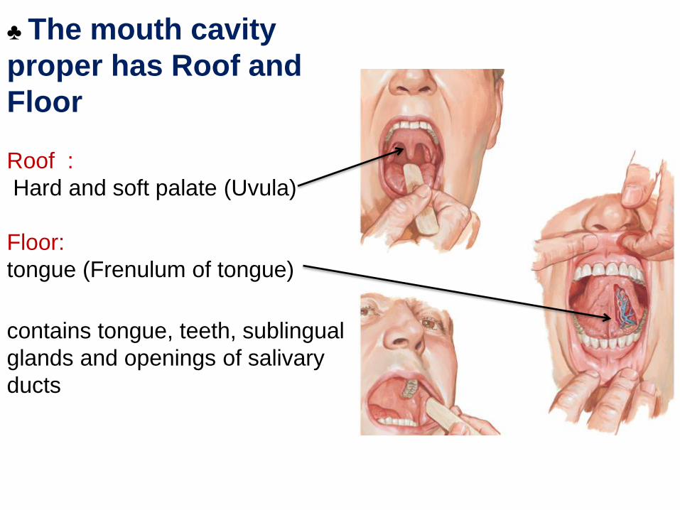
♣ The mouth cavity
proper has Roof and
Floor
Roof :
Hard and soft palate (Uvula)
Floor:
tongue (Frenulum of tongue)
contains tongue, teeth, sublingual
glands and openings of salivary
ducts

2- The PharynxThe oral cavity ends into the
oropharynx
The oropharynx continues as
which leads to esophagus
laryngopharynx

PHARYNX
It connects to the oral cavity anteriorly, and is continuous with the oesophagus. Food passes from the oral cavity into the pharynx then to the oesophagus below it.
The pharynx consists of three parts:
nasopharynx,
oropharynx, and the
laryngopharynx.
It prevents food from entering the nasal cavity (by the soft palate) and the lower respiratory tract (by the epiglottis).

Figs: Actions of soft palate and Epiglottis in Pharynx duringswallowing
EPIGLOTTIS

OESOPHAGUSAlso called gullet or esophagus, it is an organ through which food passes from the pharynx to the
stomach, aided by peristaltic contractions, of its musculature.
It is about 25 cm long and 2 cm in diameter, and lies in the median plane (mediasternum) in the thorax, anterior to the spinal column, but posterior to the trachea.

CONSTRICTIONS OF THEOESOPHAGUS
The oesophagus follows the curvature of the vertebral column.
It also has 3 constrictions (narrowing), whereadjacent structures produce impressions:
1.Cervical Constriction (Upper Oesophageal Sphincter) – where Pharynx meets Oesophagus.
2.Thoracic (Broncho-Aortic) Constriction – where it is first crossed by arch of aorta.
3.Diaphragmatic Constriction: where it passes through the oesophageal hiatus of the diaphragm at T10, before entering the stomach.

Relation of
Esophagus &
trachea
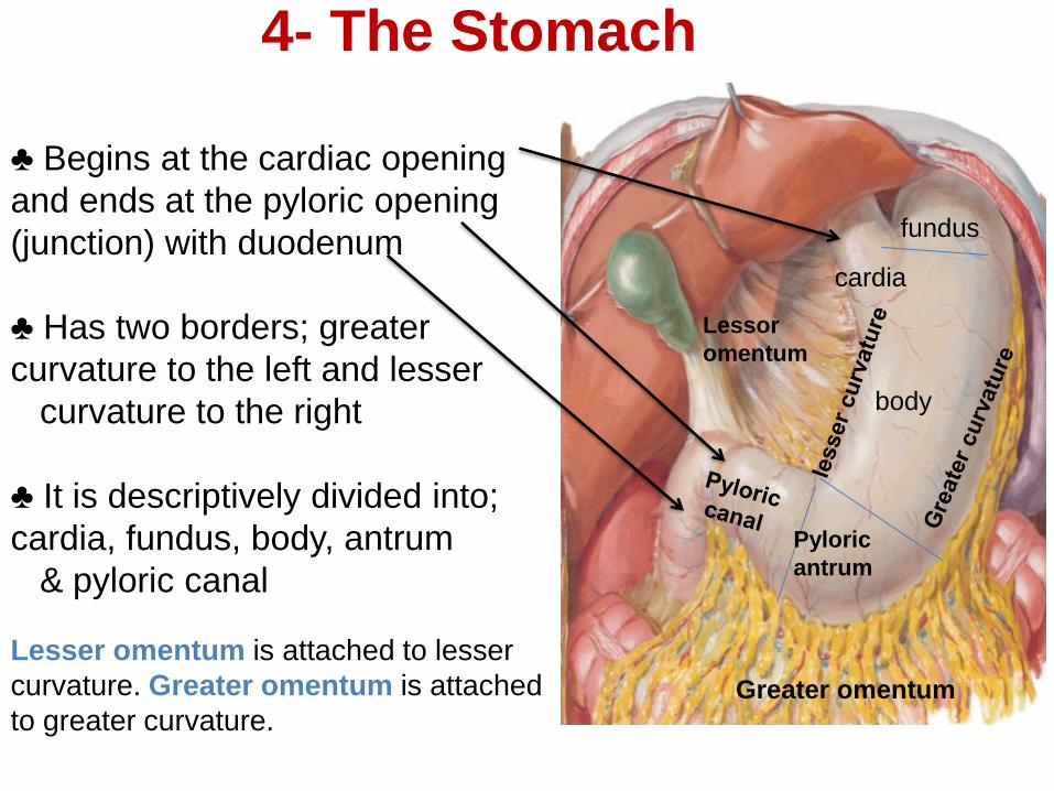
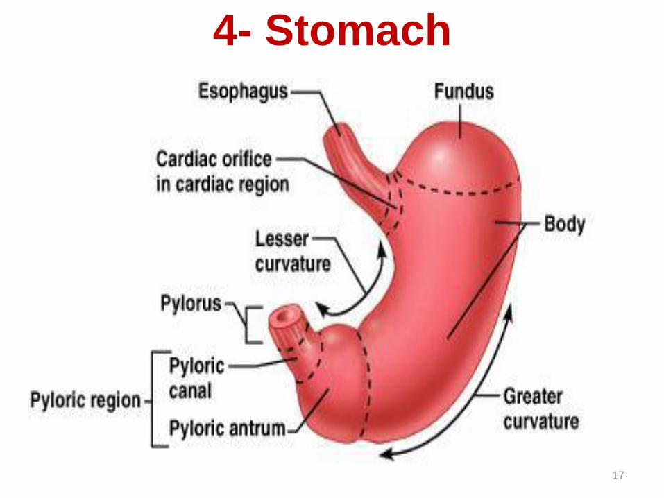
4- The Stomach
♣ Begins at the cardiac opening
and ends at the pyloric opening
(junction) with duodenum
♣ Has two borders; greater
curvature to the left and lesser
curvature to the right
♣ It is descriptively divided into;
cardia, fundus, body, antrum
& pyloric canal
fundus
cardia
body
Greater omentum
Lessor
omentum
Pyloric
antrum
Lesser omentum is attached to lesser
curvature. Greater omentum is attached
to greater curvature.

LOCATION OF THESTOMACH
It is the enlarged hollow part of the digestive tract specialized in the accumulation of ingested food, and also acts as food blender.
It is located between the oesophagus and the small intestine.
It is located in the
epigastric, umbilical & left
hypochondriac regions of
the abdominal cavity.
Gastroenterology deals with the study of diseases of the stomach and intestines and their associated organs
4- Stomach
17


Small intestine
• Is the longest portion of alimentary canal (6 meters)
• Is of three parts: duodenum, jejunum, ileum.
• It starts at pylorus and ends at iliocecal junction.
• Duodenum is 25 cm long, c shaped and surrounds the head of pancreas. It receives the opening of bile duct and major and minor pancreatic ducts.
• Most of absorption of food happens in small intestine.
• The surface area of small intestine is increased by presence of microvilli on the cell membrane of its epithelial cells.
• Also villi which are fingerlike projections of the mucosa.
• Plicea circularis are large circular folds of mucosa and submucosa.
• Peyers patches are lymphoid nodules in the submucosa of distal parts of small intestine

5-6 m long, consists of
Duodenum
Jejunum and
Ileum
5- The Small Intestine

♣ is 12 inches long, C-shaped
and surround the pancreas
♣ It has the openings of bile
and pancreatic ducts
♣ it digests the food particles
by bile from liver, and
pancreatic
A-Duodenum

B- JejunumCoiled muscular
tube, 1.5 m long
C- Ileum♣ Coiled muscular
tube, 3.5 m long
♣ Its function is absorption of digested food particles
Aggregate lymphoid
nodules (Peyer’s patches)
Anastomotic loops
(arcades) of ileal
arteries(3 or 4)
Circular folds
Circular folds
Fat near the root
Fat over the
root
Anastomotic loops
(arcades) of jejunum
arteries(1 or 2)

Large intestine
• Larger in diameter than small intestine but much shorter.
• It extends from the iliocecal junction to the anal canal
Parts of the L.N:
- Cecum - Appendix
- Ascending colon - Transverse colon
- Descending colon - Sigmoid colon
- Rectum - Anal canal
• The main function of large intestine is to absorb water and electrolytes and to store the residues of digestion and its elimination from the body as feces.
• There are no villi in the large intestine
• Appendix is commonly inflamed and infected (appendicitis) and then appendectomy may be necessary.
• The anal canal has external voluntary sphincter and internal involuntary anal sphincter.
• The external sphincter is skeletal muscle while the internal sphincter is smooth muscle
• Three bands of smooth muscle are present in the outermost layer of colon called tenea coli.
• The wall of large intestine is sacculated called haustra.

♣ 1.5 m long, surround the coils of small intestine
Consists of
,
9- Anus
Ileum
3- Ascending
colon
4- Transverse colon
5- Descending
colon
6- Sigmoid
colon
6- The Large Intestine
7- Rectum
8- Anal canal
1- Cecum
2- Appendix
Right colic (hepatic)
flexure
Left colic (splenic)
flexure

McBurney’s point
The appendix lies in
the right iliac fossa,
and in relation to the
anterior abdominal
wall its base is
situated one third of
the way up the line
joining the right
anterior superior
iliac spine to the
umbilicus
(McBurney’s point)

Different between Small and
large Intestine
•Taeniae coli
•Haustra: sacculations
• Omental appendices:
small fat accumulations
● Greater luminal
diameter

Digestive System
Part (2)
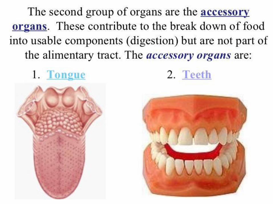


Associated digestive Glands
1- The Salivary Glands
♣ Include three pairs;
Parotid
Sublingual and
Submandibular
♣ Their ducts open
in the oral cavity

Parotid gland
• The largest one located anterior and below the auricle on the
ramus of mandible.
● Extends from external auditory meatus above to the upper part
of the side of neck on the sternomastoid muscle.
● Has a fibrous capsule and a fascial sheath derived from the
investing layer of deep fascia of neck.
● The parotid duct (Stensen’s duct) emerges from the
superficial part, crosses over masseter pierces buccinator
muscle and overlying fat & fascia to open into mouth vestibule
opposite the 2nd upper molar tooth

Submandibular Gland
● The 2nd largest salivary gland located in the neck, deep to the body
of mandible (in the submandibular fossa).
● The submandibular duct (Wharton’s duct) emerges from the deep
part to open in the floor of mouth cavity proper by a small papilla at
the side of the frenulum of tongue.
Sublingual Gland● The smallest pair of salivary glands located in the floor of mouth
cavity under the tongue.
● A major sublingual duct (Bartholin duct) also joins the
submandibular duct.

TONGUE
The tongue is a large, muscular organthat occupies most of the oral cavity.
It is attached by its base to the hyoid bone, and by thin fold of tissue called the frenulum, to the floor of the mouth.
A groove called the terminal sulcus divides the tongue into two parts.
•Anterior: covered by papillae (contains some taste buds).
•Posterior: contains few small glands and a large amount of lymphoid tissue, the lingual tonsil.
Fig: Dorsal surface of tongue & tonsils

The teeth are embedded in the mandible and maxilla bones.
Movement of the mandible (lower jaw) allows chewing. The mandible is the only moveable bone in the jaw.
There are 20 temporary teeth. Later, 32 permanent teeth replace the 20.
There are incisors (8), canines (4), premolars (8), and molars(12).
Fig: Skeletal system of the mouth
The Pancreas
• Is a soft, pink triangular gland located on the posterior abdominal wall reteroperitoneally.
• It produces digestive enzymes which are secreted into the duodenum.
• It is also an endocrine gland secreting insuline and glucagon.

♣ Large gland surrounded by duodenum
♣ It is an endocrine and exocrine
Divided into
Head, Neck,
Body and Tail
The Pancreas
Head
Neck
Body
Tail
Liver• Liver is the largest gland in the body. It is located under
the diaphragm, more to the right side of the body
• It has many metabolic functions.
• The digestive function of liver is by secretion of bile through the biliary ducts to the duodenum.
• The liver lobes are:
• Right lobe
• Left lobe
• Caudate lobe
• Quadrate lobe.

♣ The liver is large organ
♣ Anteriorly right
and left lobes
By the Falciform lig
Visceral surface
The right is subdivided into
3- The Liver & Gall Bladder
quadrate lobe
and a caudate lobe
by the ligamentum teres,
gallbladder
the inferior vena cava,
and liqamentum venosum
quadrate lobe
caudate lobe

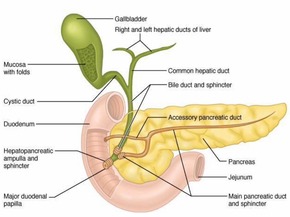
Gall bladder
• Gall bladder is a small thin walled green sac that snuggles in a shallow fossa in the inferior surface of the liver.
• Right and left hepatic ducts unites to form the common hepatic duct which unites with the cystic duct to form the common bile duct.
• Common bile duct opens in the duodenum.

♣ is muscular sac attached to
inferior of visceral surface of
liver
4- Gall bladder
The gallbladder is divided into the
fundus, body, and neck.
♣ Its duct is called cystic duct
joins common hepatic duct to form
common bile duct
fundus
body
neck
cystic duct
common hepatic
duct

♣ Small ducts unite form
Rt and lt hepatic ducts
♣ They units and for
common hepatic duct
♣ The cystic and CHD
join together form CBD
♣ The CBD join with the
pancreatic duct form
Hepatopancreatic papilla
The Biliary Passages

Peritoneum
• Is the largest serous membrane in the body.
• Parietal peritoneum is that portion of peritoneum that lines the walls of abdominopelvic cavity.
• Visceral peritoneum covers the organs.
• The slim space between visceral and parietal peritoneum is called peritoneal cavity which contains peritoneal fluid.
• Ascitis is accumulation of excess fluid inside the peritoneal cavity.

• Some organs lie on posterior abdominal wall and are covered only partially by peritoneum on their anterior surface they are called retroperitoneal organs.
• Some organs are surrounded by peritoneum and peritoneal cavity from all the sides called intraperitoneal organs.
• Examples of retroperitoneal organs:
• Kidneys, suprarenal glands, ureters, pancreas, duodenum, ascending and descending colons,
• Examples of intraperitoneal organs are:
• Stomach, spleen, liver, transverse colon, sigmoid colon, jejunum, ilium.

• There are five major peritoneal folds: the greater omentum, falciform ligament, lesser omentum, mesentery, and mesocolon..and others.