
Digestive system - kpfu.ru · 1. The stomach form fusiform dilatation of the foregut, suspended...
86
Digestive system (Systema digestorium/ alimentarium) Lecture #1
Transcript of Digestive system - kpfu.ru · 1. The stomach form fusiform dilatation of the foregut, suspended...
Digestive system (Systema digestorium/
alimentarium)
Lecture #1
Internal organs are grouped into
1. System
- Have the same functions and development
• Digestive system
• Respiratory system
2. Apparatus
- Have the same development but different
functions
• Urogenital apparatus
Internal organs:
Parenchymal organs
Hollow (tubular) organs
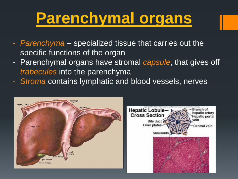
Parenchymal organs
- Parenchyma – specialized tissue that carries out the
specific functions of the organ
- Parenchymal organs have stromal capsule, that gives off
trabecules into the parenchyma
- Stroma contains lymphatic and blood vessels, nerves

Layers of the wall:
1) Tunica mucosa
2) Tunica submucosa
3) Tunica musculosa (longitudinal and circular)
4) Tunica serosa/adventitia
Tubular organs

Alimentary system
• is a complex of organs with the function of
mechanical and chemical treatment of food,
absorption of the treated nutrients, and excretion
of undigested remnants.
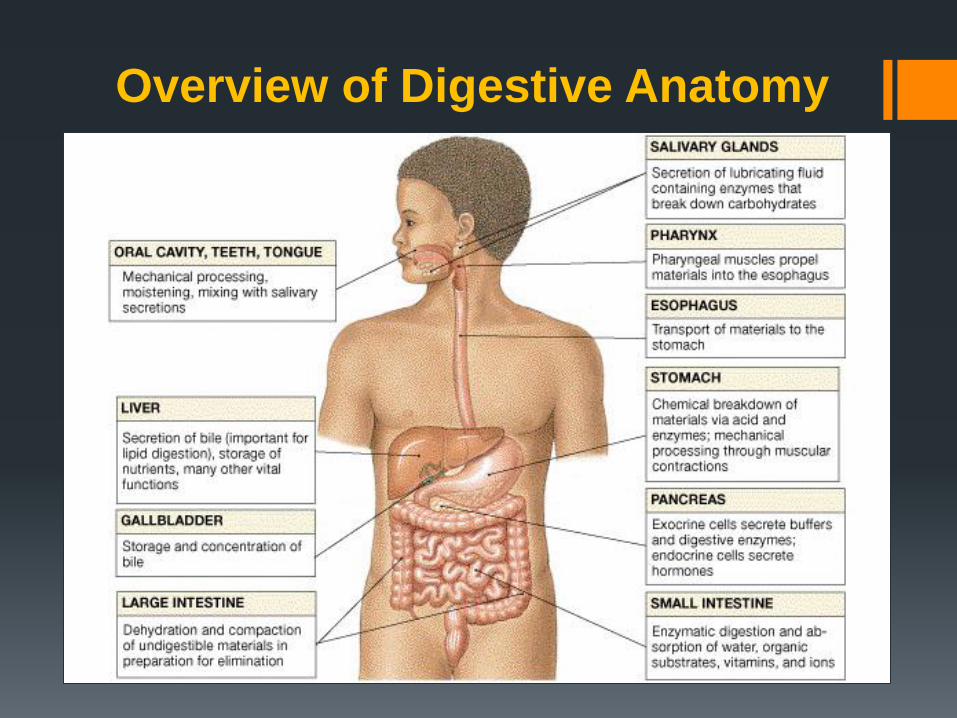
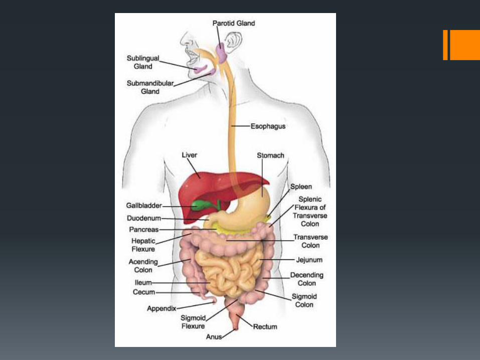
Overview of Digestive Anatomy
Development of
digestive system
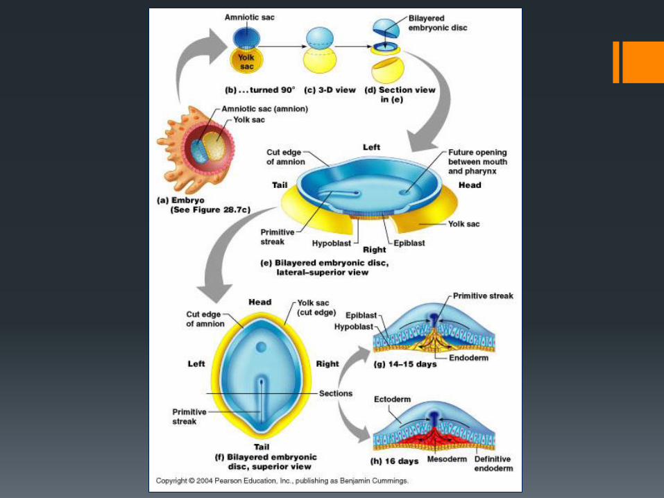
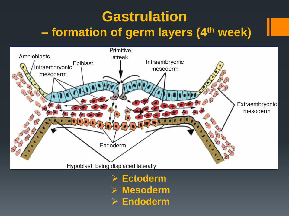
Gastrulation – formation of germ layers (4th week)
Ectoderm
Mesoderm
Endoderm
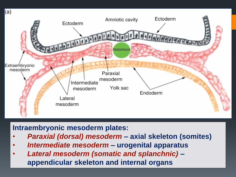
Intraembryonic mesoderm plates:
• Paraxial (dorsal) mesoderm – axial skeleton (somites)
• Intermediate mesoderm – urogenital apparatus
• Lateral mesoderm (somatic and splanchnic) –
appendicular skeleton and internal organs
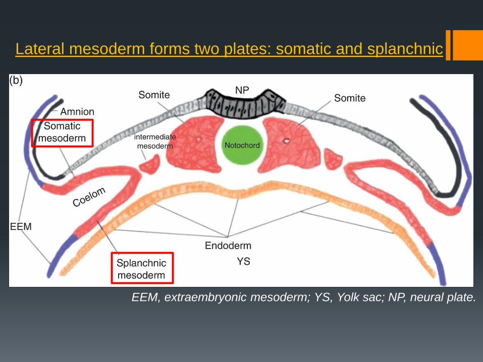
EEM, extraembryonic mesoderm; YS, Yolk sac; NP, neural plate.
Lateral mesoderm forms two plates: somatic and splanchnic
Coelom
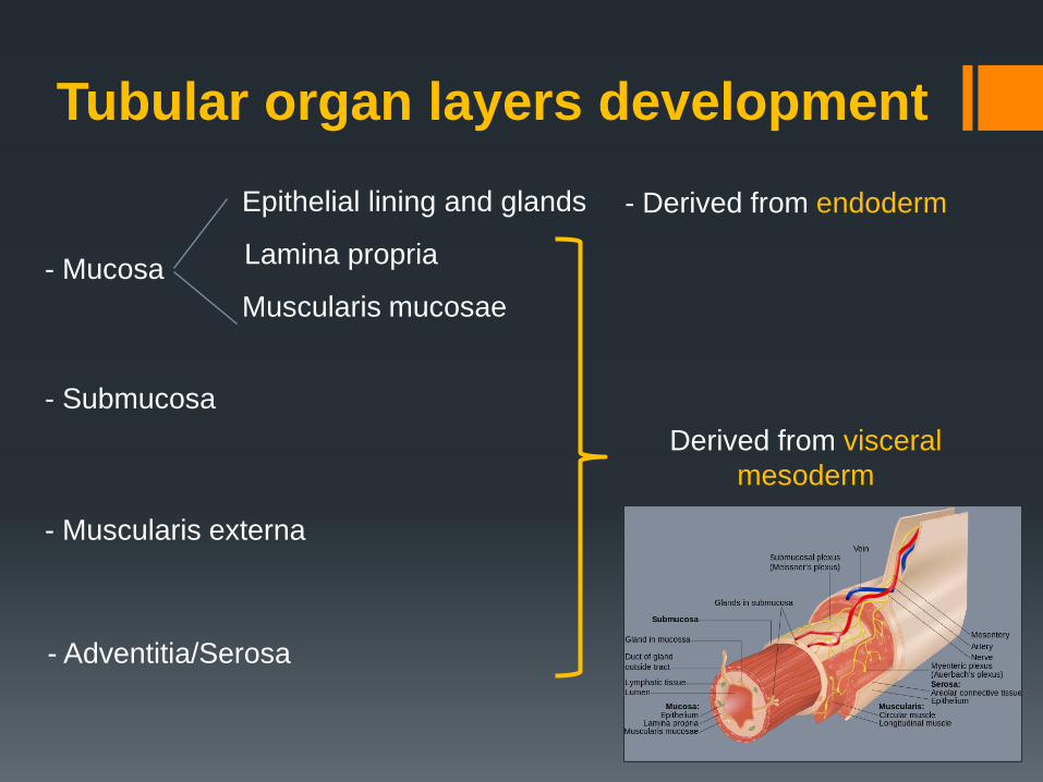
- Mucosa
Epithelial lining and glands
Lamina propria
Muscularis mucosae
- Submucosa
- Muscularis externa
- Adventitia/Serosa
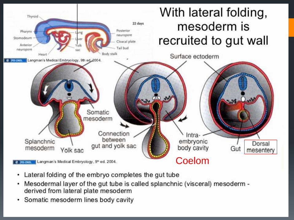
Derived from visceral
mesoderm
- Derived from endoderm
Tubular organ layers development
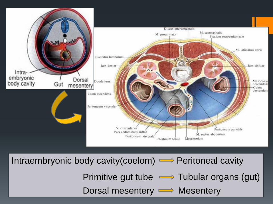
Intraembryonic body cavity(coelom) Peritoneal cavity
Primitive gut tube Tubular organs (gut)
Dorsal mesentery Mesentery
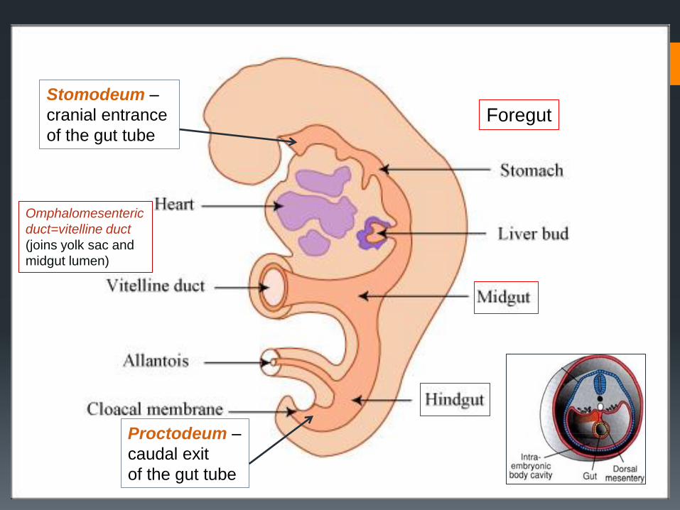
Foregut
Omphalomesenteric
duct=vitelline duct
(joins yolk sac and
midgut lumen)
Primitive gut subdivisions:
Stomodeum –
cranial entrance
of the gut tube
Proctodeum –
caudal exit
of the gut tube
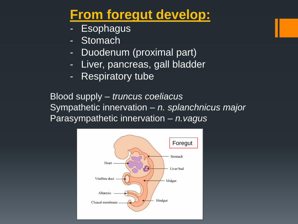
From foregut develop: - Esophagus
- Stomach
- Duodenum (proximal part)
- Liver, pancreas, gall bladder
- Respiratory tube
Blood supply – truncus coeliacus
Sympathetic innervation – n. splanchnicus major
Parasympathetic innervation – n.vagus
Foregut
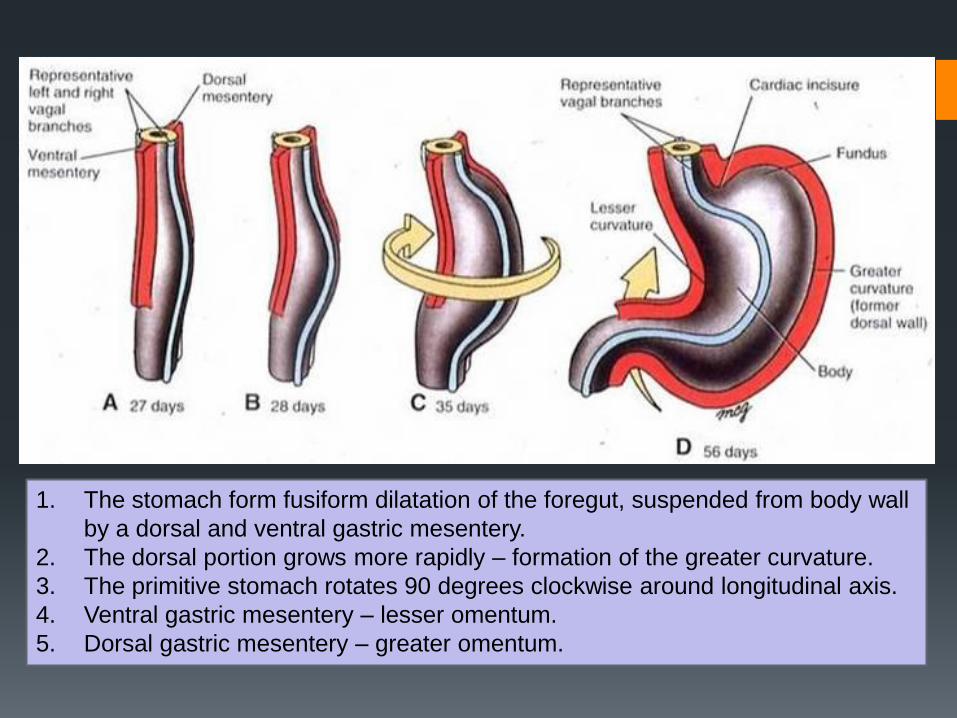
1. The stomach form fusiform dilatation of the foregut, suspended from body wall
by a dorsal and ventral gastric mesentery.
2. The dorsal portion grows more rapidly – formation of the greater curvature.
3. The primitive stomach rotates 90 degrees clockwise around longitudinal axis.
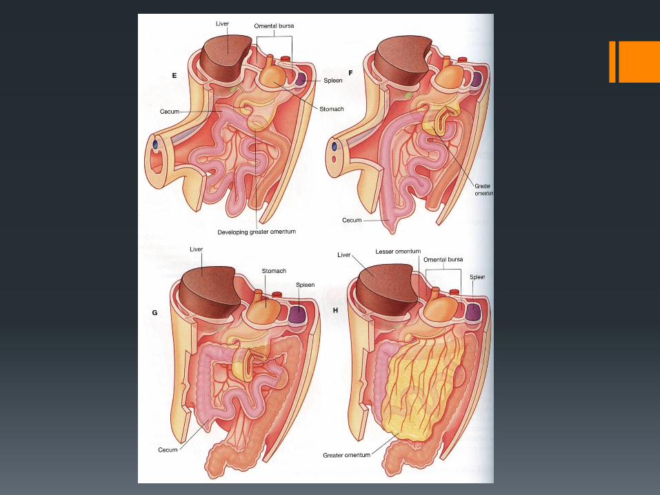
4. Ventral gastric mesentery – lesser omentum.
5. Dorsal gastric mesentery – greater omentum.
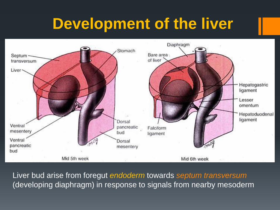
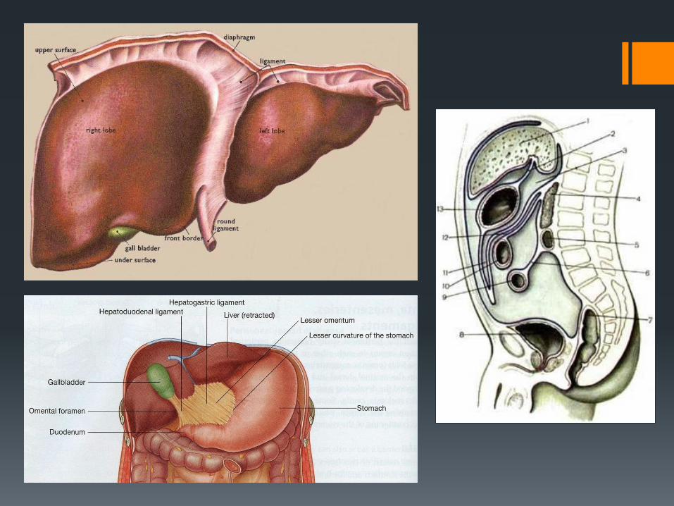
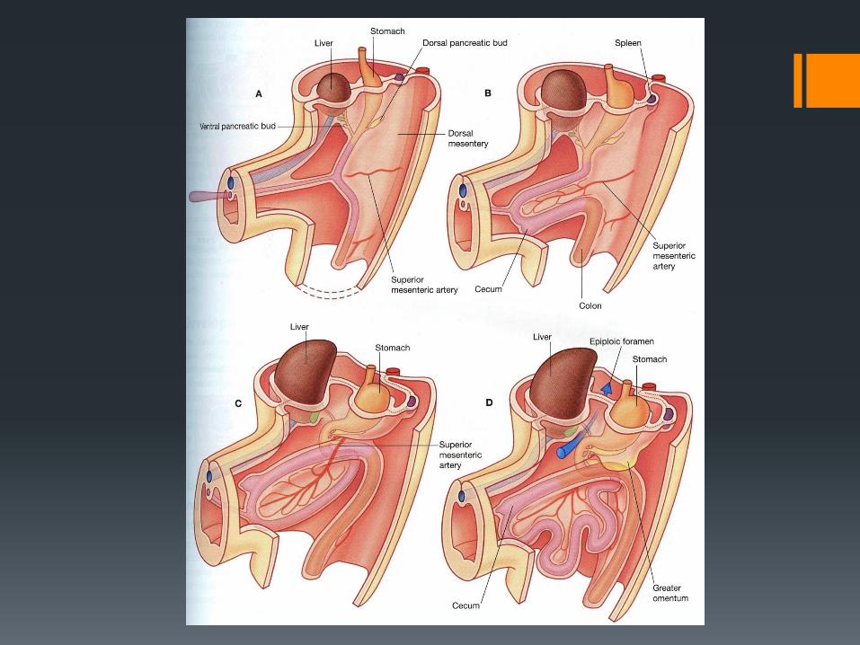
Development of the liver
Liver bud arise from foregut endoderm towards septum transversum
(developing diaphragm) in response to signals from nearby mesoderm
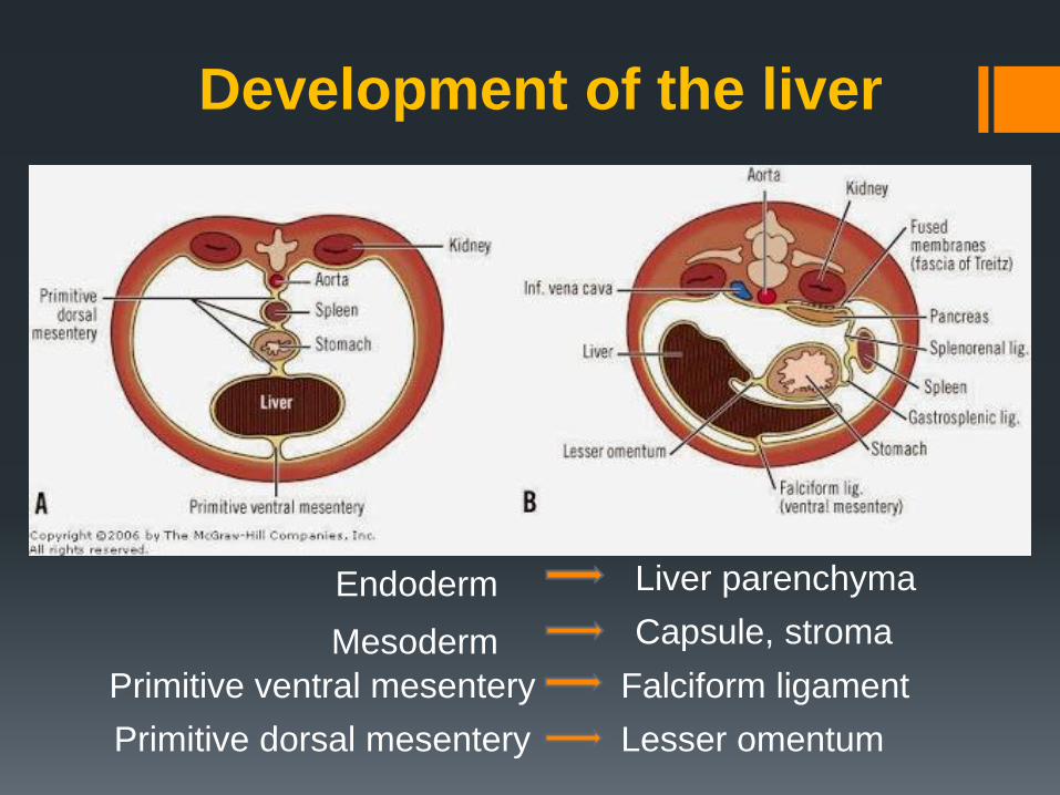
Primitive ventral mesentery Falciform ligament
Primitive dorsal mesentery Lesser omentum
Development of the liver
Endoderm Liver parenchyma
Mesoderm Capsule, stroma
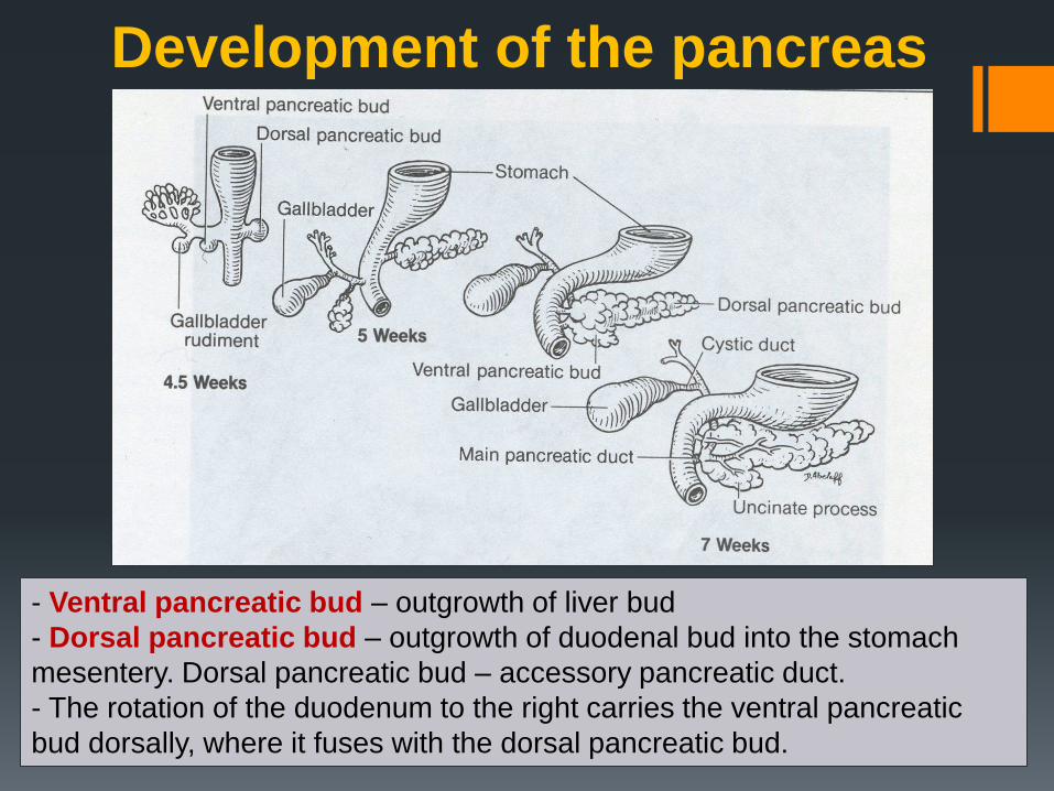
- Ventral pancreatic bud – outgrowth of liver bud
- Dorsal pancreatic bud – outgrowth of duodenal bud into the stomach
mesentery. Dorsal pancreatic bud – accessory pancreatic duct.
- The rotation of the duodenum to the right carries the ventral pancreatic
bud dorsally, where it fuses with the dorsal pancreatic bud.
Development of the pancreas
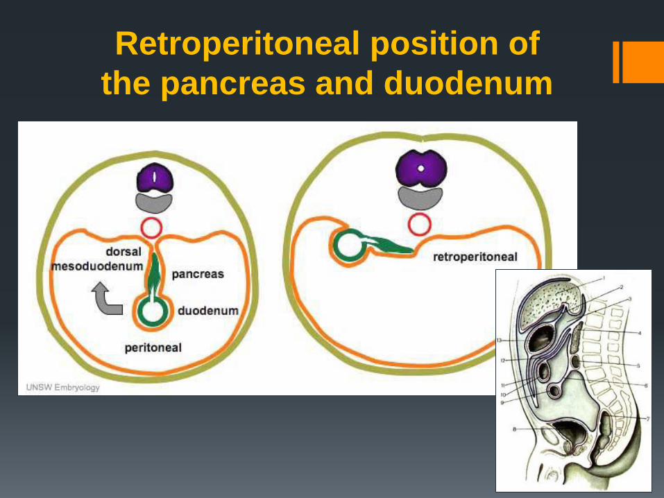
Retroperitoneal position of
the pancreas and duodenum
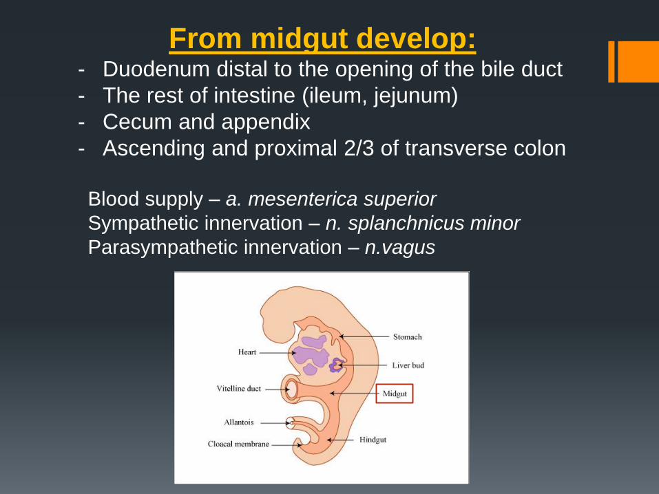
From midgut develop: - Duodenum distal to the opening of the bile duct
- The rest of intestine (ileum, jejunum)
- Cecum and appendix
- Ascending and proximal 2/3 of transverse colon
Blood supply – a. mesenterica superior
Sympathetic innervation – n. splanchnicus minor
Parasympathetic innervation – n.vagus
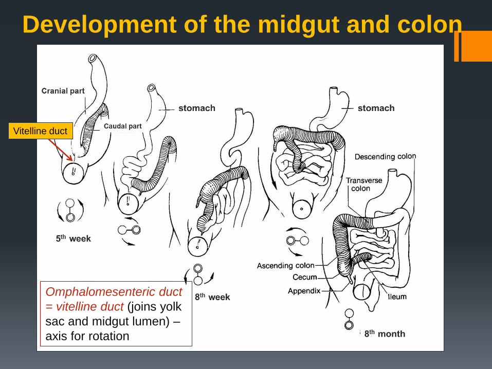
Development of the midgut and colon
Vitelline duct
Omphalomesenteric duct
= vitelline duct (joins yolk
sac and midgut lumen) –
axis for rotation
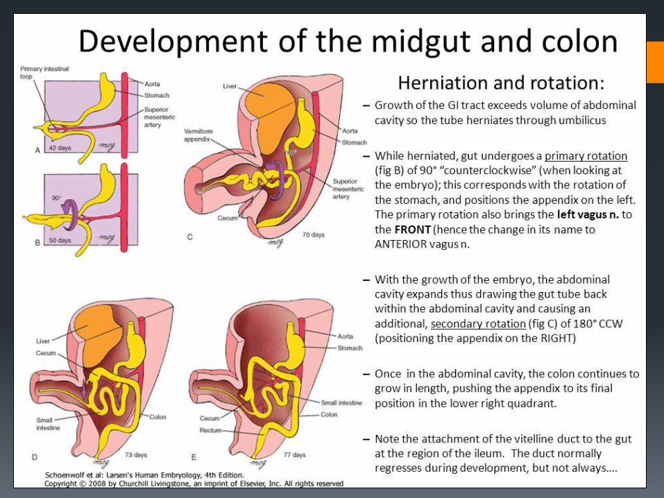
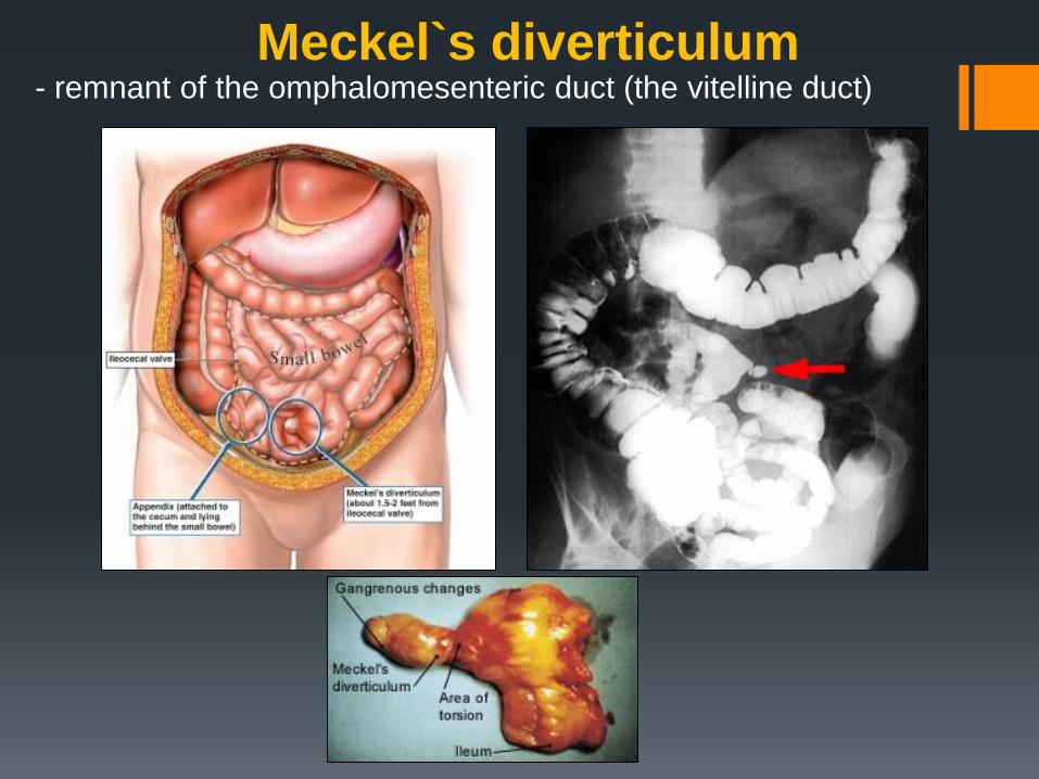
Meckel`s diverticulum - remnant of the omphalomesenteric duct (the vitelline duct)
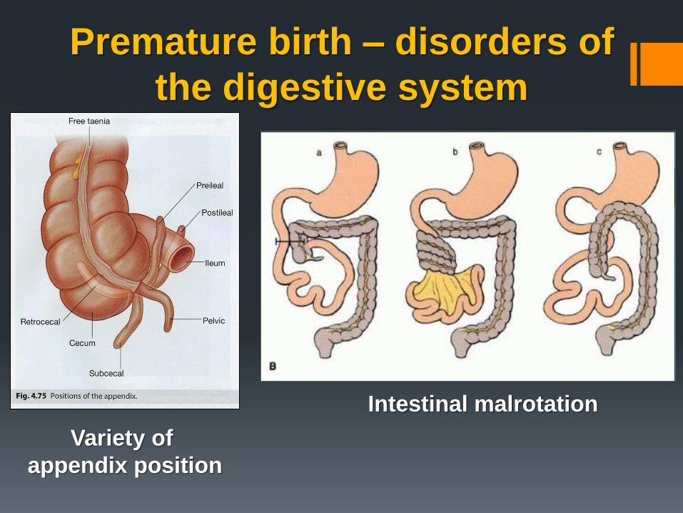
Variety of
appendix position
Intestinal malrotation
Premature birth – disorders of
the digestive system
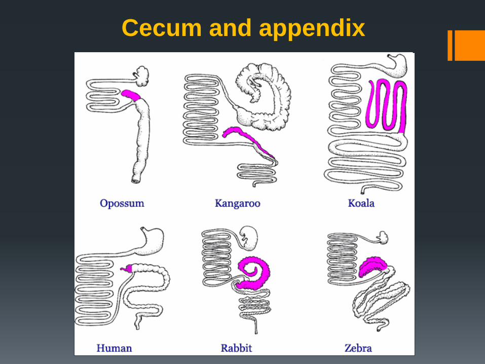
Cecum and appendix
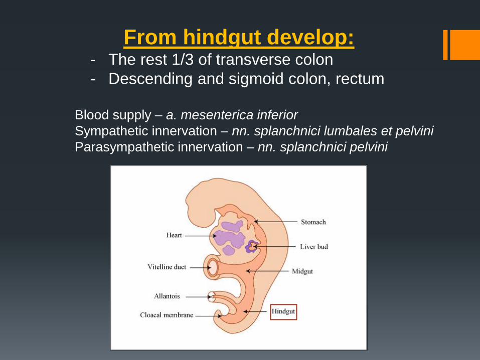
From hindgut develop: - The rest 1/3 of transverse colon
- Descending and sigmoid colon, rectum
Blood supply – a. mesenterica inferior
Sympathetic innervation – nn. splanchnici lumbales et pelvini
Parasympathetic innervation – nn. splanchnici pelvini
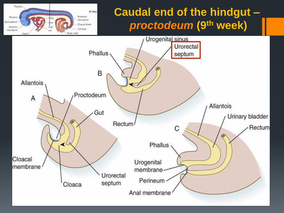
Caudal end of the hindgut –
proctodeum (9th week)
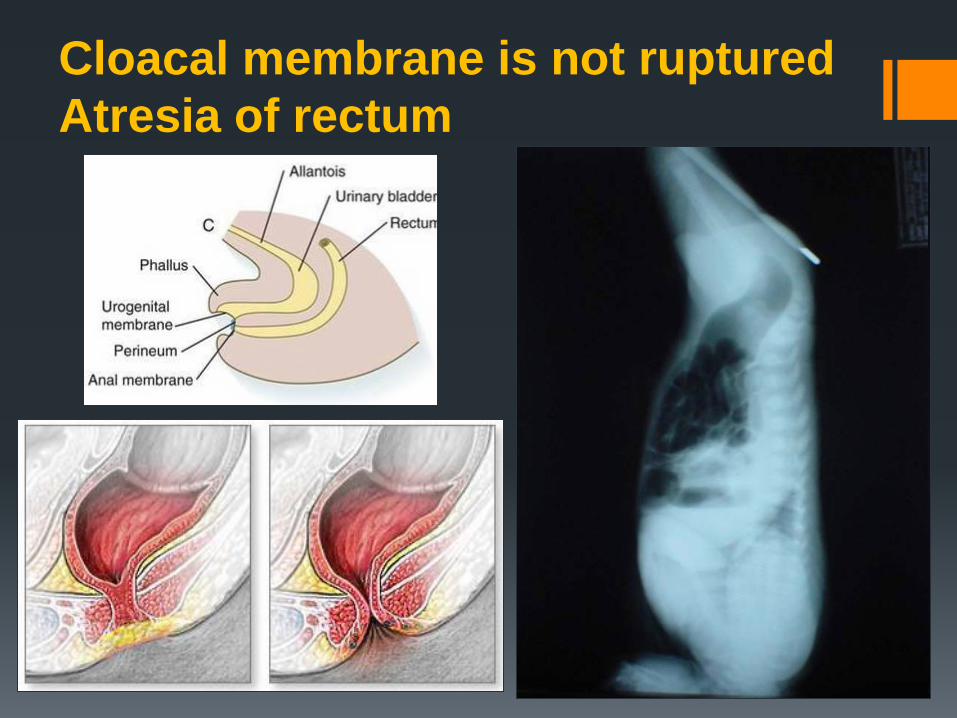
Cloacal membrane is not ruptured
Atresia of rectum
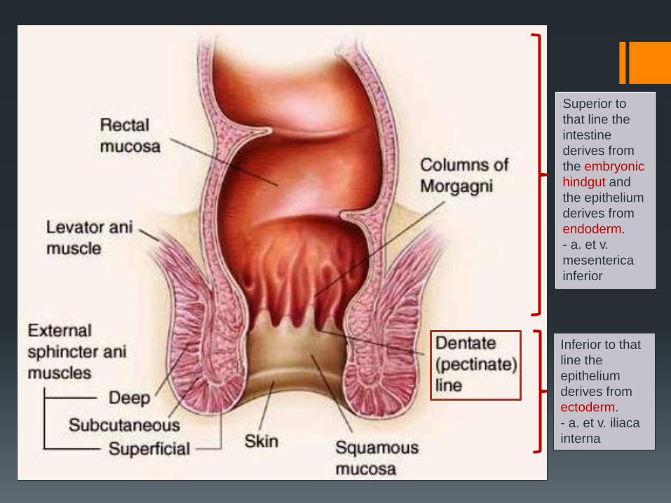
Superior to
that line the
intestine
derives from
the embryonic
hindgut and
the epithelium
derives from
endoderm.
- a. et v.
mesenterica
inferior
Inferior to that
line the
epithelium
derives from
ectoderm.
- a. et v. iliaca
interna
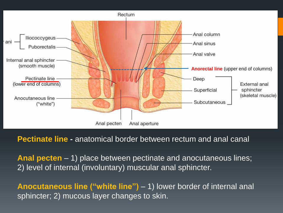
Anorectal line (upper end of columns)
(lower end of columns)
Pectinate line - anatomical border between rectum and anal canal
Anal pecten – 1) place between pectinate and anocutaneous lines;
2) level of internal (involuntary) muscular anal sphincter.
Anocutaneous line (“white line”) – 1) lower border of internal anal
sphincter; 2) mucous layer changes to skin.
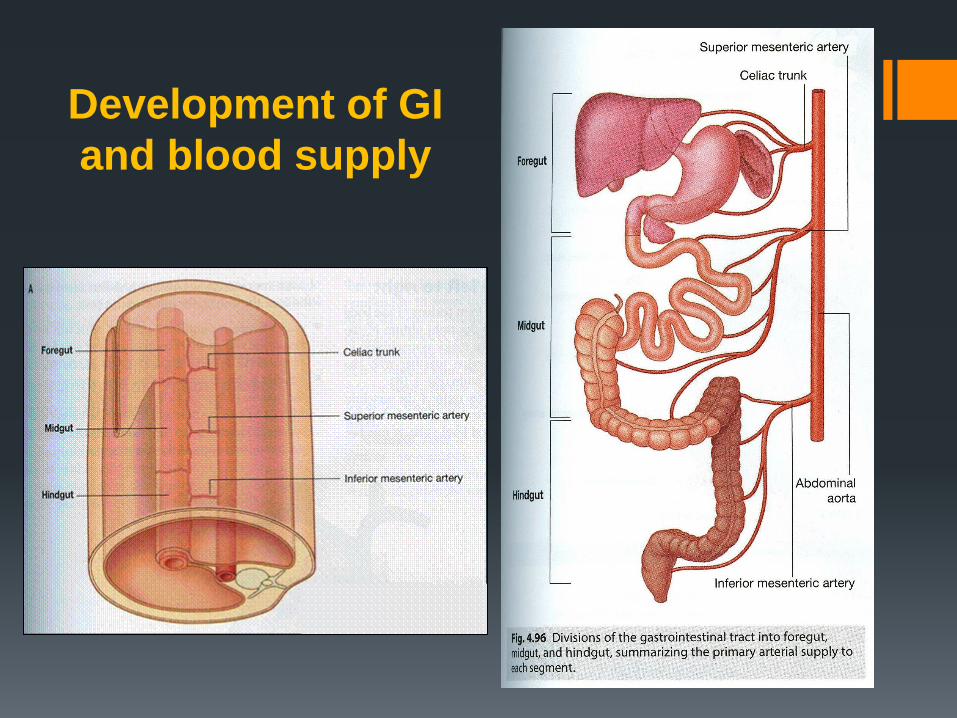
Development of GI
and blood supply
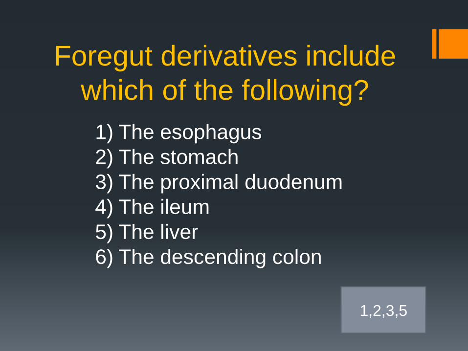
Foregut derivatives include
which of the following?
1) The esophagus
2) The stomach
3) The proximal duodenum
4) The ileum
5) The liver
6) The descending colon
1,2,3,5
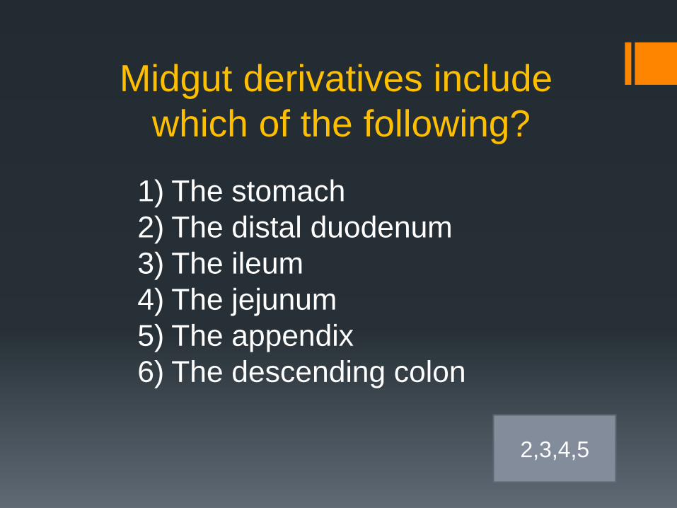
Midgut derivatives include
which of the following?
1) The stomach
2) The distal duodenum
3) The ileum
4) The jejunum
5) The appendix
6) The descending colon
2,3,4,5
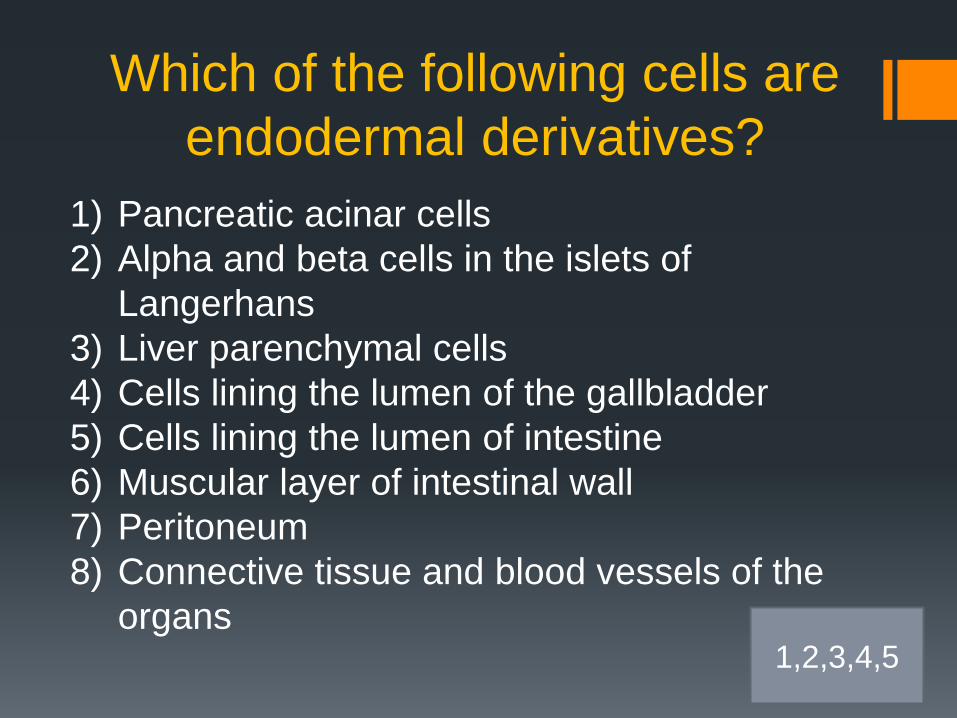
Which of the following cells are
endodermal derivatives?
1) Pancreatic acinar cells
2) Alpha and beta cells in the islets of
Langerhans
3) Liver parenchymal cells
4) Cells lining the lumen of the gallbladder
5) Cells lining the lumen of intestine
6) Muscular layer of intestinal wall
7) Peritoneum
8) Connective tissue and blood vessels of the
organs 1,2,3,4,5
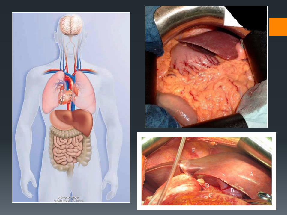
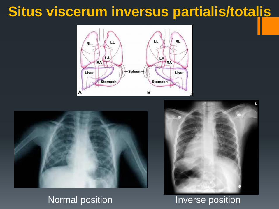
Situs viscerum inversus partialis/totalis
Normal position Inverse position
Development of
peritoneum and its
derivatives
Peritoneum – serose membrane lining organs and
walls of the abdominal cavity
- is derived from intraembryonic mesoderm
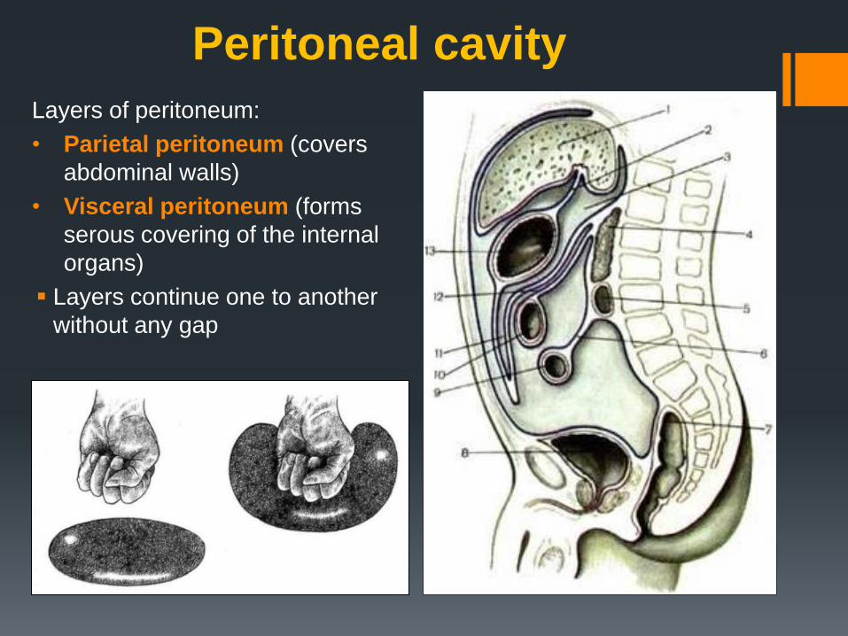
Peritoneal cavity
Layers of peritoneum:
• Parietal peritoneum (covers
abdominal walls)
• Visceral peritoneum (forms
serous covering of the internal
organs)
Layers continue one to another
without any gap

EEM, extraembryonic mesoderm; YS, Yolk sac; NP, neural plate.
Lateral mesoderm forms two plates: somatic and splanchnic
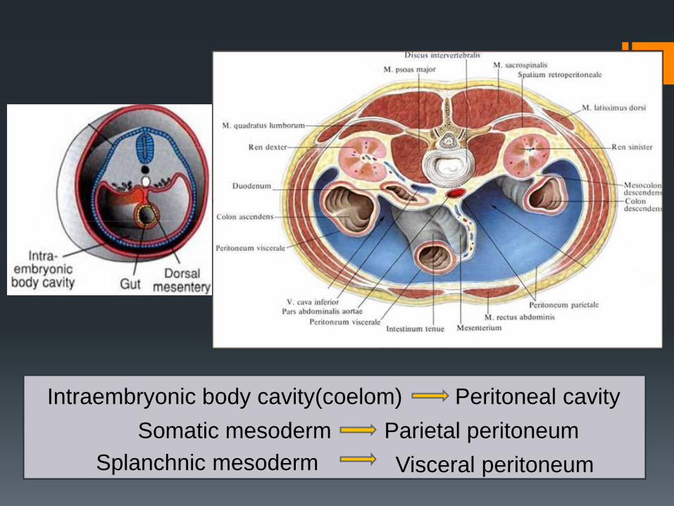
Intraembryonic body cavity(coelom) Peritoneal cavity
Somatic mesoderm Parietal peritoneum
Splanchnic mesoderm Visceral peritoneum
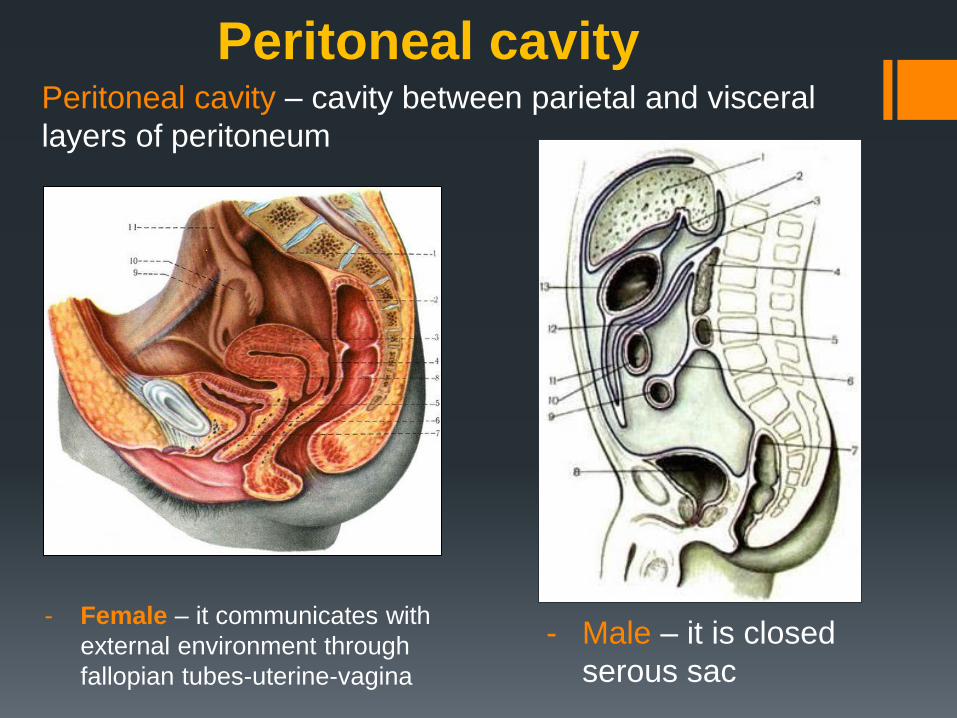
- Female – it communicates with
external environment through
fallopian tubes-uterine-vagina
Peritoneal cavity Peritoneal cavity – cavity between parietal and visceral
layers of peritoneum
- Male – it is closed
serous sac
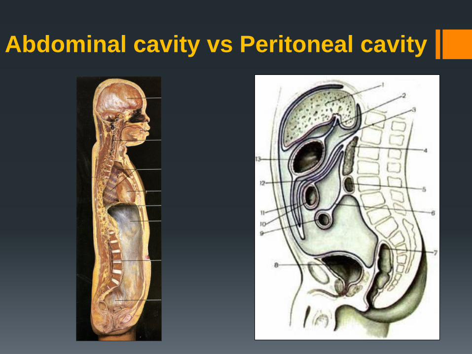
Abdominal cavity vs Peritoneal cavity
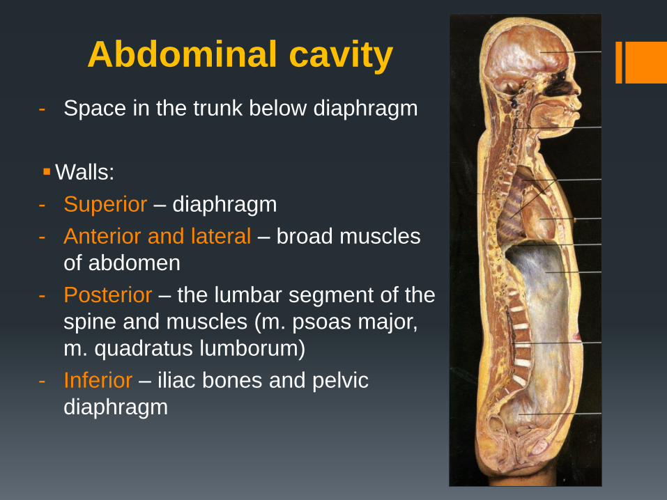
Abdominal cavity
- Space in the trunk below diaphragm
Walls:
- Superior – diaphragm
- Anterior and lateral – broad muscles
of abdomen
- Posterior – the lumbar segment of the
spine and muscles (m. psoas major,
m. quadratus lumborum)
- Inferior – iliac bones and pelvic
diaphragm
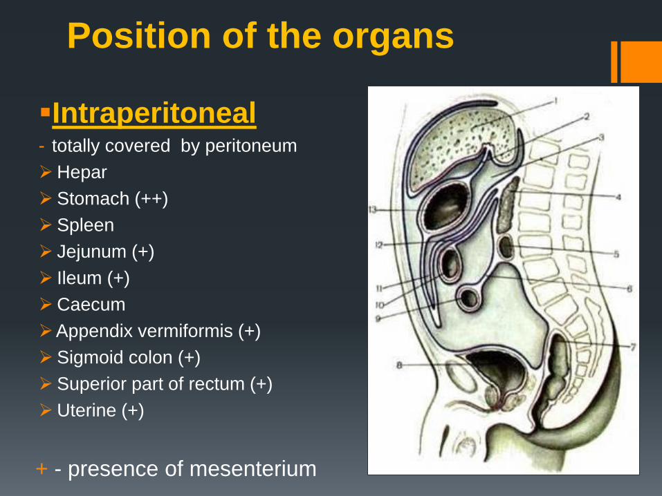
Position of the organs
Intraperitoneal - totally covered by peritoneum
Hepar
Stomach (++)
Spleen
Jejunum (+)
Ileum (+)
Caecum
Appendix vermiformis (+)
Sigmoid colon (+)
Superior part of rectum (+)
Uterine (+)
+ - presence of mesenterium
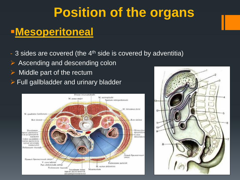
Mesoperitoneal
- 3 sides are covered (the 4th side is covered by adventitia)
Ascending and descending colon
Middle part of the rectum
Full gallbladder and urinary bladder
Position of the organs
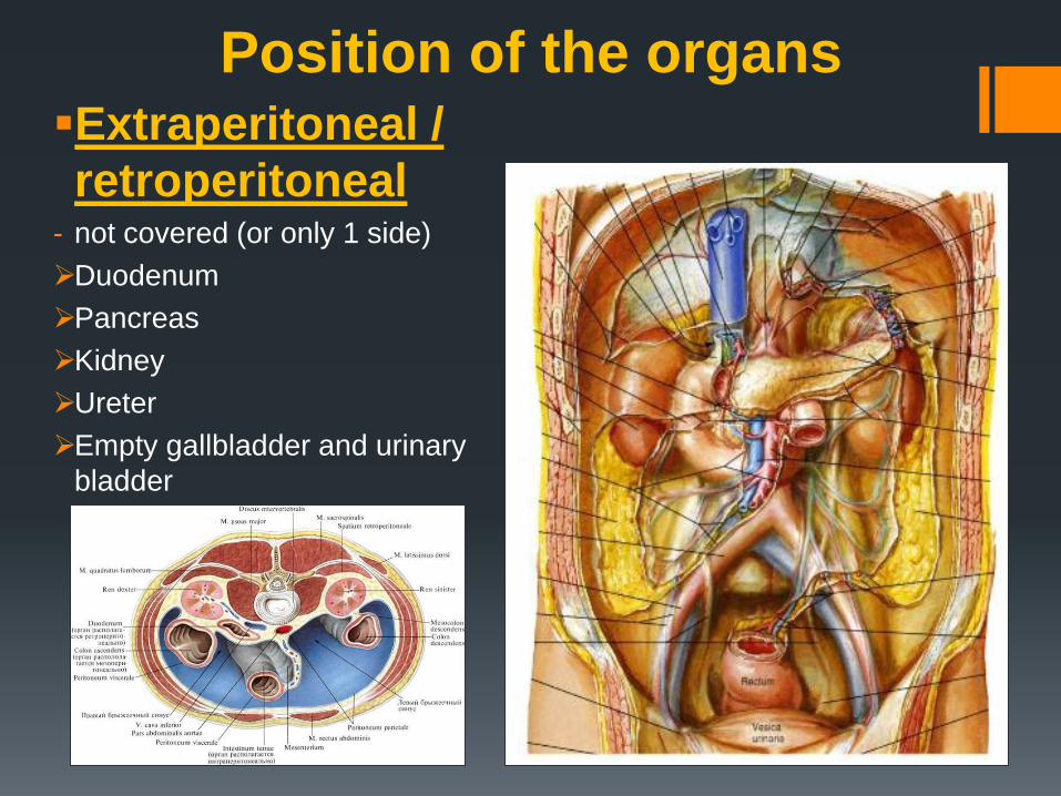
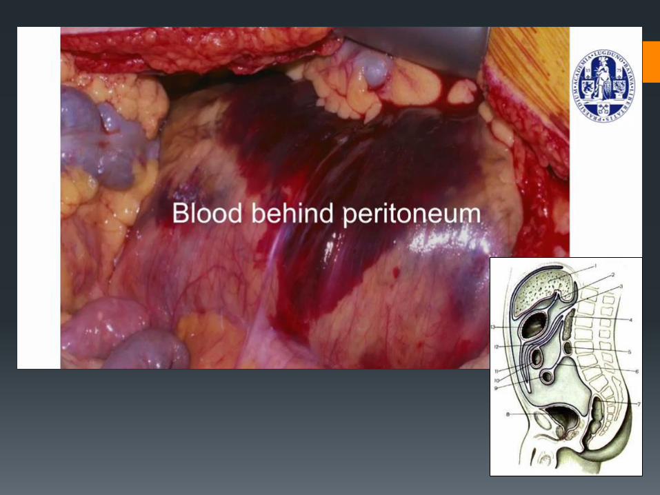
Position of the organs Extraperitoneal /
retroperitoneal - not covered (or only 1 side)
Duodenum
Pancreas
Kidney
Ureter
Empty gallbladder and urinary
bladder
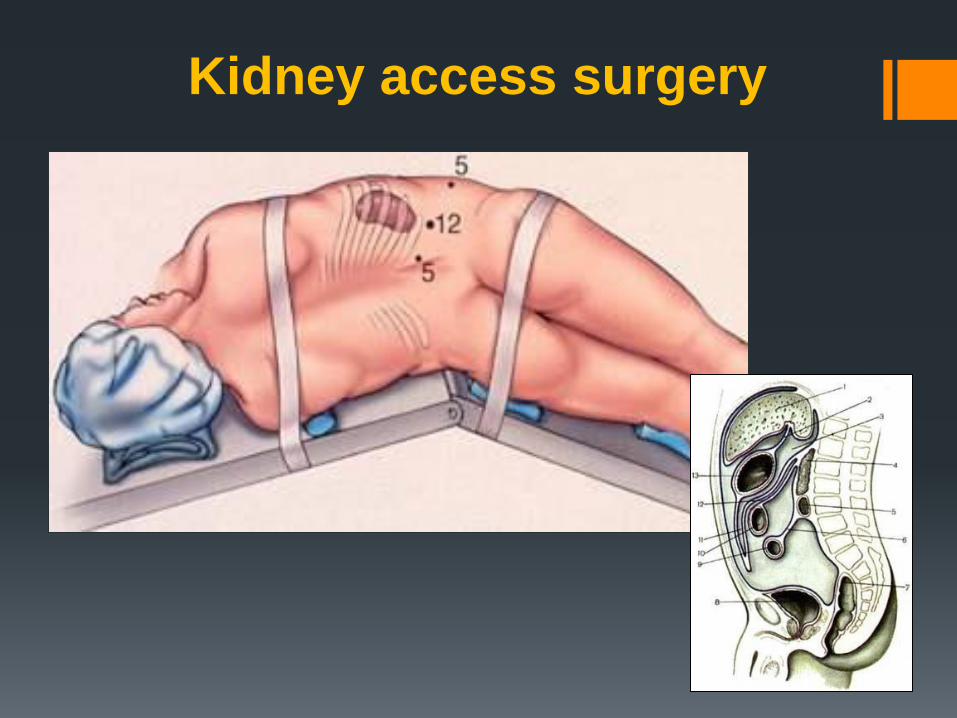
Kidney access surgery
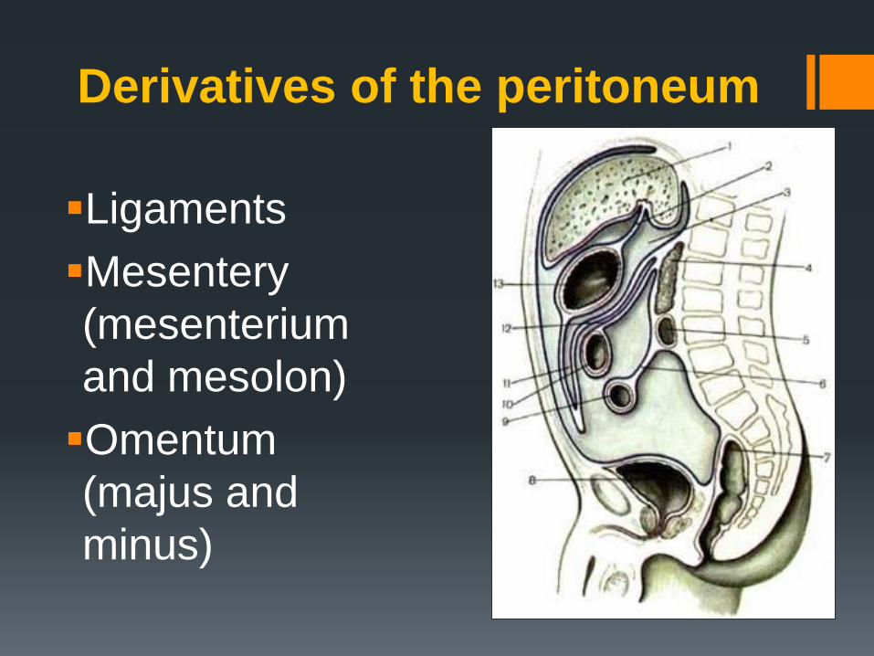
Derivatives of the peritoneum
Ligaments
Mesentery
(mesenterium
and mesolon)
Omentum
(majus and
minus)
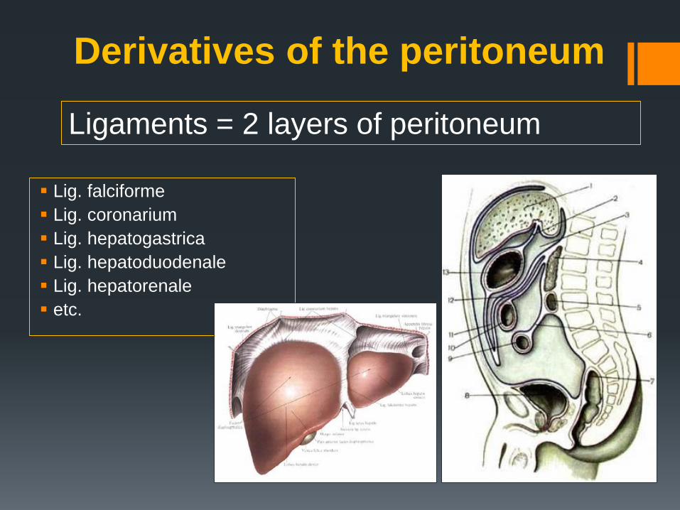
Derivatives of the peritoneum
Lig. falciforme
Lig. coronarium
Lig. hepatogastrica
Lig. hepatoduodenale
Lig. hepatorenale
etc.
Ligaments = 2 layers of peritoneum
Derivatives of the peritoneum
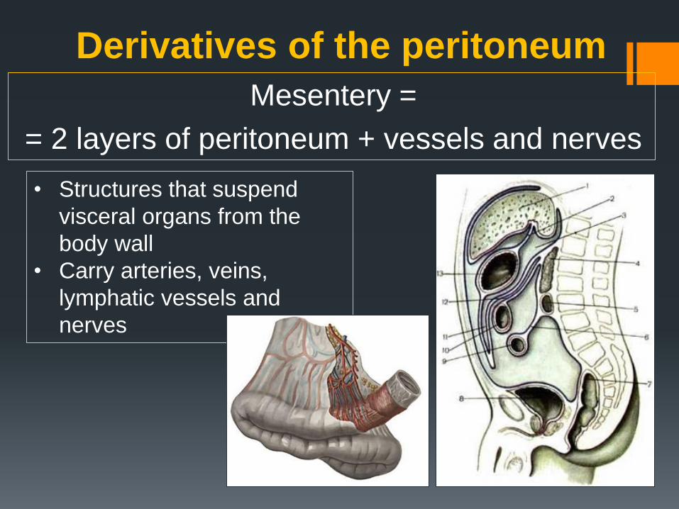
Mesentery =
= 2 layers of peritoneum + vessels and nerves
• Structures that suspend
visceral organs from the
body wall
• Carry arteries, veins,
lymphatic vessels and
nerves
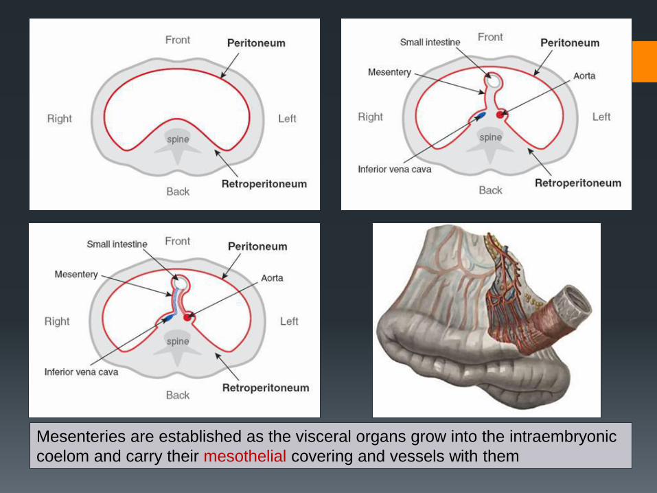
Mesenteries are established as the visceral organs grow into the intraembryonic
coelom and carry their mesothelial covering and vessels with them
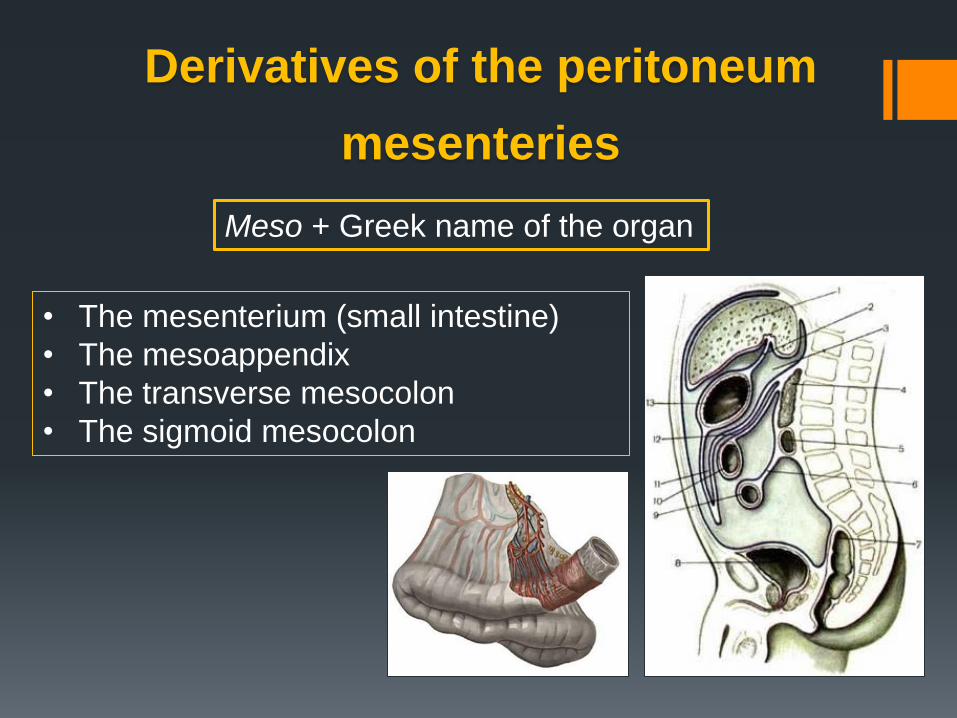
• The mesenterium (small intestine)
• The mesoappendix
• The transverse mesocolon
• The sigmoid mesocolon
Derivatives of the peritoneum
mesenteries
Meso + Greek name of the organ
Derivatives of the peritoneum
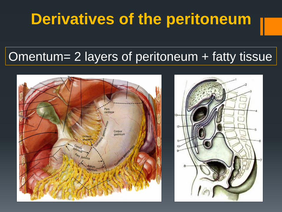
Omentum= 2 layers of peritoneum + fatty tissue

Omentum minus
• Lig. hepatogastricum
• Lig. hepatoduodenale
Contain:
- Ductus hepaticus
communis
- A. hepatica
- V. porta
Omentum minus - ventral mesentery of the stomach
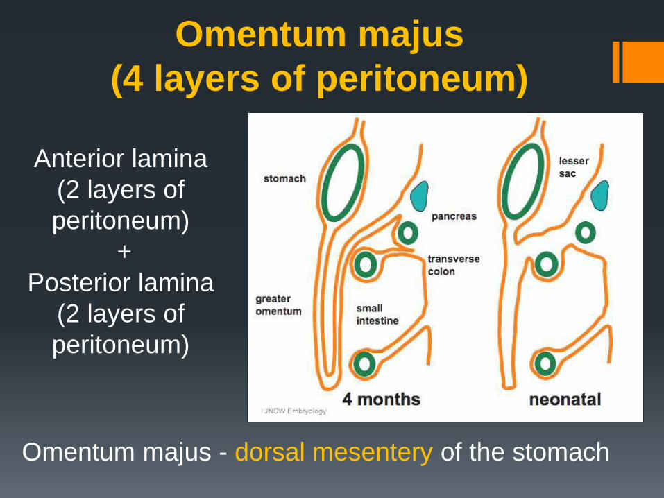
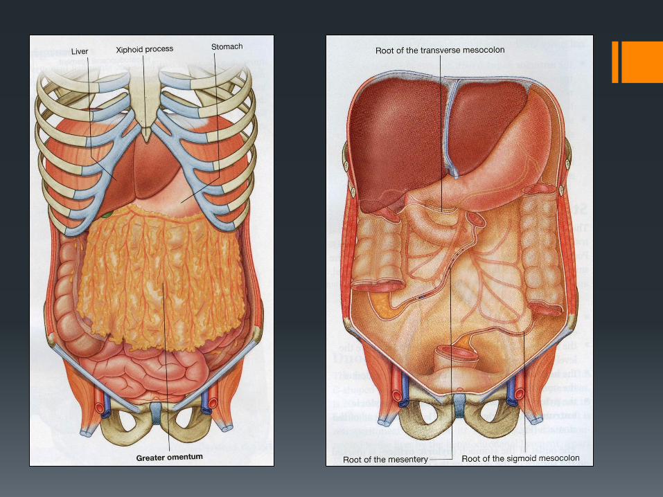
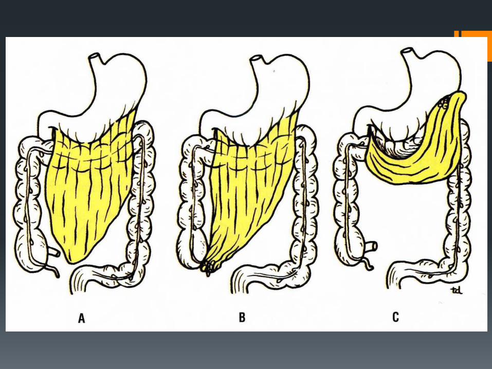
Omentum majus
(4 layers of peritoneum)
Anterior lamina
(2 layers of
peritoneum)
+
Posterior lamina
(2 layers of
peritoneum)
Omentum majus - dorsal mesentery of the stomach
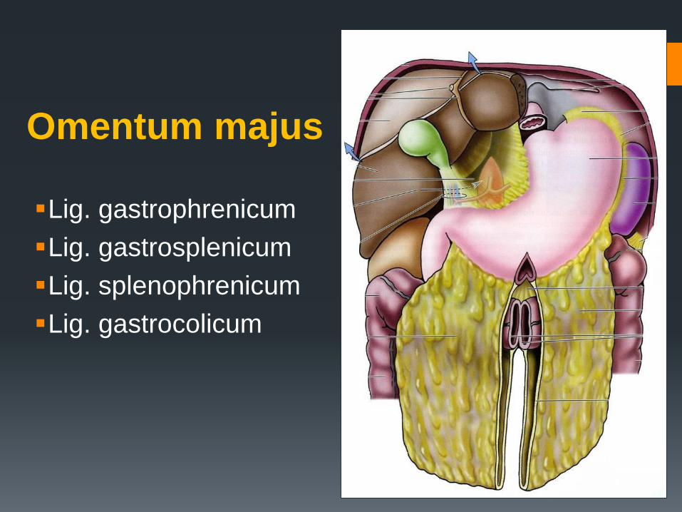
Omentum majus
Lig. gastrophrenicum
Lig. gastrosplenicum
Lig. splenophrenicum
Lig. gastrocolicum
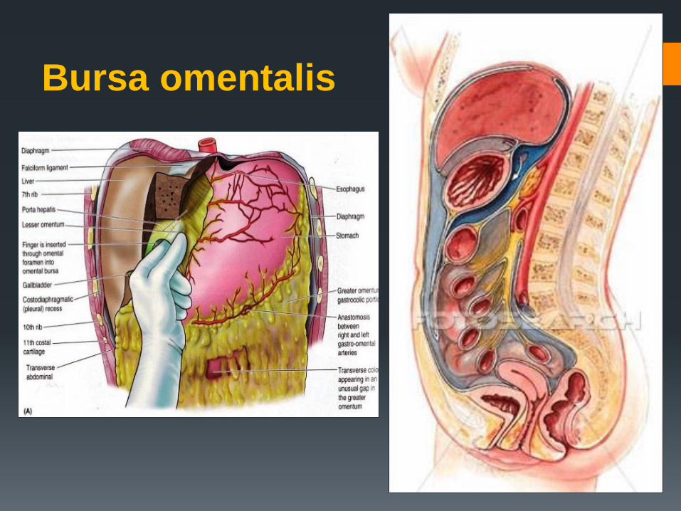
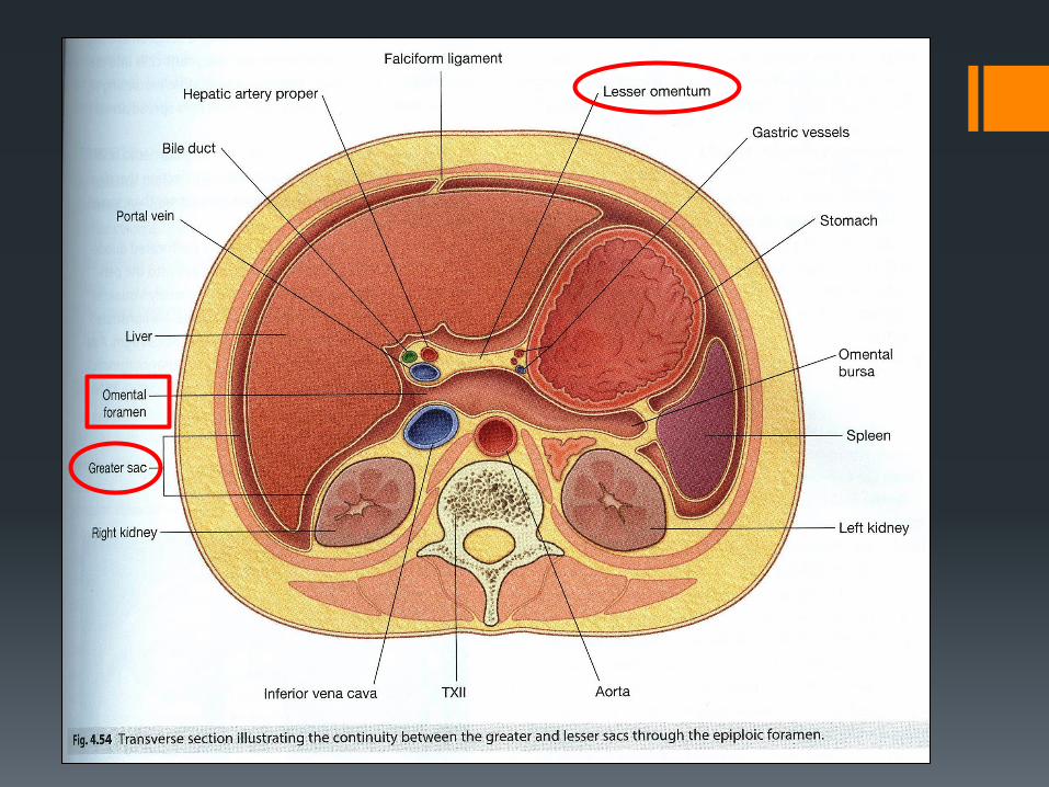
Bursa omentalis
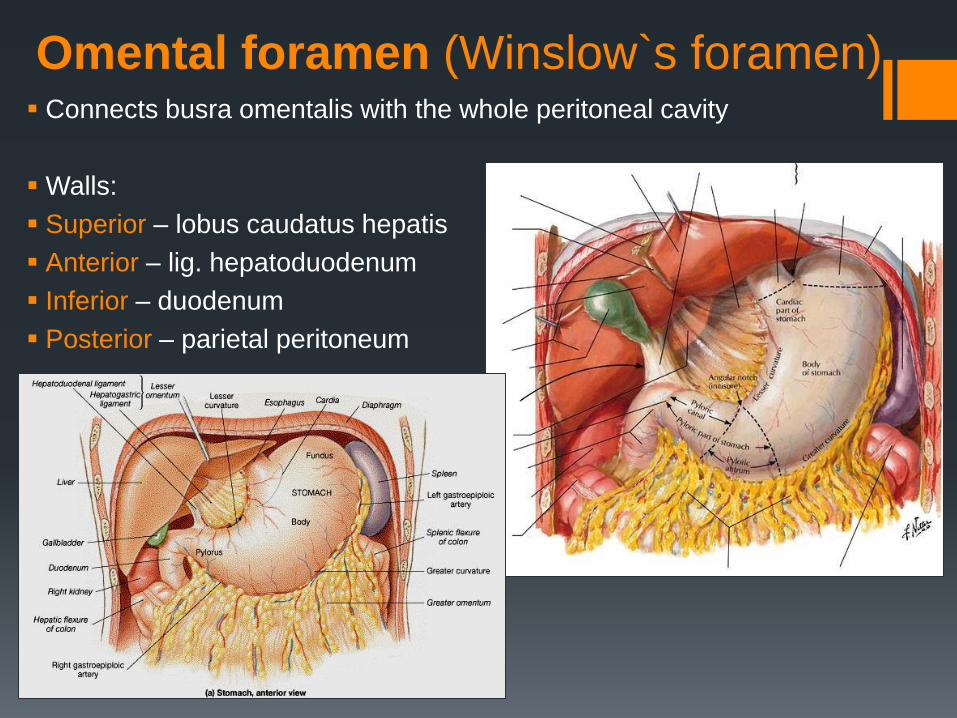
Omental foramen (Winslow`s foramen) Connects busra omentalis with the whole peritoneal cavity
Walls:
Superior – lobus caudatus hepatis
Anterior – lig. hepatoduodenum
Inferior – duodenum
Posterior – parietal peritoneum
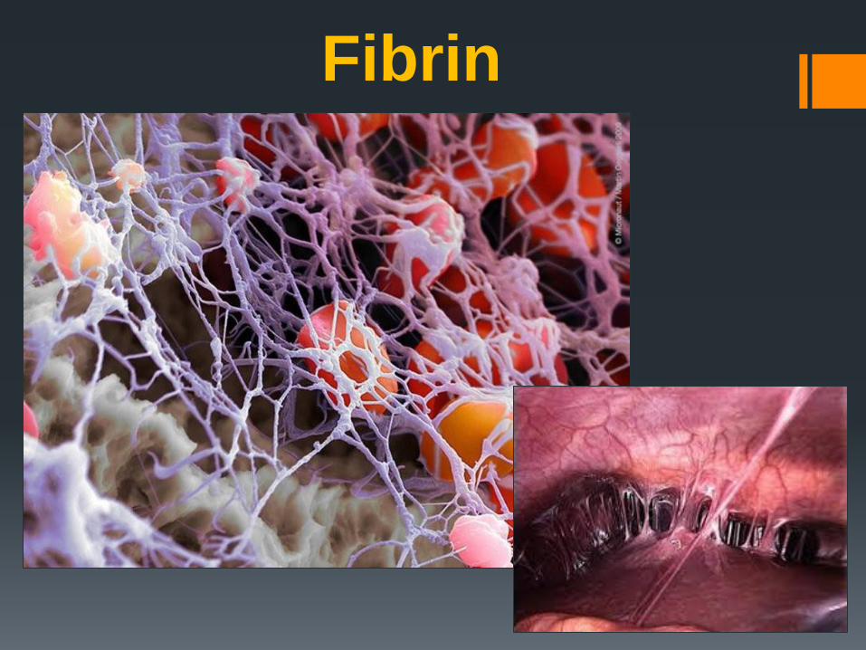
Fibrin
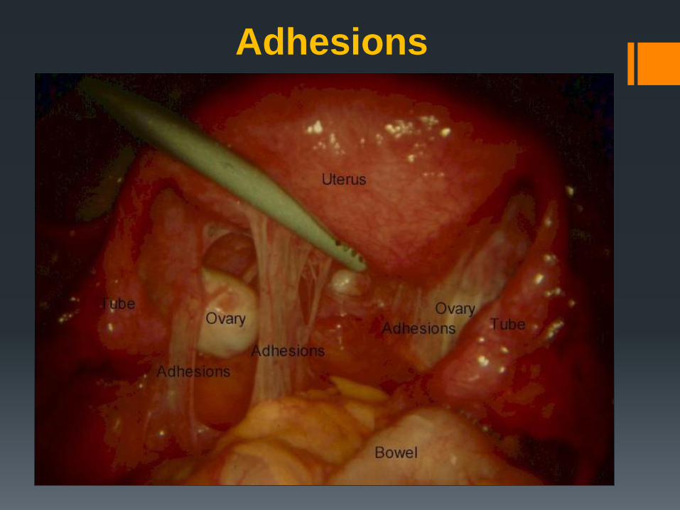
Adhesions
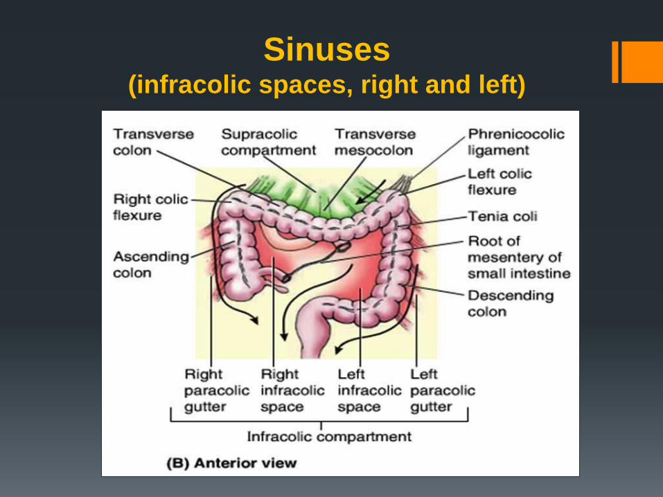
Sinuses (infracolic spaces, right and left)
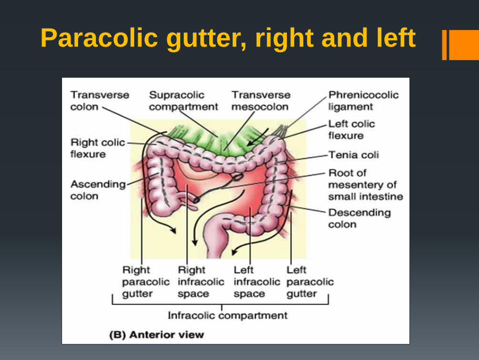
Paracolic gutter, right and left
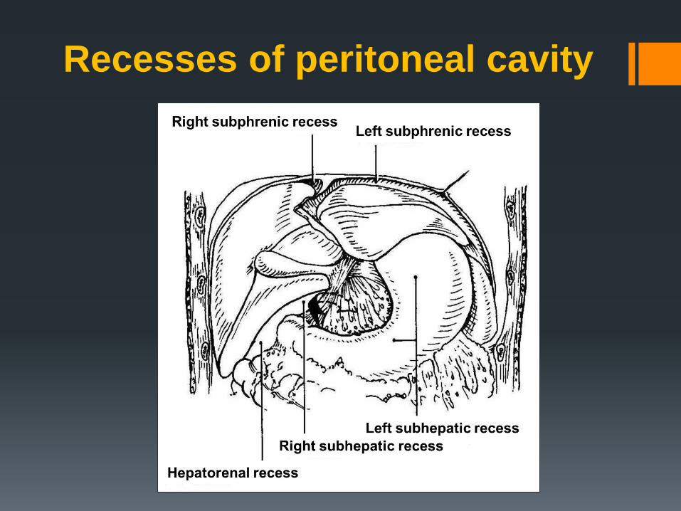
Recesses of peritoneal cavity
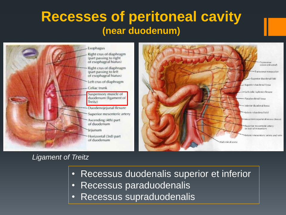
• Recessus duodenalis superior et inferior
• Recessus paraduodenalis
• Recessus supraduodenalis
Recesses of peritoneal cavity (near duodenum)
Ligament of Treitz
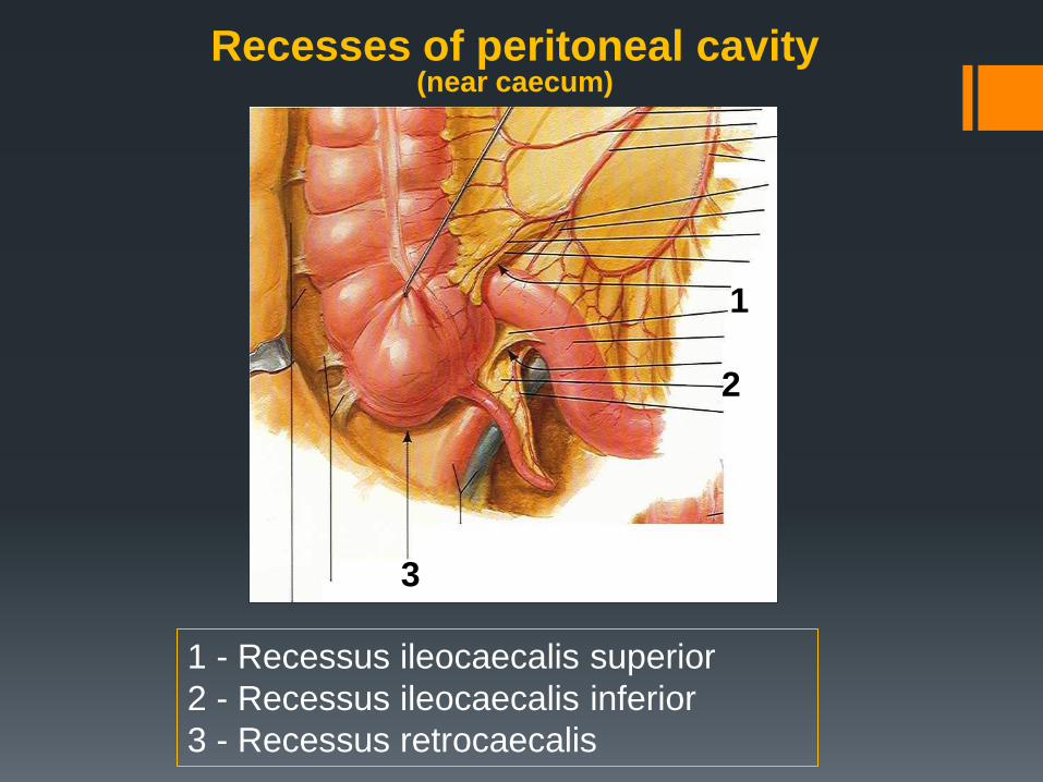
1 - Recessus ileocaecalis superior
2 - Recessus ileocaecalis inferior
3 - Recessus retrocaecalis
1
2
3
Recesses of peritoneal cavity (near caecum)
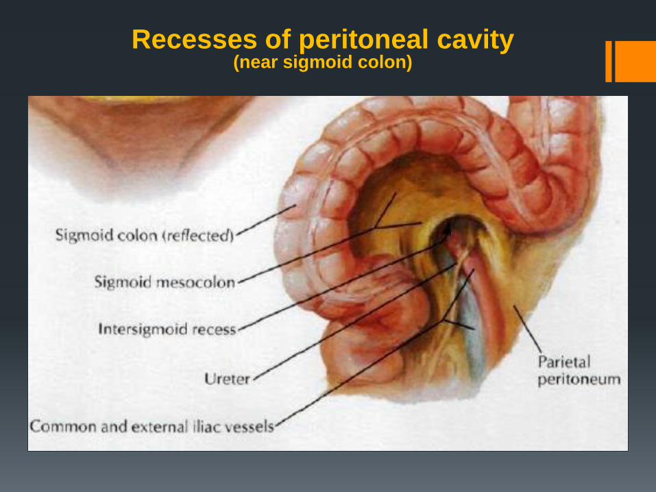
Recesses of peritoneal cavity (near sigmoid colon)
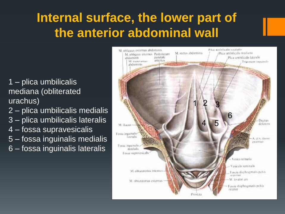
Internal surface, the lower part of
the anterior abdominal wall
1 – plica umbilicalis
mediana (obliterated
urachus)
2 – plica umbilicalis medialis
3 – plica umbilicalis lateralis
4 – fossa supravesicalis
5 – fossa inguinalis medialis
6 – fossa inguinalis lateralis
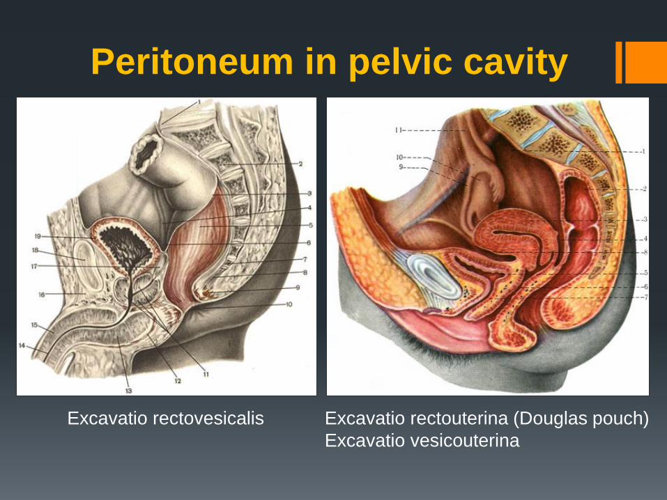
Peritoneum in pelvic cavity
Excavatio rectovesicalis Excavatio rectouterina (Douglas pouch)
Excavatio vesicouterina
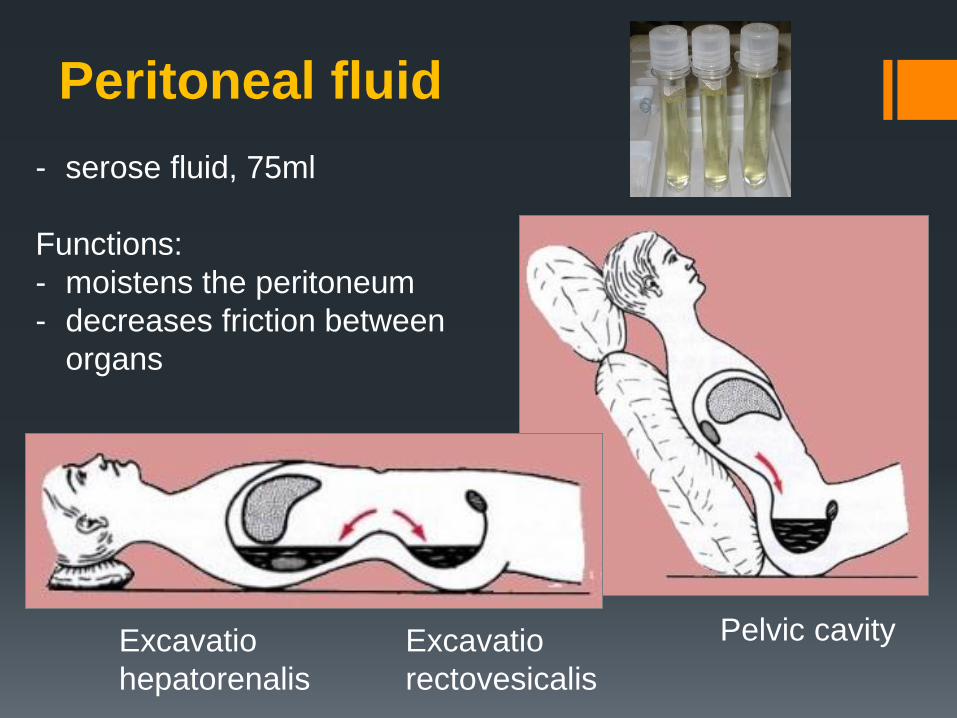
- serose fluid, 75ml
Functions:
- moistens the peritoneum
- decreases friction between
organs
Peritoneal fluid
Excavatio
hepatorenalis
Excavatio
rectovesicalis
Pelvic cavity
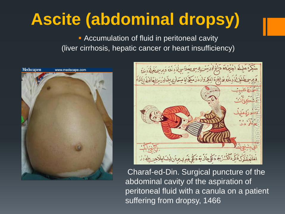
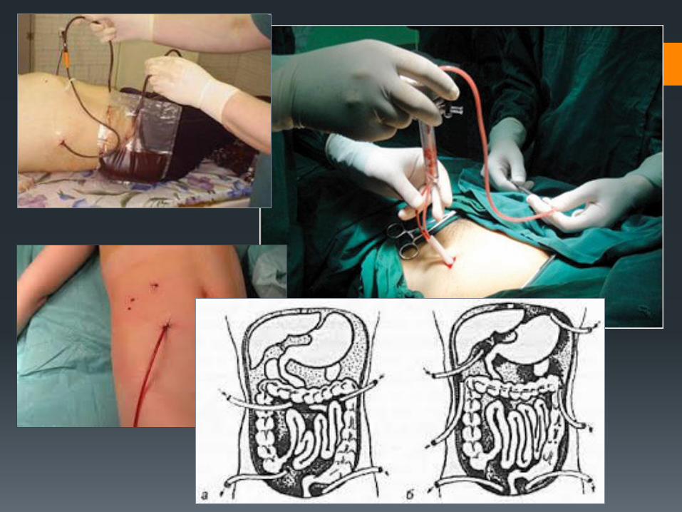
Ascite (abdominal dropsy) Accumulation of fluid in peritoneal cavity
(liver cirrhosis, hepatic cancer or heart insufficiency)
Charaf-ed-Din. Surgical puncture of the
abdominal cavity of the aspiration of
peritoneal fluid with a canula on a patient
suffering from dropsy, 1466

THANK YOU FOR YOUR ATTENTION!


![First Case of Hepatic Polycystic Echinococcosis Involving ...The involvement of liver and mesentery [5] and exclusively . the mesentery [10] are the most reported PE clinical presentation](https://static.fdocuments.net/doc/165x107/5fbdc5051c35c657811004d0/first-case-of-hepatic-polycystic-echinococcosis-involving-the-involvement-of.jpg)


