
Digestion: From Meals 3 to Molecules - University of...
36
3 T he human body has been compared to a car: We fill the tank of our car with gasoline to get down the highway; we fill our body with food to get on with life. In both “machines,” combustion with oxygen releases energy. Our bodies are machine-like in another way as well: They are virtually identical to one another. Like cars, we look different on the outside but are basically the same on the inside. The processes that drive us—like the internal combustion engine, no matter where it’s manufactured—are more similar than different because they are based on the same fundamental chemical reactions. Despite the similarities, however, there are differences between human bodies and ma- chines. An automobile cannot use gasoline to heal itself or to grow, as we do with nutrients. Although a gas tank and a stomach both store fuel, gasoline travels unchanged through the fuel line to the engine, whereas in humans the digestive system must break down the fuel into smaller units before it can be used by the body. Gas-powered cars are fueled only by gasoline, but the human digestive system must process fuel from many sources for use by the “high- performance machine” that is the human body. Digestion: From Meals to Molecules Copyright © 2012 John Wiley & Sons, Inc.
Transcript of Digestion: From Meals 3 to Molecules - University of...

3The human body has been compared to a car:
We fill the tank of our car with gasoline to get
down the highway; we fill our body with food to
get on with life. In both “machines,” combustion
with oxygen releases energy.
Our bodies are machine-like in another way
as well: They are virtually identical to one another.
Like cars, we look different on the outside but are
basically the same on the inside. The processes
that drive us—like the internal combustion engine,
no matter where it’s manufactured—are more
similar than different because they are based on
the same fundamental chemical reactions.
Despite the similarities, however, there are
differences between human bodies and ma-
chines. An automobile cannot use gasoline to
heal itself or to grow, as we do with nutrients.
Although a gas tank and a stomach both store
fuel, gasoline travels unchanged through the
fuel line to the engine, whereas in humans the
digestive system must break down the fuel into
smaller units before it can be used by the body.
Gas-powered cars are fueled only by gasoline,
but the human digestive system must process
fuel from many sources for use by the “high-
performance machine” that is the human body.
Digestion: From Meals to Molecules
Copyright © 2012 John Wiley & Sons, Inc.

63
CHAPTER OUTLINEThe Organization of Life 64
The Digestive System 68
Digestion and Absorption of Nutrients 70
Digestion in Health and Disease 79
Delivering Nutrients and Eliminating Wastes 85
An Overview of Metabolism 90
Stimulate your interest by reading the opening story and looking at the visual.
Scan the Learning Objectives in each section:
p. 64 p. 68 p. 70 p. 79 p. 85 p. 90
Read the text and study all figures and visuals. Answer any questions.
Analyze key features
Process Diagram, p. 64 p. 74 p. 76 p. 86 p. 90
Nutrition InSight, p. 75 p. 83
What a Scientist Sees, p. 78
Thinking It Through, p. 84
Stop: Answer the Concept Checks before you go on:
p. 67 p. 70 p. 78 p. 85 p. 89 p. 91
End of chapter
Review the Summary, Key Terms, and Online Resources.
Answer the Critical and Creative Thinking Questions.
Answer What is happening in this picture?
Complete the Self-Test and check your answers.
CHAPTER PLANNER
Copyright © 2012 John Wiley & Sons, Inc.

atter, be it a meal you are about to eat or the plate you are about to eat it from, is made up of atoms (Figure 3.1). Atoms combine to
form molecules, which can havedifferent properties from those of the atoms they contain. In any living system, the molecules are organized into cells, the smallest units of life. Cells that are simi-lar in structure and function form tissues. The human body contains four types of tissue: muscle, nerve, epithelial, and connective. These tissues are organized in varying
The Organization of LifeLEARNING OBJECTIVES1. Describe the organization of living things, from
atoms to organisms.
2. Name the organ systems that work with the digestive system to deliver nutrients and elimi-nate wastes.
atom The smallest
unit of an element that
retains the properties
of the element.
molecule A group
of two or more atoms
of the same or differ-
ent elements bonded
together.
cell The basic struc-
tural and functional
unit of living things.M
PR
OC
ESS
DIA
GR
AM
Tissues
H O
N
Atoms CellsMolecule
O
OH H
H
H
H
H H
N
1 Atoms linked
by chemical bonds
form molecules.
2 Molecules form the
structures that make
up cells. Each cell is
bounded by a membrane.
In multicellular
organisms, cells are
usually specialized
to perform specific
functions.
3 Groups of similar cells form
tissues. The tissue layers
shown here make up the
stomach wall.
The organization of life begins with atoms that form molecules, which are then organized
into cells to form tissues, organs, and whole organisms.
64 CHAPTER 3 Digestion: From Meals to Molecules
Copyright © 2012 John Wiley & Sons, Inc.

combinations to form organs. In most cases, an organ does not function alone but is part of an or-gan system. Moreover, an organ may be part of more than one or-gan system. For example, the pan-
creas is part of the endocrine system and also part of the digestive system.
The body’s 11 organ systems interact to perform all the functions necessary for life (Table 3.1 on page 66). For example, the digestive system, which is the primary organ system responsible for moving nutri-ents into the body, is assisted by the endocrine system,
which secretes hormones that help regulate how much we eat and how quickly food and nutri-ents travel through the digestive system. The digestive system is also aided by the nervous sys-tem, which sends nerve signals that help control the passage of food through the digestive tract; by the cardiovascular system, which transports nutrients to individual cells in the body; and by the urinary, respiratory, and integu-mentary systems, which eliminate wastes generated in the body.
hormone A chemi-
cal messenger that
is produced in one
location in the body,
is released into the
blood, and travels to
other locations, where
it elicits responses.
THE PLANNER
Organ systemOrgan Organism
4 Organs such as the stomach,
the heart, and the kidneys are
discrete structures that perform
specific functions in the body.
5 A group of organs that
work together to perform a
particular function form an
organ system.
6 The organ systems
work together to ensure
proper functioning of the
entire organism.
organ A discrete
structure composed
of more than one
tissue that performs a
specialized function.
The Organization of Life 65Copyright © 2012 John Wiley & Sons, Inc.

66 CHAPTER 3 Digestion: From Meals to Molecules
The major organ systems of the human body Table 3.1
Organ system What it includes What it does
Nervous
Respiratory
Urinary
Reproductive
Cardiovascular/circulatory
Lymphatic/immune
Copyright © 2012 John Wiley & Sons, Inc.
The Organization of Life 67
(Continued ) Table 3.1
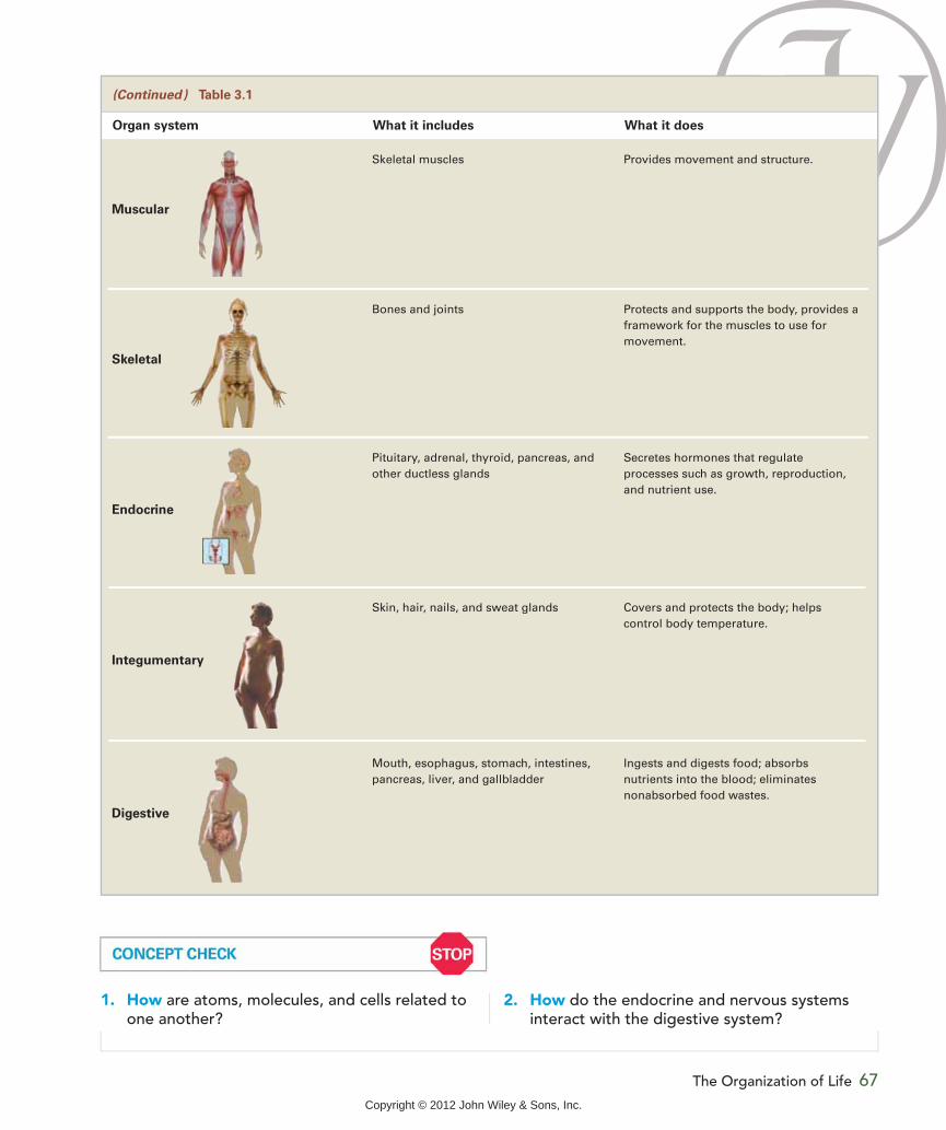
Organ system What it includes What it does
1. How are atoms, molecules, and cells related to one another?
2. How do the endocrine and nervous systems interact with the digestive system?
Muscular
Skeletal
Endocrine
Integumentary
Digestive
Copyright © 2012 John Wiley & Sons, Inc.

68 CHAPTER 3 Digestion: From Meals to Molecules
fats must be digested further. Proteins are broken down into amino acids, most of the carbohydrate is broken down into sugars, and most fats are digested to produce molecules with long carbon chains called fatty acids. The sugars, amino acids, and fatty acids can then be absorbed into the body. The fi-ber in whole grains, fruits, and vegetables cannot be digested and therefore is not absorbed into the body. It and other unab-sorbed substances pass through the digestive tract and are elimi-nated in feces.
he digestive system is the organ system that is primarily responsible for digestion and for the absorption of nutrients into the body. When you eat a taco, for example,
the tortilla, meat, cheese, let-tuce, and tomato are broken apart, releasing the nutrients and other food components they contain. Water, vitamins, and minerals are taken into the body without being bro-ken into smaller units, but proteins, carbohydrates, and
3. Describe the tissues that make up the wall of the gastrointestinal tract.
4. Explain the roles of mucus, enzymes, nerves, and hormones in digestion.
The Digestive SystemLEARNING OBJECTIVES1. Define digestion and absorption.2. List the organs that make up the digestive
system.
digestion The process
by which food is broken
down into components
small enough to be ab-
sorbed into the body.
absorption The pro-
cess of taking substances
from the gastrointestinal
tract into the interior of
the body.
feces Body waste,
including unab-
sorbed food residue,
bacteria, mucus, and
dead cells, which is
eliminated from the
gastrointestinal tract
by way of the anus.
T
Mouth: Chews food and mixes it with saliva
Salivary glands: Produce saliva, which containsa starch-digesting enzyme
Pharynx: Swallows chewed food mixed with saliva
Esophagus: Moves food to the stomach
Liver: Makes bile, which aids indigestion and absorption of fat
Gallbladder: Stores bile and releases it intothe small intestine when needed
Small intestine: Absorbs nutrients into bloodor lymph; most digestion occurs here
Stomach: Churns and mixes food; secretesacid and a protein-digesting enzyme
Pancreas: Releases bicarbonate toneutralize intestinal contents; produces enzymes that digest carbohydrate, protein, and fat
Large intestine: Absorbs water and somevitamins and minerals; home to intestinal bacteria; passes waste material
Anus: Opens to allow waste to leave the body
Colon
Rectum
a. The digestive system consists of the
organs of the digestive tract—mouth,
pharynx, esophagus, stomach, small
intestine, and large intestine—plus four
accessory organs—salivary glands, liver,
gallbladder, and pancreas.
A s k Yo u r s e l f
Copyright © 2012 John Wiley & Sons, Inc.
The Digestive System 69
feces. New mucosal cells are formed continuously to re-place those that die. To allow for this rapid replacement, the mucosa has high nutrient requirements and is one of the first parts of the body to be affected by nutrient deficiencies.
The time it takes food to travel the length of the GI tract from mouth to anus is called the transit time. The shorter the transit time, the more rapidly material is pass-ing through the digestive tract. In a healthy adult, transit time is 24 to 72 hours, depending on the composition of the individual’s diet and his or her level of physical activ-ity, emotional state, health status, and use of medications.
Digestive System SecretionsDigestion is aided by substances secreted into the digestive tract from cells in the mucosa and from a number of accessory or-gans. One of these substances is mucus, which moistens, lubri-cates, and protects the digestive
Organs of the Digestive SystemThe digestive system is composed of the gastrointestinal tract and accessory organs ( ). The gastro-intestinal tract is a hollow tube, about 30 feet long, that runs from the mouth to the anus. It is also called the gut, GI tract, alimentary canal, or digestive tract. The inside of the tube is the lumen ( ). Food in the lu-men is not technically inside the body because it has not been absorbed. When you swallow something that cannot be digested, such as a whole sesame seed or an unpopped kernel of popcorn, it passes through your digestive tract and exits in the feces, without ever entering your blood or cells. Only after substances have been absorbed into the cells that line the intestine can they be said to be inside the body.
The lumen is lined with a layer of mucosal cells called the mucosa. Because mucosal cells are in direct contact with churning food and harsh digestive secre-tions, they live only about two to five days. The dead cells are sloughed off into the lumen, where some components are digested and absorbed and the rest are eliminated in
mucus A viscous
fluid secreted by
glands in the digestive
tract and other parts
of the body. It lubri-
cates, moistens, and
protects cells from
harsh environments.
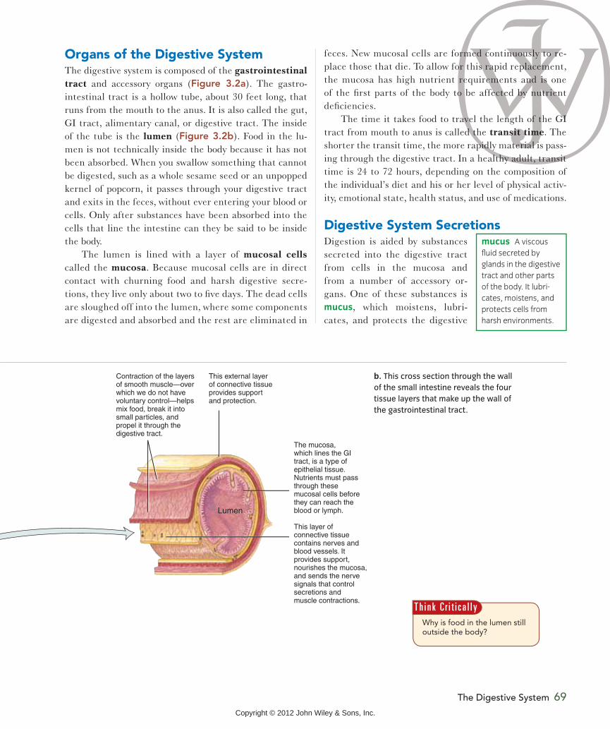
Lumen
The mucosa,which lines the GItract, is a type ofepithelial tissue.Nutrients must passthrough thesemucosal cells beforethey can reach theblood or lymph.
This layer ofconnective tissuecontains nerves andblood vessels. Itprovides support,nourishes the mucosa, and sends the nervesignals that controlsecretions and muscle contractions.
Contraction of the layers of smooth muscle—overwhich we do not have voluntary control—helps mix food, break it intosmall particles, and propel it through thedigestive tract.
This external layerof connective tissueprovides supportand protection.
b. This cross section through the wall
of the small intestine reveals the four
tissue layers that make up the wall of
the gastrointestinal tract.
Why is food in the lumen still outside the body?
T h i n k C r i t i c a l l y
Copyright © 2012 John Wiley & Sons, Inc.

70 CHAPTER 3 Digestion: From Meals to Molecules
signal the pancreas and gallbladder to secrete digestive substances into the small intestine.
tract. Enzymes are also present in digestive system secretions. They accelerate the chemical re-actions that break down food into units small enough to be absorbed (Figure 3.3).
The gastrointestinal tract is part of the endocrine sys-tem as well as the digestive system. It releases hormones that help prepare different parts of the gut for the arrival of food and thus regulate digestion and the rate at which food moves through the digestive system. Some hormonal signals slow digestion, whereas others facilitate it. For example, when the nutrients from your lunch reach your small intestine, they trigger the release of hormones that
Smaller carbohydrate molecules Large carbohydrate molecule
EnzymeEnzyme
Enzyme
Enzymes are needed to break down
different food components. The
enzyme shown here, called an amylase,
breaks large carbohydrate molecules,
such as those in bread, into smaller
ones. Amylases have no effect on fat,
whereas enzymes called lipases digest
fat and have no effect on carbohydrate.
enzyme A protein
molecule that ac-
celerates the rate of
a chemical reaction
without itself being
changed.
Digestion and Absorption of Nutrients
3. Explain how the structure of the small intestine aids in its function.
4. Distinguish passive diffusion from active transport.
magine warm slices of freshly baked bread smeared with melting butter. Is your mouth watering? You don’t even need to put food in your mouth for activity to begin in the diges-
tive tract. Sensory input alone—the sight of the bread
being lifted out of the oven, the smell of the bread, the clatter of the butter knife—may make your mouth water and your stomach begin to secrete digestive substances. This response occurs when the nervous system signals the digestive system to ready itself for a meal. In order for food
I
1. What happens during digestion and absorption?
2. Which organs make up the gastrointestinal tract?
3. What are mucosal cells?
4. How are enzymes important for digestion and absorption?
LEARNING OBJECTIVES
1. Describe what happens in each of the organs of the gastrointestinal tract.
2. Discuss factors that influence how quickly food moves through the GI tract.
Video
Copyright © 2012 John Wiley & Sons, Inc.

to swallow and increases the surface area in contact with digestive juices. The tongue helps mix food with saliva and aids chewing by constantly repositioning food between the teeth. Chewing also breaks up fiber, which traps nutrients. If the fiber is not broken up, some of the nutrients in the food cannot be absorbed. For example, if the fibrous skin of a raisin is not broken open by the teeth, the nutrients inside the raisin remain inaccessible, and the raisin travels, undi-gested, through the intestines for elimination in the feces.
The PharynxThe pharynx, the part of the gastrointestinal tract that is responsible for swallowing, is also part of the respira-tory tract. Food passes through the pharynx on its way to the stomach, and air passes through the pharynx on its way to and from the lungs. As we prepare to swallow, the tongue moves the bolus of chewed food mixed with saliva to the back of the mouth. During swallowing, the air passages are blocked by a valvelike flap of tissue called the epiglottis so that food goes to the esophagus and not to the lungs (Figure 3.4a). Sometimes eating too quickly or talking while eating interferes with the movement of the epiglottis, and food passes into an upper air passageway. This food can usually be dislodged with a cough, but if it becomes stuck and causes choking, it may need to be forced out by means of the Heimlich maneuver (Figure 3.4b).
to be used by the body, however, you need to do more than smell your meal. The food must be consumed and digested, and the nutrients must be absorbed and transported to the body’s cells. This involves the combined functions of all the organs of the digestive system, as well as the help of some other organ systems.
The MouthDigestion involves chemical and mechanical processes, both of which begin in the mouth. The presence of food
in the mouth stimulates the flow of saliva from the salivary glands. Saliva moistens the food and car-ries dissolved food molecules to the taste buds, most of which are located on the tongue. Signals from the taste buds, along with
the aroma of food, allow us to enjoy the taste of the food we eat. Saliva contains the enzyme salivary amylase, which begins the chemical digestion of food by breaking starch molecules into shorter sugar chains (see Figure 3.3). Saliva also helps protect against tooth decay because it washes away food particles and contains substances that inhibit the growth of bacteria that cause tooth decay.
Chewing food begins the mechanical aspect of diges-tion. Adult humans have 32 teeth, which are specialized for biting, tearing, grinding, and crushing foods. Chewing breaks food into small pieces. This makes the food easier
saliva A watery
fluid that is produced
and secreted into the
mouth by the salivary
glands. It contains
lubricants, enzymes,
and other substances.
epiglottis A piece
of elastic connective
tissue that covers the
opening to the lungs
during swallowing.
BolusPharynx
Epiglottis
Esophagus
Passagewayto lungs
Epiglottis returns to its original positiononce bolus has passed; airway to lungs reopens
Bolus forces epiglottis tocover passageway to lungs Diaphragm
a. When a bolus of food is swallowed, it normally pushes the epiglottis down
over the opening to the passageway that leads to the lungs.
b. If food becomes lodged in the passageway
leading to the lungs, it can block the flow of air.
The Heimlich maneuver, which involves a series of
thrusts directed upward from under the diaphragm
(the muscle separating the chest and abdominal
cavities), forces air out of the lungs, blowing the
lodged food out of the air passageway.
Copyright © 2012 John Wiley & Sons, Inc.

72 CHAPTER 3 Digestion: From Meals to Molecules
the tube of the digestive tract and acts as a valve. When the sphincter contracts, the valve is closed; when it re-laxes, the valve is open (see Figure 3.5). The sphincter, located between the esophagus and the stomach, prevents food from moving from the stomach back into the esopha-gus, but occasionally stomach contents do move in this di-rection. This is what occurs with heartburn (as discussed later in this chapter): Some of the acidic stomach contents leak up through this sphincter into the esophagus, causing a burning sensation.
Food also moves from the stomach into the esophagus during vomiting. Vomiting is initiated by a complex series of signals from the brain that cause the sphincter to relax and the muscles to contract, forcing the stomach contents upward, out of the stomach and toward the mouth.
The EsophagusThe esophagus connects the pharynx with the stomach. In the esophagus, the bolus of food is moved along by rhyth-mic contractions of the smooth muscles, an action called
peristalsis (Figure 3.5). The con-tractions of peristalsis are strong enough so that even if you ate while standing on your head, food would reach your stomach. This
contractile movement, which is controlled automatically by the nervous system, occurs throughout the gastroin-testinal tract, pushing the bolus along from the pharynx through the large intestine.
To leave the esophagus and enter the stomach, food must pass through a sphincter, a muscle that encircles
peristalsis Coordi-
nated muscular con-
tractions that move
material through the
GI tract.
Food bolus
Circular musclescontract, pushingthe bolus down
Longitudinal musclescontract, shortening the passageway aheadof the bolus
Sphincterclosed
Esophagus
Sphincteropen
Stomach
The rhythmiccontractionsof peristalsis propel fooddown the esophagus.
When a wave ofperistaltic contractionreaches the stomach,it causes the sphincterto relax, allowing thebolus to enter the stomach.
The food we swallow doesn’t just fall down the esophagus and into the stomach. It
is pushed along by muscular contractions and enters the stomach in response to the
opening and closing of the sphincter, located where the esophagus meets the stomach.
Copyright © 2012 John Wiley & Sons, Inc.

Digestion and Absorption of Nutrients 73
pits that dot the stomach lining (Figure 3.6b). Gastric juice is a mixture of water, mucus, hydrochloric acid, and an inactive form of the protein-digesting enzyme pepsin. This enzyme is secreted in an inactive form so that it will not damage the gastric glands that produce it. The hydrochloric acid in gastric juice kills most of the bacteria present in food. It also stops the activity of the carbohydrate-digesting enzyme salivary amylase and helps begin the digestion of protein by activating pepsin and unfolding proteins. A thick layer of mucus prevents the protein that makes up the stomach wall from being damaged by the hydrochloric acid and pepsin in gastric juice.
The StomachThe stomach is an expanded portion of the gastrointestinal tract that serves as a temporary storage place for food. Here the bolus is mashed and mixed with highly acidic stomach secretions to form a semiliquid food mass called chyme. The mixing of food in the stomach is aided by an extra layer of smooth muscle in the stomach wall (Figure 3.6a). Some di-gestion takes place in the stomach, but, with the exception of some water, alcohol, and a few drugs, such as aspirin and acetaminophen (Tylenol), very little absorption occurs here.
Gastric juice Chemical digestion in the stomach is caused by gastric juice produced by gastric glands in
Esophagus
Sphincter
Sphincter
Surface epithelium
Different cell types secrete mucus, hydrochloric acid, and inactive pepsin
Gastric glands
Small intestine
Mucosa
Gastric pits
Connective tissue
Connective tissue
Three smoothmuscle layers
The stomach has three musclelayers: longitudinal, circular, and diagonal.
a. Most of the gastrointestinal tract is surrounded by two layers
of smooth muscle, one that is longitudinal and one that is
circular, but the stomach contains a third smooth muscle layer
running diagonally. The presence of this diagonal layer allows
for the powerful contractions that churn and mix the stomach
contents. The sphincter at the bottom of the stomach controls
the flow of chyme into the small intestine.
b. The lining of the stomach is covered with gastric pits. Inside
these pits are the gastric glands, made up of different types of
cells that produce the mucus, hydrochloric acid, and the inactive
form of pepsin contained in gastric juice.
Why is the protein-digesting enzyme pepsin produced in an inactive form?
T h i n k C r i t i c a l l y
Copyright © 2012 John Wiley & Sons, Inc.
74 CHAPTER 3 Digestion: From Meals to Molecules
the nutrient composition of a meal affects how quickly it leaves your stomach, it affects how soon after eating you will feel hungry again (Figure 3.8).
Regulation of stomach activity How much your stomach churns, how much gastric juice is released, and how fast material empties out of the stomach are regulat-ed by signals from both nerves and hormones. These sig-nals originate from three sites—the brain, the stomach, and the small intestine (Figure 3.7).
As chyme moves out of the stomach, signals sent by the small intestine help regulate the rate at which the stomach empties. The small intestine stretches as it fills with chyme; this distension inhibits the stomach from emptying. Chyme normally empties from the stomach within two to six hours, but this rate varies with the size and composition of the meal that has been consumed. A large meal takes longer to leave the stomach than does a small meal. Liquids empty quickly, but solids linger until they are well mixed with gastric juice and are liquefied; hence, solids leave the stomach more slowly than liquids.
The nutritional composition of a meal also affects how long it stays in the stomach. A meal that consists primar-ily of starch or sugar leaves quickly, but a meal that is high in fiber or protein takes longer to leave the stomach. A high-fat meal stays in the stomach the longest. Because
PR
OC
ESS
DIA
GR
AM
Food
Decrease gastricsecretionand motility
Increase gastricsecretionand motility
Food
Gastrin
HormonesFood
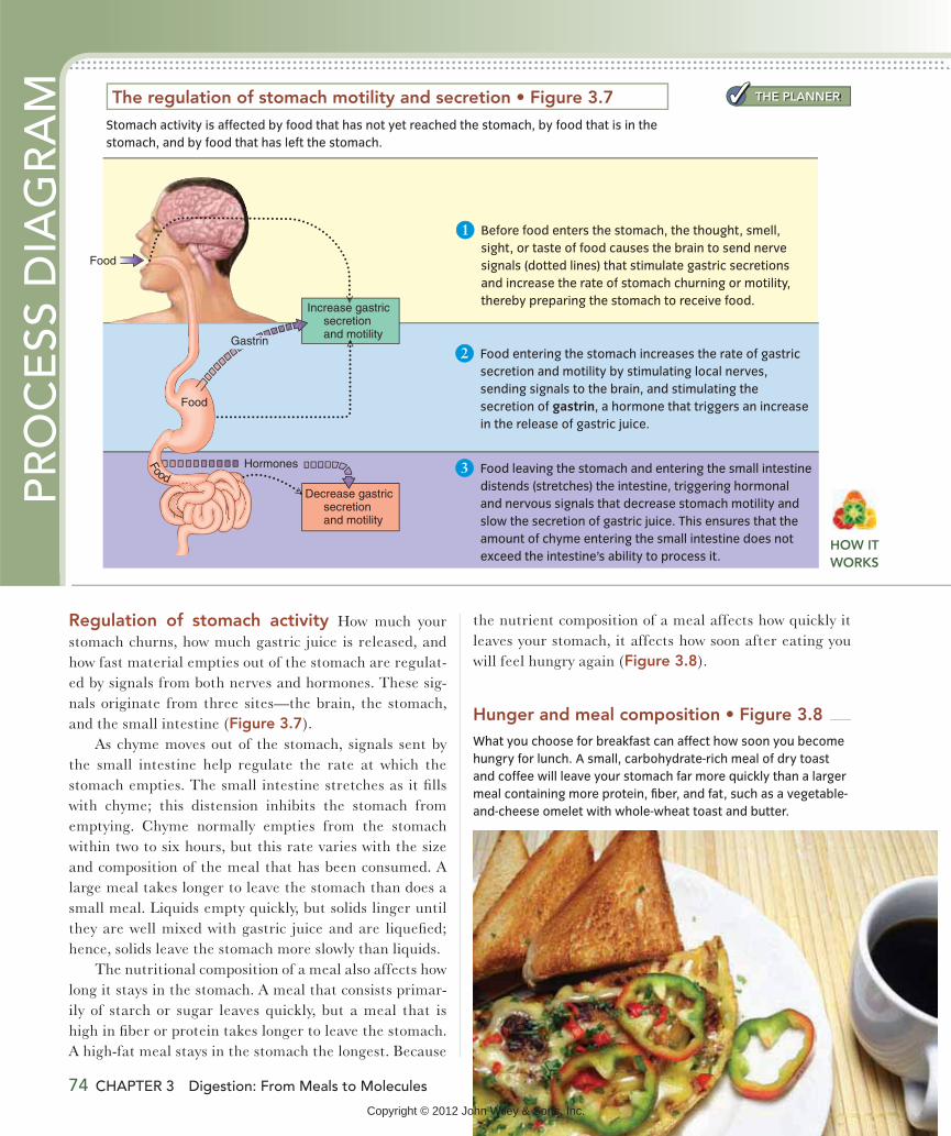
1 Before food enters the stomach, the thought, smell,
sight, or taste of food causes the brain to send nerve
signals (dotted lines) that stimulate gastric secretions
and increase the rate of stomach churning or motility,
thereby preparing the stomach to receive food.
2 Food entering the stomach increases the rate of gastric
secretion and motility by stimulating local nerves,
sending signals to the brain, and stimulating the
secretion of gastrin, a hormone that triggers an increase
in the release of gastric juice.
3 Food leaving the stomach and entering the small intestine
distends (stretches) the intestine, triggering hormonal
and nervous signals that decrease stomach motility and
slow the secretion of gastric juice. This ensures that the
amount of chyme entering the small intestine does not
exceed the intestine’s ability to process it.
Stomach activity is affected by food that has not yet reached the stomach, by food that is in the
stomach, and by food that has left the stomach.
What you choose for breakfast can affect how soon you become
hungry for lunch. A small, carbohydrate-rich meal of dry toast
and coffee will leave your stomach far more quickly than a larger
meal containing more protein, fiber, and fat, such as a vegetable-
and-cheese omelet with whole-wheat toast and butter.
THE PLANNER
HOW IT WORKS
Copyright © 2012 John Wiley & Sons, Inc.

absorption (Figure 3.9). Together these features pro-vide a surface area that is about the size of a tennis court (about 2700 ft2).
Secretions that aid digestion In the small intes-tine, secretions from the pancreas, the gallbladder, and the small intestine itself aid digestion. The pancreas se-cretes pancreatic juice, which contains bicarbonate, and digestive enzymes. Bicarbonate, which is a base, neu-tralizes the acid in the chyme, making the environment in the small intestine neutral or slightly basic rather than acidic, as in the stomach. This neutrality allows enzymes from the pancreas and small intestine to function.
The Small IntestineThe small intestine is a narrow tube about 20 feet long. Here the chyme is propelled along by peristalsis and mixed by rhythmic constrictions called segmentation that slosh the material back and forth. The small intestine is the main site for the chemical digestion of food, completing the process that the mouth and stomach have started. It is also the primary site for the absorption of water, vitamins, minerals, and the products of carbohydrate, fat, and pro-tein digestion.
The small intestine has a number of unique struc-tural features that contribute to its digestive function and enhance the amount of surface area available for
Nutrition InSight The structure of the small intestine
Large circularfolds
VilliMicrovilli
Lacteal
Capillary
Artery
Vein
Lymphvessel
Mucosal cell
Lumen
SMALL INTESTINE
Microvilli
b. The entire inner surface of
the small intestine is covered
with fingerlike projections
called villi (the singular is villus).
Each villus contains a capillary
(small blood vessel) and a lacteal
(small lymph vessel). Nutrients must
cross only the single-cell layer of
the mucosa to reach the blood
or lymph for delivery to the tissues
of the body.
a. The wall of the small
intestine is arranged
in large circular folds,
which increase the
surface area in contact
with nutrients.
c. Each villus is
covered with tiny
projections of
the mucosal cell
membrane called
microvilli (the singular
is microvillus), often
referred to as the
brush border. Some of
the digestive enzymes
produced by the small
intestine are located
in the membrane,
and some are located
inside the mucosal
cells.
A s k Yo u r s e l fWhat are the three structural features of the small intestine that increase its surface area?
THE PLANNER
Copyright © 2012 John Wiley & Sons, Inc.

two sugar units) into single sugar units and the digestion of short amino acid chains into single amino acids. The sugars from carbohydrate digestion and the amino acids from pro-tein digestion pass into the blood and are delivered to the liver (Figure 3.10).
The gallbladder stores and secretes bile, a substance that is produced in the liver and is neces-sary for the digestion and absorp-tion of fat. Bile that is secreted into the small intestine mixes with fat and divides it into small globules,
Pancreatic amylase is an enzyme that continues the job of breaking down starches into sugars that was started in the mouth by salivary amylase. Pancreatic proteases (protein-digesting enzymes), such as trypsin and chymo-trypsin, break protein into shorter and shorter chains of amino acids, and fat-digesting enzymes called lipases break down fats into fatty acids. The pancreatic proteases, like the pepsin produced by the stomach, are released in an inactive form so that they will not digest the glands that produce them. Intestinal digestive enzymes, found in the cell membranes or inside the cells lining the small intes-tine, aid the digestion of double sugars (those that contain
bile A digestive
fluid made in the
liver and stored in the
gallbladder that is
released into the small
intestine, where it aids
in fat digestion and
absorption.
Most digestion occurs in the small intestine.
PR
OC
ESS
DIA
GR
AM
Fiber
Blood
Lymph
To liver
To blood stream
Singlesugars
Aminoacids
Lipid transportparticles
Sugars,starches andfiber (from dietarycarbohydrate)
Long aminoacid chains(from dietaryprotein)
Large lipid droplets(from dietaryfat)
Singleand doublesugars
Amino acidsand short amino acid chains Small lipid
droplets
Fatty acids
8
7
6
5
4
2
Enzymes in the microvilli breakshort amino acid chains intosingle amino acids and intochains containing only two orthree amino acids. These areabsorbed into the mucosal cells,where they are digested intosingle amino acids, which passinto the blood.
Bile helps divide largefat globules. Pancreaticlipases digest the fatmolecules into fatty acidsthat combine with bile andother lipids to formsmall droplets.
Small lipid droplets aidthe absorption of fattyacids and other fat-soluble substances intothe mucosal cell.
Absorbed fats areincorporated intotransport particles thatpass into the lymph.
The digestion of starch todouble sugars and short sugarchains is completed by theenzyme pancreatic amylase.
1
Fiber, which cannot bedigested by humanenzymes, passes tothe large intestine.
Enzymes in the microvilli digestdouble sugars into single sugars,which are absorbed into the bloodfor transport to the liver.
3
The digestion of proteinsinto amino acids and shortamino acid chains is completedby pancreatic protein-digesting enzymes (proteases).
THE PLANNER
76 CHAPTER 3 Digestion: From Meals to Molecules
Copyright © 2012 John Wiley & Sons, Inc.
Digestion and Absorption of Nutrients 77
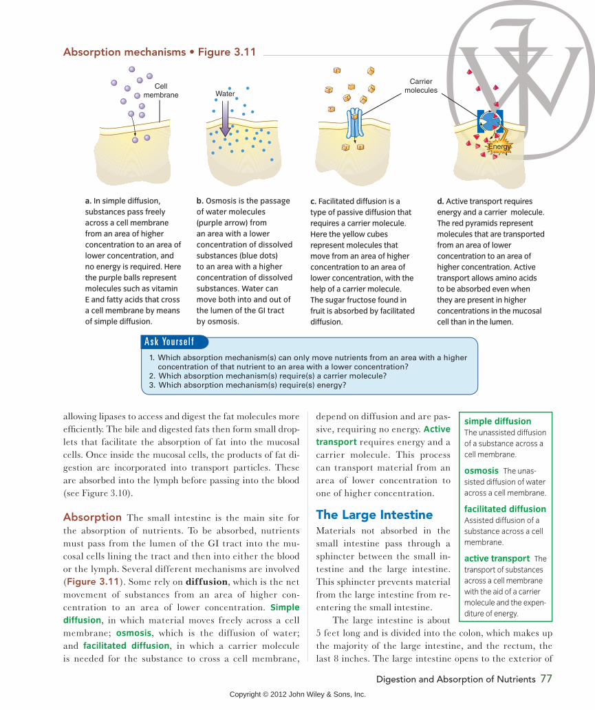
depend on diffusion and are pas-sive, requiring no energy. Active
transport requires energy and a carrier molecule. This process can transport material from an area of lower concentration to one of higher concentration.
The Large IntestineMaterials not absorbed in the small intestine pass through a sphincter between the small in-testine and the large intestine. This sphincter prevents material from the large intestine from re-entering the small intestine.
The large intestine is about 5 feet long and is divided into the colon, which makes up the majority of the large intestine, and the rectum, the last 8 inches. The large intestine opens to the exterior of
allowing lipases to access and digest the fat molecules more efficiently. The bile and digested fats then form small drop-lets that facilitate the absorption of fat into the mucosal cells. Once inside the mucosal cells, the products of fat di-gestion are incorporated into transport particles. These are absorbed into the lymph before passing into the blood (see Figure 3.10).
Absorption The small intestine is the main site for the absorption of nutrients. To be absorbed, nutrients must pass from the lumen of the GI tract into the mu-cosal cells lining the tract and then into either the blood or the lymph. Several different mechanisms are involved (Figure 3.11). Some rely on diffusion, which is the net movement of substances from an area of higher con-centration to an area of lower concentration. Simple
diffusion, in which material moves freely across a cell membrane; osmosis, which is the diffusion of water; and facilitated diffusion, in which a carrier molecule is needed for the substance to cross a cell membrane,
Water
Energy
Cellmembrane
Carriermolecules
a. In simple diffusion,
substances pass freely
across a cell membrane
from an area of higher
concentration to an area of
lower concentration, and
no energy is required. Here
the purple balls represent
molecules such as vitamin
E and fatty acids that cross
a cell membrane by means
of simple diffusion.
b. Osmosis is the passage
of water molecules
(purple arrow) from
an area with a lower
concentration of dissolved
substances (blue dots)
to an area with a higher
concentration of dissolved
substances. Water can
move both into and out of
the lumen of the GI tract
by osmosis.
c. Facilitated diffusion is a
type of passive diffusion that
requires a carrier molecule.
Here the yellow cubes
represent molecules that
move from an area of higher
concentration to an area of
lower concentration, with the
help of a carrier molecule.
The sugar fructose found in
fruit is absorbed by facilitated
diffusion.
d. Active transport requires
energy and a carrier molecule.
The red pyramids represent
molecules that are transported
from an area of lower
concentration to an area of
higher concentration. Active
transport allows amino acids
to be absorbed even when
they are present in higher
concentrations in the mucosal
cell than in the lumen.
simple diffusion
The unassisted diffusion
of a substance across a
cell membrane.
osmosis The unas-
sisted diffusion of water
across a cell membrane.
facilitated diffusion
Assisted diffusion of a
substance across a cell
membrane.
active transport The
transport of substances
across a cell membrane
with the aid of a carrier
molecule and the expen-
diture of energy.
A s k Yo u r s e l f
Copyright © 2012 John Wiley & Sons, Inc.

78 CHAPTER 3 Digestion: From Meals to Molecules
Material that is not absorbed in the colon passes into the rectum, where it is stored temporarily and then evacu-ated through the anus as feces. The feces are a mixture of undigested, unabsorbed matter, dead cells, secretions from the GI tract, water, and bacteria. The amount of bac-teria varies but can make up more than half the weight of the feces. The amount of water in the feces is affected by fiber and fluid intake. Because fiber retains water, when adequate fiber and fluids are consumed, feces have a high-er water content and are more easily passed.
the body at the anus. Although most nutrient absorption occurs in the small intestine, water and some vitamins and minerals are also absorbed in the colon.
Peristalsis occurs more slowly in the large intestine than in the small intestine. Water, nutrients, and fecal matter may spend 24 hours in the large intestine, in contrast to the 3 to 5 hours it takes these materials to move through the small intestine. This slow movement favors the growth of bacteria. These bacteria, called the intestinal microflora, are permanent, beneficial residents of this part of the gas-trointestinal tract (see What a Scientist Sees). They break down unabsorbed portions of food, such as fiber, produc-ing nutrients that can be used by the microflora or, in some cases, absorbed into the body. For example, the microflora synthesize small amounts of certain B vitamins and vitamin K, some of which can be absorbed. As the microflora break down material in the colon, they produce gas, which causes flatulence. In a healthy adult, between 200 and 2000 ml of gas is produced in the intestine each day.
The human gut is home to 300 to 500 species of bacteria. The
right mix of bacteria is important for immune function, proper
growth and development of colon cells, and optimal intestinal
motility and transit time.1 Having healthy microflora can inhibit
the growth of harmful bacteria and has been shown to prevent
the diarrhea associated with antibiotic use and to reduce the
duration of diarrhea resulting from intestinal infections and other
a. Ads claim that eating these specialized probiotic yogurts
will help regulate the digestive system. Consumers see these
products as a tasty way to help regulate digestion. Scientists
recognize that these products as well as most other yogurts
contain active cultures of beneficial bacteria, including
Lactobacillus and Bifidobacterium.
Colon mucosa
Without probiotics With probiotics
Beneficial bacteria Harmful bacteria
b. When beneficial bacteria are consumed in adequate amounts,
they live temporarily in the colon, where they inhibit the growth
of harmful bacteria and confer other health benefits on the host.
However, the bacteria must be consumed frequently because
they are flushed out in the feces.
WHAT A SCIENTIST SEESBacteria on the Menu
Why might consuming a prebiotic increase the numbers of beneficial bacteria in the gut?
T h i n k C r i t i c a l l y
1. What are the functions of the stomach?
2. How does food move through the GI tract?
3. How do the villi and microvilli aid absorption?
4. Why is active transport needed for the com-plete absorption of some nutrients?
causes.2,3 There is also evidence that having healthy microflora
may relieve constipation, reduce allergy symptoms, and modify
the risk of inflammatory bowel disease and colon cancer.4,5,6
Consuming these beneficial bacteria, called probiotics, is one
way of promoting healthy microflora. Another way is to consume
prebiotics, substances that serve as a food supply for beneficial
bacteria.
THE PLANNER
Copyright © 2012 John Wiley & Sons, Inc.

Digestion in Health and Disease 79
Digestion in Health and DiseaseIf an invading substance, or antigen, enters the lu-
men or is absorbed into the mucosa, the immune system can use a number of weapons to destroy it. These include various types of white blood cells, which circulate in the blood and reside in the mucosa of the gastrointestinal tract. They can quickly destroy most antigens that enter the body through the mucosa.
When an antigen is present, phagocytes are the first type of white blood cell to come to the body’s defense ( ). If the invader is not eliminated by the phagocytes, more specific white blood cells called lym-phocytes join the battle. Some lymphocytes destroy specific an-tigens by binding to them. This type of lymphocyte helps eliminate cancer cells, foreign tissue, and cells that have been infected by viruses and bacteria. Other lymphocytes produce and secrete protein molecules called antibodies. Antibodies bind to antigens and help destroy them. Each antibody is designed to fight off only one type of antigen. Once antibodies to a specific antigen have been made, the immune system remembers and is ready to fight that antigen any time it enters the body again.
The health of the GI tract is essential to our over-all health. The gut acts as a defense against in-vasion by disease-causing organisms and other contaminants and allows us to obtain nutrients
efficiently. Food allergies, which can be life-threatening, have their origins in the GI tract, but most common gastrointesti-nal problems are minor and do not affect long-term health.
The Digestive System and Disease PreventionFood almost always contains bacteria and other contami-nants, but it rarely makes us sick. This is because the mucosa of the GI tract contains tissue that is part of the immune system ( ). This tissue prevents dis-ease-causing bacteria and toxins from taking over the GI tract and invading the body.
LEARNING OBJECTIVES1. Explain how the gastrointestinal tract protects
us from infection.2. Describe the causes of food allergies.3. Discuss the causes and consequences of ulcers,
heartburn, and GERD.4. Explain how dental problems and gallstones
might affect food intake.
a. The darkly stained areas shown here are called Peyer’s
patches. They are made up of immune system tissue and are
embedded throughout the mucosa of the small intestine. Peyer’s
patches contain cells that participate in the immune system’s
efforts to prevent harmful organisms or materials present in the
GI tract from making us ill.
Peyer’s patches
Lymphocyte
Phagocyte
b. The cells shown here in pink are phagocytes, which can engulf
and destroy invading substances. The cells shown in green are lymphocytes, which are specific with regard to which invaders
they can attack. Some lymphocytes directly kill invaders, while
others secrete antibodies that help destroy antigens.
antigen A foreign
substance that, when
introduced into the
body, stimulates an im-
mune response.
antibody A protein,
released by a type of
lymphocyte, that inter-
acts with and deacti-
vates specific antigens.
Copyright © 2012 John Wiley & Sons, Inc.

80 CHAPTER 3 Digestion: From Meals to Molecules
If harmful organisms infect the GI tract, the body may help out the immune system by using diarrhea or vomiting to flush them out.
Food allergies Our immune system protects us from many invaders without our being aware of it. Unfortu-nately, the response of the immune system to a foreign substance is also to blame for allergic reactions. An al-
lergic reaction occurs when the immune system produces an-tibodies to a substance, called an allergen, that is present in
our diet or environment. Food allergies occur when the body sees proteins present in food as foreign substances and therefore initiates an immune response. The immune response causes symptoms that range from hives to life-threatening reactions such as breathing difficulties or a drop in blood pressure.
The first time a food is consumed, it does not trigger an allergic reaction, but in a susceptible person, this first exposure begins the process. As the food is digested, tiny fragments of undigested protein trigger the production of antibodies. When the food protein is eaten again, it binds to the antibodies, signaling the release of chemicals that cause redness, swelling, and other allergy symptoms. When the protein enters the mouth, the allergic person
may experience an itching or tingling sensation on the tongue or lips. As the protein travels down to the stomach and intestines, the allergic response may lead to vomiting and cramps. After the protein fragments are absorbed and travel through the blood, they may cause a drop in blood pressure, hives, and breathing difficulties.
The best way to avoid allergy symptoms is to avoid foods to which you are allergic (Figure 3.13).
Celiac disease Celiac disease is a condition in which the protein gluten, found in wheat, barley, and rye, trig-gers an immune system response that damages or de-stroys the villi of the small intestine. For most of us the gluten in our foods is digested and absorbed like other proteins. However, for people with this disease, consum-ing even a tiny amount of gluten can cause abdominal pain, diarrhea, and fatigue. Eventually this damage can lead to malnutrition, weight loss, anemia, osteoporo-sis, intestinal cancer, and other chronic illnesses.8,9 Ce-liac disease, also called gluten intolerance, celiac sprue, nontropical sprue, and gluten-sensitive enteropathy, is an inherited condition that affects an estimated 1 in 133 people in the population. Although gluten intolerance is currently a trendy condition (See Debate: Should You Be
Gluten Free?), the disease can be diagnosed only by a blood test or an intestinal biopsy. For people with celiac disease,
Calories from Fat 572
Total Fat 63.5g 98%Saturated Fat 12.5gTrans Fat 0g
63%
Cholesterol 70mg 0%37%
16%
Sodium 875mg15%Total Carbohydrate 44g
Dietary Fiber 4g
Sugars 4g
Protein
ALLERGY INFORMATION:
Contains cashews.
Processed in a facility that also processes products that contain peanuts, other tree nuts, milk, soy and wheat.
Consumers with food allergies,please read the ingredientsstatement carefully.
INGREDIENTS: CASHEWS, PEANUT OIL AND OR COTTONSEED OIL, SALT.
21g
Vitamin A 0% Vitamin C 0%
IronCalcium 61.65%
% Daily Value*
Cholesterol
SodiumTotal CarbohydrateSodium
Less thanLess than
300mg2,400mg300g25g
300mg2,400mg
30g375g
46%
Calories fro
Total Fat 63
Saturated Trans Fat 0s
Cholesterol
Sodium 875
Total Carbo
Dietary Fib
allergen A substance
that causes an allergic
reaction.
Food allergies affect about 2% of adults and 4 to 8% of children
and are responsible for 150 deaths in the United States each
year.7 To protect consumers, food manufacturers are required
to clearly state on the label whether a product contains any
of the eight major ingredients that are most likely to cause
allergic reactions: peanuts, tree nuts, milk, eggs, fish, shellfish,
soy, and wheat.
Copyright © 2012 John Wiley & Sons, Inc.

Debate Should You Be Gluten Free?
The Issue: Gluten-free diets are essential for people with celiac disease, but a gluten-free diet has also been promoted for weight loss and to treat a host of other ailments. Is gluten free a healthy alternative for everyone?
You see the term “gluten free” on breakfast cereals, cake mixes, pastas, soups, and a host of other products. Celebrities are touting the benefits of going gluten free. Chelsea Clinton even had her wedding cake baked without gluten. The increase in the number of gluten-free foods over the past few years is partly due to greater awareness and better diagnosis of celiac disease; however, a switch to gluten-free products has also been promoted as a healthier way of eating for everyone. Advocates claim that a gluten-free diet will promote weight loss and help those suffering with joint pain, rheumatoid arthritis, osteopo-rosis, anemia, and diabetes. They contend that individuals with these symptoms have undiagnosed celiac disease. Although a small number of people may benefit from a gluten-free diet be-cause they have celiac disease but no obvious symptoms, eating a gluten-free diet is unlikely to cure these conditions in people who do not have celiac disease.
What about going gluten free for weight loss? Gluten-free foods are not any lower in calories than other foods, but elimi-nating everything that contains gluten from your diet—most types of cereal, bread, pasta, cakes, and cookies—will help cut calories. Gluten-free foods are also not nutritionally superior to other foods, but if you are trying to lose weight, carefully choos-ing everything you put in your mouth will force you to plan your diet carefully and may help with weight loss.
INGREDIENTS: ONIONS, BLEACHEDWHEAT FLOUR, SOYBEAN OIL AND/ORAACANOLA OIL, YELLOW CORN FLOUR,SUGAR, SALT,LL SOY FLOUR, WHEY, DEX-YYTROSE, LEAVENING (MONOCALCIUMAAPHOSPHATE, SODIUM BICARBONAAA TE),AAYEAST, POLYSORBALL TE 80, CALCIUM AAPROPIONATE (PRESERVAA AVV TIVE).AA
Onion Ringsg
INGREDIENTS: POTATT TOES, VEGETAA ABLE TTOIL (PALM, SUNFLOWER, SOYBEAN, AND/OR CANOLA), SALT, DEXTROSE, DISODIUM DIHYDROGEN PYROPHOS-PHATE, ANNAAA TAA TO (VEGETABLE COLOR).TT
French Fries
Is a gluten-free diet harmful? Eliminating gluten involves carefully checking each ingredient in the foods you eat to elimi-nate products made not only from wheat, which is the major grain in the American diet, but also from barley and rye as well as the myriad of foods that have wheat added as a thickener (see the figure). The major problem with a gluten-free diet is that it eliminates most flours, breads, pasta, and breakfast ce-reals, which are important sources of B vitamins and iron. This creates a risk for nutrient deficiencies. A gluten-free diet is not harmful as long as it provides enough of all the nutrients typi-cally consumed in gluten-containing foods. People diagnosed with celiac disease generally work with a dietitian to make sure they have a well-balanced, varied diet that meets all their nutri-ent needs. People eating a gluten-free diet for other reasons generally do not.
So, although there is no research to support the benefits of eliminating gluten if you do not have celiac disease, anything that makes you consider your diet carefully is a good thing. In-dividuals with gluten sensitivity are benefiting from the gluten-free craze because it has increased the availability and quality of gluten-free foods, improved the labeling of gluten-free prod-ucts, and heightened awareness of celiac disease. Proponents think a gluten-free diet will improve everyone’s health. Skep-tics consider gluten-free another trend like the low-carb fad of a few years back.
Think critically: Neither potatoes nor onions contain glu-ten. If you had celiac disease, based on the ingredients shown here, which would be a safer choice: the French fries or the onion rings?
Digestion in Health and Disease 81
THE PLANNER
Copyright © 2012 John Wiley & Sons, Inc.

82 CHAPTER 3 Digestion: From Meals to Molecules
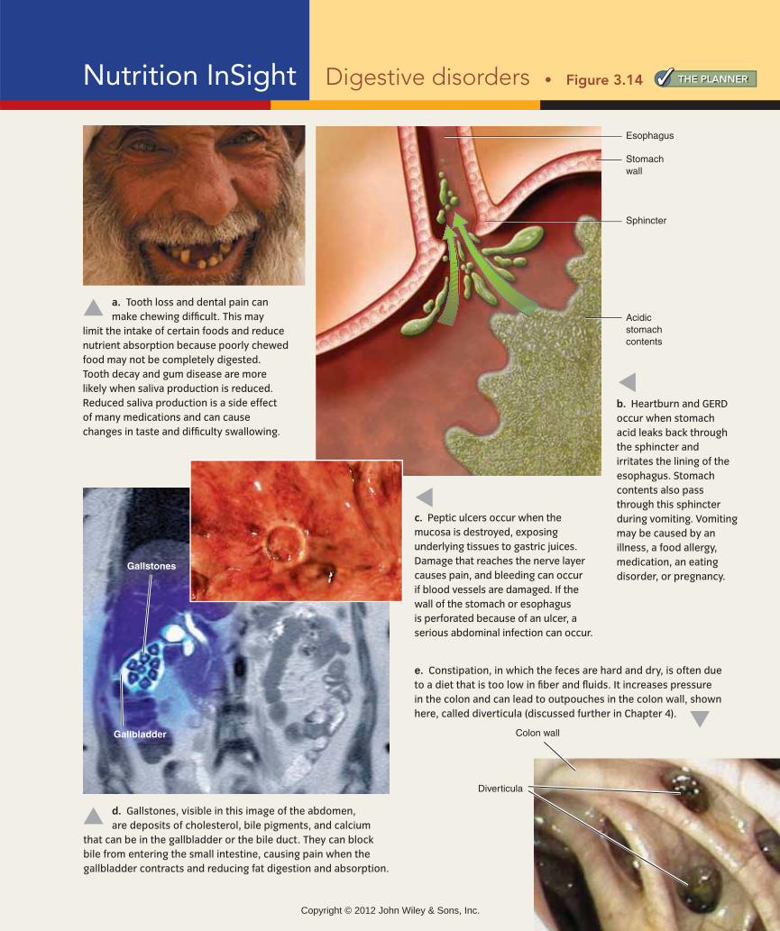
Peptic ulcers Peptic ulcers occur when the mucus barrier protecting the stomach, esopha-gus, or upper small intestine is penetrated and the acid and pepsin in digestive secretions damage the gastrointestinal lining (Figure 3.14c). Mild ulcers cause abdominal pain; more severe ulcers can cause life-threatening bleeding.
Peptic ulcers can result from GERD or from mis-use of medications such as aspirin or nonsteroidal anti-inflammatory drugs (such as Motrin and Advil) but are more often caused by infection with the bacterium Helicobacter pylori (H. pylori). These bacteria burrow into the mucus and destroy the protective mucosal layer.10 Over half of the world’s population is infected with H. pylori, but not everyone who is infected develops ulcers.11 H. pylori
infection can be treated using antibiotics.
Gallstones Clumps of solid material that accumulate in either the gallbladder or the bile duct are referred to as gallstones (Figure 3.14d). They can cause pain when the gallbladder contracts in response to fat in the intestine. Gallstones can interfere with bile secretion and reduce fat absorption. They are usually treated by removing the gallbladder. After the gallbladder has been removed, bile, which is produced in the liver, drips di-rectly into the intestine as it is produced rather than be-ing stored and squeezed out in larger amounts when fat enters the intestine.
Diarrhea and constipation Diarrhea and consti-pation are common discomforts that are related to prob-lems in the intestines. Diarrhea refers to frequent, watery stools. It occurs when material moves through the colon too quickly for sufficient water to be absorbed or when water is drawn into the lumen from cells lining the intestinal tract.
Diarrhea can be caused by bacterial or viral infections, irritants that inflame the lining of the GI tract, the pas-sage of undigested food into the large intestine, medica-tions, and chronic intestinal diseases. Diarrhea causes loss of fluids and minerals. Severe diarrhea lasting more than a day or two can be life-threatening.
Constipation refers to hard, dry stools that are difficult to pass; it occurs when the water content of the stool is too
consuming a diet that eliminates gluten provides relief from symptoms. This means eliminating products made from wheat, barley, or rye, including most breads, crack-ers, pastas, cereals, cakes, and cookies. It also requires eliminating foods ranging from packaged gravies to soy sauce that are processed with these grains.
Digestive System Problems and DiscomfortsAlmost everyone experiences digestive system problems from time to time. These problems often cause discom-fort and frequently limit the types of foods a person can consume (Figure 3.14a). They also can interfere with nu-trient digestion and absorption. Problems may occur any-where in the digestive tract, from the mouth to the anus, and can affect the accessory organs that provide the secre-tions that are essential for proper GI function.
Heartburn and GERD Heartburn occurs when the acidic contents of the stomach leak back into the esopha-gus (Figure 3.14b). The medical term for the leakage of
stomach contents into the esoph-agus is gastroesophageal reflux. Oc-casional heartburn is common, but if it occurs more than twice a week, it may indicate a condition called gastroesophageal reflux
disease (GERD). If left untreated, GERD can eventually lead to more serious health problems, such as esophageal bleeding, ulcers, and cancer.
The discomforts of heart-burn and GERD can be avoided by limiting the amounts and
types of foods consumed. Eating small meals and consum-ing beverages between rather than with meals prevents heartburn by reducing the volume of material in the stom-ach. Avoiding fatty and fried foods, chocolate, peppermint, and caffeinated beverages, which increase stomach acidity or slow stomach emptying, can help minimize symptoms. Remaining upright after eating, wearing loose clothing, avoiding smoking and alcohol, and losing weight may also help prevent heartburn. For many people, medications that neutralize acid or reduce acid secretion are needed to manage symptoms.
heartburn A burning
sensation in the chest
or throat caused when
acidic stomach con-
tents leak back into the
esophagus.
gastroesopha-
geal reflux disease
(GERD) A chronic con-
dition in which acidic
stomach contents leak
into the esophagus,
causing pain and dam-
aging the esophagus.
peptic ulcer An
open sore in the lin-
ing of the stomach,
esophagus, or upper
small intestine.
Copyright © 2012 John Wiley & Sons, Inc.
THE PLANNERNutrition InSight Digestive disorders
Colon wall
Diverticula
Gallstones
Gallbladder
Stomachwall
Sphincter
Esophagus
Acidicstomachcontents
b. Heartburn and GERD
occur when stomach
acid leaks back through
the sphincter and
irritates the lining of the
esophagus. Stomach
contents also pass
through this sphincter
during vomiting. Vomiting
may be caused by an
illness, a food allergy,
medication, an eating
disorder, or pregnancy.
c. Peptic ulcers occur when the
mucosa is destroyed, exposing
underlying tissues to gastric juices.
Damage that reaches the nerve layer
causes pain, and bleeding can occur
if blood vessels are damaged. If the
wall of the stomach or esophagus
is perforated because of an ulcer, a
serious abdominal infection can occur.
a. Tooth loss and dental pain can
make chewing difficult. This may
limit the intake of certain foods and reduce
nutrient absorption because poorly chewed
food may not be completely digested.
Tooth decay and gum disease are more
likely when saliva production is reduced.
Reduced saliva production is a side effect
of many medications and can cause
changes in taste and difficulty swallowing.
e. Constipation, in which the feces are hard and dry, is often due
to a diet that is too low in fiber and fluids. It increases pressure
in the colon and can lead to outpouches in the colon wall, shown
here, called diverticula (discussed further in Chapter 4).
d. Gallstones, visible in this image of the abdomen,
are deposits of cholesterol, bile pigments, and calcium
that can be in the gallbladder or the bile duct. They can block
bile from entering the small intestine, causing pain when the
gallbladder contracts and reducing fat digestion and absorption.
Copyright © 2012 John Wiley & Sons, Inc.

low (Figure 3.14e). Constipation can be caused by a diet containing insufficient fluid or fiber, lack of exercise, a weak-ening of the muscles of the large intestine, and a variety of
medications. It can be prevented by drinking plenty of fluids, consuming a high-fiber diet, and getting enough exercise (see Thinking It Through and What Should I Eat?).
THINKING IT THROUGHA Case Study on How Changes in the GI Tract Affect Health
Changes in the digestive system affect how our bodies process
the food we eat. For each patient described here, think about
how digestion and absorption are affected and the consequences
for the patient’s nutritional health.
A 50-year-old man is taking medication that reduces the
amount of saliva he produces.
What effect might this have on his nutrition and health?
Your answer:
An 80-year-old woman wearing dentures that don’t fit well likes
raw carrots and still eats them but can’t chew them thoroughly.
How might this affect the digestion and absorption of nutrients contained in the carrots?
Your answer:
A 47-year-old woman undergoes treatment for colon cancer, which
requires that most of her large intestine be surgically removed.
How does this change affect the amount of fluid she needs to consume?
Your answer:
A 56-year-old man has gallstones, which cause pain when his
gallbladder contracts.
What type of foods should he avoid and why?
Your answer:
A 50-year-old man has a deficiency of pancreatic enzymes.
How would this affect nutrient digestion?
Your answer:
A 40-year-old woman weighing 300 pounds has undergone a sur-
gical procedure called gastric banding to help her lose weight.
The diagram shows how her stomach was altered.
Why can’t she eat as much food as before? Will the procedure affect nutrient absorption?
Your answer:
(Check your answers in Appendix J.)
THE PLANNER
EEso hh gussophagus
SSmmamallll intintestestineine
TheThe banbandd cancanbe tigghtened orloosened overtime to changethe size of theopening.
SmSmmallstostoomach pouch
LaLaargerpoortion of stomach
84 CHAPTER 3 Digestion: From Meals to Molecules
Copyright © 2012 John Wiley & Sons, Inc.

Delivering Nutrients and Eliminating Wastes 85
lacteal A lymph vessel
in the villi of the small
intestine that picks up
particles containing the
products of fat digestion.
capillary A small,
thin-walled blood vessel
through which blood
and the body’s cells
exchange gases and
nutrients.
1. Why is the immune function of the GI tract so important?
2. How can food allergy symptoms be prevented?
3. When can antibiotics be used to treat ulcers?
4. What foods should be avoided by people with heartburn? by people with gallstones?
1. Trace the path of blood circulation.2. Discuss how blood flow is affected by eating
and activity.3. Explain the functions of the lymphatic system.4. List four ways in which waste products are
eliminated from the body.
Delivering Nutrients and Eliminating WastesLEARNING OBJECTIVES
fter food has been digested and the nutrients have been absorbed, the nutrients must be de-livered to the cells. This delivery is handled by the cardiovascular
system, which consists of the heart and blood vessels. Amino acids from protein, single sugars from carbohydrate, and the water-soluble products of fat digestion
A
are absorbed into capillaries in the villi of the small in-testine and transported via the blood to the liver (see Figure 3.9b). The products of digestion that are not water soluble, such as cholesterol and large fatty ac-ids, are absorbed into lacteals, which are part of the lymphatic system, before entering the blood.
The Cardiovascular SystemThe cardiovascular system circulates blood throughout the body. Blood carries nutrients and oxygen to the cells of all the organs and tissues of the body and removes car-bon dioxide and other waste products from these cells. Blood also carries other substances, such as hormones, from one part of the body to another.
WHAT SHOULD I EAT?For Digestive Health
Use iProfile to find the fiber content of your favorite fruits and vegetables.
Reduce your risk of adverse reactions
Reduce the chances of heartburn
Avoid constipation by consuming enough fiber and fluid
THE PLANNER
Copyright © 2012 John Wiley & Sons, Inc.
86 CHAPTER 3 Digestion: From Meals to Molecules
PR
OC
ESS
DIA
GR
AM
LUNGS
Oxygen Carbondioxide
Capillaries
Capillaries
Arteries
HEART
BODY
Capillaries
Veins
Oxygen rich
Oxygen poor
1 2
3
5
64
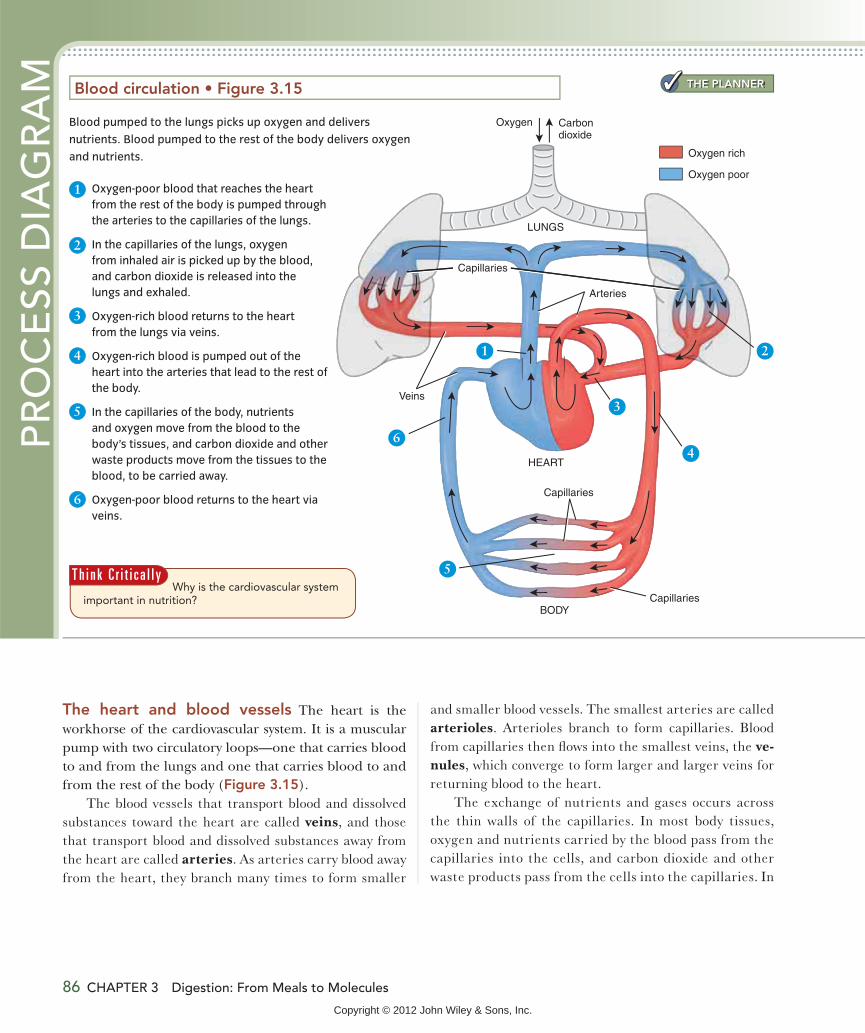
The heart and blood vessels The heart is the workhorse of the cardiovascular system. It is a muscular pump with two circulatory loops—one that carries blood to and from the lungs and one that carries blood to and from the rest of the body (Figure 3.15).
The blood vessels that transport blood and dissolved substances toward the heart are called veins, and those that transport blood and dissolved substances away from the heart are called arteries. As arteries carry blood away from the heart, they branch many times to form smaller
and smaller blood vessels. The smallest arteries are called arterioles. Arterioles branch to form capillaries. Blood from capillaries then flows into the smallest veins, the ve-nules, which converge to form larger and larger veins for returning blood to the heart.
The exchange of nutrients and gases occurs across the thin walls of the capillaries. In most body tissues, oxygen and nutrients carried by the blood pass from the capillaries into the cells, and carbon dioxide and other waste products pass from the cells into the capillaries. In
Why is the cardiovascular system important in nutrition?
T h i n k C r i t i c a l l y
Blood pumped to the lungs picks up oxygen and delivers
nutrients. Blood pumped to the rest of the body delivers oxygen
and nutrients.
1 Oxygen-poor blood that reaches the heart
from the rest of the body is pumped through
the arteries to the capillaries of the lungs.
2 In the capillaries of the lungs, oxygen
from inhaled air is picked up by the blood,
and carbon dioxide is released into the
lungs and exhaled.
3 Oxygen-rich blood returns to the heart
from the lungs via veins.
4 Oxygen-rich blood is pumped out of the
heart into the arteries that lead to the rest of
the body.
5 In the capillaries of the body, nutrients
and oxygen move from the blood to the
body’s tissues, and carbon dioxide and other
waste products move from the tissues to the
blood, to be carried away.
6 Oxygen-poor blood returns to the heart via
veins.
THE PLANNER
Copyright © 2012 John Wiley & Sons, Inc.

Delivering Nutrients and Eliminating Wastes 87
kidneys, brain, skin, and other organs.12 This distribu-tion changes when you eat or exercise. When you have eaten a large meal, a greater proportion of your blood goes to your digestive system to provide the oxygen and nutrients needed by the GI muscles and glands for di-gestion of the meal and absorption of nutrients. When you are exercising strenuously, about 70% of your blood is directed to your skeletal muscles to deliver nutrients and oxygen and remove carbon dioxide and other waste products (Figure 3.16).
the capillaries of the lungs, blood releases carbon diox-ide to be exhaled and picks up oxygen to be delivered to the cells. In the capillaries of the GI tract, blood delivers oxygen and picks up water-soluble nutrients absorbed from the diet.
The amount of blood, and hence the amounts of nu-trients and oxygen, delivered to a specific organ or tis-sue depends on the need. When you are resting, about 25% of your blood goes to your digestive system, about 20% to your skeletal muscles, and the rest to the heart,
a. At rest between meals, the amount of blood directed to the
abdomen, which includes the organs, muscles, and glands of
the digestive system, is similar to the amount that goes to the
skeletal muscles.12
b. During exercise, the demands of the muscles take priority, and
only a small proportion of the blood is directed to the abdomen.12
This is why you may get cramps if you exercise right after eating a
big meal: Your body cannot direct enough blood to the intestines
and the muscles at the same time. The muscles win out, and food
remains in your intestines, often causing cramps.
I n t e r p r e t i n g D a t a
a. skeletal muscle
b. abdomen
c. other organs
d. blood flow is equal to all areas.
Abdomen
Skeletalmuscles
Otherorgans
Distribution of blood flow at rest
Distribution of blood flow during exercise
Copyright © 2012 John Wiley & Sons, Inc.
88 CHAPTER 3 Digestion: From Meals to Molecules
cells drains into the lymphatic system. This prevents the fluid from accumulating and causing swelling.
The lymphatic system is an important part of the im-mune system. Fluid collected in the lymph vessels is fil-tered past a collection of infection-fighting cells before being returned to the blood. If the fluid contains antigen, an immune response is triggered. White blood cells and antibodies produced by this response enter the blood and help destroy the foreign substance.
In the small intestine, the lymph vessels aid in the ab-sorption and transport of fat-soluble substances such as cholesterol, fatty acids, and fat-soluble vitamins. These pass from the intestinal mucosa into the lacteals located in the villi (see Figure 3.9b). The lacteals drain into larger lymph vessels. Lymph vessels from the intestine and most other organs drain into the thoracic duct, which empties into the blood near the neck. Therefore, substances ab-sorbed into the lymphatic system do not pass through the liver before entering the general blood circulation.
Elimination of WastesMaterial that is not absorbed from the gut into the body is eliminated from the gastrointestinal tract in the feces. Wastes that are generated in the body, such as carbon dioxide, minerals, and nitrogen-containing wastes, must
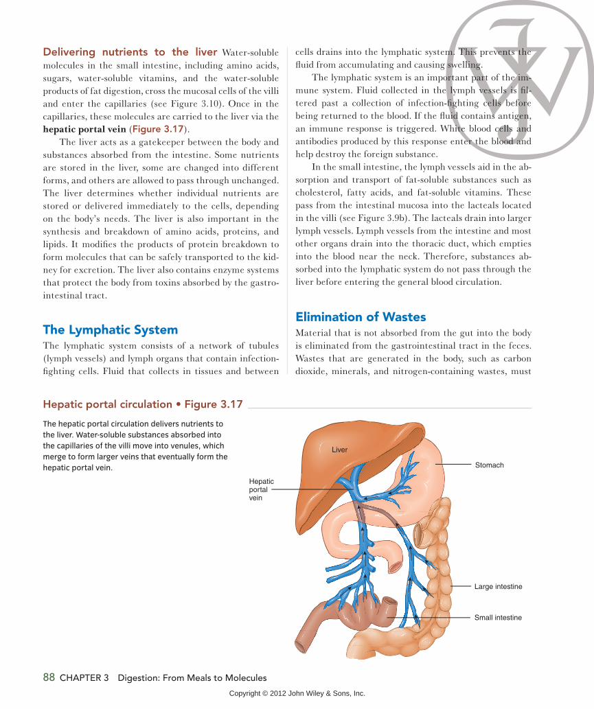
Delivering nutrients to the liver Water-soluble molecules in the small intestine, including amino acids, sugars, water-soluble vitamins, and the water-soluble products of fat digestion, cross the mucosal cells of the villi and enter the capillaries (see Figure 3.10). Once in the capillaries, these molecules are carried to the liver via the hepatic portal vein (Figure 3.17).
The liver acts as a gatekeeper between the body and substances absorbed from the intestine. Some nutrients are stored in the liver, some are changed into different forms, and others are allowed to pass through unchanged. The liver determines whether individual nutrients are stored or delivered immediately to the cells, depending on the body’s needs. The liver is also important in the synthesis and breakdown of amino acids, proteins, and lipids. It modifies the products of protein breakdown to form molecules that can be safely transported to the kid-ney for excretion. The liver also contains enzyme systems that protect the body from toxins absorbed by the gastro-intestinal tract.
The Lymphatic SystemThe lymphatic system consists of a network of tubules (lymph vessels) and lymph organs that contain infection-fighting cells. Fluid that collects in tissues and between
Stomach
Hepaticportalvein
Large intestine
Small intestine
Liver
The hepatic portal circulation delivers nutrients to
the liver. Water-soluble substances absorbed into
the capillaries of the villi move into venules, which
merge to form larger veins that eventually form the
hepatic portal vein.
Copyright © 2012 John Wiley & Sons, Inc.

Delivering Nutrients and Eliminating Wastes 89
also be eliminated. The same highway of blood vessels that picks up absorbed nutrients and oxygen helps remove wastes from the body. Carbon dioxide and some water are lost via the lungs, and water, minerals, and nitrogen- containing wastes are lost through the skin, but the kidney is the primary site for the excretion of metabolic wastes. Water, minerals, and the nitrogen-containing by-products of protein breakdown are filtered out of the blood by the kidneys and excreted in urine. Figure 3.18 illustrates how the circulatory system is involved in both the delivery of nutrients and oxygen and the elimination of wastes.
DIGESTIVESYSTEM
EXTERNAL ENVIRONMENT
FoodCarbondioxide(CO2)
Oxygen(O2)
RESPIRATORYSYSTEM
Heart
Blo
od
Nutrients
AnusUnabsorbedwaste
Nitrogen-containingmetabolic wasteproducts
Cells
URINARYSYSTEM
O2 and nutrients
CO2 andother wastes
Some water, minerals, and by-products of protein break-down are lost through theskin in perspiration or sweat.
In the kidneys, water and smallmolecules are filtered out of theblood. Filtered substances thatare needed are reabsorbed intothe blood, and those that are notare excreted in urine. The amountsof water and other substancesexcreted in urine are regulated inorder to maintain the right amountsinside the body.
Water and minerals
INTEGUMENTARY SYSTEM
(skin)
Red blood cells transportcarbon dioxide from thecells, where it is producedby metabolism, to the lungs. At the lungs, red blood cells release the carbon dioxide, which is then exhaled into the environment.
CARDIOVASCULARSYSTEM
feces urine
The nutrients taken in by the digestive system and the oxygen taken in by the respiratory system
are both distributed to all the cells in the body by the cardiovascular system. Unabsorbed materials
are eliminated in the feces. Metabolic wastes are transferred to the outside environment by the
skin and the urinary and respiratory systems.
1. Where does blood go after it leaves the lungs?
2. Why is it not a good idea to exercise after eat-ing a large meal?
3. What is the role of the lymphatic system in nutri-ent absorption?
4. What wastes are excreted by the kidneys? by the lungs?
A s k Yo u r s e l fFiber is not absorbed, so it is eliminated in the ___________.The carbon dioxide exhaled by the lungs comes from the ___________.
Copyright © 2012 John Wiley & Sons, Inc.

the chemical reactions that break down molecules to pro-vide energy and those that synthesize larger molecules are referred to as metabolism. Many of the reactions of me-tabolism occur in series known as metabolic pathways. Molecules that enter these pathways are modified at each step, with the help of enzymes. Some of the pathways use energy to build body structures, and others break large molecules into smaller ones, releasing energy. Reactions that synthesize molecules occur in different cellular com-partments from those that break down molecules for en-ergy. For example, ribosomes are cellular structures that specialize in the synthesis of proteins, and mitochondria are cell organs that are responsible for breaking down molecules to release energy.
An Overview of MetabolismLEARNING OBJECTIVES
1. Discuss the two general ways in which nutrients can be used after they have been absorbed.
2. Describe what happens in cellular respiration.3. List the types of molecules that can be made
from glucose, from fatty acids, and from amino acids.
nce they are inside the body’s cells, nutrients are used either for energy or to synthesize all the structural and regulatory molecules needed for growth and maintenance. Together,
O
Cellular respiration uses oxygen to convert glucose, fatty acids, and amino acids into carbon dioxide,
water, and energy, in the form of ATP.
PR
OC
ESS
DIA
GR
AM
C
CO2
C
Acetyl-CoA
CoA
Cell fluid
High-energyelectrons
Mitochondrion
ATPATP
Citricacidcycle
e–
e–
e–
e–
12
3
O2 H2O
Glucose
Fatty acids
Amino acids
1 In the presence of oxygen,
glucose, fatty acids, and
amino acids can be metabo-
lized to produce a two-car-
bon molecule (acetyl-CoA).
2 Each acetyl-CoA molecule
enters a circular pathway,
called the citric acid cycle,
that produces two molecules
of carbon dioxide (CO2).
3 In the final step of this meta-
bolic pathway, most of the
energy released from the
glucose, fatty acid, or amino
acid molecules is used to
produce ATP, and oxygen
combines with electrons
and hydrogen to form water.
90 CHAPTER 3 Digestion: From Meals to Molecules
THE PLANNER
HOW IT WORKS
Copyright © 2012 John Wiley & Sons, Inc.

Summary 91
Synthesizing New MoleculesGlucose, fatty acids, and amino acids that are not broken down for energy are used, with the input of energy from ATP, to synthesize structural, regulatory, or storage mole-cules. Glucose molecules are used to synthesize the glucose-storage molecule glycogen and, in some cases, fatty acids. Fatty acids are used to make body fat, cell membranes, and regulatory molecules, and amino acids are used to synthe-size the various proteins that the body needs and, when necessary, to make glucose. Excess amino acids can also be converted into fatty acids and stored as body fat.
Releasing EnergyIn the mitochondria, the sugar glucose, fatty acids, and amino acids derived from carbohydrates, fats, and pro-teins, respectively, are broken down in the presence of oxygen to produce carbon dioxide and water and release energy. This process, called cellular respiration, is like cell breathing: Oxygen goes into the cell, and carbon di-oxide comes out. The energy released by cellular respira-
tion is used to make a molecule called adenosine triphosphate
(ATP) (Figure 3.19). ATP can be thought of as the cell’s energy currency. The chemical bonds of ATP are very high in energy, and when they break, the energy is
released and can be used either to power body processes, such as muscle contraction or the transport of molecules across membranes, or to synthesize new molecules needed to maintain and repair body tissues.
adenosine
triphosphate
(ATP) A high-energy
molecule that the
body uses to power
activities that require
energy.1. How are nutrients used to produce ATP?
2. Why can cellular respiration be thought of as cell breathing?
3. What types of molecules can be made from amino acids?
Summary
1The Organization of Life 64
Our bodies and the foods we eat are all made from the same building blocks—atoms. Atoms are linked together by chemical bonds to form molecules. Molecules can form cells, as shown. Cells with similar structures and functions are organized into tissues, and tissues are organized into the organs and organ systems that make up an organism. The body organ systems work together; for example, the passage of food through the digestive system and the secre-tion of digestive substances are regulated by the nervous and endocrine systems.
Cells
2The Digestive System 68
The digestive system has two major functions: digestion and absorption. Digestion breaks down food and nutrients into units that are small enough to be absorbed. Absorption trans-ports nutrients into the body. The main component of the di-gestive system, illustrated here, is the gastrointestinal tract, which consists of a hollow tube that begins at the mouth and continues through the pharynx, esophagus, stomach, small intestine, and large intestine, ending at the anus.
The digestion of food and absorption of nutrients are aided by the secretion of mucus and enzymes.
THE PLANNER
Copyright © 2012 John Wiley & Sons, Inc.

92 CHAPTER 3 Digestion: From Meals to Molecules
Components of chyme that are not absorbed in the small intestine pass on to the large intestine, where some water and nutrients are absorbed. The large intestine is populated by intestinal microflora that digest some of the unab-sorbed materials, such as fiber, producing small amounts of nutrients and gas. The remaining unabsorbed materials are eliminated in the feces.
4Digestion in Health and Disease 79
Immune system cells and tissues located in the gastroin-testinal tract help prevent disease-causing organisms or chemicals from entering the body. An antigen entering the digestive tract is attacked first by phagocytes. If it is not eliminated by the phagocytes, lymphocytes respond specifically to the antigen by producing antibodies. The immune system protects us from disease but can also cause food allergies.
Diseases or discomforts at any point in the digestive system can interfere with food intake, digestion, or nutrient absorp-tion. Common problems include dental problems, reduced saliva production, heartburn, GERD, peptic ulcers (such as the one shown here), gallstones, vomiting, diarrhea, and constipation.
3Digestion and Absorption of Nutrients 70
The processes involved in digestion begin in response to the smell or sight of food and continue as food enters the diges-tive tract at the mouth, where it is broken down into smaller pieces by the teeth and mixed with saliva to form a bolus. Carbohydrate digestion is begun in the mouth by salivary amylase. From the mouth, the bolus passes through the pharynx and into the esophagus. The rhythmic contractions of peristalsis propel it down the esophagus to the stomach.
The stomach is a temporary storage site for food. The muscles of the stomach mix the food into a semiliquid mass called chyme, and gastric juice, which contains hydrochlo-ric acid and pepsin, begins the digestion of protein. The rate at which the stomach empties varies with the amount and composition of food consumed and is regulated by nervous and hormonal signals.
The small intestine is the primary site for nutrient digestion and absorption. The circular folds, villi, shown here, and microvilli of the small intestine, ensure a large absorptive surface area. In the small intestine, bicarbonate from the pancreas neutralizes stomach acid, and pancreatic and intestinal enzymes digest carbohydrate, fat, and protein. The digestion and absorption of fat in the small intestine are aided by bile from the gallbladder.
Microvilli
Lacteal
Capillary
Artery
Vein
Lymphvessel
Mucosal cell
Lumen Villi
The absorption of food across the intestinal mucosa occurs by means of several different transport mechanisms. Simple diffusion, osmosis, and facilitated diffusion do not require energy, but active transport does.
Copyright © 2012 John Wiley & Sons, Inc.

Summary 93
6An Overview of Metabolism 90
In the cells, glucose, fatty acids, and amino acids absorbed from the diet can be broken down by means of cellular respiration, as shown here, to provide energy in the form of ATP.
5 Delivering Nutrients and Eliminating Wastes 85
Absorbed nutrients are delivered to the cells by the cardio-vascular system. The heart pumps blood to the lungs to pick up oxygen and release carbon dioxide. From the lungs, blood returns to the heart and is then pumped to the rest of the body to deliver oxygen and nutrients and remove car-bon dioxide and other wastes before returning to the heart. Exchange of nutrients and gases occurs at the capillaries.
The products of carbohydrate and protein digestion and the water-soluble products of fat digestion enter capillaries in the intestinal villi and are transported to the liver via the hepatic portal vein, as illustrated here. The liver removes the absorbed substances for storage, converts them into other forms, or allows them to pass unaltered. The liver also protects the body from toxic substances that may have been absorbed.
Stomach
Hepaticportalvein
Large intestine
Small intestine
Liver
The fat-soluble products of digestion enter lacteals in the in-testinal villi. Lacteals join larger lymph vessels. The nutrients absorbed via the lymphatic system enter the blood without first passing through the liver.
Unabsorbed materials are eliminated in the feces. The waste products of metabolism are excreted by the lungs, skin, and kidneys.
C
CO2
C
Acetyl-CoA
CoA
Cell fluid
High-energyelectrons
Mitochondrion
ATPATP
Citricacidcycle
e–
e–
e–
e–
12
3
O2 H2O
Glucose
Fatty acids
Amino acids
In the presence of ATP, glucose, fatty acids, and amino acids can be used either to synthesize structural or regulatory molecules or to synthesize energy-storage molecules.
Copyright © 2012 John Wiley & Sons, Inc.

94 CHAPTER 3 Digestion: From Meals to Molecules
Online Resources For more information on how the digestive system works, go
to http://digestive.niddk.nih.gov/ddiseases/pubs/yrdd/.
For more information on gluten, go to www.csaceliacs.org/
AboutCSA.php.
For more information on food, nutrition, and metabolism, go to
http://health.nih.gov/category/FoodNutritionandMetabolism.
Visit your WileyPLUS site for videos, animations, podcasts,
self-study, and other media that will aid you in studying and
understanding this chapter.
Key Termsabsorption 68
active transport 77
adenosine triphosphate (ATP) 91
allergen 80
antibody 79
antigen 79
arteriole 86
artery 86
atom 64
bicarbonate 75
bile 76
bolus 71
brush border 75
capillary 85
cardiovascular system 85
celiac disease 80
cell 64
cellular respiration 91
chyme 73
constipation 82
diarrhea 82
diffusion 77
digestion 68
enzyme 70
epiglottis 71
facilitated diffusion 77
fatty acid 68
feces 68
food allergy 80
gallstone 82
gastric juice 73
gastrin 74
gastroesophageal reflux
disease (GERD) 82
gastrointestinal tract 69
heartburn 82
hepatic portal vein 88
hormone 65
intestinal microflora 78
lacteal 85
lipase 76
lumen 69
lymphatic system 85
lymphocyte 79
metabolic pathway 90
metabolism 90
microvillus 75
mitochondrion 90
molecule 64
mucosa 69
mucosal cells 69
mucus 69
organ 65
organ system 65
osmosis 77
pancreatic amylase 76
pancreatic juice 75
pepsin 73
peptic ulcer 82
peristalsis 72
phagocyte 79
pharynx 71
prebiotic 78
probiotic 78
protease 76
saliva 71
salivary amylase 71
segmentation 75
simple diffusion 77
sphincter 72
tissue 64
transit time 69
vein 86
venule 86
villi 75
Critical and Creative Thinking Questions1. For lunch, Joe, a college freshman, has a smoothie made with
blended fruit and juice. His friend has a burger, fries, and a
milk shake. Whose stomach will empty faster? Why?
2. Your grandmother’s favorite snack is a crisp fresh apple, but she
has difficulty chewing because of tooth loss and dental pain.
How might you modify her snack to make it easier for her to eat?
3. Starting in the esophagus, trace the path of food through
the digestive system. At each organ, indicate anatomical
adaptations of the general GI tract tube structure that enable
the functions of that organ. For example, how does the struc-
ture of the small intestine aid absorption?
4. After reading about the benefits of a high-fiber diet, 20-year-old
Jeff decides to increase his fiber intake by eating more whole-
grain products, beans, and fresh fruits and vegetables. Look
at the graph and decide which bar (A or B) represents his daily
stool weight after a week on his new diet. How might his higher
fiber intake affect the amount of intestinal gas produced? Why?
Dai
ly s
tool
wei
ght (
Gra
ms)
A B0
300
350
250
200
150
100
50
Copyright © 2012 John Wiley & Sons, Inc.

What is happening in this picture? 95
What is happening in this picture?This man’s digestive tract was damaged during surgery, so he cannot absorb
nutrients. Doctors are ensuring that he is being nourished by infusing a nutrient
solution into his blood through a process called total parenteral nutrition (TPN).
1. Would the TPN solution delivered meet his needs if it contained undigested starches and proteins? Why or why not?
2. What would happen to him if an essential nutrient were missing from the TPN solution?
3. How would waste products from the TPN be eliminated?
T h i n k C r i t i c a l l y
5. Water is absorbed from the small intestine and colon by
osmosis. What might happen to the body’s water balance if a
large number of small, unabsorbed molecules end up in the
colon?
6. What path does an amino acid follow from absorption to
delivery to a cell? Compare this path with the path followed by
a large fatty acid.
Copyright © 2012 John Wiley & Sons, Inc.

96 CHAPTER 3 Digestion: From Meals to Molecules
4. Which letter in this illustration points to the organ in which
bile is stored?
a. A d. F
b. C e. G
c. E
5. The secretion of digestive juices does not begin until food
enters the mouth.
a. True b. False
6. The tissue layer labeled A ___________.
A
a. secretes mucus
b. contains nerves and blood vessels
c. mixes and propels food through the GI tract
d. provides external support to the gut
7. The ___________ prevents food from entering the passageway
to the lungs during swallowing.
a. gallbladder c. epiglottis
b. pancreas d. tongue
8. The rhythmic contractions that propel food through the GI
tract are called ___________.
a. peristalsis d. gastroesophageal reflux
b. enzymatic digestion e. chewing
c. swallowing
9. Which of the following is most likely to inhibit stomach secre-
tions and stomach motility?
a. the release of gastrin
b. the smell of freshly baked cookies
c. the entry of food into the small intestine
d. the entry of food into the stomach
A
B
C
D
E
F
G
Self-Test(Check your answers in Appendix K.)
1. Which of the following is the smallest unit of life?
a. atom d. tissue
b. molecule e. organism
c. cell
2. Which of the following statements about saliva is false?
a. It lubricates the mouth.
b. It helps protect the teeth from decay.
c. It moistens food so that it can be tasted and swallowed.
d. It contains the enzyme salivary amylase.
e. It contains the protein-digesting enzyme pepsin.
Use the diagram to answer questions 3 and 4.
3. Which letter in this illustration points to the organ in which
carbohydrate digestion begins?
a. A d. D
b. B e. E
c. C
Copyright © 2012 John Wiley & Sons, Inc.

Self-Test 97
13. This diagram could represent all the following except
___________.
EsophagusStomach wall
Sphincter
Acidstomachcontents
a. heartburn c. GERD
b. diarrhea d. vomiting
14. Most digestion and absorption occur in the ___________.
a. small intestine c. mouth
b. stomach d. large intestine
15. All absorbed nutrients go directly to the liver.
a. True b. False
10. The diffusion of water across a membrane is called
___________.
a. simple diffusion c. facilitated diffusion
b. osmosis d. active transport
11. Which of the following is not true of the microvilli?
a. They help increase the surface area available for
absorption.
b. They contain digestive enzymes.
c. Each contains a capillary and a lacteal.
d. They are referred to as the brush border.
12. Which of the numbers on this illustration label blood that is
rich in oxygen?
Review your Chapter Planner on the chapter opener and
check off your completed work.
THE PLANNER
LUNGS
Oxygen Carbondioxide
Capillaries
Capillaries
Arteries
HEART
BODY
Capillaries
Veins
1 2
3
5
64
a. 1 and 2 c. 3 and 4
b. 2 and 5 d. 5 and 6
Copyright © 2012 John Wiley & Sons, Inc.


