DIFFUSION-WEIGHTED IMAGING OF EXCITOTOXIC BRAIN INJURY · detecting cytotoxic edema due to...
71
DIFFUSION-WEIGHTED IMAGING OF EXCITOTOXIC BRAIN INJURY Toshio Moritani, Akio Hiwatashi, Henry Wang, Leena Ketonen, Sven Ekholm, Per-Lennart Westesson Department of Radiology University of Rochester Medical Center Email: [email protected] Presentation material is for education purposes only. All rights reserved. ©2003 URMC Radiology Page 1 of 71
Transcript of DIFFUSION-WEIGHTED IMAGING OF EXCITOTOXIC BRAIN INJURY · detecting cytotoxic edema due to...

DIFFUSION-WEIGHTED IMAGING OF EXCITOTOXIC BRAIN INJURY
Toshio Moritani, Akio Hiwatashi,!Henry Wang, Leena Ketonen,!
Sven Ekholm, Per-Lennart Westesson"Department of Radiology!
University of Rochester Medical Center!!
Email: [email protected] Presentation material is for education purposes only. All rights reserved. ©2003 URMC Radiology Page 1 of 71

Trans-synaptic injury via excitotoxic amines is a specific type of injury in the peripheral and central nervous systems. Recent studies show that the receptors related to excitotoxic mechanisms are widely distributed in the brain, not only in the gray matter (neurons and astrocytes) but also in the white matter (astrocytes, oligodendrocytes, myelin sheaths, and axons) (1). Excitotoxic brain injury is presumed to be related to any pathological condition that causes cytotoxic edema, resulting in decreased ADC areas on diffusion-weighted (DW) imaging. Such conditions include infarction, hypoxic ischemic encephalopathy, the early phase of wallerian and transneuronal degeneration, shaken baby syndrome, status epilepticus, a corpus callosum lesion related to seizures or antiepileptic drugs, diffuse axonal injury, toxic and metabolic leukoencephalopathy, the acute phase of multiple sclerosis, and Creutzfeldt-Jakob disease. This exhibit illustrates various diseases associated with excitotoxic mechanisms and DW imaging findings.
Introduction
Presentation material is for education purposes only. All rights reserved. ©2003 URMC Radiology Page 2 of 71

Glutamate, aspartate and glycine are the dominant excitatory amino acids and the primary neurotransmitters in about half of all the synapses in the brain. Among them, glutamate is the most important and is responsible for many neurologic functions including cognition, memory, movement and sensation. In pathological conditions, glutamate mediates neuronal injury or death, particularly through activation of the N-methyl-D-aspartate (NMDA) subtype of the glutamate receptor (2). Neuronal glutamate is released from the pre-synaptic terminal into the synaptic cleft (Fig. 1). Re-uptake of extracellular glutamate is done at the pre-synaptic terminals and adjacent glial cells which seems to protect the neurons from excitotoxic injury. Energy for re-uptake of the glutamate is provided by the mitochondria.
Excitotoxic Mechanisms (Fig. 1,2)!
Presentation material is for education purposes only. All rights reserved. ©2003 URMC Radiology Page 3 of 71

The excessive glutamate binding to NMDA receptors allows entry of Ca2+ into the post-synaptic neuron, causing necrotic cell death or apoptosis. The excessive glutamate binding to non-NMDA receptors allows entry of Na+ into the post-synaptic neuron, resulting in cytotoxic edema. Glial cells also have these receptors, so the excessive glutamate leads to glial cell swelling.
Presentation material is for education purposes only. All rights reserved. ©2003 URMC Radiology Page 4 of 71

Figure 1
Presentation material is for education purposes only. All rights reserved. ©2003 URMC Radiology Page 5 of 71

Figure 2
Presentation material is for education purposes only. All rights reserved. ©2003 URMC Radiology Page 6 of 71

Excitotoxic mechanisms play an important role in various diseases. Glutamate excitotoxicity is the final common pathway resulting in brain injury for many seemingly unrelated diseases. Increased extracellular glutamate is a direct cause of excitotoxic brain injury (Fig. 2).
In acute excitotoxic injury, excessive extracellular glutamate results from
1) decreased re-uptake mainly by energy failure,
2)! increased release by excessive depolarization of neuronal membranes or by intracellular accumulation,
3) leakage due to disruption of axonal membranes. Acute
excitotoxic injury is also related to impaired glutamate receptor function and structurally similar substance to glutamate.
Presentation material is for education purposes only. All rights reserved. ©2003 URMC Radiology Page 7 of 71
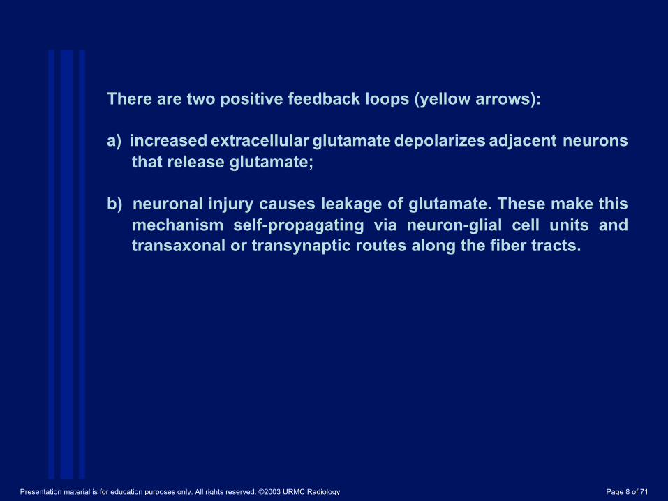
There are two positive feedback loops (yellow arrows): a)! increased extracellular glutamate depolarizes adjacent neurons
that release glutamate; b) neuronal injury causes leakage of glutamate. These make this
mechanism self-propagating via neuron-glial cell units and transaxonal or transynaptic routes along the fiber tracts.
Presentation material is for education purposes only. All rights reserved. ©2003 URMC Radiology Page 8 of 71

During ischemia, cytotoxic edema occurs primarily due to energy failure of neurons and astrocytes (Fig. 3). It is thought to extend secondarily into the region of penumbra via neuron-glial cell units and synapses by excitotoxic mechanism (3). Both mechanisms disable the sodium-potassium pump allowing sodium and water to enter the cell. Cytosol calcium ion levels may trigger protease and lipase production resulting in infarction. In a experimental study, NMDA type glutamate receptor antagonists (MK-801) reduces the volume of ischemic injury following MCA occlusion. This indicates that the pathophysiology of ischemic penumbra is associated with excitotoxic injury of glutamate (4).
Brain Infarction (Fig. 3)
Presentation material is for education purposes only. All rights reserved. ©2003 URMC Radiology Page 9 of 71

Figure 3. Hyperacute infarction
(2 hrs from onset). 39-year-old man
with the left internal carotid artery dissection,
presenting with right side weakness
A A. FLAIR image shows no apparent parenchymal abnormalities, but linear hyperintensities represent slow flow in the peripheral vessels (arrows).
Presentation material is for education purposes only. All rights reserved. ©2003 URMC Radiology Page 10 of 71

B, C. DW image shows a mild hyperintense lesion with decreased ADC in the left frontoparietal white matter, representing cytotoxic edema which is extending into the ischemic penumbra due to the propagation by excitotoxic mechanisms (arrows).
B C
Presentation material is for education purposes only. All rights reserved. ©2003 URMC Radiology Page 11 of 71

D!
D. Perfusion-weighted image shows the elongation of mean transit time in the entire left anterior and middle cerebral artery territories.
Presentation material is for education purposes only. All rights reserved. ©2003 URMC Radiology Page 12 of 71

Cerebral infarction in the territory of the middle cerebral artery can cause wallerian degeneration of the corticospinal tract and transneuronal (trans-synaptic) degeneration in the ipsilateral substantia nigra. Wallerian degeneration is antegrade degeneration of the axons and myelin sheath resulting from injury of the proximal portion of the axons or cell bodies. Transneuronal degeneration in the substantia nigra occurs secondary to striatal infarction (5). Cytotoxic edema occurring in the early phase of degeneration may be related to excitotoxic mechanisms via axons or synapses (6). DW imaging shows the early phase of wallerian or transneuronal degeneration as hyperintense associated with decreased ADC, presumably representing axonal/ intramyelinic or astrocytic swelling (7-10) (Fig 4).
Wallerian or Transneuronal Degeneration (Fig. 4)
Presentation material is for education purposes only. All rights reserved. ©2003 URMC Radiology Page 13 of 71

Figure 4. Wallerian and transneuronal degeneration.
A 76 year-old man with a large infarct in the right middle
cerebral artery (MCA) territory
(6 days after onset). A A. T2-weighted image shows a right MCA infarct as hyperintense, including the left putamen.
Presentation material is for education purposes only. All rights reserved. ©2003 URMC Radiology Page 14 of 71

C B B, C. DW image at the level of the midbrain reveals hyperintense lesions with decreased ADC in the right cerebral peduncle (arrow) including the substantia nigra (arrow), as well as a right MCA infarct in the temporal area presumably due to the propagation of excitotoxic injury via axons or synapses.
Presentation material is for education purposes only. All rights reserved. ©2003 URMC Radiology Page 15 of 71

In HIE energy depletion in neurons and glial cells causes decreased re-uptake of glutamate which leads to increased extracellular glutamate. The perinatal period of brain development is particularly vulnerable to excitotoxic injury. The high rate of generation of synapses (synaptogenesis) results in an overexpression of the receptor. NMDA receptors dominate in the immature brain when synaptic transmission is weak and extremely plastic (2). The distribution of the lesions in the putamen, thalamus, and peri-Rolandic cerebral cortex is related to intrinsic vulnerability of these areas to energy failure.
Hypoxic Ischemic Encephalopathy (HIE) (Figs. 5,6)
Presentation material is for education purposes only. All rights reserved. ©2003 URMC Radiology Page 16 of 71
A
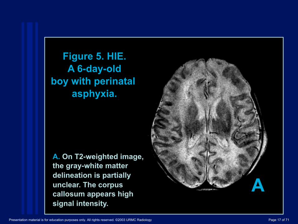
Figure 5. HIE. A 6-day-old
boy with perinatal asphyxia.
A. On T2-weighted image, the gray-white matter delineation is partially unclear. The corpus callosum appears high signal intensity.
Presentation material is for education purposes only. All rights reserved. ©2003 URMC Radiology Page 17 of 71

B C B, C. DW image shows diffuse hyperintensity with decreased ADC in the anterior and posterior corpus callosum (arrow), internal capsule (arrow), thalamus, and white matter. This distribution is presumably related to overactivity of excitatory pathways.
Presentation material is for education purposes only. All rights reserved. ©2003 URMC Radiology Page 18 of 71

The distribution of the lesions in the putamen, thalamus, and peri-Rolandic cerebral cortex is related to intrinsic vulnerability of these areas to energy failure. One potentially important link among these areas is their interconnection by excitatory circuits (11). Thus, Overactivity in these excitatory pathways could contribute to spreading of the lesions via synapses. The internal capsule, cerebral peduncle and corpus callosum can be involved as secondary involvement through these pathways, also known as wallerian or transneuronal degeneration. DW images and ADC maps clearly depict lesions in the corpus callosum, internal capsule and white matter when conventional MRI and CT are normal, or show subtle abnormalities (Figs. 5, 6) (12).
Presentation material is for education purposes only. All rights reserved. ©2003 URMC Radiology Page 19 of 71

A. DW image shows extensive hyperintense lesions involving the fronto-temporo-parietal white matter, internal capsules and basal ganglia bilaterally.
A
Figure 6. HIE. A 10-day-old Boy with HIE
due to perinatal asphyxia.
Presentation material is for education purposes only. All rights reserved. ©2003 URMC Radiology Page 20 of 71

C B
B, C. DW image shows hyperintense lesions with decreased ADC in the bilateral cerebral peduncles probably including both corticospinal tracts and substantia nigra. These findings represent the early phase of wallerian and transneuronal degeneration.
Presentation material is for education purposes only. All rights reserved. ©2003 URMC Radiology Page 21 of 71

Glutamate and glycine levels are extremely high in the CSF with shaken baby syndrome (13). In experimental acute subdural hematoma in the infant rat, glutamate in extracellular fluid in the cortex increases more than 7 times over the basal level (14). Although the pathogenesis of brain parenchymal injuries is unknown, it seems to be related to increased release of glutamate from the pre-synaptic terminal with traumatic stimuli, and decreased re-uptake of glutamate from the synapse with hypoxia or ischemia. Widespread parenchymal injury may be related to the distribution of predominant NMDA receptors in neonates and infants. Histologic similarities are observed between child abuse victims and infants with hypoxic ischemic encephalopathy. However, a history
Shaken Baby Syndrome (Fig. 7)
Presentation material is for education purposes only. All rights reserved. ©2003 URMC Radiology Page 22 of 71

history of apnea suggesting hypoxic-ischemic injury is found in only about a half of shaken babies. Diffuse axonal injury is rare in neuropathological studies (15). Usually the distribution of widespread parenchymal injury is neither related to vascular territories nor the location and size of acute subdural hematoma on CT and MRI. DWI is useful in detecting cytotoxic edema due to excitotoxic brain injury. The severity of DWI abnormality correlates with patients' outcome (16). Neuroprotective effects by several kinds of selective glutamate receptor antagonists are reported in animal studies (17-21).
Presentation material is for education purposes only. All rights reserved. ©2003 URMC Radiology Page 23 of 71

A. On T2-weghted image, the gray-white matter delineation is unclear, and subdural hematoma and multiple intraparenchymal hemorrhages are noted (arrows).
Figure 7. Shaken baby syndrome.
A 2-month-old boy.
A Presentation material is for education purposes only. All rights reserved. ©2003 URMC Radiology Page 24 of 71

B, C. DW image shows diffuse and extensive hyperintensity in the gray and white matter with decreased ADC that represents cytotoxic edema resulting from severe and extensive excitotoxic injury. Multiple insults (hypoxia/ischemia, trauma, seizure) could be combined. Pediatric brain is susceptible to excitotoxic injuries. Only the right frontal lobe is relatively spared (arrow).
B C
Presentation material is for education purposes only. All rights reserved. ©2003 URMC Radiology Page 25 of 71

In status epilepticus, neuronal injury results primarily from an excitotoxic mechanism mediated by intrinsic neuronal seizure activity (22). During status epilepticus, neuronal seizure activity increases release of glutamate from the pre-synaptic terminal of neuronal axons. Excessive glutamate crosses the synaptic cleft to bind to NMDA and non NMDA receptors, which leads cytotoxic edema in neurons and glial cells, and apoptosis or selective neuronal necrosis. Astrocytes play a significant role in cellular and tissue repair by detoxyfication of excessive glutamate (23). Cytotoxic edema of the acute phase of reactive astrocytes can be responsible for the reversible signal abnormalities (24). Encephalopathy with status epilepticus often involves the hippocampus, other parts of the limbic system, thalamus, and cerebellum. This distribution of the lesions on DW images seems to be related to the distribution of NMDA type glutamate receptors which are concentrated in the hippocampus and other parts of the limbic system (Fig. 8).
Status Epilepticus (Fig. 8)
Presentation material is for education purposes only. All rights reserved. ©2003 URMC Radiology Page 26 of 71

A
A. T2-weighted image shows diffuse hyperintense lesions in the left thalamus and cerebral cortex.
Figure 8. Status epilepticus. A 2-year-old girl.
Presentation material is for education purposes only. All rights reserved. ©2003 URMC Radiology Page 27 of 71

B. Coronal FLAIR image shows hyperintense lesions in the left hippocampus (arrow), thalamus and temporo-fronto-parietal lobes presumably related glutamate receptor distributions.
B
Presentation material is for education purposes only. All rights reserved. ©2003 URMC Radiology Page 28 of 71

C D
C, D. DW image shows these lesions as hyperintense with decreased ADC that represents cytotoxic edema due to excitotoxic injury mediated by neuronal seizure activity. The lesions were partially reversible on follow-up MRI (not shown).
Presentation material is for education purposes only. All rights reserved. ©2003 URMC Radiology Page 29 of 71

The cause of the focal lesion in the splenium of the corpus callosum is speculated to be seizures, medications, or both (25,26,27). Transient focal edema can be related to the t ranshemispher ic connect ion wi th se izure act iv i ty. Interhemispherical propagation of the seizure activity is via the splenial callosal fibers. The splenium contains decussating fibers originating in the temporal lobe, which are likely to be involved. The cause often seems to be toxic effect of antiepileptic medications such as phenytoin, carbamazepine, and vigabatrin (26). Abrupt withdrawal and reducing antiepileptic drugs may contribute to transient edema, mediated by the influence of antiepileptic medications on fluid balance systems, namely arginine-vasopressin
Focal Lesion in the Splenium of the Corpus Callosum in Epileptic Patients (Fig. 9)
Presentation material is for education purposes only. All rights reserved. ©2003 URMC Radiology Page 30 of 71

arginine-vasopressin (27). Either cause can lead to excitotoxic injury resulting in reversible cytotoxic edema, which is presumed to be in astrocytes or myelin sheaths. Recent studies shows there is fairly amounts of glutamate receptors and the enzymic activity in the corpus callosum (1, 28). Conventional MRI shows a non-hemorrhagic hyperintense lesion on T2-weighted and FLAIR images, and slightly hypointense on T1-weighted image. DW imaging shows an acute lesion in the splenium of the corpus callosum as hyperintense with decreased ADC (Fig.9) (29).
Presentation material is for education purposes only. All rights reserved. ©2003 URMC Radiology Page 31 of 71

Presentation material is for education purposes only. All rights reserved. ©2003 URMC Radiology Page 32 of 71

A. Coronal T2-weighted image at 3 days after seizure show a discrete focal hyperintense lesion in the central portion of the splenium of the corpus callosum (arrow).
A
Figure 9. Focal lesion in the
splenium of the corpus callosum in epilepsy.
A 9-year-old presented with intractable partial
seizures since the age of 4.
Presentation material is for education purposes only. All rights reserved. ©2003 URMC Radiology Page 33 of 71

B, C. Coronal DW image shows this lesion as hyperintense associated with decreased ADC.
C B
Presentation material is for education purposes only. All rights reserved. ©2003 URMC Radiology Page 34 of 71

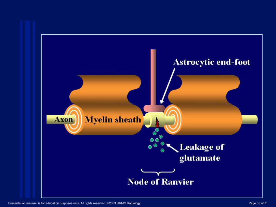
DAI is related to excitotoxic mechanisms, particularly glutamate and NMDA receptors (30). Axonal damage often occur at the node of Ranvier, a short interval between myelin sheaths (processes of oligodendrocytes), resulting in a traumatic defect in the axonal membrane. This results in a leakage of glutamate in the extracellular space (31). Astrocytic end-foot is located on the axon at the node of Ranvier and may protect the axons. The excessive glutamate by the leakage leads to glial cell and axonal swelling, resulting in cytotoxic edema, necrosis or axonal degeneration. DWI shows diffuse axonal injury as hyperintense lesions associated with decreased ADC often seen in the corpus callosum, white matter and brain stem (32) (Fig.10).
Diffuse Axonal Injury (DAI) (Fig. 10)
Presentation material is for education purposes only. All rights reserved. ©2003 URMC Radiology Page 35 of 71
Presentation material is for education purposes only. All rights reserved. ©2003 URMC Radiology Page 36 of 71

A. T2-weighted image shows a round hyperintense lesion in the body of corpus callosum (arrow).
A
Figure 10. Diffuse axonal injury.
An 11-year-old girl with a motor
vehicle accident.
Presentation material is for education purposes only. All rights reserved. ©2003 URMC Radiology Page 37 of 71
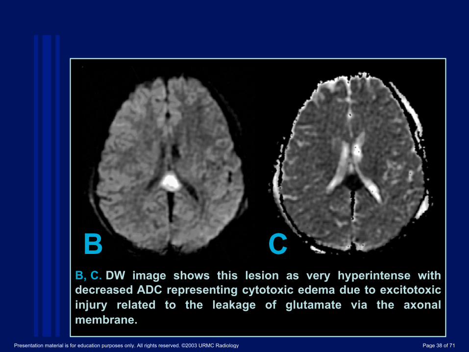
C B B, C. DW image shows this lesion as very hyperintense with decreased ADC representing cytotoxic edema due to excitotoxic injury related to the leakage of glutamate via the axonal membrane.
Presentation material is for education purposes only. All rights reserved. ©2003 URMC Radiology Page 38 of 71

The concentration of glutamate and glycine in CSF is significantly increased in encephalitis (33). These observations suggest an excitotoxic mechanism play a role in neuronal damage in herpes encephalitis. Excessive glutamate release due to free radicals generated during the immune response to infections might initiate secondary excitotoxicity. The distribution of hepes encephalitis is different in patients ages. Herpes simplex type 1 encephalitis in older children and adults usually involves the medial temporal lobe, inferior frontal lobes and insula (Fig.11).
Herpes encephalitis (Figs. 11,12)
Presentation material is for education purposes only. All rights reserved. ©2003 URMC Radiology Page 39 of 71

A
Figure 11. Herpes encephalitis
type 1. A 48 year-old
man presented with headache
and fever.
A. T2-weighted image shows hyperintense lesions in bilateral temporal lobes (arrows).
Presentation material is for education purposes only. All rights reserved. ©2003 URMC Radiology Page 40 of 71

B C
B. DW image clearly shows these lesions as hyperintense. C. ADC maps shows partially decreased ADC of these lesions (arrows).
Presentation material is for education purposes only. All rights reserved. ©2003 URMC Radiology Page 41 of 71

Neonatal herpes simplex type 2 encephalitis usually involves the cortex and white matter extensively. Widespread brain lesions in neonatal herpes encephalopathy are presumably related to the vulnerability to excitatory amines in the neonatal brain. In neonatal herpes encephalitis, MRI/DWI shows widespread, asymmetric lesions in both hemispheres including the basal ganglia and thalami (Fig.12).
Presentation material is for education purposes only. All rights reserved. ©2003 URMC Radiology Page 42 of 71

Figure 12. Herpes
encephalitis type 2.
A 2-week-old girl.
A A. T2WI shows symmetric hyperintense lesions in the thalami.
Presentation material is for education purposes only. All rights reserved. ©2003 URMC Radiology Page 43 of 71

B, C. DW image shows asymmetric but extensive hyperintense lesions with decreased ADC in the thalamus and gray and white matter of both hemispheres. This extensive distribution seems to be related to vulnerability to excitotoxic injury during the post-natal period.
C B
Presentation material is for education purposes only. All rights reserved. ©2003 URMC Radiology Page 44 of 71

Toxic metabolic diseases (Figs. 13,14,15) Methotrexate-induced Leukoencephalopathy (Fig. 13)
Intrathecal or intravenous methotrexate, either with or without radiation therapy, can occasionally cause diffuse white matter changes (34). Methotrexate itself is not toxic to astrocytes, neurons or the neurite networking. The neurotoxicity is thought to be caused by the enzymatic release of glutamate from methotrexate. Glutamate excitotoxity can damage myelin sheaths and axons. NMDA receptor antagonists can protect the glutamate neurotxocity (35). MR imaging shows diffuse or multifocal white matter lesions that are hyperintense on T2-weighted image. DW imaging shows diffuse hyperintensity with decreased ADC in the white matter before conventional MR imaging detects the lesions (Fig.13). Pathologically these lesions represent intramyelinic or axonal edema.
Presentation material is for education purposes only. All rights reserved. ©2003 URMC Radiology Page 45 of 71

Figure 13. Methotrexate
leukoencephalopathy in a 50-year-old
female.
A. On T2-weighted image does not demonstrate an appreciable abnormality in the white matter.
A!
Presentation material is for education purposes only. All rights reserved. ©2003 URMC Radiology Page 46 of 71

C B
B. DW image shows diffuse hyperintensity in the bilateral corona radiata extending into the central semiovale. C. ADC map shows diffuse white matter lesions as decreased ADC, which represent pure cytotoxic edema. The pathologic specimen showed pure intramyelinic edema (not shown).
Presentation material is for education purposes only. All rights reserved. ©2003 URMC Radiology Page 47 of 71

PKU is an autosomal recessive disorder caused by a deficiency of phenylalanine hydroxylase. L-phenylalanine impairs glutamate receptor function and thus contributes to brain dysfunction in PKU (36). Pathologic findings include delayed or defective myelination, diffuse white matter vacuolation, demyelination, and gliosis (37). MR imaging shows hyperintense lesions on T2-weighted image in the periventricular parietal and occipital regions, and in more severe cases, extending to the frontal and subcortical white matter (38). DW imaging shows these lesions as hyperintense with decreased ADC, which presumably represents intramyelinic edema and astrocytic swelling presumably due to excitotoxic injury (39) (Fig.14). These lesions can be completely reversible on follow-up MRI with dietary control.
Phenylketonuria (PKU) (Fig. 14)
Presentation material is for education purposes only. All rights reserved. ©2003 URMC Radiology Page 48 of 71

Cerebral organic acid disorders Some organic acids disorder is characterized by an accumulation of organic acids that share structural similarities with the excitotoxic amino acid glutamate (D-2.L-2,3 hydroxyglutarate, glutarate) (40). DW imaging shows diffuse hyperintensity with decreased ADC in the white matter.
Presentation material is for education purposes only. All rights reserved. ©2003 URMC Radiology Page 49 of 71

A. T2-weighted image shows hyperintense lesions in the periventricular white matter (arrows). A
Figure 14. Phenylketonuria in a 36 year-old
male.
Presentation material is for education purposes only. All rights reserved. ©2003 URMC Radiology Page 50 of 71
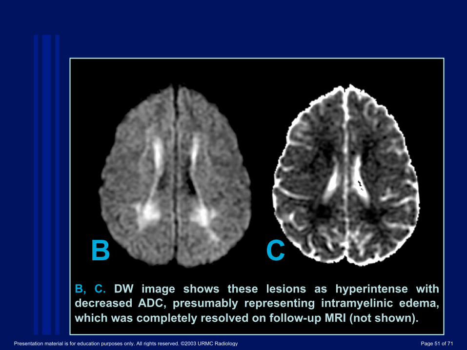
B, C. DW image shows these lesions as hyperintense with decreased ADC, presumably representing intramyelinic edema, which was completely resolved on follow-up MRI (not shown).
C B
Presentation material is for education purposes only. All rights reserved. ©2003 URMC Radiology Page 51 of 71

Wernicke encephalopathy (Fig. 15) T h i a m i n e ( v i t a m i n B 1 ) d e f i c i e n c y c a n c a u s e We r n i c k e encephalopathy characterized by confusion, ataxia, and abnormal eye movements. Without thiamine, the Krebs and pentose phosphate cycles cannot metabolize glucose (41). The enzymic inactivity leads to the accumulation of intracellular glutamate. Cellular homeostasis soon fails, resulting in release of glutamate into extracellular space (42). Pathologic findings include decreased myelination, edema, astrocytic swelling, and necrosis in the mamillary bodies, thalamic and hypothalamic nuclei, periaqueductal gray matter, walls of the third and floor of the fourth ventricle, and less commonly, caudate, frontal, and parietal cortex. With treatment of intravenous thiamine, these lesions may dissipate.
Presentation material is for education purposes only. All rights reserved. ©2003 URMC Radiology Page 52 of 71
Cerebral organic acid disorders Some organic acids disorder is characterized by an accumulation of organic acids that share structural similarities with the excitotoxic amino acid glutamate (D-2.L-2,3 hydroxyglutarate, glutarate) (40). DW imaging shows diffuse hyperintensity with decreased ADC in the white matter.
Presentation material is for education purposes only. All rights reserved. ©2003 URMC Radiology Page 53 of 71

A
Figure 15. Wernicke
Encephalopathy in a 75-year-old
male.
A. FLAIR shows a symmetrical hyperintense lesion in the hypothalamus (arrow).
Presentation material is for education purposes only. All rights reserved. ©2003 URMC Radiology Page 54 of 71

B, C. DW image shows isointense lesions with mildly increased ADC in the hypothalamus, which may represent vasogenic edema (arrow).
C B
Presentation material is for education purposes only. All rights reserved. ©2003 URMC Radiology Page 55 of 71

Glutamate excitotoxity damages not only neurons and astrocytes but also oligodendrocytes, myelin sheaths and axons (46). It seems to be an important mechanism in multiple sclerosis. Glutamate and aspartate in CSF is increased in patients with acute MS (47). In an immunohistochemical study, active MS lesions showed high glutamate production in macrophages and microglia in close proximity to axonal damage (48). Excitotoxicity in oligodendrocytes, myelin sheaths and axons may result in cytotoxic plaques. Cytotoxic plaques are very rare and show hyperintense on DWI with decreased ADC in acute MS (Fig.16). Pathology of cytotoxic plaques mainly shows intramyelinic edema.
Demyelinating and Degenerative Diseases (Figs 16,17) Multiple Sclerosis (MS)
Presentation material is for education purposes only. All rights reserved. ©2003 URMC Radiology Page 56 of 71
!
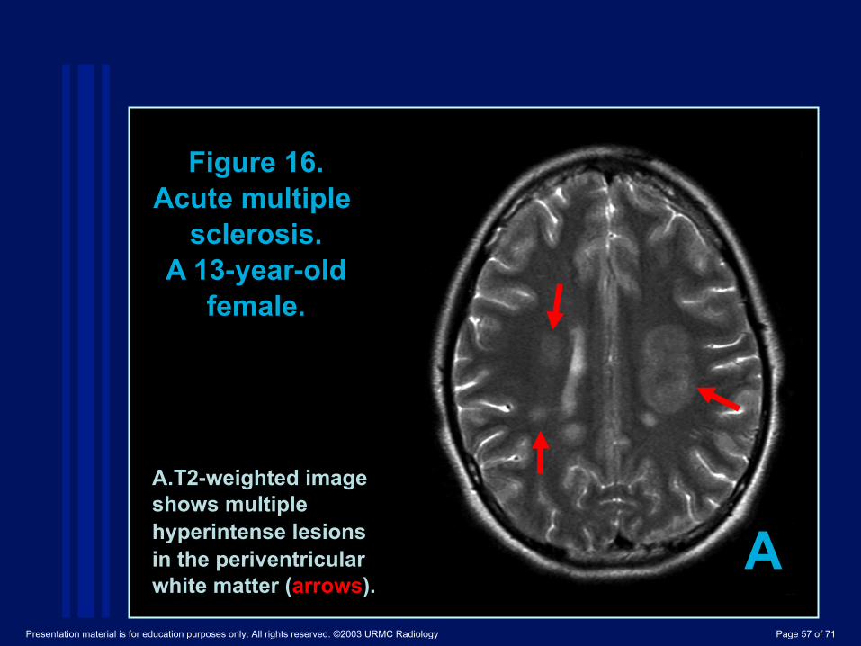
A.!T2-weighted image shows multiple hyperintense lesions in the periventricular white matter (arrows).
A
Figure 16. Acute multiple
sclerosis. A 13-year-old
female.
Presentation material is for education purposes only. All rights reserved. ©2003 URMC Radiology Page 57 of 71

!
B C B, C. DW image shows these lesions as hyperintense with decreased ADC that represents cytotoxic edema presumably related to excitotoxic injury of oligodendrocytes, myelin sheaths and axons.
Presentation material is for education purposes only. All rights reserved. ©2003 URMC Radiology Page 58 of 71

Creutzfeldt-Jakob disease (CJD) is one of the prion diseases characterized by rapidly progressive degenerative dementia, myoclonus, and ataxia. Prion diseases are characterized by accumulation of misfolded prion protein which has toxicity to endoplasmic reticulum. Marked and selective abnormalities in glutamate receptors are reported recently which may explain the characteristic distribution of brain lesions in CJD (49). T2-weighted and FLAIR images show hyperintense lesions in the cerebral cortex and bilateral basal ganglia in patients with CJD. The lesions are often involved in bilateral thalami (pulvinar sign) and periaqueductal areas in patients with variant CJD (50, 51) but this finding is also seen in sporadic CJD (52) DW imaging is more sensitive than conventional MRI for detecting abnormalities in CJD.
Creutzfeldt-Jakob disease (Fig. 17)
Presentation material is for education purposes only. All rights reserved. ©2003 URMC Radiology Page 59 of 71

DWI shows these lesions as hyperintense often associated with decreased ADC (53-56) (Fig.17). Electron microscopy shows these vacuoles as focal swelling of neuritic processes, both axonal and dendritic swelling (cellular edema), which may cause decreased ADC (58).
!
Presentation material is for education purposes only. All rights reserved. ©2003 URMC Radiology Page 60 of 71

Figure 17. Creuzfeldt-Jakob
disease. A 51 year-old man with progressive
dementia.
A
A. T2-weighted image demonstrates mild hyperintensity bilaterally in the caudate nuclei, putamina, and pulvinar of the thalami (arrows). The distribution could be related to glutamate receptor dysfunction.
Presentation material is for education purposes only. All rights reserved. ©2003 URMC Radiology Page 61 of 71

B, C. DW image clearly demonstrates these lesions as hyperintense with decreased ADC.
C B
Presentation material is for education purposes only. All rights reserved. ©2003 URMC Radiology Page 62 of 71

1) DW imaging is useful for evaluating cytotoxic edema due to excitotoxic brain injury.
2) The severity (reversibility) and distribution are different in various diseases (cell types, initial insults and their mechanisms) and in patient s age (distribution of receptors, maturity of BBB).
3) Excitotoxic amine receptors exist in neurons, axons, glial cells, and myelin sheaths. Astrocytes and myelin sheaths protect synapses and axons, and swell after absorbing excessive glutamate. Such cytotoxic edema seems to be reversible.
4) Energy failure (impaired re-uptake of glutamate) is an initial insult in infarction or HIE, and usually causes severe excitotoxic brain injury mostly resulting in necrosis and atrophy.
Summary
Presentation material is for education purposes only. All rights reserved. ©2003 URMC Radiology Page 63 of 71

5) Secondary degeneration seems to be related to excitotoxic circuits via synapses or axons.
6) Excessive release of glutamate can cause cytotoxic edema in seizure, infection, demyelination or toxic metabolic disease. Such cytotoxic edema is due to excitotoxic injury with less energy failure, and seems to be reversible.
7) Leakage of glutamate can be caused by traumatic brain injury.
8) The distribution of status epilepticus seems to be related to that of NMDA receptors.
The severity and distribution in HIE, shaken baby syndrome, and neonatal herpes encephalitis seems to be related to the vulnerability to excitotoxic injury.
9) Structurally similar substance in organic acid disorders can cause excitotoxic injury.
Presentation material is for education purposes only. All rights reserved. ©2003 URMC Radiology Page 64 of 71

10) Receptor dysfunction can occur in metabolic (PKU) and degenerative diseases (CJD).
11) Glutamate receptor antagonists will offer attractive possibilities for future therapy as a neuroprotectant in these diseases.
Presentation material is for education purposes only. All rights reserved. ©2003 URMC Radiology Page 65 of 71

1)! Hassel B, Boudingh KA, Narvesen C, et al. Glutamate transport, glutamine synthase and phosphate-activated glutamimnase in rat CNS white matter. A quantitative study. J Neurochem 2003;87:230-237.
2)! Lipton SA, Rosenberg PA. Excitatory amino acids as a final common pathway for neurologic disorders. N Engl J Med 1994;330:613-622.
3)! Sharp FR, Swanson RA, Honkaniemi J, et al. Neurochemistry and molecular biology. Barnett HJM, Mohr JP, Stein BM, et al. In Stroke pathophysiology, diagnosis, and management.P54-56, 1998.
4)! Buchan AM, Slivka A, Xue D.The effect of the NMDA receptor antagonist MK-801 on cerebral blood flow and infarct volume in experimental focal stroke. Brain Res. 1992;574:171-7.
5)! Ogawa T, Okudera T, Inugami A, et al. Degeneration of the ipsilateral substantia nigra after striatal infarction: evaluation with MR imaging. Radiology 1997;204:847-851.
6)! Battaglia G, Busceti CL, Pontarelli F, et al. Protective role of group-II metabotropic glutamate receptors against nigro-striatal degeneration induced by 1-methyl-4-phenyl-1,2,3,6-tetrahydropyridine in mice. Neuropharmacology. 2003;45:155-66.
7)! Nakase M, Tamura A, Miyasaka N, et al. Astrocytic swelling in the ipsilateral substantia niagra after occlusion of the middle cerebral artery in rats. AJNR Am J Neuroradiol 2001;22:660-663.
8) Castillo M, Mukheriji SK. Early abnormalities related to postinfarction wallerian degeneration: evaluation with MR diffusion-weighted imaging. JCAT 1999;23:1004-1007.
References
Presentation material is for education purposes only. All rights reserved. ©2003 URMC Radiology Page 66 of 71

•! Kang DW, Chu K, Yoon BW, Song IC, Chang KH, Roh JK. Diffusion-weighted imaging in wallerian degeneration. J Neurol Sci. 2000;178:167-9.
•! Kinoshita T, Moritani T, Shrier D, et al. Secondary degeneration of the substantia nigra and corticospinal tract after hemorrhagic middle cerebral artery infarction: Diffusion-weighted MR findings. Magn Reson in Med Sciences 2002;3:175-178.
•! Johnston MV. Neonatal Hypoxic-ischemic brain insults and their mechanisms. In: New concepts in cerebral ischemia. New York, NY: CRC Press, 2002;33-51.
•! Wolf RL, Zimmerman RA, Clancy R, et al. Quantitative apparent diffusion coefficient measurements in term neonates for early detection of hypoxic-ischemic brain injury: Initial experience. Radiology 2001;218:825-833.
•! Pu Y, Li QF, Zeng CM, et al. Increased detectability of alpha brain glutamate/glutamine in neonatal hypoxic-ischemic encephalopathy. AJNR 2000;21:203-212.
•! Ruppel RA, Kobanek PM, Adelson PD, et al. Excitatory amino acid concentrations in ventricular cerebrospinal fluid after severe traumatic brain injury in infants and children: The role of child abuse. J pediatr 2001;138:18-25.
•! Bullock R, Butcher SP, Chen MH, et al. Correlation of the extracellular glutamate concentration with extent of blood flow reduction after subdural hematoma in the rat. J Neurosurg 1991;74:794-802.
•! Geddes JF, Hackshaw AK, Vowles GH, et al. Neuropathology of inflicted head injury in children. Patterns of brain damage. Brain 2001;124:1290-1298.
•! Suh DY, Davis PC, Hopkins KL et al. Nonaccidental pediatric head injury: diffusion-weighted imaging findings. Neurosurgery 2001;49:309-320.
Presentation material is for education purposes only. All rights reserved. ©2003 URMC Radiology Page 67 of 71
•! Holshouser BA, Ashwal S, Luh G, et al. Proton MR spectroscopy after central nervous system injury: outcome prediction in neonates, infants, and children. Radiology 1997;202:487-496.
•! Duhaime AC, Gennarelli LM, Boardman C. Neuroprotection by dextromethorphan in acute experimental subdural hematoma in the rat. J Neurotrauma 1996;13:79-84.
•! Ikonomidou C, Qin Y, Labruyere J, Kirby C, et al. Prevention of trauma-induced neurodegeneration in infant rat brain. Pediatr Reseach 1996;39:1020-1027.
•! Smith SL, Hall ED. Tirilazad widens the therapeutic window for riluzole-induced attenuation of progressive cortical degeneration in an infant rat model of the shaken baby syndrome. J Neurotrauma 1998;15:707-719.
•! Fountain NB. Status epilepticus: risk factors and complications. Epilepsia 2000;41:S23-53.
•! Mark LP, Prost RW, Ulmer JL, et al. Pictorial review of glutamate excitotoxicity: fundamental concepts for neuroimaging. AJNR 2001;22:1813-1824.
•! Chan S, et al. Reversible signal abnormalities in the hippocampus and neocortex after prolonged seizures. AJNR 1996;17:1725-1731.
•! Cohen-Gadol AA, Britton JW, Jack CR Jr, Friedman JA, Marsh WR. Transient postictal magnetic resonance imaging abnormality of the corpus callosum in a patient with epilepsy. Case report and review of the literature. J Neurosurg. 2002;97:714-7.
•! Kim SS, Chang KH, Kim ST, Suh DC, Cheon JE, Jeong SW, Han MH, Lee SK. Focal lesion in the splenium of the corpus callosum in epileptic patients: antiepileptic drug.
•! Polster T, Hoppe M, Ebner A. Transient lesion in the splenium of the corpus callosum: three further cases in epileptic patients and a pathophysiological hypothesis. J Neurol Neurosurg Psychiatry. 2001;70:459-63.
Presentation material is for education purposes only. All rights reserved. ©2003 URMC Radiology Page 68 of 71

•! Domercq M, Matute C. Expression of glutamate transporters in the adult bovine corpus callosum. Brain Res Mol Brain Res. 1999 Apr 20;67(2):296-302.
•! Maeda M, Shiroyama T, Tsukahara H, et al. Transient splenial lesion of the corpus callosum associated with antiepileptic drugs: evaluation by diffusion-weighted MR imaging. Eur Radiol. 2003;13:1902-6.
•! Faden AI, Demediuk P, Panter SS, et al. The role of excitatory amino acids and NMDA receptors in traumatic brain injury. Science 1989;244:798-800.
•! Gennarelli TA. Mechanisms of brain injury. J Emergency Med 1993;11 Suppl:5-11.
•! Liu AY, Maldjian JA, Bagley LJ, Sinson GP, et al. Traumatic brain injury: diffusion-weighted MR imaging findings. AJNR Am J Neuroradiol 1999;20:1636-1641.
•! Launes J, Siren J,Viinikka L, et al. Does glutamate mediate brain damage in acute encephalitis? Neuroreport 1998;9:577-581.
•! Lexa FJ. Drug-induced disorders of the central nervous system. Seminars in Roentgenol 1995;30:7-17.
•! Weller M, Marini AM, Finiels-Marlier F, MK-801 and memantine protect cultured neurons from glutamate toxicity induced by glutamate carboxypeptidase-mediated cleavage of methotrexate.Eur J Pharmacol. 1993;248:303-12.
•! Glushakov AV, Dennis DM, Sumners C, L-phenylalanine selectively depresses currents at glutamatergic excitatory synapses.J Neurosci Res. 2003;72:116-24.
•! Huttenlocher PR. The neuropathology of phenylketonuria: human and animal studies. Eur J Pediatr. 2000;159 Suppl 2:S102-6.
•! Pearsen KD, Gean-Marton AD, Levy HL, Davis KR. Phenylketonuria: MR imaging of the brain with clinical correlation. Radiology. 1990;177:437-40.
Presentation material is for education purposes only. All rights reserved. ©2003 URMC Radiology Page 69 of 71

•! Phillips MD, McGraw P, Lowe MJ, Mathews VP, Hainline BE. Diffusion-weighted imaging of white matter abnormalities in patients with phenylketonuria.AJNR Am J Neuroradiol. 2001;22:1583-6.
•! Kolker S, Mayatepek E, Hoffmann GF. White matter disease in cerebral organic acid disorders: clinical implications and suggested pathomechanisms.Neuropediatrics. 2002;33:225-31.
•! Antunez E, Estruch R, Cardenal C, Nicolas JM, Fernandez-Sola J, Urbano-Marquez A. Usefulness of CT and MR imaging in the diagnosis of acute Wernicke's encephalopathy. AJR Am J Roentgenol. 1998;171:1131-7.
•! Chu K, Kang DW, Kim HJ, Lee YS, Park SH. Diffusion-weighted imaging abnormalities in wernicke encephalopathy: reversible cytotoxic edema? Arch Neurol. 2002;59:123-7.
•! Oka M, Terae S, Kobayashi R, et al. Diffusion-weighted MR findings in a reversible case of acute Wernicke encephalopathy. Acta Neurol Scand. 2001;104:178-81.
•! Doherty MJ, Watson NF, Uchino K, Hallam DK, Cramer SC. Diffusion abnormalities in patients with Wernicke encephalopathy. Neurology. 2002 26;58:655-7.
•! Rugilo CA, Roca MC, Zurru MC, Gatto EM. Diffusion abnormalities and Wernicke encephalopathy. Neurology. 2003 25;60:727-8; author reply 727-8.
•! Matute C, Alberdi E, Domercq M, et al. The link between excitotoxic oligodendroglial death and demyelinating diseases. Trends Neurosci 2001;24:224-230.
•! Stover JF, Pleines UE, Morganti-Kossman MC, et al. Neurotransmitters in cerebrospinal fluid reflect pathological activity. Eur J Clin Invest 1997;27:1038-1043.
•! Werner P, Pitt D, Raine CS. Multiple sclerosis: altered glutamate homeostasis in lesions correlates with oligodendrocyte and axonal damage. Ann Neurol 2001;50:169-180.
Presentation material is for education purposes only. All rights reserved. ©2003 URMC Radiology Page 70 of 71
•! Ferrer I, Puig B. GluR2/3, NMDAepsilon1 and GABAA receptors in Creutzfeldt-Jakob disease.Acta Neuropathol (Berl). 2003 Oct;106(4):311-8.
•! Zeidler M, Sellar RJ, Collie DA, et al. The pulvinar sign on magnetic resonance imaging in variant Creutzfeldt-Jakob disease. Lancet. 2000;355(9213):1412-8.
•! Molloy S, O'Laoide R, Brett F, Farrell M. The "Pulvinar" sign in variant Creutzfeldt-Jakob disease. AJR Am J Roentgenol. 2000;175:555-6.
•! Haik S, Brandel JP, Oppenheim C, et al. Sporadic CJD clinically mimicking variant CJD with bilateral increased signal in the pulvinar.Neurology. 2002;58:148-9.
•! Demaerel P, Baert AL, Vanopdenbosch, et al. Diffusion-weighted magnetic resonance imaging in Creutzfeldt-Jakob disease. Lancet 1997;349:847-848.
•! Bahn MM, Parchi P. Abnormal diffusion-weighted magnetic resonance images in Creutzfedlt-Jakob disease. Arch Neurol 1999;56:577-583.
•! Mittal S, Farmer P, Kalina P, Kingsley PB, Halperin J.Correlation of diffusion-weighted magnetic resonance imaging with neuropathology in Creutzfeldt-Jakob disease. Arch Neurol. 2002;59:128-34.
•! Murata T, Shiga Y, Higano S, Takahashi S, Mugikura S. Conspicuity and evolution of lesions in Creutzfeldt-Jakob disease at diffusion-weighted imaging. AJNR Am J Neuroradiol. 2002;23:1164-72.
•! Dearmond MA, Kretzschmar HA, Prusiner SB. Prion diseases. In Greenfield's Neuropathology, Seventh Edition. Graham DI and Lantos PL (eds) pp273-323, 2002.
•! Matoba M, Tonami H, Miyaji H, Yokota H, Yamamoto I. Creutzfeldt-Jakob disease: serial changes on diffusion-weighted MRI. J Comput Assist Tomogr. 2001;25:274-7.
Presentation material is for education purposes only. All rights reserved. ©2003 URMC Radiology Page 71 of 71