Diffuse swelling of the buccal mucosa and palate as first and only manifestation of an extranodal...
6
CASE REPORT Diffuse swelling of the buccal mucosa and palate as first and only manifestation of an extranodal non-Hodgkin ‘double-hit’ lymphoma: report of a case Marc Frei & Patrick Dubach & Peter A. Reichart & Anja M. Schmitt & Esther Mueller-Garamvölgyi & Michael M. Bornstein Received: 19 August 2010 / Accepted: 13 October 2010 / Published online: 28 October 2010 # Springer-Verlag 2010 Abstract Background Most of the lymphomas arising in the oral cavity are of B-cell origin. Among these, diffuse large B- cell lymphomas are the most common. Diffuse large B-cell lymphomas may exhibit more than one chromosomal rearrangement and are then referred to as ‘double-hit’ or ‘triple-hit’ lymphomas. Case report We present a case of an intraoral ‘double-hit’ lymphoma in a 76-year-old male who had been referred by an oral surgeon in private practice. Intraoral examination exhibited a firm, exophytic lesion in the region of the right hard palate and buccal mucosa with extension to the soft palate. Radiographic examination exhibited a massive thickening of the right sinus membrane, and arrosion of the lateral and basal cortical sinus walls in the right maxilla. After diagnosis of the lesion, the patient was treated with six cycles of chemotherapy. Discussion Lymphomas arising within the oral cavity account for less than 5% of all oral malignancies and typically affect the palatine tonsils and the palate. ‘Double-hit’ lymphomas are associated with older age, usually present with an advanced stage of disease, and show an aggressive clinical behaviour. They normally have a poor prognosis, even when treated with intensive chemotherapy regimens. Nevertheless, in the case presented, the patient was free of symptoms 1 year after initial diagnosis. Keywords Non-Hodgkin lymphoma . Diffuse large B-cell lymphoma . ‘Double-hit’ lymphoma . ‘B-cell lymphoma, unclassifiable, with features between diffuse large B-cell lymphoma and Burkitt lymphoma’ . Oropharyngeal lymphomas Introduction Lymphomas are a heterogeneous group of malignant neoplasms of lymphocytes and their precursor cells. They are traditionally subdivided into Hodgkin’ s lymphomas and non-Hodgkin lymphomas according to their histology and patterns of behaviour. Hodgkin’ s lymphoma is histo- logically characterised by the presence of Hodgkin– and Reed–Sternberg cells, and clinically often presents as nodal disease, with a predilection for neck and mediastinal lymph nodes. All other neoplasms of the lymphoid system are called non-Hodgkin lymphomas (NHLs) and are further subdivided according to the World Health Organisation (WHO) classification of haematopoietic and lymphoid neoplasms [1]. In contrast to Hodgkin’ s lymphoma, NHLs have a propensity to disseminate to extranodal locations. These tissues are quite diverse in type and region, including stomach, skin, lung, central nervous system, orbit, salivary glands and the oral cavity [2]. Up to 40% of NHL present at M. Frei : P. A. Reichart : M. M. Bornstein (*) Department of Oral Surgery and Stomatology, School of Dental Medicine, University of Bern, Freiburgstrasse 7, 3010 Bern, Switzerland e-mail: [email protected] P. Dubach Department of ENT, Head and Neck Surgery, Inselspital, University of Bern, Bern, Switzerland A. M. Schmitt : E. Mueller-Garamvölgyi Department of Pathology, University of Bern, Bern, Switzerland Oral Maxillofac Surg (2012) 16:69–74 DOI 10.1007/s10006-010-0254-5
Transcript of Diffuse swelling of the buccal mucosa and palate as first and only manifestation of an extranodal...

CASE REPORT
Diffuse swelling of the buccal mucosa and palate as firstand only manifestation of an extranodal non-Hodgkin‘double-hit’ lymphoma: report of a case
Marc Frei & Patrick Dubach & Peter A. Reichart &Anja M. Schmitt & Esther Mueller-Garamvölgyi &Michael M. Bornstein
Received: 19 August 2010 /Accepted: 13 October 2010 /Published online: 28 October 2010# Springer-Verlag 2010
AbstractBackground Most of the lymphomas arising in the oralcavity are of B-cell origin. Among these, diffuse large B-cell lymphomas are the most common. Diffuse large B-celllymphomas may exhibit more than one chromosomalrearrangement and are then referred to as ‘double-hit’ or‘triple-hit’ lymphomas.Case report We present a case of an intraoral ‘double-hit’lymphoma in a 76-year-old male who had been referred byan oral surgeon in private practice. Intraoral examinationexhibited a firm, exophytic lesion in the region of the righthard palate and buccal mucosa with extension to the softpalate. Radiographic examination exhibited a massivethickening of the right sinus membrane, and arrosion ofthe lateral and basal cortical sinus walls in the right maxilla.After diagnosis of the lesion, the patient was treated withsix cycles of chemotherapy.Discussion Lymphomas arising within the oral cavityaccount for less than 5% of all oral malignancies and typicallyaffect the palatine tonsils and the palate. ‘Double-hit’
lymphomas are associated with older age, usually present withan advanced stage of disease, and show an aggressive clinicalbehaviour. They normally have a poor prognosis, even whentreated with intensive chemotherapy regimens. Nevertheless,in the case presented, the patient was free of symptoms 1 yearafter initial diagnosis.
Keywords Non-Hodgkin lymphoma . Diffuse large B-celllymphoma . ‘Double-hit’ lymphoma . ‘B-cell lymphoma,unclassifiable, with features between diffuse large B-celllymphoma and Burkitt lymphoma’ . Oropharyngeallymphomas
Introduction
Lymphomas are a heterogeneous group of malignantneoplasms of lymphocytes and their precursor cells. Theyare traditionally subdivided into Hodgkin’s lymphomasand non-Hodgkin lymphomas according to their histologyand patterns of behaviour. Hodgkin’s lymphoma is histo-logically characterised by the presence of Hodgkin– andReed–Sternberg cells, and clinically often presents as nodaldisease, with a predilection for neck and mediastinal lymphnodes. All other neoplasms of the lymphoid system arecalled non-Hodgkin lymphomas (NHLs) and are furthersubdivided according to the World Health Organisation(WHO) classification of haematopoietic and lymphoidneoplasms [1].
In contrast to Hodgkin’s lymphoma, NHLs have apropensity to disseminate to extranodal locations. Thesetissues are quite diverse in type and region, includingstomach, skin, lung, central nervous system, orbit, salivaryglands and the oral cavity [2]. Up to 40% of NHL present at
M. Frei : P. A. Reichart :M. M. Bornstein (*)Department of Oral Surgery and Stomatology,School of Dental Medicine, University of Bern,Freiburgstrasse 7,3010 Bern, Switzerlande-mail: [email protected]
P. DubachDepartment of ENT, Head and Neck Surgery, Inselspital,University of Bern,Bern, Switzerland
A. M. Schmitt : E. Mueller-GaramvölgyiDepartment of Pathology, University of Bern,Bern, Switzerland
Oral Maxillofac Surg (2012) 16:69–74DOI 10.1007/s10006-010-0254-5

an extranodal site [3], with 2% to 3% of these extranodalNHL cases first arising in the oral mucosa and jaws [2, 4].
Oropharyngeal lymphomas are the second most commonmalignant disease in the oral region, after squamous cellcarcinoma [5], and the most frequent non-epithelialmalignant neoplasia in the maxillofacial region [6]. Theygenerally occur in the Waldeyer’s tonsillar ring (pharyngealtonsil or tubal tonsil). Lymphomas arising within the oralcavity account for less than 5% of all oral malignancies [5, 7]and typically affect the palatine tonsils and the palate [7–9].To the best of our knowledge, the following casepresentation describes and discusses the first manifestationof an intraoral ‘double-hit’ lymphoma, an entity in which adiffuse large B-cell lymphoma exhibits two distinctchromosomal rearrangements.
Case report
The patient, a 76-year-old male who had been a pipe smokerfor 22 years, was referred to the Department of Oral Surgeryand Stomatology by an oral surgeon in private practice. Thepatient had been referred to the oral surgeon by the nursinghome staff. During the last few months, swelling of the rightbuccal plane and palate had developed, and the patient hadstarted to complain because his upper denture did not fitanymore. His medical history included several cardiovascularrisk factors: hypertensive cardiopathy, insulin-independentdiabetes mellitus with renal insufficiency, and obesity.
At the initial examination, the patient was afebrile,without any palpable lymph nodes in the head and neckregion, and he did not mention any recent weight loss. Hereported only a little pain when the tumour was pressed.Clinically, the right side of his face was slightly asymmetricand swollen. Intraoral examination exhibited a firm,exophytic lesion in the region of the right hard palate andbuccal mucosa, with extension to the soft palate. Themucosal surface of the mass was slightly red but notparticularly noticeable (Fig. 1). Hypoesthesia in the secondbranch of the trigeminal nerve was recorded. Surprisingly,the full upper denture had been shaped and adapted aroundthe tumour (Fig. 2). The patient reported that he had visiteda dental technician after the swelling had become estab-lished because of mismatch and pain due to the upperdenture.
Limited cone beam computed tomography (CBCT; field ofview=6×6 cm; 3D Accuitomo XYZ Slice View Tomograph,J. Morita) was performed to detect potential destruction of theunderlying maxillary bone in the area. In the correspondingCBCT slices, a massive thickening of the right sinusmembrane could be observed. The images also showedarrosion of the lateral and basal cortical sinus walls in theright maxilla (Fig. 3).
Based on these clinical and radiographic findings, amalignant process was suspected. Therefore, the patientwas immediately referred to the interdisciplinary tumourboard of the Department of ENT, Head and Neck Surgery atthe University Hospital of Bern for further diagnosticprocedures. A computed tomography (CT) scan wasperformed, and the patient was hospitalised. A biopsy ofthe tumorous mass on the buccal mucosa was taken, andendoscopy of all paranasal sinuses was performed.
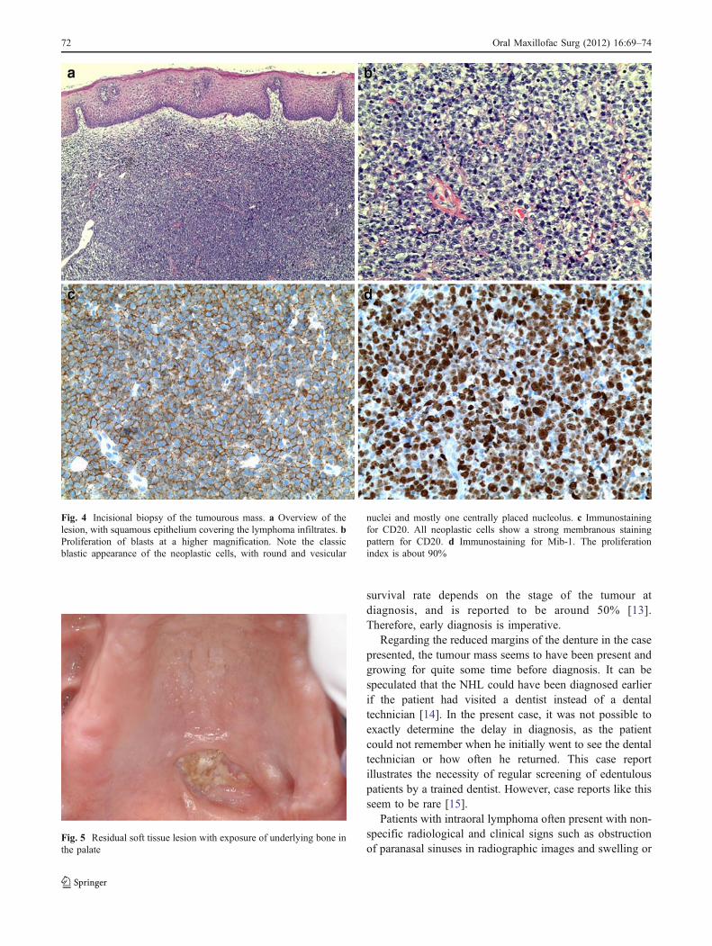
The histopathologic examination (Fig. 4a, b) of thebiopsy showed massive infiltrates of blast-like lymphoidcells with high proliferative activity (90%) invading theskeletal muscle. The tumour cells demonstrated the charac-teristic morphology of B-blasts, with large, irregularlycontoured nuclei with mostly one centrally placed nucleo-lus (Fig. 4c, d). No starry-sky pattern, as observed inBurkitt lymphoma, could be detected. The immunohisto-chemical analysis revealed a positive reaction for the B-cellmarkers CD20, CD79a and Pax-5 and co-expression ofCD10 and BCL-2 (from B-cell lymphoma 2, the second
Fig. 1 Initial clinical aspect of the exophytic, painless mass in theregion of the right hard palate and buccal mucosa
Fig. 2 Right aspect of the denture with clearly reduced margins dueto the tumorous mass
70 Oral Maxillofac Surg (2012) 16:69–74

member of a range of proteins initially described infollicular lymphomas that play a role in decreased apopto-sis), while there was only a weak positivity for BCL-6. Thetumour cells did not stain for MUM-1. Due to the highproliferative index, the positivity for BCL-6 and CD10, andthe negativity for MUM-1, the possibility of a ‘double-hit’was considered and the probe underwent further analyses.In a fluorescence in situ hybridisation (FISH), breaks at theBCL-6 and cMYC loci, but not the BCL-2 locus, weredetected. Thus, the tumour was classified as a ‘double-hit’diffuse large B-cell lymphoma. The complete stagingrevealed a stage IIIA with infiltration of the upper pole ofthe right kidney. The patient then received six cycles of R-CHOP chemotherapy, a combination of a CHOP regimen(cyclophosphamide, hydroxydaunorubicin, oncovin andprednisone) with the monoclonal CD20-antibody rituxi-mab. He received one cycle every 3 weeks. After fourcycles, a CT scan was taken, showing complete remissionof the tumour. A residual facial nerve palsy on the right sidewas noticed. In the hard palate, a soft tissue lesion withexposure of underlying bone was visible (Fig. 5). Becauseof the total absence of symptoms, no surgical revision was
initiated, and the patient was instructed in the use of achlorhexidine gel for local disinfection of the woundregion. One year following initial diagnosis, the patientremains in our care, with visits scheduled every 4 to6 months.
Discussion
Extranodal NHLs can affect patients of all ages, but thepeak incidence is between 60 and 70 years of age. TheNHLs show a predilection for the maxilla, including thesoft tissues of the palate. No gender predilection can beobserved in affected patients [10, 11]. NHLs can begrouped in subtypes based on their histologic pattern,cellular morphology and immunohistochemical staining.The WHO classification of haematopoietic and lymphoidneoplasms aims at combining the clinical aspects, histo-morphological features, immunohistochemical phenotypeand genetic features [1]. The aggressive diffuse large B-celllymphoma (DLBCL) is reported to account for 50% to 68%of all non-Hodgkin lymphomas [12]. The 5-year overall
Fig. 3 Coronal (a, b), sagittal(c) and horizontal (d) CBCTslices depict swelling in the rightmaxillary sinus and erosionof the lateral cortical sinuswall
Oral Maxillofac Surg (2012) 16:69–74 71
survival rate depends on the stage of the tumour atdiagnosis, and is reported to be around 50% [13].Therefore, early diagnosis is imperative.
Regarding the reduced margins of the denture in the casepresented, the tumour mass seems to have been present andgrowing for quite some time before diagnosis. It can bespeculated that the NHL could have been diagnosed earlierif the patient had visited a dentist instead of a dentaltechnician [14]. In the present case, it was not possible toexactly determine the delay in diagnosis, as the patientcould not remember when he initially went to see the dentaltechnician or how often he returned. This case reportillustrates the necessity of regular screening of edentulouspatients by a trained dentist. However, case reports like thisseem to be rare [15].
Patients with intraoral lymphoma often present with non-specific radiological and clinical signs such as obstructionof paranasal sinuses in radiographic images and swelling or
Fig. 5 Residual soft tissue lesion with exposure of underlying bone inthe palate
Fig. 4 Incisional biopsy of the tumourous mass. a Overview of thelesion, with squamous epithelium covering the lymphoma infiltrates. bProliferation of blasts at a higher magnification. Note the classicblastic appearance of the neoplastic cells, with round and vesicular
nuclei and mostly one centrally placed nucleolus. c Immunostainingfor CD20. All neoplastic cells show a strong membranous stainingpattern for CD20. d Immunostaining for Mib-1. The proliferationindex is about 90%
72 Oral Maxillofac Surg (2012) 16:69–74

ulceration of the mucosa. Initially, the lesions are oftenmistaken for inflammatory or reactive processes. Asdifferential diagnoses, squamous cell carcinoma and benignor malignant neoplasias of palatal salivary glands shouldalso be excluded, ideally with an incisional biopsy [16].
The most common chromosomal rearrangement indiffuse large B-cell lymphoma is the BCL-6 translocation(up to 30%), followed by BCL-2 rearrangements (20–30%)and MYC rearrangements (up to 10%). So-called double-hitlymphomas exhibit two chromosomal rearrangements [17].Based on morphological, immunohistochemical and geneticfindings, these tumours can be classified as ‘double-hit’diffuse large B-cell lymphomas or as ‘B-cell lymphoma,unclassifiable, with features between diffuse large B-celllymphoma and Burkitt lymphoma’. The latter entity wasintroduced by the 2008 WHO Classification of Tumors ofHaematopoietic and Lymphoid Tissues together withanother new entity called ‘B-cell lymphoma, unclassifiable,with features between diffuse large B-cell lymphoma andclassical Hodgkin lymphoma’ [18, 19]. These lymphomasare also referred to as ‘grey-zone lymphomas’. In the presentcase, the differential diagnosis of a ‘B-cell lymphoma,unclassifiable, with features intermediate between diffuse largeB-cell lymphoma and Burkitt lymphoma’ was considered.However, the typical morphology of the blasts, the strongpositivity for BCL-2 and the only weak positivity for BCL-6were in favour of the diagnosis of a ‘double-hit’ diffuse largeB-cell lymphoma.
‘Double-hit’ lymphomas involving a rearrangement of boththe MYC and the BCL-6 gene loci show a more aggressiveclinical behaviour and usually present with an advanced stageof disease. They normally have a poor prognosis, even whentreated with intensive chemotherapy regimens [20, 21]. Thereason for the aggressive behaviour is the overexpression ofpro-proliferative (MYC) and anti-apoptotic (BCL2, BCL6)oncoproteins at the same time. To the best of our knowledge,this case presented describes and discusses the first manifes-tation of an intraoral double-hit lymphoma.
Conclusion
The present case report of diffuse swelling of the buccalmucosa and palate as the first and only manifestation of anextranodal non-Hodgkin ‘double-hit’ lymphoma in a 76-year-old patient shows the need for immediate and accuratediagnostic procedures, including immunohistochemical analy-sis, to avoid delay and inappropriate treatment strategies.
Conflict of interest There are no financial relationships betweenany author and a commercial company that may pose a conflict ofinterest.
References
1. Swerdlow S, Campo E, Harris NL, Jaffe ES, Pileri SA, Stein H,Thiele J, Vardiman JW (2008) WHO classification of tumors ofhematopoietic and lymphoid tissues. IARC, Lyon
2. Otter R, Gerrits WBJ, van der Sandt MM, Hermanns J, WillemzeR (1989) Primary extranodal and nodal non-Hodgkin’s lymphoma.A survey of a population-based registry. Eur J Cancer Clin Oncol25:1203–1210
3. Jordan RX, Speight PM (1996) Extranodal non-Hodgkin’slymphomas of the oral cavity. Curr Top Pathol 90:125–146
4. Freeman C, Berg JW, Cutler SJ (1972) Occurrence and prognosisof extranodal lymphomas. Cancer 29:252–260
5. Epstein JB, Epstein JD, Le N, GorskyM (2001) Characteristics of oraland paraoral malignant lymphoma: a population-based review of 361cases. Oral Surg Oral Med Oral Pathol Oral Radiol Endo 92:519–525
6. Shihdoh M, Takami T, Arisue M et al (1997) Comparison betweensubmucosal (extra-nodal) and nodal non-Hodgkin’s lymphoma(NHL) in the oral and maxillofacial region. J Oral Pathol Med26:283–289
7. Eisenbud L, Sciubba J, Mir R, Sachs SA (1983) Oral presentationsin non-Hodgkin’s lymphoma: a review of 31 cases. Part I. Dataanalysis. Oral Surg Oral Med Oral Pathol 56:151–156
8. Werder P, Altermatt HJ, Zbären P, Mueller-Garamvolgyi E,Bornstein MM (2010) Palatal swelling as the first and onlymanifestation of extranodal follicular non-Hodgkin lymphoma: acase presentation. Quintessence Int 41:93–97
9. Kolokotronis A, Konstantinou N, Christakis I, Papadimitriou P,Matiakis A, Zaraboukas T, Antoniades D (2005) Localized B-cellnon-Hodgkin’s lymphoma of oral cavity and maxillofacial region:a clinical study. Oral Surg Oral Med Oral Pathol Oral Radiol Endo99:303–310
10. Urquhart A, Berg R (2001) Hodgkin’s and non-Hodgkin’slymphoma of the head and neck. Laryngoscope 111:1565–1569
11. van der Waal RI, Huijgens PC, van der Valk P, van der Waal I(2005) Characteristics of 40 primary extranodal non-Hodgkinlymphomas of the oral cavity in perspective of the new WHOclassification and the international prognostic index. Int J OralMaxillofac Surg 34:391–395
12. Kemp S, Gallagher G, Kabani S, Noonan V, O’Hara C (2008)Oral non-Hodgkin’s lymphoma: review of the literature and WorldHealth Organization classification with reference to 40 cases. OralSurg Oral Med Oral Pathol Oral Radiol Endo 105:194–201
13. Authors V (1997) A clinical evaluation of the InternationalLymphoma Study Group classification on non-Hodgkin’s lymphoma.The Non-Hodgkin's Lymphoma Classification Project. Blood89:3909–3918
14. Holmes JD, Dierks EJ, Homer LD, Potter BE (2003) Is detectionof oral and oropharyngeal squamous cancer by a dental healthcare provider associated with a lower stage at diagnosis? J OralMaxillofac Surg 61:285–291
15. George GS, Welfare RD, Lund VJ (2005) An undiagnosed case ofmalignancy: case report. Br Dent J 198:341–343
16. Werder P, Altermatt HJ, Zbären P, Bornstein MM (2009)Canalicular adenoma of a minor salivary gland on the palate: acase presentation. Quintessence Int 40:623–626
17. Stein H, Warnke RA, Chan WC, Jaffe ES, Chan JKC, Gatter KC,Campo E (2008) Diffuse large B-cell lymphoma, not otherwisespecified. In: Swerdlow S, Campo E, Harris NL, Jaffe ES, Pileri SA,Stein H, Thiele J, Vardiman JW (eds) WHO classification of tumorsof hematopoietic and lymphoid tissues. IARC, Lyon, pp 233–237
18. Kluin PM, Harris NL, Stein H, Leoncini L, Raphaël M, Campo E,Jaffe ES (2008) B-cell lymphoma, unclassifiable, with featuresintermediate between diffuse large B-cell lymphoma and Burkittlymphoma. In: SwerdlowS, Campo E,Harris NL, Jaffe ES, Pileri SA,
Oral Maxillofac Surg (2012) 16:69–74 73

Stein H, Thiele J, Vardiman JW (eds) WHO classification of tumorsof hematopoietic and lymphoid tissues. IARC, Lyon, pp 265–266
19. Jaffe ES, Stein H, Swerdlow SH, Campo E, Pileri SA, Harris NL(2008) B-cell lymphoma, unclassifiable, with features intermediatebetween diffuse large B-cell lymphoma and classical Hodgkinlymphoma. In: Swerdlow S, Campo E, Harris NL, Jaffe ES, PileriSA, Stein H, Thiele J, Vardiman JW (eds) WHO Classification oftumors of hematopoietic and lymphoid tissues. IARC, Lyon, pp267–268
20. Hasserjian RP, Ott G, Elenitoba-Johnson KSJ, Balague-Ponz O,de Jong D, de Leval L (2009) Commentary on the WHOclassification of tumors of lymphoid tissues (2008): “gray zone”lymphomas overlapping with Burkitt lymphoma or classicalHodgkin lymphoma. J Hematopathol 2:89–95
21. Kolman OK, Snuderl M, Ferry JA, Hochberg EP, Chen YB,Hasserjian RP, Rahemtullah A (2008) Clinicopathologic featuresof B-cell lymphomas with concurrent BCL-2 and c-MYC generearrangements. Mod Pathol 21(Suppl 1):260A
74 Oral Maxillofac Surg (2012) 16:69–74


![Indolent T- and NK-cell lymphoproliferative disorders of ... · extranodal site of occurrence of non-Hodgkin lymphomas [1]. Most GI lymphomas are of B-cell lineage, and T-cell lymphomas](https://static.fdocuments.net/doc/165x107/5f93d293a1c10d3ed34c6b11/indolent-t-and-nk-cell-lymphoproliferative-disorders-of-extranodal-site-of.jpg)