Diffuse Encephalitis Diagnosed on PET/CT Acquired in a Patient in Status Epilepticus with Negative...
16
Diffuse Encephalitis Diagnosed on PET/CT Acquired in a Patient in Status Epilepticus with Negative MRI J Cain 1,2 , J Hill 2 , C Coutinho 2 , S Mathur 2 1 University of Manchester, Manchester, UK, 2 Lancashire Teaching Hospitals, Preston, UK erpta Extraordinaire 03
-
Upload
ralf-roy-knight -
Category
Documents
-
view
212 -
download
0
Transcript of Diffuse Encephalitis Diagnosed on PET/CT Acquired in a Patient in Status Epilepticus with Negative...

Diffuse Encephalitis Diagnosed on PET/CT Acquired in a Patient in Status Epilepticus
with Negative MRI
J Cain1,2, J Hill2, C Coutinho2, S Mathur2 1University of Manchester, Manchester, UK, 2Lancashire Teaching Hospitals, Preston, UK
Excerpta Extraordinaire EE-03

Declaration
• I have no conflicts of interest to declare
Dr John Cain Dr Sachin Mathur

Purpose
• To describe a case of encephalitis diagnosed on PETCT performed in a ventilated patient.
• The patient had previous and subsequent MRI within normal limits.

Case Report
• 23 year old previously well female presented with seizures, reduced GCS, left sided neglect and left focal motor seizures.
• A focal abnormality was identified in temporal region on EEG.
• Patient entered prolonged period of status epilepticus.
• Seizures not controlled with quadruple anticonvulsants.
• Intubated and ventilated on ICU.

Imaging Findings
• CT head – normal
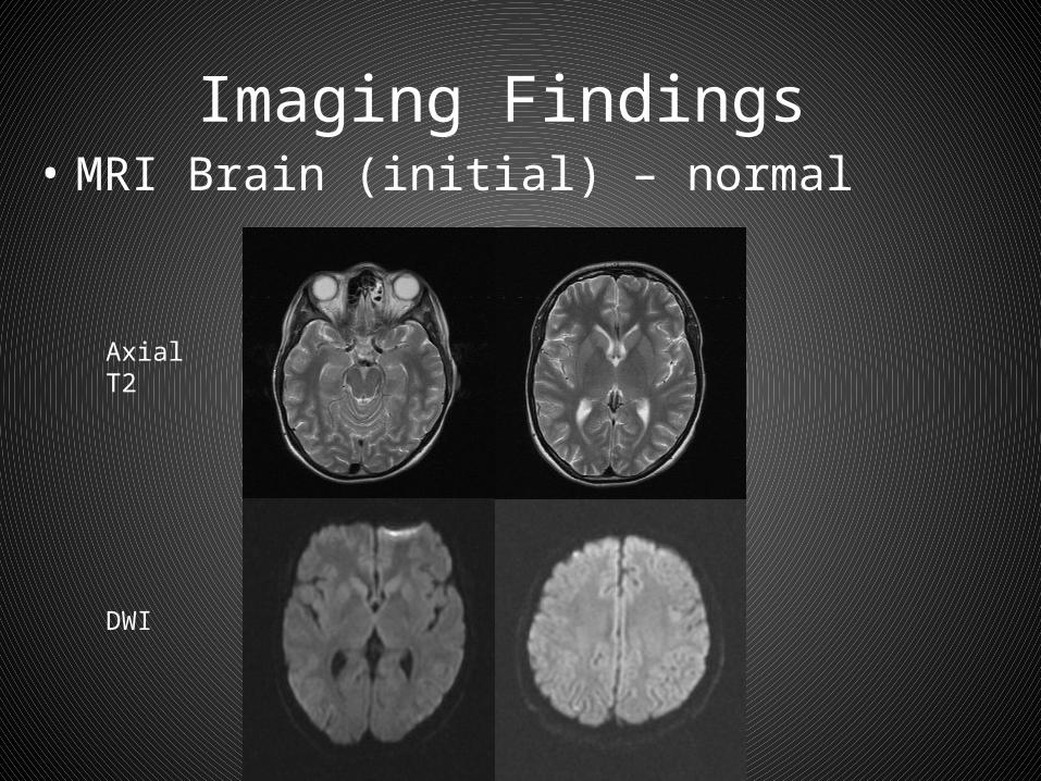
Imaging Findings • MRI Brain (initial) – normal
Axial T2
DWI

Imaging Findings
• MRI Brain (initial) – normal
Coronal FLAIR
Post contrast T1

Imaging Findings
• PET/CT (FDG) – • Diffuse reduced tracer uptake (hypo-metabolism) in
posterior frontal, temporal, occipital and parietal lobes bilaterally, with relative sparing of the anterior frontal lobes and posterior fossa.
Raw axial PET

Imaging Findings
Fused attenuation corrected PET CT

Imaging Findings
• Follow up MRI 1 month after presentation:• No focal signal abnormities, However there is moderate generalised
atrophy which seem to have been progressive.
Axial T2
SagittalT1Coronal FLAIR

Imaging Findings • Follow up MRI 4 months after presentation:• No focal signal abnormities, No obvious cortical abnormalities.
Less pronounced atrophic changes compared to previous exam.
AxialT2
Coronal FLAIR

LP findings
• Diagnosed with anti MNDA receptor encephalitis on CSF analysis.
• The remainder of the full body PETCT was within normal limits.
• No underlying malignancy identified.

Anti MNDA receptor encephalitis
• Discovered in 2005• Antibodies against NR1–NR2 heteromers of the NMDA receptor.• Progressive illness presents with psychosis, memory deficits, seizures,
and language disintegration. • Later features coma, catatonic, abnormal movements, autonomic and
breathing instability.• The disorder predominantly affects adolescents and young adults
mean 23 , 7 to 1 F>M.• Responds well to treatment but can relapse. • High incidence of underlying tumour (e.g. ovarian teratoma)• >75% of patients have substantial recovery that occurs in inverse order
of symptom development associated with a decline of antibody titres.

Patient Outcome
• Patient made good recovery on immune modulating therapy.
• Discharged home.• Slight persistent cognitive impairment.• No associated malignancy was identified.

Summary
• PET/CT is able to detect hypo-metabolism in encephalitis even in the presence of normal MRI imaging.
• The case demonstrates a possible role of performing PETCT in such patients.
• The technical difficulties of performing a PET CT in a ventilated patient are less onerous than performing a MRI.

References
• Mohr, Brandt C., and Satoshi Minoshima. "F-18 fluorodeoxyglucose PET/CT findings in a case of anti- NMDA receptor encephalitis." Clinical nuclear medicine 35, no. 6 (2010): 461-463.
• Dalmau, J; Lancaster, E; Martinez-Hernandez, E; Rosenfeld, MR; Balice-Gordon, R "Clinical experience and laboratory investigations in patients with anti-NMDAR encephalitis.” The Lancet. Neurology 10 2011(1): 63–74