Differential Regulation of Glucose-6-Phosphate · Differential Regulation of Glucose-6-Phosphate...
16
Differential Regulation of Glucose-6-Phosphate Dehydrogenase Isoenzyme Activities in Potato 1 Ru ¨ diger Hauschild 2 and Antje von Schaewen 3 * Pflanzenphysiologie, FB5 Biologie/Chemie, Universita ¨t Osnabru ¨ ck, Barbarastrae 11, 49076 Osnabru ¨ ck, Germany In plants, Glc-6-phosphate dehydrogenase (G6PDH) isoenzymes are present in the cytosol and in plastids. The plastidic enzymes (P1 and P2) are subject to redox regulation, but mechanisms that adjust cytosolic G6PDH activity are largely unknown. We adopted a leaf disc system for monitoring the effects of various conditions on G6PD isoform expression and enzyme activities in potato (Solanum tuberosum). Cytosolic G6PDH activity remained constant during water incubation in the dark. In continuous light or in the presence of metabolizable sugars in the dark, cytosolic G6PDH activity increased 6-fold within 24 h. Cycloheximide incubation demonstrated that enhanced cytosolic G6PDH activity depends on de novo protein synthesis. Osmotic change, phosphate sequestration, or oxidative stress did not affect cytosolic G6PDH activity. Further- more, enzyme activity and protein contents closely followed the corresponding mRNA levels. Together with the fact that multiple SURE elements are present in the promoter region of the gene, these results suggest that cytosolic G6PDH activity is regulated by sugar availability at the transcriptional level. Plastidic G6PDH activity stayed constant during water incubation in the light and dropped to minimal levels within 6 h in the dark. Conversely, plastidic G6PDH activity of leaf discs incubated on Paraquat rose to 10-fold higher levels, which was not prevented by cycloheximide. Similar increases were found with nitrite, nitrate, or sulfate. No major changes in protein or mRNA contents of the plastidic P1 and P2 isoforms were registered. K m (Glc-6-phosphate) values of plastidic G6PDH activity differed between samples incubated on water or Paraquat, suggesting posttranslational modification of the plastidic enzyme(s). Immunoprecipitation of 32 P-labeled samples with P1 isoform-specific antibodies showed that the chloroplast enzyme is subject to protein phosphorylation. Obviously, in extended dark periods, G6PDH activity in the stroma is restricted but can be stimulated in response to high demands for NADPH. Glc-6-phosphate dehydrogenases (G6PDHs, EC 1.1.1.49) catalyze the oxidation of Glc-6-phosphate (G6P) to 6-phosphogluconolactone concomitant with reducing NADP to NADPH. The product 6-phos- phogluconolactone is first converted to 6-phos- phogluconate by 6-phosphogluconolactonase (EC 3.1.1.31) and then decarboxylated by 6-phosphoglu- conate dehydrogenase (6PGDH, EC 1.1.1.44), yield- ing another mole of NADPH and ribulose-5- phosphate. The first enzyme, G6PDH, controls the flux through this nonreversible limb of the oxidative pentose phosphate pathway (OPPP; Williams, 1980; Copeland and Turner, 1987). In higher plants, G6PDH isoenzymes reside in two compartments, the cytosol and plastids (Heber et al., 1967; Schnarren- berger et al., 1973). Reducing power (NADPH) gen- erated by the OPPP sustains reductive biosyntheses (e.g. fatty acids, isoprenoids and aromatic amino ac- ids) in the dark and nitrogen assimilation in hetero- trophic tissues (Bowsher et al., 1992). In addition, OPPP intermediates are continuously withdrawn to fuel other metabolic pathways. For example, erythrose-4-phosphate, produced by the Calvin cycle in the light and by the OPPP in the dark, is a sub- strate of the shikimate pathway and, thus, needed as precursor of aromatic amino acids, cell wall poly- mers, phytoalexins, and pigments. All enzymatic steps of the shikimate pathway up to chorismate are confined to plastids (Schmid and Amrhein, 1995). Schnarrenberger et al. (1995) postulated that in plants, only the irreversible reactions of the OPPP might be present in the cytosol. This has recently been supported by the work of Eicks et al. (2002), who characterized a pentose phosphate translocator of the inner chloroplast membrane from Arabidopsis. Bioinformatic analyses of the Arabidopsis genome revealed that genes for both cytosolic and plastidic isoforms of Rib-5-phosphate isomerase and ribulose- 5-phosphate-3-epimerase are present, but for trans- ketolase and transaldolase, cytosolic isoforms are missing. Thus, the oxidative branch of the OPPP in the cytosol probably operates in close connection with the complete pathway in plastids. Sequences coding for NADP-dependent G6PDH enzymes exist in all organisms except for Archaebac- teria (Wendt et al., 1999). Plant G6PD-cDNA se- quences were initially elucidated from potato (Sola- 1 This work was supported by the Deutsche Forschungsgemein- schaft (Scha 541/3). 2 Present address: Institut fu ¨ r Pflanzenkrankheiten, Universita ¨t Bonn, Nuallee 9, 53115 Bonn, Germany. 3 Institut fu ¨r Botanik, Universita ¨t Mu ¨ nster, Schlogarten 3, 48149 Mu ¨ nster, Germany. * Corresponding author; e-mail [email protected]; fax 49 –251– 83–23823. Article, publication date, and citation information can be found at www.plantphysiol.org/cgi/doi/10.1104/pp.103.025676. Plant Physiology, September 2003, Vol. 133, pp. 47–62, www.plantphysiol.org © 2003 American Society of Plant Biologists 47 www.plantphysiol.org on November 19, 2018 - Published by Downloaded from Copyright © 2003 American Society of Plant Biologists. All rights reserved.
-
Upload
trinhthien -
Category
Documents
-
view
226 -
download
0
Transcript of Differential Regulation of Glucose-6-Phosphate · Differential Regulation of Glucose-6-Phosphate...

Differential Regulation of Glucose-6-PhosphateDehydrogenase Isoenzyme Activities in Potato1
Rudiger Hauschild2 and Antje von Schaewen3*
Pflanzenphysiologie, FB5 Biologie/Chemie, Universitat Osnabruck, Barbarastra�e 11, 49076 Osnabruck,Germany
In plants, Glc-6-phosphate dehydrogenase (G6PDH) isoenzymes are present in the cytosol and in plastids. The plastidicenzymes (P1 and P2) are subject to redox regulation, but mechanisms that adjust cytosolic G6PDH activity are largelyunknown. We adopted a leaf disc system for monitoring the effects of various conditions on G6PD isoform expression andenzyme activities in potato (Solanum tuberosum). Cytosolic G6PDH activity remained constant during water incubation in thedark. In continuous light or in the presence of metabolizable sugars in the dark, cytosolic G6PDH activity increased 6-foldwithin 24 h. Cycloheximide incubation demonstrated that enhanced cytosolic G6PDH activity depends on de novo proteinsynthesis. Osmotic change, phosphate sequestration, or oxidative stress did not affect cytosolic G6PDH activity. Further-more, enzyme activity and protein contents closely followed the corresponding mRNA levels. Together with the fact thatmultiple SURE elements are present in the promoter region of the gene, these results suggest that cytosolic G6PDH activityis regulated by sugar availability at the transcriptional level. Plastidic G6PDH activity stayed constant during waterincubation in the light and dropped to minimal levels within 6 h in the dark. Conversely, plastidic G6PDH activity of leafdiscs incubated on Paraquat rose to 10-fold higher levels, which was not prevented by cycloheximide. Similar increases werefound with nitrite, nitrate, or sulfate. No major changes in protein or mRNA contents of the plastidic P1 and P2 isoformswere registered. Km (Glc-6-phosphate) values of plastidic G6PDH activity differed between samples incubated on water orParaquat, suggesting posttranslational modification of the plastidic enzyme(s). Immunoprecipitation of 32P-labeled sampleswith P1 isoform-specific antibodies showed that the chloroplast enzyme is subject to protein phosphorylation. Obviously,in extended dark periods, G6PDH activity in the stroma is restricted but can be stimulated in response to high demands forNADPH.
Glc-6-phosphate dehydrogenases (G6PDHs, EC1.1.1.49) catalyze the oxidation of Glc-6-phosphate(G6P) to 6-phosphogluconolactone concomitant withreducing NADP to NADPH. The product 6-phos-phogluconolactone is first converted to 6-phos-phogluconate by 6-phosphogluconolactonase (EC3.1.1.31) and then decarboxylated by 6-phosphoglu-conate dehydrogenase (6PGDH, EC 1.1.1.44), yield-ing another mole of NADPH and ribulose-5-phosphate. The first enzyme, G6PDH, controls theflux through this nonreversible limb of the oxidativepentose phosphate pathway (OPPP; Williams, 1980;Copeland and Turner, 1987). In higher plants,G6PDH isoenzymes reside in two compartments, thecytosol and plastids (Heber et al., 1967; Schnarren-berger et al., 1973). Reducing power (NADPH) gen-erated by the OPPP sustains reductive biosyntheses(e.g. fatty acids, isoprenoids and aromatic amino ac-
ids) in the dark and nitrogen assimilation in hetero-trophic tissues (Bowsher et al., 1992). In addition,OPPP intermediates are continuously withdrawn tofuel other metabolic pathways. For example,erythrose-4-phosphate, produced by the Calvin cyclein the light and by the OPPP in the dark, is a sub-strate of the shikimate pathway and, thus, needed asprecursor of aromatic amino acids, cell wall poly-mers, phytoalexins, and pigments. All enzymaticsteps of the shikimate pathway up to chorismate areconfined to plastids (Schmid and Amrhein, 1995).Schnarrenberger et al. (1995) postulated that inplants, only the irreversible reactions of the OPPPmight be present in the cytosol. This has recentlybeen supported by the work of Eicks et al. (2002),who characterized a pentose phosphate translocatorof the inner chloroplast membrane from Arabidopsis.Bioinformatic analyses of the Arabidopsis genomerevealed that genes for both cytosolic and plastidicisoforms of Rib-5-phosphate isomerase and ribulose-5-phosphate-3-epimerase are present, but for trans-ketolase and transaldolase, cytosolic isoforms aremissing. Thus, the oxidative branch of the OPPP inthe cytosol probably operates in close connectionwith the complete pathway in plastids.
Sequences coding for NADP-dependent G6PDHenzymes exist in all organisms except for Archaebac-teria (Wendt et al., 1999). Plant G6PD-cDNA se-quences were initially elucidated from potato (Sola-
1 This work was supported by the Deutsche Forschungsgemein-schaft (Scha 541/3).
2 Present address: Institut fur Pflanzenkrankheiten, UniversitatBonn, Nu�allee 9, 53115 Bonn, Germany.
3 Institut fur Botanik, Universitat Munster, Schlo�garten 3,48149 Munster, Germany.
* Corresponding author; e-mail [email protected]; fax49 –251– 83–23823.
Article, publication date, and citation information can be foundat www.plantphysiol.org/cgi/doi/10.1104/pp.103.025676.
Plant Physiology, September 2003, Vol. 133, pp. 47–62, www.plantphysiol.org © 2003 American Society of Plant Biologists 47 www.plantphysiol.orgon November 19, 2018 - Published by Downloaded from
Copyright © 2003 American Society of Plant Biologists. All rights reserved.

num tuberosum; cytosolic isoform, Graeve et al., 1994;plastidic P1 isoform, von Schaewen et al., 1995; plas-tidic P2 isoform, Wendt et al., 1999) and are nowknown for several higher plant species. In the past,regulation of G6PDH isoenzyme activities was exam-ined in many different systems and under variousaspects. Analysis of purified or enriched enzymepreparations revealed that G6PDH activity largelydepends on reduction charge (i.e. NADPH to NADPratio). It was shown that high concentrations of theproduct NADPH inhibit the chloroplast enzyme(Lendzian and Bassham, 1975) and cytosolic G6PDH(Fickenscher and Scheibe, 1986). To avoid futile cy-cling of carbon between the OPPP and the Calvincycle, chloroplast G6PDH is inactivated in the lightby the ferredoxin-thioredoxin system (for review, seeScheibe, 1990; Buchanan, 1991). Reductive inactiva-tion can be mimicked in vitro by pre-incubating sam-ples with reduced dithiothreitol (DTTred; Johnson,1972). We used this feature in the past to discriminatein crude plant extracts between G6PDH activity de-rived from the two compartments (Scheibe et al.,1989; Graeve et al., 1994; Wenderoth et al., 1997;Wendt et al., 2000).
Information on in planta regulation of G6PD iso-forms is limited. Several studies in the past describ-ing conditions that stimulate G6PDH activity (patho-gen attack and elicitation) did not distinguishbetween the different isoenzymes nor analyze possi-ble regulatory mechanisms involved (Endo andVeech, 1969; Borner and Grisebach, 1982; Daniel etal., 1988, 1990). Steady-state transcript levels of acytosolic and a plastidic G6PD isoform were firstanalyzed in potato (von Schaewen et al., 1995). Inmost tissues, the gene coding for cytosolic G6PDH isconstitutively expressed. Compared with matureleaves, elevated mRNA levels were found in hetero-trophic tissues (etiolated shoots, tubers, or roots).Analyses of G6PDH activity during leaf developmentrevealed more or less constant levels of the cytosolicisoform. Antisense suppression, however, did notprovoke a growth phenotype (K. Graeve and A. vonSchaewen, unpublished data). Wendt et al. (2000)showed that a second plastidic isoform (P2) is tran-scribed more or less ubiquitously in potato, withhighest levels in stems and roots and lowest levels intubers. Steady-state mRNA amounts of the first char-acterized plastidic isoform (P1) are most prominentin green tissues and accumulate in stolons or root tipsin the light but can hardly be detected in soil-growntubers (von Schaewen et al., 1995; Wendt et al., 2000).In tobacco (Nicotiana tabacum) seedlings, transcriptlevels of the P1 isoform also increase during green-ing, but DTTred-sensitive enzyme activity does notcorrelate well with the mRNA amounts, suggestingeither involvement of another G6PD isoform or reg-ulation at a posttranscriptional level (C. Lange, R.Hauschild, and A. von Schaewen, unpublished data).Large variations in plastidic G6PDH activities were
also reported in other studies (Schnarrenberger et al.,1995, and refs. therein), but the basis for the effects ofdifferent stimuli on G6PDH isoenzyme activities inhigher plants remained obscure.
In heterotrophic tissues, plastidic G6PDH (and6PGDH) activities provide reductive power(NADPH) for nitrogen assimilation. Several situa-tions are known to modify metabolic fluxes throughthe plastid-localized OPPP: Oji et al. (1985) showed inwheat (Triticum aestivum) root plastids that electronsare transferred from G6P to nitrite (NO2
�) via NADPand ferredoxin-NADP reductase (FNR). Bowsher etal. (1989, 1992) demonstrated that electrons for NO2
�
reduction and Glu synthesis stem from the plastidicOPPP. Thom and Neuhaus (1995) showed that car-bohydrate flux through the plastidic OPPP can bestimulated by feeding NO2
� or Glc to isolated plas-tids and by generating a requirement for reducingequivalents to sustain metabolic fluxes in isolatedchloroplasts of green pepper (Capsicum annuum)fruits. However, regulatory mechanisms that controlincreased fluxes through the plastidic OPPP were notinvestigated. Wright et al. (1997) showed in barley(Hordeum vulgare) root plastids that levels of specificG6PDH activity decrease during nitrogen starvationconcomitant with a slight decrease in apparent Km forNADP. This was suggested to result from expressionof another G6PD isoform. Batz et al. (1998) demon-strated that genes coding for cytosolic or plastidicG6PDH enzymes are differentially transcribed uponelicitor treatment in parsley (Petroselinum crispum)suspension culture cells. Wang et al. (2000) foundstimulated expression of two P2 isoform genes afterswitching Arabidopsis plants growing in liquid cul-ture under constant illumination from ammonium(reduced nitrogen source) to nitrate (NO3
�) feeding.Knight et al. (2001) characterized a first genomicG6PD sequence (P2 isoform) from tobacco. KNO3treatment stimulated expression in root and leaf tis-sue that is assumed to be mediated through presenceof several NIT2 elements identified in the promoterregion. In contrast to nitrogen, no information isavailable about the impacts of enhanced sulfur as-similation, which is entirely located in plastids (Hell,1997).
This work aimed at elucidating factors that resultin changes of cytosolic and plastidic G6PDH activity.We adopted a leaf disc system to examine the effectsof various treatments (feeding metabolites, inhibi-tors, etc.) on G6PD isoforms in potato. We deter-mined maximal G6PDH activities (by differential in-activation of plastidic G6PDH with DTTred), proteinabundance (with isoenzyme-specific antibodies), andtranscript levels (using isoform-specific cDNAprobes) and sequenced the promoter region of agenomic DNA fragment coding for the cytosolic iso-form. Based on the obtained results, we suggest thatdiscrete mechanisms contribute to the regulation of
Hauschild and von Schaewen
48 Plant Physiol. Vol. 133, 2003 www.plantphysiol.orgon November 19, 2018 - Published by Downloaded from
Copyright © 2003 American Society of Plant Biologists. All rights reserved.

cytosolic and plastidic G6PDH isoenzyme activity inplanta.
RESULTS
Rational for the Method of Choice
We chose incubation experiments to study short-term influences of water-soluble substances onG6PDH-isoenzyme activities in potato leaf tissue. Ac-tivities were determined in leaf disc extracts afterdifferent incubation times in the dark and in thelight. In addition, mRNA levels of the different G6PDisoforms were analyzed by northern-blot hybridiza-tion and protein contents by immunodetection onwestern blots. Leaf surface served as reference toaccount for deviations in protein content that resultfrom degradation of Rubisco during long-term incu-bations in the dark. In this way, dark- and light-incubated samples and also different experimentalseries can be compared.
Effect of Incubation Conditions and SugarAvailability on Cytosolic G6PDH
Cytosolic G6PDH activity remained constant dur-ing water incubation of leaf discs in the dark (Figs.1–3). In the light, activities increased 5- to 7-foldcompared with water controls in the dark (Figs. 1 and3). Within 48 h, cytosolic G6PDH activity increasedsteadily and then remained at a constant level. Com-parable stimulation was also triggered by incubationof leaf discs on 50 mm Glc in the dark (Figs. 1–3).Incubation on 50 mm mannitol or 100 mm KCl did notaffect G6PDH activity, demonstrating that the ob-served increases are not due to osmotic or salt effects(data not shown). Presence of the electron-consuming herbicide Paraquat (5 �m methylviolo-gen) or an inhibitor of photosynthetic electron trans-port (100–500 �m DCMU) in the light had no effect,
indicating that oxidative stress and light as such arenot responsible for the activity increases.
The following experiments demonstrate the effectsof different sugars on cytosolic G6PDH activity. Leafdiscs were incubated either on Suc, Fru, Glc, or Man,respectively. Incubation on Suc (25 mm) or Fru (50mm) in darkness reproducibly led to higher increasesof cytosolic G6PDH activity and correspondingmRNA levels compared with Glc (Fig. 2, A and B).Stimulation by Fru was always higher than by Suc.Hybridization of total RNA isolated from leaf discsincubated on water or different sugars, using cDNAfragments of the cytosolic isoform (von Schaewen etal., 1995) as a probe, revealed that mRNA levelschanged similarly (Fig. 2B). Although the Glc epimerMan (50 mm) led to activity increases comparablewith Glc incubations in the dark (Fig. 2A), 3-O-methyl-Glc (3-O-MG; 50 mm) had no effect, and2-deoxy-Glc (5 mm) inhibited G6PDH activity (datanot shown). Presence of inorganic phosphate (50 mm)did not affect the stimulatory effect of Glc or Man,showing that elevated cytosolic G6PDH activity isnot a consequence of phosphate sequestration butdepends on the presence of metabolizable sugars(data not shown).
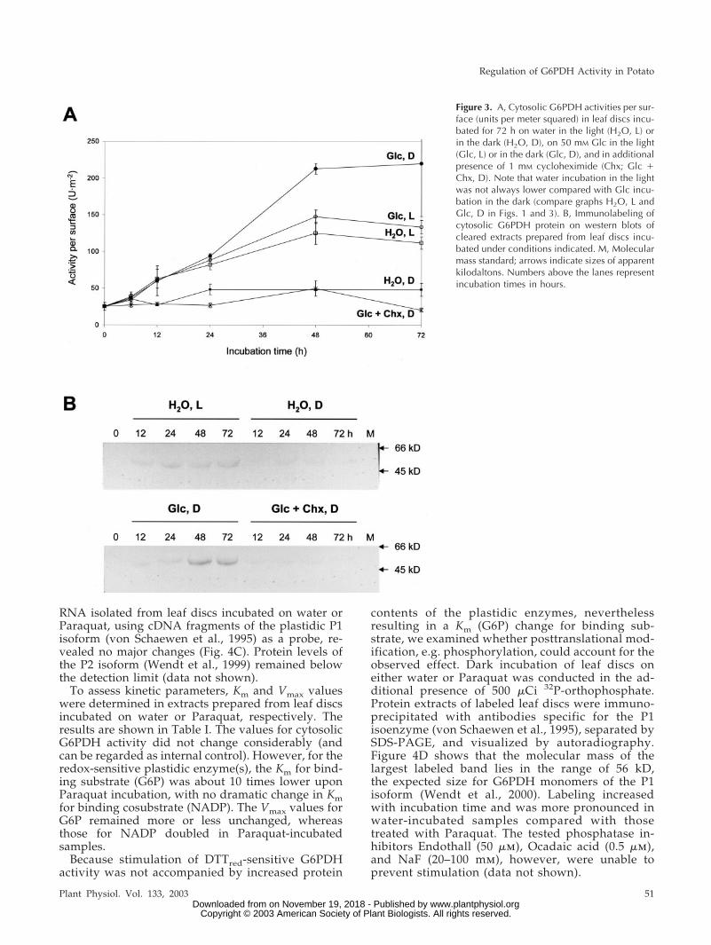
Chx (1 mm) inhibited both the increase of cytosolicG6PDH activity in water-incubated samples in thelight (not shown) and upon feeding Glc in the dark(Fig. 3A). Water incubation in the light was alwaysequivalent to Glc incubation in the dark, althoughhere Glc in the dark stimulates cytosolic G6PDHactivity to higher levels compared with water in thelight (compare with Fig. 1). Western-blot analysesusing a polyclonal antiserum specific for cytosolicG6PDH revealed that elevated protein contents cor-respond to the observed increases in activity andmRNA levels. Because Chx prevents de novo synthe-sis of cytosolic G6PDH protein (Fig. 3B), elevatedactivity of this isoform seems to result from regula-
Figure 1. Cytosolic G6PDH activities per sur-face (units per meter squared) in leaf discs incu-bated for 48 h on water in the dark (H2O, D), incontinuous light (H2O, L), or on 50 mM Glc inthe dark (Glc, D). In addition, 5 �M Paraquat inthe dark (Pq, D) or in the light (Pq, L) andincubation on 100 �M 3-(3,4-dichlorophenyl)-1,1�-dimethyl urea (DCMU), which uncouplesphotosynthetic electron transport in the light(DCMU, L), was tested.
Regulation of G6PDH Activity in Potato
Plant Physiol. Vol. 133, 2003 49 www.plantphysiol.orgon November 19, 2018 - Published by Downloaded from
Copyright © 2003 American Society of Plant Biologists. All rights reserved.

tion at the transcriptional level, most likely triggeredby accumulation of metabolizable sugars in thecytosol.
Other Substances Tested
Similar to Paraquat, incubation of potato leaf discsin the presence of hydrogen peroxide (H2O2; 0.05, 0.1,and 10 mm), iron (100 �m FeIIICl3), ammonium (20mm NH4Cl), NO2
� (20 mm NaNO2), NO3� (20, 40,
100, and 250 mm NaNO3), or sulfate (SO42�; 50, 100,
and 250 mm KSO4) had no influence on cytosolicG6PDH activity. Incubation with phosphatase inhib-itors (50 �m Endothall, 0.5 �m Okadaic acid, or 20–100 mm NaF) also had no effect.
Effect of Incubation Conditions and Stromal ReductionCharge on Plastidic G6PDH Activity
Incubation of leaf discs in the presence of Paraquat(methylviologen) in the dark has been described pre-
viously to induce oxidative stress (Bowler et al.,1991). In our experiments, incubation on 5 �m Para-quat led to remarkable increases in DTTred-sensitive(i.e. plastid-localized) G6PDH activity. Stimulationraised about 4- to 5-fold over the initial value anddiffered about 10-fold from the control (water incu-bation) determined in parallel (Fig. 4). Already after6 h, plastidic G6PDH activity reached a maximumfollowed by either slightly decreased or constantG6PDH activities (compare Figs. 4A and 5, A and B).Under these conditions, no apparent damage of theleaf discs was visible. In continuous light, the sameParaquat concentration did not alter plastidicG6PDH activity during the entire incubation period,but bleaching of the tissue was visible after about12 h.
Interestingly, stimulation of DTTred-sensitiveG6PDH activity was not inhibited by Chx (Fig. 4A).Concomitantly, protein contents of the P1 isoformremained unchanged in Paraquat- or water-incubated samples (Fig. 4B). Hybridization of total
Figure 2. A, cytosolic G6PDH activities per sur-face (units per meter squared) in leaf discs incu-bated for 48 h in the dark on either water (H2O,D), 50 mM Man (Man, D), 50 mM Glc (Glc, D),50 mM Fru (Fru, D), or 25 mM Suc (Suc, D),respectively. B, Northern-blot analyses con-ducted with total RNA (15 �g each) isolatedfrom potato leaf discs incubated on differentsugars in the dark. Samples were separated indenaturing agarose gels. After northern-blottransfer, membranes were hybridized with ra-diolabeled cDNA fragments of the cytosolic iso-form, washed under stringent conditions (threetimes at 68°C with 0.1� SSC and 0.1% [w/v]SDS), and exposed to x-ray film. Numbers abovethe lanes represent incubation time in hours.Note that an RNA sample is missing (in the lanelabeled 24 h Suc, D).
Hauschild and von Schaewen
50 Plant Physiol. Vol. 133, 2003 www.plantphysiol.orgon November 19, 2018 - Published by Downloaded from
Copyright © 2003 American Society of Plant Biologists. All rights reserved.
RNA isolated from leaf discs incubated on water orParaquat, using cDNA fragments of the plastidic P1isoform (von Schaewen et al., 1995) as a probe, re-vealed no major changes (Fig. 4C). Protein levels ofthe P2 isoform (Wendt et al., 1999) remained belowthe detection limit (data not shown).
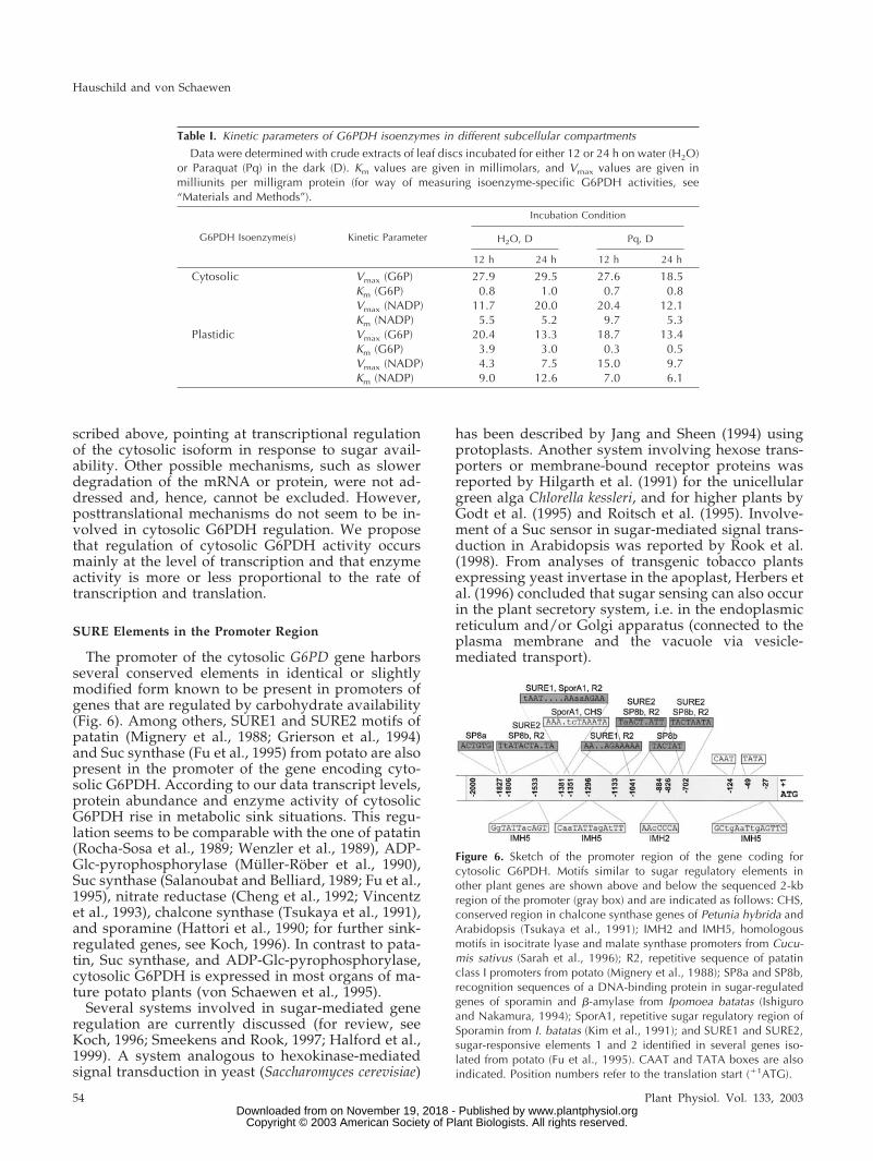
To assess kinetic parameters, Km and Vmax valueswere determined in extracts prepared from leaf discsincubated on water or Paraquat, respectively. Theresults are shown in Table I. The values for cytosolicG6PDH activity did not change considerably (andcan be regarded as internal control). However, for theredox-sensitive plastidic enzyme(s), the Km for bind-ing substrate (G6P) was about 10 times lower uponParaquat incubation, with no dramatic change in Kmfor binding cosubstrate (NADP). The Vmax values forG6P remained more or less unchanged, whereasthose for NADP doubled in Paraquat-incubatedsamples.
Because stimulation of DTTred-sensitive G6PDHactivity was not accompanied by increased protein
contents of the plastidic enzymes, neverthelessresulting in a Km (G6P) change for binding sub-strate, we examined whether posttranslational mod-ification, e.g. phosphorylation, could account for theobserved effect. Dark incubation of leaf discs oneither water or Paraquat was conducted in the ad-ditional presence of 500 �Ci 32P-orthophosphate.Protein extracts of labeled leaf discs were immuno-precipitated with antibodies specific for the P1isoenzyme (von Schaewen et al., 1995), separated bySDS-PAGE, and visualized by autoradiography.Figure 4D shows that the molecular mass of thelargest labeled band lies in the range of 56 kD,the expected size for G6PDH monomers of the P1isoform (Wendt et al., 2000). Labeling increasedwith incubation time and was more pronounced inwater-incubated samples compared with thosetreated with Paraquat. The tested phosphatase in-hibitors Endothall (50 �m), Ocadaic acid (0.5 �m),and NaF (20–100 mm), however, were unable toprevent stimulation (data not shown).
Figure 3. A, Cytosolic G6PDH activities per sur-face (units per meter squared) in leaf discs incu-bated for 72 h on water in the light (H2O, L) orin the dark (H2O, D), on 50 mM Glc in the light(Glc, L) or in the dark (Glc, D), and in additionalpresence of 1 mM cycloheximide (Chx; Glc �Chx, D). Note that water incubation in the lightwas not always lower compared with Glc incu-bation in the dark (compare graphs H2O, L andGlc, D in Figs. 1 and 3). B, Immunolabeling ofcytosolic G6PDH protein on western blots ofcleared extracts prepared from leaf discs incu-bated under conditions indicated. M, Molecularmass standard; arrows indicate sizes of apparentkilodaltons. Numbers above the lanes representincubation times in hours.
Regulation of G6PDH Activity in Potato
Plant Physiol. Vol. 133, 2003 51 www.plantphysiol.orgon November 19, 2018 - Published by Downloaded from
Copyright © 2003 American Society of Plant Biologists. All rights reserved.

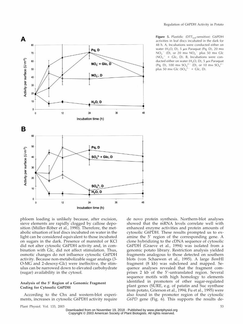
Unlike for the cytosolic enzyme, incubation of leafdiscs with 50 mm Glc in the dark had no effect onDTTred-sensitive G6PDH activity (Fig. 4A). Com-bined incubation on Paraquat plus Glc in the darkdid not influence the stimulation found with Para-quat alone. Incubation of leaf discs on Paraquat plus500 �m DCMU in the light led to a 6-fold stimulationof DTTred-sensitive G6PDH activity, whereas Para-quat in the light alone had no effect (data not shown).
Increased DTTred-sensitive G6PDH activity wasalso observed for NO2
� incubations in the dark(NO2
�, Fig. 5A) and similar time courses also forNO3
� (data not shown), either alone or in combina-tion with Glc. High concentrations (100 mm) led tosignificant stimulation of DTTred-sensitive G6PDHactivity, and simultaneous feeding of Glc sustainedthe effect. For SO4
2� (Fig. 5B), the extent of stimula-tion depended on the additional presence of sugar(acceptor carbon skeletons). Incubation on equimolarsalt solution (KCl served as a control) did not resultin altered plastidic G6PDH activities (data notshown).
In summary, G6PDH activity in chloroplasts ofdark-held leaf tissue is rapidly inactivated at theposttranslational level, probably via phosphorylationof the existing enzyme pool. Conversely, enhancedactivity of plastidic G6PDH would be triggered bydephosphorylation in situations imposing high de-mands for NADPH in the stroma.
DISCUSSION
Conditions Stimulating Cytosolic G6PDH Activity
During incubation of potato leaf discs, cytosolicG6PDH activity was stimulated about 5- to 7-foldabove the initial value on either water in the light orupon feeding sugar in the dark. Blocking photosyn-thetic electron transport by DCMU in the light pre-vented the effect, which demonstrates that stimula-tion of cytosolic G6PDH activity is not due to light assuch, but results from translocation of photosynthate(sugar) into the cytosol. In the leaf disc system, ex-port of endogenously synthesized sugars via active
Figure 4. Plastidic (DTTred-sensitive) G6PDHactivity of leaf discs incubated for 48 h. A, Leafdiscs were incubated on water in the light (H2O,L) or in the dark (H2O, D), on 5 �M Paraquat inthe dark (Pq, D), and in additional presence of 1mM Chx (Pq � Chx, D). Decreases of plastidicG6PDH activity after 24 h were observed re-peatedly but not in all experiments (compareFig. 5). B, Immunodetection of plastidic G6PDH(P1 isoform) on western blots of leaf discs incu-bated on either water or Paraquat in the dark forthe hours indicated. C, Northern-blot analysis oftotal RNA (20 �g per lane) isolated from leafdiscs incubated on either water or Paraquat inthe dark for the hours indicated. The blot washybridized with radiolabeled cDNA fragmentsof the P1 isoform (von Schaewen et al., 1995;Wendt et al., 1999) and washed under stringentconditions (three times at 68°C with 0.1� SSCand 0.1% [w/v] SDS). D, Autoradiogram of im-munoprecipitated P1 protein after radiolabelingof leaf discs with 500 �Ci 32P in the dark incu-bated on either water or Paraquat. kD, Molecu-lar mass standards. Numbers above the lanesrefer to incubation time in hours.
Hauschild and von Schaewen
52 Plant Physiol. Vol. 133, 2003 www.plantphysiol.orgon November 19, 2018 - Published by Downloaded from
Copyright © 2003 American Society of Plant Biologists. All rights reserved.
phloem loading is unlikely because, after excision,sieve elements are rapidly clogged by callose depo-sition (Muller-Rober et al., 1990). Therefore, the met-abolic situation of leaf discs incubated on water in thelight can be considered equivalent to those incubatedon sugars in the dark. Presence of mannitol or KCldid not alter cytosolic G6PDH activity and, in com-bination with Glc, did not affect stimulation. Thus,osmotic changes do not influence cytosolic G6PDHactivity. Because non-metabolizable sugar analogs (3-O-MG and 2-desoxy-Glc) were ineffective, the stim-ulus can be narrowed down to elevated carbohydrate(sugar) availability in the cytosol.
Analysis of the 5� Region of a Genomic FragmentCoding for Cytosolic G6PDH
According to the Chx and western-blot experi-ments, increases in cytosolic G6PDH activity require
de novo protein synthesis. Northern-blot analysesshowed that the mRNA levels correlate well withenhanced enzyme activities and protein amounts ofcytosolic G6PDH. These results prompted us to ex-amine the 5� region of the corresponding gene. Aclone hybridizing to the cDNA sequence of cytosolicG6PDH (Graeve et al., 1994) was isolated from agenomic potato library. Restriction analysis yieldedfragments analogous to those detected on southernblots (von Schaewen et al., 1995). A large BamHIfragment (8 kb) was subcloned and mapped. Se-quence analyses revealed that the fragment com-prises 2 kb of the 5�-untranslated region. Severalsequence motifs with high homology to elementsidentified in promoters of other sugar-regulatedplant genes (SURE, e.g. of patatin and Suc synthasefrom potato, Grierson et al., 1994; Fu et al., 1995) werealso found in the promoter region of the cytosolicG6PD gene (Fig. 6). This supports the results de-
Figure 5. Plastidic (DTTred-sensitive) G6PDHactivities in leaf discs incubated in the dark for48 h. A, Incubations were conducted either onwater (H2O, D), 5 �M Paraquat (Pq, D), 20 mM
NO2� (D), or 20 mM NO2
� plus 50 mM Glc(NO2
� � Glc, D). B, Incubations were con-ducted either on water (H2O, D), 5 �M Paraquat(Pq, D), 100 mM SO4
2� (D), or 10 mM SO42�
plus 50 mM Glc (SO42� � Glc, D).
Regulation of G6PDH Activity in Potato
Plant Physiol. Vol. 133, 2003 53 www.plantphysiol.orgon November 19, 2018 - Published by Downloaded from
Copyright © 2003 American Society of Plant Biologists. All rights reserved.
scribed above, pointing at transcriptional regulationof the cytosolic isoform in response to sugar avail-ability. Other possible mechanisms, such as slowerdegradation of the mRNA or protein, were not ad-dressed and, hence, cannot be excluded. However,posttranslational mechanisms do not seem to be in-volved in cytosolic G6PDH regulation. We proposethat regulation of cytosolic G6PDH activity occursmainly at the level of transcription and that enzymeactivity is more or less proportional to the rate oftranscription and translation.
SURE Elements in the Promoter Region
The promoter of the cytosolic G6PD gene harborsseveral conserved elements in identical or slightlymodified form known to be present in promoters ofgenes that are regulated by carbohydrate availability(Fig. 6). Among others, SURE1 and SURE2 motifs ofpatatin (Mignery et al., 1988; Grierson et al., 1994)and Suc synthase (Fu et al., 1995) from potato are alsopresent in the promoter of the gene encoding cyto-solic G6PDH. According to our data transcript levels,protein abundance and enzyme activity of cytosolicG6PDH rise in metabolic sink situations. This regu-lation seems to be comparable with the one of patatin(Rocha-Sosa et al., 1989; Wenzler et al., 1989), ADP-Glc-pyrophosphorylase (Muller-Rober et al., 1990),Suc synthase (Salanoubat and Belliard, 1989; Fu et al.,1995), nitrate reductase (Cheng et al., 1992; Vincentzet al., 1993), chalcone synthase (Tsukaya et al., 1991),and sporamine (Hattori et al., 1990; for further sink-regulated genes, see Koch, 1996). In contrast to pata-tin, Suc synthase, and ADP-Glc-pyrophosphorylase,cytosolic G6PDH is expressed in most organs of ma-ture potato plants (von Schaewen et al., 1995).
Several systems involved in sugar-mediated generegulation are currently discussed (for review, seeKoch, 1996; Smeekens and Rook, 1997; Halford et al.,1999). A system analogous to hexokinase-mediatedsignal transduction in yeast (Saccharomyces cerevisiae)
has been described by Jang and Sheen (1994) usingprotoplasts. Another system involving hexose trans-porters or membrane-bound receptor proteins wasreported by Hilgarth et al. (1991) for the unicellulargreen alga Chlorella kessleri, and for higher plants byGodt et al. (1995) and Roitsch et al. (1995). Involve-ment of a Suc sensor in sugar-mediated signal trans-duction in Arabidopsis was reported by Rook et al.(1998). From analyses of transgenic tobacco plantsexpressing yeast invertase in the apoplast, Herbers etal. (1996) concluded that sugar sensing can also occurin the plant secretory system, i.e. in the endoplasmicreticulum and/or Golgi apparatus (connected to theplasma membrane and the vacuole via vesicle-mediated transport).
Figure 6. Sketch of the promoter region of the gene coding forcytosolic G6PDH. Motifs similar to sugar regulatory elements inother plant genes are shown above and below the sequenced 2-kbregion of the promoter (gray box) and are indicated as follows: CHS,conserved region in chalcone synthase genes of Petunia hybrida andArabidopsis (Tsukaya et al., 1991); IMH2 and IMH5, homologousmotifs in isocitrate lyase and malate synthase promoters from Cucu-mis sativus (Sarah et al., 1996); R2, repetitive sequence of patatinclass I promoters from potato (Mignery et al., 1988); SP8a and SP8b,recognition sequences of a DNA-binding protein in sugar-regulatedgenes of sporamin and �-amylase from Ipomoea batatas (Ishiguroand Nakamura, 1994); SporA1, repetitive sugar regulatory region ofSporamin from I. batatas (Kim et al., 1991); and SURE1 and SURE2,sugar-responsive elements 1 and 2 identified in several genes iso-lated from potato (Fu et al., 1995). CAAT and TATA boxes are alsoindicated. Position numbers refer to the translation start (�1ATG).
Table I. Kinetic parameters of G6PDH isoenzymes in different subcellular compartments
Data were determined with crude extracts of leaf discs incubated for either 12 or 24 h on water (H2O)or Paraquat (Pq) in the dark (D). Km values are given in millimolars, and Vmax values are given inmilliunits per milligram protein (for way of measuring isoenzyme-specific G6PDH activities, see“Materials and Methods”).
G6PDH Isoenzyme(s) Kinetic Parameter
Incubation Condition
H2O, D Pq, D
12 h 24 h 12 h 24 h
Cytosolic Vmax (G6P) 27.9 29.5 27.6 18.5Km (G6P) 0.8 1.0 0.7 0.8Vmax (NADP) 11.7 20.0 20.4 12.1Km (NADP) 5.5 5.2 9.7 5.3
Plastidic Vmax (G6P) 20.4 13.3 18.7 13.4Km (G6P) 3.9 3.0 0.3 0.5Vmax (NADP) 4.3 7.5 15.0 9.7Km (NADP) 9.0 12.6 7.0 6.1
Hauschild and von Schaewen
54 Plant Physiol. Vol. 133, 2003 www.plantphysiol.orgon November 19, 2018 - Published by Downloaded from
Copyright © 2003 American Society of Plant Biologists. All rights reserved.
Due to the results of various sugar feeding studies,the response pattern of cytosolic G6PDH activity canbe compared with the regulation of genes encodingenzymes of the glyoxylate pathway (described forsuspension cultures of Cucumis sativus; Graham et al.,1994) and with those involved in photosynthesis (de-scribed for mesophyll protoplasts of maize [Zeamays]; Jang and Sheen, 1994), however, in the otherdirection: Whereas sugar incubation reduced geneexpression in these systems, carbohydrate availabil-ity induces expression of the gene coding for cytoso-lic G6PDH in potato.
In the leaf disc system, enhanced cytosolic G6PDHactivity (resulting from increased gene expression) isalso induced by Man. In the past, Man was fre-quently used to induce phosphate sequestration(Brouquisse et al., 2001, and refs. therein). Combinedincubation on Man and phosphate did not preventstimulation of cytosolic G6PDH activity in the dark.Hence, phosphate does not influence transcription ofthe cytosolic G6PD gene, and its sequestration in ametabolic sink situation plays probably not the samerole as during photosynthesis, where phosphate isneeded as counterexchange substrate for triosephos-phate export from the chloroplasts. Man is known tobe a substrate for hexokinase. The resulting Man-6-phosphate is a precursor of ascorbate biosynthesis(Wheeler et al., 1998) and can be converted to G6P viaFru-6-phosphate. In contrast, the Glc analog 3-O-MG(substrate for hexokinase but not further metabo-lized; Cortes et al., 2003) had no effect, and 2-deoxy-Glc incubation in the dark abolished cytosolicG6PDH activity. We observed that Man and2-desoxy-Glc cause unpredictable effects in the light(data not shown) similar to previous reports (Kleinand Stitt, 1998; Brouquisse et al., 2001). Therefore, weassume that only metabolizable sugars can act asinducers of enhanced cytosolic G6PD expression.
A considerable part of photosynthate is depositedas transitory starch in the stroma and can exit thechloroplast in two ways: (a) as triosephosphate (uponphosphorolytic starch breakdown via G6P) using thetriose-phosphate/3-phosphoglycerate/phosphate anti-porter (Fliege et al., 1978), or (b) as Glc (upon hydro-lytic starch mobilization) mediated by a hexose facil-itator in the inner chloroplast membrane (Weber etal., 2000). The dominant pathway seems to be thehydrolytic one, resulting in Glc export at night(Gleixner at al., 1998; Schleucher et al., 1998; Weber etal., 2000). During light-to-dark transitions, concentra-tions of the regulatory metabolite 2,6-Fru-bisphos-phate increase in leaves (Servaites et al., 1989a,1989b). Concomitantly, cytosolic and stromal levelsof Fru-1,6-bisphosphate and triose-phosphates fall tozero (Gerhardt et al., 1987), indicating that in thedark, the synthesis of triose-phosphates and theirconversion to hexose-phosphates in the cytosol arenegligible.
Here, we demonstrate that water incubation of leafdiscs in the light has the same effect as sugar incu-bation in the dark, and, thus, can be considered ametabolic sink situation. We did not observe an ad-ditive effect of Glc and light incubation (data notshown). Supposedly, the responsible sugar sensor inthe cytosol cannot differentiate between Glc im-ported across the plasma membrane and Glc ex-ported from chloroplasts. Hexose accumulationcould be sensed by hexokinase (or a yet unknowndownstream sensor), and further signaling leads toup-regulation of the gene encoding cytosolic G6PDHin the nucleus.
The results are summarized in a model of G6PDHregulation in potato leaf tissue (Fig. 7). Substrateavailability, i.e. hexoses accumulating in the cytosol(either upon export of Glc from the chloroplasts orsupplied exogenously), is perceived via stimulationof an intracellular sugar sensor—possibly by hexoki-nase or a yet unknown downstream sensor (Huijseret al., 2000; Xiao et al., 2000; Eastmond et al., 2002).Mediated by a signaling cascade, cognate transcrip-tion factors bind to SURE elements in the promoterregion and trigger enhanced transcription of the cy-tosolic G6PD gene. Increased mRNA amounts resultin enhanced translation and elevated activity of cy-tosolic G6PDH.
Because incubation of leaf discs on FeIIICl3, Para-quat, H2O2, Endothall, Okadaic acid, NO2
�, NO3�, or
SO42� did not alter cytosolic G6PDH activity, we
predict that compounds known to cause oxidativestress (FeIIICl3, Paraquat) or defense reactions (H2O2)do not influence cytosolic G6PDH activity directlybut act through changes in the cellular carbohydratestate known to switch from “source” to “sink” underthese conditions.
Conditions Stimulating DTTred-Sensitive (Plastidic)G6PDH Activity
Increased demand for reductant triggered either byParaquat, NO2
�, NO3�, or SO4
2� in the dark led torapid posttranslational stimulation of DTTred-sensitive G6PDH activity. Thus, regulation of theplastidic enzyme(s) completely differs from the cyto-solic counterpart. Conditions stimulating cytosolicG6PDH did not affect plastidic activity; conversely,sugar availability (the stimulus leading to up-regulation of the cytosolic isoform) had no effect onDTTred-sensitive G6PDH activity. This is surprisingand demonstrated best by two graphs that are basedon the same incubation experiment (compare Fig. 1with Fig. 4A). Glc in the dark stimulates only thecytosolic isoform, and Paraquat in the dark onlystimulates plastidic G6PDH activity. Substrate forplastid-localized G6PDH in the dark is most likelyprovided by mobilization of transitory starch. Thiswas shown previously by Thom and Neuhaus (1995)for isolated chloroplasts of green bell pepper fruits.
Regulation of G6PDH Activity in Potato
Plant Physiol. Vol. 133, 2003 55 www.plantphysiol.orgon November 19, 2018 - Published by Downloaded from
Copyright © 2003 American Society of Plant Biologists. All rights reserved.

Similar to potato leaf discs in the dark, green fruitsrepresent a chloroplast-containing tissue in a hetero-trophic situation.
The herbicide Paraquat, a bipyridin derivative(1,1�-dimethyl-4,4�bipyridin) is known to act asstrong electron acceptor of PSI in the light and ofNADPH in the dark, mediated by either FNR orferredoxin (Brian, 1964; Farrington et al., 1973; Oji etal., 1985). Concomitant with auto-oxidation, electronsare transferred from Paraquat to oxygen with theconsequence that the produced radicals have to beconverted to H2O2 by superoxide dismutase. Toavoid formation of the highly toxic hydroxyl radical(OH��) in the stroma, H2O2 is quickly dissipated viathe Halliwell-Asada pathway, consuming NADPH inthe final glutathione reductase reaction (Foyer andHalliwell, 1976). In the light, NADPH is amply pro-vided via photosynthetic electron flow through FNR(Shin and Arnon, 1965). In the dark, however,NADPH can only be supplied by the oxidativebranch of the plastidic OPPP, i.e. through sequentialaction of G6PDH and 6PGDH. The importance ofup-regulation of chloroplast G6PDH for sustainedstromal NADPH provision in the absence of photo-
synthetic electron transport is emphasized by incu-bation on Paraquat in the light in combination withthe uncoupling agent DCMU (Fig. 4A). In this con-dition, photosynthetic electron transport to ferre-doxin is interrupted, but demand for NADPH pro-duction in the stroma prevails due to Paraquat actingas dominant electron acceptor and through the det-rimental effects (reactive oxygen intermediate pro-duction) caused by the herbicide.
Possible Regulation through Phosphorylation
The stimulation of chloroplast G6PDH activity trig-gered by an increased stromal demand for electronsin the dark prompted us to investigate the responsi-ble regulatory mechanism. First, the increase inDTTred-sensitive G6PDH activity induced by Para-quat in the dark was not inhibited by Chx. Simulta-neous incubation on Glc, Paraquat, and Chx led tostimulation of only plastidic but not cytosolic G6PDHactivity (data not shown), which proved that thechosen Chx concentration inhibited translation ofnuclear-encoded mRNA without toxic effects on thetissue. Second, for the incubation period studied
Figure 7. Model of G6PDH regulation in the cytosol and in chloroplasts of potato leaf tissue. We chose the more generalterms “sugar” and “sugar sensor,” although our results indicate that sugar sensing occurs at the level of hexoses (withHexokinase as possible sensor). SURE stands for all regulatory promoter elements in the cytosolic G6PD gene involved insugar-mediated signaling to the nucleus (compare with Fig. 6). For clarity, redox regulation of the chloroplast enzyme wasomitted. ATG, Translation start; Chx, inhibitor of cytosolic protein translation; DCMU (inhibitor of photosynthetic electrontransport); C5P, C5-sugar phosphates; E4P, erythrose-4-phosphate; 6PG, 6-phosphogluconate. For further explanations, see“Conclusions.”
Hauschild and von Schaewen
56 Plant Physiol. Vol. 133, 2003 www.plantphysiol.orgon November 19, 2018 - Published by Downloaded from
Copyright © 2003 American Society of Plant Biologists. All rights reserved.
(72 h), we demonstrate that regulation of cytosolicand plastidic G6PDH activities operate indepen-dently. The two identified stimuli (Glc for the cyto-solic isoform, increased demand for NADPH in caseof the plastidic enzyme[s]) do not influence eachother, despite a possible interaction between cytoso-lic and plastidic OPPP at the level of C5 sugar phos-phates via the recently characterized pentose-phosphate translocator (Eicks et al., 2002) of the innerchloroplast membrane.
In contrast to cytosolic G6PDH, stimulation of plas-tidic G6PDH activity did not depend on de novoprotein synthesis. Independent of Chx inhibition,DTTred-sensitive G6PDH activity increased about4-fold (when compared with the initial value) and10-fold (when compared with water controls incu-bated in parallel, see Figs. 4 and 5). Protein amountsdetected on western blots using isoform-specific an-tibodies for P1 (Fig. 4B) or P2 (data not shown) didnot change and, hence, cannot be responsible for therapid stimulation of DTTred-sensitive G6PDHactivity.
The differences in apparent Glc6P of DTTred-sensitive G6PDH activity indicated that Paraquat in-cubation probably influences the plastidic enzyme(s)by covalent modification. Km and Vmax were deter-mined in crude extracts and, therefore, are difficult tocompare with values previously reported for en-riched or partially purified enzyme preparations. Ki-netic comparisons must also take into account thatthe values published by Scheibe et al. (1989) probablyrepresent the harvest situation (mixed state), whereasthe data of Wenderoth et al. (1997), determined withrecombinant P1 variants expressed in Escherichia coli,most likely reflect the stimulated state. In all previ-ous studies, G6PDH activity in plastids was calcu-lated as difference measured in the oxidized (active)and the reduced (inactive) state, but here we comparefor the first time, to our knowledge, plastidic G6PDHactivity of the active but unstimulated state (waterincubation in the dark) to the maximally stimulatedstate (Paraquat, NO3
�, or SO42� incubations in the
dark, compare with Figs. 4 and 5).The obtained results led to the conclusion that
posttranslational modification of plastidic G6PDH isresponsible for the rapid activity changes. An effectof redox state can be excluded because for both stim-ulated and unstimulated enzyme forms, G6PDH ac-tivity was determined as difference between samplespre-incubated with either buffer or DTTred. Incuba-tion in the presence of 32P-labeled orthophosphaterevealed that at least the P1 enzyme is subject toprotein phosphorylation. Incorporation of label in-creased with time and was much less pronounced inParaquat-incubated samples. In the dark, 32P-ortho-phosphate fed to plant cells must first enter the mi-tochondria for incorporation into ATP and is onlythen imported by plastids via counterexchange withADP (Heldt, 1976; Neuhaus et al., 1997). The reason
for the 32P labeling of the P1 protein lagging behindthe activity changes, therefore, is most likely due toan excess of unlabeled ATP in the stroma at thebeginning of the incubation experiments. Indepen-dent of knowing the exact kinetics, 32P labeling of theP1 protein correlates with decreased (and less label-ing with enhanced) plastidic G6PDH activity (Fig.4D). The smear below the major band on the auto-radiogram could reflect rapid turnover of the phos-phorylated enzyme.
For plastid-encoded genes, redox modification andphosphorylation are known to play important regu-latory roles (e.g. transcription factor, Tiller and Link,1993; Baginsky et al., 1997; endonuclease responsiblefor 3�-processing of plastidic RNA, Liere and Link,1997). In C4 and CAM plants, pyruvate-pyrophos-phate dikinase and phosphoenolpyruvate carboxy-lase are regulated by phosphorylation and are lessactive in the phosphorylated state (Wang and Chol-let, 1993; Ashton et al., 1984). In C3 plants, chloro-plast G6PDH would be the first stromal enzyme in-volved in primary metabolism whose activity is alsoregulated by phosphorylation. Until now, only onenuclear-encoded plastid-destined protein, a protein-disulfide isomerase of the green alga Chlamydomonasreinhardtii (Danon and Mayfield, 1994; Kim and May-field, 1997), has been shown to be regulated by bothredox modification and phosphorylation.
Tests intended to prevent Paraquat-mediated stim-ulation of plastidic G6PDH activity using the phos-phatase inhibitors Endothall or Okadaic acid wereunsuccessful (data not shown), possibly due to lowuptake into intact leaf tissue (discs floating upsidedown on the solutions) where the cuticle forms apotential barrier for these substances. Another reasoncould be that to act, the inhibitors have to reach thechloroplast stroma, whereas they only need to crossthe plasma membrane to contact potential cytosolictargets, as described in the work of Sheen (1993) andEhness et al. (1997). Also, different concentrations ofthe unspecific phosphatase inhibitor NaF (fluoride isa phosphate analogon) were without effect. On theother hand, the phosphatase involved in G6PDH reg-ulation might be no target for these substances,which matches the recently published results ofLukaszewski et al. (2001). These authors found thatin isolated pea (Pisum sativum) root plastids, threemajor proteins are phosphorylated. Interestingly, en-hanced fluxes through the OPPP abolished phos-phorylation of a 58-kD protein, and the phosphataseinvolved did not fall into any class known frommammalian systems. In view of the data presentedhere, this phosphoprotein could represent G6PDH.The P2 enzyme is the prominent G6PDH in plastidsof heterotrophic tissues and migrates slightly moreslowly than P1 (Wendt et al., 2000).
Large variations of G6PDH activity in chloroplastswere reported earlier (see Schnarrenberger et al.,1995, and refs. therein) and are also evident from our
Regulation of G6PDH Activity in Potato
Plant Physiol. Vol. 133, 2003 57 www.plantphysiol.orgon November 19, 2018 - Published by Downloaded from
Copyright © 2003 American Society of Plant Biologists. All rights reserved.

study (compare the different initial levels in Figs. 4and 5). After 6 h of water incubation in the dark,DTTred-sensitive G6PDH activity dropped to mini-mal values (5–10 units m�2 in crude extracts) sup-posedly corresponding to the phosphorylated state.Because incubation on Paraquat quickly resulted inpeak activities, we assume that intermediate initialG6PDH values represent mixtures of phosphorylatedand dephosphorylated enzyme molecules, reflectingdifferent levels of stimulation at the beginning of theexperiments. Previous reports on obscure (Schnar-renberger et al., 1995), largely varying, or badly re-producible plastidic G6PDH activities can now beexplained by differing ratios of unstimulated (phos-phorylated) versus stimulated (dephosphorylated)enzyme forms, probably due to local environmentalinfluences.
It appears that chloroplast G6PDH is affected bythe stromal redox state at three levels: (a) NADPH isknown to act as a competitive inhibitor of G6PDH(Lendzian and Bassham, 1975). In this case, the en-zyme itself functions as kind of a sensor (“fine con-trol”). (b) Reversible activation by the ferredoxin-thioredoxin system during light/dark transitions(Scheibe and Anderson, 1981) is also closely linked tothe prevailing NADPH to NADP ratio in the stroma(“coarse control”). (c) In addition, our results suggestthat the oxidized (dark-activated) enzyme can beswitched off posttranslationally (most likely by phos-phorylation) in extended dark periods, probably torestrict reductant flow into biosynthetic pathwaysuntil carbohydrate reserves are replenished. In life-threatening situations imposing elevated demand forstromal NADPH (e.g. oxidative stress and NO2
� re-duction), however, the enzyme can be stimulated upto 10-fold (most likely by dephosphorylation), a pro-cess that could involve redox signaling (Fig. 7). Thepossible sensor might be equivalent to the one regu-lating protein-disulfide isomerase in C. reinhardtii(Danon and Mayfield, 1994; Kim and Mayfield, 1997).Target proteins may be inactivated by a redox-sensitive kinase similar to NPH1 (Huala et al., 1997),and reactivation would require interaction with aphosphatase. Redox-sensitive kinases and/or phos-phatases have been described to regulate compo-nents of PSII, the D1 protein and light-harvestingcomplex II (Bennet, 1991; Elich et al., 1993, 1997;Silverstein et al., 1993; Durnford and Falkowski,1998).
It remains to be shown whether there is hierarchybetween redox regulation and phosphorylation invivo. Phosphorylation could act either directly byinfluencing the catalytic properties of the enzyme orindirectly by interfering with redox regulation. Wecurrently favor the idea that regulation by phosphor-ylation developed as a means to restrict plastidicG6PDH activity in the oxidized (dark-activated)state. In any case, the two posttranslational mecha-nisms ensure a quick and tight adaptation of plastidic
G6PDH activity to alterations in stromal redox statesimply by changing the kinetic properties of theenzyme.
Other Physiological Stimuli
The observation that plastidic G6PDH activity isstimulated by the demand for stromal NADPHprompted us to test other potential stimuli. The im-portance of plastidic electron transport for NO2
� as-similation was reported earlier (Paneque et al., 1964;Oji et al., 1985). Close interaction between nitrogenassimilation and the OPPP was shown for differentsystems (Bowsher et al., 1989, 1992, 1993; Aubert etal., 1994; Ritchie et al., 1994; Jin et al., 1998). Incontrast, there was no experimental evidence for alink between SO4
2� reduction and the OPPP. Becausethe complete sulfur reduction pathway is located inplastids, effects comparable with those elicited bynitrogen were also expected for sulfur assimilation.De Kok and Kuiper (1986) demonstrated that de novoreduction of SO4
2� occurs when spinach (Spinaciaoleracea) leaf discs are incubated in the dark. Neuen-schwander et al. (1991) showed that in the dark,SO4
2� assimilation in Lemna minor is limited byO-acetyl-Ser (carbon acceptor) and not by reducingequivalents. The source of the reducing equivalents,however, was not analyzed. Hell (1997) proposedthat electrons for sulfur assimilation in the dark aremost likely provided by the OPPP via FNR andferredoxin-like proteins.
We observed stimulation of plastidic G6PDH byNO2
�, NO3�, and also by SO4
2� plus Glc that werecomparable with those seen with Paraquat. Thisshows that physiological electron acceptors can re-place Paraquat in stimulating plastidic G6PDH activ-ity in the dark. A major function of the OPPP indarkened leaves seems to be sustained provision ofreducing equivalents for biosyntheses like nitrogenand sulfur assimilation. To our knowledge, this is thefirst direct experimental evidence for an interactionof the plastidic OPPP with SO4
2� reduction andproves that stimulation by Paraquat is not a result ofunspecific tissue damage. The concentrations ofNO3
� or SO42� used in this study were fairly high
but were chosen to provoke a clear response and toyield plastidic G6PDH activities comparable withthose observed with Paraquat. Because equimolarKCl concentrations did not stimulate plastidicG6PDH activity, salt effects can be excluded. The leafdisc system seems to be rather insensitive to SO4
2� orNO3
� feeding, which probably reflects absence ofhigh-affinity uptake mechanisms in leaf tissue. Theeffect of NO2
� in lower concentrations (20 mm) maybe due to its high membrane permeability and/ortoxicity and the resulting need for rapid reduction. Itis important to note that feeding cytosolic electronacceptors (FeIIICl3 and NO3
�) did not lead to in-creased cytosolic G6PDH activity, showing that this
Hauschild and von Schaewen
58 Plant Physiol. Vol. 133, 2003 www.plantphysiol.orgon November 19, 2018 - Published by Downloaded from
Copyright © 2003 American Society of Plant Biologists. All rights reserved.

enzyme is not directly regulated by the demand forreductant.
Our experiments clearly demonstrate interaction ofcarbohydrate metabolism with nitrogen and sulfurassimilation. Addition of Glc (50 mm) to NO2
�,NO3
�, or SO42� incubations resulted in higher plas-
tidic G6PDH stimulation than with NO3�, NO2
�, orSO4
2� alone because sugars provide the carbon back-bone for ammonium and sulfide acceptors (Glu andO-acetyl-Ser, respectively). The finding that NO2
�,NO3
�, and SO42� exert the same effect as Paraquat in
the dark demonstrates that up-regulation is not stim-ulus specific but a result of NADPH shortage in thestroma, which somehow influences the catalytic stateof the plastidic enzyme(s). Thus, posttranslationalregulation of chloroplast G6PDH can be summarizedas follows: Redox modification via dithiol/disulfideinterchange of two regulatory Cys in the cosubstratebinding domain represents a coarse “off/on” switchduring light/dark transitions, and the phosphoryla-tion state probably determines the extent of catalyticactivity of the dark-activated (oxidized) enzyme.
CONCLUSIONS
The results suggest that regulation of G6PDH en-zymes in the cytosol and in chloroplasts are governedby distinct mechanisms. Figure 7 shows a hypotheti-cal model based on the elucidated regulation princi-ples. High sugar levels in the cytosol trigger elevatedtranscription of the cytosolic G6PD gene via sugar-mediated signaling to the nucleus. Increased mRNAexpression results in higher enzyme levels andG6PDH activity. C5 sugar phosphates formed in thecytosol can be used for nucleotide synthesis or aretransported into plastids to replenish continuouswithdrawals of R5P and E4P (by plastid-localizednucleotide synthesis and the shikimate pathway, re-spectively), which is especially important in meta-bolic sink situations (i.e. in darkness and in hetero-trophic tissues). In contrast, G6PDH activity inplastids can be stimulated by low NADPH to NADPratios (most likely via dephosphorylation of the ex-isting enzyme pool) and is restricted in prolongeddark periods (probably via phosphorylation). Thisensures quick adaptation of the OPPP to short-termNADPH shortage in the stroma and could help topoise the important but labile balance of stromalreduction charge in the night, when alternativemechanisms like the malate valve (Fickenscher andScheibe, 1983) are inactive.
MATERIALS AND METHODS
Harvest and Incubation of Potato (Solanum tuberosumL. cv Desiree) Leaf Discs
Potato seed tubers stored at 16°C for approximately 6 months wereplanted in a soil:compost:sand mixture (3:3:1 [v/v]) and grown in climatechambers under controlled conditions (10 h of light, 200 �mol m�1 s�1 at25°C; 14 h of dark at 20°C). When the plants were about 4 weeks old, discs
were cut from both sides of the midrib using a cork borer (i.d. � 7 mm) ofleaves numbered 3 to 5 (counted from the top, the first leaf measuring about1 cm in length) always at the beginning of the light period. Up to 30 leafdiscs were floated upside-down (to minimize anaerobic effects) on differentsolutions. Water served as a control. The following substances were testedalone or in combination: 50 mm Glc, Fru, Man, 3-O-MG, or mannitol,respectively; 25 mm Suc; 100 mm KCl; 50 mm KH2PO4; 5 mm 2-deoxy-Glc;1 mm Chx; 0.1 mm DCMU; 5 �m Paraquat (methylviologen); KNO3 (20, 40,100, and 250 mm); 20 mm NaNO2 (to avoid toxic effects); K2SO4 (50, 100, and250 mm); KCl (100 and 250 mm); 50 �m Endothall; 0.5 �m Ocadaic acid; andNaF (20, 40, and 100 mm). All incubations were carried out on 20-mLvolume in disposable petri dishes at room temperature for up to 72 h in thedark or in continuous light. After 6, 12, 24, 48, and 72 h, incubated leaf discswere removed from the solutions with forceps, briefly dried on filter paper,snap frozen, and stored in liquid nitrogen. To determine initial values ofG6PDH activity (0 h of incubation), samples were directly harvested fromthe plants and frozen as above.
Enzyme Assays
To reduce variations in the measurements due to the start material (discscut from different regions of the leaf), we sampled three times two to threeleaf discs and extracted them separately. Enzyme activities were determinedtwice from each of the three extracts. Thus, means � sds are based on sixsingle values. The leaf disc extraction buffer consisted of 100 mm Tris-maleate (pH 8), 5 mm �-mercaptoethanol, 0.1 mm NADP, 1 mm Pefabloc SC(Serva, Heidelberg), and 1:100 (v/v) volume “protease-inhibitor mix for usewith plant extracts” (Sigma, Deisenhofen, Germany). Frozen material wascrushed in microcentrifuge tubes under liquid nitrogen and suspended inice-cold extraction buffer (1 �L mg�1 tissue). To each sample, 0.1 mg mg�1
tissue insoluble polyvinylpolypyrrolidone (Sigma) was added. After thaw-ing, samples were thoroughly mixed and kept on ice until centrifugation for5 min at 4°C and maximum speed. To differentiate between cytosolic andplastidic (DTTred-sensitive) isoenzymes in the supernatant (cleared extract),G6PDH activity was determined immediately under nitrogen atmosphereusing Parafilm-sealed glass cuvettes in a dual-wavelength spectrophoto-meter at 334 nm (with 410-nm reference; Sigma-Eppendorf, Hamburg, Ger-many). The standard test mixture was also set up under nitrogen atmo-sphere and consisted of 2 mm G6P and 0.2 mm NADP in 100 mm Tris-maleate buffer (pH 8) essentially as described by Graeve et al. (1994). Todetermine Km and Vmax values, the concentration of one substrate was heldconstant, and the other one was varied. Before measurements, up to 30 �Lof crude extract was used for 10 min of pre-incubation at RT (40-�L totalvolume), one sample in the presence (i.e. 10 �L of 250 mm DTTred in 300 mmTris [pH 8], final concentration � 62.5 mm DTTred) and another one in theabsence (10 �L of buffer) of reductant. Contribution of cytosolic and plas-tidic isoenzymes is based on differential calculation of G6PDH activitymeasured in the absence (total) and presence of DTTred (cytosolic activity):Total minus cytosolic is equal to plastidic G6PDH activity. Protein amountswere determined with the dye-binding assay (Bradford, 1976) and bovineserum albumin as standard protein.
Northern- and Western-Blot Analyses
Total RNA was isolated with the RNeasy Plant Mini Kit (Qiagen, Hilden,Germany). Each sample consisted of six to eight leaf discs. Isolated RNA (15�g each) was separated in denaturing agarose gels and blotted on nylonmembranes (Hybond N�, Amersham Pharmacia Biotech, Freiburg, Ger-many). Blots were hybridized with radiolabeled cDNA fragments, washed,and exposed with x-ray film as described in von Schaewen et al. (1995). Forwestern-blot analyses, 20-�L aliquots of three protein samples (extracted asmentioned above) were pooled, separated by SDS-PAGE, blotted on nitro-cellulose membranes (PROTRAN BA85, Schleicher & Schuell, Dassel, Ger-many), and subjected to immunodetection of G6PDH isoenzymes as de-scribed previously (von Schaewen et al., 1995; Wendt et al., 2000).
Immunoprecipitation of 32P-Labeled Proteins
To label phosphoproteins, leaf discs were incubated in the additionalpresence of 500 �Ci of H3
32PO4 (specific activity 9,000 Ci mmol�1; NEN,Meckenheim, Germany) diluted in 100 �m KH2PO4 (5-mL total volume).
Regulation of G6PDH Activity in Potato
Plant Physiol. Vol. 133, 2003 59 www.plantphysiol.orgon November 19, 2018 - Published by Downloaded from
Copyright © 2003 American Society of Plant Biologists. All rights reserved.

Extraction of radiolabeled leaf discs was in 1 mL of Tris-maleate buffer (alsoused for enzyme measurements and western blots; Graeve et al., 1994).Immunoprecipitation under denaturing conditions was performed essen-tially as described by Anderson and Blobel (1983) using 5 �L of polyclonalrabbit antiserum raised against the recombinant P1 protein (von Schaewenet al., 1995). Complexes bound to protein-A Sepharose (Sigma) were washedextensively and then boiled for 5 min in SDS-loading buffer. Releasedpolypeptides were separated in 12% (w/v) SDS gels. After SDS-PAGE, thegel was stained with Coomassie Brilliant Blue, dried, and exposed withHyperfilm �-max autoradiography film (Amersham, Braunschweig, Ger-many) for up to 5 d.
Isolation of Genomic Clones
A genomic potato DNA library cloned in �EMBL3 (kindly provided bythe group of Uwe Sonnewald, IPK Gatersleben, Germany) was screenedusing the entire radiolabeled NotI-cDNA fragment of the cytosolic G6PDisoform as a probe (Graeve et al., 1994). Hybridization and wash conditionswere as described for tobacco (Nicotiana tabacum) and Arabidopsis cDNAlibraries (Wendt et al., 1999). Phage DNA of identified clones was isolatedwith the Lambda System kit (Qiagen) using liquid cultures or plate lysatesas start material.
Cloning and Sequencing Procedures
DNA fragments of isolated phage clones were ligated to compatiblerestriction sites of plasmid vector pBluescript SK (Stratagene, Heidelberg)using standard procedures (Sambrook et al., 1989) and introduced into RbClcompetent (Hanahan, 1983) Escherichia coli XL1-Blue cells (Stratagene). Se-quence analysis of positive clones was conducted as described by Wendt etal. (1999). Further analyses and assembly of nucleotide sequences employedthe GCG software package (Devereux et al., 1984).
ACKNOWLEDGMENTS
The authors thank Monika Nietschke for excellent technical assistanceand the gardeners of the Plant Physiology Department in Osnabruck forcontinuous provision of healthy plants. They gratefully acknowledge thegroup of Uwe Sonnewald (IPK Gatersleben, Germany) for providing thegenomic potato library and the helpful initial advice of Andrea Polle (Uni-versitat Gottingen, Germany) on setting up a leaf disc incubation system forpotato.
Received April 17, 2003; returned for revision April 23, 2003; accepted May5, 2003.
LITERATURE CITED
Anderson DJ, Blobel G (1983) Immunoprecipitation of proteins from cell-free translations. Methods Enzymol 96: 111–120
Ashton AR, Burnell JN, Hatch MD (1984) Regulation of C4 photosynthesis:inactivation of pyruvate, Pi dikinase by ADP-dependent phosphorylationand activation by phosphorolysis. Arch Biochem Biophys 230: 492–503
Aubert S, Gout E, Bligny R, Douce R (1994) Multiple effects of glycerol onplant cell metabolism. J Biol Chem 269: 21420–21427
Baginsky S, Tiller K, Link G (1997) Transcription factor phosphorylationby a protein kinase associated with chloroplast RNA polymerase frommustard (Sinapis alba). Plant Mol Biol 34: 181–189
Batz O, Logemann E, Reinold S, Hahlbrock K (1998) Extensive reprogram-ming of primary and secondary metabolism by fungal elicitor or infectionin parsley cells. Biol Chem 379: 1127–1135
Bennet J (1991) Protein phosphorylation in green plant chloroplasts. AnnuRev Plant Physiol Plant Mol Biol 42: 281–311
Borner H, Grisebach H (1982) Enzyme induction in soybean infected byPhytophtora megasperma f. sp. glycinea. Arch Biochem Biophys 217: 65–71
Bowler C, Slooten L, Vandenbranden S, De Rycke R, Botterman J, Sy-besma C, van Montagu M, Inze D (1991) Manganese superoxide dis-mutase can reduce cellular damage mediated by oxygen radicals intransgenic plants. EMBO J 10: 1723–1732
Bowsher CG, Boulton EL, Rose J, Nayagam S, Emes MJ (1992) Reductantfor glutamate synthesis is generated by the oxidative pentose phosphatepathway in non-photosynthetic root plastids. Plant J 2: 893–896
Bowsher CG, Hucklesby DP, Emes MJ (1989) Nitrite reduction and carbo-hydrate metabolism in plastids purified from roots of Pisum sativum L.Planta 177: 359–366
Bowsher CG, Hucklesby DP, Emes MJ (1993) Induction of ferredoxin-NADP� oxidoreductase and ferredoxin synthesis in pea root plastidsduring nitrate assimilation. Plant J 3: 463–467
Bradford MM (1976) A rapid and sensitive method for the quantitation ofmicrogram quantities of protein utilising the principle of dye-binding.Anal Biochem 72: 248–254
Brian RC (1964) The classification of herbicides and types of toxicity. In LJAudus, ed, The Physiology and Biochemistry of Herbicides. AcademicPress, London, pp 1–33
Brouquisse R, Evrard A, Rolin D, Raymond P, Roby P (2001) Regulation ofprotein degradation and protease expression by mannose in maize roottips: Pi sequestration by mannose may hinder the study of its signallingproperties. Plant Physiol 125: 1485–1498
Buchanan BB (1991) Regulation of CO2 assimilation in oxygenic photosyn-thesis: the ferredoxin/thioredoxin system. Arch Biochem Biophys 288:1–9
Cheng CL, Acedo GN, Christinsin M, Conkling MA (1992) Sucrose mimicsthe light induction of Arabidopsis nitrate reductase gene transcription.Proc Natl Acad Sci USA 89: 1861–1864
Copeland L, Turner JF (1987) The regulation of glycolysis and the pentose-phosphate pathway. In A Marcus, ed, The Biochemistry of Plants, Vol 11.Academic Press, New York, pp 107–125
Cortes S, Gromova M, Evrard A, Roby C, Heyraud A, Rolin DB, RaymondP, Brouquisse RM (2003) In plants, 3-O-methylglucose is phosphorylatedby hexokinase but not perceived as a sugar. Plant Physiol 131: 824–837
Daniel S, Hinderer W, Barz W (1988) Elicitor induced changes of enzymeactivities related to isoflavone and pterocarpan accumulation in chickpea(Cicer arietinum L.) cell suspension cultures. Z Naturforsch 43c: 536–544
Daniel S, Tiemann K, Wittkampf U, Bless W, Hinderer W, Barz W (1990)Elicitor-induced metabolic changes in cell cultures of chickpea (Cicerarietinum L.) cultivars resistant and susceptible to Ascochyta rabiei: I.Investigations of enzyme activities involved in isoflavone and pterocar-pan phytoalexin biosynthesis. Planta 182: 270–278
Danon A, Mayfield SP (1994) Light-regulated translation of chloroplastmessenger RNAs through redox potential. Science 266: 1717–1719
de Kok LJ, Kuiper PJC (1986) Effect of short-term-incubation with sulfate,chloride and selenate on the glutathione content of spinach leaf discs.Physiol Plant 68: 477–482
Devereux J, Haeberli P, Smithies O (1984) A comprehensive set of sequenceanalysis programs for the VAX. Nucleic Acids Res 12: 387–395
Durnford DG, Falkowski PG (1998) Chloroplast redox regulation of nu-clear gene transcription during photoacclimation. Photosynth Res 53:229–241
Eastmond PJ, van Dijken AJH, Spielman M, Kerr A, Tissier AF, DickinsonHG, Jones JDG, Smeekens SC, Graham IA (2002) Trehalose-6-phosphate synthase 1, which catalyses the first step in trehalose synthe-sis, is essential for Arabidopsis embryo maturation. Plant J 29: 225–235
Ehness R, Ecker M, Godt D, Roitsch T (1997) Glucose and stress indepen-dently regulate source and sink metabolism via signal transduction path-ways involving protein phosphorylation. Plant Cell 9: 1825–1841
Eicks M, Maurino V, Fluegge U-I, Fischer K (2002) The plastidic pentose-phosphate translocator represents a link between the cytosolic and theplastidic pentose-phosphate pathways in plants. Plant Physiol 128:512–522
Elich TD, Edelman M, Mattoo AK (1993) Dephosphorylation of photosys-tem II core proteins is light-regulated in vivo. EMBO J 12: 4857–4862
Elich TD, Edelman M, Mattoo AK (1997) Evidence for light-regulated andlight-independent protein dephosphorylation in chloroplasts. FEBS Lett411: 236–238
Endo BY, Veech JA (1969) The histochemical localization of oxidoreductiveenzymes of soybeans infected with the root knot nematode Meloidogyneincognita acrita. Phytopathology 59: 418–425
Farrington JA, Ebert M, Land EJ, Fletcher K (1973) Bipyridylium salts andrelated compounds. V. Pulse radiolysis studies of the reaction of para-quat radical with oxygen implications for the mode of action of bipyridylherbicides. Biochim Biophys Acta 314: 372–381
Hauschild and von Schaewen
60 Plant Physiol. Vol. 133, 2003 www.plantphysiol.orgon November 19, 2018 - Published by Downloaded from
Copyright © 2003 American Society of Plant Biologists. All rights reserved.
Fickenscher K, Scheibe R (1983) Purification and properties of NADP-dependent malate dehydrogenase from pea leaves. Biochim Biophys Acta749: 249–254
Fickenscher K, Scheibe R (1986) Purification and properties of the cyto-plasmic glucose-6-phosphate dehydrogenase from pea leaves. Arch Bio-chem Biophys 247: 393–402
Fliege R, Flugge U-I, Werdan K, Heldt HW (1978) Specific transport ofinorganic phosphate, 3-phosphoglycerate and triosephosphates acrossthe inner membrane of the envelope in spinach chloroplasts. BiochimBiophys Acta 505: 232–247
Foyer C, Halliwell B (1976) The presence of glutathione and glutathionereductase in chloroplasts: A proposed role in ascorbic acid metabolism.Planta 133: 21–25
Fu H, Kim SY, Park WD (1995) High-level tuber expression and sucroseinducibility of a potato Sus4 sucrose synthase gene require 5� and 3�
flanking sequences and the leader intron. Plant Cell 7: 1387–1394Gerhardt R, Stitt M, Heldt HW (1987) Subcellular metabolite levels in
spinach leaves. Plant Physiol 83: 399–407Gleixner G, Scrimgeour C, Schmidt H-L, Viola R (1998) Stable isotope
distribution in the major metabolites of source and sink organs of Sola-num tuberosum L.: a powerful tool in the study of metabolite partitioningin intact plants. Planta 207: 241–245
Godt DE, Riegel A, Roitsch T (1995) Regulation of sucrose synthase ex-pression in Chenopodium rubrum: characterization of sugar induced ex-pression in photoautotrophic suspension cultures and sink tissue specificexpression in plants. J Plant Physiol 146: 231–238
Graeve K, von Schaewen A, Scheibe R (1994) Purification, characterizationand cDNA sequence of glucose-6-phosphate dehydrogenase from potato(Solanum tuberosum L.). Plant J 5: 353–361
Graham IA, Denby KJ, Leaver C (1994) Carbon catabolite repression reg-ulates glyoxylate cycle gene expression in cucumber. Plant Cell 6:761–772
Grierson C, Du J-S, de Torres Zabala M, Beggs K, Smith C, Holdsworth M,Bevan M (1994) Separate cis sequences and trans factors direct metabolicand developmental regulation of a potato tuber storage protein gene.Plant J 5: 816–826
Halford NG, Purcell PC, Hardie DG (1999) Is hexokinase really a sugarsensor in plants? Trends Plant Sci 4: 117–120
Hanahan D (1983) Studies on transformation of Escherichia coli with plas-mids. J Mol Biol 166: 557–580
Hattori T, Nakagawa S, Nakamura K (1990) High level expression oftuberous root storage protein genes of sweet potato in stems of plantletsgrown in vitro on sucrose medium. Plant Mol Biol 14: 595–604
Heber U, Hudson MA, Hallier UW (1967) Lokalisation von Enzymen desReduktiven und des Oxidativen Pentosephosphatzyklus in den Chloro-plasten und Permeabilitat der Chloroplastenmembranen gegenuber Me-taboliten. Z Naturforsch 22b: 1200–1215
Heldt HW (1976) Transfer of substrates across the chloroplast envelope.Horiz Biochem Biophys 2: 199–299
Hell R (1997) Molecular physiology of plant sulfur metabolism. Planta 202:138–148
Herbers K, Meuwly P, Frommer WB, Metraux J-P, Sonnewald U (1996)Systemic acquired resistance mediated by the ectopic expression of in-vertase: possible hexose sensing in the secretory pathway. Plant Cell 8:793–803
Hilgarth C, Sauer N, Tanner W (1991) Glucose increases the expression ofthe ATP/ADP translocator and the glyceraldehyde-3-phosphate dehy-drogenase genes in Chlorella. J Biol Chem 266: 24044–24047
Huala E, Oeller PW, Liscum E, Han I-S, Larsen E, Briggs WR (1997)Arabidopsis NPH1: A protein kinase with a putative redox-sensing do-main. Science 278: 2120–2123
Huijser C, Korstee A, Pego J, Wisman E, Smeekens S (2000) The Arabi-dopsis SUCROSE UNCOUPLED-6 gene is identical to ABSCISIC ACIDINSENSITIVE-4: involvement of abscisic acid in sugar responses. Plant J23: 577–585
Ishiguro S, Nakamura K (1994) Characterization of a cDNA encoding anovel DNA-binding protein, SPF1, that recognises SP8 sequences in the 5�
upstream region of genes coding for sporamin and �-amylase from sweetpotato. Mol Gen Genet 244: 563–571
Jang J-C, Sheen J (1994) Sugar sensing in higher plants. Plant Cell 6:1665–1679
Jin T, Huppe HC, Turpin DH (1998) In vitro reconstitution of electrontransport from glucose-6-phosphate and NADPH to nitrite. Plant Physiol117: 303–309
Johnson HS (1972) Dithiothreitol: An inhibitor of glucose-6-phosphate de-hydrogenase activity in leaf extracts and isolated chloroplasts. Planta 106:273–277
Kim J, Mayfield SP (1997) Protein disulfide isomerase as a regulator ofchloroplast translational activation. Science 278: 1954–1957
Kim S-R, Costa MA, An G (1991) Sugar response element enhances woundresponse of potato proteinase inhibitor II promoter in transgenic tobacco.Plant Mol Biol 17: 973–983
Klein D, Stitt M (1998) Effects of 2-deoxyglucose on the expression of rbcSand the metabolism of Chenopodium rubrum cell-suspension cultures.Planta 205: 223–234
Knight JS, Emes MJ, Debnam P (2001) Isolation and characterisation of afull-length genomic clone encoding a plastidic glucose-6-phosphate de-hydrogenase from Nicotiana tabacum. Planta 212: 499–507
Koch KE (1996) Carbohydrate-modulated gene expression in plants. AnnuRev Plant Physiol Plant Mol Biol 47: 509–540
Lendzian K, Bassham JA (1975) Regulation of glucose-6-phosphate dehy-drogenase in spinach chloroplasts by ribulose 1,5-diphosphate andNADPH/NADP� ratios. Biochem Biophys Acta 396: 260–275
Liere K, Link G (1997) Chloroplast endoribonuclease p54 involved in RNA3�-end processing is regulated by phosphorylation and redox state. Nu-cleic Acids Res 25: 2403–2408
Lukaszewski KM, Bowsher CG, Savory PJ, Emes MJ (2001) Protein phos-phorylation in pea root plastids. Plant Cell Physiol 42: 642–649
Mignery GA, Pikaard CS, Park WD (1988) Molecular characterization ofthe patatin multigene family of potato. Gene 62: 27–44
Muller-Rober BT, Ko�mann J, Hannah LC, Willmitzer L, Sonnewald U(1990) One of two different ADP-G glucose-pyrophosphorylase genesfrom potato responds strongly to elevated levels of sucrose. Mol GenGenet 224: 136–146
Neuenschwander U, Suter M, Brunold C (1991) Regulation of sulfateassimilation by light and O-acetyl-l-serine in Lemna minor L. Plant Physiol97: 253–258
Neuhaus HE, Thom E, Mohlmann T, Steup M, Kampfenkel K (1997)Characterization of a novel eukaryotic ATP/ADP translocator located inthe plastid envelope of Arabidopsis thaliana L. Plant J 11: 73–82
Oji Y, Watanabe M, Wakiuchi N, Okamoto S (1985) Nitrite reduction inbarley root plastids: Dependence on NADPH coupled with glucose-6-phosphate and 6-phosphogluconate dehydrogenases, and possible in-volvement of an electron carrier and a diaphorase. Planta 165: 85–90
Paneque A, Ramirez JM, Del Campo FF, Losada M (1964) Light and darkreduction of nitrite in a reconstituted enzymic system. J Biol Chem 239:1737–1741
Ritchie SW, Redinbaugh MG, Shiraishi N, Vrba JM, Campbell WH (1994)Identification of a maize root transcript expressed in the primary re-sponse to nitrate: characterization of a cDNA with homology toferredoxin-NADP�-oxidoreductase. Plant Mol Biol 26: 679–690
Rocha-Sosa M, Sonnewald U, Frommer W, Stratmann M, Schell J,Willmitzer L (1989) Both developmental and metabolic signals activatethe promoter of a class I patatin gene. EMBO J 8: 23–29
Roitsch T, Bittner M, Godt DE (1995) Induction of apoplastic invertase ofChenopodium rubrum by d-Glucose and a glucose analog and tissue-specific expression suggest a role in sink-source-regulation. Plant Physiol108: 285–294
Rook F, Gerrits N, Kortstee A, van Kampen M, Borrias M, Weisbeek P,Smeekens S (1998) Sucrose-specific signalling represses translation of theArabidopsis ATB2 bZIP transcription factor gene. Plant J 15: 253–263
Salanoubat M, Belliard G (1989) The steady state level of potato sucrosesynthase mRNA is dependent on wounding, anaerobiosis, and sucroseconcentration. Gene 84: 181–185
Sambrook J, Fritsch EF, Maniatis T (1989) Molecular cloning: A LaboratoryManual, Ed 2. Cold Spring Harbor Laboratory Press, Cold Spring Harbor,NY
Sarah CJ, Graham IA, Reynolds SJ, Leaver CJ, Smith SM (1996) Distinctcis-acting elements direct the germination and sugar response of thecucumber malate synthase gene. Mol Gen Genet 250: 153–161
Scheibe R (1990) Light/dark modulation: regulation of chloroplast metab-olism in a new light. Bot Acta 103: 327–334
Regulation of G6PDH Activity in Potato
Plant Physiol. Vol. 133, 2003 61 www.plantphysiol.orgon November 19, 2018 - Published by Downloaded from
Copyright © 2003 American Society of Plant Biologists. All rights reserved.

Scheibe R, Anderson LE (1981) Dark modulation of NADP-dependentmalate dehydrogenase and glucose-6-phosphate dehydrogenase in thechloroplast. Biochem Biophys Acta 636: 58–64
Scheibe R, Geissler A, Fickenscher K (1989) Chloroplast glucose-6-phosphate dehydrogenase: Km shift upon light modulation and reduc-tion. Arch Biochem Biophys 274: 290–297
Schleucher J, Vanderveer PJ, Sharkey TD (1998) Export of carbon fromchloroplasts at night. Plant Physiol 118: 1439–1445
Schmid J, Amrhein N (1995) Molecular organization of the shikimate path-way in higher plants. Phytochemistry 39: 737–749
Schnarrenberger C, Flechner A, Martin W (1995) Enzymatic evidence for acomplete oxidative pentose phosphate pathway in chloroplasts and anincomplete pathway in the cytosol of spinach leaves. Plant Physiol 108:609–614
Schnarrenberger C, Oeser A, Tolbert NE (1973) Two isoenzymes each ofglucose-6-phosphate dehydrogenase and 6-phosphogluconate dehydro-genase in spinach leaves. Arch Biochem Biophys 154: 438–448
Servaites JC, Fondy BR, Li B, Geiger DR (1989b) Sources of carbon forexport from spinach leaves throughout the day. Plant Physiol 90:1168–1174
Servaites JC, Geiger DR, Tucci MA, Fondy BR (1989a) Leaf carbon metab-olism and metabolite levels during a period of sinusoidal light. PlantPhysiol 89: 403–408
Sheen J (1993) Protein phosphatase activity is required for light-induciblegene expression in maize: phosphatase. EMBO J 12: 3497–3505
Shin M, Arnon DI (1965) Enzymic mechanisms of pyridine nucleotidereduction in chloroplasts. J Biol Chem 240: 1405–1411
Silverstein T, Cheng L, Allen JF (1993) Chloroplast thylakoid proteinphosphatase reactions are redox-independent and kinetically heteroge-neous. FEBS Lett 334: 101–105
Smeekens S, Rook F (1997) Sugar sensing and sugar-mediated signal-transduction in plants. Plant Physiol 115: 7–13
Thom E, Neuhaus HE (1995) Oxidation of imported or endogenous carbo-hydrates by isolated chloroplasts from green pepper fruits. Plant Physiol109: 1421–1426
Tiller K, Link G (1993) Phosphorylation and dephosphorylation affectfunctional characteristics of chloroplast and etioplast transcription sys-tems from mustard (Sinapis alba L.). EMBO J 12: 1745–1753
Tsukaya H, Ohshima T, Naito S, Chino M, Komeda Y (1991) Sugar-dependent expression of the CHS-A gene for chalcone synthase frompetunia in transgenic Arabidopsis. Plant Physiol 97: 1414–1421
Vincentz M, Moureaux T, Leydecker M-T, Vaucheret H, Caboche M (1993)Regulation of nitrate and nitrite reductase expression in Nicotiana plum-baginifolia leaves by nitrogen and carbon metabolites. Plant J 3: 315–324
von Schaewen A, Langenkamper G, Graeve K, Wenderoth I, Scheibe R(1995) Molecular characterization of the plastidic glucose-6-phosphatedehydrogenase from potato in comparison to its cytosolic counterpart.Plant Physiol 109: 1327–1335
Wang YH, Chollet R (1993) In vitro phosphorylation of purified tobacco-leafphosphoenol-pyruvate carboxylase. FEBS Lett 328: 215–218
Wang R, Guegler K, LaBrie ST, Crawford NM (2000) Genomic analysis ofa nutrient response in Arabidopsis reveals diverse expression patternsand novel metabolic and potential regulatory genes induced by nitrate.Plant Cell 12: 1491–1509
Weber A, Servaites JC, Geiger DR, Kofler H, Hille D, Groner F, HebbekerU, Flugge U-I (2000) Identification, purification, and molecular cloning ofa putative plastidic glucose transporter. Plant Cell 12: 787–801
Wenderoth I, Scheibe R, von Schaewen A (1997) Identification of thecysteine residues involved in redox modification of plant plastidicglucose-6-phosphate dehydrogenase. J Biol Chem 272: 26985–26990
Wendt UK, Hauschild R, Lange C, Pietersma M, Wenderoth I, von Schae-wen A (1999) Evidence for functional convergence of redox regulation inG6PDH isoforms of cyanobacteria and higher plants. Plant Mol Biol 40:487–494
Wendt UK, Wenderoth I, Tegeler A, von Schaewen A (2000) Molecularcharacterization of a novel glucose-6-phosphate dehydrogenase frompotato (Solanum tuberosum L.) Plant J 23: 723–733
Wenzler HC, Mignery GA, Fisher LM, Park WD (1989) Analysis of achimeric class I-patatin GUS gene in transgenic potato plants: high levelexpression in tubers and sucrose inducible expression in cultured leafand stem explants. Plant Mol Biol 12: 41–50
Wheeler GL, Jones MA, Smirnoff N (1998) The biosynthetic pathway ofvitamin C in higher plants. Nature 393: 365–369
Williams JF (1980) A critical examination of the evidence for the reactions ofthe pentose pathway in animal tissues. Trends Biochem Sci 5: 315–320
Wright DP, Huppe HC, Turpin DH (1997) In vivo and in vitro studies ofglucose-6-phosphate dehydrogenase from barley root plastids in relationto reductant supply for NO2
� assimilation. Plant Physiol 114: 1413–1419Xiao W, Sheen J, Jang J-C (2000) The role of hexokinase in plant sugar signal
transduction and growth and development. Plant Mol Biol 44: 451–461
Hauschild and von Schaewen
62 Plant Physiol. Vol. 133, 2003 www.plantphysiol.orgon November 19, 2018 - Published by Downloaded from
Copyright © 2003 American Society of Plant Biologists. All rights reserved.