Differential expression profiles of the salivary proteins ... · Differential expression profiles...
10
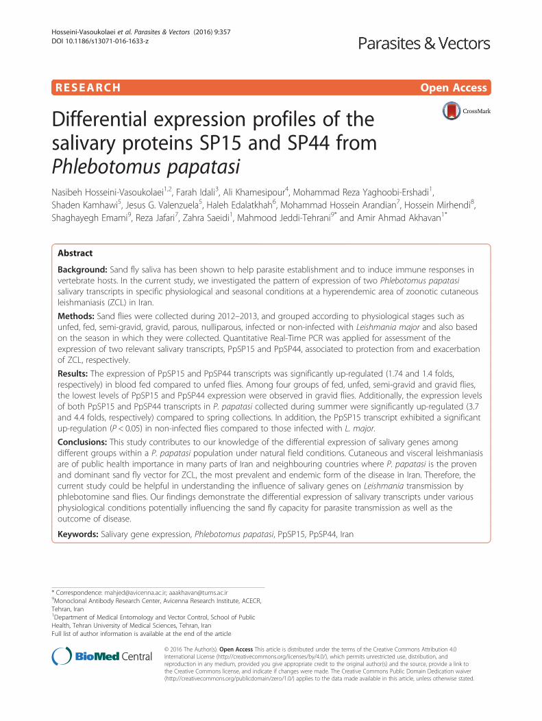
RESEARCH Open Access Differential expression profiles of the salivary proteins SP15 and SP44 from Phlebotomus papatasi Nasibeh Hosseini-Vasoukolaei 1,2 , Farah Idali 3 , Ali Khamesipour 4 , Mohammad Reza Yaghoobi-Ershadi 1 , Shaden Kamhawi 5 , Jesus G. Valenzuela 5 , Haleh Edalatkhah 6 , Mohammad Hossein Arandian 7 , Hossein Mirhendi 8 , Shaghayegh Emami 9 , Reza Jafari 7 , Zahra Saeidi 1 , Mahmood Jeddi-Tehrani 9* and Amir Ahmad Akhavan 1* Abstract Background: Sand fly saliva has been shown to help parasite establishment and to induce immune responses in vertebrate hosts. In the current study, we investigated the pattern of expression of two Phlebotomus papatasi salivary transcripts in specific physiological and seasonal conditions at a hyperendemic area of zoonotic cutaneous leishmaniasis (ZCL) in Iran. Methods: Sand flies were collected during 2012–2013, and grouped according to physiological stages such as unfed, fed, semi-gravid, gravid, parous, nulliparous, infected or non-infected with Leishmania major and also based on the season in which they were collected. Quantitative Real-Time PCR was applied for assessment of the expression of two relevant salivary transcripts, PpSP15 and PpSP44, associated to protection from and exacerbation of ZCL, respectively. Results: The expression of PpSP15 and PpSP44 transcripts was significantly up-regulated (1.74 and 1.4 folds, respectively) in blood fed compared to unfed flies. Among four groups of fed, unfed, semi-gravid and gravid flies, the lowest levels of PpSP15 and PpSP44 expression were observed in gravid flies. Additionally, the expression levels of both PpSP15 and PpSP44 transcripts in P. papatasi collected during summer were significantly up-regulated (3.7 and 4.4 folds, respectively) compared to spring collections. In addition, the PpSP15 transcript exhibited a significant up-regulation (P < 0.05) in non-infected flies compared to those infected with L. major. Conclusions: This study contributes to our knowledge of the differential expression of salivary genes among different groups within a P. papatasi population under natural field conditions. Cutaneous and visceral leishmaniasis are of public health importance in many parts of Iran and neighbouring countries where P. papatasi is the proven and dominant sand fly vector for ZCL, the most prevalent and endemic form of the disease in Iran. Therefore, the current study could be helpful in understanding the influence of salivary genes on Leishmania transmission by phlebotomine sand flies. Our findings demonstrate the differential expression of salivary transcripts under various physiological conditions potentially influencing the sand fly capacity for parasite transmission as well as the outcome of disease. Keywords: Salivary gene expression, Phlebotomus papatasi, PpSP15, PpSP44, Iran * Correspondence: [email protected]; [email protected] 9 Monoclonal Antibody Research Center, Avicenna Research Institute, ACECR, Tehran, Iran 1 Department of Medical Entomology and Vector Control, School of Public Health, Tehran University of Medical Sciences, Tehran, Iran Full list of author information is available at the end of the article © 2016 The Author(s). Open Access This article is distributed under the terms of the Creative Commons Attribution 4.0 International License (http://creativecommons.org/licenses/by/4.0/), which permits unrestricted use, distribution, and reproduction in any medium, provided you give appropriate credit to the original author(s) and the source, provide a link to the Creative Commons license, and indicate if changes were made. The Creative Commons Public Domain Dedication waiver (http://creativecommons.org/publicdomain/zero/1.0/) applies to the data made available in this article, unless otherwise stated. Hosseini-Vasoukolaei et al. Parasites & Vectors (2016) 9:357 DOI 10.1186/s13071-016-1633-z
Transcript of Differential expression profiles of the salivary proteins ... · Differential expression profiles...
RESEARCH Open Access
Differential expression profiles of thesalivary proteins SP15 and SP44 fromPhlebotomus papatasiNasibeh Hosseini-Vasoukolaei1,2, Farah Idali3, Ali Khamesipour4, Mohammad Reza Yaghoobi-Ershadi1,Shaden Kamhawi5, Jesus G. Valenzuela5, Haleh Edalatkhah6, Mohammad Hossein Arandian7, Hossein Mirhendi8,Shaghayegh Emami9, Reza Jafari7, Zahra Saeidi1, Mahmood Jeddi-Tehrani9* and Amir Ahmad Akhavan1*
Abstract
Background: Sand fly saliva has been shown to help parasite establishment and to induce immune responses invertebrate hosts. In the current study, we investigated the pattern of expression of two Phlebotomus papatasisalivary transcripts in specific physiological and seasonal conditions at a hyperendemic area of zoonotic cutaneousleishmaniasis (ZCL) in Iran.
Methods: Sand flies were collected during 2012–2013, and grouped according to physiological stages such asunfed, fed, semi-gravid, gravid, parous, nulliparous, infected or non-infected with Leishmania major and also basedon the season in which they were collected. Quantitative Real-Time PCR was applied for assessment of theexpression of two relevant salivary transcripts, PpSP15 and PpSP44, associated to protection from and exacerbationof ZCL, respectively.
Results: The expression of PpSP15 and PpSP44 transcripts was significantly up-regulated (1.74 and 1.4 folds,respectively) in blood fed compared to unfed flies. Among four groups of fed, unfed, semi-gravid and gravid flies,the lowest levels of PpSP15 and PpSP44 expression were observed in gravid flies. Additionally, the expression levelsof both PpSP15 and PpSP44 transcripts in P. papatasi collected during summer were significantly up-regulated (3.7and 4.4 folds, respectively) compared to spring collections. In addition, the PpSP15 transcript exhibited a significantup-regulation (P < 0.05) in non-infected flies compared to those infected with L. major.
Conclusions: This study contributes to our knowledge of the differential expression of salivary genes amongdifferent groups within a P. papatasi population under natural field conditions. Cutaneous and visceral leishmaniasisare of public health importance in many parts of Iran and neighbouring countries where P. papatasi is the provenand dominant sand fly vector for ZCL, the most prevalent and endemic form of the disease in Iran. Therefore, thecurrent study could be helpful in understanding the influence of salivary genes on Leishmania transmission byphlebotomine sand flies. Our findings demonstrate the differential expression of salivary transcripts under variousphysiological conditions potentially influencing the sand fly capacity for parasite transmission as well as theoutcome of disease.
Keywords: Salivary gene expression, Phlebotomus papatasi, PpSP15, PpSP44, Iran
* Correspondence: [email protected]; [email protected] Antibody Research Center, Avicenna Research Institute, ACECR,Tehran, Iran1Department of Medical Entomology and Vector Control, School of PublicHealth, Tehran University of Medical Sciences, Tehran, IranFull list of author information is available at the end of the article
© 2016 The Author(s). Open Access This article is distributed under the terms of the Creative Commons Attribution 4.0International License (http://creativecommons.org/licenses/by/4.0/), which permits unrestricted use, distribution, andreproduction in any medium, provided you give appropriate credit to the original author(s) and the source, provide a link tothe Creative Commons license, and indicate if changes were made. The Creative Commons Public Domain Dedication waiver(http://creativecommons.org/publicdomain/zero/1.0/) applies to the data made available in this article, unless otherwise stated.
Hosseini-Vasoukolaei et al. Parasites & Vectors (2016) 9:357 DOI 10.1186/s13071-016-1633-z
BackgroundZoonotic cutaneous leishmaniasis (ZCL) is a neglectedtropical disease caused by the protozoan parasiteLeishmania major [1]. ZCL is endemic in 17 out of31 provinces of Iran and represents a public health prob-lem of increasing proportions [2]. The incidence rate ofZCL in Esfahan Province, a hyperendemic zone of ZCL incentral Iran, is reported to be around 2400 cases per year(communication from the Esfahan Center for PublicHealth). This is considered to be an underestimation ofthe actual incidence.A sand fly salivates as it bites the vertebrate host skin.
Salivary glands have a unicellular epithelial layer sur-rounding a container for saliva consisting of a repertoireof proteins that vary based upon the physiological stateof adults, sex, age, generation, species and geographicallocation of the sand fly [3, 4]. Sand fly saliva contains aseries of bioactive molecules which are necessary for thesuccessful uptake of blood meals and for establishmentof Leishmania in vertebrate hosts [5, 6].Sand fly saliva has immunomodulatory characteristics
and induces a specific immunity consisting of antibodyproduction and a cellular immune response [7]. Recently,the biological activity and immunogenicity of sand fly sal-ivary proteins were comprehensively reviewed [8]. A pro-tein called PpSP15 from P. papatasi saliva is a member ofthe small odorant binding-like family of proteins [9]. Thehomologue of this protein in P. duboscqi, PdSP15, was re-cently shown to inhibit contact pathway activation bybinding to negatively charged molecules including poly-phosphate, heparin, and dextran Sulfate [10]. PpSP44, an-other protein from P. papatasi saliva is a member of theyellow family of proteins found in all sand fly species [9].The function of the sand fly yellow family of proteins wasrecently characterized as proteins that bind bioamines in-cluding epinephrine and norepinephrine [11]. Both pro-teins are also immunogenic. PpSP15 protein from P.papatasi saliva induced an immunity in rodents that con-ferred protection against Leishmania major infection [12,13]. The observed protection correlated with the induc-tion of a specific delayed-type hypersensitivity response(DTH) with a Th1 profile [12]. These studies introducedPpSP15 as a candidate vaccine against infection with L.major. Interestingly, immunization with PpSP44 produceda Th2 response in mice that enhanced L. major infection[13]. This study demonstrated a differential immune re-sponse to distinct molecules in saliva of the same sand flyspecies leading to different outcomes of the disease [13].Biologically active molecules in sand fly saliva are con-
served for some proteins and divergent for others [14–18].Few studies have been aimed at understanding the effectof physiological and seasonal factors on the expression ofsand fly salivary proteins and their subsequent effect onparasite transmission and epidemiology of leishmaniasis.
Phlebotomus papatasi is the main vector of ZCL inthe Old World and Iran [19, 20]. The behaviour of thissand fly species is well documented with regards to rest-ing places [21], blood sources [22–25], bacterial micro-flora [26], longevity [27], dispersal ability [28] andseasonal activity [29, 30]. Moreover, many studies havedemonstrated a role for biotic or abiotic factors on geneexpression [31]. Although the physiology and ecology ofP. papatasi sand flies are well known, the effect of thephysiological state and the environment on salivary geneexpression in this insect is still unknown.In this study we tested the hypothesis that the expres-
sion profiles of PpSP15 and PpSP44 salivary transcriptsvaries with the physiological state and seasonal status offemale P. papatasi sand flies, and that these variationsare different in the two salivary transcripts potentiallyimpacting the host immune response.
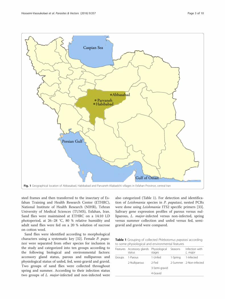
MethodsStudy areaThis investigation was accomplished during 2012–2013in three villages of Parvaneh-Aliabadchi, Habib Abadand Abbasabad in Esfahan province, central Iran (Fig. 1).The Habib Abad and Parvaneh- Aliabadchi villages arelocated 25–40 km north of the city of Esfahan (32°39′35"N, 51°40′17"E). The Abbasabad village is located 5Km from Badroud district (33°42′N, 52°2′E), Natanzcity, Esfahan province, central Iran.The biotope of the selected areas is desert with hot
summers and cold winters. The two study areas nearEsfahan city are located at an altitude of around1550 m, and Badroud district is located at an altitude of1056 m, in the foothills of Karkas Mountains. Wheat,barley, cotton, vines, beetroot, pistachio, alfalfa, Indiancorn, clover and summer crops are cultivated in theseareas (the Esfahan Agriculture Organization).In 2013, the maximum and minimum monthly tempera-
tures in Esfahan city were 39.8 °C in July and -5.3 °C inDecember. The maximum and minimum monthly relativehumidity were 81 % and 9.1 % in November and July, re-spectively. In Badrood district, the maximum and mini-mum monthly temperatures were 43 °C in July and -2.9 °C in December, respectively. The maximum relative hu-midity was 62 % in December and the minimum was 20 %in February. The total annual rainfall was 84.6 mm inEsfahan city and 77.5 mm in Badrood district (EsfahanMetrological Organization).
Sand fly collection and rearingPhlebotomines were collected using aspirating tubes andfunnel trapping from resting places throughout the se-lected study villages during the active season of sandflies in 2012–2013. Collected sand flies were moved intocloth cages with 20 × 20 × 20 cm dimensions, hanging on
Hosseini-Vasoukolaei et al. Parasites & Vectors (2016) 9:357 Page 2 of 10
steel frames and then transferred to the insectary of Es-fahan Training and Health Research Center (ETHRC),National Institute of Health Research (NIHR), TehranUniversity of Medical Sciences (TUMS), Esfahan, Iran.Sand flies were maintained at ETHRC on a 14:10 LDphotoperiod, at 26–28 °C, 80 % relative humidity andadult sand flies were fed on a 20 % solution of sucroseon cotton wool.Sand flies were identified according to morphological
characters using a systematic key [32]. Female P. papa-tasi were separated from other species for inclusion inthe study and categorized into ten groups according tothe following biological and environmental factors:accessory gland status, parous and nulliparous andphysiological status of unfed, fed, semi-gravid and gravid.Two groups of sand flies were collected throughoutspring and summer. According to their infection statustwo groups of L. major-infected and non-infected were
also categorized (Table 1). For detection and identifica-tion of Leishmania species in P. papatasi, nested PCRswere done using Leishmania ITS2 specific primers [33].Salivary gene expression profiles of parous versus nul-liparous, L. major-infected versus non-infected, springversus summer collection and unfed versus fed, semi-gravid and gravid were compared.
Fig. 1 Geographical location of Abbasabad, Habibabad and Parvaneh-Aliabadchi villages in Esfahan Province, central Iran
Table 1 Grouping of collected Phlebotomus papatasi accordingto some physiological and environmental features
Features Accessory glandsstatus
Physiologicalstages
Seasons Infection withL. major
Groups 1-Parous 1-Unfed 1-Spring 1-Infected
2-Nulliparous 2-Fed 2-Summer 2-Non-infected
3-Semi-gravid
4-Gravid
Hosseini-Vasoukolaei et al. Parasites & Vectors (2016) 9:357 Page 3 of 10
Salivary gland preservation in RNAlaterSalivary glands of collected P. papatasi females were dis-sected and the head along with the attached salivaryglands was carefully separated from the body and pre-served in 1.5 ml microtubes containing RNAlater® solu-tion (Qiagen, Germany) and incubated at 4 °C overnightthen stored at -20 °C until use.
Expression of salivary gland genes of PhlebotomuspapatasiIn this study the expression pattern of salivary glandgenes of P. papatasi was determined in ten differentgroups of sand fly as described above (Table 1). Two sal-ivary genes of P. papatasi namely PpSP15 and PpSP44were studied in this project. The amount of PpSP15 andPpSP44 transcript expression was determined by quanti-tative Real-Time PCR (qRT-PCR). Total RNA was iso-lated from salivary tissues followed by complementaryDNA (cDNA) synthesis that was later used as a templatefor qRT PCR.
RNA isolationHead plus salivary glands, preserved in RNAlater, wereremoved using sterile forceps and submerged in RNA-zole® RT (MRC, OH, USA) for total RNA isolation. Be-cause of the small size of an individual sand fly, totalRNA was isolated from a pool of ten sand fly heads plussalivary glands from each group.
DNase I treatmentRNA samples isolated from heads plus salivary glands ofP. papatasi were treated with DNase I enzyme to avoidany possible genomic DNA contamination. For removalof genomic DNA the following procedure was carriedout: 1 μl (1 u) DNase I, RNase free enzyme (Fermentas,UK), 1 μl 10X reaction buffer with MgCl2, 1 μg RNA,RiboLock™ RNase inhibitor enzyme (1u/μl) and DEPC-treated water were added to a final volume of 10 μl andincubated at 37 °C for 30 min. Then 1 μl 50 mM EDTAwas added and incubated at 65 °C for 10 min.To assess the quality and quantity of the isolated
RNA, 3 μl RNA was run on 1 % agarose gel and visual-ized using ethidium bromide. The density of extractedRNA was measured by Picodrop microliter UV/Vis spec-trophotometer (Picodrop, Cambridge, UK). IsolatedRNA samples containing at least 1 μg RNA were in-cluded in the study; samples without enough RNA wereexcluded.
cDNA synthesisWe used 1 μg of total RNA per 20 μl cDNA reaction.For cDNA synthesis, 10 μl of RNA were incubated at65 °C for 10 min. Then the following reagents wereadded to the RNA templates to a final volume of 20 μl:
Four μl of 5X reaction buffer, 1 μl (200 u) Revert Aid™reverse transcriptase (Fermentas, UK), 1 μl random hex-amer primer (20 pmol), 0.5 μl (20 u) RNasin® Plus RNaseinhibitor (Promega, USA), 2 μl dNTP mix (10 mM) and1.5 μl DEPC-treated water. The sample tubes wereplaced in a thermo cycler (Eppendorf, Hamburg,Germany) and run using the following programme:25 °C for 10 min, 42 °C for 1 h and 72 °C for 5 min.
Quantitative Real-Time polymerase chain reactionReal-Time PCRs were performed using Maxima SYBRgreen and Rotor-Gene Q instrument (Qiagen, Germany).The qRT-PCR reactions were performed in duplicatesusing 6.25 μl SYBR green master mix (Fermentas, UK),1 μl (0.8 μM) each of forward and reverse primers, 1 μlcDNA and 3.25 μl DNase/RNase-Free water to a finalvolume of 12.5 μl in 0.1 ml capillary tubes. Samples werethen put in Rotor-Gene Q and the qRT-PCR reactioninitiated with a step at 95 °C for 10 min, followed by40 cycles of 95 °C for 15 s, 60 °C for 30s and 72 °Cfor 30s.Expression of PpSP15 and PpSP44 salivary gland genes
were assessed in each of ten sand fly groups. Each reac-tion was repeated four times for each gene (in duplicatein two different runs). Specific primers for SP15 F, SP15R, SP44 F and SP44 R were used for the salivary genes,and TUB F and TUB R for the alpha-tubulin housekeep-ing control [34].
Data analysisThe data obtained from qRT-PCR were analyzed usingthe relative standard curve method to determine n-folddifferences of PpSP15 and PpSP44 salivary gene expres-sion relative to the calibrator in different groups of P.papatasi. In our experiments salivary glands from newlyemergent unfed nulliparous laboratory-reared femalesand flies were used as calibrators.
Calculation steps of the relative standard curve methodStep 1: Normalization to endogenous control:
Normalized concentration ¼ Concentration of specific geneConcentration of alpha tabulin gene
Step 2: Normalization to calibrator sample:
Fold difference ¼ Normalized concentration of sampleNormalized concentration of calibrator
In our study standard curves were run in all qRT-PCRreactions for more accurate quantitative results. Boththe target genes and the housekeeping alpha tubulingene were simultaneously examined in each run of qRT-PCR. Large pools of calibrator cDNA were generatedand aliquoted into single-use tubes. Standard curveswere generated using calibrator sample pools and the
Hosseini-Vasoukolaei et al. Parasites & Vectors (2016) 9:357 Page 4 of 10
same calibrator pools were used in all reactions through-out the study to achieve constant real time PCR results.
Statistical analysisStatistical analyses were performed using GraphPadPrism v.5.04 (Graphpad software Inc., San Diego, CA,USA). The nonparametric Kruskal-Wallis statistical testwas used for comparison among data sets of more thantwo groups and if the test was statistically significant,the nonparametric Mann-Whitney U test was used forcomparison between data sets of two groups. Correlationbetween the expression profiles of two salivary geneswas determined using the Spearman correlation test. P-values less than 0.05 were considered as significant.
ResultsThe expression profiles of PpSP15 and PpSP44 salivarygenes were determined for female P. papatasi sand fliesat different physiological stages and in different environ-mental factors. Overall, the expression of PpSP15 and
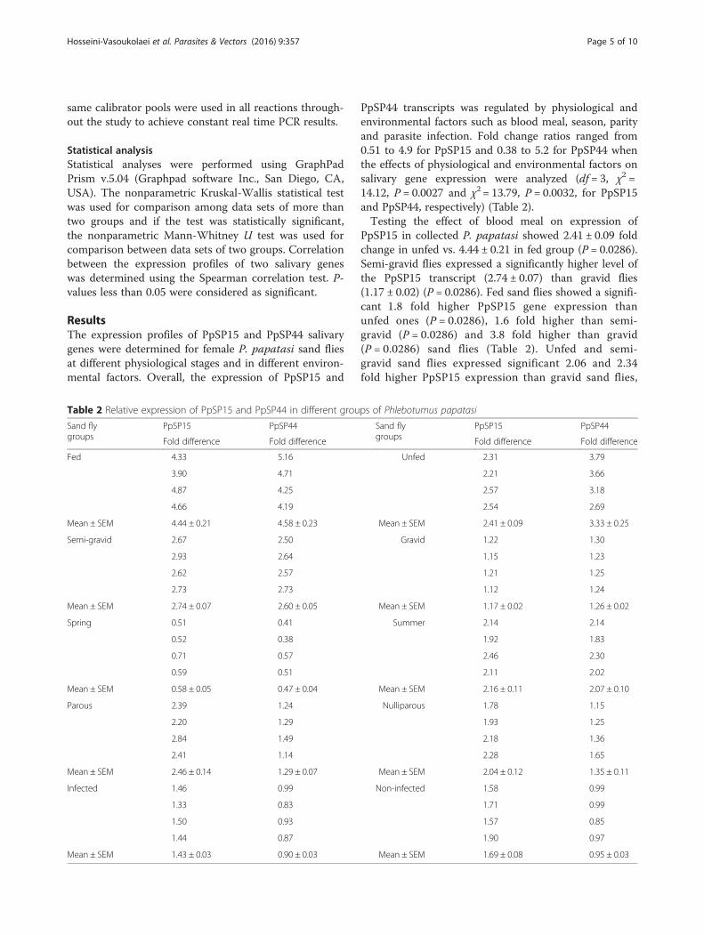
PpSP44 transcripts was regulated by physiological andenvironmental factors such as blood meal, season, parityand parasite infection. Fold change ratios ranged from0.51 to 4.9 for PpSP15 and 0.38 to 5.2 for PpSP44 whenthe effects of physiological and environmental factors onsalivary gene expression were analyzed (df = 3, χ2 =14.12, P = 0.0027 and χ2 = 13.79, P = 0.0032, for PpSP15and PpSP44, respectively) (Table 2).Testing the effect of blood meal on expression of
PpSP15 in collected P. papatasi showed 2.41 ± 0.09 foldchange in unfed vs. 4.44 ± 0.21 in fed group (P = 0.0286).Semi-gravid flies expressed a significantly higher level ofthe PpSP15 transcript (2.74 ± 0.07) than gravid flies(1.17 ± 0.02) (P = 0.0286). Fed sand flies showed a signifi-cant 1.8 fold higher PpSP15 gene expression thanunfed ones (P = 0.0286), 1.6 fold higher than semi-gravid (P = 0.0286) and 3.8 fold higher than gravid(P = 0.0286) sand flies (Table 2). Unfed and semi-gravid sand flies expressed significant 2.06 and 2.34fold higher PpSP15 expression than gravid sand flies,
Table 2 Relative expression of PpSP15 and PpSP44 in different groups of Phlebotumus papatasi
Sand flygroups
PpSP15 PpSP44 Sand flygroups
PpSP15 PpSP44
Fold difference Fold difference Fold difference Fold difference
Fed 4.33 5.16 Unfed 2.31 3.79
3.90 4.71 2.21 3.66
4.87 4.25 2.57 3.18
4.66 4.19 2.54 2.69
Mean ± SEM 4.44 ± 0.21 4.58 ± 0.23 Mean ± SEM 2.41 ± 0.09 3.33 ± 0.25
Semi-gravid 2.67 2.50 Gravid 1.22 1.30
2.93 2.64 1.15 1.23
2.62 2.57 1.21 1.25
2.73 2.73 1.12 1.24
Mean ± SEM 2.74 ± 0.07 2.60 ± 0.05 Mean ± SEM 1.17 ± 0.02 1.26 ± 0.02
Spring 0.51 0.41 Summer 2.14 2.14
0.52 0.38 1.92 1.83
0.71 0.57 2.46 2.30
0.59 0.51 2.11 2.02
Mean ± SEM 0.58 ± 0.05 0.47 ± 0.04 Mean ± SEM 2.16 ± 0.11 2.07 ± 0.10
Parous 2.39 1.24 Nulliparous 1.78 1.15
2.20 1.29 1.93 1.25
2.84 1.49 2.18 1.36
2.41 1.14 2.28 1.65
Mean ± SEM 2.46 ± 0.14 1.29 ± 0.07 Mean ± SEM 2.04 ± 0.12 1.35 ± 0.11
Infected 1.46 0.99 Non-infected 1.58 0.99
1.33 0.83 1.71 0.99
1.50 0.93 1.57 0.85
1.44 0.87 1.90 0.97
Mean ± SEM 1.43 ± 0.03 0.90 ± 0.03 Mean ± SEM 1.69 ± 0.08 0.95 ± 0.03
Hosseini-Vasoukolaei et al. Parasites & Vectors (2016) 9:357 Page 5 of 10
respectively (P = 0.0286). The significant difference inPpSP15 gene expression was also observed between unfedand semi-gravid sand flies. The expression profiles werefed > semi-gravid > unfed > gravid (Fig. 2a).The transcript level of PpSP44 showed a 3.33 ± 0.25
fold increase in unfed vs 4.58 ± 0.23 in the fed group (P= 0.0286). Semi-gravid flies expressed a significantlyhigher level of the PpSP44 transcript (2.6 ± 0.05) thangravid flies (1.26 ± 0.02) (P = 0.0286) (Table 2). In thisregard, the gene expression level of PpSP44 was highest infed and lowest in gravid sand flies. The expression profileof PpSP44 among these four groups was: fed > unfed >semi-gravid > gravid (Fig. 2b). Fed sand flies showed a sig-nificant 1.4 fold higher PpSP44 gene expression than unfed(P = 0.0286), 1.8 fold higher than semi-gravid (P = 0.0286)and 3.6 fold higher than gravid (P = 0.0286) sand flies.Gravid sand flies showed a significant 2.6 fold lowerPpSP44 expression than unfed (P = 0.0286) and 2.1 foldlower than semi-gravid (P = 0.0286) sand flies (Table 2,Fig. 2b).The summer collected sand flies showed fold changes
of 2.16 ± 0.11 and 2.07 ± 0.10 compared to the springcollection with fold changes of 0.58 ± 0.05 and 0.47 ±0.04 for PpSP15 and PpSP44, respectively (Table 2). Thelevel of PpSP15 and PpSP44 gene expression in sandflies collected during summer was a significant 3.7 and4.4 fold higher than those sand flies collected duringspring, respectively (P = 0.0286) (Fig. 2c, d).The fold change for parous and nulliparous groups of
sand flies, respectively, were 2.46 ± 0.14 and 2.04 ± 0.12for PpSP15 expression and 1.29 ± 0.07 and 1.35 ± 0.11fold for PpSP44 expression (Table 2). The expressionlevel of PpSP15 gene in parous sand flies was higherthan in nulliparous sand flies but its expression levelsand that of PpSP44 did not differ significantly betweenthese two groups (Fig. 2e, f ).Gene expression in infected and non-infected sand
flies, respectively, showed a 1.43 ± 0.03 and 1.69 ± 0.08fold change for PpSP15 and 0.9 ± 0.03 and 0.95 ± 0.03fold change for PpSP44 (Table 2). In L. major-infected P.papatasi sand flies the PpSP15 gene expression was asignificant 1.2 fold lower (P = 0.0286) than in non in-fected ones (Fig. 2g, h).The correlation between expression profiles of PpSP15
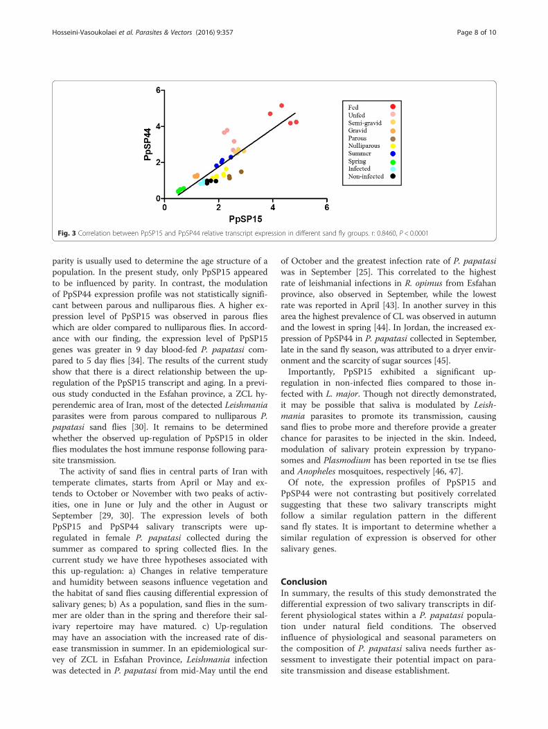
and PpSP44 genes was determined using the Spearmancorrelation test. There was a strong positive correlationbetween the expression of these two salivary genes, in 10sand fly groups (r(PpSP15,PpSP44) = 0.85, P < 0.0001) (Fig. 3).
DiscussionGenetic variations in sand fly saliva influence the processof developing a vaccine against leishmaniasis. The study ofElnaiem et al. [15] suggested that the high degree of simi-larity in P. papatasi SP15 between different populations
may be used in a vaccination strategy. The authors con-cluded that no major variation at the genetic and aminoacid level of PpSP15 reinforces its suitability as a vaccinecandidate. In contrast, several studies on Maxadilan, a sal-ivary protein in new world Lutzomyia longipalpis, demon-strated that this protein has a high amount of genetic andamino acid variations and consequently the host immuneresponse to each variant is specific, therefore reducing itsvalue as a potential vaccine candidate [35–37]. In additionto genetic variability, differences in the level of salivarygene expression could also influence vaccine development.In this study we addressed the hypothesis that some
physiological aspects such as blood feeding, parity ornulliparity, as well as Leishmania infection and seasonalstatus modulate the expression profiles of two of themost significant transcripts found in the salivary glandsof female P. papatasi sand flies [12, 13].In four groups of blood fed, unfed, semi-gravid and
gravid flies the expression levels of salivary transcripts ofPpSP15 and PpSP44 genes were assessed. Supportingthe hypothesis, expression of the two genes was up-regulated in blood fed compared to unfed flies. In ac-cordance with our results, a recent study demonstratedthe up-regulation of PpSP12, PpSP14, PpSP15, PpSP30,PpSP36, PpSP42 and PpSP44 salivary gland gene expres-sion in blood fed laboratory reared P. papatasi [34]. Thisinduction of salivary transcripts following blood feedingof sand flies reflects the important role of salivation dur-ing feeding. Higher gene expression in fed flies may bebecause of the subsequent need for regeneration of saliv-ary proteins after a meal in preparation for the nextblood feeding. In accordance with this finding, previousstudies showed that the total amount of salivary proteinsreduces following a blood meal [38–40]. In anotherstudy in our laboratory, the lower levels of salivary pro-teins in fed P. papatasi flies was also in agreement withthis finding [41]. Lower mRNA expression in unfed fliesmay be an indication that sand flies have stored enoughsaliva in their glands that could be used during the feed-ing process, down regulating salivary gene transcription.Among four groups of fed, unfed, semi-gravid and
gravid flies, the lowest levels of PpSP15 and PpSP44gene transcripts were observed in gravid sand flies. Al-though the physiological processes that may be in chargeof such differences in salivary gland gene expressions be-tween fed and gravid flies or between semi-gravid andgravid flies are not known, these findings should be con-sidered in future examinations of the interaction be-tween the salivary gene expression and sand flyphysiological status.Aging is another physiological factor we hypothesized
may influence gene expression profiles, however, it isnot thought to be as significant as genotype or sex in in-sects [42]. As nulliparous flies have never oviposited,
Hosseini-Vasoukolaei et al. Parasites & Vectors (2016) 9:357 Page 6 of 10
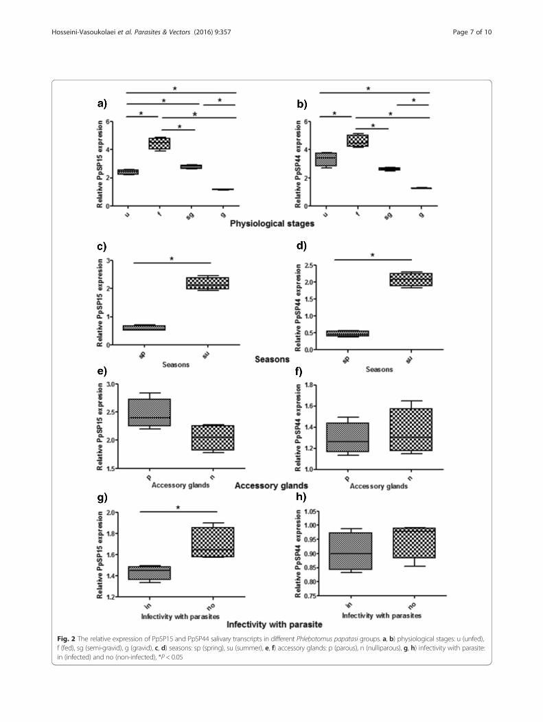
Fig. 2 The relative expression of PpSP15 and PpSP44 salivary transcripts in different Phlebotomus papatasi groups. a, b) physiological stages: u (unfed),f (fed), sg (semi-gravid), g (gravid), c, d) seasons: sp (spring), su (summer), e, f) accessory glands: p (parous), n (nulliparous), g, h) infectivity with parasite:in (infected) and no (non-infected), *P < 0.05
Hosseini-Vasoukolaei et al. Parasites & Vectors (2016) 9:357 Page 7 of 10
parity is usually used to determine the age structure of apopulation. In the present study, only PpSP15 appearedto be influenced by parity. In contrast, the modulationof PpSP44 expression profile was not statistically signifi-cant between parous and nulliparous flies. A higher ex-pression level of PpSP15 was observed in parous flieswhich are older compared to nulliparous flies. In accord-ance with our finding, the expression level of PpSP15genes was greater in 9 day blood-fed P. papatasi com-pared to 5 day flies [34]. The results of the current studyshow that there is a direct relationship between the up-regulation of the PpSP15 transcript and aging. In a previ-ous study conducted in the Esfahan province, a ZCL hy-perendemic area of Iran, most of the detected Leishmaniaparasites were from parous compared to nulliparous P.papatasi sand flies [30]. It remains to be determinedwhether the observed up-regulation of PpSP15 in olderflies modulates the host immune response following para-site transmission.The activity of sand flies in central parts of Iran with
temperate climates, starts from April or May and ex-tends to October or November with two peaks of activ-ities, one in June or July and the other in August orSeptember [29, 30]. The expression levels of bothPpSP15 and PpSP44 salivary transcripts were up-regulated in female P. papatasi collected during thesummer as compared to spring collected flies. In thecurrent study we have three hypotheses associated withthis up-regulation: a) Changes in relative temperatureand humidity between seasons influence vegetation andthe habitat of sand flies causing differential expression ofsalivary genes; b) As a population, sand flies in the sum-mer are older than in the spring and therefore their sal-ivary repertoire may have matured. c) Up-regulationmay have an association with the increased rate of dis-ease transmission in summer. In an epidemiological sur-vey of ZCL in Esfahan Province, Leishmania infectionwas detected in P. papatasi from mid-May until the end
of October and the greatest infection rate of P. papatasiwas in September [25]. This correlated to the highestrate of leishmanial infections in R. opimus from Esfahanprovince, also observed in September, while the lowestrate was reported in April [43]. In another survey in thisarea the highest prevalence of CL was observed in autumnand the lowest in spring [44]. In Jordan, the increased ex-pression of PpSP44 in P. papatasi collected in September,late in the sand fly season, was attributed to a dryer envir-onment and the scarcity of sugar sources [45].Importantly, PpSP15 exhibited a significant up-
regulation in non-infected flies compared to those in-fected with L. major. Though not directly demonstrated,it may be possible that saliva is modulated by Leish-mania parasites to promote its transmission, causingsand flies to probe more and therefore provide a greaterchance for parasites to be injected in the skin. Indeed,modulation of salivary protein expression by trypano-somes and Plasmodium has been reported in tse tse fliesand Anopheles mosquitoes, respectively [46, 47].Of note, the expression profiles of PpSP15 and
PpSP44 were not contrasting but positively correlatedsuggesting that these two salivary transcripts mightfollow a similar regulation pattern in the differentsand fly states. It is important to determine whether asimilar regulation of expression is observed for othersalivary genes.
ConclusionIn summary, the results of this study demonstrated thedifferential expression of two salivary transcripts in dif-ferent physiological states within a P. papatasi popula-tion under natural field conditions. The observedinfluence of physiological and seasonal parameters onthe composition of P. papatasi saliva needs further as-sessment to investigate their potential impact on para-site transmission and disease establishment.
Fig. 3 Correlation between PpSP15 and PpSP44 relative transcript expression in different sand fly groups. r: 0.8460, P < 0.0001
Hosseini-Vasoukolaei et al. Parasites & Vectors (2016) 9:357 Page 8 of 10
Additional file
Additional file 1: Sequences of primers used in qRT-PCR. (XLS 24 kb)
AbbreviationscDNA, complementary deoxyribonucleic acid; DTH, delayed-type hypersensitivity;ETHRC, Esfahan Training and Health Research Center; f, fed; g, gravid; in, infected;n, nulliparous; NIHR, National Institute of Health Research; no, non infected; p,parous; PpSP15, Phlebotomus papatasi SP15; PpSP44, Phlebotomus papatasi SP44;qRT-PCR, quantitative real-time polymerase chain reaction; sg, semi-gravid; sp,spring; su, summer; Th1, type 1 helper T cell; TUMS, Tehran University of MedicalSciences; u, unfed; ZCL, zoonotic cutaneous leishmaniasis.
AcknowledgmentThis work was supported by Research deputy of Tehran University of MedicalSciences (TUMS), Project No. 18511/16.5.2012 and Avicenna Research Institute,ACECR, Project No. 910206–018. We thank Dr. Shabani for his helpful comments,Ms. Babaei, Ms. Balaei and Mr. Hadavi for their technical support, from AvicennaResearch Institute, ACECR. We are very grateful to Ms. Shareghi, Esfahan HealthResearch Station, National Institute of Health Research, TUMS, for her assistancein the project. We are grateful to Ms. Ahmadi and Ms. Bolandian, School ofPublic Health, TUMS, for their assistance in sand fly rearing.
Authors’ contributionsNHV participated in data acquisition, statistical analysis and preparation ofmanuscript. AAA, MJT and FI participated in data management and statisticalanalysis. MRY, AK, JGV and SK helped in data interpretation and edition ofthe manuscript draft. MHA, ZS and RJ assisted in design and coordination ofsample collection and laboratory rearing. HE, SE and HM had technicalassistance in the study and helped to design the manuscript draft. Allauthors read and approved the final manuscript.
Competing interestsThe authors have declared that they have no competing interests.
Author details1Department of Medical Entomology and Vector Control, School of PublicHealth, Tehran University of Medical Sciences, Tehran, Iran. 2Department ofMedical Entomology and Vector Control, Health Sciences Research Center,Faculty of Health, Mazandaran University of Medical Sciences, Sari, Iran.3Reproductive Immunology Research Center, Avicenna Research Institute,ACECR, Tehran, Iran. 4Center for Research and Training in Skin Diseases andLeprosy, Tehran University of Medical Sciences, Tehran, Iran. 5VectorMolecular Biology Section, Laboratory of Malaria and Vector Research,National Institute of Allergy and Infectious Diseases, National Institute ofHealth, Rockville, MD 20852, USA. 6Reproductive Biotechnology ResearchCenter, Avicenna Research Institute, ACECR, Tehran, Iran. 7Esfahan HealthResearch Station, National Institute of Health Research, Tehran University ofMedical Sciences, Esfahan, Iran. 8Department of Medical Mycology andParasitology, School of Medicine, Isfahan University of Medical Sciences,Isfahan, Iran. 9Monoclonal Antibody Research Center, Avicenna ResearchInstitute, ACECR, Tehran, Iran.
Received: 6 February 2016 Accepted: 7 June 2016
References1. Control of the leishmaniases: report of a meeting of the WHO Expert
Commitee on the Control of Leishmaniases. Geneva: WHO technical reportseries, no: 949; 2010. p. 22–26.
2. Statistics of cutaneous leishmaniasis in Iran: Hearing before NationalLeishmaniasis Committee. Office of Zoonoses, Center of Disease Control,Ministry of Health and Medical Education. 2004.
3. Adler S, Theodor O. The mouthparts, alimentary tract, and salivary apparatusof the female in Phlebotomus papatasi. Ann Trop Med Parasitol. 1926;20:109–42.
4. Volf P, Tesarova P, Nohynkova E. Salivary proteins and glycoproteins inphlebotomine sandflies of various species, sex and age. Med Vet Entomol.2000;14(3):251–6.
5. Ribeiro J. Role of saliva in blood-feeding by arthropods. Annu Rev Entomol.1987;32(1):463–78.
6. Oliveira F, de Carvalho AM, de Oliveira CI. Sand-fly saliva-Leishmania-man:the trigger trio. Front Immunol. 2013;4:375.
7. Gomes R, Oliveira F. The immune response to sand fly salivary proteins andits influence on Leishmania immunity. Front Immunol. 2012;3:110.
8. Abdeladhim M, Kamhawi S, Valenzuela JG. What’s behind a sand fly bite?The profound effect of sand fly saliva on host hemostasis, inflammation andimmunity. Infect Genet Evol. 2014;28:691–703.
9. Abdeladhim M, Jochim RC, Ahmed MB, Zhioua E, Chelbi I, Cherni S, et al.Updating the salivary gland transcriptome of Phlebotomus papatasi(Tunisian strain): the search for sand fly-secreted immunogenic proteins forhumans. PLoS One. 2012;7(11):e47347. doi:10.1371/journal.pone.0047347.
10. Alvarenga PH, Xu X, Oliveira F, Chagas AC, Nascimento CR, Francischetti IM,et al. Novel family of insect salivary inhibitors blocks contact pathwayactivation by binding to polyphosphate, heparin, and dextran sulfate.Arterioscler Thromb Vasc Biol. 2013;33(12):2759–70.
11. Xu X, Oliveira F, Chang BW, Collin N, Gomes R, Teixeira C, et al. Structureand function of a “yellow” protein from saliva of the sand fly Lutzomyialongipalpis that confers protective immunity against Leishmania majorinfection. J Biol Chem. 2011;286(37):32383–93.
12. Valenzuela JG, Belkaid Y, Garfield MK, Mendez S, Kamhawi S, RowtonED, et al. Toward a defined anti-Leishmania vaccine targeting vectorantigens characterization of a protective salivary protein. J Exp Med.2001;194(3):331–42.
13. Oliveira F, Lawyer PG, Kamhawi S, Valenzuela JG. Immunity to distinct sandfly salivary proteins primes the anti-Leishmania immune response towardsprotection or exacerbation of disease. PLoS Negl Trop Dis. 2008;2(4):e226.doi:10.1371/journal.pntd.0000226.
14. Collin N, Gomes R, Teixeira C, Cheng L, Laughinghouse A, Ward JM, et al.Sand fly salivary proteins induce strong cellular immunity in a naturalreservoir of visceral leishmaniasis with adverse consequences for Leishmania.PLoS Pathog. 2009;5(5):e1000441. doi:10.1371/journal.ppat.1000441.
15. Elnaiem DE, Meneses C, Slotman M, Lanzaro G. Genetic variation in the sandfly salivary protein, SP‐15, a potential vaccine candidate against Leishmaniamajor. Insect Mol Biol. 2005;14(2):145–50.
16. Anderson JM, Oliveira F, Kamhawi S, Mans BJ, Reynoso D, Seitz AE, et al.Comparative salivary gland transcriptomics of sandfly vectors of visceralleishmaniasis. BMC Genomics. 2006;7(1):52.
17. Lanzaro GC, Lopes AH, Ribeiro JM, Shoemaker CB, Warburg A, Soares M, et al.Variation in the salivary peptide, maxadilan, from species in the Lutzomyialongipalpis complex. Insect Mol Biol. 1999;8(2):267–75.
18. Kato H, Anderson JM, Kamhawi S, Oliveira F, Lawyer PG, Pham VM, et al.High degree of conservancy among secreted salivary gland proteins fromtwo geographically distant Phlebotomus duboscqi sandflies populations (Maliand Kenya). BMC Genomics. 2006;7(1):226.
19. Nadim A, Seyedi-Rashti M. A brief review of the epidemiology of varioustypes of leishmaniasis in Iran. Acta MedIran. 1971;14:99–106.
20. Postigo JAR. Leishmaniasis in the world health organization easternmediterranean region. Int J AntimicrobAgents. 2010;36 Suppl 1:S62–5.
21. Schlein Y, Warburg A, Schnur L, Gunders A. Leishmaniasis in the JordanValley II. Sandflies and transmission in the central endemic area. Trans R SocTrop Med Hyg. 1982;76(5):582–6.
22. Schlein Y, Gunders A, Warburg A. Leishmaniasis in the Jordan Valley, I.Attraction of Phlebotomus papatasi (Psychodidae) to turkeys. Ann Trop MedParasitol. 1982;76(5):517–20.
23. Lane RP. Sand flies (Phlebotominae). In: Lane RP, Crosskey RW, editors.Medical insects and arachnids. London: Chapman and Hall; 1993. p. 78–119.
24. Yaghoobi-Ershadi M, Javadian E, Tahvildare-Bidruni G. Leishmania majorMON-26 isolated from naturally infected Phlebotomus papatasi (Diptera:Psychodidae) in Isfahan Province, Iran. Acta Trop. 1995;59(4):279–82.
25. Yaghoobi-Ershadi MR, Akhavan AA, Zahraei-Ramazani AR, Jalali-Zand AR,Piazak N. Bionomics of Phlebotomus papatasi (Diptera: Psychodidae) in anendemic focus of zoonotic cutaneous leishmaniasis in central Iran. J VectorEcol. 2005;30(1):115.
26. Maleki-Ravasan N, Oshaghi MA, Afshar D, Arandian MH, Hajikhani S, AkhavanAA, et al. Aerobic bacterial flora of biotic and abiotic compartments of ahyperendemic Zoonotic Cutaneous Leishmaniasis (ZCL) focus. ParasitVectors. 2015;8(1):63.
27. Yaghoobi-Ershadi MR, Shirani-Bidabadi L, Hanafi-Bojd AA, Akhavan AA,Zeraati H. Colonization and biology of Phlebotomus papatasi, the main
Hosseini-Vasoukolaei et al. Parasites & Vectors (2016) 9:357 Page 9 of 10
vector of cutaneous leishmaniasis due to Leshmania major. Iran J PublicHealth. 2007;36(3):21–6.
28. Yuval B, Warburg A, Schlein Y. Leishmaniasis in the Jordan Valley. V.Dispersal characteristics of the sandfly Phlebotomus papatasi. Med VetEntomol. 1988;2(4):391–5.
29. Yaghoobi-Ershadi M, Javadian E. Studies on sandflies in a hyperendemic areaof zoonotic cutaneous leishmaniasis in Iran. Indian J Med Res. 1997;105:61–6.
30. Yaghoobi-Ershadi M, Akhavan A. Entomological survey of sandflies (Diptera:Psychodidae) in a new focus of zoonotic cutaneous leishmaniosis in Iran.Acta Trop. 1999;73(3):321–6.
31. Hodgins-Davis A, Townsend JP. Evolving gene expression: from G to E toG× E. Trends Ecol Evol. 2009;24(12):649–58.
32. Seyedi-Rashti M, Nadim A. The genus Phlebotomus(Diptera: Psychodidae:Phlebotominae) of the countries of the eastern Mediterranean region. IranJ Public Health. 1992;21:11–50.
33. Akhavan AA, Mirhendi H, Khamesipour A, Alimohammadian MH, Rassi Y,Bates P, et al. Leishmania species: detection and identification by nestedPCR assay from skin samples of rodent reservoirs. Exp Parasitol. 2010;126(4):552–6.
34. Coutinho-Abreu IV, Wadsworth M, Stayback G, Ramalho-Ortigao M,McDowell MA. Differential expression of salivary gland genes in the femalesand fly Phlebotomus papatasi (Diptera: Psychodidae). J Med Entomol.2010;47(6):1146–55.
35. Warburg A, Saraiva E, Lanzaro GC, Titus RG, Neva F. Saliva of Lutzomyialongipalpis sibling species differs in its composition and capacity to enhanceleishmaniasis. Philos Trans R Soc Lond B Biol Sci. 1994;345(1312):223–30.
36. Yin H, Norris D, Lanzaro G. Sibling species in the Lutzomyia longipalpiscomplex differ in levels of mRNA expression for the salivary peptide,maxadilan. Insect Mol Biol. 2000;9(3):309–14.
37. Milleron RS, Mutebi J-P, Valle S, Montoya A, Yin H, Soong L, et al. Antigenicdiversity in maxadilan, a salivary protein from the sand fly vector ofAmerican visceral leishmaniasis. Am J Trop Med Hyg. 2004;70(3):286–93.
38. Marinotti O, James AA, Ribeiro JC. Diet and salivation in female Aedesaegypti mosquitoes. J Insect Physiol. 1990;36(8):545–8.
39. Golenda CF, Klein T, Coleman R, Burge R, Ward RA, Seeley DC. Depletion oftotal salivary gland protein in blood-fed Anopheles mosquitoes. J MedEntomol. 1995;32(3):300–5.
40. Prates DB, Santos L, Miranda JC, Souza A, Palma MS, Barral-netto M, et al.Changes in amounts of total salivary gland proteins of Lutzomyia longipalpis(Diptera: Psychodidae) according to age and diet. J Med Entomol.2008;45(3):409–13.
41. Hosseini-Vasoukolaei N, Mahmoudi A-R, Khamesipour A, Yaghoobi-ErshadiMR, Kamhawi S, Valenzuela JG, et al. Seasonal and physiological variations ofPhlebotomus papatasi salivary gland antigens in central Iran. J ArthropodBorne Dis. 2016;10(1):39–49.
42. Jin W, Riley RM, Wolfinger RD, White KP, Passador-Gurgel G, Gibson G. Thecontributions of sex, genotype and age to transcriptional variance inDrosophila melanogaster. Nat Genet. 2001;29(4):389–95.
43. Yaghoobi-Ershadi M, Javadian E. Epidemiological study of reservoir hosts inan endemic area of zoonotic cutaneous leishmaniasis in Iran. Bull WorldHealth Organ. 1996;74(6):587.
44. Karami M, Doudi M, Setorki M. Assessing epidemiology of cutaneousleishmaniasis in Isfahan. Iran J Vector Borne Dis. 2013;50(1):30–7.
45. Coutinho-Abreu IV, Mukbel R, Hanafi HA, Fawaz EY, El-Hossary SS, WadsworthM, et al. Expression plasticity of Phlebotomus papatasi salivary gland genes indistinct ecotopes through the sand fly season. BMC Ecol. 2011;11:24.
46. Van Den Abbeele J, Caljon G, De Ridder K, De Baetselier P, Coosemans M.Trypanosoma brucei modifies the tsetse salivary composition, altering the flyfeeding behavior that favors parasite transmission. PLoS Pathog.2010;6(6):e1000926.
47. Marie A, Holzmuller P, Tchioffo MT, Rossignol M, Demettre E, Seveno M, et al.Anopheles gambiae salivary protein expression modulated by wild Plasmodiumfalciparum infection: highlighting of new antigenic peptides as candidates ofAn. gambiae bites. Parasit Vectors. 2014;7(1):1–13.
• We accept pre-submission inquiries
• Our selector tool helps you to find the most relevant journal
• We provide round the clock customer support
• Convenient online submission
• Thorough peer review
• Inclusion in PubMed and all major indexing services
• Maximum visibility for your research
Submit your manuscript atwww.biomedcentral.com/submit
Submit your next manuscript to BioMed Central and we will help you at every step:
Hosseini-Vasoukolaei et al. Parasites & Vectors (2016) 9:357 Page 10 of 10