
Differential Expression Profile of Chicken Embryo ... · Title Differential Expression Profile of...
18
Title Differential Expression Profile of Chicken Embryo Fibroblast DF- 1 Cells Infected with Cell-Adapted Infectious Bursal Disease Virus Author(s) Hui, RKH; Leung, FCC Citation Plos One, 2015, v. 10 n. 6, p. e0111771 Issued Date 2015 URL http://hdl.handle.net/10722/227813 Rights This work is licensed under a Creative Commons Attribution- NonCommercial-NoDerivatives 4.0 International License.
Transcript of Differential Expression Profile of Chicken Embryo ... · Title Differential Expression Profile of...

TitleDifferential Expression Profile of Chicken Embryo Fibroblast DF-1 Cells Infected with Cell-Adapted Infectious Bursal DiseaseVirus
Author(s) Hui, RKH; Leung, FCC
Citation Plos One, 2015, v. 10 n. 6, p. e0111771
Issued Date 2015
URL http://hdl.handle.net/10722/227813
Rights This work is licensed under a Creative Commons Attribution-NonCommercial-NoDerivatives 4.0 International License.

RESEARCH ARTICLE
Differential Expression Profile of ChickenEmbryo Fibroblast DF-1 Cells Infected withCell-Adapted Infectious Bursal Disease VirusRaymond K. Hui1‡, Frederick C. Leung1,2*
1 School of Biological Sciences, The University of Hong Kong, Hong Kong, People’s Republic of China,2 Bioinformatics Center, Nanjing Agricultural University, Nanjing, China
‡RKH is first author on this work.* [email protected]
AbstractRNA-Seq was used to unveil the transcriptional profile of DF-1 cells at the early stage of
caIBDV infection. Total RNAs were extracted from virus-infected cells at 0, 6 and 12 hpi.
RNA-Seq datasets of respective samples mapped to 56.5–57.6% of isoforms in the refer-
ence genome Galgal4.73. At 6 hpi, 23 isoforms underwent an elevated expression, while
128 isoforms were up-regulated and 5 were down-regulated at 12 hpi in the virus-infected
group. Besides, 10 isoforms were exclusively expressed in the virus-infected cells. Though
no significant change was detected in cytokine and interferon expression levels at the first
12 hours of infection, modulations of the upstream regulators were observed. In addition to
the reported regulatory factors including EIF2AK2, MX, OAS*A, GBP7 and IFIT, IBDV in-
fection also triggered a IFIT5-IRF1/3-RSAD5 pathway in the DF-1 cells which potentially re-
stricted the viral replication cycle in the early infection stage. Over-expression of LIPA and
CH25H, together with the suppression of STARD4, LSS and AACS genes implied a modu-
lation of membrane fluidity and lipid raft arrangement in the infected cells. Alternative splic-
ing of the EFR3 homolog A gene was also through to be involved in the lipid membrane
regulation, and these cumulative responses projected an inhibition of viral endocytosis.
Recognition of viral RNA genomes and intermediates was presumably enhanced by the ele-
vated levels of IFIH1, DHX58 and TRIM25 genes which possess properties on detecting
viral dsRNA. On the other hand, the caIBDV arrested the host's apoptotic process by induc-
ing the expression of apoptosis inhibitors including NFKBIA/Z, TNFAIP2/3 and ITA at the
first 12 hours of infection. In conclusion, the differential expression landscape demonstrated
with RNA-Seq provides a comprehensive picture on the molecular interactions between
host cells and virus at the early stage of infection.
PLOS ONE | DOI:10.1371/journal.pone.0111771 June 8, 2015 1 / 17
OPEN ACCESS
Citation: Hui RK, Leung FC (2015) DifferentialExpression Profile of Chicken Embryo Fibroblast DF-1 Cells Infected with Cell-Adapted Infectious BursalDisease Virus. PLoS ONE 10(6): e0111771.doi:10.1371/journal.pone.0111771
Academic Editor: Yuntao Wu, George MasonUniversity, UNITED STATES
Received: April 14, 2014
Accepted: October 1, 2014
Published: June 8, 2015
Copyright: © 2015 Hui, Leung. This is an openaccess article distributed under the terms of theCreative Commons Attribution License, which permitsunrestricted use, distribution, and reproduction in anymedium, provided the original author and source arecredited.
Data Availability Statement: Data are available atthe NCBI Gene Expression Omnibus(GEO)repository under the accession number ofGSE60268.
Funding: This project is partly supported by HongKong Government, RGC GRF: HKU 771310M andHKU University internal Fund. The funders had norole in study design, data collection and analysis,decision to publish, or preparation of the manuscript.
Competing Interests: The authors have declaredthat no competing interests exist.

IntroductionInfectious bursal disease (IBD) has been striking chicken flocks for more than fifty years ex-erting an considerable economical impact to the global poultry industry. The disease bringsa direct mortality ratio up to 90–100% [1, 2], and as it causes destruction of B-lymphocytesin the bursa of Fabricius, it leads into severe immunosuppression and hence secondary in-fections may result in infected chickens [3, 4, 5]. Infectious bursal disease virus (IBDV) isthe causative agent of the disease. Two serotypes are identified in which serotype 1 com-prises pathogenic strains, whereas serotype 2 strains cause neither disease nor protectionagainst serotype 1 strains in chickens [6, 7, 8, 9]. It is demonstrated that the virus propagatesin the actively proliferating IgM-bearing B-lymphocytes and hence induces apoptotic effects[10, 11, 12]. Though the pathogenicity and epizootiology have been studied for a certain pe-riod of time, the molecular interactions between the host cells and the viruses have not beenwell defined yet. In recent years studies have started to focus on the molecular mechanismsinvolved in the host responses upon IBDV infection. Quantitative RT-PCR (qRT-PCR) andmicroarray assays are increasingly employed to reveal the transcriptional changes of thehost cells in response to IBDV infections [13–30]. While some studies also utilize proteomicapproaches to identify the differentially expressed protein during the course of IBDV infec-tion [31, 32]. Majority of these studies emphasized the cytokine responses including inter-leukin and interferon expressions, whereas some of these studies revealed expression ofmRNA related to apoptotic mechanisms. Up to now, however, there is no comprehensivetranscriptional landscape described in the cells upon IBDV infection. In order to explore thedifferential expression pattern in the event of IBDV infection, RNA sequencing (RNA-Seq)was employed to assay the transcript variations across the entire chicken genome. RNA-Seqreveals a high overall sensitivity on differentially expressed gene level compared with otherwhole-transcriptome expression quantification platforms including microarrays [33, 34].The prerequisite of hybridization-based microarray assays relies on existing knowledgeabout genome sequences [35, 36] and hence limits the detection of novel, rare transcript spe-cies exist in the transcriptome. Whereas RNA-Seq takes an advantage not only in determin-ing the differential expression level of transcripts, but it also provides evidence on transcriptsplice-variants, isoforms and single nucleotide polymorphism (SNPs) [37]. It has also beendemonstrated that RNA-Seq is highly accurate for determining gene expression levels asperformed with qPCR [38]. Background levels resulting from cross-hybridization is alsomuch lower than occurred in microarray assay [39]. Taking these advantages, in this studywe made use of RNA-Seq to unveil the transcriptomic dynamics upon caIBDV infection inDF-1 cells and to reveal a more comprehensive molecular interactions between the host cellsand the virus.
Materials and Methods
Cell culture and virusChicken embryonic fibroblast cells DF-1 (CRL-12203, ATCC) were maintained and culturedwith high glucose (4.5g D-Glucose/L) Dulbecco's Modified Eagle Medium DMEM-HG (LifeTechnologies, NY) supplemented with 10% (v/v) fetal bovine serum at 37°C, 5% CO2. Cell-adapted IBDV (caIBDV) was generated with propagating IBDV vaccine strain D78 (VR-2041,ATCC) in secondary chicken embryonic fibroblast cells, followed by purification with CsClgradient and 20% sucrose gradient as described previously [21]. The quantity of the purifiedvirus was determined by standard plaque assay [40].
DE Profile of DF-1 Cells Infected with caIBDV
PLOSONE | DOI:10.1371/journal.pone.0111771 June 8, 2015 2 / 17

Virus inoculationDF-1 cells were seeded into each well of 6-well plate (Costar 3516, Corning, NY) at 1 × 10⁴
cells at 24 hrs prior to virus inoculation. A total of six individual wells were prepared for eachsampling time point of both mock- and caIBDV- infected groups. The purified caIBDV was di-luted to 10 multiplicity of infection (m.o.i.) per ml with DMEM-HG without serum supple-ment. Before inoculation, culture medium was discarded and the cells were rinsed with1 × phospate-buffered saline once. One ml of diluted caIBDV was applied into each well of thevirus-infected group, whereas DMEM-HG without serum supplement was added in the mock-infected group. At 0, 6, 12 hrs post-infection (hpi), medium was aspirated from the wells ofmock- and caIBDV-infected groups. One ml of SV RNA Lysis Buffer (Promega, WI) wasadded into each well and the cells were dispersed and lysed with repeated pipetting. A total ofsix individual cell lysates were collected from each treatment group at designated time pointsand kept at -80°C for total RNA extraction thereafter.
Total RNA isolationTotal RNAs were extracted from cell lysates with SV96 Total RNA Isolation System (Promega,WI) according to manufacturer's protocol. Total RNAs extracted from each treatment group atthe same time point were pooled into one tube for the downstream processes. A total of sixRNA samples were obtained representing two treatments at three time points. Quantity of thepooled RNA samples was determined with Quant-iT RiboGreen RNA Reagent and Kit (Molec-ular Probes, OR), while the quality and integrity of RNA were confirmed with spectrophotome-try and formaldehyde 1% agarose gel electrophoresis. All total RNA samples possessed A260/A280 ratio> 1.8 and the 28S:18S rRNA intensity ratio of 1.76 estimated with Nanodrop 2000(Thermo Scientific, DE) and Quantity One Analysis Software (Bio-Rad, CA) respectively.
Complementary DNA (cDNA) library construction and normalizationFour micrograms of each pooled total RNA were used for cDNA library construction usingTruSeq RNA Sample Prep Kits v2 (Illumina, CA) according to the Low Sample (LS) Protocolof the TruSeq RNA Sample Preparation v2 Guide (Part# 15026495 Rev. D September 2012,Illumina, CA). The libraries were indexed with individual adaptor and normalized with LibraryQuantification Kit—Illumina/ Universal (Kapa Biosystems, MA) according to manufacturer'sinstructions. The normalized libraries were then pooled and loaded onto Illumina MiSeq In-strument for cluster generation and sequencing.
Whole transcriptome shotgun sequencingA 2 × 250 paired-end sequencing run was conducted using MiSeq Reagent Kit v2, 500 cycleswith a standard flow cell (14 tile) (Illumina, CA) on an Illumina MiSeq machine. Raw readsgenerated from the run were demultiplexed and underwent quality trimming prior to thedownstream analytical pipelines. Only reads with Q Score� 30, and aligned with correspond-ing paired reads were retained for the differential expression analysis.
Sequence analysisThe paired-end sequence reads of individual sample were aligned and mapped to a chicken ref-erence genome (Gallus Gallus reference genome Galgal4.73) with STAR RNA-Seq aligner 2.3.0[41] in default setups. Both the unmasked reference genome and the annotations of both cod-ing and non-coding genes were obtained from Ensembl database [42]. The mapped reads werethen converted to binary BAM files with SAMtools 1.0.2 [43] for transcript expression levels
DE Profile of DF-1 Cells Infected with caIBDV
PLOSONE | DOI:10.1371/journal.pone.0111771 June 8, 2015 3 / 17
comparison. The aligned, mapped reads from the same time point of both mock- and caIBDV-infected group were compared and the expression levels at the isoform and gene level were cal-culated with Cuffdiff package in Cufflink transcript assembly tool [44]. The same annotationand reference genome (Galgal4.73) files as for STAR aligner were used in Cuffdiff pipeline. Ex-pression level of each transcript in the samples were expressed in fragments per kilobase oftranscript per million mapped fragments, FPKM. Each observed alternation in transcript ex-pression with p<0.05 was regarded as statistical significant. Quality of the libraries and se-quencing performance were examined with RNASeQC tool [45]. The differentially expressedgenes were grouped with hierarchical clustering algorithms [46] in the RNA-Seq analysis pack-age of Gene Pattern web-based tool (Broad Institute, MIT).
The potential protein-protein inactions among the genes in the altered expression profilesderived from RNA-Seq analyses were predicted with STRING (Search Tool for the Retrieval ofInteracting Genes/Proteins) v9.05 [47].
In addition to the chicken reference genome, the paired-end sequence reads of each samplegroup were also mapped to a IBDV reference sequence (IBDV Segment A, NC_004178.1;IBDV Segment B, NC_004179.1 [48]) with GS Reference Mapper in 454 Sequencing SystemSoftware Package v2.7 (Roche, CT) in order to assess the change of caIBDV viral load in the in-fected DF-1 cells.
Nucleotide sequence accession numberThe RNA-Seq datasets have been deposited at NCBI Gene Expression Omnibus (GEO) reposi-tory under the accession number of GSE60268.
Results and Discussion
Infection experiment and RNA-SeqTo study the transcriptomic profile of chicken embryonic fibroblast DF-1 cells during caIBDVinfection, caIBDV strain D78 at 10 m.o.i. was used to infect DF-1 cells for 0, 6 and 12 hours.The relatively high dosage of virus was used for inoculation as to minimize the influence ex-erted from a large number of uninfected cells [49].
Total RNAs were isolated from both mock- and caIBDV-infected groups at respective timepoints and cDNA libraries were constructed for a 2 × 250 paired-end sequencing run on anIllumina MiSeq machine. The sequence run yielded a total of about 32 millions passed-filterreads, and 85.1% of the reads possessed Q-Score� 30. In average,> 4 million quality-trimmedreads were generated for each sample (Table 1). Though it has been suggested that high-qualityeukaryotic transcriptome reconstruction requires more than ten million reads for discoveringnew genes and transcripts [34], the relatively shallow RNA-Seq datasets are otherwise sufficientto provide accurate differential expression trends of transcript as confirmed with qRT-PCR[50, 51, 52]. Hence RNA-Seq datasets obtained in this study can illustrate the evidence on dif-ferential expression landscape between the mock- and virus-infected conditions.
Viral replication dynamicsAfter demulitplexing and quality trimming, the six datasets were mapped to IBDV referencegenome (Segment A, NC_004178.1; IBDV Segment B, NC_004179.1) with GS Reference Map-per Algorithms (Roche, CT). The mapping matrix indicates that no sequence read from themock-infected group matched to the reference genome. On the other hand, unique sequencereads of the segment A of the IBDV genome was detected in 6hpi and 12hpi, whereas thosematched to segment B was observed at 12hpi (Fig 1). The sequence reads covered 20.01% to
DE Profile of DF-1 Cells Infected with caIBDV
PLOSONE | DOI:10.1371/journal.pone.0111771 June 8, 2015 4 / 17

63.87% of the entire segment A sequence at 6hpi and 12hpi samples respectively, while 56.17%of segment B sequence was retrieved from the 12hpi sample. This finding reveals that the repli-cation machinery of caIBDV started as early as at 6 hours after the inoculation. The segment Asequences identified in the dataset did not only indicate the replication of IBDV genomicRNAs, but it also reflected the transcription of sub-genomic RNA species encoding for the viralstructural proteins VP2, 3, 4 and 5 for the assembly of a mature virion. While the segment B se-quences were observed at 12hpi, indicating that there was an expression of viral RNA polymer-ase VP1 and also the replication of this segment. The increase of mapped sequence readsindicates an active viral replication process in this 12-hours post inoculation period.
Differential expression patternsTo identify the differentially expressed transcripts in DF-1 cells upon caIBDV infection, the sixRNA-Seq datasets were then aligned and mapped to a chicken reference genome Ensembl Gal-gal4.73 with STAR-aligner. The numbers of genes and isoforms mapped to the reference ge-nome respective to each dataset remained constant throughout the course of experiment(Table 2). The datasets mapped to 57.7–58.9% of genes and 56.5–57.6% of isoforms in the ref-erence genome. There was no significant alternations in the expressed gene/ transcript isoform
Table 1. Statistics on the RNA-Seq datasets.
Mock-infected caIBDV-infected
hpi Raw read Trimmed read Raw read Trimmed read
0 R1 2,697,493 2,274,144 R1 2,548,186 2,140,585
R2 2,953,303 2,500,845 R2 2,802,826 2,368,373
6 R1 2,643,354 2,250,936 R1 2,524,045 2,166,168
R2 2,861,666 2,445,301 R2 2,730,637 2,351,470
12 R1 2,681,507 2,257,774 R1 2,334,929 2,004,679
R2 2,939,553 2,488,528 R2 2,547,933 2,196,213
doi:10.1371/journal.pone.0111771.t001
Fig 1. Number of unqiuematching reads and percentage of genome coverage to IBDV referencegenome. The demultiplexed, trimmed sequence reads were mapped to IBDV reference genome comprisingtwo segments. No IBDV sequences were identified in the mock-infected group and at 0hpi of the caIBDV-infected group. The sequence reads mapped to segment A showed up at 6hpi, with 20.01% coverage of thesegment. While at 12hpi, both segment A and B sequences were mapped covering 63.87% and 56.17% ofthe respective genome segments. Shaded: segment A; solid: segment B.
doi:10.1371/journal.pone.0111771.g001
DE Profile of DF-1 Cells Infected with caIBDV
PLOSONE | DOI:10.1371/journal.pone.0111771 June 8, 2015 5 / 17

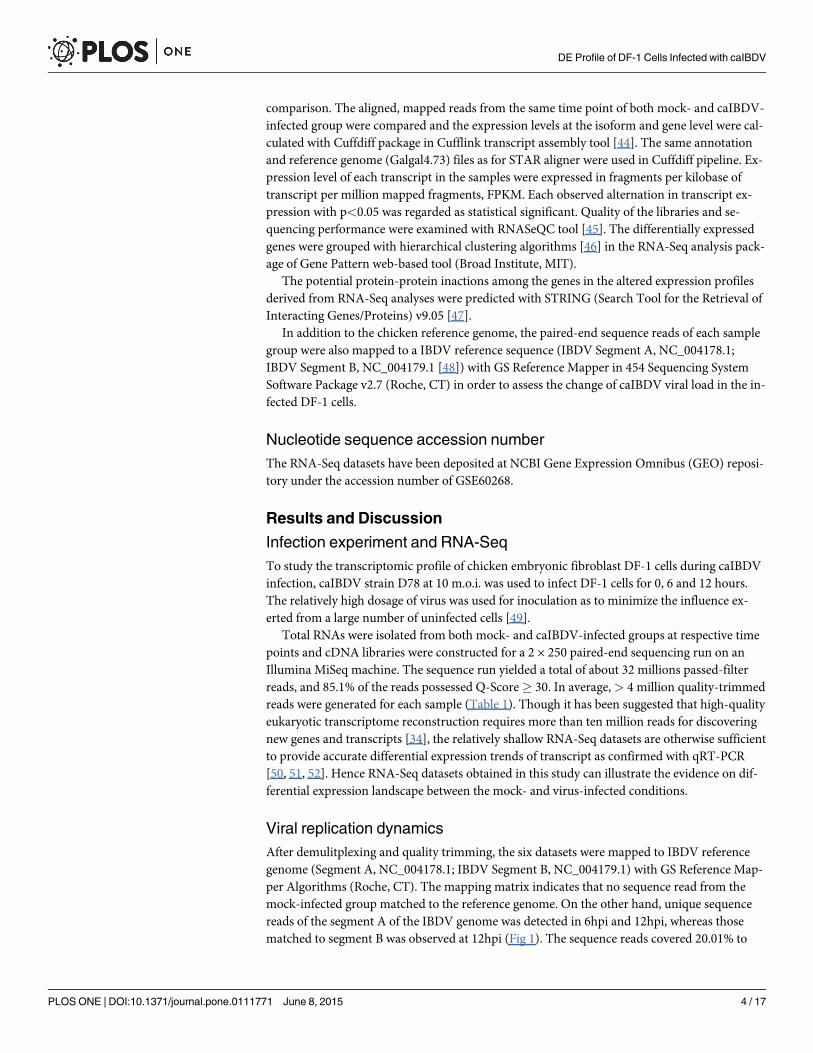
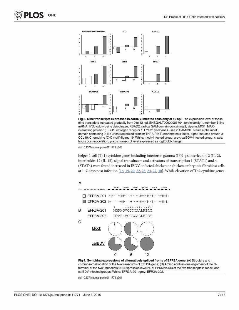
number at the early infection stage (0–12 hpi) of caIBDV infection. Expression levels of the de-tected transcript isoform from mock- and caIBDV-infected groups were compared with Cuff-diff pipeline. The scatterplot in Fig 2 describes the expression profiles of global differentialexpressed transcripts at these time points. It was demonstrated that there was no statistical sig-nificant difference between the transcriptional profiles of these two groups at 0 hpi, whereas atotal of 23 isoforms (0.12%, 23/17,942) were significantly up-regulated at 2.22–5.19 fold(p< 0.05) in the caIBDV-infected group at 6 hpi. The expression levels of 128 transcripts inthe virus-infected group were significantly altered (123 up-regulated from 2.07 to 9.7487 fold; 5down-regulated from 2.06 to 2.61 fold, p< 0.05) compared to mock-infected group at 12 hpi.Besides, 9 additional isoforms were detected in the caIBDV-infected cells only (Fig 3). The re-sult also demonstrates that there was a switch of alternatively spliced transcripts of EFR3Agene between mock- and caIBDV-infected groups (Fig 4).
The differentially expressed isoforms were classified into 12 clusters according to their ex-pression patterns in hierarchical clustering (Fig 5). Early expressed transcripts (i.e., at 6 hpi)were observed in mainly in cluster 1, 2, 3 and 9, while cluster 12 comprised 5 down-regulatedisoforms. Despite most of the expression patterns of these clusters are similar to each other andshow limited co-relation with the potential biological process activated by the caIBDV-infec-tion event according to their GO term accessions (Table 3), the result reveals that transcriptsinvolved in anti-viral responses, transcription regulation and membrane proteins were domi-nantly expressed in caIBDV-infected cells at the very early stage of infection. While transcriptsrelated to apoptotic activities, interferon-dependent immune responses and inflammatory re-sponses were tuned up at 12 hpi.
Anti-viral responses exerted by DF-1 cellsIt has been well demonstrated that host cells execute a cascade of machinery to combat againstviral particle invasion. Previous studies have shown that IBDV infection triggered expressionof cytokine genes that are typically up-regulated in general viral infections. Expression of T-
Table 2. Number of genes and transcript isoformsmapped to reference genomeGalgal4.73.
Mock-infected caIBDV-infected
0 hpi 6 hpi 12 hpi 0 hpi 6 hpi 12 hpi
Gene 10,081 9,960 9,980 9,879 9,920 9,907
Isoform 10,350 10,228 10,245 10,147 10,194 10,181
doi:10.1371/journal.pone.0111771.t002
Fig 2. Pair-wise comparison of transcript expression in mock- and caIBDV-infected DF-1 cells at 0, 6and 12 hours post-infection. Comparison of expression data was performed by XY-scatterplot analysis oflog base 2-transformed value of fragments per kilobase of transcript per million mapped fragments, FPKM.The solid line represents the predicted line of identity. The dashed line indicates the threshold of� 2-fold orless than one-half of expression ratios. Data points shown in red represents significant differentiallyexpressed transcripts, p < 0.05.
doi:10.1371/journal.pone.0111771.g002
DE Profile of DF-1 Cells Infected with caIBDV
PLOSONE | DOI:10.1371/journal.pone.0111771 June 8, 2015 6 / 17
helper 1 cell (Th1) cytokine genes including interferon gamma (IFN-γ), interleukin-2 (IL-2),interleukin-12 (IL-12), signal transducers and activators of transcription 1 (STAT1) and 4(STAT4) were found increased in IBDV-infected chicken or chicken embryonic fibroblast cellsat 1–7 days post infection [16, 19, 20, 22, 23, 24, 27, 30]. While elevation of Th2 cytokine genes
Fig 3. Nine transcripts expressed in caIBDV-infected cells only at 12 hpi. The expression level of thesenine transcripts increased gradually from 0 to 12 hpi. ENSGALT00000006704: torsin family 1, member B-likemRNA; IYD: iodotyrosine deiodinase; RSAD2: radical SAM domain-containing 2, viperin; MXI1: MAX-interacting protein 1; ESR1: estrogen receptor 1; LYG2: lysozyme G-like 2; SAMD9L: sterile alpha motifdomain containing 9-like uncharacterized protein; TNFAIP3: Tumor necrosis factor, alpha-induced protein 3;CCL19: Chemokine (C-C motif) ligand 19. White: mock-infected group; grey: caIBDV-infected group. x-axis:hours post-inoculation; y-axis: transcript level expressed as log2(fold change).
doi:10.1371/journal.pone.0111771.g003
Fig 4. Switching expressions of alternatively spliced froms of EFR3A gene. (A) Structure andchromosomal location of the two transcripts of EFR3A gene; (B) Amino acid residue alignment of the N-terminal of the two transcripts; (C) Expression level (% of FPKM value) of the two transcripts in mock- andcaIBDV-infected groups. White: EFR3A-201; grey: EFR3A-202.
doi:10.1371/journal.pone.0111771.g004
DE Profile of DF-1 Cells Infected with caIBDV
PLOSONE | DOI:10.1371/journal.pone.0111771 June 8, 2015 7 / 17

Fig 5. Heatmap indicating the pattern of 128 significant differentially expressed transcripts at 0, 6 and12 hpi time points. Expression fold change (log2 caIBDV/mock) are represented as indicated in the colorscale. The transcripts were classified into 12 clusters according to their expression patterns.
doi:10.1371/journal.pone.0111771.g005
DE Profile of DF-1 Cells Infected with caIBDV
PLOSONE | DOI:10.1371/journal.pone.0111771 June 8, 2015 8 / 17

Table 3. Differentially expressed transcripts and the respective biological process upon caIBDV infection in DF-1 cells. Transcripts with fold changeat 12 hpi� 4.00 are listed.
FoldChange
Ensembl transcript ID Symbol Description Biological function 6hpi 12hpi Cluster
ENSGALT00000010311 IFIT5 Interferon-induced protein withtetratricopeptide repeats 5
Anti-viral response 4.05 9.75 3
ENSGALT00000022096 ISG12(2) Putative ISG12-2 protein Anti-viral response 3.24 9.65 4
ENSGALT00000025999 MX Interferon-induced GTP-binding proteinMx
Anti-viral response 3.93 9.30 3
ENSGALT00000015699 ENSGALG00000009639 Uncharacterized protein Anti-viral response - 8.69 3
ENSGALT00000009906 HELZ2 Helicase with zinc finger 2, transcriptionalcoactivator
Transcription regulation 4.03 8.60 3
ENSGALT00000018067 IFIH1 Interferon-induced helicase C domain-containing protein 1
Anti-viral response 3.01 8.27 4
ENSGALT00000043751 CMPK2 Cytidine monophosphate (UMP-CMP)kinase 2, mitochondrial
Anti-viral response 3.34 8.26 3
ENSGALT00000022305 OAS*A 59 kDa 2'-5'-oligoadenylate synthase-likeprotein
Anti-viral response 3.49 8.20 3
ENSGALT00000027416 EPSTI1 Epithelial stromal interaction 1 Apoptosis - 7.76 6
ENSGALT00000026025 LY6E Lymphocyte antigen 6E precursor Transcription regulation 4.50 7.40 2
ENSGALT00000043789 SLC46A2 Solute carrier family 46, member 2 Undescribed - 7.31 3
ENSGALT00000019695 TRANK1 Tetratricopeptide repeat and ankyrinrepeat containing 1
Membrane protein/ receptor 2.90 6.83 3
ENSGALT00000019727 DTX3L Deltex 3-like protein Transporter 3.76 6.71 2
ENSGALT00000002244 IRF1 Interferon regulatory factor 1 Undescribed 5.19 6.29 9
ENSGALT00000010244 K123 K123 protein precursor Respnose to DNA damage 4.00 6.00 2
ENSGALT00000024768 ZPLD1 Zona pellucida-like domain containing 1 Anti-viral response - 5.91 5
ENSGALT00000044367 OLFML1 Olfactomedin-like 1 Undescribed - 5.88 7
ENSGALT00000000519 ITGB3 Integrin beta-3 precursor Undescribed - 5.62 7
ENSGALT00000001439 IGSF1 Immunoglobulin superfamily member 1 Undescribed 4.36 5.60 9
ENSGALT00000002240 ENSGALG00000001478 Uncharacterized protein Membrane protein/ receptor - 5.48 4
ENSGALT00000019721 PARP14 Poly (ADP-ribose) polymerase family,member 14
Membrane protein/ receptor 3.19 5.13 2
ENSGALT00000000210 DENND2D DENN/MADD domain containing 2D Undescribed - 5.04 1
ENSGALT00000007763 ZNFX1 Zinc finger, NFX1-type containing 1 Transcription regulation 3.08 5.00 2
ENSGALT00000021310 USP18 Ubiquitin carboxyl-terminal hydrolase 18 GDP-GTP conversion 2.57 4.96 3
ENSGALT00000005325 DHX58 DEXH (Asp-Glu-X-His) box polypeptide58
Undescribed - 4.93 3
ENSGALT00000006265 SDC4 Syndecan-4 precrusor Interfereon Induced - 4.82 4
ENSGALT00000018671 LOC423478 Exocyst complex component 3-like Anti-viral response 3.39 4.77 1
ENSGALT00000002226 STOML1 Stomatin (EPB72)-like 1 B-cell - 4.74 4
ENSGALT00000044107 BFIV21 MHC BF2 class I precursor Cellular balance - 4.69 6
ENSGALT00000002368 MOV10 Putative helicase MOV-10 Undescribed 2.22 4.68 3
ENSGALT00000042652 KLF4 Kruppel-like factor 4 Peptide antigen binding - 4.62 10
ENSGALT00000011423 PXK PX domain containing serine/threoninekinase
Antiviral response - 4.55 6
ENSGALT00000039921 HSPB1 Heat shock protein 25 RNA-mediated genesilencing
- 4.55 7
ENSGALT00000002125 PML Promyelocytic leukemia Transcription regulation - 4.53 6
ENSGALT00000039023 IRF-3 Interferon regulatory factor 3 Apoptotic activity 3.05 4.49 1
ENSGALT00000024766 NFKBIZ NF-kappa-B inhibitor zeta Inflammatory responses - 4.49 4
(Continued)
DE Profile of DF-1 Cells Infected with caIBDV
PLOSONE | DOI:10.1371/journal.pone.0111771 June 8, 2015 9 / 17

expressions including IL-4, IL-5, IL-13 was also reported [24]. Genes of cytokine that initiateinflammatory responses including IL-8, nitric oxide synthase (iNOS), and cyclooxygnease(COX-2) were also shown to be up-regulated [17, 18, 27]. In this study, however, no significantelevation of expression levels of these cytokine genes was identified in caIBDV-infected cells.In contrary, the regulatory factors modulating these cytokines including the well characterizedeukaryotic translation initiation factor 2-alpha kinase 2 (EIF2AK2, or protein kinase R PKR),interferon-induced GTP-binding protein Mx1 (MX), 59 kDa 2'-5'-oligoadenylate synthetase-like protein (OAS�A), guanylate binding protein (GBP7) and interferon-induced protein withtetratricopeptide (IFIT) were profoundly expressed. Among the 139 differentially expressedtranscripts, IFIT5 (interferon-induced protein with tetratricopeptide repeats 5) was expressedin the highest fold change during caIBDV infection. IFIT5 is a member of IFIT1 family whichexpression can be induced by virus infection, interferons, dsRNA and lipopolysaccharides [53,54]. It was demonstrated that IFIT5 potentiates anti-viral responses by promoting the interfer-on regulatory factor 3 (IRF3)- or nuclear factor kappa-light-chain-enhancer of activated B cells(NF-κB)-mediated gene expressions [55]. Over-expression of IFIT5 in the infected DF-1 cellswas observed accompany with the elevation of IRF3 at 6 hpi, and progressively with other IRF-3-mediated genes at 12 hpi (Fig 6). One of the dominant anti-viral components potentially trig-gered by ITIF5-IRF3 pathway was RSAD2 (radical SAM domain-containing 2, viperin) gene.RSAD2 gene was found expressed only in caIBDV-infected cells but not in mock-infected cellsat 12 hpi (Fig 3). It is an endoplasmic reticulum-associated virus inhibitory protein which canbe induced by type I, II and III IFNs, double-stranded (ds) DNA, dsRNA analog poly I:C, lipo-polysaccharide (LPS) and infection of various viruses [56–61]. Besides, it was shown thatRSAD2 could be induced via an IFN-independent pathway by transcription factor IRF-1 [62].Definite anti-viral machineries exerted by RSAD2 has not been concluded yet, but it is believedthat it involves in inhibiting viral replication indirectly by altering the cell survival control [63],and membrane fluidity modulation which prevents the budding of viruses [64]. So the switch-ing-on of RSAD2, presumably by ITIF5-IRF3 or by IRF1 regulation, may contribute to theearly viral defense in IBDV infection.
Table 3. (Continued)
FoldChange
Ensembl transcript ID Symbol Description Biological function 6hpi 12hpi Cluster
ENSGALT00000004971 TRIM25 Tripartite motif containing 25 Stress resistance 3.32 4.49 2
ENSGALT00000036426 ITA Inhibitor of apoptosis protein Transcription regulation - 4.47 4
ENSGALT00000020390 NMI N-myc (and STAT) interactor Respnose to DNA damage - 4.35 4
ENSGALT00000014519 CASP7 Caspase 7, apoptosis-related cysteinepeptidase
- 4.35 9
ENSGALT00000010406 IRF10 Interferon regulatory factor 10 Interfereon-dependentimmune responses
- 4.32 4
ENSGALT00000019729 PARP9 Poly (ADP-ribose) polymerase family,member 9
NF-kappa-B cascade - 4.21 4
ENSGALT00000040649 CCL4 Chemokine-like ligand 1 precursor Apoptotic activity - 4.19 3
ENSGALT00000010225 PFKFB3 6-phosphofructo-2-kinase/fructose-2,6-biphosphatase 3
Inflammatory responses - 4.13 5
ENSGALT00000001016 GBP7 Guanylate binding protein 7 Anti-viral response - 4.10 5
ENSGALT00000022552 ZC3HAV1 Zinc finger CCCH-type antiviral protein 1 Apoptotic activity 2.51 4.04 1
doi:10.1371/journal.pone.0111771.t003
DE Profile of DF-1 Cells Infected with caIBDV
PLOSONE | DOI:10.1371/journal.pone.0111771 June 8, 2015 10 / 17

Fig 6. Gene pathways analysis. The potential interaction among the significant differentially expressedisoforms (p < 0.05) were analysed with STRING tool v9.05. (A) 6 hpi; (B) 12 hpi. Expression fold change (log2caIBDV/mock) are represented as indicated in the color scale. Red lines: potential protein interactions startedat 6 hpi; green lines: potential protein interactions started at 12 hpi.
doi:10.1371/journal.pone.0111771.g006
DE Profile of DF-1 Cells Infected with caIBDV
PLOSONE | DOI:10.1371/journal.pone.0111771 June 8, 2015 11 / 17
Apart from the defense effectors, genes possessed with accessory anti-viral functions werealso regulated in caIBDV-infected cells. The interferon-induced helicase C domain-containingprotein 1 (IFIH1, or melanoma differentiation-associated protein 5, MDA5) was found ex-pressed in 3.01 and 8.27 fold at 6 and 12 hpi respectively. It was reported that IFIH1 interactswith probable ATP-dependent RNA helicase (DHX58, or Laboratory of Genetics and Physiolo-gy 2, LGP2; expressed to 4.93 fold at 12 hpi, Table 3) as a pattern recognition receptor to senseviral dsRNA and triggers downstream antiviral reactions [65, 66]. In addition, elevated expres-sion of tripartite motif-containing protein 25 (TRIM25) at the very beginning of the infectionevent was believed to be related to the interaction with retinoic acid-inducible gene 1 (RIG-I)in detecting the viral RNA intermediates [67, 68]. Apart from the regulation of RSAD2, thehost cells membrane condition during caIBDV infection was probably influenced with cellularcholesterol metabolism. An over-expression of lipase A (LIPA) and cholesterol 25-hydroxylase(CH25H), together with the down-regulation of StAR-related lipid transfer protein 4(STARD4), lanosterol synthase (LSS) and acetoacetyl-coA synthease (AACS) (Fig 6B), implyan evidence on the modulation of intracellular cholesterol contents in the infected cells. Thesynergetic effect of the regulation of these genes may alter the lipid raft arrangement and there-fore inhibit membrane fusion event between the infected cells and virus, limit the clathrin- andcholesterol-dependent endocytosis hence prevent the propagation of the virus [69, 70, 71].
Potential strategies used by caIBDV to outwit host cell defensesViruses make use of various mechanisms to escape from the host defense actions. One of theviral strategies is to arrest the apoptosis initiation so as to facilitate viral replication process.Apoptosis is one of the host defense mechanism to minimize the spread of viruses. Our previ-ous study demonstrated that apoptosis occurred at 48 hpi [21], while Jungmann et al. revealedthat the IBDV-induced apoptosis was first observed at 12 hpi [72]. It has been well studied thatNF-κB mediated apoptotic pathway is initiated upon viral infection. In uninfected cells, thedimer of NF-κB are sequestered in the cytoplasm by a family of κB inhibitors (IκB). Uponvirus infection, these IκB proteins undergo signal-induced degradation by proteasome trig-gered by the activation of IκB kinase (IKK) and the NF-κB complex is released into the nucleusfor the expression of specific genes leading into apoptosis and other antiviral functions. In thisstudy, expression of both NF-κB1 and NF-κB2 were observed in mock- and caIBDV-infectedDF-1 cells, but there was no significant changes between both groups at the time points tested(though the trends of both NF-κB proteins increased with time in caIBDV-infected group). Be-sides, the expression of two IKKs (IKK1 or CHUK and IKK2 or IKBKB) also showed no signifi-cant differences, implying that the degradation capacity of IκB between two groups weresimilar. The result, however, demonstrated that there was a distinct elevation of two IκB levels(NFKBIA or IκBα and NFKBIZ or IκBz) at 12 hpi in the caIBDV-infected group. The increaseof these IκBs potentially hindered the function of IKKs and eventually reduced the free NF-κBamount, hence retarded the apoptotic process. Apart from the IκBs, the levels of tumor necro-sis factor, alpha-induced protein 2 (TNFAI2) and 3 (TNFAIP3) were also provoked in the in-fected cells. It has been demonstrated that TNFAIP3 is a potent cellular inhibitor of NF-κBactivation [73, 74]. Furthermore, the caspases-mediated apoptosis was probably inhibited bythe over-expressed baculoviral IAP repeat-containing protein 2 (BIRC2 or ITA) in the infectedcells. ITA is a member of the inhibitor of apoptosis family that inhibit apoptosis by interferingwith the activation of caspases [75, 76]. Liu et al. suggested that the nonstructural protein (NS)of IBDV possessed anti-apoptotic function at the early stage of virus infection [77]. It is there-fore reflecting that the NS of caIBDV regulates the expression of IκB, TNFAIPs and ITA in the
DE Profile of DF-1 Cells Infected with caIBDV
PLOSONE | DOI:10.1371/journal.pone.0111771 June 8, 2015 12 / 17

infected cells which aims to delay the apoptosis in the first 12 hpi and reserve viable host cellsfor viral replication.
Switching of alternatively spliced EFR3A transcript isoformsRNA-Seq data reveals that two splice variants of EFR3 homolog A (S. cerevisiae) (EFR3A)
transcript were identified in mock- and caIBDV-infected DF-1 cells respectively (Fig 4). It wasshown that EFR3A-201 expressed in mock-infected cells, while the expression of EFR3A-202isoform took over the previous one at 12 hpi in the caIBDV-infected cells. Amino acid align-ment reveals that four residue differences located at the N-terminal immediately downstreamto the first methionine residue between the two variants. EFR3A gene encodes a membraneprotein [78], but the definite cellular function of EFR3A in chicken cells has not been reportedyet. Odrowaz et al. reported EFR3A participated in the negative control of a ETS transcriptionfactor ELK1 in a human epithelial cell MCF-10A [79]. Whereas it was shown to be involved tothe epidermal growth factor receptor signaling pathway [80]. It was also suggested that the dif-ferential expression of EFR3A gene in auditory brainstem neurons of mice with hearing deficit[81]. The switch of splice variant expression may be involved in altering the lipid membranecondition, but the molecular mechanism of this deviations to host-virus interaction needsfurther characterization.
ConclusionInteractions between IBDV and its host has been extensively studied. A number of researchesrevealed that infection of chicken cells, including fibroblast and bursal cells, with IBDV maylead into an elevation of cytokines and interferons at 1 dpi to 7 dpi periods in general. Virus-in-duced apoptosis via caspase- and NF-κB-mediated pathways were also demonstrated. Thisstudy, on the other hand, disclosed the early host-virus interactions. With the aid of RNA-Seq,a more comprehensive expression landscape was obtained. The result presents the events oc-curred before the elevation of downstream effectors. Apart from the regulators of cytokinesand interferons, modulations were observed in the gene candidates involved in cell membranefluidity. It is believed that the changes in membrane conditions contributes to the frontlinehost response against endocytosis of IBDV and hence prevent infections. On the other hand,the intensively expressed anti-apoptotic genes induced by caIBDV delay the programmed celldeath and hence prolong the viral replication cycle in the host cells.
This study explore the initial host response in DF-1 cells upon caIBDV infection and the po-tential virus strategy to counteract with the host's action.
AcknowledgmentsWe also thank Jovicic B and Mladenovic V of Seven Bridges Genomics Inc., MA for theirbioinformatics support.
Author ContributionsConceived and designed the experiments: FCL RKH. Performed the experiments: RKH. Ana-lyzed the data: RKH FCL. Contributed reagents/materials/analysis tools: RKH FCL. Wrote thepaper: RKH FCL.
References1. Chettle N, Stuart JC, Wyeth PJ. Outbreak of virulent infectious bursal disease in East Anglia. Vet Rec.
1989; 125: 271–272. PMID: 2552640
2. Berg TP, Gonze M, Meulemans G. Acute infectious bursal disease in poultry: Isolation and characteri-sation of a highly virulent strain. Avian Pathol. 1991; 20: 133–143. PMID: 18680006
DE Profile of DF-1 Cells Infected with caIBDV
PLOSONE | DOI:10.1371/journal.pone.0111771 June 8, 2015 13 / 17
3. Faragher JT, Allan WH, Cullen GA. Immunosuppressive effect of the infectious bursal agent in thechicken. Nat New Biol. 1972; 237: 118–119. PMID: 4503850
4. Hudson L, PattisonM, Thantrey N. Specific B lymphocyte suppression by infectious bursal agent (Gum-boro disease virus) in chickens. Eur J Immunol. 1975; 5: 675–679. PMID: 11993333
5. Lukert PD, Saif YM. Infectious bursal disease. In: Calnek BW, Barnes HJ, Beard CW, Mcdougald LR,Saif YM, editors. Diseases of poultry, 11th ed. Iowa State University Press, Iowa. 2003. pp. 161–180.
6. Cosgrove AS. An apparently new disease of chickens—avian nephrosis. Avian Dis. 1962; 6: 385–389.
7. McFerran JB, McNulty MS, McKillop ER, Connor TJ, McCracken RM, Collins DS, et al. Isolation and se-rological studies with infectious bursal disease viruses from fowl, turkeys, and ducks: demonstration ofa second serotype. Avian Pathol. 1980; 9: 395–403. PMID: 18770277
8. Jackwood DJ, Saif YM, Moorhead PD, Bishop G. Failure of two serotype II infectious bursal disease vi-ruses to affect the humoral immune response of turkeys. Avian Dis. 1984; 28: 100–116. PMID:6202292
9. Jackwood DJ, Saif YM, Moorhead PD. Immunogenicity and antigenicity of infectious bursal diseasevirus serotypes I and II in chickens. Avian Dis. 1985; 29: 1184–1194. PMID: 3008699
10. Vasconcelos AC, Lam KM. Apoptosis induced by infectious bursal disease virus. J Gen Virol. 1984; 75:1803–1806.
11. Rodenberg J, Sharma JM, Belzer SW, Nordgren RM, Naqi S. Flow cytometric analysis of B cell and Tcell subpopulations in specific-pathogen-free chickens infected with infectious bursal disease virus.Avian Dis. 1994; 38: 16–21. PMID: 8002886
12. Vasconcelos AC, Lam KM. Apoptosis in chicken embryos induced by the infectious bursal diseasevirus. J Comp Pathol. 1995; 112: 327–338. PMID: 7593755
13. Kim IJ, Karaca K, Pertile TL, Erickson SA, Sharma JM. Enhanced expression of cytokine genes inspleen macrophages during acute infection with infectious bursal disease virus in chickens. Vet Immu-nol Immunopathol. 1998; 61: 331–341. PMID: 9613445
14. RaglandWL, Novak R, El-Attrache J, Savić V, Ester K. Chicken anemia virus and infectious bursal dis-ease virus interfere with transcription of chicken IFN-alpha and IFN-gammamRNA. J Interferon Cyto-kine Res. 2002; 22: 437–441. PMID: 12034026
15. Khatri M, Palmquist JM, Cha RM, Sharma JM. Infection and activation of bursal macrophages by viru-lent infectious bursal disease virus. Virus Res. 2005; 113: 44–50. PMID: 15893401
16. Eldaghayes I, Rothwell L, Williams A, Withers D, Balu S, Davison F, et al. Infectious bursal diseasevirus: strains that differ in virulence differentially modulate the innate immune response to infection inthe chicken bursa. Viral Immunol. 2006; 19: 83–91. PMID: 16553553
17. Khatri M, Sharma JM. Infectious bursal disease virus infection induces macrophage activation via p38MAPK and NF-kappaB pathways. Virus Res. 2006; 118: 70–77. PMID: 16388870
18. Palmquist JM, Khatri M, Cha RM, Goddeeris BM, Walcheck B, Sharma JM. In vivo activation of chickenmacrophages by infectious bursal disease virus. Viral Immunol. 2006; 19: 305–315. PMID: 16817773
19. Ruby T, Whittaker C, Withers DR, Chelbi-Alix MK, Morin V, Oudin A, et al. Transcriptional profiling re-veals a possible role for the timing of the inflammatory response in determining susceptibility to a viralinfection. J Virol. 2006; 80: 9207–9216. PMID: 16940532
20. Li YP, Handberg KJ, Juul-Madsen HR, Zhang MF, Jørgensen PH. Transcriptional profiles of chickenembryo cell cultures following infection with infectious bursal disease virus. Arch Virol. 2007; 152: 463–478. PMID: 17143781
21. Wong RT, Hon CC, Zeng F, Leung FC. Screening of differentially expressed transcripts in infectiousbursal disease virus-induced apoptotic chicken embryonic fibroblasts by using cDNAmicroarrays. JGen Virol. 2007; 88: 1785–1796. PMID: 17485540
22. Khatri M, Sharma JM. IFN-gamma upregulation and protection by macrophage-adapted infectious bur-sal disease virus. Vaccine. 2008; 26: 4740–4746. doi: 10.1016/j.vaccine.2008.06.053 PMID: 18601966
23. Khatri M, Sharma JM. Response of embryonic chicken lymphoid cells to infectious bursal diseasevirus. Vet Immunol Immunopathol. 2009; 127: 316–324. doi: 10.1016/j.vetimm.2008.10.327 PMID:19081143
24. Liu H, Zhang M, Han H, Yuan J, Li Z. Comparison of the expression of cytokine genes in the bursal tis-sues of the chickens following challenge with infectious bursal disease viruses of varying virulence.Virol J. 2010; 7: 364. doi: 10.1186/1743-422X-7-364 PMID: 21143846
25. Carballeda JM, Zoth SC, Gómez E, Gravisaco MJ, Berinstein A. Activation of the immune responseagainst Infectious Bursal Disease Virus after intramuscular inoculation of an intermediate strain. Immu-nobiology. 2011; 216: 1028–1033. doi: 10.1016/j.imbio.2011.03.003 PMID: 21514000
DE Profile of DF-1 Cells Infected with caIBDV
PLOSONE | DOI:10.1371/journal.pone.0111771 June 8, 2015 14 / 17
26. Kong BW, Lee JY, Bottje WG, Lassiter K, Lee J, Foster DN. Genome-wide differential gene expressionin immortalized DF-1 chicken embryo fibroblast cell line. BMCGenomics. 2011; 12: 571. doi: 10.1186/1471-2164-12-571 PMID: 22111699
27. Rauf A, Khatri M, Murgia MV, Jung K, Saif YM. Differential modulation of cytokine, chemokine and Tolllike receptor expression in chickens infected with classical and variant infectious bursal disease virus.Vet Res. 2011; 42: 85. doi: 10.1186/1297-9716-42-85 PMID: 21749706
28. Wei L, Zhu S, Ruan G, Hou L, Wang J, Wang B, et al. Infectious bursal disease virus-induced activationof JNK signaling pathway is required for virus replication and correlates with virus-induced apoptosis.Virology. 2011; 420: 156–163. doi: 10.1016/j.virol.2011.08.027 PMID: 21968197
29. Guo X, Wang L, Cui D, RuanW, Liu F, Li H. Differential expression of the Toll-like receptor pathwayand related genes of chicken bursa after experimental infection with infectious bursa disease virus.Arch Virol. 2012; 157: 2189–2199. doi: 10.1007/s00705-012-1403-y PMID: 22828777
30. Tippenhauer M, Heller DE, Weigend S, Rautenschlein S. The host genotype influences infectious bur-sal disease virus pathogenesis in chickens by modulation of T cells responses and cytokine gene ex-pression. Dev Comp Immunol. 2013; 40: 1–10. doi: 10.1016/j.dci.2012.10.013 PMID: 23194926
31. Zheng X, Hong L, Shi L, Guo J, Sun Z, Zhou J. Proteomics analysis of host cells infected with infectiousbursal disease virus. Mol Cell Proteomics. 2008; 7: 612–625. PMID: 18056921
32. Wu Y, Peng C, Xu L, Zheng X, Liao M, Yan Y, et al. Proteome dynamics in primary target organ of infec-tious bursal disease virus. Proteomics. 2012; 12: 1844–1859. doi: 10.1002/pmic.201100479 PMID:22623289
33. Wang Z, Gerstein M, Snyder M. RNA-Seq: a revolutionary tool for transcriptomics. Nat Rev Genet.2009; 10: 57–63. doi: 10.1038/nrg2484 PMID: 19015660
34. Trapnell C, Roberts A, Goff L, Pertea G, Kim D, Kelley DR, et al. Differential gene and transcript expres-sion analysis of RNA-Seq experiments with TopHat and Cufflinks. Nat Protoc. 2012; 7: 562–578. doi:10.1038/nprot.2012.016 PMID: 22383036
35. Schulze A, Downward J. Analysis of gene expression by microarrays: cell biologist's gold mine or mine-field? J Cell Sci. 2000; 113: 4151–4156. PMID: 11069760
36. Kurella M, Hsiao LL, Yoshida T, Randall JD, Chow G, Sarang SS, et al. DNAmicroarray analysis ofcomplex biologic processes. J Am Soc Nephrol. 2001; 12: 1072–1078. PMID: 11316867
37. Cloonan N, Forrest AR, Kolle G, Gardiner BB, Faulkner GJ, Brown MK, et al. Stem cell transcriptomeprofiling via massive-scale mRNA sequencing. Nat Methods. 2008; 5: 613–619. doi: 10.1038/nmeth.1223 PMID: 18516046
38. Nagalakshmi U, Wang Z, Waern K, Shou C, Raha D, Gerstein M, et al. The transcriptional landscape ofthe yeast genome defined by RNA sequencing. Science. 2008; 320: 1344–1349. doi: 10.1126/science.1158441 PMID: 18451266
39. Okoniewski MJ, Miller CJ. Hybridization interactions between probesets in short oligo microarrays leadto spurious correlations. BMC Bioinformatics. 2006; 7: 276. PMID: 16749918
40. Hierholzer JC, Killington RA. Virus Isolation and Quantitation. In Mahy BWJ, Kangro HO, editors. Virolo-gy Methods Manual, 1st ed. Academic Press, San Diego, CA. 1996. pp. 25–46.
41. Dobin A, Davis CA, Schlesinger F, Drenkow J, Zaleski C, Jha S, et al. STAR: ultrafast universal RNA-Seq aligner. Bioinformatics. 2013; 29: 15–21. doi: 10.1093/bioinformatics/bts635 PMID: 23104886
42. Flicek P, Ahmed I, Amode MR, Barrell D, Beal K, Brent S, et al. Ensembl 2013. Nucleic Acids Research.41 Database issue:D48–D55 doi: 10.1093/nar/gks1236 PMID: 23203987
43. Li H, Handsaker B, Wysoker A, Fennell T, Ruan J, Homer N, et al. The Sequence Alignment/Map for-mat and SAMtools. Bioinformatics. 2009; 25: 2078–2079. doi: 10.1093/bioinformatics/btp352 PMID:19505943
44. Trapnell C, Williams BA, Pertea G, Mortazavi A, Kwan G, van Baren MJ, et al. Transcript assembly andquantification by RNA-Seq reveals unannotated transcripts and isoform switching during cell differenti-ation. Nat Biotechnol. 2010; 28: 511–515. doi: 10.1038/nbt.1621 PMID: 20436464
45. DeLuca DS, Levin JZ, Sivachenko A, Fennell T, Nazaire MD, Williams C, et al. RNA-SeqC: RNA-Seqmetrics for quality control and process optimization. Bioinformatics. 2012; 28: 1530–1532. doi: 10.1093/bioinformatics/bts196 PMID: 22539670
46. Eisen MB, Spellman PT, Brown PO, Botstein D. Cluster analysis and display of genome-wide expres-sion patterns. Proc Natl Acad Sci U S A. 1998; 95: 14863–8. PMID: 9843981
47. Franceschini A, Szklarczyk D, Frankild S, Kuhn M, Simonovic M, Roth A, et al. STRING v9.1: protein-protein interaction networks, with increased coverage and integration. Nucleic Acids Res. 2013. 41(Da-tabase issue):D808–15. doi: 10.1093/nar/gks1094 PMID: 23203871
DE Profile of DF-1 Cells Infected with caIBDV
PLOSONE | DOI:10.1371/journal.pone.0111771 June 8, 2015 15 / 17

48. Brown MD, Skinner MA. Coding sequences of both genome segments of a European 'very virulent' in-fectious bursal disease virus. Virus Res. 1996; 40: 1–15. PMID: 8725117
49. Sessions OM, Tan Y, Goh KC, Liu Y, Tan P, Rozen S, et al. Host Cell Transcriptome Profile duringWild-Type and Attenuated Dengue Virus Infection. PLoS Negl Trop Dis 2013; 7: e2107. doi: 10.1371/journal.pntd.0002107 PMID: 23516652
50. Chen X, Zeng D, Chen X, Xie D, Zhao Y, Yang C, et al. Transcriptome Analysis of Litopenaeus vanna-mei in Response to White Spot Syndrome Virus Infection. PLoS One. 2013; 8: e73218. doi: 10.1371/journal.pone.0073218 PMID: 23991181
51. Sun L, Yang H, Chen M, Ma D, Lin C. RNA-Seq reveals dynamic changes of gene expression in keystages of intestine regeneration in the sea cucumber Apostichopus japonicas. PLoS One. 2013; 8:e69441. doi: 10.1371/journal.pone.0069441 PMID: 23936330
52. Zeng D, Chen X, Xie D, Zhao Y, Yang C, Li Y, et al. Transcriptome analysis of Pacific white shrimp (Lito-penaeus vannamei) hepatopancreas in response to Taura syndrome Virus (TSV) experimental infec-tion. PLoS One. 2013; 8: e57515. doi: 10.1371/journal.pone.0057515 PMID: 23469011
53. Ovstebø R, Olstad OK, Brusletto B, Møller AS, Aase A, Haug KB, et al. Identification of genes particu-larly sensitive to lipopolysaccharide (LPS) in humanmonocytes induced by wild-type versus LPS-defi-cient Neisseria meningitidis strains. Infect Immun. 2008; 76: 2685–2695. doi: 10.1128/IAI.01625-07PMID: 18362127
54. Schoggins JW,Wilson SJ, Panis M, Murphy MY, Jones CT, Bieniasz P, et al. A diverse range of geneproducts are effectors of the type I interferon antiviral response. Nature. 2011; 472: 481–485. doi: 10.1038/nature09907 PMID: 21478870
55. Zhang B, Liu X, ChenW, Chen L. IFIT5 potentiates anti-viral response through enhancing innate im-mune signaling pathways. Acta Biochim Biophys Sin (Shanghai). 2013; 45: 867–874. doi: 10.1093/abbs/gmt088 PMID: 23942572
56. Boudinot P, Massin P, Blanco M, Riffault S, Benmansour A. vig-1, a new fish gene induced by the rhab-dovirus glycoprotein, has a virus-induced homologue in humans and shares conserved motifs with theMoaA family. J Virol. 1999; 73: 1846–1852. PMID: 9971762
57. Boudinot P, Riffault S, Salhi S, Carrat C, Sedlik C, Mahmoudi N, et al. Vesicular stomatitis virus andpseudorabies virus induce a vig1/cig5 homologue in mouse dendritic cells via different pathways. JGen Virol. 2000; 81: 2675–2682. PMID: 11038379
58. Grewal TS, Genever PG, Brabbs AC, Birch M, Skerry TM. Best5: a novel interferon-inducible gene ex-pressed during bone formation. FASEB J. 2000; 14: 523–531. PMID: 10698968
59. Chin KC, Cresswell P. Viperin (cig5), an IFN-inducible antiviral protein directly induced by human cyto-megalovirus. Proc Natl Acad Sci U S A. 2001; 98: 15125–15130. PMID: 11752458
60. Severa M, Coccia EM, Fitzgerald KA. Toll-like receptor-dependent and-independent viperin gene ex-pression and counter-regulation by PRDI-binding factor-1/BLIMP1. J Biol Chem. 2006; 281: 26188–26195. PMID: 16849320
61. Zhou Z, Hamming OJ, Ank N, Paludan SR, Nielsen AL, Hartmann R. Type III interferon (IFN) induces atype I IFN-like response in a restricted subset of cells through signaling pathways involving both theJak-STAT pathway and the mitogen-activated protein kinases. J Virol. 2007; 81: 7749–7758. PMID:17507495
62. Stirnweiss A, Ksienzyk A, Klages K, Rand U, Grashoff M, Hauser H, et al. IFN regulatory factor-1 by-passes IFN-mediated antiviral effects through viperin gene induction. J Immunol. 2010; 184: 5179–5185. doi: 10.4049/jimmunol.0902264 PMID: 20308629
63. Szretter KJ, Brien JD, Thackray LB, Virgin HW, Cresswell P, Diamond MS. The interferon-induciblegene viperin restricts West Nile virus pathogenesis. J Virol. 2011; 85: 11557–11566. doi: 10.1128/JVI.05519-11 PMID: 21880757
64. Wang X, Hinson ER, Cresswell P. The interferon-inducible protein viperin inhibits influenza virus re-lease by perturbing lipid rafts. Cell Host Microbe. 2007; 2: 96–105. PMID: 18005724
65. Broquet AH, Hirata Y, McAllister CS, Kagnoff MF. RIG-I/MDA5/MAVS are required to signal a protectiveIFN response in rotavirus-infected intestinal epithelium. J Immunol. 2011; 186:1618–1626. doi: 10.4049/jimmunol.1002862 PMID: 21187438
66. Liniger M, Summerfield A, Zimmer G, McCullough KC, Ruggli N. Chicken cells sense influenza A virusinfection through MDA5 and CARDIF signaling involving LGP2. J Virol. 2012; 86: 705–717. doi: 10.1128/JVI.00742-11 PMID: 22072756
67. Gack MU, Shin YC, Joo CH, Urano T, Liang C, Sun L, et al. TRIM25 RING-finger E3 ubiquitin ligase isessential for RIG-I-mediated antiviral activity. Nature. 2007; 446: 916–920. PMID: 17392790
68. Oshiumi H, Matsumoto M, Seya T. Ubiquitin-mediated modulation of the cytoplasmic viral RNA sensorRIG-I. J Biochem. 2012; 151: 5–11. doi: 10.1093/jb/mvr111 PMID: 21890623
DE Profile of DF-1 Cells Infected with caIBDV
PLOSONE | DOI:10.1371/journal.pone.0111771 June 8, 2015 16 / 17

69. Ilnytska O, Santiana M, Hsu NY, DuWL, Chen YH, Viktorova EG, et al. Enteroviruses harness the cel-lular endocytic machinery to remodel the host cell cholesterol landscape for effective viral replication.Cell Host Microbe. 2013; 14: 281–293. doi: 10.1016/j.chom.2013.08.002 PMID: 24034614
70. Liu SY, Aliyari R, Chikere K, Li G, Marsden MD, Smith JK, et al. Interferon-inducible cholesterol-25-hy-droxylase broadly inhibits viral entry by production of 25-hydroxycholesterol. Immunity. 2013; 38: 92–105. doi: 10.1016/j.immuni.2012.11.005 PMID: 23273844
71. Yang S, He M, Liu X, Li X, Fan B, Zhao S. Japanese encephalitis virus infects porcine kidney epithelialPK15 cells via clathrin- and cholesterol-dependent endocytosis. Virol J. 2013; 10: 258. doi: 10.1186/1743-422X-10-258 PMID: 23937769
72. Jungmann A, Nieper H, Müller H. Apoptosis is induced by infectious bursal disease virus replication inproductively infected cells as well as in antigen-negative cells in their vicinity. J Gen Virol. 2001; 82:1107–1115. PMID: 11297685
73. Heyninck K, De Valck D, Vanden BergheW, Van CriekingeW, Contreras R, Fiers W, et al. The zinc fin-ger protein A20 inhibits TNF-induced NF-kappaB-dependent gene expression by interfering with anRIP- or TRAF2-mediated transactivation signal and directly binds to a novel NF-kappaB-inhibiting pro-tein ABIN. J Cell Biol. 1999; 145: 1471–1482. PMID: 10385526
74. De Valck D, Jin DY, Heyninck K, Van de Craen M, Contreras R, Fiers W, et al. The zinc finger proteinA20 interacts with a novel anti-apoptotic protein which is cleaved by specific caspases. Oncogene.1999; 18: 4182–90. PMID: 10435631
75. Deveraux QL, Roy N, Stennicke HR, Van Arsdale T, Zhou Q, Srinivasula SM, et al. IAPs block apoptoticevents induced by caspase-8 and cytochrome c by direct inhibition of distinct caspases. EMBO J.1998; 17: 2215–2223. PMID: 9545235
76. Tenev T, Zachariou A, Wilson R, Ditzel M, Meier P. IAPs are functionally non-equivalent and regulateeffector caspases through distinct mechanisms. Nat Cell Biol. 2005; 7: 70–77. PMID: 15580265
77. Liu M, Vakharia VN. Nonstructural protein of infectious bursal disease virus inhibits apoptosis at theearly stage of virus infection. J Virol. 2006; 80: 3369–3377. PMID: 16537604
78. Nagase T, Seki N, Tanaka A, Ishikawa K, Nomura N. Prediction of the coding sequences of unidentifiedhuman genes. IV. The coding sequences of 40 new genes (KIAA0121-KIAA0160) deduced by analysisof cDNA clones from human cell line KG-1. DNA Res. 1995; 2: 167–74, 199–210. PMID: 8590280
79. Odrowaz Z, Sharrocks AD. ELK1 uses different DNA binding modes to regulate functionally distinctclasses of target genes. PLoS Genet. 2012; 8: e1002694. doi: 10.1371/journal.pgen.1002694 PMID:22589737
80. Havaleshko DM, Smith SC, Cho H, Cheon S, Owens CR, Lee JK, et al. Comparison of global versusepidermal growth factor receptor pathway profiling for prediction of lapatinib sensitivity in bladder can-cer. Neoplasia. 2009; 11: 1185–1193. PMID: 19881954
81. Munemoto Y, Houtani T, Kase M, Sakuma S, Baba K, Yamashita T, et al. Mouse homolog of KIAA0143protein: hearing deficit induces specific changes of expression in auditory brainstem neurons. BrainRes Mol Brain Res. 2004; 128: 131–40. PMID: 15363888
DE Profile of DF-1 Cells Infected with caIBDV
PLOSONE | DOI:10.1371/journal.pone.0111771 June 8, 2015 17 / 17