Diagnostic test accuracy for skin cancer project
45
Diagnostic test accuracy for skin cancer project: Implications for practice and research Hywel Williams on behalf of NIHR HTA Skin Cancer DTA team Universities of Nottingham and Birmingham January 8 th Paris
Transcript of Diagnostic test accuracy for skin cancer project
Diagnostic test accuracy
for skin cancer project:Implications for practice and research
Hywel Williams on behalf of NIHR HTA Skin Cancer DTA team
Universities of Nottingham and Birmingham
January 8th Paris
Rough plan
What did we do?
Why did we do it?
How did we do it?
What did we find?
What does it all mean?
Lessons I learned?
Conflicts of interest and funding
I work for the
I work for the NIHR HTA
I have no financial or research associations with any
of the diagnostic technologies assessed
Funded by the National Institute for Health Research, via Cochrane Infrastructure funding
to the Cochrane Skin Group and Cochrane Programme Grant funding. The views and
opinions expressed therein are those of the authors and do not necessarily reflect those of
the Systematic Reviews Programme, NIHR, NHS or Department of Health.
But first a diagnostic
test for you:
What are these lesions called?
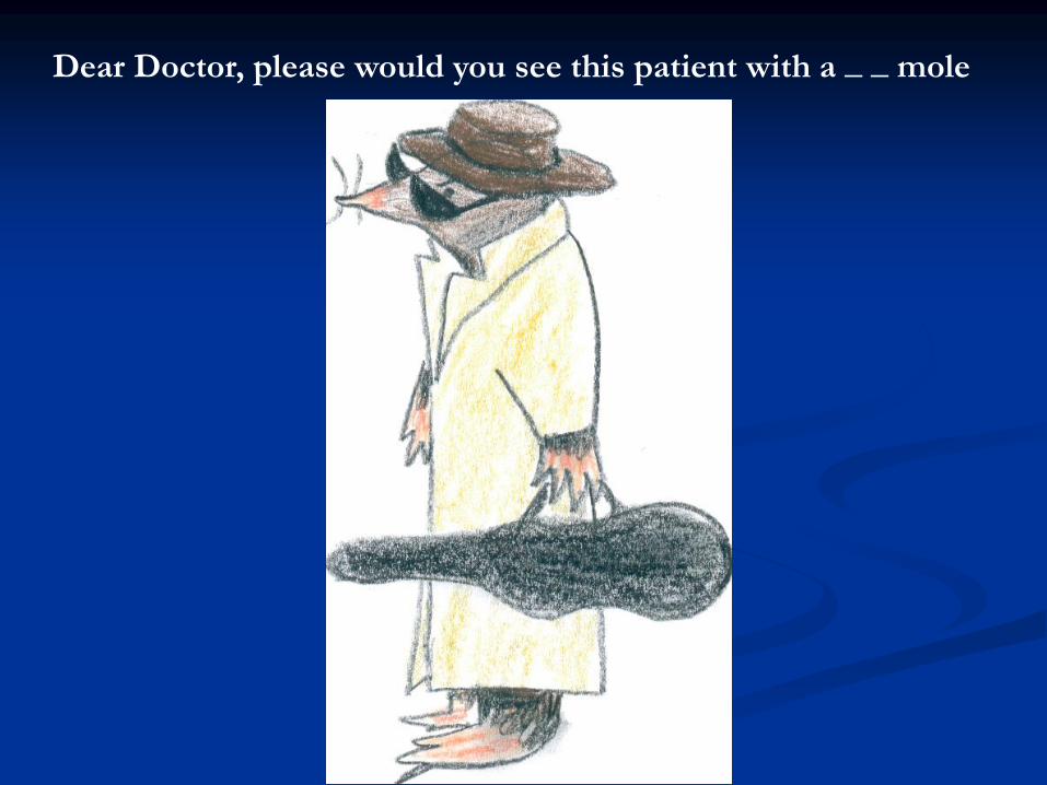
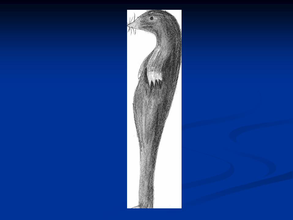
Dear Doctor, please would you see this patient with a _ _ mole
Plan
What did we do?
Why did we do it?
How did we do it?
What did we find?
What does it all mean?
Lessons I learned?
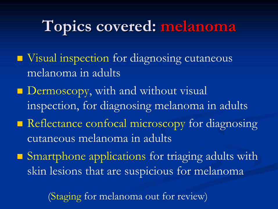
Topics covered: melanoma
Visual inspection for diagnosing cutaneous
melanoma in adults
Dermoscopy, with and without visual
inspection, for diagnosing melanoma in adults
Reflectance confocal microscopy for diagnosing
cutaneous melanoma in adults
Smartphone applications for triaging adults with
skin lesions that are suspicious for melanoma
(Staging for melanoma out for review)
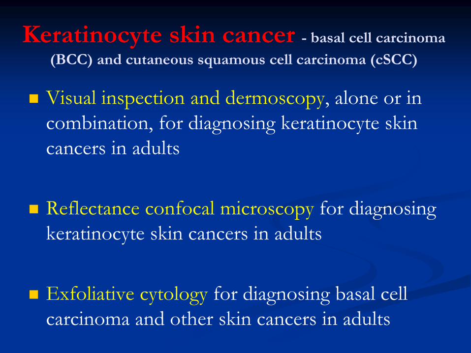
Keratinocyte skin cancer - basal cell carcinoma
(BCC) and cutaneous squamous cell carcinoma (cSCC)
Visual inspection and dermoscopy, alone or in
combination, for diagnosing keratinocyte skin
cancers in adults
Reflectance confocal microscopy for diagnosing
keratinocyte skin cancers in adults
Exfoliative cytology for diagnosing basal cell
carcinoma and other skin cancers in adults
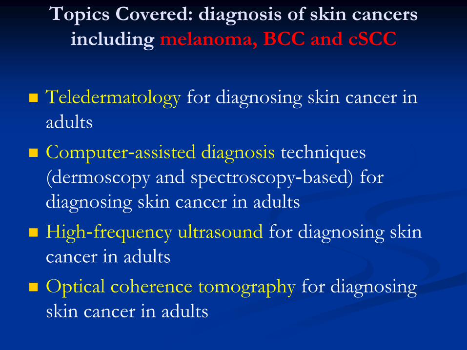
Topics Covered: diagnosis of skin cancers
including melanoma, BCC and cSCC
Teledermatology for diagnosing skin cancer in
adults
Computer‐assisted diagnosis techniques
(dermoscopy and spectroscopy‐based) for
diagnosing skin cancer in adults
High‐frequency ultrasound for diagnosing skin
cancer in adults
Optical coherence tomography for diagnosing
skin cancer in adults
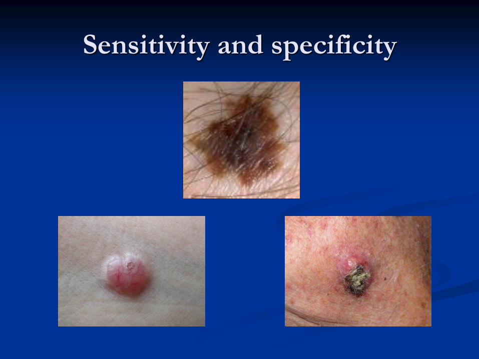
Sensitivity and specificity
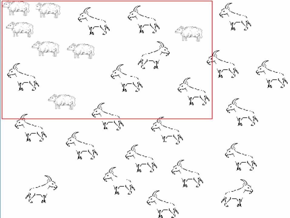
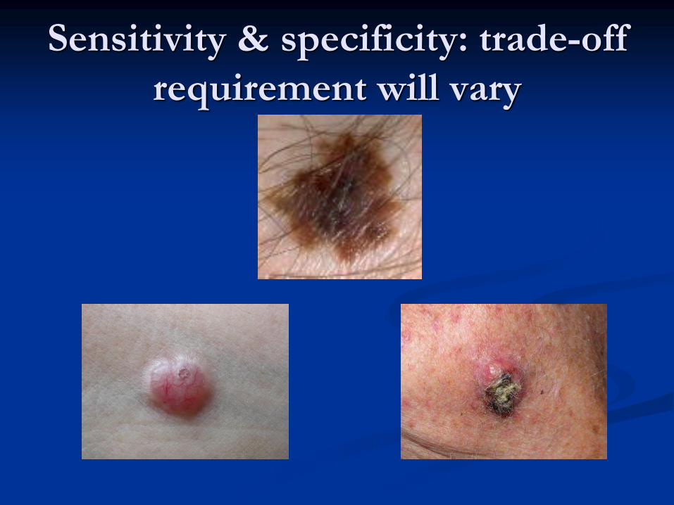
Sensitivity & specificity: trade-off
requirement will vary
Why did we do it?
Need – expanding use of technologies and
NICE guidelines
Opportunity for Cochrane Skin to expand
skills into DTA reviews with Birmingham
Patient benefit – promote well evidenced
practice and demote dodgy technologies
Money and prestige? – nearly killed us
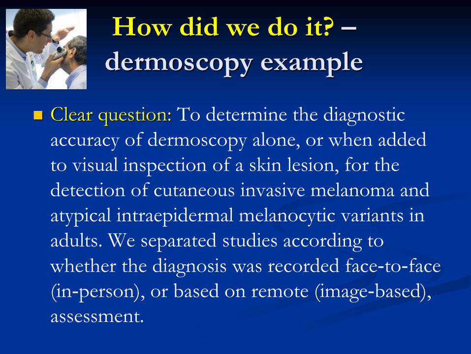
How did we do it? –
dermoscopy example
Clear question: To determine the diagnostic
accuracy of dermoscopy alone, or when added
to visual inspection of a skin lesion, for the
detection of cutaneous invasive melanoma and
atypical intraepidermal melanocytic variants in
adults. We separated studies according to
whether the diagnosis was recorded face‐to‐face
(in‐person), or based on remote (image‐based),
assessment.
Searches
Following databases from inception up to August
2016: CENTRAL; MEDLINE; Embase; CINAHL;
CPCI; Zetoc; Science Citation Index; US National
Institutes of Health Ongoing Trials Register; NIHR
Clinical Research Network Portfolio Database; and
the World Health Organization International
Clinical Trials Registry Platform.
Plus reference lists and published systematic review
articles.
Selection criteria
Studies of any design that evaluated dermoscopy
in adults with lesions suspicious for melanoma,
compared with a reference standard of either
histological confirmation or clinical follow‐up.
Data on the accuracy of visual inspection, to
allow comparisons of tests, was included only if
reported in the included studies of dermoscopy.

Data extraction
Two review authors independently extracted all
data using a standardised data extraction and
quality assessment form (based on QUADAS‐2).
We contacted authors of included studies where
information related to the target condition or
diagnostic threshold were missing. training.
Classical DTA biases for skin cancer
Incorporation bias – gold standard (histopath)
incorporates index test or index test known to
gold standard
Partial verification bias – those with +ve index
test more likely to get histopath and only those
who get histopath reported in study
Differential verification bias – only those with
+ve index test get immediate histology. Others
have clinical follow-up
Imperfect gold standard – histopath?
Data analysis
Estimated accuracy using hierarchical summary
receiver operating characteristic methods
(SROC)
Analysis of studies allowing direct comparison
between tests was done.
Computed values of sensitivity at the point on
the SROC curve with 80% fixed specificity and
values of specificity with 80% fixed sensitivity.
Investigated impact of in‐person test
interpretation using developed algorithms;
observer expertise; and dermoscopy training
Plan
What did we do?
Why did we do it?
How did we do it?
What did we find?
What does it all mean?
Lessons I learned?
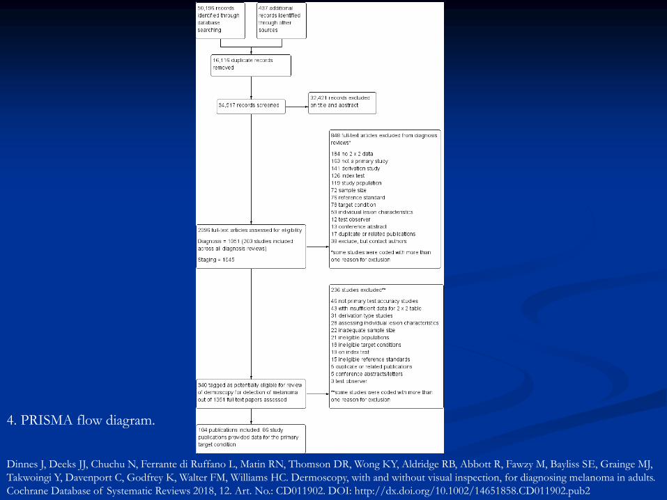
4. PRISMA flow diagram.
Dinnes J, Deeks JJ, Chuchu N, Ferrante di Ruffano L, Matin RN, Thomson DR, Wong KY, Aldridge RB, Abbott R, Fawzy M, Bayliss SE, Grainge MJ,
Takwoingi Y, Davenport C, Godfrey K, Walter FM, Williams HC. Dermoscopy, with and without visual inspection, for diagnosing melanoma in adults.
Cochrane Database of Systematic Reviews 2018, 12. Art. No.: CD011902. DOI: http://dx.doi.org/10.1002/14651858.CD011902.pub2
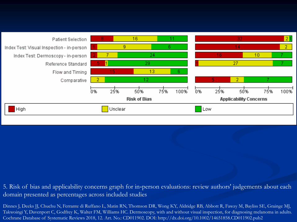
5. Risk of bias and applicability concerns graph for in‐person evaluations: review authors' judgements about each
domain presented as percentages across included studies
Dinnes J, Deeks JJ, Chuchu N, Ferrante di Ruffano L, Matin RN, Thomson DR, Wong KY, Aldridge RB, Abbott R, Fawzy M, Bayliss SE, Grainge MJ,
Takwoingi Y, Davenport C, Godfrey K, Walter FM, Williams HC. Dermoscopy, with and without visual inspection, for diagnosing melanoma in adults.
Cochrane Database of Systematic Reviews 2018, 12. Art. No.: CD011902. DOI: http://dx.doi.org/10.1002/14651858.CD011902.pub2
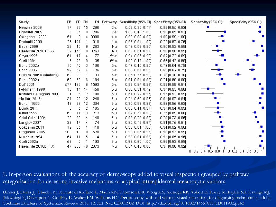
9. In‐person evaluations of the accuracy of dermoscopy added to visual inspection grouped by pathway
categorisation for detecting invasive melanoma or atypical intraepidermal melanocytic variants
Dinnes J, Deeks JJ, Chuchu N, Ferrante di Ruffano L, Matin RN, Thomson DR, Wong KY, Aldridge RB, Abbott R, Fawzy M, Bayliss SE, Grainge MJ,
Takwoingi Y, Davenport C, Godfrey K, Walter FM, Williams HC. Dermoscopy, with and without visual inspection, for diagnosing melanoma in adults.
Cochrane Database of Systematic Reviews 2018, 12. Art. No.: CD011902. DOI: http://dx.doi.org/10.1002/14651858.CD011902.pub2
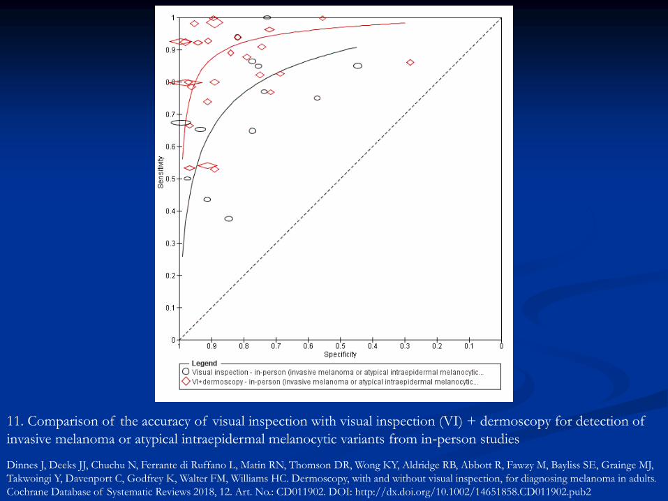
11. Comparison of the accuracy of visual inspection with visual inspection (VI) + dermoscopy for detection of
invasive melanoma or atypical intraepidermal melanocytic variants from in‐person studies
Dinnes J, Deeks JJ, Chuchu N, Ferrante di Ruffano L, Matin RN, Thomson DR, Wong KY, Aldridge RB, Abbott R, Fawzy M, Bayliss SE, Grainge MJ,
Takwoingi Y, Davenport C, Godfrey K, Walter FM, Williams HC. Dermoscopy, with and without visual inspection, for diagnosing melanoma in adults.
Cochrane Database of Systematic Reviews 2018, 12. Art. No.: CD011902. DOI: http://dx.doi.org/10.1002/14651858.CD011902.pub2
Bottom lines for some reviews
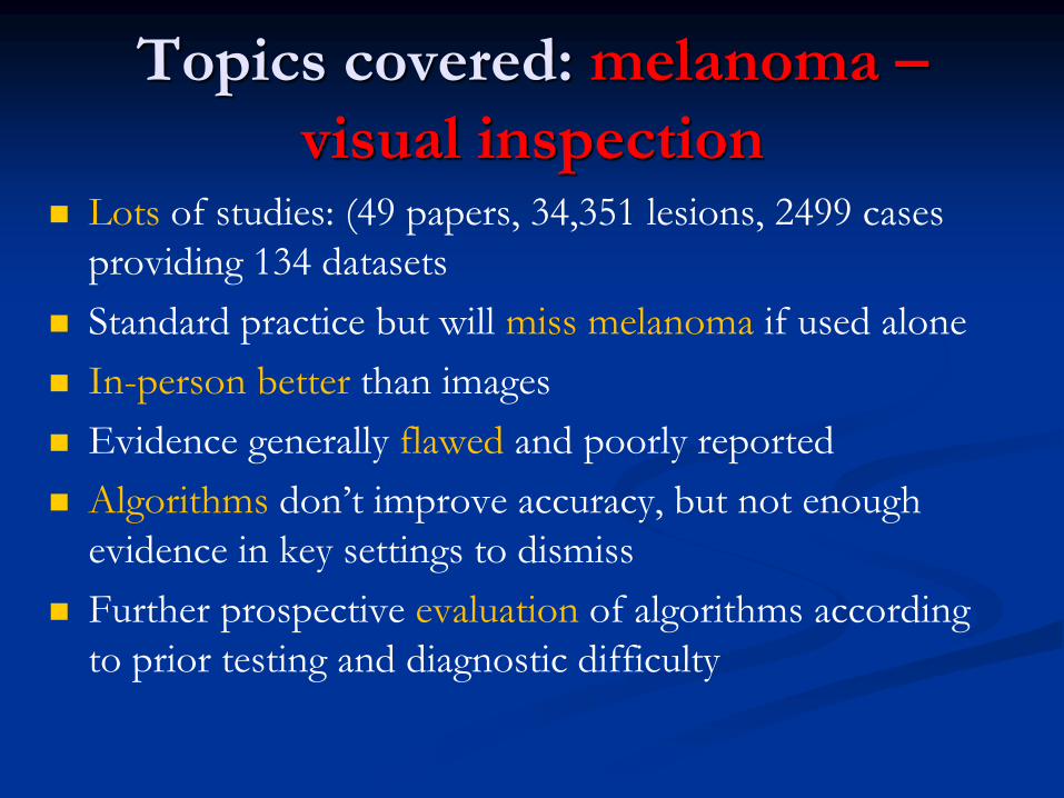
Topics covered: melanoma –
visual inspection Lots of studies: (49 papers, 34,351 lesions, 2499 cases
providing 134 datasets
Standard practice but will miss melanoma if used alone
In-person better than images
Evidence generally flawed and poorly reported
Algorithms don’t improve accuracy, but not enough
evidence in key settings to dismiss
Further prospective evaluation of algorithms according
to prior testing and diagnostic difficulty
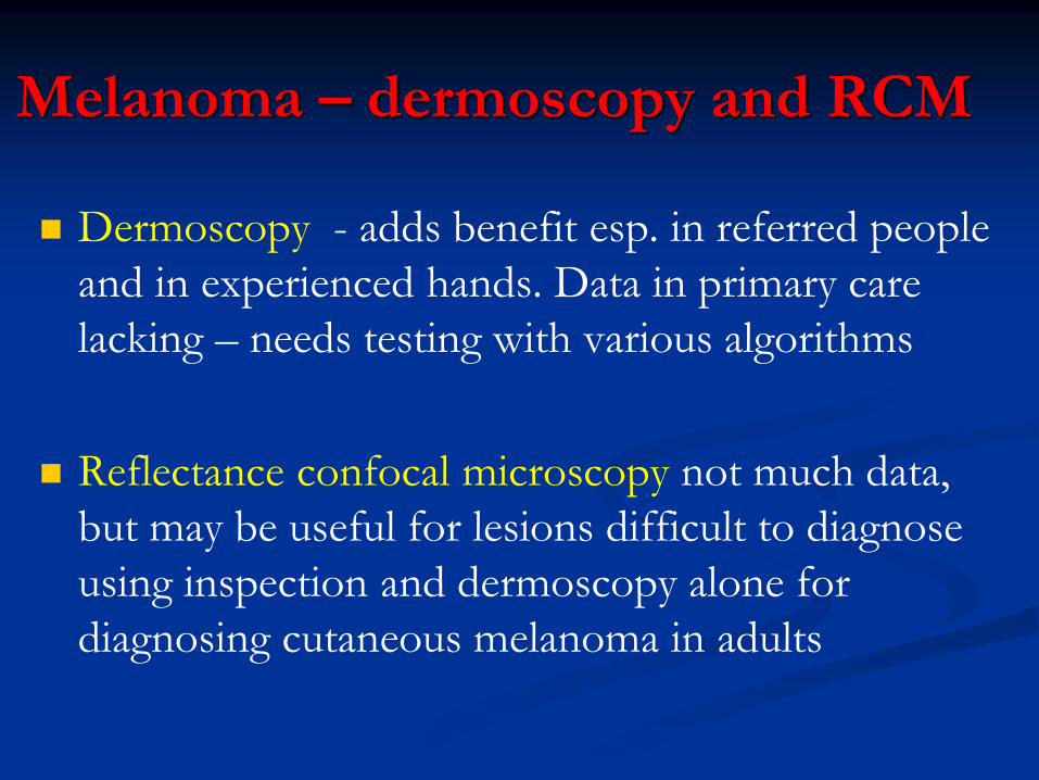
Melanoma – dermoscopy and RCM
Dermoscopy - adds benefit esp. in referred people
and in experienced hands. Data in primary care
lacking – needs testing with various algorithms
Reflectance confocal microscopy not much data,
but may be useful for lesions difficult to diagnose
using inspection and dermoscopy alone for
diagnosing cutaneous melanoma in adults
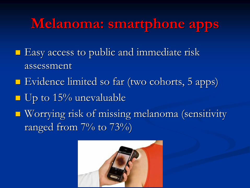
Melanoma: smartphone apps
Easy access to public and immediate risk
assessment
Evidence limited so far (two cohorts, 5 apps)
Up to 15% unevaluable
Worrying risk of missing melanoma (sensitivity
ranged from 7% to 73%)
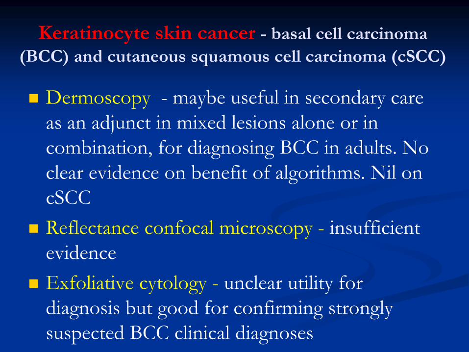
Keratinocyte skin cancer - basal cell carcinoma
(BCC) and cutaneous squamous cell carcinoma (cSCC)
Dermoscopy - maybe useful in secondary care
as an adjunct in mixed lesions alone or in
combination, for diagnosing BCC in adults. No
clear evidence on benefit of algorithms. Nil on
cSCC
Reflectance confocal microscopy - insufficient
evidence
Exfoliative cytology - unclear utility for
diagnosis but good for confirming strongly
suspected BCC clinical diagnoses
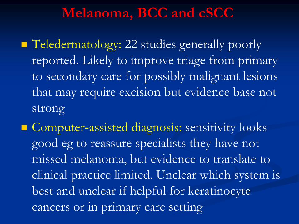
Melanoma, BCC and cSCC
Teledermatology: 22 studies generally poorly
reported. Likely to improve triage from primary
to secondary care for possibly malignant lesions
that may require excision but evidence base not
strong
Computer‐assisted diagnosis: sensitivity looks
good eg to reassure specialists they have not
missed melanoma, but evidence to translate to
clinical practice limited. Unclear which system is
best and unclear if helpful for keratinocyte
cancers or in primary care setting
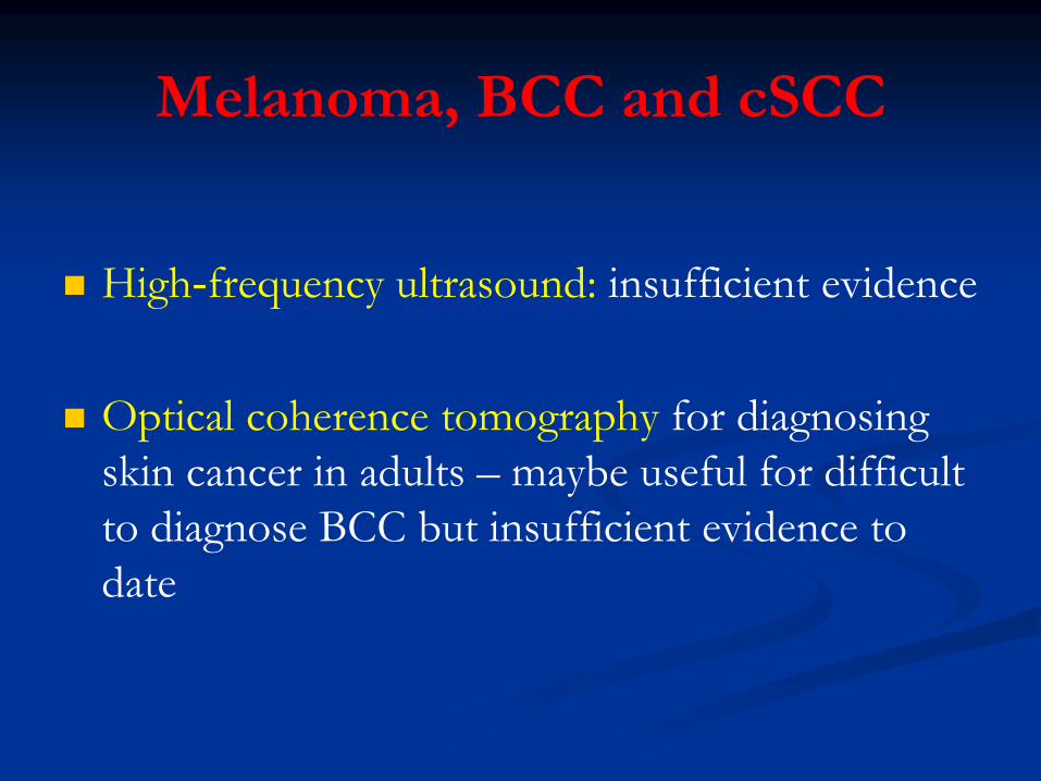
Melanoma, BCC and cSCC
High‐frequency ultrasound: insufficient evidence
Optical coherence tomography for diagnosing
skin cancer in adults – maybe useful for difficult
to diagnose BCC but insufficient evidence to
date
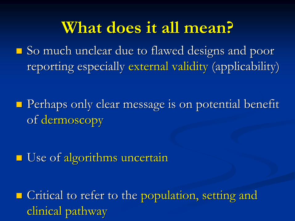
What does it all mean?
So much unclear due to flawed designs and poor
reporting especially external validity (applicability)
Perhaps only clear message is on potential benefit
of dermoscopy
Use of algorithms uncertain
Critical to refer to the population, setting and
clinical pathway
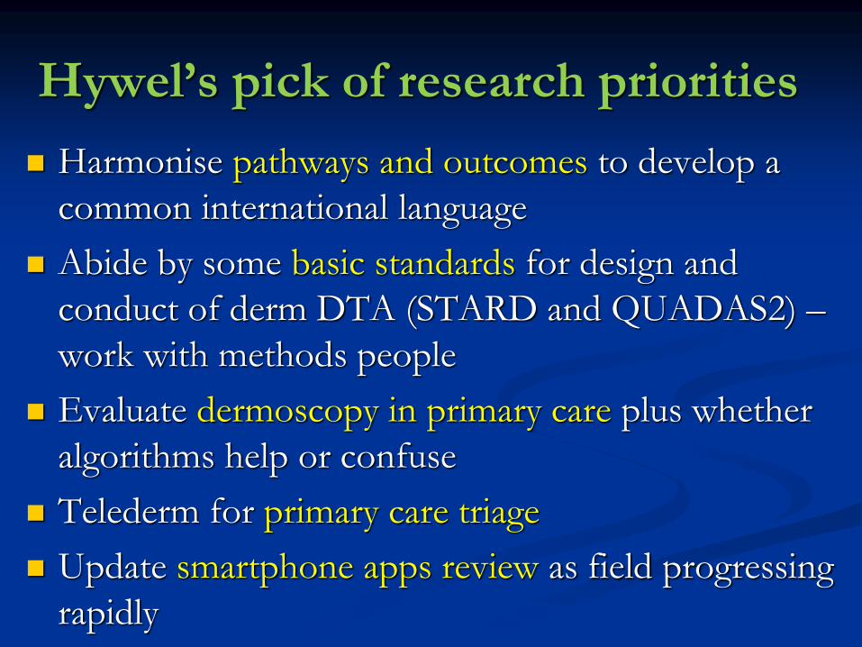
Hywel’s pick of research priorities
Harmonise pathways and outcomes to develop a
common international language
Abide by some basic standards for design and
conduct of derm DTA (STARD and QUADAS2) –
work with methods people
Evaluate dermoscopy in primary care plus whether
algorithms help or confuse
Telederm for primary care triage
Update smartphone apps review as field progressing
rapidly
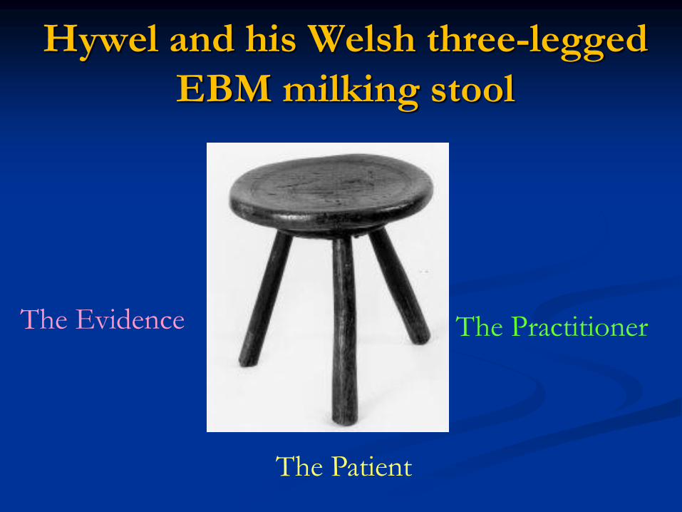
Hywel and his Welsh three-legged
EBM milking stool
The Evidence
The Patient
The Practitioner
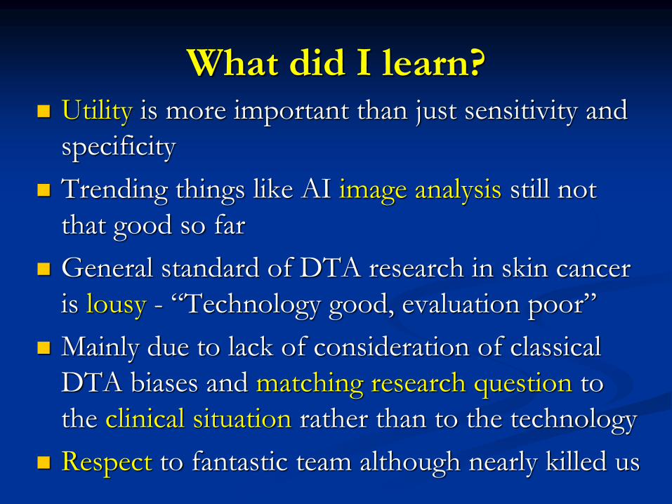
What did I learn? Utility is more important than just sensitivity and
specificity
Trending things like AI image analysis still not
that good so far
General standard of DTA research in skin cancer
is lousy - “Technology good, evaluation poor”
Mainly due to lack of consideration of classical
DTA biases and matching research question to
the clinical situation rather than to the technology
Respect to fantastic team although nearly killed us
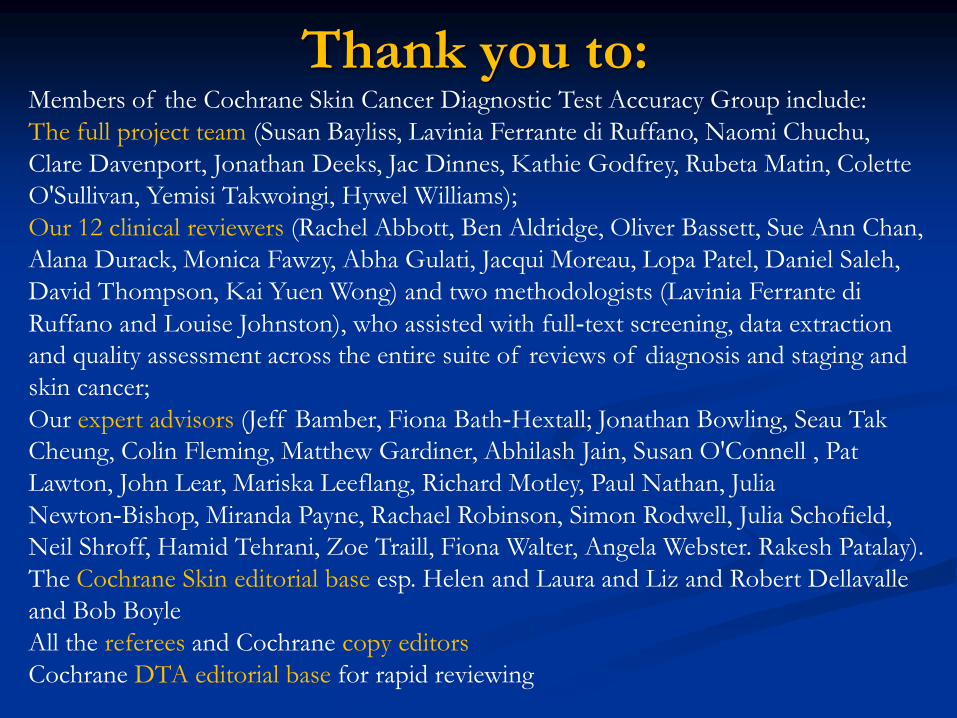
Members of the Cochrane Skin Cancer Diagnostic Test Accuracy Group include:
The full project team (Susan Bayliss, Lavinia Ferrante di Ruffano, Naomi Chuchu,
Clare Davenport, Jonathan Deeks, Jac Dinnes, Kathie Godfrey, Rubeta Matin, Colette
O'Sullivan, Yemisi Takwoingi, Hywel Williams);
Our 12 clinical reviewers (Rachel Abbott, Ben Aldridge, Oliver Bassett, Sue Ann Chan,
Alana Durack, Monica Fawzy, Abha Gulati, Jacqui Moreau, Lopa Patel, Daniel Saleh,
David Thompson, Kai Yuen Wong) and two methodologists (Lavinia Ferrante di
Ruffano and Louise Johnston), who assisted with full‐text screening, data extraction
and quality assessment across the entire suite of reviews of diagnosis and staging and
skin cancer;
Our expert advisors (Jeff Bamber, Fiona Bath‐Hextall; Jonathan Bowling, Seau Tak
Cheung, Colin Fleming, Matthew Gardiner, Abhilash Jain, Susan O'Connell , Pat
Lawton, John Lear, Mariska Leeflang, Richard Motley, Paul Nathan, Julia
Newton‐Bishop, Miranda Payne, Rachael Robinson, Simon Rodwell, Julia Schofield,
Neil Shroff, Hamid Tehrani, Zoe Traill, Fiona Walter, Angela Webster. Rakesh Patalay).
The Cochrane Skin editorial base esp. Helen and Laura and Liz and Robert Dellavalle
and Bob Boyle
All the referees and Cochrane copy editors
Cochrane DTA editorial base for rapid reviewing
Thank you to: