Diagnostic Radiography: A Survey of the Scope of ... · 1 Diagnostic Radiography: A Survey of the...
48
1 Diagnostic Radiography: A Survey of the Scope of Radiographic Practice 2015 Executive Summary The Scope of Practice Survey 2015 was conducted by the Society and College of Radiographers to garner information in order to update the Scope of Radiographic Practice Survey 2012 Report. 1 The aim was to identify any practice developments over the past four years and to quantify the scope of current practice. In order to gain a more comprehensive insight into the broader spectrum of clinical imaging, some additional survey questions were included. Seventy-five diagnostic imaging department service managers across the UK responded to an online questionnaire (which equated to 71 providers). The results indicate that the scope of practice for the diagnostic radiographic workforce continues to develop. • The scope of practice for diagnostic radiographers continues to be broad, diverse and expanding. • Assistant practitioners are utilised by just over half of all service providers. • The scope of practice across the assistant practitioner workforce is diverse and mostly consistent with the SCoR Scope of Practice for assistant practitioners. • An increasing number of services have radiographer-led examinations, interventional procedures and gastro-intestinal studies. • The majority of responding service providers utilise the skills of appropriately trained radiographers to issue formal written reports and an increasing number of providers indicate they have a ‘radiographer-led hot report service’. • The appendicular skeleton is the most common area for radiographer reporting after ultrasound. • There is evidence of radiographer reporting activity across many patient pathways, including US, CT, MRI and radionuclide imaging studies. There has been an increase in the proportion of departments with research radiographers since 2012, alongside a smaller increase in the proportion of departments with radiographers with a substantive role in clinical education over the same period. Of note, responses were not received from all clinical imaging service providers across the UK and therefore it is not possible to state if the 2015 responders are the same as those in 2012. Direct comparison and changes in trend are therefore difficult to make.
-
Upload
hoangnguyet -
Category
Documents
-
view
229 -
download
1
Transcript of Diagnostic Radiography: A Survey of the Scope of ... · 1 Diagnostic Radiography: A Survey of the...

1
Diagnostic Radiography: A Survey of the Scope of Radiographic Practice 2015
Executive Summary The Scope of Practice Survey 2015 was conducted by the Society and College of Radiographers to garner information in order to update the Scope of Radiographic Practice Survey 2012 Report.1 The aim was to identify any practice developments over the past four years and to quantify the scope of current practice. In order to gain a more comprehensive insight into the broader spectrum of clinical imaging, some additional survey questions were included. Seventy-five diagnostic imaging department service managers across the UK responded to an online questionnaire (which equated to 71 providers). The results indicate that the scope of practice for the diagnostic radiographic workforce continues to develop.
• The scope of practice for diagnostic radiographers continues to be broad, diverse and expanding.
• Assistant practitioners are utilised by just over half of all service providers.
• The scope of practice across the assistant practitioner workforce is diverse and mostly consistent with the SCoR Scope of Practice for assistant practitioners.
• An increasing number of services have radiographer-led examinations, interventional procedures and gastro-intestinal studies.
• The majority of responding service providers utilise the skills of appropriately trained radiographers to issue formal written reports and an increasing number of providers indicate they have a ‘radiographer-led hot report service’.
• The appendicular skeleton is the most common area for radiographer reporting after ultrasound.
• There is evidence of radiographer reporting activity across many patient pathways, including US, CT, MRI and radionuclide imaging studies.
There has been an increase in the proportion of departments with research radiographers since 2012, alongside a smaller increase in the proportion of departments with radiographers with a substantive role in clinical education over the same period. Of note, responses were not received from all clinical imaging service providers across the UK and therefore it is not possible to state if the 2015 responders are the same as those in 2012. Direct comparison and changes in trend are therefore difficult to make.

2
1. Background 1.1 Introduction The Scope of Practice Survey was carried out by the Society and College of Radiographers (SCoR) in the last quarter of 2015. The survey investigated diagnostic imaging practice in the National Health Service (NHS) and independent/private sectors across the United Kingdom (UK). 1.2 Background A new scope of practice for the clinical imaging and radiotherapeutic workforce was issued in 20132 (using 2012 survey results) and was used to better define professional body expectations at each level of the career framework. The report concluded that the scope of practice for UK radiographers was broad and continuing to expand and highlighted the importance of further implementation of the Career Progression Framework.3 It also highlighted the need for radiographer-led clinical research to improve patient outcomes, thereby strengthening the professional body of knowledge. 1.3 Aim of the survey The aim of the 2015 survey was to identify any practice developments over the past four years and to quantify the current scope of practice. The objectives were to:
• quantify the different roles undertaken by the radiography workforce within clinical practice
• identify role developments which have occurred within the profession over the past four years (since the publication of the Scope of Practice 2012 Report1
• provide information to education providers to support changes to curricula at pre and post registration levels of practice.

3
2. Exploring the diagnostic radiography workforce 2.1 Questionnaire An online questionnaire using Survey Monkey® was used to explore information on roles and developments in the workforce (Appendix 1). Information was sought on the different roles undertaken within clinical practice. 2.2 Participants In November 2015, email invitations containing a link to the online questionnaire were sent to 1176 diagnostic imaging managers, superintendent radiographers and consultant radiographers throughout the UK (using information sourced by the SCoR membership database). The survey was targeted at the total population. Senior service managers were asked to complete the survey for a ‘whole department’ or service provider to capture the scope of practice across a whole service. Responses were received from 71 discrete providers across the four countries of the UK. Duplicate responses from the same provider were combined for an overall single organisational response. 2.3 The current context As of 2016, NHS England identifies 163 NHS providers and 13 independent providers of clinical imaging services in their Diagnostic Imaging Department data sets for England. 4 Each individual provider covers more than one hospital. In Scotland, 14 territorial health boards are identified (each covers several hospitals) delivering patient services (including radiography). There are seven special health boards – two of which also deliver patient services in radiography. In terms of the independent sector in Scotland, 20 independent hospitals are listed by Healthcare Improvement Scotland,5 although it is not identifiable which ones provide imaging services.
In Wales there are seven Health Boards and three Trusts (only two trusts have a radiology service) and a small number of independent providers. Northern Ireland has five Health and Social Care trusts providing health and social care services across a number of hospitals.6 There are a small number of independent providers.

4
3. Results 3.1 Demographic data Q1-Q4 The region and type of hospital for those responding are shown in Table 1. Fifty-one respondents were from the NHS, 18 from the independent sector, and two ‘other’.
Table 1: UK Country and NHS/Non-NHS (n=71)
Country NHS Private/
independent Other
(University) Total
England 40 15 2 57
Northern Ireland 2 1 0 3 Scotland 4 1 0 5
Wales 5 0 0 5
UK-wide 1* 0 1 Grand Total 51 18 2 71
*One company with sites across all four countries Sixty eight respondents stated they were a radiographer by professional background (Table 2).
Table 2: Respondent is a radiographer by background (n=71)
Yes No
Number response
Total
Radiographer by background
68 3 0 71
In most cases the job titles indicated that a senior service manager did complete the questionnaire. Of the NHS providers, two of the 51 service leaders were not radiographers; of the private/independent/university providers this was one out of 20. 3.2 Career progression framework roles Q5-Q7 Respondents were asked for the number of staff in each area of the Career Progression Framework: 3 assistant practitioners; practitioners; advanced practitioners; and/or consultants. Figure 1 illustrates the total headcount per responding provider across the four sections of the workforce.

5
Figure 1:
Career progression framework roles (n=71)
Figure 2:
Career progression framework roles (2012 n=64, 2015 n=71)
Figure 2 compares the results from the 2012 and 2015 surveys (to note: the 2012 survey reported this information by department rather than by provider). The results indicate that since the 2012 survey, the numbers of providers with assistant practitioners has declined, the providers with practitioners appear to have increased, and advanced and consultant roles appear to remain static. Not all respondents answered this question. Comment The Career Progression Framework3 for radiographers described roles for assistant practitioner, practitioner, advanced practitioner and consultant practitioner. A range of educational requirements and professional outcomes are noted as relevant for each role.
248
2744
835
27
0 500 1000 1500 2000 2500 3000
Assistantpractitioners
Radiographicpractitioners
Advancedradiographicpractitioners
Consultantradiographicpractitioners
Totalheadcountofrespondingproviders
5147 49
15
40
55
47
15
0
10
20
30
40
50
60
Assistantpractitioners
Radiographicpractitioners
Advancedradiographicpractitioners
Consultantradiographicpractitioners
Numberofrespondingprovidersw
ith
role
2012 2015
6
Consultant radiographic practitioners The role of a consultant radiographer demands the ability to innovate, motivate and influence local and national agendas. Typically, a consultant radiographer will carry their own caseload, working alongside medical colleagues in the top tier of the multidisciplinary team. The consultant radiographer should be able to develop and share these traits; to evolve best practice, develop strategies, promote innovations and overcome barriers through discussion and shared knowledge. The four elements of the consultant role are:
• Expert clinical practice
• Professional leadership and consultancy • Education training and development and practice and service development • Research and evaluation.
Respondents were also asked to identify the specialties in which consultant radiographic practitioners worked, and to specify whether this was for adult, paediatric, or both. Out of the 17 responses to this question, five indicated they had consultant radiographers working in more than one area of practice. Consultants were dedicated to their specialty. Seven had consultant radiographers in breast services; five in ultrasound (both paediatric and adult); four in general radiographic imaging; one in education; one in DXA (formerly referred to as Dexa) scanning; one due to commence in CT facial and head imaging; one in GI services; and one in interventional procedures. Advanced radiographic practitioners Advanced practitioners should have a dedicated area of expert practice and will deliver elements of leadership, education and research as an integral part of their role. Respondents were asked to identify the specialties in which advanced radiographic practitioners worked, and to specify whether this was for adult, paediatric, or both. Forty-three providers responded; details can be seen in Figure 3.

7
Additional areas identified for radiographer advanced practice were in breast imaging (n=8), and single responses were recorded for lithotripsy, cardiac CT and MRI, nuclear medicine, maxillary facial, dental, CT Colonography and DXA.
Figure 3:
Advanced radiographic practitioner roles (n=43) Comment The survey indicates there is a breadth of practice for both advanced and consultant practitioners. The ‘Paediatrics’ response is confusing as four respondents indicated that they had advanced practitioners in adult paediatric services. It is only possible to say that Paediatrics is an area of radiographer advanced practice. Advanced and consultant radiographic practitioners are eligible to accredit their practice against CoR standards in the accreditation scheme. This scheme provides the quality assurance that these radiographers are working to the standards set by both the Department of Health7 and College of Radiographers. 3 More than half of the respondents provided no information regarding advanced or consultant practice.
3.3 Non-medical clinical imaging workforce (Whole time equivalent) Q8 Sixty-six respondents provided the numbers (in whole time equivalent) of the non-medical clinical imaging workforce (Figure 4).
30
4
12
16
16
30
27
19
11
3
1
2
2
11
4
2
0 10 20 30 40
Ultrasound
Paediatrics
Nuclearmedicine
MRI
Interventionalprocedures
Generalradiography
Fluroscopy
CT
Numberofproviders
Paediatric Adult

8
Figure 4:
Non-medical clinical imaging workforce (WTE) (n=66) Bands 5, 6 and 7 represented the most common pay bands with those at band 8a and above demonstrating much smaller numbers of job holders. Over 3000 members of the clinical imaging non-medical workforce are represented by the survey. Based on Society of Radiographers database figures and HCPC registration of radiographers, it is estimated this is between 10% and 15% of the current workforce.
3.4 Forensic radiographic roles Q10-Q12 Forensic radiography is a small yet important part of post registration development for radiographers, which continues to develop. The International Association of Forensic Radiographers (IAFR) provided questions for inclusion in the survey. For the purposes of the survey, examples of forensic radiography are as follows:
• Investigation of non-fatal injuries, eg Non-accidental injury (NAI), assault, industrial disease
• Location of other forensic evidence, eg Narcotic detection, ballistic material
• Cause of death, eg Decomposed remains, sudden unexpected death in infants (SUDI).
Respondents were asked to indicate if they had a named radiography lead in the different modalities (not specifically for forensics) and also to indicate if there was a specific named forensic lead within the modalities. Results, shown in Figure 5, tend to indicate that few have a named forensic lead.
362
334
189
762
1575
1021
205
54
17
0
0
21
0 200 400 600 800 1000 1200 1400 1600 1800
Band2
Band3
Band4
Band5
Band6
Band7
Band8a
Band8b
Band8c
Band8d
Band9
Non-AfCbands
Totalwholetimeequivalent(WTE)ofrespondingproviders

9
Figure 5:
Frequency of Named Lead Radiographer (n=63)
Respondents were asked to indicate the nature of any further training and education
undertaken by those radiographers who deal with forensic radiography.
The majority of training was ‘on the job’ or ‘in house’, as per Figure 7.
Other comments included:
• Attended local university study days
• Lead supported by staff with formal qualification
• Attended SoR forensic courses
• Completed some elearning modules
• Lead working toward PhD
• Forensic radiography performed elsewhere in trust
Figure 6:
Training for forensic radiography (n=46)
5
6
6
11
2
10
1
19
10
25
21
46
14
44
0 10 20 30 40 50
Trauma
Post-mortemimaging
Paediatrics
Noneofthefollowing
MRI
Forensicradiography
CT
Numberofproviders
NamedLead NamedLeadinvolvedinForensics
7
19
13
5
6
17
16
7
0 5 10 15 20
Notraining
Onthejobtrainingonly
In-housetrainingprovided
Formalqualifications
Numberofproviders
Leadradiographers Otherradiographers

10
Comment In May 2014, the SCoR with the Association of Forensic Radiographers (AFR) produced guidance on the role of the radiographer in forensic services. 8 This document describes the scope of practice for forensic work and defines the standard to be achieved for education and training. It recommends that radiographers achieve post graduate level education for forensic service provision. The reponses to the survey indicate that this level of education is rare with some radiographers having had no additional training for this area of practice. 3.5 Clinical imaging staff roles Q13 Question 13 examined the broader delivery of clinical imaging services to investigate where services were not being delivered by registered radiographers. Figure 7 illustrates the number of providers and range of services and Figure 8 by breakdown of staff type.

11
Figure 7:
Number of providers supplying specific services using clinical imaging staff (n=70)
36
37
46
51
52
39
25
3
15
48
17
25
31
38
15
29
52
5
37
31
44
24
27
22
30
42
26
41
18
0 20 40 60
Ultrasound- vascular
Ultrasound- obstetric
Ultrasound- non-obstetric
Theatrei.i's
Radiology/Generalx-ray
Paediatric
NuclearMedicine
NHSfacilitystaffedbynon-NHSemployees
Neuroradiology
MRI
MobileCT/MRunits
Maternity
Interventionalradiologyvascular
Interventionalradiologynon-vascular
HybridscanningPETCT/PETMR
Forensicservices
Fluoroscopy
FacilityprovidedbyexternalproviderstaffedwithNHS…
Dental
CTcolonographyscreeningprogram
CT
Communityhospital/department
CatheterLab
Cardiology
BreastScreening
BreastImaging
BoneDensitometry
AccidentandEmergency
AAAscreening
Numberofproviders

12
Comments:
• Extra Corporeal Shock Wave Lithotripsy (ESWL) procedure utilising image intensifier (II) and ultrasound (US) localisation and monitoring performed by specialist radiographers
• DXA scanning and Venous Doppler services are provided by separate departments employing non-radiographic imaging staff
• Mobile unit/MRI - CMRI scans only Cardiology - dual qualified radiographers for cardiac radiography and Echocardiography
• Fluoroscopy/theatres - cardiac cases in CCU pacing room and cardiothoracic theatres
• Ultrasonographers , midwives ,physicists and vascular technicians
Figure 8:
Percentage of providers offering services using different healthcare professionals Comment A number of providers indicated that for each service listed, it was solely radiographers delivering that service. Eleven providers had no professional staff other than radiographers
0% 20% 40% 60% 80% 100%
Ultrasound- vascular
Ultrasound- obstetric
Ultrasound- non-obstetric
Theatrei.i's
Radiology/GeneralX-ray
Paediatric
NuclearMedicine
NHSfacilitystaffedbynon-NHSemployees
Neuroradiology
MRI
MobileCT/MRunits
Maternity
Interventionalradiologyvascular
Interventionalradiologynon-vascular
HybridscanningPETCT/PETMR
Forensicservices
Fluoroscopy
Facilityprovidedbyexternalproviderstaffedwith…
Dental
CTcolonographyscreeningprogram
CT
Communityhospital/department
Catheterlab
Cardiology
BreastScreening
BreastImaging
BoneDensitometry
AccidentandEmergency
AAAscreening
Percentageofprovidersofferingservice
APs Radiographers NuclearMedicineTechnicians Other(egnurses)

13
involved in their service provision, for the rest there was a mixed workforce as shown in Figure 9. Intravenous injection/cannulation was the service most often provided by non-radiographic staff. Traditional areas where radiographers had been providing the service, such as PACS, MRI and CT are now being delivered by other staff groups. There is wide variation in the mix of professionals delivering services such as DXA scanning and ultrasound. Nuclear medicine services demonstrated an almost even division between radiographer-led and nuclear medicine (NM) technologist-led delivery. Further examination of the data indicated that, of the 25 responding providers supplying nuclear medicine services, nine employed radiographers but not NM technologists, eight employed NM technologists but not radiographers, seven employed both, and one used neither. Many service providers indicated they had assistant practitioners supporting service delivery across a range of service areas. The responses to Q13 regarding clinical imaging staff roles, clearly indicated that assistant practitioners and radiographers are the majority workforce for these service areas. Most assistant practitioners work in areas defined in the SCoR Scope of practice for assistant practitioners9 although there were also assistant practitioners identified in paediatrics, theatre/image intensifier and ultrasound practice, which fall outside the defined work areas for this staff group. 3.6 Support workforce (x-ray helpers) Q14-Q16 Forty-nine (69%) respondents stated they had a support workforce, specifically x-ray helpers (radiographic assistants) (Table 3). (NB this question was not including the numbers of assistant practitioners.)
Table 3: Number of providers with x-ray helpers (n=71)
Role Number of providers with role
%
Yes 49 69%
No 22 31%
The tasks performed by the support workforce and their bands are illustrated in Figure 10.

14
Other areas included:
• Image Exchange Portal (IEP) Billing; Infection Prevention and Control Link
• Booking appointments
• Ordering/replenishing non-pay stock items
• Assist with biopsies (two respondents)
• Nursing assistants undertake these roles with the exception of image manipulation and post processing, and with the addition of cannulation
Figure 9: Tasks performed by x-ray helpers (n=48)
Comment The support workforce provides vital elements of care and is an essential part of many radiographic teams. This staff group have varied job titles: radiographic assistant, x-ray helper, imaging department assistant, x-ray assistant, and x-ray support worker. Their role is to support the team, as per service need. The Agenda for Change Job profiles for radiography10 describe the first tier of worker in the team as the Clinical Support Worker (higher level) at band 3. The defined core role is:
1) to undertake a range of delegated radiographic duties 2) to carry out reception and clerical duties.
7
11
18
32
35
15
34
35
4
34
32
15
27
9
31
2
8
24
29
32
10
33
31
2
25
15
0
6
6
33
0 10 20 30 40
Qualityassurance
Postprocessing
Portering/Patienttransport
Personalpatientactivities(eg.Toileting)
Patientpreparation
Patientpositioning
Meetandgreetpatients
Maintainingacleanenvironment
Imagemanipulation
Equipmentpreparation
Contrastagentpreparation(oral)
Contrastagentpreparation(intravenous)
Canulation
Audit
Administrationofpatientdata
Numberofproviderswithtaskperformedatthatlevel
Band2 Band3

15
The survey identified that roles are at both band 2 and band 3 with both performing a range of clinically delegated tasks alongside administrative tasks. The only task not indicated as performed from the predefined list at band 2 was intravenous contrast agent preparation. 3.7 Assistant practitioners Q17-Q22 Thirty-eight providers (54%) stated they had assistant practitioners in their service (Table 4).
Table 4: Number of providers assistant practitioners (n=70)
Role Number of providers with role
%
Yes 38 54%
No 32 46% Twenty-six (68%) of these assistant practitioners had achieved College of Radiographers accreditation (Table 5).
Table 5: Number of providers with accredited assistant practitioners (n=38)
Role Number of providers with role
%
Yes 26 68% No 12 32%
Those who responded “No” commented:
• Unsure if course is known to CoR or accredited
• Work to scope of practice and job description approved by Trust (3 respondents)
• It is not a requirement
• Some have, some have not (2 respondents) • Unknown
• Cost implications
• Have foundation degree at University
• Lapsed
• Trust accreditation for the role
Thirty-six (95%) providers stated the assistant practitioners were working within the SCoR assistant practitioner scope of practice (Table 6).
Table 6: Number of providers where assistant practitioners work within SCoR scope of practice (n=38)
Role Number of providers with role
%
Yes 36 95%
No 2 5%
Those who responded “No” commented:
• The department has its own competence based scope of practice signed off under governance and audit arrangements.

16
The bands in which assistant practitioners were employed varied, although most were band 4.
The majority of assistant practitioners were working at band 4 (Table 7).
Table 7: Number of providers with assistant practitioners (by AfC band) (n=38)
Role Number of providers with role
Band 3 1
Band 4 38 Band 5 (NB: SCoR do not support employment of assistant practitioners at Band 5)
2
The main areas in which assistant practitioners worked were diverse, but the majority were working in breast imaging, dental imaging or general radiography (Table 8). Table 8: Number of providers with assistant practitioners working in specific areas (n=38)
Role Number of providers with role
Breast imaging 20 Dental imaging 10
General radiography 30
Fluoroscopy 6
CT 4
MR 5 Ultrasound 5
Nuclear Medicine 3 Other 5
Other included:
• DXA
• Catheter laboratory; cardiac rhythm management procedures EP/ablation/device implants
• Mobile chest radiography
Comment The assistant practitioner role was introduced to the radiography workforce as a result of a study into skill mix in radiography conducted by the Department of Health in 2002.11 The diversity of radiographic practice, coupled with increasing service demand and the introduction of new technologies and techniques, allowed for the potential to develop new roles. Within the Career Progression Framework,3 new roles have emerged that support service delivery by developing individuals to undertake specific tasks and activities that improve patient flow and delivery of effective and timely services. There was no suggestion that these new roles would replace radiographers, but that they would provide additional capacity in the workforce to allow radiographers to develop. Assistant practitioner roles also

17
fulfilled the need to develop career progression opportunities for the support workforce in clinical imaging and radiotherapy services.12 The survey highlighted that nearly half (46%) of all providers did not utilise this element of the workforce. Where assistant practitioners are in post they appear to work in diverse areas although the majority work in general radiography and breast imaging. The SCoR scope of practice9 defines the expected work of the assistant practitioner across the modality areas and indicates that professional supervision is required. The scope of practice is aligned to band 4 by NHS Employers Job Evaluation scheme by defining the knowledge, training and experience required alongside a measure of their freedom to act. These elements define the pay band for an assistant practitioner.13 The Career Progression Framework 3 indicates that professional radiographic staff should be honours degree qualified and pay band 5 is the starting point of the registered radiographer professional career framework. Two providers in the survey stated they had assistant practitioners at band 5. This raises some queries around their scope of practice and accountability to a registered professional. 3.8 Radiographer roles Q23,Q25 Figures 10 and 11 illustrate the range of work areas and Agenda for Change (AfC) pay banding (or equivalent) of radiographers.

18
Figure 10: Number of providers supplying specific services using radiographers (n = 58)
10
16
38
32
34
6
27
41
33
51
15
11
7
39
45
27
20
51
19
4
11
9
10
42
38
43
19
27
6
52
23
14
0 10 20 30 40 50 60
Supplementaryprescribing(ofdrugs)
Sialograms
Riskmanagementsupport
RISsupport
Research
Radiographersaspartofanyadvancedtraumalife…
Radiographer-ledGIstudies
Radiographerreporting
Radiographerreddot
Qualityassurance
Preliminaryclinicalevaluation(formerlyreddot)
Pharmacologicalstressinginradionuclideimaging
Peripherallyinsertedcentralcatheters(PICCs)
PACSsupport
MRIexaminations
ITsupport
Intravenousurograms(IVUs)
Intravenousinjection/cannulation
Hysterosalpingography
Hycosy(hysterosalpingo-contrast-sonography)
Exercisestressinginradionuclideimaging
Endoscopicgastro-intestinalprocedures
Dacro-cystograms
CTexaminations
Computedtomographycolonography(CTC)imaging
Clinicalgovernancesupport
CardiacMRI
CardiacCT
Cardiacand/orphysiologicalmeasurements(ECG,ec)
Audit
Anyformofimageguidedintervention(eg.biopsy,…
Angiographicprocedures
Numberofproviders
19
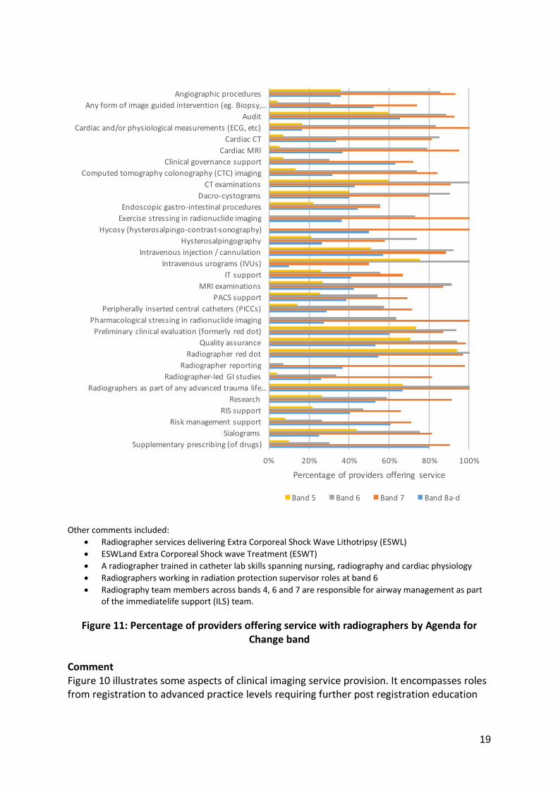
Other comments included:
• Radiographer services delivering Extra Corporeal Shock Wave Lithotripsy (ESWL)
• ESWLand Extra Corporeal Shock wave Treatment (ESWT)
• A radiographer trained in catheter lab skills spanning nursing, radiography and cardiac physiology
• Radiographers working in radiation protection supervisor roles at band 6
• Radiography team members across bands 4, 6 and 7 are responsible for airway management as part of the immediatelife support (ILS) team.
Figure 11: Percentage of providers offering service with radiographers by Agenda for Change band
Comment Figure 10 illustrates some aspects of clinical imaging service provision. It encompasses roles from registration to advanced practice levels requiring further post registration education
0% 20% 40% 60% 80% 100%
Supplementaryprescribing(ofdrugs)
Sialograms
Riskmanagementsupport
RISsupport
Research
Radiographersaspartofanyadvancedtraumalife…
Radiographer-ledGIstudies
Radiographerreporting
Radiographerreddot
Qualityassurance
Preliminaryclinicalevaluation(formerlyreddot)
Pharmacologicalstressinginradionuclideimaging
Peripherallyinsertedcentralcatheters(PICCs)
PACSsupport
MRIexaminations
ITsupport
Intravenousurograms(IVUs)
Intravenousinjection/cannulation
Hysterosalpingography
Hycosy(hysterosalpingo-contrast-sonography)
Exercisestressinginradionuclideimaging
Endoscopicgastro-intestinalprocedures
Dacro-cystograms
CTexaminations
Computedtomographycolonography(CTC)imaging
Clinicalgovernancesupport
CardiacMRI
CardiacCT
Cardiacand/orphysiologicalmeasurements(ECG,etc)
Audit
Anyformofimageguidedintervention(eg.Biopsy,…
Angiographicprocedures
Percentageofprovidersofferingservice
Band5 Band6 Band7 Band8a-d

20
and training. It provides an indication of the breadth of the scope of practice for diagnostic radiographers across the Career Progression Framework.3
Many roles, such as radiographer reporting or supplementary prescribing, require a formal post graduate qualification and would be expected to be linked to the higher Agenda for Change bands. It can be seen from Figure 11 that there was involvement of radiographers across the pay bands in most work areas, although developed skills appear to be mostly at the higher pay bands.
The radiographer practitioner at band 5 is involved in a wide range of services. There is evidence of progression within the banding structure across all service areas. It is of note that cardiac stressing and Hysterosalpingo-contrast-sonography (HyCoSy) are exclusively delivered at the higher bandings.
The results indicate that audit, general CT examinations, IV cannulation, radiographer red dot and preliminary clinical evaluation are being carried out across all radiographer levels.
The SCoR is promoting a move from terminology such as ‘red dot’ to ‘preliminary clinical evaluation ,14 however as the service still refers to ‘red dot’ it was felt important to keep this category within the question. Clinical evaluation schemes have developed from the ‘red dot’ system where an abnormality was signified by the application of a red dot to the image; however no form of interpretation was supplied. It was envisaged by the CoR in 200614 that red dot schemes would be replaced by preliminary clinical evaluation. Radiographer red dot continues to be provided across the bands in 33 responding providers. Fifteen service providers indicated that they had moved to initial image interpretation by radiographers. Six (10%) providers indicated radiographers formed part of advanced trauma life support teams (Table 9).
Table 9: Advanced trauma life support team roles frequencies (n=58)
Role Number of providers with role
% 2015 survey
% 2012 survey
% 2008 survey
Radiographers form part of advanced trauma life support team
6 10% 8% N/A
Comment Radiographers have roles across diverse work areas and their involvement in advanced trauma life support is an indication of the expansion and scope of practice beyond pure clinical imaging examinations.

21
Although it is not possible to directly compare the results with the 2012 survey, as the respondents may represent a different range of services, it is clear radiographers have a role to play in supporting advanced trauma life support. Fifty-one (88%) providers indicated radiographers were involved in Intra Venous injections and cannulation. This figure had not much changed since 2012. A notable increase in radiographers involved in image guided intervention and peripherally inserted central catheters (PICCS) could be seen compared to the 2012 survey. An increase in supplementary prescribing was also noted (Table 10).
Table 10: Injection and interventional roles frequencies (n=58)
Role Number of providers with role
% 2015 survey
% 2012 survey
% 2008 survey
IV injections / cannulation 51 88% 74% 94%
Image guided intervention 23 40% 26% N/A Peripherally inserted central catheters (PICCs)
7 12% 5% N/A
Supplementary prescribing 10 17% 13% 12%
Many providers indicated radiographers were leading investigation roles, particularly CT (72%) and MRI (78%) examinations, showing a considerable increase since the 2012 survey (Table 11).
Table 11: Radiographer-led investigation roles frequencies (n=58)
Role Number of providers with role
% 2015 survey
% 2012 survey
% 2008 survey
Angiographic procedures 14 24% 3% N/A IVUs 20 34% 18% 35%
Radiographer[-led]* CT examinations
42 72% 33% 34%
Radiographer[-led]* MRI examinations
45 78% 24% 19%
Dacro-cystograms 10 17% 2% N/A
Sialograms 16 28% 4% N/A Cardiac and/or physiological measurement
6 10% 6% 6%
Pharmacological stressing in RNI 11 19% 8% 12%
Exercise stressing in RNI 11 19% 9% 8%

22
Comment The results to this section illustrate the increased role of the radiographer in more complex or interventional radiographic procedures. There appears to be a general increase in the number of radiographers leading extended scope or advanced practices compared with 2012. The development of advanced practice level skills is not expected to be across all areas or in all services. A team approach to service provision should allow the multi-disciplinary team to develop to provide the needs of their patient population based on the skills available and required.15 The percentage of providers indicating that radiographers undertake IV cannulation and injection is reported at 88%, with radiographers undertaking image guided intervention in 40% of providers. This has remained consistently high in surveys since 2008 and could indicate that this is now considered a core competence for all diagnostic radiographers (in relevant practice areas). The question did not specify whether the radiographer was actively leading intervention or supporting these particular procedures. Supplementary prescribing may be reported in a similar manner with radiographers involved rather than leading the process. In 2012 it was unclear whether this question was accurately interpreted. Table 12 shows the number of providers with roles in gastrointestinal and gynaecological imaging.
Table 12: GI and gynae roles frequencies (n=58)
Role Number of providers with role
% 2015 survey
% 2012 survey
% 2008 survey
Barium studies 49% N/A Endoscopic gastro-intestinal procedures
9 16% 6% N/A
Computed tomography colonography (CTC) imaging
38 66% 46% N/A
Hysterosalpingography 19 33% 17% N/A HyCoSy (hysterosalpingo-contrast-sonography)
4 7% 7% N/A
Radiographer-led GI studies 27 47%
For the 2012 survey, 49% of providers indicated they had radiographer roles in barium studies. At that time barium enemas were a common examination and the majority of the GI scope of practice for a radiographer. To reflect the demise of barium enemas and a suggested change in practice this term was replaced for the 2015 survey by ‘radiographer led GI studies’ to encompass a wider range of GI examinations and to capture whether the workforce was still engaged in this area of work.

23
Comment It appears that a similar proportion of responding providers had radiographers engaged in GI studies as previously worked in barium studies. It is not possible to know if this is a transfer of skills or new workforce development. There has been a growth in providers who have radiographers with roles in CT colonoscopy, from 46% to 66%. Although The NHS Atlas of Variation 201616 indicated that for some patients, and in the face of evidence against the practice, the barium enema continues to be utilised for diagnosing bowel cancer. 3.9 Preliminary clinical evaluation (PCE) Q24 Twenty-seven (47%) providers stated that preliminary clinical evaluation (formerly ‘red dot’) is performed by radiographers, including two providers carrying out preliminary clinical evaluation in all the areas addressed in the question. The range of clinical evaluation is illustrated in Figure 12.
Other comments included:
• Breast lesion – clinical observation
• MRI
• PCE for plain film
• MSK trauma
Figure 12: Types of preliminary clinical evaluation (n=57)
Preliminary clinical evaluation was more prevalent in the NHS than in independent healthcare providers. Of the 11 independent healthcare providers, two(18%) carried out preliminary clinical evaluation. In the NHS, 24 out of the 44 respondents (55%) carried out preliminary clinical evaluation. Comment ‘Preliminary clinical evaluation’ is used to describe the practice of radiographers assessing imaging appearances, making informed clinical judgements regarding these images, and communicating these in unambiguous written forms to referrers. The aim is to influence and improve patient management.
9
8
11
9
23
18
3
2
30
6
0 5 10 15 20 25 30 35
abdomen- urgent
abdomen- routine
chest- urgent
chest- routine
musculoskeletal- trauma
musculoskeletal- routine
CT- head
CT- other
none
other
Numberofrespondingproviders
24
Preliminary clinical evaluation schemes have different descriptive names for the same process, eg ‘initial commenting’, ‘preliminary evaluation’, ‘preliminary comments’, ‘radiographer comments’, ‘initial image interpretation’. It was envisaged by the CoR in 200614 that red dot schemes would be replaced by preliminary clinical evaluation. Images that receive a preliminary clinical evaluation still require a formal clinical report by an appropriately qualified reporting radiographer, radiologist, or other health care professional able to report to the same standard. The HCPC Standards of Proficiency for Radiographers17 requires all radiographers upon registration to ‘be able to distinguish between normal and abnormal appearances evident on images’, to ‘be able to appraise image information for clinical manifestations and technical accuracy’, and ‘take further action as required’ and for diagnostic radiographers ‘to be able to distinguish disease and trauma processes as they manifest on diagnostic images’. In 2012, The College of Radiographers published ‘Preliminary clinical evaluation and clinical reporting by radiographers: policy and practice guidance’. 14 This document clearly stated that diagnostic examinations undertaken by radiographers ‘should receive an immediate preliminary clinical evaluation as part of the examination to assist in on-going patient management’. The document reinforced the message that this role is considered a core part of the scope of practice for radiographers, subject to ongoing post registration education and training. With such clear policy statements for the development of the profession and the evidence base for improvements in patient outcomes, it might be expected that 100% of clinical imaging providers would offer this service for at least a part of their service provision. The results of the survey indicated that fewer than half of responding providers offered this service as part of their provision. The area where the preliminary report is most likely to be provided is in musculo-skeletal trauma. A small number of providers have radiographers providing an initial comment on some CT scans, chest x-rays and abdominal images. In the 2012 survey, 15% of responding providers indicated they had a ‘written preliminary comment’ scheme. The 2015 survey identified an increase to 47% of responding providers using this scheme. 3.10 Radiographer reporting Q26-Q32 Of the 58 respondents, 16 provided a radiographer-led ‘hot-reporting’ service (eg the report is generated while the patient is in the department and informs their care). One indicated that the service was in operation ‘24/7’. Table 13 illustrates the responses to questions regarding reporting roles. Where an equivalent question was asked in previous surveys, the results are shown for comparison.

25
Table 13: Reporting roles frequencies
Role Number of providers with role
Total number of providers responding to question
% 2015 survey
% 2012 survey
% 2008 survey
‘Red dot’ scheme now termed Preliminary Clinical Evaluation [PCE]]
27
57
47%
57%
84%
Radiographer-led ‘hot-reporting’ service
16
58
28%
22%
18%
Comment The provision of a formal clinical report at the time of the imaging examination for an acute clinical presentation, ‘hot reporting’, should improve fracture detection rates and improve outcomes for patients by receiving a fast formal diagnosis of their injury or acute illness. The 2012 survey specifically asked for the radiographer-led hot reporting service in A&E. In the three years since the previous survey was undertaken it is clear that the hot reporting service has evolved further than just in A&E. The provision of a hot reporting service may decrease the need for an initial commenting service if robustly provided over the majority of the working hours. Only one provider indicated that they supplied this service ‘24/7’. Of note though is that the question did not seek to discover if there was a 24/7 hot reporting service provided by radiologists which may also decrease the need for the preliminary evaluation. However, no comments were received to indicate this was the case.
• General reporting practice Q27-Q28 Respondents were asked to indicate against a predetermined list of categories where radiographers were responsible for formally reporting images ie issue a definitive written report (Figure 13).

26
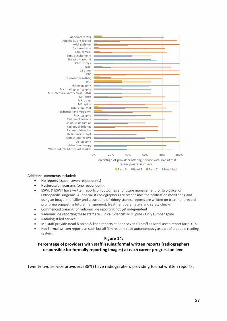
Figure 13:
Number of providers with staff (radiographers) issuing formal written reports (n=58)
The appendicular skeleton was the most common area in which radiographers issued formal reports. Radiographers are reporting across a wide range of investigations although in some areas this is within a small number of services. Figure 14 illustrates the range of pay bands offering the service.
1312
124
444566
1115
65
33
133
138
2109
158
1418
2533
8
1
1
3
1
2
1
3
11
0 5 10 15 20 25 30 35
WatersolubleGIcontraststudies
Videofluoroscopy
Venography
UltrasoundforDVT
Radionuclideother
Radionucliderenal
Radionuclidelungs
Radionuclidecardiac
Radionuclidebone
Proctography
Paediatrics(anymodality)
Orbits,pre-MRI
MRIother
MRIknee
MRIspine
MRIinternalauditorymeati(IAM)
Micturatingcystography
Mammography
IVU
Fluoroscopy(other)
CTC
CTother
CThead
Chestx-rays
Breastultrasound
Bonedensitometry
Bariummeal
Bariumenema
Axialskeleton
Appendicularskeleton
Abdomenx-rays
Numberofproviders
NHS Private University
27
Additional comments included:
• No reports issued (seven respondents)
• Hysterosalpingograms (one respondent),
• ESWL & ESWT have written reports on outcomes and future management for Urological or Orthopaedic surgeons. All specialist radiographers are responsible for localisation monitoring and using an Image Intensifier and ultrasound of kidney stones. reports are written on treatment record pro forma suggesting future management, treatment parameters and safety checks
• Commenced training for radionuclide reporting not yet independent
• Radionuclide reporting these staff are Clinical Scientist MRI Spine - Only Lumbar spine
• Radiologist led service
• MR staff provide Head & spine & knee reports at Band seven CT staff at Band seven report facial CTs
• Not Formal written reports as such but all film readers read autonomously as part of a double reading system.
Figure 14: Percentage of providers with staff issuing formal written reports (radiographers
responsible for formally reporting images) at each career progression level Twenty two service providers (38%) have radiographers providing formal written reports.
0% 20% 40% 60% 80% 100%
WatersolubleGIcontraststudiesVideofluoroscopy
VenographyUltrasoundforDVTRadionucliderenalRadionuclideotherRadionuclidelungs
RadionuclidecardiacRadionuclidebone
ProctographyPaediatrics(anymodality)
Orbits,pre-MRIMRIspineMRIotherMRIknee
MRIinternalauditorymeati(IAM)Micturatingcystography
MammographyIVU
Fluoroscopy(other)CTC
CTotherCThead
Chestx-raysBreastultrasoundBonedensitometry
BariummealBariumenemaAxialskeleton
AppendicularskeletonAbdomenx-rays
Percentageofprovidersofferingservicewithroleatthatcareerprogressionlevel
Band5 Band6 Band7 Band8a-d

28
Table 14: Number of respondents with radiographers undertaking training to provide a reporting service (n=58)
Radiographers undertaking training to report images % Number of respondents
No 62% 36 Yes 38% 22
Figure 15 illustrates the number of radiographers stated as ‘in training’ to report.
Figure 15:
Number of radiographers in training to report (n=22) Comment The complementary and combined skills of radiologists and radiographers are vital to delivering imaging services today in the UK. The 2012 RCR/SCoR joint document on team working in clinical imaging15 describes the way a modern clinical imaging multi disciplinary team supports service provision to patients. In a changing world of technology developments, economic restraint and workforce shortages, increasing patient numbers and expectations, radiographer roles are developing with appropriate education and training and with support from radiologists to support demand. This set of questions was intended to illustrate changes in practice of radiographer formal clinical reporting alongside an assessment of the current breadth of practice for radiographer reporting. The results were challenging to interpret. While there was an apparent widening of scope of practice with radiographers providing formal reports in areas not previously recorded (eg other, fluoroscopy, water soluble GI studies), the overall number of responding providers indicated that the number of services providing these reports appears to have declined. The appendicular and axial skeleton were the most common area for radiographer reporting, although ultrasound for DVT, and orbits pre MRI, were also clearly provided by more than 50% of responding providers.
8 8
4
1
0
1
0
1
2
3
4
5
6
7
8
9
1 2 3 4 5 6
Numberofproviders
Numberofradiographersintrainingtoreportatprovider

29
Radiographers providing formal reports must be trained at post graduate level via a course approved by the SCoR and must have their practice endorsed through local governance systems. It is expected that a radiographer demonstrating a scope of practice supported by education, training and knowledge would be matched to an advanced practice job description at band 7 or above.14 In the vast majority of practice areas this appears to be the case. However, it is of interest that some reporting radiographers appear to be working at band 6 specialist radiographer, particularly in bone densitometry and orbits pre-MR. This requires consideration when guidance is reviewed around reporting practice. It is recognised that timely access to a reported diagnostic imaging study has an impact on patient outcomes due to faster time to diagnosis. A Royal College of Radiologists (RCR) study in 201518 into numbers of unreported imaging examinations indicated that significant numbers of patients wait for up to 30 days for results. While this reported work forms part of a larger project on radiologist workforce shortages for the RCR, it also indicates the scale of potential work for appropriately educated and trained reporting radiographers working within the multi disciplinary team.
• Outsourcing reporting Q29 The shortfall in appropriately trained radiologists has resulted in the need to outsource reporting in order to maintain sensible timelines for report responses. Nearly a third of respondents did not outsource, but others demonstrated a range of outsourced reporting activity, as seen in Figure 16.
The “other” comments included:
• Out of hours some outsourcing
• Cardiac CT is reported by cardiologists but not incidental findings
• Outsourcing some paediatric MRI due to vacancy and sickness • Some chest and abdomen, some night work
Figure 16: Outsourcing reporting (n=58)
12
6
22
19
18
16
6
11
22
20
0 5 10 15 20 25
Outpatientgeneralradiography
Other
Noreportingisoutsourced
MRIother
MRImusculoskeletal
MRIhead
Inpatientgeneralradiography
GPgeneralradiography
CTother
CThead
Numberofproviders

30
Linking this response with the activity of reporting radiographers, there is further work to be developed on the role of reporting radiographers in reducing outsourced reporting activity.
• Radiographer-led hot reporting Q30 Forty-two respondents (72%) indicated they did not provide radiographer-led hot reporting (Figure 17).
Figure 17:
Percentage of providers with radiographer-led hot reporting (n=58) For those who responded ‘yes’ the next set of questions asked what level of service was provided.
• General x-ray image reporting Q31 The largest percentage of general radiographic images reported by radiographers was indicated at between 71% and 80%. Twenty-two providers indicated that between 0 and 20% of images were reported by their reporting radiographers (Figure 18). Radiographers reported more than 50% of the general x-ray images for five providers.
Figure 18:
Percentage of reported general x-ray images (n=58)
1,2%
15,26%
42,72%
Yes- 24/7 Yes- not24/7 No
22
9
6
9
2
5
1
1
3
0
0
0
0 5 10 15 20 25
0%
1-10%
11-20%
21-30%
31-40%
41-50%
51-60%
61-70%
71-80%
81-90%
91-99%
100%
Numberofproviders
31
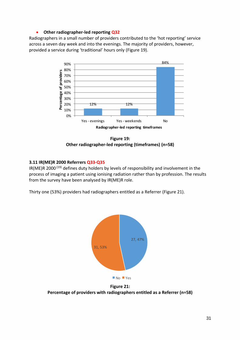
• Other radiographer-led reporting Q32 Radiographers in a small number of providers contributed to the ‘hot reporting’ service across a seven day week and into the evenings. The majority of providers, however, provided a service during ‘traditional’ hours only (Figure 19).
Figure 19:
Other radiographer-led reporting (timeframes) (n=58) 3.11 IR(ME)R 2000 Referrers Q33-Q35 IR(ME)R 2000 (19) defines duty holders by levels of responsibility and involvement in the process of imaging a patient using ionising radiation rather than by profession. The results from the survey have been analysed by IR(ME)R role. Thirty one (53%) providers had radiographers entitled as a Referrer (Figure 21).
Figure 21:
Percentage of providers with radiographers entitled as a Referrer (n=58)
12% 12%
84%
0%
10%
20%
30%
40%
50%
60%
70%
80%
90%
Yes- evenings Yes- weekends No
Percentageofproviders
Radiographer-ledreportingtimeframes
27,47%
31,53%
No Yes

32
For the providers who reported that radiographers were entitled as a ‘Referrer’, the percentages of their radiographer workforce are shown in Figure 22.
Figure 22:
Percentage of radiographer workforce holding entitlement as a Referrer (n=32) The types of examinations for which radiographers were entitled as a Referrer are illustrated in Figure 23.
Figure 23:
Examinations entitled as a Referrer (n=32)
Of the 11 respondents in the ‘other’ category, seven indicated that radiographers referred for intra ocular foreign bodies prior to MRI - one provider stating radiographers were able to refer for other foreign bodies on head, neck or chest. Two centres had radiographers referring for Kidney, Ureter and Bladder x-rays as part of a lithotripsy pathway, and 2 had sonographers who could refer for abdomen x-rays for lost Intra Uterine Contraceptive Devices (IUCD).
5
0
0
0
0
1
0
2
3
6
14
1
0 2 4 6 8 10 12 14 16
100%
91-99%
81-90%
71-80%
61-70%
51-60%
41-50%
31-40%
21-30%
11-20%
1-10%
None
Numberofproviders
11
1
18
4
8
0
0 5 10 15 20
Other
None
Limitedgeneralradiography
LimitedCT
Allgeneralradiography
AllCT
Numberofproviders

33
One respondent indicated that radiographers had referral rights within their scope of practice. One indicated that assessment of vertebral fracture after DXA was supported. In addition, one respondent stated that mammographers could refer for mammography after ultrasound on patients under age 40. Comment All radiographers at registration are entitled as ‘operators’ under IR(ME)R 2000 (19) and work within the employers’ local procedures. Non medical referrers are supported under the legislation and many allied health professionals and nurses perform this role. It appears that the scope of practice for a radiographer referrer under IR(ME)R is clearly defined and limited to their immediate scope of practice. 3.12 IR(ME)R 2000 Practitioners Q36-Q38 To act as a ‘Practitioner’ under IR(ME)R 2000 (19) the entitled professional takes responsibility for the justification of the dose of radiation; i.e. a radiographer not working to authorisation guidelines set by a medical named practitioner, but making an independent assessment for radiation dose authorisation.
Figure 24:
Radiographers entitled as a Practitioner (n=58) Of the 31 (53%) providers who reported that radiographers were entitled as a Practitioner (Figure 24), the actual percentages of the radiographer workforce are illustrated in Figure 25.
31,53%
27,47%
No Yes

34
Figure 25:
Percentage of radiographer workforce holding entitlement as a Practitioner (n=58)
Figure 26:
Examinations entitled as a Practitioner (n=27) Most radiographer IR(ME)R practitioners work in general radiography and limited CT (Figure 26). However ‘other’ showed a diversity with 2 in breast imaging, two entitled for theatre procedures, two for bone densitometry/DXA or vertebral fracture assessment, one for lithotripsy, one for ultrasound, one for GI imaging, one for cardiac catheter laboratory procedures and one provider where consultant radiographers were entitled across their scope of practice. Comment Radiographers are working as entitled Practitioners across a range of modalities and radiation areas. Slightly less than half the number of providers entitle some element of their radiographic workforce to act as Practitioner. This fits with the scope of practice for radiographers as autonomous decision makers.
0
6
3
0
3
1
0
2
0
0
1
11
0 2 4 6 8 10 12
0%
1-10%
11-20%
21-30%
31-40%
41-50%
51-60%
61-70%
71-80%
81-90%
91-99%
100%
Numberofproviders
10
4
13
15
5
0 2 4 6 8 10 12 14 16
Other
Limitedgeneralradiography
LimitedCT
Allgeneralradiography
AllCT
Numberofproviders

35
3.13 Audit and research roles Q39 Fifty-two (90%) providers had radiographers undertaking audit and seventeen (29%) had radiographers with a substantive role (0.2 whole time equivalent and above) in research (Table 15).
Table 15: Audit and research roles frequencies (n=58)
Role Number of providers with role
% 2015 survey
% 2012 survey
% 2008 survey
Radiographers undertaking audit 52 90% 99% 94% Research radiographers 17 29% 17% 5%
Other comments
• All research - department is solely research
• All forensics, cardiac, MR
• PET & MRI, Trauma, neurology, neurosurgery, neuroscience, psychiatry, oncology, cardiology, psychology, genetics, metabolism, endocrinology, MSK, pharmacology
• B6 seconded to do PhD in dementia B7 CT interest but covers all radiology research functions Lead consultant radiographers- general/role development ( all consultants expected to be doing research)
Of the 17 research radiographers, three had more than one research role.
Figure 26:
Career progression levels of radiographers with a substantive role (>0.2 WTE) in research used by providers (n=17)
Figure 26 illustrates the range of Agenda for Change pay bands for those in research radiographer roles, the most common banding being seven (65%). Of the four providers who had the role at different career progression levels, three providers were NHS and one a university.
0%
29%
65%
35%
0%
10%
20%
30%
40%
50%
60%
70%
Band5 Band6 Band7 Band8a-dPercentageofproviderswithroleat
thatlevel
Careerprogressionlevel

36
Comment In 2015, the CoR launched its 4th Research Strategy.(20) This ambitious strategy aims to ensure that radiography continues to grow as an evidence-based profession with an emphasis on improving patient care and service delivery. The SCoR vision is that all radiographers are required to engage with research. The document is aligned with research strategies of other Allied Health Professions (AHPs) and Government drivers across the UK. In the light of this and previous strategies it is encouraging to see that there is a significant increase in providers where research radiographers are employed, 17% in 2012 and 29% in 2015. This falls short of the professional body 100% target but is a steady improvement in numbers. A consideration is those radiographers who do not have research as a substantive part of their job description (greater than 20%). These radiographers may not be called ‘research radiographers’ but may still have a significant element of their role embedded in research activity. There were smaller numbers of providers indicating that radiographers have research as a substantive part of their role. Research activity for these radiographers appears to be within a specialist radiographer or advanced practice radiographer job description. 3.14 Clinical education Q40 Table 16 shows the number of providers who had radiographers with a substantive role (0.2 whole time equivalent and above) in clinical education.
Table 16: Clinical education roles frequencies (n=55)
Role Number of providers with role
% 2015 survey
% 2012 survey
% 2008 survey
Clinical education radiographers 26 47% 33% 42%
The results indicate that less than half of the responding providers had a radiographer with a substantive role in practice education. The majority in the role (69%) were banded at AfC band 7 or above (Figure 27), consistent with a job description for an advanced practice or consultant radiographer. This appears to indicate that these radiographers have undertaken post graduate training and development to develop skills to fulfil this role.

37
Figure 27:
Career progression levels of radiographers with a substantive role (>0.2 WTE) in clinical education used by providers (n=26)
Comment Radiographers with substantive roles in clinical education are described as practice educators. Practice educators provide support for learners on practice placement. The Quality Assurance Agency (QAA) Code of Practice on Placement learning (QAA 2001)(21)
requires staff involved in placement learning to be competent to fulfil their role and also ensure that the development needs of institutional placement staff are met. Practice educators are a key component of high quality clinical education and can provide the assurance to QAA that standards of placement education and support for staff delivering education are adequate. The SCoR is committed to supporting high quality education for student radiographers and has worked with other related professional bodies and education agencies to develop common standards for professional educators in clinical environments leading to accreditation as a practice educator.22 The SCoR provides a Practice Educator Accreditation scheme (PEAS) to provide the quality assurance for professionals working in these roles. The student experience in clinical placement is key to the development of high quality skills and professional behaviours. In 2015, a SCoR survey of radiography students23 indicated that many students experienced bullying and harassment while on clinical placement, in some cases leading them to leave the programme. The practice educator can provide a formal link between Higher education providers and practice placement providers helping to address any issues. 3.15 Training provided by registered professional workforce Q41 The registered professional radiographer workforce plays a role in developing skills in trainees for their own profession and the skills of other professionals. Figure 28 shows the range of training that providers indicated their radiographers support in the workplace.
0%
31%
69%
31%
0%
10%
20%
30%
40%
50%
60%
70%
80%
Band5 Band6 Band7 Band8a-d

38
“Other” responses included:
• Orthopaedic surgeons, urologists, podiatrists chiropracters, physiotherapists, sports doctors, osteopaths, vascular nurses
• Student assistant practitioners, advanced practitioners
• All groups partaking in research; physicists, all doctors from trainee to consultant, university post graduate students
• Paramedics, work experience students (year 11 and above)
• Between radiographic staff members
• Research PhD
• Sonographers
• None (two responses)
Figure 28: Staff groups receiving training by registered professional workforce (n=55)
3.16 CPD activities Q42-Q43 The Society and College of Radiographers expects all members to engage in continuing professional development (CPD) and provides a range of resources to support this. The majority of respondents, thirty-two (58%), indicated that CPD activities were partially funded (Figure 29).
46
29
9
26
16
27
0 10 20 30 40 50
Studentradiographers
Otheralliedhealthprofessionals
Other
Nursesandmidwives
Non-registeredsupportworkers
Doctorsintraining
Numberofproviders

39
Figure 29:
CPD activities are funded (n=55)
In England only, there is an Education and Training Tariff and the survey also asked whether managers knew of it and/or made use of it.
“Other” comments included:
• I am in the process of accessing this tariff to appoint 2x band 7 practice placement educator/coordinators with 1WTE band 6 backfill to release 50% of their time
• I know of this tariff and have some access via the Trust Head of Nursing and Midwifery Education but do not know how much the Trust receives directly related to radiography
• Provide multiple short placements for students from other organisations
• HEE do fund our Foundation degree students training programme for 2 years
Figure 30:
Education and Training Tariff (n=55)
20,36%
3,6%
32,58%
Yes No Partially
13
17
7
14
4
0 2 4 6 8 10 12 14 16 18
Notapplicable
IdidnotknowaboutthisTariff
IknowofthisTariffbutdonothaveaccess
IknowofthisTariffanddohaveaccess
Other
Numberofproviders

40
Seventeen indicated they were unaware of the tariff and seven were unaware of how to access it (Figure 30). Comment CPD is not simply a matter of maintaining registration with the HCPC. Active engagement with CPD can bring a number of professional and personal rewards and the College of Radiographers promotes a benefit, rather than a sanction, model of CPD. The College of Radiographers definition of CPD is:
"An ongoing professional activity, in which the practitioner identifies, undertakes and evaluates learning appropriate to the maintenance and development of the highest standards of practice within an evolving scope of practice."24
Reflective practice is the capacity to reflect on action so as to engage in a process of continuous learning. This is one of the defining characteristics of professional practice. CPD activity can take many forms eg attendance at external conferences/meetings, reading and reflecting on professional issues, professional supervision sessions, external training courses, research, audit, etc. These activities will cost time and may also carry a financial cost. In England, an Education and Training Tariff is provided to Trusts that support undergraduate student education. Some elements of this tariff may be used for professional development of staff supporting students. It is apparent that significant numbers of providers are only able to partially fund some elements of CPD activity and for a small number (6%) no financial support is provided at all. 3.17 Diagnostic ultrasound Q44 Respondents were asked to indicate against a predetermined list whether sonographers (or radiographers, where relevant) offered a service in the areas shown in Table 17. Of the 53 respondents, 16 indicated that they did not have any ultrasound services. Most commonly offered services are: abdominal, gynaecology, testes, deep vein thrombosis identification. However, all of these were at a reduced rate to the 2012 survey.

41
Table 17: Diagnostic ultrasound roles frequencies (n=53)
Role Number of providers with role
% 2015 survey
% 2012 survey
% 2008 survey
Early pregnancy 23 43% 65% 77% Obstetrics 28 53% 70% 79%
Nuchal thickness 26 49% 62% 48%
Neonatal head 20 38% 35% 42%
Gynaecology 32 60% 80% 85%
Abdominal 36 68% 85% 94% Transrectal 13 25% 17% 19%
Thyroid 29 55% 41% N/A
Testes 34 64% 70% N/A Other small parts 28 53% 51% N/A
Deep vein thrombosis identification
32 60% 66% N/A
Other vascular 23 43% 49% N/A
Musculoskeletal 30 57% 47% 39% Ultrasound guided joint injection 11 21% 10% N/A
Cardiac 2 4% 8% 7% Breast 18 34% 25% 20%
HyCoSy 3 6% 8% N/A Other contrast examinations 4 8% 8% N/A
Nerve blocks 1 2% 3% 0.0
A breakdown of services for AfC bands 6, 7 and 8a-d is provided in Figure 31.
• None of the respondents selected any of the services for band 4 staff.
• One respondent indicated that musculoskeletal services were performed by band 5 staff.
• One respondent indicated that abdominal services were performed by band 6 staff.

42
Other responses from 2012 which were not mentioned in this survey were:
• Paediatric hip (two providers)
• Transrectal biopsies (two providers)
• Trans cranial doppler (oneprovider)
• Breast vacuum biopsy (oneprovider)
• Fine needle aspiration (one provider) Other responses provided in this survey included:
• Endoscopic shock wave lithotripsy (ESWL
• Endoscopic shock wave treatment (ESWT
• Paediatrics MSK
• Obs & Gynae
• Breast and some vascular ultrasound provided but by other providers
• Ultrasound guided brachytherapy
• All paediatrics
• Fine Needle Aspiration Cytology FNAC
• Thoracentesis
• Paracentesis
Figure 31: Number of diagnostic ultrasound service providers with roles at each career progression
level (n=53)
0 10 20 30 40
Ultrasoundguidedjointinjection
Transrectal
Thyroid
Testes
Othervascular
Othersmallparts
Othercontrastexaminations
Obstetrics
Nuchalthickness
Nerveblocks
Neonatalhead
Musculoskeletal
Hycosy
Gynaecology
Earlypregnancy
Deepveinthrombosisidentification
Cardiac
Breast
Abdominal
Numberofproviderswithroleatthatcareerprogressionlevel
Band6 Band7 Band8a-d

43
3.18 Ultrasound reporting practice Q45 Excluding obstetric reporting, respondents were asked to identify which phrase best fitted their departmental reporting practice in ultrasound. The options presented were:
1. A pro-forma or tick chart is completed by the sonographer, but verified by another person (eg a radiologist).
2. A pro-forma or tick chart is completed and verified by the sonographer. 3. An independent (free text) report is produced by the sonographer but verified by
another person. 4. An independent report is produced and verified by the sonographer.
Table 18, indicates that, as in the 2012 survey, the majority of respondents selected the independent report produced and verified by the sonographer option. Of the 53 respondents, 13 indicated that this was not applicable.
Table 18: Ultrasound reporting practices (n=40)
Role Responses % 2015 survey
% 2012 survey
% 2008 survey
A pro-forma or tick chart is completed by the sonographer and verified by another person (eg radiologist).
1 2.5% 0% 1%
A pro-forma or tick chart is completed and verified by the sonographer.
0 0% 2% 5%
An independent (free text) report is produced by the sonographer and verified by another person.
2 5% 8% 3%
An independent report is produced and verified by the sonographer.
37 92.5% 90% 82%
3.19 Other radiographic roles Q46 Respondents were asked to describe any other roles carried out by radiographic staff in their provider that had not been covered in the questionnaire. Responses were as follows:
• Radiographer performed facet joint injections.
• Radiographer led triage of community diagnostics referrals.
• Partaking in discussion about research and research protocols, including set up and optimisation of protocols in MRI and CT.

44
• There is no reference to radiographers performing ESWL or ESWT (Shockwave Lithotripsy) within any areas of this questionnaire. I am responsible for all in-house training, presentations, organising study days to training all professions allied to us and medicine. All our radiographers must be specialists in this field such is the need for our imaging skills and reporting for future stone/patient management. All our staff perform ultrasound scans during ESWL treatment, ultrasound is particularly useful for renal stones and we use it for ureteric stones. It is used for pancreatic stone localisation. All our radiographers are responsible for making sure all patients are treated safely adhering to clinical practice within our guidelines and training. The teamat […] advise on further stone/patient management and must be competent on urological imaging using CT, ultrasound and plain radiography. We are also employed to use shock waves in the treatment of resistant tendinopathies, salivary stones, wound healing, andrology, etc.
• Defecating proctograms.
• Within the private sector we are involved with charging, invoicing and fee splits.
• Health and Safety Rep, Radiation Protection Supervisor Training and Education Lead Imaging Services Accreditation Scheme (ISAS) Accreditation and Quality Lead.
• Tumour volume assessment image co-registration for radiotherapy planning.
• US guided biopsy of head and neck by radiographer sonographer Trans Rectal Ultrasound biopsy by nurse specialist.
• Hysterosalpingograms.
• In education currently: Mammographyreporting, catheterisation for uro-fluoro .
• Reporting of neonates for Neonatal Intensive Care Unit by a reporting radiographer.
• Picture Archive Communication System Manager QA Superintendent.
• Specialised/ Interventional Advanced Practitioner Breast Screening Advance Practitioner - Family History, Screen Reading and Biopsies.

45
4. Summary of the diagnostic workforce The support workforce was investigated for the first time in the 2015 survey and found to be present for some but not all providers. The independent sector appears to be less likely to implement the career progression framework and utilise all four career defined roles for radiographic practice. The scope of practice for the assistant practitioner continues to develop, with the majority of those within the role identifying that their workforce works within the SCoR assistant practitioner scope of practice. The scope of practice for the diagnostic imaging radiographic workforce continues to evolve with evidence of roles developing in clinical governance and quality, alongside an increasing scope of practice in the formal reporting of a range of images across pathways and modalities. The number of consultant radiographer roles reported, while small, indicates an increase in both numbers and pathways. Providers delivered a host of services to patients and diagnostic radiographers requiring a breadth of knowledge to allow them to deliver these services safely and to a high quality.
• The scope of practice for diagnostic radiographers continues to be broad, diverse and expanding.
• Assistant practitioners are utilised in just over half of all service providers.
• The scope of practice across the assistant practitioner workforce is diverse and mostly consistent with the SCoR Scope of Practice for assistant practitioners.
• An increasing number of services have radiographer-led examinations, interventional procedures and gastro-intestinal studies.
• Many radiographers issue written reports and an increasing number of providers indicate they have a ‘radiographer-led hot report service’.
• The appendicular skeleton is the most common area for radiographer reporting after Ultrasound.
• There is evidence of radiographer reporting activity across many patient pathways, including US, CT, MRI and radionuclide imaging studies.
No service provider reported identical results, demonstrating that the workforce appears to reflect local need. There has been an increase in the proportion of departments with research radiographers since 2012, alongside a smaller increase in the proportion of departments with radiographers with a substantive role in clinical education over the same period. Access to practice educators and funded CPD was investigated, with many providers not able to identify access to funding for a practice educator role. This is of concern where diverse and developing service provision indicates that much skill and knowledge development will be taking place. The loss of NHS bursaries in England alongside new models for training and development (eg graduate level apprenticeships) might suggest that investment and consideration of these roles is paramount.

46
5. References 1. Society and College of Radiographers Scope of radiographic practice survey 2012 London: SCoR, 2012
https://www.sor.org/learning/document-library/scope-radiographic-practice-survey- 2012
2. Society and College of Radiographers The Scope of Practice 2013 London: SCoR, 2013 http://www.sor.org/learning/document-library/scope-practice-2013
3. Society and College of Radiographers Education and Career Framework for the Radiography Workforce. London: SCoR, 2013
https://www.sor.org/learning/document-library/education-and-career-framework-radiography-workforce
4. NHS England Diagnostic Imaging dataset https://www.england.nhs.uk/statistics/statistical-work-areas/diagnostic-imaging-dataset/
5. Scotland’s Health on the Web – About NHS Scotland http://www.scot.nhs.uk/about-nhs-scotland/ 6. Health and Social Care online. Health and Social Care Trusts in Northern Ireland http://online.hscni.net/hospitals/health-and-social-care-trusts/ 7. Department of Health. Meeting the Challenge: A Strategy for the Allied Health Professions. London: DoH, 2000. http://webarchive.nationalarchives.gov.uk/+/www.dh.gov.uk/assetRoot/04/05/51/80/04055180.pdf
8. Society and College of Radiographers. Guidance for Radiographers providing Forensic Radiography Services London: SCoR, 2014 https://www.sor.org/learning/document-library/guidance-radiographers-providing-forensic-radiography-services-0
9. Society and College of Radiographers Scope of Practice of Assistant Practitioners. London SCoR, 2012 https://www.sor.org/learning/document-library/scope-practice-assistant-practitioners]
10. National Profiles For Diagnostic & Therapeutic Radiography 2005 http://www.nhsemployers.org/~/media/Employers/Documents/Pay%20and%20reward/Diagnostic_and_Therapeutic_Radiography.pdf
11. Department of Health. Clinical Imaging Project: Skill mix projects (update October 2002) London: DH, 2007 (last modified date)

47
http://webarchive.nationalarchives.gov.uk/+/www.dh.gov.uk/en/Healthcare/Secondarycare/Radiographymodernisation/DH_4071546 12. NHS Employers. Developing your support workforce 2014 http://www.nhsemployers.org/your-workforce/retain-and-improve/standards-and-assurance/developing-your-support-workforce 13. The NHS Staff Council. NHS Job evaluation handbook (6th Edition) October 2016 http://www.nhsemployers.org/~/media/Employers/Documents/Pay%20and%20reward/JE%20Handbook%20fifth%20edition/NHS%20Job%20Evaluation%20Handbook%20-%20sixth%20edition.pdf
14. Society and College of Radiographers Preliminary Clinical Evaluation and Clinical Reporting by Radiographers: Policy and Practice Guidance. London: SCoR, 2013 https://www.sor.org/learning/document-library/preliminary-clinical-evaluation-and-clinical-reporting-radiographers-policy-and-practice-guidance
15. Society and College of Radiographers Team working in clinical imaging. London: SCoR, 2012 https://www.sor.org/learning/document-library/team-working-clinical-imaging
16. NHS Right Care. The NHS Atlas of variation in healthcare. September 2016 fingertips.phe.org.uk/documents/Atlas_2015%20Compendium.pdf
17. Health and Care Professions Council. Standards of proficiency - Radiographers London: HCPC, 2013 http://www.hcpc-uk.org/publications/standards/index.asp?id=51
18. Royal College of Radiologists. Unreported X-rays, computed tomography (CT) and magnetic resonance imaging (MRI) scans: Results of a snapshot survey of English National Health Service (NHS) trusts. RCR, 2015 https://www.rcr.ac.uk/sites/default/files/unreported_studies_feb2015.pdf
19. Department of Health. Ionising Radiation (Medical Exposure) Regulations 2000 (IRMER). DH, 2012
https://www.gov.uk/government/publications/the-ionising-radiation-medical-exposure-regulations-2000
20. Society and College of Radiographers Research Strategy 2016-2021. London: SCoR, 2015
http://www.sor.org/learning/document-library/research-strategy-2016-2021
21. Health and Care Professions (H&CP) Practice Education Guidance, 2016
https://www.sor.org/sites/default/files/document-versions/hcp_practice_education_guidance_3.pdf

48
22. College of Radiographers. Practice educator Accreditation scheme. CoR 2006https://www.sor.org/system/files/section/201111/PEAS%20Guidance%20-%20Practice%20Educators%20-%20Amended%20Nov%202011.pdf
23. Society and College of Radiographers. Survey into student bullying on clinical placement. London: SCoR, 2016 https://www.sor.org/learning/document-library/survey-student-bullying-clinical-placement
24. Society of Radiographers Continuing professional development (CPD), 2016 https://www.sor.org/learning/cpd (All links accessed 25/5/17)