DIAGNOSTIC IMAGING AND RADIOTHERAPY …vecchiosito.istitutotumori.mi.it/istituto/documenti... ·...
12
The Department of Diagnostic Imaging and Radiotherapy is a large multidisci- plinary structure including different modalities for Diagnostic Imaging and Radiotherapy. Three Radiologic Imaging Units RD1, RD2, and RD3, are dedi- cated to cancer diagnosis. RD1 carries out conventional radiologic tests (X-rays) and magnetic resonance imaging (MRI); RD2 performs breast cancer diagnosis (mammography, US, radioguided biopsies) and radiological tests for the gastrointestinal tract; RD3 is dedicated to computed tomography (CT), angiography, interventional radiology, and intralesional treatments. The Nuclear Medicine Unit visualizes tumors by single photon emission tomography (SPECT), planar examinations, and positron emission tomography (PET/CT). In this Unit there are also: a section for radiochemistry with a 17 MeV cyclotron; a section with protected beds dedicated to radiometabolic therapy, a radioiso- tope laboratory for radioimmunometric (IRMA) tests, and a clinic for outpatients with endocrine disease. The external beam radiotherapy (3D con- formal radiation therapy, intensity modulated radiotherapy (IMRT), Rapid Arc, and 2D conventional radiotherapy) is the main activity of the Unit of Radio- therapy 1 (RT1). The activity of Unit of Radiotherapy 2 (RT2), in addition to some applications of external beam radiotherapy, is focused on the high dose rate brachytherapy (HDR-BT) and on the combination of chemo-radiotherapy using a facility with 8 beds in 4 shielded rooms. A Medical Physics Unit sup- ports Units RT1 and RT2 in planning radiotherapy treatments and dosimetric calculations. This Units also take care of quality controls performed on the instrumentation for diagnostic imaging. UNITS RADIOLOGY AND DIAGNOSTIC IMAGING 1 Daniele Vergnaghi RADIOLOGY AND DIAGNOSTIC IMAGING 2 Alfonso V. Marchianò INTRALESIONAL TREATMENT Francesco Garbagnati RADIOLOGY AND DIAGNOSTIC IMAGING 3 AND BREAST IMAGING UNIT Silvana Bergonzi (retired December 2011) Pietro Panizza (present) DIAGNOSTIC AND INTERVENTIONAL GASTROENTEROLOGY Guido Cozzi NUCLEAR MEDICINE Emilio Bombardieri CLINICAL PET Flavio Crippa NUCLEAR MEDICINE THERAPY AND ENDOCRINOLOGY Ettore Seregni RADIOTHERAPY 1 Riccardo Valdagni RADIOTHERAPY 2 Carlo Fallai MEDICAL PHYSICS Giancarlo Zonca DIRECTOR OF DEPARTMENT Emilio Bombardieri +39 02 2390 2220 [email protected] DIAGNOSTIC IMAGING AND RADIOTHERAPY DEPARTMENT 125 Scientific Report 2011
Transcript of DIAGNOSTIC IMAGING AND RADIOTHERAPY …vecchiosito.istitutotumori.mi.it/istituto/documenti... ·...
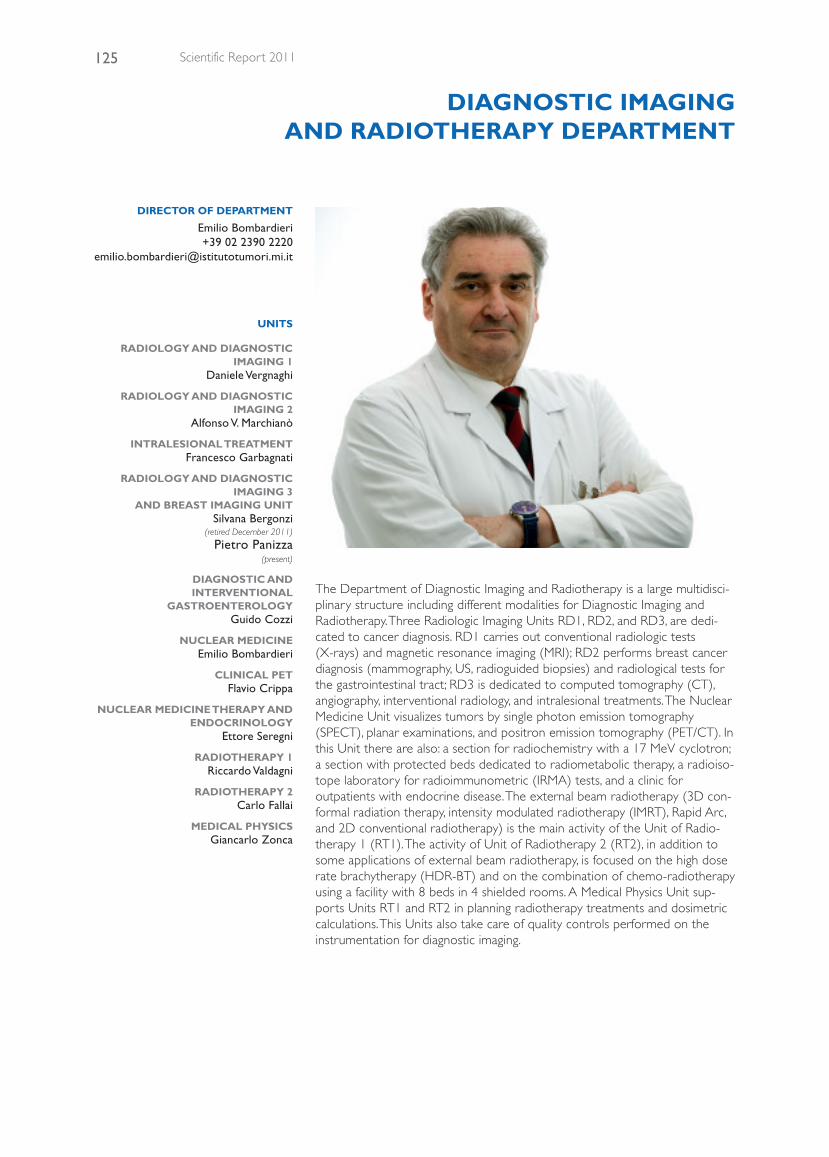
The Department of Diagnostic Imaging and Radiotherapy is a large multidisci-plinary structure including different modalities for Diagnostic Imaging andRadiotherapy. Three Radiologic Imaging Units RD1, RD2, and RD3, are dedi-cated to cancer diagnosis. RD1 carries out conventional radiologic tests(X-rays) and magnetic resonance imaging (MRI); RD2 performs breast cancerdiagnosis (mammography, US, radioguided biopsies) and radiological tests forthe gastrointestinal tract; RD3 is dedicated to computed tomography (CT),angiography, interventional radiology, and intralesional treatments. The NuclearMedicine Unit visualizes tumors by single photon emission tomography(SPECT), planar examinations, and positron emission tomography (PET/CT). Inthis Unit there are also: a section for radiochemistry with a 17 MeV cyclotron;a section with protected beds dedicated to radiometabolic therapy, a radioiso-tope laboratory for radioimmunometric (IRMA) tests, and a clinic foroutpatients with endocrine disease. The external beam radiotherapy (3D con-formal radiation therapy, intensity modulated radiotherapy (IMRT), Rapid Arc,and 2D conventional radiotherapy) is the main activity of the Unit of Radio-therapy 1 (RT1). The activity of Unit of Radiotherapy 2 (RT2), in addition tosome applications of external beam radiotherapy, is focused on the high doserate brachytherapy (HDR-BT) and on the combination of chemo-radiotherapyusing a facility with 8 beds in 4 shielded rooms. A Medical Physics Unit sup-ports Units RT1 and RT2 in planning radiotherapy treatments and dosimetriccalculations. This Units also take care of quality controls performed on theinstrumentation for diagnostic imaging.
UNITS
RADIOLOGY AND DIAGNOSTICIMAGING 1
Daniele Vergnaghi
RADIOLOGY AND DIAGNOSTICIMAGING 2
Alfonso V. Marchianò
INTRALESIONAL TREATMENTFrancesco Garbagnati
RADIOLOGY AND DIAGNOSTICIMAGING 3
AND BREAST IMAGING UNITSilvana Bergonzi
(retired December 2011)
Pietro Panizza(present)
DIAGNOSTIC ANDINTERVENTIONAL
GASTROENTEROLOGYGuido Cozzi
NUCLEAR MEDICINEEmilio Bombardieri
CLINICAL PETFlavio Crippa
NUCLEAR MEDICINE THERAPY ANDENDOCRINOLOGY
Ettore Seregni
RADIOTHERAPY 1Riccardo Valdagni
RADIOTHERAPY 2Carlo Fallai
MEDICAL PHYSICSGiancarlo Zonca
DIRECTOR OF DEPARTMENT
Emilio Bombardieri+39 02 2390 2220
DIAGNOSTIC IMAGINGAND RADIOTHERAPY DEPARTMENT
125 Scientific Report 2011
126Diagnostic Imaging and Radiotherapy Department
radiology and diagnostic imaging 1
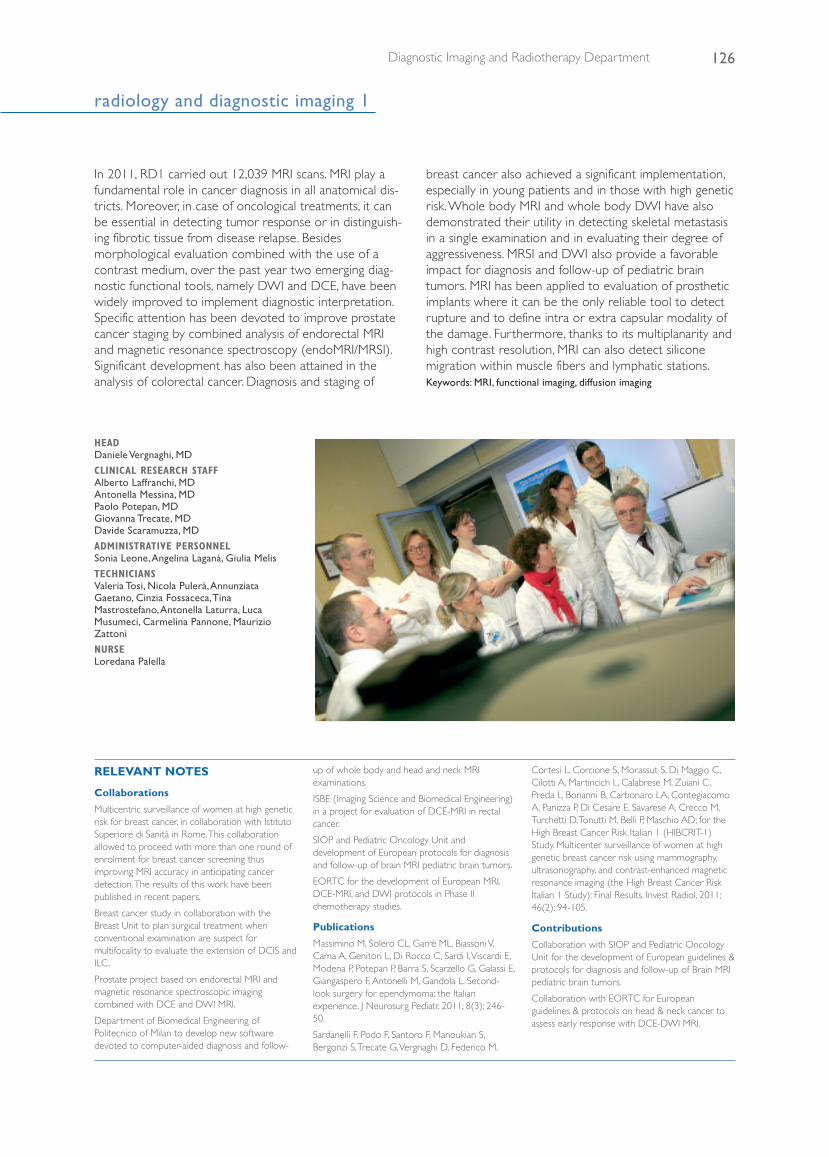
HEAD Daniele Vergnaghi, MD
CLINICAL RESEARCH STAFF Alberto Laffranchi, MD Antonella Messina, MD Paolo Potepan, MD Giovanna Trecate, MD Davide Scaramuzza, MD
ADMINISTRATIVE PERSONNELSonia Leone, Angelina Laganà, Giulia Melis
TECHNICIANSValeria Tosi, Nicola Pulerà, AnnunziataGaetano, Cinzia Fossaceca, TinaMastrostefano, Antonella Laturra, LucaMusumeci, Carmelina Pannone, MaurizioZattoni
NURSELoredana Palella
In 2011, RD1 carried out 12,039 MRI scans. MRI play afundamental role in cancer diagnosis in all anatomical dis-tricts. Moreover, in case of oncological treatments, it canbe essential in detecting tumor response or in distinguish-ing fibrotic tissue from disease relapse. Besidesmorphological evaluation combined with the use of acontrast medium, over the past year two emerging diag-nostic functional tools, namely DWI and DCE, have beenwidely improved to implement diagnostic interpretation.Specific attention has been devoted to improve prostatecancer staging by combined analysis of endorectal MRIand magnetic resonance spectroscopy (endoMRI/MRSI).Significant development has also been attained in theanalysis of colorectal cancer. Diagnosis and staging of
breast cancer also achieved a significant implementation,especially in young patients and in those with high geneticrisk. Whole body MRI and whole body DWI have alsodemonstrated their utility in detecting skeletal metastasisin a single examination and in evaluating their degree ofaggressiveness. MRSI and DWI also provide a favorableimpact for diagnosis and follow-up of pediatric braintumors. MRI has been applied to evaluation of prostheticimplants where it can be the only reliable tool to detectrupture and to define intra or extra capsular modality ofthe damage. Furthermore, thanks to its multiplanarity andhigh contrast resolution, MRI can also detect siliconemigration within muscle fibers and lymphatic stations. Keywords: MRI, functional imaging, diffusion imaging
RELEVANT NOTES
Collaborations
Multicentric surveillance of women at high geneticrisk for breast cancer, in collaboration with IstitutoSuperiore di Sanità in Rome. This collaborationallowed to proceed with more than one round ofenrolment for breast cancer screening thusimproving MRI accuracy in anticipating cancerdetection. The results of this work have beenpublished in recent papers.
Breast cancer study in collaboration with theBreast Unit to plan surgical treatment whenconventional examination are suspect formultifocality to evaluate the extension of DCIS andILC.
Prostate project based on endorectal MRI andmagnetic resonance spectroscopic imagingcombined with DCE and DWI MRI.
Department of Biomedical Engineering ofPolitecnico of Milan to develop new softwaredevoted to computer-aided diagnosis and follow-
up of whole body and head and neck MRIexaminations.
ISBE (Imaging Science and Biomedical Engineering)in a project for evaluation of DCE-MRI in rectalcancer.
SIOP and Pediatric Oncology Unit anddevelopment of European protocols for diagnosisand follow-up of brain MRI pediatric brain tumors.
EORTC for the development of European MRI,DCE-MRI, and DWI protocols in Phase IIchemotherapy studies.
Publications
Massimino M, Solero CL, Garrè ML, Biassoni V,Cama A, Genitori L, Di Rocco C, Sardi I, Viscardi E,Modena P, Potepan P, Barra S, Scarzello G, Galassi E,Giangaspero F, Antonelli M, Gandola L. Second-look surgery for ependymoma: the Italianexperience. J Neurosurg Pediatr. 2011; 8(3): 246-50.
Sardanelli F, Podo F, Santoro F, Manoukian S,Bergonzi S, Trecate G, Vergnaghi D, Federico M,
Cortesi L, Corcione S, Morassut S, Di Maggio C,Cilotti A, Martincich L, Calabrese M, Zuiani C,Preda L, Bonanni B, Carbonaro LA, ContegiacomoA, Panizza P, Di Cesare E, Savarese A, Crecco M,Turchetti D, Tonutti M, Belli P, Maschio AD; for theHigh Breast Cancer Risk Italian 1 (HIBCRIT-1)Study. Multicenter surveillance of women at highgenetic breast cancer risk using mammography,ultrasonography, and contrast-enhanced magneticresonance imaging (the High Breast Cancer RiskItalian 1 Study): Final Results. Invest Radiol. 2011;46(2): 94-105.
Contributions
Collaboration with SIOP and Pediatric OncologyUnit for the development of European guidelines &protocols for diagnosis and follow-up of Brain MRIpediatric brain tumors.
Collaboration with EORTC for Europeanguidelines & protocols on head & neck cancer toassess early response with DCE-DWI MRI.
radiology and diagnostic imaging 2
HEAD Alfonso V. Marchianò, MD
CLINICAL RESEARCH STAFFFrancesco Garbagnati, MDEnrico Civelli, MD Giuseppe Di Tolla, MD Laura F. Frigerio, MD Rodolfo Lanocita, MD Carlo Morosi, MD Carlo Spreafico, MD
ADMINISTRATIVE PERSONNELGiovanni Capone, Giuliana Manzoni, OrnellaVenegoni
TECHNICIANSPietro Basile, Marilena Barbiero, MariaFerrarello, Roberto Gallo, GiuseppinaGentile, Roberto Nioi, Antonio Perchinunno,Geremia Porcelli, Salvatore Romaniello,Luciana Tanzini, Vanni Tirella
NURSESPietro Ciccarese, Laura Fagnani, RobertaPopulin, Marinella Porceddu
Diagnostic oncology and interventional-oriented radiol-ogy represent the core activity of the Unit. Inpatients andoutpatients undergo a diagnostic work-up that includesdifferent steps of patient management: primary cancerdiagnosis, staging, follow-up, and monitoring after surgery,chemotherapy, and radiotherapy. The lung cancer screen-ing program (MILD) with “low dose” spiral CT continuedin 2011. We performed over 2000 low-dose spiral CTexams, and we are currently testing a system ofcomputer-aided detection (CAD) of pulmonary nodules.Two CT scanners, both provided with faster multi-slicescanning capabilities are available, and about 22,000 diag-nostic examinations per year and an extensive number ofinterventional radiological procedures are carried out. Alltypes of percutaneous biopsies are currently performedin our patients. Interventional radiology activities includelong-term venous central catheter placement, emboliza-tion, and chemoembolization for regional cancertreatment. Intralesional radiofrequency ablation based
methods, such as chemo-interventional procedures con-sisting in local-regional drug delivery for malignancies ofthe liver, head and neck, pelvis and limbs, have been suc-cessfully performed. An international multicentric studyon the treatment of inoperable hepatocellular carcinomawith intra-arterial injection of yttrium-90 radiolabeledmicrospheres is ongoing in collaboration with Units ofNuclear Medicine and Gastrointestinal and Hepatopan-creatobiliary Surgery and liver Transplantation. The Unithas also developed an original experience ofpercutaneous cryoblation in selected patients with smallrenal tumors. During 2011, over 1000 vascular diagnosticprocedures, 500 vascular and non-vascular interventionalprocedures, over 550 long-term venous central catheters,and 600 percutaneous biopsies in various body districtswere performed. Keywords: interventional radiology, multidetector computed
tomography, image-guided biopsy
RELEVANT NOTES
Publications
Use of a retrievable vena cava filter with low-intensity anticoagulation for prevention ofpulmonary embolism in patients with cancer: anobservational study in 106 cases. J Vasc IntervRadiol. 2011; 22(9): 1312-9.
CT-guided percutaneous cryoablation of renalmasses in selected patients. La Radiologia Medica.2011. Oct 21.
127 Scientific Report 2011
128Diagnostic Imaging and Radiotherapy Department
RELEVANT NOTES
Collaborations
Royal Marsden Hospital, London
Fondazione IRCCS Policlinico San Matteo, Pavia
University of Milan
Intralesional Treatment (Francesco Garbagnati, MD)In the Radiological Department, since 2005, a specificUnit for mini-invasive intralesional treatment with percu-taneous thermal ablation procedures (RF, microwave,etc.) was founded. At our Institute in the late 1980s,(1988 - 1989), percutaneous radiofrequency treatmentwas carried out with pioneering research and recentlyobtained scientific recognition at international levels. Since1990, we have treated more than 1400 patients mainlywith liver cancer, but also kidney, lung, and bone lesions.The Unit works within the Radio-Diagnostic ServiceDepartment of Radiology utilizing radiological interven-tional treatment procedures for centering and treatmentfollow up. In HCC and liver metastases with a diameter greater than3.5 cm, we studied the possibility of radiofrequency
hyperthermia combined with the arterial stop flow pro-cedure to achieve maximum effectiveness with minimalinvasiveness. (Treatment in local anesthesia and one-daysurgery). This procedure has also been officially acceptedand codified by the Italian Society of Interventional Radi-ology. We also treated liver metastases, inoperable lungcancer, supra renal and renal cancer, and painful bonemetastasis that were unresponsive to pain control withconventional therapies. In collaboration with theFondazione IRCCS Policlinico San Matteo (Pavia), we arestudying a minimally-invasive system to destroy tumorswith different tissue characteristics using very thin probesand with the possibility of single electrode insertion evenin large tumors. Keywords: radiofrequency, percutaneous thermoablation, tumor
thermoablation
radiology and diagnostic imaging 2
radiology and diagnostic imaging 3
HEAD Silvana Bergonzi, MD
CLINICAL RESEARCH STAFF Breast ImagingClaudio Ferranti, MDMonica Marchesini, MDGianfranco Scaperrotta, MDLaura Suman, MD
Diagnostic and Interventional GastroenterologyMonica Salvetti, MD Marco Milella, MD
TECHNICIANSFolini Cristina (Coordinator), Colombo Luisa,Dedei Luciana, Depedri Enrico, LanzillottiLuca, Mannella Maria Pia, Sala Stefania, TavolaAnna
NURSESDolores Mauro, Ferruccio Mirella
Diagnostic and Interventional Gastroenterology (GuidoCozzi, MD)The clinical activity of the Unit continues to be mainlyoriented towards diagnostic and interventional radiologyof the digestive and biliary tracts and diagnostic and inter-ventional US applications. A total of 11,795 examinationswere carried out by the Unit, with a decrease of 9.4%, forplain films of the chest, (due to the new organization ofthe Radiological Units), together with US examination. In2011, 2831 contrastographic examinations of the diges-tive and urinary tract were performed, with an increaseof 3.5%. In this diagnostic field, according to the pasttrend, examinations of functional disorders of the diges-tive tract increased, increased to 715 (+9%). Biliary andgastroenteric interventional procedures were performedin patients with different biliopancreatic or gastrointestinaldiseases. In the biliary field, definitive jaundice palliationwith drainages or stents, curative dilatation of cicatricial
stenoses, drainage of fistulas, transluminal biopsies, andother less common maneuvers were routinelyperformed. In the gastrointestinal field, in addition totreatment of complications (transluminal drainage of fluidcollections, dilatation of cicatricial stenoses) IP played abasic role in nutritional support (percutaneous gastros-tomy, positioning of feeding tubes, stenting of inoperablestenoses). It must be emphasized that a substantial partof all these interventional procedures must be performedas promptly as possible, requiring exceptional organiza-tion. Overall, 633 interventional procedures wereperformed in the Unit, with a decrease of 5.5% comparedto 2010. During 2011, 5176 US examinations were per-formed. In the context of the INT “Progetto Tiroide”, incooperation with the multidisciplinary outpatient serviceof thyroid disease, 2422 neck US examinations, most formonitoring thyroid nodules, were performed. Keywords: biliary interventional radiology, gastroenteric interventional
radiology, thyroid biopsy
129 Scientific Report 2011
Breast Imaging Unit (Silvana Bergonzi, MD, until 30
November 2011) The Breast Imaging Unit has all the diagnostic and inter-ventional tools which are necessary in a comprehensivecancer center, where patients from any referral come insearch of reassurance to not have cancer, as well as earlydiagnosis and proper treatment in the case that lesionsare present. Breast imaging consists mostly in clinicalmammography and breast ultrasound studies on sympto-matic or treated patients as well as on asymptomaticwomen with the aim of prevention. We also participate ina program of surveillance for high-risk patients and BRCAmutation carriers.Interventional examinations are directed to preoperativelocalization of non-palpable lesions and to assessment ofbreast masses or microcalcifications by core-needle biop-sies (CNB) or vacuum-assisted biopsies (VAB) withultrasonographic or stereotactic guidance. These interven-tions are basic elements of a multidisciplinary approach
to provide assistance for surgical planning. In 2011, about14,000 mammographic examinations and 9300 breastultrasound studies were performed, in addition to 400second opinion consultations. Moreover, 850 conventionalradiodiagnostic examinations on inpatients were carriedout. About 1550 interventional procedures wereperformed on patients with suspicious breast findings, andrespectively, 620 preoperative targeting of non-palpablelesions and 930 imaging-guided breast biopsies (CNB +VAB).In 2011, the breast lesion excision system (BLES) wasimplemented, consisting in a biopsy device that acquires aunique intact specimen instead of multiple samples suchas VAB, and preliminary results have been presented. Dur-ing 2011, new mammographic equipment performing 3Dexaminations (digital breast tomosynthesis) was installed,allowing better confidence in mammographic interpreta-tion and increasing sensitivity for detection of breastcancer.
This Diagnostic Radiology is composed of two Units: theBreast Imaging Unit and the Diagnostic and InterventionalGastroenterology Unit. The two Units share technicians,while clinical research staff have specific duties in each Unit.
nuclear medicine
HEAD Emilio Bombardieri, MD
CLINICAL RESEARCH STAFFAlessandra Alessi, MD Gianluca Aliberti, MD Anna Bogni, Biol Sci D Maria R. Castellani, MD Carlo Chiesa, Physicist Flavio Crippa, MD Vinicio De Sanctis, Engineer Marco Maccauro, MD Eva Orunesu, MD Claudio Pascali, Radiochemist Gianluca Serafini, MD Ettore Seregni, MD
RESEARCH STAFFAngela Coliva (Chemist), Claudio Cucchi(Chemist), Luca Laera, Chemist, GretaMaiocchi (Chemist)
POSTDOCTORAL FELLOWSBarbara Padovano, MDFederica Pallotti, MD
TECHNICIANSMonica Testoni (Coordinator), GraziaAprigliano, Danilo Baratella, Davide Bassani,Gianercico Cucchetti, Carlotta Edera, MariaDi Francesco, Pietro Di Nuzzi, MartinoFaedi, Rita Filieri, Deborah Mansi, GiovanniNido, Rossana Pavesi, Biagio Perrone, MatteoRegazzoni, Lidia Spano, Roberto Segreti
NURSESRita Sicari (Coordinator), Cristina DeSomma, Carmela Fallacara, Calogero Oliveri,Aurelio Scarabelli
ADMINISTRATIVE PERSONNELAnnaluisa De Simone Sorrentino(Coordinator), Isabella Flauto, ValentinaFracchiolla, Rosangela Ghilardi, PatriziaGobbi
This structure is a modern division including differentintegrated sections dedicated both to the diagnosis andradiometabolic therapy of cancer. Other important goalsare to develop novel radiopharmaceuticals and managepatients with endocrine diseases. The most importantareas of activity are: 1) a diagnostic Unit based ongamma-emitting radiopharmaceuticals (SPECT Unit); 2) adiagnostic Unit based on positron-emitting radiopharma-ceuticals (PET Unit); 3) a therapy Unit for radiometabolictherapy; 4) radiochemistry laboratories with a 17 MeVcyclotron equipped for the production of PET andSPECT tracers and radiopharmaceuticals for therapy; 5) alaboratory for preclinical studies equipped with amicroPET; 6) a biochemistry laboratory for IRMA andRIA tests; 7) a clinical endocrinology Unit for studyingendocrine cancer patients. Some of the most importantachievements of the programs carried out are: a) clinicalstudies of SPECT and PET tests and their validation as atool for targeting tumors; b) validation of some radiola-
beled oncotropic tracers as a prognostic index of tumorsin terms of characterization in vivo and early response; c)the discovery of a original procedure for synthesis of thePET tracer F18-FLT (patent PCT WO 2008/146316A); d)the validation of a new methodology to visualize pelviclymph nodes in patients with gynecological cancer by lym-phoscintigraphy after peri-tumoral injection of99mTc-labeled microspheres; e) successful treatment ofhepatocarcinomas by intra-arterial radioembolizationwith Y-90 microspheres; f) irradiation of NET tumors bytandem treatment with somatostatin analogues radiola-beled with dual radioisotopes (Y-90 and Lu-177); g) theoptimization of radiometabolic therapy (thyroid cancer,neuroblastoma, neuroendocrine tumors,hepatocarcinoma and lymphoma) by a pre-therapy dosi-metric approach to enhance the dose of irradiationdelivered to cancer mass.Keywords: nuclear medicine diagnosis, nuclear medicine therapy,
radiopharmaceuticals
RELEVANT NOTES
Collaborations
EANM, European Association of Nuclear Medicine(Vienna)
IAEA, International Association of Atomic Energy(Vienna)
Publications
Lopci E, Chiti A., Castellani M.R., Pepe G.,Antunovic L, Fanti S, E. Bombardieri. Matched pairsdosimetry: 124I/131I mIBG and 124I/131I and86Y/90Y antibodies. Eur J Nucl Med Mol Imaging.2011; 38: 28-40.
Seregni E, Padovano B, Coliva A, Zecca E,Bombardieri E. State of the art of palliative therapy.Q J Nucl Med Mol Imaging. 2011; 55: 411-9.
Contributions
Associate Editor : Quarterly Journal of NuclearMedicine and Molecular Imaging Minerva Medica(Turin)
Editorial Board of European Journal of NuclearMedicine and Molecular Imaging, Springer(Heidelberg)
130Diagnostic Imaging and Radiotherapy Department
131 Scientific Report 2011
Clinical PET (Crippa Flavio, MD)Routine activity is focused on FDG imaging for staging,therapy monitoring, and follow-up in almost all types ofsolid tumors and onco-hematologic malignancies. 11C-methionine to study gliomas and FLT (fluorothymidine) isunder clinical validation. In 2011, clinical research wasmainly focused on the following topics. FDG-PET inlocally advanced rectal cancer submitted to neoadjuvantchemoradiotherapy with the aim to evaluate whethertumor FDG uptake can predict pCR. PET is performed atbaseline, 2 weeks after treatment initiation, and 6 weeksafter therapy completion. The results are compared withpost-surgical histology. Thirty patients have been studiedand the results will be submitted for publication. FDG-PET to monitor pazopanib monotherapy inrelapsed/refractory urothelial cancer. PET scans areplanned at baseline and 4 weeks thereafter. They arecompared with conventional imaging and clinicaloutcome. Preliminary results have been already submitted
(ASCO 2011), and definitive results will be submitted forpublication. FLT-PET in locally advanced breast cancersubmitted to pre-surgical neoadjuvant treatment. The aimis to evaluate whether tumor FLT uptake can predictpCR. Fifteen patients will be evaluated with PET at base-line, after 1-2 cycles, and at the conclusion of therapy.Results will be compared to post-surgical histology. In col-laboration with the Experimental Oncology andMolecular Medicine and Nuclear Medicine Departmentof San Rafaelle Hospital in Milan, a microPET study hasbeen carried out in rats with a breast cancer model(MDA-BB-468) treated with taxol. Tumor response wasevaluated with FDG and FLT at baseline and 2 weeksafter treatment initiation. Tracer uptake has been com-pared with tumor volume during the study. Preliminaryresults show a reduction of FDG/FLT uptake in responderrats and an increase in non-responder rats. The definitiveresults will submitted for publication. Keywords: PET/CT, metabolic imaging, therapy monitoring
RELEVANT NOTES
Collaborations
Fondazione IRCCS Istituto Neurologico Carlo Besta, Milan
Neuro-Oncologia Unit, Department of Neuroscienze,Università e A.U.O. San Giovanni Battista, Turin
Nuclear Medicine Therapy And Endocrinology (EttoreSeregni, MD)The activities of the Unit cover three main areas involvedin routine and experimental procedures: nuclear medicinetherapy, radiosotope laboratory, and an endocrinologyoutpatient clinic. Regarding the former, two main studiesare of interest. The first is “Treatment with tandem[90Y]DOTA-TATE and [177Lu]DOTA-TATE of neuroen-docrine tumors refractory to conventional therapy”. Todate, 44 patients have entered the study. Clinical resultsare available for the first group of 26 patients. A completeresponse was observed in 2 patients (8%), partialresponse in 9 (35%), stable disease in 11 (42%), and dis-ease progression occurred in 4 patients (15%). Theoverall objective tumor response rate was 42%. Themedian overall survival at 24 months was 78%, and themedian progression-free survival (PFS) was 25 months.Global PFS at 24 months was 52%. The second study is“Efficacy of endoarterial treatment with 90Y-microspheres in hepatocellular carcinoma”. To date, 153treatments have been performed. A phase II study on the
first 58 treatments on 52 patients has been conducted.This demonstrated the relevance of a dosimetry-basedapproach. In fact, non-responding lesions had a mediandose of 199 Gy, while responding lesions of 431 Gy.Regarding the area of endocrinology, the study “Growthhormone (GH) replacement therapy in surviving patientswith primary pediatric brain tumors (PBT)” is reported.More than 700 patients with PBT were screened for GHdeficit (GHD), which was found in 110 cases. In all but 5children, growth recovery was observed after treatment.Improvement of bone mineral density was also attainedand improvement in neuropsychological performancesdue to GH therapy was seen. Disease recurrence wasfound in 7 patients, a relapse rate lower than in non-GHtreated patients. This study demonstrated that GHreplacement therapy is safe and effective in the correc-tion of GH deficiency and in improving quality of life ofpatients with PBT. Keywords: nuclear medicine therapy, endocrine oncology,
neuroendocrine tumors
RELEVANT NOTES
Publications
Seregni E, Padovano B, Coliva A, Zecca E,Bombardieri E. State of the art of palliative therapy.Q J Nucl Med Mol Imaging. 2011; 55: 411-9.
Borrello MG, Aiello A, Peissel B, Rizzetti MG,Mondellini P, Degl’Innocenti D, Catalano V, Gobbo
M, Collini P, Bongarzone I, Pierotti MA, Greco A,Seregni E. Functional characterization of the MTC-associated germline RET-K666E mutation: evidenceof oncogenic potential enhanced by the G691Spolymorphism. Endocr Relat Cancer. 2011; 18: 519-27.
Contributions
Core partner as Endocrinologist of the certifiedENETS Center of Excellence European Expert inthe definition of consensus on “Follow-up inpatients with differentiated thyroid cancer withantibody interference in thyroglobulinmeasurement” (Berlin, Germany, November 2011)
nuclear medicine
radiotherapy 1
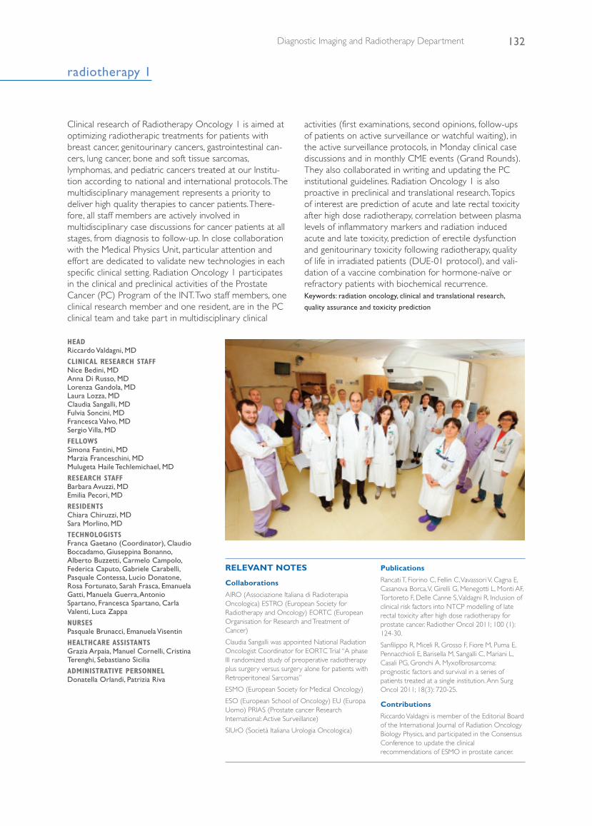
HEAD Riccardo Valdagni, MD
CLINICAL RESEARCH STAFFNice Bedini, MDAnna Di Russo, MDLorenza Gandola, MD Laura Lozza, MDClaudia Sangalli, MDFulvia Soncini, MDFrancesca Valvo, MDSergio Villa, MD
FELLOWSSimona Fantini, MDMarzia Franceschini, MDMulugeta Haile Techlemichael, MD
RESEARCH STAFFBarbara Avuzzi, MDEmilia Pecori, MD
RESIDENTSChiara Chiruzzi, MDSara Morlino, MD
TECHNOLOGISTSFranca Gaetano (Coordinator), ClaudioBoccadamo, Giuseppina Bonanno,Alberto Buzzetti, Carmelo Campolo,Federica Caputo, Gabriele Carabelli,Pasquale Contessa, Lucio Donatone,Rosa Fortunato, Sarah Frasca, EmanuelaGatti, Manuela Guerra, AntonioSpartano, Francesca Spartano, CarlaValenti, Luca Zappa
NURSESPasquale Brunacci, Emanuela Visentin
HEALTHCARE ASSISTANTSGrazia Arpaia, Manuel Cornelli, CristinaTerenghi, Sebastiano Sicilia
ADMINISTRATIVE PERSONNELDonatella Orlandi, Patrizia Riva
Clinical research of Radiotherapy Oncology 1 is aimed atoptimizing radiotherapic treatments for patients withbreast cancer, genitourinary cancers, gastrointestinal can-cers, lung cancer, bone and soft tissue sarcomas,lymphomas, and pediatric cancers treated at our Institu-tion according to national and international protocols. Themultidisciplinary management represents a priority todeliver high quality therapies to cancer patients. There-fore, all staff members are actively involved inmultidisciplinary case discussions for cancer patients at allstages, from diagnosis to follow-up. In close collaborationwith the Medical Physics Unit, particular attention andeffort are dedicated to validate new technologies in eachspecific clinical setting. Radiation Oncology 1 participatesin the clinical and preclinical activities of the ProstateCancer (PC) Program of the INT. Two staff members, oneclinical research member and one resident, are in the PCclinical team and take part in multidisciplinary clinical
activities (first examinations, second opinions, follow-upsof patients on active surveillance or watchful waiting), inthe active surveillance protocols, in Monday clinical casediscussions and in monthly CME events (Grand Rounds).They also collaborated in writing and updating the PCinstitutional guidelines. Radiation Oncology 1 is alsoproactive in preclinical and translational research. Topicsof interest are prediction of acute and late rectal toxicityafter high dose radiotherapy, correlation between plasmalevels of inflammatory markers and radiation inducedacute and late toxicity, prediction of erectile dysfunctionand genitourinary toxicity following radiotherapy, qualityof life in irradiated patients (DUE-01 protocol), and vali-dation of a vaccine combination for hormone-naïve orrefractory patients with biochemical recurrence.Keywords: radiation oncology, clinical and translational research,
quality assurance and toxicity prediction
RELEVANT NOTES
Collaborations
AIRO (Associazione Italiana di RadioterapiaOncologica) ESTRO (European Society forRadiotherapy and Oncology) EORTC (EuropeanOrganisation for Research and Treatment ofCancer)
Claudia Sangalli was appointed National RadiationOncologist Coordinator for EORTC Trial “A phaseIII randomized study of preoperative radiotherapyplus surgery versus surgery alone for patients withRetroperitoneal Sarcomas”
ESMO (European Society for Medical Oncology)
ESO (European School of Oncology) EU (EuropaUomo) PRIAS (Prostate cancer ResearchInternational: Active Surveillance)
SIUrO (Società Italiana Urologia Oncologica)
Publications
Rancati T, Fiorino C, Fellin C, Vavassori V, Cagna E,Casanova Borca, V, Girelli G, Menegotti L, Monti AF,Tortoreto F, Delle Canne S, Valdagni R. Inclusion ofclinical risk factors into NTCP modelling of laterectal toxicity after high dose radiotherapy forprostate cancer. Radiother Oncol 2011; 100 (1):124-30.
Sanfilippo R, Miceli R, Grosso F, Fiore M, Puma E,Pennacchioli E, Barisella M, Sangalli C, Mariani L,Casali PG, Gronchi A. Myxofibrosarcoma:prognostic factors and survival in a series ofpatients treated at a single institution. Ann SurgOncol 2011; 18(3): 720-25.
Contributions
Riccardo Valdagni is member of the Editorial Boardof the International Journal of Radiation OncologyBiology Physics, and participated in the ConsensusConference to update the clinicalrecommendations of ESMO in prostate cancer.
132Diagnostic Imaging and Radiotherapy Department
133 Scientific Report 2011
Radiotherapy of Breast Tumors (Laura Lozza, MD)In 2011, about 500 patients received conventional adju-vant radiotherapy (3D conformal or IMRT) afterconservative breast surgery or mastectomy. Therapeuticprograms were discussed in a multidisciplinary settingwith the Breast Surgery Unit and Medical Oncology staff.Women aged ≥70 years were given a hypofractionatedregimen (42.4 Gy, 16 fractions/3 weeks) with optimalcompliance, low toxicity, and excellent/good cosmeticresults in the majority of patients. Active collaborationwith the Plastic and Reconstructive Surgery Unit allowedoffering patients submitted to breast reconstruction tis-
sue expanders or prosthesis and the best timing for radi-ation. An experimental study together with the MedicalPhysics Unit of our Institute and the BioengineeringDepartment of the Politecnico of Milan, structural modifi-cations after radiotherapy in different silicon implantswere investigated and the publication of its results isimminent. To help patients understand the technical pro-cedures in breast radiotherapy and to improve clinicalinformation, an audiovisual tool was created by medical,physical, and technical staff. Keywords: breast radiotherapy, hypofractionated radiotherapy, implant
radiotherapy
RELEVANT NOTES
Collaborations
AIRO: Associazione Italiana RadioterapiaOncologica
Publications
Nava MB, Pennati AE, Lozza L, Spano A, ZambettiM, Catanuto G. Outcome of different timings ofradiotherapy in implant-based breastreconstructions. Plast Reconstr Surg. 2011; 128:353-9.
Contributions
EDITORIAL BOARD for Attualità in Senologia:Breast Radiotherapy Literature Review.
Guidelines AIRO (Associazione ItalianaRadioterapia Oncologica) Breast RadiotherapyGuidelines Upgrade
Radiotherapy of Digestive Tract Tumors (FrancescaValvo, MD )The goal of the Unit is to offer the best chance of cure topatients with locally-advanced cancer of the esophagusand rectum through a neoadjuvant combined radio-chemotherapy approach, in the attempt to achieve acomplete pathological response that represents an impor-tant positive prognostic factor in terms of local as well asdistant control regardless of clinical stage. For patientswith anal cancer, curative radiochemotherapy is employedwith a high rate of local control and organ preservation.
Postoperative radiochemotherapy is employed in locallyadvanced (pT3 or pN+) gastric and pancreaticneoplasms. Personalized treatment plans are elaboratedfor each patient, and volumetric intensity modulatedradiotherapy is usually adopted to reduce acute and lateside effects. An ongoing study of our multidisciplinaryteam is evaluating the effectiveness of revaluation withFdG-Pet during neaodjuvant radiation treatment as a pre-dictive tool for outcome in patients with rectal cancer. Keywords: gastrointestinal cancers, combined treatments, organ
preservation
RELEVANT NOTES
Collaborations
AIRO (Associazione Italiana RadioterapiaOncologica) Working Group on GastrointestinalCancer
GAT (Gruppo di Appropriatezza Terapeuticaregionale for Rectal Cancer Surgery)
ESTRO (European Society for Radiotherapy andOncology):
Online Course tutor: Evidence and Research inRectal Cancer
Contributions
AIOM (Associazione Italiana Oncologia Medica)Rectal Cancer Guideline
radiotherapy 1
134Diagnostic Imaging and Radiotherapy Department
Pediatric Radiotherapy (Lorenza Gandola, MD; ClinicalResearch Staff on private grant: Emilia Pecori, MD)Clinical research activity is focused on improving the ther-apeutic index of radiotherapy for childhood cancers withthe aim of optimizing cure while reducing long-termiatrogenic sequelae that is so critical in the pediatric pop-ulation. This goal is pursued through a strictmultidisciplinary approach, the development and adop-tion of institutional, national, and international treatmentprograms representing the best up-to-date knowledge inthe field of pediatric oncology, and validating the newavailable technologies for the irradiation of children. TheUnit coordinates the AIEOP “Radiotherapy WorkingGroup”, is responsible for radiation therapy of twonational protocol on ependymoma (129 childrenenrolled) and Wilms’ tumor (more than 300 patientsenrolled), and co-chairs the SIOP Europe “Brain TumorCommittee”. It is also actively involved in developing newtherapeutic strategies as a member of the AIEOP “Cen-
tral Nervous System Tumors, Bone Tumors, Renal Tumors,Neuroblastoma Working Groups” and SIOP “PNET andEpendymoma Working Groups”. In particular, within theSIOP Ependymoma Working group, the Unit is the leadradiotherapy center for a forthcoming European Study.On a national basis, the Unit is a reference center forradiation therapy of children treated in other hospitalswith limited specific expertise or lacking particular facili-ties such as anesthesiology support. In 2011, thanks to anoriginal idea of two members of the technical staff, theUnit published a picture book entitled “The cat that lostits tail”, which is a fairy tale to explain radiotherapy tochildren: the effectiveness of the book as a therapeutictool to improve pediatric compliance to radiotherapyprocedures, also reducing requirements for general anes-thesia, is under evaluation in collaboration with thepsychologist team of the Pediatric Oncology Unit. Keywords: pediatric radiotherapy, quality of life, multidisciplinary
treatments
RELEVANT NOTES
Collaborations
AIEOP: Associazione Italiana Ematologia OncologiaPediatrica
SIOP: Société Internationale d’OncologiePédiatrique
ISG: Italian Sarcoma Group
Publications
Massimino M, Gandola L, Barra S, Giangaspero F,Casali C, Potepan P, Di Rocco C, Nozza P, Collini P,Viscardi E, Bertin D, Biassoni V, Cama A, MilanaccioC, Modena P, Balter R, Tamburrini G, Peretta P,
Mascarin M, Scarzello G, Fidani P, Milano GM, SardiI, Genitori L, Garrè ML. Infant ependymoma in a10-years AIEOP experience with omitted ordeferred radiotherapy. Int J Radiat Oncol Biol Phys.2011; 80(3): 807-14.
Ferrari S, Sundby Hall K, Luksch R, Tienghi A, WiebeT, Fagiolo F, Alvegard TA, Brach Del Prever A,Tamburini A, Alberghini M, Gandola L, Mercuri M,Capanna R, Mapelli S, Prete A, Carli M, Picci P,Barbieri E, Bacci G, Smeland S. Non-metastaticEwing family tumors: high-dose chemotherapy withstem cell rescue in poor responders patients.Results of the Italian Sarcoma Group/ScandinavianSarcoma Group III protocol. Ann Oncol. 2011;22(5): 65-71.
radiotherapy 1
135 Scientific Report 2011
radiotherapy 2
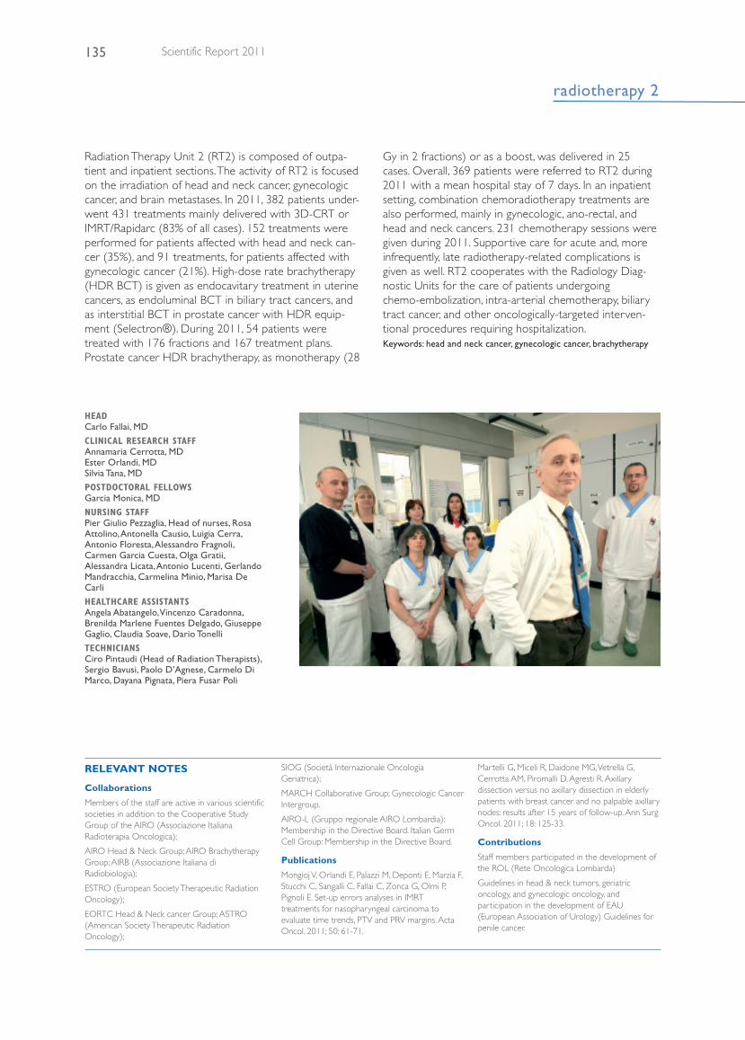
HEAD Carlo Fallai, MD
CLINICAL RESEARCH STAFF Annamaria Cerrotta, MD Ester Orlandi, MD Silvia Tana, MD
POSTDOCTORAL FELLOWSGarcia Monica, MD
NURSING STAFFPier Giulio Pezzaglia, Head of nurses, RosaAttolino, Antonella Causio, Luigia Cerra,Antonio Floresta, Alessandro Fragnoli,Carmen Garcia Cuesta, Olga Gratii,Alessandra Licata, Antonio Lucenti, GerlandoMandracchia, Carmelina Minio, Marisa DeCarli
HEALTHCARE ASSISTANTSAngela Abatangelo, Vincenzo Caradonna,Brenilda Marlene Fuentes Delgado, GiuseppeGaglio, Claudia Soave, Dario Tonelli
TECHNICIANSCiro Pintaudi (Head of Radiation Therapists),Sergio Bavusi, Paolo D’Agnese, Carmelo DiMarco, Dayana Pignata, Piera Fusar Poli
Radiation Therapy Unit 2 (RT2) is composed of outpa-tient and inpatient sections. The activity of RT2 is focusedon the irradiation of head and neck cancer, gynecologiccancer, and brain metastases. In 2011, 382 patients under-went 431 treatments mainly delivered with 3D-CRT orIMRT/Rapidarc (83% of all cases). 152 treatments wereperformed for patients affected with head and neck can-cer (35%), and 91 treatments, for patients affected withgynecologic cancer (21%). High-dose rate brachytherapy(HDR BCT) is given as endocavitary treatment in uterinecancers, as endoluminal BCT in biliary tract cancers, andas interstitial BCT in prostate cancer with HDR equip-ment (Selectron®). During 2011, 54 patients weretreated with 176 fractions and 167 treatment plans.Prostate cancer HDR brachytherapy, as monotherapy (28
Gy in 2 fractions) or as a boost, was delivered in 25cases. Overall, 369 patients were referred to RT2 during2011 with a mean hospital stay of 7 days. In an inpatientsetting, combination chemoradiotherapy treatments arealso performed, mainly in gynecologic, ano-rectal, andhead and neck cancers. 231 chemotherapy sessions weregiven during 2011. Supportive care for acute and, moreinfrequently, late radiotherapy-related complications isgiven as well. RT2 cooperates with the Radiology Diag-nostic Units for the care of patients undergoingchemo-embolization, intra-arterial chemotherapy, biliarytract cancer, and other oncologically-targeted interven-tional procedures requiring hospitalization. Keywords: head and neck cancer, gynecologic cancer, brachytherapy
RELEVANT NOTES
Collaborations
Members of the staff are active in various scientificsocieties in addition to the Cooperative StudyGroup of the AIRO (Associazione ItalianaRadioterapia Oncologica);
AIRO Head & Neck Group; AIRO BrachytherapyGroup; AIRB (Associazione Italiana diRadiobiologia);
ESTRO (European Society Therapeutic RadiationOncology);
EORTC Head & Neck cancer Group; ASTRO(American Society Therapeutic RadiationOncology);
SIOG (Società Internazionale OncologiaGeriatrica);
MARCH Collaborative Group; Gynecologic CancerIntergroup.
AIRO-L (Gruppo regionale AIRO Lombardia):Membership in the Directive Board. Italian GermCell Group: Membership in the Directive Board.
Publications
Mongioj V, Orlandi E, Palazzi M, Deponti E, Marzia F,Stucchi C, Sangalli C, Fallai C, Zonca G, Olmi P,Pignoli E. Set-up errors analyses in IMRTtreatments for nasopharyngeal carcinoma toevaluate time trends, PTV and PRV margins. ActaOncol. 2011; 50: 61-71.
Martelli G, Miceli R, Daidone MG, Vetrella G,Cerrotta AM, Piromalli D, Agresti R. Axillarydissection versus no axillary dissection in elderlypatients with breast cancer and no palpable axillarynodes: results after 15 years of follow-up. Ann SurgOncol. 2011; 18: 125-33.
Contributions
Staff members participated in the development ofthe ROL (Rete Oncologica Lombarda)
Guidelines in head & neck tumors, geriatriconcology, and gynecologic oncology, andparticipation in the development of EAU(European Association of Urology) Guidelines forpenile cancer.
136Diagnostic Imaging and Radiotherapy Department
medical physics
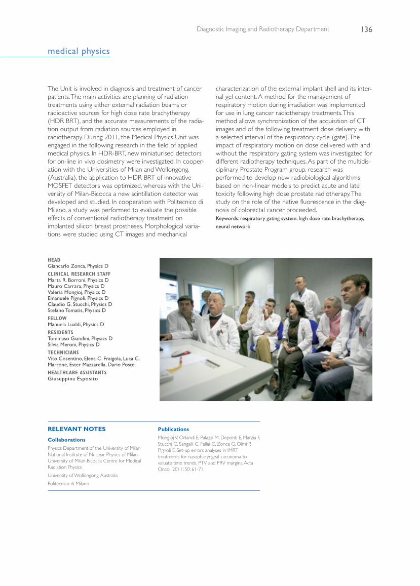
HEAD Giancarlo Zonca, Physics D
CLINICAL RESEARCH STAFFMarta R. Borroni, Physics D Mauro Carrara, Physics D Valeria Mongioj, Physics D Emanuele Pignoli, Physics DClaudio G. Stucchi, Physics D Stefano Tomatis, Physics D
FELLOWManuela Lualdi, Physics D
RESIDENTSTommaso Giandini, Physics D Silvia Meroni, Physics D
TECHNICIANSVito Cosentino, Elena C. Fraigola, Luca C.Marrone, Ester Mazzarella, Dario Postè
HEALTHCARE ASSISTANTSGiuseppina Esposito
The Unit is involved in diagnosis and treatment of cancerpatients. The main activities are planning of radiationtreatments using either external radiation beams orradioactive sources for high dose rate brachytherapy(HDR BRT), and the accurate measurements of the radia-tion output from radiation sources employed inradiotherapy. During 2011, the Medical Physics Unit wasengaged in the following research in the field of appliedmedical physics. In HDR-BRT, new miniaturised detectorsfor on-line in vivo dosimetry were investigated. In cooper-ation with the Universities of Milan and Wollongong,(Australia), the application to HDR BRT of innovativeMOSFET detectors was optimized, whereas with the Uni-versity of Milan-Bicocca a new scintillation detector wasdeveloped and studied. In cooperation with Politecnico diMilano, a study was performed to evaluate the possibleeffects of conventional radiotherapy treatment onimplanted silicon breast prostheses. Morphological varia-tions were studied using CT images and mechanical
characterization of the external implant shell and its inter-nal gel content. A method for the management ofrespiratory motion during irradiation was implementedfor use in lung cancer radiotherapy treatments. Thismethod allows synchronization of the acquisition of CTimages and of the following treatment dose delivery witha selected interval of the respiratory cycle (gate). Theimpact of respiratory motion on dose delivered with andwithout the respiratory gating system was investigated fordifferent radiotherapy techniques. As part of the multidis-ciplinary Prostate Program group, research wasperformed to develop new radiobiological algorithmsbased on non-linear models to predict acute and latetoxicity following high dose prostate radiotherapy. Thestudy on the role of the native fluorescence in the diag-nosis of colorectal cancer proceeded. Keywords: respiratory gating system, high dose rate brachytherapy,
neural network
RELEVANT NOTES
Collaborations
Physics Department of the University of MilanNational Institute of Nuclear Physics of Milan,University of Milan-Bicocca Centre for MedicalRadiation Physics
University of Wollongong, Australia
Politecnico di Milano
Publications
Mongioj V. Orlandi E, Palazzi M, Deponti E, Marzia F,Stucchi C, Sangalli C, Fallai C, Zonca G, Olmi P,Pignoli E. Set-up errors analyses in IMRTtreatments for nasopharyngeal carcinoma tovaluate time trends, PTV and PRV margins. ActaOncol. 2011; 50: 61-71.