Diagnostic Dilemma of a Rare, Giant Retroperitoneal ......Diagnostic Dilemma of a Rare, Giant...
4
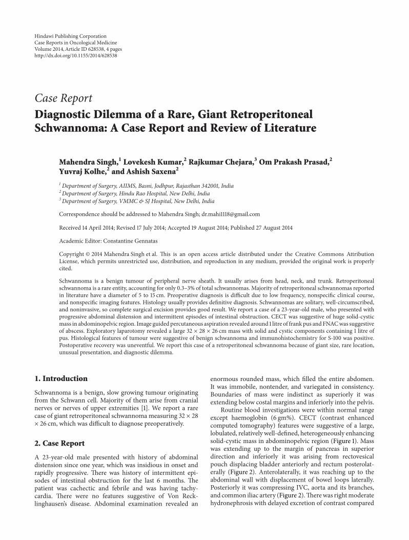
Case Report Diagnostic Dilemma of a Rare, Giant Retroperitoneal Schwannoma: A Case Report and Review of Literature Mahendra Singh, 1 Lovekesh Kumar, 2 Rajkumar Chejara, 3 Om Prakash Prasad, 2 Yuvraj Kolhe, 2 and Ashish Saxena 2 1 Department of Surgery, AIIMS, Basni, Jodhpur, Rajasthan 342001, India 2 Department of Surgery, Hindu Rao Hospital, New Delhi, India 3 Department of Surgery, VMMC & SJ Hospital, New Delhi, India Correspondence should be addressed to Mahendra Singh; [email protected] Received 14 April 2014; Revised 17 July 2014; Accepted 19 August 2014; Published 27 August 2014 Academic Editor: Constantine Gennatas Copyright © 2014 Mahendra Singh et al. is is an open access article distributed under the Creative Commons Attribution License, which permits unrestricted use, distribution, and reproduction in any medium, provided the original work is properly cited. Schwannoma is a benign tumour of peripheral nerve sheath. It usually arises from head, neck, and trunk. Retroperitoneal schwannoma is a rare entity, accounting for only 0.3–3% of total schwannomas. Majority of retroperitoneal schwannomas reported in literature have a diameter of 5 to 15cm. Preoperative diagnosis is difficult due to low frequency, nonspecific clinical course, and nonspecific imaging features. Histology usually provides definitive diagnosis. Schwannomas are solitary, well-circumscribed, and noninvasive, so complete surgical excision provides good result. We report a case of a 23-year-old male, who presented with progressive abdominal distension and intermittent episodes of intestinal obstruction. CECT was suggestive of huge solid-cystic mass in abdominopelvic region. Image guided percutaneous aspiration revealed around 1 litre of frank pus and FNAC was suggestive of abscess. Exploratory laparotomy revealed a large 32 × 28 × 26 cm mass with solid and cystic components containing 1 litre of pus. Histological features of tumour were suggestive of benign schwannoma and immunohistochemistry for S-100 was positive. Postoperative recovery was uneventful. We report this case of a retroperitoneal schwannoma because of giant size, rare location, unusual presentation, and diagnostic dilemma. 1. Introduction Schwannoma is a benign, slow growing tumour originating from the Schwann cell. Majority of them arise from cranial nerves or nerves of upper extremities [1]. We report a rare case of giant retroperitoneal schwannoma measuring 32 × 28 × 26 cm, which was difficult to diagnose preoperatively. 2. Case Report A 23-year-old male presented with history of abdominal distension since one year, which was insidious in onset and rapidly progressive. ere was history of intermittent epi- sodes of intestinal obstruction for the last 6 months. e patient was cachectic and febrile and was having tachy- cardia. ere were no features suggestive of Von Reck- linghausen’s disease. Abdominal examination revealed an enormous rounded mass, which filled the entire abdomen. It was immobile, nontender, and variegated in consistency. Boundaries of mass were indistinct as superiorly it was extending below costal margins and inferiorly into the pelvis. Routine blood investigations were within normal range except haemoglobin (6 gm%). CECT (contrast enhanced computed tomography) features were suggestive of a large, lobulated, relatively well-defined, heterogeneously enhancing solid-cystic mass in abdominopelvic region (Figure 1). Mass was extending up to the margin of pancreas in superior direction and inferiorly it was arising from rectovesical pouch displacing bladder anteriorly and rectum posterolat- erally (Figure 2). Anterolaterally, it was reaching up to the abdominal wall with displacement of bowel loops laterally. Posteriorly it was compressing IVC, aorta and its branches, and common iliac artery (Figure 2). ere was right moderate hydronephrosis with delayed excretion of contrast compared Hindawi Publishing Corporation Case Reports in Oncological Medicine Volume 2014, Article ID 628538, 4 pages http://dx.doi.org/10.1155/2014/628538
Transcript of Diagnostic Dilemma of a Rare, Giant Retroperitoneal ......Diagnostic Dilemma of a Rare, Giant...
Case ReportDiagnostic Dilemma of a Rare, Giant RetroperitonealSchwannoma: A Case Report and Review of Literature
Mahendra Singh,1 Lovekesh Kumar,2 Rajkumar Chejara,3 Om Prakash Prasad,2
Yuvraj Kolhe,2 and Ashish Saxena2
1 Department of Surgery, AIIMS, Basni, Jodhpur, Rajasthan 342001, India2Department of Surgery, Hindu Rao Hospital, New Delhi, India3 Department of Surgery, VMMC & SJ Hospital, New Delhi, India
Correspondence should be addressed to Mahendra Singh; [email protected]
Received 14 April 2014; Revised 17 July 2014; Accepted 19 August 2014; Published 27 August 2014
Academic Editor: Constantine Gennatas
Copyright © 2014 Mahendra Singh et al. This is an open access article distributed under the Creative Commons AttributionLicense, which permits unrestricted use, distribution, and reproduction in any medium, provided the original work is properlycited.
Schwannoma is a benign tumour of peripheral nerve sheath. It usually arises from head, neck, and trunk. Retroperitonealschwannoma is a rare entity, accounting for only 0.3–3% of total schwannomas. Majority of retroperitoneal schwannomas reportedin literature have a diameter of 5 to 15 cm. Preoperative diagnosis is difficult due to low frequency, nonspecific clinical course,and nonspecific imaging features. Histology usually provides definitive diagnosis. Schwannomas are solitary, well-circumscribed,and noninvasive, so complete surgical excision provides good result. We report a case of a 23-year-old male, who presented withprogressive abdominal distension and intermittent episodes of intestinal obstruction. CECT was suggestive of huge solid-cysticmass in abdominopelvic region. Image guided percutaneous aspiration revealed around 1 litre of frank pus andFNACwas suggestiveof abscess. Exploratory laparotomy revealed a large 32 × 28 × 26 cm mass with solid and cystic components containing 1 litre ofpus. Histological features of tumour were suggestive of benign schwannoma and immunohistochemistry for S-100 was positive.Postoperative recovery was uneventful. We report this case of a retroperitoneal schwannoma because of giant size, rare location,unusual presentation, and diagnostic dilemma.
1. Introduction
Schwannoma is a benign, slow growing tumour originatingfrom the Schwann cell. Majority of them arise from cranialnerves or nerves of upper extremities [1]. We report a rarecase of giant retroperitoneal schwannoma measuring 32 × 28× 26 cm, which was difficult to diagnose preoperatively.
2. Case Report
A 23-year-old male presented with history of abdominaldistension since one year, which was insidious in onset andrapidly progressive. There was history of intermittent epi-sodes of intestinal obstruction for the last 6 months. Thepatient was cachectic and febrile and was having tachy-cardia. There were no features suggestive of Von Reck-linghausen’s disease. Abdominal examination revealed an
enormous rounded mass, which filled the entire abdomen.It was immobile, nontender, and variegated in consistency.Boundaries of mass were indistinct as superiorly it wasextending below costal margins and inferiorly into the pelvis.
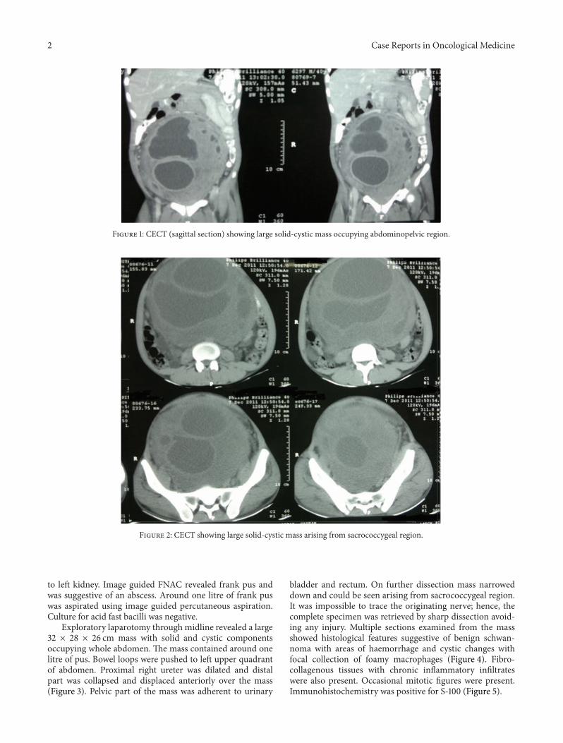
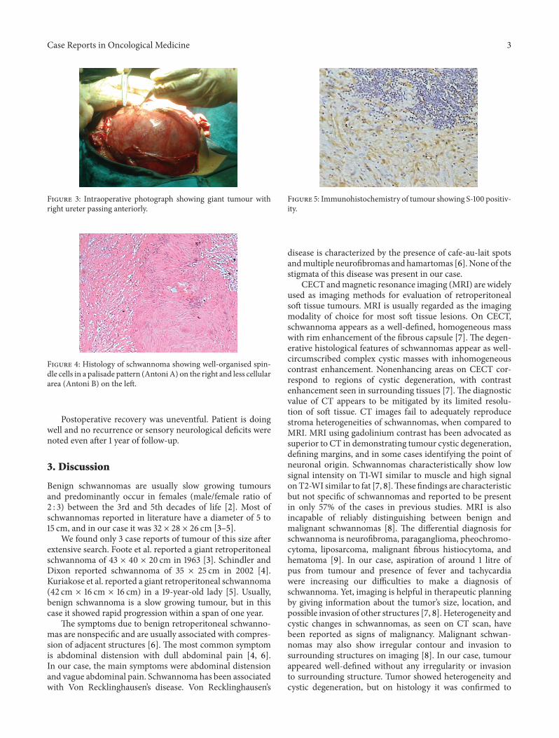
Routine blood investigations were within normal rangeexcept haemoglobin (6 gm%). CECT (contrast enhancedcomputed tomography) features were suggestive of a large,lobulated, relatively well-defined, heterogeneously enhancingsolid-cystic mass in abdominopelvic region (Figure 1). Masswas extending up to the margin of pancreas in superiordirection and inferiorly it was arising from rectovesicalpouch displacing bladder anteriorly and rectum posterolat-erally (Figure 2). Anterolaterally, it was reaching up to theabdominal wall with displacement of bowel loops laterally.Posteriorly it was compressing IVC, aorta and its branches,and common iliac artery (Figure 2).Therewas rightmoderatehydronephrosis with delayed excretion of contrast compared
Hindawi Publishing CorporationCase Reports in Oncological MedicineVolume 2014, Article ID 628538, 4 pageshttp://dx.doi.org/10.1155/2014/628538
2 Case Reports in Oncological Medicine
Figure 1: CECT (sagittal section) showing large solid-cystic mass occupying abdominopelvic region.
Figure 2: CECT showing large solid-cystic mass arising from sacrococcygeal region.
to left kidney. Image guided FNAC revealed frank pus andwas suggestive of an abscess. Around one litre of frank puswas aspirated using image guided percutaneous aspiration.Culture for acid fast bacilli was negative.
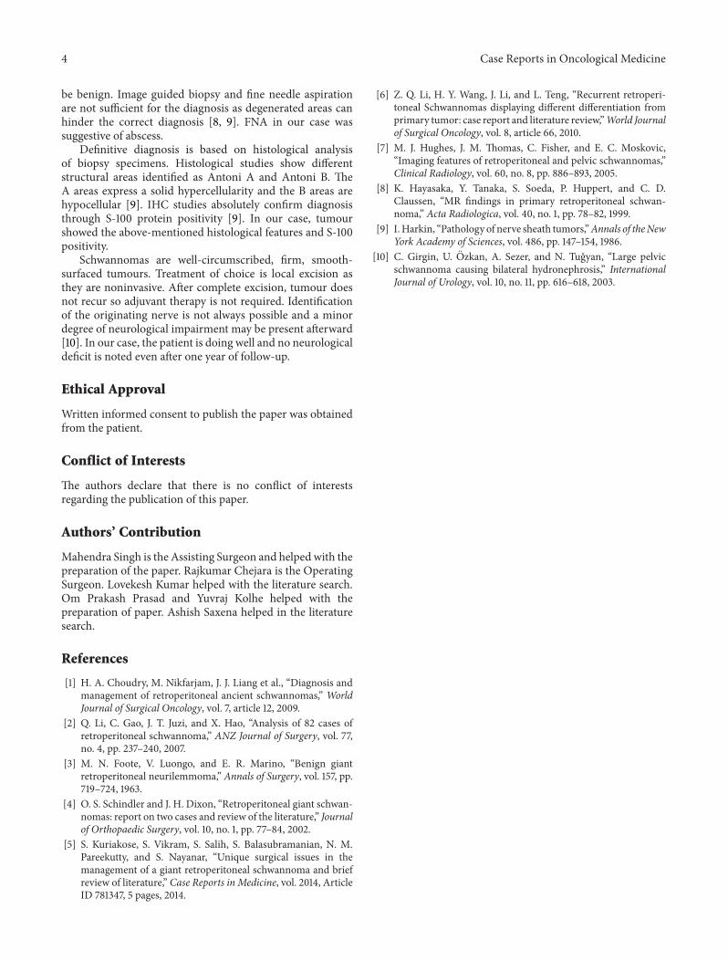
Exploratory laparotomy through midline revealed a large32 × 28 × 26 cm mass with solid and cystic componentsoccupying whole abdomen. The mass contained around onelitre of pus. Bowel loops were pushed to left upper quadrantof abdomen. Proximal right ureter was dilated and distalpart was collapsed and displaced anteriorly over the mass(Figure 3). Pelvic part of the mass was adherent to urinary
bladder and rectum. On further dissection mass narroweddown and could be seen arising from sacrococcygeal region.It was impossible to trace the originating nerve; hence, thecomplete specimen was retrieved by sharp dissection avoid-ing any injury. Multiple sections examined from the massshowed histological features suggestive of benign schwan-noma with areas of haemorrhage and cystic changes withfocal collection of foamy macrophages (Figure 4). Fibro-collagenous tissues with chronic inflammatory infiltrateswere also present. Occasional mitotic figures were present.Immunohistochemistry was positive for S-100 (Figure 5).
Case Reports in Oncological Medicine 3
Figure 3: Intraoperative photograph showing giant tumour withright ureter passing anteriorly.
Figure 4: Histology of schwannoma showing well-organised spin-dle cells in a palisade pattern (Antoni A) on the right and less cellulararea (Antoni B) on the left.
Postoperative recovery was uneventful. Patient is doingwell and no recurrence or sensory neurological deficits werenoted even after 1 year of follow-up.
3. Discussion
Benign schwannomas are usually slow growing tumoursand predominantly occur in females (male/female ratio of2 : 3) between the 3rd and 5th decades of life [2]. Most ofschwannomas reported in literature have a diameter of 5 to15 cm, and in our case it was 32 × 28 × 26 cm [3–5].
We found only 3 case reports of tumour of this size afterextensive search. Foote et al. reported a giant retroperitonealschwannoma of 43 × 40 × 20 cm in 1963 [3]. Schindler andDixon reported schwannoma of 35 × 25 cm in 2002 [4].Kuriakose et al. reported a giant retroperitoneal schwannoma(42 cm × 16 cm × 16 cm) in a 19-year-old lady [5]. Usually,benign schwannoma is a slow growing tumour, but in thiscase it showed rapid progression within a span of one year.
The symptoms due to benign retroperitoneal schwanno-mas are nonspecific and are usually associated with compres-sion of adjacent structures [6]. The most common symptomis abdominal distension with dull abdominal pain [4, 6].In our case, the main symptoms were abdominal distensionand vague abdominal pain. Schwannoma has been associatedwith Von Recklinghausen’s disease. Von Recklinghausen’s
Figure 5: Immunohistochemistry of tumour showing S-100 positiv-ity.
disease is characterized by the presence of cafe-au-lait spotsandmultiple neurofibromas andhamartomas [6].None of thestigmata of this disease was present in our case.
CECT andmagnetic resonance imaging (MRI) are widelyused as imaging methods for evaluation of retroperitonealsoft tissue tumours. MRI is usually regarded as the imagingmodality of choice for most soft tissue lesions. On CECT,schwannoma appears as a well-defined, homogeneous masswith rim enhancement of the fibrous capsule [7]. The degen-erative histological features of schwannomas appear as well-circumscribed complex cystic masses with inhomogeneouscontrast enhancement. Nonenhancing areas on CECT cor-respond to regions of cystic degeneration, with contrastenhancement seen in surrounding tissues [7]. The diagnosticvalue of CT appears to be mitigated by its limited resolu-tion of soft tissue. CT images fail to adequately reproducestroma heterogeneities of schwannomas, when compared toMRI. MRI using gadolinium contrast has been advocated assuperior to CT in demonstrating tumour cystic degeneration,defining margins, and in some cases identifying the point ofneuronal origin. Schwannomas characteristically show lowsignal intensity on T1-WI similar to muscle and high signalonT2-WI similar to fat [7, 8].These findings are characteristicbut not specific of schwannomas and reported to be presentin only 57% of the cases in previous studies. MRI is alsoincapable of reliably distinguishing between benign andmalignant schwannomas [8]. The differential diagnosis forschwannoma is neurofibroma, paraganglioma, pheochromo-cytoma, liposarcoma, malignant fibrous histiocytoma, andhematoma [9]. In our case, aspiration of around 1 litre ofpus from tumour and presence of fever and tachycardiawere increasing our difficulties to make a diagnosis ofschwannoma. Yet, imaging is helpful in therapeutic planningby giving information about the tumor’s size, location, andpossible invasion of other structures [7, 8]. Heterogeneity andcystic changes in schwannomas, as seen on CT scan, havebeen reported as signs of malignancy. Malignant schwan-nomas may also show irregular contour and invasion tosurrounding structures on imaging [8]. In our case, tumourappeared well-defined without any irregularity or invasionto surrounding structure. Tumor showed heterogeneity andcystic degeneration, but on histology it was confirmed to
4 Case Reports in Oncological Medicine
be benign. Image guided biopsy and fine needle aspirationare not sufficient for the diagnosis as degenerated areas canhinder the correct diagnosis [8, 9]. FNA in our case wassuggestive of abscess.
Definitive diagnosis is based on histological analysisof biopsy specimens. Histological studies show differentstructural areas identified as Antoni A and Antoni B. TheA areas express a solid hypercellularity and the B areas arehypocellular [9]. IHC studies absolutely confirm diagnosisthrough S-100 protein positivity [9]. In our case, tumourshowed the above-mentioned histological features and S-100positivity.
Schwannomas are well-circumscribed, firm, smooth-surfaced tumours. Treatment of choice is local excision asthey are noninvasive. After complete excision, tumour doesnot recur so adjuvant therapy is not required. Identificationof the originating nerve is not always possible and a minordegree of neurological impairment may be present afterward[10]. In our case, the patient is doing well and no neurologicaldeficit is noted even after one year of follow-up.
Ethical Approval
Written informed consent to publish the paper was obtainedfrom the patient.
Conflict of Interests
The authors declare that there is no conflict of interestsregarding the publication of this paper.
Authors’ Contribution
Mahendra Singh is the Assisting Surgeon and helped with thepreparation of the paper. Rajkumar Chejara is the OperatingSurgeon. Lovekesh Kumar helped with the literature search.Om Prakash Prasad and Yuvraj Kolhe helped with thepreparation of paper. Ashish Saxena helped in the literaturesearch.
References
[1] H. A. Choudry, M. Nikfarjam, J. J. Liang et al., “Diagnosis andmanagement of retroperitoneal ancient schwannomas,” WorldJournal of Surgical Oncology, vol. 7, article 12, 2009.
[2] Q. Li, C. Gao, J. T. Juzi, and X. Hao, “Analysis of 82 cases ofretroperitoneal schwannoma,” ANZ Journal of Surgery, vol. 77,no. 4, pp. 237–240, 2007.
[3] M. N. Foote, V. Luongo, and E. R. Marino, “Benign giantretroperitoneal neurilemmoma,” Annals of Surgery, vol. 157, pp.719–724, 1963.
[4] O. S. Schindler and J. H. Dixon, “Retroperitoneal giant schwan-nomas: report on two cases and review of the literature,” Journalof Orthopaedic Surgery, vol. 10, no. 1, pp. 77–84, 2002.
[5] S. Kuriakose, S. Vikram, S. Salih, S. Balasubramanian, N. M.Pareekutty, and S. Nayanar, “Unique surgical issues in themanagement of a giant retroperitoneal schwannoma and briefreview of literature,” Case Reports in Medicine, vol. 2014, ArticleID 781347, 5 pages, 2014.
[6] Z. Q. Li, H. Y. Wang, J. Li, and L. Teng, “Recurrent retroperi-toneal Schwannomas displaying different differentiation fromprimary tumor: case report and literature review,”World Journalof Surgical Oncology, vol. 8, article 66, 2010.
[7] M. J. Hughes, J. M. Thomas, C. Fisher, and E. C. Moskovic,“Imaging features of retroperitoneal and pelvic schwannomas,”Clinical Radiology, vol. 60, no. 8, pp. 886–893, 2005.
[8] K. Hayasaka, Y. Tanaka, S. Soeda, P. Huppert, and C. D.Claussen, “MR findings in primary retroperitoneal schwan-noma,” Acta Radiologica, vol. 40, no. 1, pp. 78–82, 1999.
[9] I.Harkin, “Pathology of nerve sheath tumors,”Annals of theNewYork Academy of Sciences, vol. 486, pp. 147–154, 1986.
[10] C. Girgin, U. Ozkan, A. Sezer, and N. Tugyan, “Large pelvicschwannoma causing bilateral hydronephrosis,” InternationalJournal of Urology, vol. 10, no. 11, pp. 616–618, 2003.