
Diagnosis and Treatment of Diabetic Foot Infections - IDSA · Diagnosis and Treatment of Diabetic...
26
Guidelines for Diabetic Foot Infections • CID 2004:39 (1 October) • 885 IDSA GUIDELINES Diagnosis and Treatment of Diabetic Foot Infections Benjamin A. Lipsky, 1,a Anthony R. Berendt, 2,a H. Gunner Deery, 3 John M. Embil, 4 Warren S. Joseph, 5 Adolf W. Karchmer, 6 Jack L. LeFrock, 7 Daniel P. Lew, 8 Jon T. Mader, 9,b Carl Norden, 10 and James S. Tan 11 1 Medical Service, Veterans Affairs Puget Sound Health Care System, and Division of General Internal Medicine, Department of Medicine, University of Washington School of Medicine, Seattle, Washington; 2 Bone Infection Unit, Nuffield Orthopaedic Centre, Oxford, United Kingdom; 3 Northern Michigan Infectious Diseases, Petoskey, Michigan; 4 Section of Infectious Diseases, Department of Medicine, University of Manitoba, Winnipeg, Manitoba; 5 Section of Podiatry, Department of Primary Care, Veterans Affairs Medical Center, Coatesville, Pennsylvania; 6 Division of Infectious Diseases, Department of Medicine, Harvard Medical School, and Beth Israel Deaconess Medical Center, Boston, Massachusetts; 7 Dimensional Dosing Systems, Sarasota, Florida; 8 Department of Medicine, Service of Infectious Diseases, University of Geneva Hospitals, Geneva, Switzerland; 9 Department of Internal Medicine, The Marine Biomedical Institute, and Department of Orthopaedics and Rehabilitation, University of Texas Medical Branch, Galveston, Texas; 10 Department of Medicine, New Jersey School of Medicine and Dentistry, and Cooper Hospital, Camden, New Jersey; and 11 Department of Internal Medicine, Summa Health System, and Northeastern Ohio Universities College of Medicine, Akron, Ohio EXECUTIVE SUMMARY 1. Foot infections in patients with diabetes cause substantial morbidity and frequent visits to health care professionals and may lead to amputation of a lower extremity. 2. Diabetic foot infections require attention to local (foot) and systemic (metabolic) issues and coordinated management, preferably by a multidisciplinary foot- care team (A-II) (table 1). The team managing these infections should include, or have ready access to, an infectious diseases specialist or a medical microbiologist (B-II). 3. The major predisposing factor to these infections is foot ulceration, which is usually related to peripheral neuropathy. Peripheral vascular disease and various im- munological disturbances play a secondary role. 4. Aerobic gram-positive cocci (especially Staphy- lococcus aureus) are the predominant pathogens in diabetic foot infections. Patients who have chronic Received 2 July 2004; accepted 2 July 2004; electronically published 10 September 2004. These guidelines were developed and issued on behalf of the Infectious Diseases Society of America. a B.A.L. served as the chairman and A.R.B. served as the vice chairman of the Infectious Diseases Society of America Guidelines Committee on Diabetic Foot Infections. b Deceased. Reprints or correspondence: Dr. Benjamin A. Lipsky, Veterans Affairs Puget Sound Health Care System, S-111-GIMC, 1660 S. Columbian Way, Seattle, WA 98108- 9804 ([email protected]). Clinical Infectious Diseases 2004; 39:885–910 This article is in the public domain, and no copyright is claimed. 1058-4838/2004/3907-0001 wounds or who have recently received antibiotic ther- apy may also be infected with gram-negative rods, and those with foot ischemia or gangrene may have obligate anaerobic pathogens. 5. Wound infections must be diagnosed clinically on the basis of local (and occasionally systemic) signs and symptoms of inflammation. Laboratory (including microbiological) investigations are of limited use for diagnosing infection, except in cases of osteomyelitis (B-II). 6. Send appropriately obtained specimens for cul- ture prior to starting empirical antibiotic therapy in all cases of infection, except perhaps those that are mild and previously untreated (B-III). Tissue specimens ob- tained by biopsy, ulcer curettage, or aspiration are pref- erable to wound swab specimens (A-I). 7. Imaging studies may help diagnose or better de- fine deep, soft-tissue purulent collections and are usu- ally needed to detect pathological findings in bone. Plain radiography may be adequate in many cases, but MRI (in preference to isotope scanning) is more sen- sitive and specific, especially for detection of soft-tissue lesions (A-I). 8. Infections should be categorized by their severity on the basis of readily assessable clinical and laboratory features (B-II). Most important among these are the specific tissues involved, the adequacy of arterial per- fusion, and the presence of systemic toxicity or meta- bolic instability. Categorization helps determine the de- gree of risk to the patient and the limb and, thus, the urgency and venue of management. 9. Available evidence does not support treat- at IDSA on August 14, 2011 cid.oxfordjournals.org Downloaded from
Transcript of Diagnosis and Treatment of Diabetic Foot Infections - IDSA · Diagnosis and Treatment of Diabetic...

Guidelines for Diabetic Foot Infections • CID 2004:39 (1 October) • 885
I D S A G U I D E L I N E S
Diagnosis and Treatment of Diabetic Foot Infections
Benjamin A. Lipsky,1,a Anthony R. Berendt,2,a H. Gunner Deery,3 John M. Embil,4 Warren S. Joseph,5
Adolf W. Karchmer,6 Jack L. LeFrock,7 Daniel P. Lew,8 Jon T. Mader,9,b Carl Norden,10 and James S. Tan11
1Medical Service, Veterans Affairs Puget Sound Health Care System, and Division of General Internal Medicine, Department of Medicine,University of Washington School of Medicine, Seattle, Washington; 2Bone Infection Unit, Nuffield Orthopaedic Centre, Oxford, United Kingdom;3Northern Michigan Infectious Diseases, Petoskey, Michigan; 4Section of Infectious Diseases, Department of Medicine, University of Manitoba,Winnipeg, Manitoba; 5Section of Podiatry, Department of Primary Care, Veterans Affairs Medical Center, Coatesville, Pennsylvania; 6Divisionof Infectious Diseases, Department of Medicine, Harvard Medical School, and Beth Israel Deaconess Medical Center, Boston, Massachusetts;7Dimensional Dosing Systems, Sarasota, Florida; 8Department of Medicine, Service of Infectious Diseases, University of Geneva Hospitals,Geneva, Switzerland; 9Department of Internal Medicine, The Marine Biomedical Institute, and Department of Orthopaedics and Rehabilitation,University of Texas Medical Branch, Galveston, Texas; 10Department of Medicine, New Jersey School of Medicine and Dentistry, and CooperHospital, Camden, New Jersey; and 11Department of Internal Medicine, Summa Health System, and Northeastern Ohio UniversitiesCollege of Medicine, Akron, Ohio
EXECUTIVE SUMMARY
1. Foot infections in patients with diabetes cause
substantial morbidity and frequent visits to health care
professionals and may lead to amputation of a lower
extremity.
2. Diabetic foot infections require attention to local
(foot) and systemic (metabolic) issues and coordinated
management, preferably by a multidisciplinary foot-
care team (A-II) (table 1). The team managing these
infections should include, or have ready access to, an
infectious diseases specialist or a medical microbiologist
(B-II).
3. The major predisposing factor to these infections
is foot ulceration, which is usually related to peripheral
neuropathy. Peripheral vascular disease and various im-
munological disturbances play a secondary role.
4. Aerobic gram-positive cocci (especially Staphy-
lococcus aureus) are the predominant pathogens in
diabetic foot infections. Patients who have chronic
Received 2 July 2004; accepted 2 July 2004; electronically published 10September 2004.
These guidelines were developed and issued on behalf of the InfectiousDiseases Society of America.
a B.A.L. served as the chairman and A.R.B. served as the vice chairman of theInfectious Diseases Society of America Guidelines Committee on Diabetic FootInfections.
b Deceased.Reprints or correspondence: Dr. Benjamin A. Lipsky, Veterans Affairs Puget Sound
Health Care System, S-111-GIMC, 1660 S. Columbian Way, Seattle, WA 98108-9804 ([email protected]).
Clinical Infectious Diseases 2004; 39:885–910This article is in the public domain, and no copyright is claimed.1058-4838/2004/3907-0001
wounds or who have recently received antibiotic ther-
apy may also be infected with gram-negative rods, and
those with foot ischemia or gangrene may have obligate
anaerobic pathogens.
5. Wound infections must be diagnosed clinically
on the basis of local (and occasionally systemic) signs
and symptoms of inflammation. Laboratory (including
microbiological) investigations are of limited use for
diagnosing infection, except in cases of osteomyelitis
(B-II).
6. Send appropriately obtained specimens for cul-
ture prior to starting empirical antibiotic therapy in all
cases of infection, except perhaps those that are mild
and previously untreated (B-III). Tissue specimens ob-
tained by biopsy, ulcer curettage, or aspiration are pref-
erable to wound swab specimens (A-I).
7. Imaging studies may help diagnose or better de-
fine deep, soft-tissue purulent collections and are usu-
ally needed to detect pathological findings in bone.
Plain radiography may be adequate in many cases, but
MRI (in preference to isotope scanning) is more sen-
sitive and specific, especially for detection of soft-tissue
lesions (A-I).
8. Infections should be categorized by their severity
on the basis of readily assessable clinical and laboratory
features (B-II). Most important among these are the
specific tissues involved, the adequacy of arterial per-
fusion, and the presence of systemic toxicity or meta-
bolic instability. Categorization helps determine the de-
gree of risk to the patient and the limb and, thus, the
urgency and venue of management.
9. Available evidence does not support treat-
at IDS
A on A
ugust 14, 2011cid.oxfordjournals.org
Dow
nloaded from

886 • CID 2004:39 (1 October) • Lipsky et al.
Table 1. Infectious Diseases Society of America–United States Public Health Service Grading System for ranking rec-ommendations in clinical guidelines.
Category, grade Definition
Strength of recommendationA Good evidence to support a recommendation for use; should always be offeredB Moderate evidence to support a recommendation for use; should generally be offeredC Poor evidence to support a recommendation; optionalD Moderate evidence to support a recommendation against use; should generally not be offeredE Good evidence to support a recommendation against use; should never be offered
Quality of evidenceI Evidence from �1 properly randomized, controlled trialII Evidence from �1 well-designed clinical trial, without randomization; from cohort or case-
controlled analytic studies (preferably from 11 center); from multiple time-series; or fromdramatic results from uncontrolled experiments
III Evidence from opinions of respected authorities, based on clinical experience, descriptivestudies, or reports of expert committees
ing clinically uninfected ulcers with antibiotic therapy (D-III).
Antibiotic therapy is necessary for virtually all infected wounds,
but it is often insufficient without appropriate wound care.
10. Select an empirical antibiotic regimen on the basis of
the severity of the infection and the likely etiologic agent(s)
(B-II). Therapy aimed solely at aerobic gram-positive cocci may
be sufficient for mild-to-moderate infections in patients who
have not recently received antibiotic therapy (A-II). Broad-
spectrum empirical therapy is not routinely required but is
indicated for severe infections, pending culture results and an-
tibiotic susceptibility data (B-III). Take into consideration any
recent antibiotic therapy and local antibiotic susceptibility data,
especially the prevalence of methicillin-resistant S. aureus
(MRSA) or other resistant organisms. Definitive therapy should
be based on both the culture results and susceptibility data and
the clinical response to the empirical regimen (C-III).
11. There is only limited evidence with which to make
informed choices among the various topical, oral, and paren-
teral antibiotic agents. Virtually all severe and some moderate
infections require parenteral therapy, at least initially (C-III).
Highly bioavailable oral antibiotics can be used in most mild
and in many moderate infections, including some cases of os-
teomyelitis (A-II). Topical therapy may be used for some mild
superficial infections (B-I).
12. Continue antibiotic therapy until there is evidence that
the infection has resolved but not necessarily until a wound
has healed. Suggestions for the duration of antibiotic therapy
are as follows: for mild infections, 1–2 weeks usually suffices,
but some require an additional 1–2 weeks; for moderate and
severe infections, usually 2–4 weeks is sufficient, depending on
the structures involved, the adequacy of debridement, the type
of soft-tissue wound cover, and wound vascularity (A-II); and
for osteomyelitis, generally at least 4–6 weeks is required, but
a shorter duration is sufficient if the entire infected bone is
removed, and probably a longer duration is needed if infected
bone remains (B-II).
13. If an infection in a clinically stable patient fails to re-
spond to �1 antibiotic courses, consider discontinuing all an-
timicrobials and, after a few days, obtaining optimal culture
specimens (C-III).
14. Seek surgical consultation and, when needed, interven-
tion for infections accompanied by a deep abscess, extensive
bone or joint involvement, crepitus, substantial necrosis or gan-
grene, or necrotizing fasciitis (A-II). Evaluating the limb’s ar-
terial supply and revascularizing when indicated are particularly
important. Surgeons with experience and interest in the field
should be recruited by the foot-care team, if possible.
15. Providing optimal wound care, in addition to appro-
priate antibiotic treatment of the infection, is crucial for healing
(A-I). This includes proper wound cleansing, debridement of
any callus and necrotic tissue, and, especially, off-loading of
pressure. There is insufficient evidence to recommend use of
a specific wound dressing or any type of wound healing agents
or products for infected foot wounds.
16. Patients with infected wounds require early and careful
follow-up observation to ensure that the selected medical and
surgical treatment regimens have been appropriate and effective
(B-III).
17. Studies have not adequately defined the role of most
adjunctive therapies for diabetic foot infections, but systematic
reviews suggest that granulocyte colony-stimulating factors and
systemic hyperbaric oxygen therapy may help prevent ampu-
tations (B-I). These treatments may be useful for severe infec-
tions or for those that have not adequately responded to ther-
apy, despite correcting for all amenable local and systemic
adverse factors.
18. Spread of infection to bone (osteitis or osteomyelitis)
may be difficult to distinguish from noninfectious osteoar-
at IDS
A on A
ugust 14, 2011cid.oxfordjournals.org
Dow
nloaded from
Guidelines for Diabetic Foot Infections • CID 2004:39 (1 October) • 887
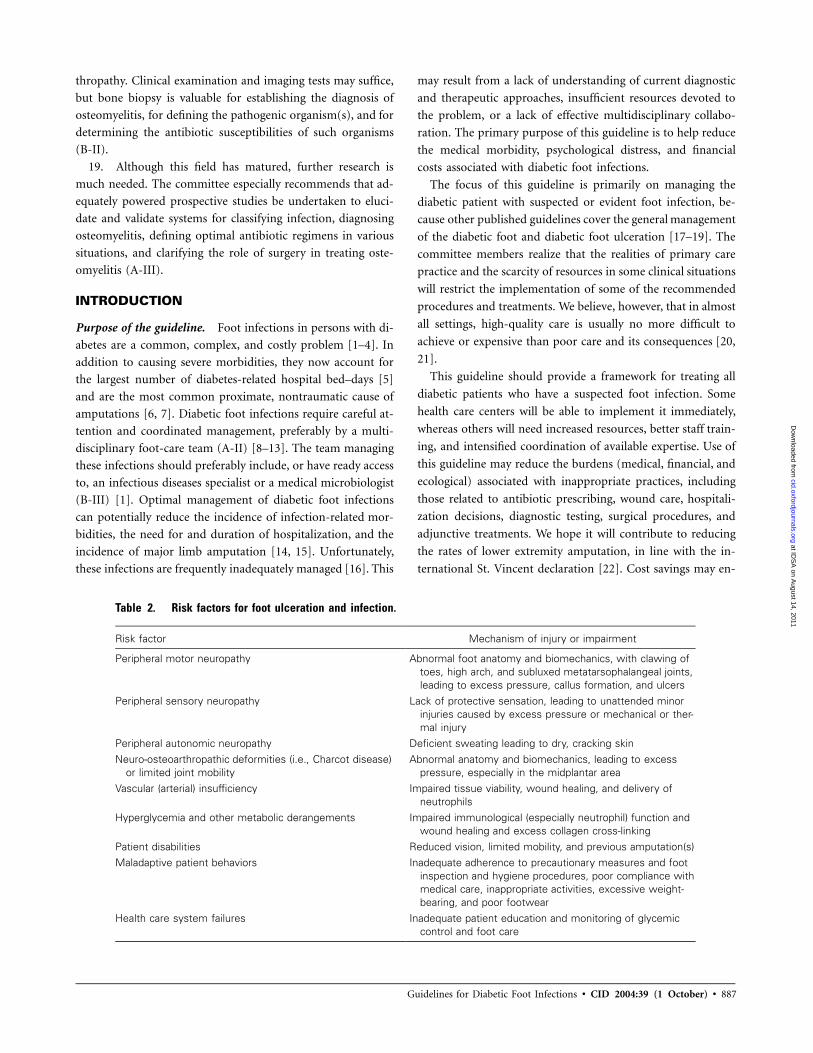
Table 2. Risk factors for foot ulceration and infection.
Risk factor Mechanism of injury or impairment
Peripheral motor neuropathy Abnormal foot anatomy and biomechanics, with clawing oftoes, high arch, and subluxed metatarsophalangeal joints,leading to excess pressure, callus formation, and ulcers
Peripheral sensory neuropathy Lack of protective sensation, leading to unattended minorinjuries caused by excess pressure or mechanical or ther-mal injury
Peripheral autonomic neuropathy Deficient sweating leading to dry, cracking skinNeuro-osteoarthropathic deformities (i.e., Charcot disease)
or limited joint mobilityAbnormal anatomy and biomechanics, leading to excess
pressure, especially in the midplantar areaVascular (arterial) insufficiency Impaired tissue viability, wound healing, and delivery of
neutrophilsHyperglycemia and other metabolic derangements Impaired immunological (especially neutrophil) function and
wound healing and excess collagen cross-linkingPatient disabilities Reduced vision, limited mobility, and previous amputation(s)Maladaptive patient behaviors Inadequate adherence to precautionary measures and foot
inspection and hygiene procedures, poor compliance withmedical care, inappropriate activities, excessive weight-bearing, and poor footwear
Health care system failures Inadequate patient education and monitoring of glycemiccontrol and foot care
thropathy. Clinical examination and imaging tests may suffice,
but bone biopsy is valuable for establishing the diagnosis of
osteomyelitis, for defining the pathogenic organism(s), and for
determining the antibiotic susceptibilities of such organisms
(B-II).
19. Although this field has matured, further research is
much needed. The committee especially recommends that ad-
equately powered prospective studies be undertaken to eluci-
date and validate systems for classifying infection, diagnosing
osteomyelitis, defining optimal antibiotic regimens in various
situations, and clarifying the role of surgery in treating oste-
omyelitis (A-III).
INTRODUCTION
Purpose of the guideline. Foot infections in persons with di-
abetes are a common, complex, and costly problem [1–4]. In
addition to causing severe morbidities, they now account for
the largest number of diabetes-related hospital bed–days [5]
and are the most common proximate, nontraumatic cause of
amputations [6, 7]. Diabetic foot infections require careful at-
tention and coordinated management, preferably by a multi-
disciplinary foot-care team (A-II) [8–13]. The team managing
these infections should preferably include, or have ready access
to, an infectious diseases specialist or a medical microbiologist
(B-III) [1]. Optimal management of diabetic foot infections
can potentially reduce the incidence of infection-related mor-
bidities, the need for and duration of hospitalization, and the
incidence of major limb amputation [14, 15]. Unfortunately,
these infections are frequently inadequately managed [16]. This
may result from a lack of understanding of current diagnostic
and therapeutic approaches, insufficient resources devoted to
the problem, or a lack of effective multidisciplinary collabo-
ration. The primary purpose of this guideline is to help reduce
the medical morbidity, psychological distress, and financial
costs associated with diabetic foot infections.
The focus of this guideline is primarily on managing the
diabetic patient with suspected or evident foot infection, be-
cause other published guidelines cover the general management
of the diabetic foot and diabetic foot ulceration [17–19]. The
committee members realize that the realities of primary care
practice and the scarcity of resources in some clinical situations
will restrict the implementation of some of the recommended
procedures and treatments. We believe, however, that in almost
all settings, high-quality care is usually no more difficult to
achieve or expensive than poor care and its consequences [20,
21].
This guideline should provide a framework for treating all
diabetic patients who have a suspected foot infection. Some
health care centers will be able to implement it immediately,
whereas others will need increased resources, better staff train-
ing, and intensified coordination of available expertise. Use of
this guideline may reduce the burdens (medical, financial, and
ecological) associated with inappropriate practices, including
those related to antibiotic prescribing, wound care, hospitali-
zation decisions, diagnostic testing, surgical procedures, and
adjunctive treatments. We hope it will contribute to reducing
the rates of lower extremity amputation, in line with the in-
ternational St. Vincent declaration [22]. Cost savings may en-
at IDS
A on A
ugust 14, 2011cid.oxfordjournals.org
Dow
nloaded from

888 • CID 2004:39 (1 October) • Lipsky et al.
Table 3. Pathogens associated with various clinical foot-infection syndromes.
Foot-infection syndrome Pathogens
Cellulitis without an open skin wound b-Hemolytic streptococcusa and Staphylococcus aureusInfected ulcer and antibiotic naiveb S. aureus and b-hemolytic streptococcusa
Infected ulcer that is chronic or was previously treated withantibiotic therapyc
S. aureus, b-hemolytic streptococcus, andEnterobacteriaceae
Ulcer that is macerated because of soakingc Pseudomonas aeruginosa (often in combination with otherorganisms)
Long duration nonhealing wounds with prolonged, broad-spectrum antibiotic therapyc,d
Aerobic gram-positive cocci (S. aureus , coagulase-negativestaphylococci, and enterococci), diphtheroids, Enterobac-teriaceae, Pseudomonas species, nonfermentative gram-negative rods, and, possibly, fungi
“Fetid foot”: extensive necrosis or gangrene, malodorousc Mixed aerobic gram-positive cocci, including enterococci,Enterobacteriaceae, nonfermentative gram-negative rods,and obligate anaerobes
a Groups A, B, C, and G.b Often monomicrobial.c Usually polymicrobial.d Antibiotic-resistant species (e.g., methicillin-resistant S. aureus, vancomycin-resistant enterococci, or extended-spectrum b-lactamase
producing gram-negative rods) are common.
sue, although this may be offset by an increased demand for
preventive foot care, diagnostic testing (especially MRI), and
vascular interventions [12].
Methodology. This guideline committee is comprised of
Infectious Diseases Society of America members with experi-
ence and interest in diabetic foot infections, many of whom
also have experience in writing guidelines. Committee members
are from several US states and other countries; their back-
grounds represent academia, bench and clinical research, in-
fectious diseases clinical practice, podiatry, and industry. Three
of the members are also members of the International Working
Group on the Diabetic Foot, which published its International
Consensus Guidelines on Diagnosing and Treating Diabetic
Foot Infections in 2003 [23]. After an extensive literature search
(which included the MEDLINE database, the EBSCO database,
the Cochrane Library, diabetic foot Web sites and bibliogra-
phies, and hand-searching of bibliographies of published ar-
ticles), committee members reviewed and discussed all available
evidence in a series of meetings and established consensus
through discussion and debate over a period of 3 years. Three
subcommittees drafted subsections that were modified and ex-
changed; these served as a basis for the final document, which
underwent numerous revisions that were based on both internal
and external reviews. Because of the relative paucity of ran-
domized controlled trials or other high-quality evidence in this
field, most of our recommendations are based on discussion
and consensus (B-II) (table 1) [24]. Thus, we elected to offer
a relatively brief summary and to provide an extensive bibli-
ography for those who would like to review the data themselves.
PATHOPHYSIOLOGY OF INFECTION
A diabetic foot infection is most simply defined as any infra-
malleolar infection in a person with diabetes mellitus. These
include paronychia, cellulitis, myositis, abscesses, necrotizing
fasciitis, septic arthritis, tendonitis, and osteomyelitis. The most
common and classical lesion, however, is the infected diabetic
“mal perforans” foot ulcer. This wound results from a complex
amalgam of risk factors [25, 26], which are summarized in
table 2. Neuropathy plays the central role, with disturbances
of sensory, motor, and autonomic functions leading to ulcer-
ation due to trauma or excessive pressure on a deformed foot
that lacks protective sensation. Once the protective layer of skin
is breached, underlying tissues are exposed to bacterial colo-
nization. This wound may progress to become actively infected,
and, by contiguous extension, the infection can involve deeper
tissues. This sequence of events can be rapid (occurring over
days or even hours), especially in an ischemic limb. Various
poorly characterized immunologic disturbances, especially
those that involve polymorphonuclear leukocytes, may affect
some diabetic patients, and these likely increase the risk and
severity of foot infections [27–30].
MICROBIOLOGY
Aerobic gram-positive cocci are the predominant microorgan-
isms that colonize and acutely infect breaks in the skin. S. aureus
and the b-hemolytic streptococci (groups A, C, and G, but
especially group B) are the most commonly isolated pathogens
[31–38]. Chronic wounds develop a more complex colonizing
flora, including enterococci, various Enterobacteriaceae, obli-
gate anaerobes, Pseudomonas aeruginosa, and, sometimes, other
nonfermentative gram-negative rods [39–43]. Hospitalization,
surgical procedures, and, especially, prolonged or broad-spec-
trum antibiotic therapy may predispose patients to colonization
and/or infection with antibiotic-resistant organisms (e.g.,
MRSA or vancomycin-resistant enterococci [VRE]) [44]. Al-
at IDS
A on A
ugust 14, 2011cid.oxfordjournals.org
Dow
nloaded from
Guidelines for Diabetic Foot Infections • CID 2004:39 (1 October) • 889
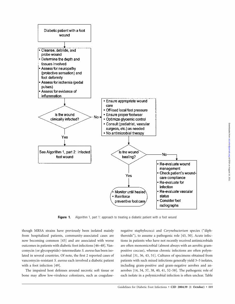
Figure 1. Algorithm 1, part 1: approach to treating a diabetic patient with a foot wound
though MRSA strains have previously been isolated mainly
from hospitalized patients, community-associated cases are
now becoming common [45] and are associated with worse
outcomes in patients with diabetic foot infections [46–48]. Van-
comycin (or glycopeptide)–intermediate S. aureus has been iso-
lated in several countries. Of note, the first 2 reported cases of
vancomycin-resistant S. aureus each involved a diabetic patient
with a foot infection [49].
The impaired host defenses around necrotic soft tissue or
bone may allow low-virulence colonizers, such as coagulase-
negative staphylococci and Corynebacterium species (“diph-
theroids”), to assume a pathogenic role [43, 50]. Acute infec-
tions in patients who have not recently received antimicrobials
are often monomicrobial (almost always with an aerobic gram-
positive coccus), whereas chronic infections are often polym-
icrobial [31, 36, 43, 51]. Cultures of specimens obtained from
patients with such mixed infections generally yield 3–5 isolates,
including gram-positive and gram-negative aerobes and an-
aerobes [14, 34, 37, 38, 40, 41, 52–58]. The pathogenic role of
each isolate in a polymicrobial infection is often unclear. Table
at IDS
A on A
ugust 14, 2011cid.oxfordjournals.org
Dow
nloaded from

890
Tabl
e4.
Eval
uatin
gth
edi
abet
icpa
tient
who
has
anin
fect
edfo
ot.
Leve
lof
eval
uatio
n,by
area
(s)
tobe
asse
ssed
Rel
evan
tpr
oble
ms
and
obse
rvat
ions
Inve
stig
atio
ns
Pat
ient
Sys
tem
icre
spon
seto
infe
ctio
nFe
ver,
chill
s,sw
eats
,vo
miti
ng,
hypo
tens
ion,
and
tach
ycar
dia
His
tory
and
phys
ical
exam
inat
ion
Met
abol
icst
ate
Volu
me
depl
etio
n,az
otem
ia,
hype
rgly
cem
ia,
tach
ypne
ahy
pero
smol
ality
,aci
dosi
sS
erum
chem
istr
yan
alys
esan
dhe
mat
olog
ical
test
ing
Psy
chol
ogic
al/c
ogni
tive
stat
eD
eliri
um,d
emen
tia,
depr
essi
on,
impa
ired
cogn
ition
,an
dst
upor
Ass
essm
ent
ofm
enta
land
psyc
holo
gica
lsta
tus
Soc
ials
ituat
ion
Sel
fne
glec
t,po
tent
ialn
onco
mpl
ianc
e,an
dla
ckof
hom
esu
ppor
tIn
terv
iew
sw
ithfa
mily
,fr
iend
s,an
dhe
alth
care
prof
essi
onal
sLi
mb
orfo
otB
iom
echa
nics
Def
orm
ities
,inc
ludi
ngC
harc
otar
thro
path
y,cl
aw/h
amm
erto
es,
and
callo
sitie
sC
linic
alfo
otex
amin
atio
nan
dra
diog
raph
y(�
2im
ages
)
Vasc
ular
stat
usA
rter
iala
Isch
emia
,ne
cros
is,
orga
ngre
neFo
otpu
lses
,bl
ood
pres
sure
s(A
BI),
TcpO
2,
dupl
exul
tras
onog
raph
y,an
dan
giog
ram
sVe
nous
Ede
ma,
stas
is,
orth
rom
bosi
sS
kin
and
soft
-tis
sue
exam
inat
ion
and
dupl
exul
tras
onog
raph
yN
euro
path
yLo
ssof
prot
ectiv
ese
nsat
ionb
Ligh
tto
uch,
mon
ofila
men
tpr
essu
re,
orvi
brat
ion
perc
eptio
nW
ound
Siz
ean
dde
pth
(tis
sues
invo
lved
)N
ecro
sis,
gang
rene
,fo
reig
nbo
dy,
and
invo
lvem
ent
ofm
uscl
e,te
ndon
,bo
ne,
orjo
int
Insp
ect,
debr
ide,
can
dpr
obed
the
wou
nd;
and
radi
o-gr
aphy
(�2
imag
es)
Pre
senc
e,ex
tent
,an
dca
use
ofin
fect
ion
Pur
ulen
ce,
war
mth
,te
nder
ness
,pa
in,
indu
ratio
n,ce
llulit
is,b
ulla
e,cr
epitu
s,ab
sces
s,fa
sciit
is,a
ndos
teom
yelit
is
Gra
mst
aini
ngan
dcu
lture
,eul
tras
onog
raph
yor
CT
for
dete
ctio
nof
deep
absc
esse
s,an
dra
diog
raph
y(�
2im
ages
)an
d/or
MR
Ifo
rde
tect
ion
ofos
teom
yelit
isf
at IDS
A on A
ugust 14, 2011cid.oxfordjournals.org
Dow
nloaded from

891
NO
TE
.A
BI,
ankl
e-br
achi
alin
dex
(rat
ioof
syst
olic
bloo
dpr
essu
res
from
arm
and
foot
);Tc
pO2,
tran
scut
aneo
uspa
rtia
lpre
ssur
eof
oxyg
en.
aA
sses
sth
efo
ot’s
arte
rials
uppl
yin
ever
ypa
tient
with
adi
abet
icfo
otin
fect
ion
(A-II
)[59
].If
dors
alis
pedi
san
dpo
ster
iort
ibia
lpul
ses
are
palp
able
,art
eria
lsu
pply
isge
nera
llyad
equa
te[6
0–62
].If
indo
ubt,
othe
rdi
agno
stic
test
sar
ein
dica
ted
[63]
.A
nytr
aine
din
divi
dual
can
perf
orm
thes
ete
sts,
but
ava
scul
arsp
ecia
list
(med
ical
orsu
rgic
al)m
ayne
edto
inte
rpre
tth
ere
sults
.Tes
tsar
eav
aila
ble
tom
easu
reth
efo
llow
ing:
(1)D
oppl
erar
teria
lpre
ssur
e,w
ithw
avef
orm
anal
ysis
;(2
)A
BI.
Valu
esm
ust
bein
terp
rete
dca
utio
usly
whe
nth
ere
isar
teria
lcal
cific
atio
n[5
9,64
–69]
,w
hich
issu
gges
ted
byA
BIs
of1
1[6
8].A
nA
BIo
f0.
50–0
.90
iden
tifies
mild
-to-
mod
erat
epe
riphe
ralv
ascu
lar
dise
ase,
and
anA
BI
of!
0.50
sugg
ests
isch
emia
that
will
likel
yim
pair
wou
ndhe
alin
g[6
6];
(3)
Ank
lebl
ood
pres
sure
(sho
uld
be150
mm
Hg)
and
toe
pres
sure
(sho
uld
be130
mm
Hg)
,with
spec
ially
desi
gned
cuff
s;(4
)Tcp
O2
[70]
,whi
chm
aypr
ovid
eth
ebe
stgu
idan
ceto
dete
rmin
eth
ean
atom
icle
vela
twhi
chth
elim
bis
adeq
uate
lype
rfus
ed(T
cpO
2,1
30m
mH
g)tis
sue
[71–
74]b
utca
nla
ckre
prod
ucib
ility
,an
dth
eyre
quire
ask
illed
tech
nici
anan
dre
lativ
ely
expe
nsiv
eeq
uipm
ent
[75,
76].
bP
erip
hera
lneu
ropa
thy
ism
ost
easi
lyde
tect
edby
loss
ofpr
otec
tive
sens
atio
n,de
fined
byth
ein
abili
tyto
dete
cta
10-g
nylo
nm
onofi
lam
ent
(Sem
mes
-W
eins
tein
)w
hen
pres
sed
agai
nst
any
2of
3si
tes
(cho
sen
from
amon
g5)
onth
efo
ot(p
lant
arsu
rfac
eof
heel
,m
etat
arsa
lhea
dsan
dar
ch,
and
tips
ofto
es)
[25,
77,
78].
cM
ost
wou
nds
need
debr
idem
ent,
whi
chin
volv
esre
mov
ing
both
the
hype
rker
atos
is(c
allu
s)su
rrou
ndin
ga
wou
ndan
dth
ene
crot
ictis
sue
and
slou
ghfr
omits
base
.Deb
ridem
ent
redu
ces
pres
sure
atca
lluse
dsi
tes,
rem
oves
colo
nizi
ngba
cter
ia,f
acili
tate
sth
eco
llect
ion
ofap
prop
riate
spec
imen
sfo
rcul
ture
,an
dpe
rmits
exam
inat
ion
for
the
pres
ence
ofde
ep-t
issu
ein
volv
emen
t[1
7,79
–82]
.Th
epa
tient
shou
ldbe
fore
war
ned
that
blee
ding
islik
ely
and
that
the
wou
ndw
illbe
larg
eraf
ter
the
proc
edur
e.Fo
llow
ing
debr
idem
ent,
mea
sure
and
reco
rdth
ew
ound
size
,the
exte
ntof
surr
ound
ing
cellu
litis
,and
the
qual
ityan
dqu
antit
yof
any
drai
nage
(incl
udin
gco
lor
and
odor
)—th
isai
dsan
yot
her
clin
icia
nsw
hoar
etr
eatin
gth
epa
tient
inth
eir
asse
ssm
ent
ofth
ehe
alin
gpr
ogre
ss(B
-III).
dU
sea
ster
ile,
blun
t,m
etal
prob
eto
mea
sure
the
dept
han
dex
tent
ofth
ew
ound
,in
clud
ing
notin
gan
yfo
reig
nbo
dies
,so
ft-t
issu
eab
sces
ses,
com
-m
unic
atio
nsw
ithjo
int
cavi
ties
orte
ndon
shea
ths,
orpa
lpab
lebo
ne(A
-II).
Bon
eto
uche
dw
itha
prob
eha
sa
char
acte
ristic
ston
yfe
el[8
3].
eK
now
ledg
eof
the
etio
logi
cag
ent(
s)th
atca
used
the
wou
ndin
fect
ion
isge
nera
llyhe
lpfu
lin
sele
ctin
gde
finiti
vean
tibio
ticth
erap
y.O
btai
nsp
ecim
ens
for
cultu
rebe
fore
initi
atin
gan
tibio
ticth
erap
y(if
poss
ible
)or
afte
rdi
scon
tinui
ngth
erap
y(in
ast
able
patie
ntw
hoha
sno
tre
spon
ded
toth
erap
y)fo
ra
few
days
(B-II
I).To
avoi
dco
ntam
inan
ts,
obta
inan
dpr
oces
ssp
ecim
ens
bym
eans
ofap
prop
riate
met
hods
.A
ttem
ptto
obta
intis
sue
sam
ples
,be
caus
eth
ese
gene
rally
prov
ide
mor
eac
cura
tecu
lture
resu
ltsth
ando
supe
rfici
alsw
absp
ecim
ens
(A-I)
.M
ost
stud
ies
[41,
42,
84,
85]
indi
cate
that
the
latt
eryi
eld
agr
eate
rra
nge
ofor
gani
sms
than
dode
eper
-tis
sue
mat
eria
land
yet
may
still
fail
toid
entif
yso
me
ofth
ede
epflo
ra.S
tand
ard
swab
spec
imen
syi
eld
few
eran
aero
bes
and
are
ofte
nm
inim
ally
proc
esse
dby
the
mic
robi
olog
yla
bora
tory
,bu
tpr
oper
lyco
llect
edan
dtr
ansp
orte
dan
aero
bic
swab
spec
imen
sm
aybe
adeq
uate
[86]
.S
kin
aspi
ratio
nm
ayyi
eld
apa
thog
enin
case
sof
cellu
litis
[87]
,bu
tth
ism
etho
dis
inse
nsiti
ve,
and
the
path
ogen
sob
tain
edfr
omca
ses
ofce
llulit
isar
epr
edic
tabl
yae
robi
cgr
am-p
ositi
veco
cci.
See
tabl
e5
for
deta
ilsof
sam
plin
gm
etho
ds.
fU
ltras
onog
raph
y(e
spec
ially
high
reso
lutio
n)[8
8]an
dC
Tsc
anni
ng[8
9]m
aybe
help
fulf
orde
tect
ing
deep
soft
-tis
sue
absc
esse
sor
sinu
str
acts
.P
lain
radi
ogra
phs
and
MR
Isar
ebe
stfo
rde
tect
ing
bone
invo
lvem
ent
(A-I)
.M
RI
may
also
prov
ide
anat
omic
info
rmat
ion
abou
tth
epr
esen
ceof
asi
nus
trac
t,ab
sces
s,or
mus
cle
invo
lvem
ent
[90–
93].
Nuc
lear
med
icin
esc
ans
(esp
ecia
llyth
ose
with
labe
led
leuk
ocyt
esan
d,so
met
imes
,of
bone
)are
high
lyse
nsiti
vean
dm
aybe
usef
ulin
som
eca
ses
[94–
97]
but
are
gene
rally
less
spec
ific
than
MR
I.
at IDS
A on A
ugust 14, 2011cid.oxfordjournals.org
Dow
nloaded from
892 • CID 2004:39 (1 October) • Lipsky et al.
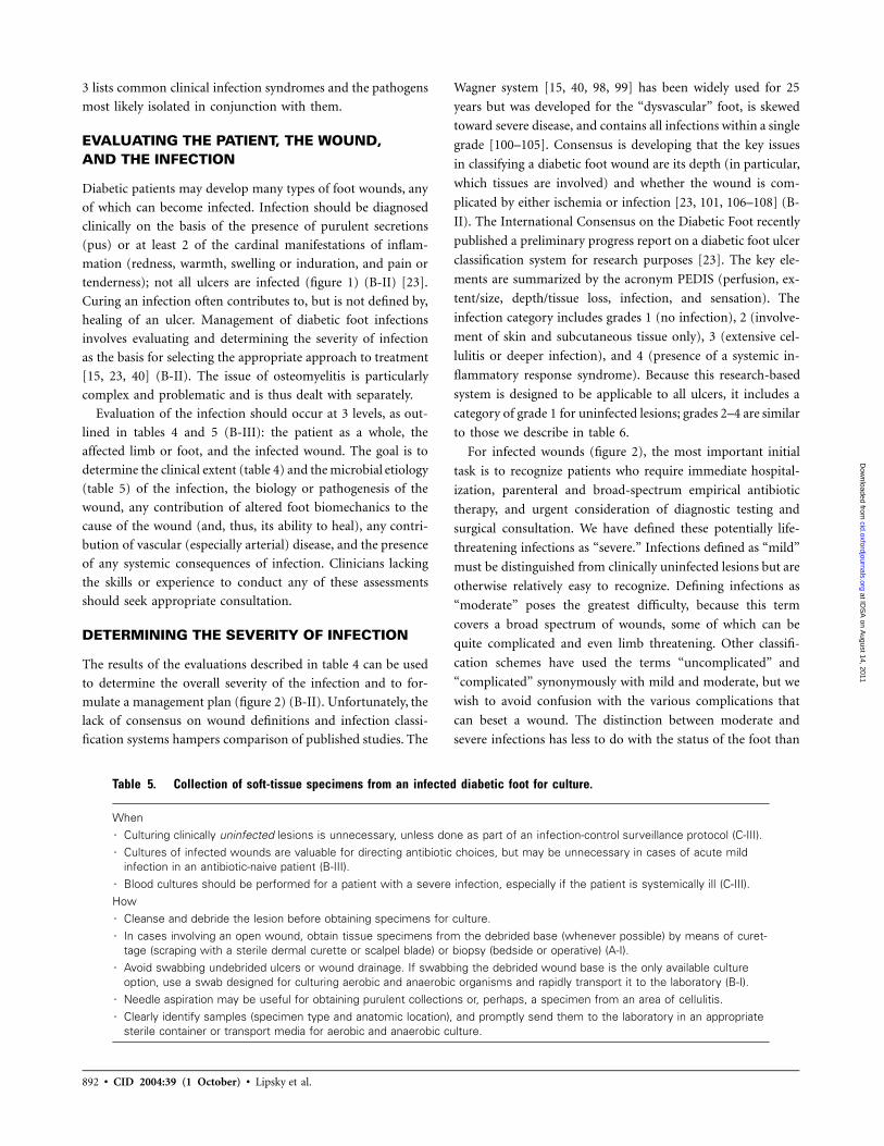
Table 5. Collection of soft-tissue specimens from an infected diabetic foot for culture.
When7 Culturing clinically uninfected lesions is unnecessary, unless done as part of an infection-control surveillance protocol (C-III).7 Cultures of infected wounds are valuable for directing antibiotic choices, but may be unnecessary in cases of acute mild
infection in an antibiotic-naive patient (B-III).7 Blood cultures should be performed for a patient with a severe infection, especially if the patient is systemically ill (C-III).How7 Cleanse and debride the lesion before obtaining specimens for culture.7 In cases involving an open wound, obtain tissue specimens from the debrided base (whenever possible) by means of curet-
tage (scraping with a sterile dermal curette or scalpel blade) or biopsy (bedside or operative) (A-I).7 Avoid swabbing undebrided ulcers or wound drainage. If swabbing the debrided wound base is the only available culture
option, use a swab designed for culturing aerobic and anaerobic organisms and rapidly transport it to the laboratory (B-I).7 Needle aspiration may be useful for obtaining purulent collections or, perhaps, a specimen from an area of cellulitis.7 Clearly identify samples (specimen type and anatomic location), and promptly send them to the laboratory in an appropriate
sterile container or transport media for aerobic and anaerobic culture.
3 lists common clinical infection syndromes and the pathogens
most likely isolated in conjunction with them.
EVALUATING THE PATIENT, THE WOUND,AND THE INFECTION
Diabetic patients may develop many types of foot wounds, any
of which can become infected. Infection should be diagnosed
clinically on the basis of the presence of purulent secretions
(pus) or at least 2 of the cardinal manifestations of inflam-
mation (redness, warmth, swelling or induration, and pain or
tenderness); not all ulcers are infected (figure 1) (B-II) [23].
Curing an infection often contributes to, but is not defined by,
healing of an ulcer. Management of diabetic foot infections
involves evaluating and determining the severity of infection
as the basis for selecting the appropriate approach to treatment
[15, 23, 40] (B-II). The issue of osteomyelitis is particularly
complex and problematic and is thus dealt with separately.
Evaluation of the infection should occur at 3 levels, as out-
lined in tables 4 and 5 (B-III): the patient as a whole, the
affected limb or foot, and the infected wound. The goal is to
determine the clinical extent (table 4) and the microbial etiology
(table 5) of the infection, the biology or pathogenesis of the
wound, any contribution of altered foot biomechanics to the
cause of the wound (and, thus, its ability to heal), any contri-
bution of vascular (especially arterial) disease, and the presence
of any systemic consequences of infection. Clinicians lacking
the skills or experience to conduct any of these assessments
should seek appropriate consultation.
DETERMINING THE SEVERITY OF INFECTION
The results of the evaluations described in table 4 can be used
to determine the overall severity of the infection and to for-
mulate a management plan (figure 2) (B-II). Unfortunately, the
lack of consensus on wound definitions and infection classi-
fication systems hampers comparison of published studies. The
Wagner system [15, 40, 98, 99] has been widely used for 25
years but was developed for the “dysvascular” foot, is skewed
toward severe disease, and contains all infections within a single
grade [100–105]. Consensus is developing that the key issues
in classifying a diabetic foot wound are its depth (in particular,
which tissues are involved) and whether the wound is com-
plicated by either ischemia or infection [23, 101, 106–108] (B-
II). The International Consensus on the Diabetic Foot recently
published a preliminary progress report on a diabetic foot ulcer
classification system for research purposes [23]. The key ele-
ments are summarized by the acronym PEDIS (perfusion, ex-
tent/size, depth/tissue loss, infection, and sensation). The
infection category includes grades 1 (no infection), 2 (involve-
ment of skin and subcutaneous tissue only), 3 (extensive cel-
lulitis or deeper infection), and 4 (presence of a systemic in-
flammatory response syndrome). Because this research-based
system is designed to be applicable to all ulcers, it includes a
category of grade 1 for uninfected lesions; grades 2–4 are similar
to those we describe in table 6.
For infected wounds (figure 2), the most important initial
task is to recognize patients who require immediate hospital-
ization, parenteral and broad-spectrum empirical antibiotic
therapy, and urgent consideration of diagnostic testing and
surgical consultation. We have defined these potentially life-
threatening infections as “severe.” Infections defined as “mild”
must be distinguished from clinically uninfected lesions but are
otherwise relatively easy to recognize. Defining infections as
“moderate” poses the greatest difficulty, because this term
covers a broad spectrum of wounds, some of which can be
quite complicated and even limb threatening. Other classifi-
cation schemes have used the terms “uncomplicated” and
“complicated” synonymously with mild and moderate, but we
wish to avoid confusion with the various complications that
can beset a wound. The distinction between moderate and
severe infections has less to do with the status of the foot than
at IDS
A on A
ugust 14, 2011cid.oxfordjournals.org
Dow
nloaded from

Guidelines for Diabetic Foot Infections • CID 2004:39 (1 October) • 893
Figure 2. Algorithm 1, part 2: approach to treating a diabetic patient with a foot infection. 1Consider hospitalization if any of the following criteriaare present: systemic toxicity (e.g., fever and leukocytosis); metabolic instability (e.g., severe hypoglycemia or acidosis); rapidly progressive or deep-tissue infection, substantial necrosis or gangrene, or presence of critical ischemia; requirement of urgent diagnostic or therapeutic interventions; andinability to care for self or inadequate home support.
with the patient to whom it is attached. This distinction is
complicated by the fact that �50% of patients with a limb-
threatening infection do not manifest systemic signs or symp-
toms. After debating several classification schemes, we propose
the one presented in table 6 as a basis for subsequent discussions
in and beyond this guideline (B-II).
TREATMENT OF INFECTION
Avoid prescribing antibiotics for uninfected ulcerations.
Some argue that many apparently uninfected diabetic foot ul-
cers are actually subclinically infected—that is, they contain a
high “bioburden” of bacteria (usually defined as 1105 organisms
per gram of tissue) that results in “critical colonization” levels
and impairs wound healing [54, 109–114]. Available published
evidence does not support the use of antibiotics for the man-
agement of clinically uninfected ulcerations, either to enhance
wound healing or as prophylaxis against infection [115, 116].
Because antibiotic use encourages antimicrobial resistance, in-
curs financial cost, and may cause drug-related adverse effects,
we discourage therapy of uninfected ulcers. In some circum-
stances, it is difficult to decide whether a chronic wound is
infected, such as when the foot is ischemic, has abnormal col-
at IDS
A on A
ugust 14, 2011cid.oxfordjournals.org
Dow
nloaded from

894 • CID 2004:39 (1 October) • Lipsky et al.
Table 6. Clinical classification of a diabetic foot infection.
Clinical manifestations of infectionInfectionseverity
PEDISgradea
Wound lacking purulence or any manifestations of inflammation Uninfected 1Presence of �2 manifestations of inflammation (purulence, or erythema, pain,
tenderness, warmth, or induration), but any cellulitis/erythema extends �2cm around the ulcer, and infection is limited to the skin or superficial subcu-taneous tissues; no other local complications or systemic illness.
Mild 2
Infection (as above) in a patient who is systemically well and metabolically sta-ble but which has �1 of the following characteristics: cellulitis extending 12cm, lymphangitic streaking, spread beneath the superficial fascia, deep-tissueabscess, gangrene, and involvement of muscle, tendon, joint or bone
Moderate 3
Infection in a patient with systemic toxicity or metabolic instability (e.g., fever,chills, tachycardia, hypotension, confusion, vomiting, leukocytosis, acidosis,severe hyperglycemia, or azotemia)
Severe 4
NOTE. Definitions of terms can be found in footnotes to table 4. Foot ischemia may increase the severity of anyinfection, and the presence of critical ischemia often makes the infection severe. PEDIS, perfusion, extent/size, depth/tissue loss, infection, and sensation.
a International Consensus on the Diabetic Foot [23].
oration or a fetid odor, has friable granulation tissue, is asso-
ciated with unexpected pain or tenderness, or when an oth-
erwise properly treated ulcer fails to show healing progress [117,
118]. In these unusual cases, a brief, culture-directed course of
antibiotic therapy may be appropriate (C-III).
Determine the need for hospitalization. Hospitalization is
the most expensive part of treating a diabetic foot infection,
and deciding on its necessity requires consideration of both
medical and social issues. Patients with infections that are either
severe or complicated by critical limb ischemia should generally
be hospitalized (C-III) [119, 120]. Some patients with appar-
ently mild infections and more patients with moderate infec-
tions may also need hospitalization; this may be for observation,
urgent diagnostic testing, or because complicating factors are
likely to affect their wound care or adherence to antibiotic
treatment. In the absence of these complicating features, most
patients with mild or moderate infections can be treated as
outpatients (A-II) [84, 121].
Stabilize the patient. Attending to the general metabolic
state of the patient is essential [25, 122]. This may involve
restoration of the fluid and electrolyte balances; correction of
hyperglycemia, hyperosmolality, acidosis, and azotemia; and
treatment of other exacerbating disorders. Critically ill patients
who require surgery should usually be stabilized before transfer
to the operating room, although surgery should usually not be
delayed for 148 h after presentation to the hospital (B-III)
[123]. The improvement of glycemic control may aid in both
eradicating the infection and healing the wound [124]. As the
infection improves, hyperglycemia may be easier to control.
Choose an antibiotic regimen. Selection of the antibiotic
regimen initially involves decisions about the route of therapy,
the spectrum of microorganisms to be covered, and the specific
drugs to administer and later involves choosing the definitive
regimen and the duration of treatment. Initial therapy is usually
empirical and should be based on the severity of the infection
and on any available microbiological data, such as recent culture
results or current Gram-stained smear findings. For severe in-
fections and for more-extensive, chronic moderate infections,
it is safest to commence therapy with broad-spectrum agents.
These should have activity against gram-positive cocci (includ-
ing MRSA in locations where this pathogen is common), as
well as gram-negative and obligate anaerobic organisms (B-III).
To ensure adequate and prompt tissue concentrations, therapy
should be given parenterally, at least initially (C-III). Although
some suggest broad-spectrum empirical therapy for most in-
fections [125–127], the majority of mild—and many moder-
ate—infections can be treated with agents with a relatively
narrow spectrum, such as those covering only aerobic gram-
positive cocci (A-II) [84]. Although anaerobic organisms are
isolated from many severe infections [42, 128], they are infre-
quent in mild-to-moderate infections [14, 84, 129], and there
is little evidence to support the need for antianaerobic therapy
in most infections (B-III). For mild-to-moderate infections in
patients without gastrointestinal absorption problems and for
whom an oral agent with the appropriate spectrum is available,
oral therapy is often appropriate, especially with highly bio-
available agents (A-II). For mildly infected open wounds with
minimal cellulitis, limited data support the use of topical an-
timicrobial therapy (B-I) [130].
Antibiotics vary in how well they achieve effective concen-
trations in infected diabetic foot lesions [131–137]; this is as-
sociated with the pharmacodynamic properties of the specific
agent and, especially, the arterial supply to the foot, rather than
with diabetes [138]. There are surprisingly few published clin-
ical trials of antibiotic therapy for diabetic foot infection. Sev-
eral antibiotic trials involving patients with various complicated
at IDS
A on A
ugust 14, 2011cid.oxfordjournals.org
Dow
nloaded from

Guidelines for Diabetic Foot Infections • CID 2004:39 (1 October) • 895
skin and soft-tissue infections have included some patients with
diabetic foot infections. Table 7 provides a list of published
clinical trials that focused on therapy of diabetic foot infections,
either exclusively or as an identified subset of a larger study.
The lack of standardization among these trials makes the com-
parison of outcomes of different regimens inappropriate. The
differing definitions of infection severity and clinical end points
that were used in these publications highlight the need to de-
velop a consensus classification system for future studies. On
the basis of the available studies, no single drug or combination
of agents appears to be superior to others [129].
Table 8 summarizes some potential empirical antibiotic reg-
imens according to the clinical severity of the infection, al-
though the available data do not allow us to recommend any
specific antibiotic regimen for diabetic foot infections (B-II).
These suggested agents are derived from available published
clinical trials and our collective experience and are not meant
to be inclusive of all potentially reasonable regimens. Similar
agents could be used, depending on various clinical, micro-
biological, epidemiological, and financial considerations. Con-
sider modifying antibiotic therapy when culture and suscep-
tibility results are available (C-III). Empirical choices for
patients who are not responding to antibiotic therapy should
include agents that cover a different or more-extended spec-
trum of organisms (B-III) (figure 3). The regimens in table 8
are listed in approximate order of increasing broad-spectrum
coverage; the order does not indicate preferences by the com-
mittee. Dosages of antibiotic agents should be selected accord-
ing to suggestions of the US Food and Drug Administration,
the drug’s manufacturers, and the experience of the prescriber
and should be modified on the basis of any relevant organ
(especially renal) dysfunction and other clinical factors.
Determine the need for surgery. Many infections require
surgical procedures that range from drainage and excision of
infected and necrotic tissues to revascularization of the lower
extremity and reconstruction of soft-tissue defects or mechan-
ical misalignments [164–168]. Unfortunately, surgical treat-
ment of diabetic foot infections is based on even less-structured
evidence than that for antibiotic therapy [169]. Seek urgent
surgical consultation for life- or limb-threatening infections,
such as those presenting with necrotizing fasciitis, gas gangrene,
extensive soft-tissue loss, or evidence of compartment syn-
drome, or those in limbs with critical ischemia (A-II) [170,
171]. A surgical specialist should also evaluate patients who
have unexplained persistent foot pain or tenderness and/or
evidence of a deep-space infection, deep abscesses, or progres-
sive infection in the face of apparently appropriate medical care
(figure 3). Timely and aggressive surgical debridement, includ-
ing limited resections or amputations, may reduce the need for
more-extensive amputation (B-II) [172, 173]. Pus under pres-
sure, especially in an ischemic foot, can cause rapid and irrep-
arable damage. For patients with less-serious infections, it may
be appropriate to delay surgery to carefully observe the effec-
tiveness of medical therapy or to determine the demarcation
line between necrotic and viable tissue [174].
The surgeon must determine the adequacy of the blood sup-
ply to the remaining viable tissues, consider common operative
pitfalls (e.g., infection spreading among foot compartments, to
the deep plantar space, or along the tendon sheaths), and for-
mulate a strategy for eventual soft-tissue cover (e.g., primary
closure, delayed primary closure, secondary intention, or tissue
transfer) [175–177]. The surgical approach should optimize the
likelihood for healing and should attempt to preserve the in-
tegrity of the walking surface of the foot (B-II) [178]. In ad-
dition to manual dexterity, the surgeon must have sufficient
knowledge and experience to judge when and how to intervene.
The surgeon’s training specialty is less important than his or
her knowledge of the anatomy of the foot, the pathophysiology
of ulceration and infection, and experience with and enthusi-
asm for the field [8]. In most instances, the surgeon should
continue to observe the patient until the infection is under
control and the wound is healing (B-III).
In some cases, amputation is the best or only option [170,
179]. Urgent amputation is usually required only when there
is extensive necrosis or life-threatening infection [180]. Elective
amputation may be considered for the patient who has recur-
rent ulceration (despite maximal preventive measures), has ir-
reversible loss of foot function, or would require unacceptably
prolonged or intensive hospital care [181, 182]. Selection of
the level of amputation must take into consideration vascular,
reconstructive, and rehabilitation issues [183, 184]. Generally,
the surgeon should attempt to save as much of the limb as
possible. However, a higher-level amputation that results in a
more functional residual stump (even if a prosthesis is required)
may be a better choice than preserving a foot that is mechan-
ically unsound, unlikely to heal, or prone to future ulceration.
When all or part of a foot has dry gangrene, it may be preferable
(especially for a patient for whom surgery is a poor option) to
let the necrotic portions autoamputate. It may also be best to
leave adherent eschars in place, especially on the heel, until
they soften enough to be more easily removed, provided there
does not appear to be an underlying focus of infection [80,
81].
If the infected limb appears to be ischemic, the patient should
be referred to a surgeon with vascular expertise [185]. In most
cases, ischemia is due to larger-vessel atherosclerosis, rather
than to “small-vessel disease” [68]. Because vessels above the
knee and below the ankle tend to be relatively spared, lower-
extremity atherosclerosis may be amenable to angioplasty or
vascular bypass [186]. Patients with noncritical ischemia (e.g.,
those with an ankle to brachial artery blood pressure index of
0.5–0.9) can usually be successfully treated without a vascular
at IDS
A on A
ugust 14, 2011cid.oxfordjournals.org
Dow
nloaded from

Table 7. Antibiotic agents used in published clinical studies of diabetic foot infections.
Antibiotic (route)
No. oftreatedpatients Study design Patient group
Type/severity ofinfection(s) Reference
Cephalosporins
Cefoxitin (iv) 8 Prospective,noncomparative
Hospitalized Presumptive anaerobic [139]
Cefoxitin (iv) 23 RDBCT Hospitalized Moderate-to-severe [140]
Cefoxitin (iv) 60 Prospective,noncomparative
Hospitalized Failing to respond totherapy
[141]
Cefoxitin (iv) 18 RDBCT Hospitalized Mild-to-severe [142]
Cefoxitin (iv) alone 12 RCT Hospitalized Mixed [143]
Cefoxitin (iv) and amdino-cillin (iv)
13 RCT Hospitalized Mixed [143]
Ceftizoxime (iv) 20 Prospective, uncontrolled Hospitalized PVD, moderate-to-severe [144]
Ceftizoxime (iv) 23 RDBCT Hospitalized Moderate-to-severe [140]
Cephalexin (po) 29 RDBCT Outpatient Mild-to-moderate [84]
Ceftriaxone (iv) 90 Prospective, observational Hospitalized Severe limb-threatening [145]
Penicillins
Ampicillin/sulbactam (iv)(then amoxicillin/clavu-lanate [po])
53 RCT Hospitalized initially Moderate [146]
Ampicillin/sulbactam (iv) 48 RDBCT Hospitalized Limb-threatening [147]
Ampicillin/sulbactam (iv) 74 Prospective,noncomparative
Hospitalized Moderate-to-severe [148]
Ampicillin/sulbactam (iv) 18 RDBCT Hospitalized Mild-to-severe [142]
Ampicillin/sulbactam (iv)and/or amoxicillin/clavu-lanate (po)
120 RCT Outpatient or hospitalized All types [121]
Amoxicillin/clavulanate (iv/po) 191 Observational,noncomparative
Mostly hospitalized Moderate [149]
Ticarcillin/clavulanate (iv) 28 RCT subgroupa Inpatient or outpatient Complicated soft-tissue [150]
Ticarcillin/clavulanate (iv) 17 RCT subgroupa Hospitalized Complicated soft-tissue [151]
Piperacillin/tazobactam (iv) 29 Prospective,noncomparative
Hospitalized Moderate-to-severe [152]
Piperacillin/tazobactam(iv/im)
38 Prospectivenoncomparative
Outpatient Parenteral, mostlymoderate
[153]
Piperacillin/tazobactam (iv) 34 RDBCT subgroupa Hospitalized Severe [154]
Fluoroquinolones
Ciprofloxacin (po) 46 Prospective, randomizeddoses
Hospitalized PVD [155]
Ciprofloxacin (iv, then po) 43 Prospective,noncomparative
Hospitalized Soft-tissue or bone [156]
Ciprofloxacin (po) andclindamycin (po)
120 Uncontrolled, quasi-prospective
Hospitalized initially, re-ceived other iv agents,and was then dis-charged home
Moderate-to-severe [157]
Ofloxacin (iv, then po) 55 RCT Hospitalized initially Moderate [146]
Ofloxacin (po) 420 RDBCT Outpatients Mild-to-moderatelyinfected ulcers
[130]
Levofloxacin (iv or po) 26 RCT subgroupa Outpatients or inpatients Complicated soft-tissue [150]
Trovafloxacin (po) 214 Prospective,noncomparative
… Soft-tissue [158]
Clinafloxacin (iv, then po) 42 RDBCT subgroupa Hospitalized Severe [154]
Ofloxacin, levofloxacin, orciprofloxacin (iv and/or po)
90 Prospective, observational Hospitalized Severe limb-threatening [145]
Carbapenems
Imipenem/cilastatin (iv) 48 RDBCT Hospitalized Limb-threatening [147]
Imipenem/cilastatin (iv) 94 Uncontrolled,noncomparative
Hospitalized Moderate-to-severe [159]
Imipenem/cilastatin (iv) 22 Randomized, open,comparative
Hospitalized Wagner grade 2–4wounds
[160]
Ertapenem (iv) 33 RDBCT Hospitalized Complicated soft-tissue [161]
(continued)
at IDS
A on A
ugust 14, 2011cid.oxfordjournals.org
Dow
nloaded from
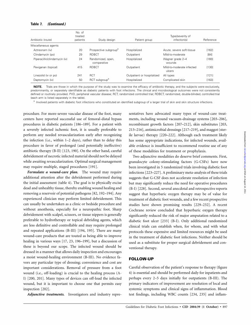
Guidelines for Diabetic Foot Infections • CID 2004:39 (1 October) • 897
Table 7. (Continued.)
Antibiotic (route)
No. oftreatedpatients Study design Patient group
Type/severity ofinfection(s) Reference
Miscellaneous agents
Aztreonam (iv) 20 Prospective subgroupa Hospitalized Acute, severe soft-tissue [162]
Clindamycin (po) 29 RDBCT Outpatient Mild-to-moderate [84]
Piperacillin/clindamycin (iv) 24 Randomized, open,comparative
Hospitalized Wagner grade 2–4wounds
[160]
Pexiganan (topical) 415 RDBCT Outpatient Mild-to-moderate infectedulcers
[130]
Linezolid (iv or po) 241 RCT Outpatient or hospitalized All types [121]
Daptomycin (iv) 50 RCT subgroupa Hospitalized Complicated skin [163]
NOTE. Trials are those in which the purpose of the study was to examine the efficacy of antibiotic therapy, and the subjects were exclusively,predominantly, or separately identifiable as diabetic patients with foot infections. The clinical and microbiological outcomes were not consistentlydefined or routinely provided. PVD, peripheral vascular disease; RCT, randomized controlled trial; RDBCT, randomized, double-blinded, controlled trial(each arm is listed separately in the table).
a Involved patients with diabetic foot infections who constituted an identified subgroup of a larger trial of skin and skin structure infections.
procedure. For more-severe vascular disease of the foot, many
centers have reported successful use of femoral-distal bypass
procedures in diabetic patients [186–189]. For a patient with
a severely infected ischemic foot, it is usually preferable to
perform any needed revascularization early after recognizing
the infection (i.e., within 1–2 days), rather than to delay this
procedure in favor of prolonged (and potentially ineffective)
antibiotic therapy (B-II) [123, 190]. On the other hand, careful
debridement of necrotic infected material should not be delayed
while awaiting revascularization. Optimal surgical management
may require multiple, staged procedures [191].
Formulate a wound-care plan. The wound may require
additional attention after the debridement performed during
the initial assessment (table 4). The goal is to physically excise
dead and unhealthy tissue, thereby enabling wound healing and
removing a reservoir of potential pathogens [82, 192–194]. Any
experienced clinician may perform limited debridement. This
can usually be undertaken as a clinic or bedside procedure and
without anesthesia, especially for a neuropathic foot. Sharp
debridement with scalpel, scissors, or tissue nippers is generally
preferable to hydrotherapy or topical debriding agents, which
are less definitive and controllable and may require prolonged
and repeated applications (B-III) [194, 195]. There are many
wound-care products that are touted as being able to improve
healing in various ways [17, 23, 196–199], but a discussion of
these is beyond our scope. The infected wound should be
dressed in a manner that allows daily inspection and encourages
a moist wound-healing environment (B-III). No evidence fa-
vors any particular type of dressing; convenience and cost are
important considerations. Removal of pressure from a foot
wound (i.e., off-loading) is crucial to the healing process (A-
I) [200, 201]. Many types of devices can off-load the infected
wound, but it is important to choose one that permits easy
inspection [202].
Adjunctive treatments. Investigators and industry repre-
sentatives have advocated many types of wound-care treat-
ments, including wound vacuum-drainage systems [203–206],
recombinant growth factors [207–212], skin substitutes [203,
213–216], antimicrobial dressings [217–219], and maggot (ster-
ile larvae) therapy [220–222]. Although each treatment likely
has some appropriate indications, for infected wounds, avail-
able evidence is insufficient to recommend routine use of any
of these modalities for treatment or prophylaxis.
Two adjunctive modalities do deserve brief comments. First,
granulocyte colony-stimulating factors (G-CSFs) have now
been investigated in 5 randomized trials involving diabetic foot
infections [223–227]. A preliminary meta-analysis of these trials
suggests that G-CSF does not accelerate resolution of infection
but may significantly reduce the need for operative procedures
(B-I) [228]. Second, several anecdotal and retrospective reports
suggest that hyperbaric oxygen therapy may be of value for
treatment of diabetic foot wounds, and a few recent prospective
studies have shown promising results [229–232]. A recent
Cochrane review concluded that hyperbaric oxygen therapy
significantly reduced the risk of major amputation related to a
diabetic foot ulcer [233] (B-I). Only additional randomized
clinical trials can establish when, for whom, and with what
protocols these expensive and limited resources might be used
in the treatment of diabetic foot infections. Neither should be
used as a substitute for proper surgical debridement and con-
ventional therapy.
FOLLOW-UP
Careful observation of the patient’s response to therapy (figure
4) is essential and should be performed daily for inpatients and
perhaps every 2–5 days initially for outpatients (B-III). The
primary indicators of improvement are resolution of local and
systemic symptoms and clinical signs of inflammation. Blood
test findings, including WBC counts [234, 235] and inflam-
at IDS
A on A
ugust 14, 2011cid.oxfordjournals.org
Dow
nloaded from

898 • CID 2004:39 (1 October) • Lipsky et al.
Table 8. Suggested empirical antibiotic regimens, based on clinical severity, for diabetic foot infections.
Route and agent(s) Mild Moderate Severe
Advised route Oral for most Oral or parenteral, basedon clinical situation andagent(s) selected
Intravenous, at leastinitially
Dicloxacillin Yes … …Clindamycin Yes … …Cephalexin Yes … …Trimethoprim-sulfamethoxazole Yes Yes …Amoxicillin/clavulanate Yes Yes …Levofloxacin Yes Yes …Cefoxitin … Yes …Ceftriaxone … Yes …Ampicillin/sulbactam … Yes …Linezolida (with or without aztreonam) … Yes …Daptomycina (with or without aztreonam) … Yes …Ertapenem … Yes …Cefuroxime with or without metronidazole … Yes …Ticarcillin/clavulanate … Yes …Piperacillin/tazobactam … Yes YesLevofloxacin or ciprofloxacin with clindamycin … Yes YesImipenem-cilastatin … … YesVancomycina and ceftazidime (with or without
metronidazole)… … Yes
NOTE. Definitive regimens should consider results of culture and susceptibility tests, as well as the clinical response to the empiricalregimen. Similar agents of the same drug class may be substituted. Some of these regimens may not have US Food and Drug Administrationapproval for complicated skin and skin-structure infections, and only linezolid is currently specifically approved for diabetic foot infections.
a For patients in whom methicillin-resistant S. aureus infection is proven or likely.
matory markers, such as the erythrocyte sedimentation rate
[122, 236, 237] and the C-reactive protein level [238], are of
limited use for monitoring response, although is it reassuring
to see elevated levels decrease and cause for concern when they
do not.
When a hospitalized patient is ready for discharge or an
outpatient returns for follow-up, the clinician should accom-
plish 4 tasks (figures 1, 2, and 4).
1. Select the definitive antibiotic regimen. Review the culture
and drug susceptibility results and inquire about any adverse
effects related to the current antibiotic therapy. Choose a de-
finitive antibiotic regimen (including the treatment duration)
on the basis of the results of cultures, imaging, or other in-
vestigations, and the initial clinical response (C-III). It is not
always necessary to cover all microorganisms isolated from cul-
tures. More virulent species (e.g., S. aureus and group A or B
streptococci) should always be covered, but in a polymicrobial
infection, less-virulent bacteria (e.g., coagulase-negative staph-
ylococci and enterococci) may be less important (B-II). If the
infection has not responded to the empirical regimen, select
agents with activity against all isolates. For a clinically stable
patient who has had �1 unsuccessful courses of therapy, con-
sider discontinuing antimicrobials for a few days and then col-
lecting optimal specimens for culture (C-III).
2. Re-evaluate the wound. Inspect the site to ensure that
the infection is responding and that the wound is healing. If
neither is occurring, reassess the need for surgical intervention.
No evidence supports giving antibiotics for the entire time that
the wound remains open. Antibiotics should be used for a
period defined by the biology of the infection and by the clinical
syndrome, as suggested in table 9 (A-II). If clinical evidence of
infection persists beyond the expected duration, check on the
patient’s compliance with antibiotics and re-evaluate for un-
addressed adverse biological factors (figure 3). These may in-
clude the development of antibiotic resistance, a superinfection,
an undiagnosed deep abscess or case of osteomyelitis, or is-
chemia that is more severe than was initially suspected.
3. Review the off-loading and wound care regimens. Deter-
mine the effectiveness of, and the patient’s compliance with,
the prescribed regimens. Suggest (or seek consultation for) al-
ternatives when necessary.
4. Evaluate glycemic control. Ensure that blood glucose lev-
els and other aspects of the patient’s metabolic status are ad-
equately controlled.
at IDS
A on A
ugust 14, 2011cid.oxfordjournals.org
Dow
nloaded from

Guidelines for Diabetic Foot Infections • CID 2004:39 (1 October) • 899
Figure 3. Algorithm 1, part 3: approach to assessing a diabetic patient with a foot infection who is not responding well to treatment. TcPO2,transcutaneous partial pressure of O2.
OSTEOMYELITIS
Dealing with osteomyelitis is perhaps the most difficult and
controversial aspect in the management of diabetic foot infec-
tions [239–244]. First among several problems is that the lack
of a consensus definition of the disease hinders the comparison
of available studies and experiences. Next, there are many avail-
able diagnostic tests, but they often yield equivocal results. Fur-
thermore, the presence of osteomyelitis increases the likelihood
of surgical intervention, including amputation, and the re-
quired duration of antibiotic therapy [240]. Finally, osteomy-
elitis impairs healing of the overlying wound and acts as a focus
for recurrent infection.
When to consider the diagnosis. Consider osteomyelitis as
a potential complication of any deep or extensive ulcer, espe-
cially one that is chronic or overlies a bony prominence (figure
5) [245]. Suspect underlying osteomyelitis when an ulcer does
not heal after at least 6 weeks of appropriate care and off-
loading. Any ulcer in which bone is either visible or can be
easily palpated with a sterile blunt metal probe is likely to be
complicated by osteomyelitis [83]. In patients with a limb-
threatening infection, positive results of a probe-to-bone test
may be taken as nearly sufficient for diagnosis, but the per-
formance characteristics of this test have not yet been fully
defined. A swollen foot in a patient with a history of foot
at IDS
A on A
ugust 14, 2011cid.oxfordjournals.org
Dow
nloaded from

900 • CID 2004:39 (1 October) • Lipsky et al.
Figure 4. Algorithm 2: approach to selecting antibiotic therapy for a diabetic patient with a foot infection. MRSA, methicillin-resistant Staphylococcusaureus.
ulceration, a “sausage toe” (i.e., a red, swollen digit) [246], or
an unexplained high WBC count [235] or inflammatory mark-
ers [236] should also arouse suspicion of osteomyelitis (B-II).
Finally, radiologically evident bone destruction beneath an ulcer
should be considered to represent osteomyelitis unless proven
otherwise [247].
Confirming the diagnosis. Because bony destruction is
usually not seen on plain radiography performed during the
early stages of disease and neuro-osteoarthropathy can mimic
infection, diagnosing osteomyelitis at the time the patient first
presents to the hospital can be difficult [248–250]. Character-
istic progressive changes on serial plain radiographs may help
in more-chronic cases [247, 251]. Radioisotope scans are more
sensitive than radiographs for detecting osteomyelitis during
the early stages of this diseases, but they are expensive and can
be time-consuming [252]. The reported performance charac-
teristics of various types of nuclear medicine scans varies, but
the specificity of technetium bone scans is generally low [240,
at IDS
A on A
ugust 14, 2011cid.oxfordjournals.org
Dow
nloaded from

Guidelines for Diabetic Foot Infections • CID 2004:39 (1 October) • 901
Table 9. Suggested route, setting, and durations of antibiotic therapy, by clinical syndrome.
Site, by severity orextent, of infection Route of administration Setting for therapy Duration of therapy
Soft-tissue onlyMild Topical or oral Outpatient 1–2 Weeks; may extend
up to 4 weeks if slowto resolve
Moderate Oral (or initial parenteral) Outpatient/inpatient 2–4 WeeksSevere Initial parenteral, switch to oral
when possibleInpatient, then
outpatient2–4 Weeks
Bone or jointNo residual infected tissue
(e.g., post-amputation)Parenteral or oral … 2–5 Days
Residual infected soft tissue(but not bone)
Parenteral or oral … 2–4 Weeks
Residual infected (but viable)bone
Initial parenteral, then consideroral switch
… 4–6 Weeks
No surgery, or residual deadbone postoperatively
Initial parenteral, then consideroral switch
… 13 Months
253–255]. MRI is the most useful of the currently available
imaging modalities (A-I) [90, 92, 94, 256–259]. MRI is the
most accurate imaging study for defining bone infection, and
it also provides the most reliable image of deep soft-tissue
infections. The performance characteristics of all these diag-
nostic tests are highly correlated with the pretest probability of
osteomyelitis, and they are most useful for intermediately prob-
able cases [260]. The criterion (gold) standard for diagnosing
osteomyelitis is isolation of bacteria from a reliably obtained
sample of bone (using measures to minimize contamination)
concomitant with histological findings of inflammatory cells
and osteonecrosis (B-II). Unfortunately, few of the studies that
have evaluated diagnostic tests or have assessed treatment out-
comes have used this standard.
MRI is usually not needed as a first-line investigation in cases
of diabetic foot infection. When osteomyelitis is a possibility,
obtaining plain radiographs often suffices. If these radiographs
show no evidence of pathological findings in bone, the patient
should be treated for ∼2 weeks for the soft-tissue infection. If
suspicion of osteomyelitis persists, perform plain radiography
again 2–4 weeks later. If the initial films show classic changes
suggestive of osteomyelitis (cortical erosion, periosteal reaction,
and mixed lucency and sclerosis), and if there is little likelihood
of a noninfectious osteoarthropathy, treat for presumptive os-
teomyelitis, preferably after obtaining appropriate specimens
for culture (B-III). If findings of radiography are only consistent
with, but not characteristic of, osteomyelitis, one of the fol-
lowing choices should be considered.
1. Additional imaging studies. MRI is the preferred imaging
study, with nuclear medicine scans (that preferably use newer
generation leukocyte [239, 261, 262] or immunoglobulin [263,
264] techniques) being a second choice. If results of the imaging
tests are negative, osteomyelitis is unlikely; if results suggest
osteomyelitis, consider whether bone biopsy is needed (vide
infra).
2. Empirical treatment. Provide antibiotic therapy for an-
other 2–4 weeks and then perform radiograph again to deter-
mine whether bony changes have progressed (which would
suggest infection).
3. Bone biopsy. Use an appropriate procedure, as defined
below. Collection of a sample of a bony lesion (either opera-
tively or percutaneously) is recommended if the diagnosis re-
mains in doubt after imaging or if osteomyelitis is likely but
the etiologic agent or antibiotic susceptibilities are not pre-
dictable (B-II) [251, 265–268]. Some physicians would also
obtain biopsy specimens of most mid- or hind-foot lesions,
because these are more difficult to treat and more often lead
to a high-level (i.e., above the ankle) amputation. Any properly
trained physician (e.g., an orthopedic surgeon, podiatrist, in-
terventional radiologist) can perform the biopsy. Percutaneous
biopsy should preferably be done under fluoroscopic or CT
guidance, traversing uninvolved skin if possible. For patients
with sensory neuropathy, anesthesia may be unnecessary. Var-
ious types of bone-cutting needles, such as Jamshidi (Perfectum
Corporation; distributed by Propper and Sons) and Ostycut
(Bard Products; distributed by Angiomed) have been used. Ob-
tain 2–3 specimens if possible, sending at least 1 for culture
and another for histological analysis [269]. With small toe
bones, it may only be possible to aspirate a few bony spicules.
We found no published reports of complications of foot bone
biopsy and consider it to be a safe procedure (B-II). Cultures
of bone specimens provide more accurate microbiologic data
than do those of soft-tissue specimens for patients with oste-
omyelitis [93, 268, 270].
at IDS
A on A
ugust 14, 2011cid.oxfordjournals.org
Dow
nloaded from
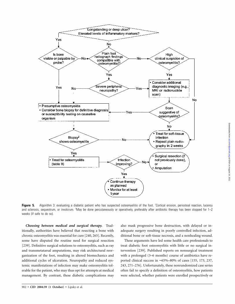
902 • CID 2004:39 (1 October) • Lipsky et al.
Figure 5. Algorithm 3: evaluating a diabetic patient who has suspected osteomyelitis of the foot. 1Cortical erosion, periosteal reaction, lucencyand sclerosis, sequestrum, or involcrum. 2May be done percutaneously or operatively, preferably after antibiotic therapy has been stopped for 1–2weeks (if safe to do so).
Choosing between medical and surgical therapy. Trad-
itionally, authorities have believed that resecting a bone with
chronic osteomyelitis was essential for cure [240, 265]. Recently,
some have disputed the routine need for surgical resection
[239]. Definitive surgical solutions to osteomyelitis, such as ray
and transmetatarsal amputations, may risk architectural reor-
ganization of the foot, resulting in altered biomechanics and
additional cycles of ulceration. Neuropathy and reduced sys-
temic manifestations of infection may make osteomyelitis tol-
erable for the patient, who may thus opt for attempts at medical
management. By contrast, these diabetic complications may
also mask progressive bone destruction, with delayed or in-
adequate surgery resulting in poorly controlled infection, ad-
ditional bone or soft-tissue necrosis, and a nonhealing wound.
These arguments have led some health care professionals to
treat diabetic foot osteomyelitis with little or no surgical in-
tervention [239]. Published reports on nonsurgical treatment
with a prolonged (3–6 months) course of antibiotics have re-
ported clinical success in ∼65%–80% of cases [155, 173, 237,
243, 271–276]. Unfortunately, these nonrandomized case series
often fail to specify a definition of osteomyelitis, how patients
were selected, whether patients were enrolled prospectively or
at IDS
A on A
ugust 14, 2011cid.oxfordjournals.org
Dow
nloaded from

Guidelines for Diabetic Foot Infections • CID 2004:39 (1 October) • 903
even consecutively, and how much nonoperative debridement
of bone was performed. The determination of which patients
are suitable for nonsurgical treatment, as well as what duration
of antibiotic therapy is needed, are important areas for future
study. Meanwhile, there are 4 cases in which nonsurgical man-
agement of osteomyelitis might be considered (B-II).
1. There is no acceptable surgical target (i.e., radical cure
of the infection would cause unacceptable loss of function).
2. The patient has ischemia caused by unreconstructable
vascular disease but desires to avoid amputation.
3. Infection is confined to the forefoot, and there is min-
imal soft-tissue loss.
4. The patient and health care professional agree that sur-
gical management carries excessive risk or is otherwise not
appropriate or desirable.
When therapy for osteomyelitis fails, consider several issues.
First, was the original diagnosis correct? Second, is there re-
sidual necrotic or infected bone or surgical hardware that
should be resected or removed? Third, did the selected anti-
biotic regimen likely cover the causative organism(s) and
achieve adequate levels in bone, and was it administered for a
sufficient duration? Fourth, was the failure to eradicate bone
infection the real cause of the current wound problem? Each
case needs an individualized approach, usually in consultation
with a knowledgeable surgeon. Selected patients may benefit
from implanted antibiotics (e.g., embedded in beads or cement)
[277–280], hyperbaric oxygen therapy, or revascularization,
whereas others may elect long-term or intermittent antibiotic
suppression or, in some cases, amputation.
Selecting an antibiotic regimen. The most appropriate du-
ration of therapy for any type of diabetic foot infection has not
been well defined [129]. It is important to consider the presence
and amount of any residual dead or infected bone and the state
of the soft tissues. When a radical resection leaves no remaining
infected tissue, minimal antibiotic therapy is needed (B-II).
Alternatively, if infected bone or soft tissue remain despite sur-
gery, continued prolonged treatment is necessary. For osteo-
myelitis, some parenteral therapy may be beneficial, especially
if an agent with suboptimal bioavailability is used (C-III). Par-
enteral therapy may be delivered in the outpatient setting, where
available [153, 281, 282]. Our recommendations for duration
of therapy are based on the clinical syndrome and are sum-
marized in table 9.
OUTCOMES
The goals of treating a diabetic foot infection are the eradication
of clinical evidence of infection and the avoidance of soft-tissue
loss and amputations. Overall, expect a good clinical response
(i.e., resolution of clinical evidence of infection) to appropriate
therapy in 80%–90% of mild-to-moderate infections [84, 121,
130, 263] and in 60%–80% of severe infections or cases of
osteomyelitis [130, 145, 147, 237, 283]. Factors associated with
a poor outcome include signs of systemic infection [237], in-
adequate limb perfusion, osteomyelitis [273, 283–285], the
presence of necrosis or gangrene [276], an inexperienced sur-
geon [286], and proximal location of the infection [287]. Re-
lapses occur in ∼20%–30% of patients, especially in those with
osteomyelitis; relapses may be difficult to differentiate from a
reinfection. A recent survey of members of the Emerging In-
fections Network found that the acceptable median failure rate
for treating diabetic foot osteomyelitis was 18% [288]. Con-
ducting systematic audits of outcomes and patient treatment
processes may be useful for individual practitioners and for
multidisciplinary foot-care teams (B-II).
PREVENTION
A patient who has had 1 foot infection is more likely to have
another, making this a good time to reinforce preventive actions
with the patient [11, 289, 290]. Detection of neuropathy before
its complications ensue is the best method to prevent foot
infections. Educate the patient about the importance of opti-
mizing glycemic control, using appropriate footwear at all
times, avoiding foot trauma, performing daily self-examination
of the feet, and reporting any changes to health care profes-
sionals (A-II). Because basic screening can be completed in a
few minutes, clinicians should reinforce these preventive mea-
sures by questioning patients about foot care and regularly
examining their feet and shoes. Patients with severe neuropathy,
substantial foot deformity, or critical ischemia should be re-
ferred to appropriate specialists to deal with these problems
(A-II).
RECOMMENDED RESEARCH
Few of the recommendations in this guideline are based on
properly designed and adequately powered randomized studies.
There are 6 areas in which future research would be particularly
helpful (A-III).
1. Establish a robust, validated, simple classification system
for infected foot lesions to facilitate multicenter comparative
studies of their natural history, diagnosis, and treatment. We
support efforts to validate the International Consensus PEDIS
system for foot-ulcer research purposes.
2. Determine whether there is a role for antibiotic therapy
in managing clinically uninfected ulcers.
3. Determine optimal antibiotic regimens (agents, routes,
and duration) for various types of soft-tissue and bone
infections.
4. Establish a consensus definition of osteomyelitis in the
diabetic foot.
5. Design and validate a simple, cost-effective algorithm
at IDS
A on A
ugust 14, 2011cid.oxfordjournals.org
Dow
nloaded from

904 • CID 2004:39 (1 October) • Lipsky et al.
for the diagnosis and treatment of infections, especially
osteomyelitis.
6. Compare the outcomes of surgical and nonsurgical man-
agement of osteomyelitis.
Acknowledgments
Conflict of interest. B.A.L.: Advisory board membership, research sup-port from, or speaker’s bureau for Pfizer, Merck, Wyeth-Ayerst, Cubist,Vicuron, and Ortho-McNeil. A.R.B.: Speaker’s bureau for Pfizer. H.G.D.:Speaker’s bureau for GlaxoSmithKline and Pfizer, and research supportfrom Theravance. J.M.E.: Advisory board membership, research supportfrom, or speaker’s bureau for AstraZeneca, Bayer, Bristol-Myers Squibb,Eli Lilly, Fujisawa, Janssen Ortho, and Pfizer. W.S.J.: Consultant andspeaker’s bureau for Pfizer and Merck. A.W.K.: Research support fromBayer, Pfizer, Merck, Ortho-McNeil, Cubist, Pharmacia, Vicuron, and Fu-jisawa and advisory board for Aventis, Pfizer, King Pharmaceuticals, Chiron,Vicuron, Cubist, and Bayer. C.N.: Former employee of Pfizer. J.S.T.: Re-search support from and speaker’s bureau for Wyeth, Merck, Pfizer, Ortho-McNeil, Bayer, and Glaxo-SmithKline. J.L.L. and D.P.L.: No conflict.
References
1. Lipsky BA. A report from the international consensus on diagnosingand treating the infected diabetic foot. Diabetes Metab Res Rev2004; 20(Suppl 1):S68–77.
2. Jeffcoate WJ, Harding KG. Diabetic foot ulcers. Lancet 2003; 361:1545–51.
3. Tennvall GR, Apelqvist J, Eneroth M. Costs of deep foot infectionsin patients with diabetes mellitus. Pharmacoeconomics 2000; 18:225–38.
4. Ramsey SD, Newton K, Blough D, et al. Incidence, outcomes, andcost of foot ulcers in patients with diabetes. Diabetes Care 1999; 22:382–7.
5. Reiber GE. The epidemiology of diabetic foot problems. Diabet Med1996; 13(Suppl 1):S6–11.
6. Reiber GE, Vileikyte L, Boyko EJ, et al. Causal pathways for incidentlower-extremity ulcers in patients with diabetes from two settings.Diabetes Care 1999; 22:157–62.
7. Pecoraro RE, Reiber GE, Burgess EM. Pathways to diabetic limb am-putation: basis for prevention. Diabetes Care 1990; 13:513–21.
8. Crane M, Werber B. Critical pathway approach to diabetic pedalinfections in a multidisciplinary setting. J Foot Ankle Surg 1999; 38:30–3; discussion 82–3.
9. Todd WF, Armstrong DG, Liswood PJ. Evaluation and treatment ofthe infected foot in a community teaching hospital. J Am PodiatrMed Assoc 1996; 86:421–6.
10. Dargis V, Pantelejeva O, Jonushaite A, Vileikyte L, Boulton AJ. Benefitsof a multidisciplinary approach in the management of recurrent di-abetic foot ulceration in Lithuania: a prospective study. Diabetes Care1999; 22:1428–31.
11. Schaper NC, Apelqvist J, Bakker K. The international consensus andpractical guidelines on the management and prevention of the diabeticfoot. Curr Diab Rep 2003; 3:475–9.
12. Ragnarson Tennvall G, Apelqvist J. Prevention of diabetes-related footulcers and amputations: a cost-utility analysis based on Markov modelsimulations. Diabetologia 2001; 44:2077–87.
13. Armstrong DG, Harkless LB. Outcomes of preventative care in adiabetic foot specialty clinic. J Foot Ankle Surg 1998; 37:460–6.
14. Armstrong DG, Liswood PJ, Todd WF. Prevalence of mixed infectionsin the diabetic pedal wound: a retrospective review of 112 infections.1995 William J. Stickel bronze award. J Am Podiatr Med Assoc1995; 85:533–7.
15. Calhoun JH, Cantrell J, Cobos J, et al. Treatment of diabetic footinfections: Wagner classification, therapy, and outcome. Foot Ankle1988; 9:101–6.
16. Edelson GW, Armstrong DG, Lavery LA, Caicco G. The acutely in-fected diabetic foot is not adequately evaluated in an inpatient setting.Arch Intern Med 1996; 156:2373–8.
17. American Diabetes Association. Consensus development conferenceon diabetic foot wound care: 7–8 April 1999, Boston, Massachusetts.Diabetes Care 1999; 22:1354–60.
18. Frykberg RG, Armstrong DG, Giurini J, et al. Diabetic foot disorders:a clinical practice guideline. American College of Foot and AnkleSurgeons. J Foot Ankle Surg 2000; 39:S1–60.
19. Pinzur MS, Slovenkai MP, Trepman E. Guidelines for diabetic footcare. The Diabetes Committee of the American Orthopaedic Foot andAnkle Society. Foot Ankle Int 1999; 20:695–702.
20. Lutter LD. Sooner beats the cost of later [editorial]. Foot Ankle Int1995:387.
21. Armstrong DG. Is diabetic foot care efficacious or cost effective? Os-tomy Wound Manage 2001; 47:28–32.
22. Piwernetz K, Home PD, Snorgaard O, Antsiferov M, Staehr-JohansenK, Krans M. Monitoring the targets of the St. Vincent Declarationand the implementation of quality management in diabetes care: theDIABCARE initiative. The DIABCARE Monitoring Group of the St.Vincent Declaration Steering Committee. Diabet Med 1993; 10:371–7.
23. International Working Group on the Diabetic Foot. International con-sensus on the diabetic foot [CD-ROM]. Brussels: International Di-abetes Foundation, May 2003.
24. Kish MA. Guide to development of practice guidelines. Clin InfectDis 2001; 32:851–4.
25. Caputo GM, Cavanagh PR, Ulbrecht JS, Gibbons GW, Karchmer AW.Assessment and management of foot disease in patients with diabetes.N Engl J Med 1994; 331:854–60.
26. Frykberg RG. Diabetic foot ulcers: current concepts. J Foot AnkleSurg 1998; 37:440–6.
27. Schubert S, Heesemann J. Infections in diabetes mellitus [in German].Immun Infekt 1995; 23:200–4.
28. Gin H. Infection and diabetes [in French]. Rev Med Interne 1993;14:32–8.
29. Joshi N, Caputo G, Weitekamp M, Karchmer A. Infections in patientswith diabetes mellitus. N Engl J Med 1999; 341:1906–12.
30. Geerlings SE, Hoepelman AIM. Immune dysfunction in patients withdiabetes mellitus (DM). FEMS Immunology and Medical Microbi-ology 1999; 26:259–65.
31. Lipsky BA, Pecoraro RE, Wheat LJ. The diabetic foot: soft tissue andbone infection. Infect Dis Clin North Am 1990; 4:409–32.
32. Grayson ML. Diabetic foot infections: antimicrobial therapy. InfectDis Clin North Am 1995; 9:143–61.
33. Joseph WS, Axler DA. Microbiology and antimicrobial therapy ofdiabetic foot infections. Clin Podiatr Med Surg 1990; 7:467–81.
34. Goldstein EJ, Citron DM, Nesbit CA. Diabetic foot infections: bac-teriology and activity of 10 oral antimicrobial agents against bacteriaisolated from consecutive cases. Diabetes Care 1996; 19:638–41.
35. El-Tahawy AT. Bacteriology of diabetic foot. Saudi Med J 2000; 21:344–7.
36. Urbancic-Rovan V, Gubina M. Bacteria in superficial diabetic footulcers. Diabet Med 2000; 17:814–5.
37. Sims D, Keating SE, DeVincentis AF. Bacteriology of diabetic footulcers. J Foot Surg 1984; 23:149–51.
38. Jones EW, Edwards R, Finch R, Jeffcoate WJ. A microbiological studyof diabetic foot lesions. Diabet Med 1985; 2:213–5.
39. Gerding DN. Foot infections in diabetic patients: the role of anaerobes.Clin Infect Dis 1995; 20(Suppl 2):S283–8.
40. Pathare NA, Bal A, Talvalkar GV, Antani DU. Diabetic foot infections:a study of microorganisms associated with the different Wagnergrades. Indian J Pathol Microbiol 1998; 41:437–41.
41. Sapico FL, Witte JL, Canawati HN, Montgomerie JZ, Bessman AN.The infected foot of the diabetic patient: quantitative microbiology
at IDS
A on A
ugust 14, 2011cid.oxfordjournals.org
Dow
nloaded from

Guidelines for Diabetic Foot Infections • CID 2004:39 (1 October) • 905
and analysis of clinical features. Rev Infect Dis 1984; 6(Suppl 1):S171–6.
42. Wheat LJ, Allen SD, Henry M, et al. Diabetic foot infections: bac-teriologic analysis. Arch Intern Med 1986; 146:1935–40.
43. Hunt JA. Foot infections in diabetes are rarely due to a single mi-croorganism. Diabet Med 1992; 9:749–52.
44. Hartemann-Heurtier A, Robert J, Jacqueminet S, et al. Diabetic footulcer and multidrug-resistant organisms: risk factors and impact. Dia-bet Med 2004; 21:710–5.
45. Eady EA, Cove JH. Staphylococcal resistance revisited: community-acquired methicillin-resistant Staphylococcus aureus—an emergingproblem for the management of skin and soft tissue infections. CurrOpin Infect Dis 2003; 16:103–24.
46. Wagner A, Reike H, Angelkort B. Highly resistant pathogens in pa-tients with diabetic foot syndrome with special reference to methi-cillin-resistant Staphylococcus aureus infections [in German]. DtschMed Wochenschr 2001; 126:1353–6.
47. Dang C, Prasad Y, Bouton A, Jude EB. Methicillin-resistant Staphy-lococcus aureus in the diabetic foot clinic: a worsening problem. DiabetMed 2003; 20:159–61.
48. Tentolouris N, Jude EB, Smirnof I, Knowles EA, Boulton AJ. Meth-icillin-resistant Staphylococcus aureus: an increasing problem in a di-abetic foot clinic. Diabet Med 1999; 16:767–71.
49. Centers for Disease Control and Prevention. Vancomycin-resistantStaphylococcus aureus—Pennsylvania, 2002. MMWR Morb MortalWkly Rep 2002; 51:902.
50. Bessman AN, Geiger PJ, Canawati H. Prevalence of Corynebacteria indiabetic foot infections. Diabetes Care 1992; 15:1531–3.
51. Ge Y, MacDonald D, Hait H, Lipsky B, Zasloff M, Holroyd K. Mi-crobiological profile of infected diabetic foot ulcers. Diabet Med2002; 19:1032–4.
52. Sapico FL, Canawati HN, Witte JL, Montgomerie JZ, Wagner FW Jr.,Bessman AN. Quantitative aerobic and anaerobic bacteriology of in-fected diabetic feet. J Clin Microbiol 1980; 12:413–20.
53. Axler DA. Microbiology of diabetic foot infections. J Foot Surg1987; 26:S3–6.
54. Bowler PG, Duerden BI, Armstrong DG. Wound microbiology andassociated approaches to wound management. Clin Microbiol Rev2001; 14:244–69.
55. Sharp CS, Bessmen AN, Wagner FW Jr., Garland D, Reece E. Mi-crobiology of superficial and deep tissues in infected diabetic gan-grene. Surg Gynecol Obstet 1979; 149:217–9.
56. Bessman AN, Sapico FL, Tabatabai M, Montgomerie JZ. Persistenceof polymicrobial abscesses in the poorly controlled diabetic host. Di-abetes 1986; 35:448–53.
57. Borrero E, Rossini M Jr. Bacteriology of 100 consecutive diabetic footinfections and in vitro susceptibility to ampicillin/sulbactam versuscefoxitin. Angiology 1992; 43:357–61.
58. Viswanathan V, Jasmine JJ, Snehalatha C, Ramachandran A. Preva-lence of pathogens in diabetic foot infection in South Indian type 2diabetic patients. J Assoc Physicians India 2002; 50:1013–6.
59. American Diabetes Association. Peripheral arterial disease in peoplewith diabetes. Diabetes Care 2003; 26:3333–41.
60. Boyko EJ, Ahroni JH, Davignon D, Stensel V, Prigeon RL, Smith DG.Diagnostic utility of the history and physical examination for pe-ripheral vascular disease among patients with diabetes mellitus. J ClinEpidemiol 1997; 50:659–68.
61. Apelqvist J, Larsson J, Agardh CD. The importance of peripheralpulses, peripheral oedema, and local pain for the outcome of diabeticfoot ulcers. Diabet Med 1990; 7:590–4.
62. McGee SR, Boyko EJ. Physical examination and chronic lower-ex-tremity ischemia: a critical review. Arch Intern Med 1998; 158:1357–64.
63. Faglia E, Favales F, Quarantiello A, et al. Angiographic evaluation ofperipheral arterial occlusive disease and its role as a prognostic de-terminant for major amputation in diabetic subjects with foot ulcers.Diabetes Care 1998; 21:625–30.
64. Apelqvist J, Castenfors J, Larsson J, Stenstrom A, Agardh CD. Prog-nostic value of systolic ankle and toe blood pressure levels in outcomeof diabetic foot ulcer. Diabetes Care 1989; 12:373–8.
65. Emanuele MA, Buchanan BJ, Abraira C. Elevated leg systolic pressuresand arterial calcification on diabetic occlusive vascular disease. Dia-betes Care 1981; 4:289–92.
66. Windsor T. Influence of arterial disease on the systolic blood pressuregradients of the extremity. Am J Med Sci 1950; 220:117–26.
67. Carter SA. Clinical measurement of systolic pressures in limbs witharterial occlusive disease. JAMA 1969; 207:1869–74.
68. LoGerfo FW, Coffman JD. Current concepts: vascular and microvas-cular disease of the foot in diabetes: implication for foot care. N EnglJ Med 1984; 311:1615–9.
69. LoGerfo FW, Gibbons GW. Ischemia in the diabetic foot: modernconcepts and management. Clinical Diabetes July/Aug 1989:72–5.
70. Boyko EJ, Ahroni JH, Stensel VL, Smith DG, Davignon DR, PecoraroRE. Predictors of transcutaneous oxygen tension in the lower limbsof diabetic subjects. Diabet Med 1996; 13:549–54.
71. Kalani M, Brismar K, Fagrell B, Ostergren J, Jorneskog G. Transcu-taneous oxygen tension and toe blood pressure as predictors of out-come of diabetic foot ulcers. Diabetes Care 1999; 22:147–51.
72. Matsen FA 3rd, Wyss CR, Pedegana LR, et al. Transcutaneous oxygentension measurement in peripheral vascular disease. Surg GynecolObstet 1980; 150:525–8.
73. Christensen KS, Klark M. Transcutaneous oxygen measurement inperipheral occlusive disease: an indicator of wound healing in legamputation. J Bone Joint Surg 1986; 68:423–6.
74. Wyss CR, Robertson C, Love SJ, Harrington RM, Matsen FA 3rd.Relationship between transcutaneous oxygen tension, ankle bloodpressure, and clinical outcome of vascular surgery in diabetic andnondiabetic patients. Surgery 1987; 101:56–62.
75. Sheffield PJ. Measuring tissue oxygen tension: a review. UnderseaHyperb Med 1988; 25:179–88.
76. Gaylarde PM, Fonseca VA, Llewellyn G, Sarkany I, Thomas PK, Dan-dona P. Transcutaneous oxygen tension in legs and feet of diabeticpatients. Diabetes 1988; 37:714–6.
77. Sosenko JM, Kato M, Soto R, Build DE. Comparison of quantitativesensory-threshold measures for their association with foot ulcerationin diabetic patients. Diabetes Care 1990; 13:1057–61.
78. Armstrong DG. The 10-g monofilament: the diagnostic divining rodfor the diabetic foot? Diabetes Care 2000; 23:887.
79. Armstrong DG, Nguyen HC. Improvement in healing with aggressiveedema reduction after debridement of foot infection in persons withdiabetes. Arch Surg 2000; 135:1405–9.
80. Jones V. Debridement of diabetic foot lesions. The Diabetic Foot1998; 1:88–94.
81. Rauwerda JA. Foot debridement: anatomic knowledge is mandatory.Diabetes Metab Res Rev 2000; 16(Suppl 1):S23–6.
82. Smith J, Thow J. Is debridement effective for diabetic foot ulcers? Asystematic review: 1. The Diabetic Foot 2001; 1:10–4.
83. Grayson ML, Gibbons GW, Balogh K, Levin E, Karchmer AW. Probingto bone in infected pedal ulcers: a clinical sign of underlying osteo-myelitis in diabetic patients. JAMA 1995; 273:721–3.
84. Lipsky BA, Pecoraro RE, Larson SA, Hanley ME, Ahroni JH. Out-patient management of uncomplicated lower-extremity infections indiabetic patients. Arch Intern Med 1990; 150:790–7.
85. Pellizzer G, Strazzabosco M, Presi S, et al. Deep tissue biopsy vs.superficial swab culture monitoring in the microbiological assessmentof limb-threatening diabetic foot infection. Diabet Med 2001; 18:822–7.
86. Johnson S, Lebahn F, Peterson LR, Gerding DN. Use of an anaerobiccollection and transport swab device to recover anaerobic bacteriafrom infected foot ulcers in diabetics. Clin Infect Dis 1995; 20(Suppl2):S289–90.
87. Lee PC, Turnidge J, McDonald PJ. Fine-needle aspiration biopsy indiagnosis of soft tissue infections. J Clin Microbiol 1985; 22:80–3.
88. Enderle MD, Coerper S, Schweizer HP, et al. Correlation of imaging
at IDS
A on A
ugust 14, 2011cid.oxfordjournals.org
Dow
nloaded from
906 • CID 2004:39 (1 October) • Lipsky et al.
techniques to histopathology in patients with diabetic foot syndromeand clinical suspicion of chronic osteomyelitis: the role of high-res-olution ultrasound. Diabetes Care 1999; 22:294–9.
89. Williamson BR, Teates CD, Phillips CD, Croft BY. Computed to-mography as a diagnostic aid in diabetic and other problem feet. ClinImaging 1989; 13:159–63.
90. Morrison WB, Ledermann HP, Schweitzer ME. MR imaging of in-flammatory conditions of the ankle and foot. Magn Reson ImagingClin N Am 2001; 9:615–37, xi–xii.
91. Boutin RD, Brossmann J, Sartoris DJ, Reilly D, Resnick D. Updateon imaging of orthopedic infections. Orthop Clin North Am 1998;29:41–66.
92. Ledermann HP, Morrison WB, Schweitzer ME. MR image analysis ofpedal osteomyelitis: distribution, patterns of spread, and frequencyof associated ulceration and septic arthritis. Radiology 2002; 223:747–55.
93. Ledermann HP, Morrison WB, Schweitzer ME. Pedal abscesses inpatients suspected of having pedal osteomyelitis: analysis with MRimaging. Radiology 2002; 224:649–55.
94. Becker W. Imaging osteomyelitis and the diabetic foot. Q J Nucl Med1999; 43:9–20.
95. Greenspan A, Stadalnik RC. A musculoskeletal radiologist’s view ofnuclear medicine. Semin Nucl Med 1997; 27:372–85.
96. Palestro CJ, Torres MA. Radionuclide imaging in orthopedic infec-tions. Semin Nucl Med 1997; 27:334–45.
97. Unal SN, Birinci H, Baktiroglu S, Cantez S. Comparison of Tc-99mmethylene diphosphonate, Tc-99m human immune globulin, andTc99m-labeled white blood cell scintigraphy in the diabetic foot. ClinNucl Med 2001; 26:1016–21.
98. Wagner FW Jr. The diabetic foot. Orthopedics 1987; 10:163–72.99. Oyibo SO, Jude EB, Tarawneh I, Nguyen HC, Harkless LB, Boulton
AJ. A comparison of two diabetic foot ulcer classification systems:the Wagner and the University of Texas wound classification systems.Diabetes Care 2001; 24:84–8.
100. Pecoraro RE. Diabetic skin ulcer classification for clinical investiga-tions. Clinical Materials 1991; 8:257–62.
101. Pecoraro RE, Reiber GE. Classification of wounds in diabetic am-putees. Wounds 1990; 2:65–73.
102. Lavery LA, Armstrong DG, Harkless LB. Classification of diabetic footwounds. J Foot Ankle Surg 1996; 35:528–31.
103. Levin ME. Classification of diabetic foot wounds [editorial]. DiabetesCare 1998; 21:681.
104. Apelqvist J, Castenfors J, Larrson J, Stenstrom A, Agardh CD. Woundclassification is more important than site of ulceration in the outcomeof diaabetic foot ulcers. Diabet Med 1989; 6:526–30.
105. Jeffcoate WJ, Macfarlane RM, Fletcher EM. The description and clas-sification of diabetic foot lesions. Diabet Med 1993; 10:676–9.
106. Macfarlane R, Jeffcoate W. Classification of diabetic foot ulcers: theS(AD) SAD system. The Diabetic Foot 1999; 2:123–31.
107. Armstrong DG, Lavery LA, Harkless LB. Validation of a diabeticwound classification system: the contribution of depth, infection, andischemia to risk of amputation. Diabetes Care 1998; 21:855–9.
108. Foster A, Edmonds ME. Simple staging system: a tool for diagnosisand management. The Diabetic Foot 2000; 3:56–62.
109. Trengove NJ, Stacey MC, McGechie DF, Mata S. Qualitative bacte-riology and leg ulcer healing. J Wound Care 1996; 5:277–80.
110. Kingsley A. The wound infection continuum and its application toclinical practice. Ostomy Wound Manage 2003; 49:1–7.
111. O’Meara SM, Cullum NA, Majid M, Sheldon TA. Systematic reviewof antimicrobial agents used for chronic wounds. Br J Surg 2001; 88:4–21.
112. Robson MC, Mannari RJ, Smith PD, Payne WG. Maintenance ofwound bacterial balance. Am J Surg 1999; 178:399–402.
113. Bowler PG. The 105 bacterial growth guideline: reassessing its clinicalrelevance in wound healing. Ostomy Wound Manage 2003; 49:44–53.
114. Edmonds M, Foster A. The use of antibiotics in the diabetic foot.Am J Surg 2004; 187:25S–28S.
115. Chantelau E, Tanudjaja T, Altenhofer F, Ersanli Z, Lacigova S, MetzgerC. Antibiotic treatment for uncomplicated neuropathic forefoot ulcersin diabetes: a controlled trial. Diabet Med 1996; 13:156–9.
116. Hirschl M, Hirschl AM. bacterial flora in mal perforant and anti-microbial treatment with ceftriaxone. Chemotherapy 1992; 38:275–80.
117. Cutting K, Harding K. Criteria for identifying wound infection. JWound Care 1994; 3:198–201.
118. Schultz GS, Sibbald RG, Falanga V, et al. Wound bed preparation: asystematic approach to wound management. Wound Repair Regen2003; 11(Suppl 1):S1–28.
119. Lipsky BA. A current approach to diabetic foot infections. Curr InfectDis Rep 1999; 1:253–60.
120. Mancini L, Ruotolo V. Infection of the diabetic foot. Rays 1997; 22:544–9.
121. Lipsky BA, Itani K, Norden C. Treating foot infections in diabeticpatients: a randomized, multicenter, open-label trial of linezolid versusampicillin-sulbactam/amoxicillin-clavulanate. Clin Infect Dis 2004;38:17–24.
122. Leichter SB, Allweiss P, Harley J, et al. Clinical characteristics of di-abetic patients with serious pedal infections. Metabolism 1988; 37:22–4.
123. Chang BB, Darling RC 3rd, Paty PS, Lloyd WE, Shah DM, LeatherRP. Expeditious management of ischemic invasive foot infections.Cardiovasc Surg 1996; 4:792–5.
124. Rubinstein A, Pierce CE Jr., Bloomgarden Z. Rapid healing of diabeticfoot ulcers with continuous subcutaneous insulin infusion. Am J Med1983; 75:161–5.
125. Asfar SK, al-Arouj M, al-Nakhi A, Baraka A, Juma T, Johny M. Footinfections in diabetics: the antibiotic choice. Can J Surg 1993; 36:170–2.
126. West NJ. Systemic antimicrobial treatment of foot infections in di-abetic patients. Am J Health Syst Pharm 1995; 52:1199–207; quiz,1239–40.
127. Cunha BA. Antibiotic selection for diabetic foot infections: a review.J Foot Ankle Surg 2000; 39:253–7.
128. Krikava K, Pospisil M. The diabetic foot syndrome—antibiotic ther-apy [in Czech]. Rozhl Chir 1999; 78:295–8.
129. Lipsky BA. Evidence-based antibiotic therapy of diabetic foot infec-tions. FEMS Immunol Med Microbiol 1999; 26:267–76.
130. Lipsky BA, McDonald D, Litka P. Treatment of infected diabetic footulcers: topical MSI-78 vs. oral ofloxacin. Diabetologia 1997; 40:482.
131. Raymakers JT, Schaper NC, van der Heyden JJ, Tordior JH, KitslaarPJ. Penetration of ceftazidime into bone from severely ischaemiclimbs. J Antimicrob Chemother 1998; 42:543–5.
132. Seabrook GR, Edmiston CE, Schmitt DD, Krepel C, Bandyk DF,Towne JB. Comparison of serum and tissue antibiotic levels in dia-betes-related foot infections. Surgery 1991; 110:671–7.
133. Duckworth C, Fisher JF, Carter SA, et al. Tissue penetration of clin-damycin in diabetic foot infections. J Antimicrob Chemother 1993;31:581–4.
134. Legat FJ, Maier A, Dittrich P, et al. Penetration of fosfomycin intoinflammatory lesions in patients with cellulitis or diabetic foot syn-drome. Antimicrob Agents Chemother 2003; 47:371–4.
135. Muller M, Brunner M, Hollenstein U, et al. Penetration of cipro-floxacin into the interstitial space of inflamed foot lesions innon–insulin-dependent diabetes mellitus patients. Antimicrob AgentsChemother 1999; 43:2056–8.
136. Kuck EM, Bouter KP, Hoekstra JB, Conemans JM, Diepersloot RJ.Tissue concentrations after a single-dose, orally administered oflox-acin in patients with diabetic foot infections. Foot Ankle Int 1998;19:38–40.
137. Mueller-Buehl U, Diehm C, Gutzler F, Adam D. Tissue concentrationsof ofloxacin in necrotic foot lesions of diabetic and non-diabeticpatients with peripheral arterial occlusive disease. Vasa 1991; 20:17–21.
138. Raymakers JT, Houben AJ, van der Heyden JJ, Tordoir JH, KitslaarPJ, Schaper NC. The effect of diabetes and severe ischaemia on the
at IDS
A on A
ugust 14, 2011cid.oxfordjournals.org
Dow
nloaded from
Guidelines for Diabetic Foot Infections • CID 2004:39 (1 October) • 907
penetration of ceftazidime into tissues of the limb. Diabet Med2001; 18:229–34.
139. Fierer J, Daniel D, Davis C. The fetid foot: lower-extremity infectionsin patients with diabetes mellitus. Rev Infect Dis 1979; 1:210–7.
140. Hughes CE, Johnson CC, Bamberger DM, et al. Treatment and long-term follow-up of foot infections in patients with diabetes or ischemia:a randomized, prospective, double-blind comparison of cefoxitin andceftizoxime. Clin Ther 1987; 10(Suppl A):36–49.
141. LeFrock JL, Blais F, Schell RF, et al. Cefoxitin in the treatment ofdiabetic patients with lower extremity infections. Infect Surg 1983:361–70.
142. Erstad BL, McIntyre KE. Prospective, randomized comparison of am-picillin/sulbactam and cefoxitin for diabetic foot infections. VascularSurgery 1997; 31:419–26.
143. File TM Jr., Tan JS. Amdinocillin plus cefoxitin versus cefoxitin alonein therapy of mixed soft tissue infections (including diabetic footinfections). Am J Med 1983; 75:100–5.
144. Anania WC, Chinkes SL, Rosen RC, Turner PR, Helfand RE. A se-lective clinical trial of ceftizoxime. J Am Podiatr Med Assoc 1987;77:648–52.
145. Lobmann R, Ambrosch A, Seewald M, et al. Antibiotic therapy fordiabetic foot infections: comparison of cephalosporins with chino-lones. Diabetes Nutr Metab 2004; 17:156–62.
146. Lipsky BA, Baker PD, Landon GC, Fernau R. Antibiotic therapy fordiabetic foot infections: comparison of two parenteral-to-oral regi-mens. Clin Infect Dis 1997; 24:643–8.
147. Grayson ML, Gibbons GW, Habershaw GM, et al. Use of ampicillin/sulbactam versus imipenem/cilastatin in the treatment of limb-threat-ening foot infections in diabetic patients. Clin Infect Dis 1994; 18:683–93.
148. Akova M, Ozcebe O, Gullu I, et al. Efficacy of sulbactam-ampicillinfor the treatment of severe diabetic foot infections. J Chemother1996; 8:284–9.
149. Gerards V, Schiewe U, Gerards HH, Machka K, Hobel W, Meister W.Amoxicillin/clavulanic acid in therapy of bacterial infections of thediabetic foot: results of an observational study [in German]. MMWFortschr Med 2000; 142:187–94.
150. Graham DR, Talan DA, Nichols RL, et al. Once-daily, high-dose lev-ofloxacin versus ticarcillin-clavulanate alone or followed by amoxi-cillin-clavulanate for complicated skin and skin-structure infections:a randomized, open-label trial. Clin Infect Dis 2002; 35:381–9.
151. File TM Jr., Tan JS. Ticarcillin-clavulanate therapy for bacterial skinand soft tissue infections. Rev Infect Dis 1991; 13(Suppl 9):S733–6.
152. Zeillemaker AM, Veldkamp KE, van Kraaij MG, Hoekstra JB, Hoynckvan Papendrecht AA, Diepersloot RJ. Piperacillin/tazobactam therapyfor diabetic foot infection. Foot Ankle Int 1998; 19:169–72.
153. Marvaso A, Esposito S, Noviello S, et al. Outpatient Parenteral An-tibiotic Therapy (OPAT) of diabetic foot infections with piperacillin/tazobactam [in Italian]. Infez Med 2002; 10:230–5.
154. Siami FS, LaFleur BJ, Siami GA. Clinafloxacin versus piperacillin/tazobactam in the treatment of severe skin and soft-tissue infectionsin adults at a Veterans Affairs medical center. Clin Ther 2002; 24:59–72.
155. Peterson LR, Lissack LM, Canter K, Fasching CE, Clabots C, GerdingDN. Therapy of lower extremity infections with ciprofloxacin in pa-tients with diabetes mellitus, peripheral vascular disease, or both. AmJ Med 1989; 86:801–8.
156. Beam TR Jr., Gutierrez I, Powell S, Hewitt R, Hocko M, Brackett M.Prospective study of the efficacy and safety of oral and intravenousciprofloxacin in the treatment of diabetic foot infections. Rev InfectDis 1989; 11(Suppl 5):S163.
157. Sesin GP, Paszko A, O’Keefe E. Oral clindamycin and ciprofloxacintherapy for diabetic foot infections. Pharmacotherapy 1990; 10:154–6.
158. Daniel R, Group TS. Once-daily oral trovafloxacin in the treatmentof diabetic foot infections: results of an open-label, non-comparative,multicentre trial. Drugs 1999; 58:291–2.
159. Calandra GB, Raupp W, Brown KR. Imipenem/cilastatin treatment
of lower extremity skin and soft tissue infections in diabetics. ScandJ Infect Dis Suppl 1987; 52:15–9.
160. Bouter KP, Visseren FLJ, van Loenhour RMM, Bartelink AKM, Er-kelens DW, Diepersloot RJA. Treatment of diabetic foot infection: anopen randomised comparison of imipenem/cilastatin and piperacillin/clindamyin combination therapy. Int J Antimicrob Agents 1996; 7:143–7.
161. Gesser R, McCarroll K, Woods G. Efficacy of ertapenem for treatmentof diabetic lower extremity infections: results of a comparative trialversus pipericillin-tazobactam [abstract 321]. In: Program and ab-stracts of the 41st Annual Meeting of the Infectious Diseases Societyof America (San Diego). Alexandria, VA: Infectious Diseases Societyof America, 2003:88.
162. Torres A, Ramirez-Ronda CH. Aztreonam in the treatment of softtissue infections including diabetic foot infections. Bol Asoc Med PR 1985; 77:191–4.
163. Lipsky BA. Daptomycin for treating diabetic ulcer infections [abstractB-115]. In: Program and abstracts of the Annual Scientific Meetingof the American College of Foot & Ankle Surgery (San Diego). Chi-cago: American College of Foot & Ankle Surgery, 2004.
164. Steffen C, O’Rourke S. Surgical management of diabetic foot com-plications: the Far North Queensland profile. Aust N Z J Surg 1998;68:258–60.
165. Giurini JM, Rosenblum BI. The role of foot surgery in patients withdiabetes. Clin Podiatr Med Surg 1995; 12:119–27.
166. Bridges RM Jr., Deitch EA. Diabetic foot infections: pathophysiologyand treatment. Surg Clin North Am 1994; 74:537–55.
167. Gill LH. Foot surgery in the patient with diabetes. J South OrthopAssoc 1994; 3:261–7.
168. Chaytor ER. Surgical treatment of the diabetic foot. Diabetes MetabRes Rev 2000; 16(Suppl 1):S66–9.
169. Armstrong DG, Frykberg RG. Classifying diabetic foot surgery: towarda rational definition. Diabet Med 2003; 20:329–31.
170. Scher KS, Steele FJ. The septic foot in patients with diabetes. Surgery1988; 104:661–6.
171. Pinzur MS, Sage R, Abraham M, Osterman H. Limb salvage in infectedlower extremity gangrene. Foot Ankle 1988; 8:212–5.
172. Tan JS, Friedman NM, Hazelton-Miller C, Flanagan JP, File TM Jr.Can aggressive treatment of diabetic foot infections reduce the needfor above-ankle amputation? Clin Infect Dis 1996; 23:286–91.
173. Ha Van G, Siney H, Danan JP, Sachon C, Grimaldi A. Treatment ofosteomyelitis in the diabetic foot: contribution of conservative surgery.Diabetes Care 1996; 19:1257–60.
174. Piaggesi A, Schipani E, Campi F, et al. Conservative surgical approachversus non-surgical management for diabetic neuropathic foot ulcers:a randomized trial. Diabet Med 1998; 15:412–7.
175. Bose K. A surgical approach for the infected diabetic foot. Int Orthop1979; 3:177–81.
176. Brodsky JW, Schneidler C. Diabetic foot infections. Orthop Clin NorthAm 1991; 22:473–89.
177. Connolly JE, Wrobel JS, Anderson RF. Primary closure of infecteddiabetic foot wounds: a report of closed instillation in 30 cases. J AmPodiatr Med Assoc 2000; 90:175–82.
178. Pinzur MS. Amputation level selection in the diabetic foot. Clin Or-thop 1993:68–70.
179. Pinzur MS, Pinto MA, Schon LC, Smith DG. Controversies in am-putation surgery. Instr Course Lect 2003; 52:445–51.
180. Durham JR, McCoy DM, Sawchuk AP, et al. Open transmetatarsalamputation in the treatment of severe foot infections. Am J Surg1989; 158:127–30.
181. Benton GS, Kerstein MD. Cost effectiveness of early digit amputationin the patient with diabetes. Surg Gynecol Obstet 1985; 161:523–4.
182. Field CK, Kerstein MD. Cost-benefit analysis of lower-extremity am-putation: ethical considerations. Wounds 1993; 5:10–3.
183. Van Damme H, Rorive M, Martens De Noorthout BM, Quaniers J,Scheen A, Limet R. Amputations in diabetic patients: a plea for foot-sparing surgery. Acta Chir Belg 2001; 101:123–9.
at IDS
A on A
ugust 14, 2011cid.oxfordjournals.org
Dow
nloaded from

908 • CID 2004:39 (1 October) • Lipsky et al.
184. Khammash MR, Obeidat KA. Prevalence of ischemia in diabetic footinfection. World J Surg 2003; 27:797–9.
185. Lepantalo M, Biancari F, Tukiainen E. Never amputate without con-sultation of a vascular surgeon. Diabetes Metab Res Rev 2000;16(Suppl 1):S27–32.
186. Gibbons GW. Lower extremity bypass in patients with diabetic footulcers. Surg Clin North Am 2003; 83:659–69.
187. Holstein PE, Sorensen S. Limb salvage experience in a multidiscipli-nary diabetic foot unit. Diabetes Care 1999; 22(Suppl 2):B97–103.
188. Estes JM, Pomposelli FB Jr. Lower extremity arterial reconstructionin patients with diabetes mellitus. Diabet Med 1996; 13(Suppl 1):S43–7.
189. Akbari CM, Pomposelli FB Jr., Gibbons GW, et al. Lower extremityrevascularization in diabetes: late observations. Arch Surg 2000; 135:452–6.
190. Taylor LM Jr., Porter JM. The clinical course of diabetics who requireemergent foot surgery because of infection or ischemia. J Vasc Surg1987; 6:454–9.
191. Steed DL, Donohoe D, Webster MW, Lindsley L. Effect of extensivedebridement and treatment on the healing of diabetic foot ulcers.Diabetic Ulcer Study Group. J Am Coll Surg 1996; 183:61–4.
192. Smith J, Thow J. Is debridement effective for diabetic foot ulcers? Asystematic review: 2. The Diabetic Foot 2001; 4:77–80.
193. Lewis R, Whiting P, ter Riet G, O’Meara S, Galanville J. A rapidsystematic review of the clinical effectiveness and cost effectivenessof debriding agents in treating surgical wounds healing by secondaryintention: NHS HTA Programme, the National Institute of ClinicalExcellence, NHS Centre for Reviews and Dissemination University ofYork, 2000:1–8.
194. Armstrong DG, Lavery LA, Vazquez JR, Nixon BP, Boulton AJ. Howand why to surgically debride neuropathic diabetic foot wounds. JAm Podiatr Med Assoc 2002; 92:402–4.
195. Singhal A, Reis ED, Kerstein MD. Options for nonsurgical debride-ment of necrotic wounds. Adv Skin Wound Care 2001; 14:96–100.
196. O’Meara SO, Cullum N, Majid M, Sheldon T. Systematic reviews ofwound care management: (3) antimicrobial agents for chronicwounds; (4) diabetic foot ulceration. Health Technol Assess 2000; 4:1–237.
197. Nwomeh BC, Yager DR, Cohen IK. Physiology of the chronic wound.Clin Plast Surg 1998; 25:341–56.
198. Eaglstein WH, Falanga V. Chronic wounds. Surg Clin North Am1997; 77:689–700.
199. Hogge J, Krasner D, Nguyen H, Harkless LB, Armstrong DG. Thepotential benefits of advanced therapeutic modalities in the treatmentof diabetic foot wounds. J Am Podiatr Med Assoc 2000; 90:57–65.
200. Armstrong DG, Nguyen HC, Lavery LA, van Schie CH, Boulton AJ,Harkless LB. Off-loading the diabetic foot wound: a randomized clin-ical trial. Diabetes Care 2001; 24:1019–22.
201. Armstrong DG, Lavery LA. Evidence-based options for off-loadingdiabetic wounds. Clin Podiatr Med Surg 1998; 15:95–104.
202. Boulton AJ. Pressure and the diabetic foot: clinical science and of-floading techniques. Am J Surg 2004; 187:17S–24S.
203. Espensen EH, Nixon BP, Lavery LA, Armstrong DG. Use of subat-mospheric (VAC) therapy to improve bioengineered tissue graftingin diabetic foot wounds. J Am Podiatr Med Assoc 2002; 92:395–7.
204. Joseph E, Hamori CA, Bergman S, Roaf E, Swann NF, Anastasi GW.A prospective, randomized trial of vacuum-assisted closure versusstandard therapy of chronic non-healing wounds. Wounds 2000; 12:60–7.
205. McCallon SK, Knight CA, Valiulus JP, Cunningham MW, McCullochJM, Farinas LP. Vacuum-assisted closure versus saline-moistenedgauze in the healing of postoperative diabetic foot wounds. OstomyWound Manage 2000; 46:28–32, 34.
206. Armstrong DG, Lavery LA, Abu-Rumman P, et al. Outcomes of su-batmospheric pressure dressing therapy on wounds of the diabeticfoot. Ostomy Wound Manage 2002; 48:64–8.
207. Nagai MK, Embil JM. Becaplermin: recombinant platelet derived
growth factor, a new treatment for healing diabetic foot ulcers. ExpertOpin Biol Ther 2002; 2:211–8.
208. Embil JM, Papp K, Sibbald G, et al. Recombinant human plate-let–derived growth factor-BB (becaplermin) for healing chronic lowerextremity diabetic ulcers: an open-label clinical evaluation of efficacy.Wound Repair Regen 2000; 8:162–8.
209. Knighton DR, Fiegel VD. Growth factors and comprehensive surgicalcare of diabetic wounds [review]. Curr Opin Gen Surg 1993:32–9.
210. Smiell JM. Clinical safety of becaplermin (rhPDGF-BB) gel. Beca-plermin Studies Group. Am J Surg 1998; 176:68S–73S.
211. Steed DL. Growth factors in the treatment of diabetic foot ulcers.Wounds 1993; 5:80–3.
212. Robson MC, Mustoe TA, Hunt TK. The future of recombinant growthfactors in wound healing. Am J Surg 1998; 176:80S–82S.
213. Williams RL, Armstrong DG. Wound healing: new modalities for anew millennium. Clin Podiatr Med Surg 1998; 15:117–28.
214. Edmonds M, Bates M, Doxford M, Gough A, Foster A. New treat-ments in ulcer healing and wound infection. Diabetes Metab Res Rev2000; 16(Suppl 1):S51–4.
215. Browne AC, Vearncombe M, Sibbald RG. High bacterial load inasymptomatic diabetic patients with neurotrophic ulcers retardswound healing after application of Dermagraft. Ostomy Wound Man-age 2001; 47:44–9.
216. Veves A, Falanga V, Armstrong DG, Sabolinski ML. Graftskin, a hu-man skin equivalent, is effective in the management of noninfectedneuropathic diabetic foot ulcers: a prospective randomized multicen-ter clinical trial. Diabetes Care 2001; 24:290–5.
217. Bowler PG. Wound pathophysiology, infection and therapeutic op-tions. Ann Med 2002; 34:419–27.
218. Wright JB, Lam K, Hansen D, Burrell RE. Efficacy of topical silveragainst fungal burn wound pathogens. Am J Infect Control 1999; 27:344–50.
219. Mayer DA, Tsapogas MJ. Povidone-iodine and wound healing: a crit-ical review. Wounds 1993; 5:14–23.
220. Mumcuoglu KY. Clinical applications for maggots in wound care. AmJ Clin Dermatol 2001; 2:219–27.
221. Rayman A, Stansfield G, Woollard T, Mackie A, Rayman G. Use oflarvae in the treatment of the diabetic necrotic foot. The DiabeticFoot 1998; 1:7–13.
222. Claxton MJ, Armstrong DG, Short B, Vazquez JR, Boulton AJ. 5Questions—and answers—about maggot debridement therapy. AdvSkin Wound Care 2003; 16:99–102.
223. Peck KR, Son DW, Song JH, Kim S, Oh MD, Choe KW. Enhancedneutrophil functions by recombinant human granulocyte colony-stimulating factor in diabetic patients with foot infections in vitro. JKorean Med Sci 2001; 16:39–44.
224. Gough A, Clapperton M, Rolando N, Foster AV, Philpott-Howard J,Edmonds ME. Randomised placebo-controlled trial of granulocytecolony-stimulating factor in diabetic foot infection. Lancet 1997; 350:855–9.
225. de Lalla F, Pelizzer G, Strazzabosco M, et al. Randomized prospectivecontrolled trial of recombinant granulocyte colony-stimulating factoras adjunctive therapy for limb-threatening diabetic foot infection.Antimicrob Agents Chemother 2001; 45:1094–8.
226. Kastenbauer T, Hornlein B, Sokol G, Irsigler K. Evaluation of gran-ulocyte colony-stimulating factor (Filgrastim) in infected diabetic footulcers. Diabetologia 2003; 46:27–30.
227. Viswanathan V, Mahesh U, Jayaraman M, Shina K, RamachandramA. Beneficial role of granulocyte colony-stimulating factor in footinfection in diabetic patients. J Assoc Physicians India 2003; 51:90–1.
228. Lipsky BA, Berendt AR, Embil J, De Lalla F. Diagnosing and treatingdaibetic foot infections. Diabetes Metab Res Rev 2004; 20(Suppl 1):S56–64.
229. Bakker DJ. Hyperbaric oxygen therapy and the diabetic foot. DiabetesMetab Res Rev 2000; 16(Suppl 1):S55–8.
230. Kalani M, Jorneskog G, Naderi N, Lind F, Brismar K. Hyperbaric
at IDS
A on A
ugust 14, 2011cid.oxfordjournals.org
Dow
nloaded from

Guidelines for Diabetic Foot Infections • CID 2004:39 (1 October) • 909
oxygen (HBO) therapy in treatment of diabetic foot ulcers: long-termfollow-up. J Diabetes Complications 2002; 16:153–8.
231. Wang C, Schwaitzberg S, Berliner E, Zarin DA, Lau J. Hyperbaricoxygen for treating wounds: a systematic review of the literature. ArchSurg 2003; 138:272–9; discussion 280.
232. Wunderlich RP, Peters EJ, Lavery LA. Systemic hyperbaric oxygentherapy: lower-extremity wound healing and the diabetic foot. Dia-betes Care 2000; 23:1551–5.
233. Kranke P, Bennett M, Roeckl-Wiedmann I, Debus S. Hyperbaric ox-ygen therapy for chronic wounds. Cochrane Database Syst Rev 2004:CD004123.
234. Lee SS, Chen CY, Chan YS, Yen CY, Chao EK, Ueng SW. Hyperbaricoxygen in the treatment of diabetic foot infection. Changgeng Yi XueZa Zhi 1997; 20:17–22.
235. Armstrong DG, Lavery LA, Sariaya M, Ashry H. Leukocytosis is apoor indicator of acute osteomyelitis of the foot in diabetes mellitus.J Foot Ankle Surg 1996; 35:280–3.
236. Kaleta JL, Fleischli JW, Reilly CH. The diagnosis of osteomyelitis indiabetes using erythrocyte sedimentation rate: a pilot study. J AmPodiatr Med Assoc 2001; 91:445–50.
237. Eneroth M, Larsson J, Apelqvist J. Deep foot infections in patientswith diabetes and foot ulcer: an entity with different characteristics,treatments, and prognosis. J Diabetes Complications 1999; 13:254–63.
238. Upchurch GR Jr., Keagy BA, Johnson G Jr. An acute phase reactionin diabetic patients with foot ulcers. Cardiovasc Surg 1997; 5:32–6.
239. Jeffcoate WJ, Lipsky BA. Controversies in diagnosing and managingosteomyelitis of the foot in diabetes. Clin Infect Dis 2004; 39(Suppl2):S115–22.
240. Lipsky BA. Osteomyelitis of the foot in diabetic patients. Clin InfectDis 1997; 25:1318–26.
241. Snyder RJ, Cohen MM, Sun C, Livingston J. Osteomyelitis in thediabetic patient: diagnosis and treatment. Part 1: overview, diagnosis,and microbiology. Ostomy Wound Manage 2001; 47:18–22, 25–30;quiz 31–2.
242. Snyder RJ, Cohen MM, Sun C, Livingston J. Osteomyelitis in thediabetic patient: diagnosis and treatment. Part 2: medical, surgical,and alternative treatments. Ostomy Wound Manage 2001; 47:24–30,32–41; quiz 42–3.
243. Embil JM. The management of diabetic foot osteomyelitis. The Di-abetic Foot 2000; 3:76–84.
244. Eckman MH, Greenfield S, Mackey WC, et al. Foot infections indiabetic patients: decision and cost-effectiveness analyses. JAMA1995; 273:712–20.
245. Newman LG, Waller J, Palestro CJ, et al. Unsuspected osteomyelitisin diabetic foot ulcers: diagnosis and monitoring by leukocyte scan-ning with indium in 111 oxyquinoline. JAMA 1991; 266:1246–51.
246. Rajbhandari SM, Sutton M, Davies C, Tesfaye S, Ward JD. “Sausagetoe”: a reliable sign of underlying osteomyelitis. Diabet Med 2000;17:74–7.
247. Shults DW, Hunter GC, McIntyre KE, Parent FN, Piotrowski JJ, Bern-hard VM. Value of radiographs and bone scans in determining theneed for therapy in diabetic patients with foot ulcers. Am J Surg1989; 158:525–9; discussion 529–30.
248. Lipman BT, Collier BD, Carrera GF, et al. Detection of osteomyelitisin the neuropathic foot: nuclear medicine, MRI and conventionalradiography. Clin Nucl Med 1998; 23:77–82.
249. Bonham P. A critical review of the literature: part 1: diagnosing os-teomyelitis in patients with diabetes and foot ulcers. J Wound OstomyContinence Nurs 2001; 28:73–88.
250. Bonham P. A critical review of the literature: part II: antibiotic treat-ment of osteomyelitis in patients with diabetes and foot ulcers. JWound Ostomy Continence Nurs 2001; 28:141–9.
251. Tan JS, File TM Jr. Diagnosis and treatment of diabetic foot infections.Baillieres Best Pract Res Clin Rheumatol 1999; 13:149–61.
252. Maugendre D, Poirier JY. Nuclear medicine in the diagnosis of diabeticfoot osteomyelitis [in French]. Diabetes Metab 2001; 27:396–400.
253. Rosenthall L. Radionuclide investigation of osteomyelitis. Curr OpinRadiol 1992; 4:62–9.
254. Newman LG. Imaging techniques in the diabetic foot. Clin PodiatrMed Surg 1995; 12:75–86.
255. Jay PR, Michelson JD, Mizel MS, Magid D, Le T. Efficacy of three-phase bone scans in evaluating diabetic foot ulcers. Foot Ankle Int1999; 20:347–55.
256. Levine SE, Neagle CE, Esterhai JL, Wright DG, Dalinka MK. Magneticresonance imaging for the diagnosis of osteomyelitis in the diabeticpatient with a foot ulcer. Foot Ankle Int 1994; 15:151–6.
257. Marcus CD, Ladam-Marcus VJ, Leone J, Malgrange D, Bonnet-Gaus-serand FM, Menanteau BP. MR imaging of osteomyelitis and neu-ropathic osteoarthropathy in the feet of diabetics. Radiographics1996; 16:1337–48.
258. Ledermann HP, Schweitzer ME, Morrison WB. Nonenhancing tissueon MR imaging of pedal infection: characterization of necrotic tissueand associated limitations for diagnosis of osteomyelitis and abscess.AJR Am J Roentgenol 2002; 178:215–22.
259. Vesco L, Boulahdour H, Hamissa S, et al. The value of combinedradionuclide and magnetic resonance imaging in the diagnosis andconservative management of minimal or localized osteomyelitis ofthe foot in diabetic patients. Metabolism 1999; 48:922–7.
260. Wrobel JS, Connolly JE. Making the diagnosis of osteomyelitis: therole of prevalence. J Am Podiatr Med Assoc 1998; 88:337–43.
261. Poirier JY, Garin E, Derrien C, et al. Diagnosis of osteomyelitis in thediabetic foot with a 99mTc-HMPAO leucocyte scintigraphy combinedwith a 99mTc-MDP bone scintigraphy. Diabetes Metab 2002; 28:485–90.
262. Rubello D, Casara D, Maran A, Avogaro A, Tiengo A, Muzzio PC.Role of anti-granulocyte Fab′ fragment antibody scintigraphy(LeukoScan) in evaluating bone infection: acquisition protocol, in-terpretation criteria and clinical results. Nucl Med Commun 2004;25:39–47.
263. Palestro CJ, Caprioli R, Love C, et al. Rapid diagnosis of pedal os-teomyelitis in diabetics with a technetium-99m–labeled monoclonalantigranulocyte antibody. J Foot Ankle Surg 2003; 42:2–8.
264. Harwood SJ, Valdivia S, Hung G-L, Quenzer RW. Use of sulesomab,a radiolabeled antibody fragment, to detect osteomyelitis in diabeticpatients with foot ulcers by leukocintigraphy. Clin Infect Dis 1999;28:1200–5.
265. Mader JT, Ortiz M, Calhoun JH. Update on the diagnosis and man-agement of osteomyelitis. Clin Podiatr Med Surg 1996; 13:701–24.
266. Wheat J. Diagnostic strategies in osteomyelitis. Am J Med 1985; 78:218–24.
267. Howard CB, Einhorn M, Dagan R, Yagupski P, Porat S. Fine-needlebone biopsy to diagnose osteomyelitis. J Bone Joint Surg Br 1994;76:311–4.
268. Khatri G, Wagner DK, Sohnle PG. Effect of bone biopsy in guidingantimicrobial therapy for osteomyelitis complicating open wounds.Am J Med Sci 2001; 321:367–71.
269. White LM, Schweitzer ME, Deely DM, Gannon F. Study of osteo-myelitis: utility of combined histologic and microbiologic evaluationof percutaneous biopsy samples. Radiology 1995; 197:840–2.
270. Zuluaga AF, Galvis W, Jaimes F, Vesga O. Lack of microbiologicalconcordance between bone and non-bone specimens in chronic os-teomyelitis: an observational study. BMC Infect Dis 2002; 2:8.
271. Venkatesan P, Lawn S, Macfarlane RM, Fletcher EM, Finch RG, Jeff-coate WJ. Conservative management of osteomyelitis in the feet ofdiabetic patients. Diabet Med 1997; 14:487–90.
272. Senneville E, Yazdanpanah Y, Cazaubiel M, et al. Rifampicin-ofloxacinoral regimen for the treatment of mild to moderate diabetic footosteomyelitis. J Antimicrob Chemother 2001; 48:927–30.
273. Pittet D, Wyssa B, Herter-Clavel C, Kursteiner K, Vaucher J, Lew PD.Outcome of diabetic foot infections treated conservatively: a retro-spective cohort study with long-term follow-up. Arch Intern Med1999; 159:851–6.
274. Yadlapalli N, Vaishnar A, Sheehan P. Conservative management of
at IDS
A on A
ugust 14, 2011cid.oxfordjournals.org
Dow
nloaded from

910 • CID 2004:39 (1 October) • Lipsky et al.
diabetic foot ulcers complicated by osteomyelitis. Wounds 2002; 14:31–5.
275. Nix DK, Cumbo TJ, Kuritzky P, Devito JM, Shentag JJ. Oral cipro-floxacin in the treatment of serious soft tissue and bone infection:efficacy, safety, and pharmacokinetics. Am J Med 1987; 82(Suppl 4A):146–53.
276. Bamberger DM, Daus GP, Gerding DN. Osteomyelitis in the feet ofdiabetic patients: long-term results, prognostic factors, and the roleof antimicrobial and surgical therapy. Am J Med 1987; 83:653–60.
277. Roeder B, Van Gils CC, Maling S. Antibiotic beads in the treatmentof diabetic pedal osteomyelitis. J Foot Ankle Surg 2000; 39:124–30.
278. Wininger DA, Fass RJ. Antibiotic-imgregnated cement and beads fororthopedic infections. Antimicrob Agents Chemother 1996; 40:2675–9.
279. Yamashita Y, Uchida A, Yamakawa T, Shinto Y, Araki N, Kato K.Treatment of chronic osteomyelitis using calcium hydraxyapatite ce-ramic implants impregnated with antibiotic. Int Orthop 1998; 22:247–51.
280. Jacobs AM, Siefert AM, Kirisits TJ, Protzel HR. Use of antibiotic-loaded bone cement in the management of common infections ofthe foot and ankle. Clin Podiatr Med Surg 1990; 7:523–44.
281. Fox HR, Karchmer AW. Management of diabetic foot infections, in-cluding the use of home intravenous antibiotic therapy. Clin PodiatrMed Surg 1996; 13:671–82.
282. Tice A, Hoaglund P, Shoultz DA. Outcomes of osteomyelitis amongpatients treated with outpatient parenteral antimicrobial therapy. AmJ Med 2003; 114:723–8.
283. Diamantopoulos EJ, Haritos D, Yfandi G, et al. Management andoutcome of severe diabetic foot infections. Exp Clin Endocrinol Di-abetes 1998; 106:346–52.
284. Eneroth M, Apelqvist J, Stenstrom A. Clinical characteristics and out-come in 223 diabetic patients with deep foot infections. Foot AnkleInt 1997; 18:716–22.
285. Balsells M, Viade J, Millan M, et al. Prevalence of osteomyelitis innon-healing diabetic foot ulcers: usefulness of radiologic and scinti-graphic findings. Diabetes Res Clin Pract 1997; 38:123–7.
286. Wong YS, Lee JC, Yu CS, Low BY. Results of minor foot amputationsin diabetic mellitus. Singapore Med J 1996; 37:604–6.
287. Kaufman J, Breeding L, Rosenberg N. Anatomic location of acutediabetic foot infection: its influence on the outcome of treatment.Am Surg 1987; 53:109–12.
288. Perencevich EN, Kaye KS, Strausbaugh LJ, Fisman DN, Harris AD.Acceptable rates of treatment failure in osteomyelitis involving thediabetic foot: a survey of infectious diseases specialists. InfectiousDiseases Society of America Emerging Infections Network. Clin InfectDis 2004; 38:976–82.
289. Apelqvist J, Bakker K, van Houtum WH, Nabuurs-Franssen MH,Schaper NC. International consensus and practical guidelines on themanagement and the prevention of the diabetic foot. Diabetes MetabRes Rev 2000; 16(Suppl 1):S84–92.
290. Inlow S, Orsted H, Sibbald RG. Best practices for the prevention,diagnosis, and treatment of diabetic foot ulcers. Ostomy Wound Man-age 2000; 46:55–68; quiz 70–1.
at IDS
A on A
ugust 14, 2011cid.oxfordjournals.org
Dow
nloaded from


![Diabetic foot infections: stateoftheart - HUG - Hôpitaux … · 2019-01-16 · Diabetic foot infections: ... osteomyelitis[21],butthisneedstobeconfirmedinlargertrials. Diagnosis](https://static.fdocuments.net/doc/165x107/5cd32c9a88c99399578cf2ae/diabetic-foot-infections-stateoftheart-hug-hopitaux-2019-01-16-diabetic.jpg)