Diagnosis and Management of Salivary Gland Disorders · Diagnosis and Management of Salivary Gland...
22
Diagnosis and Management of Salivary Gland Disorders Michael Miloro and Sterling R. Schow CHAPTER CHAPTER OUTLINE EMBRYOLOGY, ANATOMY, AND PHYSIOLOGY DIAGNOSTIC MODALITIES History and Clinical Examination Salivary Gland Radiology Plain Film Radiographs Sialography Computed Tomography, Magnetic Resonance Imaging, and Ultrasound Salivary Scintigraphy (Radioactive Isotope Scanning) Salivary Gland Endoscopy (Sialoendoscopy) Sialochernistry Fine-Needle Aspiration Biopsy Salivary Gland Biopsy OBSTRUCTIVE SALIVARY GLAND DISEASE Sialolithiasis MUCOUS RETENTION AND EXTRAVASATION PHENOMENA Mucocele Ranula SALIVARY GLAND INFECTIONS NECROTIZING SI ALOMETAPLASI A SJOGREN'S SYNDROME TRAUMATIC SALIVARY GLAND INJURIES NEOPLASTIC SALIVARY GLAND DISORDERS Benign Salivary Gland Tumors Malignant Salivary Gland Tumors he clinician is frequently confronted with the necessity of assessing and managing salivary gland disorders. A thorough knowledge of the embryology, anatomy, and pathophysiology is necessary to treat patients appropriately. This chapter examines the cause, diagnostic methodology, radiographic evaluation, and management of a variety of salivary gland disorders, including sialolithiasis and obstructive phenomena (e.g., mucocele and ranula), acute and chronic salivary gland infections, traumatic salivary gland disorders, S]6gren's syndrome (SS), necrotizing sialometaplasia, and benign and malignant salivary gland tumors. 434 EMBRYOLOGY, ANATOMY, AND PHYSIOLOG The salivary glands can be divided into two groups: the minor and major glands. All salivary glands develop from the embryonic oral cavity as buds of epithelium that extend into the underlying mesenchymal tissues. The epithelial ingrowths branch to form a primitive ductal system that eventually becomes canalized to provide for drainage of salivary secretions. The minor salivary glands begin to develop around the fortieth day in utero, where- as the larger major glands begin to develop slightly earli- er, at about the thirty-fifth day in utero. At around the seventh or eighth month in utero, secretory cells called
Transcript of Diagnosis and Management of Salivary Gland Disorders · Diagnosis and Management of Salivary Gland...
Diagnosis and Management of Salivary Gland Disorders
Michael Miloro and Sterling R. Schow
C H A P T E R
CHAPTER OUTLINE
EMBRYOLOGY, ANATOMY, AND PHYSIOLOGY DIAGNOSTIC MODALITIES
History and Clinical Examination Salivary Gland Radiology Plain Film Radiographs Sialography Computed Tomography, Magnetic Resonance
Imaging, and Ultrasound Salivary Scintigraphy (Radioactive Isotope
Scanning) Salivary Gland Endoscopy (Sialoendoscopy) Sialochernistry Fine-Needle Aspiration Biopsy Salivary Gland Biopsy
OBSTRUCTIVE SALIVARY GLAND DISEASE Sialolithiasis
MUCOUS RETENTION AND EXTRAVASATION PHENOMENA
Mucocele Ranula
SALIVARY GLAND INFECTIONS NECROTIZING SIALOMETAPLASIA SJOGREN'S SYNDROME TRAUMATIC SALIVARY GLAND INJURIES NEOPLASTIC SALIVARY GLAND DISORDERS
Benign Salivary Gland Tumors Malignant Salivary Gland Tumors
he clinician is frequently confronted with the necessity of assessing and managing salivary gland disorders. A
thorough knowledge of the embryology, anatomy, and pathophysiology is necessary to treat patients appropriately. This chapter examines the cause, diagnostic methodology, radiographic evaluation, and management of a variety of salivary gland disorders, including sialolithiasis and obstructive phenomena (e.g., mucocele and ranula), acute and chronic salivary gland infections, traumatic salivary gland disorders, S]6gren's syndrome (SS), necrotizing sialometaplasia, and benign and malignant salivary gland tumors.
434
EMBRYOLOGY, ANATOMY, AND PHYSIOLOG The salivary glands can be divided into two groups: the minor and major glands. All salivary glands develop from the embryonic oral cavity as buds of epithelium that extend into the underlying mesenchymal tissues. The epithelial ingrowths branch to form a primitive ductal system that eventually becomes canalized to provide for drainage of salivary secretions. The minor salivary glands begin to develop around the fortieth day in utero, where- as the larger major glands begin to develop slightly earli- er, at about the thirty-fifth day in utero. At around the seventh or eighth month in utero, secretory cells called
Diagnosis and Management of Salivary Gland Disorders CHAPTER 20 435
acini begin to nar cells of thserous cells, wtion, or mucoumucous secretdeveloped anacini of the mucous secretcells, as well. tures and are glands. The pawith few mucoare for the momandibular glaimately equalBetween 800 throughout theered by mucouas the anteriorgiva, and the tongue. The mlabial, buccal, lar (Carmalt's g
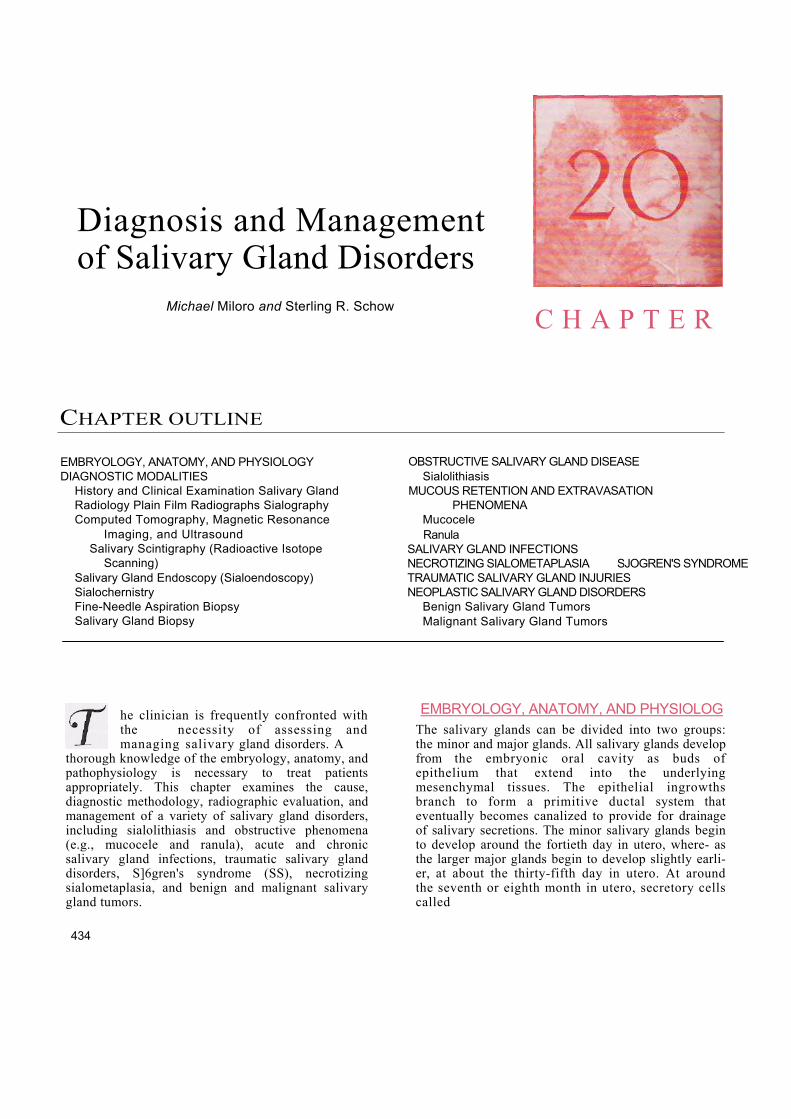
FIG. 20-1 Parotid gland anatomy. The course of Stensen's duct runs super-ficial to the masseter muscie and then curves sharply anteriorly to pierce the buccinator muscle fibers and enter the oral cavity.
develop around the ductal system. The aci-e salivary glands are classified as either hich produce a thin, watery serous secre-s cells, which produce a thicker, viscous ion. The minor salivary glands are well
d functional in the newborn infant. The minor salivary glands primarily produce ions, although some are made up of serous The major salivary glands are paired struc-the parotid, submandibular, and sublingual rotid glands contain primarily serous acini us cells. Conversely, the sublingual glands
st part composed of mucous cells. The sub-nds are mixed glands, made up of approx- numbers of serous and mucous acini. and 1000 minor salivary glands are found portions of the oral cavity that are cov-s membranes, with a few exceptions, such third of the hard palate, the attached gin-dorsal surface of the anterior third of the inor salivary glands are referred to as the palatine, tonsillar (Weber's glands), retromo-lands), and lingual glands, which are divid-
ed into three groups: (1) inferior apical (glands of Blandin Nuhn), (2) taste buds (Ebner's glands), and (3) posterior lubricating glands (Table 20-1).
The parotid glands, the largest salivary glands, lie superficial to the posterior aspect of the masseter muscle and the ascending ramus of the mandible. Peripheral por-tions of the parotid gland extend to the mastoid process, along the anterior aspect of the sternocleidomastoid mus-cle, and around the posterior border of the mandible into the pterygomandibular space (Fig. 20-1). The major branches of the seventh cranial (facial) nerve roughly divide the parotid gland into a superficial lobe and a deep lobe while coursing anteriorly from their exit at the sty-lomastoid foramen to innervate the muscles of facial expression. Small ducts from various regions of the gland coalesce at the anterosuperior aspect of the parotid to form Stensen's duct, which is the major duct of the parotid gland. Stensen's duct is about 1 to 3 mm in diam-eter and 6 cm in length.
Occasionally, a normal anatomic variation occurs in which an accessory parotid duct may aid Stensen's duct in drainage of salivary secretions. Additionally, an acces-sory portion of the parotid gland may be present some-
Akeel
436 PART IV ■ Infections
where along the course of Stensen's duct. The duct runs anteriorly from the gland and is superficial to the mas-seter muscle. At the location of the anterior edge of the masseter muscle, Stensen's duct turns sharply medial and passes through the fibers of the buccinator muscle. The duct opens into the oral cavity through the buccal mucosa, usually adjacent to the maxillary first or second molar tooth. The parotid gland receives innervation from
the ninth cranial (glossopharyngeal) nerve via the auric-lotemporal nerve from the otic ganglion.
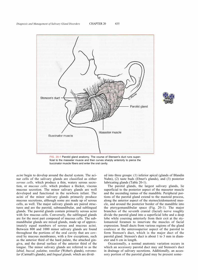
The submandibular glands are located in the sub-mandibular triangle of the neck, which is formed by the anterior and posterior bellies of the digastric muscles anc the inferior border of the mandible (Fig. 20-2). The posterosuperior portion of the gland curves upward around the posterior border of the mylohyoid muscle and
TABLE 20-1
Salivary Gland Embryology and Anatomy
In utera development Number Types
Minor Salivary Glands Day 40 800-1000 Labial Buccal Palatine Tonsillar • Weber's glands Retromolar • Carmalt's glands Lingual • Inferior apical (Glands of Blandin Nuhn) • Taste buds (Ebner's glands) • Posterior lubricating glands
Major Salivary Glands Day 35 6 Parotid Submandibular Sublingual
:FIinf
Parotid gland
Myiohyoid muscle
Anterior belly of digastric muscle
VFacial artery and vein
Submandibular gland Posterior belly of digastric muscle
Hyoid bone
G. 20-2 Submandibular gland anatomy. The anterior and posterior bellies of the digastric muscles and the erior border of the mandible form the submandibular triangle.
Diagnosis and Management of Salivary Gland Disorders CHAPTER 20 437
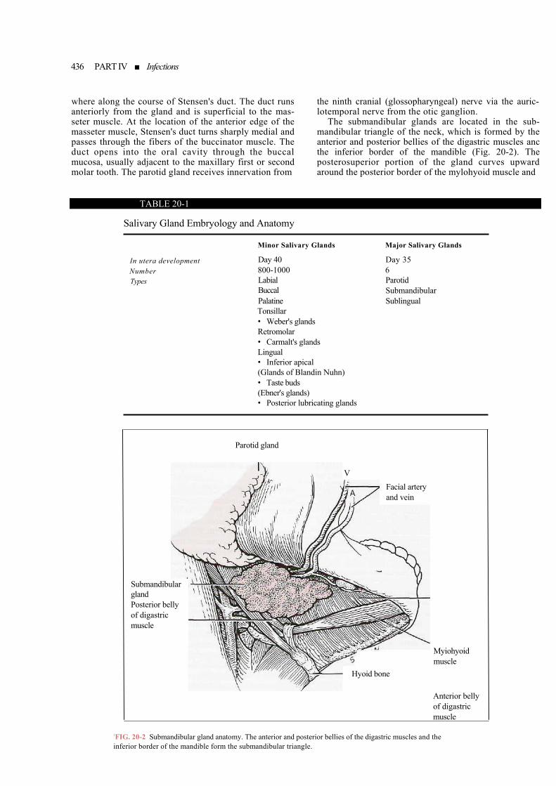
gives rise to the major duct of the submandibular gland known as Whartoris duct. This duct passes forward along the superior surface of the mylohyoid muscle in the sub-lingual space, adjacent to the lingual nerve. The anatomic relationship is such that the lingual nerve loops under Wharton's duct, from lateral to medial, in the posterior floor of the mouth. Wharton's duct is about 5 cm in length, and the diameter of its lumen is 2 to 4 mm. Wharton's duct opens into the floor of the mouth via a punctum located close to the incisors at the most anteri-or aspect of the junction of the lingual frenum and the floor of the mouth. The punctum is a constricted portion of the duct, and it functions to limit retrograde flow of bacteria-laden oral fluids. This particularly limits those bacteria that tend to colonize around the ductal orifices.
The sublingual glands lie on the superior surface of the mylohyoid muscle, in the sublingual space, and are sepa-rated from the oral cavity by a thin layer of oral mucosa (Fig. 20-3). The acinar ducts of the sublingual glands are called Bartholin's ducts and in most instances coalesce to form 8 to 20 ducts of Rivinus. These ducts of Rivinus are short and small in diameter. They either open individually directly into the anterior floor of the mouth on a crest of mucosa, known as the plica sublingualis, or they open indirectly through connections to the submandibular duct and then into the oral cavity via Wharton's duct. The sublingual and submandibular glands are innervated by the facial nerve through the submandibular ganglion via the chorda tympani nerve.
The functions of saliva are to provide lubrication for speech and mastication, to produce enzymes for diges-tion, and to produce compounds with antibacterial properties (Table 20-2). The salivary glands produce approximately 1000 to 1500 ml of saliva per day, with the highest flow rates occurring during meals. The relative contributions of each salivary gland to total daily pro-duction varies, with the submandibular gland providing 70%, the parotid gland 25%, the sublingual gland 3% to 4%, and the minor salivary glands contributing only trace amounts of saliva (Box 20-1). The electrolyte com-position of saliva also varies between salivary glands,
with parotid gland concentrations generally higher than the submandibular gland, except for submandibular cal-cium concentration, which is approximately twice the concentration of parotid calcium (see Table 20-2). The relative viscosities of saliva vary according to gland and
FIG. 20-3 Sublingual gland anatomy. The interrelsubmandibular and the sublingual glands and the relaare demonstrated.
TABLE 20-2
Composition of Normal Adult Saliva
Parotid Submandibular
Gfand Gland Sodium 23.0 mEq/L 21.0 mEq/L Potassium 20.0 mEq/L 17.0 mEq/L Chloride 23.0 mEq/L 20.0 mEq/L Bicarbonate 20.0 mEq/L 18.0 mEq/L Calcium 2.0 mEq/L 3.6 mEq/L Phosphate 6.0 mEq/L 4.5 mEq/L Magnesium 0.2 mEq/L 0.3 mEq/L Urea 15.0 mg/dl 7.0 mg/dl Ammonia 0.3 mg/dl 0.2 mg/dl Uric acid 3.0 mg/dl 2.0 mg/dl Glucose <1.0 mg/dl <1.0 mg/dl Cholesterol < 1.0 mg/dl <1.0 mg/dl Fatty acids 1.0 mg/dl <1.0 mg/dl Amino acids 1.5 mg/dl <1.0 mg/dl Proteins 250.0 mg/dl 150.0 mg/dl
a
BOX 20-1 Daily Saliva Production by Salivary Gland
Submandibular gland 70% Parotid gland 25% Sublingual gland 3%-4% Minor glands Trace
Plica sublinguaiis
Ducts of Rivinus
Lingual nerve Wharton's duct Submandibular gland
Sublingual glandBartholin's ducts'
tionships between the ductal systems of the tionship of the lingual nerve to Wharton's duct
438 PART IV Infections
correspond to the percentage of mucous and serous cell; therefore the highest viscosity is in the sublingual gland, followed by the submandibular gland, and, lastly. I parotid gland, which is composed mainly of serous eel Interestingly, the daily production of saliva begins decrease gradually after the age of 20.
BOX 20-2
Incidence of Radiopaque Stones
Submandibular gland 80% Parotid gland 40%
DIAGNOSTIC MODALITIES
FIG. sialoiitdemon
History and Clinical Examination The most important component of diagnosis in salivary gland disorders, as with most other disease processes, is the patient history and the clinical examination. In most cases the patient will guide the doctor to the diagnosis merely by relating the events that have occurred in association with the presenting complaint. The astute clinician must perform a thorough evaluation, and, in mar instances, the diagnosis can be determined without the necessity of further diagnostic evaluation. At the very least, the clinician may be able to categorize the problem as reactive, obstructive, inflammatory, infectious, metabolic, neoplastic, developmental, or traumatic in origin and guide further diagnostic testing. Occasionally, the clinician may find it necessary to use any of several diagnostic modalities.
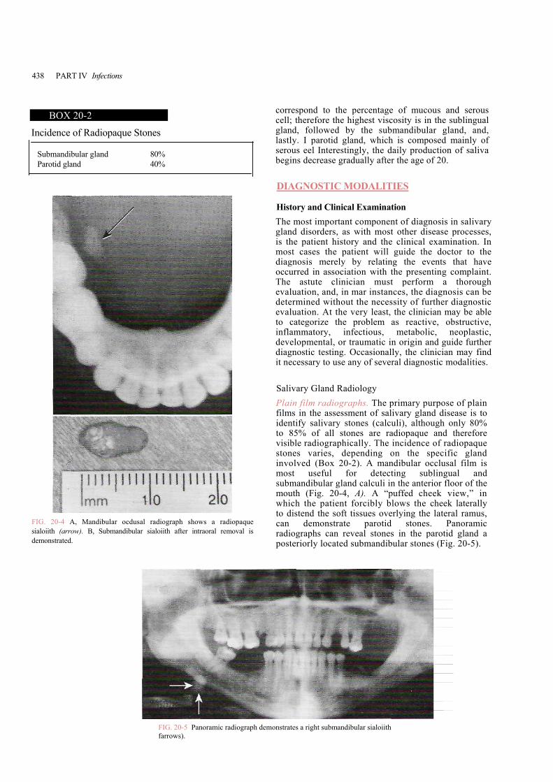
Salivary Gland Radiology Plain film radiographs. The primary purpose of plain films in the assessment of salivary gland disease is to identify salivary stones (calculi), although only 80% to 85% of all stones are radiopaque and therefore visible radiographically. The incidence of radiopaque stones varies, depending on the specific gland involved (Box 20-2). A mandibular occlusal film is most useful for detecting sublingual and submandibular gland calculi in the anterior floor of the mouth (Fig. 20-4, A). A “puffed cheek view,” in which the patient forcibly blows the cheek laterally
20-4 A, Mandibular ocdusal radiograph shows a radiopaque h (arrow). B, Submandibular sialoiith after intraoral removal is strated.
to distend the soft tissues overlying the lateral ramus, can demonstrate parotid stones. Panoramic radiographs can reveal stones in the parotid gland a posteriorly located submandibular stones (Fig. 20-5).
FIG. 20-5 Panoramic radiograph demonstrates a right submandibular sialoiith farrows).
Diagnosis and Management of Salivary Gland Disorders CHAPTER 20 439
Periapical radiographs can show calculi in each salivary gland or duct, including minor salivary glands, depending on film placement. In most instances, the radiographic image corresponds in size and shape to the actual stone (see Fig. 20-4, B).
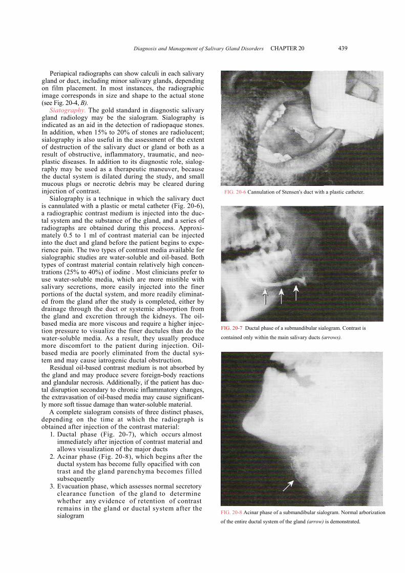
Siatography. The gold standard in diagnostic salivary gland radiology may be the sialogram. Sialography is indicated as an aid in the detection of radiopaque stones. In addition, when 15% to 20% of stones are radiolucent; sialography is also useful in the assessment of the extent of destruction of the salivary duct or gland or both as a result of obstructive, inflammatory, traumatic, and neo-plastic diseases. In addition to its diagnostic role, sialog-raphy may be used as a therapeutic maneuver, because the ductal system is dilated during the study, and small mucous plugs or necrotic debris may be cleared during injection of contrast.
Sialography is a technique in which the salivary duct is cannulated with a plastic or metal catheter (Fig. 20-6), a radiographic contrast medium is injected into the duc-tal system and the substance of the gland, and a series of radiographs are obtained during this process. Approxi-mately 0.5 to 1 ml of contrast material can be injected into the duct and gland before the patient begins to expe-rience pain. The two types of contrast media available for sialographic studies are water-soluble and oil-based. Both types of contrast material contain relatively high concen-trations (25% to 40%) of iodine . Most clinicians prefer to use water-soluble media, which are more mistible with salivary secretions, more easily injected into the finer portions of the ductal system, and more readily eliminat-ed from the gland after the study is completed, either by drainage through the duct or systemic absorption from the gland and excretion through the kidneys. The oil-based media are more viscous and require a higher injec-tion pressure to visualize the finer ductules than do the water-soluble media. As a result, they usually produce more discomfort to the patient during injection. Oil-based media are poorly eliminated from the ductal sys-tem and may cause iatrogenic ductal obstruction.
Residual oil-based contrast medium is not absorbed by the gland and may produce severe foreign-body reactions and glandular necrosis. Additionally, if the patient has duc-tal disruption secondary to chronic inflammatory changes, the extravasation of oil-based media may cause significant-ly more soft tissue damage than water-soluble material.
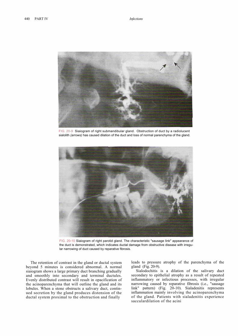
A complete sialogram consists of three distinct phases, depending on the time at which the radiograph is obtained after injection of the contrast material:
1. Ductal phase (Fig. 20-7), which occurs almost immediately after injection of contrast material and allows visualization of the major ducts
2. Acinar phase (Fig. 20-8), which begins after the ductal system has become fully opacified with con trast and the gland parenchyma becomes filled subsequently
3. Evacuation phase, which assesses normal secretory clearance function of the gland to determine whether any evidence of retention of contrast remains in the gland or ductal system after the sialogram
FIG. 20-6 Cannulation of Stensen's duct with a plastic catheter.FIG. 20-7 Ductal phase of a submandibular sialogram. Contrast is
contained only within the main salivary ducts (arrows).
FIG. 20-8 Acinar phase of a submandibular sialogram. Normal arborization
of the entire ductal system of the gland (arrow) is demonstrated.
440 PART IV Infections
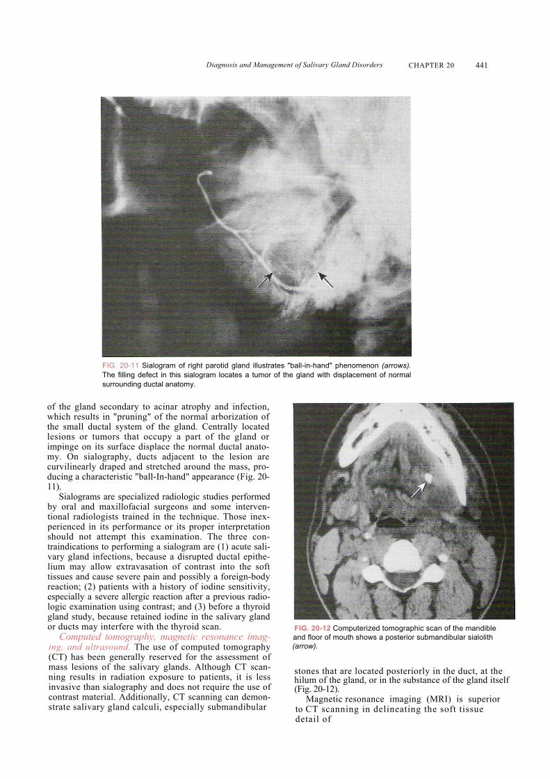
The retention ofbeyond 5 minutessiaiogram shows a and smoothly intEvenly distributed the acinoparenchylobules. When a sued secretion by ductal system pro
FIG. 20-9 Siaiogram of right submandibular gland. Obstruction of duct by a radiolucent sialolith (arrows) has caused dilation of the duct and loss of normal parenchyma of the gland.
lo
mttx
FIG. 20-10 Siaiogram of right parotid gland. The characteristic "sausage link" appearance of the duct is demonstrated, which indicates ductal damage from obstructive disease with irregu-lar narrowing of duct caused by reparative fibrosis.
contrast in the gland or ductal system is considered abnormal. A normal arge primary duct branching gradually secondary and terminal ductules.
contrast will result in opacification of a that will outline the gland and its
one obstructs a salivary duct, contin-he gland produces distension of the imal to the obstruction and finally
leads to pressure atrophy of the parenchyma of the gland (Fig. 20-9).
Sialodochitis is a dilation of the salivary duct secondary to epithelial atrophy as a result of repeated inflammatory or infectious processes, with irregular narrowing caused by reparative fibrosis (i.e., "sausage link" pattern) (Fig. 20-10). Sialadenitis represents inflammation mainly involving the acinoparenchyma of the gland. Patients with sialadenitis experience sacculardilation of the acini
Diagnosis and Management of Salivary Gland Disorders CHAPTER 20 441
of the gland swhich results the small duclesions or tumimpinge on itsmy. On sialocurvilinearly dducing a chara11).
Sialograms by oral and mtional radiologperienced in ishould not atraindications vary gland inflium may allotissues and caureaction; (2) pespecially a selogic examinagland study, bor ducts may i
Computed ing, and ultra(CT) has beenmass lesions ning results iinvasive than contrast materstrate salivary
FIG. 20-11 Sialogram of right parotid gland illustrates "ball-in-hand" phenomenon (arrows). The filling defect in this sialogram locates a tumor of the gland with displacement of normal surrounding ductal anatomy.
econdary to acinar atrophy and infection, in "pruning" of the normal arborization of tal system of the gland. Centrally located
ors that occupy a part of the gland or surface displace the normal ductal anato-graphy, ducts adjacent to the lesion are raped and stretched around the mass, pro-cteristic "ball-In-hand" appearance (Fig. 20-
are specialized radiologic studies performed axillofacial surgeons and some interven-ists trained in the technique. Those inex-
ts performance or its proper interpretation ttempt this examination. The three con-to performing a sialogram are (1) acute sali-ections, because a disrupted ductal epithe-w extravasation of contrast into the soft se severe pain and possibly a foreign-body atients with a history of iodine sensitivity, vere allergic reaction after a previous radio-tion using contrast; and (3) before a thyroid ecause retained iodine in the salivary gland nterfere with the thyroid scan. tomography, magnetic resonance imag-sound. The use of computed tomography generally reserved for the assessment of
of the salivary glands. Although CT scan-n radiation exposure to patients, it is less sialography and does not require the use of ial. Additionally, CT scanning can demon- gland calculi, especially submandibular
(
:FIG. 20-12 Computerized tomographic scan of the mandible and floor of mouth shows a posterior submandibular sialolith arrow).
stones that are located posteriorly in the duct, at the hilum of the gland, or in the substance of the gland itself (Fig. 20-12).
Magnetic resonance imaging (MRI) is superior to CT scanning in delineating the soft tissue detail of
442 PART IV Infections
salivary gland lesions, specifically tumors, with no radia-tion exposure to the patient or the necessity of contrast enhancement.
Ultrasonography is a relatively simple, noninvasive imaging modality, with poor detail resolution. The primary role of ultrasonography is in the assessment of superficial structures to determine whether a mass lesion that is being evaluated is solid or cystic (fluid-filled) in nature.
Salivary scintigraphy (radioactive isotope scanning). The use of nuclear imaging in the form of radioactive iso-tope scanning, or salivary scintigraphy, allows a thorough evaluation of the salivary gland parenchyma, with respect to the presence of mass lesions and the function of the gland itself. This study uses a radioactive isotope (usually, technetium [Tc] 99m) injected intravenously (IV), which is distributed throughout the body and taken up by a variety of tissues, including the salivary glands. The major limitation of this study, aside from patient radiation exposure, is the poor resolution of the images obtained. Salivary gland scintigraphy may demonstrate increased uptake of radioactive isotope in an acutely inflamed gland or decreased uptake in a chronically inflamed gland, as well as the presence of a mass lesion, either benign or malignant.
Salivary Gland Endoscopy (Sialoendoscopy) Minimally invasive modalities of diagnosis and treatment have recently been applied to the major salivary glands. Salivary gland endoscopy (sialoendoscopy) is a special-ized procedure that uses a small video camera (endo-scope) with a light at the end of a flexible cannula, which is introduced into the ductal orifice. The endoscope can be used diagnostically and therapeuticaUy. Salivary gland endoscopy has demonstrated strictures and kinks in the ductal system, as well as mucous plugs and calcifications. The endoscope may be used to dilate small strictures and flush clear small mucous plugs in the salivary gland ducts. Specialized devices such as small balloon catheters (similar to those used for coronary angioplasty proce-dures) may be used to dilate sites of ductal constriction, and small metal baskets may be used to retrieve stones in the ductal system
Sialochemistry An examination of the electrolyte composition of the saliva (see Table 20-2) of each gland may indicate a vari-ety of salivary gland disorders. Principally the concentra-tions of sodium and potassium, which normally change with salivary flow rate, are measured. Certain changes in the relative concentrations of these electrolytes are seen in specific salivary gland diseases. For example, an elevat-ed sodium concentration with a decreased potassium concentration may indicate an inflammatory sialadenitis.
Fine-Needle Aspiration Biopsy The use of fine-needle aspiration biopsy in the diagnosis of salivary gland tumors has been well documented. This procedure has a high accuracy rate for distinguishing
between benign and malignant lesions in superficial locations. Fine-needle aspiration biopsy is performed using syringe with a 20-gauge or smaller needle. After loc anesthesia the needle is advanced into the mass lesion the plunger is activated to create a vacuum in the syringe and the needle is moved back and forth throughout the mass, with pressure maintained on the plunger. The pressure is then released, the needle is withdrawn, and fluid cellular material and fluid is expelled onto a slide and fixed for histologic examination. This allows an immediate determination of benign versus malignant disease also offers the possibility of providing a tissue diagnosis especially if the oral surgeon and oral pathologist are experienced in performing and interpreting this examination and its results.
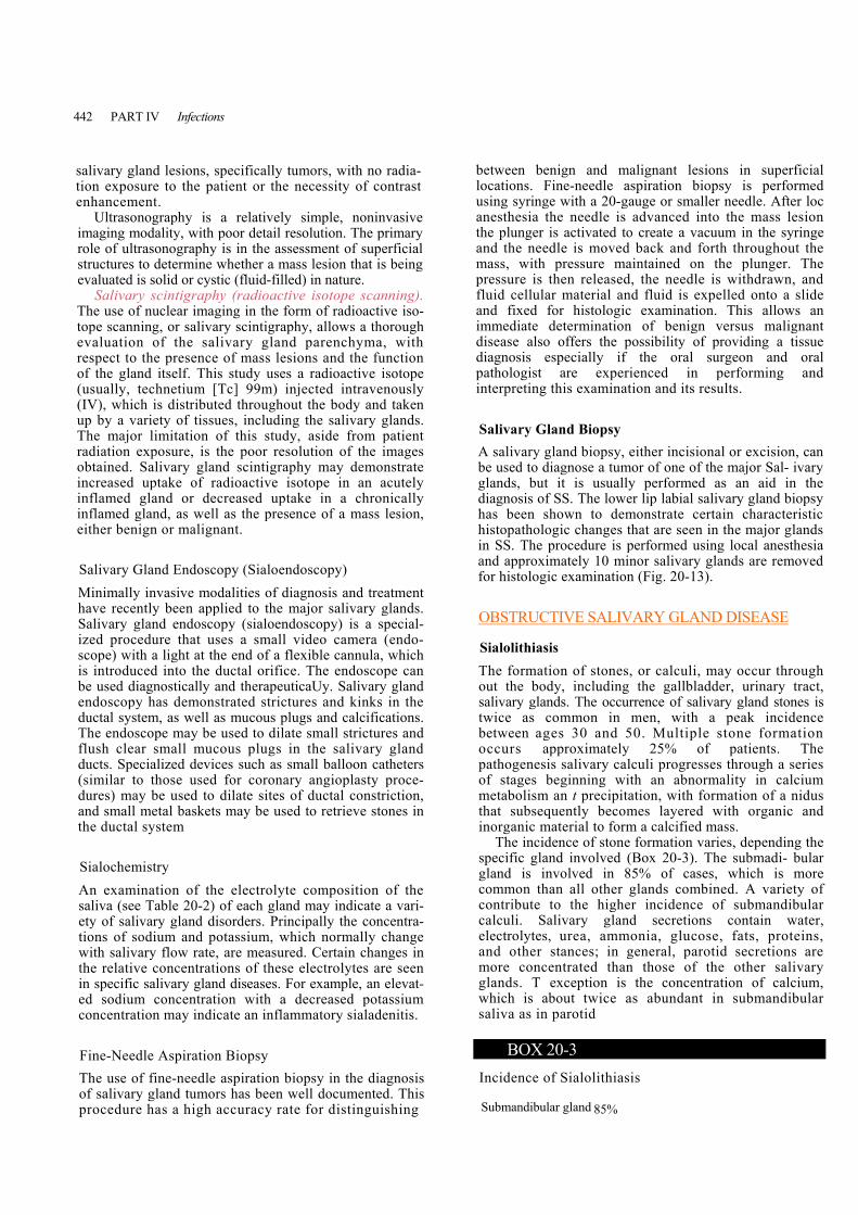
Salivary Gland Biopsy A salivary gland biopsy, either incisional or excision, can be used to diagnose a tumor of one of the major Sal- ivary glands, but it is usually performed as an aid in the diagnosis of SS. The lower lip labial salivary gland biopsy has been shown to demonstrate certain characteristic histopathologic changes that are seen in the major glands in SS. The procedure is performed using local anesthesia and approximately 10 minor salivary glands are removed for histologic examination (Fig. 20-13).
OBSTRUCTIVE SALIVARY GLAND DISEASE
Sialolithiasis The formation of stones, or calculi, may occur through out the body, including the gallbladder, urinary tract, salivary glands. The occurrence of salivary gland stones is twice as common in men, with a peak incidence between ages 30 and 50. Multiple stone formation occurs approximately 25% of patients. The pathogenesis salivary calculi progresses through a series of stages beginning with an abnormality in calcium metabolism an t precipitation, with formation of a nidus that subsequently becomes layered with organic and inorganic material to form a calcified mass.
The incidence of stone formation varies, depending the specific gland involved (Box 20-3). The submadi- bular gland is involved in 85% of cases, which is more common than all other glands combined. A variety of contribute to the higher incidence of submandibular calculi. Salivary gland secretions contain water, electrolytes, urea, ammonia, glucose, fats, proteins, and other stances; in general, parotid secretions are more concentrated than those of the other salivary glands. T exception is the concentration of calcium, which is about twice as abundant in submandibular saliva as in parotid
BOX 20-3 Incidence of Sialolithiasis
85% Submandibular gland
Diagnosis and Management of Salivary Gland Disorders
CHAPTER 443
saliva (see Table 20-2). In addition, the alkaline pH of sub-mandibular saliva may further support stone formation. In addition to salivary composition, several anatomic factors of the submandibular gland and duct are important. Wharton's duct is the longest salivary duct; therefore saliva has a greater distance to travel before being emptied into the oral cavity. In addition, the duct of the submandibular gland has two sharp curves in its course: The first occurs at the posterior border of the mylohyoid muscle, and the second is near the ductal opening in the anterior floor of the mouth. Finally, the punctum of the submandibular duct is smaller than the opening of Stensen's duct. These features contribute to a slowed sali-
vary flow and provide potential areas of stasis of salivary flow, or obstruction, that is not found in the parotid or sublingual ductal systems. Precipitated material, mucus, and cellular debris are more easily trapped in the tortuous and lengthy submandibular duct, especially when its small orifice is its most elevated location, and its flow therefore occurs against the force of gravity. The precipi-tated material forms the nidus of mucous plugs and either radiopaque or radiolucent sialoliths that may even-tually enlarge to the point of obstructing the flow of sali-va from the gland to the oral cavity.
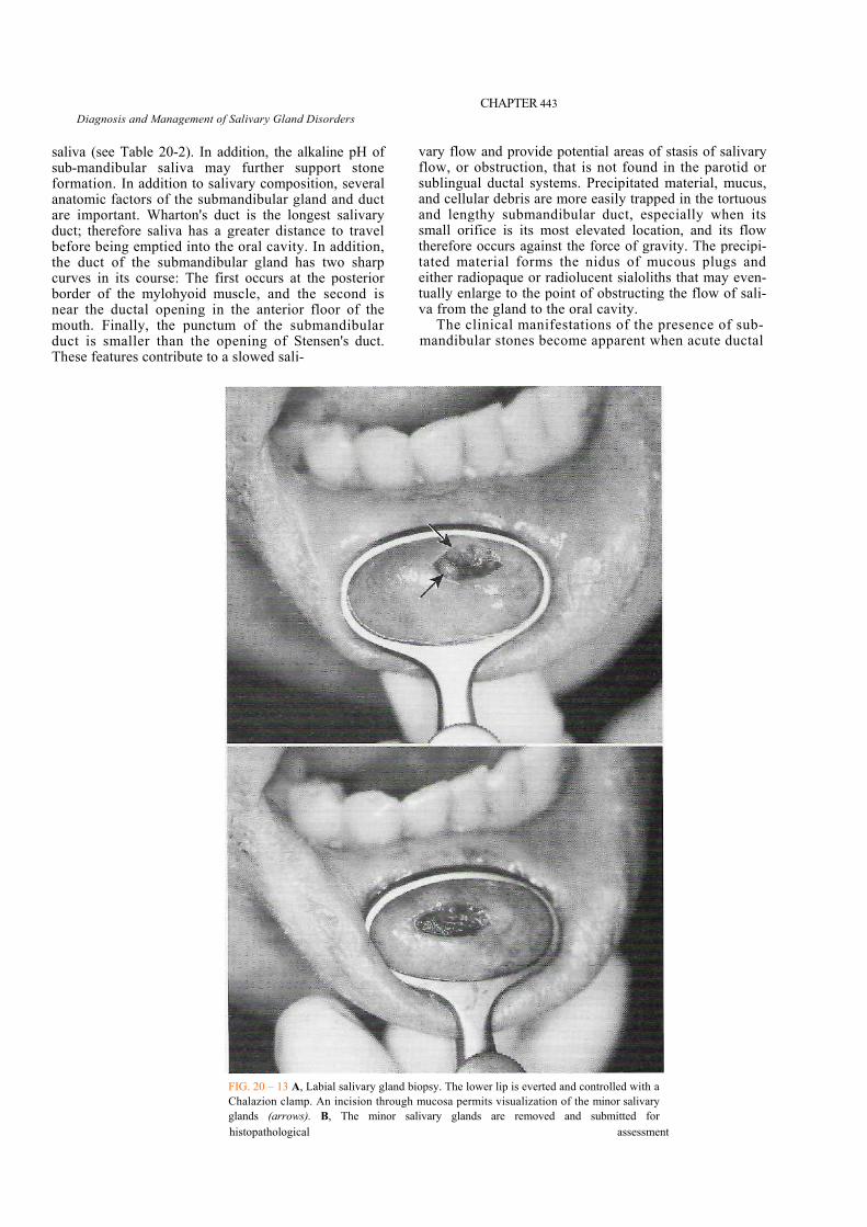
The clinical manifestations of the presence of sub-mandibular stones become apparent when acute ductal
FIG. 20 – 13 A, Labial salivary gland biopsy. The lower lip is everted and controlled with a Chalazion clamp. An incision through mucosa permits visualization of the minor salivary glands (arrows). B, The minor salivary glands are removed and submitted for histopathological assessment
444 PART IV Infections
is most commonly affected. The diagnosis can be made clinically and confirmed radiographically by plain films ultrasound, sialography, or sialoendoscopy.
The management of submandibular gland calculi depends on the duration of symptoms, the number repeated episodes, the size of the stone, and, per" most importantly, the location of the stone. I mandibular stones are classified as either anterior or poste rior stones, in relation to a transverse line between mandibular first molars. Stones that occur anterior to this line are generally well visualized on a mandibular occlusal radiograph and may be amenable to intraoral removal. Small anteriorly located stones may be rein- : through the ductal opening after dilation of the orifice._
Occasionally, it becomes necessary to remove sub- mandibular stones via an incision made in the floor of the mouth to expose the duct and the stone. A longitudinal incision is then made in the duct, the stone is retrieved, and the ductal lining is sutured to the mucosa of the floor of the mouth. Saliva will then flow out
FIG. 20'14 Clinical photograph demonstrates a right sub-mandibular swelling (arrow) secondary to obstruction from a sub-mandibular sialolith.
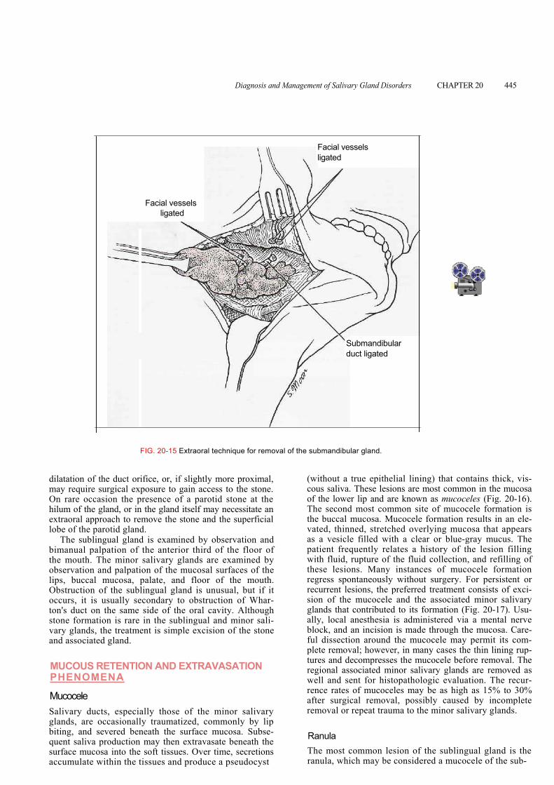
the revised duct. This procedure, known as a sialodochoplasty (i.e., revision of the salivary duct), eliminates many of the factors that contributed to formation of the stone entire length of the duct is decreased, the opening created is now larger, and gravity contributes less to salivary stasis. Regardless of the procedure performed, patient are encouraged to maintain ample salivary flow by using salivary stimulants, such as citrus fruits, flavored candies or glycerin swabs. Posterior stones occur in up to 50% of cases and may be located at the hilum of the gland or within the substance of the gland itself. A rout ine occlusal film will likely not demonstrate the stone, a panoramic radiograph (see Fig. 20-5) or a CT scan (see Fig 20-12) may be necessary to localize the stone. In cases of posterior stones that cannot be palpated intraorally and in many instances of repeated stone formation, the submandibular gland and the stone should be removed by an extraoral approach (Fig. 20-15).
Recent clinical trials using extracorporeal shock I lithotripsy (ECSWL) have been successful in small salivary gland stones. This technology uses trarnscu- taneous electromagnetic waves to break the calculus apart into smaller calcified debris particles, which can be
BOX 20-4
Sialolithiasis for the General Dentist
Classic signs and symptoms of sialolithiasis l Exacerbation of pain and swelling at mealtimes I Check for flow from Wharton's duct J Check for tenderness of submandibular gland I Palpate for stone in floor of mouth
£ Check mandibular occlusal radiograph Treatment Anterior stone
Attempt to dilate Wharton's duct with lacrimal probes Careful to not dislodge stone posteriorly "Milk" the gland to express stone
I If successful, prescribe salivary stimulants Posterior stone or no stone visualized ■ Refer to oral surgeon
obstruction occurs at mealtime, when saliva production is at its maximum and salivary flow is stimulated against a fixed obstruction. The resultant swelling is sudden and is usually very painful (Box 20-4; Fig. 20-14). Gradual reduc-tion of the swelling follows, but swelling reoccurs repeat-edly when salivary flow is stimulated. This process may continue until complete obstruction, infection, or both occurs. Obstruction, with or without infection, causes atro-phy of the secretory cells of the involved gland. Infection of the gland manifests itself by swelling in the floor of the mouth, erythema, and an associated lymphadenopathy. Palpation of the gland and simultaneous examination of the duct and its opening may reveal the total absence of salivary flow or the presence of purulent material.
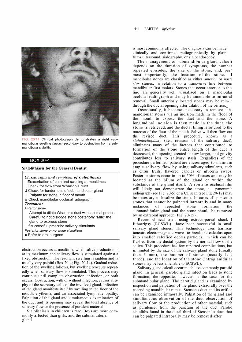
Sialolithiasis in children is rare. Boys are more com-monly affected than girls, and the submandibular gland
flushed from the ductal system by the normal flow of the saliva. This procedure has few reported complications, but is limited by the size of the salivary gland stone (usually than 3 mm), the number of stones (usually less three), and the location of the stone (intraglanldular stones may be less amenable to ECSWL).
Salivary gland calculi occur much less commonly parotid gland. In general, parotid gland infection leads to stone formation; the opposite, however, is the case for the submandibular gland. The parotid gland is examined by inspection and palpation of the gland extraorally over the ascending mandibular ramus. Stensen's duct and its orifice can be examined intraorally. Palpation of the gland and simultaneous observation of the duct observation of salivary flow or the production of other material, such as purulence, from the punctum of the duct Parotid sialoliths found in the distal third of Stensen’ s duct that can be palpated intraorally may be removed after
Diagnosis and Management of Salivary Gland Disorders CHAPTER 20 445
dilatation of may require On rare occhilum of the extraoral applobe of the p
The sublibimanual pathe mouth. observation lips, buccalObstruction occurs, it iston's duct ostone formavary glands,and associat
MUCOUS PHENOM
Mucocele Salivary duglands, are biting, and quent salivasurface mucaccumulate
Facial vessels ligated
Facial vessels ligated
Submandibular duct ligated
FIG. 20-15 Extraoral technique for removal of the submandibular gland.
the duct orifice, or, if slightly more proximal, surgical exposure to gain access to the stone. asion the presence of a parotid stone at the gland, or in e gland itself may necessitate an throach to remove the stone and the superficial arotid gland. ngual gland is examined by observation and lpation of the anterior third of the floor of
The minor salivary glands are examined by and palpation of the mucosal surfaces of the mucosa, palate, and floor of the mouth. of the sub ngual gland is unusual, but if it li usually secondary to obstruction of Whar-n the same side of the oral cavity. Although tion is rare in the sublingual and minor sali- the treatm nt is simple excision of the stone eed gland.
RETENTI N AND EXTRAVASATION OENA
cts, especially those of the minor salivary occasionally traumatized, commonly by lip severed beneath the surface mucosa. Subse- production may then extravasate beneath the osa into the soft tissues. Over time, secretions within the tissues and produce a pseudocyst
Ranula
( ithelial lining) that contains thick, vis-without a true ep saliva. Te lower li
cous hese lesions are most common in the mucosa of th p and are known as mucoceles (Fig. 20-16). The second most common site of mucocele formation is the buccal mucosa. Mucocele formation results in an ele-vated, thinned, stretched overlying mucosa that appears as a vesicle filled with a clear or blue-gray mucus. The patient frequently relates a history of the lesion filling with fluid, rupture of the fluid collection, and refilling of these lesions. Many instances of mucocele formation regress spontaneously without surgery. For persistent or recurrent lesions, the preferred treatment consists of exci-sion of the mucocele and the associated minor salivary glands that contributed to its formation (Fig. 20-17). Usu-ally, local anesthesia is administered via a mental nerve block, and an incision is made through the mucosa. Care-ful dissection around the mucocele may permit its com-plete removal; however, in many cases the thin lining rup-tures and decompresses the mucocele before removal. The regional associated minor salivary glands are removed as well and sent for histopathologic evaluation. The recur-rence rates of mucoceles may be as high as 15% to 30% after surgical removal, possibly caused by incomplete removal or repeat trauma to the minor salivary glands.
The most common lesion of the sublingual gland is the ranula, which may be considered a mucocele of the sub-
446 PART IV Infections
FIGis
lingual salivary glandsimple ranula and thfrom either mucous ductal system or muctal disruption. The sioccupied by the sublisuperior to the myloprogression to a plunextends beyond the lthe submandibular spathan mucoceles, becauand because trauma tlikely in the floor of t
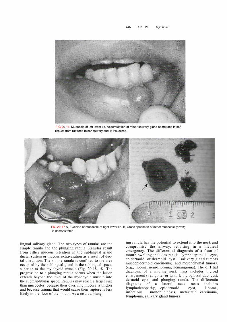
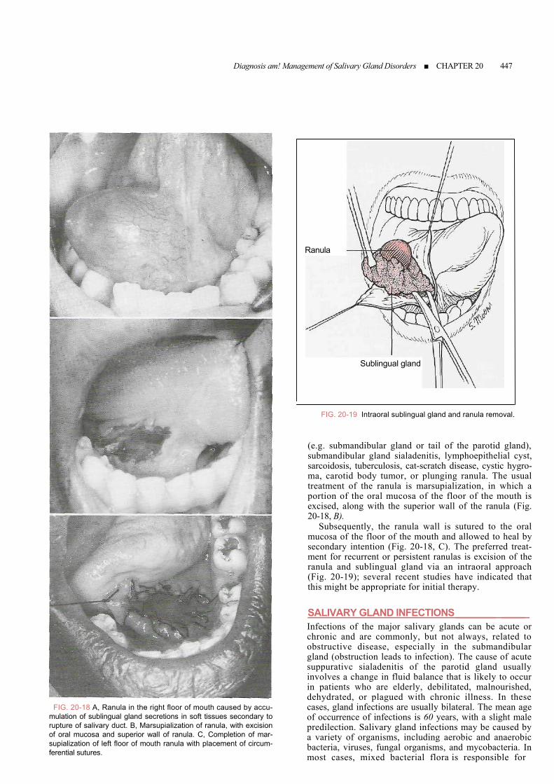
FIG.20-16 Mucocele of left lower lip. Accumulation of minor salivary gland secretions in soft tissues from ruptured minor salivary duct is visualized..20-17 A, Excision of mucocele of right lower lip. B, Cross specimen of intact mucocele (arrow) demonstrated.
. The two types of ranulas are the e plunging ranula. Ranulas result retention in the sublingual gland
ous extravasation as a result of duc-emergency. The differential diagnosis of a floor of m
mple ranula is confined to the area ngual gland in the sublingual space, hyoid muscle (Fig. 20-18, A). The ging ranula occurs when the lesion evel of the mylohyoid muscle into ce. Ranulas may reach a larger size se their overlying mucosa is thicker
hat would cause their rupture is less he mouth. As a result a plung-
ing ranula has the potential to extend into the neck and compromise the airway, resulting in a medical
outh swelling includes ranula, lymphoepithelial cyst, epidermoid or dermoid cyst, salivary gland tumors mucoepidermoid carcinoma), and mesenchymal tumors. (e.g., lipoma, neurofibroma, hemangioma). The dirf tial diagnosis of a midline neck mass includes thyroid enlargement (i.e., goiter or tumor), thyroglossal duct cyst, dermoid cyst, and plunging ranula. The differentia diagnosis of a lateral neck mass includes lymphadenopathy, epidermoid cyst, lipoma, infectious mononucleosis, metastatic carcinoma, lymphoma, salivary gland tumors
Diagnosis am! Management of Salivary Gland Disorders ■ CHAPTER 20 447
FIG. 20-18 A, Ranula in the right floor of mouth caused by accu-
mulation of sublingual gland secretions in soft tissues secondary to rupture of salivary duct. B, Marsupialization of ranula, with excision of oral mucosa and superior wall of ranula. C, Completion of mar-supialization of left floor of mouth ranula with placement of circum-ferential sutures.
Ranula
Sublingual gland
FIG. 20-19 Intraoral sublingual gland and ranula removal.
(e.g. submandibular gland or tail of the parotid gland), submandibular gland sialadenitis, lymphoepithelial cyst, sarcoidosis, tuberculosis, cat-scratch disease, cystic hygro-ma, carotid body tumor, or plunging ranula. The usual treatment of the ranula is marsupialization, in which a portion of the oral mucosa of the floor of the mouth is excised, along with the superior wall of the ranula (Fig. 20-18, B).
Subsequently, the ranula wall is sutured to the oral mucosa of the floor of the mouth and allowed to heal by secondary intention (Fig. 20-18, C). The preferred treat-ment for recurrent or persistent ranulas is excision of the ranula and sublingual gland via an intraoral approach (Fig. 20-19); several recent studies have indicated that this might be appropriate for initial therapy.
SALIVARY GLAND INFECTIONS_______ ____ Infections of the major salivary glands can be acute or chronic and are commonly, but not always, related to obstructive disease, especially in the submandibular gland (obstruction leads to infection). The cause of acute suppurativc sialadenitis of the parotid gland usually involves a change in fluid balance that is likely to occur in patients who are elderly, debilitated, malnourished,
dehydrated, or plagued with chronic illness. In these cases, gland infections are usually bilateral. The mean age of occurrence of infections is 60 years, with a slight male predilection. Salivary gland infections may be caused by a variety of organisms, including aerobic and anaerobic bacteria, viruses, fungal organisms, and mycobacteria. In most cases, mixed bacterial flora is responsible for
448 PART IV Infections
The clinical characteristics of acute bacterial s and gl
gi
hg
u flui
p
spo
re anai
doses for
ngry in the
fect
ces
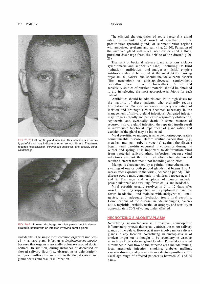
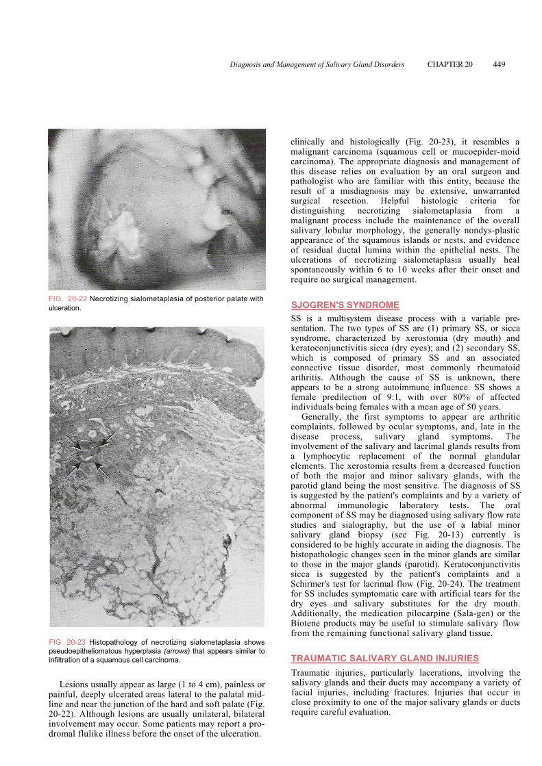
infections include rapid onset of swelling in the preauricular (parotid gland) or submandibular re ons with associated erythema and pain (Fig. 20-20). Palpation of the involved gland will reveal no flow or elicit a t ick, purulent discharge from the orifice of the duct(Fi 20-21).
Treatment of bacterial salivary gland infections incl des s atic and supportive care, including IV d ymptomhydration, antibiotics, and analgesics. Initial em iric antibiotics should be aimed at the most likely causing organism, S. aureus, and should include a cephalo rin (first generation) or antistaphylococcal semisynthetic penicillin (oxacillin or dicloxacillin). Cultu d sensitivity studies of purulent material should be obt ned to aid in selecting the most appropriate antibiotic for each patient.
Antibiotics should be administered IV in high the majority of these patients, who ordinarily require hospitalization. ons, surgery consisti of On most occasiincision and drainage (I&D) becomes necessamanagement of salivary gland infections. Untreated in : -may progress rapidly and can cause respiratory obstruction, septicemia, and, eventually, death. In some instan of
rent salivary gland infection, the repeated insults resrecur ult in irreversible functional impairment of gland ration and excision of the gland may be indicated.
Viral parotitis, or mumps, is an acute, nonsuppuppurative
FIG. 20-20 Left parotid gland infection. This infection is extreme-ly painful and may indicate another serious illness. Treatment requires hospitalization, intravenous antibiotics, and possibly surgi-cal drainage.mmunicable disease. Before routine vaccination (eg., easles, mumps, rubella vaccine) against the disease gan, viral parotitis occurred in epidemics during the inter and spring. It is important to differentiate viral om bacterial salivary gland infection, because viral fections are not the result of obstructive diseaseand quire diffe
combewfrinre rent treatment, not including antibiotics.
Mumps is characterized by a painful, nonerythematous. elling of one or both parotid glands that begins 2 to 3
eeks after exposure to the virus (incubation period). This sease occurs most commonly in children between ages 6 d 8. The signs and symptoms of mumps include eauricular pain and swelling, fever, chills, and headache. Viral parotitis usually resolves in 5 to 12
swwdianpr
days after onset. Providing supportive and symptomatic care for fe -ver, headache, and malaise with antipyretics, anal
sics, and adequate hydration treats viral parotitis. geComplications of the disease include meningitis, pancre-titis, nephritis, orchitis, testicular atrophy, and sterility in pproximately 20% of young males affected.
ECROTIZING SIALOMETAPLASIA
aa
N
FIG. 20-21 Purulent discharge from left parotid duct is demon-strated in patient with an infection involving parotid gland.sialadenitis. The single most common organism implicat-ed in salivary gland infection is Staphylococcus aureus, because this organism normally colonizes around ductal orifices. In ad
ecrotizing sialometaplasia is a reactive, nonneoplastic flammatory process that usually affects the minor salivary ands of the palate. However, it may involve minor salivary ands in any location. Necrotizing sialometaplasia is of clear origin but is thought to be secondary to vascular farction of the salivary gland lobules. Potential causes of
iminished blood flow to the affected area include trauma, cal anesthetic injection, smoking, diabetes mellitus,
Ninglglunind
dition, during instances of decreased or slowed salivary flow (i.e., obstruction or dehydration), retrograde influx of S. aureus into the ductal system and gland occurs and results in infection.
lova pressure from a denture prosthesis. The scular disease, andusual age range of affected patients is between 23 and 66 years.
Diagnosis and Management of Salivary Gland Disorders CHAPTER 20 449
clinically and histologically (Fig. 20-23), it resembles a malignant carcinoma (squamous cell or mucoepider-moid carcinoma). The appropriate diagnosis and management of this disease relies on evaluation by an oral surgeon and pathologist who are familiar with this entity, because the result of a misdiagnosis may be extensive, unwarranted surgical resection. Helpful histologic criteria for distinguishing necrotizing sialometaplasia from a malignant process include the maintenance of the overall salivary lobular morphology, the generally nondys-plastic appearance of the squamous islands or nests, and evidence of residual ductal lumina within the epithelial nests. The ulcerations of necrotizing sialometaplasia usually heal spontaneously within 6 to 10 weeks after their onset and require no surgical management.
FIG. 20-22 Necrotizing siaIometaplasia of posterior palate with ulceration.
Fpi
pl2id
SJOGREN'S SYNDROME SS is a multisystem disease process with a variable pre-sentation. The two types of SS are (1) primary SS, or sicca syndrome, characterized by xerostomia (dry mouth) and keratoconjunctivitis sicca (dry eyes); and (2) secondary SS, which is composed of primary SS and an associated connective tissue disorder, most commonly rheumatoid arthritis. Although the cause of SS is unknown, there appears to be a strong autoimmune influence. SS shows a female predilection of 9:1, with over 80% of affected individuals being females with a mean age of 50 years.
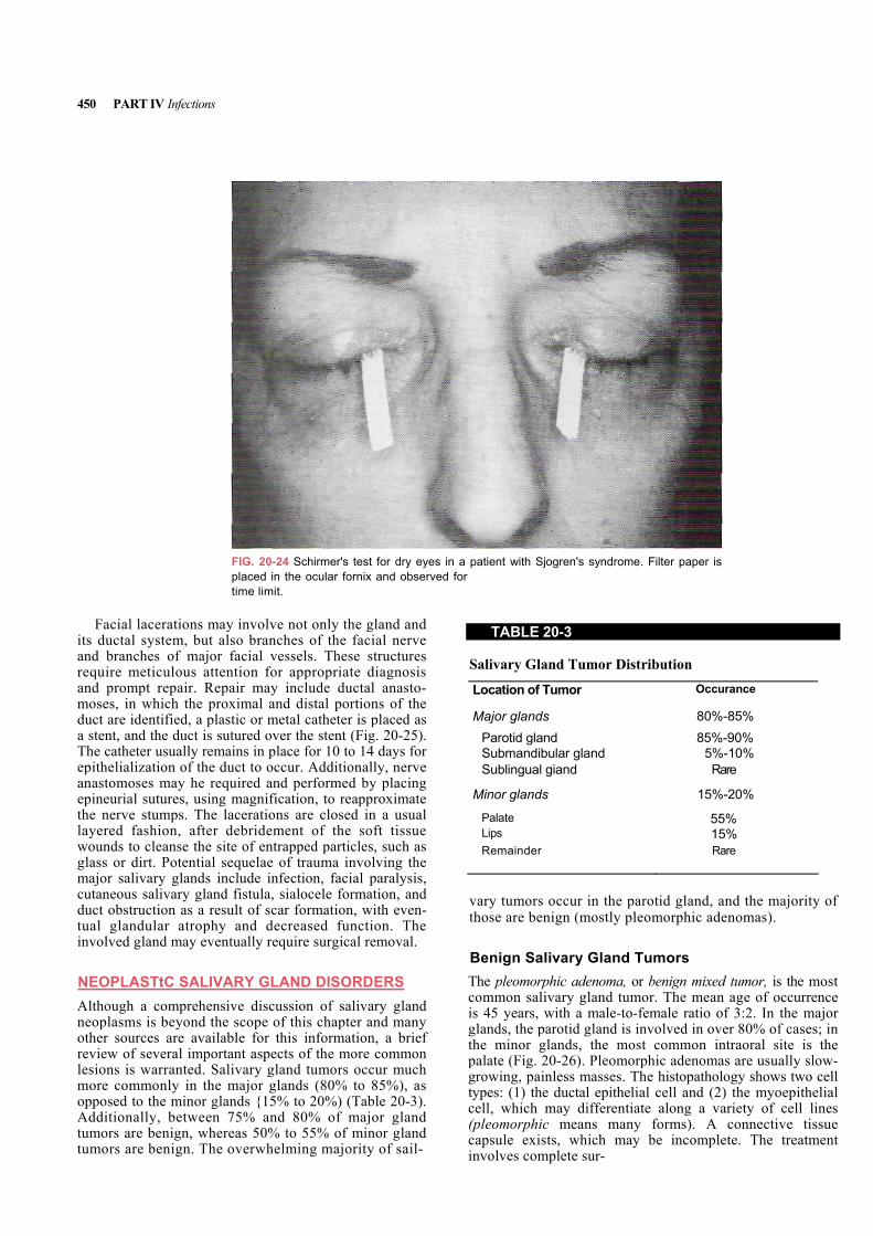
Generally, the first symptoms to appear are arthritic complaints, followed by ocular symptoms, and, late in the disease process, salivary gland symptoms. The involvement of the salivary and lacrimal glands results from a lymphocytic replacement of the normal glandular elements. The xerostomia results from a decreased function of both the major and minor salivary glands, with the parotid gland being the most sensitive. The diagnosis of SS is suggested by the patient's complaints and by a variety of abnormal immunologic laboratory tests. The oral component of SS may be diagnosed using salivary flow rate studies and sialography, but the use of a labial minor salivary gland biopsy (see Fig. 20-13) currently is considered to be highly accurate in aiding the diagnosis. The histopathologic changes seen in the minor glands are similar to those in the major glands (parotid). Keratoconjunctivitis sicca is suggested by the patient's complaints and a Schirmer's test for lacrimal flow (Fig. 20-24). The treatment for SS includes symptomatic care with artificial tears for the dry eyes and salivary substitutes for the dry mouth. Additionally, the medication pilocarpine (Sala-gen) or the Biotene products may be useful to stimulate salivary flow
IG. 20-23 Histopathology of necrotizing sialometaplasia shows seudoepitheliomatous hyperplasia (arrows) that appears similar to
nfiltration of a squamous cell carcinoma.
Lesions usually appear as large (1 to 4 cm), painless or ainful, deeply ulcerated areas lateral to the palatal mid-ine and near the junction of the hard and soft palate (Fig. 0-22). Although lesions are usually unilateral, bilateral nvolvement may occur. Some patients may report a pro-romal flulike illness before the onset of the ulceration.
from the remaining functional salivary gland tissue.
TRAUMATIC SALIVARY GLAND INJURIES Traumatic injuries, particularly lacerations, involving the salivary glands and their ducts may accompany a variety of facial injuries, including fractures. Injuries that occur in close proximity to one of the major salivary glands or ducts require careful evaluation.
450 PART IV Infections
Facial lacerations mayits ductal system, but alsand branches of major frequire meticulous attenand prompt repair. Repamoses, in which the proxduct are identified, a plasa stent, and the duct is sutThe catheter usually remaepithelialization of the duanastomoses may he requepineurial sutures, using the nerve stumps. The lalayered fashion, after dwounds to cleanse the sitglass or dirt. Potential semajor salivary glands inccutaneous salivary gland duct obstruction as a resutual glandular atrophy involved gland may event
NEOPLASTtC SALIVAAlthough a comprehensivneoplasms is beyond the other sources are availabreview of several importalesions is warranted. Salmore commonly in the mopposed to the minor glanAdditionally, between 7tumors are benign, wheretumors are benign. The o
FIG. 20-24 Schirmer's test for dry eyes in a patient with Sjogren's syndrome. Filter paper is "wetting" to a certain distance within a specific
placed in the ocular fornix and observed fortime limit.
involve not only the gland and o branches of the facial nerve acial vessels. These structures tion for appropriate diagnosis ir may include ductal anasto-imal and distal portions of the
tic or metal catheter is placed as ured over the stent (Fig. 20-25). ins in place for 10 to 14 days for ct to occur. Additionally, nerve ired and performed by placing
magnification, to reapproximate cerations are closed in a usual ebridement of the soft tissue e of entrapped particles, such as quelae of trauma involving the lude infection, facial paralysis, fistula, sialocele formation, and lt of scar formation, with even-and decreased function. The ually require surgical removal.
RY GLAND DISORDERS e discussion of salivary gland
scope of this chapter and many le for this information, a brief nt aspects of the more common ivary gland tumors occur much
ajor glands (80% to 85%), as ds {15% to 20%) (Table 20-3). 5% and 80% of major gland as 50% to 55% of minor gland verwhelming majority of sail-
TABLE 20-3
Salivary Gland Tumor Distribution
Location of Tumor Occurance
Major glands 80%-85% Parotid gland 85%-90% Submandibular gland 5%-10% Sublingual giand Rare
Minor glands 15%-20% Palate 55% Lips 15% Remainder Rare
vary tumors occur in the parotid gland, and the majority of those are benign (mostly pleomorphic adenomas).
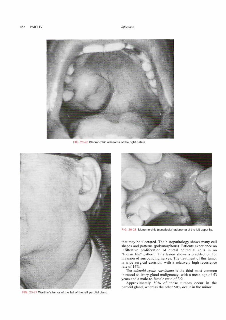
Benign Salivary Gland Tumors The pleomorphic adenoma, or benign mixed tumor, is the most common salivary gland tumor. The mean age of occurrence is 45 years, with a male-to-female ratio of 3:2. In the major glands, the parotid gland is involved in over 80% of cases; in the minor glands, the most common intraoral site is the palate (Fig. 20-26). Pleomorphic adenomas are usually slow-growing, painless masses. The histopathology shows two cell types: (1) the ductal epithelial cell and (2) the myoepithelial cell, which may differentiate along a variety of cell lines (pleomorphic means many forms). A connective tissue capsule exists, which may be incomplete. The treatment nvolves complete sur- i
Diagnosis and Management of Salivary Gland Disorders CHAPTER 20 451
FIGuredevrepducimasutunula
gical excision with a margin of normal uninvolved tissue. Parotid lesions are treated with removal of the involved lobe along with the tumor. Recurrence is possible in rare occasions, as well as a small risk (5%) of malignant trans-formation to a carcinoma ex pleomorphic adenoma.
Warthin's tumor, or papillary cystadenoma lymphoma tosum, almost exclusively affects the parotid gland, specifically the tail of the parotid gland (Fig. 20-27). The peak incidence is in the sixth decade of life, with a male-to-female ratio of 7:1. This lesion presents as a slow-growing, soft, painless mass. Warthin's tumor is believed to be caused by entrapped salivary epithelial rests within developing lymph nodes. The histopathology shows an epithelial component in a papillary pattern and a lymphoid component with germinal centers. The treatment of this lesion is simple surgical excision, and recurrence is rare.
The monomorphic adenoma is an uncommon solitary lesion composed of one cell type, affecting predominantly the upper lip minor glands (canalicular adenoma) (Fig. 20-28) and the parotid gland (basal cell adenoma). The mean age of occurrence is 61 years, and the lesion usually presents as an asymptomatic, freely movable mass. The histopathology reveals an encapsulated lesion composed of one type (monomorphic) of salivary ductal epithelial cell. The treatment is simple surgical excision.
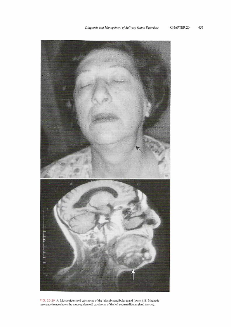
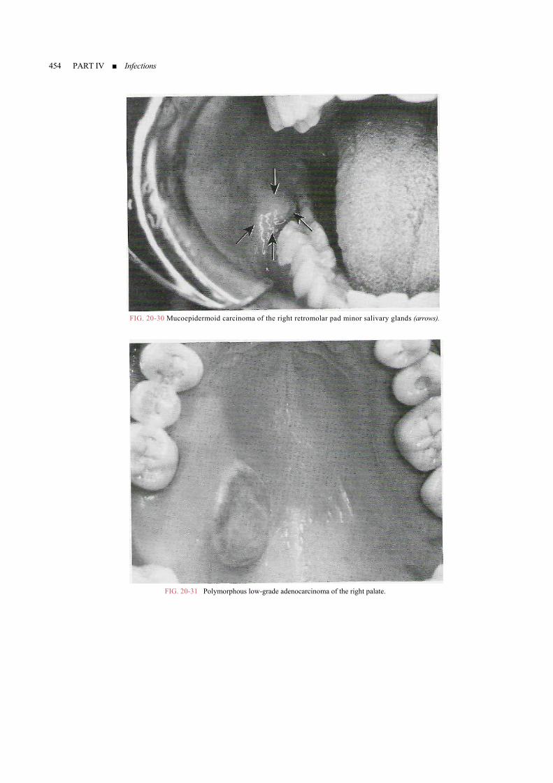
Malignant Salivary Gland Tumors The mucoepidermoid carcinoma is the most common malignant salivary gland tumor. It comprises 10% of ma rs (mostly parotid) (Fig. 20-29) and 20% jor gland tumoof minor gland tumors (mostly palate) (Fig. 20-30). This lesion may occur at any age, but the mean age is 45 years. T ratio is 3:2. The entation is a he male-to-female clinical presubmucosal mass t nful or ulcass may app tinge becucous conte hin the traosseous form of mu carcinoma a multilocular radio riopostehe histopatholog l typells, (2) epide ermediate (he pro helps t
ntermade l mo er epidermoid m, la
s hat may be pai erated. The m ear to have a bluish ause of the m nt contained wit lesion. An in coepidermoid may present as lucency of the r mandible. T y shows three cel es: (1) mucous c rmoid cells, and (3) int clear) cells. T portion of each cell type o grade the mucoepidermoid carcinoma as high-, i ediate-, or low-gr esions. The higher the grade, the predominance of cells and pleo-morphis ck of mucous cells and cystic areas, and overall more aggressive behavior. The treatment of low-grade lesions is wide surgical excision with a margin of uninvolved normal tissue; high-grade lesions require more aggressive surgical removal with margins, and, possibly, local radiation therapy. The low-grade lesions have a 95% 5-year survival rate, whereas the high-grade lesions have less than a 40% 5-year survival rate.
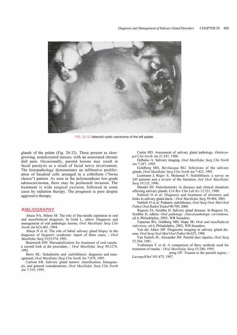
The polymorphous low-grade adenocarcinoma is the second most common intraoral salivary gland malignancy. This
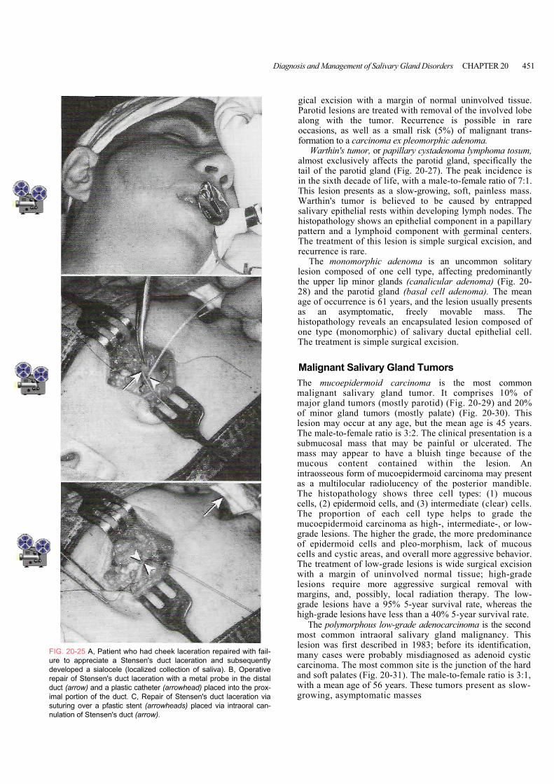
. 20-25 A, Patient who had cheek laceration repaired with fail- to appreciate a Stensen's duct laceration and subsequently eloped a sialocele (localized collection of saliva). B, Operative air of Stensen's duct laceration with a metal probe in the distal t (arrow) and a plastic catheter (arrowhead) placed into the prox-l portion of the duct. C, Repair of Stensen's duct laceration via ring over a pfastic stent (arrowheads) placed via intraoral can-tion of Stensen's duct (arrow).
lesion was first described in 1983; before its identification, many cases were probably misdiagnosed as adenoid cystic carcinoma. The most common site is the junction of the hard and soft palates (Fig. 20-31). The male-to-female ratio is 3:1, with a mean age of 56 years. These tumors present as slow-growing, asymptomatic masses
452 PART IV Infections
FIG. 20-27 Warth
FIG. 20-26 Pleomorphic adenoma of the right palate.
FIG. 20-28 Monomorphic (canalicular) adenoma of the left upper lip.
that may be ulcerated. The histopathology shows many cell shapes and patterns (polymorphous). Patients experience an infiltrative proliferation of ductal epithelial cells in an "Indian file" pattern. This lesion shows a predilection for invasion of surrounding nerves. The treatment of this tumor is wide surgical excision, with a relatively high recurrence rate of 14%.
The adenoid cystic carcinoma is the third most common intraoral salivary gland malignancy, with a mean age of 53 years and a male-to-female ratio of 3:2.
Approximately 50% of these tumors occur in the parotid gland, whereas the other 50% occur in the minor
in's tumor of the tail of the left parotid gland.
Diagnosis and Management of Salivary Gland Disorders CHAPTER 20 453
Fr
IG. 20-29 A, Mucoepidermoid carcinoma of the left submandibula and (arrow). B, Magnetic r glesonance image shows the mucoepidermoid carcinoma of the left sub dibular gland (arrow). man
454 PART IV ■ Infections
FIG. 20-30 Mucoepidermoid carcinoma of the right retromolar pad minor salivary glands (arrows).
FIG. 20-31 Polymorphous low-grade adenocarcinoma of the right palate.
Diagnosis and Management of Salivary Gland Disorders CHAPTER 20 455
glands of the pagrowing, nonulcdull pain. Occafacial paralysis The histopatholoation of basaloicheese") patternadenocarcinomatreatment is wicases by radiatioaggressive therap
BIBLIOGRAPAbaza NA, Mil
and maxillofacial management of oraNorth Am 6(3):401
Abaza N et al:diagnosis of SjogMaxillofac Surg 51
Baurmash HD:a second look at t1992.
Berry RL: Sialagement, Oral Max
Carlson ER: Ssis, and general coAm 7:519, 1995.
FIG. 20-32 Adenoid cystic carcinoma of the left palate.late (Fig. 20-32). These present as slow-erated masses, with an associated chronic sionally, parotid lesions may result in as a result of facial nerve involvement. gy demonstrates an infiltrative prolifer-
d cells arranged in a cribriform ("Swiss . As seen in the polymorphous low-grade , there may be perineural invasion. The de surgical excision, followed in some n therapy. The prognosis is poor despite y.
HY oro M: The role of fine-needle aspiration in oral diagnosis. In Gold L, editor: Diagnosis and l pathologic lesions, Oral Maxillofac Surg Clin
, 1994. The role of labial salivary gland biopsy in the ren's syndrome: report of three cases, / Oral (5):574, 1993. Marsupialization for treatment of oral ranula: he procedure, / Oral Maxillofac Surg 50:1274,
adenitis and .sialolithiasis: diagnosis and man- Youngs RP, Walsh-Willofac Surg Clin North Am 7:479, 1995. alivary gland tumors: classification, histogene-nsiderations, Oral Maxillofac Surg Clin North
Curtin HD: Assessment of salivary gland pathology, Otolaryn-gol Clin North Am 21:547, 1988.
Delbalso A: Salivary imaging, Oral Maxillofac Surg Clin North Am 7:387, 1995.
Goldberg MH, Bevilacqua RG: Infections of the salivary glands, Oral Maxillofac Surg Clin North Am 7:423, 1995.
Lustmann J, Regev E, Melamed Y: Sialolithiasis: a survey on 245 patients and a review of the literature, Int] Oral Maxillofac Surg 19:135, 1990.
Mandel ID: Sialochemistry in diseases and clinical situations affecting salivary glands, Crit Rev Clin Lab Sci 12:321, 1980.
Nahlieli O et al: Diagnosis and treatment of strictures and kinks in salivary gland ducts, / Oral Maxillofac Surg 59:484, 2001.
Nahlieli O et al: Pediatric sialolithiasis, Oral Surg Oral Med Oral Pathoi Oral Radiol Endod 90:709, 2000.
Regezzi JA, Sciubba JJ: Salivary gland diseases. In Regezzi JA, Sciubba JJ, editors: Oral pathology: clinical-pathologic correlations, ed 4, Philadelphia, 2003, WB Saunders.
Topazian RG, Goldberg MH, Hupp JR: Oral and maxillofaciai infections, ed 4, Philadelphia, 2002, WB Saunders.
Van der Akker HP: Diagnostic imaging in salivary gland dis-ease, Oral Surg Oral Med Oral Pathoi 66:625, 1988.
Van Sickels JE, Alexander JM: Parotid duct injuries, Oral Surg 52:364, 1981.
Yoshimura Y et al: A comparison of three methods used for treatment of ranula, / Oral Maxillofac Surg 53:280, 1995.
aring GP: Trauma to the parotid region, / LaryngolOtol 101:475, 1987.