Diagnosis and Management of Acute Bacterial Sinusitis ... · TM Diagnosis and Management of Acute...
54
Diagnosis and Management of Acute Bacterial Sinusitis: 2013 AAP Guideline Ellen R. Wald, MD, FAAP Professor and Chair, Department of Pediatrics University of Wisconsin School of Medicine and Public Health TM Prepared for your next patient.
Transcript of Diagnosis and Management of Acute Bacterial Sinusitis ... · TM Diagnosis and Management of Acute...
TM
Diagnosis and Management of Acute Bacterial Sinusitis:
2013 AAP Guideline
Ellen R. Wald, MD, FAAP Professor and Chair, Department of Pediatrics University of Wisconsin School of Medicine and Public Health
TM Prepared for your next patient.

TM
Disclaimers I have no relationships to declare and I do not intend to reference
unlabeled/unapproved uses of drugs or products.
Statements and opinions expressed are those of the authors and not necessarily those of the American Academy of Pediatrics.
Mead Johnson sponsors programs such as this to give healthcare professionals access to scientific and educational information provided by experts. The presenter has complete and independent control over the planning and content of the presentation, and is not receiving any compensation from Mead Johnson for this presentation. The presenter’s comments and opinions are not necessarily those of Mead Johnson. In the event that the presentation contains statements about uses of drugs that are not within the drugs' approved indications, Mead Johnson does not promote the use of any drug for indications outside the FDA-approved product label.

TM
Diagnosis and Management of Acute Sinusitis Update of 2001 guideline
Focuses on ages 1–18 years
Not subacute or chronic; not <1 year
Not anatomic abnormalities; immunodeficiencies, cystic fibrosis, ciliary dyskinesia
TM
Diagnosis and Management of Acute Sinusitis Areas of change:
1. Addition of “worsening course”
2. New data on effectiveness of antibiotics
3. Option to observe for 3 days in “persistent” infection
4. Imaging is not necessary to identify or confirm a diagnosis of acute sinusitis
TM
Key Action Statement 1 Clinicians should make a diagnosis of acute bacterial sinusitis (ABS) when a child with an upper respiratory infection (URI) presents with: Persistent illness (nasal discharge or daytime cough
or both for ≥10 days without improvement) Worsening course (worsening or new onset of nasal
discharge, daytime cough or fever after initial improvement)
Severe onset (concurrent fever and purulent nasal discharge for 3 days)
TM
TM
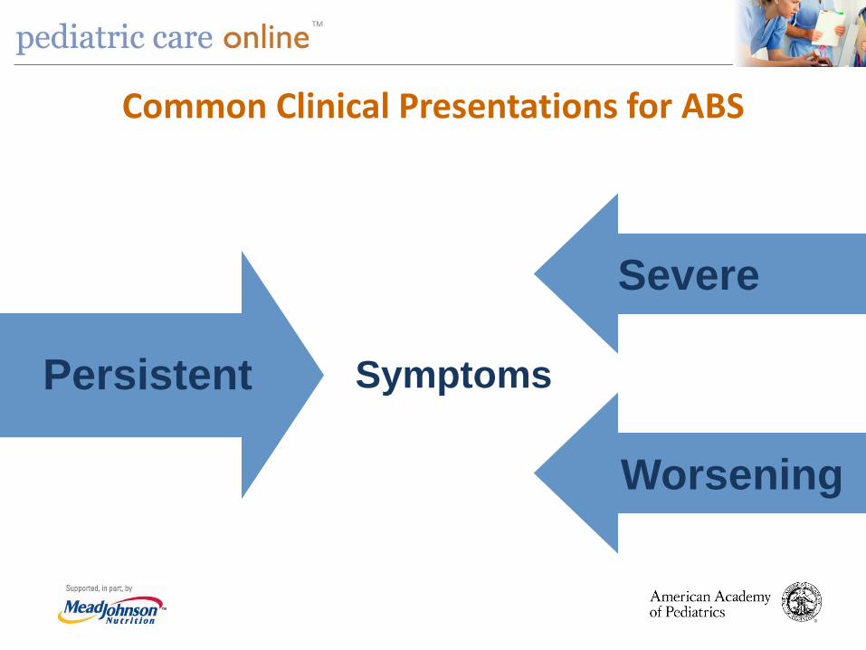
Common Clinical Presentations for ABS
Persistent
Worsening
Symptoms
Severe
TM
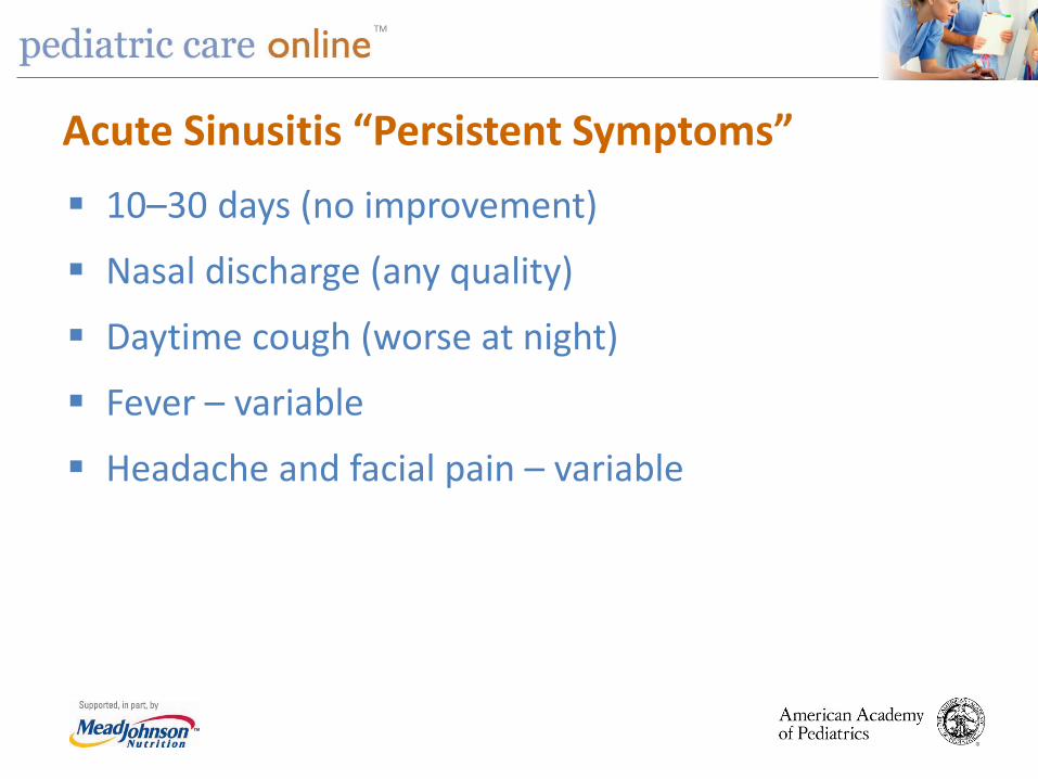
Acute Sinusitis “Persistent Symptoms” 10–30 days (no improvement)
Nasal discharge (any quality)
Daytime cough (worse at night)
Fever – variable
Headache and facial pain – variable
TM
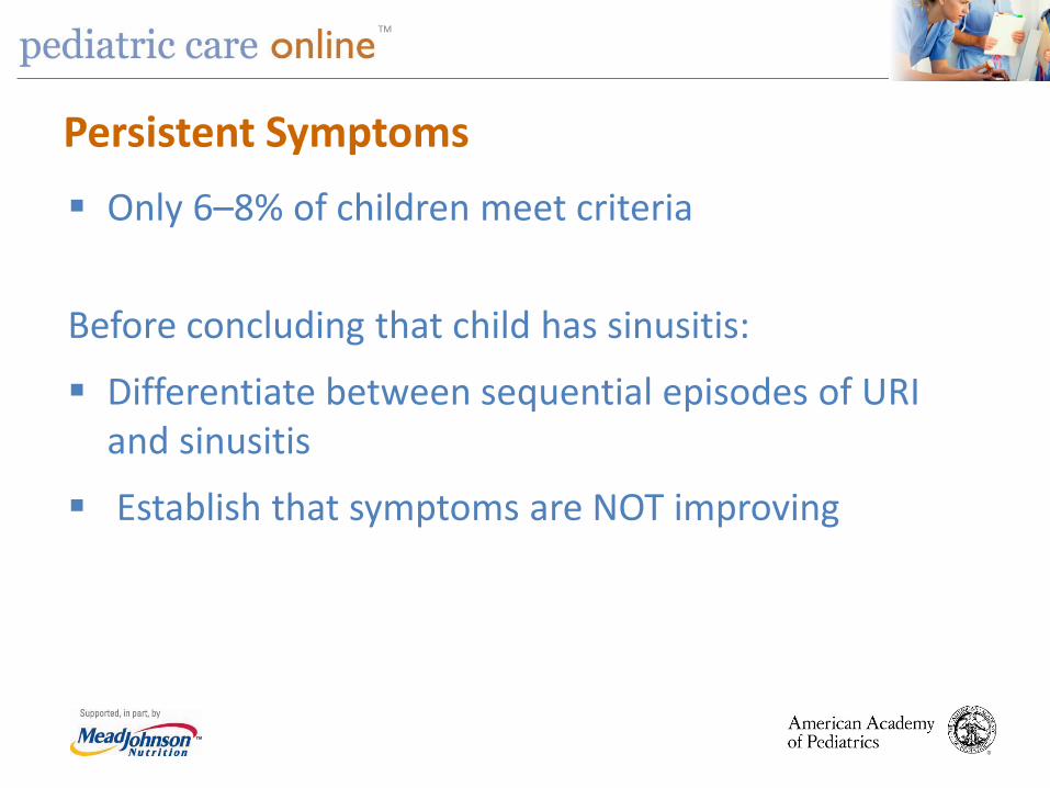
Persistent Symptoms Only 6–8% of children meet criteria
Before concluding that child has sinusitis:
Differentiate between sequential episodes of URI and sinusitis
Establish that symptoms are NOT improving
TM
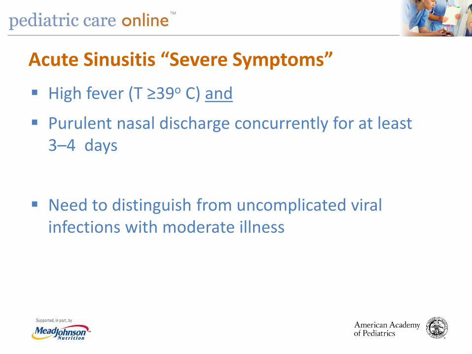
Acute Sinusitis “Severe Symptoms” High fever (T ≥39o C) and
Purulent nasal discharge concurrently for at least 3–4 days
Need to distinguish from uncomplicated viral infections with moderate illness

TM
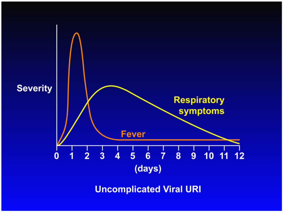
“Worsening Symptoms” Typical viral URI Nasal discharge or cough or both for 5–6 days which
is improving Sudden worsening manifests as
Increase nasal discharge or cough or both Onset of severe headache Onset of new fever

TM
Images – Key Action Statement 2A Clinicians should not obtain imaging studies (plain x-rays, computed tomography [CT] , magnetic resonance imaging [MRI] or ultrasound [U/S]) to distinguish ABS from viral URI.

TM
Images Historically, imaging was confirmatory
No longer recommended
Continuity of respiratory mucosa leads to diffuse inflammation during viral URI
Responsible for controversy regarding images
TM
Imaging of Sinuses 1940s – Observations made regarding frequency of
abnormal sinus radiographs in “healthy” children.
1970s and 1980s – Children with URI had frequent abnormalities of paranasal sinuses.
As CT scanning of central nervous system (CNS) and skull became prevalent, incidental abnormalities observed.
When MRI performed in children with URI, 70% show major abnormalities of mucosa.

TM
Computed Tomographic Study of the Common Cold
31 healthy young adults with new “cold”
Recruited within 48–96 hours
To have CT of paranasal sinuses
87% had significant abnormalities of their maxillary sinuses; 2 with air-fluid level
Conclusion: Common cold associated with frequent and striking abnormalities of sinuses
Gwaltney JM Jr, Phillips CD, Miller RD, et al. Computed tomography study of the common cold. N Engl J Med. 1994;330(1):25–30

TM

TM
Abnormalities on CT Scan
TM
Summary of Imaging
When paranasal sinuses are imaged in any way in children with uncomplicated URI, majority will be significantly abnormal.
Normal images = No sinusitis
Abnormal images cannot confirm diagnosis and are not necessary in children with uncomplicated clinical sinusitis.
TM
Images – Key Action Statement 2B Clinicians should obtain a contrast-enhanced CT scan of the paranasal sinuses and/or an MRI with contrast whenever a child is suspected of having orbital or CNS complications of ABS.
TM
Complications of Sinusitis Orbital
a. sympathetic effusion
b. subperiosteal abscess
c. orbital abscess
d. orbital cellulitis
e. cavernous sinus thrombosis

TM

TM

TM
Orbital Complications of Sinusitis Proptosis – anterior and lateral displacement of
globe
Impairment of extraocular movements
Loss of visual acuity
Chemosis – edema of conjunctiva
TM
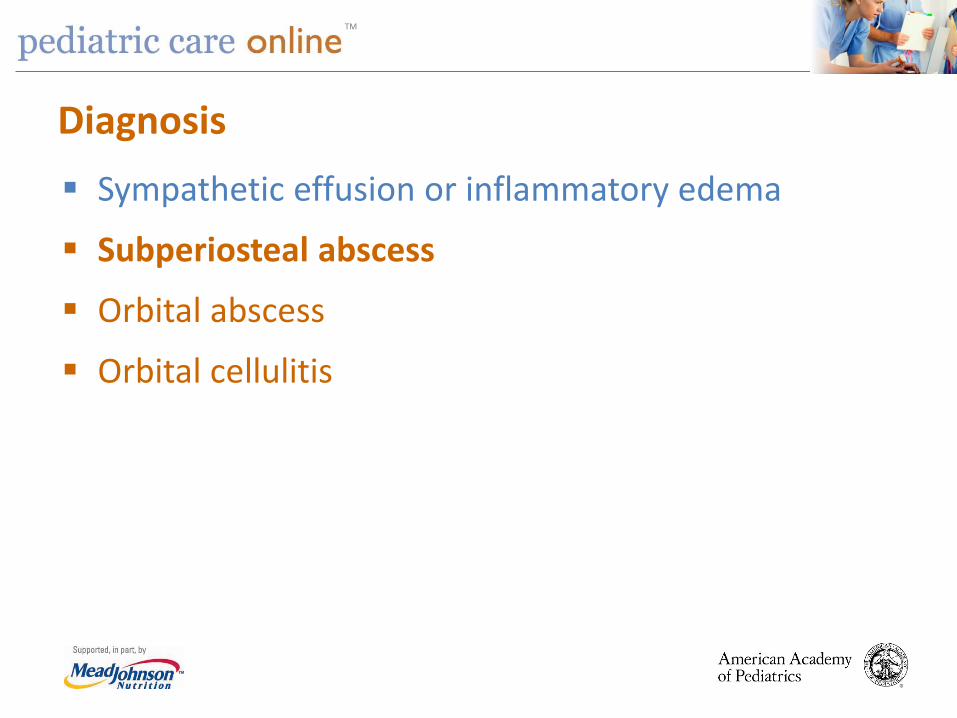
Diagnosis Sympathetic effusion or inflammatory edema
Subperiosteal abscess
Orbital abscess
Orbital cellulitis

TM

TM

TM

TM
TM
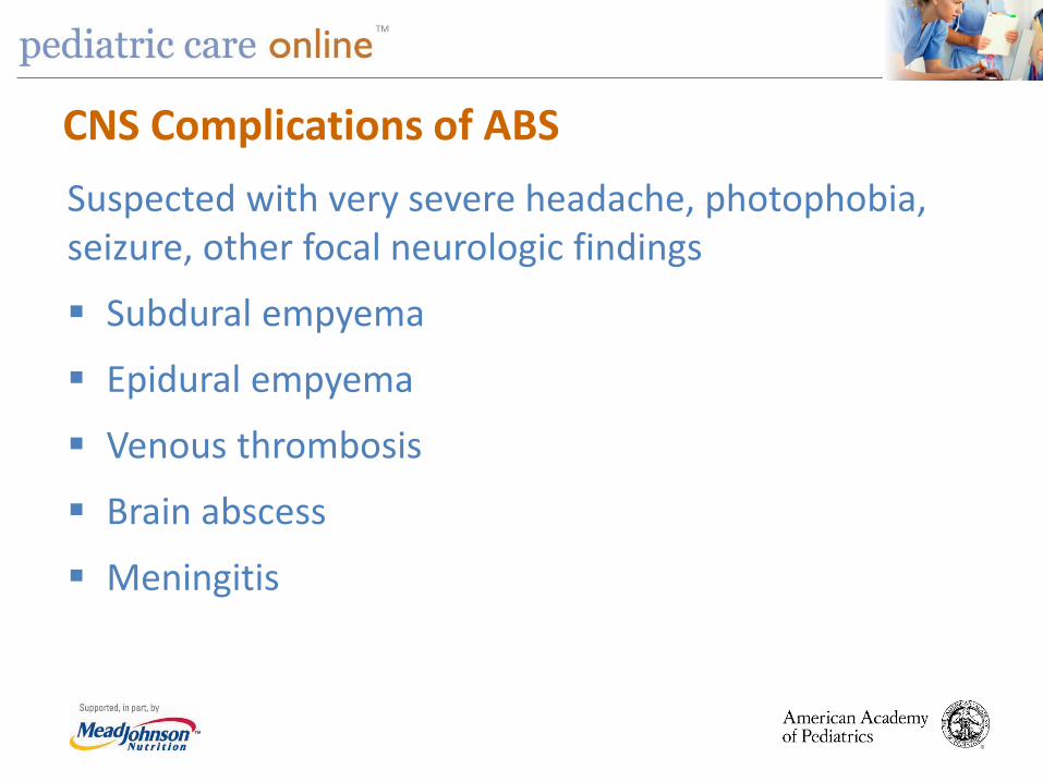
CNS Complications of ABS Suspected with very severe headache, photophobia, seizure, other focal neurologic findings
Subdural empyema
Epidural empyema
Venous thrombosis
Brain abscess
Meningitis

TM
Initial Management of ABS
Key Action Statement 3A: Clinician should prescribe antibiotic therapy for ABS in children with severe onset or worsening course.
Key Action Statement 3B: Clinician should either prescribe antibiotic therapy OR offer additional outpatient observation for 3 days to children with persistent illness.

TM
Initial Management of ABS Guidance for clinician regarding management of children with persistent symptoms:
Antibiotic therapy – starting as soon as possible after the encounter
Additional outpatient observation – for 3 days with plan to begin antibiotics if child does not improve or worsens at any time

TM
Initial Management of ABS Contrasts with 2001 AAP guideline
Acknowledges that although ABS is a bacterial infection
spontaneous resolution ~ common 10 days is a guideline; no likely harm in allowing up
to 3 more days in persistent onset
Reinforces antibiotic treatment as soon as possible in severe or worsening illness

TM
Recommendations for Initial Use of Antibiotics for ABS Clinical
Presentation Severe
ABS Worsening
ABS Persistent
ABS Uncomplicated ABS without coexisting illness
Antibiotic Antibiotic
Antibiotic OR
Additional observation
ABS with orbital or CNS complication
Antibiotic Antibiotic Antibiotic
ABS with other bacterial infection
Antibiotic Antibiotic Antibiotic
TM
Key Action Statement 4 Clinicians should prescribe amoxicillin with or without clavulanate as first-line treatment when a decision has been made to initiate antibiotic treatment of ABS.
TM
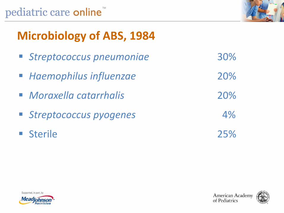
Microbiology of ABS, 1984
Streptococcus pneumoniae 30%
Haemophilus influenzae 20%
Moraxella catarrhalis 20%
Streptococcus pyogenes 4%
Sterile 25%
TM
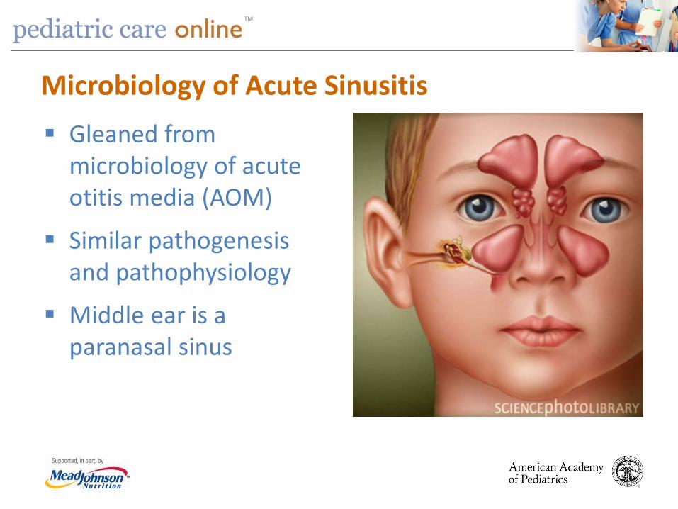
Microbiology of Acute Sinusitis Gleaned from
microbiology of acute otitis media (AOM)
Similar pathogenesis and pathophysiology
Middle ear is a paranasal sinus
TM
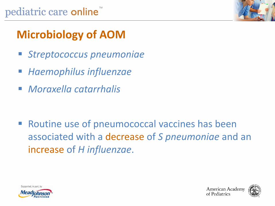
Microbiology of AOM Streptococcus pneumoniae
Haemophilus influenzae
Moraxella catarrhalis
Routine use of pneumococcal vaccines has been associated with a decrease of S pneumoniae and an increase of H influenzae.

TM
Microbiology of AOM
Early PCV7 Late PCV7 Early PCV13 S pneumoniae 30 H influenzae 50
S pneumoniae 45 H influenzae 25
S pneumoniae 20 H influenzae 55

TM

TM
Suspected Microbiology of ABS, 2013
Streptococcus pneumoniae 15–20%
Haemophilus influenzae 45–50%
Moraxella catarrhalis 10–15%
Streptococcus pyogenes 5%
Sterile 25%

TM
Antibiotic Resistance S pneumoniae: 10–15%; can increase up to 50%
H influenzae: 10–68%
M catarrhalis: 100%
LIMITED CURRENT DATA ON MICROBIOLOGY

TM
Treatment Amoxicillin – traditional first-line therapy
Amoxicillin at 45 mg/kg/day in 2 doses
If high prevalence of penicillin-resistant S pneumoniae
Amoxicillin at 90 mg/kg/day in 2 doses
TM
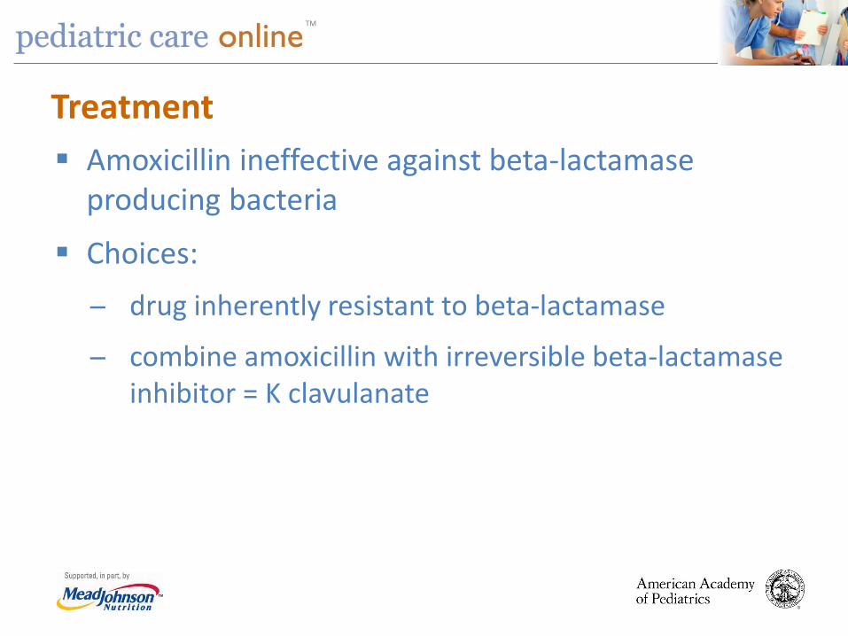
Treatment Amoxicillin ineffective against beta-lactamase
producing bacteria
Choices: drug inherently resistant to beta-lactamase
combine amoxicillin with irreversible beta-lactamase inhibitor = K clavulanate

TM
Treatment If S pneumoniae remains low or continues to decrease
and H influenzae remains high or continues to increase (including β-lactamase (+) strains)
Amoxicillin-clavulanate 45 mg/kg day
Amoxicillin-clavulanate 90 mg/kg/day

TM
Treatment 50 mg/kg Ceftriaxone IV or IM
Allergy:
Cephalosporins: cefdinir, cefuroxime, cefpodoxime
Clindamycin (or linezolid) + cefixime
Levofloxacin

TM
Treatment Optimal duration: no systematic study
Duration of therapy: 10, 14, 21, 28 days
Treat until patient is free of symptoms plus 7 days
TM
Key Action Statement 5A Clinicians should reassess initial management if there is caregiver report of worsening OR failure to improve within 72 hours.
TM
Response to Appropriate Management Most patients with ABS who are treated with an
appropriate antimicrobial agent respond promptly (within 48–72 hours)
Worsening = progression of signs/symptoms
Failure to improve = not better or worse
TM
Key Action Statement 5B If worsening symptoms or failure to improve clinicians should change antibiotics or initiate antibiotics in child managed with observation.
TM
Management of ABS at 72 Hours Whether or not antibiotics are used, a system must be in place to either add antibiotic or change the antibiotic if symptoms do not improve in 48–72 hours.

TM
Management of Worsening or No Improvement Initial
Management Worse in 72 Hours
No Improvement in 72 Hours
Observation Amoxicillin + clavulanate Observation OR Initiate antibiotic
Amoxicillin Amoxicillin-clavulanate Observation OR Amoxicillin-clavulanate
Amoxicillin-clavulanate Clindamycin + cefixime OR Linezolid + cefixime OR Levofloxacin OR Cefuroxime, Cefdinir OR Cefpodoxime
Amoxicillin-clavulanate OR Same choices as in preceding box

TM
Adjuvant Therapies – No Recommendation
Antihistamines
Intranasal steroids
Intranasal saline
Decongestants

TM
Summary Use stringent criteria to diagnose sinusitis in
children.
Avoid obtaining images.
Amoxicillin with or without clavulanate
High-dose amoxicillin plus clavulanate for resistance (most comprehensive)
Adjuvant therapy rarely indicated
TM
FREE PCO TRIAL Visit Pediatric Care Online today for additional information on this and other topics.
www.pediatriccareonline.org
Pediatric Care Online is a convenient electronic resource for immediate expert help with virtually every pediatric clinical information need with must-have resources that are
included in a comprehensive reference library and time-saving clinical tools.
Don’t have a subscription to PCO? Then take advantage of a free trial today!
Call Mead Johnson Nutrition at 888/363-2362 or, for more information, go to
https://www.pediatriccareonline.org/prepared/freetrial.html.


![Staphylococcus aureus: Is It a Pathogen of Acute Bacterial Sinusitis in Children … aureus is it a... · 2017-01-15 · acute sinusitis since 1984 [2]. The traditional and well-established](https://static.fdocuments.net/doc/165x107/5f8368a767352f565c48b168/staphylococcus-aureus-is-it-a-pathogen-of-acute-bacterial-sinusitis-in-children.jpg)