Adult Onset Type 1 Diabetes Mellitus Versus Type 2 Diabetes Mellitus-A Case Study
Diabetes Mellitus: Type 1, Pre-diabetes, and Type 2
WWW.RN.ORG® Reviewed September 2017, Expires September 2019
Provider Information and Specifics available on our Website Unauthorized Distribution Prohibited
©2017 RN.ORG®, S.A., RN.ORG®, LLC By Wanda Lockwood, RN, BA, MA
Purpose The purpose of this course is to define diabetes, explain the differences among type 1, pre-diabetes, and type 2, and describe symptoms,
complications, and treatments.
Goals Upon completion of this course, the healthcare provider should be able to:
• Explain the production and utilization of insulin. • Describe at least 4 presenting indications of diabetes mellitus
type 1. • Explain osmotic diuresis.
• Explain symptoms and management of diabetic ketoacidosis. • List and explain 5 different types of insulin.
• List and explain 5 types of diabetic monitoring. • Discuss pre-diabetes, including treatment approaches.
• Describe at least 5 symptoms of diabetes mellitus type 2. • Describe nutrition and exercise management.
• Explain the symptoms and management of HHNS. • List and explain 5 different types of oral diabetic agents.
Introduction Diabetes mellitus is a group of metabolic diseases related to impairment of insulin production, insulin utilization by the body, or
some combination. The body needs glucose for energy, and in a normal healthy person, glucose levels are maintained in balance (70 to
120 mg/dL) by insulin with glucose ingested through the gastrointestinal tract from food or formed in the liver from substances
derived from food. Insulin regulates glucose production and storage,
but with diabetes mellitus, this system is impaired. In some cases, insulin production is not adequate, but in other cases, the body cells
do not respond appropriately to insulin (insulin resistance).
While there are, in fact, about a dozen types of diabetes, the two primary types are type 1 (impaired insulin production) and type 2
(insulin resistance), often associated with pre-diabetes. In the United States alone, 17.9 million people have been diagnosed with diabetes,
and an estimated 5.7 million people have the disease but have not been diagnosed. The cost of diabetes in terms of both health problems
and dollars is staggering. Diabetes is the third leading cause of death and is the leading cause of heart disease, stroke, blindness (adult),
end-stage kidney disease, and nontraumatic lower limb amputations. People with diabetes have double the risk of developing coronary
artery disease, and about 65% have hypertension.
What is insulin? The pancreas is primarily an exocrine gland, but about 2% has an
endocrine function, and that is the part that produces insulin. Insulin, a hormone, is produced by the beta cells in the islets of Langerhans in
the pancreas. The beta cells are 1 of 4 types of cells found in the islets of Langerhans.

NIH
Insulin is usually released continuously in a 3 to 6 minute oscillating pattern into the bloodstream in small increments (basal rate) with
increased production (bolus) in response to glucose absorbed from ingested food, totaling about 40 to 50 U daily.
Mikael Haggstrom, Wikimedia Commons

Insulin is released in a precursor form, proinsulin, and then routed through the liver. Proinsulin comprises two polypeptide chains (A and
B) linked by a C-peptide chain. Proinsulin converts to insulin when the C chain is cleaved, leaving only the A and B chains. Beta cell function
can be evaluated by the presence of C peptides in serum and urine.
Insulin production usually increases markedly in the hour or so after food is ingested and then falls again. Insulin levels stay low during the
night with a slight increase in the early AM. Insulin increases to move glucose from the blood into muscles, liver, and fat cells. Without
adequate insulin, the glucose remains in the bloodstream (resulting in hyperglycemia) and cannot be used for energy. Within the cells, insulin
has a number of functions: • Transports and metabolizes glucose.
• Stimulates storage of glucose as glycogen in muscles and the
liver. • Signals the liver to stop breaking down glycogen into glucose for
release into the bloodstream. • Facilitates storage of dietary fat in adipose tissue.
• Facilitates transportation of amino acids (from ingested protein) into cells.
• Inhibits breakdown of stored glucose (glycogen), protein, and fat.
Some tissues, such as skeletal muscle and adipose tissue, contain insulin receptors and are considered insulin-dependent tissues while
other tissues (such as the brain, liver, and blood cells) don’t require insulin directly but do need an adequate blood supply of glucose. The
liver, while not an insulin-dependent tissue, does actually have insulin receptor sites, but they serve to facilitate uptake of glucose by the
liver to be converted to glycogen for storage. While the liver initially
produces glucose by breaking down glycogen, after 8 to 12 hours of fasting, the liver begins to form glucose by breaking down non-
carbohydrate substances (such as amino acids or fats) in a process referred to as gluconeogenesis. If insulin is not present, as in diabetes
type 1, then the body cannot utilize glucose for energy. Fat is often utilized, as lipids are transferred from adipose tissue to the liver to be
used for energy.
The alpha cells in the islets of Langerhans also have a role in glucose metabolism. During periods when glucose levels fall (such as during
the night or during periods of fasting), the alpha cells release another hormone, glucagon, which stimulates the liver to convert glycogen to
glucose for release into the blood stream to increase glucose levels. Other hormones, such as epinephrine, growth hormone, and cortisol,
also have a counterregulatory effect in that they stimulate glucose production and output by the liver and slow movement of glucose into
the cells. Insulin along with glucagon and (to a lesser degree) the other counterregulatory hormones usually maintain insulin levels
within a normal range. Any imbalance in these hormones can produce diabetes.
Diabetes mellitus, type 1 Diabetes mellitus type 1 was formerly called juvenile-onset diabetes or insulin-dependent diabetes, but the name was changed because, while
the disease is more common in young people, onset may also occur during adulthood. Characterizing the disorder as “insulin-dependent”
focused on treatment rather than underlying cause and/or impairment. Diabetes mellitus type 1 occurs with destruction of beta cells in the
pancreas, resulting in decreased and finally no insulin production. The
exact cause of this destruction may vary from one individual to another, and the mechanisms are not completely clear although there
appears to be a genetic component.
People don’t directly inherit diabetes mellitus type 1, but they do appear to inherit a predisposition to developing the disease. How
genetics combines with immunologic and environmental factors is not well understood. The genetic predisposition is associated with certain
human leukocyte antigen (HLA) types. HLA comprises a group of genes associated with immune response. Specific HLA types (DR3 or
DR4) are found in 95% of Caucasians with diabetes mellitus type 1. People with either HLA type have a 3 to 5 times increased risk of
developing diabetes and those with both types have 10 to 20 times increased risk over those in the general population.
Diabetes mellitus type 1 is also associated with an autoimmune response in which autoantibodies attack islet cells and endogenous
insulin. Some researchers believe this response is triggered by some type of viral infection. However, regardless of the underlying cause,
when 80 to 90% of the islet cells are destroyed, the result is hyperglycemia. Insulin is needed to inhibit the breakdown of stored
glucose (glycogenolysis) and to inhibit glyconeogenesis; so without insulin, both processes occur without restraint. While blood glucose
levels are already high, the liver continues to convert stored glycogen to glucose and to breakdown amino acids and fats, producing ketones
(a byproduct of fat breakdown). These catabolic changes (decreased insulin and increased breakdown of proteins and fats) as well as
malnourishment from the cells inability to utilize glucose lead to

polyphagia (increased hunger). Fat breakdown can lead to ketoacidosis. Glucose ingested from food cannot be stored in the liver,
so it continues to circulate in the blood as well.
As blood glucose levels increase, osmotic diuresis may occur. The renal threshold for glucose is usually between 180 mg/dL and 200
mg/dL. If the glucose concentration is >200 mg/dL, the kidneys are not able to reabsorb this excess glucose, and it spills over into the
urine, accompanied by increased loss of fluids and electrolytes. This fluid loss results in polyuria and polydipsia (increased thirst).
Symptoms associated with diabetes mellitus, type 1
Mikael Haggstrom, Wikimedia Commons
3 Ps Polyuria, polydipsia, polyphagia
Weight loss Results from inability to use glucose as well as
breakdown of protein and stored fat.
Weakness/
fatigue
Lack of glucose for energy.
The onset of diabetes mellitus, type 1, is often slow and insidious with
destruction of islet cells occurring over many months or even years. Because of this, initial symptoms may be overlooked especially if some
insulin is still produced; however, when insulin production stops, life-threatening complications often occur very rapidly.
Diabetic ketoacidosis (DKA)

With inadequate insulin, lipolysis (fat breakdown) occurs, with glycerol from fat cells and the liver converted to ketone bodies, but these are
used less efficiently than glucose for cellular metabolism. Excess ketones are excreted in the urine (ketosis) and in exhalations
(Kussmaul respirations). Kussmaul respirations comprise hyperventilation to rid the body of excess carbon dioxide and are
associated with “ketone” breath, which has a sweet, fruit, acetone odor. Ketone bodies decrease serum pH, leading to ketoacidosis.
Other symptoms may include:
• Nausea
• Vomiting. • Abdominal pain.
Fluid imbalance occurs with loss of potassium and other electrolytes
because of cellular death. This causes dehydration (dry mouth, dry skin, reduced skin turgor) and diuresis with excess thirst. Renal
failure may result from hypovolemic shock. Cardiac arrhythmias may occur because of potassium loss. Hyperglycemia may range from 300
to 800 mg/dL. Immediate treatment should be instituted: • Insulin therapy by continuous infusion initially.
• Rehydration and electrolyte replacement. • Glucose monitoring.
• Ketone monitoring.
The primary treatment for diabetes mellitus is insulin. Early insulin was prepared from the pancreas of cattle and pigs; however, synthetic
human insulin is now the standard. Human insulin is produced with recombinant DNA technology from bacteria (such as Escherichia coli)
or yeast cells, not from human donors. Human insulin is better tolerated than animal-derived insulin, causing fewer allergic reactions.
Insulin ranges from short acting to long acting, and insulin prescriptions must be tailored to each individual. Some may need only
one type of insulin while others may need a combination. All insulin preparations are developed from a base of regular insulin. Different
additives are combined with regular insulin to affect the onset of activity, peak effectiveness, and duration. Common additives include
zinc (used to make lente insulin) and protamine (combined with zinc and regular insulin to make NPH insulin). People may be allergic to
additives.
Insulin is manufactured in different forms: vials, prefilled syringes, pens, and cartridges. Note: Long-acting (Ultralente) insulin is no
longer being produced as it did not provide reliable treatment for many people, and newer insulins became more commonly used.
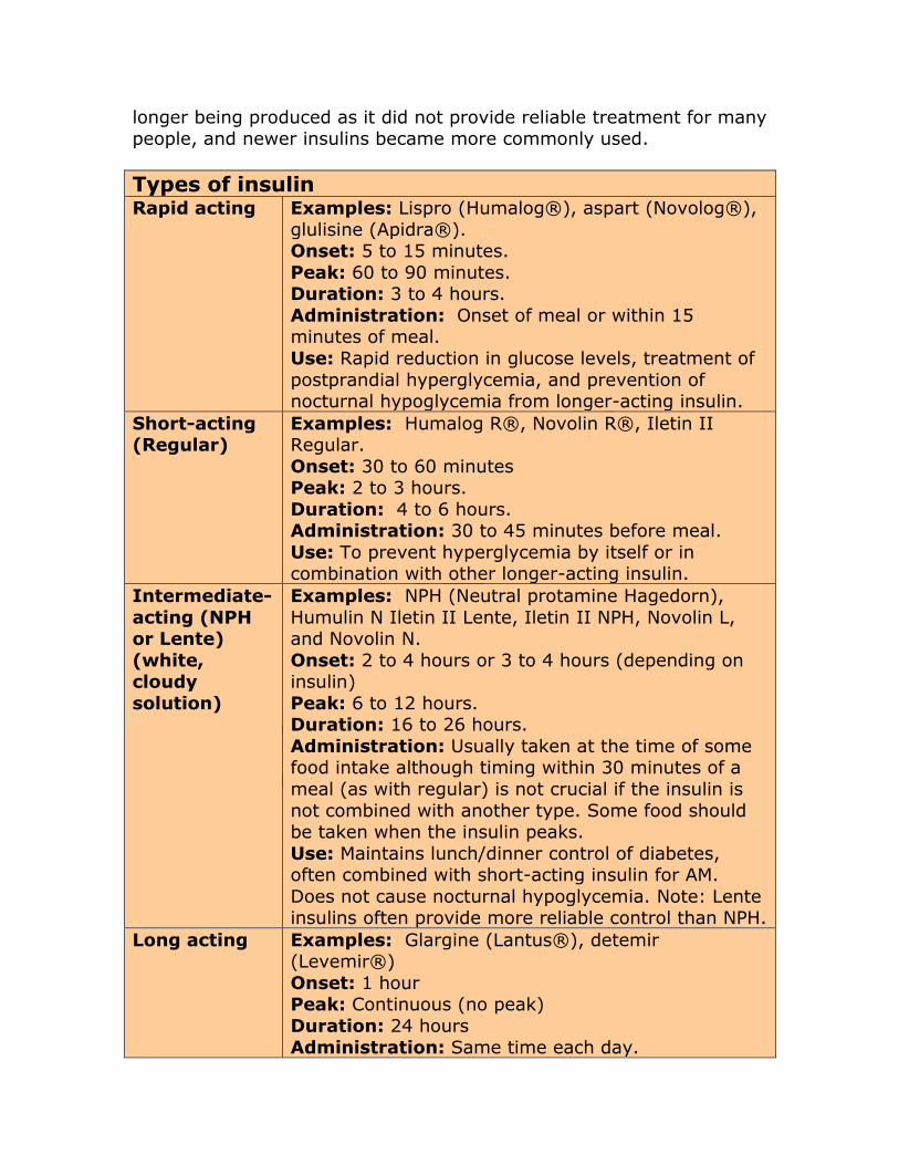
Types of insulin Rapid acting Examples: Lispro (Humalog®), aspart (Novolog®),
glulisine (Apidra®). Onset: 5 to 15 minutes.
Peak: 60 to 90 minutes. Duration: 3 to 4 hours.
Administration: Onset of meal or within 15 minutes of meal.
Use: Rapid reduction in glucose levels, treatment of
postprandial hyperglycemia, and prevention of nocturnal hypoglycemia from longer-acting insulin.
Short-acting (Regular)
Examples: Humalog R®, Novolin R®, Iletin II Regular.
Onset: 30 to 60 minutes Peak: 2 to 3 hours.
Duration: 4 to 6 hours. Administration: 30 to 45 minutes before meal.
Use: To prevent hyperglycemia by itself or in combination with other longer-acting insulin.
Intermediate-
acting (NPH or Lente)
(white, cloudy
solution)
Examples: NPH (Neutral protamine Hagedorn),
Humulin N Iletin II Lente, Iletin II NPH, Novolin L, and Novolin N.
Onset: 2 to 4 hours or 3 to 4 hours (depending on insulin)
Peak: 6 to 12 hours. Duration: 16 to 26 hours.
Administration: Usually taken at the time of some food intake although timing within 30 minutes of a
meal (as with regular) is not crucial if the insulin is
not combined with another type. Some food should be taken when the insulin peaks.
Use: Maintains lunch/dinner control of diabetes, often combined with short-acting insulin for AM.
Does not cause nocturnal hypoglycemia. Note: Lente insulins often provide more reliable control than NPH.
Long acting Examples: Glargine (Lantus®), detemir (Levemir®)
Onset: 1 hour Peak: Continuous (no peak)
Duration: 24 hours
Administration: Same time each day.
Use: To mimic basal insulin dose. Used with short-acting insulin (bolus).
Combination (Pre-mixed)
Examples: Intermediate-acting combined with rapid-acting:
• Lispro protamine/lispro (Humalog® 50/50 and
Novolog® Mix 70/30. • Aspart protamine/aspart (Novolog® Mix 50/50
& Novolog® 70/30). Intermediate-acting combined with short acting:
• NPH insulin/regular insulin (Humulin®70/30, Novolin®70/30).
Onset: Varies depending on the shortest acting insulin.
Peak: Two peaks will occur. Duration: Depends on the longest acting.
Administration: Pre-mixed are injected in one syringe.
Use: To provide better overall control.
Depending on the stability of the diabetes, insulin may be given in
regular daily doses. However, some people’s diabetes is less easily regulated, and they may require frequent glucose testing and
administration of insulin 3 to 4 or more times during the daytime on a sliding scale, depending on the glucose reading. Regimens for insulin
administration may vary. The conventional regimen is to give one or two doses of insulin daily in order to simplify management, but this
may result in periods when blood glucose levels are above normal. If people are rigid about eating at the exact time, with the same meal
pattern every day and never vary exercise, this regimen may provide adequate control. It is also advised for those who are frail or
unreliable in managing diabetes. However, the swings in blood sugar
increase risks of diabetic-associated complications, such as hypertension, stroke, and kidney disease.
The intensive regimen attempts to keep blood glucose levels as
close to normal as possible by monitoring glucose levels and taking 3 to 4 injections daily. Studies show this regimen reduces risks of
complications but also increases risks of hypoglycemic reactions. Some people may not be good candidates for the intensive regimen,
including those with nervous system disorders that may make them unaware of symptoms of hypoglycemia, with recurring episodes of
hypoglycemia, with irreversible diabetic complications, with CVA or cardiovascular disease, and those who are unable to monitor diabetes
effectively. People who have had a kidney transplant should be maintained on the intensive regimen to protect the new kidney.
If combination doses are not in pre-mixed syringes or cartridges,
patients must be taught how to combine insulin doses. The combined formulas provide convenience but may not provide adequate coverage
for all people because they allow for no flexibility in dosage.
People with poor eyesight may have difficulty filling a syringe and may miss air bubbles, causing alterations in dosage. When filling a syringe
from more than one vial, air should be injected into both vials FIRST and then insulin withdrawn from the clear insulin before the cloudy
insulin (“clear to cloudy”). Mixed insulins should be derived from the same species (human, beef, pork). Intermediate and long-acting
insulins should be rolled between the hands to thoroughly mix the
insulin prior to withdrawing into a syringe or administering if in prefilled syringes, cartridges, or pens. Insulin should be given within 5
minutes of preparation. Glargine (Lantus®), basal insulin, should not be mixed with other insulins as it has a pH of 4 and mixing with other
insulins may cause precipitation.
Alternatives to standard needle and syringe administration have made
diabetic control easier for patients. Inhaled insulin (Exubera®) was approved by the FDA in 2006, but the drug company took it off the
market in 2007 because it had not been profitable, and the FDA expressed concern regarding side effects. Other inhaled insulin
products are awaiting FDA approval.
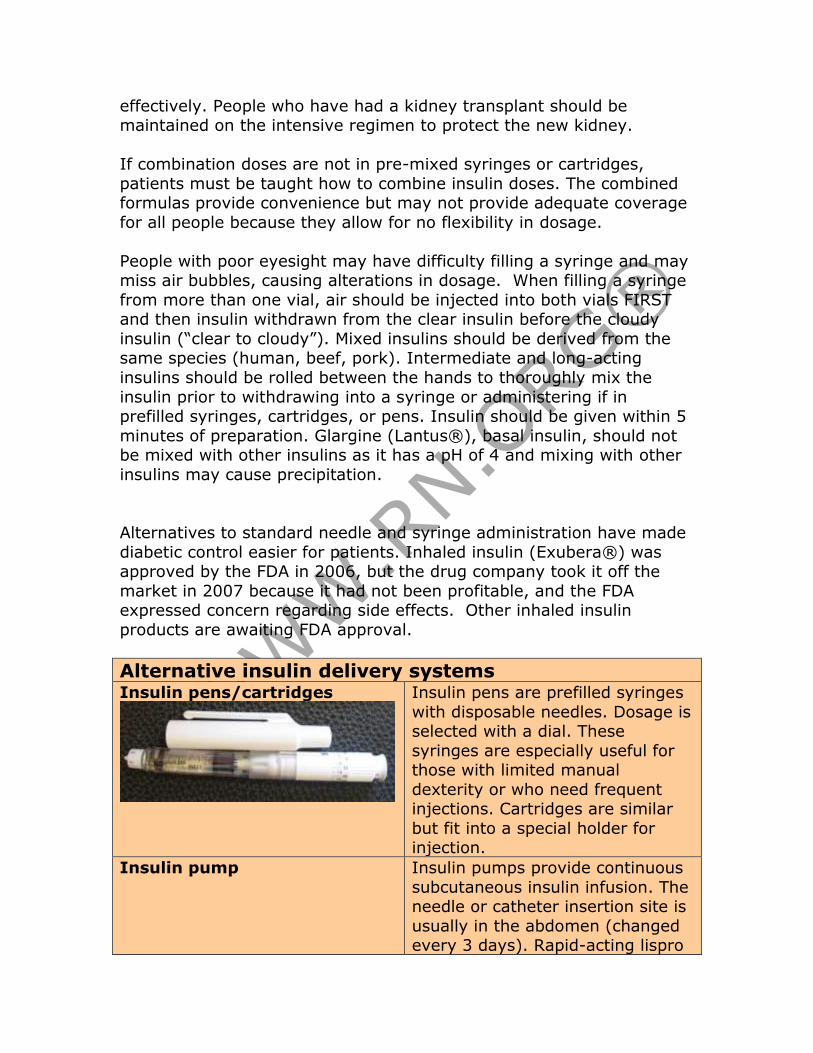
Alternative insulin delivery systems Insulin pens/cartridges
Insulin pens are prefilled syringes
with disposable needles. Dosage is selected with a dial. These
syringes are especially useful for those with limited manual
dexterity or who need frequent injections. Cartridges are similar
but fit into a special holder for injection.
Insulin pump Insulin pumps provide continuous
subcutaneous insulin infusion. The needle or catheter insertion site is
usually in the abdomen (changed every 3 days). Rapid-acting lispro
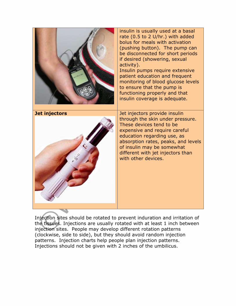
insulin is usually used at a basal rate (0.5 to 2 U/hr.) with added
bolus for meals with activation (pushing button). The pump can
be disconnected for short periods if desired (showering, sexual
activity). Insulin pumps require extensive
patient education and frequent monitoring of blood glucose levels
to ensure that the pump is
functioning properly and that insulin coverage is adequate.
Jet injectors
Jet injectors provide insulin
through the skin under pressure. These devices tend to be
expensive and require careful
education regarding use, as absorption rates, peaks, and levels
of insulin may be somewhat different with jet injectors than
with other devices.
Injection sites should be rotated to prevent induration and irritation of
the tissues. Injections are usually rotated with at least 1 inch between
injection sites. People may develop different rotation patterns (clockwise, side to side), but they should avoid random injection
patterns. Injection charts help people plan injection patterns. Injections should not be given with 2 inches of the umbilicus.
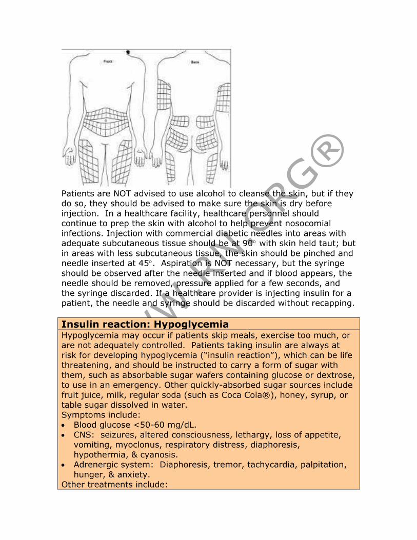
Patients are NOT advised to use alcohol to cleanse the skin, but if they do so, they should be advised to make sure the skin is dry before
injection. In a healthcare facility, healthcare personnel should continue to prep the skin with alcohol to help prevent nosocomial
infections. Injection with commercial diabetic needles into areas with adequate subcutaneous tissue should be at 90 with skin held taut; but
in areas with less subcutaneous tissue, the skin should be pinched and needle inserted at 45. Aspiration is NOT necessary, but the syringe
should be observed after the needle inserted and if blood appears, the needle should be removed, pressure applied for a few seconds, and
the syringe discarded. If a healthcare provider is injecting insulin for a patient, the needle and syringe should be discarded without recapping.
Insulin reaction: Hypoglycemia Hypoglycemia may occur if patients skip meals, exercise too much, or are not adequately controlled. Patients taking insulin are always at
risk for developing hypoglycemia (“insulin reaction”), which can be life threatening, and should be instructed to carry a form of sugar with
them, such as absorbable sugar wafers containing glucose or dextrose, to use in an emergency. Other quickly-absorbed sugar sources include
fruit juice, milk, regular soda (such as Coca Cola®), honey, syrup, or table sugar dissolved in water.
Symptoms include: • Blood glucose <50-60 mg/dL.
• CNS: seizures, altered consciousness, lethargy, loss of appetite, vomiting, myoclonus, respiratory distress, diaphoresis,
hypothermia, & cyanosis. • Adrenergic system: Diaphoresis, tremor, tachycardia, palpitation,
hunger, & anxiety.
Other treatments include:
• Glucagon injection or IV administration of glucose. Patients should be taught to recognize the signs that their blood sugar
is getting low, especially when they begin insulin treatment.
NOTE: Some people, especially those with diabetic neuropathy, the elderly and those on -adrenergic blockers may suffer from
hypoglycemia unawareness in which they are unable to recognize the signs of hypoglycemia. These people may not be able to tightly
control diabetes because the risks from hypoglycemia may outweigh those of mild hyperglycemia,
A more radical treatment for diabetes mellitus type 1 is transplantation of a whole pancreas, segment of the pancreas, or islet cells.
Pancreatic and islet cell transplantation is usually done only for those also receiving a kidney transplant because of the need for antirejection
drugs. People often require more than one islet cell transplant to achieve long-term independence from insulin injections. To date, most
islet cell transplants are successful for only about 2 years.
Additional treatments for diabetes mellitus, type 1 Nutrition Diet should include all of the necessary nutrients and
sufficient calories to meet daily needs and maintain weight at a normal level or reduce weight if the
person is obese as well as control lipid levels. Different diets are available, such as those using the
exchange system or calorie-counting diets. Carbohydrates have the greatest effect on blood
sugar and are the most closely monitored. Some diets rely on carbohydrate counting rather than
calorie counting because 100% of carbohydrates convert to glucose (minus fiber) compared to 50% of
fats and proteins, but careful meal planning is still needed to avoid weight gain.
Diet should always be managed by a registered dietician because issues may be quite complex.
Because those with diabetes mellitus type 1 receive insulin, meals should be consistent, balanced, and at
the same times each day to prevent fluctuations in blood glucose levels.
People with diabetes mellitus, type 1, may use
artificial sweeteners, but some sweeteners actually contain carbohydrates:
• Nutritive sweeteners: These have added calories from fructose, xylitol, dextrose, or
sorbitol. For example, aspartame (Equal®), which is packaged with dextrose and contains
4 calories per package. • Non-nutritive sweeteners: These do not have
added calories. Examples include saccharine and sucralose (Splenda®) although some
preparations may have some caloric content.
Alcohol intake should be moderate and diabetic
patients should eat when drinking, as drinking on an empty stomach increases the chance of developing
hypoglycemia.
Exercise Exercise for 30 minutes daily or as tolerated is
recommended to help manage diabetes. However, people should try to exercise at the same time each
day and for the same duration during the period when their blood glucose level peaks.
Exercise programs should start slowly and build to tolerance. An exercise program should NOT be
started with serum glucose 250 mg/dL or with
positive urinary ketones because exercising with
hyperglycemia causes hormonal changes that triggers the liver to convert glycogen to glucose,
further increasing hyperglycemia.
If people experience post-exercise hypoglycemia, they may need to take a snack after exercise and at
bedtime, decrease insulin dose, and/or monitor blood glucose levels more frequently.
Monitoring diabetes Serum glucose While usually referred to as “blood glucose”
most testing is actually done with only the
serum rather than whole blood or plasma. Testing is usually done after overnight
fasting. This is the most reliable form of testing and should be done periodically,
even if the patient is doing self-monitoring. Normal fasting values:
• Newborn to 2 years: 50 to 80 mg/dL. • Child: 60 to 100 mg/dl.
• Adult: 65-99 mg/dL. • Prediabetic adult: 100 to 125 mg/dL.
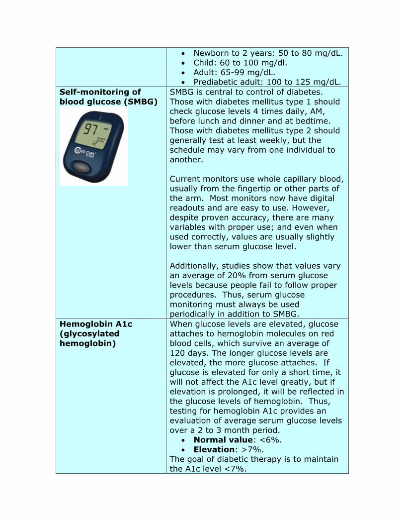
Self-monitoring of
blood glucose (SMBG)
SMBG is central to control of diabetes.
Those with diabetes mellitus type 1 should check glucose levels 4 times daily, AM,
before lunch and dinner and at bedtime. Those with diabetes mellitus type 2 should
generally test at least weekly, but the schedule may vary from one individual to
another.
Current monitors use whole capillary blood, usually from the fingertip or other parts of
the arm. Most monitors now have digital readouts and are easy to use. However,
despite proven accuracy, there are many variables with proper use; and even when
used correctly, values are usually slightly
lower than serum glucose level.
Additionally, studies show that values vary an average of 20% from serum glucose
levels because people fail to follow proper procedures. Thus, serum glucose
monitoring must always be used periodically in addition to SMBG.
Hemoglobin A1c (glycosylated
hemoglobin)
When glucose levels are elevated, glucose attaches to hemoglobin molecules on red
blood cells, which survive an average of
120 days. The longer glucose levels are elevated, the more glucose attaches. If
glucose is elevated for only a short time, it will not affect the A1c level greatly, but if
elevation is prolonged, it will be reflected in the glucose levels of hemoglobin. Thus,
testing for hemoglobin A1c provides an evaluation of average serum glucose levels
over a 2 to 3 month period. • Normal value: <6%.
• Elevation: >7%. The goal of diabetic therapy is to maintain
the A1c level <7%.

Hgb A1c (%) Serum glucose
(mg/dL)
5 90
5.5 105
6 120
6.5 135
7 150
8 180
9 210
10 240
12 300
14 360
Urine testing (glucose)
Urine testing (color-coded) with a dipstick is now rarely used to monitor diabetes as
it’s been replaced with SMBG. However, if people refuse to do SMBG, this inexpensive
method can provide limited information. Problems include:
• Urine testing does not reflect blood
glucose levels at the time of testing. • The renal threshold for glucose is 180
to 200 mg/dL, considerably higher than optimal levels. If renal disease
is present, thresholds may be even higher.
• Urine testing cannot detect hypoglycemia.
• Medications (aspirin, some antibiotics, vitamin C) may interfere
with test results.
Urine testing (ketones)
Ketones in the urine suggest that diabetes mellitus type 1 is out of control and the
person is at risk of DKA because the body is breaking down fat to use for energy.
While a ketone blood monitor is now also available, the dipstick method is most
commonly used. Testing should be done with diabetes mellitus type 1 when glucose
levels are elevated >240 mg/dL for 2 testing periods in a row or during times of
stress, such as during pregnancy or illness.
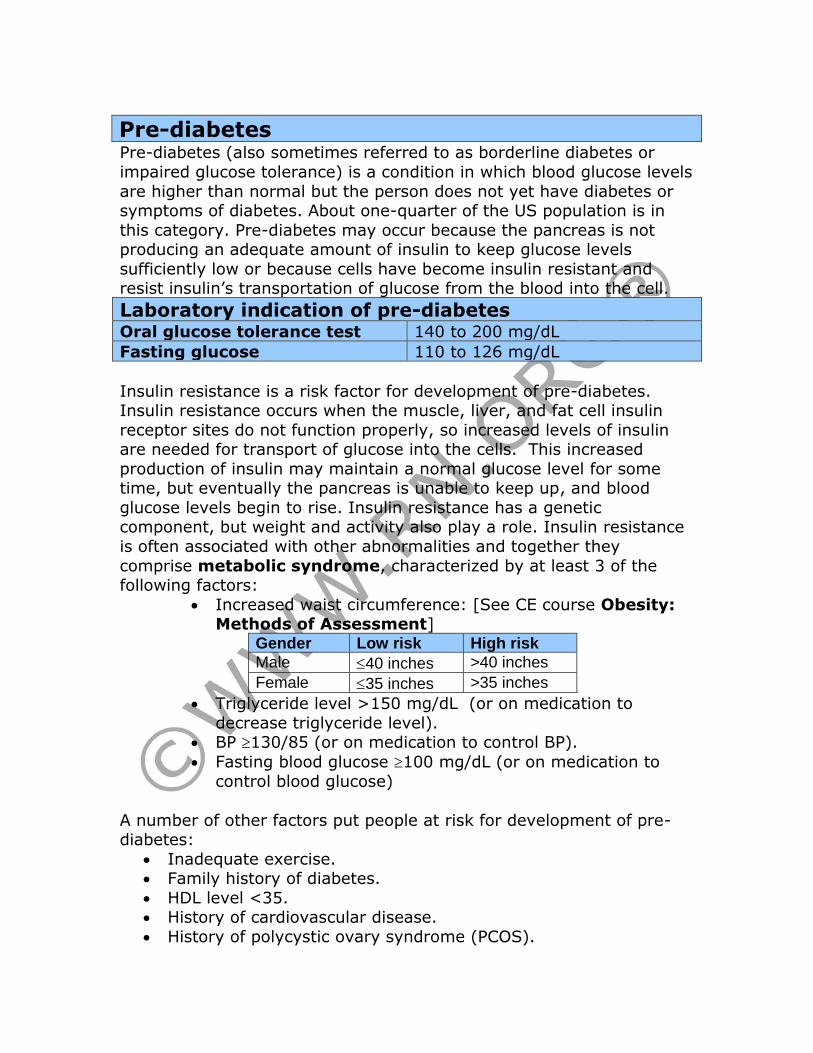
Pre-diabetes Pre-diabetes (also sometimes referred to as borderline diabetes or
impaired glucose tolerance) is a condition in which blood glucose levels
are higher than normal but the person does not yet have diabetes or symptoms of diabetes. About one-quarter of the US population is in
this category. Pre-diabetes may occur because the pancreas is not producing an adequate amount of insulin to keep glucose levels
sufficiently low or because cells have become insulin resistant and resist insulin’s transportation of glucose from the blood into the cell.
Laboratory indication of pre-diabetes Oral glucose tolerance test 140 to 200 mg/dL
Fasting glucose 110 to 126 mg/dL
Insulin resistance is a risk factor for development of pre-diabetes. Insulin resistance occurs when the muscle, liver, and fat cell insulin
receptor sites do not function properly, so increased levels of insulin are needed for transport of glucose into the cells. This increased
production of insulin may maintain a normal glucose level for some time, but eventually the pancreas is unable to keep up, and blood
glucose levels begin to rise. Insulin resistance has a genetic component, but weight and activity also play a role. Insulin resistance
is often associated with other abnormalities and together they
comprise metabolic syndrome, characterized by at least 3 of the following factors:
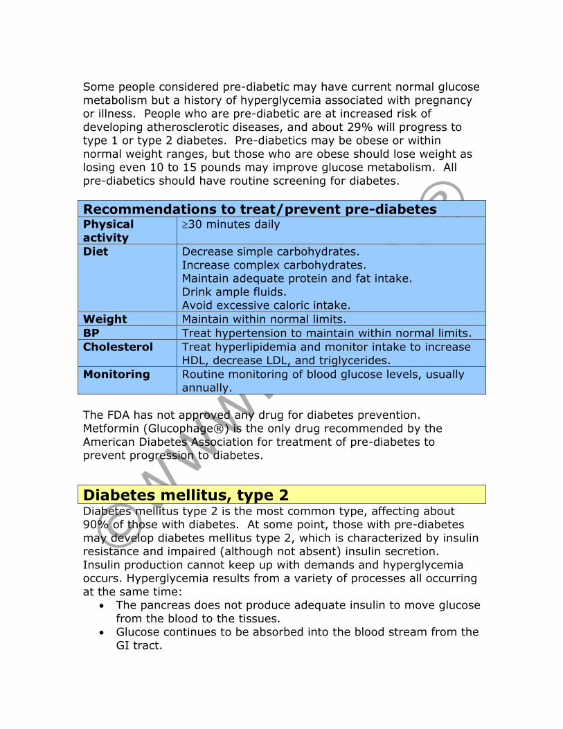
• Increased waist circumference: [See CE course Obesity: Methods of Assessment]
Gender Low risk High risk
Male 40 inches >40 inches
Female 35 inches >35 inches
• Triglyceride level >150 mg/dL (or on medication to
decrease triglyceride level). • BP 130/85 (or on medication to control BP).
• Fasting blood glucose 100 mg/dL (or on medication to
control blood glucose)
A number of other factors put people at risk for development of pre-diabetes:
• Inadequate exercise. • Family history of diabetes.
• HDL level <35. • History of cardiovascular disease.
• History of polycystic ovary syndrome (PCOS).
Some people considered pre-diabetic may have current normal glucose
metabolism but a history of hyperglycemia associated with pregnancy or illness. People who are pre-diabetic are at increased risk of
developing atherosclerotic diseases, and about 29% will progress to type 1 or type 2 diabetes. Pre-diabetics may be obese or within
normal weight ranges, but those who are obese should lose weight as losing even 10 to 15 pounds may improve glucose metabolism. All
pre-diabetics should have routine screening for diabetes.
Recommendations to treat/prevent pre-diabetes Physical
activity
30 minutes daily
Diet Decrease simple carbohydrates.
Increase complex carbohydrates. Maintain adequate protein and fat intake.
Drink ample fluids. Avoid excessive caloric intake.
Weight Maintain within normal limits.
BP Treat hypertension to maintain within normal limits.
Cholesterol Treat hyperlipidemia and monitor intake to increase
HDL, decrease LDL, and triglycerides.
Monitoring Routine monitoring of blood glucose levels, usually annually.
The FDA has not approved any drug for diabetes prevention.
Metformin (Glucophage®) is the only drug recommended by the
American Diabetes Association for treatment of pre-diabetes to prevent progression to diabetes.
Diabetes mellitus, type 2 Diabetes mellitus type 2 is the most common type, affecting about 90% of those with diabetes. At some point, those with pre-diabetes
may develop diabetes mellitus type 2, which is characterized by insulin resistance and impaired (although not absent) insulin secretion.
Insulin production cannot keep up with demands and hyperglycemia occurs. Hyperglycemia results from a variety of processes all occurring
at the same time: • The pancreas does not produce adequate insulin to move glucose
from the blood to the tissues. • Glucose continues to be absorbed into the blood stream from the
GI tract.
• The liver increases basal hepatic glucose production haphazardly rather than in response to body needs.
• Uptake of glucose into the tissues is decreased (because of resistance).
Diabetes mellitus type 2 differs from type 1 in that lipolysis and production of ketone bodies do not occur, so people are not at risk for
DKA. Diabetes mellitus type 2 also appears to have a genetic component, as it tends to run in families. The highest rates are found
in Native Americans, Hispanics, and African Americans.
Diabetes mellitus type 2 is most common in obese adults over 30 (50% are >55) although increasingly it is seen in children and
adolescents, probably because of skyrocketing rates of obesity and poor diet in children. Because of this, type 2 is no longer referred to as
“adult onset” diabetes. Up to 90% of those diagnosed with diabetes
mellitus type 2 are overweight.
Symptoms/Complications of diabetes mellitus type 2 3 Ps Polydipsia, polyuria, and polyphagia.
Weight gain Recent weight gain is often one of the presenting symptoms of diabetes mellitus type
2.
Visual disturbances Blurred vision is common.
General
malaise/discomfort
Patients may complain of vague feelings of
tiredness or lethargy as well as headache.
Skin changes Itching may occur, particularly around the vaginal and groin areas. Some development
acanthosis nigricans (dark skin changes about neck, axillae, and groin areas).
Gynecological/ GU
Erectile dysfunction or impotency may occur. Females may have increased yeast infections.
Neurological
changes
Numbness and tingling in hands and feet and
reduced sensation may occur.
Hyperosmolar
hyperglycemic nonketotic
syndrome
HHNS occurs in people who still produce a
small amount of insulin, enough to prevent DKA but not hyperglycemia. This usually
develops slowly over a long period of time in which undiagnosed diabetes leads to increasing
hyperglycemia. HHNS is characterized by
severe hyperglycemia, osmotic diuresis (which leads to fluid volume deficit, electrolyte
depletion, dehydration, and hypovolemia) and depletion of extracellular fluids. Reduced renal
perfusion, hypotension, shock, increased lactic
acid can occur as well as thrombosis from hyperviscosity of the blood. The end result can
be seizures, shock, coma, and death. HHNS is a medical emergency with a high mortality
rate.
Treatment is similar to DKA although HHNS usually requires more fluid replacement.
Treatment includes IV administration of 0.9% or 0.45% NaCl with IV bolus of regular insulin
followed by insulin infusion. Electrolyte
replacement is given according to laboratory findings. When serum glucose levels drop to
250 mg/dL, IV fluids with glucose may be given to avoid hypoglycemia. Patients must be
carefully monitored.
The best treatment for diabetes mellitus type 2 is aggressive treatment of pre-diabetes to PREVENT progression to the disease.
Initial treatment for diabetes mellitus type 2 is identical to treatment for pre-diabetes, focusing on increased physical activity, weight loss,
and diet although diet is less restrictive than for diabetes mellitus type 1 and does not include use of exchanges. However, reduced calorie
and/or reduced carbohydrate diets are common. If these interventions alone do not bring the blood glucose level within normal limits, then
other treatment is indicated. Oral medications do not function as insulin but they can stimulate increased production of insulin and
improve utilization by the body. In order for oral agents to function, people must be producing some endogenous insulin.
Oral medications for diabetes mellitus type 2 Sulfonylureas First generation:
acetohexamide, chlorpropamide, tolazamide,
and tolbutamide Second generation: glipizide,
glyburide, glimepiride
Increase production of insulin by the pancreas. These tend to be more
effective early in treatment but become less effective over time.
Excessive doses may cause hypoglycemia. Second generation
have a shorter have life and are less likely to cause hypoglycemia.
Meglitinides
Repaglinide Bateglinide
Increase production of insulin by the
pancreas. Faster acting than sulfonylureas. These are taken 30
minutes prior to a meal up to the time of the meal so that they
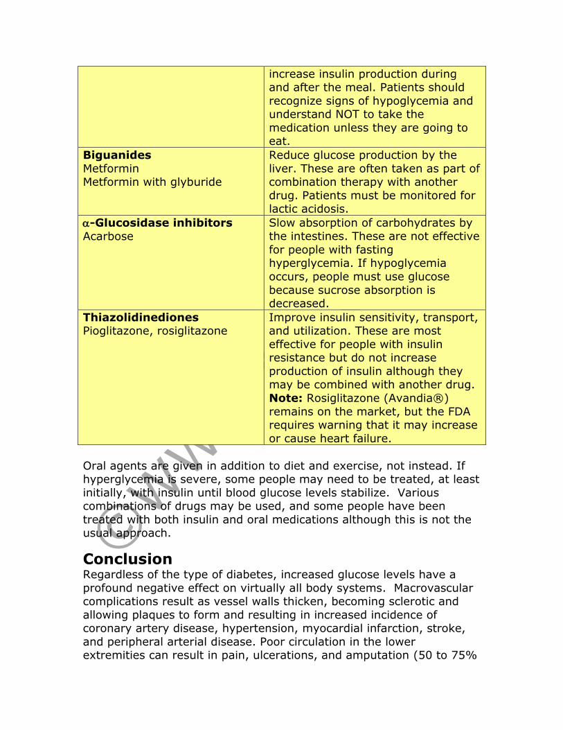
increase insulin production during and after the meal. Patients should
recognize signs of hypoglycemia and understand NOT to take the
medication unless they are going to eat.
Biguanides
Metformin Metformin with glyburide
Reduce glucose production by the
liver. These are often taken as part of combination therapy with another
drug. Patients must be monitored for lactic acidosis.
-Glucosidase inhibitors
Acarbose
Slow absorption of carbohydrates by the intestines. These are not effective
for people with fasting hyperglycemia. If hypoglycemia
occurs, people must use glucose
because sucrose absorption is decreased.
Thiazolidinediones Pioglitazone, rosiglitazone
Improve insulin sensitivity, transport, and utilization. These are most
effective for people with insulin resistance but do not increase
production of insulin although they may be combined with another drug.
Note: Rosiglitazone (Avandia®)
remains on the market, but the FDA requires warning that it may increase
or cause heart failure.
Oral agents are given in addition to diet and exercise, not instead. If hyperglycemia is severe, some people may need to be treated, at least
initially, with insulin until blood glucose levels stabilize. Various combinations of drugs may be used, and some people have been
treated with both insulin and oral medications although this is not the usual approach.
Conclusion Regardless of the type of diabetes, increased glucose levels have a
profound negative effect on virtually all body systems. Macrovascular complications result as vessel walls thicken, becoming sclerotic and
allowing plaques to form and resulting in increased incidence of coronary artery disease, hypertension, myocardial infarction, stroke,
and peripheral arterial disease. Poor circulation in the lower extremities can result in pain, ulcerations, and amputation (50 to 75%

of all lower extremity amputations). Diabetic retinopathy is the leading cause of blindness in adults in the United States. Other ocular
complications include increased risk of cataracts, lens changes, glaucoma, and extraocular muscle palsy. About half of the cases of
end-stage kidney disease are caused by diabetic nephropathy. Diabetes may cause both peripheral and autonomic neuropathies
(associated with sexual dysfunction). Diabetes mellitus type 2 is not a lesser form of diabetes than type 1. It’s a different form, but the long-
term effects on the body may be similar.
References • Diabetes. (2008, February 29). Lab Tests Online. Retrieved
November 23, 2010, from
http://www.labtestsonline.org/understanding/conditions/diabetes-4.html
• Diabetes overview. (2008, November). National Diabetes Information Clearinghouse (NDIC). Retrieved November 23,
2010, from http://diabetes.niddk.nih.gov/dm/pubs/overview/ • Diabetes type 2 medications. (2010). Drugs.com. Retrieved
November 23, 2010, from http://www.drugs.com/condition/diabetes-mellitus-type-ii.html
• Eckman, AS. (2010, November 15). Diabetes. MedlinePlus.
Retrieved November 23, 2010, from http://www.nlm.nih.gov/medlineplus/ency/article/001214.htm
• Immune system genes show links to type 1 diabetes. (2010, September 8). HealthDay. Retrieved November 23, 2010, from
http://www.nlm.nih.gov/medlineplus/news/fullstory_103081.html
• Insulin resistance and pre-diabetes. (2008, October). NIDDK. Retrieved November 25, 2010, from
http://www.diabetes.niddk.nih.gov/dm/pubs/insulinresistance/ • Lamb, WH. (2010, September 17). Diabetes mellitus, type 1.
eMedicine. Retrieved November 23, 2010, from http://emedicine.medscape.com/article/919999-overview
• Ligary, KPL, & Isley, WL. (2010, September 27). Diabetes mellitus, Type 2. eMedicine. Retrieved November 23, 2010, from
http://emedicine.medscape.com/article/117853-overview
• Mathur, R. (2008, July 22). Diabetes mellitus. MedicineNet. Retrieved November 23, 2010, from
http://www.medicinenet.com/diabetes_mellitus/page7.htm • Mayo Clinic staff. (2009, June 13). Diabetes mellitus. MayoClinic.
Retrieved November 23, 2010, from http://www.mayoclinic.com/health/type-1-diabetes/DS00329

• McPhee, S.J. & Papadakis, M.A. (2009). Current Medical Diagnosis & Treatment, 48th ed. San Francisco: McGraw Hill
Lange. • National diabetes statistics. (2008, June). National Diabetes
Information Clearinghouse (NDIC). Retrieved November 23, 2010, from http://diabetes.niddk.nih.gov/dm/pubs/statistics/
• Peri, C. (2009, November 10). Diabetic nerve pain: Do you recognize the symptoms? MedicineNet. Retrieved November 23,
2010, from http://www.medicinenet.com/script/main/art.asp?articlekey=11
3050 • Shier, D, Butler, J., & Lewis, R. Hole’s Human Anatomy &
Physiology, 11 ed. New York: McGraw Hill. • Smeltzer, S.C., Bare, B., Hinkle, J.L, & Cheever, K.H. (2009).
Brunner & Suddarth’s Medical-Surgical Nursing 11th edition.
Philadelphia: Lippincott, Williams, & Wilkins. • Types of insulin. (2010). MedTV. Retrieved November 25, 2010,
from http://diabetes.emedtv.com/insulin/types-of-insulin.html • Votey, SR. (2009, October 8). Diabetes mellitus, Type 1—A
review. eMedicine. Retrieved November 23, 2010, from http://emedicine.medscape.com/article/766036-overview
Votey, SR. (2010, September 23). Diabetes mellitus, Tpe 2—A review. eMedicine. Retrieved November 23, 2010, from
http://emedicine.medscape.com/article/766143-overview • What I need to know about diabetes medicines. (2010, October).
NIDDK. Retrieved November 23, 2010, from http://diabetes.niddk.nih.gov/dm/pubs/medicines_ez/