Development/Plasticity/Repair Neuroendocrine Mechanisms ...
11
Development/Plasticity/Repair Neuroendocrine Mechanisms Governing Sex Differences in Hyperalgesic Priming Involve Prolactin Receptor Sensory Neuron Signaling Candler Paige, 1 Priscilla A. Barba-Escobedo, 2 Jennifer Mecklenburg, 2 Mayur Patil, 2 Vincent Goffin, 3 David R. Grattan, 4 Gregory Dussor, 1 Armen N. Akopian, 2 and Theodore J. Price 1 1 School of Behavioral and Brain Sciences and Center for Advanced Pain Studies, University of Texas at Dallas, Richardson, Texas 75080, 2 Departments of Endodontics, School of Dentistry, University of Texas Health Science Center at San Antonio, San Antonio, Texas 78229, 3 Université de Paris, Institut National de la Santé et de la Recherche Médicale U1151, Paris, 75014, France, and 4 Centre for Neuroendocrinology and Department of Anatomy, University of Otago School of Biomedical Sciences, Dunedin 09001, New Zealand Many clinical and preclinical studies report higher prevalence and severity of chronic pain in females. We used hyperalgesic priming with interleukin 6 (IL-6) priming and PGE 2 as a second stimulus as a model for pain chronicity. Intraplantar IL-6 induced hypersensitivity was similar in magnitude and duration in both males and females, while both paw and intrathecal PGE 2 hypersensitivity was more persistent in females. This difference in PGE 2 response was dependent on both circulating estrogen and translation regulation signaling in the spinal cord. In males, the duration of hypersensitivity was regulated by testosterone. Since the prolactin receptor (Prlr) is regulated by reproductive hormones and is female-selectively activated in sensory neurons, we evaluated whether Prlr signaling contributes to hyperalgesic priming. Using DPRL, a competitive Prlr antagonist, and a mouse line with ablated Prlr in the Nav1.8 sensory neuronal population, we show that Prlr in sensory neu- rons is necessary for the development of hyperalgesic priming in female, but not male, mice. Overall, sex-specific mechanisms in the initiation and maintenance of chronic pain are regulated by the neuroendocrine system and, specifically, sensory neu- ronal Prlr signaling. Key words: estrogen; nociceptor; prolactin; sex dimorphism; testosterone; translation regulation Significance Statement Females are more likely to experience chronic pain than males, but the mechanisms that underlie this sex difference are not completely understood. Here, we demonstrate that the duration of mechanical hypersensitivity is dependent on circulating sex hormones in mice, where estrogen caused an extension of sensitivity and testosterone was responsible for a decrease in the duration of the hyperalgesic priming model of chronic pain. Additionally, we demonstrated that prolactin receptor expres- sion in Nav1.8 1 neurons was necessary for hyperalgesic priming in female, but not male, mice. Our work demonstrates a female-specific mechanism for the promotion of chronic pain involving the neuroendrocrine system and mediated by sensory neuronal prolactin receptor. Introduction Many chronic pain conditions, such as migraine, fibromyalgia, temporomandibular joint disorders, irritable bowel syndrome, and rheumatoid arthritis, have a 2- to 6-fold greater prevalence or symptom severity in women compared with men (Unruh, 1996; Berkley, 1997; Fillingim et al., 2009; Traub and Ji, 2013). Pain symptoms in women with these chronic pain conditions can change during the menstrual cycle as gonadal hormone con- centrations fluctuate; and some, but not all, pain conditions decrease in frequency or intensity after menopause (Houghton et al., 2002; LeResche et al., 2003; Slade et al., 2011; Mathew et al., 2013). There is a general consensus that plasticity mechanisms in peripheral and central nociceptive pathways are critical for Received June 12, 2020; revised July 20, 2020; accepted July 23, 2020. Author contributions: C.P., V.G., D.R.G., G.D., A.N.A., and T.J.P. designed research; C.P., P.A.B.-E., J.M., and M.P. performed research; C.P., A.N.A., and T.J.P. analyzed data; C.P., A.N.A., and T.J.P. wrote the first draft of the paper; C.P., A.N.A., and T.J.P. wrote the paper; G.D., A.N.A., and T.J.P. edited the paper. The authors declare no competing financial interests. This work was supported by National Institute of Neurological Disorders and Stroke/National Institutes of Health NS102161 to T.J.P. and A.N.A.; National Institutes of Health/National Institute of General Medical Sciences GM112747 to A.N.A.; National Institute of Neurological Disorders and Stroke/National Institutes of Health NS065926 to T.J.P.; National Institute of Neurological Disorders and Stroke/National Institutes of Health NS113457 to C.P.; and University of Texas BRAIN Pilot Program ID 1503083 to G.D. and A.N.A. We thank Dr. Dustin Green for advice on the experimental strategy; and Dr. Florence Boutillon for producing recombinant PRL and D1-9-G129R-hPRL (DPRL). Correspondence should be addressed to Armen N. Akopian at [email protected] or Theodore J. Price at [email protected]. https://doi.org/10.1523/JNEUROSCI.1499-20.2020 Copyright © 2020 the authors 7080 • The Journal of Neuroscience, September 9, 2020 • 40(37):7080–7090
Transcript of Development/Plasticity/Repair Neuroendocrine Mechanisms ...

Development/Plasticity/Repair
Neuroendocrine Mechanisms Governing Sex Differences inHyperalgesic Priming Involve Prolactin Receptor SensoryNeuron Signaling
Candler Paige,1 Priscilla A. Barba-Escobedo,2 Jennifer Mecklenburg,2 Mayur Patil,2 Vincent Goffin,3
David R. Grattan,4 Gregory Dussor,1 Armen N. Akopian,2 and Theodore J. Price11School of Behavioral and Brain Sciences and Center for Advanced Pain Studies, University of Texas at Dallas, Richardson, Texas 75080,2Departments of Endodontics, School of Dentistry, University of Texas Health Science Center at San Antonio, San Antonio, Texas 78229,3Université de Paris, Institut National de la Santé et de la Recherche Médicale U1151, Paris, 75014, France, and 4Centre for Neuroendocrinologyand Department of Anatomy, University of Otago School of Biomedical Sciences, Dunedin 09001, New Zealand
Many clinical and preclinical studies report higher prevalence and severity of chronic pain in females. We used hyperalgesicpriming with interleukin 6 (IL-6) priming and PGE2 as a second stimulus as a model for pain chronicity. Intraplantar IL-6induced hypersensitivity was similar in magnitude and duration in both males and females, while both paw and intrathecalPGE2 hypersensitivity was more persistent in females. This difference in PGE2 response was dependent on both circulatingestrogen and translation regulation signaling in the spinal cord. In males, the duration of hypersensitivity was regulated bytestosterone. Since the prolactin receptor (Prlr) is regulated by reproductive hormones and is female-selectively activated insensory neurons, we evaluated whether Prlr signaling contributes to hyperalgesic priming. Using DPRL, a competitive Prlrantagonist, and a mouse line with ablated Prlr in the Nav1.8 sensory neuronal population, we show that Prlr in sensory neu-rons is necessary for the development of hyperalgesic priming in female, but not male, mice. Overall, sex-specific mechanismsin the initiation and maintenance of chronic pain are regulated by the neuroendocrine system and, specifically, sensory neu-ronal Prlr signaling.
Key words: estrogen; nociceptor; prolactin; sex dimorphism; testosterone; translation regulation
Significance Statement
Females are more likely to experience chronic pain than males, but the mechanisms that underlie this sex difference are notcompletely understood. Here, we demonstrate that the duration of mechanical hypersensitivity is dependent on circulatingsex hormones in mice, where estrogen caused an extension of sensitivity and testosterone was responsible for a decrease inthe duration of the hyperalgesic priming model of chronic pain. Additionally, we demonstrated that prolactin receptor expres-sion in Nav1.81 neurons was necessary for hyperalgesic priming in female, but not male, mice. Our work demonstrates afemale-specific mechanism for the promotion of chronic pain involving the neuroendrocrine system and mediated by sensoryneuronal prolactin receptor.
IntroductionMany chronic pain conditions, such as migraine, fibromyalgia,temporomandibular joint disorders, irritable bowel syndrome,and rheumatoid arthritis, have a 2- to 6-fold greater prevalenceor symptom severity in women compared with men (Unruh,1996; Berkley, 1997; Fillingim et al., 2009; Traub and Ji, 2013).Pain symptoms in women with these chronic pain conditionscan change during the menstrual cycle as gonadal hormone con-centrations fluctuate; and some, but not all, pain conditionsdecrease in frequency or intensity after menopause (Houghton etal., 2002; LeResche et al., 2003; Slade et al., 2011; Mathew et al.,2013). There is a general consensus that plasticity mechanisms inperipheral and central nociceptive pathways are critical for
Received June 12, 2020; revised July 20, 2020; accepted July 23, 2020.Author contributions: C.P., V.G., D.R.G., G.D., A.N.A., and T.J.P. designed research; C.P., P.A.B.-E., J.M., and
M.P. performed research; C.P., A.N.A., and T.J.P. analyzed data; C.P., A.N.A., and T.J.P. wrote the first draft ofthe paper; C.P., A.N.A., and T.J.P. wrote the paper; G.D., A.N.A., and T.J.P. edited the paper.The authors declare no competing financial interests.This work was supported by National Institute of Neurological Disorders and Stroke/National Institutes of
Health NS102161 to T.J.P. and A.N.A.; National Institutes of Health/National Institute of General MedicalSciences GM112747 to A.N.A.; National Institute of Neurological Disorders and Stroke/National Institutes ofHealth NS065926 to T.J.P.; National Institute of Neurological Disorders and Stroke/National Institutes of HealthNS113457 to C.P.; and University of Texas BRAIN Pilot Program ID 1503083 to G.D. and A.N.A. We thank Dr.Dustin Green for advice on the experimental strategy; and Dr. Florence Boutillon for producing recombinantPRL and D1-9-G129R-hPRL (DPRL).Correspondence should be addressed to Armen N. Akopian at [email protected] or Theodore J. Price
at [email protected]://doi.org/10.1523/JNEUROSCI.1499-20.2020
Copyright © 2020 the authors
7080 • The Journal of Neuroscience, September 9, 2020 • 40(37):7080–7090

chronic pain development in males and females, but the precisemechanisms governing this plasticity are increasingly recognizedas sex dimorphic and are still largely unknown. Nevertheless,recent progress was made in understanding underlying mecha-nisms for sex-dependent mechanisms of nociceptive plasticity(Mogil et al., 2011; Sorge et al., 2011, 2015; Rosen et al., 2017;Martin et al., 2019). These findings on sex differences in nocicep-tive plasticity mechanisms, combined with abundant clinical androdent data on the effects of gonadal hormones on pain, indicatea critical role for gonadal hormones in regulation of pain chroni-fication (Fillingim et al., 2009; Traub and Ji, 2013).
Clearly, there are gonadal hormone-regulated mechanismsthat promote chronic pain in females, but these mechanismshave not been thoroughly characterized. Prolactin (PRL) and itsreceptor (Prlr) are prime candidates for this potential mecha-nism, since responsiveness to PRL in a variety of cells, includingsensory neurons, is closely regulated by estrogen (Childs et al.,1999; Pi and Voogt, 2002; Diogenes et al., 2006; Belugin et al.,2013). Thus, Prlr signaling sensitizes pain-related ion channelsand causes increased excitability in nociceptors, specifically infemales (Diogenes et al., 2006; Patil et al., 2013b, 2019a,b; Liu etal., 2016). Moreover, Prlr function in female nociceptors is gov-erned by estrogen signaling via a nongenomic pathway that involvessex-specific translation of Prlr mRNA (Patil et al., 2019b). Clinically,high PRL levels are linked to migraine, and blocking PRL actions inwomen with hyperprolactinemia resolves headache (Silberstein,1992; Cavestro et al., 2006; Oliveira et al., 2020). Based on these pre-vious studies, we hypothesized that Prlr signaling might differen-tially contribute to female-specific regulation of chronic pain thatcan be assessed by use of the hyperalgesic priming model (Aley etal., 2000). Our work reveals that initiation, maintenance, andmagnitude of hyperalgesic priming are governed by estrogen-de-pendent regulation of Prlr signaling in sensory neurons.Therefore, PRL signaling to Prlr is a gonadal hormone-
dependent mechanism that promotes plasticity in the nociceptivepathway, supporting development of chronic pain specifically infemales.
Materials and MethodsAnimals. All animal experiments were approved by the University
Texas Health Science Center at San Antonio and University of Texas atDallas Institutional Animal Care and Use Committee. We followedguidelines issued by the National Institutes of Health and the Society forNeuroscience to minimize suffering and the number of animals used.
Key reagents and mouse lines. Eight- to 12-week-old female and malemice were purchased from The Jackson Laboratory. Ovariectomized(OVX) and gonadectomized (GdX) mice were purchased from TheJackson Laboratory. The estrous phases in adult females were determineby vaginal gavage as described by Caligioni (2009). Estrogen and testos-terone replacement procedures to generate OVX-E-2 and GdX-T micewere performed as previously described (Diogenes et al., 2006; Nettleshipet al., 2007). 17b -Estradiol (E-2; 300mg per injection) or testosterone (T;300mg per injection) was injected intraperitoneally 2 times a week for 3weeks into OVX and GdXmice, respectively.
The Prlrfl/fl line was generated as previously described (Brown et al.,2016). Prlrfl/fl line has inverse lox sites; hence, Cre recombination ablatesthe Prlr gene and activates GFP in targeted cells.
Estrogen was purchased from Sigma Millipore (catalog #PHR1353-1G) Testosterone was purchased from Sigma Millipore (catalog #T1875-1G). 4EGI was purchased from Tocris Bioscience. Vehicle for IL-6,PGE2, PRL, and D1-9-G129R-hPRL (DPRL) was 0.9% saline or PBS.Vehicle for 4EGI-1 was 0.1% DMSO in 0.9% saline.
Human PRL was generated in an Escherichia coli expression systemcontaining plasmid with human PRL (Goffin; Institut National de laSanté et de la Recherche Médicale). Thus, PRL is fully processed,unmodified (i.e., no glycosylation and phosphorylation), and has molec-ular weight of ;23 kDa. The Prlr antagonist DPRL (Rouet et al., 2010),which is a modified PRL that binds to and blocks the function of Prlr inrat, mouse, and human (Bernichtein et al., 2003), was also synthesizedby Goffin (Institut National de la Santé et de la Recherche Médicale).
Figure 1. Persistence of hyperalgesic priming is greater in female mice. A, Schematic of the IL-6-induced hyperalgesic priming model. BL, Baseline measurements. Brown arrows indicateinjection time points. Blue arrows indicate post-PGE2 mechanical nociception measurement time points. B, Hyperalgesic priming model: IL-6 priming into paw and PGE2 injection into paw offemale and male C57BL/6 mice. C, Hyperalgesic priming model: IL-6 priming into paw and PGE2 injection into SC of female and male C57BL/6 mice. D, The same model as in C in female andmale Institute of Cancer Research (ICR) mice. Arrows indicate injection time points for IL-6/Veh and PGE2. Repeated-measures ANOVA with Bonferroni post hoc test: C, IL-6 male compared withVeh-female: ****p, 0.0001; IL-6 female compared with Veh-female:$$$$p, 0.0001;$$$p, 0.001; all others: *p, 0.05; ****p, 0.0001. n= 5–8.
Paige et al. · Prolactin Promotes Chronic Pain in Females J. Neurosci., September 9, 2020 • 40(37):7080–7090 • 7081

We and others thoroughly confirmedthe specificity of DPRL using in vitro(Bernichtein et al., 2003; Scotland et al.,2011), and in vivo studies (Rouet et al.,2010), including using Prlr KO mice(Belugin et al., 2013).
RT-PCR. RT-PCR was performed onhindpaw, L3-L5 DRG, and spinal cord (SC)total RNA. Dissected tissue was stored inRNA. Later at �20°C (QIAGEN). RNAextraction was done using the QIAzol lysisreagent and the RNAeasy Mini Kit(QIAGEN), and manufacturer’s instruc-tions were followed. cDNA was synthesizedusing Superscript III First Strand Synthesiskit (Invitrogen). Primers were as follows:Prlr-F (59-CCATTCACCTGCTGGTGGAATCCT-39), GFP-F (59-AAGGCTACGTCCAGGAGCGCACCA-39), GFP-R1 (59-CGTCCTCGATGTTGTGGCGGATC-39),and GFP-R2 (59-TGGTGCGCTCCTGGACGTAGCCTT-39). Amplification of tar-get sequences was detected on 1 or 1.5%agarose gel depending on band size.
Behavior experiments. Hyperalgesicpriming was established using a previ-ously described model (Aley et al., 2000;Kim et al., 2016). IL-6 was injected intra-plantarly, which created a transient mechani-cal hypersensitivity and initiated hyperalgesicpriming. After IL-6-induced mechanicalhypersensitivity resolved and thresholdsreturned to a baseline level, PGE2 wasadministered either intraplantarly or intra-thecally to precipitate the primed state andinduce mechanical hypersensitivity. PRL,DPRL, or 4EGI-1 was administered immedi-ately before IL-6 or PGE2 administration. Toevaluate mechanical hypersensitivity follow-ing the intraplantar or intrathecal injections,animals were habituated for 45-60min inelevated behavior racks, and then paw with-draw threshold was determined using theup-down von Frey method (Chaplan et al.,1994). Both the experimenters performingthe behavior and data analysis were doneblinded.
Experimental design and statisticalanalysis. GraphPad Prism 7.0 (GraphPad)was used for all statistical analyses of data.Data are presented as mean 6 SEM, withn indicating the number of independentanimals per group in behavioral experi-ments. The sex of the animals used ineach experiment is described in the text. Differences between groupswere assessed by either mixed-effects or repeated-measures ANOVA withBonferroni post hoc tests (noted for each figure). Statistical significancewas determined as p, 0.05. Interaction F ratios and the associatedp values are reported in the text.
ResultsSex differences in hyperalgesic priming in mice are regulatedby gonadal hormones and translation regulationChronic pain occurs more frequently in females (Unruh, 1996;Berkley, 1997; Fillingim et al., 2009; Traub and Ji, 2013). This dif-ference could be mediated by sex-dependent mechanisms con-trolling the transition from acute to chronic pain. We used the
hyperalgesic priming paradigm (Aley et al., 2000) to gain insightinto female-specific mechanisms involved in the acute to chronicpain transition. In our experiments, hyperalgesic priming was ini-tiated with an intraplantar injection of IL-6 (0.5 ng). When the ini-tial hypersensitivity from this IL-6 injection had resolved, thepresence of priming was assessed with either an intraplantar or in-trathecal injection of PGE2 (0.1mg; Fig. 1A). Female C57BL/6mice that were primed with IL-6 and then subsequently receivedan intraplantar injection of PGE2 had a slightly longer persistenceof mechanical hypersensitivity compared with males (repeated-measures ANOVA; F(11,88) = 1.437; p=0.1708; n=5; Fig. 1B).Intrathecal injection of PGE2 in females had a significantly longerduration of response to PGE2 compared with males (repeated-measures ANOVA; F(14,91) = 8.975 p, 0.0001 n=5; Fig. 1C). To
Figure 2. Contribution of gonadal hormones profoundly influences hyperalgesic priming in female and male mice. A,Hyperalgesic priming model with spinal PGE2 injection in WT female, OVX, and OVX1E (B) GdX, and (C) WT male, GdX, andGdX-T C57BL mice. B, The disparity in timelines between GDX animals and those animals that are naive or GdX-T. Arrows indicateinjection time points for IL-6 and PGE2. Repeated-measures ANOVA with Bonferroni post hoc test: A, ***p, 0.001;****p, 0.0001, for OVX-E compared with female; $$p, 0.01 for OVX compared with female; C, ***p, 0.001;$$p, 0.01 for GdX-T compared with male; n= 5 or 6.$$$$p, 0.0001.
7082 • J. Neurosci., September 9, 2020 • 40(37):7080–7090 Paige et al. · Prolactin Promotes Chronic Pain in Females

rule out a possible strain-dependent sex effect, the experimentwith spinal administration of PGE2 following intraplantar IL-6was repeated in male and female Swiss Webster mice. The differ-ence in the length of the PGE2 response following intrathecaladministration was even longer lasting in this outbred strain(repeated-measures ANOVA; F(10,140) = 8.409; p, 0.0001; n=8;Fig. 1D). To gain insight into the mechanistic underpinnings ofthe sex difference we identified, we did the remaining experimentswith intraplantar injection of IL-6 and intrathecal administrationof PGE2 in C57BL/6 mice.
In the above and following experiments, the female animalswere all in the estrous phase of the estrous cycle at the time of IL-6 injections. The hyperalgesic priming model lasts at least aweek; and despite controlling for the estrous phase at the time ofIL-6 injection, female mice cycle quickly (4-5 d) through other
phases of the cycle, and circulating bloodestrogen (E-2) levels will vary on a day-to-day basis. Hence, to control for E-2 levels,we used OVX and OVX with E-2 supple-mentation (OVX-E-2) female mice. Thesubstantial reduction of circulating E-2 inOVX mice (Green et al., 2016) did notchange the initiation phase of priming, butthe persistence of the response following thePGE2 injection was significantly shorter(mixed-measures ANOVA; F(34,220) = 8.710p=0.0011; n=5 or 6; Fig. 2A). Admini-stering E-2 to keep circulating E-2 atapproximately proestrus phase levels inOVX-E-2 mice (Green et al., 2016) resultedin a significant extension of both the initia-tion (IL-6) and priming (PGE2) phases com-pared with intact females in the hyperalgesicpriming model. Testosterone mediates male-specific nociceptive responses (Sorge et al.,2011). GdX mice have almost no circulat-ing testosterone (Green et al., 2016). TheseGdX mice had a substantially longerresponse to IL-6 injection than intact malemice. Males with IL-6-induced mechanicalhypersensitivity usually return to baselinemechanical sensitivity within 4-5 d (Kim etal., 2016) (Fig. 1B–D). In contrast, the ini-tiation phase of hyperalgesic priming lasted.31 d in GdX males, and only partiallyrecovered to baseline levels, which we dis-played on a separate graph (Fig. 2B). ThePGE2 phase was also lengthened in GdXcompared with intact male mice (repeated-measures ANOVA; F(12,84) = 5.016; p ,0.0001; n= 5 or 6; Fig. 2C). Testosterone res-cue in GdX-T animals returned the initia-tion and priming phases to the sametimeline as intact males (Fig. 2C). Theseresults show that the time course of hyperal-gesic priming is more pronounced in femalemice compared with male mice, and isclosely regulated by circulating gonadal hor-mones in both sexes.
Translation regulation plays a key role inthe development of hyperalgesic priming(Price and Inyang, 2015; Khoutorsky andPrice, 2018). In previous experiments thatwere mostly done in male animals, we have
shown that inhibition of cap-dependent translation at the time ofinitiation blocks the development of hyperalgesic priming(Melemedjian et al., 2010, 2014; Asiedu et al., 2011; Moy et al.,2017). Inhibition of cap-dependent translation during the main-tenance phase fails to reverse established priming (Asiedu et al.,2011). In concordance with previous experiments (Melemedjianet al., 2010, 2014; Asiedu et al., 2011), the cap-dependent transla-tion inhibitor 4EGI-1 (10mg) given intrathecally immediatelybefore PGE2 stimulation did not affect PGE2-induced mechanicalhypersensitivity in males (repeated-measures ANOVA; F(8,64) =0.3153; p=0.9575; n=5; Fig. 3A). In stark contrast, 4EGI-1 dra-matically reduced the persistence of PGE2 precipitated mechanicalhypersensitivity in females (repeated-measures ANOVA; F(8,64) =10.60; p, 0.0001; n=5; Fig. 3B). We next evaluated whether the
Figure 3. Spinal local translation only contributes to hyperalgesic priming in intact female mice. A, Hyperalgesic primingmodel with spinal PGE2 injection in WT male, (B) WT female, and (C) OVX mice. 4EGI-1 (10mg) or vehicle was adminis-tered spinally at 30min before PGE2 injection. Arrows indicate injection time points for IL-6 and 4EGI-1/PGE2. Repeated-measures ANOVA with Bonferroni post hoc test: ****p, 0.0001. n= 5 or 6.
Paige et al. · Prolactin Promotes Chronic Pain in Females J. Neurosci., September 9, 2020 • 40(37):7080–7090 • 7083

difference between males and females in theregulation of maintenance of hyperalgesicpriming was defined by gonadal hormonestatus. Removal of circulating E-2 usingOVX females abolished the influenceof 4EGI-1 on the hyperalgesic priming(repeated-measures ANOVA; F(9,90) =0.7394; p=0.6719; n=6; Fig. 3C). Theseresults indicate that the magnitude andmaintenance of chronic pain are regulatedby gonadal hormones in male and femalemice and that the enhanced priming effectseen in intact female mice is dependent ontranslation regulation at the level of theDRG and/or SC. Interestingly, previouswork done entirely in male rodents sug-gested that translation regulation events atthe level of the DRG and/or SC were notinvolved in maintenance of hyperalgesicpriming (Asiedu et al., 2011; Ferrari et al.,2015).
A female-specific role for sensoryneuronal Prlr in hyperalgesic primingResponsiveness to PRL in sensory neuronsis substantially higher in females (.40fold) than in males (Patil et al., 2013b,2019a,b), and strictly controlled by E-2(Diogenes et al., 2006; Patil et al., 2019b).Endogenous and extrapituitary PRL is ele-vated in paw and SC after inflammationand surgical injury (Scotland et al., 2011;Patil et al., 2013a). Accordingly, we exam-ined whether mimicking the presence ofendogenous PRL after injury could pro-long mechanical hypersensitivity inducedby spinal PGE2 in both females and males.To do this, we primed the nociceptivepathway with a single injection of exoge-nous PRL (1mg) intraplantarly (this PRLdosage is active in females but not males)(Patil et al., 2019b) and precipitated hyper-algesic priming with intrathecal PGE2 at 1d after PRL treatment (Fig. 4A). We wereable to administer PGE2 at 24 h after intra-plantar PRL because 1mg PRL produces mechanical hypersensitiv-ity for only ;3–4 h in females (Patil et al., 2019b). PGE2 evokedmechanical hypersensitivity was substantially longer lasting inPRL-treated female mice compared with vehicle-primedfemales (repeated-measures ANOVA; F(6,60) = 6.398; p ,0.0001; n = 6; Fig. 4C), but this was not the case in male mice(repeated-measures ANOVA; F(4,40) = 0.2318; p=0.9189; n=5–7;Fig. 4B).
Our present findings suggest that a translation regulationevent at the spinal level is critical for enhanced pain chronifica-tion in female mice. Our previous work demonstrated that Prlrsignaling in the central terminals of nociceptors is important foracute pain models, specifically mechanical hypersensitivity inresponse to inflammation and injury in females (Patil et al., 2019b).This sex difference can be accounted for by increased translation ofPrlr mRNA in central terminals of female mice (Patil et al., 2019b),suggesting transport of this mRNA to central terminals of nocicep-tors. To gain better insight into regulation of Prlr mRNA
localization, we used Prlrfl/fl mice and crossed them with Nav1.8cre/�
animals to generate a Nav1.8cre/�/Prlrfl/fl (Prlr CKO) in a set ofsensory neurons (Fig. 5A). The Prlrfl/fl line has inverse loxsites; hence, Cre recombination ablates the Prlr gene and acti-vates GFP expression in targeted cells driven by the Prlr pro-moter (Fig. 5A). Analysis of the Prlr CKO showed that thetruncated transgene Prlr mRNA contains the entire 59-UTR;four exons and an intron between exons 1 and 4 (EI–EIV)and GFP, but it does not have the remaining Prlr exons orthe 39-UTR (Fig. 5A). Accordingly, we observed that RT-PCR with Prlr-F and GFP-R1 primers (Fig. 5A, red arrows)produced a 2500 bp band containing an intron sequencefrom total RNA of female Prlr CKO, but not Prlrfl/fl (Fig. 5C).
The unique splicing of EI-EIV and GFP for Prlr mRNA inPrlr CKO mice allowed us to detect localization of this hybridPrlrmRNA synthesized from Prlr gene promoter in sensory neu-ronal cell bodies, as well as peripheral and central terminals. Forthese experiments, we also used a positive control, PGP9.5mRNA (Uchl1 gene), which undergoes axonal transport in DRG
Figure 4. Peripheral PRL only induces hyperalgesic priming in female mice. A, Schematic of the PRL-induced hyperalgesicpriming model. BL, Baseline measurements after PRL-induced hypersensitivity is fully resolved. Brown arrows indicate injec-tion time points. Blue arrows indicate post-PGE2 treatment mechanical nociception measurement time points. B, C,Hyperalgesic priming model; PRL or vehicle priming into paw and PGE2 injection into SC of male (B) or female (C) C57BLmice. Arrows indicate injection time points for PGE2. Repeated-measures ANOVA with Bonferroni post hoc test:***p, 0.001; ****p, 0.0001. n= 5–7.
7084 • J. Neurosci., September 9, 2020 • 40(37):7080–7090 Paige et al. · Prolactin Promotes Chronic Pain in Females

neurons (Willis et al., 2005, 2007). Uchl1 mRNA was found inthe hindpaw, but at an apparently lower level than observed inthe DRG (unpaired two-tailed t test; t=6.521 df = 4; Figs. 5B,B’).The hybrid Prlr mRNA in sensory neuronal cell bodies (DRG)and sensory neuronal central terminals (SC) was detected byamplification of the 2500bp band from total RNA isolated fromDRG and SC tissues of Prlr CKO (Fig. 5C). This 2500bp band was
not noticed in the Prlrfl/fl mouse line (Fig.5C). Interestingly, the 2500bp PCR prod-uct was detected in SC mRNA, suggest-ing constitutive axonal transport of PrlrmRNA (Fig. 5C). Moreover, hybrid PrlrmRNA was at significantly higher levelsin SC compared with DRG (unpairedtwo-tailed t test; t=4.797 df= 4; Fig. 5C’).To confirm this result with different pri-mers, we PCR-amplified GFP (253bpband) with GFP-F and GFP-R2 primers(Fig. 5A, blue arrows), using DRG andSC total RNA from Prlrfl/fl and Prlr CKOmice. GFP mRNA was detected not onlyin DRG of Prlr CKO females, but also inSC (Fig. 5D), and it was at higher densityin SC compared with DRG (unpairedtwo-tailed t test; t=3.608 df= 4; Fig. 5D’).To examine translocation of Prlr mRNAto peripheral terminals, we examined thepresence of GFP mRNA in hindpaw tis-sues of Prlr CKO mice (Fig. 5E). Again,GFP mRNA in Prlr CKO was at higherlevels in hindpaw compared with DRGtissues (unpaired two-tailed t test; t=5.036 df= 4; Fig. 5E’). These findings sug-gest that female PrlrmRNA is extensivelytranslocated to sensory neuron peripheraland central terminals, likely via an ele-ment in the 59 UTR of the mRNA that ispreserved in the transgene.
Next, we used PrlrCKOmice to exam-ine the contribution of sensory neuronalPrlr to the regulation of the developmentof chronic pain in females and males.Prlr ablation in male sensory neuronsdid not affect either the initiation or per-sistent phase in the hyperalgesic primingmodel (repeated-measures ANOVA;F(12,66) = 9.445; p, 0.001; n = 4-6; Fig.6A). In contrast, Prlr CKO female miceshowed both a significant reduction inmechanical hypersensitivity in response toIL-6 injection and these mice also showeda greatly reduced response to PGE2 injec-tion (persistent phase) compared withPrlrfl/fl mice (repeated-measures ANOVA;F(12,120) = 1.309, p=0.2219; n=5-7; Fig.6B). We conclude from this experimentthat Prlr in sensory neurons plays a keyrole in initiation and maintenance ofchronic pain in female, but not male,mice.
Prlr signaling and the initiation andmaintenance of hyperalgesic primingin female miceAblation of Prlr in sensory neurons doesnot allow for identification of peripheral
or central sites driving hyperalgesic priming or the time course ofwhen Prlr signaling occurs during hyperalgesic priming. Toexplore this in detail, the Prlr antagonist DPRL (Rouet et al., 2010;Patil et al., 2019b), was delivered at different locations and timepoints. A single injection of the antagonist DPRL (5mg) into the
Figure 5. Evidence for translocation of Prlr mRNA to sensory neuronal peripheral and central terminals in female mice. A,Schematic of Prlrfl/fl and Nav1.8cre/-/Prlrfl/fl (Prlr CKO) genes and corresponding transcribed mRNA from these genes. Location ofPrlr-F and GFP-R1 (red arrows) and GFP-F and GFP-R2 (blue arrow). B, Representative panel for PCR of PGP9.5 mRNA (Uchl1gene) from total RNA isolated from DRG and hindpaws (HP) of Prlrfl/fl (Con) and Prlr CKO female mice. B9, Quantification ofdata shown in B for Prlr CKO female mice (**p, 0.01; n= 3). C, Representative panel for PCR of hybrid 2500 bp Prlr mRNAusing Prlr-F (exon 4) and GFP-R1 primers (see A) for DRG and SC total RNA isolated from Prlrfl/fl (Con) and Prlr CKO of femalemice. C9, Quantification of data shown in C for Prlr CKO female mice (**p, 0.01; n= 3). D, Representative panel for PCR ofGFP mRNA using GFP-F and GFP-R2 primers (see A) for DRG and SC total RNA isolated from Prlrfl/fl (Con) and Prlr CKO of femalemice. D9, Quantification of data shown in D for Prlr CKO female mice (*p, 0.05; n= 3). E, Representative panel for PCR ofGFP mRNA using GFP-F and GFP-R2 primers (see A) for DRG and HP total RNA isolated from Prlrfl/fl (Con) and Prlr CKO of femalemice. E9, Quantification of data shown in E for Prlr CKO female mice (**p, 0.01; n= 3).
Paige et al. · Prolactin Promotes Chronic Pain in Females J. Neurosci., September 9, 2020 • 40(37):7080–7090 • 7085

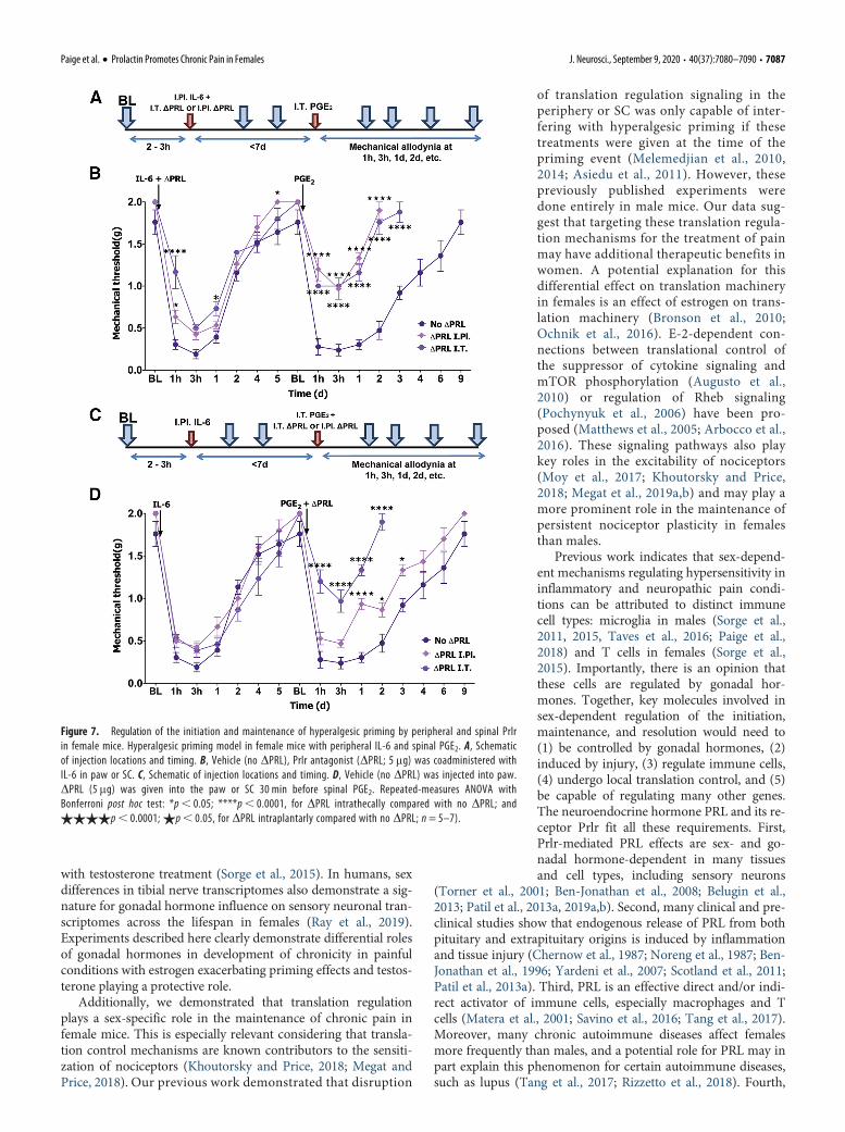
paw immediately before the intraplantar IL-6priming injection did not affect mechanicalhypersensitivity in response to the IL-6 injec-tion (Fig. 7A). However, there was a signifi-cant reduction in mechanical hypersensitivityduring the persistent, post-PGE2 phase in ani-mals that received DPRL compared withthose that received the vehicle. These DPRL-treated female mice returned to baseline levelsof mechanical sensitivity within 2d of thePGE2 injection (repeated-measures ANOVA;F(30,195) = 6.043; p, 0.0001; n=5 or 6; Fig.7A). Intrathecal administration of DPRL(5mg) immediately before intraplantar IL-6led to a transient reduction in mechanicalhypersensitivity for,3 h following intraplan-tar IL-6 injection (Fig. 7A). The degree of me-chanical hypersensitivity following PGE2injection was almost identical to thatobserved with intraplantar administration ofDPRL. We then evaluated the role of Prlr sig-naling in the maintenance of hyperalgesicpriming with intraplantar or intrathecaladministration of DPRL (5mg) before PGE2administration. Blockage of Prlr signaling inthe paw by intraplantar injection of DPRLbefore the injection of PGE2 did not influencemechanical hypersensitivity magnitude or du-ration. In contrast, intrathecal administrationof DPRL coupled with intrathecal PGE2administration led to inhibition of mechanicalhypersensitivity and a faster return to base-line (repeated-measures ANOVA; F(30,210) =7.192; p, 0.0001; n=5 or 6; Fig. 7B).
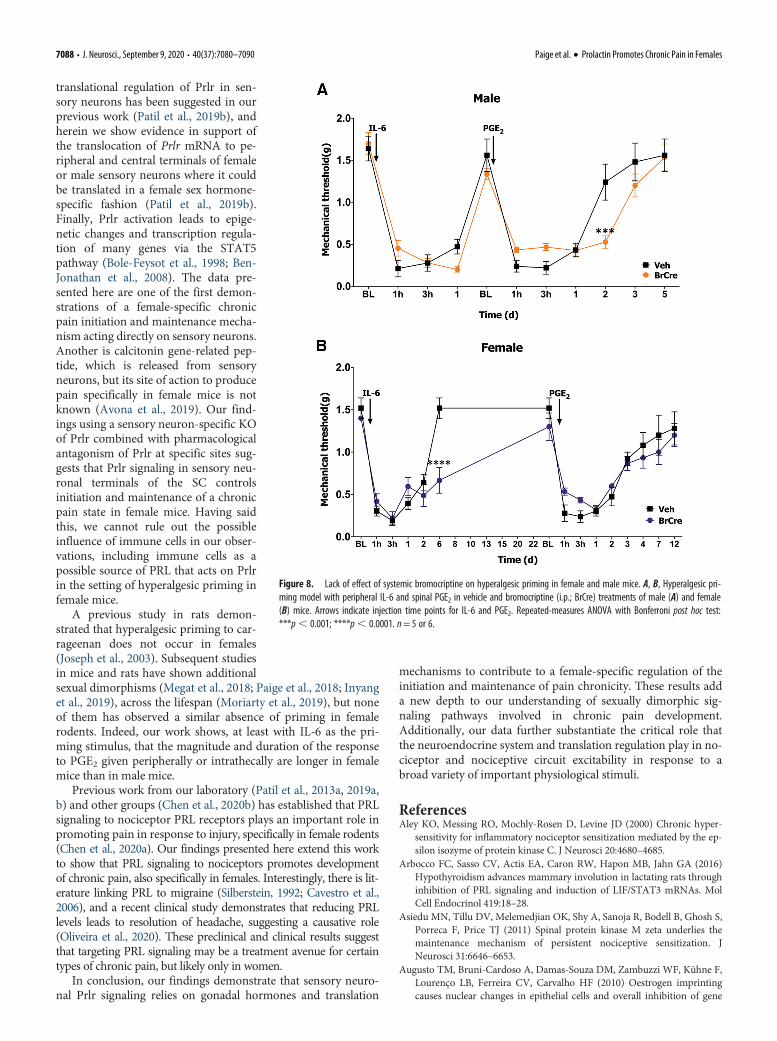
The conventional view is that endoge-nous PRL comes almost exclusively fromthe pituitary gland (Ben-Jonathan et al.,2008). However, extrapituitary sourcesfor PRL have been reported, and thesePRL sources are especially abundant inhumans (Ben-Jonathan et al., 1996). Inrodents, inflammation and tissue injury cause an increase inPRL in the paw and SC (Scotland et al., 2011; Patil et al.,2013a). We examined whether endogenous pituitary PRL isinvolved in the regulation of pain chronicity in male andfemale mice. Circulating PRL that has originated in the pitui-tary can be removed by either hypophysectomy (Green et al.,2016) or systemic treatment with bromocriptine (Grattan, 2015).Both approaches have downsides, but we opted to use thesystemic bromocriptine approach because bromocriptine isused in clinical studies and hypophysectomy drasticallyaffects gonadal hormone production. We systemicallytreated both male and female mice with bromocriptine aspreviously described (Yip et al., 2012). Removal of endoge-nous pituitary PRL did not influence mechanical hypersensi-tivity during the IL-6 phase of hyperalgesic priminginitiation in male mice but did slightly prolong the PGE2 pre-cipitated hypersensitivity (repeated-measures ANOVA; F(10,90) =2.980; p=0.0028; n = 5 or 6; Fig. 8A). In female mice, the IL-6response was enhanced in the bromocriptine-treatedfemales, but the priming response to PGE2 was equivalent in ve-hicle and bromocriptine-treated mice (repeated-measuresANOVA; F(14,126) = 3.127; p = 0.0003; n = 5 or 6; Fig. 8B).
Collectively, these results suggest that endogenous extrapituitaryPRL signaling plays a key role in hyperalgesic priming in femalemice. During initiation of chronic pain, this source can be periph-eral or central, but the crucial source of PRL during the mainte-nance of pain chronicity is likely central.
DiscussionStudies in both animals and humans demonstrate sexuallydimorphic mechanisms controlling the development and resolu-tion of chronic pain (Joseph et al., 2003; Sorge et al., 2015; Nasiret al., 2016; Taves et al., 2016; Lopes et al., 2017; Rosen et al.,2017, 2019; Mapplebeck et al., 2018; Paige et al., 2018; Dance,2019; North et al., 2019; Patil et al., 2019b; Ray et al., 2019).Among these sex differences, several factors have been discov-ered that drive chronic pain specifically in males (Sorge et al.,2015; Taves et al., 2016; Mapplebeck et al., 2018; Megat et al.,2018; Paige et al., 2018; Shiers et al., 2018; Martin et al., 2019),but relatively little is known about such chronic pain mecha-nisms in females. There is evidence that these mechanisms areclosely regulated by gonadal hormones (Traub and Ji, 2013). Forinstance, the apparent male-specific effect of microglia-drivenP2X4 signaling in neuropathic pain can be conferred to females
Figure 6. Regulation of hyperalgesic priming by sensory neuronal Prlr selectively in female mice. A, B, Hyperalgesicpriming model with peripheral IL-6/Veh and spinal PGE2 in Prlr
fl/fl (Prlr-lox; control) and Nav1.8cre/-/Prlrfl/fl (Prlr CKO) inmale (A) and female (B) mice. Arrows indicate injection time points for IL-6/Veh and PGE2. Repeated-measures ANOVAwith Bonferroni post hoc test: A, ****p, 0.001, for Prlr-lox compared with Veh-Prlr-lox; and$$$$p, 0.0001;$$$p, 0.001, for Prlr CKO compared with Veh-Prlr-lox. B, *p, 0.05; **p, 0.01; ***p, 0.001. n= 4–7.
7086 • J. Neurosci., September 9, 2020 • 40(37):7080–7090 Paige et al. · Prolactin Promotes Chronic Pain in Females
with testosterone treatment (Sorge et al., 2015). In humans, sexdifferences in tibial nerve transcriptomes also demonstrate a sig-nature for gonadal hormone influence on sensory neuronal tran-scriptomes across the lifespan in females (Ray et al., 2019).Experiments described here clearly demonstrate differential rolesof gonadal hormones in development of chronicity in painfulconditions with estrogen exacerbating priming effects and testos-terone playing a protective role.
Additionally, we demonstrated that translation regulationplays a sex-specific role in the maintenance of chronic pain infemale mice. This is especially relevant considering that transla-tion control mechanisms are known contributors to the sensiti-zation of nociceptors (Khoutorsky and Price, 2018; Megat andPrice, 2018). Our previous work demonstrated that disruption
of translation regulation signaling in theperiphery or SC was only capable of inter-fering with hyperalgesic priming if thesetreatments were given at the time of thepriming event (Melemedjian et al., 2010,2014; Asiedu et al., 2011). However, thesepreviously published experiments weredone entirely in male mice. Our data sug-gest that targeting these translation regula-tion mechanisms for the treatment of painmay have additional therapeutic benefits inwomen. A potential explanation for thisdifferential effect on translation machineryin females is an effect of estrogen on trans-lation machinery (Bronson et al., 2010;Ochnik et al., 2016). E-2-dependent con-nections between translational control ofthe suppressor of cytokine signaling andmTOR phosphorylation (Augusto et al.,2010) or regulation of Rheb signaling(Pochynyuk et al., 2006) have been pro-posed (Matthews et al., 2005; Arbocco et al.,2016). These signaling pathways also playkey roles in the excitability of nociceptors(Moy et al., 2017; Khoutorsky and Price,2018; Megat et al., 2019a,b) and may play amore prominent role in the maintenance ofpersistent nociceptor plasticity in femalesthan males.
Previous work indicates that sex-depend-ent mechanisms regulating hypersensitivity ininflammatory and neuropathic pain condi-tions can be attributed to distinct immunecell types: microglia in males (Sorge et al.,2011, 2015, Taves et al., 2016; Paige et al.,2018) and T cells in females (Sorge et al.,2015). Importantly, there is an opinion thatthese cells are regulated by gonadal hor-mones. Together, key molecules involved insex-dependent regulation of the initiation,maintenance, and resolution would need to(1) be controlled by gonadal hormones, (2)induced by injury, (3) regulate immune cells,(4) undergo local translation control, and (5)be capable of regulating many other genes.The neuroendocrine hormone PRL and its re-ceptor Prlr fit all these requirements. First,Prlr-mediated PRL effects are sex- and go-nadal hormone-dependent in many tissuesand cell types, including sensory neurons
(Torner et al., 2001; Ben-Jonathan et al., 2008; Belugin et al.,2013; Patil et al., 2013a, 2019a,b). Second, many clinical and pre-clinical studies show that endogenous release of PRL from bothpituitary and extrapituitary origins is induced by inflammationand tissue injury (Chernow et al., 1987; Noreng et al., 1987; Ben-Jonathan et al., 1996; Yardeni et al., 2007; Scotland et al., 2011;Patil et al., 2013a). Third, PRL is an effective direct and/or indi-rect activator of immune cells, especially macrophages and Tcells (Matera et al., 2001; Savino et al., 2016; Tang et al., 2017).Moreover, many chronic autoimmune diseases affect femalesmore frequently than males, and a potential role for PRL may inpart explain this phenomenon for certain autoimmune diseases,such as lupus (Tang et al., 2017; Rizzetto et al., 2018). Fourth,
Figure 7. Regulation of the initiation and maintenance of hyperalgesic priming by peripheral and spinal Prlrin female mice. Hyperalgesic priming model in female mice with peripheral IL-6 and spinal PGE2. A, Schematicof injection locations and timing. B, Vehicle (no DPRL), Prlr antagonist (DPRL; 5 mg) was coadministered withIL-6 in paw or SC. C, Schematic of injection locations and timing. D, Vehicle (no DPRL) was injected into paw.DPRL (5 mg) was given into the paw or SC 30 min before spinal PGE2. Repeated-measures ANOVA withBonferroni post hoc test: *p, 0.05; ****p, 0.0001, for DPRL intrathecally compared with no DPRL; and$$$$p, 0.0001; $p, 0.05, for DPRL intraplantarly compared with no DPRL; n = 5–7).
Paige et al. · Prolactin Promotes Chronic Pain in Females J. Neurosci., September 9, 2020 • 40(37):7080–7090 • 7087
translational regulation of Prlr in sen-sory neurons has been suggested in ourprevious work (Patil et al., 2019b), andherein we show evidence in support ofthe translocation of Prlr mRNA to pe-ripheral and central terminals of femaleor male sensory neurons where it couldbe translated in a female sex hormone-specific fashion (Patil et al., 2019b).Finally, Prlr activation leads to epige-netic changes and transcription regula-tion of many genes via the STAT5pathway (Bole-Feysot et al., 1998; Ben-Jonathan et al., 2008). The data pre-sented here are one of the first demon-strations of a female-specific chronicpain initiation and maintenance mecha-nism acting directly on sensory neurons.Another is calcitonin gene-related pep-tide, which is released from sensoryneurons, but its site of action to producepain specifically in female mice is notknown (Avona et al., 2019). Our find-ings using a sensory neuron-specific KOof Prlr combined with pharmacologicalantagonism of Prlr at specific sites sug-gests that Prlr signaling in sensory neu-ronal terminals of the SC controlsinitiation and maintenance of a chronicpain state in female mice. Having saidthis, we cannot rule out the possibleinfluence of immune cells in our obser-vations, including immune cells as apossible source of PRL that acts on Prlrin the setting of hyperalgesic priming infemale mice.
A previous study in rats demon-strated that hyperalgesic priming to car-rageenan does not occur in females(Joseph et al., 2003). Subsequent studiesin mice and rats have shown additionalsexual dimorphisms (Megat et al., 2018; Paige et al., 2018; Inyanget al., 2019), across the lifespan (Moriarty et al., 2019), but noneof them has observed a similar absence of priming in femalerodents. Indeed, our work shows, at least with IL-6 as the pri-ming stimulus, that the magnitude and duration of the responseto PGE2 given peripherally or intrathecally are longer in femalemice than in male mice.
Previous work from our laboratory (Patil et al., 2013a, 2019a,b) and other groups (Chen et al., 2020b) has established that PRLsignaling to nociceptor PRL receptors plays an important role inpromoting pain in response to injury, specifically in female rodents(Chen et al., 2020a). Our findings presented here extend this workto show that PRL signaling to nociceptors promotes developmentof chronic pain, also specifically in females. Interestingly, there is lit-erature linking PRL to migraine (Silberstein, 1992; Cavestro et al.,2006), and a recent clinical study demonstrates that reducing PRLlevels leads to resolution of headache, suggesting a causative role(Oliveira et al., 2020). These preclinical and clinical results suggestthat targeting PRL signaling may be a treatment avenue for certaintypes of chronic pain, but likely only in women.
In conclusion, our findings demonstrate that sensory neuro-nal Prlr signaling relies on gonadal hormones and translation
mechanisms to contribute to a female-specific regulation of theinitiation and maintenance of pain chronicity. These results adda new depth to our understanding of sexually dimorphic sig-naling pathways involved in chronic pain development.Additionally, our data further substantiate the critical role thatthe neuroendocrine system and translation regulation play in no-ciceptor and nociceptive circuit excitability in response to abroad variety of important physiological stimuli.
ReferencesAley KO, Messing RO, Mochly-Rosen D, Levine JD (2000) Chronic hyper-
sensitivity for inflammatory nociceptor sensitization mediated by the ep-silon isozyme of protein kinase C. J Neurosci 20:4680–4685.
Arbocco FC, Sasso CV, Actis EA, Caron RW, Hapon MB, Jahn GA (2016)Hypothyroidism advances mammary involution in lactating rats throughinhibition of PRL signaling and induction of LIF/STAT3 mRNAs. MolCell Endocrinol 419:18–28.
Asiedu MN, Tillu DV, Melemedjian OK, Shy A, Sanoja R, Bodell B, Ghosh S,Porreca F, Price TJ (2011) Spinal protein kinase M zeta underlies themaintenance mechanism of persistent nociceptive sensitization. JNeurosci 31:6646–6653.
Augusto TM, Bruni-Cardoso A, Damas-Souza DM, Zambuzzi WF, Kühne F,Lourenço LB, Ferreira CV, Carvalho HF (2010) Oestrogen imprintingcauses nuclear changes in epithelial cells and overall inhibition of gene
Figure 8. Lack of effect of systemic bromocriptine on hyperalgesic priming in female and male mice. A, B, Hyperalgesic pri-ming model with peripheral IL-6 and spinal PGE2 in vehicle and bromocriptine (i.p.; BrCre) treatments of male (A) and female(B) mice. Arrows indicate injection time points for IL-6 and PGE2. Repeated-measures ANOVA with Bonferroni post hoc test:***p, 0.001; ****p, 0.0001. n= 5 or 6.
7088 • J. Neurosci., September 9, 2020 • 40(37):7080–7090 Paige et al. · Prolactin Promotes Chronic Pain in Females

transcription and protein synthesis in rat ventral prostate. Int J Androl33:675–685.
Avona A, Burgos-Vega C, Burton MD, Akopian AN, Price TJ, Dussor G(2019) Dural calcitonin gene-related peptide produces female-specificresponses in rodent migraine models. J Neurosci 39:4323–4331.
Belugin S, Diogenes AR, Patil MJ, Ginsburg E, Henry MA, Akopian AN(2013) Mechanisms of transient signaling via short and long prolactin re-ceptor isoforms in female and male sensory neurons. J Biol Chem288:34943–34955.
Ben-Jonathan N, Mershon JL, Allen DL, Steinmetz RW (1996) Extrapituitaryprolactin: distribution, regulation, functions, and clinical aspects. EndocrRev 17:639–669.
Ben-Jonathan N, LaPensee CR, LaPensee EW (2008) What can we learnfrom rodents about prolactin in humans? Endocr Rev 29:1–41.
Berkley KJ (1997) Sex differences in pain. Behav Brain Sci 20:371–380; dis-cussion 435-513.
Bernichtein S, Kayser C, Dillner K, Moulin S, Kopchick JJ, Martial JA,Norstedt G, Isaksson O, Kelly PA, Goffin V (2003) Development of pureprolactin receptor antagonists. J Biol Chem 278:35988–35999.
Bole-Feysot C, Goffin V, Edery M, Binart N, Kelly PA (1998) Prolactin (PRL)and its receptor: actions, signal transduction pathways and phenotypesobserved in PRL receptor knockout mice. Endocr Rev 19:225–268.
Bronson MW, Hillenmeyer S, Park RW, Brodsky AS (2010) Estrogen coordi-nates translation and transcription, revealing a role for NRSF in humanbreast cancer cells. Mol Endocrinol 24:1120–1135.
Brown RS, Kokay IC, Phillipps HR, Yip SH, Gustafson P, Wyatt A, LarsenCM, Knowles P, Ladyman SR, LeTissier P, Grattan DR (2016)Conditional deletion of the prolactin receptor reveals functional subpo-pulations of dopamine neurons in the arcuate nucleus of the hypothala-mus. J Neurosci 36:9173–9185.
Caligioni CS (2009) Assessing reproductive status/stages in mice. CurrProtoc Neurosci Appendix 4:Appendix 4I.
Cavestro C, Rosatello A, Marino MP, Micca G, Asteggiano G (2006) Highprolactin levels as a worsening factor for migraine. J Headache Pain 7:83–89.
Chaplan SR, Bach FW, Pogrel JW, Chung JM, Yaksh TL (1994) Quantitativeassessment of tactile allodynia in the rat paw. J Neurosci Methods 53:55–63.
Chen Y, Navratilova E, Dodick DW, Porreca F (2020a) An emerging role forprolactin in female-selective pain. Trends Neurosci 43:635–648.
Chen Y, Moutal A, Navratilova E, Kopruszinski C, Yue X, Ikegami M, ChowM, Kanazawa I, Bellampalli SS, Xie J, Patwardhan A, Rice K, Fields H,Akopian A, Neugebauer V, Dodick D, Khanna R, Porreca F (2020b) Theprolactin receptor long isoform regulates nociceptor sensitization andopioid-induced hyperalgesia selectively in females. Sci Transl Med 12:eaay7550.
Chernow B, Alexander HR, Smallridge RC, Thompson WR, Cook D,Beardsley D, Fink MP, Lake CR, Fletcher JR (1987) Hormonal responsesto graded surgical stress. Arch Intern Med 147:1273–1278.
Childs GV, Unabia G, Miller BT, Collins TJ (1999) Differential expression ofgonadotropin and prolactin antigens by GHRH target cells from maleand female rats. J Endocrinol 162:177–187.
Dance A (2019) Why the sexes don’t feel pain the same way. Nature567:448–450.
Diogenes A, Patwardhan AM, Jeske NA, Ruparel NB, Goffin V, AkopianAN, Hargreaves KM (2006) Prolactin modulates TRPV1 in female rat tri-geminal sensory neurons. J Neurosci 26:8126–8136.
Ferrari LF, Araldi D, Levine JD (2015) Distinct terminal and cell body mech-anisms in the nociceptor mediate hyperalgesic priming. J Neurosci35:6107–6116.
Fillingim RB, King CD, Ribeiro-Dasilva MC, Rahim-Williams B, Riley JL(2009) Sex, gender, and pain: a review of recent clinical and experimentalfindings. J Pain 10:447–485.
Grattan DR (2015) 60 years of neuroendocrinology: the hypothalamo-prolac-tin axis. J Endocrinol 226:T101–T122.
Green DP, Patil MJ, Akopian AN (2016) Influence of hypophysectomy, ovar-iectomy and gonadectomy on postoperative hypersensitivity in rats. GlobAnesth Perioper Med 2:171–175.
Houghton LA, Lea R, Jackson N, Whorwell PJ (2002) The menstrual cycleaffects rectal sensitivity in patients with irritable bowel syndrome but nothealthy volunteers. Gut 50:471–474.
Inyang KE, McDougal TA, Ramirez ED, Williams M, Laumet G, KavelaarsA, Heijnen CJ, Burton M, Dussor G, Price TJ (2019) Alleviation of pacli-taxel-induced mechanical hypersensitivity and hyperalgesic priming withAMPK activators in male and female mice. Neurobiol Pain 6:100037.
Joseph EK, Parada CA, Levine JD (2003) Hyperalgesic priming in the ratdemonstrates marked sexual dimorphism. Pain 105:143–150.
Khoutorsky A, Price TJ (2018) Translational control mechanisms in persis-tent pain. Trends Neurosci 41:100–114.
Kim JY, Megat S, Moy JK, Asiedu MN, Mejia GL, Vagner J, Price TJ (2016)Neuroligin 2 regulates spinal GABAergic plasticity in hyperalgesic pri-ming, a model of the transition from acute to chronic pain. Pain157:1314–1324.
LeResche L, Mancl L, Sherman JJ, Gandara B, Dworkin SF (2003) Changes intemporomandibular pain and other symptoms across the menstrualcycle. Pain 106:253–261.
Liu TT, Qu ZW, Ren C, Gan X, Qiu CY, Hu WP (2016) Prolactin potentiatesthe activity of acid-sensing ion channels in female rat primary sensoryneurons. Neuropharmacology 103:174–182.
Lopes DM, Malek N, Edye M, Jager SB, McMurray S, McMahon SB, Denk F(2017) Sex differences in peripheral not central immune responses topain-inducing injury. Sci Rep 7:16460.
Mapplebeck JC, Dalgarno R, Tu Y, Moriarty O, Beggs S, Kwok CH, HalievskiK, Assi S, Mogil JS, Trang T, Salter MW (2018) Microglial P2X4R-evokedpain hypersensitivity is sexually dimorphic in rats. Pain 159:1752–1763.
Martin LJ, Acland EL, Cho C, Gandhi W, Chen D, Corley E, Kadoura B,Levy T, Mirali S, Tohyama S, Khan S, MacIntyre LC, Carlson EN,Schweinhardt P, Mogil JS (2019) Male-specific conditioned pain hyper-sensitivity in mice and humans. Curr Biol 29:192–201.e194.
Matera L, Mori M, Galetto A (2001) Effect of prolactin on the antigen pre-senting function of monocyte-derived dendritic cells. Lupus 10:728–734.
Mathew PG, Dun EC, Luo JJ (2013) A cyclic pain: the pathophysiology andtreatment of menstrual migraine. Obstet Gynecol Surv 68:130–140.
Matthews J, Almlof T, Kietz S, Leers J, Gustafsson JA (2005) Estrogen recep-tor-alpha regulates SOCS-3 expression in human breast cancer cells.Biochem Biophys Res Commun 335:168–174.
Megat S, Price TJ (2018) Therapeutic opportunities for pain medicines viatargeting of specific translation signaling mechanisms. Neurobiol Pain4:8–19.
Megat S, Shiers S, Moy JK, Barragan-Iglesias P, Pradhan G, Seal RP, DussorG, Price TJ (2018) A critical role for dopamine D5 receptors in painchronicity in male mice. J Neurosci 38:379–397.
Megat S, Ray PR, Tavares-Ferreira D, Moy JK Sankaranarayanan I,Wanghzou A, Lou F, Barragan-Iglesias T, Campbell P, Dussor G, PriceTJ (2019a) Differences between dorsal root and trigeminal ganglion noci-ceptors in mice revealed by translational profiling. J Neurosci 39:6829–6847.
Megat S, Ray PR, Moy JK, Lou TF, Barragan-Iglesias P, Li Y, Pradhan G,Wanghzou A, Ahmad A, Burton MD, North RY, Dougherty PM,Khoutorsky A, Sonenberg N, Webster KR, Dussor G, Campbell ZT, PriceTJ (2019b) Nociceptor translational profiling reveals the Ragulator-RagGTPase complex as a critical generator of neuropathic pain. J Neurosci39:393–411.
Melemedjian OK, Asiedu MN, Tillu DV, Peebles KA, Yan J, Ertz N, DussorGO, Price TJ (2010) IL-6- and NGF-induced rapid control of protein syn-thesis and nociceptive plasticity via convergent signaling to the eIF4Fcomplex. J Neurosci 30:15113–15123.
Melemedjian OK, Tillu DV, Moy JK, Asiedu MN, Mandell EK, Ghosh S,Dussor G, Price TJ (2014) Local translation and retrograde axonal trans-port of CREB regulates IL-6-induced nociceptive plasticity. Mol Pain10:45.
Mogil JS, Sorge RE, LaCroix-Fralish ML, Smith SB, Fortin A, Sotocinal SG,Ritchie J, Austin JS, Schorscher-Petcu A, Melmed K, Czerminski J,Bittong RA, Mokris JB, Neubert JK, Campbell CM, Edwards RR,Campbell JN, Crawley JN, Lariviere WR, Wallace MR, et al. (2011) Painsensitivity and vasopressin analgesia are mediated by a gene-sex-environ-ment interaction. Nat Neurosci 14:1569–1573.
Moriarty O, Tu YS, Sengar AS, Salter MW, Beggs S, Walker SM (2019)Priming of adult incision response by early-life injury: neonatal micro-glial inhibition has persistent but sexually dimorphic effects in adult rats.J Neurosci 39:3081–3093.
Moy JK, Khoutorsky A, Asiedu MN, Black BJ, Kuhn JL, Barragan-Iglesias P,Megat S, Burton MD, Burgos-Vega CC, Melemedjian OK, Boitano S,
Paige et al. · Prolactin Promotes Chronic Pain in Females J. Neurosci., September 9, 2020 • 40(37):7080–7090 • 7089

Vagner J, Gkogkas CG, Pancrazio JJ, Mogil JS, Dussor G, Sonenberg N,Price TJ (2017) The MNK-eIF4E signaling axis contributes to injury-induced nociceptive plasticity and the development of chronic pain. JNeurosci 37:7481–7499.
Nasir H, Mahboubi H, Gyawali S, Ding S, Mickeviciute A, Ragavendran JV,Laferriere A, Stochaj U, Coderre TJ (2016) Consistent sex-dependenteffects of PKMzeta gene ablation and pharmacological inhibition on themaintenance of referred pain. Mol Pain 12:174480691667534.
Nettleship JE, Jones TH, Channer KS, Jones RD (2007) Physiological testos-terone replacement therapy attenuates fatty streak formation andimproves high-density lipoprotein cholesterol in the Tfmmouse: an effectthat is independent of the classic androgen receptor. Circulation116:2427–2434.
Noreng MF, Jensen P, Tjellden NU (1987) Per- and postoperative changes inthe concentration of serum thyreotropin under general anaesthesia, com-pared to general anaesthesia with epidural analgesia. Acta AnaesthesiolScand 31:292–294.
North RY, Li Y, Ray P, Rhines LD, Tatsui CE, Rao G, Johansson CA, ZhangH, Kim YH, Zhang B, Dussor G, Kim TH, Price TJ, Dougherty PM(2019) Electrophysiological and transcriptomic correlates of neuropathicpain in human dorsal root ganglion neurons. Brain 142:1215–1226.
Ochnik AM, Peterson MS, Avdulov SV, Oh AS, Bitterman PB, Yee D (2016)Amplified in breast cancer regulates transcription and translation inbreast cancer cells. Neoplasia 18:100–110.
Oliveira MD, Barea LM, Horn AP, Ongaratti BR, Soares JO, Araujo B, SantosTM, Rech CL, Pereira-Lima JF (2020) Resolution of headache after reduc-tion of prolactin levels in hyperprolactinemic patients. Arq Neuropsiquiatr78:28–33.
Paige C, Maruthy GB, Mejia G, Dussor G, Price T (2018) Spinal inhibition ofP2XR or p38 signaling disrupts hyperalgesic priming in male, but notfemale, mice. Neuroscience 385:133–142.
Patil MJ, Green DP, Henry MA, Akopian AN (2013a) Sex-dependent roles ofprolactin and prolactin receptor in postoperative pain and hyperalgesiain mice. Neuroscience 253:132–141.
Patil MJ, Ruparel SB, Henry MA, Akopian AN (2013b) Prolactin regulatesTRPV1, TRPA1, and TRPM8 in sensory neurons in a sex-dependentmanner: contribution of prolactin receptor to inflammatory pain. Am JPhysiol Endocrinol Metab 305:E1154–E1164.
Patil MJ, Hovhannisyan AH, Wangzhou A, Mecklenburg J, Koek W, GoffinV, Grattan D, Boehm U, Dussor G, Price TJ, Akopian AN (2019a)Prolactin receptor expression in mouse dorsal root ganglia neuronal sub-types is sex-dependent. J Neuroendocrinol 31:e12759.
Patil MJ, Belugin S, Mecklenburg J, Wangzhou A, Paige C, Barba-EscobedoPA, Boyd JT, Goffin V, Grattan D, Boehm U, Dussor G, Price TJ,Akopian AN (2019b) Prolactin regulates pain responses via a female-selective nociceptor-specific mechanism. iScience 20:449–465.
Pi X, Voogt JL (2002) Sex difference and estrous cycle: expression of prolactinreceptor mRNA in rat brain. Brain Res Mol Brain Res 103:130–139.
Pochynyuk O, Medina J, Gamper N, Genth H, Stockand JD, Staruschenko A(2006) Rapid translocation and insertion of the epithelial Na1 channel inresponse to RhoA signaling. J Biol Chem 281:26520–26527.
Price TJ, Inyang KE (2015) Commonalities between pain and memory mech-anisms and their meaning for understanding chronic pain. Prog Mol BiolTransl Sci 131:409–434.
Ray PR, Khan J, Wangzhou A, Tavares-Ferreira D, Akopian AN, Dussor G,Price TJ (2019) Transcriptome analysis of the human tibial nerve identi-fies sexually dimorphic expression of genes involved in pain, inflamma-tion, and neuro-immunity. Front Mol Neurosci 12:37.
Rizzetto L, Fava F, Tuohy KM, Selmi C (2018) Connecting the immune sys-tem, systemic chronic inflammation and the gut microbiome: the role ofsex. J Autoimmun 92:12–34.
Rosen S, Ham B, Mogil JS (2017) Sex differences in neuroimmunity andpain. J Neurosci Res 95:500–508.
Rosen SF, Ham B, Haichin M, Walters IC, Tohyama S, Sotocinal SG, MogilJS (2019) Increased pain sensitivity and decreased opioid analgesia in T-
cell-deficient mice and implications for sex differences. Pain 160:358–366.
Rouet V, Bogorad RL, Kayser C, Kessal K, Genestie C, Bardier A, GrattanDR, Kelder B, Kopchick JJ, Kelly PA, Goffin V (2010) Local prolactin is atarget to prevent expansion of basal/stem cells in prostate tumors. ProcNatl Acad Sci USA 107:15199–15204.
Savino W, Mendes-da-Cruz DA, Lepletier A, Dardenne M (2016) Hormonalcontrol of T-cell development in health and disease. Nat Rev Endocrinol12:77–89.
Scotland PE, Patil M, Belugin S, Henry MA, Goffin V, Hargreaves KM,Akopian AN (2011) Endogenous prolactin generated during peripheralinflammation contributes to thermal hyperalgesia. Eur J Neurosci34:745–754.
Shiers S, Pradhan G, Mwirigi J, Mejia G, Ahmad A, Kroener S, Price T (2018)Neuropathic pain creates an enduring prefrontal cortex dysfunction cor-rected by the type II diabetic drug metformin but not by gabapentin. JNeurosci 38:7337–7350.
Silberstein SD (1992) The role of sex hormones in headache. Neurology42:37–42.
Slade GD, Bair E, By K, Mulkey F, Baraian C, Rothwell R, Reynolds M, MillerV, Gonzalez Y, Gordon S, Ribeiro-Dasilva M, Lim PF, Greenspan JD,Dubner R, Fillingim RB, Diatchenko L, Maixner W, Dampier D, KnottC, Ohrbach R (2011) Study methods, recruitment, sociodemographicfindings, and demographic representativeness in the OPPERA study. JPain 12:T12–T26.
Sorge RE, LaCroix-Fralish ML, Tuttle AH, Sotocinal SG, Austin JS, Ritchie J,Chanda ML, Graham AC, Topham L, Beggs S, Salter MW, Mogil JS(2011) Spinal cord Toll-like receptor 4 mediates inflammatory and neu-ropathic hypersensitivity in male but not female mice. J Neurosci31:15450–15454.
Sorge RE, Mapplebeck JC, Rosen S, Beggs S, Taves S, Alexander JK, MartinLJ, Austin JS, Sotocinal SG, Chen D, Yang M, Shi XQ, Huang H, PillonNJ, Bilan PJ, Tu Y, Klip A, Ji RR, Zhang J, Salter MW, et al. (2015)Different immune cells mediate mechanical pain hypersensitivity in maleand female mice. Nat Neurosci 18:1081–1083.
Tang MW, Garcia S, Gerlag DM, Tak PP, Reedquist KA (2017) Insight intothe endocrine system and the immune system: a review of the inflamma-tory role of prolactin in rheumatoid arthritis and psoriatic arthritis. FrontImmunol 8:720.
Taves S, Berta T, Liu DL, Gan S, Chen G, Kim YH, Van de Ven T, Laufer S, JiRR (2016) Spinal inhibition of p38 MAP kinase reduces inflammatoryand neuropathic pain in male but not female mice: sex-dependent micro-glial signaling in the spinal cord. Brain Behav Immun 55:70–81.
Torner L, Toschi N, Pohlinger A, Landgraf R, Neumann ID (2001)Anxiolytic and anti-stress effects of brain prolactin: improved efficacy ofantisense targeting of the prolactin receptor by molecular modeling. JNeurosci 21:3207–3214.
Traub RJ, Ji Y (2013) Sex differences and hormonal modulation of deep tis-sue pain. Front Neuroendocrinol 34:350–366.
Unruh AM (1996) Gender variations in clinical pain experience. Pain65:123–167.
Willis D, Li KW, Zheng JQ, Chang JH, Smit A, Kelly T, Merianda TT,Sylvester J, van Minnen J, Twiss JL (2005) Differential transport and localtranslation of cytoskeletal, injury-response, and neurodegeneration pro-tein mRNAs in axons. J Neurosci 25:778–791.
Willis DE, van Niekerk EA, Sasaki Y, Mesngon M, Merianda TT, WilliamsGG, Kendall M, Smith DS, Bassell GJ, Twiss JL (2007) Extracellular stim-uli specifically regulate localized levels of individual neuronal mRNAs. JCell Biol 178:965–980.
Yardeni IZ, Shavit Y, Bessler H, Mayburd E, Grinevich G, Beilin B (2007)Comparison of postoperative pain management techniques on endocrineresponse to surgery: a randomised controlled trial. Int J Surg 5:239–243.
Yip SH, Eguchi R, Grattan DR, Bunn SJ (2012) Prolactin signalling in themouse hypothalamus is primarily mediated by signal transducer and acti-vator of transcription factor 5b but not 5a. J Neuroendocrinol 24:1484–1491.
7090 • J. Neurosci., September 9, 2020 • 40(37):7080–7090 Paige et al. · Prolactin Promotes Chronic Pain in Females