Development/Plasticity/Repair InVivo ... · embryo by injection of a sodium channel blocker or GABA...
9
Development/Plasticity/Repair In Vivo Synaptic Scaling Is Mediated by GluA2-Lacking AMPA Receptors in the Embryonic Spinal Cord Miguel Angel Garcia-Bereguiain, Carlos Gonzalez-Islas, Casie Lindsly, Ellie Butler, Atlantis Wilkins Hill, and Peter Wenner Department of Physiology, Emory University School of Medicine, Atlanta, Georgia 30322 When spiking activity within a network is perturbed for hours to days, compensatory changes in synaptic strength are triggered that are thought to be important for the homeostatic maintenance of network or cellular spiking activity. In one form of this homeostatic plasticity, called synaptic scaling, all of a cell’s AMPAergic miniature postsynaptic currents (mEPSCs) are increased or decreased by some scaling factor. Although synaptic scaling has been observed in a variety of systems, the mechanisms that underlie AMPAergic scaling have been controversial. Certain studies find that synaptic scaling is mediated by GluA2-lacking calcium receptors (CP-AMPARs), whereas others have found that scaling is mediated by GluA2-containing calcium-impermeable receptors (CI-AMPARs). Spontaneous network activity is observed in most developing circuits, and in the spinal cord this activity drives embryonic movements. Blocking spontaneous network activity in the chick embryo by infusing lidocaine in vivo triggers synaptic scaling in spinal motoneurons; here we show that AMPAergic scaling occurs through increases in mEPSC conductance that appear to be mediated by the insertion of GluA2-lacking AMPA receptors at the expense of GluA2-containing receptors. We have previously reported that in vivo blockade of GABA A transmission, at a developmental stage when GABA is excitatory, also triggered AMPAergic synaptic scaling. Here, we show that this form of AMPAergic scaling is also mediated by CP-AMPARs. These findings suggest that AMPAergic scaling triggered by blocking spiking activity or GABA A receptor transmission represents similar phenomena, supporting the idea that activity blockade triggers scaling by reducing GABA A transmission. Introduction Homeostatic synaptic plasticity is the process of homeostatically maintaining activity levels through compensatory adjustments in synaptic strength (Rich and Wenner, 2007; Vitureira et al., 2012; Turrigiano, 2012). For example, when network activity was blocked in cultured neural networks for days, increases in the amplitude of excitatory miniature postsynaptic currents (mPSCs) and decreases in the amplitude of inhibitory mPSCs were observed (O’Brien et al., 1998; Turrigiano et al., 1998). These compensatory changes in mPSC amplitude occur through a multiplicative process where the entire distribution of ampli- tudes appears to be scaled by a multiplicative factor (synaptic scaling) (Turrigiano et al., 1998). The mechanisms that underlie compensatory changes in excitatory and inhibitory quantal am- plitude after activity blockade of cultured networks include changes in postsynaptic receptor number, subunit composition, and the amount of transmitter released per vesicle (Rich and Wenner, 2007; Turrigiano, 2008). However, much less is known about the mechanisms underlying synaptic scaling following ac- tivity perturbations in vivo. Synaptic scaling of AMPAergic mPSCs has been shown to be mediated by changes in postsynaptic AMPA receptors in several studies (Lee, 2012). However, identifying which receptor sub- units are involved has been a topic intense interest and debate as demonstrated by four separate reviews in the last two years that discuss the issue (Man, 2011; Lee, 2012; Shepherd, 2012; Turrigiano, 2012). Several studies in which network activity was blocked in cultured neurons suggest AMPAergic scaling was me- diated by GluA2-containing CI-AMPARs alone, whereas several other studies demonstrate the involvement of GluA2-lacking CP- AMPARs. Less is known about the mechanisms that mediate AMPAergic scaling after in vivo activity perturbations; however, evidence for and against the involvement of CP-AMPARs has been described in the developing visual system (Goel et al., 2006; Gainey et al., 2009; Goel et al., 2011). We have shown an in vivo form of AMPAergic synaptic scaling in the chick embryo spinal cord, where compensatory changes in synaptic strength appear to contribute to the maintenance of spontaneous network activity (SNA) (Gonzalez-Islas and Wenner, 2006). SNA is the product of a highly excitable develop- ing circuit where GABA is depolarizing and excitatory. SNA is observed in virtually all developing circuits and is thought to be important for the maturation of the synaptic networks in which it is expressed (O’Donovan et al., 1998; O’Donovan, 1999; Blankenship and Feller, 2010). In the spinal cord, SNA occurs as Received Aug. 20, 2012; revised Feb. 26, 2013; accepted March 8, 2013. Author contributions: M.A.G.-B., C.G.-I., C.L., and P.W. designed research; M.A.G.-B., C.G.-I., C.L., E.B., and A.W.H. performed research; M.A.G.-B., C.G.-I., C.L., and P.W. analyzed data; M.A.G.-B., C.G.-I., C.L., and P.W. wrote the paper. This work was supported by the National Institute of Neurological Disorders and Stroke, the Whitehall Founda- tion, and the Neilsen Foundation (P.W.). M.A.G.-B. was supported by a fellowship from Rafael del Pino Foundation. We thank Dr. Steve Traynelis for his valuable comments on the manuscript. The authors declare no competing financial interests. Correspondence should be addressed to Dr. Peter Wenner, Emory University School of Medicine, Department of Physiology, Room 601, Whitehead Building, Atlanta, GA 30322. E-mail: [email protected]. DOI:10.1523/JNEUROSCI.4025-12.2013 Copyright © 2013 the authors 0270-6474/13/336791-09$15.00/0 The Journal of Neuroscience, April 17, 2013 • 33(16):6791– 6799 • 6791
Transcript of Development/Plasticity/Repair InVivo ... · embryo by injection of a sodium channel blocker or GABA...
Development/Plasticity/Repair
In Vivo Synaptic Scaling Is Mediated by GluA2-LackingAMPA Receptors in the Embryonic Spinal Cord
Miguel Angel Garcia-Bereguiain, Carlos Gonzalez-Islas, Casie Lindsly, Ellie Butler, Atlantis Wilkins Hill,and Peter WennerDepartment of Physiology, Emory University School of Medicine, Atlanta, Georgia 30322
When spiking activity within a network is perturbed for hours to days, compensatory changes in synaptic strength are triggered that arethought to be important for the homeostatic maintenance of network or cellular spiking activity. In one form of this homeostaticplasticity, called synaptic scaling, all of a cell’s AMPAergic miniature postsynaptic currents (mEPSCs) are increased or decreased by somescaling factor. Although synaptic scaling has been observed in a variety of systems, the mechanisms that underlie AMPAergic scaling havebeen controversial. Certain studies find that synaptic scaling is mediated by GluA2-lacking calcium receptors (CP-AMPARs), whereasothers have found that scaling is mediated by GluA2-containing calcium-impermeable receptors (CI-AMPARs). Spontaneous networkactivity is observed in most developing circuits, and in the spinal cord this activity drives embryonic movements. Blocking spontaneousnetwork activity in the chick embryo by infusing lidocaine in vivo triggers synaptic scaling in spinal motoneurons; here we show thatAMPAergic scaling occurs through increases in mEPSC conductance that appear to be mediated by the insertion of GluA2-lacking AMPAreceptors at the expense of GluA2-containing receptors. We have previously reported that in vivo blockade of GABAA transmission, at adevelopmental stage when GABA is excitatory, also triggered AMPAergic synaptic scaling. Here, we show that this form of AMPAergicscaling is also mediated by CP-AMPARs. These findings suggest that AMPAergic scaling triggered by blocking spiking activity or GABAA
receptor transmission represents similar phenomena, supporting the idea that activity blockade triggers scaling by reducing GABAA
transmission.
IntroductionHomeostatic synaptic plasticity is the process of homeostaticallymaintaining activity levels through compensatory adjustments insynaptic strength (Rich and Wenner, 2007; Vitureira et al., 2012;Turrigiano, 2012). For example, when network activity wasblocked in cultured neural networks for days, increases in theamplitude of excitatory miniature postsynaptic currents(mPSCs) and decreases in the amplitude of inhibitory mPSCswere observed (O’Brien et al., 1998; Turrigiano et al., 1998).These compensatory changes in mPSC amplitude occur througha multiplicative process where the entire distribution of ampli-tudes appears to be scaled by a multiplicative factor (synapticscaling) (Turrigiano et al., 1998). The mechanisms that underliecompensatory changes in excitatory and inhibitory quantal am-plitude after activity blockade of cultured networks includechanges in postsynaptic receptor number, subunit composition,and the amount of transmitter released per vesicle (Rich and
Wenner, 2007; Turrigiano, 2008). However, much less is knownabout the mechanisms underlying synaptic scaling following ac-tivity perturbations in vivo.
Synaptic scaling of AMPAergic mPSCs has been shown to bemediated by changes in postsynaptic AMPA receptors in severalstudies (Lee, 2012). However, identifying which receptor sub-units are involved has been a topic intense interest and debate asdemonstrated by four separate reviews in the last two years thatdiscuss the issue (Man, 2011; Lee, 2012; Shepherd, 2012;Turrigiano, 2012). Several studies in which network activity wasblocked in cultured neurons suggest AMPAergic scaling was me-diated by GluA2-containing CI-AMPARs alone, whereas severalother studies demonstrate the involvement of GluA2-lacking CP-AMPARs. Less is known about the mechanisms that mediateAMPAergic scaling after in vivo activity perturbations; however,evidence for and against the involvement of CP-AMPARs hasbeen described in the developing visual system (Goel et al., 2006;Gainey et al., 2009; Goel et al., 2011).
We have shown an in vivo form of AMPAergic synaptic scalingin the chick embryo spinal cord, where compensatory changes insynaptic strength appear to contribute to the maintenanceof spontaneous network activity (SNA) (Gonzalez-Islas andWenner, 2006). SNA is the product of a highly excitable develop-ing circuit where GABA is depolarizing and excitatory. SNA isobserved in virtually all developing circuits and is thought to beimportant for the maturation of the synaptic networks in which itis expressed (O’Donovan et al., 1998; O’Donovan, 1999;Blankenship and Feller, 2010). In the spinal cord, SNA occurs as
Received Aug. 20, 2012; revised Feb. 26, 2013; accepted March 8, 2013.Author contributions: M.A.G.-B., C.G.-I., C.L., and P.W. designed research; M.A.G.-B., C.G.-I., C.L., E.B., and A.W.H.
performed research; M.A.G.-B., C.G.-I., C.L., and P.W. analyzed data; M.A.G.-B., C.G.-I., C.L., and P.W. wrote thepaper.
This work was supported by the National Institute of Neurological Disorders and Stroke, the Whitehall Founda-tion, and the Neilsen Foundation (P.W.). M.A.G.-B. was supported by a fellowship from Rafael del Pino Foundation.We thank Dr. Steve Traynelis for his valuable comments on the manuscript.
The authors declare no competing financial interests.Correspondence should be addressed to Dr. Peter Wenner, Emory University School of Medicine, Department of
Physiology, Room 601, Whitehead Building, Atlanta, GA 30322. E-mail: [email protected]:10.1523/JNEUROSCI.4025-12.2013
Copyright © 2013 the authors 0270-6474/13/336791-09$15.00/0
The Journal of Neuroscience, April 17, 2013 • 33(16):6791– 6799 • 6791
episodic bursts of activity, which drive embryonic movements(O’Donovan, 1999). Previously, we blocked SNA in the chickembryo by injection of a sodium channel blocker or GABAA an-tagonist in ovo for 2 d and observed a compensatory increase ofglutamatergic mEPSC amplitude in motoneurons (Gonzalez-Islas and Wenner, 2006; Wilhelm and Wenner, 2008). It is un-known whether CP-AMPARs are involved in the in vivoAMPAergic scaling described in the embryonic spinal cord, andhere we examine the possibility that CP-AMPARs mediate syn-aptic scaling at an early developmental stage when GABA isexcitatory.
Materials and MethodsDissection. Stage 36 (Hamburger and Hamilton, 1951) chick embryospinal cords (of either sex), with intact spinal nerves, were dissectedunder cooled (15°C) Tyrode’s solution containing the following (in mM):139 NaCl, 12 glucose, 17 NaHCO3, 3 KCl, 1 MgCl2, and 3 CaCl2) (for afull description, see Gonzalez-Islas and Wenner, 2006). After the dissec-tion, the cord was allowed to recover for at least 6 h in Tyrode’s solutionat 18°C. The cord was then transferred to a recording chamber andcontinuously perfused with Tyrode’s solution that was heated to 28°C.
Electrophysiology. Whole-cell patch-clamp recordings were made fromspinal motoneurons localized in lumbosacral segments 1–3 to assessmPSCs, as described previously (Gonzalez-Islas et al., 2010). Briefly,whole-cell recordings (electrodes, 5–10 M�) were obtained from anti-dromically identified motoneurons. Recordings were terminated when-ever significant increases in input resistance (�20%) occurred.Extracellular solution for mPSC recordings for CP465022 or N-naphthylacetylspermine (NASPM) experiments was Tyrode’s solution with anadditional 2 mM KCl (total 5 mM), TTX (1 �M), GABAA receptor antag-onist gabazine (5 �M), and NMDA receptor antagonist APV (50 �M).The intracellular patch solution for these experiments contained the fol-lowing (in mM): 5 NaCl, 100 K-gluconate, 30 KCl, 5 CsCl, 10 TEA-Cl, 10HEPES, 1 MgCl2, 0.1 CaCl2, 1 Na2ATP, 0.1 MgGTP. For I–V plots andrectification measurements, the solutions were as follows: intracellularsolution same as above with the following additions (in mM) 10 BAPTA,10 QX-314, and 0.1 verapamil; extracellular solution same as above withthe following additions (in mM) 30 TEA, 5 CsCl. Pipette solution osmo-larity was between 280 and 300 mOsm, and pH was adjusted to 7.3 withKOH. Junction potentials were corrected online. Currents were filteredonline at 5 kHz, digitized at 10 kHz.
I–V plots for mEPSCs were performed using an Axoclamp 2B, whereasall other whole-cell recordings were performed in a separate rig using anAxoclamp 200, which displayed better noise characteristics (see rootmean square [RMS] values in figure legends). RMS values were acquiredfor each cell, measured from 3 separate trace epochs devoid of mEPSCs.
mEPSC analysis. The mEPSCs were analyzed using Minianalysis soft-ware (Synaptosoft). Bar charts and associated average values were ob-tained by determining the average mEPSC amplitude for each cell(variable number of mEPSCs/cell, 5 pA cutoff), and then taking theaverage of all cells. Cumulative probability distribution shown in Figure1B were obtained by combining all the mEPSC amplitudes across cells incontrol or lidocaine-treated motoneurons. In addition, ranked mEPSCamplitude plots were constructed. Ranked plots were obtained by takingthe same number of mEPSC amplitudes (�30) from the same number ofcells in different conditions (control or treated motoneurons, before andafter adding NASPM). We then ranked them in an ascending mannerand plotted them against each other for comparison (e.g., ranked valuesfor control vs lidocaine-treated motoneurons; Fig. 1C). A strong linear fit(r � 0.98) suggested that all mEPSC amplitudes from one distributionwere multiplicatively related or scaled to the other distribution. If theslope, m, was � 1, then the amplitude distribution on the y-axis com-pared with the x-axis had increased by the multiplicative factor m.
In ovo drug injections. At embryonic day 8 (E8), lidocaine hydrochlo-ride aqueous solution (35 mg/ml plus 10 mM HEPES, pH 7.2) was con-tinuously applied onto the chorioallantoic membrane of the embryos ata rate of 13.5 �l/h, as described previously (Gonzalez-Islas and Wenner,2006); control embryos did not receive saline injections, as no difference
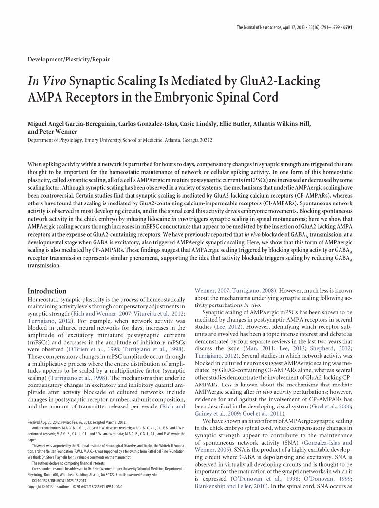
Figure 1. Lidocaine-treatedembryosexhibithigherfrequencyofSNAandupwardscalingofmEP-SCs compared with controls. A, Representative spinal nerve recordings showing the interval betweenepisodes of SNA in isolated spinal cords from control (black) and lidocaine-treated embryos (gray). B,Cumulative probability plot for glutamatergic mEPSC amplitudes from control and lidocaine-treatedmotoneurons (5 different cords, n � 400 in both cases). C, Ranked mEPSC amplitudes from activity-blocked motoneurons ( y-axis) plotted against corresponding ranked values of control motoneurons(x-axis). Black line represents expected result if amplitude distributions were the same.
6792 • J. Neurosci., April 17, 2013 • 33(16):6791– 6799 Garcia-Bereguiain et al. • CP-AMPARs Mediate Synaptic Scaling In Vivo
was observed between untreated and saline-treated embryos in this pre-vious study. For gabazine treatments, a single bolus of gabazine wasadded at E8 to reach a concentration of 10 �M in ovo, assuming a 50 mlvolume (Wilhelm and Wenner, 2008).
Drugs. Verapamil was purchased from Calbiochem; TTX, APV,CNQX, and CP465022 were purchased from Tocris Bioscience Cookson;CsCl was purchased from Fisher Scientific; BAPTA was purchased fromFluka; HEPES was purchased from Acros Organics. All other chemicalsand drugs were purchased from Sigma-Aldrich.
Immunoblot. Ventral half lumbosacral spinal cords were homogenizedin RIPA buffer supplemented with protease and phosphatase inhibitors(Sigma-Aldrich). Samples were then centrifuged at 16,000 � g for 5 minto remove cell debris. Protein concentration was quantitated using BCAreagent (Pierce). Samples were separated on 4 –15% SDS-PAGE andblotted to a nitrocellulose membrane. The primary antibodies to GluA1–GluA4 subunits were from Abcam (rabbit anti-GluA1 ab31232, rabbitanti-GluA2 ab87610, rabbit anti-GluA3 ab87609, rabbit anti-GluA4ab20673), and to actin (mouse anti-actin) from Sigma-Aldrich. The sec-ondary antibodies used were HRP-goat anti-mouse IgG and HRP-goatanti-rabbit IgG from Sigma-Aldrich. The blot was visualized by ECLchemiluminescence (GE Healthcare). Quantitative analysis of proteinexpression was performed by drawing boxes around the protein bandsand normalizing the signal intensity to actin intensity using ImageJ (Na-tional Institutes of Health). Blots were done in triplicate, and each samplerepresents a lysate of two or three different cords.
Statistics. Data are mean � SE. Most statistical analysis was performedusing a two-tailed Student’s t test (paired and unpaired) unless men-tioned otherwise. GraphPad Instat software and SigmaPlot 9 were usedfor statistical analysis.
ResultsmEPSC membrane conductance increases inactivity-blocked motoneuronsPrevious studies in our laboratory have shown that 2 d blockadeof embryonic movements in ovo led to increases in both SNAfrequency and AMPA/kainate mEPSC amplitude in motoneu-rons from the isolated cord. To block SNA in ovo, lidocaine, asodium channel blocker, was pumped continuously onto thechorioallantoic membrane of the chick from embryonic day 8 to10 (E8 –E10, stages 34 –36). Spinal cords were isolated and main-tained in recirculating Tyrode’s solution in the absence of lido-caine. As shown in Figure 1A, the interval between the bouts orepisodes of SNA was significantly reduced for lidocaine-treatedembryos (4.4 � 0.8 min, n � 6) compared with controls (9.3 �2.8 min, n � 5). Whole-cell recordings were obtained from spinalmotoneurons identified antidromically in either lidocaine-treated or control embryos; mPSCs were recorded in voltage-clamp at �70 mV, and mEPSCs were pharmacologically isolatedby bath application of gabazine (5 �M) and APV (50 �M). Theaverage amplitude of mEPSCs from motoneurons of lidocaine-treated embryos was 44% larger than in control (control, 7.0 �0.9 pA, n � 5; lidocaine-treated, 10.1 � 2.8 pA, n � 5; p � 0.01).These results were consistent with our previous study (Gonzalez-Islas and Wenner, 2006).
We also found that, after activity blockade, mEPSC ampli-tudes increased across the entire distribution (Fig. 1B). Indeed,mEPSC amplitudes underwent synaptic scaling as demonstratedin Figure 1C, which shows the plot of ranked mEPSC amplitudevalues for lidocaine-treated versus control motoneurons (r �0.99 and a slope value of 1.68 � 0.01, n � 9 cells; Fig. 1C). On theother hand, no significant change was found in the mEPSC fre-quency (1.05 � 0.47 Hz and 1.16 � 0.40 for control andlidocaine-treated embryos, respectively) or kinetics (5.8 � 1.3 msand 6.0 � 1.7 ms for control and lidocaine-treated embryos,respectively). In our previous study, we found that, after activity
blockade, AMPAergic mEPSC amplitude did not multiplica-tively scale and that mEPSC frequency was strongly increased(Gonzalez-Islas and Wenner, 2006). The discrepancy betweenour current and earlier study for these parameters is likely theresult of our improved recordings, which allow better mEPSCdetection (lower noise). In the previous study, it is likely thatmany small mEPSCs were not detected in control but were de-tected when amplitudes increased in activity-blocked motoneu-rons; this could explain the observed increase in mEPSCfrequency and result in our inability to detect multiplicative scal-ing because we were not comparing the same populations ofmEPSCs.
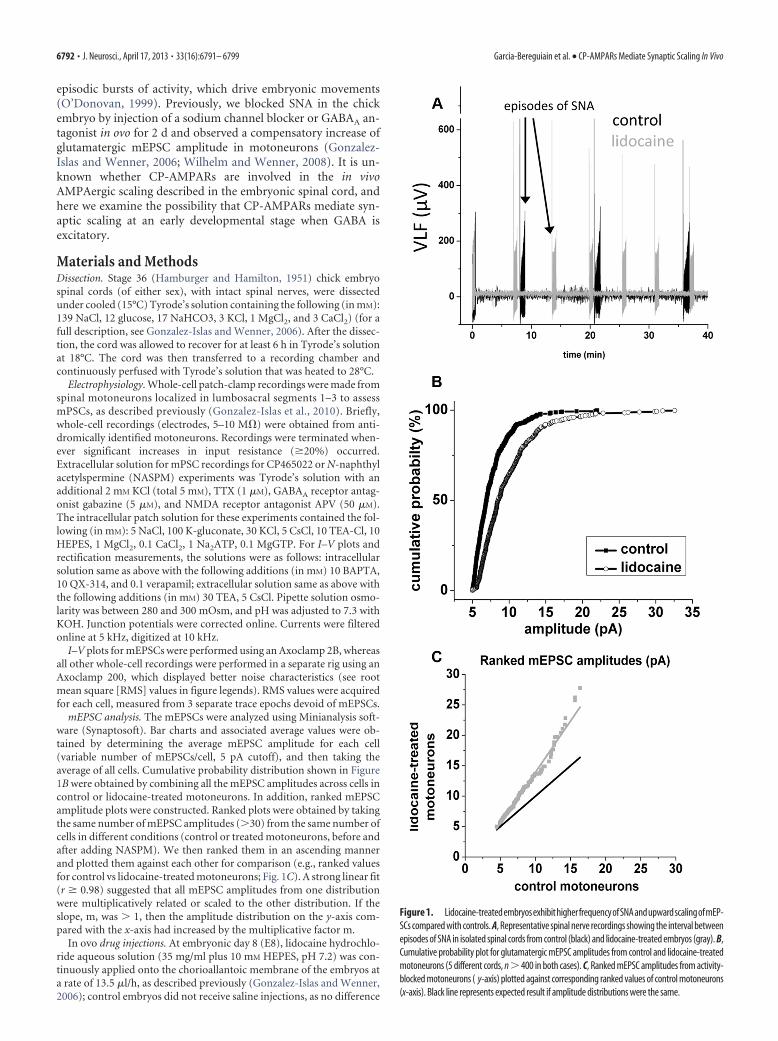
If mEPSC amplitude increases because of changes in postsyn-aptic AMPA receptors as others have shown, then we would ex-pect changes in the conductance of the total population ofchannels that mediate an mEPSC (will refer to as mEPSC con-ductance). To assess mEPSC conductance, we constructedmEPSC I–V plots using whole-cell recordings from control andactivity-blocked motoneurons (as described previously for GABA-ergic mPSCs) (Gonzalez-Islas et al., 2009). Voltage-gated channelsand NMDAergic and GABAergic transmission were blocked to iso-late non-NMDA glutamatergic mEPSCs, which were typically re-corded in voltage steps of 10 mV for 1–2 min (Fig. 2A). The averagepeak amplitude of mEPSCs at each step was then plotted againstvoltage, for control and lidocaine-treated embryos (Fig. 2B). Sperm-ine was not included in whole-cell patch solution to ensure thatglutamate receptors did not rectify at positive potentials. A linear fitof the data from �90 to 90 mV was then made, excluding datapoints from �30 to 30 mV as these amplitudes tended to be overes-timates as significant numbers of mEPSCs fell into the noise andwere not detectable. mEPSC reversal potential was then determined
Figure 2. Conductance of mEPSCs was increased in activity-blocked motoneurons. A,Voltage-clamp whole-cell recordings are shown in 20 mV voltage steps in a motoneuron froman activity-blocked embryo. B, The average amplitude of mPSCs at each step was plottedagainst the step voltage ( I–V) for control (black dots) and lidocaine-treated (gray dots) mo-toneurons. Error bars indicate SE. C, D, Scatter plots showing the average values obtained formEPSC conductance (C) and reversal potential (D) for motoneurons from control (black dots,n � 7) and lidocaine-treated embryos (gray dots, n � 9). RMS values at �70 mV holdingpotential were 5.2 � 1.2 for control and 4.7 � 1.1 for lidocaine-treated motoneurons.
Garcia-Bereguiain et al. • CP-AMPARs Mediate Synaptic Scaling In Vivo J. Neurosci., April 17, 2013 • 33(16):6791– 6799 • 6793
as the X-intercept, and mEPSC conduc-tance was taken as the slope of the fit. Therewas an increase in mEPSC conductance inlidocaine-treated embryos (287.6 � 20.4pS) compared with controls (235.8 � 16.3pS), as shown in Figure 2C. However, nosignificant difference was observed inmEPSC reversal potential between mo-toneurons from control (�2.4 � 7.2 mV)and lidocaine-treated embryos (�2.7 � 7.1mV; Fig. 2D). These results suggest that theincrease in mEPSC amplitude observed inactivity-blocked motoneurons was the re-sult of an increase in mEPSC conductance.
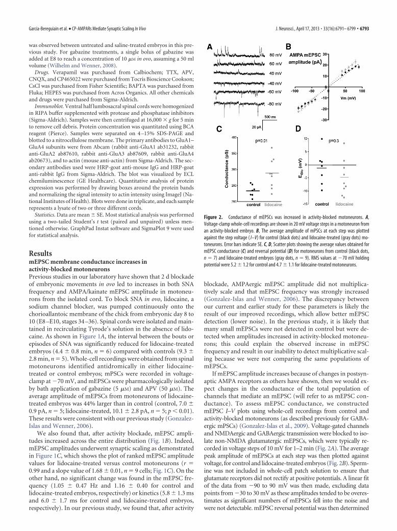
Glutamatergic mEPSCs are mediated byAMPA (GluA1–GluA4) receptors inboth control and activity-blockedmotoneuronsAs we have previously reported for E10chick motoneurons, glutamatergic mEPSCs are virtually abol-ished with bath application of the AMPA/kainate receptor antag-onist CNQX at �70 mV (Gonzalez-Islas and Wenner, 2006). Toexamine the contribution of AMPA versus kainate receptors me-diating the mEPSCs, we bath applied the AMPA-selective antag-onist (GluA1–GluA4), CP465022 (5 �M; incubation time was atleast 30 min) (Lazzaro et al., 2002). Figure 3 shows that glutama-tergic mEPSCs (isolated with the GABAA antagonist gabazine)are dramatically reduced in frequency when CP465022 is addedto the bath in both control (from 0.72 � 0.12 Hz to 0.16 � 0.04Hz, p � 0.001) and lidocaine-treated motoneurons (from 0.89 �0.30 Hz to 0.18 � 0.04 Hz, p � 0.001). Therefore, the vast major-ity of the recorded glutamatergic mEPSCs are mediated byAMPA receptors.
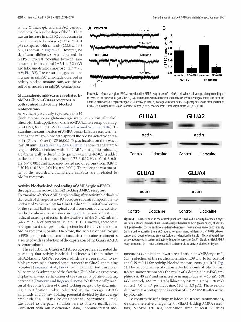
Activity blockade-induced scaling of AMPAergic mEPSCsthrough an increase of GluA2-lacking AMPA receptorsTo examine whether AMPAergic scaling after activity blockade isthe result of changes in AMPA receptor subunit composition, weperformed Western blots for GluA1–GluA4 subunits from lysatesof the ventral half of the spinal cord from control and activity-blocked embryos. As we show in Figure 4, lidocaine treatmentinduced a strong reduction in the total level of the GluA2 subunit(44.7 � 2.7% of control value, p � 0.01). However, there werenot significant changes in total protein level for any of the otherAMPA receptor subunits. Therefore, the increase of AMPAergicmEPSC amplitude and conductance after lidocaine treatment isassociated with a reduction of the expression of the GluA2 AMPAreceptor subunit.
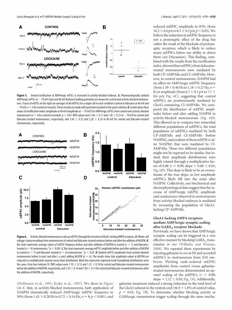
The reduction in GluA2 AMPA receptor protein suggested thepossibility that activity blockade had increased the number ofGluA2-lacking AMPA receptors, which have been shown to ex-hibit greater single-channel conductance than GluA2-containingreceptors (Swanson et al., 1997). To functionally test this possi-bility, we took advantage of the fact that GluA2-lacking receptorsdisplay an inward rectification of the current at positive holdingpotentials (Donevan and Rogawski, 1995). We functionally mea-sured the contribution of GluA2-lacking receptors by determin-ing a rectification index, calculated as the average mEPSCamplitude at a 40 mV holding potential divided by the averageamplitude at a �70 mV holding potential. Spermine (0.1 mM)was added to the patch solution here to observe rectification.Consistent with our biochemical data, lidocaine-treated mo-
toneurons exhibited an inward rectification of AMPAergic mP-SCs (reduction of the rectification index: 1.09 � 0.16 for controland 0.59 � 0.11 for activity-blocked motoneurons, p � 0.01; Fig.5). The reduction in rectification index from control to lidocaine-treated motoneurons was the result of a decrease in mPSC am-plitude at 40 mV and an increase in amplitude at �70 mV (40mV: control, 12.5 � 5.4 pA; lidocaine, 7.8 � 3.3 pA; �70 mV:control, 9.0 � 4.7 pA; lidocaine, 13.4 � 5.8 pA). These resultsdemonstrate a postsynaptic insertion of CP-AMPARs after activ-ity blockade.
To confirm these findings in lidocaine-treated motoneurons,we used a selective antagonist for GluA2-lacking AMPA recep-tors, NASPM (20 �M, incubation time at least 30 min)
Figure 3. Glutamatergic mEPSCs are mediated by AMPA receptors (GluA1–GluA4). A, Whole-cell voltage-clamp recording ofmEPSCs, in the presence of gabazine (5 �M), from motoneurons of control and lidocaine-treated embryos before and after theaddition of the AMPA receptor antagonist, CP465022 (5 �M). B, Average values for mEPSC frequency before and after addition ofCP465022 in control (n � 5) and lidocaine-treated (n � 5) motoneurons. Error bars indicate SE. *p � 0.001.
Figure 4. GluA2 subunit in the ventral spinal cord is reduced in activity-blocked embryos.Western blots are shown for GluA1–GluA4 (upper bands) and actin (lower bands) of ventral-half spinal cords of control and lidocaine-treated embryos. The average values of band intensity(normalized to actin) for the GluA2 subunit were significantly different ( p � 0.01) betweencontrol and lidocaine-treated embryos (values are described in Results). No significant differ-ence was observed in control and activity-blocked embryos for GluA1, GluA3, or GluA4 AMPAreceptor subunits (n � 4 for each subunit in both control and activity-blocked embryos).
6794 • J. Neurosci., April 17, 2013 • 33(16):6791– 6799 Garcia-Bereguiain et al. • CP-AMPARs Mediate Synaptic Scaling In Vivo
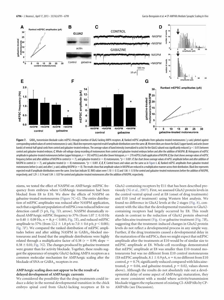
(Hollmann et al., 1991; Koike et al., 1997). We show in Figure6A–C that, in activity-blocked motoneurons, bath application ofNASPM dramatically reduced AMPAergic mEPSC frequency to50% (from 1.45 � 0.28 Hz to 0.72 � 0.16 Hz, n � 8; p � 0.001), and
reduced mEPSC amplitude to 83% (from10.2 � 0.6 pA to 8.5 � 0.2 pA; p � 0.05). Webelieve the reduction in mEPSC frequency isnot a presynaptic effect of the drug butrather the result of the blockade of postsyn-aptic receptors, which is likely to reducemany mEPSCs below our ability to detectthem (see Discussion). This finding, com-bined with the results from the rectificationindex, showed that mEPSCs from lidocaine-treated motoneurons were mediated byboth CP-AMPARs and CI-AMPARs. How-ever, in control motoneurons, NASPM hadno effect on AMPAergic mEPSC frequency(from 1.39�0.46 Hz to 1.18�0.27 Hz, n�8) or amplitude (from 6.7 � 0.1 pA to 7.1 �0.6 pA; Fig. 6C), suggesting that controlmEPSCs are predominantly mediated byGluA2-containing CI-AMPARs. We com-pared the distribution of mEPSC ampli-tudes before and after adding NASPM inactivity-blocked motoneurons (Fig. 6D).This allowed us to compare two somewhatdifferent populations of mEPSCs: the totalpopulation of mEPSCs mediated by bothCP-AMPARs and CI-AMPARs (beforeNASPM), and a subset of those mEPSCs (af-ter NASPM) that were mediated by CI-AMPARs. These two different populationsmight not be expected to be similar, but in-deed their amplitude distributions werehighly related through a multiplicative fac-tor of 0.88 (r � 0.99; slope � 0.88 � 0.01;Fig. 6D). This slope is likely to be an overes-timate of the true slope, as low amplitudemEPSCs likely fall into the noise afterNASPM. Collectively, our biochemical andelectrophysiological data suggest that the in-crease of AMPAergic mEPSC amplitudeand conductance observed in motoneuronsfrom activity-blocked embryos is mediatedby increasing the population of GluA2-lacking CP-AMPARs.
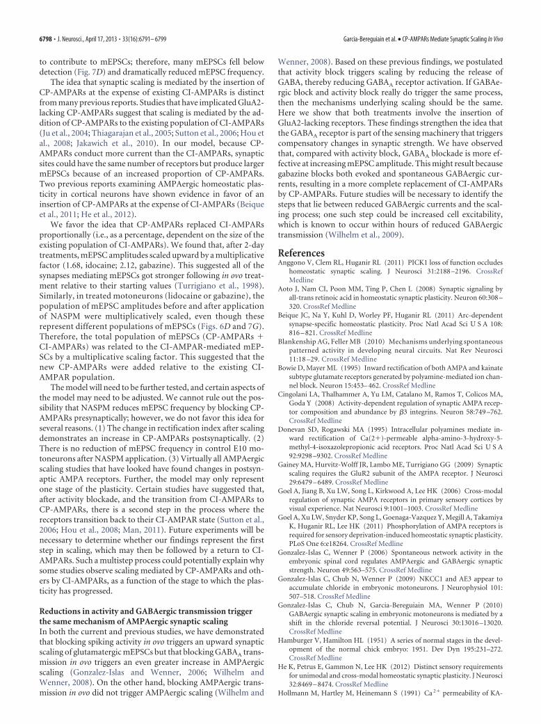
GluA2-lacking AMPA receptorsmediate AMPAergic synaptic scalingafter GABAA receptor blockadePreviously, we have shown that AMPAergicsynaptic scaling can be triggered in a veryeffective manner by blocking GABAA trans-mission in ovo (Wilhelm and Wenner,2008). We repeated these experiments byinjecting gabazine in ovo at E8 and recordedmEPSCs in motoneurons from E10 em-bryos. Plotting rank-ordered mEPSCamplitudes from control versus gabazine-treated motoneurons demonstrated an up-ward scaling of the mEPSCs (r � 0.98;slope � 2.12 � 0.03; Fig. 7A). Additionally,
gabazine treatment induced a strong reduction in the total level ofthe GluA2 subunit in the ventral cord (36.9 � 1.8% of control value,p � 0.01; Fig. 7B). To determine whether blocking activity orGABAergic transmission trigger scaling through the same mecha-
Figure 5. Inward rectification of AMPAergic mPSCs is increased in activity-blocked embryos. A, Pharmacologically isolatedAMPAergic mPSCs at �70 mV (top) and 40 mV (bottom) holding potentials are shown for control and activity-blocked motoneu-rons. Traces of mEPSC on the right are averages of all mEPSCs for a single cell in each condition (control or lidocaine at 40 mV and�70 mV; n � 6 for control or treated). These records are made with spermine included in the patch solution. B, Scatter plots showvalues of rectification index (amplitude at 40 mV/amplitude at �70 mV) for AMPAergic mPSCs from control and activity-blockedmotoneurons (n � 6 for control or treated). p � 0.01. RMS values were 1.06 � 0.11 and 1.36 � 0.21 at �70 mV for control andlidocaine-treated motoneurons, respectively; and 1.64 � 0.33 and 2.20 � 0.24 at 40 mV for control and lidocaine-treatedmotoneurons, respectively.
Figure 6. Activity-blocked motoneurons scale up mEPSCs through the insertion of GluA2-lacking AMPA receptors. A, Whole-cellvoltage-clamp recordings from motoneurons of control and lidocaine-treated embryos before and after the addition of NASPM. B,Bar chart represents average values of mEPSC frequency before and after addition of NASPM in control (n � 7) and lidocaine-treated (n � 8) motoneurons. *p � 0.001. C, Bar chart represents average mEPSC amplitude before and after addition of NASPMin control (n � 7) and lidocaine-treated (n � 8) motoneurons. *p � 0.05. D, Ranked mEPSC amplitudes from activity-blockedmotoneurons before (x-axis) and after ( y-axis) adding NASPM (n � 6). The results show that amplitude values in NASPM arereduced in a multiplicative manner across their distribution. Black line represents expected result if amplitude distributions werethe same. Error bars indicate SE. RMS values were 1.10 � 0.12 and 1.23 � 0.10 for control and lidocaine-treated motoneuronsbefore the addition of NASPM, respectively; and 1.29 � 0.14 and 1.00 � 0.11 for control and lidocaine-treated motoneurons afterthe addition of NASPM, respectively.
Garcia-Bereguiain et al. • CP-AMPARs Mediate Synaptic Scaling In Vivo J. Neurosci., April 17, 2013 • 33(16):6791– 6799 • 6795
nisms, we tested the effect of NASPM on AMPAergic mEPSC fre-quency from embryos where GABAergic transmission had beenblocked from E8 to E10. We show the effects of NASPM ongabazine-treated motoneurons (Figure 7C–G). The entire distribu-tion of mEPSC amplitudes was reduced after NASPM application,such that a significant population of mEPSCs was reduced below ourdetection cutoff (5 pA, Fig. 7D, arrow). NASPM dramatically re-duced AMPAergic mEPSC frequency to 37% (from 1.07 � 0.10 Hzto 0.40 � 0.09 Hz, n � 8; p � 0.001; Fig. 7E), and reduced mEPSCamplitude to 57% (from 12.2 � 1.8 pA to 7.0 � 0.6 pA; p � 0.001;Fig. 7F). We compared the ranked distribution of mEPSC ampli-tudes before and after adding NASPM in GABAA-blocked mo-toneurons and found that the amplitude distributions were highlyrelated through a multiplicative factor of 0.38 (r � 0.99; slope �0.38 � 0.01; Fig. 7G). The changes produced by gabazine treatmentwere greater than for activity blockade. Together, these results sug-gest the appearance of synaptic GluA2-lacking AMPA receptors as acommon molecular mechanism for AMPAergic scaling after theblockade of SNA or GABAA receptors in ovo.
AMPAergic scaling does not appear to be the result of adelayed development of AMPAergic currentsWe considered the possibility that the drug treatments could in-duce a delay in the normal developmental transition in the chickembryo spinal cord from GluA2-lacking receptors at E6 to
GluA2-containing receptors by E11 that has been described pre-viously (Ni et al., 2007). First, we assessed GluA2 protein levels inthe control ventral spinal cord at E8 (onset of drug treatments)and E10 (end of treatment) using Western blot analysis. Wefound no difference in GluA2 levels at the 2 stages (Fig. 8), con-sistent with the idea that the developmental transition to GluA2-containing receptors had largely occurred by E8. This resultstands in contrast to the reduction of GluA2 protein observedafter lidocaine treatment (Fig. 4) or gabazine treatment (Fig. 7B),suggesting that the treatment-induced changes in GluA2 proteinlevels do not reflect a developmental process in any simple way.Further, if the drug treatments caused a developmental delay inthe maturation of the mEPSCs, then we might expect that mEPSCamplitude after the treatments at E10 would be of similar size tomEPSC amplitude at E8. Whole-cell recordings demonstratedthat mEPSC amplitude at E8 was smaller than E10-treated mo-toneurons but was no different from E10 control motoneurons(E8 mEPSC amplitude, 8.1 � 0.9 pA, n � 6; no different from E10control, p � 0.79, significantly reduced compared with lidocaine-treated, p � 0.04, and gabazine-treated, p � 0.02, values shownabove). Although the results do not absolutely rule out a devel-opmental delay of some aspect of AMPAergic maturation, theyare more consistent with a model where activity/transmissionblockade triggers the replacement of existing CI-AMPARs by CP-AMPARs (see Discussion).
Figure 7. GABAA transmission blockade scales mEPSCs through insertion of GluA2-lacking AMPA receptors. A, Ranked mEPSC amplitudes from gabazine-treated motoneurons ( y-axis) plotted againstcorresponding ranked values of control motoneurons (x-axis). Black line represents expected result if amplitude distributions were the same. B, Western blots are shown for GluA2 (upper bands) and actin (lowerbands) of ventral-half spinal cords from control and gabazine-treated embryos. The average values of band intensity (normalized to actin) for the GluA2 subunit was significantly reduced ( p � 0.01) betweencontrol and gabazine-treated embryos. C, Whole-cell voltage-clamp recording of motoneurons from control and gabazine-treated embryos before and after the addition of NASPM. D, Histograms of mEPSCamplitude in gabazine-treated motoneurons before (upper histogram, n�392 mEPSCs) and after (lower histogram, n�270 mEPSCs) bath application of NASPM. E, Bar chart shows average values of mEPSCfrequency before and after addition of NASPM in control (n � 7), and gabazine-treated (n � 8) motoneurons. *p � 0.001. F, Bar chart shows average values of mPSC amplitude before and after addition ofNASPM in control (n � 7), and gabazine-treated (n � 8) motoneurons. *p � 0.001. C, E, F, Control traces and values are the same as in Figure 6. G, Ranked mEPSC amplitudes from gabazine-treatedmotoneurons before (x-axis) and after ( y-axis) adding NASPM (n � 8). The results show that amplitude values in NASPM are reduced in a multiplicative manner across their distribution. Black line representsexpected result if amplitude distributions were the same. Error bars indicate SE. RMS values were 1.10 � 0.12 and 1.06 � 0.10 for control and gabazine-treated motoneurons before the addition of NASPM,respectively; and 1.29 � 0.14 and 1.04 � 0.07 for control and gabazine-treated motoneurons after the addition of NASPM, respectively.
6796 • J. Neurosci., April 17, 2013 • 33(16):6791– 6799 Garcia-Bereguiain et al. • CP-AMPARs Mediate Synaptic Scaling In Vivo

DiscussionEvidence for AMPAergic synaptic scaling is extensive and hasbeen described by many different laboratories in several differentsystems in vitro and in vivo (Pozo and Goda, 2010; Turrigiano,2012; Vitureira et al., 2012). No such consensus exists for themechanisms underlying AMPAergic synaptic scaling, as severalstudies either support or deny the contribution of CP-AMPARs.Most work has assessed scaling mechanisms after in vitro pertur-bations; we show that AMPAergic scaling, in embryonic mo-toneurons after in vivo blockade of either spiking activity orGABAergic transmission, occurs through an increased mEPSCconductance mediated by the postsynaptic insertion GluA2-lacking CP-AMPARs, at the expense of CI-AMPARs.
AMPAergic synaptic scaling is dependent on CP-AMPARs inembryonic motoneurons in vivoMany studies have shown that AMPAergic scaling is mediated bychanges in AMPA receptors in the postsynaptic membrane. Con-sistent with changes in postsynaptic receptors, we show that scal-ing is mediated by increases in mEPSC conductance. Moreimportantly, we find that the increased mEPSC conductance wasmediated by the insertion of CP-AMPARs. These GluA2-lackingAMPA receptors are distinct from GluA2-containing AMPA re-ceptors in that they are permeable to Ca 2�, have larger single-channel conductance, and exhibit a voltage-dependent block byintracellular polyamines (Hollmann et al., 1991; Bowie andMayer, 1995; Donevan and Rogawski, 1995; Swanson et al., 1997;Traynelis et al., 2010). We demonstrate the insertion of CP-AMPARs after activity blockade in several ways: (1) through areduction in GluA2 subunit expression in the ventral cord, (2) bythe appearance of AMPAergic mPSCs that are sensitive toNASPM, and (3) by showing that mEPSCs in lidocaine-treatedmotoneurons express stronger inward rectification. Although wedid not directly show increased calcium permeability of GluA2-lacking receptors, several other studies have shown that the glu-tamate receptors that express an inward rectification and aresensitive to polyamines (e.g., NASPM) are permeable to calcium(Hollmann et al., 1991; Bowie and Mayer, 1995; Donevan andRogawski, 1995); one study in particular demonstrated that rec-tification was a good indicator of calcium-permeable glutamatereceptors in motoneurons in the chick embryo (Ni et al., 2007).
It is important to understand whether CP-AMPARs are in-volved in synaptic scaling because these receptors can influencecalcium signaling pathways. As a result, many studies have as-sessed the contribution of the GluA2 subunit to synaptic scalingin cultured networks. Several of these in vitro studies have dem-onstrated an accumulation of GluA2-lacking CP-AMPARs (Ju etal., 2004; Thiagarajan et al., 2005; Sutton et al., 2006; Aoto et al.,2008; Hou et al., 2008; Beique et al., 2011). Other studies report
an upregulation of GluA2-containing CI-AMPARs after activityblockade (O’Brien et al., 1998; Sun et al., 2005; Wierenga et al.,2005; Sutton et al., 2006; Cingolani et al., 2008; Ibata et al., 2008;Anggono et al., 2011). Different results could arise from themethod of blocking activity, the duration of the blockade, thetissue, or stage of development (Man, 2011; Lee, 2012; Shepherd,2012). To look at the contribution of CP-AMPARs in a morenatural context, two different groups have assessed the mecha-nisms of AMPAergic scaling after reductions in activity in vivo.Layer 2/3 pyramidal cells in the primary visual cortex exhibitAMPAergic scaling after 2 d of reduced visual drive in the3-week-old rat in vivo (Goel et al., 2006, 2011; Gainey et al.,2009). Surprisingly, one group found that scaling was medi-ated by CP-AMPARs (Goel et al., 2006, 2011), whereas theother group found that scaling involved CI-AMPARs (Gaineyet al., 2009). Here, we test the role of CP-AMPARs in AMPAer-gic scaling after in vivo perturbations to a different network, ata stage when GABA is excitatory. In embryonic motoneurons,we find that CP-AMPARs mediate AMPAergic scaling at theexpense of CI-AMPARs.
Model for AMPAergic scaling in embryonic motoneuronsWe propose a model that we believe is the most consistent andparsimonious explanation for our findings: (1) after activity- orGABAergic-blockade scaling of mEPSCs is mediated by the re-placement of GluA2-containing receptors by GluA2-lacking re-ceptors; and (2) GABAergic blockade triggers scaling moreeffectively than lidocaine treatment by replacing a greater pro-portion of the CI-AMPARs with the higher conductance CP-AMPARs (Fig. 9). Several findings contribute to this model. Insupport of the idea that CP-AMPARs are inserted at the expenseof GluA2-containing receptors, we found that the GluA2 proteinis reduced after scaling. Further, although mEPSC frequency wasthe same for control and treated (lidocaine or gabazine) mo-toneurons, blockade of CP-AMPARs with NASPM acutely re-duced mEPSC frequency only for the treated cells (to 50% forlidocaine treatment, to 37% for gabazine treatment). Gabazinetreatment may have produced a stronger increase in mEPSC am-plitude than lidocaine treatment because more CI-AMPARs werereplaced with CP-AMPARs (Fig. 9); when CP-AMPARs werethen acutely blocked with NASPM, fewer CI-AMPARs remained
Figure 8. GluA2 protein levels in the control ventral spinal cord are the same at E8 and E10.Western blots are shown for GluA2 (upper bands) and actin (lower bands). The average values ofband intensity (normalized to actin) for the GluA2 subunit were not significantly differentbetween E8 (0.34 � 0.02) and E10 (0.33 � 0.01). Figure 9. Model for synaptic scaling mediated by the insertion of GluA2-lacking AMPA re-
ceptors. Model shows GluA2-lacking AMPA receptors replacing existing GluA2-containing re-ceptors in a 1:1 manner. Gabazine treatment produces a larger increase in mEPSC amplitudethan activity blockade. This is consistent with a model where a greater proportion of GluA2-containing receptors are replaced with the NASPM-sensitive GluA2-lacking receptors. Such amodel would explain why NASPM reduces the frequency of mEPSCs in gabazine-treated mo-toneurons to a greater extent than for activity-blocked motoneurons, as more of the mEPSCs fallbelow detection.
Garcia-Bereguiain et al. • CP-AMPARs Mediate Synaptic Scaling In Vivo J. Neurosci., April 17, 2013 • 33(16):6791– 6799 • 6797
to contribute to mEPSCs; therefore, many mEPSCs fell belowdetection (Fig. 7D) and dramatically reduced mEPSC frequency.
The idea that synaptic scaling is mediated by the insertion ofCP-AMPARs at the expense of existing CI-AMPARs is distinctfrom many previous reports. Studies that have implicated GluA2-lacking CP-AMPARs suggest that scaling is mediated by the ad-dition of CP-AMPARs to the existing population of CI-AMPARs(Ju et al., 2004; Thiagarajan et al., 2005; Sutton et al., 2006; Hou etal., 2008; Jakawich et al., 2010). In our model, because CP-AMPARs conduct more current than the CI-AMPARs, synapticsites could have the same number of receptors but produce largermEPSCs because of an increased proportion of CP-AMPARs.Two previous reports examining AMPAergic homeostatic plas-ticity in cortical neurons have shown evidence in favor of aninsertion of CP-AMPARs at the expense of CI-AMPARs (Beiqueet al., 2011; He et al., 2012).
We favor the idea that CP-AMPARs replaced CI-AMPARsproportionally (i.e., as a percentage, dependent on the size of theexisting population of CI-AMPARs). We found that, after 2-daytreatments, mEPSC amplitudes scaled upward by a multiplicativefactor (1.68, idocaine; 2.12, gabazine). This suggested all of thesynapses mediating mEPSCs got stronger following in ovo treat-ment relative to their starting values (Turrigiano et al., 1998).Similarly, in treated motoneurons (lidocaine or gabazine), thepopulation of mEPSC amplitudes before and after applicationof NASPM were multiplicatively scaled, even though theserepresent different populations of mEPSCs (Figs. 6D and 7G).Therefore, the total population of mEPSCs (CP-AMPARs �CI-AMPARs) was related to the CI-AMPAR-mediated mEP-SCs by a multiplicative scaling factor. This suggested that thenew CP-AMPARs were added relative to the existing CI-AMPAR population.
The model will need to be further tested, and certain aspects ofthe model may need to be adjusted. We cannot rule out the pos-sibility that NASPM reduces mEPSC frequency by blocking CP-AMPARs presynaptically; however, we do not favor this idea forseveral reasons. (1) The change in rectification index after scalingdemonstrates an increase in CP-AMPARs postsynaptically. (2)There is no reduction of mEPSC frequency in control E10 mo-toneurons after NASPM application. (3) Virtually all AMPAergicscaling studies that have looked have found changes in postsyn-aptic AMPA receptors. Further, the model may only representone stage of the plasticity. Certain studies have suggested that,after activity blockade, and the transition from CI-AMPARs toCP-AMPARs, there is a second step in the process where thereceptors transition back to their CI-AMPAR state (Sutton et al.,2006; Hou et al., 2008; Man, 2011). Future experiments will benecessary to determine whether our findings represent the firststep in scaling, which may then be followed by a return to CI-AMPARs. Such a multistep process could potentially explain whysome studies observe scaling mediated by CP-AMPARs and oth-ers by CI-AMPARs, as a function of the stage to which the plas-ticity has progressed.
Reductions in activity and GABAergic transmission triggerthe same mechanism of AMPAergic synaptic scalingIn both the current and previous studies, we have demonstratedthat blocking spiking activity in ovo triggers an upward synapticscaling of glutamatergic mEPSCs but that blocking GABAA trans-mission in ovo triggers an even greater increase in AMPAergicscaling (Gonzalez-Islas and Wenner, 2006; Wilhelm andWenner, 2008). On the other hand, blocking AMPAergic trans-mission in ovo did not trigger AMPAergic scaling (Wilhelm and
Wenner, 2008). Based on these previous findings, we postulatedthat activity block triggers scaling by reducing the release ofGABA, thereby reducing GABAA receptor activation. If GABAe-rgic block and activity block really do trigger the same process,then the mechanisms underlying scaling should be the same.Here we show that both treatments involve the insertion ofGluA2-lacking receptors. These findings strengthen the idea thatthe GABAA receptor is part of the sensing machinery that triggerscompensatory changes in synaptic strength. We have observedthat, compared with activity block, GABAA blockade is more ef-fective at increasing mEPSC amplitude. This might result becausegabazine blocks both evoked and spontaneous GABAergic cur-rents, resulting in a more complete replacement of CI-AMPARsby CP-AMPARs. Future studies will be necessary to identify thesteps that lie between reduced GABAergic currents and the scal-ing process; one such step could be increased cell excitability,which is known to occur within hours of reduced GABAergictransmission (Wilhelm et al., 2009).
ReferencesAnggono V, Clem RL, Huganir RL (2011) PICK1 loss of function occludes
homeostatic synaptic scaling. J Neurosci 31:2188 –2196. CrossRefMedline
Aoto J, Nam CI, Poon MM, Ting P, Chen L (2008) Synaptic signaling byall-trans retinoic acid in homeostatic synaptic plasticity. Neuron 60:308 –320. CrossRef Medline
Beique JC, Na Y, Kuhl D, Worley PF, Huganir RL (2011) Arc-dependentsynapse-specific homeostatic plasticity. Proc Natl Acad Sci U S A 108:816 – 821. CrossRef Medline
Blankenship AG, Feller MB (2010) Mechanisms underlying spontaneouspatterned activity in developing neural circuits. Nat Rev Neurosci11:18 –29. CrossRef Medline
Bowie D, Mayer ML (1995) Inward rectification of both AMPA and kainatesubtype glutamate receptors generated by polyamine-mediated ion chan-nel block. Neuron 15:453– 462. CrossRef Medline
Cingolani LA, Thalhammer A, Yu LM, Catalano M, Ramos T, Colicos MA,Goda Y (2008) Activity-dependent regulation of synaptic AMPA recep-tor composition and abundance by �3 integrins. Neuron 58:749 –762.CrossRef Medline
Donevan SD, Rogawski MA (1995) Intracellular polyamines mediate in-ward rectification of Ca(2�)-permeable alpha-amino-3-hydroxy-5-methyl-4-isoxazolepropionic acid receptors. Proc Natl Acad Sci U S A92:9298 –9302. CrossRef Medline
Gainey MA, Hurvitz-Wolff JR, Lambo ME, Turrigiano GG (2009) Synapticscaling requires the GluR2 subunit of the AMPA receptor. J Neurosci29:6479 – 6489. CrossRef Medline
Goel A, Jiang B, Xu LW, Song L, Kirkwood A, Lee HK (2006) Cross-modalregulation of synaptic AMPA receptors in primary sensory cortices byvisual experience. Nat Neurosci 9:1001–1003. CrossRef Medline
Goel A, Xu LW, Snyder KP, Song L, Goenaga-Vazquez Y, Megill A, TakamiyaK, Huganir RL, Lee HK (2011) Phosphorylation of AMPA receptors isrequired for sensory deprivation-induced homeostatic synaptic plasticity.PLoS One 6:e18264. CrossRef Medline
Gonzalez-Islas C, Wenner P (2006) Spontaneous network activity in theembryonic spinal cord regulates AMPAergic and GABAergic synapticstrength. Neuron 49:563–575. CrossRef Medline
Gonzalez-Islas C, Chub N, Wenner P (2009) NKCC1 and AE3 appear toaccumulate chloride in embryonic motoneurons. J Neurophysiol 101:507–518. CrossRef Medline
Gonzalez-Islas C, Chub N, Garcia-Bereguiain MA, Wenner P (2010)GABAergic synaptic scaling in embryonic motoneurons is mediated by ashift in the chloride reversal potential. J Neurosci 30:13016 –13020.CrossRef Medline
Hamburger V, Hamilton HL (1951) A series of normal stages in the devel-opment of the normal chick embryo: 1951. Dev Dyn 195:231–272.CrossRef Medline
He K, Petrus E, Gammon N, Lee HK (2012) Distinct sensory requirementsfor unimodal and cross-modal homeostatic synaptic plasticity. J Neurosci32:8469 – 8474. CrossRef Medline
Hollmann M, Hartley M, Heinemann S (1991) Ca 2� permeability of KA-
6798 • J. Neurosci., April 17, 2013 • 33(16):6791– 6799 Garcia-Bereguiain et al. • CP-AMPARs Mediate Synaptic Scaling In Vivo
AMPA– gated glutamate receptor channels depends on subunit compo-sition. Science 252:851– 853. CrossRef Medline
Hou Q, Zhang D, Jarzylo L, Huganir RL, Man HY (2008) Homeostatic reg-ulation of AMPA receptor expression at single hippocampal synapses.Proc Natl Acad Sci U S A 105:775–780. CrossRef Medline
Ibata K, Sun Q, Turrigiano GG (2008) Rapid synaptic scaling induced bychanges in postsynaptic firing. Neuron 57:819 – 826. CrossRef Medline
Jakawich SK, Nasser HB, Strong MJ, McCartney AJ, Perez AS, Rakesh N,Carruthers CJ, Sutton MA (2010) Local presynaptic activity gates ho-meostatic changes in presynaptic function driven by dendritic BDNF syn-thesis. Neuron 68:1143–1158. CrossRef Medline
Ju W, Morishita W, Tsui J, Gaietta G, Deerinck TJ, Adams SR, Garner CC,Tsien RY, Ellisman MH, Malenka RC (2004) Activity-dependent regu-lation of dendritic synthesis and trafficking of AMPA receptors. Nat Neu-rosci 7:244 –253. CrossRef Medline
Koike M, Iino M, Ozawa S (1997) Blocking effect of 1-naphthyl acetylspermine on Ca(2�)-permeable AMPA receptors in cultured rat hip-pocampal neurons. Neurosci Res 29:27–36. CrossRef Medline
Lazzaro JT, Paternain AV, Lerma J, Chenard BL, Ewing FE, Huang J, WelchWM, Ganong AH, Menniti FS (2002) Functional characterization ofCP-465,022, a selective, noncompetitive AMPA receptor antagonist.Neuropharmacology 42:143–153. CrossRef Medline
Lee HK (2012) Ca-permeable AMPA receptors in homeostatic synapticplasticity. Front Mol Neurosci 5:17. Medline
Man HY (2011) GluA2-lacking, calcium-permeable AMPA receptors: in-ducers of plasticity? Curr Opin Neurobiol 21:291–298. CrossRef Medline
Ni X, Sullivan GJ, Martin-Caraballo M (2007) Developmental characteris-tics of AMPA receptors in chick lumbar motoneurons. Dev Neurobiol67:1419 –1432. CrossRef Medline
O’Brien RJ, Kamboj S, Ehlers MD, Rosen KR, Fischbach GD, Huganir RL(1998) Activity-dependent modulation of synaptic AMPA receptor ac-cumulation. Neuron 21:1067–1078. CrossRef Medline
O’Donovan MJ (1999) The origin of spontaneous activity in developingnetworks of the vertebrate nervous system. Curr Opin Neurobiol 9:94 –104. CrossRef Medline
O’Donovan MJ, Chub N, Wenner P (1998) Mechanisms of spontaneousactivity in developing spinal networks. J Neurobiol 37:131–145. CrossRefMedline
Pozo K, Goda Y (2010) Unraveling mechanisms of homeostatic synapticplasticity. Neuron 66:337–351. CrossRef Medline
Rich MM, Wenner P (2007) Sensing and expressing homeostatic synapticplasticity. Trends Neurosci 30:119 –125. CrossRef Medline
Shepherd JD (2012) Memory, plasticity and sleep: a role for calcium perme-able AMPA receptors? Front Mol Neurosci 5:49. CrossRef Medline
Sun X, Zhao Y, Wolf ME (2005) Dopamine receptor stimulation modulatesAMPA receptor synaptic insertion in prefrontal cortex neurons. J Neuro-sci 25:7342–7351. CrossRef Medline
Sutton MA, Ito HT, Cressy P, Kempf C, Woo JC, Schuman EM (2006) Min-iature neurotransmission stabilizes synaptic function via tonic suppres-sion of local dendritic protein synthesis. Cell 125:785–799. CrossRefMedline
Swanson GT, Kamboj SK, Cull-Candy SG (1997) Single-channel propertiesof recombinant AMPA receptors depend on RNA editing, splice varia-tion, and subunit composition. J Neurosci 17:58 – 69. Medline
Thiagarajan TC, Lindskog M, Tsien RW (2005) Adaptation to synaptic in-activity in hippocampal neurons. Neuron 47:725–737. CrossRef Medline
Traynelis SF, Wollmuth LP, McBain CJ, Menniti FS, Vance KM, Ogden KK,Hansen KB, Yuan H, Myers SJ, Dingledine R (2010) Glutamate receptorion channels: structure, regulation, and function. Pharmacol Rev 62:405–496. CrossRef Medline
Turrigiano G (2012) Homeostatic synaptic plasticity: local and globalmechanisms for stabilizing neuronal function. Cold Spring Harbor Per-spect Biol 4:a005736. CrossRef Medline
Turrigiano GG (2008) The self-tuning neuron: synaptic scaling of excitatorysynapses. Cell 135:422– 435. CrossRef Medline
Turrigiano GG, Leslie KR, Desai NS, Rutherford LC, Nelson SB (1998)Activity-dependent scaling of quantal amplitude in neocortical neurons.Nature 391:892– 896. CrossRef Medline
Vitureira N, Letellier M, Goda Y (2012) Homeostatic synaptic plasticity:from single synapses to neural circuits. Curr Opin Neurobiol 22:516 –521.CrossRef Medline
Wierenga CJ, Ibata K, Turrigiano GG (2005) Postsynaptic expression of ho-meostatic plasticity at neocortical synapses. J Neurosci 25:2895–2905.CrossRef Medline
Wilhelm JC, Wenner P (2008) GABAA transmission is a critical step in theprocess of triggering homeostatic increases in quantal amplitude. ProcNatl Acad Sci U S A 105:11412–11417. CrossRef Medline
Wilhelm JC, Rich MM, Wenner P (2009) Compensatory changes in cellularexcitability, not synaptic scaling, contribute to homeostatic recovery ofembryonic network activity. Proc Natl Acad Sci U S A 106:6760 – 6765.CrossRef Medline
Garcia-Bereguiain et al. • CP-AMPARs Mediate Synaptic Scaling In Vivo J. Neurosci., April 17, 2013 • 33(16):6791– 6799 • 6799