Development/Plasticity/Repair ...79877/UQ79877_OA.pdf · Development/Plasticity/Repair...
11
Development/Plasticity/Repair Distinct Physiological Mechanisms Underlie Altered Glycinergic Synaptic Transmission in the Murine Mutants spastic, spasmodic, and oscillator Brett A. Graham, 1 Peter R. Schofield, 2,3,4 Pankaj Sah, 5 Troy W. Margrie, 6 and Robert J. Callister 1 1 School of Biomedical Sciences, Faculty of Health and Hunter Medical Research Institute, The University of Newcastle, Callaghan, New South Wales 2308, Australia, 2 Garvan Institute of Medical Research, Darlinghurst, Sydney, New South Wales 2010, Australia, 3 University of New South Wales, Sydney, New South Wales 2052, Australia, 4 Prince of Wales Medical Research Institute, Randwick, New South Wales 2031, Australia, 5 Queensland Brain Institute, The University of Queensland, Brisbane, Queensland 4072, Australia, and 6 Department of Physiology, University College London, London WC1E 6JJ, United Kingdom Spastic (spa), spasmodic (spd), and oscillator (ot) mice have naturally occurring glycine receptor (GlyR) mutations, which manifest as motor deficits and an exaggerated “startle response.” Using whole-cell recording in hypoglossal motoneurons, we compared the physi- ological mechanisms by which each mutation alters GlyR function. Mean glycinergic miniature IPSC (mIPSC) amplitude and frequency were dramatically reduced (50%) compared with controls for each mutant. mIPSC decay times were unchanged in spa/spa (4.5 0.3 vs 4.7 0.2 ms), reduced in spd/spd (2.7 0.2 vs 4.7 0.2 ms), and increased in ot/ot (12.3 1.2 vs 4.8 0.2 ms). Thus, in spastic, GlyRs are functionally normal but reduced in number, whereas in spasmodic, GlyR kinetics is faster. The oscillator mutation results in complete absence of 1-containing GlyRs; however, some non-1-containing GlyRs persist at synapses. Fluctuation analysis of membrane current, induced by glycine application to outside-out patches, showed that mean single-channel conductance was increased in spa/spa (64.2 4.9 vs 36.1 1.4 pS), but unchanged in spd/spd (32.4 2.1 vs 35.3 2.1 pS). GlyR-mediated whole-cell currents in spa/spa exhibited increased picrotoxin sensitivity (27 vs 71% block for 100 M), indicating 1 homomeric GlyR expression. The picrotoxin sensitivity of evoked glycinergic IPSCs and conductance of synaptic GlyRs, as determined by nonstationary variance analysis, were identical for spa/spa and controls. Together, these findings show the three mutations disrupt GlyR-mediated inhibition via different physiological mecha- nisms, and the spastic mutation results in “compensatory” 1 homomeric GlyRs at extrasynaptic loci. Key words: hypoglossal motoneuron; glycine receptor; mouse; inhibition; ion channel; brainstem Introduction Glycine receptors (GlyRs) are ligand-gated ion channels (LGICs) mediating fast inhibitory synaptic transmission in brainstem and spinal cord circuits (Rajendra et al., 1997; Legendre, 2001; Lynch, 2004). They are heteromultimeric pentamers composed of and subunits arranged in a 2:3 stoichiometry (Grudzinska et al., 2005). Our understanding of GlyR biology and LGIC function has advanced considerably by studying naturally occurring GlyR mutations, which occur in humans, cattle, horses, dogs, and mice (Rajendra and Schofield, 1995). Three murine mutants, spastic (spa), spasmodic (spd), and os- cillator (ot), have distinct GlyR defects that produce almost iden- tical motor phenotypes. All three mutations are recessive disor- ders and homozygous affected animals exhibit a heightened “startle response,” muscle rigidity, tremor, and impaired righting when disturbed (Chai, 1961; Lane et al., 1987; Buckwalter et al., 1994). In spastic mice (Chai, 1961), an intronic insertion of a LINE 1 transposable element in the GlyR subunit gene (Kingsmore et al., 1994; Mu ¨lhardt et al., 1994) causes exon- skipping and decreased transcriptional efficiency of the sub- unit. This reduces GlyR levels in the spinal cord and brainstem (White and Heller, 1982; Becker, 1990). In contrast, the spas- modic mouse (Lane et al., 1987) has a single point mutation (A52S) in the 1 subunit (Ryan et al., 1994; Saul et al., 1994). GlyR levels appear to be normal; however, agonist sensitivity is reduced (Ryan et al., 1994; Saul et al., 1994). In the lethal oscillator mutation, a microdeletion in exon 8 of the 1 subunit gene re- sults in virtual absence of 1 protein and functional GlyRs in membranes from spinal cord and brainstem (Buckwalter et al., 1994; Kling et al., 1997). The physiological properties of mutant GlyRs, especially ho- momeric subunit-containing receptors, have been studied ex- tensively in recombinant systems (Legendre, 2001; Lynch, 2004). Fewer studies have examined the consequences of GlyR muta- Received Sept. 20, 2005; revised Feb. 21, 2006; accepted March 14, 2006. This work was supported by the National Health and Medical Research Council of Australia (Grants 993050, 157209, 276403, and 980382), the Hunter Medical Research Institute, and the University of Newcastle. We thank Sarah Walker and Anna Scimone for providing the ligand binding data (supplemental Table 1, available at www. jneurosci.org as supplemental material) and genotyping, respectively. Correspondence should be addressed to Dr. Robert J. Callister, School of Biomedical Sciences, Faculty of Health, The University of Newcastle, Callaghan, New South Wales 2308, Australia. E-mail: robert.callister@ newcastle.edu.au. DOI:10.1523/JNEUROSCI.3991-05.2006 Copyright © 2006 Society for Neuroscience 0270-6474/06/264880-11$15.00/0 4880 • The Journal of Neuroscience, May 3, 2006 • 26(18):4880 – 4890
Transcript of Development/Plasticity/Repair ...79877/UQ79877_OA.pdf · Development/Plasticity/Repair...

Development/Plasticity/Repair
Distinct Physiological Mechanisms Underlie AlteredGlycinergic Synaptic Transmission in the Murine Mutantsspastic, spasmodic, and oscillator
Brett A. Graham,1 Peter R. Schofield,2,3,4 Pankaj Sah,5 Troy W. Margrie,6 and Robert J. Callister1
1School of Biomedical Sciences, Faculty of Health and Hunter Medical Research Institute, The University of Newcastle, Callaghan, New South Wales 2308,Australia, 2Garvan Institute of Medical Research, Darlinghurst, Sydney, New South Wales 2010, Australia, 3University of New South Wales, Sydney, NewSouth Wales 2052, Australia, 4Prince of Wales Medical Research Institute, Randwick, New South Wales 2031, Australia, 5Queensland Brain Institute, TheUniversity of Queensland, Brisbane, Queensland 4072, Australia, and 6Department of Physiology, University College London, London WC1E 6JJ, UnitedKingdom
Spastic (spa), spasmodic (spd), and oscillator (ot) mice have naturally occurring glycine receptor (GlyR) mutations, which manifest asmotor deficits and an exaggerated “startle response.” Using whole-cell recording in hypoglossal motoneurons, we compared the physi-ological mechanisms by which each mutation alters GlyR function. Mean glycinergic miniature IPSC (mIPSC) amplitude and frequencywere dramatically reduced (�50%) compared with controls for each mutant. mIPSC decay times were unchanged in spa/spa (4.5 � 0.3 vs4.7 � 0.2 ms), reduced in spd/spd (2.7 � 0.2 vs 4.7 � 0.2 ms), and increased in ot/ot (12.3 � 1.2 vs 4.8 � 0.2 ms). Thus, in spastic, GlyRs arefunctionally normal but reduced in number, whereas in spasmodic, GlyR kinetics is faster. The oscillator mutation results in completeabsence of �1-containing GlyRs; however, some non-�1-containing GlyRs persist at synapses. Fluctuation analysis of membrane current,induced by glycine application to outside-out patches, showed that mean single-channel conductance was increased in spa/spa (64.2 � 4.9vs 36.1 � 1.4 pS), but unchanged in spd/spd (32.4 � 2.1 vs 35.3 � 2.1 pS). GlyR-mediated whole-cell currents in spa/spa exhibitedincreased picrotoxin sensitivity (27 vs 71% block for 100 �M), indicating �1 homomeric GlyR expression. The picrotoxin sensitivity ofevoked glycinergic IPSCs and conductance of synaptic GlyRs, as determined by nonstationary variance analysis, were identical for spa/spaand controls. Together, these findings show the three mutations disrupt GlyR-mediated inhibition via different physiological mecha-nisms, and the spastic mutation results in “compensatory” �1 homomeric GlyRs at extrasynaptic loci.
Key words: hypoglossal motoneuron; glycine receptor; mouse; inhibition; ion channel; brainstem
IntroductionGlycine receptors (GlyRs) are ligand-gated ion channels (LGICs)mediating fast inhibitory synaptic transmission in brainstem andspinal cord circuits (Rajendra et al., 1997; Legendre, 2001; Lynch,2004). They are heteromultimeric pentamers composed of � and� subunits arranged in a 2:3 stoichiometry (Grudzinska et al.,2005). Our understanding of GlyR biology and LGIC functionhas advanced considerably by studying naturally occurring GlyRmutations, which occur in humans, cattle, horses, dogs, and mice(Rajendra and Schofield, 1995).
Three murine mutants, spastic (spa), spasmodic (spd), and os-cillator (ot), have distinct GlyR defects that produce almost iden-
tical motor phenotypes. All three mutations are recessive disor-ders and homozygous affected animals exhibit a heightened“startle response,” muscle rigidity, tremor, and impaired rightingwhen disturbed (Chai, 1961; Lane et al., 1987; Buckwalter et al.,1994). In spastic mice (Chai, 1961), an intronic insertion of aLINE 1 transposable element in the GlyR � subunit gene(Kingsmore et al., 1994; Mulhardt et al., 1994) causes exon-skipping and decreased transcriptional efficiency of the � sub-unit. This reduces GlyR levels in the spinal cord and brainstem(White and Heller, 1982; Becker, 1990). In contrast, the spas-modic mouse (Lane et al., 1987) has a single point mutation(A52S) in the �1 subunit (Ryan et al., 1994; Saul et al., 1994).GlyR levels appear to be normal; however, agonist sensitivity isreduced (Ryan et al., 1994; Saul et al., 1994). In the lethal oscillatormutation, a microdeletion in exon 8 of the �1 subunit gene re-sults in virtual absence of �1 protein and functional GlyRs inmembranes from spinal cord and brainstem (Buckwalter et al.,1994; Kling et al., 1997).
The physiological properties of mutant GlyRs, especially ho-momeric � subunit-containing receptors, have been studied ex-tensively in recombinant systems (Legendre, 2001; Lynch, 2004).Fewer studies have examined the consequences of GlyR muta-
Received Sept. 20, 2005; revised Feb. 21, 2006; accepted March 14, 2006.This work was supported by the National Health and Medical Research Council of Australia (Grants 993050,
157209, 276403, and 980382), the Hunter Medical Research Institute, and the University of Newcastle. We thankSarah Walker and Anna Scimone for providing the ligand binding data (supplemental Table 1, available at www.jneurosci.org as supplemental material) and genotyping, respectively.
Correspondence should be addressed to Dr. Robert J. Callister, School of Biomedical Sciences, Faculty of Health,The University of Newcastle, Callaghan, New South Wales 2308, Australia. E-mail: [email protected].
DOI:10.1523/JNEUROSCI.3991-05.2006Copyright © 2006 Society for Neuroscience 0270-6474/06/264880-11$15.00/0
4880 • The Journal of Neuroscience, May 3, 2006 • 26(18):4880 – 4890
tions on the function of “native” receptors (�1/� heteromers),the dominant form at adult synaptic connections. In this regard,the spastic mutation is the best characterized mutant. Consistentwith the reduction in GlyR levels in this mutant, the amplitude ofglycinergic miniature IPSPs (Biscoe and Duchen, 1986) and min-iature IPSCs (mIPSCs) (Callister et al. 1999; Graham et al., 2003;von Wegerer et al., 2003) are reduced in spinal cord and brain-stem neurons. Similarly, inhibitory synaptic transmission is dra-matically altered in oscillator dorsal horn neurons (Graham et al.,2003) and spasmodic brainstem neurons (Callister et al., 1999).
Here, we use hypoglossal motoneurons (MNs) to compare theeffects of each mutation on glycinergic synaptic transmission atintact/functional synaptic connections. Our data show that gly-cinergic drive is dramatically reduced in all three mutants, but bydistinct mechanisms. In spastic, the number of functional GlyRsis reduced at postsynaptic densities, but receptor kinetics is un-changed. Furthermore, �1 homomeric GlyRs are assembled inthe spastic mutant and are restricted to extrasynaptic locations.The spasmodic mutation dramatically changes GlyR kinetics withfaster mIPSC rise and decay times. In oscillator, few functionalGlyRs are present in animals older than 16 d; however, someGlyRs with slower mIPSC decay times persist.
Materials and MethodsAnimals. Spastic, spasmodic, and oscillator mice were obtained from TheJackson Laboratory (Bar Harbor, ME). Experiments were performed onwild-type, spastic, spasmodic, or oscillator mice (both sexes) backcrossedonto the C57BL/6 genetic background. Ages for each genotype are pro-vided in Results. Spastic and spasmodic animals were bred by matinghomozygous affected (spa/spa or spd/spd) females with heterozygous un-affected (spa/� or spd/�) males. Because homozygous oscillator mice die�21 d after birth (Buckwalter et al., 1994) (i.e., before they are sexuallymature), ot animals were bred by mating heterozygous (ot/�) animals.All experimental procedures were approved by the University of New-castle and Garvan Institute of Medical Research Animal Experimentationand Ethics Committees. Homozygous affected spa, spd, and ot animalswere easily identified �2 weeks after birth according to four criteria: (1)constant resting tremor, (2) clenching of limbs when picked up by thetail, (3) an impaired righting reflex, and (4) a tendency to walk on tiptoewith an arched back (Simon, 1995).
Genotyping. Genotypes were determined on 2–5 mm tail tip biopsies,obtained under methoxyflurane anesthesia, from all breeding stock andexperimental animals. Tail tips were digested with proteinase K at 0.63mg/ml in 10 mM Tris-HCl, pH 7.5, 400 mM NaCl, 100 mM EDTA, 0.6%SDS in a total volume of 640 �l overnight at 42°C. The digest was clearedby addition of 170 �l of saturated NaCl and centrifuged 10 min at13,000 � g. DNA was precipitated from supernatants by the addition of800 �l of ice-cold 100% ethanol and spun at 13,000 � g for 5 min. Pelletswere washed with 800 �l of ice-cold 70% ethanol and resuspended over-night in 100 �l of H2O. Genotypes were determined by PCR using thefollowing primers. Spastic: SpaWT forward, 5�-GCAACTTGAGAGC-TGTATGT-3�, and SpaWT reverse, 5�-ACTTGGCTGGGCTTACATAT-3�; wild-type allele, 348 bp; Spa forward, 5�-TTCCTAAGTTCCGGT-CTGTG-3�, and Spa reverse, 5�-CAATTATCAAGGCTGATGGC-3�; spaallele, 358 bp. Spasmodic: SpdWT forward, 5�-TCAGCCTCCTGTGAG-GTATA-3�, and SpdWT reverse, 5�-TCACCTATGGTTGTCTCAGC-3�;wild-type allele, 280 bp; Spd forward, 5�-AGTCTAGTCTGGCAGA-GATG-3�, and Spd reverse, 5�-TCACCTATGGTTGTCTCAGA-3�; spdallele, 235 bp. Oscillator: Ot WT forward, 5�-AGTGACAGGTAGAG-GGTAGT-3�, and Ot WT reverse, 5�-AGTTCCTTGTGTTGCCGAGA-3�; wild-type allele, 313 bp; Ot forward, 5�-TTTGGCCACAGTCCA-TCTGA-3�, and Ot reverse, 5�-ATCGAAGCAGTTCCTTGTCA-3�; otallele, 341 bp. All reactions were performed using AmpliTaq Gold (Ap-plied Biosystems, Foster City, CA) according to the manufacturer’s in-structions with 2 mM MgCl2 and the addition of 2 �l of Gene Releaser(Bio Ventures, Murfreesboro, TN) per 25 �l reaction. PCRs were initi-ated by incubation at 94°C for 12 min, followed by 36 cycles of 94°C for 1
min, 60°C for 1 min, and 72°C for 45 s, followed by 1 cycle at 72°C for 10min. PCR products were resolved by agarose gel electrophoresis.
Tissue preparation for electrophysiology. Mice were anesthetized withketamine (100 mg/kg, i.p.) and decapitated. The brainstem was rapidlyremoved and immersed in ice-cold oxygenated sucrose substituted arti-ficial CSF (S-ACSF). This solution was continually bubbled with Carbo-gen gas (95% O2 and 5% CO2) and contained the following (in mM): 250sucrose, 25 NaHCO2, 10 glucose, 2.5 KCl, 1 NaH2PO4, 1 MgCl2, and 2.5CaCl2. The brainstem was placed on a Styrofoam support block andglued rostral side down onto a brass or Teflon platform with cyanoacry-late glue (Loctite 454; Loctite, Caringbah, Australia). The platform wasthen placed in a chamber containing oxygenated S-ACSF. Transverseslices (300 �m thick) were obtained from the brainstem region contain-ing the hypoglossal nucleus (�0.5 mm above and below the obex) usinga tissue slicer (Vibroslice 752; Campden Instruments, Sileby, UK; orLeica VT1000S; Leica Microsystems, Nussloch, Germany). HypoglossalMNs are clustered in a reliably identified and discrete brainstem nucleusand receive powerful inhibitory drive from interneurons in the adjacentreticular formation (Umemiya and Berger, 1995). The three to four slicescontaining the hypoglossal nucleus were transferred to a storage chambercontaining oxygenated ACSF (118 mM NaCl substituted for sucrose inS-ACSF) and allowed to recover for 1 h before commencing electrophys-iological recording.
Recording of evoked and miniature spontaneous glycinergic IPSCs.Transverse brainstem slices containing the hypoglossal nucleus were heldin a recording chamber, using nylon netting fixed to a U-shaped plati-num frame, and continually superfused with oxygenated ACSF (ex-change rate, 4 – 6 bath vol/min). Whole-cell voltage-clamp recordingswere made at room temperature (21�23°C) from hypoglossal motoneu-rons, which were visualized using infrared differential interference con-trast (IR-DIC) optics and a Hamamatsu CCD camera (model C-2400)connected to a video monitor. Under IR-DIC optics, hypoglossal mo-toneurons are readily distinguished by their large size (diameter, 20 –35�m, vs 10 –15 �m for local interneurons) and their large capacitance(�35 pF for motoneurons vs �20 pF for interneurons). Patch electrodes(2–3 M resistance), made from thin-walled borosilicate glass (1.5 mmouter diameter; Vitrex 1631; Modulohm, Herlev, Denmark), were filledwith an internal solution containing the following (in mM): 130 CsCl, 10HEPES, 10 EGTA, 1 MgCl2, 2 ATP, and 0.3 GTP, pH adjusted to 7.35with 1 M CsOH.
After obtaining the whole-cell recording configuration, series resis-tance and neuron input resistance were calculated based on the responseto a 5–10 mV hyperpolarizing voltage step from a holding potential of–70 mV. These values were monitored at the beginning and end of eachrecording session and data were rejected if values changed by �20%.Miniature glycinergic IPSCs (mIPSCs), which represent the postsynapticresponse to spontaneous release of single vesicles of neurotransmitter(Katz, 1969; Bekkers and Stevens, 1989) were pharmacologically isolatedby including the sodium channel blocker, tetrodotoxin (TTX) (1 �M),the AMPA-kainate receptor antagonist 6-cyano-7-nitroquinoxaline-2,3-dione (CNQX) (10 �M), and the GABAA receptor antagonist bicuculline(10 �M) in the bath perfusate. At least 3 min of data were acquired underthese conditions. Addition of strychnine (1 �M) abolished all synapticactivity (see Fig. 1 A, B), confirming that mIPSCs recorded under theseconditions were GlyR mediated. Evoked glycinergic synaptic currents(IPSCs) were acquired by extracellular stimulation, using a bipolar stim-ulating electrode placed on the surface of the slice in the adjacent ipsilat-eral reticular formation (ventrolateral to the hypoglossal nucleus)(Umemiya and Berger, 1995). All glycinergic currents were recorded at aholding potential of –70 mV, amplified and filtered at 5 kHz with anAxopatch 1D amplifier (Molecular Devices, Foster City, CA). Signalswere recorded onto videotape using a video recorder (VCR) (Vetter,Rebersburg, PA) and subsequently digitized off-line at 10 kHz [usingWhole Cell Program (WCP), Strathclyde Electrophysiology Software; orAxograph version 4.6 – 4.8 software]. In later experiments, data weredigitized on-line using Axograph software.
Analysis of IPSCs. In early experiments, glycinergic mIPSCs were de-tected and captured using a threshold detection criterion (�5 pA) pro-vided in the WCP analysis package (kindly provided by J. Dempster,
Graham et al. • Glycinergic Neurotransmission in Murine Mutants J. Neurosci., May 3, 2006 • 26(18):4880 – 4890 • 4881
Strathclyde Electrophysiology Software, Glasgow, UK). In later experi-ments, a sliding template method (semiautomated procedure withinAxograph software package) was used to detect and capture mIPSCs(Clements and Bekkers, 1997). Captured mIPSCs were individually in-spected and excluded from the analysis if they included overlapping mIP-SCs or had an unstable baseline before the rise or after the decay phase ofthe mIPSC. Data were rejected if a significant trend was evident in eithermIPSC amplitude or instantaneous frequency over the course of theexperiment. Analyses were performed on averaged mIPSCs, generated byaligning the rising phase of all accepted mIPSCs. Peak amplitude, rise-time (calculated over 10 –90% of peak amplitude), and decay time con-stant (calculated over 20 – 80% of the decay phase) were obtained usingautomated procedures within the WCP and Axograph analysis pro-grams. mIPSC frequency was obtained by dividing the number of cap-tured events by the recording time in seconds.
Ligand binding assays. Brainstem and spinal cords were removed fromkilled animals, stored at – 80°C, and homogenized ice-cold in Polytronhomogenizer in PBS with protease inhibitors (leupeptin, 10 �g/ml; bac-itracin, 100 �g/ml; PMSF, 1 mM). Membrane pellets were collected bycentrifugation at 100,000 � g and resuspended in ice-cold PBS withprotease inhibitors. This process was repeated three times and mem-branes were stored frozen at �80°C. Strychnine binding isotherms wereobtained using aliquots of 250 –750 �g membrane protein, determinedusing the Bio-Rad Protein Assay (Bio-Rad, Hercules, CA), incubatedwith [ 3H]strychnine (1–50 nM) (41 Ci/mmol; New England Nuclear,Boston, MA) with and without 10 mM unlabeled strychnine. After incu-bation to equilibrium at 4°C for 60 min, the membranes were harvestedusing a Brandel cell harvester onto Whatman GF-B filters, presoaked in0.3% polyethleneimine, and washed twice with 2 ml of ice-cold PBS.Radioactivity remaining on the filters was counted in 5 ml of BCS liquidscintillant (Amersham Biosciences, Buckinghamshire, UK). Binding as-say data were analyzed using Inplot4 software (GraphPad, San Diego,CA).
Fluctuation analysis of glycine-induced membrane currents. In someexperiments, outside-out membrane patches (Hamill et al., 1981) wereexcised from motoneuron somata at the completion of mIPSC record-ings, to determine mean single-channel conductance of GlyRs in thespastic and spasmodic mutants. This was achieved by fluctuation or“noise” analysis (Sigworth, 1980) of the membrane current induced bybath application of low concentrations of glycine (25–100 �M). Currentsignals were recorded (filtered at 10 kHz) onto a PCM video recorder andanalyzed off-line using the SPAN software in the Stathclyde Electrophys-iology Software program suite. Plots of variance versus mean membranecurrent were obtained from epochs (256 ms) recorded during the risingphase of the current response to bath-applied glycine. Background vari-ance, from sources other than the glycine-induced current, was removedby subtracting the mean value of �30 epochs recorded before glycineapplication. Single-channel current was estimated from a linear functionfit to the initial phase of the mean current versus variance plots.
Whole-cell responses to glycine and picrotoxin. The effect of the plantalkaloid picrotoxin on glycine-mediated whole-cell currents (holdingpotential, –70 mV) was used to test for the presence of homomeric orheteromeric GlyRs in spa/spa animals (Pribilla et al., 1992; Handford etal., 1996). Glycine (50 �M) and picrotoxin (50 or 100 �M) were dissolvedin ACSF and applied to the somata of hypoglossal motoneurons via au-tube device. The u-tube outflow was placed directly over the hypoglos-sal nucleus, just touching the slice surface. Solutions were applied via theu-tube (3 s application) in the following order: glycine/glycine plus pic-rotoxin/glycine. This protocol was repeated three times with a 30 s inter-val between successive applications to avoid GlyR desensitization. Peakresponses and area within the first 2 s of each response were measuredand averaged across the three successive protocols.
Measuring GlyR-channel conductance at synaptic locations. The single-channel conductance of the channels underlying miniature GlyR-mediated synaptic currents, was analyzed using peak scaled nonstation-ary noise analysis provided in the Mini Analysis Program version 6(Synaptosoft, Fort Lee, NJ). This method calculates a weighted mean ofthe underlying multiple conductance states of synaptically located chan-nels (Robinson et al., 1991; Traynelis et al., 1993) [Singer and Berger
(1999), as applied to GlyR-mediated mIPSCs]. Briefly, mIPSCs arealigned (midpoint of rise) and averaged. This average mIPSC is scaled tothe peak amplitude of the individual mIPSCs for each neuron. The peakscaled average current is then subtracted from individual mIPSCs toobtain a difference current, which represents random channel fluctua-tions around the mean. Difference currents are then binned into 20divisions over the mIPSC decay phase. The variance is then plottedagainst the mean current. A parabolic function is fit to the data: vari-ance I [current] � [current 2]/N P � baseline noise, where I is single-channel current and N P is average number of channels open at mIPSCpeak (Traynelis et al., 1993).
Statistical analysis. Multivariate ANOVA with one between factor(mouse genotype) was used to determine whether the properties of GlyR-mediated mIPSCs differed across the four mouse genotypes. Student–Newman–Keuls post hoc tests were used to determine where differencesexisted among genotypes. Comparisons of mean single-channel conduc-tance in spastic and spasmodic lines were analyzed using single-factor(genotype) ANOVA and post hoc Student’s t tests after Bonferroni’s cor-rection for multiple comparisons (Sokal and Rohlf, 1995). All two-sample comparisons were made using two-sample Student’s t tests. Sta-tistical significance was set at p � 0.05. All data are presented as mean �SEM unless otherwise indicated.
Chemicals. TTX was obtained from Alomone Laboratories (Jerusalem,Israel). All other drugs were purchased from Sigma (St. Louis, MO).
ResultsRecordings were made from hypoglossal neurons in seven mousegenotypes. Animal ages (days) for each genotype were as follows(mean � SEM): wild-type (�/�), 32 � 3, n 12; spa/�, 36 � 9,n 13; spa/spa, 33 � 5, n 11; spd/�, 73 � 16, n 11; spd/spd,51 � 12, n 9; ot/� or �/�, 17 � 1, n 5; ot/ot, 17 � 1, n 7.Previous studies in rodents indicate that the development of gly-cinergic transmission is complete by 2–3 weeks after birth(Becker et al., 1992; Singer et al., 1998). Therefore, all recordingswere obtained from adult mice (i.e., �21 d), except for oscillatoranimals. Oscillator animals aged 16 –18 d were used for compar-isons because the mutation is fatal at �21 d (Buckwalter et al.,1994). In oscillators, the startle phenotype is evident by 16 –18 d;however, the animals are still healthy. Oscillator control datawere obtained from unaffected littermates (ot/� or �/�) (seebreeding regimen in Materials and Methods). Both access (�7M) and input resistances (�70 M) were similar for neuronsrecorded from each genotype (multivariate ANOVA with geno-type as within factor). This indicates that any differences in syn-aptic physiology, observed across genotypes, was not attributableto recording conditions.
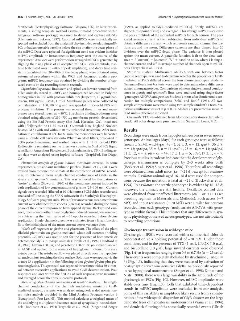
Glycinergic transmission in wild-type miceGlycinergic mIPSCs were recorded with a symmetrical chlorideconcentration at a holding potential of –70 mV. Under theseconditions, and in the presence of TTX (1 �M), CNQX (10 �M),and bicuculline (10 �M), large inward currents were observed(Fig. 1A) at frequencies ranging from 0.6 to 6.7 Hz (n 23 cells).These events were completely abolished by strychnine (1 �M; n 6) (Fig. 1B), indicating that they were mediated by activation ofpostsynaptic strychnine-sensitive GlyRs. As previously reportedin rat hypoglossal motoneurons (Singer et al., 1998; Donato andNistri, 2000), there was a large variability in the amplitude of theglycinergic mIPSCs (Fig. 1C). However, mIPSC amplitudes werestable over time (Fig. 1D). Cells that exhibited time-dependenttrends in mIPSC amplitude were excluded from our analysis.Some of this variability in amplitude likely results from a combi-nation of the wide spatial dispersion of GlyR clusters on the largedendritic trees of hypoglossal motoneurons (Viana et al., 1994)and dendritic filtering of the somatically recorded events (Ulrich
4882 • J. Neurosci., May 3, 2006 • 26(18):4880 – 4890 Graham et al. • Glycinergic Neurotransmission in Murine Mutants
and Luscher, 1993). Indeed, plots of rise time against event am-plitude reveal that small events had larger average rise times (Fig.1E). The average mIPSC had a 10 –90% rise time of 0.9 � 0.1 ms,and the decay was well fit by a single exponential with a timeconstant of 4.9 � 0.2 ms (n 23 cells) (Fig. 1F).
Local electrical stimulation in the presence of CNQX (10 �M)and bicuculline (10 �M) generated an IPSC that was completelyblocked by strychnine (1 �M; n 3 cells) (Fig. 1G). The IPSCshad a linear current–voltage relationship and a reversal potentialof �5 � 1 mV (n 4 cells) (Fig. 1H). This is consistent with thecalculated reversal potential for chloride and confirms that themajor ionic species underlying the large inward currents ischloride.
Glycinergic transmission in wild-type and heterozygotemutant miceTo establish appropriate controls for examining the effect of GlyRmutations, we first compared glycinergic mIPSCs from wild-typemice and animals carrying a single mutant allele (i.e., spa/�,spd/�, and ot/�). Whereas mIPSC frequency, rise, and decaytime constant are similar in these genotypes, average amplitudeof mIPSCs differed (Table 1). Specifically, mIPSC amplitude issmaller in spasmodic and oscillator heterozygotes compared withwild-type and heterozygote spastic mice (56.6 � 2.7 and 49.1 �4.1 pA vs 71.7 � 3.5 and 65.0 � 4.5 pA, respectively; multivariateANOVA, p � 0.05). These data suggest that GlyR density maydiffer in wild-type vs spasmodic and oscillator heterozygotes.
There are several explanations for these observations. First, itis possible that a mixed population of GlyRs are present in spas-modic heterozygotes. For example, receptors might containvarious combinations of � subunits (i.e., 2 �1wt, 2 �1A52S, or1 �1wt and 1 �1A52S). Genetic and biochemical consider-ations as well as our data, however, suggest these factors do notdramatically affect the GlyR physiology. Moreover, heterozy-gotes (carriers) of recessive genetic disorders, such as the threemouse mutants under study, are typically without overt phe-notypic symptoms.
For oscillator heterozygotes, the above considerations also ap-ply. Additionally, previous studies have shown that glycinergicmIPSC amplitude increases in the first 3 postnatal weeks in rats(Singer et al., 1998). This factor may help explain the reducedmIPSC amplitude observed in oscillator controls because muchyounger animals were used (16 –18 vs �32 d). In summary, theseresults show that not all features of glycinergic transmission arecomparable in wild-type and heterozygote animals. Therefore,when assessing the effect of each mutation on GlyR properties, weuse data from age-matched heterozygotes rather than wild-typeanimals as controls. In this way, we are able to describe the spe-cific physiological changes that are responsible for the phenotypicchanges in each mutant line.
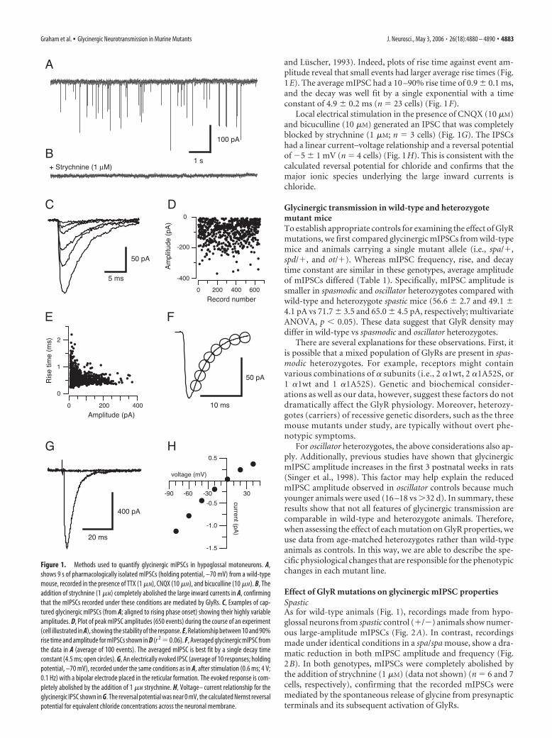
Effect of GlyR mutations on glycinergic mIPSC propertiesSpasticAs for wild-type animals (Fig. 1), recordings made from hypo-glossal neurons from spastic control (�/�) animals show numer-ous large-amplitude mIPSCs (Fig. 2A). In contrast, recordingsmade under identical conditions in a spa/spa mouse, show a dra-matic reduction in both mIPSC amplitude and frequency (Fig.2B). In both genotypes, mIPSCs were completely abolished bythe addition of strychnine (1 �M) (data not shown) (n 6 and 7cells, respectively), confirming that the recorded mIPSCs weremediated by the spontaneous release of glycine from presynapticterminals and its subsequent activation of GlyRs.
Figure 1. Methods used to quantify glycinergic mIPSCs in hypoglossal motoneurons. A,shows 9 s of pharmacologically isolated mIPSCs (holding potential, –70 mV) from a wild-typemouse, recorded in the presence of TTX (1 �M), CNQX (10 �M), and bicuculline (10 �M). B, Theaddition of strychnine (1 �M) completely abolished the large inward currents in A, confirmingthat the mIPSCs recorded under these conditions are mediated by GlyRs. C, Examples of cap-tured glycinergic mIPSCs (from A; aligned to rising phase onset) showing their highly variableamplitudes. D, Plot of peak mIPSC amplitudes (650 events) during the course of an experiment(cell illustrated in A), showing the stability of the response. E, Relationship between 10 and 90%rise time and amplitude for mIPSCs shown in D (r 2 0.06). F, Averaged glycinergic mIPSC fromthe data in A (average of 100 events). The averaged mIPSC is best fit by a single decay timeconstant (4.5 ms; open circles). G, An electrically evoked IPSC (average of 10 responses; holdingpotential, –70 mV), recorded under the same conditions as in A, after stimulation (0.6 ms; 4 V;0.1 Hz) with a bipolar electrode placed in the reticular formation. The evoked response is com-pletely abolished by the addition of 1 �M strychnine. H, Voltage– current relationship for theglycinergic IPSC shown in G. The reversal potential was near 0 mV, the calculated Nernst reversalpotential for equivalent chloride concentrations across the neuronal membrane.
Graham et al. • Glycinergic Neurotransmission in Murine Mutants J. Neurosci., May 3, 2006 • 26(18):4880 – 4890 • 4883
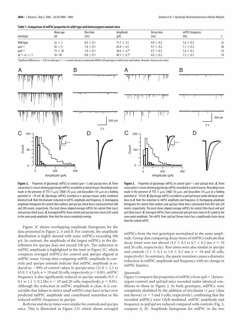
Figure 2C shows overlapping amplitude histograms for thedata presented in Figure 2, A and B. For controls, the amplitudedistribution is highly skewed with some mIPSCs exceeding 300pA. In contrast, the amplitude of the largest mIPSCs in the dis-tribution for spa/spa does not exceed 100 pA. The reduction inmIPSC amplitude is highlighted in the inset in Figure 2C, whichcompares averaged mIPSCs for control and spa/spa aligned atmIPSC onset. Group data comparing mIPSC amplitude in con-trols and spa/spa animals indicate that mIPSC amplitude is re-duced to �30% of control values in spa/spa mice (21.0 � 2.1 vs65.0 � 4.5 pA; n 19 and 20 cells, respectively; p � 0.05). mIPSCfrequency is also significantly reduced in spa/spa animals (0.5 �0.1 vs 1.1 � 0.2 Hz; n 19 and 20 cells, respectively; p � 0.05).Although the reduction in mIPSC amplitude is clear, it is con-ceivable that failure to detect small mIPSCs may have in fact overpredicted mIPSC amplitude and contributed somewhat to thereduced mIPSC frequency in spa/spa.
Both rise and decay times were similar for controls and spa/spamice. This is illustrated in Figure 2D, which shows averaged
mIPSCs from the two genotypes normalized to the same ampli-tude. Group data comparing decay times of mIPSCs indicate thatdecay times were not altered (4.5 � 0.3 vs 4.7 � 0.2 ms; n 19and 20 cells, respectively). Rise times were also similar in spa/spaand controls (1.1 � 0.1 vs 1.0 � 0.1 ms; n 19 and 20 cells,respectively). In summary, the spastic mutation causes a dramaticreduction in mIPSC amplitude and frequency with no change inmIPSC kinetics.
SpasmodicFigure 3 compares the properties of mIPSCs from spd/� (hetero-zygote control) and spd/spd mice recorded under identical con-ditions to those in Figure 2. In both genotypes, mIPSCs werecompletely abolished by the addition of strychnine (1 �M) (datanot shown) (n 5 and 5 cells, respectively), confirming that therecorded mIPSCs were GlyR-mediated. mIPSC amplitude andfrequency in spd/spd are reduced compared with controls (Fig. 3,compare A, B). Amplitude histograms for mIPSC in the two
Table 1. Comparison of mIPSC properties in wild-type and heterozygote mutant mice
GenotypeMean age(d)
Rise time(ms)
Amplitude(pA)
Decay time(ms)
mIPSC frequency(Hz) n
Wild type 32 � 3 0.9 � 0.1 71.7 � 3.5 4.9 � 0.2 1.6 � 0.3 23spa/� 36 � 9 1.0 � 0.1 65.0 � 4.5 4.7 � 0.2 1.1 � 0.2 20spd/� 73 � 16 1.0 � 0.1 56.6 � 2.7a 4.7 � 0.2 1.6 � 0.2 23ot/� or �/� 16 –18 0.8 � 0.1 49.1 � 4.1a 4.8 � 0.2 1.5 � 0.3 14aSignificant difference (p � 0.05) to wild-type (�/�) controls (based on multivariate ANOVA with genotype as within factor and Student–Newman–Keuls post hoc tests).
Figure 2. Properties of glycinergic mIPSCs in control (spa/�) and spa/spa mice. A, Threeconsecutive 5 s traces showing glycinergic mIPSCs recorded in a control mouse. Recordings weremade in the presence of TTX (1 �M), CNQX (10 �M), and bicuculline (10 �M) at a holdingpotential of –70 mV. B, Glycinergic mIPSCs recorded in a spa/spa mouse under conditionsidentical to A. Note the dramatic reduction in mIPSC amplitude and frequency. C, Overlappingamplitude histograms for control (thin outline) and spa/spa (thick lines) constructed from 888and 290 events, respectively. The inset shows aligned average mIPSCs for control (thin trace)and spa/spa (thick trace). D, Averaged mIPSCs from control and spa/spa mice (inset in C) scaledto the same peak amplitude. Note that the traces completely overlap.
Figure 3. Properties of glycinergic mIPSCs in control (spd/�) and spd/spd mice. A, Threeconsecutive 5 s traces showing glycinergic mIPSCs recorded in a control mouse. Recordings weremade in the presence of TTX (1 �M), CNQX (10 �M), and bicuculline (10 �M) at a holdingpotential of –70 mV. B, Glycinergic mIPSCs recorded in a spd/spd mouse under identical condi-tions to A. Note the reduction in mIPSC amplitude and frequency. C, Overlapping amplitudehistograms for control (thin outline) and spd/spd (thick lines) constructed from 893 and 228events, respectively. The inset shows aligned average mIPSCs for control (thin trace) and spd/spd (thick trace). D, Averaged mIPSCs from control and spd/spd mice (inset in C) scaled to thesame peak amplitude. The mIPSC from spd/spd (heavy trace) has a significantly faster decaythan the control mIPSC.
4884 • J. Neurosci., May 3, 2006 • 26(18):4880 – 4890 Graham et al. • Glycinergic Neurotransmission in Murine Mutants
genotypes show that, as for spa/� animals, the distribution ishighly skewed for controls with some mIPSCs exceeding 250 pA,whereas the largest mIPSC for spd/spd does not exceed 100 pA(Fig. 3C). This reduction in mIPSC amplitude is highlighted inthe inset in Figure 3C. Group data comparing mIPSC amplitudein control and spd/spd animals shows that amplitude is reducedby �50% (29.0 � 3.0 vs 56.6 � 2.7 pA; n 20 and 23 cells,respectively; p � 0.05). mIPSC frequency is also significantly re-duced in spd/spd animals (1.0 � 0.3 vs 1.6 � 0.2 Hz; n 20 and23 cells, respectively; p � 0.05).
When averaged mIPSCs are scaled to the same amplitude, thedecay time was faster for spd/spd (Fig. 3D) (2.7 � 0.2 vs 4.7 � 0.2ms; n 20 and 23 cells, respectively; p � 0.05). The faster decaytimes in spd/spd were also accompanied by faster mIPSC risetimes (0.7 � 0.1 vs 1.0 � 0.1 ms; n 20 and 23 cells, respectively;p � 0.05). In summary, as with the spastic mutation, the spas-modic mutation reduces mIPSC amplitude and frequency. How-ever, the spasmodic mutation also produces more rapid mIPSCkinetics. These data are similar to those previously published in abrief conference report from our laboratory (Callister et al.,1999).
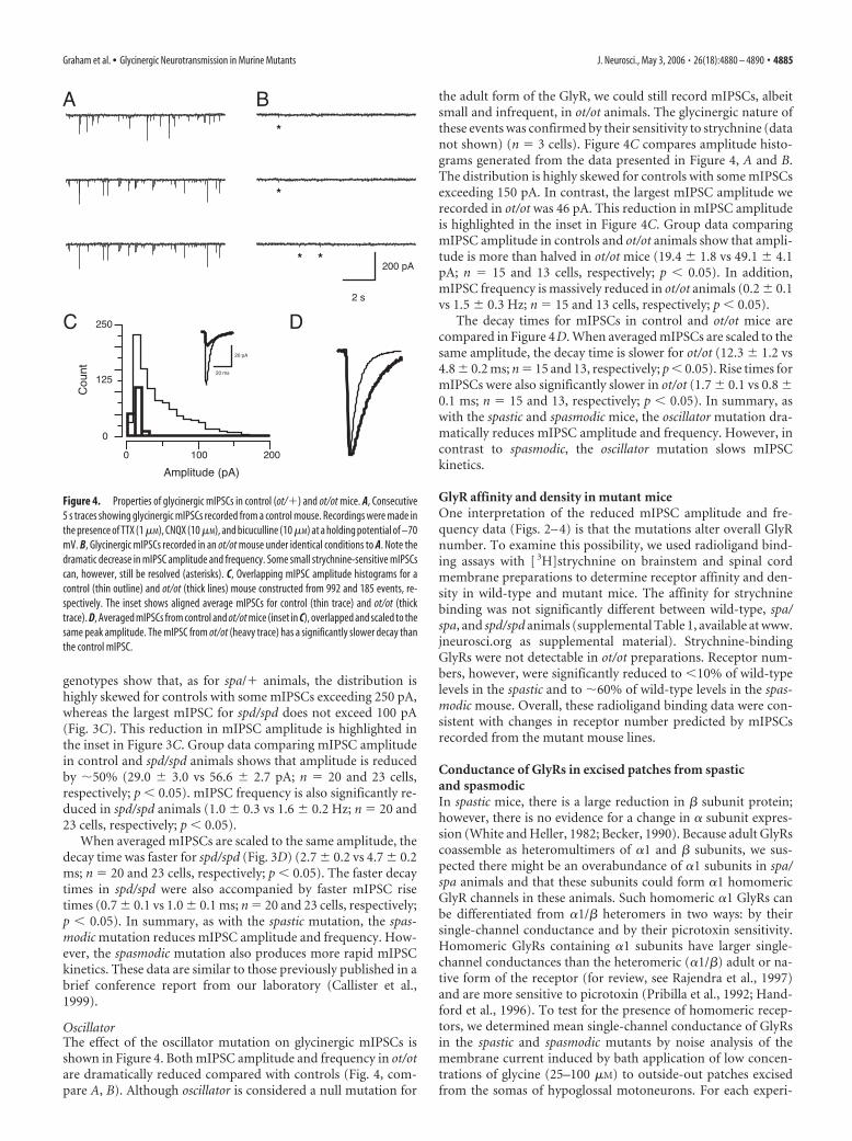
OscillatorThe effect of the oscillator mutation on glycinergic mIPSCs isshown in Figure 4. Both mIPSC amplitude and frequency in ot/otare dramatically reduced compared with controls (Fig. 4, com-pare A, B). Although oscillator is considered a null mutation for
the adult form of the GlyR, we could still record mIPSCs, albeitsmall and infrequent, in ot/ot animals. The glycinergic nature ofthese events was confirmed by their sensitivity to strychnine (datanot shown) (n 3 cells). Figure 4C compares amplitude histo-grams generated from the data presented in Figure 4, A and B.The distribution is highly skewed for controls with some mIPSCsexceeding 150 pA. In contrast, the largest mIPSC amplitude werecorded in ot/ot was 46 pA. This reduction in mIPSC amplitudeis highlighted in the inset in Figure 4C. Group data comparingmIPSC amplitude in controls and ot/ot animals show that ampli-tude is more than halved in ot/ot mice (19.4 � 1.8 vs 49.1 � 4.1pA; n 15 and 13 cells, respectively; p � 0.05). In addition,mIPSC frequency is massively reduced in ot/ot animals (0.2 � 0.1vs 1.5 � 0.3 Hz; n 15 and 13 cells, respectively; p � 0.05).
The decay times for mIPSCs in control and ot/ot mice arecompared in Figure 4D. When averaged mIPSCs are scaled to thesame amplitude, the decay time is slower for ot/ot (12.3 � 1.2 vs4.8 � 0.2 ms; n 15 and 13, respectively; p � 0.05). Rise times formIPSCs were also significantly slower in ot/ot (1.7 � 0.1 vs 0.8 �0.1 ms; n 15 and 13, respectively; p � 0.05). In summary, aswith the spastic and spasmodic mice, the oscillator mutation dra-matically reduces mIPSC amplitude and frequency. However, incontrast to spasmodic, the oscillator mutation slows mIPSCkinetics.
GlyR affinity and density in mutant miceOne interpretation of the reduced mIPSC amplitude and fre-quency data (Figs. 2– 4) is that the mutations alter overall GlyRnumber. To examine this possibility, we used radioligand bind-ing assays with [ 3H]strychnine on brainstem and spinal cordmembrane preparations to determine receptor affinity and den-sity in wild-type and mutant mice. The affinity for strychninebinding was not significantly different between wild-type, spa/spa, and spd/spd animals (supplemental Table 1, available at www.jneurosci.org as supplemental material). Strychnine-bindingGlyRs were not detectable in ot/ot preparations. Receptor num-bers, however, were significantly reduced to �10% of wild-typelevels in the spastic and to �60% of wild-type levels in the spas-modic mouse. Overall, these radioligand binding data were con-sistent with changes in receptor number predicted by mIPSCsrecorded from the mutant mouse lines.
Conductance of GlyRs in excised patches from spasticand spasmodicIn spastic mice, there is a large reduction in � subunit protein;however, there is no evidence for a change in � subunit expres-sion (White and Heller, 1982; Becker, 1990). Because adult GlyRscoassemble as heteromultimers of �1 and � subunits, we sus-pected there might be an overabundance of �1 subunits in spa/spa animals and that these subunits could form �1 homomericGlyR channels in these animals. Such homomeric �1 GlyRs canbe differentiated from �1/� heteromers in two ways: by theirsingle-channel conductance and by their picrotoxin sensitivity.Homomeric GlyRs containing �1 subunits have larger single-channel conductances than the heteromeric (�1/�) adult or na-tive form of the receptor (for review, see Rajendra et al., 1997)and are more sensitive to picrotoxin (Pribilla et al., 1992; Hand-ford et al., 1996). To test for the presence of homomeric recep-tors, we determined mean single-channel conductance of GlyRsin the spastic and spasmodic mutants by noise analysis of themembrane current induced by bath application of low concen-trations of glycine (25–100 �M) to outside-out patches excisedfrom the somas of hypoglossal motoneurons. For each experi-
Figure 4. Properties of glycinergic mIPSCs in control (ot/�) and ot/ot mice. A, Consecutive5 s traces showing glycinergic mIPSCs recorded from a control mouse. Recordings were made inthe presence of TTX (1 �M), CNQX (10 �M), and bicuculline (10 �M) at a holding potential of –70mV. B, Glycinergic mIPSCs recorded in an ot/ot mouse under identical conditions to A. Note thedramatic decrease in mIPSC amplitude and frequency. Some small strychnine-sensitive mIPSCscan, however, still be resolved (asterisks). C, Overlapping mIPSC amplitude histograms for acontrol (thin outline) and ot/ot (thick lines) mouse constructed from 992 and 185 events, re-spectively. The inset shows aligned average mIPSCs for control (thin trace) and ot/ot (thicktrace). D, Averaged mIPSCs from control and ot/ot mice (inset in C), overlapped and scaled to thesame peak amplitude. The mIPSC from ot/ot (heavy trace) has a significantly slower decay thanthe control mIPSC.
Graham et al. • Glycinergic Neurotransmission in Murine Mutants J. Neurosci., May 3, 2006 • 26(18):4880 – 4890 • 4885
ment, the applied glycine concentration was adjusted to generatean inward current that did not exceed �50 pA from a holdingpotential of –50 mV.
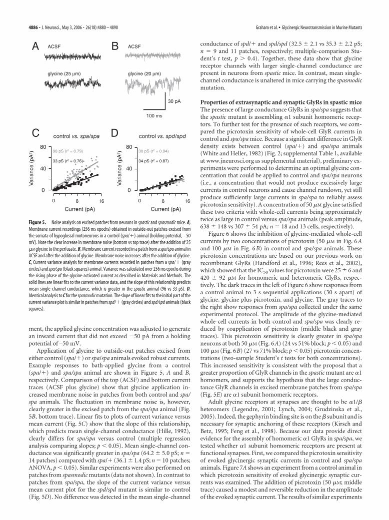
Application of glycine to outside-out patches excised fromeither control (spa/�) or spa/spa animals evoked robust currents.Example responses to bath-applied glycine from a control(spa/�) and spa/spa animal are shown in Figure 5, A and B,respectively. Comparison of the top (ACSF) and bottom currenttraces (ACSF plus glycine) show that glycine application in-creased membrane noise in patches from both control and spa/spa animals. The fluctuation in membrane noise is, however,clearly greater in the excised patch from the spa/spa animal (Fig.5B, bottom trace). Linear fits to plots of current variance versusmean current (Fig. 5C) show that the slope of this relationship,which predicts mean single-channel conductance (Hille, 1992),clearly differs for spa/spa versus control (multiple regressionanalysis comparing slopes; p � 0.05). Mean single-channel con-ductance was significantly greater in spa/spa (64.2 � 5.0 pS; n 14 patches) compared with spa/� (36.1 � 1.4 pS; n 10 patches;ANOVA, p � 0.05). Similar experiments were also performed onpatches from spasmodic mutants (data not shown). In contrast topatches from spa/spa, the slope of the current variance versusmean current plot for the spd/spd mutant is similar to control(Fig. 5D). No difference was detected in the mean single-channel
conductance of spd/� and spd/spd (32.5 � 2.1 vs 35.3 � 2.2 pS;n 9 and 11 patches, respectively; multiple-comparison Stu-dent’s t test, p � 0.4). Together, these data show that glycinereceptor channels with larger single-channel conductance arepresent in neurons from spastic mice. In contrast, mean single-channel conductance is unaltered in mice carrying the spasmodicmutation.
Properties of extrasynaptic and synaptic GlyRs in spastic miceThe presence of large conductance GlyRs in spa/spa suggests thatthe spastic mutant is assembling �1 subunit homomeric recep-tors. To further test for the presence of such receptors, we com-pared the picrotoxin sensitivity of whole-cell GlyR currents incontrol and spa/spa mice. Because a significant difference in GlyRdensity exists between control (spa/�) and spa/spa animals(White and Heller, 1982) (Fig. 2; supplemental Table 1, availableat www.jneurosci.org as supplemental material), preliminary ex-periments were performed to determine an optimal glycine con-centration that could be applied to control and spa/spa neurons(i.e., a concentration that would not produce excessively largecurrents in control neurons and cause channel rundown, yet stillproduce sufficiently large currents in spa/spa to reliably assesspicrotoxin sensitivity). A concentration of 50 �M glycine satisfiedthese two criteria with whole-cell currents being approximatelytwice as large in control versus spa/spa animals (peak amplitude,638 � 148 vs 307 � 54 pA; n 18 and 13 cells, respectively).
Figure 6 shows the inhibition of glycine-mediated whole-cellcurrents by two concentrations of picrotoxin (50 �M in Fig. 6Aand 100 �M in Fig. 6B) in control and spa/spa animals. Thesepicrotoxin concentrations are based on our previous work onrecombinant GlyRs (Handford et al., 1996; Rees et al., 2002),which showed that the IC50 values for picrotoxin were 25 � 6 and420 � 92 �M for homomeric and heteromeric GlyRs, respec-tively. The dark traces in the left of Figure 6 show responses froma control animal to 3 s sequential applications (30 s apart) ofglycine, glycine plus picrotoxin, and glycine. The gray traces tothe right show responses from spa/spa collected under the sameexperimental protocol. The amplitude of the glycine-mediatedwhole-cell currents in both control and spa/spa was clearly re-duced by coapplication of picrotoxin (middle black and graytraces). This picrotoxin sensitivity is clearly greater in spa/spaneurons at both 50 �M (Fig. 6A) (24 vs 51% block; p � 0.05) and100 �M (Fig. 6B) (27 vs 71% block; p � 0.05) picrotoxin concen-trations (two-sample Student’s t tests for both concentrations).This increased sensitivity is consistent with the proposal that agreater proportion of GlyR channels in the spastic mutant are �1homomers, and supports the hypothesis that the large conduc-tance GlyR channels in excised membrane patches from spa/spa(Fig. 5E) are �1 subunit homomeric receptors.
Adult glycine receptors at synapses are thought to be �1/�heteromers (Legendre, 2001; Lynch, 2004; Grudzinska et al.,2005). Indeed, the gephyrin binding site is on the � subunit and isnecessary for synaptic anchoring of these receptors (Kirsch andBetz, 1995; Feng et al., 1998). Because our data provide directevidence for the assembly of homomeric �1 GlyRs in spa/spa, wetested whether �1 subunit homomeric receptors are present atfunctional synapses. First, we compared the picrotoxin sensitivityof evoked glycinergic synaptic currents in control and spa/spaanimals. Figure 7A shows an experiment from a control animal inwhich picrotoxin sensitivity of evoked glycinergic synaptic cur-rents was examined. The addition of picrotoxin (50 �M; middletrace) caused a modest and reversible reduction in the amplitudeof the evoked synaptic current. The results of similar experiments
Figure 5. Noise analysis on excised patches from neurons in spastic and spasmodic mice. A,Membrane current recordings (256 ms epochs) obtained in outside-out patches excised fromthe somata of hypoglossal motoneurons in a control (spa/�) animal (holding potential, –50mV). Note the clear increase in membrane noise (bottom vs top trace) after the addition of 25�M glycine to the perfusate. B, Membrane current recorded in a patch from a spa/spa animal inACSF and after the addition of glycine. Membrane noise increases after the addition of glycine.C, Current variance analysis for membrane currents recorded in patches from a spa/� (graycircles) and spa/spa (black squares) animal. Variance was calculated over 256 ms epochs duringthe rising phase of the glycine-activated current as described in Materials and Methods. Thesolid lines are linear fits to the current variance data, and the slope of this relationship predictsmean single-channel conductance, which is greater in the spastic animal (96 vs 33 pS). D,Identical analysis to C for the spasmodic mutation. The slope of linear fits to the initial part of thecurrent variance plot is similar in patches from spd/� (gray circles) and spd/spd animals (blacksquares).
4886 • J. Neurosci., May 3, 2006 • 26(18):4880 – 4890 Graham et al. • Glycinergic Neurotransmission in Murine Mutants
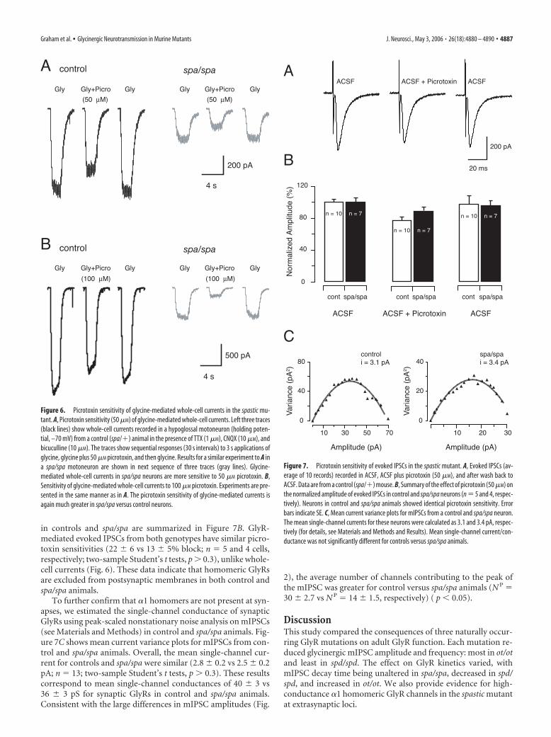
in controls and spa/spa are summarized in Figure 7B. GlyR-mediated evoked IPSCs from both genotypes have similar picro-toxin sensitivities (22 � 6 vs 13 � 5% block; n 5 and 4 cells,respectively; two-sample Student’s t tests, p � 0.3), unlike whole-cell currents (Fig. 6). These data indicate that homomeric GlyRsare excluded from postsynaptic membranes in both control andspa/spa animals.
To further confirm that �1 homomers are not present at syn-apses, we estimated the single-channel conductance of synapticGlyRs using peak-scaled nonstationary noise analysis on mIPSCs(see Materials and Methods) in control and spa/spa animals. Fig-ure 7C shows mean current variance plots for mIPSCs from con-trol and spa/spa animals. Overall, the mean single-channel cur-rent for controls and spa/spa were similar (2.8 � 0.2 vs 2.5 � 0.2pA; n 13; two-sample Student’s t tests, p � 0.3). These resultscorrespond to mean single-channel conductances of 40 � 3 vs36 � 3 pS for synaptic GlyRs in control and spa/spa animals.Consistent with the large differences in mIPSC amplitudes (Fig.
2), the average number of channels contributing to the peak ofthe mIPSC was greater for control versus spa/spa animals (N P 30 � 2.7 vs N P 14 � 1.5, respectively) ( p � 0.05).
DiscussionThis study compared the consequences of three naturally occur-ring GlyR mutations on adult GlyR function. Each mutation re-duced glycinergic mIPSC amplitude and frequency: most in ot/otand least in spd/spd. The effect on GlyR kinetics varied, withmIPSC decay time being unaltered in spa/spa, decreased in spd/spd, and increased in ot/ot. We also provide evidence for high-conductance �1 homomeric GlyR channels in the spastic mutantat extrasynaptic loci.
Figure 6. Picrotoxin sensitivity of glycine-mediated whole-cell currents in the spastic mu-tant. A, Picrotoxin sensitivity (50 �M) of glycine-mediated whole-cell currents. Left three traces(black lines) show whole-cell currents recorded in a hypoglossal motoneuron (holding poten-tial, –70 mV) from a control (spa/�) animal in the presence of TTX (1 �M), CNQX (10 �M), andbicuculline (10 �M). The traces show sequential responses (30 s intervals) to 3 s applications ofglycine, glycine plus 50 �M picrotoxin, and then glycine. Results for a similar experiment to A ina spa/spa motoneuron are shown in next sequence of three traces (gray lines). Glycine-mediated whole-cell currents in spa/spa neurons are more sensitive to 50 �M picrotoxin. B,Sensitivity of glycine-mediated whole-cell currents to 100 �M picrotoxin. Experiments are pre-sented in the same manner as in A. The picrotoxin sensitivity of glycine-mediated currents isagain much greater in spa/spa versus control neurons.
Figure 7. Picrotoxin sensitivity of evoked IPSCs in the spastic mutant. A, Evoked IPSCs (av-erage of 10 records) recorded in ACSF, ACSF plus picrotoxin (50 �M), and after wash back toACSF. Data are from a control (spa/�) mouse. B, Summary of the effect of picrotoxin (50 �M) onthe normalized amplitude of evoked IPSCs in control and spa/spa neurons (n 5 and 4, respec-tively). Neurons in control and spa/spa animals showed identical picrotoxin sensitivity. Errorbars indicate SE. C, Mean current variance plots for mIPSCs from a control and spa/spa neuron.The mean single-channel currents for these neurons were calculated as 3.1 and 3.4 pA, respec-tively (for details, see Materials and Methods and Results). Mean single-channel current/con-ductance was not significantly different for controls versus spa/spa animals.
Graham et al. • Glycinergic Neurotransmission in Murine Mutants J. Neurosci., May 3, 2006 • 26(18):4880 – 4890 • 4887
The effect of GlyR mutations on synaptic transmissionBased on strychnine binding, the spastic mutation reduces GlyRnumbers without altering affinity (supplemental Table 1, avail-able at www.jneurosci.org as supplemental material) (White andHeller, 1982; Becker et al., 1986). Consistent with these bindingdata and previous physiological studies (Biscoe and Duchen,1986; Callister et al., 1999; Graham et al., 2003; von Wegerer et al.,2003), our recordings in spa/spa show that GlyR-mediatedmIPSC amplitude and frequency are decreased but decay time isunchanged (Fig. 2). These findings, coupled with a decrease in theaverage number of channels contributing to the peak of themIPSC (Fig. 7C), are consistent with decreased GlyR density atindividual release sites. The reduced mIPSC frequency in spa/spaalso provides evidence for a decrease in the number of connec-tions/release sites (Turrigiano, 1999; Kilman et al., 2002). Analternative to reduced GlyR density is a preferential redistribu-tion of GlyRs to distal dendritic loci. This would lead to greaterelectrotonic filtering of mIPSCs in spa/spa, reduced mIPSC am-plitude, and slowed mIPSC time course. The similar mIPSC riseand decay times in spa/spa and controls, however, argues againstchanges in GlyR distribution. Thus, although the spastic muta-tion decreases � subunit protein production (Kingsmore et al.,1994; Mulhardt et al., 1994), the transcribed � subunit is still ableto combine effectively with �1 subunits, form normal receptors,and be targeted to postsynaptic densities.
In spd/spd, strychnine binding (supplemental Table 1, avail-able at www.jneurosci.org as supplemental material), mIPSC am-plitude, and frequency are decreased. mIPSC time course is sub-stantially faster (Fig. 3), whereas noise analysis on excised patches(Fig. 5) shows that mean single-channel conductance is unaf-fected. As for spa/spa, somatodendritic redistribution of mutantGlyRs could partly account for these observations. For instance,distally located GlyRs would reduce mIPSC amplitude and slowthe mIPSC time course. The reduced mIPSC amplitude and fastertime course we observed in spd/spd argues against this, and favorsan explanation based on altered channel properties.
Previous studies have measured a sixfold reduction in glycinesensitivity in recombinant �1 homomeric receptors carrying thespasmodic mutation (Ryan et al., 1994; Saul et al., 1994). The 50%reduction in mIPSC amplitude we observed in spd/spd is consis-tent with these data. Likewise, the reduced strychnine binding(supplemental Table 1, available at www.jneurosci.org as supple-mental material) suggests GlyR density might also contribute toreduced mIPSC amplitude. The faster mIPSC rise and decaytimes we observed suggests the main effect of the spasmodic mu-tation is to increase the rate of glycine unbinding and shorten thetime the receptor remains activated (Legendre, 2001). Thus, thefaster mIPSC decay rate is likely to be attributable to a concomi-tant reduction in channel mean open time. Detailed single-channel analysis is needed to test this issue directly. Functionally,the faster mIPSC time course in spd/spd mice results in substan-tially lower charge transfer for mIPSCs of similar amplitude. Thiscould account for the very similar phenotypes for spd/spd andspa/spa animals despite differences in mean mIPSC amplitude.
In ot/ot, glycinergic mIPSC amplitude, frequency, and strych-nine binding (supplemental Table 1, available at www.jneurosci.org as supplemental material) are reduced to levels approachingzero (Fig. 4). However, we could still resolve small amplitudestrychnine-sensitive mIPSCs in ot/ot animals that had slow kinet-ics compared with controls (Fig. 4). The presence of any glycin-ergic mIPSCs in ot/ot animals is surprising, because the mutationis considered functionally null for the adult form (�1/�) of theGlyR (Kling et al., 1997). One explanation is that GlyRs in oscil-
lator mice contain other � subunit isoforms (�2, �3, or �4 sub-units) (Malosio et al., 1991; Harvey et al., 2004). For example, �3and �4 subunits are expressed in spinal cord and cerebellum,respectively. In the hypoglossal nucleus, the �2 subunit is heavilyexpressed in neonates but levels decline by approximately post-natal day 14 (Becker et al., 1992; Singer et al., 1998). However,unlike the other mutants, our findings in ot/ot (low amplitudeand slowed mIPSCs) are consistent with a distal dendritic loca-tion for these remaining GlyR clusters. Regardless of their soma-todendritic location, these compensatory GlyRs can still clusterpostsynaptically in ot/ot. The level of glycinergic inhibition theyprovide, however, is not sufficient to sustain life.
The spastic mutation alters GlyR subunit assemblyand expressionAdult GlyRs are �1/� heteromers and, in expression systems,have a substantially lower mean channel conductance than �1homomers (�40 vs �85 pS) (Bormann et al., 1993; Rajendra etal., 1997). In agreement, the major in vivo adult form of the GlyR(�1/� heteromer) also has a low single-channel conductance of30 – 40 pS (Singer et al., 1998), suggesting either �1 homomersare not present in the adult or are present in very low densities.
Our data showing a single-channel conductance of 36 pS inpatches and 40 pS in mIPSCs from control mice are comparablewith the above literature. Noise analysis on patches from spa/sparevealed the presence of high conductance GlyRs that were notevident in controls or the other mutants (Fig. 5). The 64 � 5 pSmean channel conductance recorded in spa/spa patches is com-parable with substates of 30 – 88 pS measured in recombinant �1homomeric GlyRs (Bormann et al., 1993; Rajendra et al., 1995,1997). These results indicate that, when � subunits are dramati-cally reduced in spa/spa mice, the pool of available �1 subunitscan assemble as homomeric GlyRs.
To further test this hypothesis, we used the different picro-toxin sensitivities (Pribilla et al., 1992; Handford et al., 1996) ofheteromeric and homomeric GlyRs to conclude that the spa/spamutant is assembling and expressing homomeric �1 GlyRs (Fig.6). These results are consistent with a recent report by Molon etal. (2006), using real-time reverse transcription-multiplex PCRand Western blot analysis, which showed that overexpression of�1 GlyR subunits correlated with symptomatic recovery in adultspa/spa animals. Our data show that these “compensatory” ho-momeric GlyRs are not present at functional synapses (Fig. 7).We conclude that synaptic GlyRs in both spa/spa and controls are�1/� heteromers and that “compensatory” �1 homomeric GlyRsin spa/spa are confined to extrasynaptic loci.
Function of extrasynaptic GlyRs in spasticThere is now considerable evidence that LGICs, including GlyRs,can reside at both synaptic and extrasynaptic locations (Flint etal., 1998; Collingridge et al., 2004; Triller and Choquet, 2005). Arole for extrasynaptic LGICs is well established for the GABAA
receptor, with extrasynaptic receptors providing tonic inhibition(Semyanov et al., 2004; Farrant and Nusser, 2005). Could theextrasynaptic GlyRs in spa/spa play a similar role? If so, this mayalso explain why spa/spa mice are viable, but ot/ot mice and thebovine recessive �1 subunit myoclonus mutation (Pierce et al.,2001) are lethal.
Extrasynaptic GlyRs are present in various CNS regions (Flintet al., 1998; Delaney and Sah, 1999; Mori et al., 2002); however,unlike the GABAA receptor, their physiological role is unknown.One report suggests extrasynaptic GlyRs play a developmentalrole (Flint et al., 1998). In cortical and hippocampal neurons,
4888 • J. Neurosci., May 3, 2006 • 26(18):4880 – 4890 Graham et al. • Glycinergic Neurotransmission in Murine Mutants
extrasynaptic GlyRs are sensitive to locally released taurine and�-alanine (Mori et al., 2002), suggesting these GlyRs can modu-late cell excitability. Future experiments in the spastic mouse ad-dressing the function of extrasynaptic �1 homomeric receptorsmay help determine the role of GlyRs in CNS regions rostral tothe brainstem.
The absence of homomeric GlyRs at synaptic locations in spa/spa has implications for our understanding of GlyR development,because the exact composition of fetal synaptic GlyRs has beencontroversial (Legendre, 2001). Single-channel analysis and im-munolabeling suggest fetal GlyRs have an �2 homomeric sto-chiometry (Takahashi et al., 1992). This observation is at oddswith the notion that the GlyR � subunit stabilizes or clustersGlyRs at postsynaptic densities (Meyer et al., 1995). Our datashow that �1 homomeric GlyRs are not targeted to synapses inspa/spa and further confirms that the � subunit is essential fortargeting fetal or adult GlyRs to synapses, arguing against an �2homomeric stoichiometry for synaptic fetal GlyRs.
In summary, our analysis of GlyR mutants reveals that differ-ent genetic mutations, although producing similar behavioralphenotypes, alter GlyR properties and composition and result indistinct alterations in glycinergic transmission. The mechanismsunderlying the effect of each mutation on GlyR assembly andexpression are summarized in supplemental Figure 1 (available atwww.jneurosci.org as supplemental material). These distinct al-terations in the efficacy of glycinergic transmission in motoneu-rons seem likely to underlie the severe motor phenotype observedin a number of species and clinical conditions.
ReferencesBecker C-M (1990) Disorders of the inhibitory glycine receptor: the spastic
mouse. FASEB J 4:2767–2774.Becker C-M, Hermans-Borgmeyer I, Schmitt B, Betz H (1986) The glycine
receptor deficiency of the mutant mouse spastic: evidence for normalglycine receptor structure and localization. J Neurosci 6:1358 –1364.
Becker C-M, Schmeiden V, Tarroni P, Strasser U, Betz H (1992) Isoform-selective deficit of glycine receptors in the mouse mutant spastic. Neuron8:283–289.
Bekkers JM, Stevens CF (1989) NMDA and non-NMDA receptors are co-localized at individual excitatory synapses in cultured rat hippocampus.Nature 341:230 –233.
Biscoe TJ, Duchen MR (1986) Synaptic physiology of spinal motoneuronesof normal and spastic mice: an in vitro study. J Physiol (Lond)379:275–292.
Bormann J, Rundstrom N, Betz H, Langosch D (1993) Residues withintransmembrane segment M2 determine chloride conductance of glycinereceptor homo- and hetero-oligomers. EMBO J 12:3729 –3737.
Buckwalter MS, Cook SA, Davisson MT, White WF, Camper SA (1994) Aframeshift mutation in the mouse alpha 1 glycine receptor gene (Glra1)results in progressive neurological symptoms and juvenile death. HumMol Genet 3:2025–2030.
Callister RJ, Schofield PR, Sah P (1999) Use of murine mutants to studyglycine receptor function. Clin Exp Pharmacol Physiol 26:929 –931.
Chai CK (1961) Hereditary spasticity in mice. J Hered 52:241–243.Clements JD, Bekkers JM (1997) Detection of spontaneous synaptic events
with an optimally scaled template. Biophys J 73:220 –229.Collingridge GL, Isaac JT, Wang YT (2004) Receptor trafficking and synap-
tic plasticity. Nat Rev Neurosci 5:952–962.Delaney AJ, Sah P (1999) GABA receptors inhibited by benzodiazepines
mediate fast inhibitory transmission in the central amygdala. J Neurosci19:9698 –9704.
Donato R, Nistri A (2000) Relative contribution by GABA or glycine toCl �-mediated synaptic transmission on rat hypoglossal motoneurons invitro. J Neurophysiol 84:2715–2724.
Farrant M, Nusser Z (2005) Variations on an inhibitory theme: phasic andtonic activation of GABAA receptors. Nat Rev Neurosci 6:215–229.
Feng G, Tintrup H, Kirsch J, Nichol MC, Kuhse J, Betz H, Sanes JR (1998)
Dual requirement for gephyrin in glycine receptor clustering and molyb-doenzyme activity. Science 282:1321–1324.
Flint AC, Liu X, Kriegstein AR (1998) Nonsynaptic glycine receptor activa-tion during early neocortical development. Neuron 20:43–53.
Graham BA, Schofield PR, Sah P, Callister RJ (2003) Altered inhibitory syn-aptic transmission in superficial dorsal horn neurones in spastic and os-cillator mice. J Physiol (Lond) 551:905–916.
Grudzinska J, Schemm R, Haeger S, Nicke A, Schmalzing G, Betz H, Laube B(2005) The beta subunit determines the ligand binding properties ofsynaptic glycine receptors. Neuron 45:727–739.
Hamill OP, Neher ME, Sakmann B, Sigworth FJ (1981) Improved patch-clamp techniques for high-resolution current recording from cells andcell-free membrane patches. Pflugers Arch 391:85–100.
Handford CA, Lynch JW, Baker E, Webb GC, Ford JH, Sutherland GFR,Schofield PR (1996) The human glycine receptor � subunit: primarystructure, functional characterisation and chromosomal localisation ofthe human and murine genes. Brain Res Mol Brain Res 35:211–219.
Harvey RJ, Depner UB, Wassle H, Ahmadi S, Heindl C, Reinold H, Smart TG,Harvey K, Schutz B, Abo-Salem OM, Zimmer A, Poisbeau P, Welzl H,Wolfer DP, Betz H, Zeilhofer HU, Muller U (2004) GlyR alpha3: anessential target for spinal PGE2-mediated inflammatory pain sensitiza-tion. Science 304:884 – 887.
Hille B (1992) Ionic channels of excitable membranes. Sunderland, MA:Sinauer.
Katz B (1969) The release of neural transmitter substances. Liverpool, UK:Liverpool UP.
Kilman V, van Rossum MC, Turrigiano GG (2002) Activity deprivation re-duces miniature IPSC amplitude by decreasing the number of postsynap-tic GABAA receptors clustered at neocortical synapses. J Neurosci22:1328 –1337.
Kingsmore SF, Giros B, Suh D, Bieniarz M, Caron MG, Seldin MF (1994)Glycine receptor �-subunit gene mutation in spastic mouse associatedwith LINE-1 element insertion. Nat Genet 7:136 –142.
Kirsch J, Betz H (1995) The postsynaptic localization of the glycinereceptor-associated protein gephyrin is regulated by the cytoskeleton.J Neurosci 15:4148 – 4156.
Kling C, Koch M, Saul B, Becker C-M (1997) The Frameshift mutation os-cillator (Glra1spd-ot) produces a complete loss of glycine receptoralpha1-polypeptide in mouse central nervous system. Neuroscience78:411– 417.
Lane PW, Ganser AL, Kerner A-L, White WF (1987) Spasmodic, a mutationon chromosome 11 in the mouse. J Hered 78:353–356.
Legendre P (2001) The glycinergic inhibitory synapse. Cell Mol Life Sci58:760 –793.
Lynch JW (2004) Molecular structure and function of the glycine receptorchloride channel. Physiol Rev 84:1051–1095.
Malosio ML, Marqueze-Pouey B, Kuhse J, Betz H (1991) Widespread ex-pression of glycine receptor subunit mRNAs in the adult and developingrat brain. EMBO J 10:2401–2409.
Meyer G, Kirsch J, Betz H, Langosh D (1995) Identification of a gephyrinbinding motif on the glycine � subunit. Neuron 15:563–572.
Molon A, Di Giovanni S, Hathout Y, Natale J, Hoffman EP (2006) Func-tional recovery of glycine receptors in spastic murine model of startledisease. Neurobiol Dis 21:291–304.
Mori M, Gahwiler BH, Gerber U (2002) beta-Alanine and taurine as endog-enous agonists at glycine receptors in rat hippocampus in vitro. J Physiol(Lond) 539:191–200.
Mulhardt C, Fischer M, Gass P, Simon-Chazottes D, Guenet J-L, Kuhse J, BetzH, Becker C-M (1994) The spastic mouse: aberrant splicing of glycinereceptor � subunit mRNA caused by intronic insertion of L1 element.Neuron 13:1003–1015.
Pierce KD, Handford CA, Morris R, Vafa B, Dennis JA, Healy PJ, Schofield PR(2001) A nonsense mutation in the alpha1 subunit of the inhibitory gly-cine receptor associated with bovine myoclonus. Mol Cell Neurosci17:354 –363.
Pribilla I, Takagi T, Langosch D, Bormann J, Betz H (1992) The atypical M2segment of the beta subunit confers picrotoxinin resistance to inhibitoryglycine receptor channels. EMBO J 11:4305– 4311.
Rajendra S, Schofield PR (1995) Molecular mechanisms of inherited startlesyndromes. Trends Neurosci 18:80 – 82.
Rajendra S, Lynch J, Schofield P (1997) The glycine receptor. PharmacolTher 73:121–146.
Graham et al. • Glycinergic Neurotransmission in Murine Mutants J. Neurosci., May 3, 2006 • 26(18):4880 – 4890 • 4889
Rajendra S, Lynch JW, Pierce KD, French CR, Barry PH, Schofield PR (1995)Mutation of an arginine residue in the human glycine receptor transformsbeta-alanine and taurine from agonists into competitive antagonists.Neuron 14:169 –175.
Rees MI, Lewis TM, Kwok JB, Mortier GR, Govaert P, Snell RG, Schofield PR,Owen MJ (2002) Hyperekplexia associated with compound heterozy-gote mutations in the beta-subunit of the human inhibitory glycine re-ceptor (GLRB). Hum Mol Genet 11:853– 860.
Robinson HP, Sahara Y, Kawai N (1991) Nonstationary fluctuation analysisand direct resolution of single channel currents at postsynaptic sites. Bio-phys J 59:295–304.
Ryan SG, Buckwalter MS, Lynch JW, Handford CA, Segura L, Shiang R,Wasmuth JJ, Camper SA, Schofield PA, O’Connell P (1994) A missensemutation in the gene encoding the �1 subunit of the inhibitory glycinereceptor in the spasmodic mouse. Nat Genet 7:131–135.
Saul B, Schmeiden V, Kling C, Mulhardt C, Gass P, Kuhse J, Becker C-M(1994) Point mutation of glycine receptor �1 subunit in the spasmodicmouse affects agonist responses. FEBS Lett 350:71–76.
Semyanov A, Walker MC, Kullmann DM, Silver RA (2004) Tonically activeGABAA receptors: modulating gain and maintaining the tone. TrendsNeurosci 27:262–269.
Sigworth FJ (1980) The variance of sodium current fluctuations at the nodeof Ranvier. J Physiol (Lond) 307:97–129.
Simon ES (1995) Involvement of glycine and GABAA receptors in thepathogenesis of spinal myoclonus: in vitro studies in the isolated neonatalrodent spinal cord. Neurology 45:1883–1892.
Singer JH, Berger AJ (1999) Contribution of single-channel properties tothe time course and amplitude variance of quantal glycine currents re-corded in rat motoneurons. J Neurophysiol 81:1608 –1616.
Singer JH, Talley EM, Bayliss DA, Berger AJ (1998) Development of glycin-
ergic synaptic transmission to rat brain stem motoneurons. J Neuro-physiol 80:2608 –2620.
Sokal R, Rohlf F (1995) Biometry: the principles and practice of statistics inbiological research, Ed 3. New York: Freeman.
Takahashi T, Momiyama A, Hirai K, Hishinuma F, Akagi H (1992) Func-tional correlation of fetal and adult forms of glycine receptors with devel-opmental changes in inhibitory synaptic receptor channels. Neuron9:1155–1161.
Traynelis SF, Silver RA, Cull-Candy SG (1993) Estimated conductance ofglutamate receptor channels activated during EPSCs at the cerebellarmossy fiber-granule cell synapse. Neuron 11:279 –289.
Triller A, Choquet D (2005) Surface trafficking of receptors between synap-tic and extrasynaptic membranes: and yet they do move! Trends Neurosci28:133–139.
Turrigiano GG (1999) Homestatic plasticity in neuronal networks: themore things change, the more they stay the same. Trends Neurosci22:221–227.
Ulrich D, Luscher HR (1993) Miniature excitatory synaptic currents cor-rected for dendritic cable properties reveal quantal size and variance.J Neurophysiol 69:1769 –1773.
Umemiya M, Berger AJ (1995) Presynaptic inhibition by serotonin of gly-cinergic inhibitory synaptic currents in the rat brain stem. J Neurophysiol73:1192–1200.
Viana F, Bayliss DA, Berger AJ (1994) Postnatal changes in rat hypoglossalmotoneuron membrane properties. Neuroscience 59:131–148.
von Wegerer J, Becker K, Glockenhammer D, Becker CM, Zeilhofer HU,Swandulla D (2003) Spinal inhibitory synaptic transmission in the gly-cine receptor mouse mutant spastic. Neurosci Lett 345:45– 48.
White WF, Heller AH (1982) Glycine receptor alteration in the mutantmouse spastic. Nature 298:655– 657.
4890 • J. Neurosci., May 3, 2006 • 26(18):4880 – 4890 Graham et al. • Glycinergic Neurotransmission in Murine Mutants