
Developmental toxicity of ambroxol in zebrafish …iii An Abstract of Developmental Toxicity of...
58
e University of Toledo e University of Toledo Digital Repository eses and Dissertations 2010 Developmental toxicity of ambroxol in zebrafish embryos/larvae: relevance of SULT-mediated sulfation of ambroxol Shaban Amani Al e University of Toledo Follow this and additional works at: hp://utdr.utoledo.edu/theses-dissertations is esis is brought to you for free and open access by e University of Toledo Digital Repository. It has been accepted for inclusion in eses and Dissertations by an authorized administrator of e University of Toledo Digital Repository. For more information, please see the repository's About page. Recommended Citation Al, Shaban Amani, "Developmental toxicity of ambroxol in zebrafish embryos/larvae: relevance of SULT-mediated sulfation of ambroxol" (2010). eses and Dissertations. 775. hp://utdr.utoledo.edu/theses-dissertations/775
Transcript of Developmental toxicity of ambroxol in zebrafish …iii An Abstract of Developmental Toxicity of...
The University of ToledoThe University of Toledo Digital Repository
Theses and Dissertations
2010
Developmental toxicity of ambroxol in zebrafishembryos/larvae: relevance of SULT-mediatedsulfation of ambroxolShaban Amani AlThe University of Toledo
Follow this and additional works at: http://utdr.utoledo.edu/theses-dissertations
This Thesis is brought to you for free and open access by The University of Toledo Digital Repository. It has been accepted for inclusion in Theses andDissertations by an authorized administrator of The University of Toledo Digital Repository. For more information, please see the repository's Aboutpage.
Recommended CitationAl, Shaban Amani, "Developmental toxicity of ambroxol in zebrafish embryos/larvae: relevance of SULT-mediated sulfation ofambroxol" (2010). Theses and Dissertations. 775.http://utdr.utoledo.edu/theses-dissertations/775

A Thesis
Entitled
Developmental Toxicity of Ambroxol in Zebrafish
Embryos/Larvae: Relevance of SULT-mediated Sulfation of
Ambroxol
By
Amani Al Shaban
Submitted to the Graduate Faculty as a partial fulfillment of the requirement
for the Master of Science in Pharmacology and Toxicology
_______________________________
Dr. Ming-Cheh Liu, Committee Chair
_____________________________________
Dr. Frederick Williams, Committee Member
_____________________________________
Dr. Zahoor Shah, Committee Member
_____________________________________
Dr. Patricia Komuniecki
College of Graduate Studies
The University of Toledo
May 2010

Copyright 2010, Amani Al Shaban
This document is copyrighted material. Under copyright law, no parts of this document may be reproduced without the expressed permission of the author.
iii
An Abstract of
Developmental Toxicity of Ambroxol in Zebrafish Embryos/Larvae: Relevance of
SULT-mediated Sulfation of Ambroxol
by
Amani Al Shaban
Submitted to the Graduate Faculty as a partial fulfillment of the requirement for the Master Degree in Pharmacology and Toxicology
The University of Toledo
May 2010
Ambroxol is an active metabolite of bromexine that has been proven to possess a great
bronchosecretolytic effect and has been used to treat infants from 0 to 6 month and
children till over 12 years of age, as well as adults. My thesis research was aimed to
detect potential adverse effects of ambroxol on development using zebrafish
embryos/larvae as a model and to investigate the possible involvement of the zebrafish
cytosolic sulfotransferases (SULTs) in the protection against the possible adverse effects.
Developing eggs at 24 hpf, 48 hpf, and 72 hpf were exposed to different concentrations
(1mM, 0.5 mM, 0.25 mM, 0.125 mM, and 0.05 mM) of ambroxol in triplicate and
observations were made daily for eleven consecutive days. Ambroxol induced cardiac
edema and bradycardia at different stages of development in a dose-dependent manner.
Enzymatic assay of purified zebrafish SULTs showed significant sulfation of ambroxol
by SULT2 ST1 and SULT3 ST1, 2, 3, 4, and 5. How these SULTs may be involved in
protection against the adverse effects of ambroxol remains to be clarified.

iv
Acknowledgments
I would first like to acknowledge my research supervisor for the opportunity to
work on this interesting project. I would also like to thank him for his teaching,
mentoring, and assistance throughout my graduate study. I would like to thanks my lab
mates for their help and cooperation to accomplish this project.
This research project would not have been possible without the support of many people. I
owe my deepest gratitude to my husband for his support and encouragement. I am
grateful to my parents for supporting me and giving me everything to make this possible.

v
Contents
Abstract iii
Acknowledgment s iv
Contents v
List of Tables vii
List of Figures viii
Introduction 1
1. Adverse Drug Reactions of Obstetric and Pediatric drugs………........ 1
2. Ontogeny of drug-metabolizing enzymes and the sensitivity to adverse
drug reactions during development …………………………………….. 2
3. Asthma Medications ………………………………………………… 6
3.1 Ambroxol …………………………………………………. 8

vi
4. Cytosolic sulfotransferases (SULTs) and their possible functional role 12
5. Zebrafish as a model for research on developmental pharmacology 14
5.1 Stages of Embryonic development of the zebrafish ………… 16
5.2 Drug metabolism in zebrafish………………………………. 19
Objectives and goals 22
Material and method 23
Results 27
Discussion 39
References 44

vii
List of Tables
Table1: Ambroxol doses as a pediatric drops…………………………………….. 10
Table 2: The average daily dose of Ambroxol as syrup………………………….. 10
Table 3: Heart rates (beat/min) of zebrafish larvae at different developmental stages in
response to different concentrations of Ambroxol. Numbers shown are the mean ± SD for
the 24 hpf exposure set ………………………………………………………… 33
Table 4: Heart rates (beat/min) of zebrafish larvae at different developmental stages in
response to different concentration of Ambroxol. Numbers shown are the mean ± SD for
the 48 hpf exposure set ..................................................................................... 34
Table 5: Heart rates (beat/min) of zebrafish larvae at different developmental stages in
response to different concentration of Ambroxol. Numbers shown are the mean ± SD for
the 72 hpf exposure set ………………………………………………………… 35
Table 6: Specific activity of the zebrafish SULT3 ST5 and SULT 5A1 toward drug
compound as substrates. The data represent mean ± SD from three independent
experiments. ND refers to the activity not detected ……………………………. 38

viii
List of Figures
Figure1: Stages of embryonic development of the zebrafish …………………… 18
Figure 2: Zebrafish exposed to 1mM Ambroxol at 48 hour post fertilization (hpf) showed
cardiac edema ………………………………………........................................... 28
Figure 3: Control group of zebrafish larvae at 48 hpf …………………………... 28
Figure 4: Zebrafish exposed to 0.5 mM Ambroxol at 72 hpf showed cardiac edema 28
Figure 5: Zebrafish exposed to 0.25 mM Ambroxol at72 hpf showed cardiac edema 29
Figure 6: Control group of zebrafish larvae at 72 hpf ………………………….. 29
Figure7: Zebrafish larva exposed to 1.0 mM Ambroxol at 96 hpf showed cardiac
edema …………………………………………………………………………. 29
Figure 8: Zebrafish larva exposed to 0.5 mM Ambroxol at96 hpf showed cardiac
edema……………………………………………………………………………. 29
Figure 9: Zebrafish larva exposed to 0.25 mM Ambroxol at 96 hpf showed cardiac
edema …………………………………………………………………………. 30

ix
Figure 10: Zebrafish larva exposed to 0.25 mM Ambroxol at 120 hpf showed cardiac
edema …………………………………………………………………………..... 30
Figure 11: Heart beat chart for 24 hpf Ambroxol-exposed-zebrafish larvae showed
significant bradycardia among 1 mM, 0.5 mM, and 0.25 mM Ambroxol-exposed
groups…………………………………………………………………………….. 31
Figure 12: Heart beat chart for 48 hpf Ambroxol-exposed-zebrafish larva showed
significant bradycardia among 1 mM, 0.5 mM, and 0.25 mM Ambroxol-exposed
groups ………………………………………………………………………… 32
Figure 13: Heart beat chart for 72 hpf Ambroxol-exposed-zebrafish larvae showed
significant bradycardia among 1 mM, 0.5 mM, 0.25 mM, and 0.125 mM Ambroxol
exposed ….………………………………………………………………………… 32
Figure 14: Mortality% chart for 24 hpf Ambroxol-exposed-zebrafish larvae showed
significant increase in the mortality in 1 mM, 0.5 mM, and 0.25 mM Ambroxol-exposed
groups …………………………………………………………………………. 36
Figure 15: Mortality% chart for 48 hpf Ambroxol-exposed-zebrafish larvae showed
significant increase in the mortality in 1 mM, 0.5 mM, and 0.25 mM Ambroxol-exposed
groups ………………………………………………………………………….. 36

x
Figure 16: 72 hour post fertilization, Mortality% chart for 72 hpf Ambroxol-exposed-
zebrafish larvae showed significant increase in the mortality in 1 mM, 0.5 mM, 0.25 mM,
and 0.125 mM Ambroxol-exposed groups …………………………………… 37

1
INTRODUCTION
1. Adverse Drug Reactions of Obstetric and Pediatric drugs
Although some adverse drug reactions are not very serious, others cause death,
hospitalization, or serious injury of more than 2 million people in the United States each
year. This issue is more serious especially during pregnancy and in the pediatric
population. Some drugs have been shown to be contraindicated during pregnancy, either
because they may cause developmental defects or death in extreme cases to the
embryos/fetus. Teratogenicity or growth retardation and other functional defects may also
manifest late in life. Any possible adverse effects on the embryos/fetus depend not only
on the ability to cross the placental barrier and directly access the embryo/fetus; it could
also depend on any detoxification in the embryonic/fetal liver. After birth, the safety of
medication in children is a major issue. Drug development for the pediatric population
especially neonates and infants is generally lacking. Most drugs are empirically
administered to newborns once the efficacy has been demonstrated in adults and
usefulness is suspected or demonstrated in the older pediatric population [Amanda
Clarkson et al, 2004]. Studies regarding drug usage suggested that 90% neonates received
at least 1 drug without adequate labeling to guide its use in this special population and led
to serious adverse effects in this special population [George P. Giacoia, MD et al, 2005].
The concern about the lack of knowledge of adverse drug reactions in pediatric

2
population and especially the neonatal age group is well founded. For example, some
studies suggest that adverse drugs reaction in pediatric population is a significant public
health problem. Multiple factors may increase the susceptibility to adverse drug reactions
in this population. These factors include immature drug detoxification mechanisms,
multiple drug exposures, coexistence with multiple organ dysfunctions, and the use of a
concentrated drug solution. One of the main factors that increase the susceptibility to
adverse drug reactions in children is the immature drug detoxification mechanism which
plays the major role in eliminating toxic compound and reduces their toxicity. Fetal
exposure to drug is modulated to a considerable degree by the metabolic capability of the
mother and the placenta during pregnancy and neonate/child liver after birth. In order to
avoid certain adverse effect, efficient biotransformation of the drugs by Phase I and Phase
II drug-metabolizing enzymes is an essential process. [Michael J. Blake et al, 2005].
2. Ontogeny of drug-metabolizing enzymes and the sensitivity to
adverse drug reactions during development
The combination of human ontogeny and genetic constitution exerts a profound effect
on both pharmacokinetics and pharmacodynamics. It is well recognized that changes in
pharmacokinetics occur during human development and these changes contribute to
differences in therapeutic efficacy and toxicant susceptibility of certain drugs. Although
changes in drug-metabolizing enzyme (DME) expressions during development are well
recognized, the knowledge needed to understand and predict therapeutic dosing and
avoidance of toxicity during maturation is incomplete.

3
Our knowledge of the developmental expression of human Phase I drug-metabolizing
enzymes is incomplete. Expression has been observed as early as organogenesis, but this
observed only in few enzymes. Many of Phase I drug-metabolizing enzymes exhibit a
prenatal expression changes that are regulated by mechanisms linked to birth and
maturity. The different stages of ontogeny are characterized by dynamic changes in gene
expression [Ronald N. Hines & Gail McCarver, 2001]. The cytochrome P450 gene
superfamily is responsible for biotransformation of a large array of endogenous
compound, pharmacologic agent, and environmental xenobiotics. Cytochrome P450s are
responsible for the majority of Phase I reactions in human and are represented by over 50
known enzymes grouped in to families based on sequence homology. The CYP3A
subfamily is the most abundant and clinically important of the CYPs enzymes in the liver
and small intestine, being responsible for the metabolism of about 50% of the most
commonly used drugs. The genes encoding CYP3A have been shown to include four
major functional isoforms (CYP3A4, CYP3A5, CYP3A7, and CYP3A43). Total hepatic
CYP3A content appears to be stable as the infant passes from fetal to neonatal life,
although the expression of specific isoform will change following birth and the activity of
CYP3A enzymes increases as the infant matures. The pattern of the increase appears to
be clearly associated with age.
Catalytic activity of CYP3A7 has been observed in embryonic liver as early as
50-60 days gestation and expression increases throughout pregnancy, peaking at two
weeks postnatal age and then decline to the lowest level. CYP3A4 is the primary hepatic
CYP that is expressed in the postnatal period. It accounts for approximately 30% of the
total P450 content in the liver and intestine, and it is the primary enzyme involved in

4
catalyzing the biotransformation of over 75 commonly used therapeutic drugs. CYP3A4
expression increases to 50% of adult values between 6 and 12 months of age with a
corresponding increase in functional activity. The developmental delay in the expression
of CYP3A4 has been implicated in the decreased clearance of benzodiazepine in infant
up to 3 months of age [Michael J. Blake et al, 2005]. CYP3A5 is the primary CYP3A
isoform that is expressed extrahepatically. It has been identified in fetal and adult liver
with expression and functional activity showing a high degree of inter individual
variability. This large variability is seen during all stages of development and no clear
development pattern has been identified. CYP2E1 is highly expressed in postnatal
development but less during gestation, suggesting that postnatal events are an important
for enzyme expression. CYP2D6 immunoreactive protein expression is very low in fetal
liver microsomes but can be identified in neonates by seven days of age.
Changes in the expression of Phase II drug-metabolizing enzyme expression levels
during development, as well as the balance seen between Phase I and Phase II enzymes,
can significantly alter the pharmacokinetics for a given drug or toxicant. Although our
knowledge is incomplete, many of the Phase II enzymes are expressed early in
development [D. Gail McCarver and Ronald N. Hines]. The Phase II reactions process
conjugating xenobiotics with small molecules such as UDP-glucuronic acid, glutathione,
or acetyl coenzyme A, which generally results in pharmacological inactivation or
detoxification by These reactions are catalyzed by a variety of enzymes, the activities of
which appear to be associated with development. The impact of ontogeny on Phase II
enzymes has not been investigated to the same extent as for Phase I enzyme.
Understanding their known developmental profile is important for an overall
5
understanding of the acquisition of metabolic competence in the neonate and the potential
therapeutic implications. Glutathione S transferase, GSTA1and GSTA2 have been
identified in human fetal liver tissue as early as 10 weeks gestational age with adult level
not reached until the 1-2 years postnatal age while glutathione S transferase M (GSTM) is
low in fetal liver and increases dramatically to adult level shortly after birth.
UDP-glucuronotransferase (UGT) is another Phase II enzyme which is responsible
for glucuronidation of hundreds of endogenous and exogenous compounds. Low level of
immunoreactive UGT protein is found early in gestation in liver, spleen, and kidney.
After birth it increases immediately, suggesting that post natal events are essential for the
expression of the gene. One of the most important groups of Phase II DMEs is the
cytosolic sulfotransferases. Sulfation is catalyzed by members of cytosolic
sulfotransferase (SULT) enzymes families that contain at least 11 distinct isoforms that
transfer a sulfuryl moiety to a wide range of endogenous and exogenous chemicals.
Sulfation generally results in a reduction in biological activity relative to the parent
compound, and it is the major mechanism for protection against chemical damage during
development. Additionally, the sulfation reaction plays a key role in the metabolism of
endogenous compound such as steroid hormone biosynthesis, catecholamine metabolism,
and thyroid hormone homeostasis. In the liver both enzyme activities and proteins levels
of SULT2A1 are low at first 25 weeks of gestation and then increase to near adult levels
in the neonate. The developmental expressions and activities of the major SULT enzymes
responsible for the metabolism of catecholamine (SULT1A3) and thyroid hormone
(SULT1A1) has been investigated. Hepatic SULT1A3 activity expressed at high level

6
early in the fetal and neonatal period, and then is essentially absent in adult liver [Michael
J. Blake et al, 2005].
Knowledge regarding the ontogeny of DME has permitted the development of models
based on their physiological and pharmacokinetic properties and improved capability to
predict drug disposition in pediatric patients. Changes in pharmacokinetics parameters
during development affect the therapeutic efficacy and adverse drug reaction observed in
children. However, the knowledge needed for better understanding and predicting
therapeutic dosing and avoidance of adverse reaction during maturation is still
incomplete [Ronald N. Hines, 2008].
3. Asthma Medications
Asthma is a lung disease that makes breathing difficult for nearly 23 million
Americans, including 7 million children. It causes 4000 death a year in United States.
Medication such as an inhaled short acting beta-2- agonist may be used to treat acute
attacks, but asthma can become persistent. Significant chronic air flow obstruction is
often present despite anti-asthma therapy and can lead to more serious conditions like
asthmatic bronchitis and emphysema.
In children, bronchial hyperactivity, immunological abnormalities and life
threatening childhood infection can cause and/or aggravate asthmatic bronchitis. The
most commonly used asthmatic bronchitis medications are Salmeterol, Fluticason, and
Salbutamol. In severe cases oral inhaled steroid may be necessary to reduce the
inflammation. In addition to treatment of asthmatic condition itself, a number of

7
complication may arise that would require hospitalization. These include serious
infection complications such as acute bronchitis, pneumonia or sinusitis. The use of other
medications beside the bronchodilators and corticosteroids is necessary to prevent the
exacerbation of asthma, decrease the severity, and improve the life style especially of
children where the immune system is not well developed. Mucolytic drugs were proven
to decrease the aggravation of asthmatic bronchitis by loosening and thinning the mucus
and facilitate breathing. Many mucolytic drugs have been used such as Guanafenesin, N-
acetylcystine, Dembrexine and Ambroxol. Reports of side effects from asthma drugs are
poor growth, decrease bone density, glaucoma, headache, and liver test abnormalities.
This lead us to wonder if we are doing more harm than good especially in early stages of
life such as neonate and children. Further testing of possible side effects of these groups
is required to asses any possible side effects in a dose dependent manner.
Ambroxol was proven to possess a greater bronchosecretolytic effect and has been used
in different ages, from 0 to 6 month till adults and children over 12 years.

8
3.1 Ambroxol
Ambroxol is the active metabolite of bromexine and it has proven that this
metabolite possesses a great bronchosecretolytic effect than bromhexine. It improves
sputum rheology by a hydrating mechanism leading to liquefaction of the mucus and
reducing dyspnea. It stimulates the production of phospholipids of surfactants by the
alveolar cell, thus contributing to the lowering of superficial tension in the alveoli. The
surfactant is also important to reduce the adhesion of mucus to the bronchial wall, and
providing protection against bacterial aggression and irritating agents. It also reduces
bronchial hyperactivity. Ambroxol produces anti-inflammatory effect by inhibiting the
production of cellular cytokines and arachidonic acid metabolites. In patients with
COPD, ambroxol traditionally improves airway patency. Ambroxol has also been shown
to have local anesthetic properties which contribute to the soothing effect and relieving
pain in acute sore throat. In addition to its anti-inflammatory effect, ambroxol is a very
potent inhibitor of the neuronal Na+ channels [Wolfram Gaida et al, 2005]. It also seems
to have additional antioxidant properties as it protects cellular lipid from oxidative stress
related to endotoxemia or inflammatory responses.

A) Chemical structure of Ambroxol
Ambroxol is used in acute and chronic
concomitant with formulation of viscous and hardly separated expectoration, for therapy
of asthmatic bronchitis, bronchial asthma with hard expectoration elimination,
bronchiectatic disease, chronic pneumonia,
rhinopharyngeal tract (laryngitis, pharyn
treatment of emphysema. During acute exacerbation of bronchitis it should be given with
an appropriate antibiotic.
Ambroxol is available
authorization in 1978. A major product is syrup with two concentrations of ambroxol
available, 30 mg/ml and 15 mg/5ml, which can be given to
respectively. Other formulation
sucked with 15mg ambroxol. There is also a sustained release form with 75mg to be
given just once a day to adults and children over 12 year
9
Chemical structure of Ambroxol
Ambroxol is used in acute and chronic diseases of upper respiratory tract
concomitant with formulation of viscous and hardly separated expectoration, for therapy
of asthmatic bronchitis, bronchial asthma with hard expectoration elimination,
bronchiectatic disease, chronic pneumonia, and inflammatory disease of the
laryngitis, pharyngitis, sinusitis, and rhinitis). It is
treatment of emphysema. During acute exacerbation of bronchitis it should be given with
Ambroxol is available in different formulation since the first marketing
authorization in 1978. A major product is syrup with two concentrations of ambroxol
15 mg/5ml, which can be given to adults and infant/children
Other formulations are tablets containing 30mg or 60mg, and a pastille to be
sucked with 15mg ambroxol. There is also a sustained release form with 75mg to be
adults and children over 12 years old.
of upper respiratory tract
concomitant with formulation of viscous and hardly separated expectoration, for therapy
of asthmatic bronchitis, bronchial asthma with hard expectoration elimination,
the
also used in the
treatment of emphysema. During acute exacerbation of bronchitis it should be given with
the first marketing
authorization in 1978. A major product is syrup with two concentrations of ambroxol
infant/children
containing 30mg or 60mg, and a pastille to be
sucked with 15mg ambroxol. There is also a sustained release form with 75mg to be
10
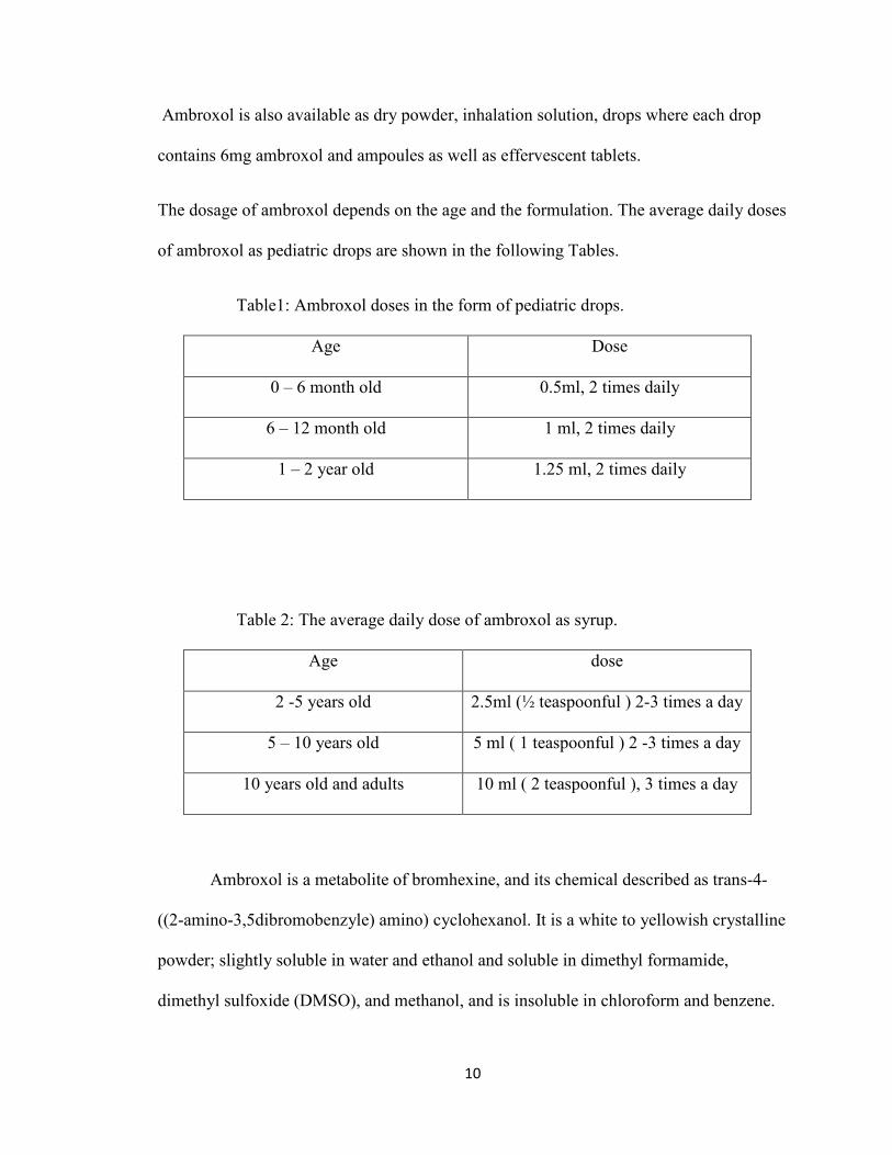
Ambroxol is also available as dry powder, inhalation solution, drops where each drop
contains 6mg ambroxol and ampoules as well as effervescent tablets.
The dosage of ambroxol depends on the age and the formulation. The average daily doses
of ambroxol as pediatric drops are shown in the following Tables.
Table1: Ambroxol doses in the form of pediatric drops.
Age Dose
0 – 6 month old 0.5ml, 2 times daily
6 – 12 month old 1 ml, 2 times daily
1 – 2 year old 1.25 ml, 2 times daily
Table 2: The average daily dose of ambroxol as syrup.
Age dose
2 -5 years old 2.5ml (½ teaspoonful ) 2-3 times a day
5 – 10 years old 5 ml ( 1 teaspoonful ) 2 -3 times a day
10 years old and adults 10 ml ( 2 teaspoonful ), 3 times a day
Ambroxol is a metabolite of bromhexine, and its chemical described as trans-4-
((2-amino-3,5dibromobenzyle) amino) cyclohexanol. It is a white to yellowish crystalline
powder; slightly soluble in water and ethanol and soluble in dimethyl formamide,
dimethyl sulfoxide (DMSO), and methanol, and is insoluble in chloroform and benzene.
11
In humans, ambroxol is metabolized to dibromoanthranilic acid (DBAA) and 6,8dibromo
3(trans-4-hydroxycyclohexyl)-1, 2, 3,4-tetrahydroquinazoline (DHTQ). The formulation
of DHTQ proceeds non-enzymatically, where that of DBAA requires Nicotinamide
adenine dinucleotide phosphate NADP. A study has been performed to identify the CYP
isozymes involved in the formation of DBAA using human liver microsomes expressing
recombinant human CYP isozyme (1A1,1A2, 2A6,2B6, 2C8, 2C9, 2C19, 2D6, 2E1, 3A4,
and 4A11) showed that CYP3A4 is the only isozyme that metabolized ambroxol to
DBAA [ N. Ishiguro et al, 1999]. In the same study CYP inhibitor were examined such as
furafaylline sulphaphenazole, quinidine, diethyldithiocarbamic acid and ketokinazole.
Only ketoconazol inhibited the production of DBAA (>80%), these results suggest that
CYP3A4 is predominantly involved in the metabolism of ambroxol to DBAA in human.
Phase II drug-metabolizing enzymes involvement in the metabolism of ambroxol has not
been studied in humans but the hydroxyl group in its structure suggests possible
metabolism by the SULTs to prevent adverse effect and facilitate elimination. Ambroxol
is well absorbed and excreted in the urine ,about 50% as gucuronide of unchanged drug,
10% as oxidized metabolized 3,5- dibromo-2-aminobenzoic acid (DBABA) and a minute
amount as 6,8-dibro-3(trans-4-hydroxycyclohexyle)-1,2,3,3-tetrahydroquinazoline
DHTQ.
Gastrointestinal side effects like epigastric pain may occur occasionally. Rarely
allergic reaction such as skin rash, hives, and angioneurotic edema, sometimes allergic
contact dermatitis and anaphylactic shock may occur. Weakness, headache, diarrhea, dry
mouth and respiratory tract, exanthema, rhino-rhea, bolts and dysuria are also rarely
12
occurring. During prolonged application at higher doses, gastralgia, nausea and vomiting
may occur.
Teratogenic and fetal toxicity studies have shown no harmful effect of ambroxol.
However, it is contraindicated during pregnancy, especially during the first trimester.
Safety during lactation has not been established yet. It also should not be given to a
patient with hypersensitivity to ambroxol or a patient with hepatic insufficiency.
4. The cytosolic sulfotransferases (SULTs) and their possible functional roles
Biological sulfation was discovered when phenyl sulfate was isolated from the urine
of a patients who had been treated with phenol as an antiseptics. Sulfation mechanism
remained unknown despite the extensive studies that have been done in vivo until the co-
substrate, adenosine 3’-phosphate 5’ phosphosulfate (PAPS) was discovered. Sulfation,
by definition, is the transfer of a sulfonate group (SO3 -2) from PAPS to a substrate, which
is catalyzed by SULT enzymes. Many endogenous and xenobiotic molecules are
substrates for the SULT enzymes [Nirangali Gamage et al, 2005]. Different physiological
processes, including, inactivation and bioactivation of xenobiotics, inactivation of
hormones and catecholamines, modification of the structure and function of
macromolecules, and elimination of end products of catabolism have been affected by
sulfation mechanism. As mentioned above, PAPS is the co-substrate that is synthesized in
the cells to create the activated form of sulfate needed for sulfation reactions.

13
Sulfation is an important reaction in the metabolism of numerous xenobiotics, drugs,
as well as some key endogenous compounds. The addition of sulfonate moiety to a
compound increases its water-solubility and decreases its biological activity in most
cases. However, some of the SULT enzymes are also capable of bioactivating
procarcinogenes to reactive electrophiles. Sulfation is a key reaction in the body defense
against harmful chemicals and may have a major function during early development;
SULTs are highly expressed in the human fetus.
The SULT gene superfamily has been categorized to several gene families based on
the amino acid sequences of known vertebrate SULTs. [Werner, M.U. et al, 2002]. The
major gene families of the SULT family are the phenol sulfotransferase (PST) family
(designated SULT1) and hydroxysteroid sulfotransferase (HSST) family (designated
SULT2). The PST family consists of at least four sub-families, PSTs (SULT1A),
Dopa/tyrosine sulfotransferases (SULT1B), hydroxyarylamine sulfotransferases
(SULT1C), and estrogen sulfotransferases (SULT1E). The HSST family comprises two
subfamilies, DHEA sulfotransferases (SULT2A) and cholesterol/pregnolone
sulfotransferase (SULT2B).
The SULTs are highly conserved across vertebrate species, and homologous SULT
enzymes are found in different vertebrate animals due to the important roles they play in
detoxification and in the homeostasis of compounds involved in the nervous and
endocrine systems. Eleven SULTs have been detected and characterized in human. Eight
human SULTs (SULT1A1, SULT1A2, SULT1A3, SULT 1B1, SULT1E1, SULT2A1,
SULT2B1a, SULT2B1b, and SULT4A1) have been proposed to be involved either in
detoxification of xenobiotics or in regulating the levels and the activities of key

14
endogenous substrates .However, the two SULT1C enzymes and the brain ST functions
remain unclear. The functional roles proposed for the various human SULTs based
primarily on their substrate specificities. Moreover, little information is available
regarding the ontogeny of the SULT enzymes and the cell type, tissue, and organ-specific
expression of the different SULTs. The possible coordination between the developmental
expression of the SULTs that are involved in the sulfation and modulation of monoamine
and catecholamine hormones, thyroid/steroid hormones and possible detoxification of
xenobiotics and drugs such as ambroxol has not been investigated. To resolve these
outstanding issues, we plan to perform systematic studies using zebrafish as a model.
5. Zebrafish as a model for research on developmental
pharmacology
In the past decade the zebrafish (Danio rerio) has recently emerged as a powerful
model system in developmental biology and in studies utilizing transgenic techniques.
The initial interest in zebrafish as a model goes back to the early 1970s when it was
selected to be the first vertebrate to study genetic screening. During the subsequent 20
years, the zebrafish model was almost exclusively used to study development. This
resulted in the characterization of large number of genes involved in vertebrate pathways
and the establishment of the zebrafish as a relevant model for human disease and
pharmaceutical research.
There are numerous advantages for the use of the zebrafish as a model species. The
main benefit of using zebrafish as development and toxicity model is the external

15
development with optically transparent embryos. Their optical clarity allows for easy
observation of developmental stages, identification of phenotypic traits during
mutagenesis screening and assessment of endpoint of toxicity during toxicity testing. The
zebrafish is small in size. Adults are only approximately 1- 1.5 inch long, which allows
for the maintenance of large number of animals. Therefore, only micrograms of
compound are needed for screening per assay as the developing fish can live in as little as
50 µl of fluid. Moreover, one pair of adult fish is capable of laying 200-300 eggs, and if
appropriately maintained, they can provide this yield every 5-7 days [Westerfield, M.,
2007].
In addition to its developmental advantage, it has a great potential to serve as a model
for human diseases and tremendous potential as a genetic model. The rapid maturation of
zebrafish also allows for easy experimentation for transgenerational endpoint required for
mutagenesis screening, establishing transgenic lines, and assessing chemicals for
teratogenicity [Paul Goldsmith, 2004].
The sequencing of the zebrafish genome provides a good starting point to assess
whether a target of interest is conserved in zebrafish, although it should be underscored
that only about half of genes can be found readily by database crunching. This is an
important limitation, because results from zebrafish must always be interpreted in terms
of the similarities of any particular pathway. Protein homology and signaling pathway
conservation must be considered, as well as pharmacokinetic factors that could lead to
false positive or negative results.

16
There are many areas where zebrafish approach is particularly advantageous, e.g.,
analysis of environmental contaminant effects, disease model, ophthalmology, behavioral
disorder, cancer, heart development and drug toxicity. The zebrafish model offers a
system that can detect probable failure in the heart much earlier than it could be done
with any other model [Westerfield, M. 2007]. Therefore, zebrafish embryos are
particularly well suited to investigate the heart disorder and the effect of many drugs and
chemicals on the heart. Human and zebrafish hearts have much in common. Both have
muscles designed to pump oxygen carrying blood through valves that ensure blood flow
in the single direction. And, in both cases, the heart pumps in a regular rhythmic way.
The rhythmic beating of the heart in part depends on specialized heart muscle cells called
myocytes that are normally highly organized part of the heart structure [Adrian J. Hill et
al, 2005 ].
5.1 Stages of Embryonic development of the zebrafish
It is important to know the staging series to provide accuracy in developmental
studies. This is because different embryos, even within a single clutch, develop at slightly
different rates.
The first period in the development is called the zygote until the first cleavage that occurs
about 40 minutes after fertilization. Following the first cleavage, after ¾ hour, about 2-7
cell cycles occur rapidly and synchronously. After 2 ¼ hours it reaches the blastula
period, where the embryos enter midblastula transition.

17
The embryos reach glastrula period after 5 ½ hours, where morphogenic movements of
involution, convergence, and extension from the epiblast, hypoblast, and embryonic axis
occur. The primary organogenesis occurs after 10 hours in the segmentation periods. The
extension of the tail rudiment barely begins to elongate the embryos and after 20 hours
morphogenesis associated with the constriction of the yolk begins to straighten out the
posterior trunk, and this, along with continued development of the tail. At the pharyngula
period, 24 hours, the axis will start to straighten from early curvature with circulation,
pigmentation, and finnage beginning to develop. Hatching occur after 48 hours, where
the embryos will complete the rapid morphogenesis of primary organ systems like
cartilage development in the head pectoral fin. By day 3, or 72 hours, the hatched larva
has completed most of its organ morphogenesis, and it continues to grow rapidly. The
inflation of the swim bladder and the anterior dorsal protrusion of the mouth change
during the next day. Whereas during the hatching period the embryos are usually at rest,
the early larva gradually begins to swim about actively, and moves its jaws and eyes. The
developments produce swift escape response, the seeking of prey, and feeding. Zebrafish
is DMSO tolerant and unlike invertebrates with their cuticles, it readily absorbs
chemicals. Until 12-14 days post fertilization, oxygen is primarily supplied by diffusion
across the skin and it seems that this is the major route for absorption of small molecules
(Figure 1).
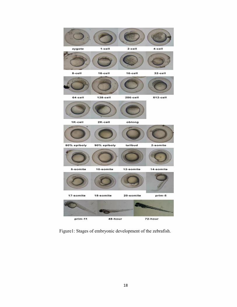
18
Figure1: Stages of embryonic development of the zebrafish.

19
5.2 Drug metabolism in zebrafish
The zebafish shows many similarities regarding the physiological function with
human and it is important to identify the possible metabolic processes involved in
zebrafish especially in testing chemical or drug effects on developmental toxicity studies.
CYP3A isozymes comprise the largest portion of the liver and small intestinal CYPs
proteins in mammals. They are involved in the metabolism of an extensive range of
endogenous substrates and xenobiotics and termination of the action of steroid hormones.
Regarding Phase I drug- metabolizing enzymes expression in zebrafish, a full-
length cDNA of the CYP3A gene named CYP3A65, was cloned from zebrafish. Upon
hatching of the zebrafish embryos the CYP3A65 mRNA was initially transcribed only in
the liver and intestine [Hua Pin Teseng et al, 2004]. Moreover, a new cytochrom P450
subfamily CYP3C was also cloned from zebrafish, CYP3C1. The CYP3C1 has 44-54%
identities with mammalian CYP3A and CYP 3B forms [Graham E. Corley-Smith et al,
2005].
Phase II drug-metabolizing enzymes are also important for drug transformation
and their enzymes expressions have been studied in zebrafish. The cytosolic
sulfotransferases are a family of Phase II drug-metabolizing enzymes that catalyze the
transfer of a sulfonate group from 3-phosphoadenosine-5-phosphosulfate to endogenous
and xenobiotic compounds. Several SULT isoforms have been identified in zebrafish.
Two partial cDNA clones encoding the 5 and 3 region of putative cytosolic
sulfotransferase have been identified. By analyzing the sequence data, they found that
these novel zebrafish SULTs display 49, 46, and 45% amino acid sequence identity to
20
human SULT1A1 and they also found that SULT1 isoform 4 from the zebrafish
displayed significant sulfating activity toward thyroid hormone, estrone, and
dehydroepiandosterone [Ming Yih Liu et al, 2005]. Two of the zebrafish cDNA encoding
putative cyctosolic sulfotransferase (SULTs) were also identified. The sequence analysis
indicated that these two zebrafish SULTs belong to the cytosolic SULT2 gene family,
designated as SULT2 ST2 and SULT2 ST3, and it showed activity toward verity of
endogenous compounds like dehydroepiandosterone, corticosterone and xenobiotics
[Shin Yasuda et al, 2006]. The unique characteristics of the zebrafish makes it an
excellent model for a systematic study on the ontogeny of the enzyme expression, and
physiological involvement of the SULTs, as well for studies on the adverse effects of
drugs during the developmental process and the relevance of SULTs and sulfation in this
process. There is relatively little information available concerning the ontogeny of
SULTs expressions during development, and their physiological involvement due to the
limitations in using mammalian animal models.
It is important to know the correlation between zebrafish developmental stages and
stages of fetal and child development for accurate and better incorporation of the data. In
human the blastocyte period which is the period between conception and embryonic stage
may resembles the zebrafish blastula period, 2 ½ to 5 ½ hpf, where the blastodisc begins
to look ball-like until the time of onset of gastulation. Before 24 hpf in zebrafish the tail
will start to develop whereas in human, the tail has disappeared in this period which is
less than 4 weeks post fertilization. The pregnancy period in the human, from fertilization
to 38 weeks could be presented by the period from fertilization up to 48 hpf in zebrafish
where it hatches, so at this time it’s best to test the teratogenic and developmental effects

21
of the drugs to the embryos during pregnancy. The neonatal period in the zebrafish, 0-30
days in human, may start from 24 hpf up to 72 hpf , about up to 3 ½ days. After 72 hpf
the fish is able to eat and swim, and up to 3 weeks it may represents the juvenile period in
zebrafish and the pediatric period in human, 12 weeks to maturity. Drug exposure during
this period could test the possible effect on pediatric population. After 3 weeks post
fertilization in zebrafish may represent the adulthood in zebrafish.

22
OBJECTIVES AND GOALS
The cytosolic sulfotransferases (SULTs) are important in regulating the levels and
activities of endogenous compound such as thyroid hormones, steroid hormones,
catecholamine hormones, and cholesterol and its metabolites, and also in the
detoxification of exogenous compound such as environmental toxins, and other
xenobiotics including therapeutic agents [Mulder, G.J. and Jakoby, 1990; Duffel,
M.W.,1997]. Based on the important role of these enzymes in the detoxification of
drugs, we hypothesize that the susceptibility of developing zebrafish embryo/larva, and
likewise infant/child, to any potential adverse effects of drugs may be dependent on the
ontogeny and cell type/tissue/organ-specific expression of relevant drug-sulfating
SULTs. As part of an effort to verify this hypothesis, an asthma drug, ambroxol, that is
known to have a multiple mechanisms of action and been used in pediatric population,
was tested on developing to zebrafish embryos/larvae. The objectives of this study were
to examine possible adverse effect of ambroxol in a dose-dependent manner on
developing zebrafish embryos/larvae and to analyze the ambroxol-sulfating activity of a
set of zebrafish SULTs previously prepared in our laboratory. The ultimate goal is to
elucidate the involvement of ambroxol-sulfating SULTs in protection against the adverse
effects of this drug at different stages during zebrafish development.
23
MATERIALS AND METHODS
1. Study of developmental toxicity of ambroxol in zebrafish
embryos/larvae
1.1 Preparation of fertilized zebrafish eggs
Adult zebrafish (Danio rerio) were purchased from the Zebrafish International
Resource Center (ZIRC) at the University of Oregon (Eugene, OR). The fish were kept
in fish tanks containing buffered water (pH 7.2) at 28ºC, and fed daily live brine shrimp
naupli and Tetramin dried flake food (Tetra, Blacksburg, VA). The day: night cycle was
maintained at 14 hours: 10 hours and spawning and fertilization of unexposed parent
fish was stimulated by the onset of the first light. Marbles were used to cover the bottom
of the spawning tank to protect newly laid eggs and facilitate their retrieval for study.
Fertilized zebrafish embryos were collected from the bottom of the tank by siphoning
with a disposable pipette. The eggs were placed in a 100 mm Petri dish and washed
thoroughly with buffered water containing 60 mg sea salt (Instant Ocean, Mentor, OH)
per liter of water. Groups of 10 fertilized eggs were then placed in individual wells of 6-
well plates and used in the experiment.

24
1.2 Treatment of zebrafish embryos/larvae with ambroxol
For treatment with ambroxol, three sets of freshly prepared fertilized eggs were used.
Each set included six groups of 10 eggs placed in individual wells of a 6-well plate. The
ambroxol treatment for the three sets of fertilized eggs began at 24, 48, and 72 hours post
fertilization (hpf), respectively.
The 24 hpf set
For the 24 hpf exposure set, eggs in the six wells were exposed to, respectively, 0
mM, 0.05 mM, 0.125 mM, 0.25 mM, 0.5 mM, and 1 mM of ambroxol at 24 hpf when the
eggs had developed normally through blastula, gastrula and segmentation stages. The
experiment was performed in triplicate. Mortality and deformities of embryos/larvae, if
any, were recorded at 24 hr intervals for 11 consecutive days. The solutions were
changed after observation and the micro-worms were added at 72 hpf as the fish starts to
eat at this stage. Morphological deformities and heart rates in embryos/larvae were
closely observed using an inverted microscope (Zeiss, Axiovert25) and images were
captured using Sony DSC-S75 digital/video camera. Teratogenic effects were recorded.
The 48 hpf set
For the 48 hpf exposure set, eggs were placed in the six wells were exposed to,
respectively, 0 mM, 0.05 mM, 0.125 mM, 0.25 mM, 0.5 mM, and 1 mM of ambroxol at
48 hpf when the eggs had developed normally through blastula, gastrula, segmentation,
25
and pharyngula stages. The experiment was performed in triplicate. Mortality and
deformities of embryos/larvae, if any, were recorded at 24 hr interval for 11 consecutive
days. The solutions were changed daily after observation and the micro-worms were
added at 72 hpf as the fish starts to eat at this stage. Morphological deformities and heart
rates in embryos/larvae were closely observed using an inverted microscope (Zeiss,
Axiovert25) and images were captured using Sony DSC-S75 digital/video camera.
Teratogenic effects were recorded.
The 72 hpf set
For the 72 hpf exposure set, eggs were placed in the six wells were exposed to,
respectively, 0 mM, 0.05 mM, 0.125 mM, 0.25 mM, 0.5 mM, and 1 mM of ambroxol at
48 hpf when the eggs had developed normally through blastula, gastrula, segmentation,
pharyngula, and hatching stages. Testing was performed in triplicate. Mortality and
deformities of larvae, if any, were recorded at 24 hr interval for 11 consecutive days. The
solutions were changed daily after observation and micro-worms were added from the
first day of exposure as the fish have the ability to eat at this stage. Morphological
deformities and heart rate in embryos/larvae were closely observed under an inverted
microscope (Zeiss, Axiovert25) and images were captured using Sony DSC-S75
digital/video camera. Teratogenic effects were recorded.

26
2. Sulfotransferase Assay
The sulfating activity of purified recombinant zebrafish cytosolic SULT1s (ST1, 2, 3,
4, 5, 6, 7, 8, and 9), SULT2s (ST 1, 2, and 3), SULT3s (ST 1, 2, 3, 4, and 5), and SULT
5A1 were assayed using radioactive PAP [35S] as the sulfate donor. The standard assay
mixture, with a final volume of 12.5 µl, containing 2.5 µl MOPS buffer at pH 7.0, 0.5 µl
PAP [35S] (15 Ci/mmol), 0.125 µl DTT, and 0.625µl ambroxol (1mM). DMSO or water
controls were also prepared. The reaction was started by the addition of 3µl enzyme,
allowed to proceed for 30 minutes at 28ºC, and terminated by heating at 100ºC for 3
minutes. The precipitates formed were cleared by centrifugation, and the supernatant was
subjected to analysis of [35S]-sulfated product using a previously developed TLC
procedure with n-butanol/isopropanol/88%formic acid/water (3:1:1:1; by volume) as the
solvent system.
27
RESULTS
Three sets of zebrafish embryos/larvae were exposed at different time points (24
hpf, 48 hpf, 72 hpf) during the embryonic/larval development. Observation for
morphological and functional changes, heart rate, and mortality rate were made every 24
hours for 11 days. Morphological deformity and changes in heartbeat in embryos/larvae
were carefully observed under an inverted microscope and images were captured. The
changes described below for each of the three sets (24 hpf, 48 hpf, and 72 hpf) refer to a
particular stage during the embryonic/larval development.
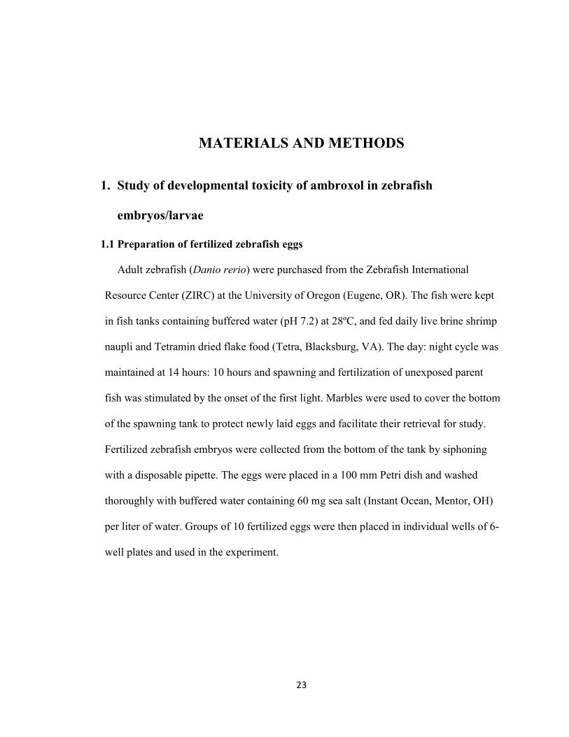
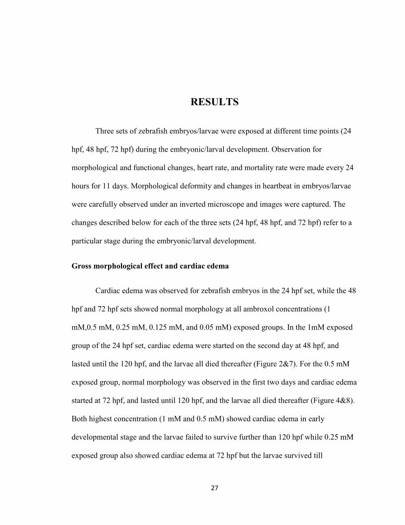
Gross morphological effect and cardiac edema
Cardiac edema was observed for zebrafish embryos in the 24 hpf set, while the 48
hpf and 72 hpf sets showed normal morphology at all ambroxol concentrations (1
mM,0.5 mM, 0.25 mM, 0.125 mM, and 0.05 mM) exposed groups. In the 1mM exposed
group of the 24 hpf set, cardiac edema were started on the second day at 48 hpf, and
lasted until the 120 hpf, and the larvae all died thereafter (Figure 2&7). For the 0.5 mM
exposed group, normal morphology was observed in the first two days and cardiac edema
started at 72 hpf, and lasted until 120 hpf, and the larvae all died thereafter (Figure 4&8).
Both highest concentration (1 mM and 0.5 mM) showed cardiac edema in early
developmental stage and the larvae failed to survive further than 120 hpf while 0.25 mM
exposed group also showed cardiac edema at 72 hpf but the larvae survived till
28
144 hpf, (Figure 5, 9, &10). Normal morphology was observed for group exposed to the
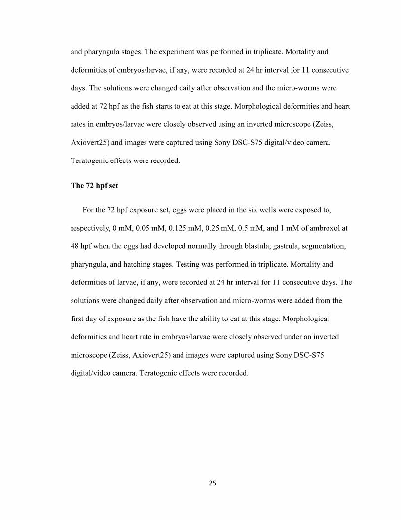
lowest concentration (0.05 Mm) and the control groups (DMSO and water) in all three
(24 hpf, 48 hpf, 72 hpf) sets (Figure 3&6). No craniofacial malformations were observed
among larvae in all sets throughout the experiment.
Figure 2: Zebrafish exposed to 1mM Ambroxol at 48 hour post fertilization (hpf) showed cardiac edema
Figure 3: Control group of zebrafish larvae at 48 hpf
Figure 4: Zebrafish exposed to 0.5 mM Ambroxol at 72 hpf showed
cardiac edema

29
Figure 5: Zebrafish exposed to 0.25 mM Ambroxol at72 hpf showed
cardiac edema
Figure 6: Control group of zebrafish larvae at 72 hpf
Figure7: Zebrafish larva exposed to 1.0 mM Ambroxol at 96 hpf showed cardiac edema
Figure 8: Zebrafish larva exposed to 0.5 mM Ambroxol at96 hpf showed cardiac edema

30
Figure 9: Zebrafish larva exposed to 0.25 mM Ambroxol at 96 hpf showed cardiac edema
Figure 10: Zebrafish larva exposed to 0.25 mM Ambroxol at 120 hpf showed cardiac edema.
Heart Rate
The heart rate was counted every 24 hours. Embryos/larvae exposed to the higher
concentrations (1mM, 0.5mM, 0.25mM) of ambroxol showed significant bradycardia, at
~80 b/m, from the first day of exposure for all three (24 hpf, 48 hpf, and 72 hpf) sets.
Lesser bradycardia effects, at ~115 b/m, were observed for embryos/larvae exposed to
the lower concentration (0.125 mM) of ambroxol. The heart rates were found to be
inversely proportional to the concentration of ambroxol to which the embryos/larvae
were exposed (Figure 11, 12, &13), and the severity of bradycardia increased with time
during the course of the study. The bradycardia started from the first day of exposure
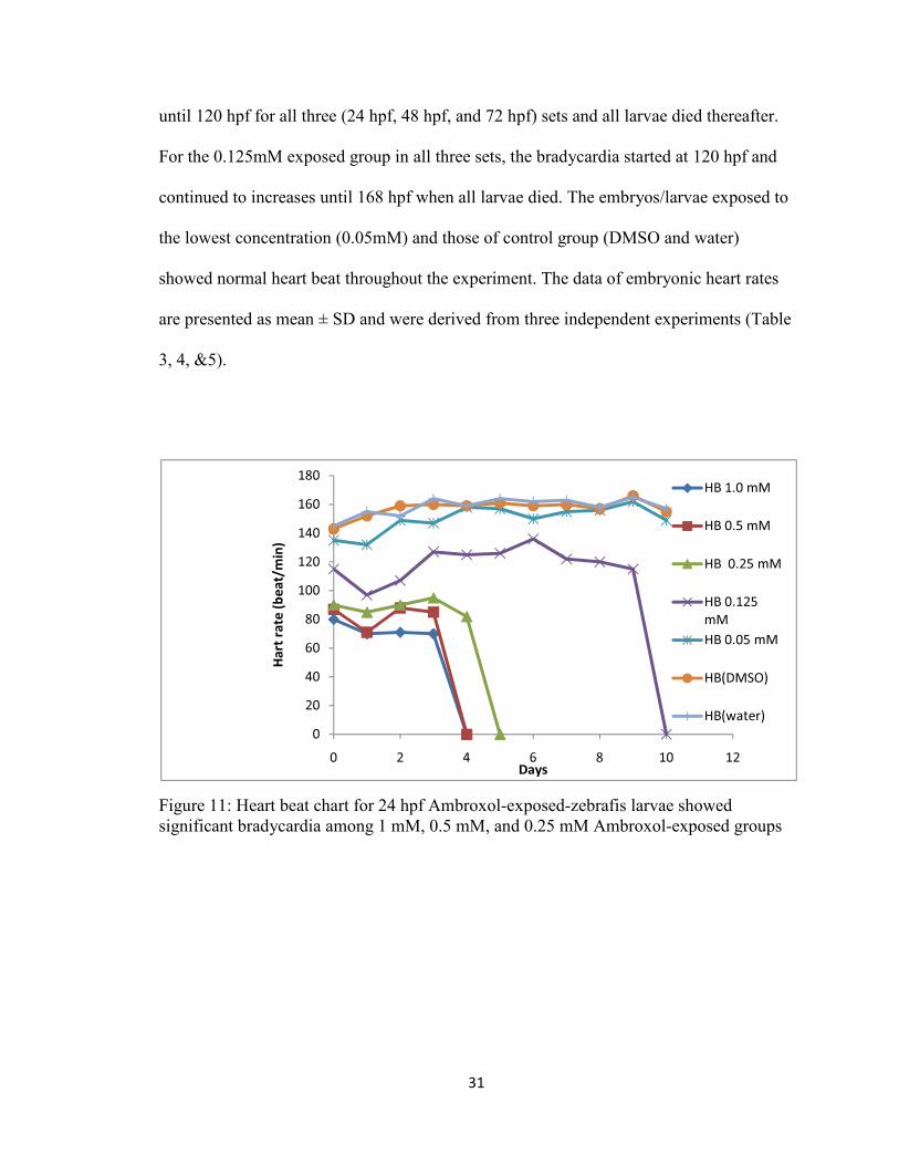
31
until 120 hpf for all three (24 hpf, 48 hpf, and 72 hpf) sets and all larvae died thereafter.
For the 0.125mM exposed group in all three sets, the bradycardia started at 120 hpf and
continued to increases until 168 hpf when all larvae died. The embryos/larvae exposed to
the lowest concentration (0.05mM) and those of control group (DMSO and water)
showed normal heart beat throughout the experiment. The data of embryonic heart rates
are presented as mean ± SD and were derived from three independent experiments (Table
3, 4, &5).
Figure 11: Heart beat chart for 24 hpf Ambroxol-exposed-zebrafis larvae showed significant bradycardia among 1 mM, 0.5 mM, and 0.25 mM Ambroxol-exposed groups
0
20
40
60
80
100
120
140
160
180
0 2 4 6 8 10 12
Ha
rt r
ate
(b
ea
t/m
in)
Days
HB 1.0 mM
HB 0.5 mM
HB 0.25 mM
HB 0.125
mM
HB 0.05 mM
HB(DMSO)
HB(water)

32
Figure 12: Heart beat chart for 48 hpf Ambroxol-exposed-zebrafish larva showed significant bradycardia among 1 mM, 0.5 mM, and 0.25 mM Ambroxol-exposed groups.
Figure 13: Heart beat chart for 72 hpf Ambroxol-exposed-zebrafish larvae showed significant bradycardia among 1 mM, 0.5 mM, 0.25 mM, and 0.125 mM Ambroxol exposed groups.
0
20
40
60
80
100
120
140
160
180
0 2 4 6 8 10 12
Ha
rt r
ate
(b
ea
t/m
in)
Days
HB 1.0 mM
HB 0.5 mM
HB 0.25 mM
HB 0.125 mM
HB 0.05 mM
HB(DMSO)
HB(water)
0
20
40
60
80
100
120
140
160
180
0 2 4 6 8 10 12
Ha
rt r
ate
(b
ea
t/m
in)
Days
HB 1.0 mM
HB 0.5 mM
HB 0.25 mM
HB 0.125 mM
HB 0.05 mM
HB (DMSO)
HB (water)

33
Table 3: Heart rates (beat/min) of zebrafish larvae at different developmental stages in response to different concentrations of Ambroxol. Numbers shown are the mean ± SD for the 24 hpf exposure set.
day HB1.0 mM
±SD
HB 0.5 mM
±SD
HB 0.25 mM
±SD
HB0.125mM
±SD
HB0.05 mM
±SD
HB DMSO
±SD
HB water
±SD
1
2
3
4
5
6
7
8
9
10
11
80±0.8
70±1.1
71±0.8
70±1.1
0±0
0±0
0±0
0±0
0±0
0±0
0±0
87±0.7
71±1.1
88±0.7
85±0.7
0±0
0±0
0±0
0±0
0±0
0±0
0±0
90±0.83
85±0.7
90±1.1
95±0.7
82±1.9
0±0
0±0
0±0
0±0
0±0
0±0
115±0.7
97±1.4
107±1.1
127±1.1
125±2.1
126±2.28
136±1.94
122±1.6
120±1.6
115±0.7
0±0
135±1.58
132±1.5
149±1.14
147±0.7
158±2.1
157±1.1
150±1.3
155±1.2
156±3.2
162±3.2
149±1.58
143±1.9
152±2.2
159±0.7
160±0.7
159±1.14
161±1.1
159±1.5
160±1.5
157±3.3
166±1.6
155±0.7
145±0.7
155±1.8
152±2.1
164±0.7
159±1.14
164±0.7
162±1.5
163±1.6
158±3.1
165±2.1
157±1.14
34
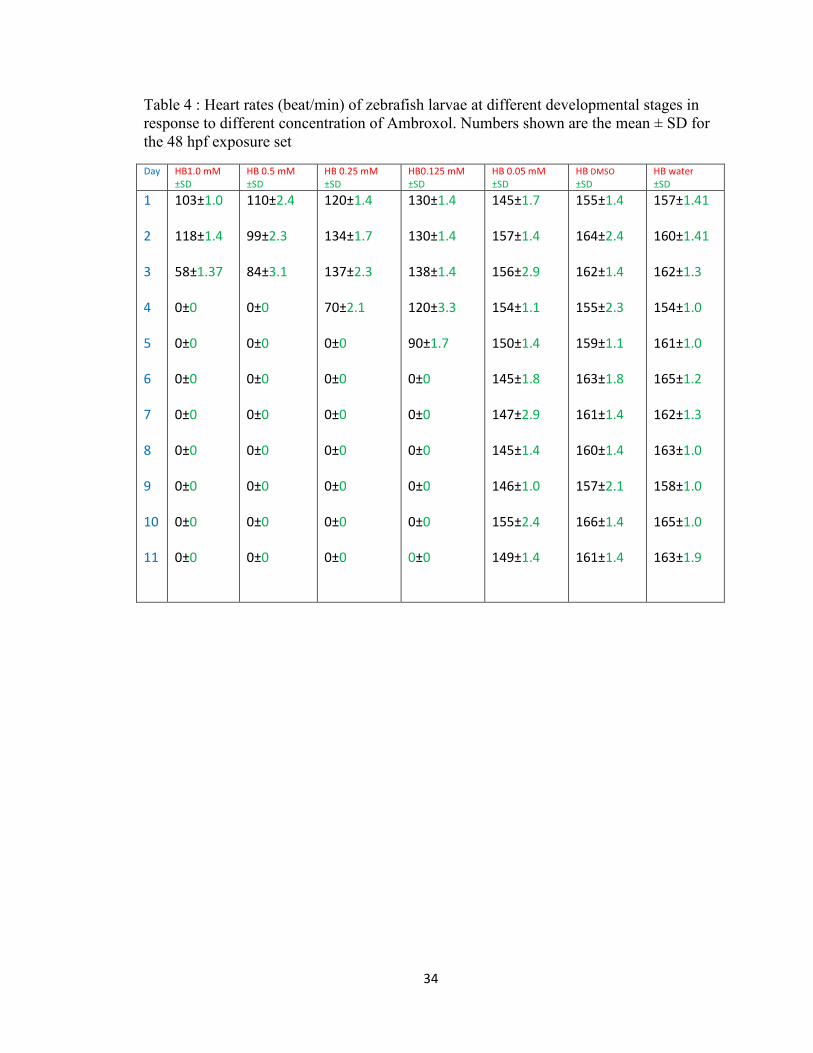
Table 4 : Heart rates (beat/min) of zebrafish larvae at different developmental stages in response to different concentration of Ambroxol. Numbers shown are the mean ± SD for the 48 hpf exposure set
Day HB1.0 mM
±SD
HB 0.5 mM
±SD
HB 0.25 mM
±SD
HB0.125 mM
±SD
HB 0.05 mM
±SD
HB DMSO
±SD
HB water
±SD
1
2
3
4
5
6
7
8
9
10
11
103±1.0
118±1.4
58±1.37
0±0
0±0
0±0
0±0
0±0
0±0
0±0
0±0
110±2.4
99±2.3
84±3.1
0±0
0±0
0±0
0±0
0±0
0±0
0±0
0±0
120±1.4
134±1.7
137±2.3
70±2.1
0±0
0±0
0±0
0±0
0±0
0±0
0±0
130±1.4
130±1.4
138±1.4
120±3.3
90±1.7
0±0
0±0
0±0
0±0
0±0
0±0
145±1.7
157±1.4
156±2.9
154±1.1
150±1.4
145±1.8
147±2.9
145±1.4
146±1.0
155±2.4
149±1.4
155±1.4
164±2.4
162±1.4
155±2.3
159±1.1
163±1.8
161±1.4
160±1.4
157±2.1
166±1.4
161±1.4
157±1.41
160±1.41
162±1.3
154±1.0
161±1.0
165±1.2
162±1.3
163±1.0
158±1.0
165±1.0
163±1.9

35
Table 5: Heart rates (beat/min) of zebrafish larvae at different developmental stages in response to different concentration of Ambroxol. Numbers shown are the mean ± SD for the 72 hpf exposure set
Day HB 1.0 mM
±SD
HB 0.5 mM
±SD
HB 0.25 mM
±SD
HB0.125mM
±SD
HB 0.05 mM
±SD
HB DMSO
±SD
HB water
±SD
1
2
3
4
5
6
7
8
9
10
11
120±1.3
45±1.4
0±0
0±0
0±0
0±0
0±0
0±0
0±0
0±0
0±0
122±1.5
70±1.7
0±0
0±0
0±0
0±0
0±0
0±0
0±0
0±0
0±0
120±1.0
105±1.6
86±1.4
0±0
0±0
0±0
0±0
0±0
0±0
0±0
0±0
130±0.9
126±2.2
115±2.1
101±1.4
0±0
0±0
0±0
0±0
0±0
0±0
0±0
145±1.0
151±1.4
145±1.0
144±1.4
150±1.2
145±1.4
147±1.0
148±1.7
151±1.3
142±1.4
149±1.41
155±2.0
155±1.03
154±1.4
155±1.3
159±1.0
160±1.4
168±1.2
160±0.8
159±1.4
147±0.8
149±0.8
157±1.1
159±2.0
151±1.1
157±2.1
161±1.6
159±2.0
168±1.0
159±1.1
158±1.7
150±1.4
152±0.8
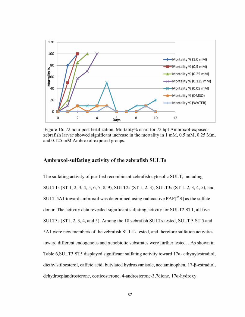
Morality Rate
For all three ( 24hpf, 48 hpf, and 72 hpf) sets, the larvae exposed to the highest
concentrations, 1mM and 0.5mM, of ambroxol did not survive beyond 120 hpf, and the
mortality rate continued to increase from the first day of exposure till it reached 100%. In
contrast to the heart rates, mortality rates were proportional to the ambroxol
concentration and it reached 80% at 96 hpf of the 72 hpf exposed groups, and in all three
sets, the 1mM, 0.5mM, and 0.25mM ambroxol-exposed groups reached 100% at 120 hpf,

36
while 0.125mM ambroxol-exposed group reached 100% mortality at 144 hpf. (Figure 14,
15, & 16). The embryos/larvae exposed to the lowest concentration (0.05 mM) and those
in the control group (DMSO and water) survived till the end of the experiments with no
significant increase in mortality%, which was ~10%.
Figure 14: Mortality% chart for 24 hpf Ambroxol-exposed-zebrafish larvae showed significant increase in the mortality in 1 mM, 0.5 mM, and 0.25 mM Ambroxol-exposed groups.
Figure 15: Mortality% chart for 48 hpf Ambroxol-exposed-zebrafish larvae showed significant increase in the mortality in 1 mM, 0.5 mM, and 0.25 mM Ambroxol-exposed groups
0
20
40
60
80
100
120
0 2 4 6 8 10 12
Mo
rta
lity
%
Days
Mortality % (1.0 mM)
Mortality % (0.5 mM)
Mortality % (0.25 mM)
Mortality % (0.125 mM)
Mortality % (0.05 mM)
Mortality % (DMSO)
Mortality % (WATER)
0
20
40
60
80
100
120
0 2 4 6 8 10 12
Mo
rta
lity
%
Days
Mortality % (1.0 mM)
Mortality % (0.5 mM)
Mortality % (0.125 mM)
Mortality % (0.05 mM)
Mortality % (DMSO)
Mortality % (WATER)
Mortality % (0.25 mM)
37
Figure 16: 72 hour post fertilization, Mortality% chart for 72 hpf Ambroxol-exposed-zebrafish larvae showed significant increase in the mortality in 1 mM, 0.5 mM, 0.25 Mm, and 0.125 mM Ambroxol-exposed groups.
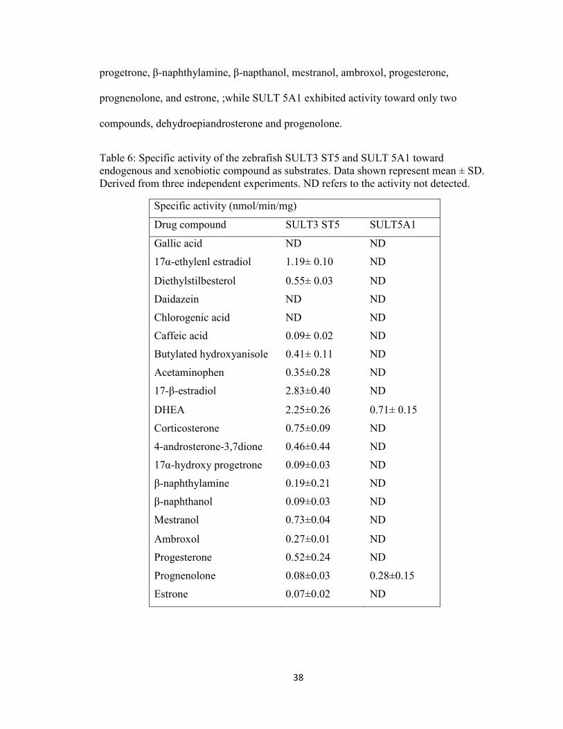
Ambroxol-sulfating activity of the zebrafish SULTs
The sulfating activity of purified recombinant zebrafish cytosolic SULT, including
SULT1s (ST 1, 2, 3, 4, 5, 6, 7, 8, 9), SULT2s (ST 1, 2, 3), SULT3s (ST 1, 2, 3, 4, 5), and
SULT 5A1 toward ambroxol was determined using radioactive PAP[35S] as the sulfate
donor. The activity data revealed significant sulfating activity for SULT2 ST1, all five
SULT3s (ST1, 2, 3, 4, and 5). Among the 18 zebrafish SULTs tested, SULT 3 ST 5 and
5A1 were new members of the zebrafish SULTs tested, and therefore sulfation activities
toward different endogenous and xenobiotic substrates were further tested. . As shown in
Table 6,SULT3 ST5 displayed significant sulfating activity toward 17α- ethynylestradiol,
diethylstilbesterol, caffeic acid, butylated hydroxyanisole, acetaminophen, 17-β-estradiol,
dehydroepiandrosterone, corticosterone, 4-androsterone-3,7dione, 17α-hydroxy
0
20
40
60
80
100
120
0 2 4 6 8 10 12
Mo
rta
lity
%
Days
Mortality % (1.0 mM)
Mortality % (0.5 mM)
Mortality % (0.25 mM)
Mortality % (0.125 mM)
Mortality % (0.05 mM)
Mortality % (DMSO)
Mortality % (WATER)
38
progetrone, β-naphthylamine, β-napthanol, mestranol, ambroxol, progesterone,
prognenolone, and estrone, ;while SULT 5A1 exhibited activity toward only two
compounds, dehydroepiandrosterone and progenolone.
Table 6: Specific activity of the zebrafish SULT3 ST5 and SULT 5A1 toward endogenous and xenobiotic compound as substrates. Data shown represent mean ± SD. Derived from three independent experiments. ND refers to the activity not detected.
Specific activity (nmol/min/mg)
Drug compound SULT3 ST5 SULT5A1
Gallic acid ND ND
17α-ethylenl estradiol 1.19± 0.10 ND
Diethylstilbesterol 0.55± 0.03 ND
Daidazein ND ND
Chlorogenic acid ND ND
Caffeic acid 0.09± 0.02 ND
Butylated hydroxyanisole 0.41± 0.11 ND
Acetaminophen 0.35±0.28 ND
17-β-estradiol 2.83±0.40 ND
DHEA 2.25±0.26 0.71± 0.15
Corticosterone 0.75±0.09 ND
4-androsterone-3,7dione 0.46±0.44 ND
17α-hydroxy progetrone 0.09±0.03 ND
β-naphthylamine 0.19±0.21 ND
β-naphthanol 0.09±0.03 ND
Mestranol 0.73±0.04 ND
Ambroxol 0.27±0.01 ND
Progesterone 0.52±0.24 ND
Prognenolone 0.08±0.03 0.28±0.15
Estrone 0.07±0.02 ND

39
DISCUSSION
Considering the important role of the SULT enzymes in the detoxification of
drugs, we hypothesize that the susceptibility of a developing zebrafish embryo/larva, and
likewise infant/child, to any potential adverse effects of drugs may be dependent on the
ontogeny and cell type/tissue/organ-specific expression of relevant drug-sulfating
SULTs. As part of an effort to verify this hypothesis, we tested an asthma drug,
ambroxol, which is known to have a multiple mechanisms of action and has been used in
pediatric population. The zebrafish was used as a model to assess the toxicity or
developmental effect at different stages during embryonic and larval development.
Ambroxol is a mucolytic agent used in the treatment of respiratory disorders
associated with viscid or excessive mucus such as asthmatic bronchitis and chronic
pneumonia. Ambroxol has been used in infants of different age groups, ranging from 0-6
month, and children over 12 years, as well as adults. Ambroxol has many mechanisms of
actions which makes it a useful anti-inflammatory and mucolytic agent, but at the same
time increase the possibility of exerting sever adverse effects especially for infants and
neonates. In addition to its anti-inflammatory effect, ambroxol is a potent inhibitor of the
neuronal Na+ channels, which may cause adverse effects to neonates and infants. The
recommended dosage of ambroxol depends on the age and the formulation.
40
In a normal human body, ambroxol is metabolized to dibromoanthranilic acid
(DBAA) and 6,8- dibromo3(trans-4-hydroxycyclohexyl)-1,2,3,4-tetrahydroquinazoline
(DHTQ). CYP3A4 is the only enzyme capable of metabolizing ambroxol to DBAA in the
Phase I metabolism [N. Ishiguro et al, 2000]. With regard to the Phase II metabolism, our
lab has recently demonstrated significant sulfating activities of SULT2 ST1, and SULT3
ST1, 2, 3, 4, and 5 toward ambroxol [Liu et al., unpublished data] which may aid in the
inactivation and the excretion of ambroxol, thereby alleviating possible adverse effects.
The possibility of potential adverse effects of ambroxol, however, still remains an open
question, which may depend on the dose used, the developmental stage, and the ontogeny
of enzymes involved in the metabolism of ambroxol. In this study we focus on exposing
ambroxol to different age groups of zebrafish embryos/larvae by taking into
consideration the expression of the SULT enzymes during zebrafish developmental
stages and the doses for each age group.
Exposure of the first set of zebrafish embryos to ambroxol started at 24 hpf, when
at this developmental stage the heart begins to beat, and the circulation and pigmentation
starts to develop. For the second and third set of zebrafish embryos/larvae, the exposure
to ambroxol started at 48 hpf and 72 hpf, respectively. Previous studies have
demonstrated that CYP3A4 is the enzyme responsible for metabolizing ambroxol in
Phase I metabolism reaction [N. Ishiguro et al, 1999]. A full-length cDNA encoding
zebrafish CYP3A had been detected as early as 24 hpf and the expression level was
found to increase upon hatching and thereafter [Ronald N. Hines & Gail McCarver,
2001]. CYP3A was detected in gill and heart and, at lower levels, in brain and eye [Hua-
Pin Tseng, 2004]. Phase II drug-metabolizing enzymes involvement in the metabolism of
41
ambroxol have not been studied in humans but the hydroxyl group in its structure
suggests possible metabolism by the SULTs. Our laboratory had previously cloned and
characterized fifteen distinct zebrafish SULTs .Of these zebrafish SULTs, eight belong to
the SULT1 family, three are categorized into the SULT2 gene family, three fall in the
SULT3 gene family, and one appears to belong to a novel SULT gene family (SULTX)
[Shin Yasuda et al,2009]. In my study, I carried out enzymatic assays to examine the
sulfating activity of these zebrafish SULTs with ambroxol as substrate. Activity data
showed significant sulfating activity of SULT2 ST1, SULT3 ST1, ST 2, ST3, ST4, and
ST5 toward ambroxol. The expressions of these enzymes during zebrafish development
had been studied previously [Shin Yasuda et al, 2006]. The mRNA encoding SULT 3
ST3 was detected in unfertilized eggs. Upon fertilization, however, no SULT3 ST3
mRNA was detected until cleavage period (1hpf) which then continues at hatching period
(48 hpf) and remained high in the larval stage until maturity [Shin Yausda et al, 2009].
Based on my data and those reported previously [Shin Yausda et al, 2009; N.
Ishiguro et al, 1999], it appears that Phase I CYP3A4, and the Phase II SULT2 ST1, and
SULT 3 ST 1, 2, and 3 may be involved in the metabolism and therefore the protection
against possible adverse effects of ambroxol. Such protection effect may correlate with
their expression during embryonic and larval development. The developmental
expression of other SULT3 enzymes, SULT3 ST4 and 5, that are capable of sulfating
ambroxol, however, has not yet been studied.
Observations of the ambroxol-exposed zebrafish embryos/larvae indicated that the
major toxicity is on the heart, which is in line with previous finding that ambroxol is a
potent inhibitor of the neuronal Na+ channels [Wolfram Gaida et al, 2005]. The results
42
presented in the Results section showed significant bradycardia in 1mM ambroxol-
exposed groups from the first day of exposure in all three (24 hpf, 48 hpf, and 72 hpf)
sets. The 24 hpf set showed cardiac edema along with the bradycardia, and according to
the heart development in zebrafish, the heart begins to beat at that stage.
For all three ( 24hpf, 48 hpf, and 72 hpf) sets, the larvae exposed to the highest
concentrations, 1mM and 0.5mM, of ambroxol did not survive beyond 120 hpf, and the
mortality rate continued to increase from the first day of exposure till it reached 100%.
Mortality rates were proportional to the ambroxol concentration and it reached 80% at 96
hpf of the 72 hpf exposed groups, and in all three sets, the 1 mM, 0.5 mM, and 0.25 mM
ambroxol-exposed groups reached 100% at 120 hpf, while 0.125 mM ambroxol-exposed
group reached 100% mortality at 144 hpf.
The expressions of the SULT 2 ST1 and SULT3 ST1, 2, and 3 that are likely
involved in the metabolism of ambroxol had been studied previously during zebrafish
development [Shin Yasuda et al, 2006], and were shown to be present during the
exposure periods adopted in this study. This suggested that the protective effect of those
enzymes against adverse effect like bradycardia and cardiac edema may not be sufficient.
However, the developmental expression of other SULT3 enzymes, SULT3 ST4 and 5,
that are capable of sulfating ambroxol, has not yet been studied. Additionally, a couple of
new SULTs have been cloned and have not yet been characterized with regard to their
involvement in the metabolism of ambroxol and their expressions during development of
the zebrafish.

43
More studies will be needed in order to extrapolate these results from zebrafish to
humans. Nevertheless, the dose commonly used for infants aged 0 to 6 month, 0.5 ml/2
times per day equals approximately 0.72 mM (by taking in consideration the average total
body weight and the volume of distribution at this stage) is within the concentration range
of ambroxol tested in this study. Any protection effect of SULTs against ambroxol in
infants/young children will depend on the expression of ambroxol-metabolizing enzymes
during the developmental stage. In the liver both enzyme activity and protein level of the
human SULT2A1, which may be equivalent to the zebrafish SULT2 ST1, has been
shown to be low at 25 weeks gestation and then increase, particularly in the last half of
gestation, to near adult level in the neonate [Michael J. Blake et al, 2005]. More studies
are warranted in order to understand the protection effects of particular SULTs that may
be involved in protection against the adverse effects of ambroxol in infants and children.
44
References
Adrian J. Hill, Hiroki Teraoka, Warren Heideman, and Richard E.Peterson,
“Zebrafish as a Model vertebrate for Investigating Chemical Toxicity,” February 2005.
Amanda Clarkson, Sharon Conroy, Karissa Burroughs, Imti Choonara,
“Surveillance for adverse drug reaction in children: a pediatric regional monitoring
center”, 2004.
D. Gail McCarver and Ronald N. Hines, “The ontogeny of human drug metabolizing
enzymes: phase II conjugation enzymes and regulatory mechanisms”, Oct. 31, 2001.
Duffel, M.W.”Sulfotransferases in Comprehensive Toxicology” (Guengerich, F.P., Ed.),
pp. 365-383, Elsevier Science, Ltd., Oxford, 1997.
Etminan, M., Sadatsafavi, M., Jafari, S., Doyle-Waters, M., Aminzadeh, K. and
Fitzgerald, J.M. “Acetaminophen Use and the Risk of Asthma in Children and
Adults: A Systematic Review and Metaanalysis”, Chest. Aug 20, 2009.
George P. Giacoia, Debra L. Birenbaum, Hari Cheryl Sachs and Donald R.
Mattison,” The newborn drug development initiative”, 2006.
Graham E. Corley-Smith, Hsiao-Ting Su, Jun-Lan Wang –Buhler, Hua-Pin Tseng,
Chin-Hwa Hu, Thuy Hoang, Woon-Gye Chung, Donald R. Buhlar.” CYP3C1,
the first member of a new cytochrome P450 subfamily found in zebrafish (Danio
reri)”, Dec. 2005.
45
Gregory L. Kearns, PhD, Division of pediatric clinical pharmacology and toxicology,
“Impact of Developmental Pharmacology on Pediatric Study Desig: Overcoming the
Challenge;”2000.
Hua-Pin Tseng, Tzong-Hsiung Hseu, Donald R.Buhler, Wen-Der Wang, Chin-Hwa
Hu, “Constitutive and Xenobiotics-induced expression of novel CYP3A gene from
zebrafish,” October 2004.
James LP, Marotti T, Stowe CD, Farrar MC, Taylor B J, Kearns GL,
“Pharmacokinetics and Pharmacodynamics of Famotidine in infant”, 1998; 38: 1089-95.
Lacroix D, Sonnier M, Moncion A, Cheron G, Cresteil T, “Expression of CYP3A in
human liver: evidence that the shift between CYP3A7 and CYP3A4 occur immediately
after birth”. Eur J Biochem 1997; 247: 625-34.
Leeder JS, Kearns GL,” Pharmacogenetics in pediatric: implications for practice”,
1997; 44: 55-77.
Marshall JD, Kearns GL,”developmental pharmacodynamics of cyclosporine”, 1999;
66:66-75.
Ming-Yih Liu, Yuh- Shyong Yang, Takuya Sugahara, Shin Yasuda, Ming-Cheh
Liu,” Identification of a novel zebrafish SULT1 cytosolic sulfotransferases: Cloning,
expression, characterization, and developmental expression study”, February 2005.
Michael J. Blake, Lisa Castro, J. Steven Leeder, Gregory L. Kearns, “Ontogeny of
drug metabolizing enzymes in the neonate”, 2005.

46
Mulder, G.J. and Jakoby, W.B. “Sulfation, In Conjugation Reactions in Drug
Metabolism “ (Mulder, G.J. and Jakoby, W., Eds), 107-161, Taylor and Francis, Ltd.,
London, 1990.
N. Ishiguro, C. Senda, W. Kishimoto, K. Sakai, Y. Funae, and T. Igarashi
“Identification of CYP3A4 as the predominant isoform responsible for the metabolism of
ambroxol in human liver microsoms, June 1999.
Niranjali Gamage, Amanda Barnett, Nadine Hempel, Ronald G. Duggleby, Kelly F.
Windmill, Jennifer L. Martin, and Michael E. McManus,” Human sulfotransferases
and their role in chemical metabolism”, November 28, 2005.
Paul Goldsmith, “Zebrafish as a pharmacological tool: the how, why and when”. July
2004.
Ronald N. Hines,” the ontogeny of drug metabolizing enzymes and implication for
adverse drug events”, 2008.
Renold N. Hines and D. Gail McCarver,” The ontogeny of human drug metabolizing
enzyme: phase I oxidative enzymes”, Oct.31, 2001.
Shin Yasuda, Meredith Burgess, Tomoko Yasuda, Ming-Yih Liu, Shakhawat
Bhuiyan, Frederick E. Williams, Katsuhisa Kurogi, Yoichi Sakakibara, Masahito
Suiko and Ming-Cheh Liu,” A novel hydroxysteroid sulfating cytosolic
sulfotransferase, SULT3 ST 3 from zebrafish: Identification, characterization, and
antigenic study”, 2009.

47
TP Barros, WK Alderton, HM Reynolds, AG Roach and S Berghmans,” Zebrafish:
an emerging technology for in vivo pharmacological assessment to identify potential
safety liabilities in early drug discovery”, May 2008.
Werner, M.U., Nielsen, P.R., Romsing, J., García Rodriguez, L.A. and Hernandez
Diaz, S. “Acetaminophen and upper gastrointestinal complications, Epidemiology “, 13,
605-606, 2002.
Wolfram Gaida, Klaus Klinder, Kirsten Arndt, Thomas Weiser, ”Ambroxol, a
Nav1.8- preferring Na+ Channel blocker, effectively suppresses pain symptoms in animal
models of chronic, neuropathic and inflammatory pain”, August 2005.
Westerfield, M. (2007) The Zebrafish Book, 5th Edition; A guide for the laboratory use
of zebrafish (Danio rerio), Eugene, University of Oregon
Yasuda, S., Liu, C.-C., Takahashi, S., Suiko, M., Chen, L., Snow, R. and Liu, M.-C.
“Identification of a novel estrogen-sulfating cytosolic SULT from zebrafish: molecular
cloning, expression, characterization, and ontogeny study”, Biochem. Biophys. Res.
Commun. 330, 219-225, 2005.
Yasuda, T., Yasuda, S., Williams, F.E., Liu, M.-Y., Sakakibara, Y., Bhuiyan, S.,
Snow, R., Carter, G. and Liu, M.-C.” Characterization and antigenic study of novel
steroid-sulfating SULT3 sulfotransferases from zebrafish, Mol. Cell. Endocrinol”. 294,
29-36, 2008.


![Welcome! [hesiglobal.org]hesiglobal.org/wp-content/uploads/sites/11/2016/06/HESI_ECETOC.pdf · •Biological significance of DNA adducts. ... ¾48 h embryo toxicity assay using Zebrafish,](https://static.fdocuments.net/doc/165x107/5a9d0b617f8b9a8a6a8b6809/welcome-biological-significance-of-dna-adducts-48-h-embryo-toxicity.jpg)