Meningitic Escherichia coli-induced upregulation of PDGF-B ...
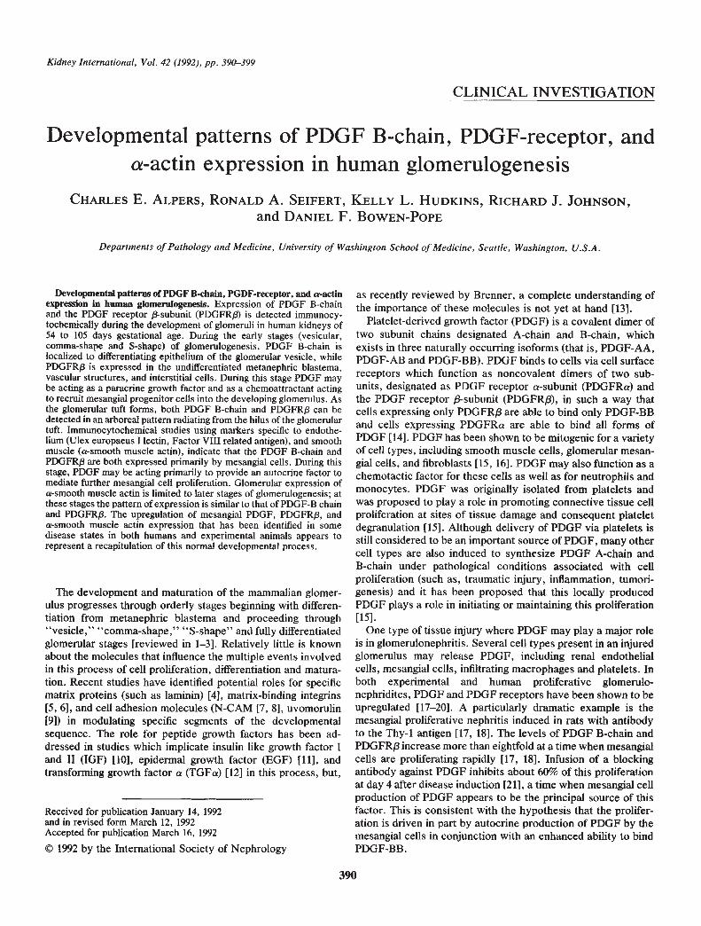
Kidney International, Vol. 42 (1992), pp. 390—399
CLINICAL INVESTIGATION
Developmental patterns of PDGF B-chain, PDGF-receptor, anda-actin expression in human glomerulogenesis
CHARLES E. ALPERS, RONALD A. SEIFERT, KELLY L. HUDKINS, RICHARD J. JOHNSON,and DANIEL F. BOWEN-POPE
Departments of Pathology and Medicine, University of Washington School of Medicine, Seattle, Washington, U.S.A.
Developmental patterns of PDGF B-chain, PGDF-receptor, and a-actinexpression In human glomerulogenesis. Expression of PDGF B-chainand the PDGF receptor 13-subunit (PDGFRj3) is detected immunocy-tochemically during the development of glomeruli in human kidneys of54 to 105 days gestational age. During the early stages (vesicular,comma-shape and S-shape) of glomerulogenesis, PDGF B-chain islocalized to differentiating epithelium of the glomerular vesicle, whilePDGFRI3 is expressed in the undifferentiated metanephric blastema,vascular structures, and interstitial cells. During this stage PDGF maybe acting as a paracrine growth factor and as a chemoattractant actingto recruit mesangial progenitor cells into the developing glomerulus. Asthe glomerular tuft forms, both PDGF B-chain and PDGFRI3 can bedetected in an arboreal pattern radiating from the hilus of the glomerulartuft. Immunocytochemical studies using markers specific to endothe-hum (Ulex europaeus I lectin, Factor VIII related antigen), and smoothmuscle (a-smooth muscle actin), indicate that the PDGF B-chain andPDGFRI3 are both expressed primarily by mesangial cells, During thisstage, PDGF may be acting primarily to provide an autocrine factor tomediate further mesangial cell proliferation. Glomerular expression ofa-smooth muscle actin is limited to later stages of glomerulogenesis; atthese stages the pattern of expression is similar to that of PDGF-B chainand PDGFR/3. The upregulation of mesangial PDGF, PDGFRI3, anda-smooth muscle actin expression that has been identified in somedisease states in both humans and experimental animals appears torepresent a recapitulation of this normal developmental process.
The development and maturation of the mammalian glomer-ulus progresses through orderly stages beginning with differen-tiation from metanephric blastema and proceeding through"vesicle," "comma-shape," "S-shape" and fully differentiatedglomerular stages [reviewed in 1—31. Relatively little is knownabout the molecules that influence the multiple events involvedin this process of cell proliferation, differentiation and matura-tion. Recent studies have identified potential roles for specificmatrix proteins (such as laminin) [4], matrix-binding integrins[5, 6], and cell adhesion molecules (N-CAM [7, 8], uvomorulin[9]) in modulating specific segments of the developmentalsequence. The role for peptide growth factors has been ad-dressed in studies which implicate insulin like growth factor Iand II (IGF) [10], epidermal growth factor (EGF) [11], andtransforming growth factor a (TGFa) [121 in this process, but,
Received for publication January 14, 1992and in revised form March 12, 1992Accepted for publication March 16, 1992
© 1992 by the International Society of Nephrology
as recently reviewed by Brenner, a complete understanding ofthe importance of these molecules is not yet at hand [13].
Platelet-derived growth factor (PDGF) is a covalent dimer oftwo subunit chains designated A-chain and B-chain, whichexists in three naturally occurring isofonns (that is, PDGF-AA,PDGF-AB and PDGF-BB). PDGF binds to cells via cell surfacereceptors which function as noncovalent dimers of two sub-units, designated as PDGF receptor a-subunit (PDGFRa) andthe PDGF receptor 13-subunit (PDGFR/3), in such a way thatcells expressing only PDGFRJ3 are able to bind only PDGF-BBand cells expressing PDGFRa are able to bind all forms ofPDGF [14]. PDGF has been shown to be mitogenic for a varietyof cell types, including smooth muscle cells, glomerular mesan-gial cells, and fibroblasts [15, 16]. PDGF may also function as achemotactic factor for these cells as well as for neutrophils andmonocytes. PDGF was originally isolated from platelets andwas proposed to play a role in promoting connective tissue cellproliferation at sites of tissue damage and consequent plateletdegranulation [151. Although delivery of PDGF via platelets isstill considered to be an important source of PDGF, many othercell types are also induced to synthesize PDGF A-chain andB-chain under pathological conditions associated with cellproliferation (such as, traumatic injury, inflammation, tumori-genesis) and it has been proposed that this locally producedPDGF plays a role in initiating or maintaining this proliferation[15].
One type of tissue injury where PDGF may play a major roleis in glomerulonephritis. Several cell types present in an injuredglomerulus may release PDGF, including renal endothelialcells, mesangial cells, infiltrating macrophages and platelets. Inboth experimental and human proliferative glomerulo-nephridites, PDGF and PDGF receptors have been shown to beupregulated [17—20]. A particularly dramatic example is themesangial proliferative nephritis induced in rats with antibodyto the Thy-i antigen [17, 18]. The levels of PDGF B-chain andPDGFRI3 increase more than eightfold at a time when mesangialcells are proliferating rapidly [17, 18]. Infusion of a blockingantibody against PDGF inhibits about 60% of this proliferationat day 4 after disease induction [21], a time when mesangial cellproduction of PDGF appears to be the principal source of thisfactor. This is consistent with the hypothesis that the prolifer-ation is driven in part by autocrine production of PDGF by themesangial cells in conjunction with an enhanced ability to bindPDGF-BB.
390

Alpers et a!: PDGF and actin in human glomerulogenesis 391
Although much less is known about possible roles of PDGF inembryonic development, there is some indication that thePDGF/PDGF receptor system may also be involved in devel-opmental processes. PDGFRa is expressed by the mesodermfrom the time that it can first be identified as a distinct germlayer, and continues to be expressed by a subset of mesodermalderivatives [22]. Mouse embryos which are homozygous for adeletion of the PDGF receptor a-subunit [23] display multiplemesodermal abnormalities and die before birth [22]. By con-trast, PDGFR/3 (which is the form of the receptor which seemsto be induced in a number of adult disease states) cannot bedetected in extracts of whole mouse embryos until later indevelopment [24], nor is the identity of the embryonic cellsexpressing this receptor known. In the present study, we reportthat PDGF B-chain and PDGFR/3 are expressed by the imma-ture glomerulus at specific stages of development. To identifythe cells expressing PDGF receptor /3-subunit we have em-ployed immunocytochemical markers of endothelium [FactorVill-related antigen (F111), Ulex europaeus I lectin] and thesmooth muscle-like glomerular mesangial cells (a-smooth mus-cle actin). We report that the pattern of expression of a-smoothmuscle actin and of PDGF B-chain and PDGFR/3 in differenti-ated fetal glomeruli closely resembles the pattern seen inresponse to mesangial injury and speculate on the roles thatPDGF may be playing in these processes.
Methods
Source of tissue
Human fetal kidneys (N = 44) were obtained fresh fromtissue examined after therapeutic abortions. Fifteen of these(estimated gestational age ranging from 54 to 105 days) werefixed in methyl Carnoy's (methacarn) solution (60% methanol,30% chloroform, 10% acetic acid) and processed and embeddedin paraffin according to conventional techniques. Twenty-fivekidneys (estimated gestational age ranging from 54 to 105 days)were fixed overnight in cold 2% or 4% paraformaldehyde inphosphate buffer, transferred to 30% sucrose in 0.01 M phos-phate buffer, equilibrated overnight at 4°C, and snap frozen inOCT compound (Miles, Inc., Elkhart, Indiana, USA). Fourkidneys (estimated gestational age ranging from 57 to 84 days)were fixed overnight in cold periodate-lysine-paraformaldehyde(PLP) solution, washed, dehydrated through 90% ethanol,embedded in LR White resin (Ted Pella, Inc., Redding, Cali-fornia, USA), and polymerized overnight at 55°C.
Immunohistochemis try
Sections of methyl Carnoy's fixed tissue were deparaffinizedwith xylene and graded ethanols, blocked with 3% hydrogenperoxide, and washed with PBS (138 mM) NaCI, 2.7 mri KCI,3.2 m Na2HPO4, 1.5 m KH2PO4, pH 7.3). The tissue wasthen incubated with one of the primary murine monoclonalantibodies (see below), and subsequently processed using astreptavidin-biotin immunoperoxidase method with 3,3'-diami-nobenzidine (with nickel chloride enhancement) as the chro-mogen. Sections were counterstained with methyl green.
Frozen sections of 2% or 4% paraformaldehyde fixed tissuewere hydrated in PBS, blocked with 3% hydrogen peroxide andwashed in PBS. The sections were incubated overnight at 4°Cwith antibody 7212, washed, and subsequently processed as
above using a streptavidin-biotin immunoperoxidase method,counterstained with methyl green, dehydrated and cover-slipped.
For all samples, a negative control consisted of substitutionof the primary antibody with both irrelevant murine monoclonalantibodies, non-immune rabbit sera in the case of Factor VIIIrelated antigen, and PBS. Isotype-matched irrelevant murineantibodies were specifically utilized as controls for PDGFB-chain, PDGFRI3, and smooth muscle a-actin. Positive inter-nal controls in each biopsy consisted of actin-positive smoothmuscle cells comprising the renal vasculature for the a-SM-iantibody and arterial endothelial cells for Ulex lectin andanti-Factor VIII related antigen antibodies.
Antibodies
PDGF B-chain. Murine monoclonal antibody PG7-007 (pro-vided by R. Ross and Mochida Pharmaceutical, Tokyo, Japan)has been previously characterized and its ability to specificallyrecognize PDGF B-chain in methacarn fixed tissue has beendemonstrated [25, 26]. The specificity of the immunohistochem-ical reactivity of this antibody has been demonstrated in cellculture where its activity has been abolished by pre-incubationwith immunizing PDGF peptide [25] and by studies by ourgroup which tightly link increased expression of the PDGFpeptide as recognized by this antibody in tissue sections withincreased synthesis of mRNA encoding PDGF as detected byNorthern analysis and in situ hybridization in rat glomeruli [17,18].
PDGFRp. Murine monoclonal antibody PR7212 has beenpreviously characterized by Western blotting and competitivebinding studies and shown to recognize the /3 subunit of thePDGF receptor [14, 27]. We have shown that this antibodyrecognizes PDGFRI3 but not PDGFRa in extracts of BHK cellstransfected with the human a or /3 subunits by Western blottingtechniques. The epitope recognized by this antibody is stable inparaformaldehyde and PLP but not methacarn fixed tissues.
Smooth muscle a-actin isoform. Murine monoclonal anti-body a-SM-i (gift of G. Gabbiani, now commercially availablethrough DAKO Corp., Carpinteria, California, USA) has beencharacterized by tissue immunohistochemistry and Westernblotting [28] and has been previously demonstrated to recognizesmooth muscle a-actin in methacarn-fixed tissues [29, 30]. Wehave previously demonstrated the specificity of the increasedglomerular expression of a-smooth muscle actin expressiondetected by tissue immunohistochemistry with this antibody byconcurrent Northern analysis for a-actin mRNA synthesis inisolated glomeruli obtained in a rat model of mesangiolyticinjury [29].
Muscle-spec/fic actin. Antibody HHF-35 (gift of Allen Gown,commercially available through DAKO Corp.) has been shownto recognize the four muscle specific isoforms of actin (smoothmuscle a- and y-actin, striated muscle a-actin, cardiac musclea-actin, but not cytoplasmic /3 and a actins) [31] and was usedas previously described [29, 30].
Smooth muscle (desmin). A murine monoclonal antibody tohuman desmin (DAKO Corp.) was used as previously described[32].
Endothelial cells. Rabbit anti-human Factor VIII relatedantigen/von Willebrand Factor (DAKO Corp.) was used aspreviously described [33]. Endothelial cells were also identified

392 Alpers et a!: PDGF and actin in human glomerulogenesis
by lectin binding studies using Ulex europaeus I lectin (VectorLaboratories, Burlingame, California, USA) as previously de-scribed [34, 35].
Proliferating cell nuclear antigen (PCNA). 19A2, a murinemonoclonal antibody to PCNA/cyclin (Coulter Corp., Hialeah,Florida, USA) was used as previously described [36].
Immunoelectronmicroscopy
Frozen, 4% paraformaldehyde fixed kidneys were sectionedat 6 pm and sections adhered to gelatin coated slides and airdried for 30 minutes. The sections were then stained byhydrating in PBS for 15 minutes, incubated in 0.02 M periodicacid in PBS to inactivate endogenous peroxidase and thenincubated with normal horse serum in PBS containing 2% BSA(Sigma, St. Louis, Missouri, USA) for 30 minutes at roomtemperature to block nonspecific staining. Sections were thenincubated with either antibody PR7212 or a-SM-i at 4 pg/ml inPBS/BSA overnight at 4°C, and rubsequently processed using astreptavidin-biotin immunoperoxidase method with 3,3'-diami-nobenzidine (DAB) as the chromogen and horse anti-mouseantisera as the secondary detecting antibody system. Sectionswere reacted with 1% 0s04 in phosphate buffer for one hour atroom temperature, rinsed and then dehydrated through gradedethanols and into propylene oxide. Sections were then infil-trated with a 50/50 mixture of PolyBed (PolySciences, Inc.,Warrington, Pennsylvania, USA) and propylene oxide for onehour. Beem capsules were filled with PolyBed, inverted overthe sections, infiltrated overnight and then polymerized at 55°Cfor 48 hours. The blocks were removed by heating the slidebriefly and quickly snapping off the capsule. Thin, 0.1 micronsections were cut and mounted on formvar coate nickel gridsand examined in a Philips 410 electron microscope.
Alternately, in efforts to improve preservation of morpho-logic detail, renal tissue fixed in PLP solution, was processedand embedded in LR white resin (Polysciences, Inc.). Thinsections were incubated with antibody a-SM-I or control irrel-evant monoclonal antibody overnight at 4°C, rinsed with PBS,and subsequently incubated with goat anti-mouse IgG conju-gated to gold (10 nm particles) as previously described [29].
Results
Kidney tissue was obtained at gestational ages of 54 to 105days. Kidneys from the earliest gestational ages studied (ap-proximately 54 to 65 days) typically contain metanephric blast-ema, ureteric buds, interstitial tissue, several layers of imma-ture glomeruli, and virtually no detectable structuresresembling mature tubules, collecting ducts, medulla or anorganized arterial or venous system. Kidneys obtained fromolder fetuses typically contain metanephric blastema, intersti-tium, ureteric buds, and glomeruli exhibiting all stages ofdevelopment ranging from earliest vesicles to apparently fullydifferentiated, albeit immature, structures with identifiablemesangium, capillary loops, and visceral and parietal epithelialcells. A representative histologic section from such a kidney isshown in Figure 1. Also present in later kidneys are identifiabletubular segments, developing medulla, renal pelvis with urothe-hal lining, and distinct arterial and venous structures.
Immunohistochemistry
The immunohistochemical findings in glomeruli of this studyare summarized in Table 1.
PDGF B-chain. Two developmental stage-specific patterns ofPDGF B-chain expression in developing glomeruli were identi-fied. B-chain could be first identified at the earliest stages ofglomerular development, at or immediately following the vesi-cle stage, when the differentiating epithelium of the immatureglomerulus is still in close contact with the inductive differen-tiating stimulus, the ureteric bud (Fig. 2A, B). In addition, thenascent parietal epithehium lining the future urinary space alsoexpresses PDGF B-chain. In contrast, there is no detectableexpression by the blastema or interstitial tissue. The uretericbud was most often negative, but occasional staining for PDGFB-chain at the luminal surface of the ureteric bud was seen.
In later stages of glomerular development, PDGF B-chain isexpressed in an arboreal pattern radiating from the hilus of theglomerular tuft, suggesting a mesangial origin (Fig. 2C). Extra-glomerular structures showing expression of this protein in-clude some immature tubules (segments distinct from uretericstructures and with features suggesting they are in contiguitywith the still positive parietal epithelium, indicating proximaltubule lineage), and the superficial layers of the urotheiumlining the renal pelvis and ureters.
PDGFRp. Unlike PDGF B-chain, PDGFRI3 was not detectedin the glomerulus during the early stages of differentiation.Outside of the glomerulus, PDGFR/3 is expressed abundantly inthe metanephric blastema, and in vascular structures and inter-stitial cells which are seen as arcades in the most immature andsuperficial areas of the developing cortex (Fig. 2D). Glomerularexpression of PDGFRf3 occurs in later stages of development,where it localizes in a mesangial pattern (Fig. 2E, F). Occasion-ally, expression of PDGFR/3 appeared to extend to the endo-thelium lining peripheral glomerular capillary walls (Fig. 2F). Inadjacent maturing portions of the cortex PDGFR/3 continued tobe expressed by interstitial cells and arterial vessels.
Smooth muscle cell markers/mesangial cell markers. Nospecific markers that identify mesangial cells in tissue sectionscurrently have been identified. However, a-smooth muscleactin, a marker for smooth muscle cells, has been shown to bean inducible marker of activated mesangial cells in diseasestates in adult rat and human glomeruli [29, 30]. Using anantibody to this smooth muscle marker, as well as antibodies totwo additional muscle markers, pan-muscle actin and desinin,the expression of these "muscle-specific" proteins in glomeru-lar development was studied.
Developing glomeruhi show a gradient of expression ofa-smooth muscle actin, ranging from absent in the earlieststages of development, to pronounced mesangial expression inmore mature glomeruli (Fig. 3A, B). The results with theantibodies to both a-smooth muscle actin and muscle-specificactin were identical, with the only notable difference beingsomewhat stronger staining intensity obtained with the a-SM-iantibody. The onset of smooth muscle actin expression bymesangial cells in the course of glomerular development coin-cided with the identification of an organized intraglomerularvasculature having contiguity with the extraglomeruiar intersti-tial vasculature (as detected by staining for Factor VIII related

Alpers et a!: PDGF and Odin in human glomerulogenesis 393
Fig. 1. Low power micrograph of human fetal kidney (approximately 86 days gestation). Immediately beneath the renal capsule is theundifferentiated metanephric blastema (B). Invading the blastema from below are multiple profiles of ureteric buds (U), and occasional aggregatesof differentiating cells adjacent to the ureteric buds, the early glomerular vesicles (GV), can be identified. Glomeruli in zone of glomerulogenesis(within brackets) show progressive differentiation from vesicle stage, to early comma and S-stages (EG), and culminating in fully differentiatedstructures (G). PAS, 50x.
antigen and Ulex I lectin). There was also prominent extraglo-merular expression of actin by differentiated smooth musclecells of the arterial and arteriolar vasculature, but not by othercells comprising the interstitium.
Desmin could be identified in the smooth muscle cells com-prising the media of muscular arteries present in the developingrenal hilus. In contrast to the muscle specific actins, there wasno localization of desmin in glomeruli at any stage of develop-ment, and no expression of this protein detectable by theantibody utilized in this study by metanephric blastema or theinterstitium.
Endothelial cell markers. Factor Vill-related antigen couldbe identified in the aggregate of interstitial and vascular cellsinvaginating into the glomerular epithelium at the comma andS-shape stages of development. In more mature glomeruli, thispattern of localization evolved to a predominantly mesangialpattern similar to that of PDGF B-chain, PDGFRI3, anda-smooth muscle actin, as seen in Figure 3C. Extraglomerularstaining was confined to the endothelial lining of the vascula-ture.
Binding of the Ulex Europaeus I lectin showed patterns ofreactivity similar to Factor Vill-related antigen in the earlystages of glomerulogenesis and in the extra-glomerular vascu-lature. More mature glomeruli showed discrete staining of theendothelial lining of glomerular capillaries, but did not show the
apparent mesangial staining seen in the studies of FactorVill-related antigen (Fig. 3D).
Cell proliferation. Staining for the proliferation markerPCNA was most heavily concentrated in the nephrogenic zonein the first stages of glomerular epithelial differentiation, whereat times virtually every cell in differentiating vesicles, commaand S-shaped glomeruli could be shown to be actively replicat-ing as evidenced by PCNA expression (Fig. 3E). The surround-ing blastema also showed a high replicative rate. There was adecreasing gradient of PCNA expression corresponding toprogressively greater glomerular maturation, in distinct con-trast to the patterns of PDGFRI3 and actin expression, althougheven the most mature glomeruli continued to exhibit somereplicative activity.
ImmunoelectronmicroscopyPDGFRI3. In relatively mature glomeruli, reaction product
indicative of PDGFRI3 localization was confined to the mem-branes of cells present in mesangial locations (Fig. 4). Noexpression was detected in peripheral portions of glomerularcapillary loops where only endothelial cells or visceral epithelialcells would be expected to be present. When endothelial cellbodies could be identified overlying the portion of mesangiumadjacent to the capillary lumina, PDGFR could be localized to
394 Alpers et a!: PDGF and actin in human glomerulogenesis
Table 1. Phenotypic characterization of stages of glomerulogenesis inhuman fetal kidney
Metanephncblastema Vesicle
Early glomerulardifferentiation(comma, S-
stage)Differentiatedglomerulus
PDGF B-chain — focal" + mesangiumPDGFR/3 + — — mesangiuma-smooth muscle — mesangium
actinDesmin - - - -Factor VIII capillaries mesangium'
related antigen! endothelium"von Willebrandfactor
Ulex lectin capillaries capillaryendothelium
Expression of PDGF, its receptor, and differentiation markers ofmesenchymal cell components of the glomerulus at the principal stagesof glomerulogenesis.
Symbols are:aComponents of vasculature present in blastema and interstitium
express these markers.b j remains unresolved whether there is focal expression of PDGF in
some early vesicular elements or whether these areas represent tangen-tial sections through portions of later developing comma or S-stagevesicles; uncertainty whether Factor VIII related antigen expression islimited to endothelium or may be present in fetal mesangium ispresented in the text.
PDGF—platelet-derived growth factor, PDGFRI3—platelet-derivedgrowth factor (/3 subunit).
the mesangial cell borders, but not those of the endothelialcells. Because of the requirement for gentle fixation to preservethe antigenicity of the epitope recognized by the antibody toPDGFRI3, morphologic preservation of tissue was compro-mised to some degree, allowing confident localization of reac-tion product only in the anatomically well-defined structures ofrelatively mature glomeruli.
Muscle-specific actin (a-SM-i). The peroxidase label indicat-ing presence of muscle-specific actin was concentrated predom-inantly within the cytoplasm of cells in mesangial areas identicalin appearance and location to those showing staining of the cellmembranes for PDGFR/3 (Fig. 5). Morphologic preservation oftissues fixed in paraformaldehyde or PLP solution were bothinadequate to determine whether the peroxidase or gold labelswere localized to specific microfilament bundles.
Discussion
In this study, we have employed immunohistochemical tech-niques to study expression and distribution of PDGF B-chain,the PDGF receptor /3-subunit, and markers of mesenchymal celltypes in order to determine the developmental sequence andpattern of expression and provide some insight into how thePDGF system might be involved in human kidney development.All sections were from normal human fetal kidneys obtainedfrom elective therapeutic abortions at estimated gestations of 54to 105 days. During this developmental period, human kidneysdisplay a gradient of differentiation from undifferentiated blast-ema at the outer margin of the cortex to relatively matureglomeruli in the inner portion of the cortex, as seen in Figure 1.This permits evaluations of a relatively wide span of develop-mental stages within a single tissue section.
Within the glomerulogenic zone, comprising a rim of corticaltissue composed of metanephric blastema, branching termini ofthe ureteric bud, glomeruli at the earliest stages of glomerulo-genesis (vesicle, comma, and S-shaped glomeruli) and a primi-tive vasculature organized in arcades. Expression of PDGFB-chain was localized to differentiating cells of glomeruli vesi-des and the epithelia of primitive glomeruli immediately adja-cent to the ureteric bud. PDGF B-chain was not detected in theadjacent mesenchyme, blastema and blood vessels, and gener-ally not detected in the ureteric bud. PDGFR/3, in contrast, wasnot detected within the primitive glomeruli but was abundantlyexpressed by the metanephric blastema and interstitial tissuessurrounding glomeruli within the nephrogenic zone. The highestlevel of cell proliferation, as detected by staining for PCNA,was identified in this zone. Proliferating cells could most oftenbe identified as epithelial cells in the earliest stages of glomer-ulogenesis and as metanephnc blastemal cells. This suggeststhat PDGF released by primitive glomerular cells could behaving a paracrine effect on the surrounding PDGFRI3 positiveprimordial interstitial cells. Within the glomerulogenic zone,cells exhibiting features of muscle cell phenotype (positive formuscle actin) and endothelial phenotype (positive for F111related antigen and Ulex lectin) were confined to the intersititalvasculature.
In more mature glomeruli located outside of the zone ofglomerulogenesis, the glomerular tuft architecture assumes amore adult form and expression of PDGF B-chain, PDGFR/3,and muscle-specific actin within the glomerulus is prominentand restricted to the mesangial areas. The expression of muscle-specific actin by developing mesangial cells appeared to occurconcurrently with identification of an organized intraglomerularvasculature having contiguity with the extraglomerular intersti-tial vasculature (as detected by staining for the markers ofendothelium). These patterns of glomerular expression contin-ued to be present throughout the period of development (to day105) encompassed by this study. In normal adult kidneys lowlevels of PDGFR/3 are still expressed by mesangial cells [37],but smooth muscle actin levels are greatly reduced [30], andexpression of PDGF B-chain can no longer be detected byimmunohistochemical techniques. The time at which fetalmesangium converts to an adult phenotype remains to bedetermined.
In the developing glomerulus, particularly in early stages ofglomerulogenesis, the cell lineage of specific cells can bediflicult to ascertain on histologic grounds alone. The histologicevidence that it is the mesangial cells which express PDGFB-chain and PDGFR/3 in more mature glomeruli is supported bythe congruent pattern of immunocytochemical localization ofa-smooth muscle actin. By contrast, the endothelial cell-spe-cific marker Ulex lectin stains cells with a distribution expectedfor glomerular endothelial cells. The immunoelectronmicro-scopic results confirm, at a higher level of resolution, thata-smooth muscle actin and PDGFR/3 are both expressed bycells in mesangial locations and neither are expressed by cells inendothelial locations. We therefore propose that a-smoothmuscle actin is a marker for developing mesangial cells. Aparticularly noteworthy feature of this muscle-specific proteinis that it is expressed at very low levels by mesangial cells in

Alpers et a!: PDGF and actin in human glomerulogenesis 395
Fig. 2. Human fetal kidney, approximately 86 days gestation. A. PDGF-B chain is detected in early vesicle and comma- to S-shaped glomeruli(EG), while blastema and ureteric buds are negative. More differentiated glomeruli (G) show mesangial pattern of expression. A tubular segment(T) between a glomerulus and uretenc bud also expresses PDGF-B chain. 125x. B. Higher power view of early glomeruli seen in A. Staining ofdifferentiating epithelium which will become organized into a glomerular tuft, as well as epithelial cells which will become the parietal epithelialcells lining Bowman's capsule, for PDGF-B chain is evident. 250x. C. Differential-d glomerular tuft showing mesangial localization of PDGF-Bchain. There is uniform staining of parietal epithelial cells lining Bowman's capsule. 250x. D. Paraformaldehyde fixed tissue, from same kidneyas shown in A through C, stained for PDGFR/3. There is localization of PDGFR/3 to blastema, vascular structures (present in arcades of interstitialtissue), interstitial cells, and mesangial areas (arrows) of more mature glonieruli. 60X. E. Differentiated glomerulus stained for PDGFR/3. Stainingis confined to mesangial areas and extraglomerular interstitium. 250x. F. Differentiated glomerulus stained for PDGFR/3. In addition to mesangialexpression of this protein, occasional staining seemed to extend to glomerular capillary endothelium (arrows). 250x.
normal mature human glomeruli, and its expression is dramat-ically upregulated in rat [291 and human [30] glomeruli inresponse to injury.
Nonetheless, there are some limitations in our ability todefinitively state that PDGF and PDGF-R are expressed only by
mesangial cells. Immunostaining with antibody to F111-re1atedantigen, an antigen that is generally considered to be expressedexclusively by endothelial cells, reveals a pattern of localizationin fetal glomeruli in later stages of development that can beindistinguishable from that of a-smooth muscle actin, PDGF,
i1r
p

396 Alpers et al: PDGF and actin in human glomerulogenesis
Fig. 3. Same fetal kidney illustrated in Figure 2. A. Expression of a-smooth muscle actin, There is localization in vessels coursing through theblastema, arterial and arteriolar smooth muscle cells (arrows) and in the mesangium in later stages of glomerular development (G). 60X. B. Higherpower view of a-smooth muscle actin expression in mesangium and in adjacent arteriolar smooth muscle cells (arrow). 250x. C. Expression ofFactor VIII related antigen also appears to have a mesangial rather than pure endothelial pattern. These findings present a paradox, suggesting inthe fetus that there are glomerular (apparently mesangial) cells with features of both endothelial and smooth muscle derivation. 250x. 0. Bindingof Ulex I lectin contrasts with that of Factor VIII related antigen in clearly exhibiting an endothelial pattern of reactivity including glomerularcapillary walls and interstitial vessels and capillaries. 125x. E. Staining for proliferating cell nuclear antigen (PCNA), indicative of repllcating cells,is concentrated in blastema and in epithelial cells comprising early glomeruli in zone of glomerulogenesis. Positively stained cells are less numerousbut still present in more differentiated glomeruli as well as developing tubules and interstitium. 60x.
and PDGF-R. There are at least two possible explanations forthis. (1) The first possibility is that mesangial cells originatefrom a mesenchymal precursor with some properties of bothendothelial cells (F,111 related antigen) and smooth muscle cells(a-smooth muscle actin) and that expression of F111 is lost
relatively late in mesangial cell differentiation. (2) A secondpossibility is that the apparent F511 staining of mesangial cellsis an artifact resulting from a close apposition of endothelialcells to the mesangium, with regional concentration of F111related antigen in those parts of the endothelial cells (nucleus
—
4 tIp
.. a

Alpers et a!: PDGF and actin in human g!omeru!ogenesis 397
Fig. 4. Ultrastructural immunolocalization ofPDGFRI3. Black peroxidase reaction productis confined to the cell membranes of cellswithin mesangial areas (M). Visceral epithelialcells (V), and capillary endothelium (E) arenot reactive. C, capillary lumen. 8800x.
and large parts of the cell body) adjacent to the mesangial cells,rather than distributed in the thin layers of fenestrated endo-thelium which line the peripheral parts of the glomerularcapillary wall. Such a distribution would explain why the otherendothelial cell marker, Ulex europaeus I lectin, did not show amesangial staining pattern, since Ulex binds to an aL-fucosecarbohydrate moiety distributed diffusely over the surface ofthe endothelial cell [341 rather than to a cytoplasmic compo-nent. Resolution of this issue may come when we or othersdevelop the ability to successfully combine markers for F111related antigen, actin, and PDGF-R in single sections studied byimmunoelectron microscopy.
The spatial and chronological patterns of expression of PDGFB-chain and PDGFR/3 in the developing metanephric kidneysuggest that this growth factor/receptor interaction might havemultiple roles in the process of glomerular differentiation andmaturation. At the earliest stages of glomerulogenesis, PDGFB-chain is expressed by the vesicle while the PDGFRJ3 isexpressed only outside the vesicle by blastemal and connectivetissue elements. This suggests a possible paracrine interactionwithin the kidney, in which PDGF from the vesicle maymodulate the processes of glomerular induction or begin tostimulate migration of receptor-positive cells from the vascular!connective tissue into the glomerular cleft where they developinto the mesangial stalk. There is growing evidence that PDGFcan act as such a migratory chemotactic factor for fibroblasts[38], smooth muscle cells [39—40] and even mature mesangialcells [41]. Once the mesangial-like cells have entered theglomerulus they continue to express PDGFR/3 but now alsoexpress PDGF B-chain. This might provide an autocrine stim-ulus for further mesangial cell proliferation, migration, and/ormodulation. Eventually, expression of both PDGF B-chain andPDGFR/3 is downregulated, further proliferation virtually
ceases, and the glomerulus achieves its mature/adult pheno-type.
The autocrine phase of development may be reactivated inadults under pathological conditions in which mesangial cellproliferation is reactivated. We have recently provided evi-dence that induced expression of PDGF B-chain and PDGFRI3may mediate mesangial cell proliferation and participate inreconstitution of the mesangium following antibody-inducedmesangiolytic injury in the rat [17, 18]. In man, there is somelimited evidence that upregulation of PDGF-R may occur in thecourse of some forms of glomerulonephritis [19] as well asevidence that PDGF may localize to the mesangium in someforms of mesangial proliferative glomerulonephritis [20, 42].Furthermore, it has been demonstrated that cultured mesangialcells stimulated by a variety of mediator substances known toparticipate in renal injury will produce PDGF [16, 43, 44]. It isparticularly noteworthy that in both the rat [17, 29] and humans[30], this apparent upregulation of the PDGF/PDGF receptorsystem in disease states is associated with phenotypic modula-tion by mesangial cells to increase expression of muscle-specificactin, similar to that identified in fetal glomeruli. We believethese lines of evidence demonstrate that the PDGF/PDGFreceptor system mediates at least in part the proliferative andreparative phases of mesangial injury in experimental animalsand in humans. The data presented in this study thereforesuggests that this response to injury recapitulates a normalprocess of mesangial development that occurs in utero.
Acknowledgments
This work was supported in part by grants DK40802, DK43422, GM35501, and HL-03 174 from the National Institutes of Health. We thankthe Central Laboratory for Human Embryology at the University of
1

-I
IC
m
398 Alpers et al: PDGF and actin in human glomerulogenesis
Fig. 5. Ultrastructural immunolocalization of a-smooth muscle actin. Black peroxidase reaction product is confined to cells in mesangial areas(M). Visceral epithelial cells (V), endothelial cells (E), mesangial cell nuclei (N) and connective tissue matrix (CT) in mesangium and glomerularbasement membranes are not reactive. C, capillary lumen. 8300x.
Washington (supported by grant HD-00836 from the N.I.H.) for assis-tance in providing fetal tissue. The authors thank Tamara Carlson forher patience and secretarial assistance.
Reprint requests to Charles E. Alpers, M.D., Department of Pathol-ogy, RC-72, University of Washington Medical Center, Seattle, Wash-ington 98195, USA
References
1. SAXEN L: Organogenesis of the Kidney. Cambridge, CambridgeUniversity Press, 1987
2. EKBLOM P, WELLER A: Ontogeny of tubulointerstitial cells. Kidneymt 39:394—400, 1991
3. ABRAHAMSON DR: Glomerulogenesis in the developing kidney.Sem Nephrol 11:375—389, 1991
4. EKELOM P: Developmentally regulated conversion of mesenchymeto epithelium. FASEB J 3:2141—2150, 1989
5. KORHONEN M, YLANNE J, LAITINEN L, VIRTANEN I: The a1-a6subunits of integrins are characteristically expressed in distinctsegments of developing and adult human nephron. J Cell Biol111:1245—1254, 1990
6. S0R0KIN L, SONNENBERG A, AUMAILLEY M, TIMPL R, EKELOM P:Recognition of the laminin E8 cell-binding site by an integrinpossessing the a6 subunit is essential for epithelial polarization indeveloping kidney tubules. J Cell Biol 111:1265—1273, 1990
7. KLEIN G, LANGEGGER M, G0RIDI5 C, EKBL0M P: Neural celladhesion molecules during embyonic induction and development ofthe kidney. Development 102:749—761, 1988
8. LACKIE PM, ZUBER C, ROTI4 J: Polysialic acid and N-CAMlocalization in embryonic rat kidney: Mesenchymal and epithelial
elements show different patterns of expression. Development 110:933—947, 1990
9. VESTWEBER D, KEMLER R, EKBLOM P: Cell-adhesion moleculeuvomorulin during kidney development. Dev Biol 112:213—221,1985
10. ROGERS SA, RYAN G, HAMMERMAN MR: Insulin-like growthfactors I and II are produced in the metanaplros and are requiredfor growth and development in vitro. J Cell Biol 113:1447—1453,1991
11. AVNER ED, SWEENEY WE JR: Polypeptide growth factors inmetanephric growth and segmental nephron differentiation. PediatrNephrol 4:372—377, 1990
12. BACALLAO R, FINE LG: Molecular events in the organization ofrenal tubular epithelium from nephrogenesis to regeneration. Am JPhysiol 257(Renal Fluid Electrolyte Physiol 26):F913—F924, 1989
13. BRENNER BM: Determinants of epithelial differentiation duringearly nephrogenesis. JAm Soc Nephrol 1:127—139, 1990
14. SEIFERT RA, HART CE, PHILLIPS PE, FORSTROM JW, Ross R,MURRAY MJ, BOWEN-POPE DF: Two different subunits associate tocreate isoform-specific platelet-derived growth factor receptors. JBiol Chem 264:8771—8778, 1989
15. Ross R, RAINES EW, BOWEN-POPE DF: The biology of PDGF. Cell46:155—169, 1986
16. SCHULTZ PJ, DICORLETO PE, SILVER BJ, ABBOUD HE: Mesangialcells express PDGF mRNAs and proliferate in response to PDGF.Am J Physiol 255(Renal Fluid Electrolyte Physiol 24):F674—F684,1988
17. IIDA H, SEIFERT R, ALPERS CE, GRONWALD RGK, PHILLIPS PE,GoiwoN K, GOWN AM, Ross R, BOWEN-POPE DF, JOHNSON RJ:Platelet-derived growth factor (PDGF) and PDGF receptor areinduced in mesangial proliferative nephritis in the rat: An effect

Alpers et al: PDGF and actin in human glomerulogenesis 399
mediated by platelets and complement. Proc Nail Acad Sci USA88:6560—6564, 1991
18. YOSHIMURA A, GORDON K, ALPERS CE, FLOEGE J, PRJTZL P, RossR, COUSER WG, BOWEN-POPE DF, JOHNSON RJ: Demonstration ofPDGF B-chain mRNA in glomeruhi in mesangial proliferativenephritis by in situ hybridization. Kidney mt 40:470—476, 1991
19. FELLSTROM B, KLARESKOG L, HELDIN CH, LARSSON E, RO-HHSTRAND L, TEltRAclo L, TUFUESON G, WAHLBERG J, RUBIN K:Platelet-derived growth factor receptors in the kidney-upregulatedexpression in inflammation. Kidney mt 36:1099—1102, 1989
20. GESUALDO L, PINZANI M, FLORIANO JJ, Hss MO, NAGY NU,SCHENA FP, EMANCIPATOR SN, ABBOUD HE: Platelet-derivedgrowth factor expression in mesangial proliferative glomerulone-phritis. Lab Invest 65:160—167, 1991
21. JOHNSON RI, RAINES E, FLOEGE J, Y05HIMuRA A, PRITZL P,ALPERS C, Ross R: Inhibition of mesangial cell proliferation andmatrix expansion in glomerulonephritis by antibody to platelet-derived growth factor. J Exp Med 175:1413—1416, 1992
22. SCHA1-FEMAN GC, MORRIsON-GRAHAM K, WESTON JA, BOWEN-POPE DF: PDGF receptor alpha-subunit expression during mousedevelopment: Pattern of expression and consequences of genedisruption. Development (in press)
23. STEPHENSON DA, ANDERSON E, WANG C, MERCOLA M, STILESCD, BOWEN-POPE DF, CHAPMAN VM: The mouse mutation patch(Pj) carries a deletion in the gene for platelet-derived growth factorreceptor alpha-subunit. Proc Nat! Acad Sci USA 88:6—10, 1991
24. MERCOLA M, WANG C, KELLY J, BROWNLEE C, JACKSON-GRUSBYL, STILES C, BOWEN-POPE D: Selective expression of PDGF A andits receptor during early mouse embryogenesis. Dev Biol 138:114—122, 1990
25. Ross R, MASUDA J, RAINES EW, GOWN AM, KATSUDA S, SA5A-HARA M, MALDEN LT, MASUKO H, SATO H: Localization ofPDGF-B protein in macrophages in all phases of atherogenesis.Science 248:1009—1012, 1990
26. SASAHARA M, FRIES JWU, RAINES EW, GOWN AM, WESTRUMLE, FROSCH MP, BONTHRON DT, Ross R, COLLINS T: PDGFB-chain in neurons of the central nervous system, posterior pitu-itary, and in a transgenic model. Cell 64:217—227, 1991
27. HART CE, SEIFERT RA, Ross R, BOWEN-POPE DF: Synthesis,phosphorylation, and degradation of multiple forms of the platelet-derived growth factor receptor studied using a monoclonal anti-body. JBiol Chem 262:10780—10785, 1987
28. SKALLI 0, Roiz P, TRZECIAK A, BENZONANA G, GILLESSEN D,GABBIANI G: A monoclonal antibody against a-smooth muscleactin: A new probe for smooth muscle differentiation. J Cell Biol103:2787—2796, 1986
29, JOHNSON RJ, IIDA H, ALPERS CE, MAJESKY MW, SCHWARTZ SM,PRITZL P, GORDON K, GOWN AM: Expression of smooth musclecell phenotype by rat mesangial cells in immune complex nephritis:
a-smooth muscle actin is a marker of mesangial cell proliferation. JC/in Invest 87:847—858, 1991
30. ALPERS CE, HUDKINS KL, GOWN AM, JOHNSON RI: Enhancedexpression of "muscle-specific" actin in glomerulonephritis. Kid-ney mt 41:1134—1142, 1992
31. TSUKADA T, MCNUTT MA, Ross R, GOWN AM: HHF3S, a muscleactin-specific monoclonal antibody. Am J Pathol 127:389—402, 1987
32. GOWN AM, VOGEL AM: Monoclonal antibodies to intermediatefilament proteins of human cells: II. Distribution of filament pro-teins in normal human tissue. Am J Patliol 114:309—321, 1984
33. TURNER RR, BECKSTEAD JH, WARNKE RA, WOOD GS: Endothelialcell phenotypic diversity. Am J C/in Pathol 87:569—575, 1987
34. HOLTHOF'ER H, VIRTANEN I, KARINIEMI A-L, HORMIA M, LINDERE, MIETTINEN A: Ulex europaeus I lectin as a marker for vascularendothelium in human tissues. Lab Invest 47:60—66, 1982
35. ALPERS CE, BECESTEAD JH: Monocyte/macrophage-derived cellsin normal and transplanted human kidneys. C/in immunol Immu-nopathol 36:129—140, 1985
36. JOHNSON RJ, GARCIA RL, PRITZL P, ALPERS CE: Platelets mediateglomerular cell proliferation in immune complex nephritis inducedby anti-mesangial cell antibodies in the rat. Am J Pathol 136:369—374, 1990
37. ALPERS CE, JOHNSON RI, HUDICINS K, BOWEN-POPE D: Identifi-cation of platelet-derived growth factor receptor (PDGF-R) in fetaland adult human and in non-human primate kidneys. (abstract) JAm Soc Nephrol 1:521, 1990
38. SEPPA H, GROTENDORST G, SEPPA S, SCHIFFMAN E, MARTIN UR:Platelet-derived growth factor is chemotactic for fibroblasts. J CellBiol 92:584—588, 1982
39. FERNS GAA, RAINES EW, SPRUGEL KH, MOTANI AS, REIDY MA,Ross R: Inhibition of neointimal smooth muscle accumulation afterangioplasty by an antibody to PDGF. Science 253:1129—1132, 1991
40. JAWIEN A, BOWEN-POPE DF, LINDNER V, SCHWARTZ SM,CLOWES AW: Platelet-derived growth factor promotes smoothmuscle migration and intimal thickening in a rat model of balloonangioplasty. J Clin Invest 89:507—511, 1992
41. BARNES JL, HEVEY KA: Glomerular mesangial cell migration inresponse to platelet-derived growth factor. Lab Invest 62:379—382,1990
42. NAKAJIMA M, HEWITSON TD, MATHEWS DC, KINCAID-SMITH P:Platelet-derived growth factor mesangial deposits in mesangial IgAglomerulonephritis. Nephro/ Dial Transplant 6:11—16, 1991
43. SILVER BJ, JAFFER FE, ABBOUD HE: Platelet-derived growthfactor synthesis in mesangiah cells: Induction by multiple peptidemitogens. Proc NatI Acad Sci USA 86:1056-1060, 1989
44. JAFFER FE, KNAUSS TC, POPTIC E, ABBOUD HE: Endothehinstimulates PDGF synthesis in cultured human mesangial cells.Kidney Int 38:1193—1 198, 1990