DEVELOPMENTAL INFLUENCES ACROSS THE LIFESPAN. Developmental Influences Across the Lifespan No human...
49
DEVELOPMENTAL INFLUENCES ACROSS THE LIFESPAN
-
Upload
quinton-baham -
Category
Documents
-
view
220 -
download
2
Transcript of DEVELOPMENTAL INFLUENCES ACROSS THE LIFESPAN. Developmental Influences Across the Lifespan No human...
DEVELOPMENTAL INFLUENCES ACROSS THE
LIFESPAN

Developmental Influences Across the Lifespan
No human activity has greater biological and social significance than feeding.
An organism that does not consume energy effectively does not survive to reproduce.
A species that does not feed its young effectively jeopardizes the transmission of its genes.
Because ingestive behavior is crucial to the survival of the species, eating and drinking, and the behaviors that support them, are imbued with emotional resonance for the individual, the family, and the community.
Thus, the participants in a feeding relationship come well prepared by the selective contingencies of evolution to engage effectively in the give and take of feeding.
Embryology
The anatomy of the oral cavity, pharynx, larynx, and esophagus is the result of embryological processes that begin at fertilization of the ovum and continue through infancy and more slowly through childhood and adulthood.
In the very early stages of development, the embryo consists of three layers of tissue that will give rise to all of the structures of the body.

Embryology
The ectoderm, or outermost layer, forms the epidermis of the skin, teeth, hair, nails, as well as the epithelial tissue of the entire nervous system and oropharyngeal tracts.
Embryology
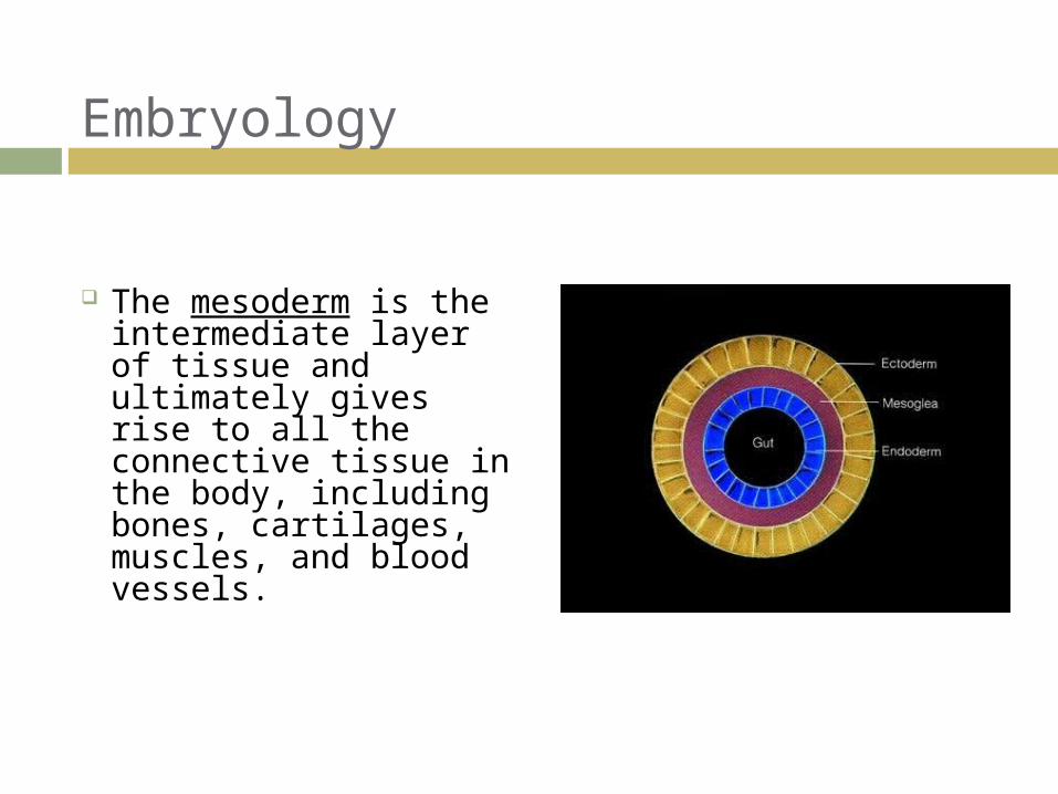
The mesoderm is the intermediate layer of tissue and ultimately gives rise to all the connective tissue in the body, including bones, cartilages, muscles, and blood vessels.
Embryology
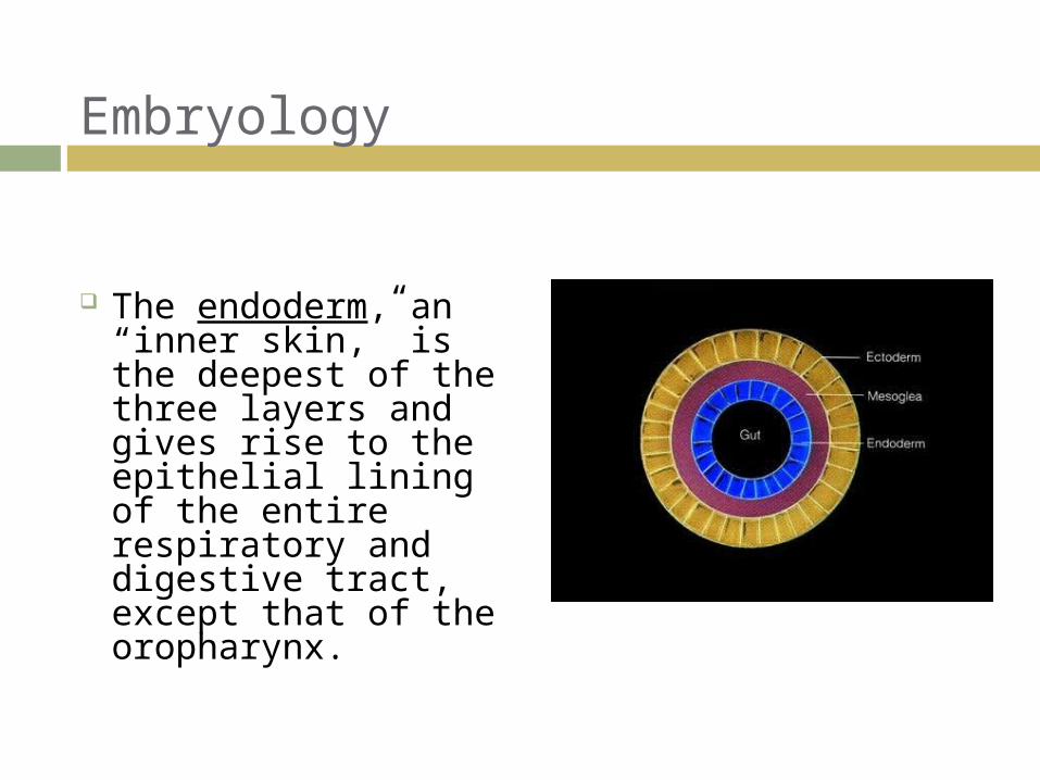
The endoderm, an “inner skin,” is the deepest of the three layers and gives rise to the epithelial lining of the entire respiratory and digestive tract, except that of the oropharynx.
3-week-old embryo
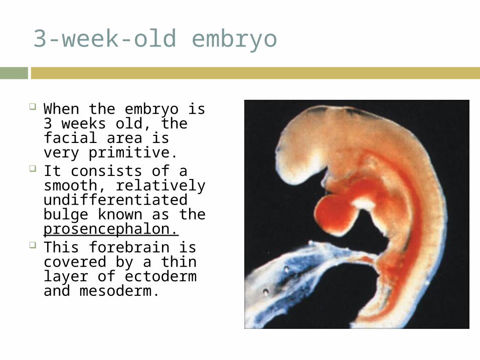
When the embryo is 3 weeks old, the facial area is very primitive.
It consists of a smooth, relatively undifferentiated bulge known as the prosencephalon.
This forebrain is covered by a thin layer of ectoderm and mesoderm.
3-week-old embryo
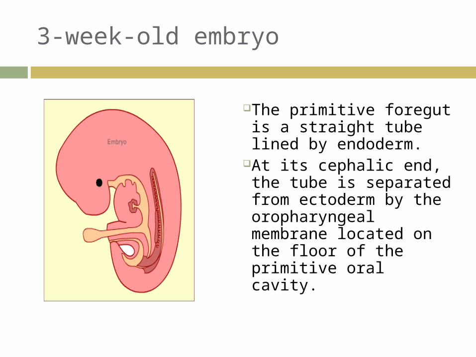
The primitive foregut is a straight tube lined by endoderm.
At its cephalic end, the tube is separated from ectoderm by the oropharyngeal membrane located on the floor of the primitive oral cavity.
8

4-week-old embryo
During the 4th week, the endoderm of the foregut separates laterally from the surface ectoderm by a layer of mesoderm which splits into vertical bars known as the pharyngeal or branchial arches.
Each arch forms a swelling on the surface of the embryo and on the wall of the foregut.
9

Branchial Arches, Pouches, and Grooves
The grooves on the surface are known as branchial grooves.
On the lateral wall of the foregut, the grooves are known as pharyngeal pouches.
10

Branchial Arches, Pouches, and Grooves
On the surface of the embryo there are five branchial arches and four branchial grooves between them.
Each arch contains a cartilaginous core, an artery, and a definite cranial nerve.
Each cranial nerve will supply the structures that develop from the mesenchyme of the arch.
11

Cranial Nerves & Branchial Arches
The nerve of the first arch is the mandibular branch of Trigeminal CN V.
The nerve of the second arch is Fcial CN VII.
The nerve of the third arch is Glossopharyngeal CN IX.
The nerve of the fourth arch is the superior laryngeal branch of Vagus CN X.
Finally, the nerve of the fifth arch is the inferior laryngeal branch of Vagus CN X.
12
Cranial Nerves & Branchial Arches
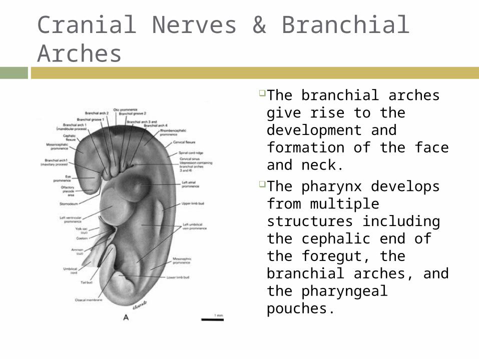
The branchial arches give rise to the development and formation of the face and neck.
The pharynx develops from multiple structures including the cephalic end of the foregut, the branchial arches, and the pharyngeal pouches.

General Overview of Head & Neck Development
The 1st and 2nd branchial arches develop into the mouth, tongue, and the muscles of mastication.
The mesoderm of the 3rd arch develops into the superior constrictors of the pharynx and the endoderm provides the epithelial lining of the tongue, pharynx, a portion of the epiglottis, and pyriform sinuses.
The mesoderm of the 4th arch gives rise to the inferior constrictors of the pharynx and its endoderm provides the remaining epithelium of the hypopharynx.

Esophageal Development
The esophagus begins to develop by the end of the first month at the level of the 4th branchial arch.
The esophagus is initially short tube, but extends in length as the heart and diaphragm descend.
The esophagus is lined with stratified epithelium two to three cells deep.
15

Facial Development
The first step in the development of the face takes place when the 1st branchial arch on either side of the embryo bifurcates unevenly into a small maxillary process and a much larger mandibular prominence.

Facial Development
The maxillary portions come to lie on either side of the stomodeum, the future oral cavity.
The mandibular prominence from each side begin to merge into a mandibular arch just beneath this primitive mouth.
Oral Cavity
18
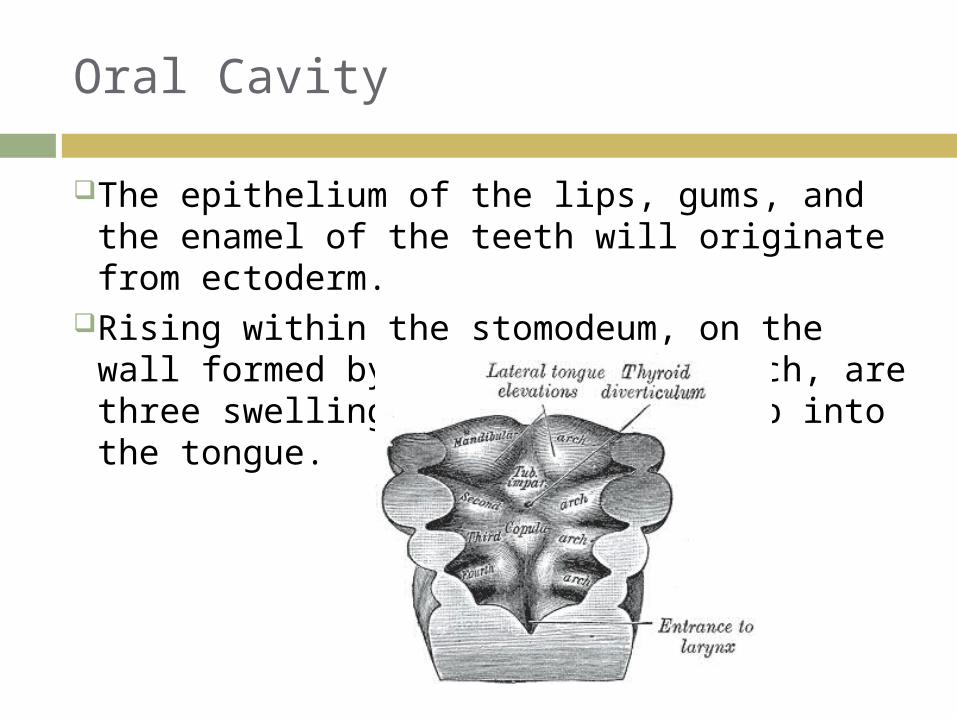
The epithelium of the lips, gums, and the enamel of the teeth will originate from ectoderm.
Rising within the stomodeum, on the wall formed by the mandibular arch, are three swellings that will develop into the tongue.
Tongue Development
19
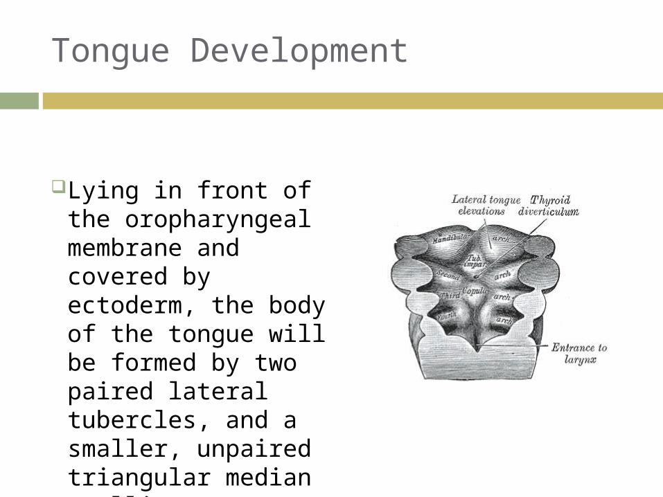
Lying in front of the oropharyngeal membrane and covered by ectoderm, the body of the tongue will be formed by two paired lateral tubercles, and a smaller, unpaired triangular median swelling.

Tongue Development
20
The two lateral tubercles increase in size until they eventually merge, overgrowing the median tubercle.
They form the anterior 2/3 of the tongue.

Tongue Development
21
The copula, another medial swelling located behind the unpaired tubercle, extends forward, and becomes engulfed by migration of the mesoderm of the 3rd branchial arch.
These structures fuse in the midline and form the posterior third, or root, of the tongue.
Pharyngeal Development
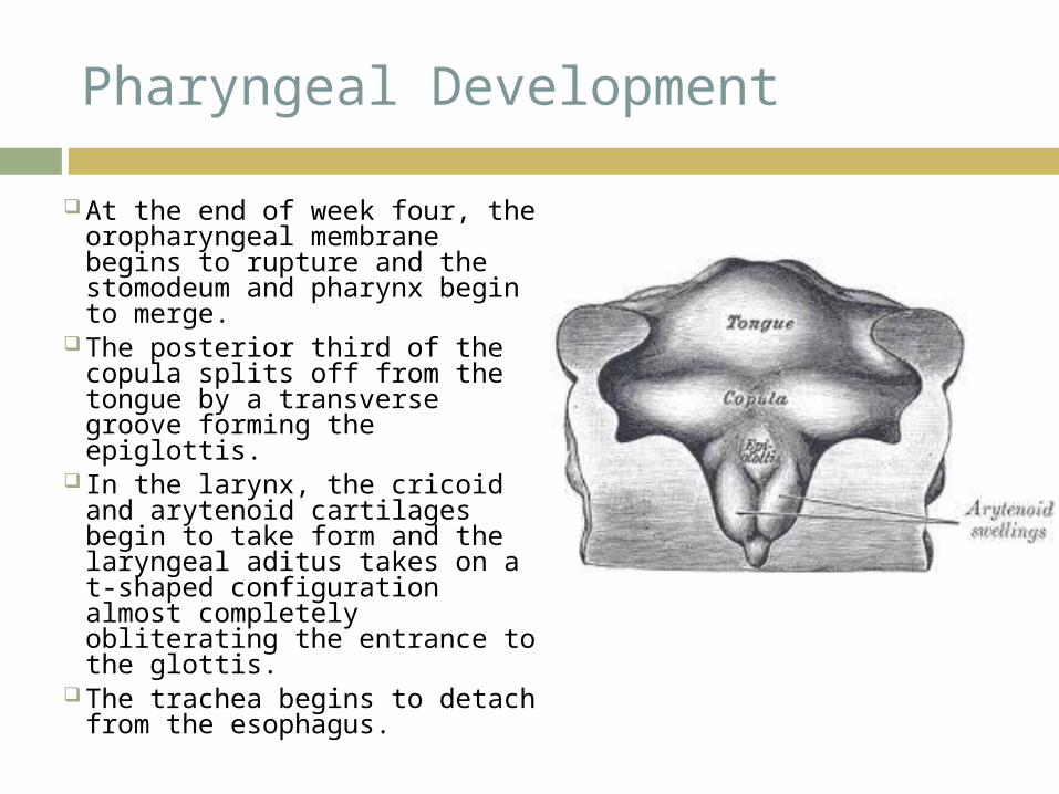
At the end of week four, the oropharyngeal membrane begins to rupture and the stomodeum and pharynx begin to merge.
The posterior third of the copula splits off from the tongue by a transverse groove forming the epiglottis.
In the larynx, the cricoid and arytenoid cartilages begin to take form and the laryngeal aditus takes on a t-shaped configuration almost completely obliterating the entrance to the glottis.
The trachea begins to detach from the esophagus.
5-Week Old Embryo
23
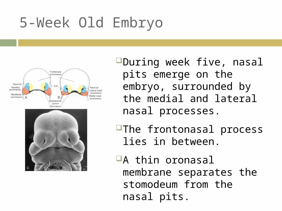
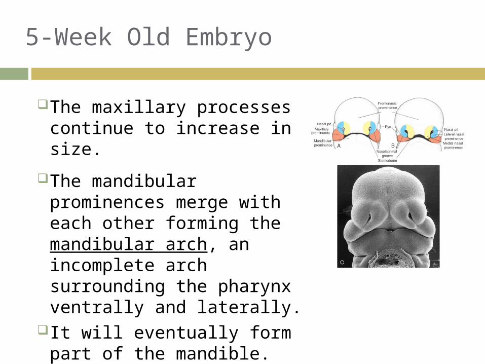
During week five, nasal pits emerge on the embryo, surrounded by the medial and lateral nasal processes.
The frontonasal process lies in between.
A thin oronasal membrane separates the stomodeum from the nasal pits.
5-Week Old Embryo
24
The maxillary processes continue to increase in size.
The mandibular prominences merge with each other forming the mandibular arch, an incomplete arch surrounding the pharynx ventrally and laterally.
It will eventually form part of the mandible.

5-Week Old Embryo
25
Beneath the mandibular arch, the 2nd branchial arch also enlarges and merges in midline.
These merged elevations, the hyoid arch, will eventually form part of the hyoid bone.
Branchial arches 3 and 4 are now hidden from view and the embryo loses its gill-like apparatus.

6-Week Old Embryo
During this week there is marked growth of the maxillary and medial nasal processes.
The maxillary processes become more prominent and grow toward the midline, crowding the nasal processes closer to each other.

6-Week Old Embryo
The medial nasal processes approach each other to form a single globular process that, in time, gives rise to the nasal tip, columella, prolabium, frenulum, and the primary palate.
As this occurs, the frontonasal process collapses inward to form the nasal septum.

6-Week Old Embryo
Inside the stomodeum, the primordia of the tongue are well-developed and there is differentiation of the posterior third of the tongue from the epiglottis.
Major salivary glands and many of the minor salivary glands are developing from stomodeal ectoderm.
The primary palate is developing from the innermost part of the intermaxillary segment.
The ectoderm covering the oral cavity proliferates into a thick band of epithelium to form the horseshoe shape of the future alveolar process.
28
6-Week Old Embryo
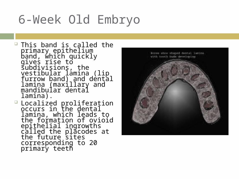
This band is called the primary epithelium band, which quickly gives rise to subdivisions, the vestibular lamina (lip furrow band) and dental lamina (maxillary and mandibular dental lamina).
Localized proliferation occurs in the dental lamina, which leads to the formation of ovioid epithelial ingrowths called the placodes at the future sites corresponding to 20 primary teeth

7-Week Old Embryo
During the 7th week, the formation of the upper jaw is completed by the maxillary process fusing with the medial nasal fold of the globular process, forming a true nostril as it gives rise to the lateral lip element.
30
1. Stomodeum2. Eye3. Maxillary swelling4. Mandibular swelling5. Nasal pit6. Frontal swelling7. Lateral nasal swelling8. Medial nasal swelling9. Nasolacrimal groove
7-Week Old Embryo
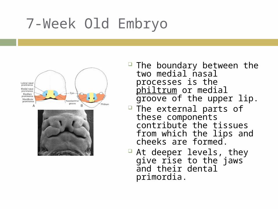
The boundary between the two medial nasal processes is the philtrum or medial groove of the upper lip.
The external parts of these components contribute the tissues from which the lips and cheeks are formed.
At deeper levels, they give rise to the jaws and their dental primordia.
31
7-Week Old Embryo
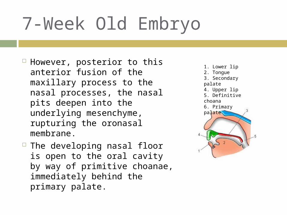
However, posterior to this anterior fusion of the maxillary process to the nasal processes, the nasal pits deepen into the underlying mesenchyme, rupturing the oronasal membrane.
The developing nasal floor is open to the oral cavity by way of primitive choanae, immediately behind the primary palate.
1. Lower lip2. Tongue3. Secondary palate4. Upper lip5. Definitive choana6. Primary palate

7-Week Old Embryo
The entire area occupied by the fused medial nasal processes and the frontonasal prominence, termed the primary palate, is composed of three components:
A labial component, the prolabium, which will make up the median portion of the upper lip, and which is continuous with the columella and the bridge of the nose;
33

7-Week Old Embryo
A middle portion, the premaxilla, the portion of the upper jaw which will contain the maxillary incisor teeth; and
The deepest part, the primary medial palatal process, is a small triangular plate just behind the premaxilla and continuous with the cartilaginous septum.
34
7-Week Old Embryo
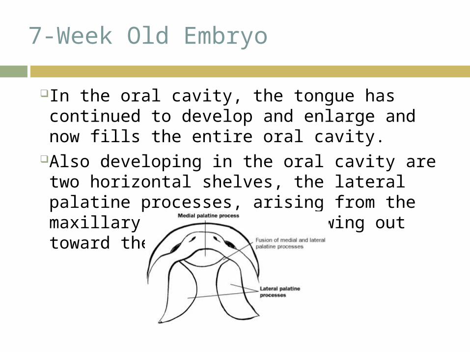
In the oral cavity, the tongue has continued to develop and enlarge and now fills the entire oral cavity.
Also developing in the oral cavity are two horizontal shelves, the lateral palatine processes, arising from the maxillary processes and growing out toward the midline.
7-Week Old Embryo
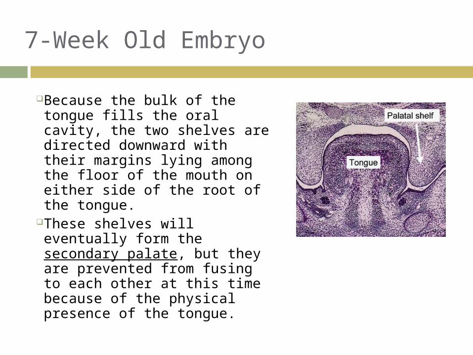
Because the bulk of the tongue fills the oral cavity, the two shelves are directed downward with their margins lying among the floor of the mouth on either side of the root of the tongue.
These shelves will eventually form the secondary palate, but they are prevented from fusing to each other at this time because of the physical presence of the tongue.
36

7-Week Old Embryo
Also during this week, the first subdivision of the lip and developing gum takes places.
The cells in the middle of the prolabium begin to disintegrate to form the labial groove, separating two sheets, the labial lamina, the future lip, and the dental lamina, the future alveolus.
Also occurring in this week, the definitive shape of the laryngeal entrance begins to emerge.
The aryepliglottic folds differentiate in the margins of the arytenoid swellings, and enclose the cuneiform and corniculate cartilages.
The vocal folds emerge as differentiations in the lateral wall of the larynx.

8-Week Old Embryo
By the start of the 8th week, the bridge of the nose begins to develop.
The nostrils fill with epithelial plugs which will remain until the sixth month.
The alae of the nose, along side the nares, are all that is left of the lateral nasal processes.
Inside the mouth, the muscles of the tongue are well differentiated at taste buds begin to appear.

8-Week Old Embryo
By the end of the week, the position of the tongue has shifted downward because the mandibular arch has grown wider.
With the sides of the tongue no longer blocking the palatine processes, the margins of the palatal shelves are free to swing upward, and grow toward each other and midline.
9-Week Fetus
By arbitrary but customary use, the developing embryo is now termed a fetus.
Important changes in the development of the palate occur.
Specifically, the palatal shelves, having continued to grow and to be pushed up toward midline by the tongue dorsum, are ready to make contact.

9-Week Fetus
The most anterior part of each shelf first contacts and then fuses with the medial palatal triangle.
Subsequently, in a wave of backward fusion, the remaining portion of the palatal shelves join with each other.
9-Week Fetus
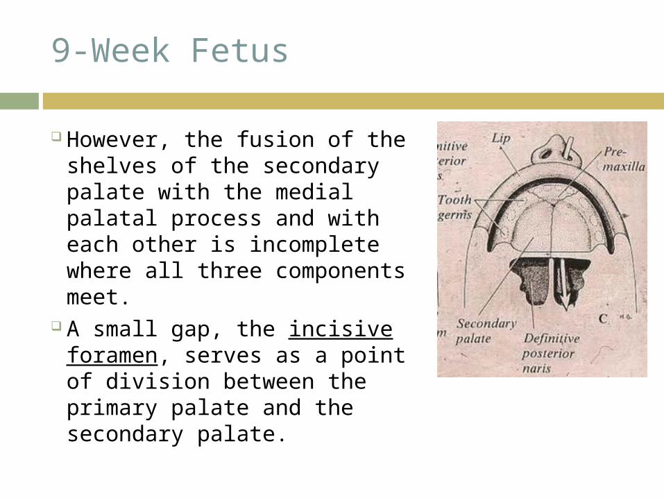
However, the fusion of the shelves of the secondary palate with the medial palatal process and with each other is incomplete where all three components meet.
A small gap, the incisive foramen, serves as a point of division between the primary palate and the secondary palate.
9-Week Fetus
It not only separates the two palates physically, it also separates the two palates developmentally, by the difference in time and type of their formation.
Intra-orally, the incisive foramen is covered with mucous membrane, the incisive papillae.
At the same time as the palate is fusing, the nasal septum, no longer contacted by the tongue, grows downward toward the palatal shelves.
Fusion with the palate occurs rapidly along the anterior 3/4s of the free surface of the cartilaginous nasal septum.
9 & 10Week Fetus
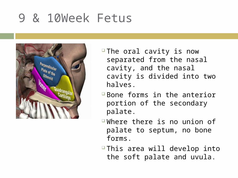
The oral cavity is now separated from the nasal cavity, and the nasal cavity is divided into two halves.
Bone forms in the anterior portion of the secondary palate.
Where there is no union of palate to septum, no bone forms.
This area will develop into the soft palate and uvula.
11 & 12 Week Fetus
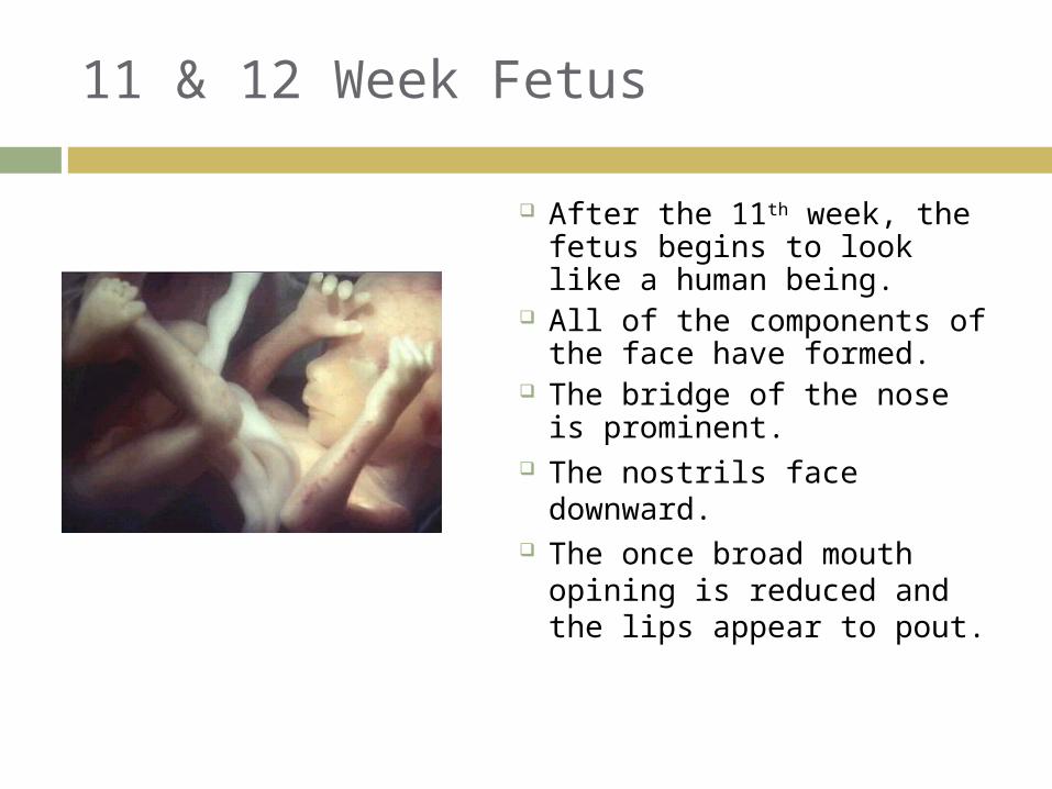
After the 11th week, the fetus begins to look like a human being.
All of the components of the face have formed.
The bridge of the nose is prominent.
The nostrils face downward.
The once broad mouth opining is reduced and the lips appear to pout.
45

11 & 12 Week Fetus
The cheeks are well established.
In the mouth, the palate has completely fused, with the possible exception of the uvula.
No malformations of the face can occur after 12 weeks.
Esophagus is entirely lined with ciliated cells.
46
Prenatal Life
During prenatal life, the orofacial region matures well ahead of limb regions.
Since the mouth is so concerned with a number of vital functions, such as respiration, nursing, and protection of the airway, it must be fully operative at the time of birth.
There is an orderly, sequential staging of events in prenatal orofacial maturation—a staging seen throughout the body, but which is much more advanced in the oropharyngeal region.

Prenatal Life
The earliest report of swallowing amniotic fluid has be demonstrated at 12 weeks gestation.
Respiratory reflexes, jaw closure reflexes, gag reflex, suckling, and swallowing are all developed in a scheduled way between the 14th and 32nd week of intrauterine life.
By 14 weeks, when the mouth area is stimulated, facial and orbicular muscle responses are produced.
Respiratory movements of the chest and abdomen are first seen at 16 weeks.
Prenatal Life
The gag reflex has been demonstrated at about 18 ½ weeks, and suckling at 24 weeks gestation.
Full swallowing and suckling reflexes are not thought to be developed until at least 32 weeks gestation.
All these vita functions have to be established by the time of birth in order for the child to survive.