Developmental Biology - Harvard University · PDF fileReview Review of fate-mapping studies of...
12
Review Review of fate-mapping studies of osteogenic cranial neural crest in vertebrates Joshua B. Gross ⁎, James Hanken Museum of Comparative Zoology, Harvard University, Cambridge, MA 02138, USA article info abstract Article history: Received for publication 5 June 2007 Received 20 February 2008 Accepted 21 February 2008 Available online 7 March 2008 Recent years have witnessed renewed interest in defining the embryonic cell populations that directly contribute to the bony skull. This question lies at the intersection of several important developmental, clinical and evolutionary interests. Until recently, our collective understanding of the embryonic origin of the vertebrate osteocranium has been based on a small number of reports published solely using avian models. As data gradually accumulates from other, distantly related species (e.g., mouse and frog), we can begin to evaluate long-standing assumptions regarding the behavior of osteogenic (bone-forming) neural crest cells within a wider phylogenetic and comparative context. In this review, we summarize data collected to date in three major vertebrate taxa: amphibians, birds and mammals. We highlight three largely unexplored topics within the field of osteogenic neural crest development: 1) disagreements in bone tissue origin within and across current model systems; 2) whether the pattern of neural crest cell contribution to skull bone is evolutionarily conservative or labile; and 3) how our understanding of development and morphology will benefit from fate maps using currently unexamined animal models. © 2008 Elsevier Inc. All rights reserved. Keywords: Osteocranium Skull Vertebrates Homology Introduction A long-standing goal of developmental biology is to define the relation between embryonic cell populations and the adult structures to which they directly contribute. One method used to address this issue is the construction of ‘fate maps’ that depict long-term (i.e., adult) derivatives of embryonic tissues. For the developmental biologist, fate maps provide an essential step towards understanding morphological and genetic interactions required to generate complex structures (Clarke and Tickle, 1999). For the evolutionary biologist, comparison of fate maps may offer insight into how embryonic tissues generate species-specific morphologies (Rudel and Sommer, 2003). In the field of craniofacial development, comprehensive fate maps have been technically difficult to obtain for a variety of reasons. Primary among these is the requirement of an indelible cell marker for labeling embryonic cell populations that give rise to late-forming tissues, such as skull bones. This topic is further complicated by the fact that the skull is comprised of multiple components, each arising from a unique combination of embryonic tissue precursors and mode of ossification, which are tightly integrated in the adult organism. These components are the viscerocranium, neurocranium, dermal skull roof and sclerotomal occipital tissues (Morriss-Kay, 2001). The viscerocranium comprises the lower jaw, its supporting structures and other elements of the branchial-arch skeleton and is regarded as derived principally from neural crest (Kuratani et al., 1997; Cerny et al., 2006). The neurocranium, which includes the floor of the braincase and associated sensory capsules (nasal, optic and otic), is regarded as derived principally from embryonic mesoderm (Kuratani et al., 1997). Cartilaginous elements of both the viscerocranium and neurocranium form first and undergo endochondral ossification in most species (Morriss-Kay, 2001; Colnot, 2005). Associated dermal bones form later (e.g., dentary, palatine). The third component of the skull, the dermal roof (Morriss-Kay, 2001), lacks a cartilaginous precursor and instead arises via intramembranous ossification of osteogenic mesenchymal cells (Franz-Odendaal et al., 2006). The embryonic origin of these cells has been, and remains, controversial depending on the model system being explored (Kuratani, 2005; Cerny et al., 2006). Finally, the fourth and most caudal component of the skull is the occipital region, which comprises endochondral bones that are believed to be derived, either exclusively or in large part, from mesodermal somitic tissues of the occipital region (Morriss-Kay, 2001). In the present context, ‘cranium’ shall refer specifically to the bony adult skull, excluding the lower jaw. Both the cranium and bony jaw elements shall be described collectively as the skull. Until the end of the last century, most knowledge regarding tissue origins of the vertebrate skull arose from research on a single species, the domestic chicken, Gallus gallus (Le Douarin and Barq, 1969). One consequence of the paucity of data from other species is that patterns of crest contribution to the skull have been widely extrapolated across vertebrate taxa, e.g., from avians to humans (Johnston et al., 1973; Noden, 1988; Kardong, 2002). The assumption, common among developmental biologists, that neural crest contributions to the skull are evolutionarily conservative Developmental Biology 317 (2008) 389–400 ⁎ Corresponding author. Current address: Harvard Medical School, Department of Genetics, Boston, MA 02115, USA. Fax: +1 617432 6595. E-mail address: [email protected] (J.B. Gross). 0012-1606/$ – see front matter © 2008 Elsevier Inc. All rights reserved. doi:10.1016/j.ydbio.2008.02.046 Contents lists available at ScienceDirect Developmental Biology journal homepage: www.elsevier.com/developmentalbiology
-
Upload
trinhnguyet -
Category
Documents
-
view
217 -
download
4
Transcript of Developmental Biology - Harvard University · PDF fileReview Review of fate-mapping studies of...
Developmental Biology 317 (2008) 389–400
Contents lists available at ScienceDirect
Developmental Biology
j ourna l homepage: www.e lsev ie r.com/deve lopmenta lb io logy
Review
Review of fate-mapping studies of osteogenic cranial neural crest in vertebrates
Joshua B. Gross ⁎, James HankenMuseum of Comparative Zoology, Harvard University, Cambridge, MA 02138, USA
a r t i c l e i n f o
⁎ Corresponding author. Current address: Harvard MGenetics, Boston, MA 02115, USA. Fax: +1 617 432 6595.
E-mail address: [email protected] (J.
0012-1606/$ – see front matter © 2008 Elsevier Inc. Aldoi:10.1016/j.ydbio.2008.02.046
a b s t r a c t
Article history:Received for publication 5 June 2007Received 20 February 2008Accepted 21 February 2008Available online 7 March 2008
Recent years have witnessed renewed interest in defining the embryonic cell populations that directlycontribute to the bony skull. This question lies at the intersection of several important developmental, clinicaland evolutionary interests. Until recently, our collective understanding of the embryonic origin of thevertebrate osteocranium has been based on a small number of reports published solely using avian models. Asdata gradually accumulates from other, distantly related species (e.g., mouse and frog), we can begin toevaluate long-standing assumptions regarding the behavior of osteogenic (bone-forming) neural crest cellswithin a wider phylogenetic and comparative context. In this review, we summarize data collected to date inthree major vertebrate taxa: amphibians, birds and mammals. We highlight three largely unexplored topicswithin the field of osteogenic neural crest development: 1) disagreements in bone tissue origin within andacross current model systems; 2) whether the pattern of neural crest cell contribution to skull bone isevolutionarily conservative or labile; and 3) how our understanding of development and morphology willbenefit from fate maps using currently unexamined animal models.
© 2008 Elsevier Inc. All rights reserved.
Keywords:OsteocraniumSkullVertebratesHomology
Introduction
A long-standing goal of developmental biology is to define therelation between embryonic cell populations and the adult structuresto which they directly contribute. One method used to address thisissue is the construction of ‘fate maps’ that depict long-term (i.e.,adult) derivatives of embryonic tissues. For the developmentalbiologist, fate maps provide an essential step towards understandingmorphological and genetic interactions required to generate complexstructures (Clarke and Tickle, 1999). For the evolutionary biologist,comparison of fatemaps may offer insight into how embryonic tissuesgenerate species-specific morphologies (Rudel and Sommer, 2003).
In the field of craniofacial development, comprehensive fate mapshave been technically difficult to obtain for a variety of reasons.Primary among these is the requirement of an indelible cell marker forlabeling embryonic cell populations that give rise to late-formingtissues, such as skull bones. This topic is further complicated by thefact that the skull is comprised of multiple components, each arisingfrom a unique combination of embryonic tissue precursors and modeof ossification, which are tightly integrated in the adult organism.These components are the viscerocranium, neurocranium, dermalskull roof and sclerotomal occipital tissues (Morriss-Kay, 2001). Theviscerocranium comprises the lower jaw, its supporting structures andother elements of the branchial-arch skeleton and is regarded asderived principally from neural crest (Kuratani et al., 1997; Cerny et al.,
edical School, Department of
B. Gross).
l rights reserved.
2006). The neurocranium, which includes the floor of the braincaseand associated sensory capsules (nasal, optic and otic), is regarded asderived principally from embryonic mesoderm (Kuratani et al., 1997).Cartilaginous elements of both the viscerocranium and neurocraniumform first and undergo endochondral ossification in most species(Morriss-Kay, 2001; Colnot, 2005). Associated dermal bones form later(e.g., dentary, palatine).
The third component of the skull, the dermal roof (Morriss-Kay,2001), lacks a cartilaginous precursor and instead arises viaintramembranous ossification of osteogenic mesenchymal cells(Franz-Odendaal et al., 2006). The embryonic origin of these cellshas been, and remains, controversial depending on the model systembeing explored (Kuratani, 2005; Cerny et al., 2006). Finally, the fourthand most caudal component of the skull is the occipital region, whichcomprises endochondral bones that are believed to be derived, eitherexclusively or in large part, from mesodermal somitic tissues of theoccipital region (Morriss-Kay, 2001). In the present context, ‘cranium’
shall refer specifically to the bony adult skull, excluding the lower jaw.Both the cranium and bony jaw elements shall be describedcollectively as the skull.
Until the end of the last century, most knowledge regarding tissueorigins of the vertebrate skull arose from research on a single species,the domestic chicken, Gallus gallus (Le Douarin and Barq, 1969). Oneconsequence of the paucity of data from other species is that patternsof crest contribution to the skull have beenwidely extrapolated acrossvertebrate taxa, e.g., from avians to humans (Johnston et al., 1973;Noden, 1988; Kardong, 2002).
The assumption, common among developmental biologists, thatneural crest contributions to the skull are evolutionarily conservative
390 J.B. Gross, J. Hanken / Developmental Biology 317 (2008) 389–400
is understandable from an historical perspective. Indeed, thisassumption is validated by previous studies of the viscerocranium,for which a rich literature stretching back several decades hasdemonstrated consistently a nearly exclusively neural crest origin.And while early studies provide evidence primarily regardingviscerocranial cartilages in bony fishes (Langille and Hall, 1988) andamphibians (Platt, 1893; Landacre, 1921; Stone, 1926, 1929; Raven,1931; de Beer, 1947; Hörstadius, 1950), subsequent studies in chickendemonstrate a comparable embryonic origin for viscerocranial bones(e.g., Le Lièvre, 1974).
Extrapolation of fate-mapping data across distantly relatedvertebrate taxa also has provided an important heuristic tool forresearchers interested in the etiology of a variety of human clinicaldisorders. As a starting point for the study of neurocristopathies(reviewed in Hall, 1999), several human craniofacial malformationsare believed to be a consequence of aberrant migration and/ordevelopment of the neural crest or of an abnormal local signalingenvironment that secondarily influences the neural crest. Many suchpredictions have been confirmed by subsequent analysis involvingnon-human models.
For example, arrested crest cell migration in Treacher–Collinssyndrome, in which affected individuals suffer from hypoplasia ofderivatives of the first branchial arch, has been linked to a defect in thegene TCOF1 (Gorlin et al., 1990; Farlie et al., 2004). Mouse models ofthis disorder have been produced through mutant mice that carry aheterozygous mutation for TCOF1 (Edwards et al., 1997). The mutantphenotype reveals extensive cell death in cranial neural crest (CNC)cells prior to their departure from the neural tube andmigration to thefirst branchial arch (Dixon et al., 2000). Other neurocristopathies inhumans include some forms of cleft lip, cleft palate, and frontonasaldysplasia (Beauchamp and Knepper, 1984; Poswillo, 1988; Sulik et al.,1988; Fukiishi and Morriss-Kay, 1992).
Yet, from developmental and evolutionary perspectives theextrapolation from primary literature (fate-mapping) studies tohuman clinical manifestations may be inappropriate, or at leastpremature. For example, assuming that the distribution of osteogenicneural crest in a bird (e.g., domestic chicken) is the same as those inhigher primates (e.g., humans) implies that patterns of crestcontribution to bone are invariant across these distantly relatedtaxa. While this assumption may be correct, it requires more ‘datapoints’ to test its validity. Moreover, the developmental origin of bonytissues, and whether the embryonic populations that constitute thesestructures are evolutionary invariant or labile, remain to be investi-gated in a rigorous comparative framework.
To facilitate additional studies that assess the range of osteogenicpotential of neural crest cells among different species of vertebrates,we summarize the results of published studies that report crestcontributions to the bony skull. In particular, we focus on regions ofthe skull that have long been controversial with respect to theirorigin, including the dermal skull roof, the columella, and the oticcomplex. We also highlight the need for comparable data fromother potential model systems, including bony fishes and reptiles, inthe hope that such attention will stimulate study of these neglectedtaxa.
Bony fishes and reptiles
There are no published reports that directly assess contributions ofneural crest cells to the ossified adult skull of bony fishes. Such data,when placed within an appropriate phylogenetic context, will beinvaluable for defining the generalized condition of “crest-domains”(i.e., regions of the skull to which the neural crest directly contributescells) in the vertebrate subphylum (Knight and Schilling, 2006). Thesame data would inform our understanding of the ancestral pattern ofneural crest contribution to individual skull bones in vertebrates andthe extent to which this pattern is retained or modified in more
recently evolved fishes, amphibians, and amniotes. Finally, given theextreme cranial diversity among extant species (Schultze, 1993),comparative studies of neural crest derivation likely would provideinsights into the developmental mechanisms that underlie theevolution of morphological novelty and adaptive diversification ofcranial form.
Along with bony fish, there currently exists no fate map depictinglong-term contributions of osteogenic cranial neural crest in areptilian model system. The embryonic origin of postcranial bonesin turtles, however, is gaining increased attention (e.g., Toerien, 1965;Meier and Packard, 1984; Clark et al., 2001; Cebra-Thomas et al., 2005,2007; Gilbert et al., 2007), including the potential role of the neuralcrest. Importantly, these studies offer evidence that at least somedermal bones in the turtle shell are derived from postcranial, or trunkneural crest, a claim that contradicts the widely accepted principlethat osteogenic neural crest is limited to the cranial region in allvertebrates (see below).
Gilbert et al. (2001), using molecular markers to assess celllineage (e.g., HNK-1, pdgfβ), provides indirect evidence of neuralcrest derivation of dermal bones of the turtle shell (both carapaceand plastron). In this study, HNK-1 expression is used to assessembryonic origin because it is commonly expressed in earlymigrating neural crest cells (Bronner-Fraser, 1986). While previouswork by Clark et al. (2001) demonstrated unequivocally that cells ofthe developing turtle shell are HNK-1-positive, neither this reportnor the one by Gilbert et al. (2001) involves fate-mapping astypically defined, insofar as the associated experiments did not applyan extrinsic label to migratory cells to follow their fate into laterdevelopmental time points.
More recently, Cebra-Thomas et al. (2007) and Gilbert et al. (2007)provide additional evidence of a neural-crest origin of the HNK-1-positive cells by co-labeling with other known molecular markers ofthe neural crest, including p75 and FoxD3. Furthermore, by using DiIlabeling they demonstrate migration of these cells from the dorsalneural tube in stage 17 embryos to the developing plastron in theventral shell, where they differentiate into bone via intramembranousossification.
Fate-mapping studies and studies in which the ability of trunkneural crest to form skeletal tissues have been investigated in othervertebrates document a highly conserved difference between cranialand trunk neural crest with respect to their skeletogenic potential(e.g., Lumsden, 1988; Graveson et al., 1997; Hall, 1999). The apparentcapacity of trunk neural crest cells to differentiate into bones of thepostcranial skeleton in turtles is an obvious exception to this generalrule. Indeed, it may represent a novel developmental feature in turtlesthat facilitated the evolution of their protective shell. Alternatively,osteogenic potential of trunk neural crest may be more widelydistributed among vertebrates than is currently recognized (e.g.,alligators; Gilbert et al., 2007), reflecting the limited taxonomicsampling of relevant studies to date.
Amphibians
Most early studies that examined the fate of the CNC in thevertebrate head skeleton utilized amphibian models because of theirexperimental tractability. Amphibian embryos are simple to obtainand, owing to their large size and external development, make avariety of experimental approaches tractable and straightforward(e.g., tissue ablations and grafting).
Classical studies, however, did not focus on the derivation of skullbones in model amphibians. Rather, most reports focused on thecontribution of the neural crest to the larval cartilaginous skull. Theprotracted life history of metamorphic amphibians (particularlyanurans) requires a cell label that can persist through the extendeddevelopmental time before cranial ossification commences in larvae,shortly before or during metamorphosis (Trueb and Hanken, 1992),
391J.B. Gross, J. Hanken / Developmental Biology 317 (2008) 389–400
and cell labels with these characteristics have been developed only inthe last few years (see below). Nevertheless, several studiesattempted to trace the long-term derivatives of CNC throughhistological observations, tissue ablation, chromatic and radioactivelabel applications, and heterospecific grafting (reviewed in Gross andHanken, 2004). As an example, Stone (1926) performed ablationexperiments in which a small portion of premigratory neural crestand overlying neural ectoderm was removed from developingembryos. After 2 or 3 days, the larval animal was assessed foraltered morphology of the cartilaginous skull and hyobranchialskeleton. Results provided the basis for one of the first fate maps ofpremigratory neural crest in amphibians. Other researchers haveused a combination of tissue ablation and wild type-to-albinochimeric grafting techniques as a means of demonstrating the neuralcrest origin of much of the body pigmentation in amphibian embryosin both classical (DuShane, 1935; Twitty and Bodenstein, 1939) andcontemporary investigations (Barlow and Northcutt, 1995; Barlowand Northcutt, 1997).
By themiddle of the last century, two techniques led to reports thatneural crest contributes to skull bone as well as cartilage. By inferringpatterns of crest-cell migration from histological sections of embryosof the salamander Ambystoma mexicanum (based on intrinsic featuresin the cytoplasm between the neural crest and other cells, e.g., yolk),de Beer (1947) proposed an “ectomesenchymal” (i.e., neural crest)contribution to the intramembranous splenial bone of the lower jaw(Table 1). This is the first claim of neural crest derivation of theosteocranium in any vertebrate. Wagner (1949) and Andres (1949)subsequently applied a technique of heterospecific grafting of portionsof the embryonic neural fold (which includes the neural crest andoverlying ectoderm) between a frog (Bombina) and a newt (Triturus)and reported donor neural crest-derived cells within several bones ofthe late-larval skull of the host, including the premaxilla, dentary,splenial and vomeropalatine (reviewed in Noden, 1983a; Table 1).These studies also took advantage of the fact that these bones form atdifferent times in frogs and salamanders (before vs. after metamor-phosis) and assume characteristically different shapes in the twogroups.
There are at least two potential problems associated with theseearly experiments with amphibians, which, until very recently, hadnot been validated with more contemporary techniques for tracingcell derivatives. First, histology alone may not be a reliable fate-mapping technique because of the difficulty tracking migration ofunlabeled cells. Secondly, because individual grafts in these experi-ments included the entire neural fold, as opposed to grafts comprisedexclusively of neural crest tissue, one cannot conclude that donor-derived cells came from neural crest and not from overlying ectoderm.While the overlying epithelium is not predicted to form hard tissues ofthe skull, it is likely that migratory multipotent cells derived fromplacodal tissue reside in the transplanted surface epithelium. Placode-derived cells are known to interact with neural crest cells later in
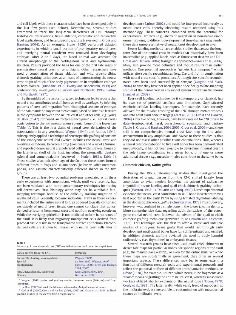
Table 1Summary of cranial neural crest (CNC) contributions to skull bones in amphibians
Bones derived from the CNC Reference
Premaxilla, dentary, vomeropalatine Wagner, 1949a
Splenial de Beer, 1947; Wagner, 1949b
Frontoparietal Carl et al., 2000; Gross and Hanken,2004, 2005c
Nasal, parasphenoid, squamosal Gross and Hanken, 2004Dentary Gross et al., 2006
a Wagner (1949) performed grafting studies between newts (Triturus) and frogs(Bombina).
b de Beer (1947) utilized the Mexican salamander, Ambystoma mexicanum.c Carl et al. (2000), Gross and Hanken (2004, 2005) and Gross et al. (2006) utilized
grafting studies in the clawed frog, Xenopus laevis.
development (Barlow, 2002) and could be interpreted incorrectly asneural crest cells, thereby obscuring results obtained using thismethodology. These concerns, combined with the potential forexperimental artifacts (e.g., aberrant migration in non-native envir-onments owing to different developmental time-frames), may renderthese data unrepresentative of neural crest development in vivo.
Newer labeling methods have enabled studies that assess the long-term fate of the neural crest in models that historically have beeninaccessible (e.g., applied labels, such as fluorescent dextran and DiI—Gross and Hanken, 2004; transgenic approaches—Gross et al., 2006).Many also provide more definitive and robust results than earliermethods. One potential approach is “genetic fate mapping,” whichutilizes site-specific recombinases (e.g., Cre and flp) in combinationwith neural crest-specific promoters. Although site-specific recombi-nases have been used successfully in Xenopus (Gargioli and Slack,2004), to date they have not been applied specifically to fate-mappingstudies of the neural crest in any model system other than the mouse(Jiang et al., 2002).
Every cell labeling technique, be it contemporary or classical, hasits own set of potential artifacts and limitations. Sophisticatedextrinsic cellular labeling techniques, for example, have recentlyallowed for the reliable tracking of the CNC through metamorphosisand into adult skull bone in frogs (Carl et al., 2000; Gross and Hanken,2004). Only five bones, however, have been assessed for CNC origin todate—frontoparietal, nasal, parasphenoid, squamosal and dentary(Table 1; Gross and Hanken, 2004, 2005; Gross et al., 2006)—and therestill is no comprehensive neural crest fate map for the adultosteocranium in any amphibian. One caveat to these studies is thatthey did not assess other possible tissue contributions. Thus, whereasa neural crest contribution to five skull bones has been demonstratedunequivocally, it has not been possible to determine if neural crest isthe sole tissue contributing to a given bone, or if one or moreadditional tissues (e.g., mesoderm) also contribute to the same bone.
Domestic chicken, Gallus gallus
During the 1960s, fate-mapping studies that investigated thederivation of cranial tissues from the CNC shifted largely fromamphibian to avian models following the advent of radioactive(thymidine) tissue-labeling and quail-chick chimeric grafting techni-ques (Weston, 1963; Le Douarin and Barq, 1969). Direct experimentalevidence that neural crest contributes to skull bone in vertebrates wasfirst reported in the early 1970s by using tritiated thymidine labelingin the domestic chicken, G. gallus (Johnston et al.,1973). This discovery,however, was confined to a single bone in the lower jaw, the dentary.More comprehensive data regarding adult derivatives of the osteo-genic cranial neural crest followed the advent of the quail-to-chickchimeric grafting technique (reviewed in Le Douarin and Kalcheim,1999). This technique was the first to offer a stable and indeliblemarker of embryonic tissue grafts that would last through earlydevelopment until cranial bones have fully differentiated and ossified.In addition, chimeric grafting obviated the need to apply harmfulradioactivity (i.e., thymidine) to embryonic tissues.
Several research groups have since used quail-chick chimeras toderive fate maps for particular bones, for specific regions of the skull(e.g., the mandibular skeleton), or even for the entire skull. Yet whilethese maps are substantially in agreement, they differ in severalimportant aspects. These differences may be, to some extent, afunction of different research goals and experimental protocols andreflect the potential artifacts of different transplantation methods. LeLièvre (1978), for example, utilized whole neural tube fragments as a‘coarse’means of grafting the entire neural crest, whereas subsequentworkers utilized shorter explants of the neural tube (Noden, 1978;Couly et al., 1993). The latter grafts, while easily freed of mesoderm atthemidbrain level, are susceptible to contaminationwithmesodermaltissues at hindbrain levels.
392 J.B. Gross, J. Hanken / Developmental Biology 317 (2008) 389–400
It remains to be seenwhich of the conflicting claims of neural crestderivation are accurate. A universally accepted, definitive fate map forthe bony skull of birds is yet to be produced. In this section we reviewthe landmark studies authored by four groups. Each reports the long-term fate of premigratory CNC in the adult skull of the domesticchicken.
Fate maps of Le Lièvre (1974, 1975, 1978)
Three pioneering reports by Le Lièvre led the way for manysubsequent investigations using the quail-to-chick chimera techniqueto examine regional fate mapping of neural crest in the head skeleton.The first report describes Meckel's cartilage as derived from bothmesencephalic and anterior rhombomeric neural crest (Le Lièvre,1974; see below). The other reported contributions from mesence-phalic crest include the dentary, opercular, supra-angular and angularbones.
This work also was the first to define an important phenom-enon related to crest contributions to the head skeleton: discretecrest populations that originate from different regions of the neuraltube may jointly participate in the development of a single skeletalelement, while retaining regional identity. Specifically, the articularregion of Meckel's cartilage is derived from both “mesencephalic”and “anterior rhombencephalic” crest, whereas its distal region isderived solely from “mesencephalic” crest (Table 2; Le Lièvre, 1974).This observation would be expanded many years later in a moredetailed study of the bony lower jaw, which documented retentionof “cryptic” segmental boundaries between adjacent embryoniccrest populations in adult tissues (Köntges and Lumsden, 1996; seebelow).
In a second report on crest contributions, Le Lièvre and Le Douarin(1975) characterized mesenchymal derivatives, including the dermis,portions of striated cranial muscle, vasculature and other connectivetissues. All but one of these proposed derivatives have been confirmedby subsequent analyses. With the exception of specialized ciliarymuscles in the eye (Couly et al., 2002), Le Lièvre's claim of a neural-crest origin of skeletal myocytes has not been confirmed. Rather, thedirect contribution of neural crest cells to myogenesis appears to belimited to their serving as a source of tendinous and other muscularconnective tissues (Noden, 1983a; Köntges and Lumsden, 1996;Matsuoka et al., 2005).
The third report contains Le Lièvre's (1978) most thoroughdescription of neural crest contributions to the bony skull, includingan assessment of the origin of bones of the facial skeleton, cranial vaultand cranial base (Table 2). Contributions are organized according tothe region(s) from which neural crest initially migrates from the
Notes to Table 2:Several additional bones and cartilages were reported as not derived from the neural crest inbasioccipital), bones of the sphenoid complex (basisphenoid, alisphenoid, orbitosphenoalisphenoid, and the otic complex (Noden, 1978); the basioccipital, exoccipital, pars canalicul1993).“n/s”: bones and cartilages derived from neural crest in which the source of the neural cres
a Couly et al. (1993) report the following structures as crest-derived: sutures between mdermis of the parietal bones.
b Le Lièvre (1978) describes only the anterior maxilla and the anterior palatine as derivedparasphenoid are reported as derived from mesencephalic crest.
c “Forebrain” grafts of neural crest include the posterior diencephalon and the anteriormesthe metencephalon.
d Described in Le Lièvre (1978) as “mixed” in origin, presumably indicating contributionse Noden (1978) describes the frontal bone as “mixed” in origin, presumably indicating cof Noden (1978) describes the proximal portion of the quadrate bone as derived frommeteg Noden did not describe an embryonic origin of the columella bone in his 1978 study. H
neural crest and mesoderm (Noden, 1982, 1984).h Couly et al. (1993) report the pars canalicularis as mixed in origin, presumably indicatii Köntges and Lumsden (1996) report the pterygoquadrate as derived from rhombomere 1
and rhombomere 2 neural crest, respectively. The articular bone is reported as derived fromderived from rhombomeres 3–5.
neural tube, viz., prosencephalon, mesencephalon and rhombence-phalon. Neural crest emigrating from the prosencephalon wasdetected in the anterior maxilla, palatine, nasal, premaxilla andvomer bones. Cells derived from mesencephalic neural crest con-tribute to the largest amount of skull tissue, including the rostralparasphenoid, a small portion of the frontal bone, the jugal,quadratojugal, pterygoid, maxilla and entoglossum bones, as well asMeckel's cartilage (described previously). Cells derived from themesencephalon and anterior rhombencephalon contribute to thecolumella and squamosal bones and the articular region of Meckel'scartilage.
This report also described amixed contribution of both neural crestand mesoderm in the formation of the “orbital skeleton,” viz., theregion of the skull that includes the rostral portion of the frontal bone(Le Lièvre, 1978; p. 26). With regard to the rest of the cranial vault, LeLièvre observed no contribution from grafted crest cells and thereforeattributed its origin to mesoderm. Beginning with this claim andcontinuing to the present day, the cranial vault has remained the mostdisputed and controversial region with respect to embryonic origin.
Fate maps of Noden (1978, 1982, 1984), Noden and Trainor (2005) andEvans and Noden (2006)
Beginning in the late 1970s, another series of studies focused onosteogenic neural crest in the domestic chickenwere performed usingthe quail-chick chimeric method. The principal aim of the first reportby Noden (1978) was to investigate whether cells derived frompremigratory neural crest were restricted in their developmentalpotential. Small portions of embryonic crest corresponding todifferent axial levels, or “populations,” were reciprocally graftedbetween regions and assessed with respect to their effects onsubsequent skeletal morphology. Results indicated that the extra-cellular environment mediates the adult morphology of crest-derivedskeletal structures after crest cells migrate from the neural tube.
Patterns of neural crest derivation outlined in this study largelyconfirm previous work (Tables 2 and 3), even though the particularregions of the neural ridge from which grafts were removed differsomewhat between studies. Le Lièvre (1974), for example, removedsuccessively overlapping regions of the neural tube that extend alongthe rostrocaudal axis, from the developing prosencephalon to theposterior rhombencephalon. Similarly, Noden (1978) utilized over-lapping regions from the caudal prosencephalon through themetencephalon and periotic regions (with the exception of the thirdrhombomere). The primary distinction between these two graftingprotocols centers on the use of longer grafts of the neural tube (LeLièvre, 1974) versus shorter grafts of the neural folds (Noden, 1978).
these reports, including the parietal bone, “occipital” bones (supraoccipital, exoccipital,id), acrochordal cartilages, and polar cartilages (Le Lièvre, 1978); the basisphenoid,aris, supraoccipital, basipostsphenoid, orbitosphenoid, and pleurosphenoid (Couly et al.,
t is not specified. “ant.”=anterior; “post.”=posterior.embranous bones of the cranial vault, dermis of the scalp, meninges of the forebrain,
from CNC originating from the prosencephalon. The posterior palatine and the rostral
encephalon. “Hindbrain” grafts of neural crest include the posterior mesencephalon and
from both neural crest and mesoderm.ntributions from both neural crest and mesoderm.ncephalic crest; distal portions are derived from posterior mesencephalic crest (p. 300).is later reports, however, describe the columella as a composite structure derived from
ng contributions from both neural crest and mesoderm.neural crest, with dorsal articulations and ventral articulations derived from midbrainrhombomeres 1+2, the retroarticular process (proximal articular bone) is reported as
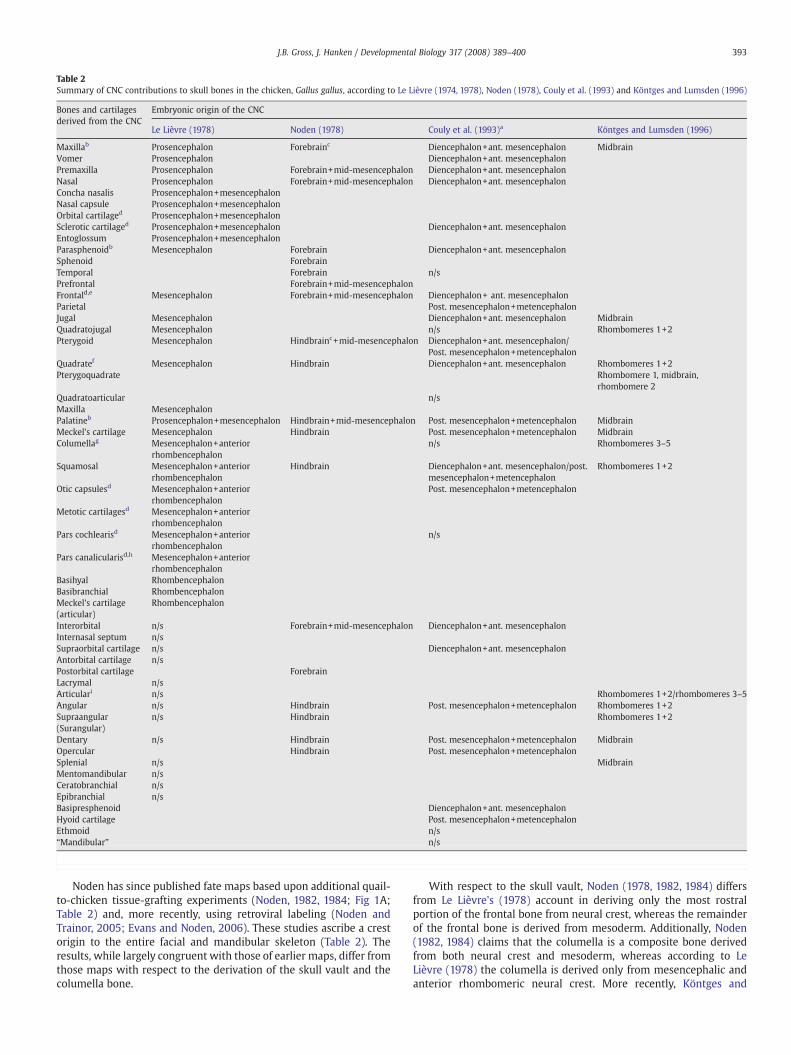
Table 2Summary of CNC contributions to skull bones in the chicken, Gallus gallus, according to Le Lièvre (1974, 1978), Noden (1978), Couly et al. (1993) and Köntges and Lumsden (1996)
Bones and cartilagesderived from the CNC
Embryonic origin of the CNC
Le Lièvre (1978) Noden (1978) Couly et al. (1993)a Köntges and Lumsden (1996)
Maxillab Prosencephalon Forebrainc Diencephalon+ant. mesencephalon MidbrainVomer Prosencephalon Diencephalon+ant. mesencephalonPremaxilla Prosencephalon Forebrain+mid-mesencephalon Diencephalon+ant. mesencephalonNasal Prosencephalon Forebrain+mid-mesencephalon Diencephalon+ant. mesencephalonConcha nasalis Prosencephalon+mesencephalonNasal capsule Prosencephalon+mesencephalonOrbital cartilaged Prosencephalon+mesencephalonSclerotic cartilaged Prosencephalon+mesencephalon Diencephalon+ant. mesencephalonEntoglossum Prosencephalon+mesencephalonParasphenoidb Mesencephalon Forebrain Diencephalon+ant. mesencephalonSphenoid ForebrainTemporal Forebrain n/sPrefrontal Forebrain+mid-mesencephalonFrontald,e Mesencephalon Forebrain+mid-mesencephalon Diencephalon+ ant. mesencephalonParietal Post. mesencephalon+metencephalonJugal Mesencephalon Diencephalon+ant. mesencephalon MidbrainQuadratojugal Mesencephalon n/s Rhombomeres 1+2Pterygoid Mesencephalon Hindbrainc+mid-mesencephalon Diencephalon+ant. mesencephalon/
Post. mesencephalon+metencephalonQuadratef Mesencephalon Hindbrain Diencephalon+ant. mesencephalon Rhombomeres 1+2Pterygoquadrate Rhombomere 1, midbrain,
rhombomere 2Quadratoarticular n/sMaxilla MesencephalonPalatineb Prosencephalon+mesencephalon Hindbrain+mid-mesencephalon Post. mesencephalon+metencephalon MidbrainMeckel's cartilage Mesencephalon Hindbrain Post. mesencephalon+metencephalon MidbrainColumellag Mesencephalon+anterior
rhombencephalonn/s Rhombomeres 3–5
Squamosal Mesencephalon+anteriorrhombencephalon
Hindbrain Diencephalon+ant. mesencephalon/post.mesencephalon+metencephalon
Rhombomeres 1+2
Otic capsulesd Mesencephalon+anteriorrhombencephalon
Post. mesencephalon+metencephalon
Metotic cartilagesd Mesencephalon+anteriorrhombencephalon
Pars cochlearisd Mesencephalon+anteriorrhombencephalon
n/s
Pars canalicularisd,h Mesencephalon+anteriorrhombencephalon
Basihyal RhombencephalonBasibranchial RhombencephalonMeckel's cartilage(articular)
Rhombencephalon
Interorbital n/s Forebrain+mid-mesencephalon Diencephalon+ant. mesencephalonInternasal septum n/sSupraorbital cartilage n/s Diencephalon+ant. mesencephalonAntorbital cartilage n/sPostorbital cartilage ForebrainLacrymal n/sArticulari n/s Rhombomeres 1+2/rhombomeres 3–5Angular n/s Hindbrain Post. mesencephalon+metencephalon Rhombomeres 1+2Supraangular(Surangular)
n/s Hindbrain Rhombomeres 1+2
Dentary n/s Hindbrain Post. mesencephalon+metencephalon MidbrainOpercular Hindbrain Post. mesencephalon+metencephalonSplenial n/s MidbrainMentomandibular n/sCeratobranchial n/sEpibranchial n/sBasipresphenoid Diencephalon+ant. mesencephalonHyoid cartilage Post. mesencephalon+metencephalonEthmoid n/s“Mandibular” n/s
393J.B. Gross, J. Hanken / Developmental Biology 317 (2008) 389–400
Noden has since published fate maps based upon additional quail-to-chicken tissue-grafting experiments (Noden, 1982, 1984; Fig 1A;Table 2) and, more recently, using retroviral labeling (Noden andTrainor, 2005; Evans and Noden, 2006). These studies ascribe a crestorigin to the entire facial and mandibular skeleton (Table 2). Theresults, while largely congruent with those of earlier maps, differ fromthose maps with respect to the derivation of the skull vault and thecolumella bone.
With respect to the skull vault, Noden (1978, 1982, 1984) differsfrom Le Lièvre's (1978) account in deriving only the most rostralportion of the frontal bone from neural crest, whereas the remainderof the frontal bone is derived from mesoderm. Additionally, Noden(1982, 1984) claims that the columella is a composite bone derivedfrom both neural crest and mesoderm, whereas according to LeLièvre (1978) the columella is derived only from mesencephalic andanterior rhombomeric neural crest. More recently, Köntges and
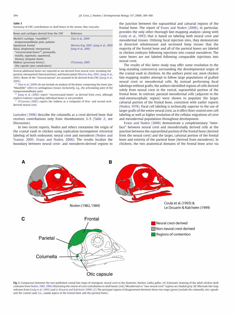
Table 3Summary of CNC contributions to skull bones in the mouse, Mus musculus
Bones and cartilages derived from the CNC Reference
Meckel's cartilage, “mandible”a,temporomandibular joint, palatine
Chai et al., 2000
Squamosal, frontal Morriss-Kay, 2001; Jiang et al., 2002Nasal, alisphenoid, interparietal,“viscerocranial bones”b, premaxilla,maxilla, zygomatic, squamosal,dentary, tympanic bones
Jiang et al., 2002
Malleus (processus brevis)c,Otic capsule (pars canalicularis)
O'Gorman, 2005
Several additional bones are reported as not derived from neural crest, including theparietal, interparietal (lateral portion), and basioccipital (Morriss-Kay, 2001; Jiang et al.,2002). Bones of the “viscerocranium” are assumed to be derived from CNC (Jiang et al.,2002).
a Chai et al. (2000) do not include an analysis of the bones comprising the lower jaw.“Mandible” refers to cartilaginous tissues exclusively, e.g., the articulating joint of thetemporomandibular joint.
b Jiang et al. (2002) report “viscerocranial bones” as derived from crest, althoughexplicit evidence regarding individual bones is not provided.
c O'Gorman (2005) reports the malleus as a composite of first- and second arch-derived neural crest.
394 J.B. Gross, J. Hanken / Developmental Biology 317 (2008) 389–400
Lumsden (1996) describe the columella as a crest-derived bone thatreceives contributions only from rhombomeres 3–5 (Table 2; seeDiscussion).
In two recent reports, Noden and others reexamine the origin ofthe cranial vault in chicken using replication-incompetent retrovirallabeling of both embryonic neural crest and mesoderm (Noden andTrainor, 2005; Evans and Noden, 2006). The results localize theboundary between neural crest- and mesoderm-derived regions to
Fig. 1. Comparison between the two published cranial fate maps of osteogenic neural crest(redrawn from Noden, 1982, 1984) illustrating the extent of crest contribution to skull bonesredrawn from Couly et al. (1993) and Le Douarin and Kalcheim (1999). (C) The principal regioand the cranial vault (i.e., caudal aspect of the frontal bone and the parietal bone).
the junction between the supraorbital and calvarial regions of thefrontal bone. The report of Evans and Noden (2006), in particular,provides the only other thorough fate-mapping analysis (along withCouly et al., 1993) that is based on labeling both neural crest andmesodermal tissues. Utilizing focal injection sites, they demonstratein dissected wholemount and sectioned bony tissues that themajority of the frontal bone and all of the parietal bones are labeledin chicken embryos following injections into cranial mesoderm. Thesame bones are not labeled following comparable injections intoneural crest.
The results of this latter study may offer some resolution to thelong-standing controversy surrounding the developmental origin ofthe cranial vault in chickens. As the authors point out, most chickenfate-mapping studies attempt to follow large populations of graftedneural crest or mesodermal cells. By instead performing focallabelings without grafts, the authors identified regions of cells derivedsolely from neural crest in the rostral, supraorbital portion of thefrontal bone. In contrast, paraxial mesodermal cells (adjacent to themid-mesencephalic region) were shown to populate the largercalvarial portion of the frontal bone, consistent with earlier reports(Noden, 1978). Focal cell labeling is technically superior to the use oflarger grafts of the entire neural crest, as it offers finer control over celllabeling as well as higher resolution of the cellular migrations of crestand mesodermal populations throughout development.
Evans and Noden (2006) demonstrate a complementary “inter-face” between neural crest and mesodermally derived cells at thejunction between the supraorbital portion of the frontal bone (derivedfrom the neural crest) and the larger, calvarial portion of the frontalbone and entirety of the parietal bone (derived from mesoderm). Inchickens, the two anatomical domains of the frontal bone arise via
in the domestic chicken, Gallus gallus. (A) Schematic drawing of the adult chicken skull(red). Mesodermal or “non-neural crest” regions are shaded grey. (B) Alternate fate mapns of disagreement between these two maps (green) include the columella, otic capsule
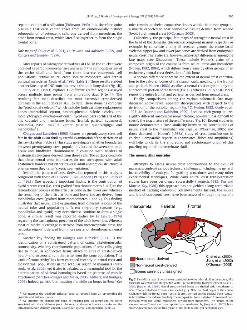
Fig. 2. Partial fate map of neural crest contribution to the adult skull in the mouse, Musmusculus, collected from study of theWnt1-Cre/R26R mouse transgenic line (Chai et al.,
395J.B. Gross, J. Hanken / Developmental Biology 317 (2008) 389–400
separate centers of ossification (Erdmann, 1940). It is, therefore, quiteplausible that each center arises from an embryonically distinctsubpopulation of osteogenic cells, one derived from mesoderm, theother from neural crest, which later fuse together to form the singlefrontal bone.
Fate maps of Couly et al. (1993), Le Douarin and Kalcheim (1999) andKöntges and Lumsden (1996)
Later reports of osteogenic derivatives of CNC in the chicken wereobtained as part of comprehensive analyses of the composite origin ofthe entire skull and head from three discrete embryonic cellpopulations: cranial neural crest, somitic mesoderm, and cranialparaxial mesoderm (Couly et al., 1993; Table 2). These results yieldedanother fate map of CNC contributions to the adult bony skull (Fig. 1B).
Couly et al. (1993) explores 11 different grafted regions assayedacross multiple time points from embryonic days 9 to 14. Theresulting fate map describes the most extensive crest-deriveddomains in the adult chicken skull to date. These domains comprisethe “prechordal skeleton,”which includes both cartilage-replacementbones (interorbital septum, basipresphenoid, scleric ossicles, eth-moid, pterygoid, quadrato-articular,1 hyoid and pars cochlearis of theotic capsule) and membrane bones (frontal, parietal, squamosal,columella, nasal, maxilla, vomer, palatine, quadratojugal andmandibular2).
Köntges and Lumsden (1996) focuses on premigratory crest cellfate in the adult avian skull by careful examination of the derivation ofthe jaw skeleton (Table 2). This study investigates whether boundariesbetween premigratory crest populations located between the mid-brain and hindbrain rhombomere 7 coincide with borders ofanatomical structures derived from those cells. The authors concludethat these neural crest boundaries do not correspond with adultanatomical borders, but rather traverse adult anatomical structures, aphenomenon they term “cryptic segmentation.”
Overall, the pattern of crest derivation reported in this study iscongruent with those of Le Lièvre (1978), Noden (1978) and Couly etal. (1993). One especially important finding is the contribution ofhyoid stream crest (i.e., crest grafted from rhombomeres 3, 4, 5) to theretroarticular process of the articular bone in the lower jaw, whereasthe remainder of the articular bone and lower jaw are derived frommandibular crest (grafted from rhombomeres 1 and 2). This findingillustrates that neural crest originating from different regions of theneural tube and populating different migratory streams (e.g.,mandibular and hyoid) may nevertheless combine to form a singlebone. A similar result was reported earlier by Le Lièvre (1974)regarding the cartilaginous precursor of the adult lower jaw. Whereasmost of Meckel's cartilage is derived from mesencephalic crest, the‘articular region’ is derived from more posterior rhombomeric neuralcrest.
Another key finding by Köntges and Lumsden (1996) is theidentification of a constrained pattern of cranial skeletomuscularconnectivity, whereby rhombomeric populations of crest cells givingrise to muscular connective tissue attach to sites of crest-derivedneuro- and viscerocranium that arise from the same population. This‘code of connectivity’ has been extended recently to neural crest andmesodermal populations in the scapular region of mammals (Mat-suoka et al., 2005), yet it also is debated as a meaningful tool for thedetermination of skeletal homologies based on patterns of muscleattachment (Sánchez-Villagra and Maier, 2006; Ahlberg and Köntges,2006). Indeed, genetic fate-mapping of middle ear bones in Hoxb1-Cre
1 We interpret the ‘quadrato-articular’ bone, as reported here, as representing thequadrate and articular bones.
2 We interpret the ‘mandibular’ bone, as reported here, as comprising the bonesassociated with the adult lower jaw in chickens, i.e., the endochondral articular and theintramembranous dentary, angular, surangular, splenial and opercular (Table 2).
mice reveals unlabeled connective tissues within the tensor tympani,a muscle expected to bear connective tissues derived from second(hyoid) arch neural crest (O'Gorman, 2005).
Collectively, the principal fate maps of osteogenic neural crest inthe head of the domestic chicken are congruent in most respects. Forexample, by consensus among all research groups the entire facialskeleton, upper jaw and lower jaw bones are derived from embryonicneural crest. There also are, however, important differences among thefate maps (see Discussion). These include Noden's claim of acomposite origin of the columella from neural crest and mesoderm(Noden, 1982, 1984), which differs from claims by other groups of anexclusively neural crest derivation of this bone.
A second difference concerns the extent of neural crest contribu-tion to the calvarial bones of the cranial vault, specifically the frontaland parietal. Noden (1982) ascribes a neural crest origin to only thesupraorbital portion of the frontal (Fig. 1C), whereas Couly et al. (1993)report the entire frontal and parietal as derived from neural crest.
Finally, comparisons among the various schematic fate mapsdiscussed above reveal apparent discrepancies with respect to thederivation of the occipital region (Fig. 1C; Noden, 1982; Couly et al.,1993; Le Douarin and Kalcheim, 1999). Because each group utilizesslightly different anatomical nomenclature, however, it is difficult tospecify the exact nature of these differences (Fig. 1C). Recent studies inmouse demonstrate a close similarity between the contributions ofneural crest to the mammalian otic capsule (O'Gorman, 2005) andthose depicted in Noden's (1983a) study of crest contributions inchicken. Comparable reports in anamniotes (fishes and amphibians)will help to clarify the embryonic and evolutionary origin of thispuzzling region of the vertebrate skull.
The mouse, Mus musculus
Attempts to assess neural crest contributions to the skull ofmammals confront serious technical challenges, including the generalinaccessibility of embryos for grafting procedures and many otherexperimental techniques. While early neural crest transplantationstudies have been performed successfully (Jaenisch, 1985; Tan andMorriss-Kay, 1986), this approach has not yielded a long-term, viablemethod of tracking embryonic cell movements. Instead, the sourceand fate of osteogenic crest have been assessed through the use of a
2000; Jiang et al., 2002). Neural crest-derived bones are shaded red; mesoderm- orother “non-crest-derived” bones are shaded grey. Note the dual origin of the cranialvault, in which the frontal bone (rostral) is crest-derived but the parietal bone (caudal)is derived frommesoderm. Similarly, the interparietal bone is derived from neural crestmedially, with the lateral component derived from mesoderm. The “bones of theviscerocranium” (unshaded) are reported as crest-derived by Jiang et al. (2002), but astudy explicitly focused on this region of the skull has not yet been published.
396 J.B. Gross, J. Hanken / Developmental Biology 317 (2008) 389–400
double-heterozygous transgenic mouse, Wnt1-Cre/R26R (Chai et al.,2000; Jiang et al., 2002).
In this system, the enzyme Cre recombinase catalyzes the removalof a stop codon upstream of the ubiquitous R26R reporter allele, whichallows sustained expression of β-galactosidase. Because expression ofCre recombinase is under the control of the Wnt-1 promoter, which isexpressed in the dorsal portion of the neural tube (includingpremigratory neural crest), skull bones derived from these cellsexpress β-galactosidase (Morriss-Kay, 2001; Jiang et al., 2002). β-Galactosidase, in turn, is easily visualized in serial sections and wholemounts.
Two reports have used this method to trace the cranial neural-crestorigin of bones and cartilages of the viscerocranium and cranial vaultin M. musculus (Chai et al., 2000; Jiang et al., 2002; Table 3). Thefollowing skull bones are ascribed a crest origin: palatine, squamosal,frontal, nasal, alisphenoid and interparietal (medial portion). Severalother bones are assigned a mesodermal origin: parietal, interparietal(lateral portion) and basioccipital (Table 3, Fig. 2). Results of thesegenetic studies have so far only been assessed as crest vs. non-crestcontributions to particular bones. The respective contributions ofindividual crest migratory streams have not been evaluated. Further-more, the embryonic cellular origins of the several remaining bones ofthe adult skull, including those of the upper and lower jaws, are notaddressed experimentally, although bones of the viscerocranium areassumed to be derived from neural crest (Jiang et al., 2002).
It would be very informative to apply an intersectional fate-mapping approach (Awatramani et al., 2003; see below) to explorethe stream-level derivation of the viscerocranial bones. Such ananalysis would, presumably, confirm the neural-crest derivation ofthese bones. More importantly, it would reveal if the relativecontributions of different neural crest streams to the lower jaw ofmammals, which comprises a single (paired) bone, the dentary,resemble those of other vertebrates, which have one or moreadditional bones in the adult jaw.
Two results from fate-mapping studies in the mouse areunexpected when compared to fate-mapping results from avian andamphibian studies. First, derivation of the bones of the cranial vaultshows a surprising distribution that does not agree completely withany published report in chicken or frogs. Specifically, the frontal bonewas demonstrated as derived from the CNC, while the parietal bone isderived (demonstrated using DiI labeling) from cranial mesoderm(Jiang et al., 2002). This report, when compared to studies in chickenand frogs, illuminates variation at the species level in the embryonicorigin of the cranial vault (see Discussion).
Secondly, “genetic fate mapping” in the mouse reveals a mixeddistribution of both labeled and unlabeled cells within Meckel'scartilage, the primary cartilaginous core of the vertebrate lower jaw(Jiang et al., 2002; Table 3). Initially, these data might appearinconsistent with the exclusively neural crest derivation of eachskeletal element in the lower jaw, including Meckel's cartilage, asreported in both chicken and amphibian models. Unlabeled cellspresent in Meckel's cartilage of these mice, however, may still bederived from neural crest cells, but cells that for whatever reason failedto express the Wnt-1 transgene. Such premigratory neural crest cellswould not drive sufficient expression of Cre recombinase in thesetransgenic mice, as a result of which their progeny would not appearlabeled, despite their neural crest derivation. In the absence ofadditional data demonstrating a direct contribution of other embryoniccell populations to Meckel's cartilage (e.g., mesoderm), these results donot provide sufficient evidence to conclude that the murine lower jawis derived from any embryonic source other than neural crest.
A recent study by O'Gorman (2005) applies site-specific, recombi-nase-mediated lineage tracing of mammalian second-branchial(hyoid) arch mesenchyme using a doubly heterozygous, Hoxb1-Cremouse. The study aims to evaluate several longstanding assumptionsregarding the homology of mammalian middle ear bones based on
their tissue of origin (Reichert, 1837). By using the Hoxb1-Cretransgenic line, neural crest cells derived exclusively from rhombo-mere 4 could be traced to their final position in the middle ear.Potential contributions from neural crest cells that emerge from otherrhombomeres, however, could not be assessed. In non-mammaliantetrapods, the middle ear comprises a single bone, the columella,which is considered homologous to the mammalian stapes (Romerand Parsons, 1977). Two additional bones of the middle ear in Recentmammals, the malleus and incus, are considered homologous to thearticular and quadrate bones, respectively, of other vertebrates. Themalleus and incus are regarded as components of the first branchial(mandibular) arch, whereas the columella (stapes) is considered asecond-arch component (Reichert, 1837).
This study, however, reports second branchial-arch crest contribu-tions to the malleus (specifically, the processus brevis) and to portionsof the cartilaginous otic capsule (O'Gorman, 2005; Table 3). Whileinitially surprising in light of conventional assignments of archidentity for middle ear bones, this result regarding the malleus agreeswith Köntges and Lumsden's (1996) demonstration of second-archneural crest derivation of the retroarticular process of the articularbone in chicken (see above). Thus, these two homologous bones inmammals and birds, which traditionally are assigned to the first archon anatomical criteria, nevertheless are associated with the secondarch on developmental features.
Because contributions of second-arch neural crest to the oticcapsule in Hoxb1-Cre mice appear identical to those reported forchicken (Noden, 1983a), O'Gorman further concludes that incorpora-tion of neural crest cells into the otic capsule likely predated theevolutionary divergence of birds andmammals. Comparable studies inanamniotes are needed to determine if contributions of neural crest tothe cartilaginous otic capsule are a shared trait among all vertebrates,among only tetrapods, or exclusively among amniotes.
More detailed studies that assess neural crest contributions fromdifferent axial levels (e.g., by following crest migrations fromindividual rhombomeres) are yet to be performed. Such studies arepossible with the application of “intersectional fate mapping,” whichallows anatomically defined regions of embryonic tissues (defined byintersections of coordinately expressed genes) to be fate mappedusing a recombinase-based method (Awatramani et al., 2003). Thisapproach already has been applied to mapping the fate of embryonicrhombic lip contributions to the choroid plexus, hindbrain roof plateand brainstem cochlear nuclear complex (Awatramani et al., 2003;Farago et al., 2006). Successful application of this technique wouldyield a higher resolution map of crest contributions to the skull in themouse and allow stream-level comparisons of crest contributions toskull bones among chickens, frogs and mice.
Discussion
Neural crest contributions to the bony skull in vertebrates
In this review we provide a reference tool that summarizes theprimary literature reporting direct contributions of premigratory CNCto the bony skull. Many shared patterns of embryonic derivationemerge when comparing fate-mapping studies performed to date inchicken, mouse and frog. For example, there is broad agreement, in themodel organisms so far examined, that the neural crest gives rise to allthe bones comprising the facial skeleton (e.g., nasal and maxillary)and the mandibular skeleton (e.g., dentary). Similarly, there isconsistent evidence of a neural crest contribution to the rostral-most region of the cranial vault (i.e., the rostral frontal bone). Finally,bones at the base of the skull (e.g., the occipital complex) consistentlylack a neural crest contribution and instead are derived from cranial orsomitic mesoderm.
Several regions of the skull, however, remain controversial, or atleast inconsistent, with respect to embryonic origin. These include the
397J.B. Gross, J. Hanken / Developmental Biology 317 (2008) 389–400
columella, bones of the otic complex, and the dermal bones thatcomprise the remainder of the cranial vault. Critical differencesregarding the derivation of these regions are apparent in comparisonsamong species and even, in at least one instance, among accounts forthe same species from different laboratories.
For example, three different research groups offer contrastingaccounts of the embryonic derivation of the avian cranial vault basedon detailed studies of the domestic chicken. These accounts rangefrom complete derivation of frontal and parietal bones from neuralcrest; to mesodermal derivation of the parietal and compositederivation (neural crest and mesoderm) of the frontal; to anexclusively or largely mesodermal derivation of both bones (Table 2;Fig. 1).
The topography of ossification of the cranial vault may offervaluable insights into this dispute and help clarify main points ofcontention. Whereas endochondral bones of the skull arise through acartilaginous template that is later replaced by bone, membranousbones arise directly through ossification of cranial mesenchyme.Importantly, the membranous avian frontal bone arises initially fromtwo separate pairs of ossification—a rostral ‘supraorbital’ pair and acaudal ‘postorbital’ pair—which later fuse to form the adult bone(Lillie, 1908; Erdmann, 1940; de Beer, 1947; Jollie, 1957).
There is consensus in chicken supporting a neural crest origin forthe rostral region of the frontal bone, which is derived from thesupraorbital center of ossification. There is no complete consensus,however, regarding the embryological origin of the remaining caudalregion. The calvarial region of the frontal bone, which arises from thepostorbital ossification center, is reported as mesodermally derivedin most studies (Le Lièvre and Le Douarin, 1975; Le Lièvre, 1978;Noden, 1978; Noden, 1983a; Noden, 1983b; Evans and Noden, 2006),with only two accounts claiming a neural crest origin (Couly et al.,1992, 1993). While it would be speculative for us to assign the“correct” account(s) of calvarial origin in chickens, we find itcompelling that the majority of reports, which utilize two differentlabeling techniques (i.e., chimeric grafting and focal retrovirallabeling), assign a mesodermal origin to the postorbital region ofthe cranial vault. Definitive determination and acceptance of theembryonic origins of the postorbital pair of ossifications likely wouldresolve the current disagreement regarding the derivation of theavian cranial vault.
Data from the frog, Xenopus laevis, when compared to othermodels, provide an example of interspecific variation in the neuralcrest origin of individual bones. The cranial vault in adult Xenopuscomprises a single, median bone, which arises from laterally pairedossification centers that fuse in the midline. This bone, unique toanurans, has long been recognized as the “frontoparietal,” represent-ing the fusion of separate frontal and parietal bones of early tetrapods(Parker, 1871, 1876; de Beer, 1937). Not all authors, however, haveaccepted this evolutionary scenario. Eaton (1942), for example, arguedfor homology of the anuran frontoparietal with only the frontal boneof other tetrapods because in some anuran species the frontoparietalforms from only a single pair of ossification centers (Eaton, 1939;Sedra, 1948). More recently, this proposal has been largely dismissedbased on patterns of ossification in many additional frog species,which show two distinct rostral and caudal centers of ossification oneach side of the head (Griffiths, 1954; Trueb, 1973).
In Xenopus, the frontoparietal receives contributions from all threemigratory streams of cranial neural crest along its entire length(Hanken and Gross, 2005). Assuming that this bone is homologous tothe frontal and parietal bones in amniotes, this pattern of derivationmost closely resembles the account of the avian cranial vault offeredby Couly et al. (1993) yet contrasts sharply with the account of themurine cranial vault offered by Morriss-Kay (2001), in which only thefrontal bone is derived from neural crest and the parietal bone isderived from mesoderm. If, instead, one assumes that the anuranfrontoparietal is homologous only to the amniote frontal (Eaton,1942),
then its observed pattern of derivation most closely resembles thatreported for the mouse but differs from two of the three alternativefate maps of the cranial vault offered for birds.
Published fate-mapping studies of a small number of diversemodel organisms indicate that 1) patterns of neural crest contributionto the vertebrate osteocranium are variable among species, and 2) therelative contributions of neural crest and mesoderm to the bony skullare evolutionarily labile. As discussed below, however, these infer-ences rely critically on current hypotheses of skeletal homology andrepresent a very narrow sampling of vertebrate cranial and develop-mental diversity. At best, they represent working hypotheses in needof further testing and confirmation.
Derivation of the columella
There are at least three differing accounts of the embryonicderivation of the middle ear skeleton (columella) in birds. All arederived from studies of the domestic chicken (Table 2). Noden (1982,1984) reports that the columella is a composite bone derived fromboth neural crest and mesoderm. Le Lièvre (1978) and, more recently,Köntges and Lumsden (1996) describe the columella as exclusivelycrest-derived, although the source of neural crest along the neuraltube differs between these two accounts: mesencephalic and anteriorrhombomeric crest (Le Lièvre, 1978) vs. crest derived from rhombo-meres 3–5 (Köntges and Lumsden, 1996). Couly et al. (1993) similarlyreport the columella as CNC-derived, but they do not assign itsderivation to any specific axial level along the neural tube.
Köntges and Lumsden's claim is most consistent with establishedanatomical convention, which regards the columella as a second(hyoid) arch derivative (Reichert, 1837). Indeed, there is no otherpublished report of mesencephalic neural crest migrating into thesecond arch in any vertebrate, as proposed by Le Lièvre. We suggestthat her claim of mesencephalic/anterior rhombencephalic crestcontribution to the columella likely is a consequence of the longergrafts used in her experiments (see above). Presumably, the neuralcrest cells that contributed directly to the columella were derivedfrom the caudal, rhombencephalic portion of the grafts, although thisdetail is not addressed in her report. Importantly, in neither of thesereports is it clear if neural crest contributions were evaluated in boththe columellar footplate and shaft (Le Lièvre, 1978) or if the bone itself,versus only the associated muscular connective tissues, was evaluatedin detail (Köntges and Lumsden, 1996). Both features were evaluatedin Noden's studies.
The only empirical data from any other vertebrate groupsregarding the embryonic derivation of the columella (or its mamma-lian homolog, the stapes) reported a second arch-contribution to thestapes in Hoxb1-Cre mice (O'Gorman, 2005).
Origin of the mammalian interparietal bone
The interparietal bone in the mouse has a dual origin: its medialportion is crest-derived, whereas the lateral portion is mesoderm-derived (Jiang et al., 2002; Table 3; Fig. 2). The developmental historyof this bone is especially intriguing considering that it is absent fromall other vertebrate species in which fate-mapping studies have beenperformed. The mammalian interparietal is generally regarded ashomologous to the postparietal bone of archaic reptiles, a dermal bonethat overlies the cerebellum and articulates posteriorly with theendochondral supraoccipital bone (Morriss-Kay, 2001). Among Recentreptiles, the postparietal has been observed to date only in theAmerican alligator (Klembara, 2001), and it will be very interesting tolearn if this putative homolog of the mammalian interparietal issimilarly derived from both neural crest and mesoderm once a fatemap of this species is produced.
Alternatively, the interparietal bone is regarded by some ashomologous to paired parietal bones. Evidence for this interpretation
398 J.B. Gross, J. Hanken / Developmental Biology 317 (2008) 389–400
comes from the skull of the fallow deer, Dama dama. Instead ofhaving a discrete interparietal, the bone's characteristic positionwithin the cranium is occupied by expanded parietal bones (Kierdorfand Kierdorf, 1992). Similarly, the location of the interparietal withinthe caudal portion of the cranial vault in the mouse correspondsclosely to the location of the parietal in the avian skull. As discussedabove, the avian parietal is reported to have an exclusivelymesodermal origin by some authors (Le Lièvre, 1978; Noden, 1982)and an exclusively neural crest origin by others (Couly et al., 1993).Unlike the interparietal, no one has yet reported a dual embryonicorigin for the parietal bone.
Unresolved issues in the osteogenic neural crest literature
Given the variation among model systems examined so far, howcan we hope to clarify primitive versus derived patterns of neuralcrest contribution to the skull bones of vertebrates? For example,which, if any, of the contrasting patterns of derivation of the cranialvault reported from Xenopus, chicken and mouse is characteristic ofbony fishes, or do these vertebrates display unique patterns? Fromcomparative and developmental perspectives, only by constructionof additional, long-term fate maps of the CNC across a widerphylogenetic framework (e.g., bony fishes, additional amphibians,reptiles) may we resolve which features are primitive and whichare derived, and, more generally, determine the full extent towhich patterns of crest cell contribution to the bony skull areinvariant (i.e., rigidly conserved across vertebrates) or evolutiona-rily labile.
Representing extant organisms as surrogate “primitive” datapoints can be problematic. Individual species may not accuratelyreplicate either the primitive or the generalized condition of thebroader taxonomic group to which they belong (Hanken, 1993). Thisincludes model organisms, which may display species-specific traits,including unique features of life history and developmental mode. Byamassing comparable data from a wider array and number ofvertebrates, however, we can more readily identify unique featuresof model species and more confidently resolve how, and to whatextent, crest contributions to the adult skull have changed over thecourse of evolution (Helms et al., 2005).
It is reasonable to assume that homologous skull bones amongdifferent vertebrates are derived from the same embryonic tissue(s),yet this assumption remains largely untested. Moreover, the limitedcomparative data available at this time suggests that this relationshipdoes not always hold true. Since the same principal migratory creststreams (viz., mandibular, hyoid and branchial) are all present inamphibians, birds and mammals, it is possible to directly test such apriori hypotheses for the stream-level derivation of bones andcartilages of the skull, at least among these tetrapods. Thesecomparisons would answer the question of whether embryonic tissueof origin is a valid criterion for evaluating the homology of individualcranial elements. Existing data suggest that embryonic derivation—mesoderm vs. neural crest—of at least some skull bones is evolutio-narily labile, and that shared features of development is not anappropriate criterion of homology for these elements in all inter-specific comparisons. Alternatively, contrasting patterns of embryonicderivation of what is regarded as the same (homologous) bone amongdistantly related species may indicate that the correspondinghomology statement itself is incorrect. Accumulation of additionaldetailed information regarding the embryonic derivation and devel-opmental biology of cranial bones in diverse species may eventuallyforce a reevaluation of the traditional assessment of specific cranialbone homologies among vertebrate classes, which was codifiedlargely in the absence of such data and before the advent ofexperimental and molecular biology.
Future studies must extend fate-mapping research to additionalmodel and non-model species in order to document more widely the
patterns of embryonic derivation and the extent to which thesepatterns correlate with different, taxon-specific morphologies and lifehistories. This avenue of research, which aims to define the full scopeof variation in the pattern of neural crest derivation of theosteocranium, would facilitate unification of the fields of vertebratemorphology, development and neural crest biology. Moreover,additional studies in diverse organisms will help define the ancestralpattern of cranial development at key “junctures” in vertebrateevolution, such as the origin of jaws and the evolution of tetrapods.Finally, a synthetic and comprehensive evaluation of neural crestderivatives in additional species will better characterize the role andbehavior of this intriguing embryonic tissue in the development andevolution of the vertebrate skull, and especially in the origin of novelfeatures.
Why are there disagreements among neural crest fate mapping studies?
An important but largely unresolved question within the field ofneural crest biology is why differences in derivation schemes and fatemaps for a given species arise among different research groups. Somediscrepancies in the literature may be due to the fact that differentchicken fate-mapping studies have utilized slightly different graftingprocedures at slightly different developmental stages. Compare, forexample, grafts of “mesencephalic” neural crest at the 9-somite stage(Le Lièvre, 1974) to grafts of the entire rostrocaudal axis of thedeveloping neural tube at embryonic days 8 and 14 (Couly et al., 1993).Indeed, Couly et al. (1993) suggest that differences in the timing ofgrafts and their subsequent recovery (following initiation of bonedevelopment) explain conflicting results with respect to CNCcontribution to the frontal bone reported in earlier experiments byLe Lièvre (1978).
A second potential source of variability may be contamination ofpurportedly pure grafts of one embryonic cell type with other cells.Schneider (1999) performed experiments in which embryonic tissuesthat give rise to the lateral wall of the braincase (a strictly mesoderm-derived structure in vivo) were replaced with grafts of neural crest.The resulting braincase appeared morphologically normal andcontained cells derived from the neural crest, demonstrating thatboth tissues can respond in the same way to cues that mediateskeletogenesis and morphological patterning. Since neural crest isable to form at least some skeletal structures that normally are derivedfrom mesoderm, even slight contamination of neural crest in grafts ofmesoderm—and vice versa—may significantly skew fate-mappingresults that are based on analysis of chimeric embryos. This under-scores both the importance of minimizing contamination of grafts insuch studies and the need to verify specific fate-mapping results withseveral alternate methods in order to control for potential experi-mental artifacts associated with any particular one.
A third potential explanation of discrepancies in the literature isthe intriguing possibility of significant variation in craniofacialdevelopment among inbred strains of domestic chicken. If thisexplanation were true, then the contrasting claims offered bydifferent research groups may not conflict, but instead reflect realdifferences among source populations on which the studies arebased.
Perhaps different chicken strains, or even the same strain bred andreared in different countries, evince characteristic differences in theembryonic derivation of the craniofacial skeleton. Published fate mapsfor the domestic chicken are based on studies of several inbred strains,including White Plymouth Rock (Noden, 1978), White Leghorn (Coulyet al., 1993; Evans and Noden, 2006) and Rhode Island Red Hen(Köntges and Lumsden, 1996). Strain type was not reported by LeLièvre (1978). Presumably, even slight differences in the timing andpattern of neural crest migration among strains could alter thederivation of craniofacial tissues. This potential explanation, however,remains to be explored adequately.
399J.B. Gross, J. Hanken / Developmental Biology 317 (2008) 389–400
How can we resolve issues and differences in fate maps amongspecies? One way might be to apply the same methodologicalapproach to pursuing a fate map across different model systems. Forexample, would application of non-invasive, recombinase-basedgenetic fate mapping based on the same early genetic driver producethe same patterns of derivation in other vertebratemodel systems as ithas in the mouse? This approach would obviate the need forpainstaking grafting studies that are susceptible to human error andother experimental artifacts. As with any methodological approach,however, genetic fate-mapping studies carried out in multiplevertebrate models would also be susceptible to experimental artifacts.For example, the presence of both labeled and unlabeled cells inindividual tissues, as reported for Meckel's cartilage inWnt1-Cre mice(Jiang et al., 2002), would require additional direct-labeling studies todetermine if this is an artifact of the labeling method or an indicationof tissue derivation from other embryonic sources in addition toneural crest.
Additionally, this approach would require a suitable Cre recombi-nase-driver (e.g., the Wnt1 promoter) that accurately and exclusivelylabels early cranial neural crest in all species. At this time, no such earlymarker, which maintains the same expression pattern in early cranialneural crest across all vertebrates, has been identified. Interestingly,the only report to date that utilizes genetic fate mapping to assessneural crest contributions to the middle ear does not exclusively labelneural crest, but rather cells derived from only a single rhombomere,which includes some neural crest (O'Gorman, 2005).
Alternatively, the chimeric grafting approach could be extended toa wider array of organisms. Using this strategy, the same regions ofneural crest in each model could be excised and grafted into a hostembryo, as has been applied in chicken and frogs. The advantage ofthis approach is that it would reveal patterns of crest contribution tothe skull in a wider assortment of animals, e.g., reptiles, cartilaginousfish, bony fish. The disadvantage is that it will not resolve long-standing disagreements that apply to a single species (e.g., domesticchicken).
In light of current disagreements among fate maps produced bydifferent laboratories, some studies have begun to utilize more fine-grained experimental procedures as an additional means of resolvinglong-standing questions regarding the contribution of neural crest tocranial structures. For example, Cerny et al. (2004) and Lee et al.(2004) combined molecular marker analysis and vital dye (DiI)staining of highly specific sub-populations of late-emerging neuralcrest cells to determine the distinct origins of the palatoquadrate,Meckel's cartilage and trabecular cartilage condensations. Additionaluse of highly focused crest cell labeling procedures such as these mayhelp to resolve existing disagreements regarding neural crestcontributions in the chicken skull.
Contemporary neural crest research must aim to better define thesignificance of boundaries and interfaces between the neural crestand other embryonic tissues (e.g., mesoderm) throughout develop-ment. For example, is the embryonic origin of cells or their localinteractions post-migration more important in defining the eventualfate of embryonic contributions to the skull? In order to betterunderstand the nature of evolutionary alterations to cranial fatemaps, it will be critical that additional studies be carried out in bothmodel and non-model systems. Ultimately, as patterns of neural crestcontribution to the skull are better characterized in the coming years,so too will the details of how diversity in cranial form arose amongvertebrates.
Acknowledgments
This study was supported by the U.S. National Science Foundation,grant no. EF-0334846 to J.H. The authors wish to thank twoanonymous reviewers for their helpful comments on earlier drafts ofthis manuscript.
References
Ahlberg, P.E., Köntges, G., 2006. Homologies and cell populations: a response toSánchez-Villagra and Maier. Evol. Dev. 8, 116–118.
Andres, G., 1949. Untersuchungen an Chimären von Triton und Bombinator. Genetica 24,387–534.
Awatramani, R., Soriano, P., Rodriguez, C., Mai, J.J., Dymecki, S.M., 2003. Crypticboundaries in roof plate and choroid plexus identified by intersectional geneactivation. Nat. Genet. 35, 70–75.
Barlow, LA., 2002. Cranial nerve development: placodal neurons ride the crest. Curr.Biol. 12, R171–R173.
Barlow, L.A., Northcutt, R.G., 1995. Embryonic origin of amphibian taste buds. Dev. Biol.169, 273–285.
Barlow, L.A., Northcutt, R.G., 1997. Taste buds develop autonomously from endodermwithout induction by cephalic neural crest or paraxial mesoderm. Development124, 949–957.
Beauchamp, G.R., Knepper, P.A., 1984. Role of the neural crest in anterior segmentdevelopment and disease. J. Pediatr. Ophthalmol. Strabismus 21, 209–214.
Bronner-Fraser, M., 1986. Analysis of the early stages of trunk neural crest migration inavian embryos using monoclonal antibody HNK-1. Dev. Biol. 115, 44–55.
Carl, T.F., Dufton, C., Vourgourakis, Y., Klymkowsky, M., Hanken, J., 2000. Greenfluorescent protein used to assess cranial neural crest derivatives in the frog, Xe-nopus laevis. In: Jacobson, C.-O., Olsson, L. (Eds.), Regulatory Processes inDevelopment. Portland Press, London, pp. 167–172.
Cebra-Thomas, J.A., Tan, F., Sistla, S., Estes, E., Bender, G., Kim, C., Riccio, P., Gilbert, S.F.,2005. How the turtle forms its shell: a paracrine hypothesis of carapace formation. J.Exp. Zool. B. Mol. Dev. Evol. 304, 558–569.
Cebra-Thomas, J.A., Betters, E., Yin, M., Plafkin, C., McDow, K., Gilbert, S.F., 2007.Evidence that a late-emerging population of trunk neural crest cells forms theplastron bones in the turtle Trachemys scripta. Evolut. Develop. 9, 267–277.
Cerny, R., Lwigale, P., Ericsson, R., Meulemans, D., Epperlein, H.H., Bronner-Fraser, M.,2004. Developmental origins and evolution of jaws: new interpretation of“maxillary” and “mandibular”. Dev. Biol. 276, 225–236.
Cerny, R., Horácek, I., Olsson, L., 2006. The trabecula cranii: development and homologyof an enigmatic vertebrate head structure. Anim. Biol. 56, 503–518.
Chai, Y., Jiang, X., Ito, Y., Bringas Jr., P., Han, J., Rowitch, D.H., Soriano, P., McMahon, A.P.,Sucov, H.M., 2000. Fate of the mammalian cranial neural crest during tooth andmandibular morphogenesis. Development 127, 1671–1679.
Clark, K., Bender, G., Murray, B.P., Panfilio, K., Cook, S., Davis, R., Murnen, K., Tuan, R.S.,Gilbert, S.F., 2001. Evidence for the neural crest origin of turtle plastron bones.Genesis 31, 111–117.
Clarke, J.D., Tickle, C., 1999. Fate maps old and new. Nat. Cell Biol. 1, E103–E109.Colnot, C., 2005. Cellular and molecular interactions regulating skeletogenesis. J. Cell.
Biochem. 95, 688–697.Couly, G.F., Coltey, P.M., Le Douarin, N.M., 1992. The developmental fate of the cephalic
mesoderm in quail-chick chimeras. Development 114, 1–15.Couly, G.F., Coltey, P.M., Le Douarin, N.M., 1993. The triple origin of skull in higher
vertebrates: a study in quail-chick chimeras. Development 117, 409–429.Couly, G.F., Creuzet, S., Bennaceur, S., Vincent, C., Le Douarin, N.M., 2002. Interactions
between Hox-negative cephalic neural crest cells and the foregut endoderm inpatterning the facial skeleton in the vertebrate head. Development 129,1061–1073.
de Beer, G.R., 1937. The Development of the Vertebrate Skull. Clarendon Press, Oxford.552 pp.
de Beer, G.R., 1947. The differentiation of neural crest cells into visceral cartilages andodontoblasts in Amblystoma, and a re-examination of the germ-layer theory. Proc.R. Soc. London, Ser B. Biol. Sci. 134, 377–398.
Dixon, J., Brakebusch, C., Fassler, R., Dixon, M.J., 2000. Increased levels of apoptosis in theprefusion neural folds underlie the craniofacial disorder, Treacher Collinssyndrome. Hum. Mol. Genet. 9, 1473–1480.
DuShane, G.P., 1935. An experimental study of the origin of pigment cells in Amphibia. J.Exp. Zool. 72, 1–31.
Eaton Jr., T.H., 1939. Development of the frontoparietal bones in frogs. Copeia 1939,95–97.
Eaton Jr., T.H., 1942. Are ‘frontoparietal’ bones in frogs actually frontals? J. Wash. Acad.Sci. 32, 151–153.
Edwards, S.J., Gladwin, A.J., Dixon, M.J., 1997. The mutational spectrum in TreacherCollins syndrome reveals a predominance of mutations that create a premature-termination codon. Am. J. Hum. Genet. 60, 515–524.
Erdmann, K., 1940. Zur Entwicklungsgeschichte der Knochen im Schädel des Huhnes biszum Zeitpunktdes Ausschlüpfen aus dem Ei. Z. Morphol. Okol. Tiere Berlin 36,315–400.
Evans, D.J., Noden, D.M., 2006. Spatial relations between avian craniofacial neural crestand paraxial mesoderm cells. Dev. Dyn. 235, 1310–1325.
Farago, A.F., Awatramani, R.B., Dymecki, S.M., 2006. Assembly of the brainstem cochlearnuclear complex is revealed by intersectional and subtractive genetic fate maps.Neuron 50, 205–218.
Farlie, P.G., McKeown, S.J., Newgreen, D.F., 2004. The neural crest: basic biology andclinical relationships in the craniofacial and enteric nervous systems. Birth DefectsRes. C 72, 173–189.
Franz-Odendaal, T., Hall, B.K., Witten, P.E., 2006. Buried alive: how osteocytes becomebone. Dev. Dyn. 235, 176–190.
Fukiishi, Y., Morriss-Kay, G.M., 1992. Migration of cranial neural crest cells to thepharyngeal arches and heart in rat embryos. Cell Tissue Res. 268, 1–8.
Gargioli, C., Slack, J.M., 2004. Cell lineage tracing during Xenopus tail regeneration.Development 131, 2669–2679.
400 J.B. Gross, J. Hanken / Developmental Biology 317 (2008) 389–400
Gilbert, S.F., Loredo, G.A., Brukman, A., Burke, A.C., 2001. Morphogenesis of the turtleshell: the development of a novel structure in tetrapod evolution. Evol. Dev. 3,47–58.
Gilbert, S.F., Bender, G., Betters, E., Yin, M., Cebra-Thomas, J.A., 2007. The contribution ofneural crest cells to the nuchal bone and plastron of the turtle shell. Int. Comp. Biol.47, 401–408.
Gorlin, R.J., Cohen Jr., M.M., Levin, L.S., 1990. Syndromes of the Head and Neck. OxfordUniv. Press, New York. 977 pp.
Graveson, A.C., Smith, M.M., Hall, B.K., 1997. Neural crest potential for toothdevelopment in a urodele amphibian: developmental and evolutionary signifi-cance. Dev. Biol. 188, 34–42.
Griffiths, I., 1954. On the nature of the fronto-parietal in Amphibia, Salientia. Proc. Zool.Soc. Lond. 123, 781–792.
Gross, J.B., Hanken, J., 2004. Use of fluorescent dextran conjugates as a long-termmarker of osteogenic neural crest in frogs. Dev. Dyn. 230, 100–106.
Gross, J.B., Hanken, J., 2005. Cranial neural crest contributes to the bony skull vault inadult Xenopus laevis: Insights from cell labeling studies. J. Exp. Zool. B Mol. Dev.Evol. 304, 169–176.
Gross, J.B., Hanken, J., Oglesby, E., Marsh-Armstrong, N., 2006. Use of a ROSA26:GFPtransgenic line for long-term Xenopus fate-mapping studies. J. Anat. 209,401–413.
Hall, B.K., 1999. The Neural Crest in Development and Evolution. Springer-Verlag, NewYork. 313 pp.
Hanken, J., 1993. Model systems versus outgroups: alternative approaches to the studyof head development and evolution. Am. Zool. 33, 448–456.
Hanken, J., Gross, J.B., 2005. Evolution of cranial development and the role of neuralcrest: insights from amphibians. J. Anat. 207, 437–446.
Helms, J.A., Cordero, D., Tapadia, M.D., 2005. New insights into craniofacialmorphogenesis. Development 132, 851–861.
Hörstadius, S., 1950. The Neural Crest: Its Properties and Derivatives in the Light ofExperimental Research. Oxford Univ. Press, London. 111 pp.
Jaenisch, R., 1985. Mammalian neural crest cells participate in normal embryonicdevelopment on microinjection into post-implantation mouse embryos. Nature318, 181–183.
Jiang, X., Iseki, S., Maxson, R.E., Sucov, H.M., Morriss-Kay, G.M., 2002. Tissue origins andinteractions in the mammalian skull vault. Dev. Biol. 241, 106–116.
Johnston,M.C., Bhakdinaronk, A., Reid, Y.C., 1973. An expanded role of the neural crest inoral and pharyngeal development. In: Bosma, J.F. (Ed.), Fourth Symposium on OralSensation and Perception: Development in the Fetus and Infant. U.S. Dept. ofHealth, Education, and Welfare, Bethesda, pp. 37–52.
Jollie, M.T., 1957. The head skeleton of the chicken and remarks on the anatomy of thisregion in other birds. J. Morphol. 100, 389–436.
Kardong, K.V., 2002. Vertebrates: comparative anatomy, function, Evolution, 3rd ed.McGraw-Hill, Boston. 762 pp.
Kierdorf, U., Kierdorf, H., 1992. Studies on the parietal region of the cervid skull. II. Theparietooccipital region in the skull of the fallow deer (Dama dama L.). Anat. Histol.Embryol: J. Vet. Med. Series C 21, 373–379.
Klembara, J., 2001. Postparietal and prehatching ontogeny of the supraoccipital in Al-ligator mississippiensis (Archosauria, Crocodylia). J. Morphol. 249, 147–153.
Knight, R.D., Schilling, T.F., 2006. Cranial neural crest and development of the headskeleton. Adv. Exp. Med. Biol. 589, 120–133.
Köntges, G., Lumsden, A., 1996. Rhombencephalic neural crest segmentation ispreserved throughout craniofacial ontogeny. Development 122, 3229–3242.
Kuratani, S., 2005. Craniofacial development and the evolution of vertebrates: the oldproblems on a new background. Zool. Sci. 22, 1–19.
Kuratani, S., Matsuo, I., Aizawa, S., 1997. Developmental patterning and evolution of themammalian viscerocranium: genetic insights into comparative morphology. Dev.Dyn. 209, 139–155.
Landacre, F.L., 1921. The fate of the neural crest in the head of Urodeles. J. Comp. Neurol.33, 1–43.
Langille, R.M., Hall, B.K., 1988. Role of the neural crest in development of thecartilaginous cranial and visceral skeleton of the medaka, Oryzias latipes (Teleostei).Anat. Embryol. (Berl.) 177, 297–305.
Le Douarin, N., Barq, G., 1969. Use of Japanese quail cells as “biological markers” inexperimental embryology. C. R. Acad. Sci. Hebd. Seances Acad. Sci. D 269,1543–1546.
Le Douarin, N.M., Kalcheim, C., 1999. The Neural Crest, 2nd ed. Cambridge Univ. Press,Cambridge. 445 pp.
Lee, S.H., Bedard, O., Buchtova, M., Fu, K., Richman, J.M., 2004. A new origin for themaxillary jaw. Dev. Biol. 276, 207–224.
Le Lièvre, C.S., 1974. Rôle des cellules mésectodermiques issues des crêtes neuralescéphaliques dans la formation des arcs branchiaux et du squelette viscéral. J.Embryol. Exp. Morphol. 31, 453–477.
Le Lièvre, C.S., 1978. Participation of neural crest-derived cells in the genesis of the skullin birds. J. Embryol. Exp. Morphol. 47, 17–37.
Le Lièvre, C.S., Le Douarin, N.M., 1975. Mesenchymal derivatives of the neural crest:analysis of chimaeric quail and chick embryos. J. Embryol. Exp. Morphol. 34,125–154.
Lillie, F.R., 1908. The Development of the Chick. An Introduction to Embryology. HenryHolt and Co., New York. 472 pp.
Lumsden, A.G., 1988. Spatial organization of the epithelium and the role of neuralcrest cells in the initiation of the mammalian tooth germ. Development 103,155–169.
Matsuoka, T., Ahlberg, P.E., Kessaris, N., Iannarelli, P., Dennehy, U., Richardson, W.D.,McMahon, A.P., Köntges, G., 2005. Neural crest origins of the neck and shoulder.Nature 436, 347–355.
Meier, S., Packard Jr., D.S., 1984. Morphogenesis of the cranial segments and distributionof neural crest in the embryos of the snapping turtle, Chelydra serpentina. Dev. Biol.102, 309–323.
Morriss-Kay, G.M., 2001. Derivation of the mammalian skull vault. J. Anat. 199, 143–151.Noden, D.M., 1978. The control of avian cephalic neural crest cytodifferentiation. I.
Skeletal and connective tissues. Dev. Biol. 67, 296–312.Noden, D.M., 1982. Patterns and organization of craniofacial skeletogenic and myogenic
mesenchyme: a perspective. In: Sarnat, B.G. (Ed.), Progress in Clinical and BiologicalResearch: Factors and Mechanisms Influencing Bone Growth. Alan R. Liss, Inc., NewYork, pp. 167–203.
Noden, D.M., 1983a. The role of the neural crest in patterning avian cranial skeletal,connective, and muscle tissues. Dev. Biol. 96, 144–165.
Noden, D.M., 1983b. The embryonic origins of avian craniofacial muscles and associatedconnective tissues. Am. J. Anat. 168, 257–276.
Noden, D.M., 1984. The use of chimeras in analyses of craniofacial development. In: LeDouarin, N.M., McLaren, A. (Eds.), Chimeras in Developmental Biology. AcademicPress, London, pp. 241–280.
Noden, D.M., 1988. Interactions and fates of avian craniofacial mesenchyme. Develop-ment 103 Suppl., 121–140.
Noden, D.M., Trainor, P.A., 2005. Relations and interactions between cranial mesodermand neural crest populations. J. Anat. 207, 575–601.
O'Gorman, S., 2005. Second branchial arch lineages of the middle ear of wild-type andHoxa2 mutant mice. Dev. Dyn. 234, 124–131.
Parker, W.K., 1871. On the structure and development of the skull of the common frog.Philos. Trans. R. Soc. Lond. 161, 137–211.
Parker, W.K., 1876. On the structure and development of the skull in Batrachia—Part II.Philos. Trans. R. Soc. Lond. 166, 601–669.
Platt, J.B., 1893. Ectodermic origin of the cartilages of the head. Anat. Anz. 8,506–509.
Poswillo, D., 1988. The aetiology and pathogenesis of craniofacial deformity. Develop-ment 103 Suppl., 207–212.
Raven, C.P., 1931. Zur Entwicklung der Ganglienleiste. I: Die Kinematik der Gang-lienleisten Entwicklung bei den Urodelen. Roux' Arch. Entwicklungsmech. Org. 125,210–292.
Reichert, C., 1837. Über die Visceralbogen der Wirbelthiere im Allgemeinen und derenMetamorphosen bei den Vögeln und Säugethieren. Archiv. Anat., Physiol. Wiss.Med. 120–222.
Romer, A.S., Parsons, T.S., 1977. The Vertebrate Body, 5th ed. Saunders CollegePublishing, Philadelphia. 624 pp.
Rudel, D., Sommer, R.J., 2003. The evolution of developmental mechanisms. Dev. Biol.264, 15–37.
Sánchez-Villagra, M.R., Maier, W., 2006. Homologies of the mammalian shoulder girdle:a response to Matsuoka et al. (2005). Evolut. Develop. 8, 113–115.
Schneider, R.A., 1999. Neural crest can form cartilages normally derived frommesodermduring development of the avian head skeleton. Dev. Biol. 208, 441–455.
Schultze, H.-P., 1993. Patterns of diversity in the skull of jawed fishes. In: Hanken, J., Hall,B.K. (Eds.), The Skull. Patterns of Structural and Systematic Diversity, vol. 2.University of Chicago Press, Chicago, pp. 189–254.
Sedra, S.N., 1948. On the homology of certain elements in the skull of Bufo regularisReuss (Salientia). Proc. Zool. Soc. Lond. 119, 633–641.
Stone, L.S., 1926. Further experiments on the extirpation and transplantation ofmesectoderm in Amblystoma punctatum. J. Exp. Zool. 44, 95–131.
Stone, L.S., 1929. Experiments showing the role of migrating neural crest (mesecto-derm) in the formation of head skeleton and loose connective tissue in Ranapalustris. Wilhelm Roux Arch. Dev. Biol. 118, 40–77.
Sulik, K.K., Cook, C.S., Webster, W.S., 1988. Teratogens and craniofacial malformations:relationships to cell death. Development 103 Suppl., 213–231.
Tan, S.S., Morriss-Kay, G.M., 1986. Analysis of cranial neural crest cell migration andearly fates in postimplantation rat chimaeras. J. Embryol. Exp. Morphol. 98, 21–58.
Toerien, M.J., 1965. Experimental studies on the columella–capsular interrelationship inthe turtle Chelydra serpentina. J. Embryol. Exp. Morphol. 14, 265–272.
Trueb, L., 1973. Bones, frogs, and evolution. In: Vial, J.L. (Ed.), Evolutionary Biology of theAnurans: Contemporary Research on Major Problems. University of Missouri Press,Columbia, pp. 65–132.
Trueb, L., Hanken, J., 1992. Skeletal development in Xenopus laevis (Anura: Pipidae). J.Morphol. 214, 1–41.
Twitty, V.C., Bodenstein, D., 1939. Correlated genetic and embryological experiments onTriturus. III. Further transplantation experiments on pigment development. IV: Thestudy of pigment cell behavior in vitro. J. Exp. Zool. 81, 357–398.
Wagner, G., 1949. Die Bedeutung der Neuralleiste für die Kopfgestaltung derAmphibienlarven: Untersuchungen an Chimären von Triton und Bombinator. Rev.Suisse Zool. 56, 519–620.
Weston, J.A., 1963. A radiographic analysis of the migration and localization of trunkneural crest cells in the chick. Dev. Biol. 6, 279–310.