DEVELOPMENT OF THE ALIMENTARY SYSTEM G.LUFUKUJA 1.
72
DEVELOPMENT OF THE ALIMENTARY SYSTEM G.LUFUKUJA 1
-
Upload
bernadette-douglas -
Category
Documents
-
view
225 -
download
4
Transcript of DEVELOPMENT OF THE ALIMENTARY SYSTEM G.LUFUKUJA 1.
DEVELOPMENT OF
THE ALIMENTARY SYSTEM
G.LUFUKUJA 1

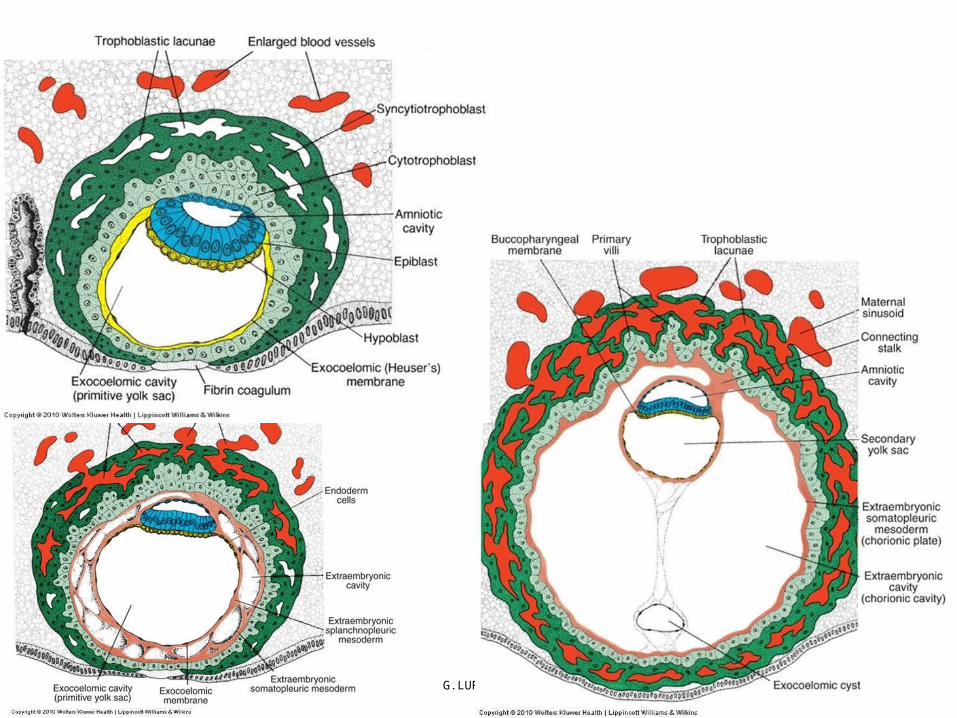
The primitive alimentary canal• The primitive alimentary canal is a simple endodermal tube
that extends from the buccopharyngeal membrane to the cloacal membrane.
• As a result of cephalocaudal and lateral folding of the embryo, a portion of the endoderm-lined yolk sac cavity is incorporated into the embryo to form the primitive gut.
• Two other portions of the endoderm-lined cavity, the yolk sac and the allantois, remain outside the embryo
G.LUFUKUJA 2
G.LUFUKUJA 3

The primitive alimentary canal
G.LUFUKUJA 4
Endoderm forms the epithelial lining of the digestive tract and gives rise to the parenchyma of glands, such as the liver and pancreas. Muscle, connective tissue, and peritoneal components of the wall of the gut are derived from splanchnic mesoderm.
….the primitive gut• Development of the primitive gut and its derivatives is
usually discussed in four sections:• (a) The pharyngeal gut, or pharynx, extends from the buccopharyngeal membrane to the tracheobronchial diverticulum since this section is particularly important for development of the head and neck, it is already discussed
G.LUFUKUJA 5

….the primitive gut
• (b) The foregut lies caudal to the pharyngeal tube and extends as far caudally as the liver outgrowth.
• (c) The midgut begins caudal to the liver bud and extends to the junction of the proximal two-thirds and distal third of the transverse colon in the adult.
• (d) The hindgut extends from the distal third of the transverse colon to the cloacal membrane
G.LUFUKUJA 6
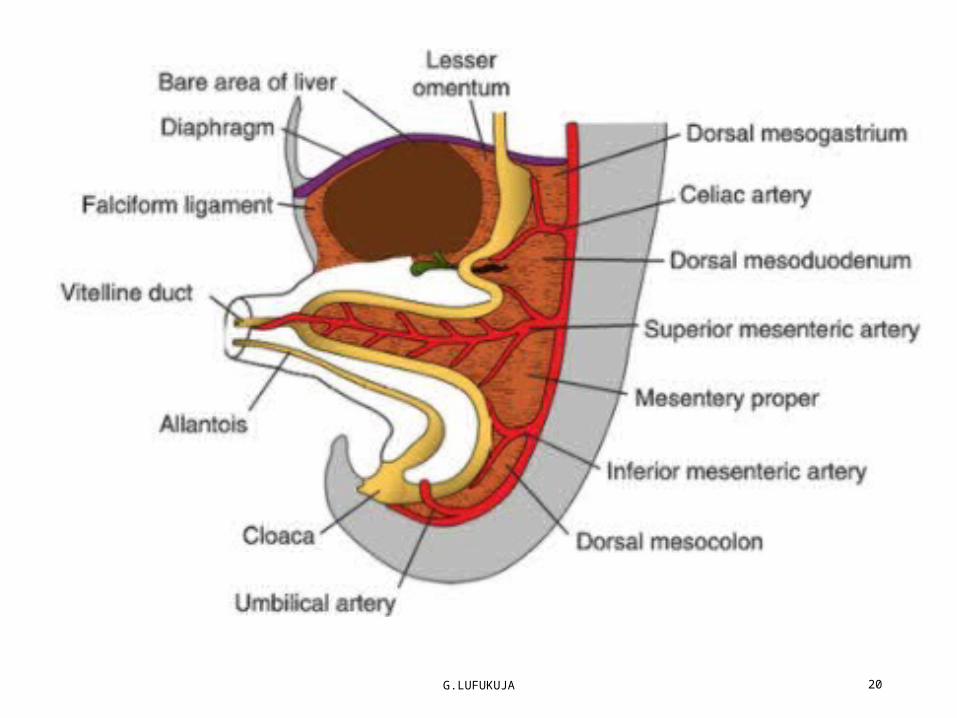
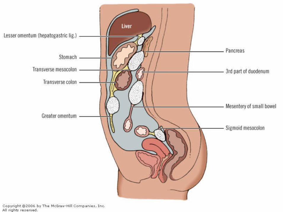
….the primitive gut• The gut tube is suspended from the dorsal body wall by a midline dorsal mesentery, through which three arteries pass from the dorsal aorta to supply the gut tube
• The dorsal mesentery is also given names according to the gut tube region: dorsal mesogastrium or greater omentum in the region of the stomach; mesoduodenum in the region of the duodenum; and mesocolon in the region of the colon. The ventral mesogastrium develops from the caudal part of the septum transversum. It is attached to the under surface of the developing diaphragm and the anterior abdominal wall down to the umbilicus (falciform ligament)
G.LUFUKUJA 7

G.LUFUKUJA 8
dorsal mesentery

G.LUFUKUJA 9
ventral mesogastrium

Molecular Regulation of Gut Tube Development
• Differentiation of various regions of the gut and its derivatives is dependent upon a reciprocal interaction between the endoderm (epithelium) of the gut tube and surrounding splanchnic mesoderm.
• Sonic hedgehog (SHH) expressed throughout the gut endoderm & the HOX code in the mesoderm
G.LUFUKUJA 10
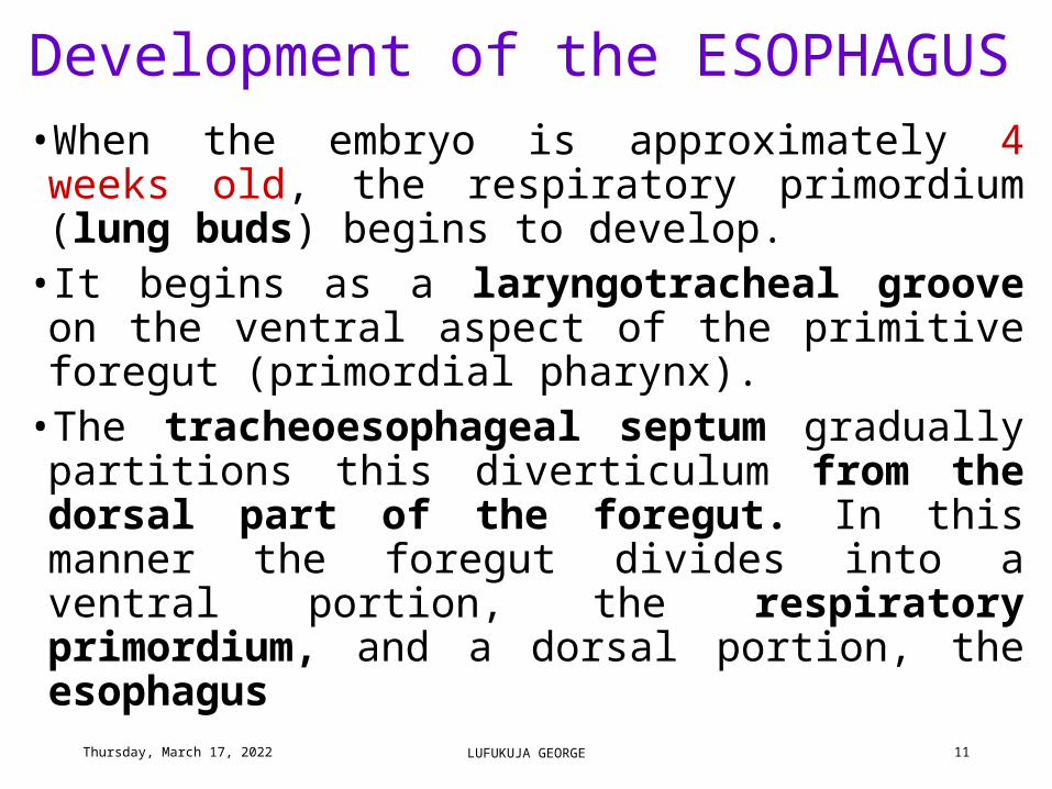
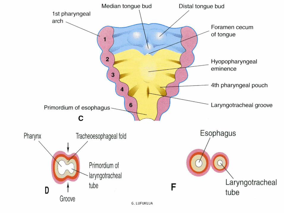
Development of the ESOPHAGUS•When the embryo is approximately 4 weeks old, the respiratory primordium (lung buds) begins to develop.
• It begins as a laryngotracheal groove on the ventral aspect of the primitive foregut (primordial pharynx).
•The tracheoesophageal septum gradually partitions this diverticulum from the dorsal part of the foregut. In this manner the foregut divides into a ventral portion, the respiratory primordium, and a dorsal portion, the esophagus
Tuesday, April 18, 2023 LUFUKUJA GEORGE 11
G.LUFUKUJA 12

ESOPHAGUS…• At first the esophagus is short, but with descent of the heart
and lungs it lengthens rapidly. The muscular coat, which is formed by surrounding splanchnic mesenchyme, is striated in its upper two-thirds and innervated by the vagus; the nerve of the 4th & 6th branchial arch. The muscle coat is smooth in the lower third and is innervated by the splanchnic plexus
G.LUFUKUJA 13

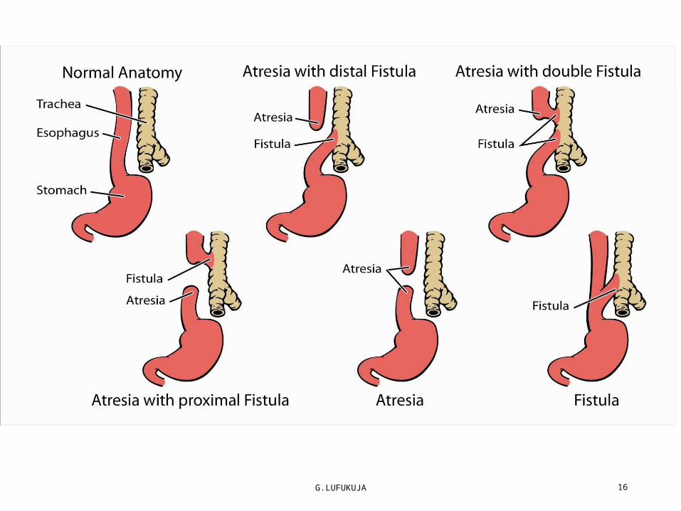
Esophageal Stenosis & atresia • Atresia of the esophagus prevents normal passage of amniotic fluid into the intestinal tract, resulting in accumulation of excess fluid in the amniotic sac (polyhydramnios). In addition to atresias, the lumen of the esophagus may narrow, producing esophageal stenosis, usually in the lower third. Stenosis may be caused by incomplete recanalization
G.LUFUKUJA 14
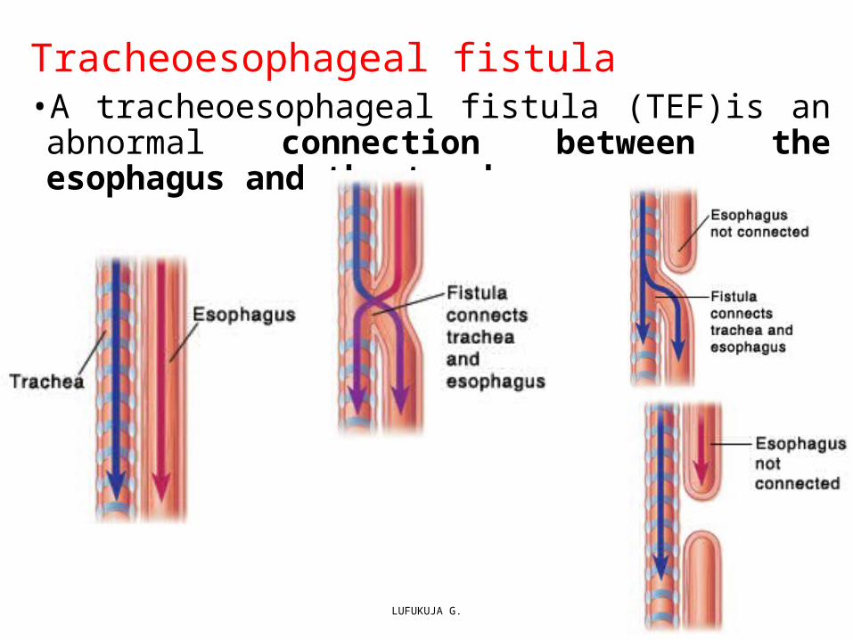
Tracheoesophageal fistula • A tracheoesophageal fistula (TEF)is an abnormal connection between the esophagus and the trachea
LUFUKUJA G. 15
G.LUFUKUJA 16

STOMACH•The stomach appears as a fusiform dilation of the foregut in the fourth week of development.
•During the following weeks, its appearance and position change greatly as a result of the different rates of growth in various regions of its wall and the changes in position of surrounding organs.
•Positional changes of the stomach are most easily explained by assuming that it rotates around a longitudinal and an anteroposterior axis
G.LUFUKUJA 17
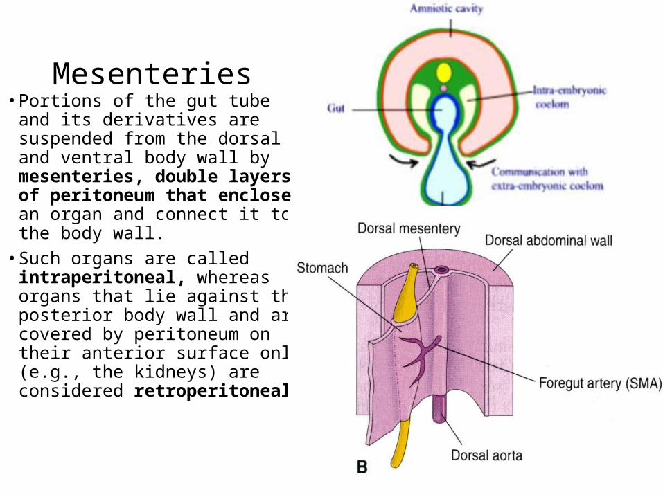
Mesenteries • Portions of the gut tube and its
derivatives are suspended from the dorsal and ventral body wall by mesenteries, double layers of peritoneum that enclose an organ and connect it to the body wall.
• Such organs are called intraperitoneal, whereas organs that lie against the posterior body wall and are covered by peritoneum on their anterior surface only (e.g., the kidneys) are considered retroperitoneal.
G.LUFUKUJA 18

G.LUFUKUJA 19
Mesenteries Mesenteries
G.LUFUKUJA 20

Stomach…
G.LUFUKUJA 21
4th–8thweek the developing stomach grows in all directions to become a sac-like structure
5thweek–the dorsal border grows faster than the ventral border giving rise to the greater and lesser curvatures, respectively.
1st Rotation– In the 5th week, the stomach rotates 90° clockwise around its longitudinal axis so that the original dorsal side becomes the left side, left becomes ventral, ventral – right and right - dorsal.
(note: LT &RT VAGUS)

Stomach…•2nd 90° Rotation: (In the 7th week ) around an anteroposterior axis, such rotation the caudal or pyloric part moves to the right and upward and the cephalic or cardiac portion moves to the left and slightly downward. The stomach thus assumes its final position, its axis running from above left to below right.
G.LUFUKUJA 22

Stomach…
G.LUFUKUJA 23

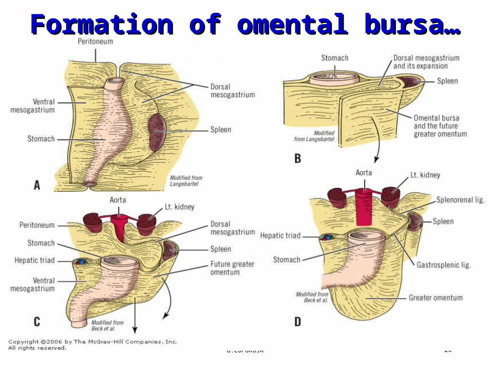
Formation of omental bursa• Since the stomach is attached to the dorsal body wall by the dorsal mesogastrium and to the ventral body wall by the ventral mesogastrium, its rotation and disproportionate growth alter the position of these mesenteries.
• Note: the space behind the stomach called the omental bursa (lesser peritoneal sac)
G.LUFUKUJA 24

G.LUFUKUJA 25
G.LUFUKUJA 26
Formation of omental Formation of omental bursa…bursa…
G.LUFUKUJA 27
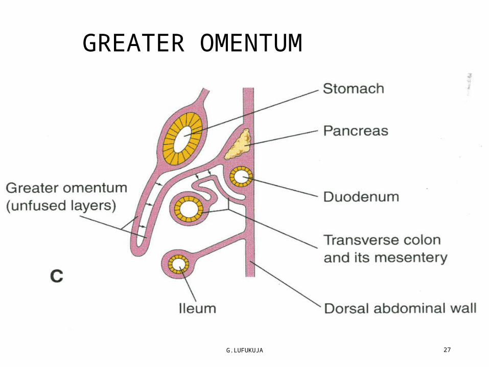
GREATER OMENTUM
G.LUFUKUJA 28

G.LUFUKUJA 29
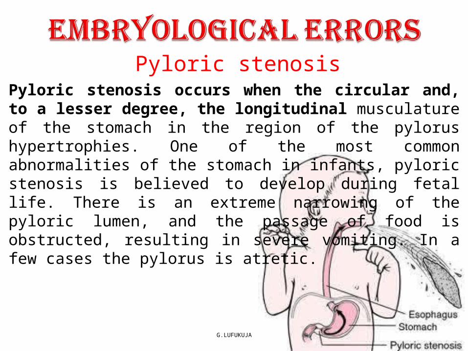
Pyloric stenosis
G.LUFUKUJA 30
Pyloric stenosis occurs when the circular and, to a lesser degree, the longitudinal musculature of the stomach in the region of the pylorus hypertrophies. One of the most common abnormalities of the stomach in infants, pyloric stenosis is believed to develop during fetal life. There is an extreme narrowing of the pyloric lumen, and the passage of food is obstructed, resulting in severe vomiting. In a few cases the pylorus is atretic.
The Duodenum• The terminal part of the foregut and the cephalic part of the midgut form the duodenum, thus receives a dual blood supply from foregut and midgut arteries (celiac and superior mesenteric)
• As the stomach rotates, the duodenum takes on the form of a C-shaped loop and rotates to the right.
• The duodenum is carried to the right (with the liver) with the rotation of the foregut
• Note: The mesentery fuses with the dorsal parietal peritoneum by zygosis, making the duodenum a retroperitoneal structure.
G.LUFUKUJA 31

G.LUFUKUJA 32

Duodenum…• During the second month, the lumen of the duodenum is obliterated by proliferation of cells in its walls.
• However, the lumen is recanalized shortly thereafter
G.LUFUKUJA 33
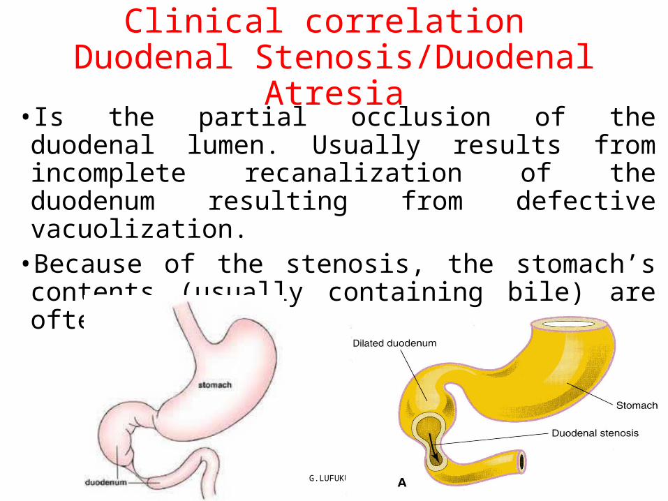
Clinical correlation Duodenal Stenosis/Duodenal Atresia
• Is the partial occlusion of the duodenal lumen. Usually results from incomplete recanalization of the duodenum resulting from defective vacuolization.
•Because of the stenosis, the stomach’s contents (usually containing bile) are often vomited.
G.LUFUKUJA 34

Glands of foregut
G.LUFUKUJA 35

Liver and the biliary system•The liver and the biliary system arise as a common diverticulum, the hepatic diverticulum which grows from the duodenum into the ventral mesogastrium. The diverticulum divides into two branches - the caudal one differentiates into the gall bladder and its (cystic) duct; and the cranial one that forms the rest of the biliary system and the hepatic parenchyma.
G.LUFUKUJA 36

G.LUFUKUJA 37

Liver and Gallbladder…• While hepatic cells continue to penetrate the septum, the
connection between the hepatic diverticulum and the foregut (duodenum) narrows, forming the bile duct.
• A small ventral outgrowth is formed by the bile duct, and this outgrowth gives rise to the gallbladder and the cystic duct.
• Hematopoietic cells, Kupffer cells, and connective tissue cells are derived from mesoderm of the septum transverse mesoderm.
G.LUFUKUJA 38

Liver and Gallbladder…• The latter develop as cords of endodermal cells that invade the vascular mesenchyme of the septum transversum, breaking up the vitelline veins to form a complex network of sinusoids.
• The connective tissue and haematopoietic tissue of the liver is derived from the septum transversum.
• Hematopoietic function.• Large nests of proliferating cells, which produce red and white blood cells, lie between hepatic cells and walls of the vessels. This activity gradually subsides during the last 2 months of intrauterine life, and only small hematopoietic islands remain at birth. The weight of the liver is then only 5%of the total body weight
G.LUFUKUJA 39

Liver and Gallbladder…• Molecular Regulation of Liver Induction• Fibroblast growth factors (FGFs) secreted by cardiac mesoderm.
Thus, the cardiac mesoderm “instructs” gut endoderm to express liver specific genes by inhibiting an inhibitory factor of these same genes.
G.LUFUKUJA 40

Pancreas• The pancreas is formed by two buds originating from the endodermal
lining of the duodenum• Whereas the dorsal pancreatic bud is in the dorsal mesentery, the
ventral pancreatic bud is close to the bile duct When the duodenum rotates to the right and becomes C-shaped, the ventral pancreatic bud moves dorsally in a manner similar to the shifting of the entrance of the bile duct
G.LUFUKUJA 41
•Finally the ventral bud comes to lie immediately below and behind the dorsal bud •Later the parenchyma and theduct systems of the dorsal and ventral pancreatic buds fuse
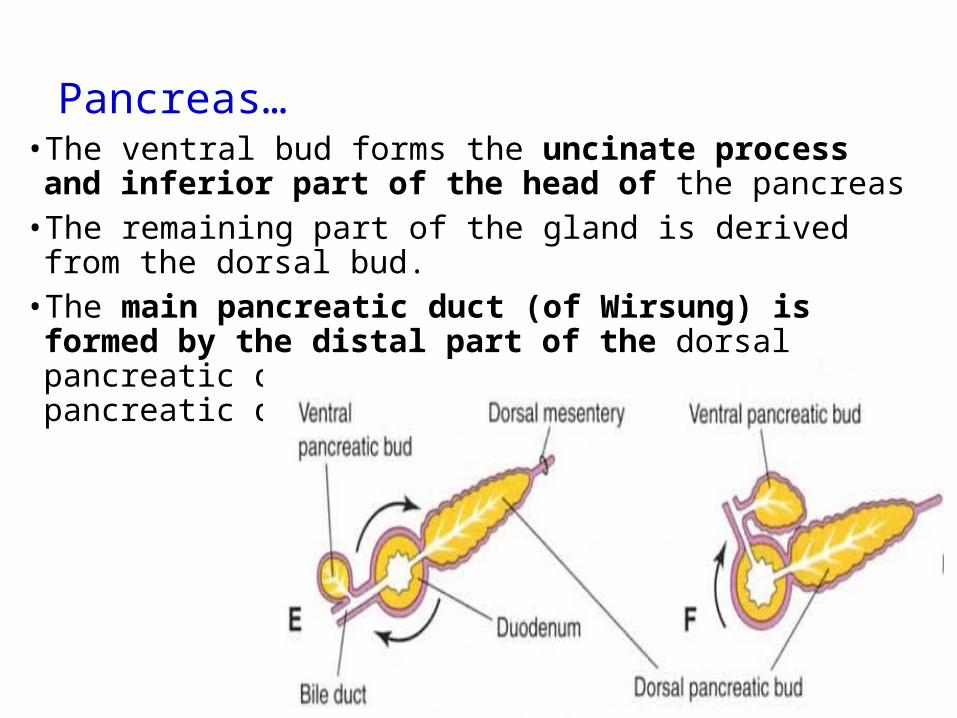
Pancreas…• The ventral bud forms the uncinate process and inferior part of
the head of the pancreas • The remaining part of the gland is derived from the dorsal bud.• The main pancreatic duct (of Wirsung) is formed by the distal
part of the dorsal pancreatic duct and the entire ventral pancreatic duct
G.LUFUKUJA 42

G.LUFUKUJA 43
Pancreas…Pancreas…
Pancreas…• In the third month of fetal life, pancreatic islets (of Langerhans)
develop from the parenchymatous pancreatic tissue and scatter throughout the pancreas.
• Insulin secretion begins at approximately the fifth month. Glucagon- and somatostatin-secreting cells also develop from parenchymal cells.
• Splanchnic mesoderm surrounding the pancreatic buds forms the pancreatic connective tissue
G.LUFUKUJA 44

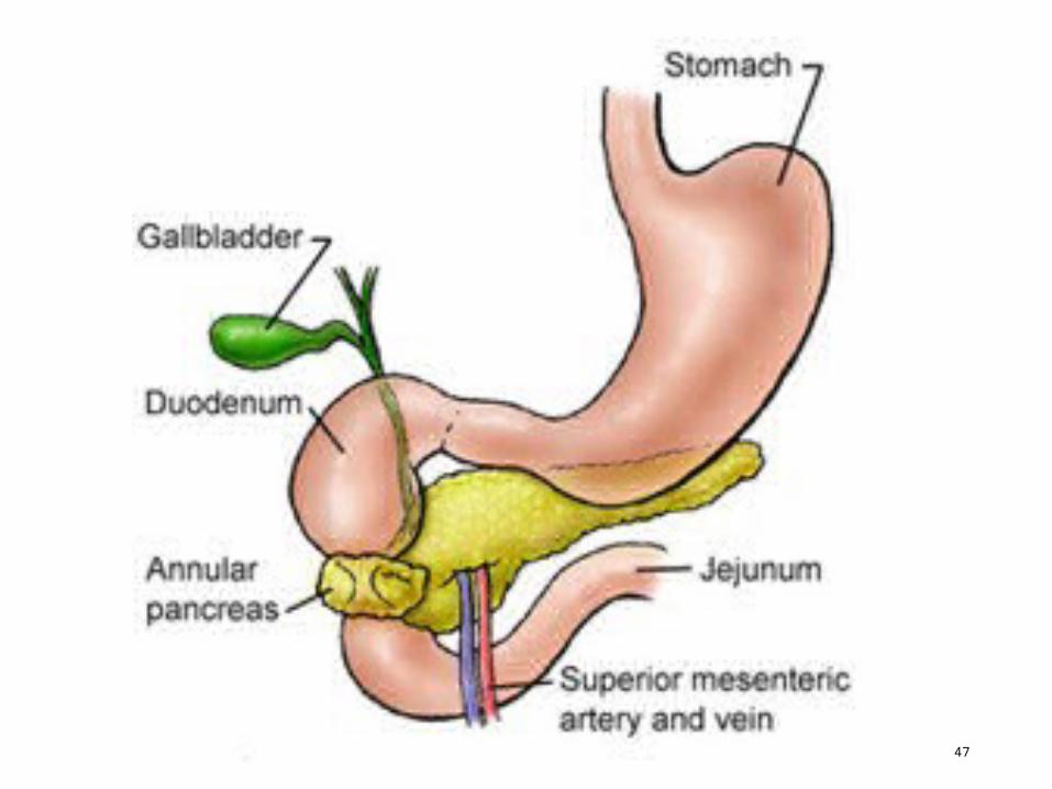
Annular pancreas• The ventral pancreatic bud consists of two components that normally fuse and rotate around the duodenum.
• Occasionally, however, the right portion of the ventral bud migrates along its normal route, but the left migrates in the opposite direction.
• In this manner, the duodenum is surrounded by pancreatic tissue which may produce duodenal obstruction.
G.LUFUKUJA 45

G.LUFUKUJA 46
G.LUFUKUJA 47
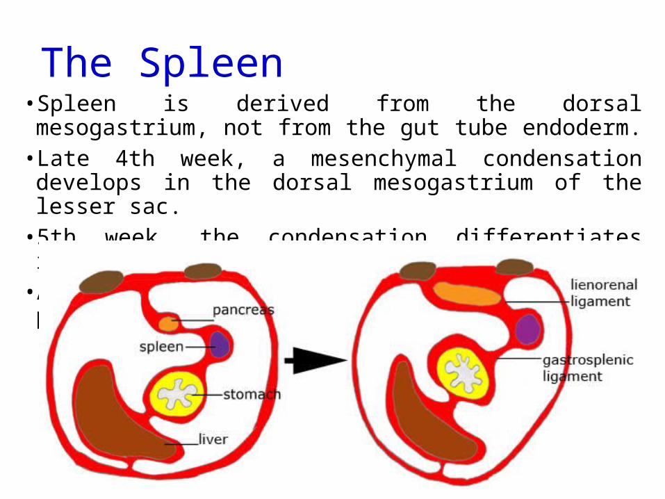
The Spleen• Spleen is derived from the dorsal mesogastrium, not from the gut
tube endoderm. • Late 4th week, a mesenchymal condensation develops in the
dorsal mesogastrium of the lesser sac. • 5th week, the condensation differentiates into the spleen. • Accessory spleens may also develop near the primary spleen.
G.LUFUKUJA 48

Midgut• In the 5-week-old embryo, the midgut is suspended from the dorsal
abdominal wall by a short mesentery and communicates with the yolk sac by way of the vitelline duct or yolk stalk
• Development of the midgut is characterized by rapid elongation of the gut and its mesentery, resulting in formation of the primary intestinal loop. At its apex, the loop remains in open connection with the yolk sac by way of the narrow vitelline duct
G.LUFUKUJA 49
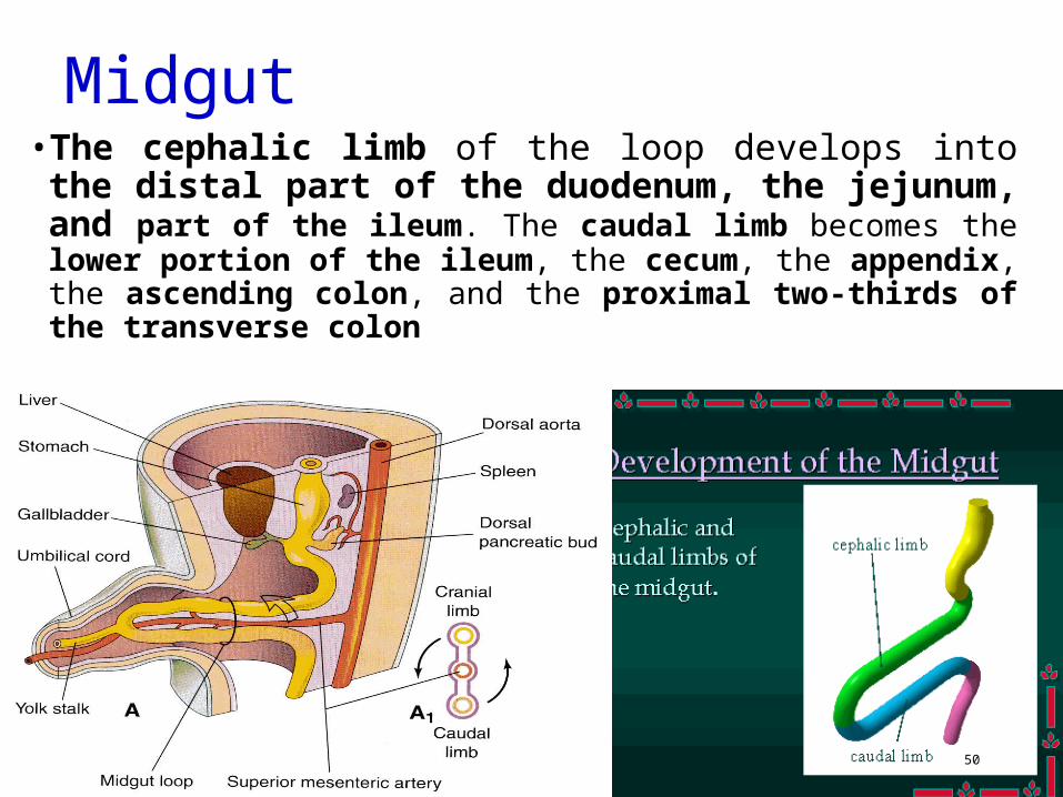
Midgut• The cephalic limb of the loop develops into the distal part of the
duodenum, the jejunum, and part of the ileum. The caudal limb becomes the lower portion of the ileum, the cecum, the appendix, the ascending colon, and the proximal two-thirds of the transverse colon
G.LUFUKUJA 50
Midgut…
G.LUFUKUJA 51
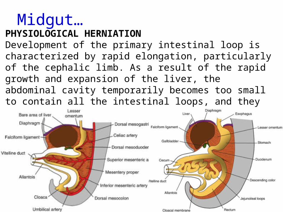
PHYSIOLOGICAL HERNIATIONDevelopment of the primary intestinal loop is characterized by rapid elongation, particularly of the cephalic limb. As a result of the rapid growth and expansion of the liver, the abdominal cavity temporarily becomes too small to contain all the intestinal loops, and they enter the extraembryonic cavity in the umbilical cord during the sixth week of development (physiological umbilical herniation)
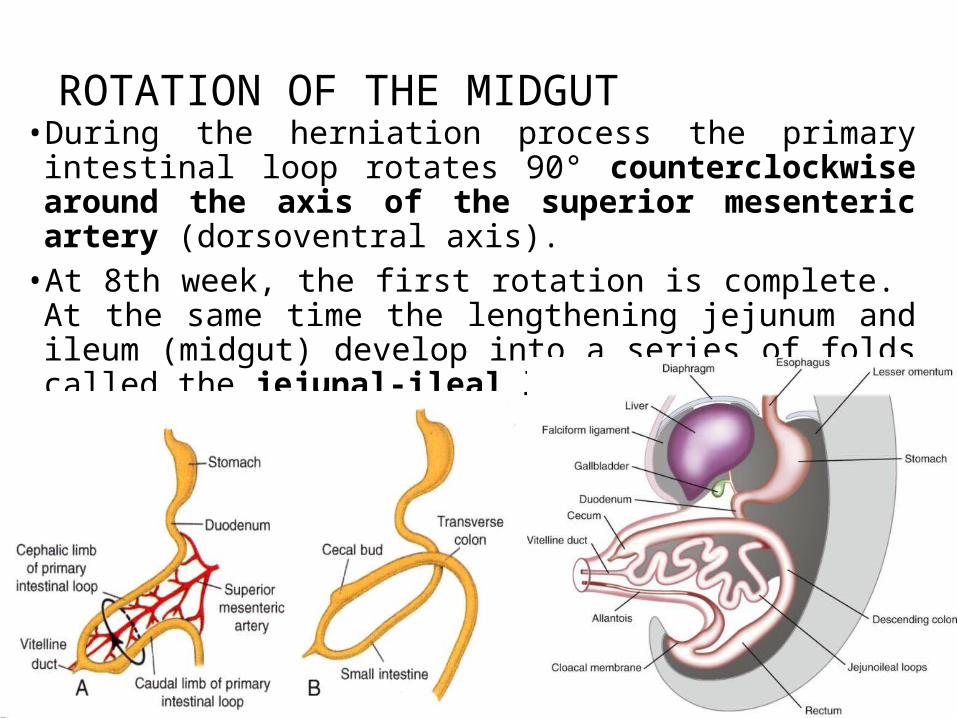
ROTATION OF THE MIDGUT• During the herniation process the primary intestinal loop rotates 90° counterclockwise around the axis of the superior mesenteric artery (dorsoventral axis).
• At 8th week, the first rotation is complete. At the same time the lengthening jejunum and ileum (midgut) develop into a series of folds called the jejunal-ileal loops.
G.LUFUKUJA 52

The midgut returns to the abdominal cavity, rotating further 180° • As the intestinal loop re-enters the abdomen it rotates
an additional 180° counterclockwise. Thus the retracting colon has rotated a total of 270° relative to the posterior wall of the abdominal cavity.
• By 11th week, rotation and retraction are complete. • The cecum is now in a position just inferior to the liver.
G.LUFUKUJA 53

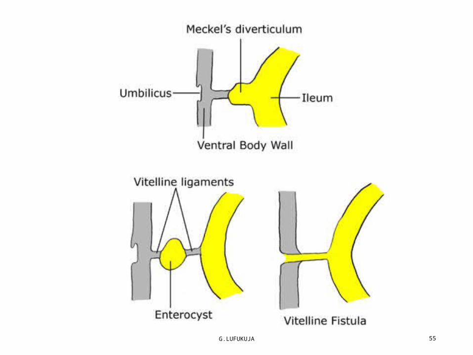
Embryological Errors •Ileal Diverticulum• An ileal diverticulum (of Meckel) is a remnant of the proximal
part of the embryonic yolk stalk, the diverticulum usually appears as a finger-like pouch. It may be free (74%) or attached to the umbilicus (26%). Although its mucosa is mostly ileal in type, it may also include areas of acid-producing gastric tissue, pancreatic tissue, or jejunal or colonic mucosa.
G.LUFUKUJA 54
G.LUFUKUJA 55

Embryological Errors •Reversed Rotation.In very unusual cases, the mid gut loop rotates in a clockwise rather than a
counterclockwise direction.As a result, the duodenum lies anterior to the superior mesenteric artery rather
than posterior to it, and the transverse colon lies posterior instead of anterior to it.
In this infant the transverse colon may be obstructed by pressure from the superior mesenteric artery.
G.LUFUKUJA 56
G.LUFUKUJA 57
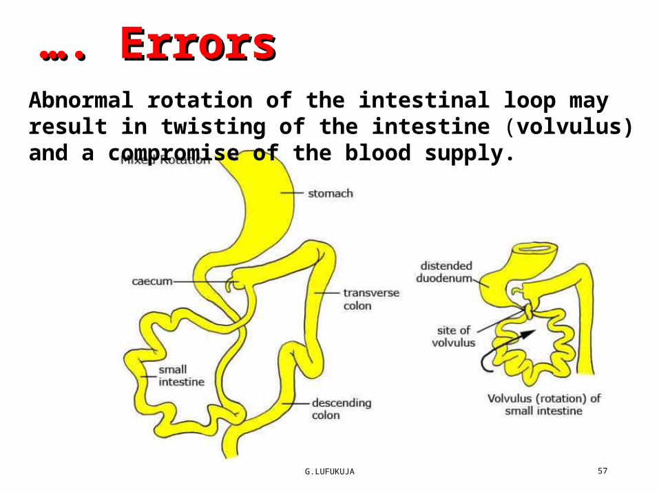
……. Errors . Errors Abnormal rotation of the intestinal loop may result in twisting of the intestine (volvulus) and a compromise of the blood supply.

…. Errors •Subhepatic Cecum and Appendix.• If the cecum adheres to the inferior surface of the liver when it
returns to the abdomen, it will be drawn superiorly as the liver diminishes in size, as a result the cecum remains in its fetal position.
G.LUFUKUJA 58
The subhepatic cecum and appendix may be seen in adults and create a problem in the diagnosis of appendicitis and during the surgical removal of the appendix (Appendectomy).

…. Errors •Omphalocele •Failure of midgut return.• Involves herniation of abdominal viscera through an enlarged umbilical ring.
• The viscera, which may include liver, small and large intestines, stomach, spleen, or gallbladder, are covered by amnion.
G.LUFUKUJA 59

…. Errors •Gastroschisis •Represents a congenital defect characterized by a defect in the anterior abdominal wall through which the abdominal contents freely protrude
G.LUFUKUJA 60
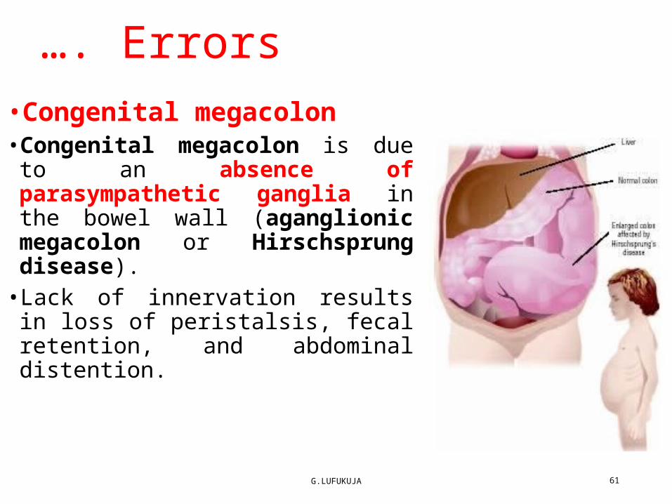
…. Errors •Congenital megacolon• Congenital megacolon is due to an absence of parasympathetic ganglia in the bowel wall (aganglionic megacolon or Hirschsprung disease).
• Lack of innervation results in loss of peristalsis, fecal retention, and abdominal distention.
G.LUFUKUJA 61
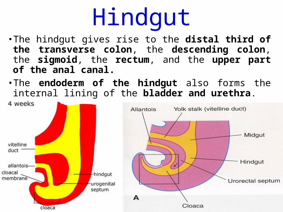
Hindgut• The hindgut gives rise to the distal third of the transverse colon, the descending colon, the sigmoid, the rectum, and the upper part of the anal canal.
• The endoderm of the hindgut also forms the internal lining of the bladder and urethra.
G.LUFUKUJA 62

Hindgut…• The terminal portion of the hindgut enters into the posterior
region of the cloaca or the primitive anorectal canal. • The allantois enters into the anterior portion or the primitive
urogenital sinus.
G.LUFUKUJA 63

G.LUFUKUJA 64

Hindgut…•A layer of mesoderm, the urorectal septum, separates the region between the allantois and hindgut.
G.LUFUKUJA 65
Hindgut…• The urorectal septum septum is derived from the
merging of mesoderm covering the yolk sac and surrounding the allantois.
• As the embryo grows and caudal folding continues, the tip of the urorectal septum comes to lie close to the cloacal membrane.
• The transverse line of fusion between the septum and cloacal membrane forms the perineal body.
G.LUFUKUJA 66

The anal canal
G.LUFUKUJA 67
The anal canal is developed from two sources; Upper part from the endodermal cloaca &
Lower par from the ectodermal proctodeum. The position of the anal membrane is
represented in adult by the pectinate line (Hilton’s white line) of the anal canal (anal valves). At this line, the epithelium changes from columnar in upper anal canal to stratified squamous epithelium in lower anal canal.
Thus, the upper part of the anal canal is supplied by the the superior rectal artery a continuation of the inferior mesenteric artery. The lower part of the anal canal is supplied by inferior rectal arteries, branches of the internal pudendal arteries..

G.LUFUKUJA 68

The anal canal…• The anal membrane is encircled by mesodermal anal hillocks. The anal hillocks fuse and result in the formation an anal pit called proctodaeum, which is covered by skin ectoderm.
G.LUFUKUJA 69

The anal canal…•At the end of the seventh week, the cloacal membrane ruptures, creating the anal opening for the hindgut and a ventral opening for the urogenital sinus.
•Congenital errors • Imperforate anus is a condition that occurs when there is a failure of breakdown of the anal membrane.
G.LUFUKUJA 70

G.LUFUKUJA 71
Imperforate anus

G.LUFUKUJA 72
THANK YOUTHANK YOU