Development of soft nanocarriers from novel … multiphasic physically cross-linked nanogel...
228
General rights Copyright and moral rights for the publications made accessible in the public portal are retained by the authors and/or other copyright owners and it is a condition of accessing publications that users recognise and abide by the legal requirements associated with these rights. • Users may download and print one copy of any publication from the public portal for the purpose of private study or research. • You may not further distribute the material or use it for any profit-making activity or commercial gain • You may freely distribute the URL identifying the publication in the public portal If you believe that this document breaches copyright please contact us providing details, and we will remove access to the work immediately and investigate your claim. Downloaded from orbit.dtu.dk on: Dec 15, 2017 Development of soft nanocarriers from novel amphiphilic hyaluronic acid derivatives towards drug delivery Eenschooten, Corinne Diane; Kontogeorgis, Georgios; Stenby, Erling Halfdan; Longin, Fanny; Schwach- Abdellaoui, Khadija; Delie, Florence; Gurny, Robert Publication date: 2008 Document Version Publisher's PDF, also known as Version of record Link back to DTU Orbit Citation (APA): Eenschooten, C. D., Kontogeorgis, G., Stenby, E. H., Longin, F., Schwach-Abdellaoui, K., Delie, F., & Gurny, R. (2008). Development of soft nanocarriers from novel amphiphilic hyaluronic acid derivatives towards drug delivery.
Transcript of Development of soft nanocarriers from novel … multiphasic physically cross-linked nanogel...

General rights Copyright and moral rights for the publications made accessible in the public portal are retained by the authors and/or other copyright owners and it is a condition of accessing publications that users recognise and abide by the legal requirements associated with these rights.
• Users may download and print one copy of any publication from the public portal for the purpose of private study or research. • You may not further distribute the material or use it for any profit-making activity or commercial gain • You may freely distribute the URL identifying the publication in the public portal
If you believe that this document breaches copyright please contact us providing details, and we will remove access to the work immediately and investigate your claim.
Downloaded from orbit.dtu.dk on: Dec 15, 2017
Development of soft nanocarriers from novel amphiphilic hyaluronic acid derivativestowards drug delivery
Eenschooten, Corinne Diane; Kontogeorgis, Georgios; Stenby, Erling Halfdan; Longin, Fanny; Schwach-Abdellaoui, Khadija; Delie, Florence; Gurny, Robert
Publication date:2008
Document VersionPublisher's PDF, also known as Version of record
Link back to DTU Orbit
Citation (APA):Eenschooten, C. D., Kontogeorgis, G., Stenby, E. H., Longin, F., Schwach-Abdellaoui, K., Delie, F., & Gurny, R.(2008). Development of soft nanocarriers from novel amphiphilic hyaluronic acid derivatives towards drugdelivery.
Department of Chemical and Biochemical EngineeringDTU Building 229Søltofts PladsDK-2800 Kgs. Lyngbywww.kt.dtu.dk
Development of soft nanocarriers from novel amphiphilic hyaluronic acid derivatives towards drug delivery
Corinne Eenschooten2008
C. E
enschootenD
evelopment of soft nanocarriers from
novel amphiphilic hyaluronic acid derivatives tow
ards drug delivery2008

Development of soft nanocarriers from novel amphiphilic hyaluronic acid derivatives towards drug delivery
Corinne Eenschooten2008
1

Copyright © Corinne Eenschooten
2008 ISBN-13: 978-87-91435-84-6
Printed by J&R Frydenberg A/S, Copenhagen, Denmark
2

Development of soft nanocarriers from novel amphiphilic hyaluronic acid derivatives towards drug delivery
Corinne Eenschooten
3

iv
English abstract
Drug delivery is an important pharmaceutical research area currently facing
limitations such as drug insolubility and instability in biological media, poor
bioavailability and unspecific targeting. As a consequence, high doses are used to
achieve a therapeutic effect which increases the risk for toxicity in patients and health
costs. The design of advanced drug delivery systems addressing these challenges has
therefore become more and more crucial. Hyaluronic acid (HA) is a natural linear
glycosaminoglycan ubiquitous and identical in all mammals. Due to its native intrinsic
biocompatibility, resorbability and biological functions, and the possibility for an easy
chemical functionalisation, HA constitutes an excellent starting material for the design
of advanced biomaterials. The objective of this project was to develop HA-based
nanocarriers towards the encapsulation and delivery of hydrophobic drugs.
However, due to its high hydrophilicity, HA exhibits physicochemical
properties incompatible with the spontaneous and stable formation of segregated
structures in aqueous solutions. Therefore, HA was first rendered amphiphilic by the
covalent binding of hydrophobic groups onto its backbone. After a preliminary
investigation of two different modification methods, the proprietary technology
eventually selected was based on the grafting of octenyl succinic anhydride and was
optimised so as to develop a reproducible and robust process for the synthesis of
functionalised polymers. Fundamental properties such as the structure, degree of
hydrophobicity, substitution pattern as well as intra- and intermolecular interactions of
a selection of derivatives were then studied. Submicronic multiphasic physically cross-
linked nanogel particles capable of solubilising a model hydrophobic substance were
prepared by spontaneous self-association and characterized with regard to their
morphology, size and surface charge. A multivesicular model was finally put forward
to explain their internal structure.
In conclusion, a molecular method based on controlled self-association was
developed towards the formation of HA soft nanostructures. These represent novel and
promising biomaterial templates for the fabrication of stable and multifunctional HA-
based nanogel particles and could be valuable to the industry since their underlying
production methods are aqueous and easily upscalable.
4

v
Dansk abstrakt
Fordeling af lægemidler er et vigtigt farmaceutisk forskningsområde, der i
øjeblikket står overfor begrænsninger som lægemidlets uopløselighed og instabilitet i
biologiske miljøer, ringe tilgængelighed og uspecifik levering. Som en konsekvens
heraf, anvendes høje doser for at opnå en terapeutisk virkning, hvilket forøger risikoen
for toksicitet i patienter og sundhedsudgifter. Design af avancerede systemer til
fordeling af lægemidler, der tager udgangspunkt i disse udfordringer, er derfor blevet
mere og mere afgørende. Hyaluronsyre (HA) er en naturlig lineær glycosaminoglycan,
der er tilstede og identisk i alle pattedyr. HA udgør et fortrinligt udgangsstof i designet
af avancerede biomaterialer på grund af dets naturlige biokompatibilitet, resorption,
biologiske funktioner og muligheden for simpel kemisk funktionalisering. Formålet for
projektet var at udvikle HA-baserede nanobærere til indkapsling og levering af
hydrofobiske stoffer.
HA er meget hydrofilt og besidder fysisk-kemiske egenskaber, der er
uforenelige med en spontan og stabil dannelse af isolerede strukturer i vandige
opløsninger. HA blev derfor først gjort amfifilt ved kovalent binding af hydrofobiske
grupper på dets rygrad. Efter en forberedende undersøgelse af to forskellige
derivatiserings-metoder, den endelige valgte patenterede teknologi var baseret på
bindingen af octenyl succinic anhydride og blev optimeret for at udvikle en
reproducerbar og robust proces til syntese af funktionaliserede polymerer.
Grundlæggende egenskaber som struktur, grad af hydrofobicitet, substitutionsmønster
samt intra- og intermolekulær vekselvirkning blev herefter undersøgt for et udvalg af
derivater. Submikroniske multifasiske fysisk krydsforbundne nanogelpartikler, der kan
opløse et hydrofobiskt modelstof, blev dannet ved spontan selvsamling og
karakteriseret ud fra deres morfologi, størrelse og overfladeladning. En multivesikulær
model blev til sidst opstillet for at forklare deres indre struktur.
Afslutningsvist blev der udviklet en molekylær metode til dannelsen af bløde
HA nanostrukturer baseret på kontrolleret selvsamling. Disse repræsenterer nye og
lovende biomaterialeskabeloner til fremstilling af stabile og multifunktionelle HA-
baserede nanogelpartikler og kunne være værdifulde for industrien, eftersom deres
produktionsmetoder er vandige og let opskalerbare.
5

vi
Preface
The present manuscript is a summary of the work I, Corinne Eenschooten,
carried out in the period May 2005-July 2008 towards the achievement of the
Industrial Philosophy Doctorate (Industrial Ph.D.) degree. This double diploma is
granted by Danish universities and the Danish Ministry of Science, Technology and
Innovation (VTU) to Ph.D. students having undertaken their research project in the
industry. The Danish industrial Ph.D. programme has indeed for purpose to educate
scientists with an insight in the commercial aspects of Research and Development
(R&D), increase the R&D and innovative capacity in private companies and build
networks disseminating knowledge between universities and private companies.
This project was a collaboration between the Technical University of
Denmark (DTU), Department of Chemical and Biochemical Engineering, Center for
Phase Equilibria and Separation Processes (IVC-SEP, Building 229, 2800 Lyngby,
Denmark), Novozymes Biopolymer A/S (NZBP, Krogshøjvej 36, 2880 Bagsværd,
Denmark) and the University of Geneva, University of Lausanne (UGL), School of
Pharmaceutical Sciences, Department of Pharmaceutics and Biopharmaceutics (DPBP,
30 quai Ernest-Ansermet, 1211 Geneva 4, Switzerland) which acted as a third party.
Most of the work was achieved at IVC-SEP and NZBP. The rest was conducted at
DPBP within the frame of two study trips in the periods July-August 2006 and March-
June 2007.
The project was co-funded by the Danish Ministry of Science, Technology and
Innovation (Grant number: 07-001687) and Novozymes Biopolymer A/S, the
employer.
The project supervisors were, from IVC-SEP, Doctor Georgios M.
Kontogeorgis and IVC-SEP director Professor Erling H. Stenby, from NZBP, Doctors
Fanny Longin and Khadija Schwach-Abdellaoui, Science Manager and Senior R&D
Manager, respectively and, from DPBP, Doctor Florence Delie and President of the
School of Pharmaceutical Sciences (UGL) Professor Robert Gurny.
6

vii
The overall purpose of the project was to design and implement a method for
the preparation of hyaluronic acid-based nanocarriers towards drug delivery.
The dissertation consists of five chapters. A general introduction (Chapter I) opens the
thesis and is followed by a review on a subject related to the project area (Chapter II).
The experimental work is described in the following two chapters (Chapter III and
Chapter IV). The thesis continues with a conclusion (Chapter V) which summarises
the work, puts it into perspectives and suggests future research directions. The
manuscript ends with appendices containing a record of the educational activities
completed during the Ph.D. programme.
Chapter II, Chapter III, B and Chapter IV are written in a recognisable paper
format and will after a few adjustments be submitted to scientific journals by the time
of the Ph.D. defence or immediately after.
The experimental work presented in Chapter III, sections A and B was entirely
performed by me while that of section C was partly accomplished by Sara Sparre
Kofoed. Sara was a master student at the Technical University of Denmark,
Department of Chemistry (DC) who carried out her master’s thesis in collaboration
with NZBP in the period September 2007-February 2008 under Associate Professor
Charlotte Held Gotfredsen’s and my own supervision. In Chapter IV, Andrea Vaccaro
(UGL, Department of Inorganic Analytical and Applied Chemistry, DIAAC)
completed the light scattering experiments and participated to the simulation work.
The compilation and writing of all the material in this thesis is the result of my own
efforts.
During my education, I had the opportunity to evolve in a multidisciplinary
environment and to become acquainted with exceptionally knowledgeable and
inspiring people whom I would like to express my gratitude to.
I address my first acknowledgements to my supervisors Dr. Georgios M.
Kontogeorgis, Prof. Erling H. Stenby, Dr. Fanny Longin, Dr. Khadija Schwach-
Abdellaoui, Dr. Florence Delie and Prof. Robert Gurny.
I wish to thank Dr. Georgios M. Kontogeorgis and Prof. Erling H. Stenby (IVC-SEP)
for their support and encouragements at an earlier stage in the process of finding a
7

viii
Ph.D. project and an industrial partner, their following-up on the course of my studies
and active participation to our scientific discussions.
I address Dr. Georgios M. Kontogeorgis (IVC-SEP) my greatest recognition for having
given me a durable taste for colloid and surface chemistry and entrusted me with the
mission to teach this topic twice by his side.
To Dr. Fanny Longin (NZBP), I express all my thankfulness and acknowledge her for
her large availability and dynamism. I have learned a lot from her about
polysaccharide chemistry and modification and surely, these years would have had
another flavour without her optimism and positivism.
My gratitude also goes to Dr. Khadija Schwach-Abdellaoui (NZBP) who gave me the
fantastic opportunity to work in an emerging and fascinating area of research and who
benevolently saw to my educational and personal development.
I owe a lot to Dr. Florence Delie for kindly accompanying me during my external stay
at DPBP, contributing to make my study trips fruitful and enriching experiences and
sharing her expertise about polymeric nanoparticulate systems.
Prof. Robert Gurny who generously welcomed me at DPBP and recommended me to
various disciplinary experts to help me come forward with my project also deserves
strong acknowledgements.
I am grateful to my present and former colleagues at DTU, NZBP and UGL
for their contribution to a productive and pleasant working environment.
I particularly wish to thank Dr. Kristoffer Tømmeraas and Dr. Birgitte Mølholm Malle
(NZBP) for sharing their knowledge in polysaccharide chemistry, modification and
industrial applications, Sara Sparre Kofoed (DC) for letting me experience my
fledgling teaching skills and for the quality of the work she performed, Dr. Charlotte
Held Gotfredsen (DC) for co-supervising Sara’s project and having Sara and myself
benefited from her expertise in nuclear magnetic resonance spectroscopy (NMR) and
both of them for pursuing NMR experiments even after the term of Sara’s project.
I am obliged to Dr. Christoph Bauer (UGL, National Centre of Competence in
Research, Frontiers in Genetics, Bioimaging Platform) for having acquired the first
transmission electron microscopy (TEM) micrographs and trained me in TEM with
great patience.
8

ix
Dr. Andrea Vaccaro and Prof. Michal Borkovec (DIAAC) who shared their insight in
colloid chemistry and made ingenious propositions to explain aggregation phenomena
have also earned my infinite credit.
My gratitude finally goes to the administrative staff at IVC-SEP (Anne Louise Biede
and Patricia Wagner), NZBP (Nadia Ehlers Nielsen), Novozymes A/S (Asta Wielandt
Pedersen) and DPBP (Myrtha Copin and Florence von Ow) for their help in numerous
occasions and to Patent Attorney Peter Würtz Lindum (Novozymes A/S) for carrying
through the patent applications and mediating my disclosure of information.
I am especially appreciative of VTU and DTU for their constantly evolving
and improving educational structures and their following-up on industrial Ph.D.
students. I also salute the quality of the complementary learning and networking
activities organised by VTU along the industrial Ph.D. programme.
My kindest thoughts are directed to my family, my mother and father
(Martine Aubert and Richard Eenschooten) and my friends in France (Thibault
Roques, Éric Piel and Laurène Champalle) and in Denmark (Sylvain Verdier, Mette
Fuglsang Larsen and Michelangelo Dall’Ora) who always supported and encouraged
me.
I finally gratefully acknowledge the Danish Ministry of Science, Technology
and Innovation (Grant number: 07-001687) and Novozymes Biopolymer A/S for
funding.
Corinne Eenschooten
Lyngby, July 2008
9
x
Table of content
English abstract ..............................................................................................................iv
Dansk abstrakt .................................................................................................................v
Preface ............................................................................................................................vi
Table of content...............................................................................................................x
List of abbreviations.....................................................................................................xvi
List of figures ..............................................................................................................xxii
List of tables ...............................................................................................................xxiv
List of equations ..........................................................................................................xxv
CHAPTER I
Introduction .....................................................................................................................1
I. Hyaluronic acid .....................................................................................................3
I.1. Origin and molecular structure .......................................................................3
I.2. Industrial manufacture ....................................................................................4
I.3. Structures in aqueous solutions.......................................................................5
I.4. Biological functions in the skin ......................................................................9
I.5. Applications ..................................................................................................13
II. Hyaluronic acid-mediated dermal drug delivery ...............................................14
II.1. First generation systems ..............................................................................15
II.2. Second generation systems ..........................................................................15
III. Hyaluronic acid-based colloidal particles.........................................................16
III.1. Microparticles.............................................................................................16
III.2. Nanoparticles..............................................................................................18
IV. Hydrophobically modified hyaluronic acid......................................................23
V. The Ph.D. project ...............................................................................................25
V.1. Objectives and challenges ...........................................................................25
V.2. Experimental approach................................................................................27
V.3. Thesis structure............................................................................................28
References...............................................................................................................29
10

xi
CHAPTER II
Review .......................................................................................................................... 37
Towards an understanding of the mechanisms underlying polymeric nanoparticle-
mediated (trans)dermal drug delivery........................................................................ 39
Abstract...................................................................................................................... 39
I. Introduction......................................................................................................... 40
II. Technical advances in assessing skin penetration pathways ............................. 44
II.1. Skin penetration pathways .......................................................................... 44
II.2. Microscopic imaging................................................................................... 47
II.2.1. Confocal laser scanning microscopy .................................................... 47
II.2.2. Multiphoton laser scanning microscopy ............................................... 48
II.3. Skin penetration quantification ................................................................... 49
II.3.1. The skin sandwich model ..................................................................... 49
II.3.2. Differential stripping............................................................................. 52
III. Mechanisms underlying polymeric nanoparticle-mediated drug delivery....... 53
III.1. Redistribution and accumulation of polymeric nanoparticles in the skin.. 53
III.2. Possible mechanisms underlying polymeric nanoparticle skin penetration
............................................................................................................................ 56
III.2.1. Influence of nanoparticle size.............................................................. 58
III.2.2. Influence of nanoparticle surface charge............................................. 59
III.2.3. Influence of nanoparticle shape........................................................... 60
III.3. Hypothesis with regards to how polymeric nanoparticles enhance
percutaneous penetration and/or sustain drug release......................................... 61
IV. Future challenges in polymeric nanoparticle-mediated (trans)dermal drug
delivery................................................................................................................... 63
IV.1. Nanoparticle design ................................................................................... 63
IV.2. Nanoparticle performance assessment....................................................... 64
IV.2.1. Fate ...................................................................................................... 64
IV.2.2. Skin penetration................................................................................... 65
IV.3. Nanoparticle risk assessment..................................................................... 66
V. Conclusion......................................................................................................... 68
References .............................................................................................................. 69
11

xii
CHAPTER III
Preparation and structural characterisation of amphiphilic hyaluronic acid derivatives
.......................................................................................................................................79
A. Investigation of novel modification methods for the preparation of amphiphilic
hyaluronic acid derivatives ........................................................................................81
Abstract ......................................................................................................................81
I. Introduction .........................................................................................................81
II. Materials and methods .......................................................................................83
II.1. Materials ......................................................................................................83
II.2. Methods .......................................................................................................83
II.2.1. Preparation of low molecular weight hyaluronic acid...........................83
II.2.2. Preparation of aryl/alkyl vinyl sulfone-modified hyaluronic acid ........85
II.2.3. Preparation of alkenyl succinic anhydride-modified hyaluronic acid...87
II.2.4. Characterisation techniques...................................................................90
III. Results and discussion ......................................................................................92
III.1. Characterisation of aryl/alkyl vinyl sulfone-modified hyaluronic acid......92
III.2. Characterisation of alkenyl succinic anhydride-modified hyaluronic acid 99
III.3. Comparison between the two preparation methods .................................103
IV. Conclusion......................................................................................................106
References.............................................................................................................107
B. Optimisation of the preparation of octenyl succinic anhydride-modified
hyaluronic acid.........................................................................................................109
Foreword...............................................................................................................109
References.............................................................................................................113
Preparation and structural characterisation of novel and versatile amphiphilic
octenyl succinic anhydride-modified hyaluronic acid..........................................114
Abstract.................................................................................................................114
I. Introduction .......................................................................................................115
II. Materials and methods .....................................................................................117
II.1. Materials ....................................................................................................117
II.2. Methods .....................................................................................................117
12

xiii
II.2.1. Preparation of low molecular weight hyaluronic acid ........................ 117
II.2.2. Preparation of octenyl succinic anhydride-modified hyaluronic acid 118
II.2.3. Characterisation methods.................................................................... 119
III. Results and discussion.................................................................................... 119
III.1. Characterisation of octenyl succinic anhydride-modified hyaluronic acid
.......................................................................................................................... 119
III.2. Influence of the reaction conditions on the degree of substitution of octenyl
succinic anhydride-modified hyaluronic acid................................................... 123
IV. Conclusion ..................................................................................................... 128
References ............................................................................................................ 129
C. Additional characterisation of octenyl succinic anhydride-modified hyaluronic
acid – Substitution pattern, molecular weight and conformation in aqueous media132
Abstract ................................................................................................................ 132
I. Introduction....................................................................................................... 132
II. Materials and methods..................................................................................... 134
II.1. Materials.................................................................................................... 134
II.2. Methods..................................................................................................... 135
III. Results and discussion.................................................................................... 136
III.1. Structural study ........................................................................................ 136
III.1.1. Molecular level .................................................................................. 137
III.1.2. Macromolecular level ........................................................................ 142
III.2. Molecular weight and conformation study .............................................. 144
IV. Conclusion ..................................................................................................... 149
References ............................................................................................................ 150
CHAPTER IV
Physicochemical characterisation of amphiphilic hyaluronic acid derivatives .......... 153
Novel self-associative multi-phase nanostructured soft carriers based on amphiphilic
hyaluronic acid derivatives...................................................................................... 155
Abstract ................................................................................................................ 155
I. Introduction....................................................................................................... 156
II. Materials and methods..................................................................................... 157
13

xiv
II.1. Materials ....................................................................................................157
II.2. Methods .....................................................................................................158
II.2.1. Critical aggregation concentration of the OSA-HA derivatives .........158
II.2.2. Morphology of the OSA-HA polymeric micelles ...............................159
II.2.3. Size distribution of the OSA-HA polymeric micelles.........................159
II.2.4. Zeta potential of the OSA-HA polymeric micelles .............................161
III. Results and discussion ....................................................................................161
III.1. Critical aggregation concentration of the OSA-HA derivatives ..............161
III.2. Morphology of the OSA-HA polymeric micelles ....................................165
III.3. Size distribution of the OSA-HA polymeric micelles..............................166
III.4. Zeta potential of the OSA-HA polymeric micelles ..................................171
III.5. Molecular structure of the OSA-HA polymeric micelles.........................171
IV. Conclusion......................................................................................................176
References.............................................................................................................177
CHAPTER V
Conclusion and future work ........................................................................................179
I. Summary ...........................................................................................................181
II. Future work ......................................................................................................182
Appendices ..................................................................................................................185
Educational activities ...............................................................................................187
I. Training .............................................................................................................187
I.1. Courses........................................................................................................187
I.2. Conferences.................................................................................................188
I.3. External study trips .....................................................................................188
II. Dissemination of knowledge............................................................................189
II.1. Publications................................................................................................189
II.1.1. Posters .................................................................................................189
II.1.2. Oral presentations................................................................................190
II.1.3. Manuscript contributions.....................................................................191
II.1.4. Patents .................................................................................................192
II.1.5. Articles in preparation.........................................................................192
14

xv
II.2. Teaching .................................................................................................... 192
II.2.1. Assistantship in master’s courses ....................................................... 192
II.2.2. Co-supervision .................................................................................... 193
15

xvi
List of abbreviations
ADH Adipic acid dihydrazide
AlkS Alkenyl succinate
AlkSA Alkenyl succinic anhydride
ar Aromatic
Ar/AlVS Aryl/alkyl vinyl sulfone
Ar/AlVS-HA Aryl/alkyl vinyl sulfone-modified hyaluronic acid
Arlacel® A Mannide monostearate
as Asymmetric
A2 Osmotic second virial coefficient
B Bacillus
bd Bending
BS Bacillus Subtilis
c Polymer concentration
CA Carboxylic acid
CAC Critical aggregation concentration
CLSM Confocal laser scanning microscopy
C(X) Concentration of the entity X
d Doublet
DC Department of Chemistry
DDSA Dodecyl succinic anhydride
dH Hydrodynamic diameter
di Diameter of the micelle class i
DIAAC Department of Inorganic Analytical and Applied Chemistry
DLS Dynamic light scattering
DMF N,N-dimethylformamide
DMSO Dimethyl sulfoxide
DPBP Department of Pharmaceutics and Biopharmaceutics
DQF-COSY Double quantum filtered–correlation spectroscopy
DS Degree of substitution
DTU Technical University of Denmark
DVS Divinyl sulfone
16

xvii
D2O Deuterated water
ECM Extracellular matrix
EDC 1-ethyl-3-[3-dimethyl amino] propyl carbodiimide
EVS Ethyl vinyl sulfone
EVS-HA Ethyl vinyl sulfone-modified hyaluronic acid
fi Probability density of the micelle class i
FITC Fluorescein 5-isothiocyanate
f–log-normal log-normal distribution
f–Schulz Schulz distribution
FT-IR Fourier transform infrared spectroscopy
G Glucuronic acid
GAG Glycosaminoglycan
gHMBC Gradient heteronuclear multiple bond correlation
gHSQC Gradient heteronuclear single quantum correlation
HA Hyaluronic acid
HA-C12 Dodecyl bromide-modified hyaluronic acid
HA-C18 Octadecyl bromide-modified hyaluronic acid
HAu Hyaluronic acid unit
HAX ~X,000-Da hyaluronic acid
HLB Hydrophilic/lipophilic balance
hmHA Hydrophobically modified hyaluronic acid
HPAEC High performance anion-exchange chromatography
HPLC High performance liquid chromatography
HYAFF® Fidia’s trademark for hydrophobically modified HA obtained
by reacting HA and alkyliodides in dimethyl sulfoxide, using
the tetrabutylammonium salt of hyaluronic acid
H2BC Heteronuclear 2-bond correlation
H3PO4 Phosphoric acid
IVC-SEP Center for Phase Equilibria and Separation Processes
K Constant defined in Chapter III, C, Equation III.C.2
KBr Potassium bromide
KCl Potassium chloride
KH2PO4 Potassium dihydrogen phosphate
17

xviii
LPGC Low pressure gas chromatography
LS Light scattering
LSM Laser scanning microscopy
l(X) Length of the entity X
-m Methyl
m Multiplet
MALDI-TOF-MS Matrix-assisted laser desorption/ionisation–time-of-flight–
mass spectrometry
MALLS Multi-angle laser light scattering
MHKS Mark-Houwink-Kunh-Sakurada
MP Microparticle
MPLSM Multiphoton laser scanning microscopy
MR Molar ratio
MV Model validity
MW Molecular weight
MWCO Molecular weight cut-off
MWW Weight-averaged molecular weight
MW(X) Molecular weight of the entity X
n Refractive index of a given polymer solution
N Acetylglucosamine
NA Avogadro’s number
NaCl Sodium chloride
NaHCO3 Sodium bicarbonate
NaOAc Sodium acetate
NaOH Sodium hydroxide
Na2HPO4 Disodium hydrogen phosphate
NHSS Sodium N-hydroxysulfosuccinimide
NMR Nuclear magnetic resonance spectroscopy
NOESY Nuclear Overhauser enhancement spectroscopy
NP Nanoparticle
N(X) Number of the entity X
NZBP Novozymes Biopolymer A/S
n0 Refractive index of a given solution medium
18

xix
ODH Oxalic acid dihydrazide
ODSA Octadecenyl succinic anhydride
ODSA-HA Octadecenyl succinic anhydride-modified hyaluronic acid
ODSA-HA30 (A) Octadecenyl succinic anhydride-modified hyaluronic acid
prepared from 30,000 Da hyaluronic acid with the
combination of molar equivalents A
oop Out-of-plane
OS Octenyl succinate
OSA Octenyl succinic anhydride
OSA-HA Octenyl succinic anhydride-modified hyaluronic acid
OSA-HAu Octenyl succinic anhydride-modified hyaluronic acid unit
OSA-HAX Octenyl succinic anhydride-modified hyaluronic acid with a
degree of substitution equal to X % per disaccharide unit
OSA-HA30 (A) Octenyl succinic anhydride-modified hyaluronic acid prepared
from 30,000 Da hyaluronic acid with the combination of
molar equivalents A
o/w Oil-in-water
P Function defined in Chapter III, C, Equation III.C.3
PBS Phosphate buffer saline
PEG Poly(ethylene glycol)
PG Proteoglycan
PH Polyhydrazide
P-HA20 Palmitoyl chloride-modified hyaluronic acid prepared from
20,000-Da hyaluronic acid
P-HA2000 Palmitoyl chloride-modified hyaluronic acid prepared from
2,000,000-Da hyaluronic acid
Ph.D. Philosophy doctorate
PLGA poly(lactide-co-glycolide)
PLS Partial least square
PVS Phenyl vinyl sulfone
PVS-HA Phenyl vinyl sulfone-modified hyaluronic acid
PVS-HAX (A) Phenyl vinyl sulfone prepared from X,000-Da hyaluronic acid
with the combination of molar equivalents A
19
xx
q Modulus of the scattering vector
Q2 Prediction coefficient
R Excess Rayleigh ratio
R&D Research and development
RC Reproducibility coefficient
RE Reaction efficiency
RGD Rayleigh-Gans-Debye
RH Hydrodynamic radius
RHAMM Receptor for HA-mediated mobility
ROS Reactive oxygen species
Rrms Root mean square radius
R(X) Radius of the entity X
R2 Regression coefficient
s Singlet
S Streptococcus
SC Stratum corneum
SEC-MALLS Size exclusion chromatography–multi-angle laser light
scattering
Span 80 Sorbitan monostearate
Span 85 Sorbitan tristearate
st Stretch
sy Symmetric
t Triplet (in nuclear magnetic resonance spectroscopy) or time
T Temperature
TEM Transmission electron microscopy
THF Tetrahydrofurane
TLC Thin layer chromatography
TOCSY Total correlation spectroscopy
u Function of a variable
UGL University of Geneva, University of Lausanne
US United States
UV Ultraviolet
VTU Danish Ministry of Science, Technology and Innovation
20

xxi
w Wavelength
w/o Water-in-oil
WR Weight ratio
w(X) Width of the entity X
1D One-dimension 1H NMR Proton nuclear magnetic resonance spectroscopy
3D Three-dimension
4-HA Hyaluronic acid tetrasaccharide
6-HA Hyaluronic acid hexasaccharide
8-HA Hyaluronic acid octasaccharide
10-HA Hyaluronic acid decasaccharide
12-HA Hyaluronic acid dodecasaccharide
14-HA Hyaluronic acid tetradecasaccharide
� Swelling coefficient
� Chemical shift
[�] Intrinsic viscosity
�0 Wavelength of the laser light in vacuum
� Scattering angle
� Non-dimensional fitting parameter representing the mean
polymeric micelle diameter
�(X) Density of the entity X
Non-dimensional fitting parameter representing the standard
deviation of the polymeric micelle diameter
§ Paragraph
21

xxii
List of figures
Figure I.1: Molecular structure of the HA repeating unit................................................3
Figure I.2: Intramolecular hydrogen bonding and hydrogen bonding with the solvent in
aqueous HA solutions ....................................................................................6
Figure I.3: Plan (1) and elevation (2) computer drawn projections of HA and view
along the two-fold helix axis (3)....................................................................7
Figure I.4: The hydrophilic and non-polar/hydrophobic patches in HA.........................7
Figure I.5: Illustration of the viscoelastic properties of HA............................................8
Figure I.6: Distribution of HA in adult human skin ......................................................10
Figure I.7: Scheme of HA matrices in aqueous solutions .............................................11
Figure I.8: Scheme of the organisation of proteoglycans and collagen in HA matrices
.......................................................................................................................................11
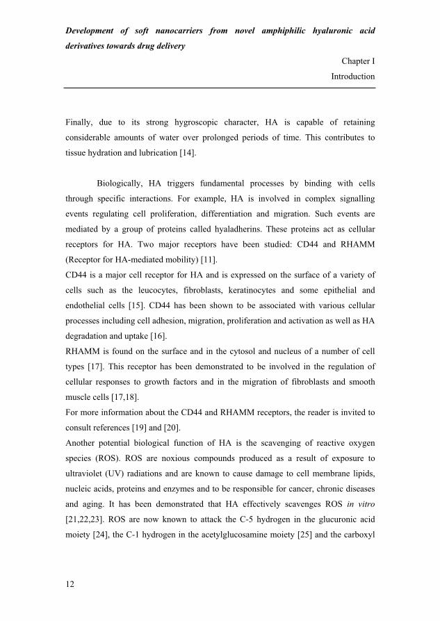
Figure I.9: Illustration of selected hydrophobic modifications of HA ..........................24
Figure II.1: Drug encapsulation in polymeric nanoparticles.........................................43
Figure II.2: Structure of mammalian skin and possible skin penetration pathways......45
Figure II.3: Penetration through the intercellular space between corneocytes..............45
Figure II.4: Schematic representation of the skin sandwich model ..............................50
Figure III.A.1: Modification of HA with Ar/AlVS.......................................................92
Figure III.A.2: Mechanism of the reaction between HA and Ar/AlVS ........................93
Figure III.A.3: FT-IR spectra of HA300 and PVS-HA300 (1) .....................................94
Figure III.A.4: 1H NMR spectrum of HA300 ...............................................................96
Figure III.A.5: 1H NMR spectrum of PVS-HA300 (1) .................................................97
Figure III.A.6: Modification of HA with AlkSA ........................................................100
Figure III.A.7: Mechanism of the reaction between HA and AlkSA..........................100
Figure III.A.8: 1H NMR spectrum of OSA-HA30 (2) ................................................102
Figure III.B.i: Competitive reactions accompanying the modification of starch with
succinic anhydrides in aqueous alkaline media ...................................109
Figure III.B.1: Molecular structure of the HA repeating unit .....................................115
Figure III.B.2: Scheme of the fishbone structure of OSA-HA....................................120
22

xxiii
Figure III.B.3: 1H NMR spectrum of HA20 ............................................................... 121
Figure III.B.4: 1H NMR spectrum of OSA-HA (8) .................................................... 122
Figure III.B.5: Evaluation of the model used to fit the DS of the OSA-HA derivatives
.................................................................................................................................... 127
Figure III.B.6: Contour plot of the DS of the OSA-HA derivatives........................... 128
Figure III.C.1: HPLC chromatograms of the digestion mixture................................. 138
Figure III.C.2: MALDI-TOF-MS spectrum of the product of the reaction between the
HA oligosaccharides and OSA ........................................................... 140
Figure III.C.3: Putative reaction mechanism between HA and OSA ......................... 143
Figure III.C.4: Illustration of the experimental difficulties encountered when
performing MALLS............................................................................ 148
Figure IV.1: Molecular structure of the HA repeating unit ........................................ 156
Figure IV.2: Maximum fluorescence emission wavelength of Nile Red as a function of
the OSA-HA concentration .................................................................... 162
Figure IV.3: Critical aggregation concentration as a function of the degree of
substitution of OSA-HA......................................................................... 163
Figure IV.4: Selected TEM micrographs of the OSA-HA polymeric micelles.......... 165
Figure IV.5: Number-weighed size distribution of the OSA-HA polymeric micelles
based on micelle diameter extraction from TEM micrographs.............. 167
Figure IV.6: Number-weighed cumulative size distribution of the OSA-HA polymeric
micelles based on micelle diameter extraction from TEM micrographs168
Figure IV.7: Experimental and simulated hydrodynamic diameter of the OSA-HA
polymeric micelles as a function of the scattering angle ....................... 169
Figure IV.8: Zeta potential of the OSA-HA polymeric micelles................................ 172
Figure IV.9: Putative representation of the structure of the OSA-HA polymeric
micelles .................................................................................................. 173
23

xxiv
List of tables
Table I.1: Biomedical applications of HA.....................................................................14
Table I.2: Preparation methods of HA microparticles ..................................................17
Table I.3: Preparation methods of HA nanoparticles ....................................................19
Table II.1: Advantages of using the skin as a drug administration route......................40
Table II.2: Redistribution of topically applied polymeric nanoparticles on the surface
of the skin ....................................................................................................54
Table III.A.1: Number of molar equivalents of HA, NaOH and PVS used to prepare
the PVS-HA derivatives .........................................................................86
Table III.A.2: Reaction yields of the PVS-HA derivatives...........................................86
Table III.A.3: Number of molar equivalents of HA and OSA used to prepare the OSA-
HA derivatives........................................................................................88
Table III.A.4: Reaction yields of the OSA-HA derivatives ..........................................88
Table III.A.5: Number of molar equivalents of HA and ODSA used to prepare the
ODSA-HA derivatives ...........................................................................89
Table III.A.6: Reaction yields of the ODSA-HA derivatives .......................................89
Table III.A.7: Molecular weight of HA300 and selected PVS-HA300 derivatives......93
Table III.A.8: Degree of substitution of the PVS-HA derivatives ................................98
Table III.A.9: Degree of substitution of the OSA-HA derivatives .............................102
Table III.A.10: Advantages and drawbacks associated to the preparation of the
Ar/AlVS-HA and AlkSA-HA derivatives..........................................105
Table III.B.i: Optimisation of the preparation of AlkSA-starch .................................111
Table III.B.1: Reaction parameters and ranges used in the experimental plan ...........118
Table III.B.2: Reaction conditions and degree of substitution of the OSA-HA
derivatives.............................................................................................125
Table III.C.1: MALDI-TOF-MS analysis of the digestion mixture after 8 hours.......139
Table III.C.2: Molecular weight, root mean square radius and osmotic second virial
coefficient of HA20 and of the OSA-HA derivatives ..........................145
24

xxv
List of equations
Equation III.A.1 ............................................................................................................ 90
Equation III.C.1 .......................................................................................................... 136
Equation III.C.2 .......................................................................................................... 136
Equation III.C.3 .......................................................................................................... 136
Equation IV.1.............................................................................................................. 160
Equation IV.2.............................................................................................................. 160
Equation IV.3.............................................................................................................. 160
Equation IV.4.............................................................................................................. 174
Equation IV.5.............................................................................................................. 174
Equation IV.6.............................................................................................................. 175
Equation IV.7.............................................................................................................. 175
25

xxvi
26

1
CHAPTER I
Introduction
27

2
28

Development of soft nanocarriers from novel amphiphilic hyaluronic acid
derivatives towards drug delivery
Chapter I
Introduction
3
I. Hyaluronic acid
I.1. Origin and molecular structure
Hyaluronic acid (HA) belongs to the class of amino sugar-containing
polysaccharides known as glycosaminoglycans (GAGs) and is the only non-sulphated
GAG. In vertebrates, GAGs are essential components of connective tissues. While in
vivo, most GAGs are synthesised in the Golgi apparatus and are added to protein cores,
HA is produced at the cell surface and is directly extruded in the extracellular space
without a protein core. This process is carried out by a group of proteins called HA
synthases located in cell membranes [1].
HA was discovered in 1934 by Karl Meyer and John Palmer in the vitreous
humour of bovine eyes [2]. It is a linear polysaccharide consisting of D-glucuronic acid
and N-acetyl-D-glucosamine linked through �-1,3 glycosidic bonds while consecutive
repeating disaccharide units are linked through �-1,4 bonds (Figure I.1). The name
hyaluronic acid originates from hyaloid (vitreous) and uronic acid [3].
OHO
NHO
OOH
OH
OHO
CH3
O
OHO
n
Figure I.1: Molecular structure of the HA repeating unit.
Depending on the ionic strength of the solution in which HA is dissolved, the
pKa value of its carboxyl groups is around 3 [4]. As a result, HA does not occur, at
physiological pH, as its protonated acid form but as a polyanion which negative
29

Development of soft nanocarriers from novel amphiphilic hyaluronic acid
derivatives towards drug delivery
Chapter I
Introduction
4
charges are balanced by mobile cations such as Na+, K+, Ca2+ or Mg2+ [5]. Therefore,
HA is also often referred to as hyaluronan or hyaluronate.
I.2. Industrial manufacture
Historically, HA was first produced by extraction from rooster combs.
However, this production method is expensive (poor yields) and requires an extensive
purification of the crude product to remove antigenic avian proteins [6]. For these
reasons, and more importantly, due to ethical issues, alternatives to the animal
extraction of HA were pursued and industrial techniques based on microorganism
fermentation eventually emerged in the 1990’s.
A key breakthrough in the HA production occurred when HA was first
obtained by the more sophisticated fermentation of certain strains of Streptococcus (S)
which naturally synthesise HA as a part of their outer capsule [6]. After this major
industrial shift and until recently, the streptococcal production of HA remained the
golden standard.
However, S can be difficult and expensive to ferment and is challenging to genetically
modify. S is also a known pathogenic microorganism in virtue of which the final HA
product can contain both endo- and exotoxins [6]. Finally, the streptococcal production
of HA requires the use of substantial volumes of organic solvents.
The newest progress in the manufacture of HA is the use of Bacillus (B) as
expression host. B strains have long been used as industrial workhorses for the
production of specialty chemicals and Bacillus Subtilis (BS) was recently shown a
superior system for the production of HA:
(i) BS grows on minimal media in contrast to S which is a fastidious microorganism
requiring more expensive and complex media for growth;
30

Development of soft nanocarriers from novel amphiphilic hyaluronic acid
derivatives towards drug delivery
Chapter I
Introduction
5
(ii) BS secretes HA into its surrounding medium and HA is not cell associated which
simplifies the recovery step compared to that of the S-based process;
(iii) The BS-based process is aqueous thus environmentally friendly;
(iv) HA molecular weight (MW) and polydispersity are better controlled,
(v) HA yields are higher,
(vi) BS is a non-pathogenic microorganism and the final HA product does not contain
any endo- or exotoxins [6].
This last aspect obviously makes BS-HA an advantageous alternative to S-HA for
applications requiring HA internalisation in the body.
BS-HA (Novozymes Biopolymer A/S) was used in the present Ph.D. project for its
environmentally friendly production process and the ultra-purity of this novel
biosynthetic polymer (see Chapters III and IV).
I.3. Structures in aqueous solutions
The structures of HA in aqueous solutions have extensively been studied and
only key elements are summarised below which allow to understand some of the
fundamental properties and biological functions of HA.
HA is a highly hydrophilic biopolymer capable of binding water molecules
and expanding its volume up to 1,000 times [7].
In aqueous solutions, the macromolecular structure of HA is not random as it is
stiffened by a combination of intra- and intermolecular hydrogen bonding, hydrogen
bonding with the solvent and intermolecular non-polar/hydrophobic interactions. As a
result, HA exhibits several preferred structures referred to as the primary, secondary
and tertiary structures [5].
The primary structure refers to the sequence of HA disaccharide units [5]. The
secondary structure is due to intramolecular hydrogen bonding and hydrogen bonding
31

Development of soft nanocarriers from novel amphiphilic hyaluronic acid
derivatives towards drug delivery
Chapter I
Introduction
6
with the solvent. The sugar rings being relatively fixed in their shapes, rotation along
HA chains is only allowed at the glycosidic bonds through revolution around the
oxygen atom joining one disaccharide unit to the next. Although the substituents
attached on both sides of this oxygen atom can in principle rotate 360 degrees around,
only limited configurations are possible and each disaccharide unit is twisted by
180 degrees compared with the one ahead and behind in the chain due to
intramolecular hydrogen bonding and hydrogen bonding with the solvent [5]
(Figure I.2).
Figure I.2: Intramolecular hydrogen bonding and hydrogen bonding with the solvent in
aqueous HA solutions (adapted from [5]). G and N stand for glucuronate and acetylglucosamine, respectively. The dotted lines represent hydrogen
bonding. Hydrogen bonding with the solvent occurs through the water bridge between the N2 acetamido
and G1 carboxylate group.
Two disaccharide unit twists bringing the original orientation back, HA assumes a
two-fold helix structure [5] (Figure I.3).
Finally, the tertiary structure stems from intermolecular non-
polar/hydrophobic interactions and intermolecular hydrogen bonding between
neighbouring HA chains. Although HA is a highly hydrophilic molecule due to its
equatorial hydroxyl, carboxyl and acetamido groups, it also possesses a number of CH
groups stretching along each disaccharide unit. The corresponding axial hydrogen
atoms form a non-polar, relatively hydrophobic patch. In addition, the
32

Development of soft nanocarriers from novel amphiphilic hyaluronic acid
derivatives towards drug delivery
Chapter I
Introduction
7
hydroxymethylene group of the acetylglucosamine moiety can easily rotate to
contribute to this patch [8] (Figure I.4).
Figure I.3: Plan (1) and elevation (2) computer drawn projections of HA and view
along the two-fold helix axis (3) [5].
Figure I.4: The hydrophilic and non-polar/hydrophobic patches in HA [8].
The blue and red ribbons represent the hydrophilic and the non-polar/hydrophobic patches in HA,
respectively.
33

Development of soft nanocarriers from novel amphiphilic hyaluronic acid
derivatives towards drug delivery
Chapter I
Introduction
8
The existence of the non-polar/hydrophobic patches in HA explains the association of
HA chains in aqueous solutions despite the dense distribution of anionic charges along
the polymer backbone. Indeed, the curves in each HA molecules (see Figure I.3, (1)
and (2)) closely follow the same course which allows the hydrophobic patches to
closely engage with each other in an antiparallel fashion. In addition, in this
configuration, the acetamido and carboxylate groups on neighbouring HA chains
become within hydrogen bonding distances [5].
It is noteworthy that similar structures are possible for other GAGs such as
chondroitin, keratan and dermatan sulfates and mixtures of two or more including HA
[9] (also see § I.4).
In vivo, HA occurs in a wide range of MW from a few thousands to several
millions Daltons. However, physiological concentrations (e.g. 1-4 g/L) of high MW
HA can result in entangled polymeric networks exhibiting viscoelastic properties.
Indeed, due to the fundamental structures in aqueous solutions described above, HA
networks can resist rapid, short-duration fluid flow and display elastic properties by
returning the integrity of the shear forces applied. They can also absorb a fraction of
these and feature viscous properties by partially separating and aligning HA molecules
under slow, long-duration fluid flow [8] (Figure I.5).
Figure I.5: Illustration of the viscoelastic properties of HA [8].
34

Development of soft nanocarriers from novel amphiphilic hyaluronic acid
derivatives towards drug delivery
Chapter I
Introduction
9
The viscoelastic properties of HA have important biological consequences. For
example, the presence of HA in the synovial fluid contributes to both shock-absorption
and lubrication in the knee.
HA networks have another important function related to the regulation of
solute transport: small molecules such as water, electrolytes, nutrients and waste
products can freely diffuse through the network. However, large molecules such as
proteins, proteases and pathogens are partially excluded from the latter due to their
larger hydrodynamic dimensions. Since HA chains are constantly in movement, these
bulky molecules can in principle statistically diffuse through the network but with
various degrees of retardation. As a result, their concentration in the network is limited
compared to that of the surrounding HA-free compartments. This allows to avoid the
spreading of pathogens and unwanted proteolytic damages [8] (also see § I.4).
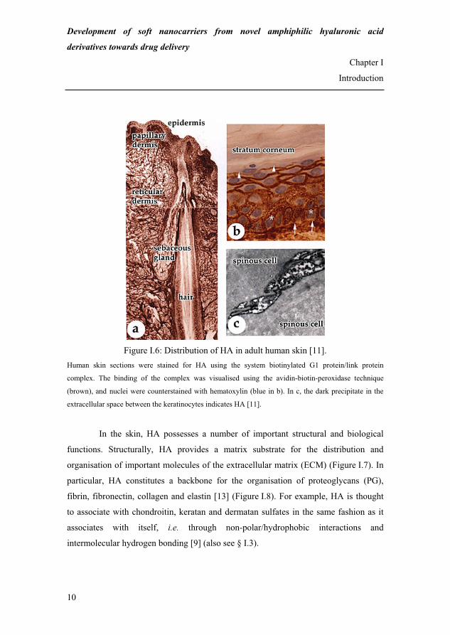
I.4. Biological functions in the skin
HA is ubiquitous in the body but is most abundant in the skin where it
amounts to approximately 5 g, i.e. a third of the total human body’s HA content [10].
In normal skin, HA occurs both in the dermis and the epidermis [11] (Figure I.6).
In the dermis, HA is mostly found in the papillary dermis and, in the reticular dermis,
at the level of the skin appendages (e.g. the sebaceous glands and hair follicles,
Figure I.6, a). Its concentration in the dermis is around 0.5 mg/g of wet tissue [4].
In the epidermis, HA is present in the extracellular space between the keratinocytes in
the stratum granulosum, stratum spinosum and stratum basale (Figure I.6, b between
the arrow heads and Figure I.6, c). Its concentration in the epidermis approaches
0.1 mg/g of wet tissue [4]. Recently, Sakai et al. [12] demonstrated that HA also
occurs in the stratum corneum (SC), in the extracellular space between the corneocytes
where its dry weight content is about 20 μg/g of SC.
35
Development of soft nanocarriers from novel amphiphilic hyaluronic acid
derivatives towards drug delivery
Chapter I
Introduction
10
Figure I.6: Distribution of HA in adult human skin [11].
Human skin sections were stained for HA using the system biotinylated G1 protein/link protein
complex. The binding of the complex was visualised using the avidin-biotin-peroxidase technique
(brown), and nuclei were counterstained with hematoxylin (blue in b). In c, the dark precipitate in the
extracellular space between the keratinocytes indicates HA [11].
In the skin, HA possesses a number of important structural and biological
functions. Structurally, HA provides a matrix substrate for the distribution and
organisation of important molecules of the extracellular matrix (ECM) (Figure I.7). In
particular, HA constitutes a backbone for the organisation of proteoglycans (PG),
fibrin, fibronectin, collagen and elastin [13] (Figure I.8). For example, HA is thought
to associate with chondroitin, keratan and dermatan sulfates in the same fashion as it
associates with itself, i.e. through non-polar/hydrophobic interactions and
intermolecular hydrogen bonding [9] (also see § I.3).
36

Development of soft nanocarriers from novel amphiphilic hyaluronic acid
derivatives towards drug delivery
Chapter I
Introduction
11
Figure I.7: Scheme of HA matrices in aqueous solutions [5].
Figure I.8: Scheme of the organisation of proteoglycans and collagen in HA matrices
[5].
Due to its naturally forming networks, HA acts as a filler which maintains both space
and cohesion between skin cells. This facilitates the diffusion of nutrients to and waste
products from upper skin cells and the movement of the immune system cells (i.e.
lymphocytes and Langerhans cells) while restricting the movement of proteins,
proteases and pathogens [8] (also see § I.3).
37
Development of soft nanocarriers from novel amphiphilic hyaluronic acid
derivatives towards drug delivery
Chapter I
Introduction
12
Finally, due to its strong hygroscopic character, HA is capable of retaining
considerable amounts of water over prolonged periods of time. This contributes to
tissue hydration and lubrication [14].
Biologically, HA triggers fundamental processes by binding with cells
through specific interactions. For example, HA is involved in complex signalling
events regulating cell proliferation, differentiation and migration. Such events are
mediated by a group of proteins called hyaladherins. These proteins act as cellular
receptors for HA. Two major receptors have been studied: CD44 and RHAMM
(Receptor for HA-mediated mobility) [11].
CD44 is a major cell receptor for HA and is expressed on the surface of a variety of
cells such as the leucocytes, fibroblasts, keratinocytes and some epithelial and
endothelial cells [15]. CD44 has been shown to be associated with various cellular
processes including cell adhesion, migration, proliferation and activation as well as HA
degradation and uptake [16].
RHAMM is found on the surface and in the cytosol and nucleus of a number of cell
types [17]. This receptor has been demonstrated to be involved in the regulation of
cellular responses to growth factors and in the migration of fibroblasts and smooth
muscle cells [17,18].
For more information about the CD44 and RHAMM receptors, the reader is invited to
consult references [19] and [20].
Another potential biological function of HA is the scavenging of reactive oxygen
species (ROS). ROS are noxious compounds produced as a result of exposure to
ultraviolet (UV) radiations and are known to cause damage to cell membrane lipids,
nucleic acids, proteins and enzymes and to be responsible for cancer, chronic diseases
and aging. It has been demonstrated that HA effectively scavenges ROS in vitro
[21,22,23]. ROS are now known to attack the C-5 hydrogen in the glucuronic acid
moiety [24], the C-1 hydrogen in the acetylglucosamine moiety [25] and the carboxyl
38

Development of soft nanocarriers from novel amphiphilic hyaluronic acid
derivatives towards drug delivery
Chapter I
Introduction
13
and acetamido groups [26]. ROS clearance from the epidermis is currently speculated
to happen through the natural rapid HA turnover [11].
In summary, these selected structural and biological functions of HA in the
skin show that HA is the most active guardian of the ECM homeostasis.
I.5. Applications
Due to its unmatched natural biocompatibility, resorbability, physicochemical
properties and biological functions (see § I.3 and I.4), HA has been used in numerous
cosmetic, pharmaceutical and biomedical applications.
In the cosmetic industry, HA is a well-known and widely spread moisturising
and skin softening agent [27,28]. Indeed, the topical application of HA-containing
formulations has been reported to restore skin hydration and elasticity and thus provide
an anti-ageing effect [4]. In addition, the incorporation of HA in sunscreens could help
protect the skin against UV radiation damages.
The biomedical applications of HA are currently considerable [16,29,30].
According to Balazs [31] these can be classified into five main categories, namely (i)
visco-surgery, (ii) visco-separation, (iii) visco-protection, (iv) visco-supplementation
and (v) visco-augmentation (Table I.1). HA provides tissue protection, lubrication and
space during surgery, prevents adhesion and excessive scar formation after surgery and
promote wound healing by regulating cell proliferation and migration. Cross-linked
HA can also be used to replace or supplement defective tissue fluids such as the
synovial fluid in patients affected by osteoarthritis and possesses filling properties that
can exploited for tissue augmentation.
39

Development of soft nanocarriers from novel amphiphilic hyaluronic acid
derivatives towards drug delivery
Chapter I
Introduction
14
Table I.1: Biomedical applications of HA [31].
Biomedical application HA role
Visco-surgery Tissue protection and lubrication
HA provides space during surgery
Visco-separation Separation and lubrication of traumatised connective tissue
surfaces
Prevention of tissue adhesion and excessive scar formation
Visco-protection Tissue protection from dryness and environmental noxious
agents
Healing promotion
Visco-supplementation Tissue fluid replacement or supplementation
Visco-augmentation Tissue filling and augmentation
As for pharmaceutical applications, HA has been widely studied towards
ophthalmic, nasal, pulmonary and parenteral drug delivery [32] for which it is
generally thought to act as a mucoadhesive compound retaining the drug at its site of
action/absorption and modifying the drug’s in vivo release/absorption rate.
Recently, HA was envisaged as a potential promising excipient for dermal drug
delivery.
II. Hyaluronic acid-mediated dermal drug delivery
Dermal drug delivery has become an important research field due to the
advantages the dermal route presents compared to the more conventional enteral and
parenteral routes (see Chapter II, § I). It has been estimated that the market for dermal
and transdermal drug delivery represented 1.57 billion dollars in the United State (US)
in 2002 and will amount to nearly 5.67 billion dollars by 2009, i.e. approximately 6 %
of the total US drug delivery market [33].
40

Development of soft nanocarriers from novel amphiphilic hyaluronic acid
derivatives towards drug delivery
Chapter I
Introduction
15
Due to its natural presence in the skin, HA represents an attractive
biocompatible and resorbable excipient for the dermal administration of drugs. For
example, HA can be co-formulated with drugs (first generation systems) or constitute
the building block of more advanced biomaterials (second generation systems).
II.1. First generation systems
The use of HA in dermal drug delivery is relatively recent [34] and has
mainly consisted in co-formulating HA with the drugs. Co-formulations of HA and
therapeutic agents such as dichlorofenac, ibuprofen, clindamycin and cyclosporin has
been rigorously investigated in vitro, in vivo and in clinical studies and reviewed in an
excellent article by Liao et al. [32]. These studies have shown that HA significantly
enhances drug partitioning into human skin and drug retention and localisation in the
epidermis compared to other GAGs and more ordinary pharmaceutical formulations.
However, the exact mechanisms by which HA, as an excipient, achieves such
performances remains unclear [32].
While HA/drug co-formulations seem to sustain drug absorption in skin
layers, drug release, in this configuration, remains non-specific. In cases of more
challenging and demanding therapeutic situations, such as when the drugs need to be
protected and targeted to specific tissues, more advanced systems than co-formulations
are required. These form the basis of the second generation HA-based systems.
II.2. Second generation systems
The interest in nanotechnology in pharmacy is somewhat new and is mainly
due to drug-associated issues such as poor solubility and instability in biological milieu
(i.e. short half-life), poor bioavailability and unspecific targeting. As a result, high drug
dosages are used to achieve a therapeutic effect which increases the risk for toxicity in
41

Development of soft nanocarriers from novel amphiphilic hyaluronic acid
derivatives towards drug delivery
Chapter I
Introduction
16
patients and health cost [35]. Therefore, the design of advanced drug delivery systems
addressing these challenges has become more and more crucial. In this perspective, it
is believed that drug encapsulation in nanovehicles could allow the formulation of
improved systems [35].
With its unusual and beneficial properties, HA represents an attractive candidate for
the encapsulation and release of drugs in biological environments, including skin
layers. HA-based advanced biomaterials such as colloidal particles therefore constitute
potential valuable second generation dermal drug delivery systems.
III. Hyaluronic acid-based colloidal particles
III.1. Microparticles
The preparation of HA-based colloidal particles towards drug delivery first
addressed the development of microparticles (MPs). HA MPs in which HA constitutes
the only component of the polymeric matrix have been prepared according to a few
different methods which are reviewed in Table I.2. These methods can be classified in
two main categories according to the type of HA modification involved, namely cross-
linking [36,37] or hydrophobic modification [38,39].
In the first case, HA and a cross-linking agent are mixed and emulsified into mineral
oil with the aid of a biodegradable surfactant to produce a microemulsion. Cross-
linking is then initiated in the microdroplets containing HA and the cross-linking agent
by addition of a coupling agent [36]. Alternatively, two auto-cross-linkable HA
derivatives can be used [37].
In the second case, HA is first hydrophobically modified (HYAFF®). HYAFF®
solutions are then spray-dried into the form of microparticles [38] or emulsified in
mineral oil with the aid of a biodegradable surfactant [38,39]. The polar organic
solvent in the microdroplets is then either removed by extraction with another polar
organic solvent [38] or evaporated by mixing at room temperature [39]. In methods
42

Development of soft nanocarriers from novel amphiphilic hyaluronic acid
derivatives towards drug delivery
Chapter I
Introduction
17
[38] and [39], the particles are stabilised by hydrophobic interactions between the
hydrophobic groups grafted on the HA chains.
Table I.2: Preparation methods of HA microparticles. ADH, Span 80, EDC, DMSO, Arlacel® A stand for adipic acid dihydrazide, sorbitan monostearate, 1-
ethyl-3-[3-dimethyl amino] propyl carbodiimide, dimethyl sulfoxide and mannide monostearate,
respectively. HYAFF® is Fidia’s trademark for hydrophobically modified HA obtained by reacting HA
and alkyliodides in DMSO, using the tetrabutylammonium salt of HA (also see § IV).
Reference HA MW
MP size Principle
[36] 1,600-3,300 kDa
5-15 μm
Emulsification of HA and ADH into mineral oil with Span 80
Cross-linking in the presence of EDC
[37] 490-1,300 kDa
10 μm
Emulsification of oxidized HA and ADH-modified HA into mineral
oil with Span 80
Cross-linking in the presence of EDC
[38] unspecified
1-10 μm Spray drying of aqueous HYAFF® solutions
[38] unspecified
1-100 μm
Emulsification of HYAFF® in DMSO into mineral oil with
Arlacel® A
Extraction of DMSO with ethyl acetate
[39] 150 kDa
10-100 μm
Emulsification of HYAFF® in hexafluoropropanol into mineral oil
with Arlacel® A
Evaporation of hexafluoropropanol by mixing at room temperature
The applications of HA MPs towards drug delivery are numerous and in vivo
release studies have shown that HA MP-mediated drug delivery presents some
advantages compared to conventional drug formulations. For example, HA MPs have
been used to modulate the nasal absorption of insulin in sheeps [40], enhance the
vaginal assimilation of salmon calcitonin [41], the oral bioavailability of cyclosprorin
[42] and piroxicam [43] in rats and the production of growth factors when injected
subcutaneously to monkeys and dogs [44]. These examples show that HA MPs are
43

Development of soft nanocarriers from novel amphiphilic hyaluronic acid
derivatives towards drug delivery
Chapter I
Introduction
18
currently designed in the perspective of targeting all three drug administration routes,
namely the topical, enteral and parenteral routes.
However, for topical routes such as the skin, the size of the particulate system is an
important factor for its performances since skin penetration is an extremely selective
phenomenon. With their large dimensions, MPs have often been considered unsuitable
devices for targets other than the surface of the skin or the skin appendages [45].
Nanoparticles (NPs), however, could present more favourable dimensions towards skin
penetration and their interaction with the skin is a currently studied topic (see
Chapter II).
III.2. Nanoparticles
To the best of our knowledge, and as until January 2008, four patent
applications and three articles have since 1995 claimed the preparation of HA NPs in
which HA constitutes the only component of the polymeric matrix. The techniques
disclosed are reviewed in Table I.3. As for HA MPs, the preparation methods of HA
NPs can be classified in the two main categories described before, i.e. according to
whether HA is hydrophobically modified [46] or cross-linked [47-51].[47,48,49,50,51]
However, HA can also be directly complexed with the drug to be encapsulated [52].
In the second case, where particles are produced by cross-linking of transient HA
colloidal assemblies, the parameter which seems to allow a size reduction from micro-
to nanoparticles is the type of solvent used as the continuous phase: mineral oil for HA
MPs versus water, water/acetone mixtures or (mixtures of) light non-polar organic
solvents for HA NPs. These solvents or solvent mixtures are much less viscous that
mineral oil which results in much smaller droplet size and by extension particle size.
44

Development of soft nanocarriers from novel amphiphilic hyaluronic acid
derivatives towards drug delivery
Chapter I
Introduction
19
Table I.3: Preparation methods of HA nanoparticles. DMSO, EDC, ADH, ODH, Span 80, HLB, PH, NHSS, Span 85, HLB stand for dimethyl sulfoxide, 1-
ethyl-3-[3-dimethyl amino] propyl carbodiimide, adipic acid dihydrazide, oxalic acid dihydrazide,
sorbitan monostearate, hydrophilic lipophilic balance, polyhydrazide, sodium N-
hydroxysulfosuccinimide, sorbitan tristearate, hydrophilic lipophilic balance, respectively. HYAFF® is
Fidia’s trademark for hydrophobically modified HA obtained by reacting HA and alkyliodides in
DMSO, using the tetrabutylammonium salt of HA (also see § IV). For some of the preparation methods,
the mechanism underlying the formation of the nanoparticles is explained as claimed by the authors or
in the absence thereof as presumed by us in Comment.
Reference HA MW
NP size Principle Comment
[46] unspecified
500-1,000 nm
Instillation, under pressure, of
pure CO2 into the HYAFF®/active
ingredient solution in DMSO
Calcitonin, granulocyte
macrophage colony stimulating
factor and human insulin were
encapsulated.
Nanoprecipitation of HYAFF® in
the supercritical CO2 antisolvent is
induced by DMSO solubilisation in
CO2 and evaporation. Insoluble
HYAFF® in CO2 precipitates into
nanoparticles.
[47]
20-2,500 kDa
20 nm-
200 μm
Auto cross-linking of HA
carboxyl and hydroxyl groups in
the presence of EDC, in aqueous
HA solutions
or alternatively,
Cross-linking with polyamines, in
the presence of EDC, in aqueous
HA solutions
Cross-linking of HA in aqueous
solutions converts the initially
randomly coiled HA into spherical
HA nanoparticles.
[48]
200-
1,500 kDa
200 nm
Cross-linking with ADH, in the
presence of EDC, in water/acetone
mixtures
Water/acetone mixtures provide a
poor solvent environment for HA
which tends to self-associate into
nanoassemblies. Acetone catalyses
intra- and intermolecular cross-
linking by breaking HA hydrogen
bonds.
45

Development of soft nanocarriers from novel amphiphilic hyaluronic acid
derivatives towards drug delivery
Chapter I
Introduction
20
Table I.3: Preparation methods of HA nanoparticles (continued).
Reference HA MW
NP size Principle Comment
[49] unspecified
50-200 nm
Mixing of HA and EDC with an
emulsion of aminophospholipid
Cross-linking with the
phospholipids in the presence of
EDC
Paclitaxel was encapsulated
We believe that the process
involves interfacial cross-linking of
HA at the surface of the
phospholipid droplets and that the
nanoparticles form as the
phospholipids are consumed.
[50] MW > 20 kDa
10-160 nm
Emulsification of HA, ODH and
EDC in a non-polar organic solvent
with Span 80
Cross-linking with ODH, in the
presence of EDC
Fluorescein isothiocyanate, atrial
natriuretic peptide and green
fluorescence protein were
encapsulated
Despite the fact the authors
mention the formation of an o/w
emulsion, we believe, according to
the Bancroft rule [53], that a w/o
emulsion is rather formed due to
the low HLB value of Span 80. We
therefore presume that the cross-
linking reaction takes place inside
the water droplets containing HA,
ODH and EDC.
Interestingly, when this method
was applied to 20-40 kDa HA, the
size of the nanoparticles was
reduced between 10 and 40 nm.
[51] 174 kDa
200-300 nm
Emulsification of HA, PH, EDC
and NHSS in ethyl
acetate/propylene carbonate with
Span 85
Cross-linking with PH, in the
presence of EDC and NHSS
The droplets of the nanoemulsion
constitute nanoreactors in which
HA is cross-linked with PH. A
mixture of EDC and NHSS was
used to catalyse the reaction as
cross-linking in the presence of
EDC alone was too rapid and
yielded nanoparticles with
uncontrolled properties.
[52] 102 kDa
100-200 nm
Spontaneous complexation
between HA and cisplatin
46

Development of soft nanocarriers from novel amphiphilic hyaluronic acid
derivatives towards drug delivery
Chapter I
Introduction
21
The preparation methods of Table I.3 present some advantages and drawbacks
which are detailed below.
HYAFF®-based NPs [46] contain both hydrophilic and hydrophobic domains where
hydrophilic and/or hydrophobic active ingredients can be encapsulated. In addition,
HYAFF® polymers are biocompatible and resorbable in rats [54] which makes them
potentially suitable for pharmaceutical purposes. However, the nanoprecipitation step
requiring relatively high working pressures, the process might be unsuitable for the
encapsulation of fragile active ingredients.
The NPs obtained by intra- and intermolecular cross-linking between HA carboxyl
and hydroxyl groups [47] do not contain any cross-linking agent molecules making up
the bridges between HA chains. As a result, these NPs are potentially completely
biocompatible. Similarly, when HA is cross-linked with aminophospholipids naturally
present in biological membranes [49], the resulting NPs consist of fully resorbable
materials.
Nevertheless, since HA assemblies are randomly formed in method [47], the size of the
resulting particles is quite polydisperse. This could limit their use for applications
where the latter needs to be accurately controlled and narrowly distributed.
A common advantage to the methods involving HA emulsification [50,51] is the
possibility to quite precisely control the NP size by managing that of the nanodroplets.
Conversely, a common drawback is the use of organic solvents and surfactants.
Organic solvents raise occupational and environmental issues and limit the
upscalability of the methods while surfactants are known irritants which complete
removal can be problematic.
Native HA-based NPs [47-52] present the disadvantage that only
hydrophiphilic/polar ingredients can durably be encapsulated since highly hydrophilic
HA represents an unfavourable environment for hydrophobic compounds.
Finally, HA/drug complexes [52] are cross-linking agent-free and do not require
modifying HA or using organic solvents, surfactants or harsh conditions. However, the
technique is not versatile since HA/drug complexation is not a universal phenomenon.
47

Development of soft nanocarriers from novel amphiphilic hyaluronic acid
derivatives towards drug delivery
Chapter I
Introduction
22
In summary, the preparation methods of HA NPs are still facing challenges
such as (i) environmental unfriendliness, (ii) potential surfactant residues, (iii) NP size
polydispersity, (iv) poor versatility of the encapsulated active ingredients and (v)
limited upscalability.
When comparing the evolutionary development stage of HA micro- and
nanoparticles it becomes clear that that HA MPs are in a paradigmatic stage where
dominant designs have emerged and research has been taken beyond the investigation
of preparation methods towards in vivo release studies. HA NPs, however, are still in a
preparadigmatic stage where focus is still drawn on spotting out dominant designs (no
two similar methods are described in the existing scarce literature) rather than on
investigating the uses and applications of the nanoparticles. HA NP-mediated drug
delivery therefore represents a challenging research field with room for innovation,
intellectual property and application achievements.
With the exception of Pallado et al. [46], there are no reported methods for
the preparation of HA NPs based on hydrophobically modified HA (hmHA). Despite
the fact the working conditions of this method may be too harsh for the encapsulation
of delicate active ingredients, the concept of using hmHA is innovative and currently
not widespread according to the literature study. Indeed, with its altered
physicochemical properties and HLB, hmHA constitutes a more favourable starting
material for the production of nanostructures and the durable encapsulation of
hydrophobic ingredients than native HA. For these reasons, some currently available
preparation methods of hmHA are reviewed in the next section.
48

Development of soft nanocarriers from novel amphiphilic hyaluronic acid
derivatives towards drug delivery
Chapter I
Introduction
23
IV. Hydrophobically modified hyaluronic acid
Hydrophobically modified HA is conventionally prepared by targeting
reactions at HA carboxyl and/or hydroxyl groups [55].
Historically, Jeanloz and Forchielli [56] were the first to report the modification of HA
carboxyl groups with diazomethane, acetic anhydride and triphenylchloromethane
towards the preparation of methylated, acetylated and triphenylchloromethylated HA.
In the late 1980’s, Della Valle and Romeo [57] patented the modification of HA
carboxyl groups with alkyliodides, in DMSO, using the tetrabutylammonium salt of
HA. The resulting derivatives currently form the basis of Fidia’s trademark HYAFF®
which has frequently been used for the preparation of colloidal particles towards drug
delivery (see § III).
Other more recent modification methods non-exhaustively include modification of the
HA carboxyl groups with:
(i) alkylaldehydes in water/ethanol mixtures, in the presence of sodium
cyanoborohydride, using an adipic dihydrazide-modified HA intermediate obtained in
the presence of 1-ethyl-3-[3-dimethyl amino] propyl carbodiimide [58],
(ii) alkylbromides in DMSO [59],
and of the HA hydroxyl groups with
(iii) acylchlorides in N,N-dimethylformamide (DMF), in the presence of pyridine [60],
(iv) alkyloxymethyloxiranes in water/DMSO mixtures [61],
(v) alkylalcohols in DMSO, in the presence of potassium hydroxide, using a p-toluene
sulfonyl-modified HA intermediate [62] (Figure I.9).
49
HA
OH
O
OR
Br
RH
AO
O
RR
= (C
H2)
nC
H3
HA
OH
Cl
R
O
HA
OR
O
R =
(CH
2)n
CH
3
OO
RR
= (C
H2)
nC
H3
Cl
SO O
CH
3
OH
RH
AO
RR
= (C
H2)
nC
H3
R =
(CH
2)n
CH
3
HA
OO
R
OH
OSO O
CH
3H
A
HA
N HNH
N HN
H2
O
O
O
HA
N HNH
N HN
H2
O
O
Oin
wat
er/e
than
ol, N
aBH
3CN
pH 5
.1, r
t, 24
hou
rs
in D
MSO
, rt,
24 h
ours
n =
11 to
15
[5
9]
in D
MF,
pyr
idin
e, rt
, 2 w
eeks
n =
2 to
16
[
60]
in w
ater
/DM
SO, p
H 9
.0-1
0.0
30-4
5°C
, ove
rnig
htn
= 3
to 1
3
[61]
in D
MSO
, KO
Hrt,
ove
rnig
htn
= 5
to 1
5
[62]
n =
7 to
15
[5
8]
in N
2, 0-
5°C
, 1.5
hou
rs
AD
H in
wat
er, E
DC
, pH
4.7
5
Fi
gure
I.9:
Illu
stra
tion
of se
lect
ed h
ydro
phob
ic m
odifi
catio
ns o
f HA
.
50
Development of soft nanocarriers from novel amphiphilic hyaluronic acid
derivatives towards drug delivery
Chapter I
Introduction
25
However, the modification methods currently available for the preparation of
amphiphilic HA present some drawbacks.
First, the modification reactions are often conducted in organic solvents or organic
solvent/water mixtures. In addition to raise occupational and environmental issues and
limit the upscalability of the preparation methods, the use of organic solvents or
mixtures of these often requires converting HA into its tetraalkylammonium salt or
preparing reactive HA intermediates. This not only adds complex steps to the
preparation methods but also often results in HA degradation [59].
Secondly, the modification reactions involving HA carboxyl groups modify the
distribution of negative charges along the polymer backbone at physiological pH. This
could potentially affect some of the attractive biological and/or pharmacological
properties of native HA [62].
Therefore, simple modification reactions in aqueous media, under mild conditions and
exclusively involving HA hydroxyl groups could represent advantageous alternatives
for the preparation of amphiphilic HA derivatives.
V. The Ph.D. project
V.1. Objectives and challenges
For the reasons outlined all along this introduction chapter, HA currently
represents one of the most attractive building block for the preparation of advanced
biomaterials with applications in pharmacy.
While the superficial administration of certain HA/drug co-formulations has been
shown to enhance drug partitioning into human skin and drug retention and
localisation in the epidermis, there is a lack of truly biocompatible and resorbable
solutions for more challenging and demanding therapeutic situations requiring drug
protection and targeting (see § II).
51

Development of soft nanocarriers from novel amphiphilic hyaluronic acid
derivatives towards drug delivery
Chapter I
Introduction
26
It is well known that tumourous cells generally over-express HA-binding receptors at
their surface. Consequently, these show enhanced HA binding and internalisation [63].
Anti-cancer drug-containing HA-based systems therefore potentially represent
valuable and efficient multi-functional targeting devices towards superficial tumoured
tissues. In addition, due to HA’s natural presence in the skin, HA-based systems are
likely to interact with the skin without altering the epithelium’s physiology.
Therefore, the overall objective of this project was to design and develop advanced
HA-based drug delivery systems such as HA nanoparticles towards the encapsulation
and targeted delivery of chemically instable and/or poorly soluble drugs in aqueous
media. In the first place, it was envisaged to evaluate these devices for dermal drug
delivery and later extend their use to other delivery routes.
The methods currently available for the preparation of HA NPs are still facing
challenges such as (i) environmental unfriendliness, (ii) potential surfactant residues,
(iii) NP size polydispersity, (iv) poor versatility of the encapsulated active ingredients
and (v) limited upscalability. In addition, they use native HA which is unsuitable for
the encapsulation of hydrophobic ingredients. It is believed that hydrophobically
modified HA with its altered physicochemical properties and HLB, constitutes a more
favourable starting material for the production of stable nanostructures and the durable
encapsulation of hydrophobic ingredients (see § III.2).
However, the current preparation methods of amphiphilic HA present some drawbacks
such as the (i) use of organic solvents, (ii) potential alteration of some fundamental
biological and/or pharmacological properties of HA through modification of the
carboxyl groups and (iii) limited upscalability (see § IV).
Therefore, other important objectives of this project were to elaborate simple and
easily upscalable, aqueous and versatile techniques for both the preparation of
amphiphilic HA and of HA NPs. Focus was drawn on exclusively modifying the HA
hydroxyl groups in order to preserve the distribution of anionic charges along the
polymer backbone and on avoiding the use of surfactants to formulate reasonably
52

Development of soft nanocarriers from novel amphiphilic hyaluronic acid
derivatives towards drug delivery
Chapter I
Introduction
27
uniformly distributed NPs capable of encapsulating both hydrophilic and hydrophobic
compounds.
V.2. Experimental approach
Due to its high hydrophilicity, native HA possesses physicochemical
properties incompatible with the spontaneous and stable formation of segregated
structures in aqueous media and the durable encapsulation of hydrophobic active
ingredients. In addition, native HA displays a poor in vivo biological stability and
residence time as evidenced by its high turnover rate [64].
Therefore, HA was first rendered amphiphilic by the covalent binding of
hydrophobic groups onto its backbone. For this purpose, two novel proprietary
methods were evaluated and one was eventually selected for further study based on the
superior versatility and functionality of the resulting HA derivatives.
This method was then optimised in order to develop a reproducible and robust process
for the routine preparation of functionalised HA polymers with tailored degrees of
substitution.
Fundamental properties such as the structure, degree of hydrophobicity, substitution
pattern as well as intra- and intermolecular interactions of a selection of derivatives
were subsequently studied.
Submicronic multiphasic physically cross-linked nanogel particles capable of
solubilising a model hydrophobic substance were prepared by spontaneous self-
association and characterized with regard to their morphology, size and surface charge.
A multivesicular model was finally put forward to explain their internal structure.
Finally, methods to prepare stable NPs based on the chemical stabilisation of self-
assembled HA templates were explored. Indeed, while the methods currently available
for the preparation of HA NPs rely on random collisions in the medium to create
structures, focus was here drawn on the development of a novel molecular approach
53
Development of soft nanocarriers from novel amphiphilic hyaluronic acid
derivatives towards drug delivery
Chapter I
Introduction
28
based on self-assembling multiphasic nanoarchitectured HA templates potentially
yielding more complex NP configurations than single ingredient or core-shell
structures.
V.3. Thesis structure
After this introduction chapter, the thesis is organised in four more chapters
written in the form of research reports and/or articles to be submitted shortly.
Chapter II is a review article about the current knowledge about the mechanisms
underlying polymeric nanoparticle-mediated (trans)dermal drug delivery. It
investigates, among others, the potential of this delivery route, the research gaps and
the challenges still faced by this branch of research.
Chapter III is organised in three different sections. Section A reports the initial
investigation of novel modification methods to prepare amphiphilic HA and the
selection of one class of HA derivatives for further study. Section B concerns the
optimisation of the preparation of these HA derivatives which detailed molecular
structure and solution properties in aqueous media is investigated in section C.
Chapter IV reports the physicochemical properties of the selected class of HA
derivatives and the preparation of multiphasic nanostructured HA templates.
Chapter V reviews the work achieved, gives a conclusion for the thesis and outlines
some possible future work. It also suggests some potential applications for the systems
developed.
Finally, a record of the educational activities conducted during the industrial Ph.D.
programme is presented in the appendices.
54

Development of soft nanocarriers from novel amphiphilic hyaluronic acid
derivatives towards drug delivery
Chapter I
Introduction
29
References
[1] Lee, J. Y.; Spicer, A. P. Hyaluronan: a multifunctional, megaDalton, stealth
molecule. Curr. Opin. Cell Biol. 2000, 12, 581-586.
[2] Meyer, K.; Palmer, J. The polysaccharide of the vitreous humor. J. Biol. Chem.
1934, 107, 629-634.
[3] Weissman, B.; Meyer, K. The structure of hyalobiuronic acid and of hyaluronic
acid from umbilical cord. J. Am. Chem. Soc. 1954, 76, 1753-1757.
[4] Brown, M. B.; Jones, S. A. Hyaluronic acid: a unique topical vehicle for the
localized delivery of drugs to the skin. J. Eur. Acad. Dermatol. Venereol. 2006,
20, 1348-1349.
[5] Glycoforum,
http://www.glycoforum.gr.jp/science/hyaluronan/HA02/HA02E.html, last updated
March 15, 1998.
[6] Widner, B.; Behr, B.; Von Dollen, S.; Tang, M.; Heu, T.; Sloma, A.; Sternberg,
D.; DeAngelis, P. L.; Weigel, P. H.; Brown, S. Hyaluronic acid production in
bacillus subtilis. Appl. Environ. Microbiol. 2005, 71, 3747-3752.
[7] Laurent, T. C.; Fraser, J. R. E. Hyaluronan. Faseb J. 1992, 6, 2397-2404.
[8] Glycoforum,
http://www.glycoforum.gr.jp/science/hyaluronan/HA01/HA01E.html, last updated
December 15, 1997.
[9] Scott, J. E.; Presti, D. Hyaluronan-mediated protective effect against cell damage
caused by enzymatically produced hydroxyl radicals is dependent on hyaluronan
molecular mass. Cell Biochem. Funct. 1994, 12, 281-288.
[10] Banks, J.; Kreider, J. W.; Bhavanadan, V. P.; Davidson, E. A. Anionic
polysaccharide production and tyrosine activation in cultured human melanoma
cells. Cancer Res. 1976, 36, 424-431.
55

Development of soft nanocarriers from novel amphiphilic hyaluronic acid
derivatives towards drug delivery
Chapter I
Introduction
30
[11] Glycoforum,
http://www.glycoforum.gr.jp/science/hyaluronan/HA04/HA04E.html, last updated
June 15, 1998.
[12] Sakai, S.; Yasuda, R.; Sayo, T.; Ishikawa, O.; Inoue, S. Hyaluronan exists in the
normal stratum corneum. J. Invest. Dermatol. 2000, 114, 1184-1187.
[13] Chen, W. Y. J.; Abatangelo, G. Functions of hyaluronan in wound repair. Wound
Rep. Reg. 1999, 7, 79-89.
[14] Yates, J. R. Mechanism of water uptake by skin. In Biophysical properties of the
skin; Eden, N. R., Ed.; Wiley Interscience: New York, 1971.
[15] Isacke, C. M.; Yarwood, H. The hyaluronan receptor, CD44. Int J. Biochem. Cell
Biol. 2002, 34, 718-721.
[16] Leach, J. B.; Schmidt, C. E. Hyaluronan. In Encyclopedia of biomaterials and
biomedical engineering; Bowlin, G. L.; Wnek, G., Eds.; Marcel Dekker: New
York, 2004.
[17] Cheung, W. F.; Cruz, T. F.; Turley, E. A. Receptor for hyaluronan-mediated
motility (RHAMM), a hyaladherin that regulates cell responses to growth factors.
Biochem. Soc. Trans. 1999, 27, 135-142.
[18] Toole, B. P. Hyaluronan in morphogenesis. Semin. Cell Dev. Biol. 2001, 12, 79-
87.
[19] Glycoforum,
http://www.glycoforum.gr.jp/science/hyaluronan/HA10/HA10E.html, last updated
January 13, 2005.
[20] Glycoforum,
http://www.glycoforum.gr.jp/science/hyaluronan/HA11/HA11E.html, last updated
July 25, 1999.
[21] Tokita, Y.; Sakashita, H.; Okamoto, A.; Kubota, K. Kinetic study of a radical
scavenging effect of hyaluronic acid. Polymer Int. 1995, 38, 161-164.
56

Development of soft nanocarriers from novel amphiphilic hyaluronic acid
derivatives towards drug delivery
Chapter I
Introduction
31
[22] Trommer, H.; Wartewig, S.; Böttcher, R.; Pöppl, A.; Hoentsch, J.; Ozegowski, J.
H.; Neubert, R. H. H. The effects of hyaluronan and its fragments on lipid models
exposed to UV irradiation. Int. J. Pharm. 2003, 254, 223-234.
[23] Campo, G. M.; Avenoso, A.; Campo, S.; D’Ascola, A.; Ferlazzo, A. M.;
Calatroni, A. The antioxidant and antifibrogenic effects of the
glycosaminoglycans hyaluronic acid and chondroitin-4-sulphate in a subchronic
rat model of carbon tetrachloride-induced liver fibrogenesis. Chem. Biol. Interact.
2004, 148, 125-138.
[24] Gilbert, B. C.; King, D. M.; Thomas, C. B. The oxidation of some
polysaccharides by the hydroxyl radical: an E.S.R. investigation. Carbohydr. Res.
1984, 125, 217-235.
[25] Uchiyama, H.; Dobashi, Y.; Ohkouchi, K.; Nagasawa, K. Chemical change
involved in the oxidative reductive depolymerization of hyaluronic acid. J. Biol.
Chem. 1990, 265, 7753-7759.
[26] Lapcik, L.; Chabrecek, P.; Stasko, A. Photodegradation of hyaluronic acid: EPR
and size exclusion chromatography study. Biopolymers 1991, 31, 1429-1435.
[27] Balazs, E. A.; Band, P. Hyaluronic acid: its structure and use. Cosmetics &
Toiletries 1984, 99, 65-72.
[28] Oka, T.; Ueno, N.; Yanaki, T. Differential scanning calorimetry studies on the
mechanism of skin-softening effect on sodium acethylhyaluronate. Polymer 2000,
41, 6055-6059.
[29] Lapcik, L. Jr.; Lapcik, L.; De Smedt, S.; Demeester, J.; Chabrecek, P.
Hyaluronan: preparation, structure, properties and applications. Chem. Rev. 1998,
98, 2663-2684.
[30] Kogan, G.; Soltes, L.; Stern, R.; Gemeiner, P. Hyaluronic acid: a natural
biopolymer with a broad range of biomedical and industrial applications.
Biotechnol. Lett. 2007, 29, 17-25.
57

Development of soft nanocarriers from novel amphiphilic hyaluronic acid
derivatives towards drug delivery
Chapter I
Introduction
32
[31] Balazs, E. A. Viscoelastic properties of hyaluronan and its therapeutic use. In
Chemistry and biology of hyaluronan; Garg, H. G.; Hales, C. A., Eds.; Elsevier:
Amsterdam, 2004.
[32] Liao, Y. H.; Jones, S. A.; Forbes, B.; Martin, G. P.; Brown, M. B. Hyaluronan:
pharmaceutical characterization and drug delivery. Drug Deliv. 2005, 12, 327-
342.
[33] Frost & Sullivan, U.S. Emerging Transdermal Drug Delivery Technologies
Markets. http://www.frost.com, May 9, 2003.
[34] Brown, M. B.; Forbes, B.; Martin, G. P. The use of hyaluronan in topical drug
delivery. In Hyaluronan: biomedical, medical and clinical aspects; Kennedy, J.;
Phillips, G. O.; Williams, P. A.; Hascall, V., Eds.; Woodhead Publishing:
Cambridge, 2002.
[35] Rawat, M.; Singh, D.; Saraf, S.; Saraf, S. Nanocarriers: promising vehicle for
bioactive drugs. Biol. Pharm. Bull. 2006, 29, 1790-1798.
[36] Yun, Y. H.; Goetz, D. J.; Yellen, P.; Chen, W. Hyaluronan microspheres for
sustained gene delivery and site-specific targeting. Biomaterials 2004, 25, 147-
157.
[37] Jia, X. Q.; Yeo, Y.; Clifton, R. J.; Jiao, T.; Kohane, D. S.; Kobler, J. B.; Zeitels, S.
M.; Langer, R. Hyaluronic acid-based microgels and microgel networks for vocal
fold regeneration. Biomacromolecules 2006, 7, 3336-3344.
[38] Ghezzo, E.; Benedetti, L.; Rochira, M.; Biviano, F.; Callegaro, L. Hyaluronate
derivative microspheres as NGF delivery devices: preparation methods and in
vitro release characterization. Int. J. Pharm. 1992, 87, 21-29.
[39] Benedetti, L. M.; Topp, E. M.; Stella, V. J. Microspheres of hyaluronic acid
esters. Fabrication methods and in vitro hydrocortisone release. J. Controlled
Release 1990, 13, 33-41.
58

Development of soft nanocarriers from novel amphiphilic hyaluronic acid
derivatives towards drug delivery
Chapter I
Introduction
33
[40] Illum, L.; Farraj, N. F.; Fisher, A. N.; Gill, I.; Miglietta, M.; Benedetti, L. M.
Hyaluronic acid ester microspheres as a nasal delivery system for insulin. J.
Controlled Release 1994, 29, 133-141.
[41] Richardson, J. L.; Ramires, P. A.; Miglietta, M. R.; Rochira, M.; Bacelle, L.;
Callegaro, L.; Benedetti, L. M. Novel vaginal delivery systems for calcitonin: I.
Evaluation of HYAFF/calcitonin microspheres in rats. Int. J. Pharm. 1995, 115,
9-15.
[42] Woo, J. S.; Piao, M. G.; Li, D. X.; Ryu, D. S.; Choi, J. Y.; Kim, J. A.; Kim, J. H.;
Jin, S. G.; Kim, D. D.; Lyoo, W. S.; Yong, C. S.; Choi, H. G. Development of
cyclosporin A-loaded hyaluronic microspheres with enhanced oral bioavailability.
Int. J. Pharm. 2007, 345, 134-141.
[43] Piao, M. G.; Kim, J. H.; Kim, J. O.; Lyoo, W. S.; Lee, M. H.; Yong, C. S.; Choi,
H. G. Enhanced oral bioavailability of piroxicam in rats by hyaluronate
microspheres. Drug Dev. Ind. Pharm. 2007, 33, 485-491.
[44] Kim, S. J.; Hahn, S. K.; Kim, M. J.; Kim, D. H.; Lee, Y. P. Development of a
novel sustained release formulation of recombinant human growth hormone using
sodium hyaluronate microparticles. J. Controlled Release 2005, 104, 323-335.
[45] Toll, R.; Jacobi, U.; Richter, H.; Lademann, J.; Schaefer, H.; Blume-Peytavi, U.
Penetration profile of microspheres in follicular targeting of terminal hair
follicles. J. Invest. Dermatol. 2004, 123, 168-176.
[46] Pallado, P.; Benedetti, L.; Callegaro, L. Nanospheres comprising a biocompatible
polysaccharide. WO 96/29998, October 10, 1996.
[47] Borbely, J.; Tunde, R.; Magnolna, B. Hyaluronic acid-based cross-linked
nanoparticles. US 2007/0224277 A1, September 27, 2007.
[48] Hu, Z.; Xia, X.; Tang, L. Process for synthesizing oil and surfactant-free
hyaluronic acid nanoparticles and microparticles. US 2006/0040892 A1, February
23, 2006.
59

Development of soft nanocarriers from novel amphiphilic hyaluronic acid
derivatives towards drug delivery
Chapter I
Introduction
34
[49] Yerushalmi, N.; Peer, D.; Margalit, R.; Rivkin, I. Formulations of water insoluble
or poorly water soluble drugs in lipidated glycosaminoglycan particles and their
use for diagnostics and therapy. WO 2006/050246 A2, May 11, 2006.
[50] Mohapatra, S. S.; Sahoo, B.; Kumar, A.; Behera, S. A method of transdermal drug
delivery using hyaluronic acid nanoparticles. US 2007/0036728 A1, September
27, 2007.
[51] Pitarresi, G.; Craparo, F.; Palumbo, F. S.; Carlisi, B.; Giammona, G. Composite
nanoparticles based on hyaluronic acid chemically cross-linked with �,�-
polyaspartylhydrazide. Biomacromolecules 2007, 8, 1890-1898.
[52] Jeong, Y. I.; Kim, S. T.; Jin, S. G.; Ryu, H. H.; Jin, Y. H.; Jung, T. Y.; Kim I. Y.;
Jung, S. Cisplatin-incorporated hyaluronic acid nanoparticles based on ion-
complex formation. J. Pharm. Sci. 2008, 97, 1268-1276.
[53] Bancroft, W. D. Theory of emulsification. J. Phys. Chem. 1913, 17, 501-519.
[54] Benedetti, L.; Cortivo, R.; Berti, T.; Berti, A.; Pea, F.; Mazzo, M.; Moras, M.;
Abatangelo, G. Biocompatibility and biodegradation of different hyaluronan
derivatives (Hyaff) implanted in rats. Biomaterials 1993, 14, 1154-1160.
[55] Vercruysse, K. P.; Prestwich, G. D. Hyaluronate derivatives in drug delivery. Crit.
Rev. Ther. Drug Carrier Syst. 1998, 15, 513-555.
[56] Jeanloz, R. W.; Forchielli, E. Studies on hyaluronic acid and related substances. I.
Preparation of hyaluronic acid and derivatives from human umbilical cord. J.
Biol. Chem. 1950, 186, 495-511.
[57] Della Valle, F.; Romeo, A. Esters of hyaluronic acid. US 4,851,521, July 25,
1989.
[58] Creuzet, C.; Kadi, S.; Rinaudo, M.; Auzely-Velty, R. New associative systems
based on alkylated hyaluronic acid. Synthesis and aqueous solution properties.
Polymer 2006, 47, 2706-2713.
60

Development of soft nanocarriers from novel amphiphilic hyaluronic acid
derivatives towards drug delivery
Chapter I
Introduction
35
[59] Pelletier, S.; Hubert, P.; Payan, E.; Marchal, P.; Choplin, L.; Dellacherie, E.
Amphiphilic derivatives of sodium alginate and hyaluronate for cartilage repair:
rheological properties. J. Biomed. Mater. Res. 2001, 54, 102-108.
[60] Kawaguchi, Y.; Matsukawa, K.; Gama, Y.; Ishigami, Y. New polysaccharide
surfactants from hyaluronate. Chem. Express 1991, 6, 647-650.
[61] Mlochova, P.; Hajkova, V.; Steiner, B.; Bystricky, S.; Koos, M.; Medova, M.;
Velebny, V. Preparation and characterization of biodegradable alkylether
derivatives of hyaluronan. Carbohydr. Polym. 2007, 69, 344-352.
[62] Benesova, K.; Pekar, M.; Lapcik, L.; Kucerik, J. Stability evaluation of n-alkyl
hyaluronic acid derivatives by DSC and TG measurement. J. Therm. Anal.
Calorim. 2006, 83, 341-348.
[63] Slevin, M.; Krupinski, J.; Gaffney, J.; Matou, S.; West, D.; Delisser, H.; Savani,
R. C.; Kumar, S. Hyaluronan-mediated angiogenesis in vascular disease:
uncovering RHAMM and CD44 receptor signaling pathways. Matrix Biol. 2007,
26, 58-68.
[64] Fraser, J. R.; Laurent, T. C.; Laurent, U. B. Hyaluronan: its nature, distribution,
functions and turnover. J. Intern. Med. 1997, 242, 27-33.
61

37
62

37
CHAPTER II
Review
63

38
64

Development of soft nanocarriers from novel amphiphilic hyaluronic acid
derivatives towards drug delivery
Chapter II
Review
39
Towards an understanding of the mechanisms underlying polymeric
nanoparticle-mediated (trans)dermal drug delivery
Corinne Eenschooten1,2, Fanny Guillaumie1, Georgios M. Kontogeorgis2, Khadija
Schwach-Abdellaoui1
1Novozymes Biopolymer A/S, Krogshøjvej 36, 2880 Bagsværd, Denmark 2Technical University of Denmark, Department of Chemical and Biochemical
Engineering, Centre for Phase Equilibria and Separation Processes (IVC-SEP),
Building 229, 2800 Lyngby, Denmark
Abstract
Due to their unmatched properties, polymeric nanoparticles represent promising and
versatile devices towards drug delivery. In the field of (trans)dermal drug delivery,
they have been shown to enhance the percutaneous penetration and/or sustain the
release within dermal layers of active ingredients. However, the mechanisms by which
such feats are achieved are still not fully elucidated. Understanding the interactions
between polymeric nanoparticles and the skin is nevertheless crucial in order to design
suitable systems towards specific delivery scenari. This article reviews the current
knowledge about the mechanisms underlying polymeric nanoparticle-mediated
(trans)dermal drug delivery. It appraises some technical advances to assess skin
penetration and examines the research gaps and future challenges of this research field.
Key words nanoparticle, skin penetration, drug release, confocal and multiphoton laser
scanning microscopy, skin sandwich model, differential stripping, nanoparticle risk
assessment
65

Development of soft nanocarriers from novel amphiphilic hyaluronic acid
derivatives towards drug delivery
Chapter II
Review
40
I. Introduction
Due to its large surface and significant advantages compared to the more
conventional enteral and parenteral administration routes, the skin has been recognised
as a valuable possible pathway for a variety of penetrants with heterogeneous
properties. Indeed, drug administration to the skin presents some technical, patient-
related and societal advantages such as the circumvention of the first pass metabolism,
convenient and painless administration and overall reduction of health costs [1]
(Table II.1).
Table II.1: Advantages of using the skin as a drug administration route (adapted from
[1]).
Circumvention of the first pass metabolism
Direct access to superficial diseased sites
Sustained and controlled drug delivery over time Technical
advantages More efficient dose management
Easier dose termination in case of dose adverse reactions
Convenient and painless drug administration
Better patient acceptance and compliance Patient-related
advantages Reduction of the side effects associated with systemic toxicity
Minimisation of peaks and troughs in the blood drug concentration
Societal
advantages
Overall reduction of health costs
Lesser need for trained personnel
Dose reduction
66

Development of soft nanocarriers from novel amphiphilic hyaluronic acid
derivatives towards drug delivery
Chapter II
Review
41
Consequently, an important market has developed for (trans)dermal drug delivery
which, according to some estimates, represented 1.57 billion dollars in the United
States (US) in 2002 and will amount to nearly 5.67 billion dollars by 2009, i.e.
approximately 6 % of the total US drug delivery market [2].
Dermal and transdermal drug delivery follow two different approaches. On
the one hand, dermal drug delivery refers to the treatment of pathologies within the
skin and requires concentrating drugs in the superficial skin layers. Transdermal drug
delivery, on the other hand, is related to the treatment of diseases located in other
organs than the skin and necessitates drugs’ systemic absorption [1].
Due to its intrinsic structure, the skin and, in particular, the stratum corneum
(SC) naturally acts as a protective barrier against exogenous matter and constitutes an
obstacle for the transport of drugs within or through its layers. Strategies to circumvent
this barrier and enhance the skin uptake of topically applied drug formulations can be
classified in passive or active methods.
Passive strategies have involved the use of chemical enhancers [3], supersaturated
formulations [4], prodrugs [5], metabolites [6] and particulate systems such as soft
carriers [7] (for example, liposomes [8], ethosomes [9], niosomes [10], transferosomes
[11]) and rigid carriers (for instance, solid lipid [12] and polymeric [13] nanoparticles).
Active strategies can be classified in electrical (electroporation [14]), mechanical
(microneedle-assisted perforation [15]) and iontophoretic [16] methods. Sonophoresis
[17], phonophoresis [18], thermophoresis and magnetophoresis [1] constitute
additional active methods for enhancing the penetration of topically applied
substances.
In some instances, passive and active methods have even been combined to yield a
synergetic effect [19,20].
67
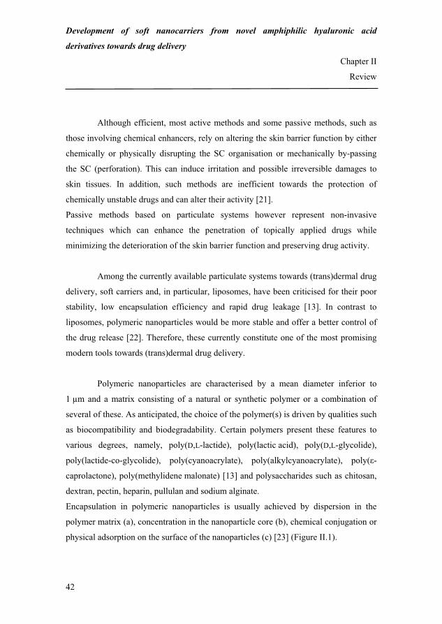
Development of soft nanocarriers from novel amphiphilic hyaluronic acid
derivatives towards drug delivery
Chapter II
Review
42
Although efficient, most active methods and some passive methods, such as
those involving chemical enhancers, rely on altering the skin barrier function by either
chemically or physically disrupting the SC organisation or mechanically by-passing
the SC (perforation). This can induce irritation and possible irreversible damages to
skin tissues. In addition, such methods are inefficient towards the protection of
chemically unstable drugs and can alter their activity [21].
Passive methods based on particulate systems however represent non-invasive
techniques which can enhance the penetration of topically applied drugs while
minimizing the deterioration of the skin barrier function and preserving drug activity.
Among the currently available particulate systems towards (trans)dermal drug
delivery, soft carriers and, in particular, liposomes, have been criticised for their poor
stability, low encapsulation efficiency and rapid drug leakage [13]. In contrast to
liposomes, polymeric nanoparticles would be more stable and offer a better control of
the drug release [22]. Therefore, these currently constitute one of the most promising
modern tools towards (trans)dermal drug delivery.
Polymeric nanoparticles are characterised by a mean diameter inferior to
1 μm and a matrix consisting of a natural or synthetic polymer or a combination of
several of these. As anticipated, the choice of the polymer(s) is driven by qualities such
as biocompatibility and biodegradability. Certain polymers present these features to
various degrees, namely, poly(D,L-lactide), poly(lactic acid), poly(D,L-glycolide),
poly(lactide-co-glycolide), poly(cyanoacrylate), poly(alkylcyanoacrylate), poly(�-
caprolactone), poly(methylidene malonate) [13] and polysaccharides such as chitosan,
dextran, pectin, heparin, pullulan and sodium alginate.
Encapsulation in polymeric nanoparticles is usually achieved by dispersion in the
polymer matrix (a), concentration in the nanoparticle core (b), chemical conjugation or
physical adsorption on the surface of the nanoparticles (c) [23] (Figure II.1).
68

Development of soft nanocarriers from novel amphiphilic hyaluronic acid
derivatives towards drug delivery
Chapter II
Review
43
a b c
Polymer matrix
Active ingredient Figure II.1: Drug encapsulation in polymeric nanoparticles.
Some studies have shown that polymeric nanoparticles can noticeably
enhance the topical delivery of, for example, chlorhexidine [24], octyl
methoxycinnamate [25] and flufenamic acid [26]. However, the mechanisms of such
achievements are still questioned.
In view of this, the objective of this article was to take stock of the current knowledge
about the mechanisms underlying polymeric nanoparticle-mediated (trans)dermal drug
delivery. First, new and improved methods for detecting skin penetration pathways are
reviewed. Then, polymeric nanoparticle fate subsequent to topical administration is
discussed based on the available experimental data. This discussion in particular
addresses the current hypothesis with regards to how polymeric nanoparticles can
enhance percutaneous penetration and/or sustain drug release within dermal skin
layers. Finally, the research gaps and future challenges of the research field are
examined.
69

Development of soft nanocarriers from novel amphiphilic hyaluronic acid
derivatives towards drug delivery
Chapter II
Review
44
II. Technical advances in assessing skin penetration pathways
II.1. Skin penetration pathways
The skin is composed of a dermis and an epidermis consisting of several
morphologically distinct layers. These are, from the inside to the outside, the stratum
basale, stratum spinosum, stratum granulosum and stratum corneum. The first three
layers constitute the viable epidermis which is a dynamic and constantly self-renewing
tissue. Cells proliferate in the basal layer and differentiate while migrating towards the
surface of the skin. At the interface between the stratum granulosum and SC, they
undergo a major terminal differentiation and are transformed into flattened, dead and
keratin-filled cells without nuclei (corneocytes). In the SC, the ultimately differentiated
corneocytes are covered by a densely cross-linked protein layer, the cell envelope, and
are embedded in lipid lamellar regions parallelly oriented to the corneocyte surface.
This results in a densely packed structure [27].
Hair follicles and the sweat apparatus are important skin structures resulting from
epidermal invaginations and which present orifices at the surface of the skin.
Considering the general anatomy of the skin (Figure II.2), three penetration
pathways potentially contribute to the transport of exogenous matter from the surface
of the skin into epidermal or dermal layers. These consist of the intercellular space
between corneocytes and of the two skin appendages mentioned above namely, hair
follicles and sweat ducts.
Historically, the intercellular path has first and foremost been considered the main skin
penetration route and is by far the most studied and discussed pathway for the infusion
of a variety of chemical substances [28].
70

Development of soft nanocarriers from novel amphiphilic hyaluronic acid
derivatives towards drug delivery
Chapter II
Review
45
Epidermis
Dermis
Hair follicleSweat duct
Sebaceous gland
Sweat gland
Arrector pili muscle
Hair shaft
Blood vessel
Pacinian corpuscle
Stratum corneum
Figure II.2: Structure of mammalian skin and possible skin penetration pathways
(adapted from [29]). The black arrows point to the potential skin penetration pathways.
It is usually agreed that the barrier function of the skin is primarily provided
by the SC. Due to the rather impermeable character of the cornified envelope around
the corneocytes, substances entering the skin through this pathway are believed to
penetrate along the tortuous intercellular space [27] (Figure II.3).
Stratum corneum
Corneocyte
Intercellular spaceIntercellular pathway
Figure II.3: Penetration through the intercellular space between corneocytes.
71

Development of soft nanocarriers from novel amphiphilic hyaluronic acid
derivatives towards drug delivery
Chapter II
Review
46
Although skin appendage orifices only represent a small fraction of the total
surface of the skin, hair follicles and sweat ducts constitute possible additional skin
penetration pathways.[30,31,32,33]
An interest for drug delivery via hair follicles has existed for several decades [30-33]
and recent reviews have addressed the potential of using this pathway for therapeutic
purposes [34,35,36]. It is usually agreed, that the advantages of follicular delivery lie
in the possibility to more directly access deep compartments of the skin and in the
large and long-term storage capacity of the hair follicle reservoir [37] which has been
estimated equal to that of the SC [38]. Interestingly, the follicular pathway is currently
considered to play a significant role in particulate drug delivery [33,39].
In contrast to hair follicles, sweat ducts have been a relatively poorly studied
system and attempts to elucidate their role in skin penetration processes remain sparse
and at an early stage of investigation. Wilke et al. [40] have addressed the question of
the sweat duct barrier function by studying the distribution of tight junction proteins
such as occludin, claudin 1 and claudin 4 throughout the eccrine and apocrine
apparatuses. They showed that the sweat duct barrier function was a non-homogeneous
property throughout these two structures. Whereas the co-localisation of occludin and
claudin 4 in sweat glands and dermal sweat ducts was considered to provide tight
junctions (i.e. a good barrier function), the sole presence of occludin in intra-epidermal
sweat ducts was recognised as being a sign of looser junctions (i.e. a reduced barrier
function). The authors therefore speculated that the intra-epidermal region of sweat
ducts could serve as an alternative penetration pathway. Although an important
finding, this theory necessitates further confirmation by specific and quantitative
penetration studies through sweat ducts. Some qualitative studies have been reported
on this subject [41,42] with only a few examples of positive penetration [42].
For an extensive insight into the various skin penetration routes, the reader is
invited to consult the excellent book chapter by Schaefer and Redelmeier [43].
72

Development of soft nanocarriers from novel amphiphilic hyaluronic acid
derivatives towards drug delivery
Chapter II
Review
47
The existence of the three (potential) skin penetration pathways implies that
chemical substances and formulated systems may penetrate through the skin by
following different paths. However, in order to understand the penetration process(es)
of a given system, a deconvolution of its parallel fluxes is necessary in order to reason
out the main entry route for this system.
Whereas microscopic imaging can provide a qualitative picture of penetration routes
into and through the skin, selective diffusion experiments or diffusion experiments
followed by selective extractions offer the possibility to quantify the amount of
penetrated matter into the various compartments of the skin and establish a hierarchy
between the different penetration routes observed by microscopy.
II.2. Microscopic imaging
Advances in microscopic imaging have crystallised into modern laser
scanning microscopy (LSM) with two major variations, namely confocal and
multiphoton laser scanning microscopy (CLSM and MPLSM).
In LSM, the sample is illuminated by a beam of laser light focused into a point at the
focal plane. This beam can be scanned in the X, Y and Z directions by a computer-
controlled scanning mirror. The fluorescence emission created at the successively
scanned points is detected by a photon multiplier tube. This information is processed
by computer hardware into an image. Spatial scanning of the sample allows
reconstructing 3-dimension (3D) images [44].
II.2.1. Confocal laser scanning microscopy
In CLSM, gas lasers are used as the illumination source. Since illumination of
the focal plane is inherently accompanied by the creation of out-of-focus fluorescence
above and below the focal plane, a pin hole is placed between the specimen and the
photon multiplier tube to avoid blurring of the final images. This pin hole
73

Development of soft nanocarriers from novel amphiphilic hyaluronic acid
derivatives towards drug delivery
Chapter II
Review
48
discriminates out-of-focus information so that the focal plane of illumination and
detection are confounded or confocal. Sharp images of the specimen can thus be
derived [44].
II.2.2. Multiphoton laser scanning microscopy
MPLSM uses solid state lasers as the illumination source. These lasers have
the particularity to generate photons in pulses and light characterised by a longer
wavelength than in CLSM. Due to technical considerations that are beyond the scope
of this review, the region of the specimen excited by MPLSM is much more confined
than that by CLSM. As a consequence, minimum or no out-of-focus fluorescence is
generated in this mode and all the information arising from the focal plane directly
reaches the photon multiplier tube without the need for a pin hole [44].
CLSM and MPLSM present decisive advantages over other more
conventional microscopy techniques such as light microscopy, transmission electron
microscopy:
(i) the endogenous fluorescence of the skin allows achieving images on which the
structural details of this tissue can clearly be identified,
(ii) mechanical sectioning is no longer necessary which allows circumventing artefacts
related to sample fixation and traditional histological investigations,
(iii) samples are non-invasively studied and high-resolution 3D images can be derived
[45].
However, some technical differences between CLSM and MPLSM give
further advantages to MPLSM which is more sensitive than CLSM because all the
information generated at the focal plane directly reaches the detector. Besides, the
longer wavelength light used in MPLSM is less damaging to samples though more
penetrating than the shorter wavelength light used in CLSM [44].
74

Development of soft nanocarriers from novel amphiphilic hyaluronic acid
derivatives towards drug delivery
Chapter II
Review
49
For further technical information on CLSM and MPLSM, the reader is invited to
consult the reviews by Oheim et al. [46] and Diaspro et al. [47], respectively.
CLSM and MPLSM have allowed significant improvements in the
observation and understanding of biological phenomena and have recently been used,
as will be shown in part II, for the qualitative deconvolution of polymeric nanoparticle
skin penetration pathways.
The quantitative assessment of the different skin penetration pathways constitutes the
second aspect in understanding the fate of topically applied formulations. Advances in
this field are the subject of the following paragraph.
II.3. Skin penetration quantification
In the past, the distinction between SC and follicular penetration has been
tackled by comparing penetration fluxes through skin containing follicles and skin
devoid of these. However, interpretation of the results generated using such an
approach is problematic as hairless and hairy skins can exhibit different barrier
functions due to their structural and biochemical differences.
Innovative approaches were recently proposed to circumvent this problem and allow to
distinguish between SC and appendageal penetration. These are the skin sandwich
model and differential stripping.
II.3.1. The skin sandwich model
The skin sandwich model is a composite skin system composed of a stack of a
human whole epidermis (nucleated epidermis and SC) and of an additional SC
membrane sampled from the same donor. As skin appendage orifices only represent
0.1 % of the total skin surface [38], little chance exists that the shunts of the two
different skin components superimpose. Therefore, in this composite skin tissue, the
75
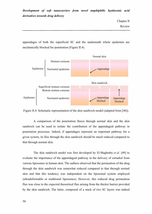
Development of soft nanocarriers from novel amphiphilic hyaluronic acid
derivatives towards drug delivery
Chapter II
Review
50
appendages of both the superficial SC and the underneath whole epidermis are
mechanically blocked for penetration (Figure II.4).
Nucleated epidermis
Stratum corneum
Epidermis
Normal skin
Appendage
Nucleated epidermis
Superficial stratum corneumBottom stratum corneum
Epidermis
Skin sandwich
Appendage blocked
Appendage blocked
Figure II.4: Schematic representation of the skin sandwich model (adapted from [48]).
A comparison of the penetration fluxes through normal skin and the skin
sandwich can be used to isolate the contribution of the appendageal pathway in
penetration processes. Indeed, if appendages represent an important pathway for a
given system, its flux through the skin sandwich should be much reduced compared to
that through normal skin.
The skin sandwich model was first developed by El Maghraby et al. [49] to
evaluate the importance of the appendageal pathway in the delivery of estradiol from
various liposomes in human skin. The authors observed that the permeation of the drug
through the skin sandwich was somewhat reduced compared to that through normal
skin and that this tendency was independent on the liposomal system employed
(ultradeformable or traditional liposomes). However, this reduced drug permeation
flux was close to the expected theoretical flux arising from the thicker barrier provided
by the skin sandwich. The latter, composed of a stack of two SC layers was indeed
76

Development of soft nanocarriers from novel amphiphilic hyaluronic acid
derivatives towards drug delivery
Chapter II
Review
51
twice as thick as the barrier provided by normal skin. The authors therefore excluded
the appendageal pathway as the main penetration route for the delivery of estradiol
from liposomes in human skin.
Barry [48] later systematically evaluated the performances of the skin
sandwich model using various model compounds such as estradiol (lipophilic),
mannitol (hydrophilic) and L-glutamic acid (negatively charged) administered by
iontophoresis. A repeated consistency between the theoretical and experimental
behaviours of these compounds confirmed the accuracy and validity of the skin
sandwich model. For instance, due to its negative charge, L-glutamic acid was
expected to follow the path of least resistance and to penetrate through damaged
regions of the skin and skin appendages. In addition, the creation of artificial pores due
to the temporary disruption of the intercellular lipids was expected to contribute to the
enhancement of its transport. Experimentally, the flux ratio between sandwiched skin
and normal skin was close to zero for L-glutamic acid which confirmed that the
appendageal pathway dominated the iontophoretic transport of L-glutamic acid.
However, when electroporation was used the flux ratio did not fall to zero evidencing
that the electrical pulses created transient aqueous pores in the intercellular lipids
through which L-glutamic acid was transported across sandwiched skin.
The identical source between the skin sandwich and normal skin and the
mechanical shunt obstruction without exogenous material circumvent interpretational
difficulties related to the different barrier functions of hairless and hairy skins. In
addition, Barry [48] demonstrated using the Monte Carlo approach that results
remained consistent even in the case of slightly imperfect adherence between the two
different skin components in the skin sandwich. Interestingly, the author emphasised
that the skin sandwich model could potentially be used to deconvolute the penetration
pathways of colloidal particles.
77

Development of soft nanocarriers from novel amphiphilic hyaluronic acid
derivatives towards drug delivery
Chapter II
Review
52
II.3.2. Differential stripping
Differential stripping is based on the combination of two existing techniques
namely, tape stripping and the cyanoacrylate follicular biopsy.
On the one hand, tape stripping is a frequently used method to investigate the
penetration of topically applied substances into the skin [50]. After applying a
formulation onto the skin, several successive skin layers are removed with adhesive
films. Each skin strip contains corneocytes and variable amounts of the topically
applied system which can further be extracted by adequate techniques. The correlation
between the amount of extracted substance from each individual strip and the strip
number allows tracking the penetration profile of a given system.
On the other hand, the cyanoacrylate follicular biopsy uses a quick-setting polymer to
extract the infundibular content of pilosebaceous units [51]. This technique is useful
for studying the contents of follicles and can be employed for analysing the penetration
of topically applied compounds or formulations through hair follicles.
Teichmann et al. [52] were the first to combine the two above described
techniques to selectively and non-invasively study the penetration of topically applied
substances into the SC and follicular structures. This method called differential
stripping uses tape stripping to first collect the amount of topically applied substances
penetrated into the SC and the cyanoacrylate follicular biopsy to secondly collect the
amount penetrated into follicular structures.
By using these two successive stripping techniques, the authors determined the amount
of sodium fluorescein penetrated in vivo in the SC and hair follicles of human back
skin. After 30 min, 54 % ± 8.1 % of the dye was recovered of which 95 % ± 14 % in
the SC and 5 % ± 0.9 % in the follicular infundibula. After 24 and 48 hours, sodium
fluorescein was still detected in follicular structures although the concentration of the
dye decreased over time.
78
Development of soft nanocarriers from novel amphiphilic hyaluronic acid
derivatives towards drug delivery
Chapter II
Review
53
Differential tape stripping therefore constitutes a novel method to selectively study
skin penetration pathways. It gives a number of opportunities for the study of the
pharmacokinetics of topically applied substances and more generally for research in
the field of dermatology.
The use of CLSM and MPLSM and/or selective extraction techniques has
allowed new insights into the mechanisms underlying polymeric nanoparticle-
mediated drug delivery which are reviewed hereafter.
III. Mechanisms underlying polymeric nanoparticle-mediated drug delivery
III.1. Redistribution and accumulation of polymeric nanoparticles in the skin
As shown in Table II.2, the redistribution of topically applied polymeric
nanoparticles on the surface of the skin has been shown to occur towards the (i) skin’s
superficial reliefs [26,53,54], (ii) hair follicles [55,56,57], (iii) intercellular space in the
SC and (iv) subepidermal skin tissues [58].
In studies [53] and [54], for which the nanoparticles were founds in the skin
furrows/dermatoglyphs, optical cross-sections of non-follicular skin regions further
showed that the nanoparticle distribution was only superficial. This suggested that skin
furrows/dermatoglyphs were not penetration pathways for the nanoparticles.
Studies [55] to [57] reported that hair follicles constituted a significant penetration
pathway for nanoparticles with a diameter between 40 and 320 nm.
However, polymer-coated intrinsically fluorescent quantum dots with hydrodynamic
diameters between 14 and 45 nm were localised in the SC and in the skin subepidermal
layers [58]. This shows that the intercellular space constitute another penetration
pathway for nanoparticles with small diameters.
79

Tabl
e II
.2: R
edis
tribu
tion
of to
pica
lly a
pplie
d po
lym
eric
nan
opar
ticle
s on
the
surf
ace
of th
e sk
in.
NP
stan
ds fo
r nan
opar
ticle
.
Ref
eren
ce
NP
type
/enc
apsu
late
d su
bsta
nce/
size
A
dmin
istra
tion
and
dete
ctio
n
cond
ition
s R
esul
ts
[26]
Po
ly(la
ctid
e-co
-gly
colid
e)/fl
ufen
amic
acid
/330
nm
30-
min
incu
batio
n w
ith h
uman
skin
in v
itro
MPL
SM im
agin
g
Uni
form
NP
dist
ribut
ion
on th
e su
rfac
e of
the
skin
and
with
in th
e
skin
der
mat
ogly
phs
No
NPs
foun
d be
twee
n or
with
in c
orne
ocyt
es
[53]
Car
boxy
late
-mod
ified
Flu
oSph
eres
®
(flu
ores
cent
pol
ysty
rene
)/flu
ores
cein
-
5-is
othi
ocya
nate
/20
or 2
00 n
m
30-
, 60-
or 1
20-m
in in
cuba
tion
with
porc
ine
skin
in v
itro
C
LSM
imag
ing
Tim
e de
pend
ent N
P ac
cum
ulat
ion
arou
nd th
e fo
llicu
lar o
peni
ngs
and
in th
e fu
rrow
s of t
he sk
in
The
long
er th
e in
cuba
tion
time,
the
high
er th
e de
tect
ed
fluor
esce
nce
[54]
5-flu
ores
cein
amin
e-co
vale
ntly
-
labe
lled
poly
(lact
ide-
co-
glyc
olid
e)/T
exas
Red
/300
nm
5-h
our i
ncub
atio
n w
ith h
uman
skin
in v
itro
MPL
SM im
agin
g
NP
loca
tion
in th
e sk
in d
erm
atog
lyph
s
[55]
Pol
y(�-
capr
olac
tone
)-bl
ock-
poly
(eth
ylen
e gl
ycol
)/rub
rene
/40
or
130
nm
12-
hour
incu
batio
n w
ith g
uine
a pi
g
skin
in v
ivo
CLS
M im
agin
g of
bio
psie
d gu
inea
pig
skin
NP
accu
mul
atio
n in
hai
r fol
licle
s and
thei
r sur
roun
ding
tiss
ues
[56]
F
luoS
pher
es®
(flu
ores
cent
poly
styr
ene/
40 n
m
15
to 1
6-ho
ur in
cuba
tion
with
hum
an sk
in in
vitr
o
CLS
M im
agin
g of
folli
cula
r
cryo
sect
ions
of c
yana
cryl
ate
strip
ped
hum
an sk
in
Pre
senc
e in
ha
ir fo
llicl
es
dow
n to
a
dept
h of
22
5±34
μm
(cor
resp
ondi
ng to
the
occu
rren
ce o
f seb
aceo
us g
land
s)
80

Tabl
e II
.2: R
edis
tribu
tion
of to
pica
lly a
pplie
d po
lym
eric
nan
opar
ticle
s on
the
surf
ace
of th
e sk
in (c
ontin
ued)
.
[57]
R
esom
er®
/5-
fluor
esce
inam
ine/
320
nm
1-h
our i
ncub
atio
n w
ith p
orci
ne sk
in
in v
itro
LSM
imag
ing
of p
hysi
cal s
kin
cros
s-
sect
ions
Pen
etra
tion
alon
g ha
ir fo
llicl
es d
own
to a
dep
th o
f 15
00 μ
m
whe
n th
e na
nopa
rticl
es w
ere
mas
sage
d on
the
surf
ace
of th
e sk
in
[58]
Po
lym
er-c
oate
d in
trins
ical
ly
fluor
esce
nt q
uant
um d
ots/
14-4
5 nm
8 o
r 24-
hour
incu
batio
n w
ith p
orci
ne
skin
in v
itro
CLS
M im
agin
g
NP
pene
tratio
n an
d lo
calis
atio
n in
the
SC
and
sub
epid
erm
al
laye
rs
81

Development of soft nanocarriers from novel amphiphilic hyaluronic acid
derivatives towards drug delivery
Chapter II
Review
56
In summary, according to the recent studies focusing on understanding the
fate of topically applied nanoparticles, these can superficially redistribute to skin
reliefs such as the skin furrows/dermatoglyphs and follicular openings. They can also
penetrate along hair follicles down to macroscopic depths or through the intercellular
space down to subepidermal layers. This last finding by Ryman-Rasmussen et al. [58]
is unusual and in apparent contradiction with the results of Alvarez-Roman et al. [53],
Shim et al. [55] and Vogt et al. [56]. However, it is not the only one of its kind since
Menzel et al. [59] demonstrated using an ion beam analysis that lanceolate metal
(titanium oxide) nanoparticles (45- to 150-nm long, 17- to -35-nm wide) penetrated
porcine skin from the SC to the stratum granulosum through the intercellular space
between corneocytes after an 8 hour incubation of the nanoparticles in vivo.
Considering the similarity in size of 20- [53] and 40-nm [55,56] polystyrene
nanoparticles on the one hand and 14- to 45-nm [58] polymer-coated quantum dots on
the other hand and their different penetration profiles, polymeric nanoparticle skin
penetration seems a more complex phenomenon than one just dependant on the size of
the nanoparticles. Some possible mechanisms of their transport into the skin are
presented in the following paragraph.
III.2. Possible mechanisms underlying polymeric nanoparticle skin penetration
Logically, it can be conceived that the dimensions of the penetration pathway
is a major factor for the selective diffusion of polymeric nanoparticles.
Penetration though the SC can therefore be imagined to require that the nanoparticle
size complies with the narrow dimensions of the intercellular space between
corneocytes. Bouwstra et al. [60] described the structure of the intercellular lipids as a
stack of lamellae with characteristic periodicities of 6.4 and 13.4 nm [61] (the average
thickness of the intercellular space is about 100 nm). Nanoparticles presenting at least
one dimension within the order of magnitude of that of the periodic phases can
hypothetically penetrate through the intercellular space [58].
82

Development of soft nanocarriers from novel amphiphilic hyaluronic acid
derivatives towards drug delivery
Chapter II
Review
57
Penetration of larger particles may also be possible when the lipid organisation is
disrupted such as in the case of high tissue hydration [62,63] and certain skin diseases
[64].
In addition, the physicochemical properties of the nanoparticles can also be
accountable for the penetration through the intercellular space of larger particles. For
example, the poor solubility of the polymer matrix in the intercellular lipids can cause
a decrease in the apparent hydrodynamic diameter of the nanoparticles facilitating their
penetration through the intercellular space [58].
Nanoparticle penetration along hair follicles however seems to be less
restricted than through the intercellular space due to larger dimensions of follicular
infundibula [65,66]. Ossadnik et al. [57] explained that nanoparticle penetration along
hair follicles might occur owing to a pumping effect by hair shafts caused by hair
tilting. Indeed, the cuticles arising from keratinocyte desquamation form a structured
surface, which can be assimilated to a zigzag relief. The latter is determined by the
thickness of the keratin cells which is between 500 and 800 nm. With the continuous
movement of hair, the cuticle cells may act as a geared pump and push particles down
to the hair infundibulum. According to Ossadnik et al. [57], the structural
characteristics of follicular infundibula imply that the optimum nanoparticle size for
penetration along hair follicles is below 700 nm.
Based on these general mechanisms, the study of the influence of
characteristics such as the nanoparticle size, surface charge and shape can further give
valuable insight in explaining the mechanism of their skin penetration.
83

Development of soft nanocarriers from novel amphiphilic hyaluronic acid
derivatives towards drug delivery
Chapter II
Review
58
III.2.1. Influence of nanoparticle size
Shim et al. [55] correlated the efficiency of mixnoxidil permeation through
hairy guinea pig skin to the size of the poly(�-caprolactone)-block-poly(ethylene
glycol) nanoparticles. They found that the in vitro permeation of mixnoxidil
encapsulated in 40-nm nanoparticles was 1.7-fold higher than that of mixnoxidil
encapsulated in 130-nm nanoparticles after 18 hours. Interestingly, no significant
influence of the size of the nanoparticles on the permeation of mixnoxidil could be
evidenced in hairless guinea pig skin. Moreover, mixnoxidil permeation was
substantially lower in hairless than in hairy guinea pig skin.
Alvarez-Roman et al. [53] observed that 20-nm fluorescent polystyrene
nanoparticles (carboxylate-modified FluoSpheres®) redistributed more rapidly and
selectively to the follicular openings of porcine skin in vitro than the 200-nm
nanoparticles after 120 min. This phenomenon was attributed to the higher specific
surface area developed by the smaller nanoparticles.
Vogt et al. [56] detected 40-nm polystyrene nanoparticles (FluoSpheres®) in
follicular CD1a+ cells in cyanoacrylate stripped human skin after 15 to 16 hours in
vitro. By contrast, the 750- and 1,500-nm nanoparticles (FluoresbriteTM) were not
detected in these cells. CLSM imaging of follicular cryosections evidenced the
penetration of the 40-nm nanoparticles along hair follicles down to a maximum depth
of 225 ± 34 μm whereas the larger nanoparticles (750 and 1,500 nm) remained in the
uppermost part of the follicles.
Ryman-Rasmussen et al. [58] however demonstrated a rather size-
independent penetration of differently sized (hydrodynamic diameters between 14 and
45 nm) and coated (poly(ethylene glycol), carboxylic acids and poly(ethylene glycol
84

Development of soft nanocarriers from novel amphiphilic hyaluronic acid
derivatives towards drug delivery
Chapter II
Review
59
amine)) spherical quantum dots in porcine skin. All nanoparticle types penetrated
down epidermal or dermal skin layers after 8 hour in vitro.
These results suggest that the size of the nanoparticles is an important factor
of the penetration kinetics [53,55] and pathway. Nanoparticles presenting dimensions
incompatible with that of the penetration pathway are retained superficially [56] while
those presenting dimensions under several tens of nanometres penetrate the SC
through the intercellular space between corneocytes [58]. Larger nanoparticles but
which diameters do not exceed 700 nm preferentially follow the follicular pathway
[53,55,56]. Within this size range, differences in nanoparticle size result in differences
in penetration velocity and extent: the smaller the nanoparticles, the faster and deeper
their transport in the follicles.
III.2.2. Influence of nanoparticle surface charge
Ryman-Rasmussen et al. [58] observed a charge-independent penetration of
spherical quantum dots and a charge-dependent penetration of ellipsoid quantum dots
in porcine skin. Whether coated with neutral poly(ethylene glycol) (PEG), anionic
carboxylic acids (CA) or cationic poly(ethylene glycol amine) (PEG-amine), similarly
sized spherical quantum dots penetrated epidermal (PEG and CA) and dermal (PEG-
amine) skin layers after 8 hours in vitro. However, only PEG and PEG-amine coated
ellipsoid quantum dots penetrated epidermal skin layers after 8 hours whereas CA
ellipsoid quantum dots only penetrated epidermal skin layers after 24 hours. This
suggested that the negatively charged ellipsoid quantum dots were more retained in the
intercellular space than the neutral and positively charged ellipsoid quantum dots.
Kohli and Alpar [67] however evidenced the in vitro penetration and
accumulation in porcine SC and viable epidermis of negatively charged fluorescent
latex nanoparticles (diameters 50 and 500 nm) after 6 hours. Although unable to fully
85

Development of soft nanocarriers from novel amphiphilic hyaluronic acid
derivatives towards drug delivery
Chapter II
Review
60
explain the mechanism by which these nanoparticles penetrated the in the SC, the
authors speculated that the repulsive forces between the negatively charged
nanoparticles and skin lipids might have resulted in the formation of temporary
channels through which the 50- and 500-nm nanoparticles could be transported.
The scarcity of specific studies on the influence of nanoparticle surface charge
on their skin penetration makes it a difficult task to draw general conclusions about
this phenomenon. The negative charge of the lipid bilayers in the intercellular space
should logically favour the transport of positively charged species. However, the study
by Kohli and Alpar [67] is in apparent contradiction with this hypothesis. Further
investigation of this topic will be necessary to understand the electrokinetic
interactions between polymeric nanoparticles and skin tissues.
III.2.3. Influence of nanoparticle shape
Ryman-Rasmussen et al. [58] observed that PEG-amine-coated spherical
quantum dots penetrated down to dermal skin layers whereas similarly coated ellipsoid
quantum dots only penetrated down to epidermal skin layers in porcine skin after
8 hours in vitro. CA-coated spherical quantum dots penetrated and accumulated in the
epidermis faster (after 8 hours) than similarly coated ellipsoid quantum dots (after
24 hours). As the difference in hydrodynamic diameter between these spherical and
ellipsoid quantum dots was minor (less than 5 nm) the variation in their penetration
depths/times was attributed to their shape. The more homogenously shaped spherical
quantum dots penetrated in deeper skin layers than their heterogeneously shaped
ellipsoid analogues.
These results suggest that the more homogeneous the shape of the nanoparticles the
easier their penetration into the intercellular space.
86

Development of soft nanocarriers from novel amphiphilic hyaluronic acid
derivatives towards drug delivery
Chapter II
Review
61
With a better knowledge of the fate of topically applied nanoparticle and of
the mechanisms underlying their penetration, it is now possible to formulate
hypothesis with regards to how these enhance and/or sustain drug release.
III.3. Hypothesis with regards to how polymeric nanoparticles enhance percutaneous
penetration and/or sustain drug release
In most of the cases, due to the relatively large dimensions of most of the
currently available polymeric nanoparticles (> 40 nm), enhancement of the
percutaneous penetration and/or sustained release of encapsulated substances through
non-follicular regions of the skin cannot be attributed to the co-penetration of the
carrier and the encapsulated substance through skin tissues.
For example, Luengo et al. [26] observed an increase in the flufenamic acid in vitro
transport through human abdominal skin when the drug was encapsulated in
poly(lactide-co-glycolide) (PLGA) nanoparticles compared to that of the free drug
after 12 hours. However, the PLGA nanoparticles remained homogeneously
distributed on the surface of the skin and within skin dermatoglyphs and no
nanoparticles were detected within or between corneocytes.
Stracke et al. [54] followed the release, accumulation and penetration of Texas Red-
containing PLGA nanoparticles by MPLSM. Multi fluorescence labelling of this
system enable the authors to follow the fate of both the nanoparticles and the
encapsulated compound. The Texas Red concentration profiles over time in various
skin compartment acquired by multiphoton spectral imaging showed that Texas Red
started to be absorbed by the SC immediately after topical application. This suggested
that release from the nanoparticle had probably already begun before the penetration
experiment within the hydrogel used for the formulation.
Alvarez-Roman et al. [53] have put forward that the penetration improvement
achieved by using nanoparticles is due to an efficient liberation of the encapsulated
substance into the SC.
87

Development of soft nanocarriers from novel amphiphilic hyaluronic acid
derivatives towards drug delivery
Chapter II
Review
62
Certain interactions between polymeric nanoparticles and the surface of the
skin can explain the sustained release of the encapsulated substances.
For example, Olvera-Martinez et al. [68] observed lower penetrated amounts of the
sunscreen octyl methoxycinnamate into human skin in vivo from cellulose acetate
phthalate nanocapsules (diameter 460 nm) than from a conventional oil-in-water
emulsion or a nanoemulsion prepared with the non-ionic emulsifying agent poly(vinyl
alcohol). The authors suggested that the slower and more superficial release achieved
by the nanocapsules was probably due to the formation of a nanoparticle superficial
depot from which the sunscreen was progressively infused to the outermost layers of
the skin.
Lboutounne et al. [24] showed that chlorhexidine-loaded poly(�-caprolactone)
nanoparticles partially wetted porcine skin. The contact angle determined by scanning
electron microscopy and surface tension for these nanoparticles were 124 degrees and
147 dyn/cm, respectively. The authors argued that this partial wetting was responsible
for the sustained release, enhanced concentration within SC and reduced percutaneous
absorption of chlorhexidine. Indeed, the sufficient flexibility of the nanoparticles
allowed their adherence to the skin and their sufficient rigidity permitted avoiding
rapid drug diffusion [69].
Other studies have shown that nanoparticles could penetrate through hair
follicles [37,53,55,56] and could be stored in the follicular structures of human claves
for up to 10 days [39]. However the different authors admit that the release
mechanisms in such conditions remain unclear.
Finally, nanoparticle penetration through non-follicular skin structures could
be a possible mechanism to explain the penetration enhancement of encapsulated
substances [58]. However, to the best of our knowledge, very few studies using
microscopic imaging have specifically demonstrated this phenomenon.
88

Development of soft nanocarriers from novel amphiphilic hyaluronic acid
derivatives towards drug delivery
Chapter II
Review
63
Although recent advances have been achieved in the understanding of
polymeric nanoparticles-mediated (trans)dermal drug delivery, this research field
remains fraught with both scientific and regulatory challenges. These are examined
hereafter.
IV. Future challenges in polymeric nanoparticle-mediated (trans)dermal drug
delivery
IV.1. Nanoparticle design
Polymeric nanoparticles possessing a high drug loading capacity and which
can target diseased body sites, be triggered at the right moment and sustain drug
release for an appropriate period of time, represent the Holy Grail of (trans)dermal
drug delivery.
However, drug loading in polymeric nanoparticles is limited by their size. It can
therefore be expected that the improved skin penetration of small nanoparticles will be
at the expense of the amount of encapsulated drug. For this reason, an optimal balance
will have to be found for each given application between the desired extent of
penetration (partly determined by the size of the nanoparticles) and the dose required
to achieve a therapeutic effect.
Nanoparticle targeting, triggering and sustained drug release represent other
major challenges of polymeric nanoparticle design.
Nanoparticle targeting is probably the most complex feature to achieve. It requires the
modification of the nanoparticle surface in order to avoid non-specific interactions
with cells and proteins and drug accumulation in non-target tissues [70].
Barry [21] commented that (trans)dermal drug delivery would be optimal if it could be
coordinated with a continuous diagnosis of the actual physiological condition of each
specific patient. Drugs could thus be delivered only when required and for the time
89

Development of soft nanocarriers from novel amphiphilic hyaluronic acid
derivatives towards drug delivery
Chapter II
Review
64
needed. The release of encapsulated substances from topically applied nanoparticles is
however currently more often undergone than controlled. Studies on this subject have
been limited to the observation of the fate of nanoparticulate systems when
administered on the skin. Controlled triggering is not yet an achieved feature of
polymeric nanoparticles. It will require the development of advanced devices able to
respond to external stimuli. For dermal applications, these stimuli could consist in light
illumination or the delayed application of a kick-off agent able to induce a therapeutic
cascade effect [54].
Optimisation and conjugation of the above-described complex aspects of
polymeric nanoparticle design are necessary to create “smart” nanoparticles. To
appreciate the performances of these advanced devices, it will be necessary to
overcome some of the remaining technical limitations in their detection for an accurate
assessment of their in situ behaviour.
IV.2. Nanoparticle performance assessment
IV.2.1. Fate
In the past, encapsulation of a fluorescent label into polymeric nanoparticles
was usually used to follow their fate by fluorescence microscopy. However, this
approach only truly allows monitoring the fate of the label and not specifically that of
the whole system. Indeed, it can difficult to control the diffusion of the label outside
the nanoparticles during the experiment.
Alvarez-Roman et al. [53] circumvented this issue by covalently binding fluorescein 5-
isothiocyanate (FITC) to non-biodegradable polystyrene nanoparticles before
following their fate in porcine skin. The covalent binding of the label to the
nanoparticles and nanoparticle non-biodegradability ensured that the fluorescence
90
Development of soft nanocarriers from novel amphiphilic hyaluronic acid
derivatives towards drug delivery
Chapter II
Review
65
detected for FITC could unambiguously be attributed to the presence of intact
polystyrene nanoparticles.
Similarly, Stracke et al. [54] used a dually labelled system consisting of 5-
fluoresceinamine-bound PLGA nanoparticles and encapsulated Texas Red.
In summary, intrinsic fluorescence or in the absence of thereof, covalent
fluorescence labelling of the nanoparticles combined with fluorescence (labelling) of
the encapsulated substance are means to ensure selective monitoring in skin tissues.
Non-biodegradability of the nanoparticles is a further guarantee that these will be
identified as integral structures.
IV.2.2. Skin penetration
The methods currently available for evaluating polymeric nanoparticle skin
penetration still entail artefacts related to the nature of the analytical technique chosen.
The lack of skin vasculature in in vitro penetration/permeation experiments, possible
modification of the skin lipid organisation and redistribution of the penetrants while
handling the samples, are aspects which can affect the veracity of the experimental
results.
However, systematic in vivo assessments in animals or humans involving biopsies
represent invasive methods. Future analytical techniques will therefore have to focus
on the development of systems mimicking or approaching actual in vivo conditions and
analytical tools involving minimal disturbance of these systems.
For skin visualisation, Stracke et al. [54] emphasise that samples should ideally be
visualised in vivo, without fixation, non-invasively, on the same sample of human skin
examined at different time in order to avoid the problem of sample variability in time-
dependent penetration studies.
91

Development of soft nanocarriers from novel amphiphilic hyaluronic acid
derivatives towards drug delivery
Chapter II
Review
66
The accurate assessment of polymeric nanoparticle penetration is essential to
measure the benefits but also the risks of such systems. Risk assessment will in the
future be a crucial step towards using polymeric nanoparticles for actual therapeutic
applications.
IV.3. Nanoparticle risk assessment
Due to their unique properties, polymeric nanoparticles are expected in the
near future to dramatically improve humans’ quality of life. However, unequalled
features such as their size, specific area and surface properties make them potential
triggering agents of reactions at the cellular levels that are currently unknown. As with
many emerging and promising research fields there is a risk that the development of
nanoparticle technologies overtakes investigations to assess their health and
environment entailments.
To date only little formal research exist on the health and environmental
impact of nanoparticles and scientific reviews with only a few exceptions [71] mainly
addressed the risk assessment of airborne nanoparticles [72,73,74]. Morgan [75]
recently underlined that due to the scarcity of scientific data on nanoparticle risk
assessment, scientific and regulatory workers have the opportunity to address this
question strategically by driving research to selected crucial areas for the
understanding of the risk involved in each given nanoparticle applications. Therefore,
general screening strategies have been proposed [75,76].
The working group of Oberdorster et al. [76] proposed a general data
gathering strategy for the hazard identification of engineered nanomaterials for the
oral, dermal, inhalation and injection routes. The three key element of this strategy are
physicochemical characterisation, in vitro (cellular and non-cellular) and in vivo
assays.
92

Development of soft nanocarriers from novel amphiphilic hyaluronic acid
derivatives towards drug delivery
Chapter II
Review
67
Nanomaterial physico-chemical characterisation is indeed essential if experimental
data are to be interpreted in relation with the material properties and should encompass
particle size distribution, agglomeration state, shape, crystal structure, chemical
composition surface area, surface chemistry, surface charge and porosity.
Nanomaterial characterisation after administration in vitro or in vivo is considered
ideal although it currently presents significant analytical challenges. Alternatively,
characterisation of the material as administered can provide valuable information.
Although there exists some well-documented issues with in vitro tests, these are
considered important adjuncts to in vivo studies since they allow specific pathways to
be isolated and tested under controlled conditions which is not feasible in vivo. Non-
cellular in vitro tests can provide information about nanomaterial biopersistence, free
radical generation, interaction with proteins and activation of humoral systems.
In vivo tests constitute a crucial step in nanomaterial risk assessment since they provide
an evaluation of the systems’ performances in situ. However, these are currently
limited by research gaps in the design of viable in vivo detection techniques and in the
development of standardized and well characterized nanomaterial samples.
Oberdorster et al. [76] further provide recommendations with regards to how these
three key fields can be investigated. Their work is a major contribution to the emerging
nanomaterial risk assessment.
Morgan [75] also proposed a preliminary and general framework for
nanomaterial hazard identification in the form of an influence diagram. The latter was
developed based on expert elicitation and describes the presumed variables, their inter-
connexions and influence on the uptake, transport, fate, and toxicity of nanoparticles,
four properties considered as closely related human health and ecological risk. The
purpose of the expert elicitation was to collect and organise the knowledge of
experienced people working with various aspects of nanoparticle technology in order
to create a valuable tool for the systematic identification and prioritisation of crucial
research areas.
93
Development of soft nanocarriers from novel amphiphilic hyaluronic acid
derivatives towards drug delivery
Chapter II
Review
68
For the (trans)dermal use of nanoparticles, the influence diagram spots nanoparticle
size distribution, surface reactivity, shape and potential binding to a matrix as the main
properties affecting nanoparticle uptake by the skin. It anticipates that nanoparticle size
distribution, surface reactivity and coating (including added molecular groups),
adsorption tendency and durability are likely to influence nanoparticle transport and
fate. Finally nanoparticle size distribution, surface reactivity and coating, adsorption
tendency and chemical composition are expected to impact nanoparticle toxicity.
The advantages of Morgan’s approach lie in its capacity to incorporate new knowledge
as it is generated and to remain in the long run a relevant tool for nanoparticle risk
assessment.
V. Conclusion
The purpose of this article was to review the current knowledge about the
mechanisms underlying polymeric nanoparticle-mediated (trans)dermal drug delivery,
spot out research gaps and outline the challenges faced by this scientific field.
From the data available today, it appears that topically applied polymeric
nanoparticles on the skin redistribute towards its superficial reliefs or penetrate along
hair follicles or through the intercellular space. The penetration pathways followed by
the particles certainly depend on a variety of properties such as nanoparticle shape,
surface composition and charge but a number of studies have reported that
nanoparticle size is probably the most determining factor. However, it is difficult to
draw general rules due to the behavioural variability of nanoparticles presenting
similar properties.
Although the mechanisms by which nanoparticles enhance and/or sustain drug
release within the skin still remain partially questioned, there are some indications that
the formation of a nanoparticle depot at the surface of the skin or in the hair follicles
94
Development of soft nanocarriers from novel amphiphilic hyaluronic acid
derivatives towards drug delivery
Chapter II
Review
69
from which drugs are efficiently released may be involved. Release in the intercellular
space has not yet been unequivocally evidenced but is a priori not to be excluded.
Many past studies have followed the fate of fluorescent encapsulated
substances in the nanoparticles. This does not allow understanding release mechanisms
and more recent studies have underlined the need for labelling each of the individual
components of a given nanoparticulate system and performing selective penetration
studies (skin sandwich model, differential stripping).
Scientific limitations are mainly related to research gaps in the development
of standardized and well characterized nanomaterial samples and in the design of
viable in vivo non-invasive detection techniques. Nanoparticle targeting and release
triggering will also represent major technical challenges.
The future of polymeric nanoparticles-mediated (trans)dermal drug delivery will rely
on how well our present and next generations of researchers will take up these
challenges and coordinate interdisciplinary collaborations for the improvement of
patient condition worldwide.
References [1] Brown, M. B.; Martin, G. P.; Jones, S. A.; Akomeah, F. K. Dermal and
transdermal drug delivery systems: current and future prospects. Drug Deliv.
2006, 13, 175-187.
[2] Frost & Sullivan. U.S. emerging transdermal drug delivery technologies markets.
Business report from 2003 available at http://www.frost.com/prod/servlet/frost-
home.pag.
[3] Trommer, H.; Neubert, R. H. H. Overcoming the stratum corneum: the
modulation of skin penetration. Skin Pharmacol. Physiol. 2006, 19, 106-121.
95

Development of soft nanocarriers from novel amphiphilic hyaluronic acid
derivatives towards drug delivery
Chapter II
Review
70
[4] Moser, K.; Kriwet, K.; Naik, A.; Kalia, Y. N.; Guy, R. H. Passive skin penetration
enhancement and its quantification in vitro. Eur. J. Pharm. Biopharm. 2001, 52,
103-112.
[5] Sloan, K. B.; Wasdo, S. C.; Rautio, J. Design for optimized topical delivery:
prodrugs and a paradigm change. Pharm. Res. 2006, 23, 2729-2747.
[6] Paudel, K. S.; Nalluri, B. N.; Hammell, D. C.; Valiveti, S.; Kiptoo, P.; Hamad, M.
O.; Crooks, P. A.; Stinchcomb, A. L. Transdermal delivery of naltrexone and its
active metabolite 6-beta-naltrexol in human skin in vitro and guinea pigs in vivo.
J. Pharm. Sci. 2005, 94, 1965-1975.
[7] Malmsten, M. Soft drug delivery systems. Soft Matter 2006, 2, 760-769.
[8] Fang, J. Y.; Hwang, T. L.; Huang, Y. L. Liposomes as vehicles for enhancing
drug delivery via skin routes. Curr. Nanosci. 2006, 2, 55-70.
[9] Godin, B.; Touitou, E. Ethosomes: new prospects in transdermal delivery. Crit.
Rev. Ther. Drug Carrier Syst. 2003, 20, 63-102.
[10] Choi, M. J.; Maibach, H. I. Liposomes and niosomes as topical drug delivery
systems. Skin Pharmacol. Physiol. 2005, 18, 209-219.
[11] Nanda, A.; Nanda, S.; Dhall, M.; Rao, R. Transferosomes: a novel
ultradeformable vesicular carrier for transdermal drug delivery. Drug Deliv.
Technol. 2005, 5 available at http://www.drugdeliverytech.com.
[12] Muller, R. H.; Mader, K.; Gohla, S. Solid lipid nanoparticles (SLN) for controlled
drug delivery - A review of the state of the art. Eur. J. Pharm. Biopharm. 2000,
50, 161-177.
[13] Soppimath, K. S.; Aminabhavi, T. M.; Kulkarni, A. R.; Rudzinski, W. E.
Biodegradable polymeric nanoparticles as drug delivery devices. J. Controlled
Release 2001, 70, 1-20.
[14] Gehl, J. Electroporation: theory and methods, perspectives for drug delivery, gene
therapy and research. Acta Physiol. Scand. 2003, 177, 437-447.
96

Development of soft nanocarriers from novel amphiphilic hyaluronic acid
derivatives towards drug delivery
Chapter II
Review
71
[15] Teo, A. L.; Shearwood, C.; Ng, K. C.; Lu, J.; Moochhala, S. Transdermal
microneedles for drug delivery applications. Mater. Sci. Eng. B-Solid State Mater.
Adv. Technol. 2006, 132, 151-154.
[16] Kalia, Y. N.; Naik, A.; Garrison, J.; Guy, R. H. Iontophoretic drug delivery. Adv.
Drug Deliv. Rev. 2004, 56, 619-658.
[17] Sivakumar, M.; Tachibana, K.; Pandit, A. B.; Yasui, K.; Tuziuti, T.; Towata, A.;
Iida, Y. Transdermal drug delivery using ultrasound: theory, understanding and
critical analysis. Cell. Mol. Biol. 2005, 51, 767-784.
[18] Machet, L.; Boucaud, A. Phonophoresis: efficiency, mechanisms and skin
tolerance. Int. J. Pharm. 2002, 243, 1-15.
[19] Lee, S.; McAuliffe, D. J.; Kollias, N.; Flotte, T. J.; Doukas, A. G.
Permeabilization and recovery of the stratum corneum in vivo: the synergy of
photomechanical waves and sodium lauryl sulfate. Lasers Surg. Med. 2001, 29,
145-150.
[20] Nicoli, S.; Santi, P.; Couvreur, P.; Couarraze, G.; Colombo, P.; Fattal, E. Design
of triptorelin loaded nanospheres for transdermal iontophoretic administration.
Int. J. Pharm. 2001, 214, 31-35.
[21] Barry, B. W. Is transdermal drug delivery research still important today? Drug
Discov. Today 2001, 6, 967-971.
[22] Suh, H.; Jeong, B. M.; Rathi, R.; Kim, S. W. Regulation of smooth muscle cell
proliferation using paclitaxel-loaded poly(ethylene oxide)-poly(lactide/glycolide)
nanospheres. J. Biomed. Mater. Res. 1998, 42, 331-338.
[23] Hans, M. L.; Lowman, A. M. Biodegradable nanoparticles for drug delivery and
targeting. Curr. Opin. Solid State Mater. Sci. 2002, 6, 319-327.
[24] Lboutounne, H.; Chaulet, J. F.; Ploton, C.; Falson, F.; Pirot, F. Sustained ex vivo
skin antiseptic activity of chlorhexidine in poly(�-caprolactone) nanocapsule
encapsulated form and as a digluconate. J. Controlled Release 2002, 82, 319-334.
97

Development of soft nanocarriers from novel amphiphilic hyaluronic acid
derivatives towards drug delivery
Chapter II
Review
72
[25] Alvarez-Roman, R.; Naik, A.; Kalia, Y. N.; Guy, R. H.; Fessi, H. Enhancement of
topical delivery from biodegradable nanoparticles. Pharm. Res. 2004, 21, 1818-
1825.
[26] Luengo, J.; Weiss, B.; Schneider, M.; Ehlers, A.; Stracke, F.; Koenig, K.; Kostka,
K. H.; Lehr, C. M.; Schaefer, U. F. Influence of nanoencapsulation on human skin
transport of flufenamic acid. Skin Pharmacol. Physiol. 2006, 19, 190-197.
[27] Bouwstra, J. A.; Ponec, M. The skin barrier in healthy and diseases state.
Biochim. Biophys. Acta 2006, 1758, 2080-2095.
[28] Honeywell-Nguyen, P. L.; Groenink, H. W. W.; Bouwstra, J. A. Elastic vesicles
as a tool for dermal and transdermal delivery. J. Liposome Res. 2006, 16, 273-
280.
[29] University of Trieste, Department of Physiology and Pathology (Trieste, Italy)
http://fc.units.it/ppb/neurobiol/neuroscienze%20per%20tutti/receptor.html (July 2008).
[30] Hueber, F.; Wepierre, J.; Schaeffer, H. Role of transepidermal and transfollicular
routes in percutaneous absorption of hydrocortisone and testosterone: in vivo
study in the hairless rat. Skin Pharmacol. 1992, 5, 99-107.
[31] Matschiner, S.; Neubert, R.; Wohlrab, W.; Matschiner, F. Influence of ion pairing
on ex vivo penetration of erythromycin into sebaceous follicles. Skin Pharmacol.
1996, 9, 270-273.
[32] Ogiso, T.; Shiraki, T.; Okajima, K.; Tanino, T.; Iwaki, M.; Wada, T.
Transfollicular drug delivery: penetration of drugs through human scalp skin and
comparison of penetration between scalp and abdominal skins in vitro. J. Drug
Target. 2002, 10, 369-378.
[33] Jung, S.; Otberg, N.; Thiede, G.; Richter, H.; Sterry, W.; Panzner, S.; Lademann,
J. Innovative liposomes as a transfollicular drug delivery system: penetration into
porcine hair follicles. J. Invest. Dermatol. 2006, 126, 1728-1732.
98

Development of soft nanocarriers from novel amphiphilic hyaluronic acid
derivatives towards drug delivery
Chapter II
Review
73
[34] Lademann, J.; Otberg, N.; Jacobi, U.; Hoffman, R. M.; Blume-Peytavi, U.
Follicular penetration and targeting. J. Investig. Dermatol. Symp. Proc. 2005, 10,
301-303.
[35] Meidan, V. M.; Bonner, M. C.; Michniak, B. B. Transfollicular drug delivery: is it
a reality? Int. J. Pharm. 2005, 306, 1-14.
[36] Vogt, A.; Mandt, N.; Lademann, J.; Schaefer, H.; Blume-Peytavi, U. Follicular
targeting: a promising tool in selective dermatotherapy. J. Investig. Dermatol.
Symp. Proc. 2005, 10, 252-255.
[37] Lademann, J.; Richter, H.; Schaefer, U. F.; Blume-Peytavi, U.; Teichmann, A.;
Otberg, N.; Sterry, W.; Hair follicles - A long-term reservoir for drug delivery.
Skin Pharmacol. Physiol. 2006, 19, 232-236.
[38] Otberg, N.; Richter, H.; Schaefer, H.; Blume-Peytavi, U.; Sterry, W.; Lademann,
J. Variations of hair follicle size and distribution in different body sites. J. Invest.
Dermatol. 2004, 122, 14-19.
[39] Lademann, J.; Richter, H.; Teichmann, A.; Otberg, N.; Blume-Peytavi, U.;
Luengo, J.; Weiss, B.; Schaefer, U. F.; Lehr, C. M.; Wepf, R.; Sterry, W.
Nanoparticles - An efficient carrier for drug delivery into the hair follicles. Eur. J.
Pharm. Biopharm. 2007, 66, 159-164.
[40] Wilke, K.; Wepf, R.; Keil, F. J.; Wittern, K. P.; Wenck, H.; Biel, S. S. Are sweat
glands an alternate penetration pathway? Understanding the morphological
complexity of the axillary sweat gland. Skin Pharmacol. Physiol. 2006, 19, 38-49.
[41] Grams, Y. Y.; Whitehead, L.; Lamers, G.; Sturmann, N.; Bouwstra, J. A. On-line
diffusion profile of a lipophilic model dye in different depths of a hair follicle in
human scalp skin. J. Invest. Dermatol. 2005, 125, 775-782.
[42] Changez, M.; Chander, J.; Dinda, A. K. Transdermal permeation of tetracaine
hydrochloride by lecithin microemulsion in vivo. Colloids Surf. B Biointerfaces
2006, 48, 58-66.
99

Development of soft nanocarriers from novel amphiphilic hyaluronic acid
derivatives towards drug delivery
Chapter II
Review
74
[43] Schaefer, H.; Redelmeier, T. E. Skin Penetration. In Contact Dermatitis. Fourth
edition; Frosch, P. J.; Lepoittevin, J. P.; Menne, T., Eds; Springer: Berlin
Heidelberg, 2006.
[44] Radboud University Nijmegen, Department of Cellular Animal Physiology
(Nijmegen, The Netherlands) http://www.ru.nl/celanphy/, last updated April
2006.
[45] Alvarez-Roman, R.; Naik, A.; Kalia, Y. N.; Fessi, H.; Guy, R. H. Visualization of
skin penetration using confocal laser scanning microscopy. Eur. J. Pharm.
Biopharm. 2004, 58, 301-316.
[46] Oheim, M.; Michael, D. J.; Geisbauer, M.; Madsen, D.; Chow, R. H. Principles of
two-photon excitation fluorescence microscopy and other nonlinear imaging
approaches. Adv. Drug Deliv. Rev. 2006, 58, 788-808.
[47] Diaspro, A.; Chirico, G.; Collini, M. Two-photon fluorescence excitation and
related techniques in biological microscopy. Q. Rev. Biophys. 2005, 38, 97-166.
[48] Barry, B. W. Drug delivery routes in skin: a novel approach. Adv. Drug Deliv.
Rev. 2002, 54, S31-S40.
[49] El Maghraby, G. M. M.; Williams, A. C.; Barry, B. W. Skin hydration and
possible shunt route penetration in controlled estradiol delivery from
ultradeformable and standard liposomes. J. Pharm. Pharmacol. 2001, 53, 1311-
1322.
[50] Surber, C.; Schwarb, F. P.; Smith, E. W. Tape-stripping technique. J. Toxicol.
Cutaneous Ocul. Toxicol. 2001, 20, 461-474.
[51] Mills, O. H.; Kligman, A. M. The follicular biopsy. Dermatologica 1983, 167, 57-
63.
[52] Teichmann, A.; Jacobi, U.; Ossadnik, M.; Richter, H.; Koch, S.; Sterry, W.;
Lademann, J. Differential stripping: determination of the amount of topically
applied substances penetrated into the hair follicles. J. Invest. Dermatol. 2005,
125, 264-269.
100

Development of soft nanocarriers from novel amphiphilic hyaluronic acid
derivatives towards drug delivery
Chapter II
Review
75
[53] Alvarez-Roman, R.; Naik, A.; Kalia, Y. N.; Guy, R. H.; Fessi, H. Skin penetration
and distribution of polymeric nanoparticles. J. Control. Release 2004, 99, 53-62.
[54] Stracke, F.; Weiss, B.; Lehr, C. M.; Konig, K.; Schaefer, U. F.; Schneider, M.
Multiphoton microscopy for the investigation of dermal penetration of
nanoparticle-borne drugs. J. Invest. Dermatol. 2006, 126, 2224-2233.
[55] Shim, J.; Kang, H. S.; Park, W. S.; Han, S. H.; Kim, J.; Chang, I. S. Transdermal
delivery of mixnoxidil with block copolymer nanoparticles. J. Controlled Release
2004, 97, 477-484.
[56] Vogt, A.; Combadiere, B.; Hadam, S.; Stieler, K. M.; Lademann, J.; Schaefer, H.;
Autran, B.; Sterry, W.; Blume-Peytavi, U. 40 nm, but not 750 or 1,500 nm
nanoparticles enter epidermal CD1a+ cells after transcutaneous application on
human skin. J. Invest. Dermatol. 2006, 126, 1316-1322.
[57] Ossadnik, M.; Richter, H.; Teichmann, A.; Koch, S.; Schäfer, U.; Wepf, R.;
Sterry, W.; Lademann, J. Investigation of differences in follicular penetration of
particle- and nonparticle-containing emulsions by laser scanning microscopy.
Laser Phys. 2006, 16, 747-750.
[58] Ryman-Rasmussen, J. P.; Riviere, J. E.; Monteiro-Riviere, N. A. Penetration of
intact skin by quantum dots with diverse physicochemical properties. Toxicol. Sci.
2006, 91, 159-165.
[59] Menzel, F.; Reinert, T.; Vogt, J.; Butz, T. Investigations of percutaneous uptake
of ultrafine TiO2 particles at the high energy ion nanoprobe LIPSION. Nucl.
Instrum. Methods Phys. Res. B 2004, 219, 82-86.
[60] Bouwstra, J. A.; Gooris, G. S.; Dubbelaar, F. E. R.; Ponec, M. The lipid
organisation in the skin barrier. Acta Derm. Venereol. Supp. 2000, 208, 23-30.
[61] Bouwstra, J. A.; Gooris, G. S.; Van der Spek, J. A.; Bras, W. Structural
investigations of human stratum corneum by small-angle X-ray scattering, J.
Invest. Dermatol. 1991, 97, 1005-1012.
101
Development of soft nanocarriers from novel amphiphilic hyaluronic acid
derivatives towards drug delivery
Chapter II
Review
76
[62] Tang, H.; Blankschtein, D.; Langer, R. Prediction of steady-state skin
permeabilities of polar and non-polar permeants across excised pig skin based on
measurements of transient diffusion: characterization of hydration effects on the
skin porous pathway. J. Pharm. Sci. 2002, 91, 1891-1907.
[63] Warner, R. R.; Stone, K. J.; Boissy, Y. L. Hydration disrupts human stratum
corneum ultrastructure. J. Invest. Dermatol. 2003, 120, 275-284.
[64] Grando, S. A.; Terman, A. K.; Stupina, A. S.; Glukhenky, B. T.; Romanenko, A.
B. Ultrastructural study of clinically uninvolved skin of patients with pemphigus
vulgaris. Clin. Exp. Dermatol. 1991, 16, 359-363.
[65] Corcuff, P.; Roguet, R.; Kermici, M. A method for measuring the various
constituents of the human-hair follicle. J. Microsc. 1989, 156, 115-123.
[66] Lee, M. S.; Kossard, S.; Wilkinson, B.; Doyle, J. A. Quantification of hair follicle
parameters using computer image analysis: a comparison of androgenetic alopecia
with normal scalp biopsies. Australas. J. Dermatol. 1995, 36, 143-147.
[67] Kholi, A. K.; Alpar, H. O. Potential use of nanoparticles for transcutaneous
vaccine delivery: effect of particle size and charge. Int J Pharm. 2004, 275, 13-
17.
[68] Olvera-Martinez, B. I.; Cazares-Delgadillo, J.; Calderilla-Faardo, S. B.;
Villalobos-Garcia, R.; Ganem-Quintanar, A.; Quintanar-Guerrero, D. Preparation
of polymeric nanocapsules containing octyl methoxycinnamate by the
emulsification-diffusion technique: Penetration across the stratum corneum. J.
Pharm. Sci. 2005, 94, 1552-1559.
[69] Lboutounne, H.; Faivre, V.; Falson, F.; Pirot, F. Characterization of transport of
chlorhexidine-loaded nanocapsules through hairless and Wistar rat skin. Skin
Pharmacol. Physiol. 2004, 17, 176-182.
[70] Lemarchand, C.; Gref, R.; Couvreur, P. Polysaccharide-decorated nanoparticles.
Eur. J. Pharm. Biopharm. 2004, 58, 327-341.
102
Development of soft nanocarriers from novel amphiphilic hyaluronic acid
derivatives towards drug delivery
Chapter II
Review
77
[71] Tsuji, J. S.; Maynard, A. D.; Howard, P. C.; James, J. T.; Lam, C.; Warheit, D. B.;
Santamariak, A. B. Research strategies for safety evaluation of Nanomaterials.
Part IV: risk assessment of nanoparticles. Toxicol. Sci. 2006, 89, 42-50.
[72] Kreyling, W. G.; Semmler-Behnke, M.; Moller, W. Health implications of
nanoparticles. J. Nanopart. Res. 2006, 8, 543-562.
[73] Seaton, A. Nanotechnology and the occupational physician. Occup. Med. 2006,
56, 312-316.
[74] Herve-Bazin, B. Potential health impacts of nanoparticles. Anal. Chim. Sci. Mater.
2006, 31, 339-350.
[75] Morgan, K. Development of a preliminary framework for informing the risk
analysis and risk management of nanoparticles. Risk Anal. 2005, 25, 1621-1635.
[76] Oberdörster, G.; Maynard, A.; Donaldson, K.; Castranova, V.; Fitzpatrick, J.;
Ausman, K.; Carter, J.; Karn, B.; Kreyling, W.; Lai, D.; Olin, S.; Monteiro-
Riviere, N; Warheit, D.; Yang, H. Principles for characterizing the potential
human health effects from exposure to nanomaterials: elements of a screening
strategy. Part. Fibre Toxicol. 2005, 2 (8) available at
http://www.particleandfibretoxicology.com.
103

79
104
79
CHAPTER III
Preparation and structural characterisation of
amphiphilic hyaluronic acid derivatives
105

80
106
Development of soft nanocarriers from novel amphiphilic hyaluronic acid
derivatives towards drug delivery
Chapter III
Preparation and structural characterisation of amphiphilic hyaluronic acid derivatives
81
A. Investigation of novel modification methods for the preparation of
amphiphilic hyaluronic acid derivatives
Abstract
Amphiphilic hyaluronic acid (HA) derivatives were prepared and structurally
characterised by size exclusion chromatography–multi-angle laser light scattering
(SEC-MALLS), Fourier transform infrared spectroscopy (FT-IR) and proton nuclear
magnetic resonance spectroscopy (1H NMR). HA was chemically modified in
aqueous alkaline media and at room temperature with on the one hand phenyl and
ethyl vinyl sulfone and on the other hand octenyl and octadecenyl succinic anhydride.
The influence of reaction parameters such as the reaction medium alkalinity, reagent
concentration and type on the degree of substitution (DS) of the resulting HA
derivatives was investigated. Amphiphilic HA derivatives with a maximum DS of
55 % per disaccharide unit according to 1H NMR were obtained. A comparison
between the versatility of the two modification methods ultimately led to the
selection of the succinic anhydride-based technology for further study.
I. Introduction
Alteration of the physicochemical properties of HA and introduction of a
self-associative behaviour of this biopolymer towards the preparation of nanocarriers
requires modifying the hydrophilic/hydrophobic balance of HA. This can be achieved
by covalently grafting hydrophobic moieties onto the hydrophilic HA backbone.
Amphiphilic HA has mainly been prepared by modification of the HA carboxyl and
hydroxyl groups [1] and a wide range of HA derivatives with a large number of
chemistries is currently available (see Chapter I, § IV).
107

Development of soft nanocarriers from novel amphiphilic hyaluronic acid
derivatives towards drug delivery
Chapter III
Preparation and structural characterisation of amphiphilic hyaluronic acid derivatives
82
However, the modification methods currently available for the preparation
of amphiphilic HA present some drawbacks, namely the (i) use of organic solvents,
(ii) complexity and (iii) limited upscalability of the techniques and (iv) potential
alteration of some of the fundamental biological and/or pharmacological properties of
HA through modification of the polymer’s carboxyl groups (see Chapter I, § IV).
With this in mind, the objective of this work was to design and implement a
new and improved modification method for the preparation of amphiphilic HA based
on simple and easily upscalable reactions in aqueous media exclusively involving the
HA hydroxyl groups. Accordingly, two novel modification methods based on the
reaction of HA with aryl/alkyl vinyl sulfone (Ar/AlVS) and alkenyl succinic
anhydride (AlkSA) reagents were investigated.
The first technique is an extension of the use of divinyl sulfone (DVS) to cross-link
HA in aqueous alkaline media [2]. The method involves fast reaction of the HA
hydroxyl groups and typically results in the formation of macroscopic solid HA
hydrogels insoluble in water. The lone reactive site CH2=CH–SO2–R of
monofunctional vinyl sulfone compounds is believed to react with the HA hydroxyl
groups in the same fashion as DVS and yield HA derivatives with grafted R
hydrophobic groups.
The second technique is an application to HA of an existing method to
hydrophobically modify starch with cyclic dicarboxylic acid anhydrides in aqueous
alkaline media [3] (also see section B of this chapter, Foreword). This technology
which also involves the modification of the HA hydroxyl groups is believed to be
easily transferable to HA.
The prepared HA derivatives were structurally characterised by size exclusion
chromatography–multi-angle laser light scattering (SEC-MALLS), Fourier transform
infrared spectroscopy (FT-IR) and proton nuclear magnetic resonance spectroscopy
(1H NMR).
108
Development of soft nanocarriers from novel amphiphilic hyaluronic acid
derivatives towards drug delivery
Chapter III
Preparation and structural characterisation of amphiphilic hyaluronic acid derivatives
83
II. Materials and methods
II.1. Materials
Medium molecular weight (MW) HA (HyaCare®, MW~750,000 Da, HA750) was
provided by Novozymes Biopolymer A/S (Bagsværd, Denmark). Low MW HA
(HA300 and HA30) was prepared using the procedure described hereafter. Phenyl
vinyl sulfone (PVS, 168.22 Da, purity 99 %) and ethyl vinyl sulfone (EVS,
120.17 Da, purity 98 %) were purchased from Fluka (Buchs, Switzerland) and
Sigma-Aldrich (Saint Louis, Missouri, United States), respectively. Octenyl succinic
anhydride (OSA, 210.27 Da, purity 97 %, mixture of cis and trans) was obtained
from Sigma-Aldrich (Saint Louis, Missouri, United States). Octadecenyl succinic
anhydride (ODSA, 352.56 Da, mixture of molecules with a random distribution of
the unsaturation along the octadecenyl chains) was a non-commercial product kindly
provided by Pentagon (Workington, United Kingdom). The water used for the sample
preparation and analysis was distilled and purified to a resistivity of 18.2 M�.cm in a
milli-Q apparatus (Millipore, Billerica, Massachusetts, United States).
II.2. Methods
II.2.1. Preparation of low molecular weight hyaluronic acid
HA300 was prepared from HA750 by acid degradation using phosphoric acid
(H3PO4), at 60 °C. HA750 (70 g) was first dissolved in milli-Q water (7 L) for
16 hours (overnight), at 4 °C. H3PO4 (85 %, 30 mL) was added to HA750 and the
resulting acidic solution was stirred for 10 min. This mixture was then incubated for
3.5 hours, at 60 °C, in a water bath. 1 mL of the digestion mixture was sampled after
1, 2.5 and 3 hours and used to determined the MW of HA by SEC-MALLS (see
§ II.2.4). The degradation was stopped when the MW of HA was close to
109

Development of soft nanocarriers from novel amphiphilic hyaluronic acid
derivatives towards drug delivery
Chapter III
Preparation and structural characterisation of amphiphilic hyaluronic acid derivatives
84
300,000 Da. The pH of the mixture was then adjusted to 7.0 with sodium hydroxide
(NaOH, 4 M). The resulting product was purified by filtration through glass
microfiber filters (GF/F 1825 110, porosity 0.7 μm; Whatman, Maidstone, United
Kingdom). The permeate was purified by ultra filtration (Centramate™ Tangential
Flow Systems; Pall, East Hills, New York, United States) through a membrane
cassette (Omega™ Centramate™, MW cut-off (MWCO) 3,000 Da; Pall, East Hills,
New York, United States). The purification was monitored by conductivity
measurements of the permeate and was stopped when the conductivity had reached a
value inferior to 5 μS/cm. The purified retentate containing HA300 was finally
freeze-dried.
HA30 was prepared using the same method as that described for HA300 but with
160 mL of H3PO4 and a 25-hour incubation time.
Approximately 52 g of HA300 and 48 g of HA30, i.e. 74 and 69 %, respectively of
the starting HA750 were recovered.
SEC-MALLS
The exact MW of HA300 and HA30 was 346,900 ± 1,387 and 33,930± 339 Da,
respectively.
FT-IR
(KBr, 4000-500 cm-1) w 3431 (O–H and N–H st), 2931 (C–H st), 1700-1600 (COO-
st as, N–H bd), 1423 (COO- st sy), 1392 (C–O–H bd), 1154 (C–O–C st as), 1077 and
1038 (C–OH st), 954 and 900 (COO- bd), 808 (C–O–C st sy) (see Figure III.A.3). 1H NMR
(D2O, 400 MHz) � 2.0 (s, 3 H, H-m), 3.2-4.2 (m, 10 H, H-2, H-5’, H-4’, H-3, H-3’,
H-5, H-6’b, H-4, H-6’a, H-2), 4.3-4.6 (d, 2 H, H-1, H-1’) (see Figure III.A.4).
110
Development of soft nanocarriers from novel amphiphilic hyaluronic acid
derivatives towards drug delivery
Chapter III
Preparation and structural characterisation of amphiphilic hyaluronic acid derivatives
85
II.2.2. Preparation of aryl/alkyl vinyl sulfone-modified hyaluronic acid
Phenyl vinyl sulfone-modified HA (PVS-HA)
PVS-HA was prepared by reacting HA300 or HA750 with PVS in water/acetone
mixtures, under alkaline conditions, at room temperature. HA (100 mg) was first
dissolved in milli-Q water (10 mL) for 6 hours, at room temperature. A NaOH
solution (0.2 M) was prepared from concentrated NaOH (4 M) by dilution in milli-Q
water. PVS was dissolved in acetone (10 mL) for 10 min. NaOH (0.2 M) was added
to the HA solution and mixed for 10 min. The PVS solution was added drop-wise to
the alkaline HA solution using a separation funnel. The mixture was then stirred for
16 hours (overnight), at room temperature. The resulting product was dialysed
against milli-Q water (7.5 L), at 4 °C, using molecularporous membrane tubing
(Spectra/Por®4, MWCO 12,000-14,000 Da; Spectrum Laboratories, Rancho
Dominguez, California, United States). The surrounding milli-Q water was changed
every third hour three times, then once after 16 hours (overnight) until the
conductivity had reached a value inferior to 5 μS/cm. Purified PVS-HA was finally
freeze-dried [4]. The number of molar equivalents of HA, NaOH and PVS used to
prepare the PVS-HA derivatives and the reaction yields of the products are reported
in Table III.A.1 and Table III.A.2, respectively.
SEC-MALLS
The MW values of PVS-HA300 (1) and PVS-HA300 (3) were 242,400 ± 4,848 and
148,300 ± 2,966 Da, respectively.
FT-IR
(KBr, 4000-500 cm-1) w 3431 (O–H and N–H st), 2931 (C–H st), 1700-1600 (COO-
st as, N–H bd, ar C=C st), 1450 (ar C=C st), 1423 (COO- st sy), 1392 (C–O–H bd),
1308 (S=O st as), 1154 (C–O–C st as, S=O st sy), 1077 and 1038 (C–OH st), 954 and
900 (COO- bd), 808 (C–O–C st sy), 730 and 690 (ar C–H bd oop) (see
Figure III.A.3).
111

Development of soft nanocarriers from novel amphiphilic hyaluronic acid
derivatives towards drug delivery
Chapter III
Preparation and structural characterisation of amphiphilic hyaluronic acid derivatives
86
1H NMR
(D2O, 400 MHz) � 2.0 (s, 3 H, H-m), 3.0-4.2 (m, 12 H, H-2, H-5’, H-4’, H-3, H-3’,
H-5, H-6’b, H-4, H-6’a, H-2, H-2’’), 4.2-4.6 (d, 4 H, H-1, H-1’, H-1’’), 7.6-8.0 (t,
5 H, H-3’’-H-7’’) (see Figure III.A.5).
Table III.A.1: Number of molar equivalents of HA, NaOH and PVS used to prepare
the PVS-HA derivatives. PVS-HAX (A) stands for PVS-HA prepared from HAX (X = 300 or 750) with the combination of
molar equivalents A. One molar equivalent of HA corresponded to 0.250 mmol of disaccharide units.
For the calculations, the MW of one disaccharide unit was approximated to 400 Da.
Number of molar equivalentsSample
HA NaOH PVS
PVS-HAX (1) 1 1 1.5
PVS-HAX (2) 1 5 1.5
PVS-HAX (3) 1 10 1.5
PVS-HAX (4) 1 1 5
PVS-HAX (5) 1 1 10
Table III.A.2: Reaction yields of the PVS-HA derivatives.
Sample Yield (mg)
PVS-HA300 (1) 93
PVS-HA300 (2) 88
PVS-HA300 (3) 85
PVS-HA300 (4) 97
PVS-HA300 (5) 118
PVS-HA750 (1) 94
PVS-HA750 (4) 96
PVS-HA750 (5) 115
112

Development of soft nanocarriers from novel amphiphilic hyaluronic acid
derivatives towards drug delivery
Chapter III
Preparation and structural characterisation of amphiphilic hyaluronic acid derivatives
87
Ethyl vinyl sulfone-modified HA (EVS-HA)
EVS-HA was prepared using the same method as that described for PVS-HA. EVS-
HA300 was obtained from 1, 1 and 1.5 molar equivalents of HA300, NaOH and
EVS, respectively (1 molar equivalent of HA corresponded to 0.250 mmol of
disaccharide units). The reaction yield of EVS-HA300 was 96 mg.
FT-IR
(KBr, 4000-500 cm-1) w 3431 (O–H and N–H st), 2931 (C–H st), 1700-1600 (COO-
st as, N–H bd), 1423 (COO- st sy), 1392 (C–O–H bd), 1308 (S=O st as), 1154
(C–O–C st as, S=O st sy), 1077 and 1038 (C–OH st), 954 and 900 (COO- bd), 808
(C–O–C st sy). 1H NMR
(D2O, 400 MHz) � 1.2-1.4 (s, 3 H, SO2–CH2–CH3), � 2.0 (s, 3 H, H-m), 3.2-4.0 (m,
14 H, H-2, H-5’, H-4’, H-3, H-3’, H-5, H-6’b, H-4, H-6’a, H-2, SO2–CH2–CH2–O,
SO2–CH2–CH3), 4.2-4.6 (d, 4 H, H-1, H-1’, SO2–CH2–CH2–O).
II.2.3. Preparation of alkenyl succinic anhydride-modified hyaluronic acid
Octenyl succinic anhydride-modified HA (OSA-HA)
OSA-HA was prepared by reacting HA30 with OSA in aqueous media, under
alkaline conditions, at room temperature. HA30 was first dissolved in milli-Q water
at a concentration of 5 mg/mL, for 6 hours, at room temperature. A NaOH solution
(0.5 M) was prepared from concentrated NaOH (4 M) by dilution in milli-Q water.
OSA was added drop-wise to the HA solution with a volumetric pipette, under
vigorous agitation. The pH of the resulting mixture was adjusted to 8.5 with NaOH
(0.5 M). The mixture was then stirred for 16 hours (overnight), at room temperature.
The pH of the reaction mixture was maintained at 8.5 by a titration manager
(TIM856; Radiometer analytical, Lyon, France) using NaOH (0.5 M). The resulting
product was dialysed as described in § II.2.2. Purified OSA-HA was finally freeze-
113
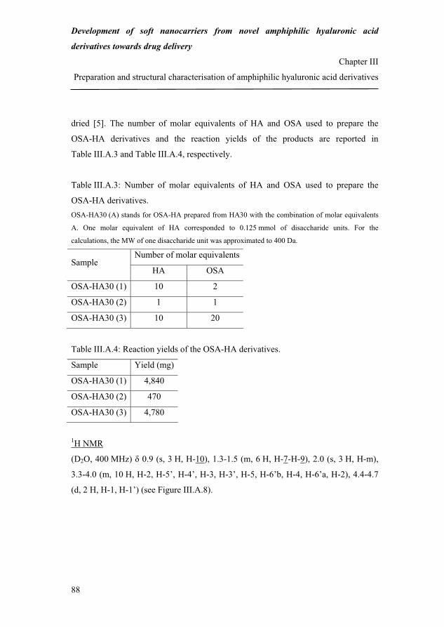
Development of soft nanocarriers from novel amphiphilic hyaluronic acid
derivatives towards drug delivery
Chapter III
Preparation and structural characterisation of amphiphilic hyaluronic acid derivatives
88
dried [5]. The number of molar equivalents of HA and OSA used to prepare the
OSA-HA derivatives and the reaction yields of the products are reported in
Table III.A.3 and Table III.A.4, respectively.
Table III.A.3: Number of molar equivalents of HA and OSA used to prepare the
OSA-HA derivatives. OSA-HA30 (A) stands for OSA-HA prepared from HA30 with the combination of molar equivalents
A. One molar equivalent of HA corresponded to 0.125 mmol of disaccharide units. For the
calculations, the MW of one disaccharide unit was approximated to 400 Da.
Number of molar equivalentsSample
HA OSA
OSA-HA30 (1) 10 2
OSA-HA30 (2) 1 1
OSA-HA30 (3) 10 20
Table III.A.4: Reaction yields of the OSA-HA derivatives.
Sample Yield (mg)
OSA-HA30 (1) 4,840
OSA-HA30 (2) 470
OSA-HA30 (3) 4,780
1H NMR
(D2O, 400 MHz) � 0.9 (s, 3 H, H-10), 1.3-1.5 (m, 6 H, H-7-H-9), 2.0 (s, 3 H, H-m),
3.3-4.0 (m, 10 H, H-2, H-5’, H-4’, H-3, H-3’, H-5, H-6’b, H-4, H-6’a, H-2), 4.4-4.7
(d, 2 H, H-1, H-1’) (see Figure III.A.8).
114
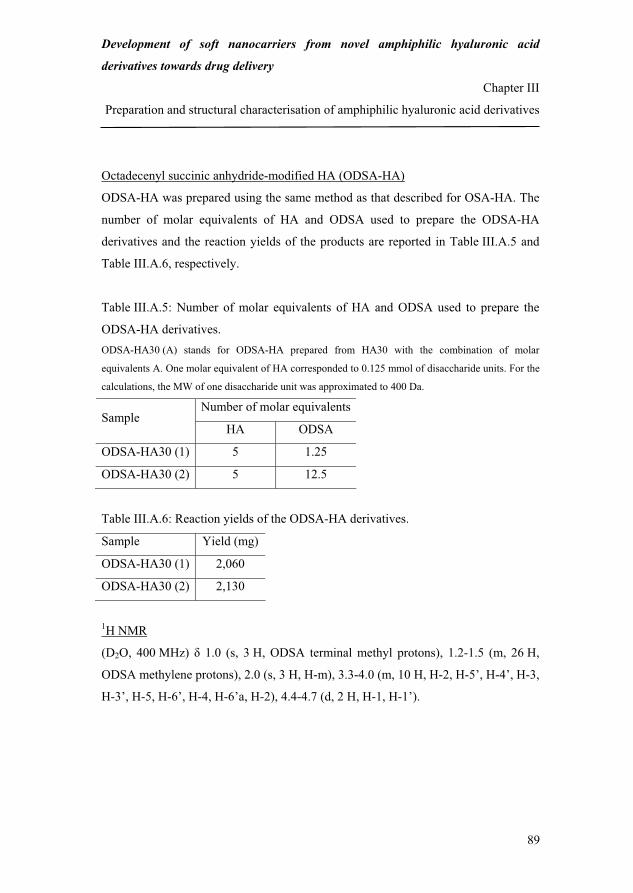
Development of soft nanocarriers from novel amphiphilic hyaluronic acid
derivatives towards drug delivery
Chapter III
Preparation and structural characterisation of amphiphilic hyaluronic acid derivatives
89
Octadecenyl succinic anhydride-modified HA (ODSA-HA)
ODSA-HA was prepared using the same method as that described for OSA-HA. The
number of molar equivalents of HA and ODSA used to prepare the ODSA-HA
derivatives and the reaction yields of the products are reported in Table III.A.5 and
Table III.A.6, respectively.
Table III.A.5: Number of molar equivalents of HA and ODSA used to prepare the
ODSA-HA derivatives. ODSA-HA30 (A) stands for ODSA-HA prepared from HA30 with the combination of molar
equivalents A. One molar equivalent of HA corresponded to 0.125 mmol of disaccharide units. For the
calculations, the MW of one disaccharide unit was approximated to 400 Da.
Number of molar equivalentsSample
HA ODSA
ODSA-HA30 (1) 5 1.25
ODSA-HA30 (2) 5 12.5
Table III.A.6: Reaction yields of the ODSA-HA derivatives.
Sample Yield (mg)
ODSA-HA30 (1) 2,060
ODSA-HA30 (2) 2,130
1H NMR
(D2O, 400 MHz) � 1.0 (s, 3 H, ODSA terminal methyl protons), 1.2-1.5 (m, 26 H,
ODSA methylene protons), 2.0 (s, 3 H, H-m), 3.3-4.0 (m, 10 H, H-2, H-5’, H-4’, H-3,
H-3’, H-5, H-6’, H-4, H-6’a, H-2), 4.4-4.7 (d, 2 H, H-1, H-1’).
115

Development of soft nanocarriers from novel amphiphilic hyaluronic acid
derivatives towards drug delivery
Chapter III
Preparation and structural characterisation of amphiphilic hyaluronic acid derivatives
90
II.2.4. Characterisation techniques
SEC-MALLS
Each sample (10 mg) was dissolved in the mobile phase (10 mL) which was prepared
as followed: NaCl (45.0 g), NaH2PO4 (7.8 g) and NaN3 (1.25 g) on the one hand and
NaCl (45.0 g), Na2HPO4 (22.4 g) and NaN3 (1.25 g) on the other hand were first
dissolved in milli-Q water (5.0 L) in two volumetric flasks to constitute two distinct
solutions (S1 and S2). S1 and S2 were mixed. The pH of the mixture was measured
and, if necessary, adjusted to 7.0 with either HCl (4 M) or NaOH (4 M). The resulting
mobile phase was finally filtered through glass microfiber filters (GF/F 1825 110,
porosity 0.7 μm; Whatman, Maidstone, United Kingdom). Each sample was first
fractionated by high performance liquid chromatography (HPLC, Alliance Waters
2695; Waters, Milford, Massachusetts, United States) through a HPLC column
(7.8 x 300 mm TSK-GEL®, G5000PWXL; Tosoh Corporation, Tokyo, Japan). The
various sample fractions were then analysed by the MALLS (DAWN EOS; Wyatt
Technology Corporation, Santa Barbara, California, United States) and refractive
index (Optilab Rex; Wyatt Technology Corporation, Santa Barbara, California,
United States) detection instruments. The flow rate was kept constant at 0.5 mL/min.
The values of the osmotic second virial coefficient and of the variation of the
polymer solution refractive index n as a function of the polymer concentration c
(dn/dC) were 0.0023 mol.mL/g2 and 0.153 mL/g, respectively. The HPLC on the one
hand and the MALLS and RI instruments on the other hand were piloted by the
softwares Empower 1154 (Waters, Milford, Massachusetts, United States) and
ASTRA 5.3.1.4 (Wyatt Technology Corporation, Santa Barbara, California, United
States).
The MW loss of the PVS-HA derivatives was calculated as followed:
100)300(
)300()300(�
���
HAMWHAMWHAPVSMWMWloss Equation III.A.1
116
Development of soft nanocarriers from novel amphiphilic hyaluronic acid
derivatives towards drug delivery
Chapter III
Preparation and structural characterisation of amphiphilic hyaluronic acid derivatives
91
Thin layer chromatography (TLC)
Drops of PVS (1 mg/mL in acetone) and of the crude and purified PVS-HA products
(10 mg/mL in milli-Q water) were spotted on the bottom line of a TLC plate (Silica
gel 60 F254; Merck, Whitehouse Station, New Jersey, United States). The
compounds were eluted using a heptane/ethyl acetate mixture (50:50, v/v). The plate
was revealed under ultraviolet light (254 nm). The retention time of each spot was the
ratio between the distance travelled by the spot and that travelled by the elution
system (control).
FT-IR
Each sample (1 mg) was ground in potassium bromide (KBr, ~25 mg) and a pellet
was prepared using a Qwik Handi-Press (P/N 0016-125; Thermo Spectra-Tech,
Shelton, Connecticut, United States) and a 3-mm Die Set (P/N 0016-115; Thermo
Spectra-Tech, Shelton, Connecticut, United States). The spectra were acquired at
room temperature, in the wavelength range 4000-500 cm-1 (Nicolet Nexus
470 FT-IR, equipped with a Smart ARKTM system; Thermoelectron, Copenhagen,
Denmark). Each spectrum was an overlap of 64 scans.
The spectra of native HA and of the HA derivatives were assigned based on
Alkrad et al. [6], Haxaire et al. [7] and Pretsch et al. [8].
1H NRM
Each sample (10 to 30 mg) was dissolved in D2O (0.8 mL) and transferred into an
NMR glass tube. The spectra were acquired at 80 °C and 400 MHz (Varian Mercury
TX 400 MHz; Varian, Palo Alto, California, United States). The DS of the
derivatives, which corresponds to the number of grafted reagent molecules per 100
disaccharide units, was calculated by comparing the intensity per proton of the
phenyl protons (PVS-HA, � = 7.6-8.0 ppm), the terminal methyl protons close to the
sulfonyl group (EVS-HA, � = 1.2-1.4 ppm) or the terminal methyl protons on the
117

Development of soft nanocarriers from novel amphiphilic hyaluronic acid
derivatives towards drug delivery
Chapter III
Preparation and structural characterisation of amphiphilic hyaluronic acid derivatives
92
alkenyl chain (� = 0.9 ppm for OSA-HA and � = 1.0 ppm for ODSA-HA) to that of
the methyl protons on native HA (� = 2.0 ppm).
The spectra of native HA and of the HA derivatives were assigned based on
Welti et al. [9], Pavia et al. [10] and Pretsch et al. [8].
III. Results and discussion
III.1. Characterisation of aryl/alkyl vinyl sulfone-modified hyaluronic acid
HA was reacted with phenyl and ethyl vinyl sulfone in water/acetone
mixtures, under alkaline conditions, at room temperature and the influence of the
NaOH/HA and PVS/HA molar ratios, reagent type and HA MW on the DS of the
resulting derivatives was investigated.
The reaction between HA and Ar/AlVS is illustrated in Figure III.A.1 and its
mechanism is presented in Figure III.A.2.
OHO
NHO
OOH
OH
ONaO
CH3
O
OHO
n
R'SO
O
R'SO
OCH2
ROO
NHO
ORO
OR
ONaO
CH3
O
ORO
n
+
HA
NaOHwater/acetone
rt
Ar/AlVS
R = H or or CH2-CH3and R' =
Ar/AlVS-HA
Figure III.A.1: Modification of HA with Ar/AlVS.
118

Development of soft nanocarriers from novel amphiphilic hyaluronic acid
derivatives towards drug delivery
Chapter III
Preparation and structural characterisation of amphiphilic hyaluronic acid derivatives
93
HA OH HA O H+NaOH
HA O R'S
O
OHA O CH2 CH S
O
O
R' HA O CH2 CH2 S
O
O
R'H
+
+-
-
-+
+
Figure III.A.2: Mechanism of the reaction between HA and Ar/AlVS.
The products obtained were white spongy materials partially soluble in milli-Q water.
At concentrations above 1 mg/mL in milli-Q water, all products gave slightly turbid
solutions. This was not the case for native HA which evidenced that the products had
different properties than the starting HA.
Residual ungrafted PVS in the crude and purified PVS-HA products was
assessed by TLC. The results showed that the crude PVS-HA products contained
phenyl-based compounds while the purified PVS-HA products were free of these.
This demonstrated that ungrafted PVS was eliminated during the purification step.
Dialysis was thereby proved efficient to purify the products of the reaction between
HA and PVS and by extension HA and EVS.
The MW values of HA300, PVS-HA300 (1) and PVS-HA300 (3) were
determined by SEC-MALLS to evaluate the extent of HA degradation during the
preparation of PVS-HA (Table III.A.7).
Table III.A.7: Molecular weight of HA300 and selected PVS-HA300 derivatives.
Sample MW (Da) MW loss (%)
HA300 346,900 ± 1387 -
PVS-HA300 (1) 242,400 ± 4848 30
PVS-HA300 (3) 148,300 ± 2966 57
119

Development of soft nanocarriers from novel amphiphilic hyaluronic acid
derivatives towards drug delivery
Chapter III
Preparation and structural characterisation of amphiphilic hyaluronic acid derivatives
94
The results showed that the alkaline treatment needed to activate the HA hydroxyl
groups led to a manifest polymer degradation and that the latter was an increasing
function of the NaOH/HA molar ratio between 1 (PVS-HA300 (1)) and 10 (PVS-
HA300 (3)). The treatments with NaOH/HA molar ratios equal to 1 and 10 led to
30 % and 57 % HA degradation, respectively, suggesting that NaOH/HA molar ratios
lower than 1 are more suitable to limit the degradation of HA and retain its MW. This
was taken into consideration when preparing PVS-HA300 (4) and (5) and PVS-
HA750 (1), (4) and (5) (see Table III.A.1).
The structure of the vinyl sulfone-based HA derivatives was qualitatively
confirmed by FT-IR by comparing the spectra of native HA300 and of the
derivatives. As an example, the spectra of HA300 and PVS-HA300 (1) are shown in
Figure III.A.3.
4
5
6
7
8
9
10
11
12
13
14
15
16
17
18
19
20
500 1000 1500 2000 2500 3000 3500 4000 Wavenumbers (cm-1)Wavenumber (cm-1)
Tran
smitt
ance
(%)
HA300
PVS-HA300 (1)
6907301308
1450
1154 Figure III.A.3: FT-IR spectra of HA300 and PVS-HA300 (1).
120
Development of soft nanocarriers from novel amphiphilic hyaluronic acid
derivatives towards drug delivery
Chapter III
Preparation and structural characterisation of amphiphilic hyaluronic acid derivatives
95
Compared to the spectrum of HA300, additional bands at 1450, 1308, 730 and
690 cm-1 were found on the spectrum of PVS-HA300 (1). These correspond to
aromatic C=C stretching (second band), S=O asymmetric stretching and aromatic C–
H out-of-plane bending (bands at 730 and 690 cm-1), respectively. In addition, the
two bands at 730 and 690 cm-1 were typical of monosubstituted aromatic rings.
The intensity of the broad band between 1700 and 1600 cm-1 and that of the band at
1154 cm-1 was significantly higher in the case of PVS-HA300 (1). On the spectrum
of HA300, the band between 1700 and 1600 cm-1 was most likely due to COO-
asymmetric stretching and N–H bending. However, on the spectrum of PVS-
HA300 (1), this band probably also additionally resulted from aromatic C=C
stretching (first absorption). Similarly, the sharp band at 1154 cm-1 most certainly
arose from C–O–C asymmetric stretching on the spectrum of HA300 and to a
combination of the latter and S=O symmetric stretching on the spectrum of PVS-
HA300 (1) (see § II.2.2).
The univocal presence of aromatic C=C and C–H and S=O bands on the spectrum of
PVS-HA300 (1) qualitatively confirmed the modification of HA with PVS.
Similar spectra were observed for the other PVS-HA derivatives while only
S=O asymmetric stretching around 1300 cm-1 on the spectrum of EVS-HA300 could
confirm the modification of HA with EVS (see § II.2.2).
1H NMR was used to quantitatively confirm the structure of the vinyl
sulfone-based HA derivatives and calculate their DS. As an example, the spectra of
native HA300 and PVS-HA300 (1) are shown in Figure III.A.4 and Figure III.A.5,
respectively.
Due to the large MW of HA300 and to peak overlapping in the region 3.2 to 4.2 ppm,
it was not possible to clearly assign the shifts of protons 3 to 5 and 2’ to 6’ (see
§ II.2.1). For this reason, lower MW HA was used to prepare the succinic anhydride-
modified HA derivatives (see § II.2.3).
121

Development of soft nanocarriers from novel amphiphilic hyaluronic acid
derivatives towards drug delivery
Chapter III
Preparation and structural characterisation of amphiphilic hyaluronic acid derivatives
96
OHO
NHO
OOH
OH
ONaO
CH3
O
OHO
n
2 13
45
1’2’3’
4’ 5’
GlcA GlcNAc
m
2
3, 4’, 5’
1’
1
2’, 6’a, 4, 6’b, 5, 3’
m
6’
Figure III.A.4: 1H NMR spectrum of HA300.
m stands for methyl. For the multiplet occurring between 3.2 and 4.2 ppm, protons are written in the
order of their spectral occurrence according to [9].
Compared to the spectrum of HA300, an additional triplet was found between 7.6 and
8.0 ppm on the spectrum of PVS-HA300 (1). It corresponds to the aromatic protons
of grafted PVS (H-3’’ to H-7’’). In addition, the intensities of the resonances between
3.0 and 4.2 ppm and 4.2 and 4.6 ppm were relatively more important compared to
those found in the same regions on the spectrum of HA300 (see Figure III.A.4). This
was due to the contribution of the methylene protons vicinal to the sulfonyl group
122

Development of soft nanocarriers from novel amphiphilic hyaluronic acid
derivatives towards drug delivery
Chapter III
Preparation and structural characterisation of amphiphilic hyaluronic acid derivatives
97
(H-2’’) and those vicinal to the ether bond (H-1’’), respectively (see § II.2.2). The DS
of PVS-HA300 (1) was 11 % per disaccharide unit.
HA OS
O
O
1’’
2’’7’’
3’’4’’
5’’
6’’
m
3’’-7’’
1’’
2’’
Figure III.A.5: 1H NMR spectrum of PVS-HA300 (1).
Similar spectra were observed for the other PVS-HA derivatives. However,
the relative intensity of the triplet between 7.6 and 8.0 ppm and therefore the DS of
the derivatives were dependent on the reaction conditions. The DS values of the PVS-
HA derivatives are reported in Table III.A.8.
123
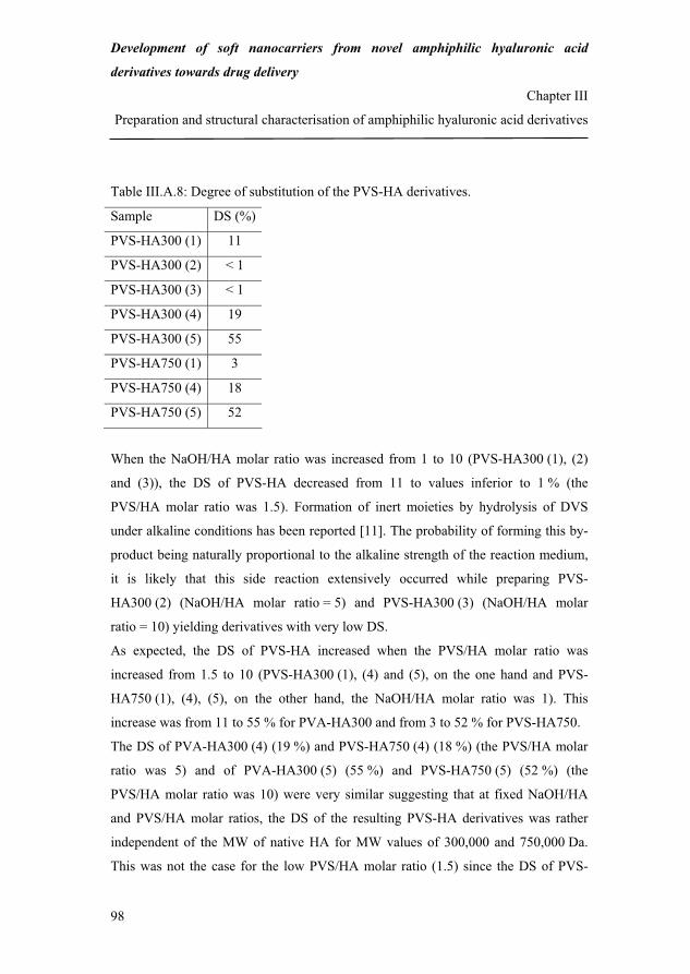
Development of soft nanocarriers from novel amphiphilic hyaluronic acid
derivatives towards drug delivery
Chapter III
Preparation and structural characterisation of amphiphilic hyaluronic acid derivatives
98
Table III.A.8: Degree of substitution of the PVS-HA derivatives.
Sample DS (%)
PVS-HA300 (1) 11
PVS-HA300 (2) < 1
PVS-HA300 (3) < 1
PVS-HA300 (4) 19
PVS-HA300 (5) 55
PVS-HA750 (1) 3
PVS-HA750 (4) 18
PVS-HA750 (5) 52
When the NaOH/HA molar ratio was increased from 1 to 10 (PVS-HA300 (1), (2)
and (3)), the DS of PVS-HA decreased from 11 to values inferior to 1 % (the
PVS/HA molar ratio was 1.5). Formation of inert moieties by hydrolysis of DVS
under alkaline conditions has been reported [11]. The probability of forming this by-
product being naturally proportional to the alkaline strength of the reaction medium,
it is likely that this side reaction extensively occurred while preparing PVS-
HA300 (2) (NaOH/HA molar ratio = 5) and PVS-HA300 (3) (NaOH/HA molar
ratio = 10) yielding derivatives with very low DS.
As expected, the DS of PVS-HA increased when the PVS/HA molar ratio was
increased from 1.5 to 10 (PVS-HA300 (1), (4) and (5), on the one hand and PVS-
HA750 (1), (4), (5), on the other hand, the NaOH/HA molar ratio was 1). This
increase was from 11 to 55 % for PVA-HA300 and from 3 to 52 % for PVS-HA750.
The DS of PVA-HA300 (4) (19 %) and PVS-HA750 (4) (18 %) (the PVS/HA molar
ratio was 5) and of PVA-HA300 (5) (55 %) and PVS-HA750 (5) (52 %) (the
PVS/HA molar ratio was 10) were very similar suggesting that at fixed NaOH/HA
and PVS/HA molar ratios, the DS of the resulting PVS-HA derivatives was rather
independent of the MW of native HA for MW values of 300,000 and 750,000 Da.
This was not the case for the low PVS/HA molar ratio (1.5) since the DS of PVS-
124

Development of soft nanocarriers from novel amphiphilic hyaluronic acid
derivatives towards drug delivery
Chapter III
Preparation and structural characterisation of amphiphilic hyaluronic acid derivatives
99
HA300 (1) (11 %) was much higher compared to that of PVS-HA750 (1) (3 %). This
could be due to the higher viscosity of HA750 aqueous solutions which might have
limited the mixing and diffusion of small quantities of PVS.
The spectrum of EVS-HA300 showed an additional triplet between 1.2 and
1.4 ppm corresponding to the terminal methyl protons close to the sulfonyl group
(SO2–CH2–CH3). In addition, as for PVS-HA, the intensities of the resonances
between 3.2 and 4.2 ppm and 4.2 and 4.6 ppm was relatively more important
compared to that found in the same regions on the spectrum of HA300. This was due
to the contribution of the methylene protons vicinal to the sulfonyl group (SO2–CH2–
CH2–O and SO2–CH2–CH3) and of those vicinal to the ether bond (SO2–CH2–CH2–
O), respectively (see § II.2.2).
The DS of EVS-HA300 was 13 % per disaccharide unit. This value was close to that
obtained for PVS-HA300 (1) (11 %) suggesting that at fixed NaOH/HA and vinyl
sulfone reagent/HA molar ratios the DS of the resulting derivatives was rather
independent of the pendant group attached for the substituents PVS and EVS.
III.2. Characterisation of alkenyl succinic anhydride-modified hyaluronic acid
HA was reacted with octenyl and octadecenyl succinic anhydride in aqueous
media, under alkaline conditions, at room temperature and the influence of the
reagent/HA molar ratio and succinic anhydride reagent type on the DS of the
resulting derivatives was investigated.
The reaction between HA and AlkSA is illustrated in Figure III.A.6 and its
mechanism is presented in Figure III.A.7.
125
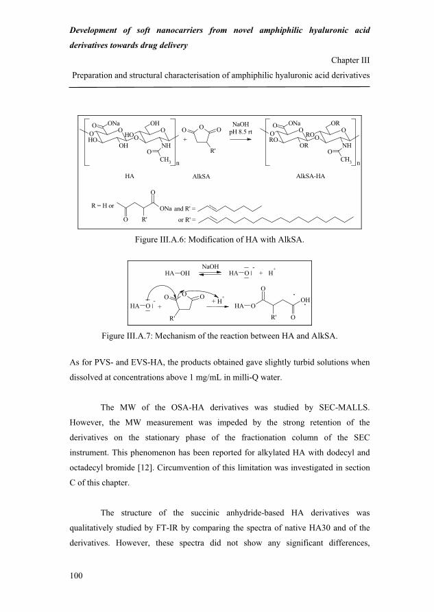
Development of soft nanocarriers from novel amphiphilic hyaluronic acid
derivatives towards drug delivery
Chapter III
Preparation and structural characterisation of amphiphilic hyaluronic acid derivatives
100
OHO
NHO
OOH
OH
ONaO
CH3
O
OHO
n
OO O
R'
ONa
O
O
R'
ROO
NHO
ORO
OR
ONaO
CH3
O
ORO
n
+
HA
NaOHpH 8.5 rt
AlkSA
and R' =
or R' =
R = H or
AlkSA-HA
Figure III.A.6: Modification of HA with AlkSA.
HA OH HA O H+NaOH
HA O HA OOH
O
OR'
OO O
R'
H+
+-
-+
+
Figure III.A.7: Mechanism of the reaction between HA and AlkSA.
As for PVS- and EVS-HA, the products obtained gave slightly turbid solutions when
dissolved at concentrations above 1 mg/mL in milli-Q water.
The MW of the OSA-HA derivatives was studied by SEC-MALLS.
However, the MW measurement was impeded by the strong retention of the
derivatives on the stationary phase of the fractionation column of the SEC
instrument. This phenomenon has been reported for alkylated HA with dodecyl and
octadecyl bromide [12]. Circumvention of this limitation was investigated in section
C of this chapter.
The structure of the succinic anhydride-based HA derivatives was
qualitatively studied by FT-IR by comparing the spectra of native HA30 and of the
derivatives. However, these spectra did not show any significant differences,
126

Development of soft nanocarriers from novel amphiphilic hyaluronic acid
derivatives towards drug delivery
Chapter III
Preparation and structural characterisation of amphiphilic hyaluronic acid derivatives
101
independently of the DS of the derivatives. This lack of difference was probably due
the overlapping of the characteristic bands of HA, OSA- and ODSA-HA. Indeed,
vinyl C–H stretching was most likely indiscernible from HA O–H stretching.
Likewise, C=C stretching and C=O (ester) stretching were certainly hidden by HA
C=O stretching. However, in the region 800-500 cm-1, which was free of any bands
on the HA spectrum, CH2 bending/rocking along the octenyl succinate (OS) chains
and vinyl C–H out-of-plane bending could not clearly be detected, probably due to
the low DS of the OSA- and ODSA-HA derivatives (see Table III.A.9 and below).
As a result, FT-IR could not confirm the modification of HA with OSA or ODSA.
1H NMR, however, confirmed grafting of OSA onto HA and was used to
calculate the DS of the OSA-HA derivatives. As an example, the spectrum of OSA-
HA30 (2) is shown in Figure III.A.8.
Compared to the spectrum of HA30, an additional singlet and a multiplet were found
at 0.9 ppm and between 1.3 and 1.5 ppm, respectively on the spectrum of OSA-
HA30 (2). The former corresponds to the terminal methyl protons (H-10) and the
latter to the methylene protons (H-7 to H-9) on the octenyl chain of grafted OS. Due
to the low signal intensity in the regions 2.2-3.0 and 4.8-5.8 ppm, it was not possible
to clearly assign the chemical shifts of protons 1 to 6 (see § II.2.3). The DS of OSA-
HA30 (2) was approximately 3 % per disaccharide unit.
Similar spectra were observed for the other OSA-HA derivatives. However,
the relative intensity of the singlet at 0.9 ppm and therefore the DS of the derivatives
were dependent on the reaction conditions. The DS values of the OSA-HA
derivatives are reported in Table III.A.9.
127

Development of soft nanocarriers from novel amphiphilic hyaluronic acid
derivatives towards drug delivery
Chapter III
Preparation and structural characterisation of amphiphilic hyaluronic acid derivatives
102
HA O
OONa
O1
2
34
56
78
910
m
10
7, 8, 9
Figure III.A.8: 1H NMR spectrum of OSA-HA30 (2).
Table III.A.9: Degree of substitution of the OSA-HA derivatives.
Sample DS (%)
OSA-HA30 (1) < 1
OSA-HA30 (2) 3
OSA-HA30 (3) 4
As expected, the DS of OSA-HA increased when the OSA/HA molar ratio was
increased from 0.2 (OSA-HA30 (1)) to 2 (OSA-HA30 (3)). This increase was from
< 1 to 4 %.
128
Development of soft nanocarriers from novel amphiphilic hyaluronic acid
derivatives towards drug delivery
Chapter III
Preparation and structural characterisation of amphiphilic hyaluronic acid derivatives
103
The spectra of the ODSA-HA derivatives showed two additional singlets at
1.0 ppm and between 1.2 and 1.5 ppm (broad). The former corresponds to the
terminal methyl protons and the latter to the 26 methylene protons localised after the
unsaturation on the octadecenyl chain of grafted ODSA. As for OSA-HA, the
chemical shifts of the remaining protons (H-1 to H-6) were difficult to clearly assign.
The DS of ODSA-HA30 (1) and ODSA-HA30 (2) were < 1 and 2 % per disaccharide
unit, respectively. Increasing the ODSA/HA molar ratio from 0.25 (ODSA-
HA30 (1)) to 2.5 (ODSA-HA30 (2)) did not result in a substantial increase of the DS.
This might be due to the low solubility in the aqueous reaction medium of ODSA
which in both cases limited the number of reagent molecules available for reaction.
The DS of the ODSA-HA derivatives were lower compared to those of the
OSA-HA derivatives when using comparable reaction conditions. This might be due
to the lower solubility in aqueous media of ODSA.
III.3. Comparison between the two preparation methods
In summary, the modification of HA with vinyl sulfone and succinic
anhydride reagents was not only proved feasible but also efficient under alkaline
conditions, at room temperature. In addition, the reactions exclusively involved the
HA hydroxyl groups, were simple, well controlled and yielded HA derivatives with
high DS (vinyl sulfone-based HA derivatives).
Ar/AlVS-HA derivatives with a DS up to 52-55 % per disaccharide unit according to 1H NMR were prepared from HA300 and HA750. The DS of the derivatives was an
increasing function of the reagent/HA molar ratio and was rather independent on the
HA MW. In addition, the same reaction conditions (same NaOH/HA and reagent/HA
molar ratios) yielded derivatives with a similar DS, independently of the reagents
(PVS or EVS). The alkaline treatment with NaOH causing a substantial degradation
129

Development of soft nanocarriers from novel amphiphilic hyaluronic acid
derivatives towards drug delivery
Chapter III
Preparation and structural characterisation of amphiphilic hyaluronic acid derivatives
104
of native HA, NaOH/HA molar ratios below 1 were subsequently used to limit the
degradation of HA and retain its MW.
AlkSA-HA derivatives with a DS up to 4 % per disaccharide unit according to 1H NMR were obtained from HA30. The DS of the AlkSA-HA derivatives was an
increasing function of the reagent/HA molar ratio and was dependent on the reagent
type: the use of ODSA yielded derivatives with lower DS most likely due to the
lower solubility in the reaction medium of the longer octadecenyl chains.
The DS of the AlkSA-HA derivatives was lower than that of the Ar/AlVS-HA
derivatives at comparable reagent/HA molar ratios most likely due to the lower
solubility and higher sensitivity to hydration of the AlkSA reagents.
A comparison between the two modification methods is presented in
Table III.A.10.
Although both preparation methods involve the HA hydroxyl groups and are easily
upscalable, the preparation of AlkSA-HA is more environmentally friendly than that
of Ar/AlVS-HA. Indeed, the former is conducted in aqueous media while the latter is
achieved in water/acetone mixtures. However, a higher through-put is possible in the
case of Ar/AlVS-HA since only one sample of AlkSA-HA can be prepared at a time
due to the use of a titration manager to control the pH of the reaction medium.
The vinyl sulfone reagents are fewer, relatively more expensive and less available
compared to the succinic anhydride reagents which are very diversified, relatively
less expensive and largely available according to three major chemical databases
[13]. Vinyl sulfone reagents with substituted groups other than methyl, ethyl and
phenyl are scarce. Some suppliers list compounds with larger aliphatic groups or
more complex molecular arrangements containing heteroatoms but these are often
rare and non commercial compounds with a small production volume [13].
Alternatively, more diverse vinyl sulfone reagents could be prepared by the selective
addition of nucleophilic compounds such as secondary amines, alcohols or thiols with
various chain lengths on one double bond of divinyl sulfone, as described by
130
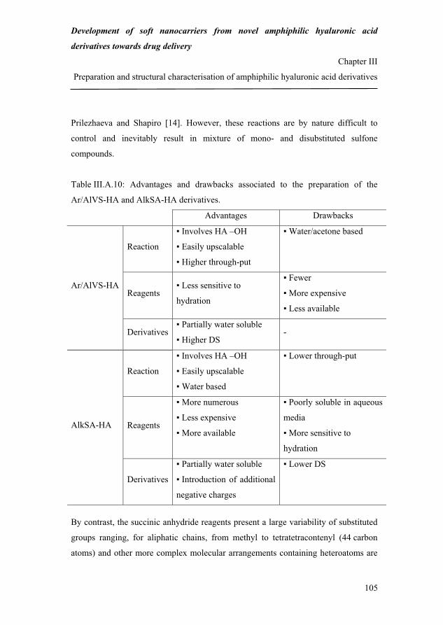
Development of soft nanocarriers from novel amphiphilic hyaluronic acid
derivatives towards drug delivery
Chapter III
Preparation and structural characterisation of amphiphilic hyaluronic acid derivatives
105
Prilezhaeva and Shapiro [14]. However, these reactions are by nature difficult to
control and inevitably result in mixture of mono- and disubstituted sulfone
compounds.
Table III.A.10: Advantages and drawbacks associated to the preparation of the
Ar/AlVS-HA and AlkSA-HA derivatives.
Advantages Drawbacks
Reaction
Involves HA –OH
Easily upscalable
Higher through-put
Water/acetone based
Reagents Less sensitive to
hydration
Fewer
More expensive
Less available
Ar/AlVS-HA
Derivatives Partially water soluble
Higher DS -
Reaction
Involves HA –OH
Easily upscalable
Water based
Lower through-put
Reagents
More numerous
Less expensive
More available
Poorly soluble in aqueous
media
More sensitive to
hydration
AlkSA-HA
Derivatives
Partially water soluble
Introduction of additional
negative charges
Lower DS
By contrast, the succinic anhydride reagents present a large variability of substituted
groups ranging, for aliphatic chains, from methyl to tetratetracontenyl (44 carbon
atoms) and other more complex molecular arrangements containing heteroatoms are
131

Development of soft nanocarriers from novel amphiphilic hyaluronic acid
derivatives towards drug delivery
Chapter III
Preparation and structural characterisation of amphiphilic hyaluronic acid derivatives
106
also listed [13]. However, the modification of HA with succinic anhydride reagents is
limited by their relative poor solubility in aqueous media and high sensitivity to
hydration. As a result, the derivatives exhibit lower DS values than the vinyl sulfone-
based HA derivatives.
Both types of derivatives present a partial solubility in aqueous media but AlkSA-HA
possesses a higher charge density than Ar/AlVS-HA at neutral pH. This is due the
additional carboxyl groups introduced upon ring opening of AlkSA (see
Figure III.A.6) which increases the repulsion between the polymer chains. This
property could advantageously be exploited for the electrostatic stabilisation of future
nanocarriers based on AlkSA-HA.
Due to the additional advantages of the succinic anhydride-based technology, namely
(i) the environmental friendliness, (ii) large variability and availability of the succinic
anhydride reagents and (iii) increase of the negative charge density on the polymer,
this method was selected for further study.
IV. Conclusion
Aryl/alkyl vinyl sulfone- and alkenyl succinic anhydride-modified HA were
prepared and structurally characterised and their preparation methods were compared
on the basis of their versatility.
The environmental friendliness of the preparation method of AlkSA-HA, the large
variability and availability of the AlkSA reagents and the increase of the negative
charge density on the polymer motivated the selection of the succinic anhydride-
based technology for further study. Indeed, these properties provide the AlkSA-HA
derivatives with a potential higher versatility and functionality compared to the
Ar/AlVS-HA derivatives.
However, the lower preparation through-put of AlkSA-HA, poorer solubility
and higher sensitivity to hydration of the AlkSA reagents limiting the DS of the HA
132
Development of soft nanocarriers from novel amphiphilic hyaluronic acid
derivatives towards drug delivery
Chapter III
Preparation and structural characterisation of amphiphilic hyaluronic acid derivatives
107
derivatives were aspects which remained to be addressed. In addition, the preparation
method of AlkSA-HA needed to be optimised in order to develop a reproducible and
robust process for the routine production of HA derivatives with a controlled DS.
This optimisation work is reported in the following section.
References [1] Vercruysse, K. P.; Prestwich, G. D. Hyaluronate derivatives in drug delivery.
Crit. Rev. Ther. Drug Carrier Syst. 1998, 15, 513-555.
[2] Balazs, E. A.; Leshchiner, A. Cross-linked gels of hyaluronic acid and products
containing such gels. US 4,582,865, December 6, 1984.
[3] Caldwell, C. G.; Wurzburg, O. B. Polysaccharide derivatives of substituted
dicarboxylic acids. US 2,661,349, December 1, 1953.
[4] Eenschooten, C.; Christensen, M. W. Aryl/alkyl vinyl sulfone hyaluronic acid
derivatives. WO 2007/098770 A1, September 7, 2007.
[5] Tømmeraas, K.; Eenschooten, C. Aryl/alkyl succinic anhydride hyaluronan
derivatives. WO 2007/033677 A1, March 29, 2007.
[6] Alkrad, J. A.; Mrestani, Y.; Stroehl, D.; Wartewig, S.; Neubert, R.
Characterization of enzymatically digested hyaluronic acid using NMR, Raman,
IR, and UV-Vis spectroscopies. J. Pharm. Biomed. Anal. 2003, 31, 545-550.
[7] Haxaire, K.; Marechal, Y.; Milas, M.; Rinaudo, A. Hydration of polysaccharide
hyaluronan observed by IR spectrometry. I. Preliminary experiments and band
assignments. Biopolymers 2003, 72, 10-20.
[8] Pretsch, E.; Buhlmann, P.; Affolter, C. Structure determination of organic
compounds. Tables of spectral data. Third edition; Springer: Berlin, 2000.
[9] Welti, D.; Rees, D. A.; Welsh, E. J. Solution conformation of
glycosaminoglycans: assignment of the 300-MHz 1H-magnetic resonance
spectra of chondroitin 4-sulphate, chondroitin 6-sulphate and hyaluronate, and
133
Development of soft nanocarriers from novel amphiphilic hyaluronic acid
derivatives towards drug delivery
Chapter III
Preparation and structural characterisation of amphiphilic hyaluronic acid derivatives
108
investigation of an alkali-induced conformation change. Eur. J. Biochem. 1979,
94, 505-514.
[10] Pavia, D. L.; Lampman, G. M.; Kriz, G. S. Introduction to spectroscopy. Third
edition; Brooks/Cole Cengage Learning: Belmont, 2001.
[11] Cirino, V. O.; Bullock, A. L.; Rowland, S. P. The structure of reagent residues in
cotton cellulose treated with divinyl sulfone. Carbohydr. Res. 1971, 17, 67-78.
[12] Pelletier, S.; Hubert, P.; Lapicque, F.; Payan, E.; Dellacherie, E. Amphiphilic
derivatives of sodium alginate and hyaluronate: synthesis and physico-chemical
properties of aqueous dilute solutions. Carbohydr. Polym. 2000, 43, 343-349.
[13] ByersGuideChem (Netvertise, Garbsen, Germany), ChemExpert (Court-St-
Etienne, Belgium), ChemIDplus (Advanced, Division of specialized information
services, United States national library of medicine, Bethesda, Maryland, United
States).
[14] Prilezhaeva, E. N.; Shapiro, E. S. Synthesis based on divinyl sulfone. Dokl.
Akad. Nauk. SSSR 1967, 6, 1345-1348.
134
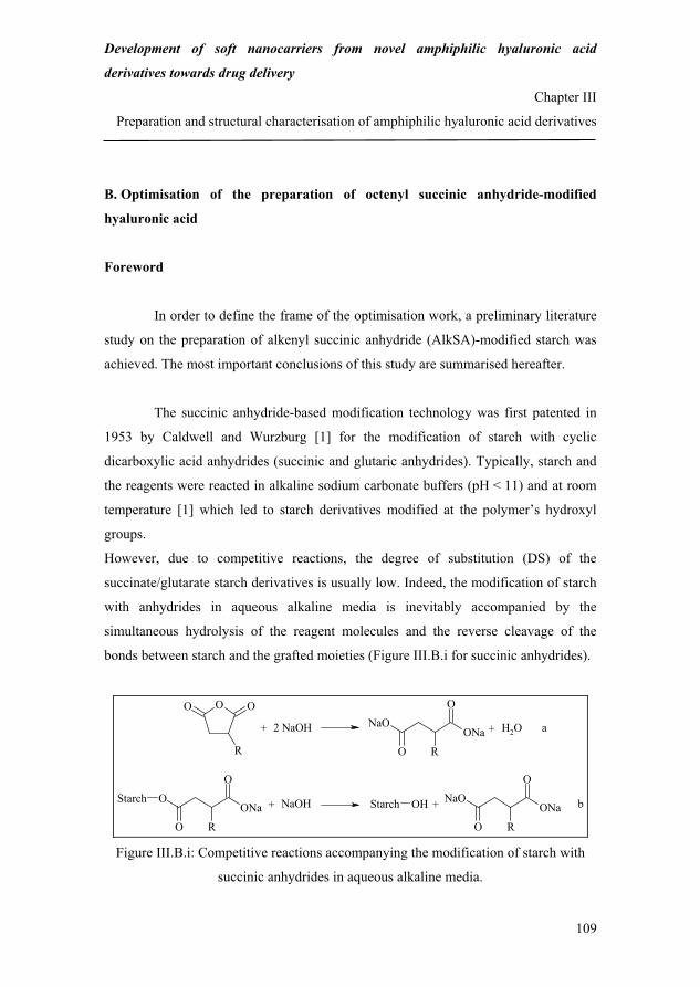
Development of soft nanocarriers from novel amphiphilic hyaluronic acid
derivatives towards drug delivery
Chapter III
Preparation and structural characterisation of amphiphilic hyaluronic acid derivatives
109
B. Optimisation of the preparation of octenyl succinic anhydride-modified
hyaluronic acid
Foreword
In order to define the frame of the optimisation work, a preliminary literature
study on the preparation of alkenyl succinic anhydride (AlkSA)-modified starch was
achieved. The most important conclusions of this study are summarised hereafter.
The succinic anhydride-based modification technology was first patented in
1953 by Caldwell and Wurzburg [1] for the modification of starch with cyclic
dicarboxylic acid anhydrides (succinic and glutaric anhydrides). Typically, starch and
the reagents were reacted in alkaline sodium carbonate buffers (pH < 11) and at room
temperature [1] which led to starch derivatives modified at the polymer’s hydroxyl
groups.
However, due to competitive reactions, the degree of substitution (DS) of the
succinate/glutarate starch derivatives is usually low. Indeed, the modification of starch
with anhydrides in aqueous alkaline media is inevitably accompanied by the
simultaneous hydrolysis of the reagent molecules and the reverse cleavage of the
bonds between starch and the grafted moieties (Figure III.B.i for succinic anhydrides).
O OO
RONa
NaO
O
O
R
ONaO
O
O
R
StarchONa
NaO
O
O
R
OHStarch
+ 2 NaOH + H2O
+ NaOH
a
b+
Figure III.B.i: Competitive reactions accompanying the modification of starch with
succinic anhydrides in aqueous alkaline media.
135

Development of soft nanocarriers from novel amphiphilic hyaluronic acid
derivatives towards drug delivery
Chapter III
Preparation and structural characterisation of amphiphilic hyaluronic acid derivatives
110
In order to improve the DS of succinate/glutarate starch and allow the grafting
of a variety of anhydride reagents, it is necessary to limit the extent of the side
reactions presented in Figure III.B.i. This can be achieved by finely controlling the
reaction conditions and supposes that their influence on the grafting of the anhydride
reagents be known.
Most of the literature available on the optimisation of the preparation of
modified starch with cyclic dicarboxylic acid anhydrides concerns AlkSA-modified
starch. For example, Jeon et al. [2], Bhosale and Singhal [3] and Song et al. [4] have
systematically studied the influence of reaction parameters such as the starch
concentration, reagent/starch weight ratio, reaction pH, temperature and time and
reagent type on the reaction efficiency (RE*) and DS of AlkSA-starch. Indeed, these
parameters potentially all affect, in a more or less complex and/or contradictory
fashion, the extent of starch modification.
For example, it may seem desirable to work at high starch concentrations to increase
the probability that AlkSA be esterified with starch instead of being hydrolysed.
However, concentrated starch suspensions are viscous and can hinder the mixing and
diffusion of the AlkSA molecules in the reaction medium. Similarly, one could think
that high values of the reaction pH and temperature would enhance the reactivity of the
starch hydroxyl groups and the solubility and diffusivity of the AlkSA molecules.
However, these conditions can also favour the hydrolysis of AlkSA. It is obvious that
sufficiently long reaction times be applied in order to allow the completion of the
esterification reactions. Nevertheless, past a certain time the reverse cleavage of the
bonds created between starch and alkenyl succinate (AlkS) can occur due to the
*RE is defined as the ratio between the experimental and theoretical DS of the starch
derivatives. The theoretical DS is calculated in the hypothetical case where all the
reagent molecules added react with starch.
136

Development of soft nanocarriers from novel amphiphilic hyaluronic acid
derivatives towards drug delivery
Chapter III
Preparation and structural characterisation of amphiphilic hyaluronic acid derivatives
111
consumption of AlkSA in the esterification and hydrolysis reactions. Finally, the
reactivity of AlkSA most likely depends on its chain length.
Consequently, in order to improve the DS of AlkSA-starch, optima for the above-
mentioned parameters need to be found.
The reaction parameters tested in references [2] to [4], their range and optima
are summarised in Table III.B.i.
Table III.B.i: Optimisation of the preparation of AlkSA-starch. C(starch), WR, T, t, DDSA and OSA stand for starch concentration, weight ratio, temperature, time,
dodecyl succinic anhydride and octenyl succinic anhydride, respectively.
Jeon et al. [2]
Bhosale and
Singhal [3] Song et al. [4]
Starch origin
Waxy corn
starch
Amaranth and
waxy corn starch Indica rice starch
Rea
ctio
n pa
ram
eter
s C(starch)
AlkSA/starch WR
pH
T
t
Alkenyl chain length
25-65 % w/w
5-40 % (DDSA)
7.5-10.5
15-45 °C
5-95 hours
8-18 C atoms
25 % w/w
1-3 % (OSA)
7.0-11.0
20-60 °C
6-30 hours
8 C atoms
20-40 % w/w
3-15 % (OSA)
7.0-10.0
25-45 °C
2-6 hours
8 C atoms
Opt
ima
C(starch)
AlkSA/starch WR
pH
T
t
Alkenyl chain length
Maximum DS
no influence
10 % (DDSA)
8.5-9.0
23 °C
24 hours
8 C atoms
4.0 %
-
3 % (OSA)
8.0
30 °C
6-24 hours
-
2.5 %
35 % w/w
15 % (OSA)
8.5
35 °C
6 hours
-
4.6 %
137

Development of soft nanocarriers from novel amphiphilic hyaluronic acid
derivatives towards drug delivery
Chapter III
Preparation and structural characterisation of amphiphilic hyaluronic acid derivatives
112
The modification methods used in references [2] to [4] were very similar to the
original method by Caldwell and Wurzburg [1] and only differed from the latter with
regards to the pH control. Typically, AlkSA was progressively added to the starch
suspension for 2 hours. The pH was maintained at the desired value by the continuous
addition of sodium hydroxide (NaOH). At the end of the reaction, the pH was adjusted
to 6.5 with HCl. The resulting AlkSA-starch was filtered, washed and dried.
Despite differences in the starch botanical origin and granule morphology, the results
of the studies presented in Table III.B.i were consistent and showed that:
(i) C(starch) had a variable influence on the DS of the derivatives,
(ii) the higher the AlkSA WR, the higher the DS, but the lower the RE,
(iii) the optimal ranges for the reaction pH, T and t were 8.0-9.0, 23-35 °C and 6-
24 hours, respectively,
(iv) T above 23 °C did not allow a significant improvement of the DS of the
derivatives,
(v) RE and DS decreased rapidly when the alkenyl chain length was increased above 8
carbon atoms,
(vi) finally, the maximum DS of the starch derivatives was approximately 5 % per
starch repeating unit.
In references [2] to [4], the reaction parameters tested were varied
independently from each other. This method requires that all parameters but one are
kept constant and that this parameter is varied until an optimal value is found for it.
After the parameter has been set to the optimal value, a second parameter is varied.
This is repeated until an optimal value has been found for each of the tested
parameters. Although rigorous, this technique exhibits some drawbacks. Firstly,
changing one separate parameter at a time does not always lead to the real optimum
and gives different implications with different starting points. Secondly, the approach
requires a large number of experiments which in turn bring relatively little information
138

Development of soft nanocarriers from novel amphiphilic hyaluronic acid
derivatives towards drug delivery
Chapter III
Preparation and structural characterisation of amphiphilic hyaluronic acid derivatives
113
due to the inefficient distribution of the experiments. Finally, this method does not
allow detecting interactions between parameters [5].
By contrast, the construction of a carefully prepared set of representative experiments
(experimental plan) in which all parameters are simultaneously varied allows drawing
more information from fewer experiments and uncovering interactions and synergies
between parameters. In addition, this method still permits to evaluate the influence of
each separate parameter [5]. Therefore, it is more suitable when screening the
parameters mostly influencing the extent of a given chemical reaction.
With this in mind, an experimental plan was used in the following optimisation study
which frame was defined based on the conclusions drawn from the optimisation of the
preparation of AlkSA-starch.
References [1] Caldwell, C. G.; Wurzburg, O. B. Polysaccharide derivatives of substituted
dicarboxylic acids. US 2,661,349, December 1, 1953.
[2] Jeon, Y. S.; Viswanathan, A.; Gross, R. A. Studies of starch esterification:
reactions with alkenyl-succinates in aqueous slurry systems. Starch/Stärke 1995,
51, 90-93.
[3] Bhosale, R.; Singhal, R. Process optimization for the synthesis of octenyl succinyl
derivative of waxy corn and amaranth starches. Carbohydr. Polym. 2006, 66, 521-
527.
[4] Song, X.; He, G.; Ruan, H.; Chen, Q. Preparation and properties of octenyl succinic
anhydride modified early Indica rice starch. Starch/Stärke 2006, 58, 109-117.
[5] Montgomery, D. C. Design and analysis of experiments. Sixth edition; Wiley:
Hoboken, 2004.
139

Development of soft nanocarriers from novel amphiphilic hyaluronic acid
derivatives towards drug delivery
Chapter III
Preparation and structural characterisation of amphiphilic hyaluronic acid derivatives
114
Preparation and structural characterisation of novel and versatile amphiphilic
octenyl succinic anhydride-modified hyaluronic acid
Corinne Eenschooten1,2, Fanny Guillaumie1, Georgios M. Kontogeorgis2, Erling H.
Stenby2, Khadija Schwach-Abdellaoui1.
1Novozymes Biopolymer A/S, Krogshøjvej 36, 2880 Bagsværd, Denmark. 2Technical University of Denmark, Department of Chemical and Biochemical
Engineering, Centre for Phase Equilibria and Separation Processes (IVC-SEP),
Building 229, 2800 Lyngby, Denmark.
Abstract
Amphiphilic octenyl succinic anhydride-modified hyaluronic acid (OSA-HA) was
prepared in aqueous alkaline media, at room temperature and was structurally
characterised by Fourier transform infrared spectroscopy (FT-IR) and proton nuclear
magnetic resonance spectroscopy (1H NMR). The influence of reaction parameters
such as the HA and buffer concentrations, OSA/HA molar ratio and reaction time on
the degree of substitution (DS) of the OSA-HA derivatives was studied by means of an
experimental plan. The results showed that the OSA/HA molar ratio, NaHCO3
concentration and interaction between these had the largest influence on the DS of the
derivatives while the HA concentration and reaction time only had a negligible effect.
The maximum DS achieved was 44 % per disaccharide unit according to 1H NMR.
Moreover, optimal reaction conditions were identified for the preparation of versatile
OSA-HA derivatives with a DS between 1.5 and 44 %.
Keywords amphiphilic hyaluronic acid derivative, octenyl succinic anhydride, degree
of substitution, experimental plan.
140

Development of soft nanocarriers from novel amphiphilic hyaluronic acid
derivatives towards drug delivery
Chapter III
Preparation and structural characterisation of amphiphilic hyaluronic acid derivatives
115
I. Introduction
Hyaluronic acid (HA) is a natural linear polysaccharide consisting of D-
glucuronic acid and N-acetyl-D-glucosamine linked through �-1,3 glycosidic bonds
while consecutive repeating disaccharide units are linked through �-1,4 bonds [1]
(Figure III.B.1).
OHO
NHO
OOH
OH
OHO
CH3
O
OHO
n
Figure III.B.1: Molecular structure of the HA repeating unit.
HA is found in all vertebrate organs and fluids and is particularly abundant in the
extracellular matrix of soft connective tissues [2].
Due to its natural biocompatibility and resorbability, HA is currently one of
the most attractive building blocks for the preparation of novel biomaterials with
applications in the cosmetic, pharmaceutical and medical industries. However, native
HA has limited applications in these fields due to its intrinsic poor mechanical and
physicochemical properties [3]. Chemical modifications of HA have therefore been
developed to prepare mechanically robust HA derivatives with tailored
physicochemical properties.
For example, amphiphilic HA derivatives have been obtained by grafting hydrophobic
moieties onto the hydrophilic HA backbone using the carboxyl and hydroxyl functions
of the polymer [3]. The resulting derivatives, which possess an altered aqueous
solubility compared to native HA [4,5,6], potentially represent attractive starting
materials for the formulation of self-associative HA-based systems towards drug
encapsulation and delivery.
141

Development of soft nanocarriers from novel amphiphilic hyaluronic acid
derivatives towards drug delivery
Chapter III
Preparation and structural characterisation of amphiphilic hyaluronic acid derivatives
116
However, the modification methods currently available for the preparation of
amphiphilic HA present some drawbacks. Firstly, the modification reactions are often
conducted in organic solvents or organic solvent/water mixtures [7,8,9]. In addition to
raise occupational and environmental issues and limit the upscalability of the
preparation method, the use of organic solvents or mixtures of these often requires
converting HA into its tetraalkylammonium salt [10,11] or preparing reactive
intermediates [10,12]. This not only adds complex steps to the preparation method but
also often results in HA degradation [4,8]. Secondly, the modification reactions
involving HA carboxyl groups modify the distribution of negative charges along the
polymer backbone at neutral pH. This could potentially affect some of the attractive
biological and/or pharmacological properties of native HA [13].
Therefore, simple modification reactions in aqueous media, under mild conditions and
exclusively involving HA hydroxyl groups could represent advantageous alternatives
for the preparation of amphiphilic HA derivatives.
In this regard, a novel, simple and easily upscalable modification method for
the preparation of amphiphilic HA was previously developed in our laboratories which
is based on the reaction between HA and octenyl succinic anhydride (OSA) in aqueous
mild alkaline media [14].
The objective of the present work was to optimise this method and prepare versatile
HA derivatives with tailored and extended degrees of substitution (DS). To achieve
this, an experimental plan was implemented in which four different reaction
parameters were simultaneously varied and their influence on the DS of the OSA-HA
derivatives was detailedly studied. The chemical structure of the derivatives was
characterised by Fourier transform infrared spectroscopy (FT-IR) and proton nuclear
magnetic resonance spectroscopy (1H NMR).
142

Development of soft nanocarriers from novel amphiphilic hyaluronic acid
derivatives towards drug delivery
Chapter III
Preparation and structural characterisation of amphiphilic hyaluronic acid derivatives
117
II. Materials and methods
II.1. Materials
Medium molecular weight (MW) HA (HyaCare®, MW~750,000 Da, HA750) was
provided by Novozymes Biopolymer A/S (Bagsværd, Denmark). Low MW HA
(HA20) was prepared using the procedure described hereafter. Octenyl succinic
anhydride (OSA, 210.27 Da, purity 97 %, mixture of cis and trans) was obtained
from Sigma-Aldrich (Saint Louis, Missouri, United States). Sodium bicarbonate
(NaHCO3) and potassium bromide (KBr) were used as purchased without further
purification. The water used for the sample preparation and analysis was distilled and
purified to a resistivity of 18.2 M�.cm in a milli-Q apparatus (Millipore, Billerica,
Massachusetts, United States).
II.2. Methods
II.2.1. Preparation of low molecular weight hyaluronic acid
HA20 was prepared using the same method as that described for HA300 (see section A
of this chapter, § II.2.1) but with 250 mL of H3PO4 and a 24-hour incubation time.
Approximately 56 g of HA20, i.e. 80 % of the starting HA750 were recovered.
SEC-MALLS
The exact MW of HA20 was 19,090 ± 191 Da.
FT-IR
(KBr, 4000-500 cm-1) w 3431 (O–H and N–H st), 2931 (C–H st), 1700-1600 (COO- st
as, N–H bd), 1423 (COO- st sy), 1392 (C–O–H bd), 1154 (C–O–C st as), 1077 and
1038 (C–OH st), 954 and 900 (COO- bd), 808 (C–O–C st sy).
143

Development of soft nanocarriers from novel amphiphilic hyaluronic acid
derivatives towards drug delivery
Chapter III
Preparation and structural characterisation of amphiphilic hyaluronic acid derivatives
118
1H NMR
(D2O, 400 MHz) � 2.0 (s, 3 H, H-m), 3.2-4.0 (m, 10 H, H-2, H-5’, H-4’, H-3, H-3’, H-
5, H-6’b, H-4, H-6’a, H-2), 4.3-4.7 (d, 2 H, H-1, H-1’) (see Figure III.B.3).
II.2.2. Preparation of octenyl succinic anhydride-modified hyaluronic acid
HA20 was first dissolved in milli-Q water (50 mL) for 6 hours, at room temperature.
NaHCO3 was added to the HA solution and mixed for 1 hour, at room temperature. A
NaOH solution (0.5 M) was prepared from concentrated NaOH (4 M) by dilution in
milli-Q water. The pH of the HA solution was measured and, if necessary, adjusted to
8.5 with NaOH (0.5 M). OSA was added drop-wise to the alkaline HA solution with a
volumetric pipette, under vigorous agitation. The mixture was then stirred for the
desired time, at room temperature. The resulting product was dialysed against milli-Q
water as described in section A of this chapter, § II.2.2.
The reaction parameters studied in the experimental plan and their ranges are reported
in Table III.B.1.
Table III.B.1: Reaction parameters and ranges used in the experimental plan. C(HA), C(NaHCO3), OSA/HA MR and t stand for the HA and NaHCO3 concentrations, OSA/HA molar
ratio and reaction time, respectively. The number of moles of HA was expressed in terms of number of
moles of repeating disaccharides units. For the calculations the MW of one disaccharide unit was
approximated to 400 Da.
Reaction parameter Range
C(HA) 5-25 mg/mL
C(NaHCO3) 0.2-1.0 M
OSA/HA MR 1-20
t 6-24 hours
144
Development of soft nanocarriers from novel amphiphilic hyaluronic acid
derivatives towards drug delivery
Chapter III
Preparation and structural characterisation of amphiphilic hyaluronic acid derivatives
119
The combinations of reaction conditions tested (see Table III.B.2) were determined
using an experimental planning software (MODDE 8; Umetrics, Umeå, Sweden) and a
two-level interaction full factorial design. The experimental DS values were fitted with
a “linear plus interaction model” using a partial least square (PLS) regression analysis.
The accuracy of the model was determined by separating the residual sum of squares
into pure error (experimental error) and lack of fit.
1H NMR
(D2O, 400 MHz) � 0.9 (s, 3 H, H-10’’), 1.3 (m, 6 H, H-7’’-H-9’’), 1.4-1.8 (m, 2 H, H-
6’’), 2.0 (s, 3 H, H-m), 2.2-3.0 (m, 5 H, H-1’’-H-3’’), 3.4-4.2 (m, 10 H, H-2, H-5’, H-
4’, H-3, H-3’, H-5, H-6’b, H-4, H-6’a, H-2), 4.6-4.8 (d, 2 H, H-1, H-1’), 4.9-5.7 (m,
2 H, H-4’’, H-5’’) (see Figure III.B.4).
II.2.3. Characterisation methods
(see section A of this chapter, § II.2.4 for SEC-MALLS, FT-IR and 1H NMR)
III. Results and discussion
III.1. Characterisation of octenyl succinic anhydride-modified hyaluronic acid
OSA-HA was prepared by reacting HA and OSA in aqueous mild alkaline
media, at room temperature (see section A of this chapter, Figure III.A.6).
Due to the linear structure of HA, the products obtained can be regarded as fishbone-
like polymers consisting of a hydrophiphilic HA backbone and a number of
hydrophobic octenyl succinate (OS) side chains which depends on the degree of
substitution of the derivative (Figure III.B.2).
145

Development of soft nanocarriers from novel amphiphilic hyaluronic acid
derivatives towards drug delivery
Chapter III
Preparation and structural characterisation of amphiphilic hyaluronic acid derivatives
120
Hydrophobic OSA side chain
Hydrophilic HA backbone Figure III.B.2: Scheme of the fishbone structure of OSA-HA.
The structure of the OSA-HA derivatives was qualitatively studied by FT-IR
by comparing the spectra of native HA20 and of the derivatives. However, these
spectra did not show any significant differences, independently of the DS of the
derivatives. This lack of difference was probably due the overlapping of the
characteristic bands of HA20 and OSA-HA (see section A of this chapter, § III.2). As a
result, FT-IR could not confirm the modification of HA with OSA.
1H NMR, however, proved efficient in demonstrating the grafting of OSA
onto HA and was used to calculate the DS of the OSA-HA derivatives. The spectrum
of native HA20 is presented in Figure III.B.3 while that of OSA-HA (8) (see
Table III.B.2 for a definition of the notation) is shown as an example in Figure III.B.4.
Due to peak overlapping in the region 3.2-4.0 ppm, it was not possible to clearly assign
the shifts of protons 3 to 5 and 2’ to 6’.
The spectrum of OSA-HA (8) showed an additional singlet at 0.9 and a multiplet at
1.3 ppm. The former corresponds to the terminal methyl protons 10’’ and the latter to
the methylene protons 7’’ to 9’’ on the octenyl chain of grafted OS. Due to the low
signal intensity in the regions 1.4-1.8, 2.2-3.0 and 4.9-5.7 ppm, it was not possible to
clearly assign the chemical shifts of protons 1’’ to 6’’.
146

Development of soft nanocarriers from novel amphiphilic hyaluronic acid
derivatives towards drug delivery
Chapter III
Preparation and structural characterisation of amphiphilic hyaluronic acid derivatives
121
2
3, 4’, 5’
1’1
2’
6’a, 4, 6’b, 5, 3’
OHO
NHO
OOH
OH
ONaO
CH3
O
OHO
n
2 13
45
1’2’
3’
4’ 5’
GlcA GlcNAc
m
6’
m
Figure III.B.3: 1H NMR spectrum of HA20.
However, in the group of protons 1’’ to 3’’ and 6’’, the methylene protons 6’’ are
expected to occur most upfield, possibly in the region 1.4-1.8 ppm due their
farthermost position with respect to the anisotropic fields of the carboxylate and ester
groups. Conversely, the methylene protons 1’’ and the methine proton 2’’ are expected
to occur most downfield in the region 2.2-3.0 ppm due their closest position with
respect to these fields. Finally, the vinyl proton 4’’ should appear downfield of the
vinyl proton 5’’ for a similar reason.
147

Development of soft nanocarriers from novel amphiphilic hyaluronic acid
derivatives towards drug delivery
Chapter III
Preparation and structural characterisation of amphiphilic hyaluronic acid derivatives
122
10’’
7’’, 8’’, 9’’
m
4’’, 5’’ 1’’, 2’’, 3’’ 6’’
HA O
OONa
O1’’
2’’
3’’4’’
5’’6’’
7’’8’’
9’’10’’
Figure III.B.4: 1H NMR spectrum of OSA-HA (8).
It is noteworthy that the signal of protons 1’’ to 6’’ is higher than that observed for
OSA-HA30 (2) (see section A of this chapter, Figure III.A.8). This is probably due to
the higher DS of OSA-HA (8) which was 18 % per disaccharide unit (see
Table III.B.2).
148

Development of soft nanocarriers from novel amphiphilic hyaluronic acid
derivatives towards drug delivery
Chapter III
Preparation and structural characterisation of amphiphilic hyaluronic acid derivatives
123
Similar spectra were observed for the other OSA-HA derivatives. However,
the relative intensity of the singlet at 0.9 ppm and therefore the DS of the derivatives
were dependent on the reaction conditions.
III.2. Influence of the reaction conditions on the degree of substitution of octenyl
succinic anhydride-modified hyaluronic acid
In order to identify the reaction parameters mostly influencing the DS of the
OSA-HA derivatives, an experimental plan (2-level interaction full factorial design)
was implemented in which four different reaction parameters were simultaneously
varied.
The frame of this experimental plan was defined based on a preliminary literature
study on the optimisation of the preparation of alkenyl succinic anhydride (AlkSA)-
modified starch [15,16,17] (see Foreword). According to the conclusions of this study,
the following settings were applied:
(i) the reaction pH was set to 8.5 and was maintained at this value using a sodium
bicarbonate (NaHCO3) buffer,
(ii) the reaction temperature was between 20 and 23 °C (room temperature),
(iii) the reaction parameters studied were the HA and buffer concentrations, OSA/HA
molar ratio and reaction time,
(iv) these were varied between 5 and 25 mg/mL, 0.2 and 1.0 M, 1 and 20 and 6 and
24 hours, respectively (see Table III.B.1),
(v) the starting HA used had a molecular weight of 20,000 Da.
The choice of working with low MW HA was made in the perspective of the
potential future applications of the OSA-HA derivatives, namely their use for the
formulation of nanocarriers.
Kawaguchi et al. [7] have indeed shown that the emulsification capacity of palmitoyl
chloride-modified HA prepared from 20,000-Da HA (P-HA20, DS = 3 % per
149

Development of soft nanocarriers from novel amphiphilic hyaluronic acid
derivatives towards drug delivery
Chapter III
Preparation and structural characterisation of amphiphilic hyaluronic acid derivatives
124
disaccharide unit) was clearly superior to that of the same HA derivative prepared from
2,000,000 Da-HA (P-HA2000). The emulsification of soybean oil and liquid paraffin
(5 % w/v) with P-HA20 yielded stable emulsions in which the average oil droplet size
was 315 ± 98 and 341 ± 79 nm, respectively. However, when P-HA2000 was used
rather instable emulsions were formed which almost immediately phase-separated in
the case of liquid paraffin. P-HA20 had a better emulsification capacity than P-
HA2000 most likely due to its ability to move faster to interfaces and stabilise smaller
oil droplets.
Similarly, Mohapatra et al. [18] experienced that the use of 20,000- to 40,000-Da HA
for the production of HA nanoparticles by cross-linking in w/o emulsions could reduce
the particle size from 160 nm to between 10 to 40 nm. This size reduction was most
likely due to the formation of smaller HA-containing water droplets owing to the lower
viscosity of low MW HA solutions.
Therefore, it is believed that the use of low MW HA as starting material will be an
important factor for the formation of future nanocarriers from OSA-HA.
The low, medium and high levels of the four reaction parameters studied were
associated into 17 different combinations including a centre point which was replicated
four times in order to calculate the experimental error. This brought the total number of
samples to 20. The DS of the resulting OSA-HA derivatives was determined by 1H NMR (see § II.2.3 and Table III.B.2).
The DS of the OSA-HA derivatives was between 1.5 and 18 % per disaccharide unit
and the two combinations of reaction conditions allowing a DS of 18 % were those of
OSA-HA (8) and OSA-HA (15) (also highlighted in grey in Table III.B.2), i.e. when
the OSA/HA MR and buffer concentration were the highest.
The DS values of the four replicates OSA-HA (17), (18), (19) and (20), which were
prepared on different days, were very similar which indicated a general good
reproducibility of the experiments.
150

Development of soft nanocarriers from novel amphiphilic hyaluronic acid
derivatives towards drug delivery
Chapter III
Preparation and structural characterisation of amphiphilic hyaluronic acid derivatives
125
Table III.B.2: Reaction conditions and degree of substitution of the OSA-HA
derivatives. C(NaHCO3), C(HA), OSA/HA MR and t stand for the NaHCO3 and HA concentrations, OSA/HA molar
ratio and reaction time, respectively.
Reaction conditions Sample name
C(NaHCO3) (M) C(HA) (mg/mL) OSA/HA MR t (hours) DS
OSA-HA (1) 0.2 5 1 6 3.5
OSA-HA (2) 0.2 25 1 6 2.5
OSA-HA (3) 0.2 5 20 6 10
OSA-HA (4) 0.2 25 20 6 7
OSA-HA (5) 1.0 5 1 6 1.5
OSA-HA (6) 1.0 25 1 6 2
OSA-HA (7) 1.0 5 20 6 16
OSA-HA (8) 1.0 25 20 6 18
OSA-HA (9) 0.2 5 1 24 3
OSA-HA (10) 0.2 25 1 24 4
OSA-HA (11) 0.2 5 20 24 13
OSA-HA (12) 0.2 25 20 24 8
OSA-HA (13) 1.0 5 1 24 4
OSA-HA (14) 1.0 25 1 24 2
OSA-HA (15) 1.0 5 20 24 18
OSA-HA (16) 1.0 25 20 24 17
OSA-HA (17) 0.6 15 10.5 15 6
OSA-HA (18) 0.6 15 10.5 15 7
OSA-HA (19) 0.6 15 10.5 15 6
OSA-HA (20) 0.6 15 10.5 15 7.5
151

Development of soft nanocarriers from novel amphiphilic hyaluronic acid
derivatives towards drug delivery
Chapter III
Preparation and structural characterisation of amphiphilic hyaluronic acid derivatives
126
The initial coefficient plot showed that only a few parameters and
combinations of parameters had a significant influence on the DS of the OSA-HA
derivatives, namely the OSA/HA molar ratio, NaHCO3 concentration and interaction
between these. Indeed, the amplitude of the confidence intervals for these parameters
was much lower than the magnitude of their associated coefficients. This was due to
the fact the OSA/HA molar ratio is directly related to the number of available reagent
molecules and the NaHCO3 concentration is associated to the buffering capacity of the
reaction medium which needs to be sufficient in order to maintain an alkaline pH (8.5)
for the activation of the HA hydroxyl groups.
The HA concentration and reaction time had little influence on the DS of the OSA-HA
derivatives (see for example OSA-HA (7) and OSA-HA (8) on the one hand and OSA-
HA (8) and OSA-HA (16) on the other hand). This suggested that the viscosity of the
25-mg/mL HA solutions did not hinder the mixing and diffusion of the OSA molecules
nor limit the competitive hydrolysis of OSA more than the 5-mg/mL HA solutions.
This means that OSA-HA can be prepared from relatively concentrated HA solutions
without compromising the extent of the modification. Finally, increasing the reaction
time from 6 to 24 hours did not result in higher DS values which suggested that the
reactions between HA and OSA were already completed after 6 hours.
After elimination of the parameters and combinations of parameters
insignificantly influencing the DS of the OSA-HA derivatives, the experimental DS
values were fitted with a linear plus interaction model using a PLS regression analysis.
The model was evaluated by means of calculating four different coefficients: the
regression (R2) and prediction (Q2) coefficients, the model validity (MV) and the
reproducibility coefficient (RC) (Figure III.B.5). The high values of R2, Q2, MV and
RC suggested that the choice of the mathematical model used to modelise the influence
of the reaction conditions on the DS of the OSA-HA derivatives was justified and
suitable.
152

Development of soft nanocarriers from novel amphiphilic hyaluronic acid
derivatives towards drug delivery
Chapter III
Preparation and structural characterisation of amphiphilic hyaluronic acid derivatives
127
0.0
0.1
0.2
0.3
0.4
0.5
0.6
0.7
0.8
0.9
1.0
R2 = 0.96
Q2 = 0.82
MV = 0.52
RC = 0.98
Figure III.B.5: Evaluation of the model used to fit the DS of the OSA-HA derivatives.
R2 and Q2 are measures of fit and prediction of new data from the model according to cross-validations,
respectively. MV is a measure of the validity of the model. When MV is larger than 0.25 (which is the
case here), the model error is in the same range as the experimental error. RC is an assessment of the
reproducibility of the response under the same conditions (experimental error).
Therefore, a contour plot of the DS of the OSA-HA derivatives as a function
of the OSA/HA molar ratio and NaHCO3 concentration was computed and is presented
in Figure III.B.6.
As already observed, the contour plot predicts that the highest DS values are obtained
for the highest OSA/HA molar ratio and buffer concentration, typically 20 and 1.0 M,
respectively. Indeed, the higher the OSA/HA molar ratio, the larger the number of
OSA molecules available for reaction. Likewise, the higher the NaHCO3
concentration, the greater the buffering capacity and consequently the longer the
activation of HA hydroxyl groups.
Interestingly, when a higher OSA/HA molar ratio (50) and NaHCO3
concentration (2.0 M) were used (the HA concentration and reaction time were
25 mg/mL and 24 hours, respectively), the DS of the resulting OSA-HA derivatives
153

Development of soft nanocarriers from novel amphiphilic hyaluronic acid
derivatives towards drug delivery
Chapter III
Preparation and structural characterisation of amphiphilic hyaluronic acid derivatives
128
could be increased to 44 % which demonstrated that the OSA modification technology
was very versatile. Moreover, this maximum DS was much higher than that reported
for OSA-starch which does not exceed 5 % per repeating unit [15,16,17] (see
Foreword).
0.2 0.3 0.4 0.5 0.6 0.7 0.8 0.9 1.0NaHCO3 concentration
20
18
16
14
12
10
8
6
4
2
OSA
/HA
mol
ar ra
tio 10.78.9
16.114.3
12.5
3.5
5.3
7.1
Figure III.B.6: Contour plot of the DS of the OSA-HA derivatives
(C (HA) = 15 mg/mL).
IV. Conclusion
Amphiphilic hyaluronic acid derivatives were prepared using a simple water-
based modification method targeting the HA hydroxyl groups thus preserving the
carboxyl groups of the polysaccharide.
HA was alkenylated with octenyl succinic anhydride to yield fishbone-like OSA-HA
polymers and the influence of reaction parameters such as the HA and buffer
154

Development of soft nanocarriers from novel amphiphilic hyaluronic acid
derivatives towards drug delivery
Chapter III
Preparation and structural characterisation of amphiphilic hyaluronic acid derivatives
129
(NaHCO3) concentrations, OSA/HA molar ratio and reaction time on the degree of
substitution of the OSA-HA derivatives was studied by means of an experimental plan.
The results showed that the OSA/HA molar ratio, NaHCO3 concentration and
interaction between these had the largest influence on the DS of the derivatives while
the HA concentration and reaction time only had a negligible effect.
Combinations of reaction conditions allowing the preparation of OSA-HA
derivatives with a DS between 1.5 and 44 % per disaccharide unit according to 1H NMR were identified. This represents a wide DS range compared to that reported
for OSA-starch (DS < 5 % per repeating unit) and shows that the OSA modification
technology is very versatile.
Moreover, it was demonstrated that the reactions were fast (6 hours) and could be
conducted in relatively concentrated HA solution (25 mg/mL). These aspects could
potentially represent advantages in terms of volume and through-put in the perspective
of upscaling the method.
The introduction of octenyl chains onto HA is expected to alter the properties
of the native polymer and especially its solution properties. These, together with the
detailed structure of OSA-HA, were investigated in the following section.
References
[1] Weissman, B.; Meyer, K. The structure of hyalobiuronic acid and of hyaluronic
acid from umbilical cord. J. Am. Chem. Soc. 1954, 76, 1753-1757.
[2] Baier Leach, J.; Schmidt, C. E. Hyaluronan. In Encyclopaedia of biomaterials and
biomedical engineering; Wnek, G. E.; Bowlin G. L., Eds.; Marcel Dekker: New
York, 2004; pp 779-789.
[3] Vercruysse, K. P., Prestwich, G. D. Hyaluronate derivatives in drug delivery. Crit.
Rev. Ther. Drug Carrier Syst. 1998, 15, 513-555.
155

Development of soft nanocarriers from novel amphiphilic hyaluronic acid
derivatives towards drug delivery
Chapter III
Preparation and structural characterisation of amphiphilic hyaluronic acid derivatives
130
[4] Pelletier, S.; Hubert, P.; Lapicque, F.; Payan, E.; Dellacherie, E. Amphiphilic
derivatives of sodium alginate and hyaluronate: synthesis and physico-chemical
properties of aqueous dilute solutions. Carbohydr. Polym. 2000, 43, 343-349.
[5] Creuzet, C.; Kadi, S.; Rinaudo, M.; Auzély-Velty, R. New associative systems
based on alkylated hyaluronic acid. Synthesis and aqueous solution properties.
Polymer 2006, 47, 2706-2713.
[6] Mlcochova, P.; Bystricky, S.; Steiner, B.; Machova, E.; Koos, M.; Velebny, V.;
Krcmar, M. Synthesis and characterization of new biodegradable hyaluronan
alkyl derivatives. Biopolymers 2006, 82, 74-79.
[7] Kawaguchi, Y.; Matsukawa, K.; Gama, Y.; Ishigami, Y. New polysaccharide
surfactants from hyaluronates. Chemistry Express 1991, 6, 647-650.
[8] Bergman, K.; Elvingson, C.; Hilborn, J.; Svensk, G.; Bowden, T. Hyaluronic acid
derivatives prepared in aqueous media by triazine-activated amidation.
Biomacromolecules 2007, 8, 2190-2195.
[9] Mlcochova, P.; Hajkova, V.; Steiner, B.; Bystricky, S.; Koos, M.; Medova, M.;
Velebny, V. Preparation and characterization of biodegradable alkylether
derivatives of hyaluronan. Carbohydr. Polym. 2007, 69, 344-352.
[10] Della Valle, F.; Romeo, A. Esters of hyaluronic acid. US 4,851,521, July 25,
1989.
[11] Salvatore, F. S.; Pitarresi, G.; Mandracchia, D.; Tripodo, G.; Giammona, G. New
graft copolymers of hyaluronic acid and polylactic acid: synthesis and
characterization. Carbohydr. Polym. 2006, 66, 379-385.
[12] Bulpitt, P.; Aeschlimann, D. New strategy for chemical modification of
hyaluronic acid: preparation of functionalized derivatives and their use in the
formation of novel biocompatible hydrogels. J. Biomed. Mater. Res. 1999, 47,
152-169.
156

Development of soft nanocarriers from novel amphiphilic hyaluronic acid
derivatives towards drug delivery
Chapter III
Preparation and structural characterisation of amphiphilic hyaluronic acid derivatives
131
[13] Benesova, K.; Pekar, M.; Lapcik, L.; Kucerik, J. Stability evaluation of n-alkyl
hyaluronic acid derivatives by DSC and TG measurement. J. Therm. Anal.
Calorim. 2006, 83, 341-348.
[14] Tømmeraas, K.; Eenschooten, C. Aryl/alkyl succinic anhydride hyaluronan
derivatives. WO 2007/033677 A1, March 29, 2007.
[15] Jeon, Y. S.; Viswanathan, A.; Gross, R. A. Studies of starch esterification:
reactions with alkenyl-succinates in aqueous slurry systems. Starch/Stärke 1995,
51, 90-93.
[16] Bhosale, R.; Singhal, R. Process optimization for the synthesis of octenyl succinyl
derivative of waxy corn and amaranth starches. Carbohydr. Polym. 2006, 66,
521-527.
[17] Song, X.; He, G.; Ruan, H.; Chen, Q. Preparation and properties of octenyl
succinic anhydride modified early Indica rice starch. Starch/Stärke 2006, 58, 109-
117.
[18] Mohapatra, S. S.; Sahoo, B.; Kumar, A.; Behera, S. A method of transdermal drug
delivery using hyaluronic acid nanoparticles. US 2007/0036728 A1, September
27, 2007.
157
Development of soft nanocarriers from novel amphiphilic hyaluronic acid
derivatives towards drug delivery
Chapter III
Preparation and structural characterisation of amphiphilic hyaluronic acid derivatives
132
C. Additional characterisation of octenyl succinic anhydride-modified hyaluronic
acid – Substitution pattern, molecular weight and conformation in aqueous media
Abstract
The substitution pattern, molecular weight (MW) and conformation in aqueous media
of octenyl succinic anhydride-modified hyaluronic acid (OSA-HA) were investigated
by high performance liquid chromatography (HPLC), matrix-assisted laser
desorption/ionisation–time-of-flight–mass spectrometry (MALDI-TOF-MS), nuclear
magnetic resonance spectroscopy (NMR) and multi-angle laser light scattering
(MALLS). Although these studies were not performed under optimal conditions due to
experimental difficulties related to the purification and/or intrinsic nature of the
samples, the results suggested that the substitution pattern of OSA-HA was most likely
(i) random at the molecular level, i.e. all HA hydroxyl groups were potential targets for
the grafting of OSA, and (ii) heterogeneous at the macromolecular level due to the
biphasic nature of the reaction between HA and OSA. It was also shown that OSA-HA
underwent a major conformation change in aqueous media at a concentration which
was dependent on its degree of substitution. However, it was not possible to
unequivocally evidence the presumed randomness of the grafting of OSA at the
molecular level or determine the MW, monomolecular size and affinity for the solvent
of the OSA-HA derivatives.
I. Introduction
Previously in this chapter, the grafting of octenyl succinic anhydride (OSA)
onto hyaluronic acid (HA) was represented as potentially occurring on any of the four
hydroxyl groups of the HA repeating disaccharide unit (see section A of this chapter,
158

Development of soft nanocarriers from novel amphiphilic hyaluronic acid
derivatives towards drug delivery
Chapter III
Preparation and structural characterisation of amphiphilic hyaluronic acid derivatives
133
Figure III.A.6). However, this had not analytically been confirmed. Likewise, the
macromolecular distribution of the octenyl succinate (OS) chains along the HA
backbone had not been investigated. Knowledge of the substitution pattern of OSA-
HA would yet be useful in order to understand the reaction mechanism between HA
and OSA and could potentially be exploited to describe some aspects of the solution
and physicochemical behaviour of OSA-HA.
Also reported earlier, the molecular weight (MW) of the OSA-HA derivatives
could not be determined by size exclusion chromatography–multi-angle laser light
scattering (SEC-MALLS) due to their strong retention on the stationary phase of the
fractionation column (see section A of this chapter, § III.2). Knowledge of the MW of
the OSA-HA derivatives would yet be valuable in order to assess MW degradation
during their preparation and could be used to both evidence the modification of HA
with OSA and cross-validate the degree of substitution (DS) values determined by
proton nuclear magnetic resonance spectroscopy (1H NMR).
The introduction of OSA onto HA is expected to alter the solution properties
of the native polymer and in particular the compactedness of its conformation in
aqueous media, in the dilute regime, due to the intramolecular hydrophobic
interactions between the grafted OS chains of the same OSA-HA molecule and the
lower affinity of OSA-HA for aqueous solvents. It is important to evidence these new
solutions properties and put them into perspective with that of semi-dilute and
concentrated OSA-HA solutions (see Chapter IV) in order to get a continuous
understanding of the polymers’ behaviour in aqueous media.
In view of the above-mentioned unknowns, the objectives of this work were
to study the substitution pattern of OSA-HA and determine the MW, monomolecular
size and affinity for aqueous solvents of the OSA-HA derivatives.
159

Development of soft nanocarriers from novel amphiphilic hyaluronic acid
derivatives towards drug delivery
Chapter III
Preparation and structural characterisation of amphiphilic hyaluronic acid derivatives
134
Focus was drawn on uncovering which type of HA hydroxyl groups are involved in the
reaction with OSA and how the OS chains are distributed along the HA backbone. It
was also attempted to develop a method allowing the measurement of intrinsic
properties such as MW, root mean square radius (Rrms) and osmotic second virial
coefficient (A2) but avoiding the use of the SEC-MALLS fractionation column.
The structural study at the molecular level was performed by master student,
Sara Sparre Kofoed, co-supervised by associate professor Charlotte Held Gotfredsen
(Technical University of Denmark, Department of Chemistry, Lyngby, Denmark) and
Ph.D. student Corinne Eenschooten. The reader is referred to the master thesis [1] for
the extensive study and only a few illustrative elements and the main conclusions of
the work will be presented hereafter.
II. Materials and methods
A detailed Materials and methods section is available from [1] and only the materials
and methods used for the SEC-MALLS study are described hereafter.
II.1. Materials
The 20,000-Da HA (HA20) and the OSA-HA derivatives (OSA-HA6, -18 and -44)
used were prepared as described in section B of this chapter, § II.2.1 and II.2.2.
Sodium chloride (NaCl), potassium chloride (KCl), disodium hydrogen phosphate
(Na2HPO4) and potassium dihydrogen phosphate (KH2PO4) were used as purchased
without further purification. The water used for the sample preparation and analysis
was distilled and purified to a resistivity of 18.2 M�.cm in a milli-Q apparatus
(Millipore, Billerica, Massachusetts, United States).
160

Development of soft nanocarriers from novel amphiphilic hyaluronic acid
derivatives towards drug delivery
Chapter III
Preparation and structural characterisation of amphiphilic hyaluronic acid derivatives
135
II.2. Methods
The MW, Rrms and A2 of HA20 and of the OSA-HA derivatives were determined in a
phosphate buffer saline (PBS, pH = 7.4) which was prepared as followed: NaCl
(8.0 g), KCl (0.2 g), Na2HPO4 (1.4 g) and KH2PO4 (0.2 g) were first dissolved in
milli-Q water (1 L) for 1 hour, at room temperature. The pH of the mixture was then
measured and, if necessary, adjusted to 7.4 with HCl (0.2 M) or NaOH (0.2 M). The
resulting buffer was finally filtered through glass microfiber filters (GF/F 1825 110,
porosity 0.7 μm; Whatman, Maidstone, United Kingdom). HA20 was dissolved in PBS
for 16 hours (overnight) at the following concentrations: 0.5, 1, 1.5, 2, 3 and 4 g/L.
OSA-HA was dissolved in PBS for 16 hours (overnight) at concentrations between
0.001 and 1 g/L. The OSA-HA solutions with a concentration superior or equal to
0.1 g/L were prepared from the 1-g/L solution. Each of the remaining solutions was
prepared from the 10-fold more concentrated solution by mixing 1 g of the latter with
9 g of PBS. The dilutions of each HA and OSA-HA sample were injected in a queue
directly in the MALLS cell (DAWN EOS; Wyatt Technology Corporation, Santa
Barbara, California, United States) using a 5-mL plastic syringe mounted on a filter
(Minisart®plus, porosity 0.45 μm; Sartorius, Göttingen, Germany). During each
injection, the flow rate was maintained at 25 mL/h using a syringe pump (A-99; Razel
Scientific Instruments, Stanford, Connecticut, United States). After injection of about
2 mL of the dilution to be tested, the flow was then stopped and measurements were
allowed to take place for 2 min or until voltage plateaus were stable over 2 consecutive
minutes. Experiments were repeated 3 times per sample. The variation of the polymer
solution refractive index n as a function of the polymer concentration c (dn/dc) was set
to 0.153 mL/g for HA. As a first approximation, the same dn/dc value was used for
OSA-HA. The MALLS instrument was piloted by the software ASTRA 5.3.1.4 (Wyatt
Technology Corporation, Santa Barbara, California, United States). The MW, Rrms and
A2 of HA20 and of the OSA-HA derivatives were calculated from Equation III.C.1 by
extrapolation at a low scattering angle and concentration [2]:
161

Development of soft nanocarriers from novel amphiphilic hyaluronic acid
derivatives towards drug delivery
Chapter III
Preparation and structural characterisation of amphiphilic hyaluronic acid derivatives
136
� � � � cAPMWcR
Kcw
221,
��
Equation III.C.1
where:
(i) K is the constant 40
22
24
�
A
o
N
ndcdn� �
���
Equation III.C.2
for which n is the refractive index of the polymer solution, c the polymer
concentration, n0 the refractive index of the solvent, NA Avogadro's number and �0 the
wavelength of the laser light in vacuum,
(ii) R(�,c) is the excess Rayleigh ratio of the solution as a function of the scattering
angle � and the polymer concentration c,
(iii) MWw is the weight-averaged molecular weight,
(iv) P(�) is the angular dependence of the scattered intensity as a function of the
particle size. The expansion of P(�) to the first order gives:
� � 222
0
20
2
2sin
3161 rmsRnP �
�
�����
� Equation III.C.3
for which Rrms is the root mean square radius of the particles,
(v) A2 is the osmotic second virial coefficient.
III. Results and discussion
III.1. Structural study
The substitution pattern of OSA-HA is twofold; first the type of HA hydroxyl
groups involved in the reaction with OSA (molecular level) and secondly the
distribution of the OS chains along the HA backbone (macromolecular level).
162

Development of soft nanocarriers from novel amphiphilic hyaluronic acid
derivatives towards drug delivery
Chapter III
Preparation and structural characterisation of amphiphilic hyaluronic acid derivatives
137
III.1.1. Molecular level
It is usually assumed that the primary hydroxyl groups of saccharides are
appreciably more reactive than the secondary ones [3]. This suggests that the grafting
of OSA preferentially occurs at the HA primary hydroxyl groups. However, the
grafting of OSA onto starch has been reported to occur both at the starch primary and
secondary hydroxyl groups [4,5]. Therefore, it was judged relevant to investigate
which HA hydroxyl groups are involved in the reaction with OSA in order to improve
the understanding of the reaction mechanism between HA and OSA and the properties
of the OSA-HA derivatives.
The grafting of OSA onto HA was investigated by various NMR techniques.
Indeed, the replacement of HA hydroxyl protons with OSA is expected to alter the
chemical shifts of the axial and hydroxymethylene protons and those of the sugar ring
carbon atoms on HA. However, these shift differences cannot be identified on the
spectra of the ~20,000-Da OSA-HA derivatives due the broad and overlapping
resonances yielded by these macromolecules. Therefore, the NMR study was
conducted on OSA-HA oligosaccharides. These were obtained by either reacting HA
oligosaccharides and OSA or enzymatically digesting OSA-HA into oligosaccharides.
Prior to the NMR study, the various OSA-HA fractions were characterised by high
performance liquid chromatography (HPLC) and matrix-assisted laser
desorption/ionisation–time-of-flight–mass spectrometry (MALDI-TOF-MS).
In order to obtain HA oligosaccharides on which OSA would be grafted,
medium MW HA (MW~650,000 Da) was enzymatically digested with Streptomyces
hyaluronlyticus hyaluronidase in a sodium acetate (NaOAc) buffer, at pH 6.0 and
50 degrees, for 8 hours. The digestion mixture was sampled after 4, 6 and 8 hours of
digestion and analysed by HPLC (Figure III.C.1).
163

Development of soft nanocarriers from novel amphiphilic hyaluronic acid
derivatives towards drug delivery
Chapter III
Preparation and structural characterisation of amphiphilic hyaluronic acid derivatives
138
Figure III.C.1: HPLC chromatograms of the digestion mixture.
4-, 6-, 8-, 10-, 12-, 14-HA stand for HA tetra-, hexa-, octa-, deca-, dodeca- and tetradecasaccharide,
respectively.
An analysis of a mixture of the NaOAc buffer and deactivated enzyme showed that the
peaks between 0 and 3.5 min could be attributed to the digestion system. Those
between 3.5 and 9.5 min most probably corresponded to the elution of the HA
oligosaccharides in the order 4-, 6-, 8-, 10-, 12- and 14-HA. Indeed, it has been shown
that the shortest HA oligosaccharide produced by Streptomyces hyaluronlyticus
hyaluronidase consist of four sugar units [6]. The expanded portion of the
superimposed chromatograms showed that the proportion of the oligosaccharides
larger than 4-HA decreased over time and that almost only 4- and 6-HA were left in
the digestion mixture after 8 hours which evidenced that the digestion method used
was fairly selective.
0,0 2,5 5,0 7,5 10,0 12,5 15,0 17,5 20,0 22,5 25,0 27,5 30,0 32,5 36,0-100
0
100
200
300
400
500
600
700
min
321
3,52 4,00 4,50 5,00 5,50 6,00 6,50 7,00 7,50 8,00 8,50 9,00 9,50 10,41-12,3
-5,0
0,0
5,0
10,0
15,0
20,0
25,0
30,0
35,0
min
321
4-HA
6-HA8-HA
10-HA
12-HA14-HA
4-hour digestion
6-hour digestion
8-hour digestion
164

Development of soft nanocarriers from novel amphiphilic hyaluronic acid
derivatives towards drug delivery
Chapter III
Preparation and structural characterisation of amphiphilic hyaluronic acid derivatives
139
The composition of the digestion mixture after 8 hours was confirmed by
MALDI-TOF-MS in positive reflective mode using a desalted sample. The HA
oligosaccharides were mainly detected as their sodium salts (Table III.C.1).
Table III.C.1: MALDI-TOF-MS analysis of the digestion mixture after 8 hours.
Expected mass (Da) Experimental mass (m/z) HA oligosaccharide
[M + Na]+ [M – H + 2Na]+ [M + Na]+ [M – H + 2Na]+
4-HA 781.62 803.60 781.15 803.43
6-HA 1160.94 1182.92 1160.26 1182.25
8-HA 1540.25 1562.24 1539.37 1561.36
The experimental masses of the HA oligosaccharides were in very good agreement
with their expected masses. This confirmed the presence of 4-, 6- and 8-HA in the
digestion mixture (see Figure III.C.1).
A separation of the HA oligosaccharides using low pressure gas
chromatography (LPGC) was attempted in order to isolate 4-HA on which the reaction
with OSA would be performed. However, given the large quantity of material involved
compared to the capacity of the LPGC column, this process was fastidious and due to
time limitation during the master’s project, the grafting of OSA was eventually
performed on a desalted mixture of 4-, 6- and 8-HA.
After reaction of the HA oligosaccharides with OSA in a NaHCO3 buffer, at
pH 9.0 and room temperature, for 16 hours (overnight), the resulting desalted product
was again analysed by MALDI-TOF-MS (Figure III.C.2)
165

Development of soft nanocarriers from novel amphiphilic hyaluronic acid
derivatives towards drug delivery
Chapter III
Preparation and structural characterisation of amphiphilic hyaluronic acid derivatives
140
Figure III.C.2: MALDI-TOF-MS spectrum of the product of the reaction between the
HA oligosaccharides and OSA.
The spectrum displayed in Figure III.C.2 was detailedly assigned in [1] and only key
elements are summarized hereafter. The product of the reaction between the HA
oligosaccharides and OSA was a mixture of non-modified and modified HA
oligosaccharides. The various HA and OSA-HA oligosaccharides were detected as
alkali salts such as [M + Na]+, [M + K]+, [M – H + 2Na]+, [M –H + Na + K]+,
[M – H + 2K]+ and [M – H + Na + 2K]+ which explains the numerous peaks observed
for each of the oligosaccharides.
The spectrum indicated that only one OS chain was grafted on each of the modified
oligosaccharides. This was surprising as a large excess of OSA was used and should
have resulted in a larger number of grafted OS substituents per oligosaccharide.
Several explanations for this are possible: either the extent of the reaction between the
HA oligosaccharides and OSA was actually low evidencing a poor reaction efficiency
or it may not have been possible to ionise, with the matrix used, and thus detect by
MALDI-TOF-MS OSA-HA oligosaccharides bearing more than one OS substituent.
The first explanation could be consistent with the fact that solutions of HA
71 9 .0 939 .6 116 0 .2 1380 .8 1601 .4 1822 .0Mass (m/z )
0
2975
0
10
20
30
40
50
60
70
80
90
100
% In
tens
ity
74 1 .2373
1176 .3326
1160 .3 528
7 97 .213 0
1177 .3380781 .2401
742 .2393
1161 .3544 1386 .4548
1 178 .33 351007 .3 475 1387 .4596 1555 .43901120 .3436798 .2231
1556 .4436991 .3723 11 62 .361 9743 .2210 79 9 .2276 1371 .48171121 .34881008 .3573 1540 .46641179 .3 3309 51 .362 173 5 .3779 835 .1863 1163 .3697 1408 .4343 1541 .47491 009 .35 45957 .26237 60 .250 3 1749 .58591330 .44041 236 .28 22897 .2333837 .2135 1615 .3 6301 123 .39 56982 .2570 1 543 .48 36
4-HA
6-HA
8-HA6-OSA-HA
8-OSA-HA4-OSA-HA
166

Development of soft nanocarriers from novel amphiphilic hyaluronic acid
derivatives towards drug delivery
Chapter III
Preparation and structural characterisation of amphiphilic hyaluronic acid derivatives
141
oligosaccharides are much less viscous than those of several thousand-Da HA leaving
OSA much more sensitive to competitive hydration. However, a comparison between
the intensity of protons H-4 of the terminal glucuronate and H-4 on grafted OS (see
section A of this chapter, Figure III.A.8 for the definition of the notation H-4) in the
NMR spectrum of the product of the reaction between the HA oligosaccharides and
OSA revealed that there were almost as much of these two protons. Given the presence
of unmodified HA oligosaccharides in the mixture analysed, this means that some
OSA-HA oligosaccharides must have had more than one OS substituent. Therefore, it
is more likely that OSA-HA oligosaccharides with more than one OS substituent
cannot be ionised with the matrix used which rules out the first hypothesis.
The various HA and OSA-HA oligosaccharides were finally analysed by 7
different NMR techniques (1D 1H, DQF-COSY, TOCSY, NOESY, gHSQC and
gHMBC) at 500 and 800 MHz.
Interestingly, a combination of these techniques uncovered structural alterations such
as the change in configuration of the terminal N-acetylglucosamine, which for some
oligosaccharides had shifted from a beta to an alpha configuration and the creation of
an unsaturation between protons 4 and 5 of the terminal glucuronate. These changes
were attributed to the enzymatic action [7,8].
Although it could be expected that the substitution of HA hydroxyl protons with –COR
groups would result in adjacent proton shifts of about 0.5 ppm downfield, alterations in
neither the proton nor the carbon resonances of the OSA-HA oligosaccharides could be
observed.
For comparison a ~20,000-Da OSA-HA derivative (with a DS of
approximately 16 % per disaccharide unit) was digested into oligosaccharides and
characterised as described before. However, as for the OSA-HA oligosaccharides
prepared by reaction between HA oligosaccharides and OSA, the NMR spectra did not
167

Development of soft nanocarriers from novel amphiphilic hyaluronic acid
derivatives towards drug delivery
Chapter III
Preparation and structural characterisation of amphiphilic hyaluronic acid derivatives
142
display any shift differences. As a result, it was not possible to determine which type
of HA hydroxyl groups bore the OS substituents.
A combination of the following reasons can be put forward to explain the lack
of difference between the chemical shifts of the HA and OSA-HA oligosaccharides:
(i) despite the low polymerisation degree of the fractions analysed, there was still a
heavy overlap of the carbohydrate resonances,
(ii) the NMR analysis was conducted on a mixture of oligosaccharides. Therefore,
multiple similar spin systems were present and had a broadening effect on the
resonances,
(iii) the extent of modification of the oligosaccharides was low and it is likely that the
HA resonances dominated in the NMR spectra,
(iv) the grafting of OSA was probably random and many different OSA-HA
oligosaccharides were most likely obtained. The low abundance of single species
resulted, as in point (ii), in resonance broadening.
In the absence of determining proofs, there were however more indications that the
grafting of OSA onto HA was more random than otherwise. It is therefore believed
that all HA hydroxyl groups are potential targets for the grafting of OSA.
III.1.2. Macromolecular level
The reaction between HA and OSA is not homogeneous since it occurs
between an aqueous phase containing HA and a dispersed oil phase consisting of OSA.
The grafting of OSA is therefore believed to occur at the surface of the OSA oil
droplets which size diminishes as OSA is consumed in the esterification and hydrolysis
reactions.
Each time an OSA molecule is grafted onto HA, an anchoring site is most likely
created which maintains the modified polymer adsorbed at the surface of the OSA
droplet like a surfactant due to hydrophobic interactions between grafted OS and free
168

Development of soft nanocarriers from novel amphiphilic hyaluronic acid
derivatives towards drug delivery
Chapter III
Preparation and structural characterisation of amphiphilic hyaluronic acid derivatives
143
OSA. The polymer eventually desorbs when the OSA molecules in the droplets have
completely been consumed. Adsorbed HA at the surface of the OSA droplets probably
adopts tail-train-loop configurations such as those described by Kawaguchi et al. at
the surface of soybean oil droplets [9] (Figure III.C.3).
train
tail
loop
OSA
HA
Dispersion of OSA in aqueous HA Figure III.C.3: Putative reaction mechanism between HA and OSA.
As a result, it is possible that the degree of freedom of the polymer be reduced and that
certain polymer regions be excluded for reaction.
This putative reaction mechanism implies that the distribution of OS on HA is
most likely non-homogenous at the macromolecular level and that the distribution of
the DS of the OSA-HA derivatives is probably fairly broad. This could explains the
size polydispersity of the OSA-HA polymeric micelles (see Chapter IV, § III.2 and
III.3).
169

Development of soft nanocarriers from novel amphiphilic hyaluronic acid
derivatives towards drug delivery
Chapter III
Preparation and structural characterisation of amphiphilic hyaluronic acid derivatives
144
III.2. Molecular weight and conformation study
Due to the strong retention of the OSA-HA derivatives on the stationary phase
of the SEC-MALLS fractionation column, their MW, Rrms and A2 in PBS were
determined directly by MALLS (batch mode). Sample dilutions with increasing
concentrations were simply injected in the MALLS cells and light scattering
measurements were taken on unfractionated samples.
Provided that the preparation of OSA-HA is not accompanied by a significant
HA MW decrease, the introduction of OSA chains onto HA is expected to increase the
MW of the native polymer. According to the DS values determined by 1H NMR, the
MW of OSA-HA6, -18 and -44 should approach 19,600, 20,800 and 23,400 Da,
respectively since the MW of the starting HA (HA20) was about 19,000 Da (see
section B of this chapter, § II.2.1).
Due to the presence of hydrophobic groups on OSA-HA, the aqueous
solubility of HA and of the OSA-HA derivatives should be different and the
conformation, in the dilute regime, of OSA-HA should be all the more compact that
the DS of OSA-HA is high.
Taglienti et al. [10] for example showed that the higher the DS of methylprednisolone-
modified HA (45 and 60 % of grafted substituents, respectively), the lower the values
of intrinsic viscosity ([�]) of these modified polymers in water (~8.0 and 4.8 L/g,
respectively). Considering that the MW of the two polymers is almost the same and
that the quantity MW[�] is proportional to the hydrodynamic radius (RH), these results
suggested that the higher the DS of methylprednisolone-modified HA, the lower its RH
and the more compact its conformation due to the increasing hydrophobic interactions
between the grafted substituents.
Similarly, Pelletier et al. [11] observed that the introduction of dodecyl (5 %) and
octadecyl (1 %) chains onto HA (HA-C12 and HA-C18, respectively) resulted in a
170

Development of soft nanocarriers from novel amphiphilic hyaluronic acid
derivatives towards drug delivery
Chapter III
Preparation and structural characterisation of amphiphilic hyaluronic acid derivatives
145
decrease of [�] from 0.93 L/g (unmodified HA, MW~480,000 Da) to 0.27 L/g (HA-
C12) or 0.17 L/g (HA-C18) in 0.15 M NaCl. In this case, the longer the hydrophobic
chains, the more compact the conformation of modified HA due to the stronger
interactions between the longer alkyl chains.
Pravata et al. [12] demonstrated, using SEC-MALLS and the Mark-Houwink-Kunh-
Sakurada (MHKS) relationship, a change in the conformation of HA modified with
DL-lactic acid oligomers. The conformation of the native polymer was a random coil
while that of the modified polymer was a sphere. This was again attributed to the
hydrophobic interactions between the grafted substituents.
In this work, the conformation of HA and of the OSA-HA derivatives was
assessed by means of two physical quantities, namely Rrms and A2. Based on the
studies above, it was expected that Rrms and A2 would decrease with increasing the DS
of OSA-HA. Indeed, the higher the DS of OSA-HA, the greater the possibility for
hydrophobic interactions and thus the lower the Rrms and the affinity of OSA-HA for
aqueous solvents. The values of MW, Rrms and A2 obtained are presented in
Table III.C.2.
Table III.C.2: Molecular weight, root mean square radius and osmotic second virial
coefficient of HA20 and of the OSA-HA derivatives. Sample MW (Da) Rrms (nm) A2×103(mol.mL/g2)
HA20 19,100 ± 200 (± 1.0 %) 28.9 ± 1.5 (± 5.2 %) -3.836 ± 0.164 (± 4.2 %)
OSA-HA6 27,300 ± 500 (± 1.8 %) 47.8 ± 1.8 (± 3.8 %) -35.990 ± 0.790 (± 2.2 %)
OSA-HA18 244,100 ± 15,300 (± 6.3 %) 83.8 ± 4.8 (± 5.7 %) -3.638 ± 0.328 (± 9.0 %)
OSA-HA44 15,830,000 ± 170,000 (± 1.1 %) 60.9 ± 1.0 (± 1.6 %) -3.503 ± 0.123 (± 3.5 %)
The MW of HA20 was 19,100 ± 200 Da which was very close to the value determined
by SEC-MALLS (19,090 ± 191 Da, see section B of this chapter, § II.2.1). The MW
values of the OSA-HA derivatives were higher than that of HA20 and increased with
increasing the DS of OSA-HA. However, the MW values of OSA-HA18 and OSA-
171

Development of soft nanocarriers from novel amphiphilic hyaluronic acid
derivatives towards drug delivery
Chapter III
Preparation and structural characterisation of amphiphilic hyaluronic acid derivatives
146
HA44 were far much larger than expected. This suggested that OSA-HA18 and OSA-
HA44 were aggregated and that the MALLS measurements were not performed in the
dilute regime of these polymers. However, due to the low intensity of light scattered by
lower polymer concentrations, it was not possible to acquire further measurements (see
below).
Rrms increased as a function of DS except for OSA-HA44 and A2 was relatively
constant except for OSA-HA6. The negativity of A2 meant that PBS was a poor
solvent for both HA20 and the OSA-HA derivatives. In general, these results were in
disagreement with our predictions and with the known good affinity between HA and
aqueous solvents.
The inconsistency observed between the expected and experimental results
most likely arose from the technical difficulties encountered when performing the
MALLS experiments. These difficulties were related to both the nature of the
derivatives and to experimental limitations.
It is noteworthy that the OSA-HA concentration window in which light scattering
measurements were possible was very narrow and varied with the DS of the OSA-HA
derivatives. The light scattering signal (voltage) was characterised for each OSA-HA
sample by three phases from the injection of the most diluted solution (0.001 g/L) to
the most concentrated solution (1 g/L):
(i) for each sample, up to a certain OSA-HA concentration which was dependent on
the DS of the derivative, no light scattering different from that of the control (PBS)
was observed,
(ii) then, an increasing value of the voltage was detected with increasing the OSA-HA
concentration (narrow concentration window),
(iii) finally, high voltage values above the detection limit of the equipment were
observed for more concentrated OSA-HA solutions.
The first phase was due to the dilution of the solutions injected which was too high for
the objects present in solution to scatter enough detectable light. In the second phase,
172
Development of soft nanocarriers from novel amphiphilic hyaluronic acid
derivatives towards drug delivery
Chapter III
Preparation and structural characterisation of amphiphilic hyaluronic acid derivatives
147
the presence of a sufficient amount of objects yielded a measurable signal which
intensity was a continuous and increasing function of the OSA-HA concentration. The
third phase was related to an important transition occurring in concentrated OSA-HA
solutions, most likely the creation of macromolecular OSA-HA structures scattering
large amounts of light which saturated the MALLS detectors.
As a consequence, light scattering measurements were performed in the second phase
of each sample. The concentration window of this second phase was dependent on the
DS of the OSA-HA derivatives: the higher the DS, the less concentrated the OSA-HA
solutions at which measurements were possible. This was due to the fact the higher the
DS, the lower the concentration range at which the transition occurred. Accordingly,
measurements were recorded from 0.005 to 0.008 g/L for OSA-HA44, 0.01 to 0.04 g/L
for OSA-HA18 and 0.05 to 0.08 g/L for OSA-HA6.
However, in the optimal concentration windows, the voltage intensity was quite low
(< 4 V) due to the low molecular weight of the OSA-HA derivatives. Moreover, the
difference in intensity between the voltage plateaus corresponding to the various
concentrations tested was equally small.
Finally, by-passing the fractionation column resulted in a rather unstable signal due to
the polydispersity of the OSA-HA MW fractions present in each sample and it was
difficult to obtain a stable signal.
All the above-mentioned aspects, which are also illustrated Figure III.C.4, affected the
quality of the raw data used to determine the MW, Rrms and A2 of the OSA-HA
derivatives and it is likely that the experimental error was important.
Obviously, the conditions in which the MW and conformation study were
performed were not optimal and it was not possible to draw conclusions with respect to
the MW degradation during the preparation of OSA-HA and the solution properties of
OSA-HA in the dilute regime since the measurements were not performed in the same
conditions of concentration.
173

Development of soft nanocarriers from novel amphiphilic hyaluronic acid
derivatives towards drug delivery
Chapter III
Preparation and structural characterisation of amphiphilic hyaluronic acid derivatives
148
Vol
tage
(V)
Time (s)
Too dilutedMeasurable window
Too concentrated
Transition
Unstablesignal
Small span between voltage plateaus Figure III.C.4: Illustration of the experimental difficulties encountered when
performing MALLS. The computer interface is sketched for a given OSA-HA sample and MALLS detector.
However, a positive outcome was that the concentration ranges at which the sharp
increases in the intensity of light scattered occurred were consistent with the values of
critical aggregation concentration later found for the OSA-HA derivatives (see
Chapter IV, § III.1).
174

Development of soft nanocarriers from novel amphiphilic hyaluronic acid
derivatives towards drug delivery
Chapter III
Preparation and structural characterisation of amphiphilic hyaluronic acid derivatives
149
IV. Conclusion
The substitution pattern, molecular weight and conformation in aqueous
media of octenyl succinic anhydride-modified hyaluronic acid were investigated by
HPLC, MALDI-TOF-MS, NMR and MALLS.
Although these studies were not not performed under optimal conditions due to
experimental difficulties related to the purification and/or intrinsic nature of the
samples, the results suggested that the substitution pattern of OSA-HA was most likely
(i) random at the molecular level, i.e. all HA hydroxyl groups were potential targets for
the grafting of OSA, and (ii) heterogeneous at the macromolecular level due to the
biphasic nature of the reaction between HA and OSA. It was also shown that OSA-HA
underwent a major conformation change in aqueous media at a concentration which
was dependent on its degree of substitution. However, it was not possible to
unequivocally evidence the presumed randomness of the grafting of OSA at the
molecular level or determine the MW, monomolecular size and affinity for the solvent
of the OSA-HA derivatives.
In order to improve the conditions for the determination of the substitution
pattern of OSA-HA at the molecular level, it would be advisable to perform the
structural study on a unique oligosaccharide length, for example on OSA-modified
tetrasaccharides in order to avoid resonance broadening. This supposes separating the
HA oligosaccharides using a preparative technique such as high performance anion-
exchange chromatography (HPAEC) [13]. A higher degree of modification and
separation of the unmodified HA oligosaccharides from the product would improve the
proportion of neighbouring protons to the octenyl succinate grafts and thus the
detection of chemical shift differences. Finally, the performance of edited H2BC
experiments could allow determining the shifts of methylene protons H-6 and H-6’ on
the glucuronate moieties. This would bring additional valuable information.
175
Development of soft nanocarriers from novel amphiphilic hyaluronic acid
derivatives towards drug delivery
Chapter III
Preparation and structural characterisation of amphiphilic hyaluronic acid derivatives
150
As for the MW and conformation study by MALLS, a higher MW of the
OSA-HA derivatives would increase the intensity of light scattered and the voltage
span between consecutive dilutions. It could also be advantageous to precisely
determine the boundaries of the polymers’ dilute regimes, for example using intrinsic
viscosity measurements in order to work under the critical overlap concentration
(provided that the latter can be evidenced), i.e. when monomolecular coils occur in
solution.
Ideally, one should perform experiments on a range of OSA-HA derivatives with
different MW values but the same DS and derivatives with the same MW but different
DS values and apply the MHKS relationship in order to determine the monomolecular
shape of the OSA-HA derivatives [12]. In this way, more reliable information about
the conformation of the OSA-HA derivatives could be obtained.
The major conformation change of the OSA-HA derivatives suspected during
the MALLS experiments (see above) and the physicochemical properties of the
derivatives were subsequently studied. This work is reported in the following chapter.
References
[1] Sparre Kofoed, S. Structural characterization of a novel amphiphilic derivative of
hyaluronic acid. Master thesis, Technical University of Denmark, Lyngby,
Denmark, March 2008.
[2] Wyatt, P. J. Light scattering and the absolute characterization of macromolecules.
Anal. Chim. Acta 1993, 272, l-40.
[3] Collins, P. M.; Ferrier, R. J. Monosaccharides. Their chemistry and their roles in
natural products; Wiley: Chichester, 1995.
[4] Shih, F. F.; Daigle, K. W. Gelatinization and pasting properties of rice starch
modified with 2-octen-1-ylsuccinic anhydride. Nahrung 2003, 47, 64-67.
176

Development of soft nanocarriers from novel amphiphilic hyaluronic acid
derivatives towards drug delivery
Chapter III
Preparation and structural characterisation of amphiphilic hyaluronic acid derivatives
151
[5] Nilsson, L.; Bergenstahl, B. Adsorption of hydrophobically modified anionic
starch at oppositely charged oil/water interfaces. J. Colloid Interface Sci. 2007,
308, 508-513.
[6] Ohya, T.; Kaneko, Y. Novel hyaluronidase from Streptomyces. Biochim. Biophys.
Acta 1970, 198, 607-609.
[7] Tawada, A.; Masa, T.; Oonuki, Y.; Watanabe, A.; Matsuzaki, Y.; Asari, A. Large-
scale preparation, purification, an characterization of hyaluronan oligosaccharides
from 4-mers to 52-mers. Glycobiology 2002, 12, 421-426.
[8] Blundell, C. D.; Reed, M. A. C.; Almond, A. Complete assignment of hyaluronan
oligosaccharides up to hexasaccharides. Carbohydr. Res. 2006, 341, 2803-2815.
[9] Kawaguchi, Y.; Matsukawa, K.; Ishigami, Y. Conformational changes of
hyaluronates with partial palmitoylation and the adsorption structures on the
surface of oil droplets. Carbohydr. Polym. 1993, 20, 183-187.
[10] Taglienti, A.; Valentini, M.; Sequi, P.; Crescenzi, V. Characterization of
methylprednisolone esters of hyaluronan in aqueous solution: conformation and
aggregation behavior. Biomacromolecules 2005, 6, 1648-1653.
[11] Pelletier, S.; Hubert, P.; Lapicque, F.; Payan, E.; Dellacherie, E. Amphiphilic
derivatives of sodium alginate and hyaluronate: synthesis and physico-chemical
properties of aqueous dilute solutions. Carbohydr. Polym. 2000, 43, 343-349.
[12] Pravata, L.; Braud, C.; Boustta, M.; El Ghzaoui, A.; Tømmeraas, K.; Guillaumie,
F.; Schwach-Abdellaoui, K.; Vert, M. New amphiphilic lactic acid oligomer-
hyaluronan conjugates: synthesis and physicochemical characterization.
Biomacromolecules 2008, 9, 340-348.
[13] Guillaumie, F.; Justesen, S. F. L.; Mutenda, K. E.; Roepstorff, P.; Jensen, K. J.;
Thomas, O. R. T. Fractionation, solid-phase immobilization and chemical
degradation of long pectin oligogalacturonides. Initial steps towards sequencing
of oligosaccharides. Carbohydr. Res. 2006, 341, 118-129.
177

153
178

153
CHAPTER IV
Physicochemical characterisation of amphiphilic
hyaluronic acid derivatives
179

154
180

Development of soft nanocarriers from novel amphiphilic hyaluronic acid
derivatives towards drug delivery
Chapter IV
Physicochemical characterisation of amphiphilic hyaluronic acid derivatives
155
Novel self-associative multi-phase nanostructured soft carriers based on
amphiphilic hyaluronic acid derivatives
Corinne Eenschooten1,2, Andrea Vaccaro3, Florence Delie4, Fanny Guillaumie1,
Kristoffer Tømmeraas1, Georgios M. Kontogeorgis2, Khadija Schwach-Abdellaoui1,
Michal Borkovec3, Robert Gurny4
1Novozymes Biopolymer A/S, Krogshøjvej 36, 2880 Bagsværd, Denmark 2Technical University of Denmark, Department of Chemical and Biochemical
Engineering, Centre for Phase Equilibria and Separation Processes (IVC-SEP),
Building 229, 2800 Lyngby, Denmark 3University of Geneva, University of Lausanne, Department of Inorganic Analytical
and Applied Chemistry, 30 quai Ernest-Ansermet, 1211 Geneva 4, Switzerland 4University of Geneva, University of Lausanne, School of Pharmaceutical Sciences,
Department of Pharmaceutics and Biopharmaceutics, 30 quai Ernest-Ansermet, 1211
Geneva 4, Switzerland
Abstract
The physicochemical properties of water soluble amphiphilic octenyl succinic
anhydride-modified hyaluronic acid (OSA-HA) were evaluated in aqueous media. The
self-associative properties of OSA-HA were studied by fluorescence spectroscopy
using Nile Red as fluorophore. The morphology, size and surface charge of the OSA-
HA assemblies were determined by transmission electron microscopy, dynamic light
scattering and by means of measuring their electrophoretic mobility, respectively.
OSA-HA was shown to self-associate in aqueous media into negatively charged multi-
phasic spherical nanostructures with a hydrodynamic diameter between 170 and
230 nm and to solubilise hydrophobic compounds such as Nile Red. This is a good
indication that OSA-HA could be used as building blocks for the formulation of soft
181

Development of soft nanocarriers from novel amphiphilic hyaluronic acid
derivatives towards drug delivery
Chapter IV
Physicochemical characterisation of amphiphilic hyaluronic acid derivatives
156
nanocarriers towards the encapsulation and controlled delivery of hydrophobic active
ingredients or drugs.
Keywords amphiphilic hyaluronic acid derivative, octenyl succinic anhydride, self-
associative polysaccharide, critical aggregation concentration, polymeric micelle,
colloidal size distribution
I. Introduction
Hyaluronic acid (HA) is a natural linear polysaccharide consisting of D-
glucuronic acid and N-acetyl-D-glucosamine linked through �-1,3 glycosidic bonds
while consecutive disaccharide units are linked through �-1,4 glycosidic bonds [1]
(Figure IV.1).
OHO
NHO
OOH
OH
OHO
CH3
O
OHO
n
Figure IV.1: Molecular structure of the HA repeating unit.
HA is found in all mammalian tissues and is particularly abundant in the vitreous
humor of the eye, the synovial fluid and the skin [2].
Due to its biocompatibility and resorbability, HA constitutes an interesting
building block for applications in drug delivery [3]. However, the development of HA
carriers is impeded by the high hydrophilicity and the poor biomechanical properties of
the native molecule [4]. A variety of chemical modifications have been devised to
provide HA with new improved physicochemical properties [4]. In particular, the
182

Development of soft nanocarriers from novel amphiphilic hyaluronic acid
derivatives towards drug delivery
Chapter IV
Physicochemical characterisation of amphiphilic hyaluronic acid derivatives
157
introduction of alkyl chains onto the HA backbone has shown that the resulting HA
derivatives exhibit significantly different physicochemical properties compared to the
native polymer and that new associative systems can be created [5,6].
We previously developed a novel, simple and easily upscalable modification
method for the preparation of amphiphilic HA derivatives based on the reaction
between HA and octenyl succinic anhydride (OSA) in aqueous mild alkaline media
[7].
The objective of this study was to evaluate the physicochemical properties of
a selection of OSA-HA derivatives. This investigation was conducted in the
perspective of using these derivatives to formulate nanocarriers towards the
encapsulation and controlled delivery of hydrophobic active ingredients or drugs.
II. Materials and methods
II.1. Materials
The OSA-HA derivatives with a degree of substitution (DS) of 6, 18 and 44 % (OSA-
HA6, OSA-HA18 and OSA-HA44) were prepared as described in Chapter III, B.
Sodium chloride (NaCl), potassium chloride (KCl), disodium hydrogen phosphate
(Na2HPO4), potassium dihydrogen phosphate (KH2PO4), tetrahydrofurane (THF),
acetone, Nile Red and uranyl acetate were used as purchased without further
purification. The water used for sample preparation or analysis was distilled and
purified to a resistivity of 18.2 M�.cm in a milli-Q apparatus. The physicochemical
properties of the OSA-HA derivatives were investigated in a phosphate buffer saline
(PBS, pH = 7.4) which was prepared as followed: NaCl (8.0 g), KCl (0.2 g), Na2HPO4
(1.4 g) and KH2PO4 (0.2 g) were first dissolved in milli-Q water (1 L) for 1 hour, at
room temperature. The pH of the resulting mixture was then measured and, if
183

Development of soft nanocarriers from novel amphiphilic hyaluronic acid
derivatives towards drug delivery
Chapter IV
Physicochemical characterisation of amphiphilic hyaluronic acid derivatives
158
necessary, adjusted to 7.4 with HCl (0.2 M) or NaOH (0.2 M). The resulting buffer
was finally filtered through glass microfiber filters (GF/F 1825 110, porosity 0.7 μm;
Whatman, Maidstone, United Kingdom). The uranyl acetate solution used to stain the
OSA-HA polymeric micelles was a distilled water/saturated uranyl acetate mixture
(60:40 v/v).
II.2. Methods
II.2.1. Critical aggregation concentration of the OSA-HA derivatives
The critical aggregation concentration (CAC) of the OSA-HA derivatives was
determined in PBS (pH = 7.4) at 25 °C by fluorescence spectroscopy (FluoroMax;
HORIBA Jobin Yvon Inc, Edison, New Jersey, United States) using Nile Red as
fluorophore. OSA-HA was dissolved in PBS at the following concentrations: 0.001,
0.002, 0.004, 0.006, 0.008, 0.01, 0.02, 0.04, 0.06, 0.08, 0.1, 0.2, 0.4, 0.6, 0.8 and 1 g/L.
The 0.2, 0.4, 0.6 and 0.8-g/L OSA-HA solutions were prepared from the 1-g/L OSA-
HA solution by mixing 2, 4, 6 and 8 g of the latter into 8, 6, 4 and 2 g of PBS. Each of
the remaining OSA-HA solutions was prepared from the 10-fold more concentrated
OSA-HA solution by mixing 1 g of the solution into 9 g of PBS. Nile Red (3.184 mg)
was dissolved in a THF/acetone mixture (50:50 v/v, 10 mL). A fraction of the resulting
solution (0.01 mL) was added to each OSA-HA sample (10 mL) so that the final
concentration of Nile Red in each sample was 1 μmol/L. The OSA-HA samples were
stirred overnight, at room temperature and in the dark. Cuvettes were rinsed prior to
use with 0.5 mL of the sample to be tested. Nile Red was excited at 543 nm and
emission spectra were recorded from 580 to 700 nm. Three consecutive fluorescence
spectra per sample were recorded. The emission spectra were fitted with a polynomial
function (order 6) to determine the wavelength corresponding to the maximum
intensity of the fluorescence emission. The averaged wavelengths were plotted as a
184

Development of soft nanocarriers from novel amphiphilic hyaluronic acid
derivatives towards drug delivery
Chapter IV
Physicochemical characterisation of amphiphilic hyaluronic acid derivatives
159
function of the OSA-HA concentration and the CAC was derived at the abscissa of the
inflexion point of the plot.
II.2.2. Morphology of the OSA-HA polymeric micelles
The morphology of the OSA-HA polymeric micelles was studied in PBS (pH = 7.4), at
room temperature, by transmission electron microscopy (TEM, EM 410; Philips
Electronic Instruments, Eindhoven, The Netherlands) using ionized carbon-coated
copper grids and a uranyl acetate negative staining. OSA-HA (10 mg) was dissolved in
PBS (10 mL) at room temperature, overnight. The carbon-coated copper grids were
ionized for 20 s, under a pressure of 0.3 Torr and a voltage of 400 V and then
deposited onto a 30-μL drop of the OSA-HA micellar solution for 30 s. Excess of the
solution was drained from the grid which was then deposited onto a first 100-μL drop
of uranyl acetate solution for a few seconds and immediately after onto a second
100-μL drop of uranyl acetate for 30 s. Excess of the solution was drained and the grid
was dried prior to observation. Three grids per OSA-HA micellar solution were
prepared and a minimum of four micrographs per grid were acquired at an accelerating
voltage of 60 kV.
II.2.3. Size distribution of the OSA-HA polymeric micelles
The size distribution of the OSA-HA polymeric micelles was determined by extracting
micelle diameters on TEM micrographs. The diameters of more than 600 micelles
were individually and manually measured using the software ImageJ (United States
National Institutes of Health, Bethesda, Massachusetts, United States). The size
distribution obtained was fitted with the Schulz [8] and log-normal [9] distributions.
The hydrodynamic diameter of the OSA-HA polymeric micelles was determined in
PBS (pH = 7.4), at room temperature, by dynamic light scattering (DLS, ALV-CGS-8,
equipped with eight-fibre optic detectors and a 532 nm solid-state laser as light source,
185

Development of soft nanocarriers from novel amphiphilic hyaluronic acid
derivatives towards drug delivery
Chapter IV
Physicochemical characterisation of amphiphilic hyaluronic acid derivatives
160
ALV, Langen, Germany). OSA-HA (10 mg) was first dissolved in PBS (10 mL), at
room temperature, overnight. The resulting solution was then filtered to remove dust
particles. Four consecutive measurements of the diffusion coefficient per scattering
angle were recorded from 20 to 120 degrees.
The hydrodynamic diameter of the OSA-HA polymeric micelles was also simulated,
based on the size distribution obtained by TEM, by applying the Rayleigh-Gans-Debye
(RGD) theory of dynamic light scattering [10] which most relevant relationship is
presented in Equation IV.1.
� �i
i
ii
ii
ii
H
fqdPd
fqdPdd
�
�
� �
���
� �
���
�
2
25
6
Equation IV.1
In Equation IV.1, dH, �, di, and fi represent the average hydrodynamic diameter of the
micelles (nm), the scattering angle (degrees), the diameter of the micelle class i (nm)
and the probability density of the micelle class i (nm-1), respectively. P is a function
defined in Equation IV.2 and the relationship between the modulus of the scattering
vector q (nm-1) and the scattering angle � is presented in Equation IV.3.
� � � �26 cossin9 uuuu
uP �� Equation IV.2
2sin4
�nq � Equation IV.3
In Equation IV.3, n and � represent the refractive index of the dispersion medium and
the wavelength of light in vacuum (nm), respectively.
The diameter values of the micelle classes on the size distribution obtained by TEM
were adjusted so as to take into account micelle swelling. Micelle swelling was
characterised by means of a swelling coefficient � which was defined as the ratio
between the volume of water and the volume of OSA-HA making up the fully
186
Development of soft nanocarriers from novel amphiphilic hyaluronic acid
derivatives towards drug delivery
Chapter IV
Physicochemical characterisation of amphiphilic hyaluronic acid derivatives
161
hydrated micelles. The diameter values of the micelle classes on the size distribution
obtained by TEM were accordingly multiplied by the constant 3 1�� .
II.2.4. Zeta potential of the OSA-HA polymeric micelles
The zeta potential of the OSA-HA polymeric micelles (1 g/L) was determined in a
diluted NaCl solution (0.001 mol/L), at 25 °C, by means of measuring their
electrophoretic mobility (Zetasizer 3000 HS, Malvern, Worcestershire, United
Kingdom). Three consecutive measurements of the zeta potential were recorded per
OSA-HA micellar solution.
III. Results and discussion
III.1. Critical aggregation concentration of the OSA-HA derivatives
The introduction of OSA chains onto HA is expected to alter the
physicochemical properties of the native HA and in particular the intra- and
intermacromolecular interactions between the HA chains.
The CAC of OSA-HA was determined in PBS (pH = 7.4) at 25 °C by fluorescence
spectroscopy using Nile Red as fluorophore.
The fluorescence of Nile Red and more specifically its maximum
fluorescence emission wavelength is dependent on the polarity of its surrounding
environment. This wavelength is lower in non-polar environments than in polar
environments [11]. Due to this intrinsic property, Nile Red can be used to determine
the CAC of associative amphiphilic polymers in aqueous media. Indeed, this physical
transition typically involves the formation of segregated non-polar colloidal domains
where Nile Red can readily be incorporated.
187

Development of soft nanocarriers from novel amphiphilic hyaluronic acid
derivatives towards drug delivery
Chapter IV
Physicochemical characterisation of amphiphilic hyaluronic acid derivatives
162
Nile Red was dissolved in a range of OSA-HA PBS-buffered solutions with
increasing OSA-HA concentration from 0.001 to 1 g/L. The PBS was used to buffer
both ionic strength and acidity variations among the different OSA-HA samples. The
maximum fluorescence emission wavelength of Nile Red was followed as a function
of the OSA-HA concentration for OSA-HA derivatives with a DS of 6, 18 and 44 %
(Figure IV.2).
600
610
620
630
640
650
660
0.001 0.01 0.1 1
Concentration (g/L)
Wav
elen
gth
(nm
)
OSA-HA6OSA-HA18OSA-HA44
Figure IV.2: Maximum fluorescence emission wavelength of Nile Red as a function of
the OSA-HA concentration.
On Figure IV.2, the maximum fluorescence emission wavelength of Nile Red suddenly
decreases from about 650 to 610 nm when the OSA-HA concentration increases.
188

Development of soft nanocarriers from novel amphiphilic hyaluronic acid
derivatives towards drug delivery
Chapter IV
Physicochemical characterisation of amphiphilic hyaluronic acid derivatives
163
This abrupt wavelength change indicates that the immediate environment of the dye
changes from polar to less polar owing to the interactions between Nile Red and the
octenyl succinate (OS) chains upon formation of OSA-HA polymeric micelles.
Furthermore, the concentration at which this rapid transition is observed depends on
the DS of the OSA-HA derivative. A plot of the CAC as a function of the DS of the
OSA-HA derivatives indicated that the CAC linearly decreased with increasing DS
(Figure IV.3).
y = -0.0022x + 0.1104R2 = 0.99
0
0.02
0.04
0.06
0.08
0.1
0.12
0 10 20 30 40 50
Degree of substitution (%)
Cri
tical
agg
rega
tion
conc
entr
atio
n (g
/L)
Figure IV.3: Critical aggregation concentration as a function of the degree of
substitution of OSA-HA. CAC = -0.0022DS + 0.1104; R2 = 0.99.
189

Development of soft nanocarriers from novel amphiphilic hyaluronic acid
derivatives towards drug delivery
Chapter IV
Physicochemical characterisation of amphiphilic hyaluronic acid derivatives
164
Indeed, the increased attraction between the OS chains at high DS resulted in the
formation of OSA-HA polymeric micelles at lower concentrations.
Interestingly, the trend observed between the CAC and the DS of the OSA-HA
derivatives validated the determination of DS by proton nuclear magnetic resonance
spectroscopy in Chapter III, B.
The polymer concentration values corresponding to the formation of
hydrophobic domains found in the present work (0.015-0.1 g/L) were of the same
order of magnitude than those previously reported for alkylated HA derivatives. For
instance, Pelletier et al. [5] have shown using fluorescence spectroscopy and pyrene as
probe that 480,000-Da HA modified with alkyl chains containing 12 carbon atoms
(C12, DS = 5 %) and 18 carbon atoms (C18, DS = 2 %) self-associated and formed
hydrophobic domains in which pyrene was solubilised at polymer concentrations of
0.05 g/L (HA-C12) and 0.01 g/L (HA-C18). Likewise Creuzet et al. [6] have shown
using a similar experimental procedure that 300,000-Da HA modified with alkyl
chains containing 10 carbon atoms (C10, DS = 5 %) and 12 carbon atoms (C12,
DS = 4 %) rearranged into polymeric aggregates at polymer concentrations between
0.01 and 0.1 g/L. Although a direct comparison between these results and those of the
present study is made difficult by the differences in HA molecular weight (MW),
modification chemistry, nature of the hydrophobic pendant groups and DS, the order of
magnitude of the OSA-HA concentrations at which self-association occurred indicates
a relatively good consistency with the literature.
In summary, OSA-HA was demonstrated to self-associate in aqueous media
and solubilise Nile Red above a CAC which was dependent on the DS of the OSA-HA
derivative. Micellisation of OSA-HA is therefore a controllable property through the
DS of the OSA-HA derivative and can be predicted for derivatives with DS between 6
and 44 %.
190

Development of soft nanocarriers from novel amphiphilic hyaluronic acid
derivatives towards drug delivery
Chapter IV
Physicochemical characterisation of amphiphilic hyaluronic acid derivatives
165
Due to its higher hydrophobic character, OSA-HA44 was chosen as a model
compound for the detailed study of the morphology, size distribution, surface charge
and molecular structure of the polymeric micelles.
III.2. Morphology of the OSA-HA polymeric micelles
The morphology of the OSA-HA polymeric micelles was studied in PBS
(pH = 7.4), at room temperature, by transmission electron microscopy using a uranyl
acetate negative staining (Figure IV.4).
500 nm
500 nm
Figure IV.4: Selected TEM micrographs of the OSA-HA polymeric micelles.
The micrograph on the left of Figure IV.4 shows that the OSA-HA polymeric micelles
are relatively polydisperse spherical assemblies with a diameter between 10 and
120 nm. The difference in size between the micelles was most likely due to the
difference in the number of OSA-HA molecules involved in the micelles and to the
potential non-homogenous DS of OSA-HA44 (see Chapter III, C, § III.1.2).
The set of four micrographs on the right of Figure IV.4 shows that the micelle structure
is non-homogenous which suggests that the micelles are composed of distinct colloidal
191
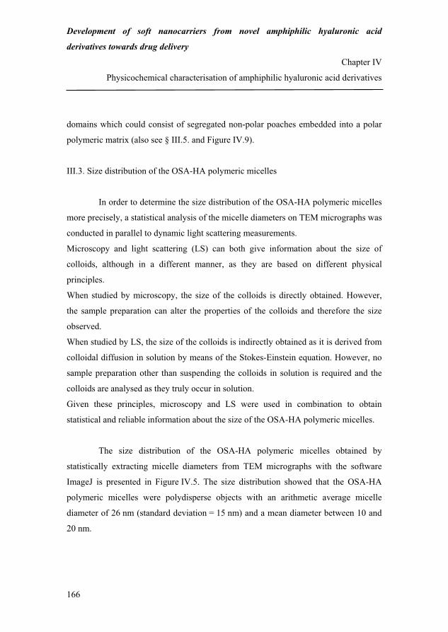
Development of soft nanocarriers from novel amphiphilic hyaluronic acid
derivatives towards drug delivery
Chapter IV
Physicochemical characterisation of amphiphilic hyaluronic acid derivatives
166
domains which could consist of segregated non-polar poaches embedded into a polar
polymeric matrix (also see § III.5. and Figure IV.9).
III.3. Size distribution of the OSA-HA polymeric micelles
In order to determine the size distribution of the OSA-HA polymeric micelles
more precisely, a statistical analysis of the micelle diameters on TEM micrographs was
conducted in parallel to dynamic light scattering measurements.
Microscopy and light scattering (LS) can both give information about the size of
colloids, although in a different manner, as they are based on different physical
principles.
When studied by microscopy, the size of the colloids is directly obtained. However,
the sample preparation can alter the properties of the colloids and therefore the size
observed.
When studied by LS, the size of the colloids is indirectly obtained as it is derived from
colloidal diffusion in solution by means of the Stokes-Einstein equation. However, no
sample preparation other than suspending the colloids in solution is required and the
colloids are analysed as they truly occur in solution.
Given these principles, microscopy and LS were used in combination to obtain
statistical and reliable information about the size of the OSA-HA polymeric micelles.
The size distribution of the OSA-HA polymeric micelles obtained by
statistically extracting micelle diameters from TEM micrographs with the software
ImageJ is presented in Figure IV.5. The size distribution showed that the OSA-HA
polymeric micelles were polydisperse objects with an arithmetic average micelle
diameter of 26 nm (standard deviation = 15 nm) and a mean diameter between 10 and
20 nm.
192
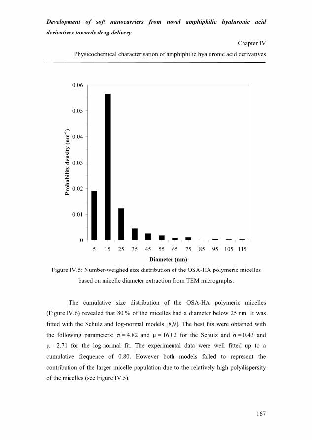
Development of soft nanocarriers from novel amphiphilic hyaluronic acid
derivatives towards drug delivery
Chapter IV
Physicochemical characterisation of amphiphilic hyaluronic acid derivatives
167
0
0.01
0.02
0.03
0.04
0.05
0.06
5 15 25 35 45 55 65 75 85 95 105 115
Diameter (nm)
Prob
abili
ty d
ensi
ty (n
m-1
)
Figure IV.5: Number-weighed size distribution of the OSA-HA polymeric micelles
based on micelle diameter extraction from TEM micrographs.
The cumulative size distribution of the OSA-HA polymeric micelles
(Figure IV.6) revealed that 80 % of the micelles had a diameter below 25 nm. It was
fitted with the Schulz and log-normal models [8,9]. The best fits were obtained with
the following parameters: = 4.82 and � = 16.02 for the Schulz and = 0.43 and
� = 2.71 for the log-normal fit. The experimental data were well fitted up to a
cumulative frequence of 0.80. However both models failed to represent the
contribution of the larger micelle population due to the relatively high polydispersity
of the micelles (see Figure IV.5).
193

Development of soft nanocarriers from novel amphiphilic hyaluronic acid
derivatives towards drug delivery
Chapter IV
Physicochemical characterisation of amphiphilic hyaluronic acid derivatives
168
Figure IV.6: Number-weighed cumulative size distribution of the OSA-HA polymeric
micelles based on micelle diameter extraction from TEM micrographs.
The hydrodynamic diameter of the OSA-HA polymeric micelles was
determined in PBS (pH = 7.4), at room temperature, by DLS and was simulated from
the micelle size distribution obtained by TEM. Figure IV.7 presents the variation of the
hydrodynamic diameter as a function of the scattering angle as measured by DLS and
as simulated from the micelle size distribution obtained by TEM for two different
values of the swelling coefficient � (� = 0 for dehydrated micelles and � = 12 for
hydrated micelles).
0
0.1
0.2
0.3
0.4
0.5
0.6
0.7
0.8
0.9
1
0 20 40 60 80 100 120
Diameter (nm)
Cum
ulat
ive
freq
uenc
e
Microscopy dataSchulz fitlog-normal fit
� � � � ��
���
� ��
����
���
� ���
�
xxxSchulzf��
��� ��
1exp1
11
� � �� ����
0
1 dteta ta
� ���
���
� ����� 2
2
2lnexp
21)(log
��
��x
xxnormalf
194

Development of soft nanocarriers from novel amphiphilic hyaluronic acid
derivatives towards drug delivery
Chapter IV
Physicochemical characterisation of amphiphilic hyaluronic acid derivatives
169
0
50
100
150
200
250
300
20 40 60 80 100 120
Scattering angle (degree)
Hyd
rody
nam
ic d
iam
eter
(nm
)
DLS data
simulated microscopy data - � = 12
simulated microscopy data - � = 0
Figure IV.7: Experimental and simulated hydrodynamic diameter of the OSA-HA
polymeric micelles as a function of the scattering angle.
The values of the hydrodynamic diameter measured by DLS were
considerably higher than those simulated from the crude microscopy data, i.e. when
� = 0. Indeed, the OSA-HA polymeric micelles were hydrated during the DLS
analysis, whereas they had lost their water content before the TEM analysis. Taking
into account micelle hydration in the simulation (� = 12), a better fit between the
values of the hydrodynamic diameter obtained by DLS and those obtained by TEM
was observed over most of the scattering angles. This indicated that the micelles
analysed by DLS contained a volume of water of up to 12 times larger than the volume
of OSA-HA making up the fully hydrated micelles.
195
Development of soft nanocarriers from novel amphiphilic hyaluronic acid
derivatives towards drug delivery
Chapter IV
Physicochemical characterisation of amphiphilic hyaluronic acid derivatives
170
The swelling coefficient found in this study was of the same order of
magnitude as previously reported values of the swelling ratio in chemically cross-
linked HA hydrogels.
Srinivas and Ramamurthi [12] have namely reported that the swelling ratio of HA
hydrogels obtained by cross-linking with divinyl sulfone (DVS) (HA/DVS weight
ratio = 3) could reach values up to 7. In this study, the swelling ratio was defined as the
ratio of the difference between the weight of the swollen and the dried gel and the
weight of the dried gel when the latter was obtained by freeze-drying. Since TEM
micrographs are acquired under vacuum, this degree of dehydration of the hydrogel is
comparable to that of the OSA-HA polymeric micelles observed by TEM. It could be
demonstrated that the definition of the swelling ratio by Srinivas and Ramamurthi is
equivalent to that of the present study if the density of OSA-HA is close to 1.0.
Therefore the value of the swelling coefficient found in this study (12) can directly be
compared to that found by Srinivas and Ramamurthi (7).
Similarly, Smeds et al. [13] observed that HA hydrogels obtained by photocross-
linking of methacrylated HA could swell up to 11 times their dry weight when the
dried gels were obtained by freeze drying. This swelling ability was dependent on the
degree of cross-linking (DS between 3 and 17 %). The gels with the highest degree of
cross-linking had the least ability to swell due to their tight polymeric networks. In this
study, the swelling coefficient was defined as the ratio of the weight of the swollen gel
and that of the dried gel. It could be shown that the relationship between this swelling
ratio A and the swelling ratio � defined in the present study is � = A – 1 if the density
of OSA-HA is close to 1.0. In other words, the value of the swelling coefficient found
in the present study (12) is to be compared with an adjusted value of 10 [13].
Interestingly, the OSA-HA polymeric micelles exhibited comparable swelling
properties as chemically cross-linking HA but with the difference that they were not
stabilised by chemical cross-linking but by hydrophobic interactions. Therefore they
can be regarded as physically cross-linked nanogels.
196

Development of soft nanocarriers from novel amphiphilic hyaluronic acid
derivatives towards drug delivery
Chapter IV
Physicochemical characterisation of amphiphilic hyaluronic acid derivatives
171
The fairly large deviation of the hydrodynamic diameter measured by DLS
from that simulated by microscopy (with � = 12) at low scattering angles indicated that
a few structures with dimensions superior to the detection range of the DLS instrument
were present in solution. These few structures could be micelle aggregates although
micelle aggregation could not clearly be identified on the TEM micrographs.
In summary, the data provided by the microscopy and DLS analysis showed
that the OSA-HA polymeric micelles were highly hydrated structures in solution with
a mean hydrodynamic diameter falling between 170 and 230 nm.
III.4. Zeta potential of the OSA-HA polymeric micelles
The zeta potential of the OSA-HA polymeric micelles was determined in PBS
(pH = 7.4), at 25 °C, by means of measuring their electrophoretic mobility. The results
showed that the surface of the OSA-HA polymeric micelles was negatively charged
with a zeta potential of approximately -28 mV under the applied conditions (pH ~ 3.0-
4.0, Figure IV.8). This negative charge was due to the presence of carboxylate groups
on OSA-HA, arising from both the native HA backbone and the hemiesters generated
during the reaction between HA and OSA.
III.5. Molecular structure of the OSA-HA polymeric micelles
Chen et al. [14] have described the structure of polymeric micelles based on
hydrophobically modified HA with 2300 Da poly(caprolactone) chains as a spherical
HA hydrophilic corona surrounding a poly(caprolactone) hydrophobic core. Given the
relatively short length of the OS chains, the aggregation of OSA-HA is unlikely to
occur in this fashion.
197

Development of soft nanocarriers from novel amphiphilic hyaluronic acid
derivatives towards drug delivery
Chapter IV
Physicochemical characterisation of amphiphilic hyaluronic acid derivatives
172
0
10
20
30
40
50
-200 -150 -100 -50 0 50 100 150 200
Zeta potential (mV)
Inte
nsity
(a.u
.)
Figure IV.8: Zeta potential of the OSA-HA polymeric micelles.
Substituted disaccharide units on OSA-HA were assumed to behave like
monomeric surfactant moieties capable of self-associating into conventional spherical
monomeric micellar domains. OSA-HA polymeric micelles were therefore believed to
consist of a dispersion of hydrophobic domains formed by the modified HA units in a
hydrophilic matrix structured by the remaining unmodified HA units (Figure IV.9). In
this multi-vesicular model, the OS chains involved in the hydrophobic domains
potentially can originate from the same or different OSA-HA molecules and the
micelle structure is believed to be stabilized by a combination of hydrophobic
interactions between the OS chains and the three fundamental HA structures in
solution discussed in Chapter I.
198

Development of soft nanocarriers from novel amphiphilic hyaluronic acid
derivatives towards drug delivery
Chapter IV
Physicochemical characterisation of amphiphilic hyaluronic acid derivatives
173
Monomeric micellar domain
HA chains
OSA side chains
500 nm
Figure IV.9: Putative representation of the structure of the OSA-HA polymeric
micelles.
The number of substituted HA units per hydrophobic domains, of
hydrophobic domains per micelle and the average distance between vicinal
hydrophobic domains were calculated for the most occurring micelle, i.e. a micelle
with a radius of approximately 8 nm in the dehydrated state (cf. Figure IV.5) or 19 nm
in the hydrated state. The dimensions of the modified disaccharide unit used in the
calculations were the following: the length and width of the disaccharide unit were
approximated to 1 and 0.5 nm, respectively [15]. The length of the OS chains was
approximated to 1 nm as it consists of about 10 carbon-to-carbon bonds. It should be
noted that the influence of the geometric isomerism of the OS chains (cis/trans) on the
packing of the hydrophobic domains was disregarded as the distribution of the cis and
199

Development of soft nanocarriers from novel amphiphilic hyaluronic acid
derivatives towards drug delivery
Chapter IV
Physicochemical characterisation of amphiphilic hyaluronic acid derivatives
174
trans OS moieties on HA is unknown. In the following calculations, the densities of
HA and OSA-HA were considered to be close to 106 g/m3.
The number of OSA-HA units per hydrophobic domain was calculated by
dividing the volume of one hydrophobic domain by the volume of one OSA-HA unit.
The volume of one OSA-HA unit can further be decomposed as the sum of the volume
of one HA unit and the volume of the cone deployed by one OS chain. Substitution
with more elementary physical quantities led to Equation IV.4:
� �� � � �� �
� �� �
� � � �OSAlHAulNHA
HAuMW
OSAlHAuwdomainHAuOSAN
A
2
3
231
34
/� �
����
���
��
� Equation IV.4
where N(OSA-HAu)/domain, w(HAu), l(OS), MW(HAu), �(HA), NA and l(HAu)
represent the number of OSA-HA units per hydrophobic domain, the width of the HA
unit, the length of the OS chain, the molecular weight of the HA unit, the density of
HA, the Avogadro number and the length of the HA unit, respectively.
The number of hydrophobic domains per micelle was calculated by dividing
the number of OSA-HA units per micelle by the number of OSA-HA units per
hydrophobic domain. The number of OSA-HA units per micelle corresponds to the
number of OSA-HA molecules per micelle multiplied by the number of OSA-HA units
per OSA-HA molecule. This number is related to the DS of OSA-HA. The number of
OSA-HA molecules per micelle can further be calculated by dividing the volume of
one micelle by the volume of one OSA-HA molecule. Subsequent substitutions and
rearrangements led to Equation IV.5:
� �
� � � �� � � �
� � domainHAuOSAN
OSAMWDS
HAuMW
NHAOSAmicelleR
micelledomainsN
A
/
34
/
3
�
�
�
�
��
Equation IV.5
200

Development of soft nanocarriers from novel amphiphilic hyaluronic acid
derivatives towards drug delivery
Chapter IV
Physicochemical characterisation of amphiphilic hyaluronic acid derivatives
175
where N(domains)/micelle, R(micelle), �(OSA-HA), NA, MW(HAu), DS, MW(OSA)
and N(OSA-HAu)/domain stand for the number of hydrophobic domains per micelle,
the average radius of the most occurring micelle, the density of OSA-HA, the
Avogadro number, the molecular weight of the HA unit, the degree of substitution of
OSA-HA, the molecular weight of OSA and the number of OSA-HA unit per
hydrophobic domain, respectively.
Finally, the distance between vicinal hydrophobic domains was calculated by
applying the relation shown in Equation IV.6.
� �3
1).(domainC
domainsbetweenl � Equation IV.6
In Equation IV.6, l(between.domains) and C(domain) correspond to the distance
between vicinal hydrophobic domains and concentration of hydrophobic domains
inside the micelle, respectively. The latter can be expressed as the ratio between the
number of hydrophobic domains per micelle and the volume of the hydrated micelle
(Equation IV.7).
� �� �� �
� �3
33
/
134
.micelledomainsN
micelleRdomainsbetweenl
��
�� Equation IV.7
In Equation IV.7, l(between.domains), �, R(micelle) and N(domains)/micelle represent
the distance between vicinal hydrophobic domains, the swelling coefficient, the radius
of the most occurring micelle and the number of hydrophobic domains per micelle,
respectively.
According to our calculations, the most occurring OSA-HA micelle consisted
of about 72 hydrophobic domains separated by a distance of 6-9 nm, with a diameter
of 3 nm, each involving 16 OSA-HA units. Although approximate, this model is
believed to provide a rough but realistic picture of the internal structure of the micelles.
201

Development of soft nanocarriers from novel amphiphilic hyaluronic acid
derivatives towards drug delivery
Chapter IV
Physicochemical characterisation of amphiphilic hyaluronic acid derivatives
176
IV. Conclusion
The physicochemical properties of water soluble amphiphilic octenyl succinic
anhydride-modified hyaluronic acid were investigated in aqueous media. OSA-HA
was shown to self-associate and solubilise Nile Red into the hydrophobic domains
formed by the OS chains above a critical aggregation concentration which was
dependent on the degree of substitution of the derivatives. The CAC of OSA-HA was a
linear decreasing function of the derivatives’ DS between 6 and 44 % per disaccharide
unit and can be predicted for OSA-HA derivatives with a DS in this range.
The OSA-HA polymeric micelles were negatively charged polydisperse
spherical physically cross-linked nanogels characterised by a high degree of hydration
(typically 12 g of water per g of polymer) and a hydrodynamic diameter between 170
and 230 nm.
The molecular structure of the OSA-HA polymeric micelles is most probably a
distribution of OS-rich hydrophobic monomeric micellar nanodomains into a HA-rich
hydrophilic polymeric matrix.
The nanoengineered structures of the OSA-HA polymeric nanogels constitute
biocompatible and resorbable multi-functional systems which could be used to
solubilise and carry poorly soluble active ingredients in aqueous media. It is believed
that the presence of multiple hydrophobic nanodomains in the nanogels could be useful
for the controlled release of hydrophobic active ingredients or drugs.
The formulation of stabilised OSA-HA nanoparticles insensitive to dilution and study
of the release of model hydrophobic substances in vitro and in vivo will be the subject
of future investigations.
202
Development of soft nanocarriers from novel amphiphilic hyaluronic acid
derivatives towards drug delivery
Chapter IV
Physicochemical characterisation of amphiphilic hyaluronic acid derivatives
177
References
[1] Weissman, B.; Meyer, K. The structure of hyalobiuronic acid and of hyaluronic
acid from umbilical cord. J. Am. Chem. Soc. 1954, 76, 1753-1757.
[2] Baier Leach, J.; Schmidt, C. E. Hyaluronan. In Encyclopedia of biomaterials and
biomedical engineering; Wnek, G. E.; Bowlin G. L., Eds.; Marcel Dekker: New
York, 2004; pp 779-789.
[3] Gustafson, S. Hyaluronan in drug delivery. In The chemistry, biology and medical
applications of hyaluronan and its derivatives; Laurent, T. C., Ed.; Portland
Press: London, 1998; pp 291-304.
[4] Prestwich, G. P.; Marecek, D. M.; Marecek, J. F.; Vercruysse, K. P.; Ziebell, M.
R. Chemical modification of hyaluronic acid for drug delivery, biomaterials and
biochemical probes. In The chemistry, biology and medical applications of
hyaluronan and its derivatives; Laurent, T. C., Ed.; Portland Press: London,
1998.
[5] Pelletier, S.; Hubert, P.; Lapicque, F.; Payan, E.; Dellacherie, E. Amphiphilic
derivatives of sodium alginate and hyaluronate: synthesis and physico-chemical
properties of aqueous dilute solutions. Carbohydr. Polym. 2000, 43, 343-349.
[6] Creuzet, C.; Kadi, S.; Rinaudo, M.; Auzley-Velty, R. New associative systems
based on alkylated hyaluronic acid. Synthesis and aqueous solution properties.
Polymer 2006, 47, 2706-2713.
[7] Tømmeraas, K.; Eenschooten, C. Aryl/alkyl succinic anhydride hyaluronan
derivatives. WO 2007/033677 A1, March 29, 2007.
[8] Schulz, G. V. The kinetics of chain polymerisation V. The influence of various
types of reactions on the poly-molecularity. Z. Phys. Chem. Abt. B 1939, 43, 25-
46.
[9] Limpert, E.; Stahel, W.; Abbt, M. Log-normal distributions across the sciences:
keys and clues. Bioscience 2001, 51, 341-352.
203

Development of soft nanocarriers from novel amphiphilic hyaluronic acid
derivatives towards drug delivery
Chapter IV
Physicochemical characterisation of amphiphilic hyaluronic acid derivatives
178
[10] Berne, B. J.; Pecora, R. Dynamic Light Scattering; Dover Publications: Mineola,
New York, 2000.
[11] Dutt, G. B.; Doriswami, S. Picosecond reorientational dynamics of polar dye
probes in binary aqueous mixtures. J. Chem. Phys. 1992, 96, 2475-2491.
[12] Srinivas, A.; Ramamurthi, A. Effects of gamma-irradiation on physical and
biologic properties of cross-linked hyaluronan tissue engineering scaffolds. Tissue
Eng. 2007, 13, 447-459.
[13] Smeds, K. A.; Pfister-Serres, A.; Miki, D.; Dastgheib, M.; Inoue, M.; Hatchell, D.
L.; Grinstaff, M. W. Photocrosslinkable polysaccharides for in situ hydrogel
formation. J. Biomed. Mater. Res. 2001, 55, 254-255.
[14] Chen, J. H.; Tsai, B. H.; Chang, H. T.; Chen, M. L.; Chen, Y. H.; Jan, S. H.; Liu,
M. J. Biodegradable hyaluronic acid derivative and biodegradable polymeric
micelle composition. US 2005/0123505 A1, June 9, 2005.
[15] Ionov, R.; El-Abed, A.; Goldmann, M.; Peretti, P. Interactions of lipid monolayers
with the natural biopolymer hyaluronic acid. Biochim. Biophys. Acta 2004, 1667,
200-207.
204

179
CHAPTER V
Conclusion and future work
205

180
206

Development of soft nanocarriers from novel amphiphilic hyaluronic acid
derivatives towards drug delivery
Chapter V
Conclusion and future work
181
I. Summary
This project was initiated in the perspective of providing biocompatible and
resorbable nanocarriers based on hyaluronic acid (HA) towards drug delivery and in
particular dermal drug delivery (see Chapter I).
A literature review (see Chapter II) about polymeric nanoparticle-mediated
(trans)dermal drug delivery reaffirmed the high potential of using the skin as an
administration route and identified new approaches to better understand the fate of
particulate systems in this tissue. It also took inventory of the current hypothesis with
regards to how polymeric nanoparticles can enhance drug uptake and/or sustain drug
release and spotted the challenges faced by this branch of research. Interestingly, the
studies reported mostly concern synthetic polymeric nanoparticles which by nature
are not truly biocompatible. For this reason, systems based on the naturally occurring
glycosaminoglycan hyaluronic acid were developed in this project.
Whereas most of the currently available methods for the preparation of HA
nanoparticles rely on random collisions in the medium to create structures, a novel
molecular technique was here developed which relies on the controlled self-assembly
of amphiphilic HA leading to multiphasic nanoarchitectured HA templates.
Such properties of HA were obtained by modifying the biopolymer’s
hydrophilic/lipophilic balance via the covalent grafting of hydrophobic groups onto
the HA backbone. After a preliminary evaluation of two novel proprietary methods to
hydrophobically modify HA, octenyl succinic anhydride-modified HA (OSA-HA)
was chosen as starting material for the preparation of the HA-based nanostructures
(see Chapter III, A). Its preparation process was optimised so as to develop a
reproducible and robust technique for the routine laboratory production of OSA-HA.
With the reaction conditions tested, HA derivatives with a maximum degree of
substitution (DS) of 44 % per disaccharide unit according to proton nuclear magnetic
resonance spectroscopy were prepared (see Chapter III, B).
207
Development of soft nanocarriers from novel amphiphilic hyaluronic acid
derivatives towards drug delivery
Chapter V
Conclusion and future work
182
Although it was not possible to precisely determine the substitution pattern and
measure the molecular weight and monomolecular size of the OSA-HA derivatives,
there were some indications that the substitution pattern of OSA-HA was most likely
random at the molecular level and heterogeneous at the macromolecular level and
that OSA-HA undergoes a major conformation change in aqueous media at a
concentration which is dependent on its degree of substitution (see Chapter III, C).
The self-assembly in aqueous media of OSA-HA was due to hydrophobic
interactions between the grafted octenyl succinate (OS) chains on HA and was
controlled by the DS of the derivatives. The critical aggregation concentration (CAC)
of OSA-HA was indeed all the more low that its DS was high. Self-assembling of
OSA-HA was therefore a controllable and predictable property for derivatives with a
DS up to 44 %. In addition, it was demonstrated that OSA-HA could solubilise the
model hydrophobic compound Nile Red. Above the CAC, the OSA-HA assemblies
were rather polydisperse spherical and negatively charged polymeric micelles with a
hydrodynamic diameter between 170 and 230 nm. They were characterised by a high
degree of hydration (on averaged 12 g of water per g of polymer). This suggested that
the fundamental mechanism of their stabilisation was a physical cross-linking
mediated by the hydrophobic interactions between the grafted OS chains. The
internal structure of the OSA-HA nanogels was believed to consist of a dispersion of
hydrophobic monomeric micellar domains formed by the OS chains embedded in a
hydrophilic polymeric matrix composed of the unmodified HA segments (see
Chapter IV).
The development of OSA-HA nanogels described herein is believed to
constitute a valuable first step towards formulating HA-based nanoparticles with
more complex configurations than single ingredient or core-shell structures. In
particular, the presence of multiple vesicle-like substructures inside the nanogels
could be an interesting feature for sustaining drug delivery as these could delay drug
release in biological environments.
208

Development of soft nanocarriers from novel amphiphilic hyaluronic acid
derivatives towards drug delivery
Chapter V
Conclusion and future work
183
Moreover, it should be emphasised that the methods underlying the OSA-HA
nanogels are simple, aqueous and easily upscalable thus potentially valuable for the
industry. The modification with OSA preserves and even increases the distribution of
negative charges along the modified polymer at neutral pH which could constitutes
an advantageous property for the electrostatic stabilisation of future nanoparticles
from OSA-HA.
II. Future work
As for now, the OSA-HA nanogels only exist above their CAC. This sets the
lower limit of their concentration when incorporated into formulations. In addition, it
is unlikely that they maintain their shape when administered and/or exhibit higher in
vivo residence times compared to native HA unless they are stabilised. The next main
challenge therefore concerns the preparation of stable OSA-HA nanoparticles.
For an application such as dermal drug delivery, it could be necessary to
reduce the size of the OSA-HA nanogels down to several tens of nanometres and
better control their polydispersity (see Chapter II).
In the perspective of using the stabilised OSA-HA nanogels for concrete
applications, it is foreseeable that future work will also involve biocompatibility tests
and release studies in vitro and in vivo. Incorporation and stability in final
formulations will also constitute an important challenge.
209

184
210

185
Appendices
211

186
212

Development of soft nanocarriers from novel amphiphilic hyaluronic acid
derivatives towards drug delivery
Appendices
187
Educational activities
I. Training
I.1. Courses
� Chemical and Biochemical Product Design, Technical University of Denmark,
Lyngby, Denmark, autumn 2003 (10 ECTS).
� Patents and patent search, Novozymes A/S, Bagsværd, Denmark, June 17, 2005
(0.6 ECTS).
� Design of experiment, Umetrics, Bagsværd, Denmark, September 1, 2005
(2.2 ECTS).
� Intensive course in dermato-cosmetic sciences, Faculty of Medicine and Pharmacy,
Brussels, Belgium, September 12-16, 2005 (3.6 ECTS).
� Polymeric surfactants, Institute for Surface Chemistry, Stockholm, Sweden,
December 12-14, 2005 (3 ECTS).
� Fundamentals of percutaneous penetration, pre-conference course of the 10th
International Conference in Perspectives in Percutaneous Penetration, La Grande
Motte, France, April 18, 2006 (0.6 ECTS).
� Spectroscopy of organic compounds, Technical University of Denmark, Lyngby,
Denmark, spring 2006 (5 ECTS).
� Business course, Danish Ministry of Science, Technology and Innovation through
mandated Copenhagen Business School and Haslund & Alsted, Pharmakon, Hillerød,
213

Development of soft nanocarriers from novel amphiphilic hyaluronic acid
derivatives towards drug delivery
Appendices
188
Denmark, October 16-17, 2006, February 12-13, 2007, September 11-12, 2007
(5 ECTS).
I.2. Conferences
� Kick-off meeting Nano-DTU/Nano-Science Centre, Hørsholm, Denmark, March 2,
2006.
� 6th European Workshop on Particulate Systems, Geneva, Switzerland, March 23-24,
2006.
� 10th International Conference in Perspectives in Percutaneous Penetration, La
Grande Motte, France, April 18-22, 2006.
� Skin and Formulation 2nd Symposium, Versailles, France, October 8-10, 2006.
� 8th Annual Meeting of Skin Forum, London, United Kingdom, March 3-4, 2007.
� 7th International Conference on Hyaluronan, Charleston, South Carolina, United
State, April 22-27, 2007.
� 21st Conference of the European Colloid and Interface Society, Geneva,
Switzerland, September 10-14, 2007.
I.3. External study trips
The external study trips were carried out at the University of Geneva, University of
Lausanne, School of Pharmaceutical Sciences, Department of Pharmaceutics and
Biopharmaceutics, 30 quai Ernest-Ansermet, CH-1211 Geneva 4, Switzerland under
214

Development of soft nanocarriers from novel amphiphilic hyaluronic acid
derivatives towards drug delivery
Appendices
189
the supervision of Professor Robert Gurny and Associate Professor Florence Delie in
the periods July 3-August 31, 2006 and March 12-June 29, 2007.
II. Dissemination of knowledge
II.1. Publications
II.1.1. Posters
� Eenschooten, C.; Guillaumie-Longin, F.; Kontogeorgis, G. M.; Schwach-
Abdellaoui, K. Synthesis and formulation of modified hyaluronic acid (HA) for
cosmetic applications. Annual discussion meeting 2005 of the Centre for Phase
Equilibria and Separation Processes, Roskilde, Denmark, May 25-27, 2005.
� Eenschooten, C.; Guillaumie-Longin, F.; Gurny, R.; Kontogeorgis, G. M.;
Schwach-Abdellaoui, K.; Stenby E. H. Development and formulation of hyaluronic
acid-based nanoparticles for cosmetic applications. Kick-off meeting Nano-
DTU/Nano-Science Centre, Hørsholm, Denmark, March 2, 2006.
� Eenschooten, C.; Delie, F.; Guillaumie-Longin, F.; Gurny, R.; Kontogeorgis, G. M.;
Schwach-Abdellaoui, K.; Stenby, E. H. Development of colloidal carriers from
modified hyaluronic acid. Skin and Formulation 2nd Symposium, Versailles, France,
October 8-10, 2006.
� Eenschooten, C.; Delie, F.; Guillaumie-Longin, F.; Kontogeorgis, G. M.; Stenby, E.
H.; Schwach-Abdellaoui, K.; Gurny, R. Development of colloidal carriers from
modified hyaluronic acid. 8th Annual Meeting of the Skin Forum, London, United
Kingdom, March 3-4, 2007.
215

Development of soft nanocarriers from novel amphiphilic hyaluronic acid
derivatives towards drug delivery
Appendices
190
� Eenschooten, C.; Delie, F.; Guillaumie-Longin, F.; Kontogeorgis, G. M.; Stenby, E.
H.; Schwach-Abdellaoui, K.; Gurny, R. Development of colloidal carriers from
modified hyaluronic acid. 7th International Conference on Hyaluronan, Charleston,
South Carolina, United State, April 22-27, 2007.
� Eenschooten, C.; Vaccaro, A.; Delie, F.; Guillaumie-Longin, F.; Kontogeorgis, G.
M.; Stenby, E. H.; Schwach-Abdellaoui, K.; Borkovec, M.; Gurny, R. Development of
colloidal carriers from a novel amphiphilic hyaluronic acid. 21st Conference of the
European Colloid and Interface Society, Geneva, Switzerland, September 10-14, 2007.
II.1.2. Oral presentations
Seminars of the Centre for Phase Equilibria and Separation Processes
� Eenschooten, C. Development of derivatized hyaluronic acid and formulation of
nanoparticles for the delivery of cosmetic actives and drugs. Centre for Phase
Equilibria and Separation Processes, Department of Chemical and Biochemical
Engineering, Technical University of Denmark, Lyngby, Denmark, May 18, 2006.
� Eenschooten, C. Development of colloidal carriers from modified hyaluronic acid.
Centre for Phase Equilibria and Separation Processes, Department of Chemical and
Biochemical Engineering, Technical University of Denmark, Lyngby, Denmark,
February 15, 2007.
Annual discussion meetings of the Centre for Phase Equilibria and Separation
Processes
� Eenschooten, C. Development of derivatized hyaluronic acid and formulation of
nanoparticles for the delivery of cosmetic actives and drugs. Annual discussion
216

Development of soft nanocarriers from novel amphiphilic hyaluronic acid
derivatives towards drug delivery
Appendices
191
meeting 2006 of the Centre for Phase Equilibria and Separation Processes, Elsinore,
Denmark, June 26-28, 2006.
� Eenschooten, C. Development of novel colloidal carriers from chemically modified
hyaluronic acid. Annual discussion meeting 2007 of the Centre for Phase Equilibria
and Separation Processes, Elsinore, Denmark, May 30-June 1, 2007.
� Eenschooten, C. Development of soft nanocarriers from novel amphiphilic
hyaluronic acid derivatives towards topical drug delivery. Annual discussion meeting
2008 of the Centre for Phase Equilibria and Separation Processes, Værløse, Denmark,
June 16-18, 2008.
II.1.3. Manuscript contributions
� Eenschooten, C.; Guillaumie, F.; Kontogeorgis, G. M.; Stenby, E. H.; Schwach-
Abdellaoui, K. Development of hyaluronic acid-based nanoparticles for topical
delivery of cosmetic actives. In Department of Chemical Engineering, Technical
University of Denmark, Graduate schools yearbook; Dam-Johansen K.; Bøjer, M.,
Eds.; BookPartner, Nørhaven Digital: Copenhagen, 2006.
� Eenschooten, C.; Guillaumie, F.; Kontogeorgis, G. M.; Stenby, E. H.; Schwach-
Abdellaoui, K. Development of hyaluronic acid-based nanocarriers towards the topical
delivery of cosmetic active ingredients or pharmaceutical drugs. In Department of
Chemical Engineering, Technical University of Denmark, Graduate schools yearbook;
Dam-Johansen K.; Bøjer, M., Eds.; BookPartner, Nørhaven Digital: Copenhagen,
2007.
217

Development of soft nanocarriers from novel amphiphilic hyaluronic acid
derivatives towards drug delivery
Appendices
192
II.1.4. Patents
� Tømmeraas, K.; Eenschooten, C. Aryl/alkyl succinic anhydride hyaluronan
derivatives. WO2007/033677 A1, March 29, 2007.
� Eenschooten, C.; Christensen, M. W. Aryl/Alkyl vinyl sulfone hyaluronic acid
derivatives. WO2007/098770 A1, September 7, 2007.
II.1.5. Articles in preparation
� Eenschooten, C.; Guillaumie, F.; Delie, F.; Kontogeorgis, G. M.; Schwach-
Abdellaoui, K. Towards an understanding of the mechanisms underlying polymeric
nanoparticle-mediated (trans)dermal drug delivery (based on Chapter II).
� Eenschooten, C.; Guillaumie, F.; Kontogeorgis, G. M.; Stenby, E. H.; Schwach-
Abdellaoui, K. Preparation and structural characterization of novel and versatile
amphiphilic hyaluronic acid derivatives (based on Chapter III, section B).
� Eenschooten, C.; Vaccaro, A.; Delie, F.; Guillaumie, F.; Tømmeraas, K.;
Kontogeorgis, G. M.; Schwach-Abdellaoui, K.; Borkovec, M.; Gurny, R. Novel self-
associative multi-phase nanostructured soft carriers based on amphiphilic hyaluronic
acid derivatives (based on Chapter IV).
II.2. Teaching
II.2.1. Assistantship in master’s courses
� Soft Nanotechnology in Colloid and Surface Chemistry, master’s course, Technical
University of Denmark, DTU Department of Chemical and Biochemical Engineering,
Autumn 2005 and 2006.
218
Development of soft nanocarriers from novel amphiphilic hyaluronic acid
derivatives towards drug delivery
Appendices
193
II.2.2. Co-supervision
� Structural characterization of a novel amphiphilic derivative of hyaluronic acid,
master’s project, Technical University of Denmark, Department of Chemistry, Autumn
2007.
219

194
NOTES
220

195
NOTES
221

196
NOTES
222

197
NOTES
223

198
NOTES
224

199
225

Department of Chemical and Biochemical EngineeringDTU Building 229Søltofts PladsDK-2800 Kgs. Lyngbywww.kt.dtu.dk
Development of soft nanocarriers from novel amphiphilic hyaluronic acid derivatives towards drug delivery
Corinne Eenschooten2008
C. E
enschootenD
evelopment of soft nanocarriers from
novel amphiphilic hyaluronic acid derivatives tow
ards drug delivery2008