Susceptibility of ePTFE vascular grafts and bioengineered ...
Journal of Biomaterials Science 22 (2011) 195–206brill.nl/jbs
Development of Small-Diameter Vascular Grafts Based onSilk Fibroin Fibers from Bombyx mori for
Vascular Regeneration
Yasumoto Nakazawa a, Michiko Sato b, Rui Takahashi b, Derya Aytemiz b,
Chiyuki Takabayashi c, Toshiki Tamura c, Soichiro Enomoto d,
Masataka Sata e and Tetsuo Asakura a,b,∗
a Nature and Science Museum, Tokyo University of Agriculture and Technology,Koganei, Tokyo 184-8588, Japan
b Graduate School of Engineering, Tokyo University of Agriculture and Technology,Koganei, Tokyo 184-8588, Japan
c Laboratory of New Silk Materials, National Institute of Agrobiological Sciences,Okaya, Nagano 394-0021, Japan
d Department of Cardiovascular Medicine, University of Tokyo Graduate School of Medicine,Tokyo 113-0033, Japan
e Department of Cardiovascular Medicine, Institute of Health Biosciences, The University ofTokushima Graduate School, Tokushima 770-8503, Japan
Received 2 September 2009; accepted 18 November 2009
AbstractIn the field of surgical revascularization, the need for functional small-diameter (1.5–4.0 mm in diameter)vascular grafts is increasing. Several synthetic biomaterials have been tested for this purpose, but in manycases they cause thrombosis. In this study, we report the development of small-diameter vascular graftsmade from silk fibroin fibers from the domestic silkworm Bombyx mori or recombinant silk fibroin fibersfrom a transgenic silkworm. The vascular grafts were prepared by braiding, flattening and winding the silkfibers twice onto a cylindrical polymer tube followed by coating with an aqueous silk fibroin solution. Thegrafts, which are 1.5 mm in inner diameter and 10 mm in length, were implanted into rat abdominal aorta.An excellent patency (ca. 85%, n = 27) at 12 months after grafting with wild-type silk fibers was obtained.Endothelial cells and smooth muscle cells migrated into the silk fibroin graft early after implantation, andbecame organized into an endothelium and a media-like smooth muscle layer.© Koninklijke Brill NV, Leiden, 2011
KeywordsSilk fibroin fiber, small-diameter vascular graft, transgenic silkworm
* To whom correspondence should be addressed. Tel.: (81-42) 383-7733; Fax: (81-42) 383-7733; e-mail:[email protected]
© Koninklijke Brill NV, Leiden, 2011 DOI:10.1163/092050609X12586381656530
196 Y. Nakazawa et al. / Journal of Biomaterials Science 22 (2011) 195–206
1. Introduction
Artificial vascular grafts manufactured from synthetic materials, for example, ex-panded polytetrafluoroethylene (ePTFE) and Dacron [1–3], have been routinelyused to reconstruct blood flow in patients with various cardiovascular disorders.Large-diameter vascular grafts have sufficient patency for implantation into the hu-man body. However, only 15–30% of small-diameter vascular grafts remain patentafter 5 years in vivo [4]. Moreover, in vivo studies have shown only a 20–25% pa-tency rate with 1-mm-diameter PTFE microvessels, while all vein grafts in similarsettings remained patent [5, 6]. The reason is that the vascular grafts made usingDacron or PTFE did not affect the cellular proliferation on the lumen of the vasculargraft.
Conventional grafts have clinically shown satisfactory durability; however, theystill have several disadvantages, such as thrombogenicity, late stenosis and occlu-sion from intimal hyperplasia (especially in small-diameter grafts), susceptibilityto infection and lack of growth potential [7]. To overcome these limitations in thesearch for an ideal artificial graft, various tissue-engineered vascular grafts havebeen developed. The utility and clinical experience of these new grafts has beenreported [8–14].
In this study, we report the development of new vascular grafts with a smalldiameter made from silk fibroin from the domestic silkworm, Bombyx mori, on thebasis of our accumulated information on the structures of the silk fibroin. B. moriproduces a protein fiber with excellent mechanical properties such as high strengthand high toughness, which has a long history of use as suture [15]. In addition, fromaqueous or organic solutions of the silk fibroin, it is possible to prepare a form offilm, sponge, powder, gel and regenerated fiber. Much information on its structuresand structural changes including the excellent physical properties of silk fibroin inseveral forms has been accumulated by us and other investigators [16–28]. Recently,there has been a significant increase in the number of reports of applications ofsilk fibroin to biomaterials [17–20, 27–32]. Moreover, the recent development ofbiotechnology permits an improvement of the cell-adhesive character of B. morisilk fibroin by incorporation of cell-adhesive amino-acid sequences into the silkprotein [33–41]. In particular, the incorporation of cell-adhesive sequences into silkfibroin fiber and the production of such a recombinant silk fiber directly from thecocoon was possible using a transgenic silkworm. This recombinant silk fiber wasalso used for preparation of vascular grafts with a small diameter, and in preliminarystudies showed improved performance compared to the wild-type silk.
2. Materials and Methods
2.1. Preparation of the Silk Fibroin Fiber
Cocoons produced by B. mori were placed in water at 95◦C after which the threadswere reeled. The dried silk threads were placed in a mixture of sodium carbonate
Y. Nakazawa et al. / Journal of Biomaterials Science 22 (2011) 195–206 197
(0.08%, w/v) and Marseille soap (0.12%, w/v) at 95◦C for 120 min [16–18]. Thisprocess was repeated in order to remove silk sericin from the raw silk fibers com-pletely. The removal of silk sericin was checked by a scanning electron microscope(SEM VE-7800, Keyence, Japan). Pure silk fibroin fibers prepared in this way wereused for the braiding and winding process, and also for preparing aqueous fibroinsolutions during the production of the small-diameter grafts.
2.2. Production of Recombinant Silk Fibroin from Transgenic Silkworm
We previously designed recombinant silk fibroins with a high cell-adhesive se-quences and produced them from transgenic silkworms [41]. In this paper, forpreparation of the vascular graft, we used a recombinant silk fibroin fiber incor-porating the sequences of active sites from collagen. The details have been reportedpreviously [41]. Only limited amounts of the silk fiber samples were available and,therefore, the grafts prepared from wild-type and recombinant silk fibroin fiberswere compared only at the initial stage of the growth of endothelial cells after graft-ing.
2.3. 13C CP/MAS NMR Analysis
13C CP/MAS NMR spectra of wild-type silk fibroin fiber from B. mori and recom-binant silk fibroin fiber from transgenic silkworm were observed to characterize thestructure in the dried and hydrated states [18, 21]. The information on the structurein the hydrated state is important in order to characterize the structure in vivo. Forthe NMR observation of hydrated samples, two kinds of silk fibroin fiber whichare wild-type and recombinant samples were hydrated by immersing them in wa-ter for 1 week. A small amount of water attached to the surfaces of the fibers wasremoved just before the NMR observation. 13C CP/MAS NMR experiments wereperformed on a Bruker Avance 400 MHz spectrometer with an operating frequencyof 100.6 MHz for 13C at a sample spinning rate of 8 kHz in a 4 mm diameter ZrO2rotor. A total of 4096 scans for the samples were collected over a spectral widthof 35 kHz with a recycle delay of 5 s. A 150 kHz radio-frequency field strengthwas used for 1H–13C decoupling with an acquisition period of 15 ms. A 90◦ pulsewidth of 4.5 µs with a 1 ms cross-polarization contact time was employed. Phase cy-cling was used to minimize artifacts. The experimental conditions were the same fordried and hydrated silk fibroin fiber samples. 13C chemical shifts were calibratedindirectly using adamantane and are presented in ppm relative to tetramethylsi-lane.
2.4. Preparation of the Aqueous Solution of Silk Fibroin
Pure silk fibroin fibers were dissolved in 9 M LiBr aqueous solution to a concen-tration of 10% (w/v) at 60◦C for 4 h and then dialyzed against distilled water for3 days at 4◦C using a cellulose membrane (Viskase Sales, USA) [16]. The finalconcentration of the fibroin/water solution was 3–4% (w/v). Freshly prepared silkfibroin aqueous solution was used for coating the silk graft [22].
198 Y. Nakazawa et al. / Journal of Biomaterials Science 22 (2011) 195–206
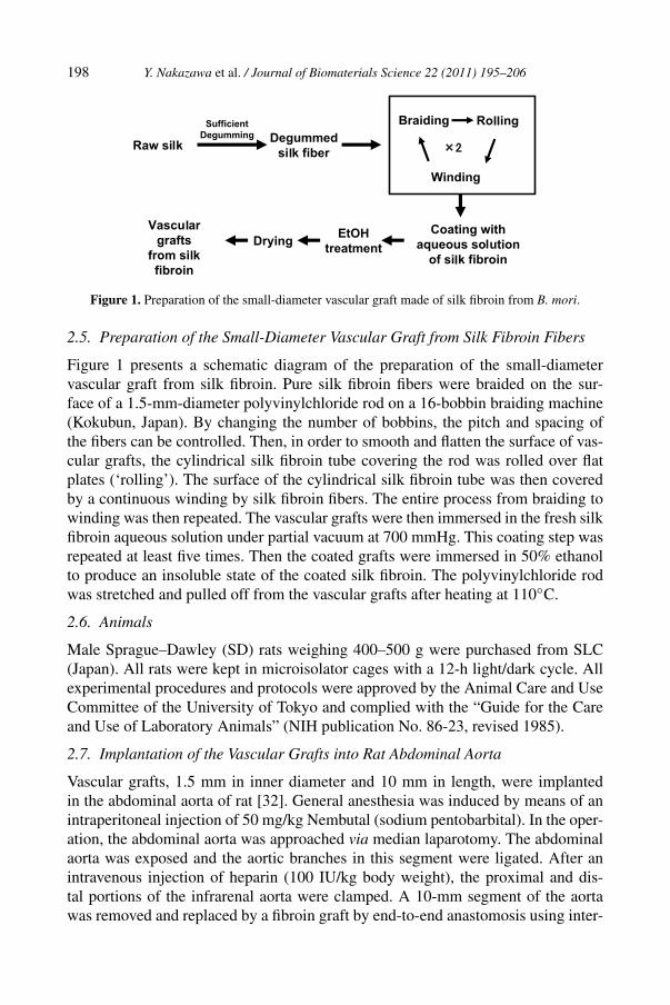
Figure 1. Preparation of the small-diameter vascular graft made of silk fibroin from B. mori.
2.5. Preparation of the Small-Diameter Vascular Graft from Silk Fibroin Fibers
Figure 1 presents a schematic diagram of the preparation of the small-diametervascular graft from silk fibroin. Pure silk fibroin fibers were braided on the sur-face of a 1.5-mm-diameter polyvinylchloride rod on a 16-bobbin braiding machine(Kokubun, Japan). By changing the number of bobbins, the pitch and spacing ofthe fibers can be controlled. Then, in order to smooth and flatten the surface of vas-cular grafts, the cylindrical silk fibroin tube covering the rod was rolled over flatplates (‘rolling’). The surface of the cylindrical silk fibroin tube was then coveredby a continuous winding by silk fibroin fibers. The entire process from braiding towinding was then repeated. The vascular grafts were then immersed in the fresh silkfibroin aqueous solution under partial vacuum at 700 mmHg. This coating step wasrepeated at least five times. Then the coated grafts were immersed in 50% ethanolto produce an insoluble state of the coated silk fibroin. The polyvinylchloride rodwas stretched and pulled off from the vascular grafts after heating at 110◦C.
2.6. Animals
Male Sprague–Dawley (SD) rats weighing 400–500 g were purchased from SLC(Japan). All rats were kept in microisolator cages with a 12-h light/dark cycle. Allexperimental procedures and protocols were approved by the Animal Care and UseCommittee of the University of Tokyo and complied with the “Guide for the Careand Use of Laboratory Animals” (NIH publication No. 86-23, revised 1985).
2.7. Implantation of the Vascular Grafts into Rat Abdominal Aorta
Vascular grafts, 1.5 mm in inner diameter and 10 mm in length, were implantedin the abdominal aorta of rat [32]. General anesthesia was induced by means of anintraperitoneal injection of 50 mg/kg Nembutal (sodium pentobarbital). In the oper-ation, the abdominal aorta was approached via median laparotomy. The abdominalaorta was exposed and the aortic branches in this segment were ligated. After anintravenous injection of heparin (100 IU/kg body weight), the proximal and dis-tal portions of the infrarenal aorta were clamped. A 10-mm segment of the aortawas removed and replaced by a fibroin graft by end-to-end anastomosis using inter-
Y. Nakazawa et al. / Journal of Biomaterials Science 22 (2011) 195–206 199
rupted 9-0 monofilament nylon sutures (Bear, Japan), starting with two stay suturesat 180◦ to each other, then suturing the front wall followed by the back wall. Thenumber of stitches used for each anastomosis ranged from 10 to 12. The distal, thenthe proximal vascular clamps were slowly removed, and flow was restored throughthe fibroin graft. Graft patency was monitored by color Doppler imaging and pulsewaves recorded with a 12-MHz sector probe and an echo-imaging apparatus (En-Visor M2540A, Philips, Japan) under anesthesia with pentobarbital. Graft diameterand blood flow velocity were measured. Signs of thrombosis and aneurysm forma-tion were carefully checked.
2.8. Histological Examination
The rats were killed with an overdose of Nembutal. At death, the rats were perfusedwith 0.9% NaCl solution via the left ventricle. The grafts were carefully removedwith surrounding tissue, cut transversely in the midline into two pieces, and fixedin methanol or snap-frozen in OCT compound (Tissue-Tek, Japan) for histologicalanalyses. Methanol-fixed samples were embedded in paraffin. Paraffin-embeddedsections (4 µm thick) were processed for hematoxylin and eosin staining. Forimmunohistochemistry [32], the sections were incubated with primary antibodies(alkaline phosphatase-conjugated anti-α-smooth muscle actin (clone 1A4, Sigma,USA), anti-rat CD31 (clone TLD-3A12, BD Biosciences, USA) or anti-CD68(clone ED1, Serotec, UK)), followed by incubation with biotinylated anti-mouseIgG secondary antibody (Dako, Denmark) and subsequent use of the avidin–biotincomplex technique and Vector Red substrate (Vector Laboratories, USA). Nucleiwere counterstained with hematoxylin. Sirius red polarization microscopy was per-formed to visualize interstitial collagen and fibroin. Frozen sections (5 µm) wererinsed with distilled water and incubated with 0.1% sirius red (Sigma-Aldrich,USA) in saturated picric acid for 90 ms. Sections were rinsed twice with 0.03 MHCl for 1 min each time and then immersed in distilled water. After dehydrationwith 70% ethanol for 30 s, the sections were coverslipped.
3. Results and Discussion
3.1. Morphological Examination of the Silk Fibroin Vascular Graft During thePreparation Process
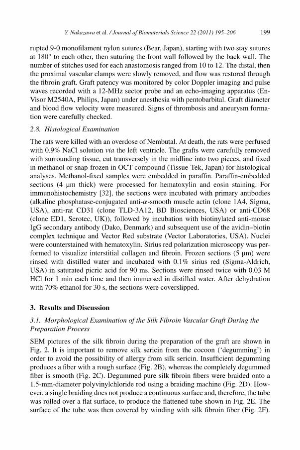
SEM pictures of the silk fibroin during the preparation of the graft are shown inFig. 2. It is important to remove silk sericin from the cocoon (‘degumming’) inorder to avoid the possibility of allergy from silk sericin. Insufficient degummingproduces a fiber with a rough surface (Fig. 2B), whereas the completely degummedfiber is smooth (Fig. 2C). Degummed pure silk fibroin fibers were braided onto a1.5-mm-diameter polyvinylchloride rod using a braiding machine (Fig. 2D). How-ever, a single braiding does not produce a continuous surface and, therefore, the tubewas rolled over a flat surface, to produce the flattened tube shown in Fig. 2E. Thesurface of the tube was then covered by winding with silk fibroin fiber (Fig. 2F).
200 Y. Nakazawa et al. / Journal of Biomaterials Science 22 (2011) 195–206
Figure 2. Scanning electron micrographs of silk fibers and silk grafts at several processing stages.(A) Raw silk, (B) a silk fibroin fiber after insufficient degumming, (C) a silk fibroin fiber after completeremoval of silk sericin, (D) silk graft after braiding, (E) silk graft after rolling, (F) silk graft afterwinding and (G) the final silk graft.
The entire braiding, rolling and winding process was then repeated to produce agraft of sufficient strength. In addition, in order to make the graft fully watertight,coating with silk fibroin is required. Vascular grafts were immersed in silk fibroinaqueous solution [22] under partial vacuum to help osmosis. The coating step wasrepeated at least five times. The graft was then immersed in 50% ethanol to makethe coated silk fibroin insoluble by a structural change to β-sheet [18, 22]. Thus,vascular grafts with alternating braided and spiraled fiber layers, coated by silk fi-broin, were prepared with a final appearance as shown in Fig. 2G.
3.2. Structural Analysis of Recombinant Silk Fibroin by 13C CP/MAS NMR
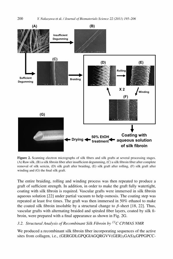
We produced a recombinant silk fibroin fiber incorporating sequences of the activesites from collagen, i.e., (GERGDLGPQGIAGQRGVV(GER)3GAS)8GPPGPCC-
Y. Nakazawa et al. / Journal of Biomaterials Science 22 (2011) 195–206 201
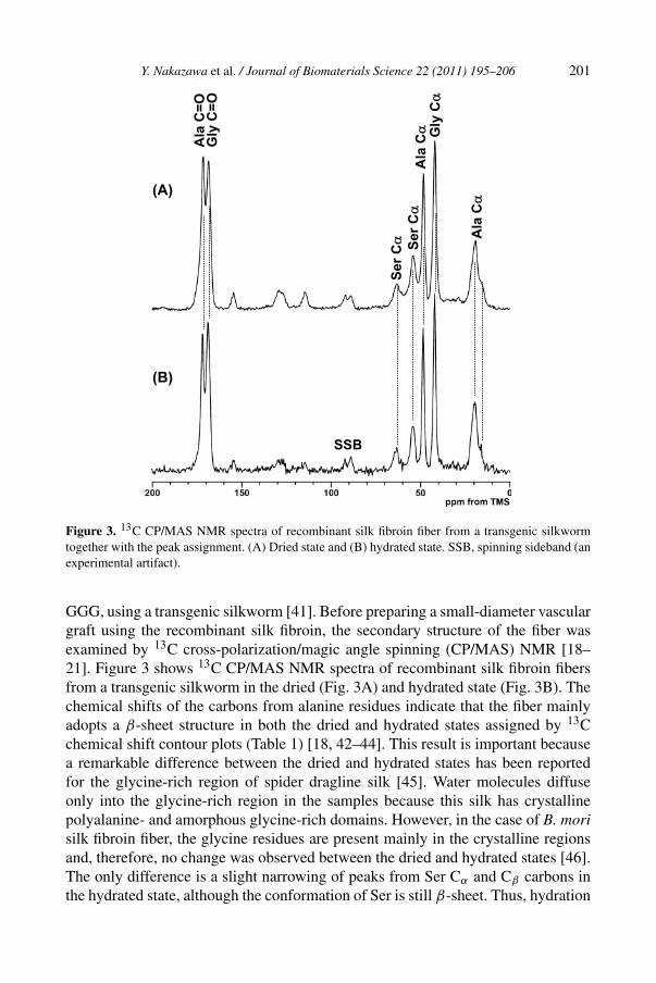
Figure 3. 13C CP/MAS NMR spectra of recombinant silk fibroin fiber from a transgenic silkwormtogether with the peak assignment. (A) Dried state and (B) hydrated state. SSB, spinning sideband (anexperimental artifact).
GGG, using a transgenic silkworm [41]. Before preparing a small-diameter vasculargraft using the recombinant silk fibroin, the secondary structure of the fiber wasexamined by 13C cross-polarization/magic angle spinning (CP/MAS) NMR [18–21]. Figure 3 shows 13C CP/MAS NMR spectra of recombinant silk fibroin fibersfrom a transgenic silkworm in the dried (Fig. 3A) and hydrated state (Fig. 3B). Thechemical shifts of the carbons from alanine residues indicate that the fiber mainlyadopts a β-sheet structure in both the dried and hydrated states assigned by 13Cchemical shift contour plots (Table 1) [18, 42–44]. This result is important becausea remarkable difference between the dried and hydrated states has been reportedfor the glycine-rich region of spider dragline silk [45]. Water molecules diffuseonly into the glycine-rich region in the samples because this silk has crystallinepolyalanine- and amorphous glycine-rich domains. However, in the case of B. morisilk fibroin fiber, the glycine residues are present mainly in the crystalline regionsand, therefore, no change was observed between the dried and hydrated states [46].The only difference is a slight narrowing of peaks from Ser Cα and Cβ carbons inthe hydrated state, although the conformation of Ser is still β-sheet. Thus, hydration
202 Y. Nakazawa et al. / Journal of Biomaterials Science 22 (2011) 195–206
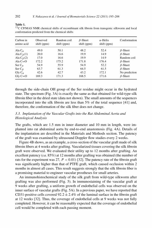
Table 1.13C CP/MAS NMR chemical shifts of recombinant silk fibroin from transgenic silkworm and localconformation predicted from the chemical shifts
Carbon in Observed Random coil β-Sheet α-Helix Conformationamino acid shift (ppm) shift (ppm) shift (ppm) shift (ppm)
Ala Cα 49.0 50.1 48.2 52.4 β-SheetAla Cβ(1) 20.0 16.6 19.9 14.9 β-SheetAla Cβ(2) 17.0 16.6 19.9 14.9 Random coilAla C=O 172.2 175.2 171.8 176.4 β-SheetSer Cα 54.9 55.9 54.9 52.2 β-SheetSer Cβ 63.7 61.3 64.5 61.5 β-SheetGly Cα 42.6 42.7 43.2 172.1 No predictionGly C=O 169.3 171.3 168.5 171.6 β-Sheet
through the side-chain OH group of the Ser residue might occur in the hydratedstate. The spectrum (Fig. 3A) is exactly the same as that obtained for wild-type silkfibroin fiber in the dried state (data not shown). The small amounts of the sequencesincorporated into the silk fibroin are less than 5% of the total sequence [41] and,therefore, the conformation of the silk fiber does not change.
3.3. Implantation of the Vascular Grafts into the Rat Abdominal Aorta andHistological Analysis
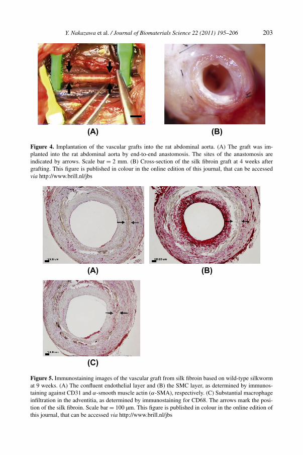
The grafts, which are 1.5 mm in inner diameter and 10 mm in length, were im-planted into rat abdominal aorta by end-to-end anastomosis (Fig. 4A). Details ofthe implantation are described in the Materials and Methods section. The patencyof the graft was examined by ultrasound Doppler flow studies every 2 weeks.
Figure 4B shows, as an example, a cross-section of the vascular graft made of silkfibroin fibers at 4 weeks after grafting. Vascularized tissues covering the silk fibroingraft were observed. We evaluated their utility up to 12 months after grafting. Anexcellent patency (ca. 85%) at 12 months after grafting was obtained (the number ofrats for the experiment was 27, P < 0.01) [32]. The patency rate of the fibroin graftwas significantly higher than that of PTFE graft, which caused occlusion within 2months in almost all cases. This result suggests strongly that the silk fibroin fiber isa promising material to engineer vascular prostheses for small arteries.
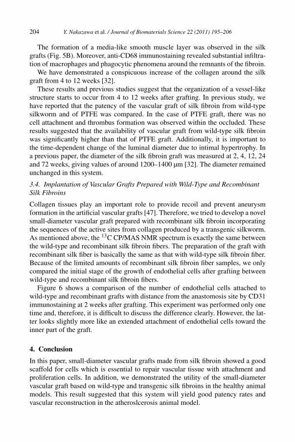
An immunohistochemical study of the silk graft from wild-type silkworm aftergrafting was also performed (Fig. 5). In immunostaining of the vascular graft at9 weeks after grafting, a uniform growth of endothelial cells was observed on theinner surface of vascular grafts (Fig. 5A). In a previous paper, we have reported thatCD31-positive cells covered 92.2 ± 2.4% of the luminal surface in the fibroin graftat 12 weeks [32]. Thus, the coverage of endothelial cells at 9 weeks was not fullycompleted. However, it can be reasonably expected that the coverage of endothelialcell would be completed with each passing moment.
Y. Nakazawa et al. / Journal of Biomaterials Science 22 (2011) 195–206 203
Figure 4. Implantation of the vascular grafts into the rat abdominal aorta. (A) The graft was im-planted into the rat abdominal aorta by end-to-end anastomosis. The sites of the anastomosis areindicated by arrows. Scale bar = 2 mm. (B) Cross-section of the silk fibroin graft at 4 weeks aftergrafting. This figure is published in colour in the online edition of this journal, that can be accessedvia http://www.brill.nl/jbs
Figure 5. Immunostaining images of the vascular graft from silk fibroin based on wild-type silkwormat 9 weeks. (A) The confluent endothelial layer and (B) the SMC layer, as determined by immunos-taining against CD31 and α-smooth muscle actin (α-SMA), respectively. (C) Substantial macrophageinfiltration in the adventitia, as determined by immunostaining for CD68. The arrows mark the posi-tion of the silk fibroin. Scale bar = 100 µm. This figure is published in colour in the online edition ofthis journal, that can be accessed via http://www.brill.nl/jbs
204 Y. Nakazawa et al. / Journal of Biomaterials Science 22 (2011) 195–206
The formation of a media-like smooth muscle layer was observed in the silkgrafts (Fig. 5B). Moreover, anti-CD68 immunostaining revealed substantial infiltra-tion of macrophages and phagocytic phenomena around the remnants of the fibroin.
We have demonstrated a conspicuous increase of the collagen around the silkgraft from 4 to 12 weeks [32].
These results and previous studies suggest that the organization of a vessel-likestructure starts to occur from 4 to 12 weeks after grafting. In previous study, wehave reported that the patency of the vascular graft of silk fibroin from wild-typesilkworm and of PTFE was compared. In the case of PTFE graft, there was nocell attachment and thrombus formation was observed within the occluded. Theseresults suggested that the availability of vascular graft from wild-type silk fibroinwas significantly higher than that of PTFE graft. Additionally, it is important tothe time-dependent change of the luminal diameter due to intimal hypertrophy. Ina previous paper, the diameter of the silk fibroin graft was measured at 2, 4, 12, 24and 72 weeks, giving values of around 1200–1400 µm [32]. The diameter remainedunchanged in this system.
3.4. Implantation of Vascular Grafts Prepared with Wild-Type and RecombinantSilk Fibroins
Collagen tissues play an important role to provide recoil and prevent aneurysmformation in the artificial vascular grafts [47]. Therefore, we tried to develop a novelsmall-diameter vascular graft prepared with recombinant silk fibroin incorporatingthe sequences of the active sites from collagen produced by a transgenic silkworm.As mentioned above, the 13C CP/MAS NMR spectrum is exactly the same betweenthe wild-type and recombinant silk fibroin fibers. The preparation of the graft withrecombinant silk fiber is basically the same as that with wild-type silk fibroin fiber.Because of the limited amounts of recombinant silk fibroin fiber samples, we onlycompared the initial stage of the growth of endothelial cells after grafting betweenwild-type and recombinant silk fibroin fibers.
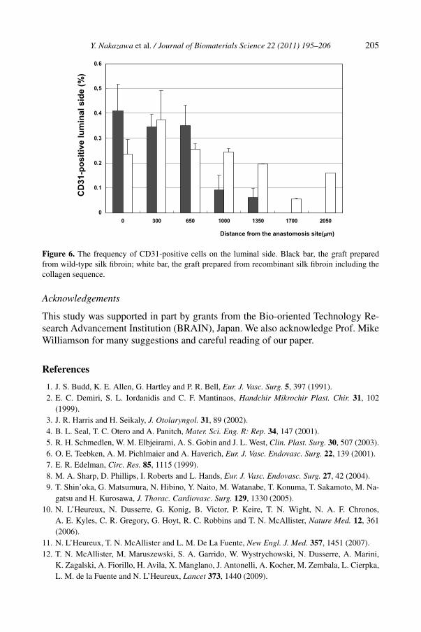
Figure 6 shows a comparison of the number of endothelial cells attached towild-type and recombinant grafts with distance from the anastomosis site by CD31immunostaining at 2 weeks after grafting. This experiment was performed only onetime and, therefore, it is difficult to discuss the difference clearly. However, the lat-ter looks slightly more like an extended attachment of endothelial cells toward theinner part of the graft.
4. Conclusion
In this paper, small-diameter vascular grafts made from silk fibroin showed a goodscaffold for cells which is essential to repair vascular tissue with attachment andproliferation cells. In addition, we demonstrated the utility of the small-diametervascular graft based on wild-type and transgenic silk fibroins in the healthy animalmodels. This result suggested that this system will yield good patency rates andvascular reconstruction in the atheroslcerosis animal model.
Y. Nakazawa et al. / Journal of Biomaterials Science 22 (2011) 195–206 205
Figure 6. The frequency of CD31-positive cells on the luminal side. Black bar, the graft preparedfrom wild-type silk fibroin; white bar, the graft prepared from recombinant silk fibroin including thecollagen sequence.
Acknowledgements
This study was supported in part by grants from the Bio-oriented Technology Re-search Advancement Institution (BRAIN), Japan. We also acknowledge Prof. MikeWilliamson for many suggestions and careful reading of our paper.
References
1. J. S. Budd, K. E. Allen, G. Hartley and P. R. Bell, Eur. J. Vasc. Surg. 5, 397 (1991).2. E. C. Demiri, S. L. Iordanidis and C. F. Mantinaos, Handchir Mikrochir Plast. Chir. 31, 102
(1999).3. J. R. Harris and H. Seikaly, J. Otolaryngol. 31, 89 (2002).4. B. L. Seal, T. C. Otero and A. Panitch, Mater. Sci. Eng. R: Rep. 34, 147 (2001).5. R. H. Schmedlen, W. M. Elbjeirami, A. S. Gobin and J. L. West, Clin. Plast. Surg. 30, 507 (2003).6. O. E. Teebken, A. M. Pichlmaier and A. Haverich, Eur. J. Vasc. Endovasc. Surg. 22, 139 (2001).7. E. R. Edelman, Circ. Res. 85, 1115 (1999).8. M. A. Sharp, D. Phillips, I. Roberts and L. Hands, Eur. J. Vasc. Endovasc. Surg. 27, 42 (2004).9. T. Shin’oka, G. Matsumura, N. Hibino, Y. Naito, M. Watanabe, T. Konuma, T. Sakamoto, M. Na-
gatsu and H. Kurosawa, J. Thorac. Cardiovasc. Surg. 129, 1330 (2005).10. N. L’Heureux, N. Dusserre, G. Konig, B. Victor, P. Keire, T. N. Wight, N. A. F. Chronos,
A. E. Kyles, C. R. Gregory, G. Hoyt, R. C. Robbins and T. N. McAllister, Nature Med. 12, 361(2006).
11. N. L’Heureux, T. N. McAllister and L. M. De La Fuente, New Engl. J. Med. 357, 1451 (2007).12. T. N. McAllister, M. Maruszewski, S. A. Garrido, W. Wystrychowski, N. Dusserre, A. Marini,
K. Zagalski, A. Fiorillo, H. Avila, X. Manglano, J. Antonelli, A. Kocher, M. Zembala, L. Cierpka,L. M. de la Fuente and N. L’Heureux, Lancet 373, 1440 (2009).
206 Y. Nakazawa et al. / Journal of Biomaterials Science 22 (2011) 195–206
13. J. E. Puskas and Y. Chen, Biomacromolecules 5, 1141 (2004).14. R. D. Sayers, S. Raptis, M. Berce and J. H. Miller, Br. J. Surg. 85, 934 (1998).15. T. Asakura and D. L. Kaplan, in: Encyclopedia of Agricultural Science, C. J. Arutzen (Ed.), Vol. 4,
p. 1. Academic Press, New York, NY (1994).16. T. Asakura, Y. Watanabe, A. Uchida and H. Minagawa, Macromolecules 17, 1075 (1984).17. M. Demura and T. Asakura, Biotechnol. Bioeng. 33, 598 (1989).18. M. Ishida, T. Asakura, M. Yokoi and H. Saito, Macromolecules 23, 88 (1990).19. M. Demura, T. Asakura and T. Kuroo, Biosensors 4, 361 (1989).20. M. Demura and T. Asakura, J. Membr. Sci. 59, 39 (1991).21. H. Yoshimizu and T. Asakura, J. Appl. Polym. Sci. 40, 127 (1990).22. T. Asakura, M. Kitaguchi, M. Demura, H. Sakai and K. Komatsu, J. Appl. Polym. Sci. 46, 49
(1992).23. M. Demura, T. Takekawa, T. Asakura and A. Nishikawa, Biomaterials 13, 276 (1992).24. K. A. Trabbic and P. Yager, Macromolecules 31, 462 (1998).25. A. Seidel, O. Liivak, S. Calve, J. Adaska, G. Ji, Z. Yang, D. Grubb, D. B. Zax and L. W. Jelinski,
Macromolecules 33, 775 (2000).26. J. Yao, H. Masuda, C. Zhao and T. Asakura, Macromolecules 35, 6 (2002).27. Y. Tamada, Biomacromolecules 6, 3100 (2005).28. L. Meinel, S. Hofmann, V. Karageorgiou, L. Zichner, R. Langer, D. Kaplan and G. Vunjak-
Novakovic, Biotechnol. Bioeng. 88, 379 (2004).29. G. H. Altman, F. Diaz, C. Jakuba, T. Calabro, R. L. Horan, J. Chen, H. Lu, J. Richmond and
D. L. Kaplan, Biomaterials 24, 401 (2003).30. X. Zhang, X. Wang, V. Keshav, J. T. Johanas, G. G. Leisk and D. L. Kaplan, Biomaterials 30,
3213 (2009).31. K. Makaya, S. Terada, K. Ohgo and T. Asakura, J. Biosci. Bioeng. 108, 68 (2009).32. S. Enomoto, M. Sumi, K. Kajimoto, Y. Nakazawa, R. Takahashi, C. Takabayashi, T. Asakura and
M. Sata, J. Vasc. Surg., in press (2010).33. J. Cappello and F. Ferrari, Plast. Microb. 3, 35 (1994).34. S. A. Maskarinec and D. A. Tirrell, Curr. Opin. Biotechnol. 16, 422 (2005).35. J. O’Brien, S. Fahnestock, Y. Termonia and K. Gardner, Adv. Mater. 10, 1185 (1998).36. C. Wong Po Foo and D. L. Kaplan, Adv. Drug Deliv. Rev. 54, 1131 (2002).37. J. Yao, S. Yanagisawa and T. Asakura, J. Biochem. 136, 643 (2004).38. T. Asakura, K. Nitta, M. Yang, J. Yao, Y. Nakazawa and D. L. Kaplan, Biomacromolecules 4, 815
(2003).39. M. Yang and T. Asakura, J. Biochem. 137, 721 (2005).40. M. Yang, J. Kawamura, Z. Zhu, K. Yamauchi and T. Asakura, Polymer 50, 117 (2009).41. S. Yanagisawa, Z. Zhu, I. Kobayashi, K. Uchino, Y. Tamada, T. Tamura and T. Asakura, Bio-
macromolecules 8, 3487 (2007).42. H. Saito, Y. Iwanaga, R. Tabeta, M. Narita and T. Asakura, Chem. Lett. 12, 427 (1983).43. H. Saito, I. Ando and A. Naito, Solid State NMR Spectroscopy for Biopolymers — Principles and
Applications. Springer, New York, NY (2006).44. T. Asakura, M. Iwadate, M. Demura and M. P. Williamson, Int. J. Biol. Macromol. 24, 167 (1999).45. Z. T. Yang, O. Liivak, A. Seidel, G. LaVerde, D. B. Zax and L. W. Jelinski, J. Am. Chem. Soc.
122, 9019 (2000).46. T. Asakura, J. Yao, T. Yamane, K. Umemura and A. S. Ulrich, J. Am. Chem. Soc. 124, 8794 (2002).47. B. C. Isenberg, C. Williams and R. T. Tranquillo, Circ. Res. 98, 25 (2006).


![Stem cells in vascular graft tissue engineering for ... · 649 Stem cells in vascular graft tissue engineering for congenital heart surgery REVIEW Darcon grafts [13].Other studies](https://static.fdocuments.net/doc/165x107/5e7a747788383848980b07bd/stem-cells-in-vascular-graft-tissue-engineering-for-649-stem-cells-in-vascular.jpg)