DEVELOPMENT OF Salmonella typhi Ty21a AS A … · VACCINE AGAINST TUBERCULOSIS: ... Dr. Najeeb Abu...
47
DEVELOPMENT OF Salmonella typhi Ty21a AS A POTENTIAL ORAL VACCINE AGAINST TUBERCULOSIS: SURFACE DISPLAY AND DNA VACCINE CARRIER OF A SYNTHETIC MULTI-EPITOPE MYCOBACTERIAL GENE by MOHAMMED ABDEL AZIZ AHMAD SARHAN June 2004 Thesis submitted in fulfilment of the requirements for the degree of Doctor of Philosophy in Biomedical Sciences (Molecular and Cell Biology)
Transcript of DEVELOPMENT OF Salmonella typhi Ty21a AS A … · VACCINE AGAINST TUBERCULOSIS: ... Dr. Najeeb Abu...
DEVELOPMENT OF Salmonella typhi Ty21a AS A POTENTIAL ORAL
VACCINE AGAINST TUBERCULOSIS: SURFACE DISPLAY AND DNA
VACCINE CARRIER OF A SYNTHETIC MULTI-EPITOPE
MYCOBACTERIAL GENE
by
MOHAMMED ABDEL AZIZ AHMAD SARHAN
June 2004
Thesis submitted in fulfilment of the requirements for the degree of Doctor of Philosophy in
Biomedical Sciences (Molecular and Cell Biology)

ii
This thesis is dedicated to my Parents and to my wife for her patient and encouragement and my children, Haya, Ahmad, Reema and Samar.

iii
Acknowledgements
All praise and thanks are due to Allah; the possessor of all Excellencies; for gratuitously
giving me the ingredients of success. Invoke the blessings of Allah on the noble Prophet
Mohammed peace be upon him, who taught us to be thankful.
Over the span of time during which this research project was conducted I have received
assistance and/or advice from several people whom I wish to acknowledge at this time.
Above all, I would like to thank my supervisor, Prof. Dr. Zainul F. Zainuddin for his
support, excellent guidance and supervision throughout the experimental work, research
investigations and writing of the manuscript, and also for providing all the necessary
facilities to carry out this study. His steadfast guidance and constant accessibility are
greatly appreciated. It has been a privilege to work with you.
I would also like to express my gratitude to my co-supervisor Prof. Madya Dr. Mustaffa
Musa for encouragement, kind guidance, overall comments and helpful discussions
during this study.
I feel very grateful to Prof. Dr. Norazmi Mohd Nor and Dr. M. Ravichandran, who
have provided advice and offered suggestions whenever required. I sincerely
acknowledge the encouragement given by Dr. Fawwaz al Juddi. I would like to extend
thanks to my friends and collegues at the molecular biology and immunology research
laboratories, who provided both friendship and assistance during the course of this
study.

iv
To all my friends outside the laboratory especially Mr. Mohd Arifin, Dr. Najeeb Abu
Rub, Dr. Ayman Saleem, Dr. Nasr al Meree, Dr. Sediq and Dr. Rassheed; thanks for
being there and encouraging me when needed.
A special thanks to my parents, who provided me with the inspiration to pursue my
study. Finally I wish to acknowledge the greatest support, love and encouragement from
my wife, Amal, my son Ahmad, and daughters Haya, Reema and Samar, who have been
very patient when I spent time working on this thesis which should have been spent
with them.
There are many people that I would like to thank for contributing in one way or another
to this work during the last years. I cannot mention you all so I hope you feel my
gratitude.

v
Table of Contents Acknowledgment ............................................................................................................................... iii List of Tables........................................................................................................................................ xi List of Figures...................................................................................................................................... xii List of Abbreviations.......................................................................................................................... xv Abstract.................................................................................................................................................. xvi Abstrak................................................................................................................................................... xix
Chapter One Introduction
1.1. Background …................................................................................................................................1 1.2. Human TB: A historical perspective……………………………………………................1 1.3. Epidemiology of TB in humans……………………………………………………...............4 1.4. The TB organism: Mycobacterium tuberculosis…………………………………............6 1.5. Chemical composition of the M. tuberculosis cell wall structure…………………..7 1.6. Genetics of M. tuberculosis .................................................................................... 10 1.7. Pathophysiology of TB ........................................................................................... 10
1.7.1. TB, the disease .................................................................................................. 10 1.7.2. Symptoms of TB ............................................................................................... 11 1.7.3. Transmission of M. tuberculosis ....................................................................... 13
1.8. Immune response to TB ........................................................................................ 14 1.8.1. Early response ................................................................................................... 14 1.8.2. Pathogenicity and initial defense against M. tuberculosis infection ................. 14
1.8.2.1. Primary TB ................................................................................................ 15 1.8.2.2. Secondary TB ............................................................................................ 16
1.8.3. Immunity to TB ................................................................................................ 17 1.8.3.1. Humoral immunity against TB .................................................................. 17 1.8.3.2. Cellular immunity against TB .................................................................... 18
1.9. Mycobacterial antigens .......................................................................................... 23 1.10. Diagnosis of TB. .................................................................................................... 25 1.11. Control of TB ......................................................................................................... 27
1.11.1. Treatment with anti-TB drugs ......................................................................... 27 1.11.2. Vaccines .......................................................................................................... 29
1.11.2.1. Vaccination to prevent TB ....................................................................... 30 1.11.2.2. The BCG vaccine and its efficacy ........................................................... 31 1.11.2.3. Development of candidate vaccines other than BCG .............................. 32
1.11.2.3.1. Recombinant BCG ............................................................................ 37 1.11.2.3.2. Attenuated strains of M. tuberculosis ............................................... 38 1.11.2.3.3. Subunit vaccines ............................................................................... 39 1.11.2.3.4. DNA vaccines ................................................................................... 39 1.11.2.3.5. Live recombinant vaccines ............................................................... 42
1.12. S. typhi Ty21a as a live oral vaccine ................................................................... 43 1.13. Bacterial surface display systems ....................................................................... 46
1.13.1. Ice-nucleation protein ..................................................................................... 51 1.14. The aims of this study .......................................................................................... 55
vi
Chapter Two Materials and Methods
2.1. Materials ................................................................................................................. 58
2.1.1. Mice .................................................................................................................. 58 2.1.2. Bacterial strains, culture media and growth conditions .................................... 58 2.1.3. Plasmids ............................................................................................................ 60 2.1.4. Chemicals .......................................................................................................... 60 2.1.5. Antibodies and peptides .................................................................................... 60 2.1.6. Kits, consumables and laboratory equipments ................................................. 60 2.1.7. Water and sterilization ...................................................................................... 60 2.1.8. Media ................................................................................................................ 70
2.1.8.1. Luria-Bertani (LB) Broth ........................................................................... 70 2.1.8.2. Luria-Bertani (LB) Agar ............................................................................ 70 2.1.8.3. Tryptic Soy Broth for Salmonella typhi Ty21a ......................................... 71 2.1.8.4. Tryptic Soy Agar (TSA) ............................................................................ 71
2.1.9. Buffers .............................................................................................................. 72 2.1.9.1. Phosphate- buffered saline ......................................................................... 72 2.1.9.2. 1X Tris/EDTA (TE) buffer ........................................................................ 72 2.1.9.3. 10X Tris-Borate-EDTA (TBE) electrophoresis buffer .............................. 72 2.1.9.4. Loading buffer for agarose gel electrophoresis ......................................... 73 2.1.9.5. Transformation Storage Buffer (TSB) ....................................................... 73 2.1.9.6. Resolving gel buffer ................................................................................... 74 2.1.9.7. Stacking gel buffer ..................................................................................... 74 2.1.9.8. Running buffer ........................................................................................... 75 2.1.9.9. Sample buffer for SDS-PAGE ................................................................... 75 2.1.9.10. Tris Buffered Saline (TBS) ...................................................................... 75 2.1.9.11. Tris Buffered Saline-Tween (TBST) ....................................................... 76 2.1.9.12. Staining buffer for Western blot .............................................................. 76 2.1.9.13. Bacterial lysis buffer ................................................................................ 77 2.1.9.14. Transfer buffer for Western blot .............................................................. 77 2.1.9.15. Skimmed milk (3%) ................................................................................. 77 2.1.9.16. RPMI medium .......................................................................................... 78 2.1.9.17. ACK lysis buffer (6X) for lysis of erythrocytes ...................................... 78 2.1.9.18. Staining buffer for flow cytometry .......................................................... 79
2.1.10. Solutions ......................................................................................................... 79 2.1.10.1. Ampicillin stock solution, (100 mg/ml) ................................................... 79 2.1.10.2. Glucose (2 M) .......................................................................................... 79 2.1.10.3. NaOH (1 N) ............................................................................................. 79 2.1.10.4. MgCl2 (10mM) ........................................................................................ 80 2.1.10.5. CaCl2 (100 mM) ....................................................................................... 80 2.1.10.6. Na2-EDTA (0.5 M; pH 8.0) ..................................................................... 80 2.1.10.7. Sodium acetate (3 M) ............................................................................... 81 2.1.10.8. Ethidium bromide (10 mg/ml) ................................................................. 81 2.1.10.9. Isopropyl-beta-D-thiogalactopyranoside (IPTG) ..................................... 81 2.1.10.10. Lysozyme solution (10 mg/ml) .............................................................. 82 2.1.10.11. Coomassie brilliant blue protein gel stain .............................................. 82 2.1.10.12. 5X Coomassie destaining solution ......................................................... 82 2.1.10.13. Staining Solution for Western blot ........................................................ 83
vii
2.1. 10.14. NaHCO3 (3%) ....................................................................................... 83 2.1. 10.15. Phenylmethylsulfonyl fluoride (PMSF 100 mM) ................................. 83
2.1.11. Enzymes .......................................................................................................... 83 2.1.12. Molecular weight markers .............................................................................. 84
2.1.12.1. DNA molecular weight markers .............................................................. 84 2.1.12.2. Low molecular weight Marker (SDS-PAGE) .......................................... 84 2.1.12.3. 6xHis protein ladder for Western blot ..................................................... 84
2.1.13. Primers and Oligos .......................................................................................... 86 2.2. Methods ................................................................................................................... 89
2.2.1. Competent cells preparation and transformation .............................................. 89 2.2.1.1. Preparation of competent cells by CaCl2 method ...................................... 89 2.2.1.2. Transformation into CaCl2 competent cells ............................................... 90 2.2.1.3 Preparation of competent cells by PEG method ......................................... 91 2.2.1.4. Transformation into TSB competent cells ................................................. 91
2.2.2. Long-term storage of transformed bacteria ...................................................... 92 2.2.3. Plasmid preparation .......................................................................................... 92 2.2.4. Polymerase Chain Reaction (PCR) ................................................................... 94
2.2.4.1. Preparation of PCR Master Mix ................................................................ 94 2.2.5. A-Tailing protocol ............................................................................................ 95 2.2.6. Cloning of the PCR product using A/T cloning ............................................... 95 2.2.7. Screening of transformants and identification of positive recombinant colonies .................................................................................................................................... 98 2.2.8. DNA sequencing ............................................................................................... 99 2.2.9. Restriction endonuclease digestion of DNA ..................................................... 99 2.2.10. Determination of purity and concentration of DNA ..................................... 100 2.2.11. DNA agarose gel electrophoresis ................................................................. 100 2.2.12. Estimation of the size and concentration of DNA fragments ....................... 101 2.2.13. DNA recovery (extraction) from agarose gel ............................................... 102 2.2.14. Rapid ligation ................................................................................................ 102 2.2.15. Determination of protein concentration ........................................................ 103 2.2.16. Protein analysis by SDS-PAGE gel electrophoresis ..................................... 103
2.2.16.1. Separation of protein by SDS-PAGE gel electrophoresis ..................... 105 2.2.16.2. The semi-dry Western blot protocol ...................................................... 105 2.2.16.3. Immunoassay on Western blot ............................................................... 106
2.2.17. Immunogenicity studies ................................................................................ 107 2.2.17.1. Preparation of vaccine and controls for the immunization .................... 107 2.2.17.2. Immunization of mice: ........................................................................... 107 2.2.17.3. Collection of blood ................................................................................ 109 2.2.17.4. Splenocyte preparation .......................................................................... 110 2.2.17.5. Cell culture ............................................................................................. 111 2.2.17.6. Proliferation assay .................................................................................. 112 2.2.17.7. Assessment of IFN-γ in the culture supernatant by ELISA ................... 113 2.2.17.8. Cell surface antigen and intracellular cytokine staining ........................ 115 2.2.17.9. Flow cytometric analysis ....................................................................... 116

viii
Chapter Three
Synthesis of ice nucleation protein-N terminal gene of Pseudomonas syringae by assembly PCR
3.1. Introduction .......................................................................................................... 117
3.1.1. Ice nucleation protein ...................................................................................... 117 3.1.2. Assembly of synthetic genes ........................................................................... 118
3.2. Experimental design and results ........................................................................ 121 3.2.1. Assembly PCR to synthesize Inak-n gene ...................................................... 121
3.2.1.1. Oligonucleotide design ............................................................................ 121 3.2.1.2. Gene assembly ......................................................................................... 121 3.2.1.3. Amplification of Inak-n gene ................................................................... 127
3.2.1.3.1. Primers and template ........................................................................ 127 3.2.1.3.2. Optimization ..................................................................................... 127
3.2.1.3.2.1. Thermostable polymerase .......................................................... 127 3.2.1.3.2.2. Number of cycles ....................................................................... 130 3.2.1.3.2.3. Annealing temperature ............................................................... 130 3.2.1.3.2.4. Primer concentration .................................................................. 130 3.2.1.3.2.5. MgCl2 and dNTP concentrations ............................................... 133
3.2.1.3.3 Gene amplification ............................................................................. 133 3.2.3. A/T cloning of PCR products ......................................................................... 136
3.2.3.1. A-Tailing of PCR products ...................................................................... 136 3.2.3.2. Cloning of the Inak-n PCR product into pCR®2.1-TOPO® vector .......... 136 3.2.3.3. Orientation of the cloned Inak-n synthetic gene ...................................... 141 3.2.3.4. Confirmation of construct in pMSInak-n by sequencing ......................... 141
3.2.4. Site Directed Mutagenesis .............................................................................. 144 3.2.4.1. Primer Design .......................................................................................... 144 3.2.4.2. PCR –based site directed mutagenesis ..................................................... 144
Chapter Four Modification of the synthetic Mycobacterial gene VacII for
fusion with the Inak-n gene
4.1. Introduction .......................................................................................................... 151 4.2. Experimental design and results ........................................................................ 152
4.2.1. Strategy for amplification and addition of MRGS-6xH tag sequence by PCR ........................................................................................................... 152 4.2.2. Amplification and tagging of VacII gene by PCR .......................................... 155 4.2.3. Cloning of VacII-6xH into pCR®2.1-TOPO®cloning Vector ....................... 158 4.2.4. Restriction analysis of pTVacII ...................................................................... 158 4.2.5. DNA sequencing of pTVacII .......................................................................... 161 4.2.6. Cloning of VacII-6xH into pTZ57R ............................................................... 161 4.2.7. Site directed mutagenesis of pTZVacII .......................................................... 164
4.2.7.1. Sequencing of pTZVacII ......................................................................... 166

ix
Chapter Five Cell surface display of VacII protein on Salmonella typhi Ty21a
5.1. Introduction .......................................................................................................... 170 5.2. Experimental design and results ........................................................................ 172
5.2.1. Construction of the Inak-nVacII fusion gene ................................................. 172 5.2.2. Construction of expression plasmid pKMSInak-nVacII ................................ 175 5.2.3. Expression Studies .......................................................................................... 179
5.2.3.1. Expression in E. coli XL1-Blue ............................................................... 179 5.2.3.2. Transformation of pKMSInak-nVacII into Ty21a ................................... 183 5.2.3.3. Plasmid stability tests ............................................................................... 184 5.2.3.4. Expression in Ty21a ................................................................................ 184
5.2.4. Extraction of cell surface proteins .................................................................. 185 5.2.5. Purification of Inak-nVacII protein by metal chelate affinity ........................ 190
Chapter Six In vitro proliferation and cytokine production by splenocytes in mice after
oral vaccination with recombinant Salmonella typhi Ty21a displayingVacII gene
6.1. Introduction ......................................................................................................... 194 6.1.1. Bacterial live recombinant vaccine ................................................................ 194 6.1.2. Cytokine assays .............................................................................................. 195
6.2. Experimental design and results ....................................................................... 198 6.2.1. Safety studies ................................................................................................. 198 6.2.2. Determination of serum IgG antibodies against VacII .................................. 198 6.2.3. Proliferative response of splenic T-cells ........................................................ 201 6.2.4. Assessment of IFN-γ in culture supernatant by ELISA ................................. 205 6.2.5. Assessment of intracellular cytokine by Flow cytometry .............................. 207
Chapter Seven Use of live attenuated Salmonella typhi Ty21a for
oral delivery of DNA vaccine 7.1. Introduction ......................................................................................................... 215 7.2. Experimental design and results ....................................................................... 219
7.2.1. Mice ............................................................................................................... 219 7.2.2. Preparation of vaccine candidate for immunization and blood collection ..... 219 7.2.3. Evaluation of serum IgG antibody level against VacII by ELISA ................ 219 7.2.4. Splenocyte preparation and culture ................................................................ 220 7.2.5. Proliferation assay .......................................................................................... 220 7.2.6. Assessment of IFN-γ in culture supernatant by ELISA ................................. 224 7.2.7. Assessment of intracellular cytokine by flow cytometry ............................... 224
x
Chapter Eight
Discussion 8.1. General view of TB vaccines ................................................................................ 231 8.2. Delivery Systems ................................................................................................... 233 8.3. Construction and expression of of r-STVII ........................................................... 234 8.4. Safety of the new vaccine candidates .................................................................... 239 8.5. Rationale for multiepitopes vaccine ..................................................................... 240 8.6. Antibody response ................................................................................................. 243 8.7. CD4+ and CD8+ T-Cell response ........................................................................... 243
8.7.1. CD4+ T-cells ................................................................................................... 244 8.7.2. CD8+ T-cells ................................................................................................... 246
8.8. Comparison between r-STVII and STVII-c vaccine..................................................247 Conclusion and future work.......................................................................................250 References....................................................................................................................251 Appendices: Abstracts of conferences.......................................................................281

xi
List of Tables
Table 1. 1 Deaths from diseases for which vaccines are needed ........ 2
Table 1. 2 Important cytokines during M. tuberculosis infection....... 22
Table 1. 3 Types of anti-TB vaccines other than BCG and examples of candidate vaccine……………………………………...
34
Table 2. 1 List of bacterial species and strains used in this study....... 59
Table 2. 2 List of chemicals, and reagents used in this study............. 64
Table 2. 3 List of antibodies used in this study................................... 66
Table 2. 4 List of peptide sequences used in this study...................... 67
Table 2. 5 List of Kits, and miscellaneous reagents used in this study...................................................................................
68
Table 2. 6 List of equipments used in this study................................. 69
Table 2. 7 Restriction enzymes, DNA polymerases and T4 DNA ligase..................................................................................
85
Table 2. 8 List of primers.................................................................... 87
Table 2. 9 List of oligos for assembly PCR of Inak-n........................ 88
Table 2. 10 PCR master mixture........................................................... 96
Table 2. 11 Cycling conditions of Taq DNA polymerase..................... 97
Table 3. 1 Summary of parameter optimization conditions for amplification of Inak-n gene.............................................
128
xii
List of Figures
Fig. 1. 1 Cases of TB reported to the Center for Disease Control and Prevention…………………………………………………........
5
Fig. 1. 2 Comparison between Gram-positive, Gram-negative and Mycobacterial cell wall…………………………………….......
9
Fig. 1. 3 Percentage of extrapulmonary tuberculosis by anatomic sites…………………………………………………………......
12
Fig. 1. 4 The natural history of Salmonella typhi infection…………....... 45Fig. 1. 5 Applications of microbial cell surface display……………........ 48Fig. 1. 6 Cell surface display systems in Gram-negative bacteria…......... 50Fig. 1. 7 Flow chart for cloning, expression and animal studies……....... 57Fig. 2. 1 The maps of the plasmids used in this study………………....... 61Fig. 2. 2 Ultrafree™-DA centrifugal filter device for DNA extraction
from agarose gels………………………………........................ 104
Fig. 2. 3 Schematic diagram showing the arrangement of items in the transfer sandwich………………………………………............
108
Fig. 3. 1 Strategy for synthesis of the synthetic Inak-n gene by assembly PCR…………………………………………….........
122
Fig. 3. 2 Sequence of the N- terminal of the ice nucleation protein N-terminal gene of Pseudomonas syringae………………............
123
Fig. 3. 3 Design of overlapping oligonucleotides of Inak-n for use in assembly PCR………………………………………….............
124
Fig. 3. 4 Analytical agarose gel electrophoresis of assembly PCR ………………………………………………............................
126
Fig. 3. 5 Amplification of Inak-n gene using various enzyme amounts………………………………………………...............
129
Fig. 3. 6 Optimization of PCR using various annealing temperatures………………........................................................
131
Fig. 3. 7 Results of PCR using various primer concentrations………….. 132Fig. 3. 8 Results of PCR using various MgCl2 concentrations………….. 134Fig. 3. 9 Analytical agarose gel electrophoresis of the product of the
second PCR reaction........…………………………………....... 135
Fig. 3. 10 Cloning of Inak-n PCR product into PCR® 2.1- TOPO® to create pMSInak-n............................................................…........
137
Fig. 3. 11 Screening of the presence of insert using EcoRI restriction enzyme digestion of extracted plasmids……………………......
139
Fig. 3. 12 Restriction enzyme digestion of plasmid pMSInak-n……......... 140Fig. 3. 13 Schematic diagram illustrated the orientation of the cloned
Inak-n........................................................................................... 142
Fig. 3. 14 Alignment of the designed Inak-n gene sequence with the constructed gene by assembly PCR………................................
143
Fig. 3. 15 Analytical agarose gel electrophoresis for the site directed mutagenesis……………………………………….....................
146
Fig. 3. 16 Analysis of pMSInak-n after site directed mutagenesis……...... 148Fig. 3. 17 Strategy for site directed mutagenesis…………………............. 149

xiii
Fig. 3. 18 Multiple alignment of the designed Inak-n gene sequence with the assembled sequences before and after site directed mutagenesis…………………………………………………......
150
Fig. 4. 1 Complete sequence of the designed VacII gene aligned with the translated amino acids sequence including the MRGS-6xH tag…………….............................................................................
153
Fig. 4. 2 The strategy for construction of VacII-MRGS-6xH by PCR…………………………………………………………......
154
Fig. 4. 3 Agarose gel electrophoresis of PCR product using F1 and R1 and R2 primers………………………………………….............
156
Fig. 4. 4 Agarose gel electrophoresis of PCR products using F1 and R2, R3 and R4 primers……………………………………...............
157
Fig. 4. 5 The cloning strategy of VacII-6xH into pCR®2.1-TOPO®......... 159Fig. 4. 6 Restriction analysis of pTVacII-6xH………………………...... 160Fig. 4. 7 Designed sequence alignment with the tagged VacII-6xH by
PCR…………………………………………………….............. 162
Fig. 4. 8 Cloning of VacII-6xH into pTZ57R………………………....... 163Fig. 4. 9 Restriction analysis of pTZVacII-6xH……………………........ 165Fig. 4. 10 Repair of pTZVacII-6xH by site directed mutagenesis….......... 167Fig. 4. 11 Alignment of VacII-6xH sequence with the mutated tagged
VacII-6xH and the repair sequence by site directed mutagenesis…………………………………………………......
168
Fig. 5. 1 The strategy for the construction of the recombinant plasmid pMSInak-nVacII………………………………..........................
173
Fig. 5. 2 Inak-nVacII fusion gene sequence and the deduced amino acid sequence.......................................................................................
174
Fig. 5. 3 Analytical agarose gel electrophoresis of restriction digests of pMSInak-nVacII..........................................................................
176
Fig. 5. 4 Construction of plasmid pKMSInak-nVacII................................ 177Fig. 5. 5 Analytical gel elctrophoresis of restriction digest of
pKMSInak-nVacII....................................................................... 178
Fig. 5. 6 Flowchart of the expression studies............................................ 180Fig. 5. 7 SDS-PAGE and Western blot analyses of Inak-nVacII protein
expression in E. coli XL1-Blue.................................................... 182
Fig. 5. 8 SDS-PAGE analysis of Inak-nVacII protein expression in r-STVII at 37ºC............................................................................
186
Fig. 5. 9 SDS-PAGE and Western blot analyses of Inak-nVacII protein expression in r-STVII at 25ºC......................................................
187
Fig. 5. 10 SDS-PAGE of surface protein extracted from r-STVII............... 189Fig. 5. 11 Western blot analysis of surface protein extracted from
r-STVII......................................................................................... 191
Fig. 5. 12 SDS–PAGE analysis of Inak-nVacII protein after Ni-NTA metal affinity agarose purification...............................................
193
Fig. 6. 1 The growth curve of Ty21a and r-STVII in the presence or absence of 1% galactose..............................................................
199
Fig. 6. 2 Analysis of serum IgG antibody levels against Ty21 antigens or Inak-nVacII..............................................................................
202

xiv
Fig. 6. 3 Stimulation Index of splenocytes of mice vaccinated with Ty21a or TypK or r-STVII cultured in the presence of M. tuberculosis peptides and purified Inak-nVacII protein two weeks after the first immunization...............................................
204
Fig. 6. 4 The Stimulation Index of splenocytes of mice vaccinated with TypK or r-STVII cultured in the presence of M. tuberculosis peptides and purified Inak-nVacII protein after two weeks of the second immunization.............................................................
206
Fig. 6. 5 Concentration of interferon (IFN)-γ cytokine production in supernatants of splenocytes of mice vaccinated with TypK or r-STVII following in vitro re-stimulation.......................................
208
Fig. 6. 6 Intracellular IFN-γ staining of splenocytes from mice vaccinated with TypK or r-STVII ...............................................
210
Fig. 6. 7 Intracellular IL-2 staining of splenocytes from mice vaccinated with TypK or r-STVII .................................................................
212
Fig. 6. 8 Intracellular IL-4 staining of splenocytes from mice vaccinated with TypK or r-STVII .................................................................
214
Fig. 7. 1 Analysis of serum IgG antibody levels against Ty21a antigens or Inak-nVacII .............................................................................
221
Fig. 7. 2 Stimulation Index of splenocytes of mice vaccinated with Ty21a or TypJ or STVII-c cultured in the presence of M. tuberculosis peptides and purified Inak-nVacII protein two weeks after the first immunization...............................................
222
Fig. 7. 3 The Stimulation Index of splenocytes of mice vaccinated with TypJ or STVII-c cultured in the presence of M. tuberculosis peptides and purified Inak-nVacII protein after two weeks of the second immunization.............................................................
223
Fig. 7. 4 Concentration of interferon (IFN)-γ cytokine production in supernatants splenocytes of mice vaccinated with TypJ or STVII-c following in vitro re-stimulation...................................
225
Fig. 7. 5 Intracellular IFN-γ staining of splenocytes from mice vaccinated with TypJ or STVII-c ................................................
227
Fig. 7. 6 Intracellular IL-2 staining of splenocytes from mice vaccinated with TypJ or STVII-c ..................................................................
228
Fig. 7. 7 Intracellular IL-4 staining of splenocytes from mice vaccinated with TypJ or STVII-c ..................................................................
229

xv
List of Abbreviations
Amp Ampicillin AP Alkaline phosphatase bp Base pair BCG Bacille Calmette-Güerin BSA Bovine serum albumin DCIP 5-Bromo-4-chloro-3-indolylphosphate DMSO Dimethyl sulfoxide DNA Deoxyribonucleic acid DNTP Deoxy nucleotide triphosphates DTT Dithiothreitol EDTA Ethylene diamine tetra acetic acid FITC Fluorescein isothiocyanate IPTG Isopropyl-β-D-thiogalactopyranoside Kb Kilobase kDa Kilodalton MHC Major histocompatapility complex NTB Nitroblue tetrazolium OD Optical density PBS Phosphate bufferd saline PCR Polymerase chain reaction PEG Polyethylene glycol Pfu DNA polymerase Pyrococcus furiousus DNA polymerase PE Phycoerythrin PMSF Phenylmethylsulfonyl fluoride PerCP Peridinin chlorophyll protein RNase Ribonuclease r-STVII Recombinant S. typhi Ty21a SDS Sodium dodecyl sulphate STVII-c S. typhi Ty21a carries pJWVacII Taq DNA polymerase Thermus aquaticus DNA polymerase TB Tuberculosis TBE Tris-Boric-EDTA TE Tris-EDTA TSA Tryptic soy agar TSB Transformation storage -buffer TBS Tris buffered saline Ty21a S. typhi Ty21a TypJ S. typhi Ty21a transformed with pJW4303 TypK S. typhi Ty21a transformed with pKK223-3 U Unit UV Ultraviolet WHO World Health Organization X-gal 5-bromo-4-chloro-3-indolyl-β-D-galactosylpyranoside
xvi
Abstract Despite the discovery of the causative agent of tuberculosis (TB), Mycobacterium
tuberculosis, more than 120 years ago TB remains a major worldwide health problem.
Currently, the attenuated strain of M. bovis, Bacille Calmette-Güerin (BCG) is the only
vaccine available against TB. Although BCG is the world's most widely used vaccine,
its protective value as an anti-TB vaccine for adults in certain areas of the world, has
been shown to be low or even non-existent. Thus there is general agreement that new
novel vaccines are required for TB control and prevention especially in developing
countries.
In this study, the use of the live attenuated typhoid vaccine, S. typhi Ty21a, for
development as candidate vaccines against TB was explored in which the organism was
utilized in a surface display system as well as a carrier of a DNA vaccine.
In the surface display approach, a surface display expression system was developed by
the construction of a synthetic gene coding for the N-terminal of the ice nucleation
protein (Inak-n) from Pseudomonas syringae using a method called assembly
polymerase chain reaction (PCR). In this method, the Inak-n gene was assembled from
34 overlapping chemically synthesized oligonucleotides in a single step and amplified
by PCR using specific cloning primers. The gene was cloned into the pCR®2.1-TOPO®
vector to create a recombinant plasmid designated as pMSInak.
Cloning of a previously constructed 0.82kb synthetic gene known as VacII [which
contained selected T cell epitopes of several M. tuberculosis genes namely ESAT6,
MTP40, 38 kDa and MPT64 and further modified to include six consecutive histidine

xvii
(6xH) residues at the C-terminal end for affinity purification purposes] into pMSInak
resulted in the fusion of the Inak-n and VacII genes and the resultant recombinant
plasmid was designated as pMSInak-nVacII. The fused Inak-n::VacII (Inak-nVacII)
gene from pMSInak-nVacII was then cloned into an expression vector pKK223-3
resulting in the final construct designated as pKMSInak-nVacII which when
transformed into S. typhi Ty21a (creating the recombinant strain, r-STVII) and
expressed allowed the fusion protein, Inak-nVacII, to be displayed on the surface of the
host bacterial cells.
In the second approach, S. typhi Ty21a was utilized as a carrier of DNA vaccine. In this
study, S. typhi Ty21a was transformed with a previously constructed DNA vaccine
called pJWVacII to create a strain called STVII-c.
Both newly constructed vaccine candidates, r-STVII and STVII-c, were shown to be
safe when tested in C57BL/6 mice. The immunogenicity of the two vaccine candidates
in C57BL/6 mice were compared with each other and with the appropriate controls.
Each mouse was immunized orally with a dose of 2X10⁹ CFU of r-STVII or STVII-c
(or controls) on Day 0 and Day 14 respectively and analyses were performed two weeks
after the second immunization. The spleen cells of vaccinated mice were harvested and
tested with the following assays: (i) Proliferation of T cells by thymidine uptake (ii)
IFN-γ in spleen cell culture supernatant by ELISA and (iii) intracellular expression of
IFN-γ by flow cytometry. In these studies, the purified recombinant protein (Inak-
nVacII) and the synthetic peptides corresponding to single epitopes in the VacII protein
were used as antigen specific stimulants.

xviii
The stimulation index of splenocytes from vaccinated mice with r-STVII was found to
be about 2 fold higher than that of mice vaccinated with STVII-c. Conversely however,
the concentration of IFN-γ secreted in the culture medium of splenocytes from mice
vaccinated with STVII-c was 2 fold higher than that of r-STVII.
Intracellular cytokines analysis showed that both CD4+ and CD8+ T cells produced
IFN-γ when splenocytes were stimulated in vitro with purified Inak-nVacII or the single
epitope peptides. The data also showed that IFN-γ produced by CD4+ T-cells from mice
vaccinated with STVII-c was 1.3 fold higher than mice vaccinated with r-STVII when
the cells were stimulated with purified Inak-nVacII. However, the data also showed that
CD8+ T-cells from mice vaccinated with STVII-c secreted 1.5 fold higher IFN-γ than
mice vaccinated with r-STVII when stimulated with the same protein.
The importance of targeting both CD4+ and CD8+ T cells to stimulate effective
protection against M. tuberculosis have been noted by many workers. In conclusion, the
results obtained suggest that oral vaccination with the two new vaccine candidates
produced in this study might be an efficient method for generating a broad and
protective immune response against TB in the mouse model. The data generated by this
study therefore may have an important impact in the strategy for developing newer
vaccines against TB in humans.

xix
PEMBANGUNAN Salmonella typhi Ty21a SEBAGAI VAKSIN ORAL YANG BERPOTENSI TERHADAP TUBERKULOSIS: KAEDAH
PAMERAN PERMUKAAN DAN PEMBAWA VAKSIN DNA UNTUK GEN SINTETIK MULTIEPITOP MIKOBAKTERIA
Abstrak
Walaupun agen penyebab tuberkulosis (TB) iaitu Mycobacterium tuberculosis telah
ditemui lebih daripada 120 tahun yang lalu, TB masih kekal sebagai antara masaalah
kesihatan terbesar di dunia. Pada waktu ini strain M. bovis teratenuat, Bacille Calmette-
Güerin (BCG) masih merupakan satu-satunya vaksin yang ada terhadap TB. Walaupun
BCG merupakan vaksin yang paling tinggi kegunaannya di dunia, keberkesanan
perlindungannya sebagai vaksin anti-TB untuk orang dewasa adalah rendah ataupun
tiada lansung seperti yang ditunjukkan dalam kajian di beberapa tempat di dunia. Oleh
itu adalah dipersetujui umum bahawa vaksin-vaksin baru perlu dibangunkan untuk
membantu kawalan dan pencegahan TB terutamanya di negara-negara membangun.
Di dalam kajian ini, penggunaan vaksin hidup teratenuat untuk demam tifoid, S. typhi
Ty21a, sebagai vaksin terhadap TB telah diterokai melalui penggunaannya dalam sistem
pameran permukaan dan sebagai pembawa vaksin DNA.
Di dalam pendekatan pameran permukaan, sistem ekspresi permukaan telah disediakan
melalui pembangunan gen sintetik yang mengkodkan terminal-N "ice nucleation
protein", (Inak-n), daripada Pseudomonas syringae dengan menggunakan kaedah
tindakbalas rantaian polimerase [polymerase chain reaction (PCR)] pemasangan.
Melalui kaedah ini gen Inak-n telah dipasang dalam satu langkah dengan menggunakan
34 oligonukleotida sintetik bertindih yang kemudiannya di amplifikasikan melalui PCR
xx
dengan menggunakan primer spesifik. Gen ini telah diklonkan kedalam vektor
pCR®2.1-TOPO® untuk menghasilkan plasmid rekombinan pMSInak.
Pengklonan gen sintetik bersaiz 0.82kb bernama VacII (yang mengandungi epitop sel T
terpilih dari gen-gen M. tuberculosis iaitu ESAT6, MTP40, 38 kDa dan MPT64 serta di
modifikasikan untuk mengandungi 6 residu histidina di terminal C protein ini bagi
tujuan penulenan afiniti) yang telah dibangunkan sebelum ini, kedalam pMSInak telah
menghasilkan gabungan gen Inak-n dan VacII. Plasmid rekombinan yang dihasilkan di
namai pMSInak-nVacII. Gen bergabung Inak-n::VacII (Inak-nVacII) daripada
pMSInak-nVacII kemudiannya telah diklonkan ke dalam plasmid ekspresi pKK223-3
untuk menghasilkan plasmid rekombinan pKMSInak-nVacII. Plasmid ini apabila
ditransformasikan ke dalam S. typhi Ty21a (dan menghasilkan strain r-STVII) dan
diekspresikan akan menyebabkan protein bergabung ini, protein Inak-nVacII,
dipamerkan di permukaan sel perumah ini.
Di dalam pendekatan kedua, S. typhi Ty21a telah digunakan sebagai pembawa vaksin
DNA. Di dalam kajian ini S. typhi Ty21a telah ditransformasikan dengan vaksin DNA
yang telah dibangunkan sebelum ini dan dinamai pJWVacII, untuk menghasilkan strain
STVII-c.
Kedua-dua calon vaksin yang baru dibangunkan ini, r-STVII and STVII-c, didapati
selamat bila diuji dalam mencit C57BL/6. Imunogenisiti kedua-dua calon vaksin ini
dibandingkan di antara satu sama lain dan dengan kontrol-kontrol yang sesuai dalam
mencit C57BL/6.

xxi
Setiap mencit imunisasikan secara oral dengan dos yang mengandungi 2X10⁹ CFU
bakteria r-STVII atau STVII-c (atau kontrol) pada Hari Ke 1 dan Ke 14 dan analisis
dijalankan 2 minggu selepas imunisasi ke dua. Sel spleen daripada mencit yang
divaksinasikan telah dituai dan diuji dengan asai berikut: (i) Percambahan sel T melalui
kaedah ambilnaik timidina (ii) IFN-γ dalam supernatan sel spleen melalui kaedah
ELISA (iii) Ekspresi intrasel IFN-γ melalui flositometri. Dalam kajian-kajian ini protein
rekombinan Inak-nVacII yang ditulenkan serta peptida-peptida sintetik yang mewakili
epitop-epitop tunggal dalam protein VacII telah digunakan sebagai antigen spesifik
perangsang.
Splenosit daripada mencit yang divaksinasikan dengan r-STVII di dapati mempunyai
indeks stimulasi 2 kali ganda lebih tinggi daripada mencit yang divaksinasikan dengan
STVII-c. Sebaliknya, kepekatan IFN-γ yang dirembeskan ke dalam medium kultur
splenosit dari mencit yang divaksinasikan dengan STVII-c adalah 2 kali ganda lebih
tinggi berbanding spelnosit daripada mencit yang divaksinasikan dengan r-STVII.
Analisis sitokin intrasel menunjukkan bahawa kedua-dua sel T CD4+ dan CD8+
menghasilkan IFN-γ apabila splenosit di ransangkan secara in vitro dengan Inak-nVacII
yang ditulenkan ataupun peptida epitop tunggal. Data juga menunjukkan bahawa sel T
CD4+ daripada mencit yang divaksinasikan dengan STVII-c menghasilkan 1.3 ganda
lebih banyak IFN-γ berbanding sel T CD4+ daripada mencit yang divaksinasikan dengan
r-STVII apabila diransangkan dengan protein Inak-nVacII yang ditulenkan. Walau
bagaimanapun, sel T CD8+ daripada mencit yang divaksinasikan dengan STVII-c

xxii
menghasilkan 1.5 ganda lebih banyak IFN-γ berbanding sel T CD8+ daripada mencit
yang divaksinasikan dengan r-STVII apabila diransangkan dengan protein yang sama.
Kepentingan mensasarkan kedua-dua sel T CD4+ and CD8+ untuk meransang
perlindungan yang berkesan terhadap M. tuberculosis telah di nyatakan oleh ramai
penyelidik. Sebagai rumusan, keputusan yang didapati mencadangkan bahawa vaksinasi
oral dengan kedua-dua calon vaksin yang dihasilkan dalam kajian ini kemungkinan
merupakan kaedah berkesan untuk menjana tindakbalas imun yang luas dan memberi
perlindungan terhadap TB dalam model mencit. Oleh itu data yang dijanakan oleh
kajian ini mempuyai impak yang besar terhadap strategi bagi membangunkan vaksin-
vaksin baru terhadap TB dalam manusia.

Introduction
1
Chapter One
Introduction
1.1. Background
Tuberculosis (TB) is an infectious disease caused by the tubercle bacillus,
Mycobacterium tuberculosis which can attack different organs in the body, but most
commonly the lungs. M. tuberculosis is a very serious human pathogen, and the World
Health Organization have declared it among the leading fatal infectious diseases, as it
remains the second leading killer infection after HIV/AIDS (Table 1.1). More than 3
million people die from TB (including 0.9 million HIV patients), and nearly 8 million
new cases of this disease are reported each year (WHO, 2000, Sacksteder and Nacy,
2002). The vast majority of the reported TB cases and deaths occur in developing
countries, due to poverty, rapid population growth, malnutrition, homelessness,
crowded shelter, and lack of medical care. Since TB is easily transmissible between
persons, the increase in TB in any sector of the population represents a risk to all
sectors of the population (Dye et al., 1999, Dye, 2000, Ainsa et al., 2001). The global
incidence rate of TB is growing at an annual rate of approximately 0.4% (WHO, 2003).
1.2. Human TB: A historical perspective
Medical historians suggest that TB is among the oldest infectious diseases that have
affected humankind more than 5000 years ago. Tissue samples from Egyptian
mummies grave sites, dated back to 3400 B.C., have shown evidence, either by
morphological sign of TB, and/or by DNA analysis, that is consistent with an original
M. tuberculosis complex similar to one that can be found today (Morse et al., 1964,
Crubezy et al., 1998, Zink et al., 2003). Around 460 B.C., Hippocrates described the

Introduction
2
Table1. 1. Deaths from diseases for which vaccines are needed
Diseases Deaths %
AIDS 2,285,000 44.29 Tuberculosis 1,498,000 29.03 Malaria 1,110,000 21.15 Schistosomiasis 150,000 2.90 Leishmaniasis 42,000 0.81 Trympanosomiasis 40,000 0.77 Chagas disease 17,000 0.32 Dengue 15,000 0.29 Leprosy 2,000 0.03 Total deaths 5,159,000 100.00
Modified from: M. Kremer, Public Policies to Stimulate the Development of Vaccines and Drugs for the Neglected Diseases. CMH Working Paper Series Paper No. WG 2:8.
Introduction
3
clinical features of both pulmonary and spinal TB: he wrote that TB was the most
common disease of humans and can be transmitted from man to man, and he further
noted that it was nearly always fatal.
TB has been known by many names such as Pthisis (Wasting), Pott’s disease (TB of the
bones), Lupus vulgaris (TB of the skin), Consumption (the “classic” case of lung
disease), and White Plague. Tuberculosis-like diseases were reported in ancient
writings of the Hindus and Chinese (Ayvazian, 1993, Daniel et al., 1994). However, the
first description of the transmissible nature of TB from a consumptive to healthy person
was clearly established by the English physician Benjamin Martin in 1722. The control
of TB was started in 1868, when a French military physician, Jean-Antoine Villemin
proved that TB was contagious (Barnes, 2000). In Berlin on the 24th of March, 1882,
Robert Koch announced the discovery of the TB bacillus after his success in growing
them in culture. At that time, TB was very common and killed one out of every seven
people living in the United States and Europe. Thus, this discovery was the most
important step taken towards the control and elimination of this deadly disease (Barnes,
2000, Kaufmann, 2003). Nevertheless, in the 1900’s, TB remained a common disease
among the elderly people, and the only way suggested to lift the burden of the disease
from the old people was to protect the future generations: infants, children, and the
youth, before becoming infected. Thus, soon after the discovery of the tubercle bacilli
by Robert Koch, the Sanatorium era began (Bloom and Murray, 1992).
Following these dates, TB declined in industrialized countries, as a result of the
introduction of the effective vaccine Bacille Calmette-Güerin (BCG) in 1906, and the
anti-tuberculosis drugs, streptomycin in 1944 and isoniazid in 1952, which cured

Introduction
4
established disease, and prevented progression of TB infection to disease (Raviglione
et al., 1995, Maes, 1999). Consequently, there was a general decline in the attention to
research in TB. However, hopes that the disease could be completely eliminated have
declined since the rise of drug-resistant strains in the mid 1980s but this phenomenon
have initiated renewed interest in the disease (Cole, 1994).
1.3. Epidemiology of TB in humans
In April 1993, the WHO took the exceptional step of declaring TB to be a global health
emergency gaining attention to the problem that had been largely ignored over the
preceding few decades (WHO, 1994). Since World War II until 1984, the incidence of
the disease declined in Western Europe and North America due to anti-tuberculosis
medications, awareness of the disease, and improved living conditions. TB has declined
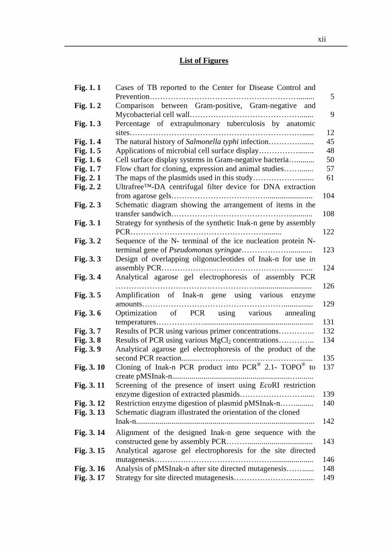
in USA from 84,304 reported cases in 1953 to 22,255 cases in 1984 (Fig.1.1), but the
progressive decline in incidence stopped and the case level plateaued and then
increased by 5% to 23,495 in 1989, and by 6% in 1990 (Groves, 1997).
Today, it is estimated that 2 billion people i.e., one third of the world's population are
infected with M. tuberculosis. Over 30 million of those infected people harbour active
disease. Every minute, more than 10 individuals develop TB amounting to 8 million
new cases annually, and over 2 million of those TB sufferers are expected to die of the
disease, making this disease the leading cause of death from a single pathogen in the
world (Dye et al., 1999). The incidence of TB has increased dramatically in areas with
high rates of HIV infection.

Introduction
5
Year
10,000
20,000
*
*
30,000
50,000
70,000
100,000
Cas
es(L
og S
cale
)
53 60 70 80 90
Fig.1. 1. Cases of TB reported to the Center for Disease Control and Prevention (CDC), in United States from 1953-1992. Changes in case definition, data obtained from Groves(1997)
Introduction
6
Thus, TB is the leading infectious cause of death among people more than 5 years of
age in South-East Asia, and accounts for approximately 40% of all the cases of TB in
the world. Within South-East Asia, more than 95% of cases are found in India,
Indonesia, Bangladesh, Thailand, and Myanmar (Murray et al., 1990, Kochi, 1991,
Bloom and Murray, 1992). The Ministry of Health in Malaysia reported that 10,000-
12,000 new cases were registered every year from 1972-1995. In the year 2000, WHO
reported that 8,156 smear positive cases were notified in Malaysia (WHO, 2003).
Numerous factors have been associated with the reappearance and increased TB
incidence which include: immigration from TB endemic areas, the emergence of multi-
drug resistant (MDR) strains, and increased numbers of immunocompromised patients,
especially HIV-infected. The above statistics put TB in the unfavorable list of the top
major killers, together with AIDS and malaria (Kabra et al., 2002).
1.4. The TB organism: Mycobacterium tuberculosis
Mycobacteria belong to the family Mycobacteriaceae and the order Actinomycetales.
They are non-motile, non-spore forming, straight or slightly curved rod shaped
microbes, 1-4 μm in length, and between 0.3-0.6 μm in diameter, making them smaller
than most bacterial pathogens (Iseman, 2000). Mycobacteria are considered ''acid-fast'',
which means that they retain dyes following an acid-alcohol decolorization step, and
this characteristic is related to the complex cell wall structure that contains derivatives
of mycolic acid (Floyd et al., 1992). These organisms usually contain granules and
vacuoles but they do not form capsules, flagella, or spores. In culture, these organisms
grow slowly and divide once every 18 to 24 hours. They can be grown for 2 to 12
weeks, until they reach 103-104 in number (Dannenberg, 1992). They are resistant to

Introduction
7
drying especially in sputum, where they can remain viable for 6-8 months. They are
also resistant to 3% HCl and 6% H2SO4, and to 4% NaOH. However, Mycobacteria are
sensitive to moist heat at 60ºC for 30 min, to disinfectants such as alcohol,
glutaraldehyde, formaldehyde, and Ultraviolet (uv) irradiation (Tortora et al., 2001).
Several species of mycobacteria with similar growth characteristics and biochemical
reactions are classified together into the M. tuberculosis complex (Cole, 2002). In
addition to M. tuberculosis, the complex includes M. bovis, M. africanum, and M.
microti which are also causative agents of TB in mammals (Brosch et al., 2000). M.
bovis is the causal agent of bovines and infects a wide variety of mammalian species
including humans. M. africanum has been reported to infect humans in sub-Saharan
Africa as well as monkeys (Thorel, 1980). M. microti causes TB in small rodents such
as voles (Hart and Sutherland, 1977).
Although the mycobacterial cell wall is weakly Gram-positive, this cell wall
characteristic do not really indicate whether M. tuberculosis is more related to Gram-
positive or Gram-negative bacteria since it has features of both in this respect. Recently
Fu & Fu-Liu (2002) showed that M. tuberculosis is more related to gram-negative
bacteria by construction of a genome tree based on the conserved gene content which
revealed the evolutionary distance between nearest ancestral units.
1.5. Chemical composition of the M. tuberculosis cell wall structure
The cell walls of Gram-positive bacteria are made up of peptidoglycan layers combined
with teichoic acid molecules, whereas those of Gram-negative bacteria contain much
less peptidoglycan, with no teichoic acid (Fig.1.2). The Gram-negative cell wall has a

Introduction
8
true lipid bilayer outer membrane that is attached to the characteristic endotoxic
lipopolysaccharide (Sussman, 2002).
However, the cell wall structure of M. tuberculosis deserves special attention because it
is unique among prokaryotes and may be a major determinant of the virulence of the
bacterium. Biochemical and electron microscopic studies indicate that the cell wall of
M. tuberculosis possesses four layers. The first layer (innermost) is the peptidoglycan
layer while the next three surface layers are composed of lipids such as mycolic acid,
glycolipids, cord factor and wax D (Sussman, 2002). The most important feature of the
mycobacterial cell wall is the presence of up to 60% of the total mass of lipid
components, particularly, the very long-chain mycolic acids, which are attached by
ester bonds to the terminal arabinose units of the arabinogalactan, thereby forming a
pseudolipid bilayer (Fig.1.2) (Brennan and Besra, 1997, Brennan, 2003).
In the cell wall of M. tuberculosis the lipids fall under two important classes,
sulpholipids and trehalose dimycolates, which are also known as, cord factors. The
sulpholipids are strongly acidic compounds covalently bound to trehalose sulphate.
They may be involved in the virulence of M. tuberculosis as they have been shown to
prevent phagosome/ lysosome fusion in macrophages infected with M. tuberculosis.
Several waxes are also present, which increase the impermeability of the mycobacterial
cell wall (Slots and Taubman, 1992).
This highly hydrophobic cell wall is not only responsible for the acid-fastness, but also
for resistance to acidic or alkaline chemicals, and for its relative stability in simple
disinfectants, in addition to the high adjuvanticity of the cell wall (Tortora et al., 2001).

Introduction
9
Porin
Lipoarabinomannan
Acyl lipids
(LAM) Mycolic acid
Lipid + LPS
Arabinogalactan
Lipid bilayer
Peptidoglycan
Lipid bilayer
Peptidoglycan
Gram-positive Gram-negative Mycobacterium
Fig.1. 2. Comparison between Gram-positive, Gram-negative and Mycobacterial cell wall.
Adapted from: http://web.uct.ac.za/depts/mmi/lsteyn/cellwall.html.
Introduction
10
1.6. Genetics of M. tuberculosis
Genome sequencing of M. tuberculosis was completed in 1998 and analysis of the data
show an estimate of 4,411,529 base pairs and 3,924 predicted open reading frames. M.
tuberculosis is a difficult organism to study because of some unique features. One of
these features is the high content of guanine and cytosine in its DNA. The high GC
content of 65.6% may be one survival strategy employed by bacteria, since stability of
DNA increases directly with number of GC bonds (Cole et al., 1998).
1.7. Pathophysiology of TB
1.7.1. TB, the disease
Tuberculosis (TB) is defined as a pulmonary and systemic infectious disease caused by
M. tuberculosis and characterized by formation of granulomas and by cell-mediated
hypersensitivity, in which M. tuberculosis multiply and attack different parts of the
body (Daniel et al., 1994). The course of the disease is the result of a balance between
the severity of the causative agent and the immunity of the host. Infection is
encountered by inhalation of a droplet nuclei (1-5 μm in diameter) carrying the
organism. Inside the body, the tubercle bacilli do not produce endotoxins or exotoxins
(Edwards and Kirkpatrick, 1986, Dannenberg, 1992, Gonzalez-Juarrero et al., 2001).
Damage is caused by uncontrolled progressive, chronic inflammation and by the
organisms living inside macrophages. TB is generally classified into latent infection and
active infection.
In latent TB infection, M. tuberculosis is present in the body but there are no signs or
symptoms of TB. People who have latent infection cannot spread the bacteria to other
people but are at risk of developing active TB disease. Patients with active TB disease

Introduction
11
exhibit signs and symptoms of disease. It occurs in 10% of those who are infected with
M. tuberculosis. People who have active TB disease can spread the bacteria to others.
TB can be categorized into two main types, according to where in the body the infection
manifests itself. The two types are described as pulmonary and extrapulmonary (or non-
pulmonary) TB. Pulmonary TB accounts for most of the cases of infection and about
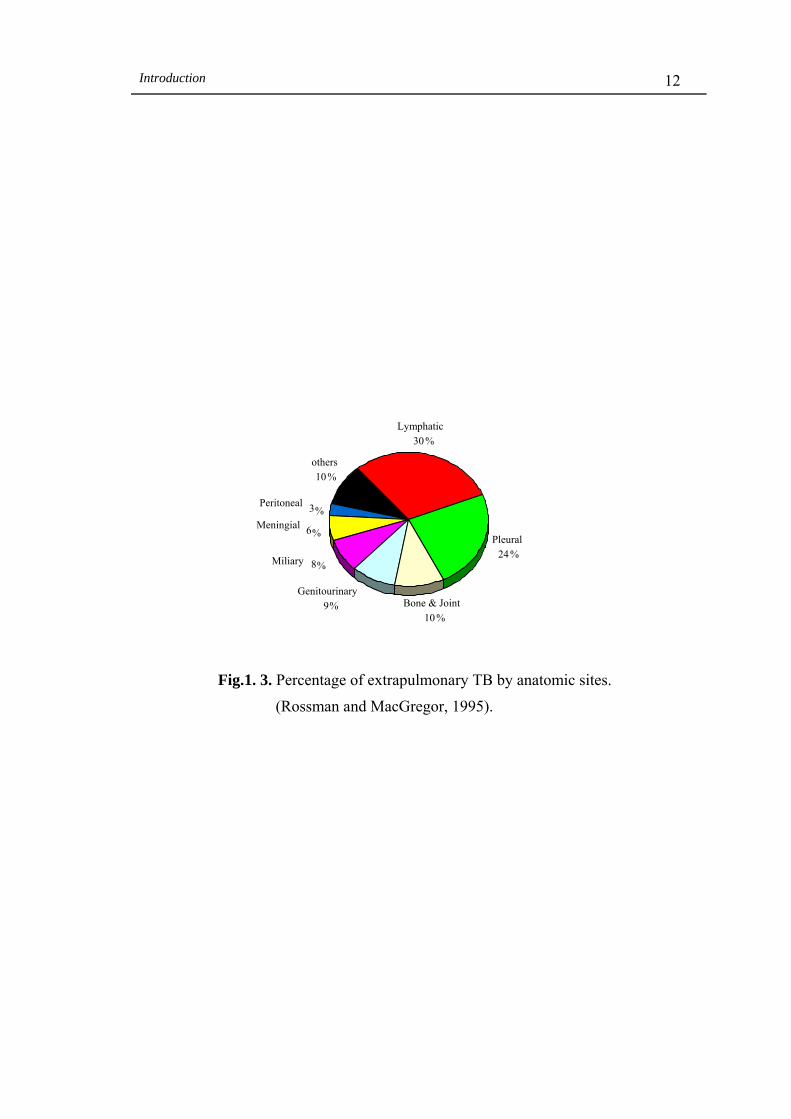
85% of TB deaths (Rossman and MacGregor, 1995).
Extrapulmonary TB is a general term that encompasses TB infection that has
disseminated to various sites around the body from the lungs. Extrapulmonary TB can
affect many different parts of the body including lymphatic, pleural, spinal, bones and
joints, meninges, genitourinary, peritoneal, and miliary (disseminated) TB. Pleural and
lymphatic TB are the most common types of extrapulmonary TB (Fig 1.3).
Extrapulmonary TB is very common among patients co-infected with HIV and
pulmonary TB; 60-80% of these patients develop extrapulmonary TB in contrast to 17%
of non-HIV patients who develop extrapulmonary TB. In addition, 30% of children with
primary pulmonary TB infection develop extrapulmonary TB (Rossman and
MacGregor, 1995).
1.7.2. Symptoms of TB
Most people infected with TB have inactive disease, which causes no symptoms.
However, in primary pulmonary TB more than half of the people with this type of TB
have no symptoms other than fever. In secondary or reactivation TB various symptoms
are involved including cough, chest pain, night sweats, poor appetite and loss of weight.
As the illness progresses, people may cough up blood (hemoptysis) and develop severe
breathing problems (Mandell et al., 1990).
Introduction
12
Lymphatic
30%
others 10
%
%
Miliary 8%
Meningial 6%
Peritoneal 3 %
Pleural24%
Genitourinary
Bone & Joint9%10
Fig.1. 3. Percentage of extrapulmonary TB by anatomic sites.
(Rossman and MacGregor, 1995).

Introduction
13
The symptoms of extrapulmonary TBs depend on where the TB has spread. Thus, if TB
affects the lymph nodes, it can cause swollen glands, usually at the sides and base of the
neck. In TB of the bones and joints, there can be a hunchback curvature of the spine or
pain and swelling of a knee or hip, with patients usually developing a limp.
Genitourinary TB, can cause pain in the side (between the ribs and hip), frequent and
painful urination, with blood in the urine (Rossman and MacGregor, 1995).
1.7.3. Transmission of M. tuberculosis
Transmission of M. tuberculosis (infection) occurs when people with active pulmonary
disease expel millions of water droplets containing M. tuberculosis into the air. These
droplets are small airborne particles of water that can remain floating in the air for
several hours (Baum and Wolinsky, 1983). Every one may acquire M. tuberculosis
through these airborne particles regardless of age, sex and race, but not everyone
exposed to the bacterium becomes infected nor does everybody develops clinical
symptoms of TB. Studies conducted worldwide have shown a rapid spread of TB in
crowded living conditions, such as in nursing homes, hospitals (Clarke and Higgins,
1995), homeless shelters, schools, military barracks, and prisons (Nettleman, 1993,
MacIntyre et al., 1999, Brewer et al., 2001, Drobnieuski et al., 2003). Passengers on
commercial aircrafts are particularly susceptible for the transmission of TB among
humans (Bignell, 1994, Miller et al., 1996, Al-Jahdali et al., 2003).
There are at least three factors influencing transmission of M. tuberculosis: (1) the
number of viable bacilli in patient’s sputum and their concentration in the air, (2) the
length of time an exposed person breathes the contaminated air, and (3) the immune
status of the exposed individual (Horsburgh, 1996).
Introduction
14
1.8. Immune response to TB
1.8.1. Early response
Early after the initial infection with M. tuberculosis, granulocytes respond to this
invasion and migrate from the blood into tissues and participate in an early
inflammatory response. However, there is some controversy on the involvement of
these cells in the killing of M. tuberculosis (Denis, 1991). M. tuberculosis induced an
influx of leukocytes including polymorphonuclear neutrophils (PMNs), lymphocytes,
and monocytes (Appelberg and Silva, 1989, Appelberg, 1992). The importance of
neutrophils in host defenses against mycobacteria has been dismissed because the
neutrophil has a short life span, and the bacilli grow inside macrophages, and are thus
protected from the phagocytotic activity of neutrophils. These two reasons were
supported by the findings of Denis (1991) who reported that human neutrophils were
unable to kill M. tuberculosis. Furthermore, Pedrosa et al., (2000) showed in their study
that M. tuberculosis is associated with or within macrophages and not with neutrophils.
It has also been shown that eosinophils may be play a role in the inflammatory response
initiated by M. tuberculosis (Castro et al., 1991). These findings have led the
researchers to focus their efforts on the M. tuberculosis inactivating mechanisms of
macrophages instead of granulocytes.
1.8.2. Pathogenicity and initial defense against M. tuberculosis infection
1.8.2.1. Primary TB
Primary TB is a disease, or a response to infection by a host who have not been
previously exposed to/or vaccinated against TB. After entry into the host, the droplet
nuclei are carried down the bronchial tree and become implanted in the alveoli. The
bacteria are ingested by the resident alveolar macrophages (AM) which kill or limit the

Introduction
15
replication of mycobacteria, due to the action of lysosomal enzymes and reactive
nitrogen and oxygen species (Fang, 1997, Miller and Britigan, 1997, van Crevel et al.,
2002). However, mycobacteria resist lysosomal degradation and escape from the
phagolysosomes into the cytoplasm of the macrophage, where somehow, they manage
to survive and even multiply until their number reaches 103-104 which is sufficient to
elicit a cellular immune response (Smith and Wiengeshaus, 1989). Finally,
mycobacteria burst out of the infected macrophage, killing it and then infect other
macrophages in the area. As macrophages die by necrosis, they pour their lysosomal
contents into the surrounding area or in the neighboring lung tissue, causing tissue
damage, and initiating an inflammatory response. These events may be the most
important stages in establishing infection in the host (Sompayrac, 1999).
Inflammation is necessary for the proper functioning of the host defenses, including the
immune defenses, because it attracts circulating antimicrobial factors to the site of
infection. These include phagocytes, lymphocytes, antibodies, complement and other
antimicrobial components of plasma. The immune system continues to send
macrophages to destroy the bacteria resulting in an accumulation of living and dead
macrophages at the site of infection creating a structure called a tubercle (Dannenberg,
1989, van Crevel et al., 2002). Two to three weeks after infection, cellular immunity
developed, with antigen-specific T lymphocytes that proliferate within the early
tubercles, and activated macrophages to kill the intracellular mycobacteria. As a result,
most of the organisms die, and lesions in the lung and draining lymph nodes heal by
fibrosis, and sometimes calcify to inhibit extracellular growth of the remaining
mycobacteria. Some of these microorganisms remain viable for long periods (Daniel,
1994).
Introduction
16
1.8.2.2. Secondary TB
The standard assumption of the recurrence of TB is by reactivation of the existing
latent infection. However, reinfection by a new strain is also possible. Reactivation can
occur when the immune system is weakened, as a result of malnutrition, and co-
infection by other diseases such as AIDS (Chan et al., 1996). When alveolar
macrophages fail to kill the mycobacteria, the immune system’s next line of defense is
to form granulomas around the infected macrophages. Granulomas are, essentially,
layers of T-cells sealing the mycobacteria inside a barrier from which it cannot escape.
Two to three weeks after the inhalation of the M. tuberculosis, the host possesses both
cell mediated immunity (CMI) and delayed-type hypersensitivity (DTH). With the
emergence of DTH, infected macrophages in the interior of each granuloma are killed as
the periphery becomes fibrotic and slowly become caseated (cheesy-like, semi-solid
debris composed of lipid and proteins from tubercle bacilli and macrophages), and
eventually merge into larger lesions (Dannenberg 1991). With time, proteases produced
by activated macrophages liquefy the caseous material and form air filled tuberculous
cavities that provide the bacilli with a suitable extracellular site for reproduction.
Rupture of a tuberculous cavity into the pleural space may lead to the bacteria extending
into the blood stream via regional nodes, where they are ingested by monocytes in the
blood. Moreover, as the expanding lesion erodes through the wall of the bronchus, the
liquefied content is discharged and thus allowing the bacteria to spread to and colonize
virtually every organ in the host. A well aerated cavity is also formed where the
organisms can actively proliferate (Dannenberg, 1991). Since inflammation of the
surface of the bronchi causes increased mucus secretion and stimulation of the cough
reflex, patients cough up sputum. Mycobacterial growth is not kept under control and
can cause major destruction of tissues. In advanced TB, blood vessels may become

Introduction
17
exposed to the cavities produced by necrosis, and patients may die of hemorrhage, if
these vessels are ruptured (van Crevel et al., 2002). Secondary TB usually becomes
noticeable one or two years after the primary disease, probably because it takes that
long a time to develop full blown delayed-type hypersensitivity.
1.8.3. Immunity to TB
1.8.3.1. Humoral immunity against TB
Humoral immunity is mediated by antibodies produced by B cells and their progeny
when a foreign antigen enters the blood stream of a mammal. These antibodies bind
specifically to antigens eliciting the immune response. The immune system then
neutralizes or eliminates them from the body through ingestion and degradation of the
antibody-antigen complex by phagocytes. One more method of elimination of foreign
antigens is by degradation of viruses and other proteinaceous substances by proteolysis
and killing of bacteria by cell lysis (Abbas and Lichtman, 2001).
Antibodies are produced in response to mycobacterial infection, but there is no definite
evidence that immunoglobulins (Ig) play a significant role in protective immunity to
TB (Dunlap and Briles, 1993). The role of the polymorphonuclear cells (PMNs) in TB
is not well understood, but studies have shown that PMNs can diminish growth of M.
tuberculosis by non-oxidative processes (Brown, 1987).
During the primary infection, IgM antibody responses are directed chiefly at nonspecific
polysaccharide antigens. They develop early but never reach high titer, and this level
does not correlate well with the presence or absence of active disease (Daniel and
Debanne, 1987, Daniel et al., 1994).

Introduction
18
Levels of IgG antibody detectable by ELISA and other immunoassay are usually an
indication of active TB. Studies with many TB antigens using several techniques
showed that few control subjects have measurable IgG antibody levels (Daniel and
Debanne, 1987).
IgA antibodies have been found at low levels in the serum of patients with active TB,
but not in control subjects (Daniel and Debanne, 1987). Recently, Cardona, et al.,
(2002) showed for the first time the stimulation of antibodies against the glycolipids
from the M. tuberculosis cell wall which include diacyltrehaloses (DAT) and
sulpholipid I (SL-I) in murine models. Their results showed that these antigens elicit
higher antibody levels than protein antigens like the Ag85 complex, culture filtrate
proteins (CFP) and purified protein derivative (PPD).
The results from the studies described above suggest that although humoral immune
system is induced by M. tuberculosis infection, it appears to play little role in protecting
the host from the disease progression.
1.8.3.2. Cellular immunity against TB
Cell-mediated immunity (CMI) response is believed to involve different T-cell subsets.
T-cells can be divided into two major classes, CD4+ and CD8+ T-cells. CD4+ and CD8+
T-cell recognition of antigens requires that the antigens are processed and displayed on
the surface of antigen presenting cells (APC) bound to specialized molecules called
major histocompatibility (MHC) molecules. MHC molecules include class I and class II
molecules (Germain, 1999).

Introduction
19
One feature characterizing the class I MHC molecules is that they typically present
peptides derived from intracellular antigens such as endogenous proteins or viral
proteins synthesized during infection, and they are expressed on all nucleated cells.
Peptide-MHC-I complexes are recognized by CD8+ T-cells. These cells have cytolytic
activity and will kill the target cells upon peptide-MHC recognition. They also have
cytolytic functions and are generally called cytotoxic T lymphocytes (CTL). When a
CTL recognizes its target, it releases cytotoxic molecules, perforins which will induce
apoptosis in the target cells (Abbas and Lichtman, 2001, Esser et al., 2003).
Class II MHC molecules are expressed by APC only and typically present peptides
derived from extracellular antigens that have been internalized by the APC. The
peptide-MHC complexes are mainly recognized by CD4+ T-cells. CD4+ T-cells can be
further divided into two subsets of effector CD4+ T lymphocytes, called T helper type 1
(Th1) and 2 (Th2) respectively, which differ in their cytokine production (Abbas and
Lichtman, 2001, Esser et al., 2003). Th1 cells are important in infections with
intracellular bacteria such as M. tuberculosis and they produce interferon gamma
(IFN-γ), interleukine-12 (IL-12) and tumor necrosis factor (TNF)-α. On the other hand,
Th2 cells are important in antibody-mediated (humoral) immunity. These cells produce
cytokines such as IL-4, IL-5, IL-6 and IL-10 that provide the necessary signals to B
cells in producing antibody (Abbas and Lichtman, 2001, Esser et al., 2003).
Cell-mediated immunity is the major protective immune response against mycobacterial
infection. T-cells contribute in the acquired immune response by means of two major
functions. First, they produce cytokines, particularly IFN-γ which is the major activator
of antimicrobial macrophage functions. This cytokine is produced by all T stimulated
Introduction
20
cells in response to mycobacterial infection, such as CD4+ and CD8+ T-cell clones
(Schaible et al., 1999). Second, these cells also express cytolytic activities, whereby
they lyse infected target cells (Kaufmann, 1999).
CD4+ cells of the Th1 type play a key role in protective immunity against M.
tuberculosis. This was established, initially, in experimental models of TB (Orme and
Collins, 1984). In humans, the strongest evidence for a predominant role of CD4+ T-
cells in protective immunity is the increased susceptibility of HIV-infected populations
to the development of active TB (Caruso et al., 1999, Orme, 2001). Once macrophages
are activated, and the phagocytosed bacteria are processed in the endosomal
compartment of the macrophage, peptides will be presented to CD4+ T-cells in
association with class II major histocompatibility complex (MHC) molecules on the
surface of the macrophage. CD4+ T-cells recognize the complex and become activated,
and consequently induce the production of Th1 cytokines, IL-2 and IFN-γ (Flynn et al.,
1993).
The role of CD8+ T-cells starts when mycobacteria gain access to the cytoplasmic
compartment of macrophages and become associated with MHC-I molecules. This
antigen-MHC-I complex can then stimulate CD8+ T-cells to produce cytokines such as
IFN-γ and TNF-α to activate macrophages. CD8+ T-cells facilitate the release of intact
bacteria entrapped in macrophages. In this way, bacteria can be released from
ineffective macrophages and phagocytosed by more efficient cells. Strong evidence
from mouse experiments suggests a major role of CD8+ T-cells, in addition to CD4+
T-cells, in protection against TB (Flynn et al., 1992, Lewinsohn et al., 2000, Smith and
Dockrell, 2000, Klein and Fox, 2001). Consequently, it is most likely that the different

Introduction
21
T-cell populations are required for optimum protection since they have diverse, yet
versatile mechanism (Triccas et al., 2002).
The protein and non-protein antigens of M. tuberculosis are strong stimuli for induction
of cytokine production in human mononuclear phagocytes which most likely affect the
outcome of infection at any stage of the M. tuberculosis infection (van Crevel et al.,
2002). Studies in cytokine production during M. tuberculosis infections have shown that
several, proinflammatory cytokines, particularly IFN-γ, TNF-α, IL-1, IL-6, IL-12, and
IL-18 are produced after the recognition of M. tuberculosis by phagocytic cells
(Table 1.2). These cytokines act in the autocrine or paracrine networks to influence the
function of cells in immunity to M. tuberculosis (van Crevel et al., 2002).
Tumor necrosis factor alpha (TNF-α) plays an important role in the host response to
infection with M. tuberculosis. In mice, TNF-α is essential for formation of tuberculous
granulomas which serves to isolate the virulent bacterium and without this cytokine,
effective granuloma formation is diminished and bacterial numbers rapidly increase
resulting in the death of the mice (Engele et al., 2002).
The importance of IFN-γ in protective immunity against TB derives from two separate
lines of evidence. Studies using IFN-γ knockout mice have shown definitively that
IFN-γ, a cytokine that activates infected macrophages to kill intracellular bacteria, is
vitally important for protection against M. tuberculosis (Cooper et al., 1993). Studies of
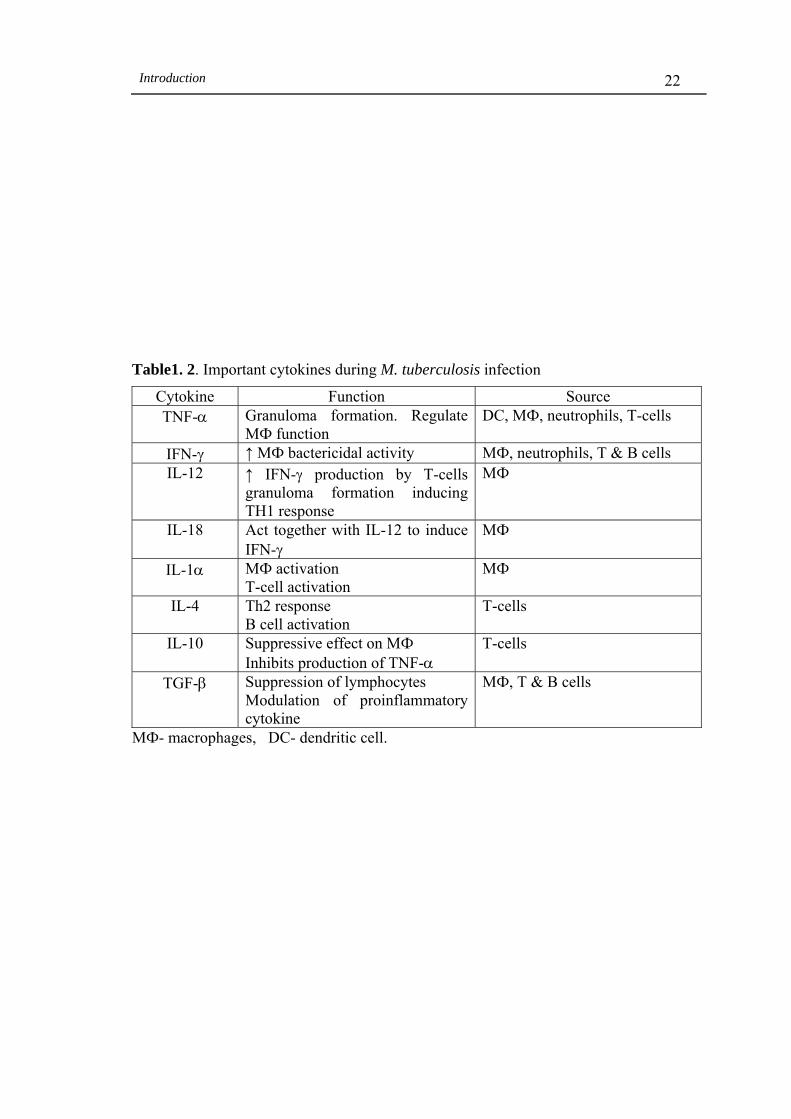
Introduction
22
Table1. 2. Important cytokines during M. tuberculosis infection
Cytokine Function Source TNF-α Granuloma formation. Regulate
MФ function DC, MФ, neutrophils, T-cells
IFN-γ ↑ MФ bactericidal activity MФ, neutrophils, T & B cells IL-12 ↑ IFN-γ production by T-cells
granuloma formation inducing TH1 response
MФ
IL-18 Act together with IL-12 to induce IFN-γ
MФ
IL-1α MФ activation T-cell activation
MФ
IL-4 Th2 response B cell activation
T-cells
IL-10 Suppressive effect on MФ Inhibits production of TNF-α
T-cells
TGF-β Suppression of lymphocytes Modulation of proinflammatory cytokine
MФ, T & B cells
MФ- macrophages, DC- dendritic cell.

Introduction
23
humans who are homozygous for a mutation in IFN-γ receptors show that they are
extremely sensitive to fatal mycobacterial infection (Jouanguy et al., 1996, Newport
et al., 1996). Individuals who lack the ability to produce IFN-γ, or who cannot respond
to it or lack the receptor for it, are susceptible to severe systemic infection with
mycobacterial species that do not usually cause significant disease in immunocompetent
individuals (Jouanguy et al., 1996, Newport et al., 1996).
Early studies indicated that the purified protein derivative (PPD) of M. tuberculosis
induces the proinflammatory cytokine IL-1 (Wallis et al., 1996) and TNF-α (Valone et
al., 1988). On the other hand the intact M. tuberculosis, but not its PPD, was strong in
the induction of IL-12 (which stimulates IFN-γ production) in monocytes (Fulton et al.,
1998). Both TNF-α and IFN-γ induce microbicidal pathways by producing reactive
oxygen and nitrogen intermediaries in phagocytes (Flesch and Kaufmann, 1990).
1.9. Mycobacterial antigens
As previously described, protection against TB is provided through cell mediated
immune responses, and the production of IFN-γ by both CD4+ and CD8+ T-cells appears
to be essential components of resistance to M. tuberculosis. A primary criterion for
selecting candidate antigens for vaccine design is their ability to induce protective T-cell
responses. Thus, it is required that the major antigens and epitopes of M. tuberculosis be
identified (see Section 1.11.2). A variety of M. tuberculosis antigen preparations
obtained from the cell wall, cytosolic fractions and culture filtrates have been reported
(Young et al., 1992, Mustafa, 1996, 2002). These antigens have several hundred amino
acids, whereas T-cells usually recognize epitopes ranging between 8 and 20 amino acids

Introduction
24
presented in the context of MHC molecules. Thus a complete protein antigen may have
several T-cell epitopes. To identify the T-cell epitopes mediating protective immunity,
several studies have been conducted to identify and characterize the epitopes of M.
tuberculosis antigens recognized by human T-cells of the Th1 type (Mustafa, 2002).
Although bacteria consist of several thousands of proteins, the range of proteins
recognized by antibodies produced in different laboratories was restricted leading to the
concept of immunodominance, i.e. that there were certain proteins which were
selectively recognized by the immune system. When these antigenic proteins were
identified by screening mycobacterial recombinant DNA expression libraries with
antibody probes (Young et al., 1985), most of these antigens were found to belong to a
group of molecules whose function is to protect the organism from environmental
damage (Young et al., 1988). Testing of these antigens with T-cell lines and clones
showed that human T-cells recognized most of them (Mustafa, 1988, Oftung et al.,
1998, Mustafa et al., 1999). Among these recombinant antigens is the antigen 18-kDa
that belongs to the heat shock protein (hsp)18 family (Booth et al., 1988), the 65-kDa
which belongs to hsp65 family (Shinnick, 1987) and the 70-kDa which belongs to hsp70
family (Garsia et al., 1989). These antigens were also recognized by human T-cells as
natural mycobacterial antigens (Mustafa, 2002). A novel 24 kDa secreted lipoprotein
was also identified by direct screening of recombinant DNA libraries with human T-cell
clones. Interestingly the T-cell epitopes of this antigen were found to be common to the
two most important pathogenic mycobacterial species M. tuberculosis and M. leprae
and was lacking in the vaccine strains of BCG and most environmental mycobacteria
(Oftung et al., 1997, Mustafa et al., 1998).

Introduction
25
Secreted protein antigens present in culture filtrate preparations have also been studied
extensively. These studies showed that these antigens induce CMI responses and are
protective against TB in mice and guinea pigs (Andersen, 1994, Horwitz et al., 1995,
Roberts et al., 1995). These proteins were separated into discrete fractions according to
size by polyacrylamide gel electrophoresis and then tested for T-cell reactivity and
proliferation (Abou-zeid et al., 1987). Mouse models of TB infection have shown that
T-cells from the memory immune mice secreted large quantities of IFN-γ in response to
two fractions of the culture filtrate protein known as the 6-10 kDa proteins and antigen
85 (Ag85) complex (Andersen et al., 1995). Testing of human T-cells with purified
antigens of M. tuberculosis in proliferation assays and for IFN-γ production showed that
the secreted antigens, which include Ag85B, ESAT6, MPT64 and MPB70, were better
stimulators of human T-cells compared the antigens of cytosolic origin (Mustafa et al.,
1998, Ravn et al., 1999).
Mice immunized with plasmid DNA encoding the Ag85A (DNA-Ag85A) showed a
strong T-cell response when restimulated in vitro with synthetic 20-mer peptides of the
native Ag85. This response was characterized by elevated levels of Th1-type cytokines,
IL-2 and IFN-γ. In addition, DNA-Ag85A vaccines conferred protection upon
subsequent infection with live mycobacteria (Denis et al., 1998).
1.10. Diagnosis of TB.
The diagnosis of TB is established when tubercle bacilli are identified in specimens
such as sputum, urine, and body fluids. The most common specimen received for
tubercle examination is sputum (Kalafer, 1991). Acid fast staining of smears to identify
the bacillus is a rapid technique, but it has low sensitivity and cannot differentiate