Development of nervous system
20
To MBBS 2 nd year Dr Laxman Khanal Assistant Professor, Department of Anatomy BPKIHS, Dharan, Nepal 12-02-2017 Development of nervous system
-
Upload
dr-laxman-khanal -
Category
Health & Medicine
-
view
66 -
download
1
Transcript of Development of nervous system
To MBBS 2nd year
Dr Laxman Khanal
Assistant Professor, Department of Anatomy
BPKIHS, Dharan, Nepal
12-02-2017
Development of nervous system

Cranial nerves1. Motor parts2. Sensory parts3. Autonomic parts
Parasympathetic
Spinal nerves1. Motor parts2. Sensory parts3. Autonomic parts
A. Sympathetic (thoracic and lumbar segment)
B. Parasympathetic (sacral segment)
Parts of nervous system1. CNS- brain and spinal cord2. PNS- cranial and spinal nerve3. ANS- contributed by both CNS and PNSANS -cardiac muscle, smooth muscle and glands.
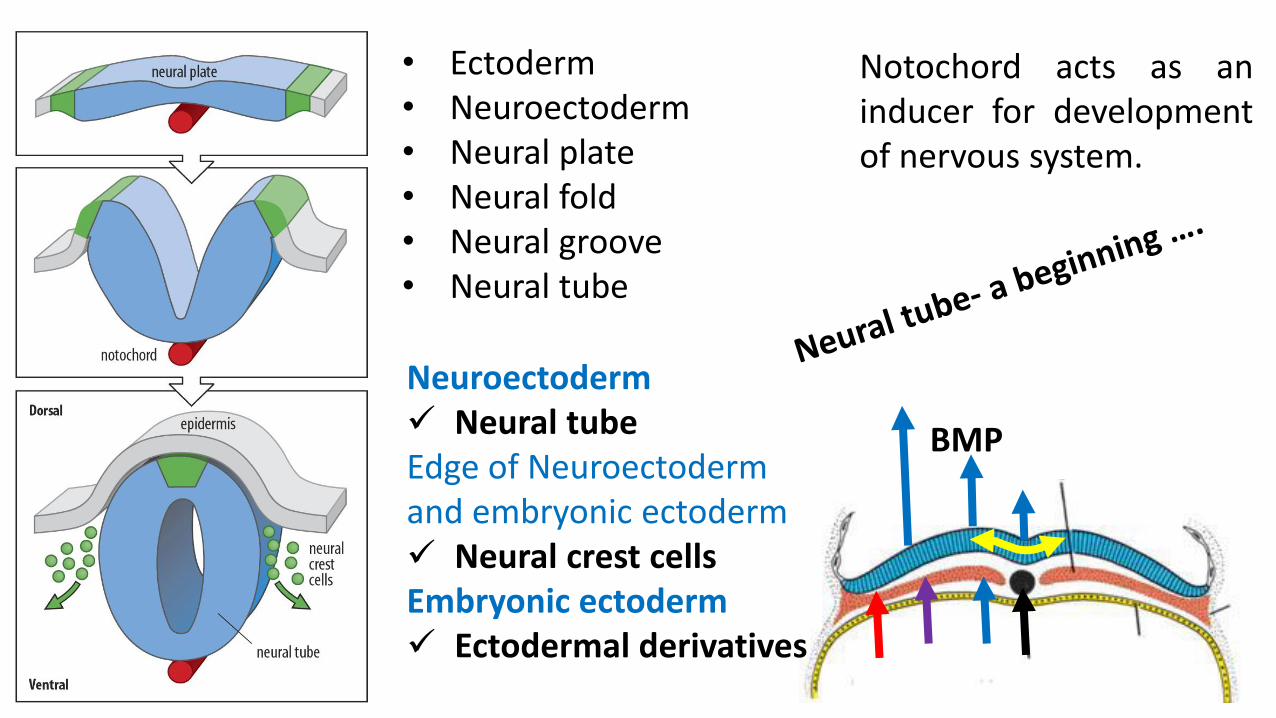
Notochord acts as aninducer for developmentof nervous system.
BMP
• Ectoderm • Neuroectoderm• Neural plate• Neural fold• Neural groove• Neural tube
Neuroectoderm Neural tubeEdge of Neuroectoderm and embryonic ectoderm Neural crest cellsEmbryonic ectoderm Ectodermal derivatives

Development of neural crest cells
Epithelial Mesenchymal E- Cadherin

Pharyngeal arch derivatives• Skeletal component• Nerves and ganglia of each arch• Dermis overlying each arch• Blood vessels of face.
Migration of neural crest cells

Cranial neuroporeClosure at 25th day
Caudal neuroporeClosure at 27th day
Brain
SC
Alpha feto protein (AFP) increasesin maternal AF if neuropores fail toclose (neural tube defects).

Anencephaly Craniorachischisis
Neural tube defects of cranial part of neural tube

Neural tube defects of caudal part of neural tube
Rachischisis
Myelomeningocele
Spina bifida occulta

Prevented by maternal use of folic acid prior to and during pregnancy.
Spina bifida occulta Meningocele Myelomeningocele
Rachischisis

Anterior portion of neural tube forms three primary brain vesicles.
1. Prosencephalon: Telencephalon + Diencephalon2. Mesencephalon:3. Rhombencephalon: Metencephalon + Mylencephalon
I & II CN
III & IV CN
V- XII CN

Development of ventricular system
4th

Development of spinal cord
L
TS neural tube
Neuroepithelial cellsL
Neuroblast cell
L
Mantle cell layer
Marginal cell layer
L
A
B
A
B
R
F
Anterior horn
Posterior horn
intermediate horn
Sensory
Motor
Autonomic

Neuroblast Glioblast
Microglial cells
Ependymal cells
Origin of various types of cells

Brain stemArrangement of alar andbasal plate is intact butthe arrangement isdifferent.
Higher centerBasal plate regress andalar plate accentuate.
A
B

GSE
SVE
GVE
Development of functional columns in brain stem
Basal plate – motor componentsSkeletal muscle1. Somatic muscles - GSE2. Pharyngeal arch muscles- SVESmooth muscle- GVE (autonomic)
CN supplying somatic skeletal muscles
Motor nerve of pharyngeal archCN with parasympathetic actions

GSE
SVE
GVE
GSA
SVA
GVA
SSA
Development of functional columns in brain stem
Alar plate- sensory columnSenses• General senses • Special sense- taste• Senses from viscera• Special sense-hearing & Eqb
Pain, touch, temperature, pressure
Taste Sense of distension of viscera
Hearing & equilibrium

Mesencephalic flexure
Cervical flexure
Pontine flexureRhombic lip• Postero-lateral extension of alar plate
of metencephalon.• Give rise to cerebellum.
Rhombic lip

TS of caudal part of Pons
Rhombic lip Cerebellar plate
TS Mesencephalon
Alar plate
Basal plate
Alar plateSup colliculusInf colliculus
Basal plateNuclei of 3rd CNNucleus of 4th CN

Development of Prosencephalon
Diencephalon3rd ventricle
Telencephalon2 lateral ventricles
1. Thalamus2. Hypothalamus3. Epithalamus4. Subthalamus 5. Metathalamus6. Neurohypophysis

Summary
• The CNS develops from a dorsal thickening of ectoderm-the neuralplate, which appears around the middle of the third week.
• The neural plate is induced by the underlying notochord and paraxialmesoderm to form neural tube.
• The cranial end of the neural tube forms the brain and the remainderforms the spinal cord.
• The neural canal, the lumen of the neural tube, becomes theventricles of the brain and the central canal of the spinal cord.
• Defects in the closure of the neural tube (NTDs) account for mostsevere anomalies of nervous system.


