Development of CRISPR-Cas systems for genome editing and ... · repeat (CRISPR)-Cas...
31
Quarterly Reviews of Biophysics cambridge.org/qrb Perspective Cite this article: Zhang F (2019). Development of CRISPR-Cas systems for genome editing and beyond. Quarterly Reviews of Biophysics 52, e6, 1–31. https://doi.org/10.1017/ S0033583519000052 Received: 8 April 2019 Revised: 8 May 2019 Accepted: 13 May 2019 Key words: Cas9; Cas12; Cas13; CRISPR; genome editing; SHERLOCK Author for correspondence: F. Zhang, E-mail: [email protected] © The Author(s) 2019. This is an Open Access article, distributed under the terms of the Creative Commons Attribution- NonCommercial-NoDerivatives licence (http:// creativecommons.org/licenses/by-nc-nd/4.0/), which permits non-commercial re-use, distribution, and reproduction in any medium, provided the original work is unaltered and is properly cited. The written permission of Cambridge University Press must be obtained for commercial re-use or in order to create a derivative work. Development of CRISPR-Cas systems for genome editing and beyond F. Zhang 1 Broad Institute of MIT and Harvard, Cambridge, MA 02142 USA; 2 Department of Brain and Cognitive Sciences, Department of Biological Engineering, McGovern Institute for Brain Research, Massachusetts Institute of Technology, Cambridge, MA 02139 USA and 3 Howard Hughes Medical Institute, Cambridge, MA 02139 USA Abstract The development of clustered regularly interspaced short-palindromic repeat (CRISPR)-Cas systems for genome editing has transformed the way life science research is conducted and holds enormous potential for the treatment of disease as well as for many aspects of biotech- nology. Here, I provide a personal perspective on the development of CRISPR-Cas9 for genome editing within the broader context of the field and discuss our work to discover novel Cas effectors and develop them into additional molecular tools. The initial demonstra- tion of Cas9-mediated genome editing launched the development of many other technologies, enabled new lines of biological inquiry, and motivated a deeper examination of natural CRISPR-Cas systems, including the discovery of new types of CRISPR-Cas systems. These new discoveries in turn spurred further technological developments. I review these exciting discoveries and technologies as well as provide an overview of the broad arrayof applications of these technologies in basic research and in the improvement of human health. It is clear that we are only just beginning to unravel the potential within microbial diversity, and it is quite likely that we will continue to discover other exciting phenomena, some of which it may be possible to repurpose as molecular technologies. The transformation of mysterious natural phenomena to powerful tools, however, takes a collective effort to discover, characterize, and engineer them, and it has been a privilege to join the numerous researchers who have contributed to this transformation of CRISPR-Cas systems. Introduction Close observers of the diversity in the natural world generally appreciate why evolution has been likened not to the work of an engineer, but to that of a tinkerer (Jacob, 1977). By repur- posing a genetic material under selective pressure, nature has evolved a myriad of ‘field-tested’ solutions to the challenges organisms face. Evolutionary tinkering is particularly evident in the microbial world, where selective pressure is high, effective population size is large, generation time is short, and genetic information can be exchanged widely and relatively quickly. As biol- ogists delve ever deeper into the molecular and genetic mechanisms underlying the observed phenotypic diversity, we continue to learn more about fundamental biological processes and uncover new natural systems and phenomena. In addition to providing insight into the molec- ular underpinnings of life, some of these novel systems have been developed into various molecular technologies. For example, heat-stable polymerases discovered in thermophilic bac- teria enabled the development of polymerase chain reaction (PCR), and restriction enzymes discovered by studying host responses to phages enabled recombinant deoxyribonucleic acid (DNA) technologies. One of the latest examples of how nature’s solutions have been successfully adapted into a molecular technology is the development of clustered regularly interspaced short-palindromic repeat (CRISPR)-Cas (CRISPR-associated) systems for eukaryotic genome editing. CRISPR-Cas-mediated genome editing is a robust, easy-to-use method to precisely alter DNA sequences within the genome of living organisms. Because of the simplicity and effi- ciency of the system, it has been widely adopted and further developed, leading to an extraor- dinarily powerful molecular toolbox. Once microbiological curiosity, CRISPR has become a part of the common language of molecular biology, with its reach extending into nearly every corner of the life sciences and its impact going far beyond the confines of the laboratory. The story of CRISPR is one with two-intertwined aspects (Fig. 1): biological investigation to better understand these elegant systems and engineering of these systems into powerful molec- ular technologies. As the impact of these technologies spreads, it spurs further work into the biology, which continues to provide additional technological opportunities. Thus, the early part of the CRISPR revolution involved engineering Cas9 as genome editing technology, but through the recent discovery and development of additional Cas effectors, particularly the ribonucleic acid (RNA)-targeting Cas13 family, it has continued to expand into new https://www.cambridge.org/core/terms. https://doi.org/10.1017/S0033583519000052 Downloaded from https://www.cambridge.org/core. IP address: 54.39.106.173, on 23 Mar 2020 at 15:20:17, subject to the Cambridge Core terms of use, available at
Transcript of Development of CRISPR-Cas systems for genome editing and ... · repeat (CRISPR)-Cas...
Quarterly Reviews ofBiophysics
cambridge.org/qrb
Perspective
Cite this article: Zhang F (2019). Developmentof CRISPR-Cas systems for genome editingand beyond. Quarterly Reviews of Biophysics52, e6, 1–31. https://doi.org/10.1017/S0033583519000052
Received: 8 April 2019Revised: 8 May 2019Accepted: 13 May 2019
Key words:Cas9; Cas12; Cas13; CRISPR; genome editing;SHERLOCK
Author for correspondence:F. Zhang, E-mail: [email protected]
© The Author(s) 2019. This is an Open Accessarticle, distributed under the terms of theCreative Commons Attribution-NonCommercial-NoDerivatives licence (http://creativecommons.org/licenses/by-nc-nd/4.0/),which permits non-commercial re-use,distribution, and reproduction in any medium,provided the original work is unaltered and isproperly cited. The written permission ofCambridge University Press must be obtainedfor commercial re-use or in order to create aderivative work.
Development of CRISPR-Cas systems forgenome editing and beyond
F. Zhang
1Broad Institute of MIT and Harvard, Cambridge, MA 02142 USA; 2Department of Brain and Cognitive Sciences,Department of Biological Engineering, McGovern Institute for Brain Research, Massachusetts Institute ofTechnology, Cambridge, MA 02139 USA and 3Howard Hughes Medical Institute, Cambridge, MA 02139 USA
Abstract
The development of clustered regularly interspaced short-palindromic repeat (CRISPR)-Cassystems for genome editing has transformed the way life science research is conducted andholds enormous potential for the treatment of disease as well as for many aspects of biotech-nology. Here, I provide a personal perspective on the development of CRISPR-Cas9 forgenome editing within the broader context of the field and discuss our work to discovernovel Cas effectors and develop them into additional molecular tools. The initial demonstra-tion of Cas9-mediated genome editing launched the development of many other technologies,enabled new lines of biological inquiry, and motivated a deeper examination of naturalCRISPR-Cas systems, including the discovery of new types of CRISPR-Cas systems. Thesenew discoveries in turn spurred further technological developments. I review these excitingdiscoveries and technologies as well as provide an overview of the broad array of applicationsof these technologies in basic research and in the improvement of human health. It is clearthat we are only just beginning to unravel the potential within microbial diversity, and it isquite likely that we will continue to discover other exciting phenomena, some of which itmay be possible to repurpose as molecular technologies. The transformation of mysteriousnatural phenomena to powerful tools, however, takes a collective effort to discover,characterize, and engineer them, and it has been a privilege to join the numerous researcherswho have contributed to this transformation of CRISPR-Cas systems.
Introduction
Close observers of the diversity in the natural world generally appreciate why evolution hasbeen likened not to the work of an engineer, but to that of a tinkerer (Jacob, 1977). By repur-posing a genetic material under selective pressure, nature has evolved a myriad of ‘field-tested’solutions to the challenges organisms face. Evolutionary tinkering is particularly evident in themicrobial world, where selective pressure is high, effective population size is large, generationtime is short, and genetic information can be exchanged widely and relatively quickly. As biol-ogists delve ever deeper into the molecular and genetic mechanisms underlying the observedphenotypic diversity, we continue to learn more about fundamental biological processes anduncover new natural systems and phenomena. In addition to providing insight into the molec-ular underpinnings of life, some of these novel systems have been developed into variousmolecular technologies. For example, heat-stable polymerases discovered in thermophilic bac-teria enabled the development of polymerase chain reaction (PCR), and restriction enzymesdiscovered by studying host responses to phages enabled recombinant deoxyribonucleic acid(DNA) technologies.
One of the latest examples of how nature’s solutions have been successfully adapted into amolecular technology is the development of clustered regularly interspaced short-palindromicrepeat (CRISPR)-Cas (CRISPR-associated) systems for eukaryotic genome editing.CRISPR-Cas-mediated genome editing is a robust, easy-to-use method to precisely alterDNA sequences within the genome of living organisms. Because of the simplicity and effi-ciency of the system, it has been widely adopted and further developed, leading to an extraor-dinarily powerful molecular toolbox. Once microbiological curiosity, CRISPR has become apart of the common language of molecular biology, with its reach extending into nearlyevery corner of the life sciences and its impact going far beyond the confines of the laboratory.The story of CRISPR is one with two-intertwined aspects (Fig. 1): biological investigation tobetter understand these elegant systems and engineering of these systems into powerful molec-ular technologies. As the impact of these technologies spreads, it spurs further work into thebiology, which continues to provide additional technological opportunities. Thus, the earlypart of the CRISPR revolution involved engineering Cas9 as genome editing technology,but through the recent discovery and development of additional Cas effectors, particularlythe ribonucleic acid (RNA)-targeting Cas13 family, it has continued to expand into new
https://www.cambridge.org/core/terms. https://doi.org/10.1017/S0033583519000052Downloaded from https://www.cambridge.org/core. IP address: 54.39.106.173, on 23 Mar 2020 at 15:20:17, subject to the Cambridge Core terms of use, available at
areas. CRISPR-based technologies are being employed in diverseways to improve human health and offer the potential to funda-mentally change the way we treat disease.
Here, I briefly overview the natural function of CRISPR-Cassystems, followed by a personal account and perspective of thetime period over which CRISPR-Cas9 was developed for genomeediting in eukaryotic cells. I also discuss the continuing study andremarkable biotechnological development of CRISPR-Cas systemsbeyond Cas9 (Fig. 2). In particular, I highlight some of the excit-ing applications of this technology and identify areas for future
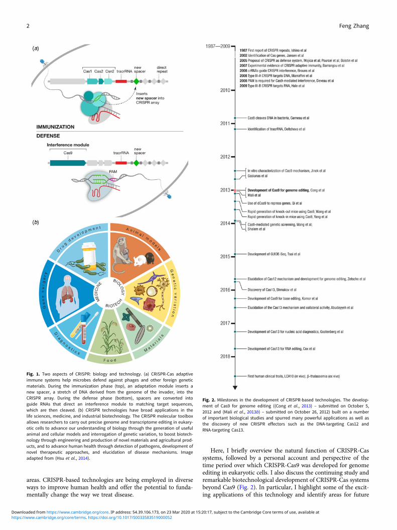
Fig. 1. Two aspects of CRISPR: biology and technology. (a) CRISPR-Cas adaptiveimmune systems help microbes defend against phages and other foreign geneticmaterials. During the immunization phase (top), an adaptation module inserts anew spacer, a stretch of DNA derived from the genome of the invader, into theCRISPR array. During the defense phase (bottom), spacers are converted intoguide RNAs that direct an interference module to matching target sequences,which are then cleaved. (b) CRISPR technologies have broad applications in thelife sciences, medicine, and industrial biotechnology. The CRISPR molecular toolboxallows researchers to carry out precise genome and transcriptome editing in eukary-otic cells to advance our understanding of biology through the generation of usefulanimal and cellular models and interrogation of genetic variation, to boost biotech-nology through engineering and production of novel materials and agricultural prod-ucts, and to advance human health through detection of pathogens, development ofnovel therapeutic approaches, and elucidation of disease mechanisms. Imageadapted from (Hsu et al., 2014).
Fig. 2. Milestones in the development of CRISPR-based technologies. The develop-ment of Cas9 for genome editing ((Cong et al., 2013) – submitted on October 5,2012 and (Mali et al., 2013b) – submitted on October 26, 2012) built on a numberof important biological studies and spurred many powerful applications as well asthe discovery of new CRISPR effectors such as the DNA-targeting Cas12 andRNA-targeting Cas13.
2 Feng Zhang
https://www.cambridge.org/core/terms. https://doi.org/10.1017/S0033583519000052Downloaded from https://www.cambridge.org/core. IP address: 54.39.106.173, on 23 Mar 2020 at 15:20:17, subject to the Cambridge Core terms of use, available at
improvement. Although I have striven to include many primarystudies, I apologize in advance to those whose work might haveunintentionally been omitted. In addition to this perspective,there are a number of general reviews covering this topic(Doudna and Charpentier, 2014; Hsu et al., 2014; van der Oostet al., 2014; Marraffini, 2015; Sontheimer and Barrangou, 2015;Mojica and Rodriguez-Valera, 2016; Barrangou and Horvath,2017; Koonin and Makarova, 2017; Lemay et al., 2017; Ishinoet al., 2018). I also refer readers to several reviews focused on var-ious aspects related to CRISPR-Cas technologies, including thestructure and mechanism of Cas effectors (Jackson andWiedenheft, 2015; Garcia-Doval and Jinek, 2017; Jiang andDoudna, 2017), classification and evolution of CRISPR-Cas sys-tems (Koonin and Makarova, 2017), and applications of theCRISPR technology in agriculture (Voytas and Gao, 2014; Gao,2018), animal and cellular modeling (Hotta and Yamanaka,2015), genetic screening (Shalem et al., 2015; Doench, 2017;Jost and Weissman, 2018), genome editing specificity (Tsai andJoung, 2016), base editing (Hess et al., 2017; Rees and Liu,2018), drug discovery and development (Fellmann et al., 2017),and therapeutic applications (Cox et al., 2015; Porteus, 2015;Xiong et al., 2016).
I would also like to take this opportunity to acknowledge all ofthe members of the CRISPR research community, who have con-tributed to elucidating the mechanism of CRISPR-Cas systemsand developing and applying this extraordinary technology. Ithas been tremendously inspiring to see the multitude of waysthat CRISPR-Cas systems continue to be applied. In addition, Iam grateful to all of the collaborators and trainees with whomI have been fortunate to work alongside to uncover novelCRISPR biology and to develop and apply these remarkabletechnologies.
Biology of CRISPR-Cas-mediated adaptive immunity
Overview and nomenclature of CRISPR-Cas systems
CRISPR-Cas systems are adaptive immune systems found inroughly 50% of bacterial species and nearly all archaeal speciessequenced to date (Makarova et al., 2015). These systems evolvedover billions of years to defend microbes from the invasion of for-eign nucleic acids such as bacteriophage genomes and conjugatingplasmids by targeting their DNA or RNA. The molecular machin-ery involved in CRISPR-Cas immunity is encoded by the CRISPRlocus as two sets of genetic components that are often located nextto each other in microbial genomes: (1) an operon of multiple casgenes, and (2) a set of non-coding CRISPR RNAs (crRNAs)including ones encoded by the signature repetitive CRISPRarray consisting of spacers sandwiched between short-CRISPRrepeats (Fig. 1a). Using these components, CRISPR-Cas systemsmediate adaptive immunity (immunization and defense) throughthree general phases: adaptation, crRNA processing, and interfer-ence. First, during the adaptation phase, a subset of Cas proteinscalled the ‘adaptation module’ obtains and inserts fragments of aninvading virus or other foreign genetic material as a ‘spacer’sequence into the beginning of the CRISPR array in the hostgenome along with a newly duplicated CRISPR repeat. Thesequence on the virus or plasmid matching the acquired spaceris called a protospacer. Second, the CRISPR array is transcribedand processed into individual crRNAs, each bearing an RNA frag-ment corresponding to the previously encountered virus or plas-mid along with a portion of the CRISPR repeat. Third, during the
interference phase, crRNAs guide the ‘interference module’,encoded either by complex comprising Cas effector subunits orby a single-effector protein, to destroy the invader.
There are many variations on the CRISPR theme, however,and the natural diversity of CRISPR-Cas systems is remarkablyextensive, including systems that target DNA, systems that targetRNA, and systems that target both DNA and RNA. CRISPR-Cassystems also operate in different ways, recognizing and cleavingtheir nucleic acid targets through distinct mechanisms mediatedby various effector-crRNA complexes. Based on their uniqueeffector proteins, CRISPR-Cas systems are currently classifiedinto six types (I through VI), which are in turn grouped intotwo-broad classes (Makarova et al., 2015; Shmakov et al., 2017):class 1 systems (types I, III, and IV) use a multi-protein complexto achieve interference, and class 2 systems (types II, V, and VI)utilize a single-nuclease effector such as Cas9, Cas12, and Cas13for interference.
Discovery and characterization of CRISPR-Cas systems
In 1987, a series of regularly-interspaced repeats of unknownfunction was observed in the genome of E. coli, documentingthe first instance of a CRISPR array (Ishino et al., 1987). Inearly 2002, clues to the function of CRISPR-Cas systems camefrom two-bioinformatics studies, one of which reported the pres-ence of conserved operons that appeared to encode a novel DNArepair system, which we now know are cas genes (Makarova et al.,2002), and the other of which reported the association betweenCRISPR arrays and cas genes (Jansen et al., 2002). Next, it wasobserved that spacer sequences in between CRISPR repeatsmatched sequences in phage genomes, leading to the suggestionthat CRISPR arrays could be involved in immunity against thecorresponding phages (Mojica et al., 2005; Pourcel et al., 2005).Third, work focused on Streptococcus thermophilus similarlyfound that more spacers matched phage sequences and identifieda large CRISPR-associated protein containing the DNA-cleavingHNH domain, which is now known as Cas9, the hallmark proteinin type II systems (Bolotin et al., 2005). Despite the linkagebetween CRISPR-Cas and phage infection, the specific role thatCRISPR spacers played in providing immunity remained unclear.
Experimental work with the type II system of S. thermophilusshowed that the spacers in the CRISPR array are acquired fromphages and specify immunity against specific phages carryingmatching sequences. Moreover, cas genes are required for bothimmunization and phage interference (Barrangou et al., 2007).These exciting results established CRISPR-Cas as a microbialadaptive immune system. Insight into the molecular mechanismof CRISPR-Cas immunity came from work using a type ICRISPR-Cas system, which revealed that the CRISPR array istranscribed and processed into short crRNAs that provide recog-nition of the invading phages and that the effector module can bedirected to multiple targets by changing the crRNA sequences(Brouns et al., 2008). Although the prevailing hypothesis at thetime was that CRISPR-Cas systems achieved interference usingan RNAi-like mechanism (Makarova et al., 2006), there was evi-dence that the target was DNA, rather than RNA (Brouns et al.,2008). Another study reported that a type III-A CRISPR-Cas sys-tem limits horizontal gene transfer by targeting DNA (Marraffiniand Sontheimer, 2008). However, other systems, such as the typeIII-B CRISPR-Cas system, target RNA instead (Hale et al., 2009),highlighting the substantial mechanistic differences betweenCRISPR-Cas systems.
Quarterly Reviews of Biophysics 3
https://www.cambridge.org/core/terms. https://doi.org/10.1017/S0033583519000052Downloaded from https://www.cambridge.org/core. IP address: 54.39.106.173, on 23 Mar 2020 at 15:20:17, subject to the Cambridge Core terms of use, available at
As the overall picture of CRISPR-Cas-mediated adaptiveimmunity began to take shape, studies also started to clarify thenatural mechanism of type II CRISPR-Cas systems, which usesthe nuclease effector Cas9. In one study, it was shown that ashort well-conserved sequence motif at the end of CRISPR targets,called a protospacer adjacent motif (PAM) (Mojica et al., 2009), isrequired for Cas9-mediated interference (Deveau et al., 2008). In2010, it was shown that S. thermophilus Cas9 is guided by crRNAsto create blunt double-strand breaks (DSBs) in DNA 3 bpupstream from the PAM at targeted sites in phage genomes andin plasmids and that Cas9 is the only protein required for DNAcleavage (Garneau et al., 2010). In 2011, small-RNA sequencingof Streptococcus pyogenes revealed the presence of an additionalsmall RNA associated with the CRISPR array. This additionalRNA, termed tracrRNA, forms a duplex with direct repeatsequences on the pre-crRNA to produce mature crRNA, and itis required for Cas9-based interference (Deltcheva et al., 2011).Another study in 2011 showed that the CRISPR-Cas locus fromS. thermophilus could be expressed in E. coli, where it could medi-ate interference against plasmid DNA (Sapranauskas et al., 2011).These studies collectively established that the nuclease complex ofthe natural Cas9 system contains three components (Cas9,crRNA, and tracrRNA) and that the DNA target site needs tobe flanked by the appropriate PAM.
As the biology of CRISPR-Cas systems became better under-stood, it began to be adapted for use, first as an aid for bacterialstrain typing (Pourcel et al., 2005; Horvath et al., 2008, 2009), andthen in its native context by inoculating S. thermophilus withviruses to generate phage-resistant strains that can be deployedin industrial dairy applications, such as yogurt and cheese making(Quiberoni et al., 2010). Additional suggestions for its applicationwere also raised, including microbial gene silencing (Sorek et al.,2008), combating antibiotic resistance, and targeted DNAdestruction (Marraffini and Sontheimer, 2008; Garneau et al.,2010).
Development of CRISPR-Cas9 for genome editing
The ability to make precise changes to the genome holds greatpromise for advancing our understanding of biology andhuman health as well as providing new approaches to treatinggrievous diseases. The demonstration in 1987, the same yearthat CRISPR was first reported, of targeted gene insertion viahomologous recombination in mice was a major breakthrough(Doetschman et al., 1987; Thomas and Capecchi, 1987), but theefficiency in mammalian cells was extremely low outside ofmouse embryonic stem cells. Work in both yeast and mammaliancells demonstrated that the efficiency of gene insertion could beincreased through the generation of a DSB at the target site(Rudin et al., 1989; Plessis et al., 1992; Rouet et al., 1994).These observations motivated the development of targetablenucleases such as meganucleases, zinc finger nucleases, and tran-scription activator-like effector (TALE) nucleases that can be cus-tomized to recognize specific DNA sequences and generate DSBsat specific loci to facilitate genome editing (reviewed in (Urnovet al., 2010; Joung and Sander, 2013; Kim and Kim, 2014)).However, the targeting capacity of each of these technologieswas limited, and it was challenging to reprogram them in practice,ultimately dampening their impact.
As a Junior Fellow at Harvard in 2009, I had experienced first-hand the challenges of working with zinc finger nucleases. Afterreading studies describing the DNA recognition mechanism of
microbial TALE proteins (Boch et al., 2009; Moscou andBogdanove, 2009), I asked Le Cong, a rotation graduate student,to join me to develop TALEs for use in mammalian cells(Zhang et al., 2011). In 2010, I accepted a faculty position atMIT and the Broad Institute, planning to build a research pro-gram around genome and transcriptome editing. I started to setup my lab in January 2011, and Cong joined as my first graduatestudent. The very next month, I heard Michael Gilmore speak atthe Broad Institute about his studies on Enterococcus bacteria,during which he mentioned that Enterococcus carriedCRISPR-Cas systems, which contained a new class of nucleases.Given my interest in genome editing, I was intrigued by the pros-pect of a new class of nucleases. After studying the CRISPR-Casliterature, I immediately recognized that CRISPR-Cas would beeasier to reprogram than TALEs, and I decided to refocus a signif-icant portion of my genome editing efforts on adapting Cas9 forgenome editing in eukaryotic cells.
In early 2011, it was already known that Cas9 could cleaveDNA in bacterial cells when directed by a crRNA (Garneauet al., 2010). Based on the literature, it was also known that thenuclease complex of the natural Cas9 system contains three com-ponents (Cas9, crRNA, and tracrRNA). However, CRISPR-Cassystems had only been studied in bacterial and biochemical sys-tems, and they had not been explored in the context of eukaryoticcells. Thus, the key question that needed to be answered, in mymind, was whether Cas9 could be engineered to achieve genomeediting in eukaryotic cells. Bacterial enzymes evolved to functionoptimally in their native bacterial environment, which has sub-stantially different biochemical properties than that of the intra-cellular environment of a eukaryotic cell. Indeed, I knew thatprevious attempts to harness bacterial systems for use in eukary-otic cells had failed, including Group II introns (Mastroianniet al., 2008) and ribozymes (Link and Breaker, 2009). From mypast experiences developing microbial opsins for use in mamma-lian neurons for optogenetics (Boyden et al., 2005; Zhang et al.,2007) and TALEs for use in mammalian cells for genome editing(Zhang et al., 2011), I decided to directly answer the question ofwhether Cas9 could be used as a programmable nuclease ineukaryotic cells by using a human cell culture system. Workingwith human cells, I developed a three-component CRISPR-Cas9system – Cas9, crRNA, and tracrRNA – for genome editing.
As a brand new Assistant Professor, I not only designed butalso carried out experiments in the laboratory myself, while men-toring trainees, recruiting lab members, and applying for grants.In one of the grants submitted to the National Institutes ofHealth in January 2012, I described my strategy to use a three-component Cas9 system (Cas9, crRNA, and tracrRNA) forgenome editing in mammalian cells, as was later published inour study (Fig. 3) (Cong et al., 2013). The strategy for thiswork was based on the synthesis of the available literature inthe CRISPR field, which had established the requirement forthese three components for function in bacteria.
During the course of our experiments, a detailed biochemicalanalysis of the mechanism of Cas9 in vitro was published (Jineket al., 2012). First, it showed that the purified S. pyogenesCas9-crRNA-tracrRNA complex cleaves DNA 3 bp upstream ofthe PAM of the target site, in agreement with previous in vivoresults with the S. thermophilus system (Garneau et al., 2010).Second, it showed that tracrRNA and crRNA are both requiredfor target cleavage by the Cas9-crRNA-tracrRNA complex.Furthermore, by truncating the tracrRNA, the study found thata short fragment of the tracrRNA (nucleotides 23 to 48, without
4 Feng Zhang
https://www.cambridge.org/core/terms. https://doi.org/10.1017/S0033583519000052Downloaded from https://www.cambridge.org/core. IP address: 54.39.106.173, on 23 Mar 2020 at 15:20:17, subject to the Cambridge Core terms of use, available at
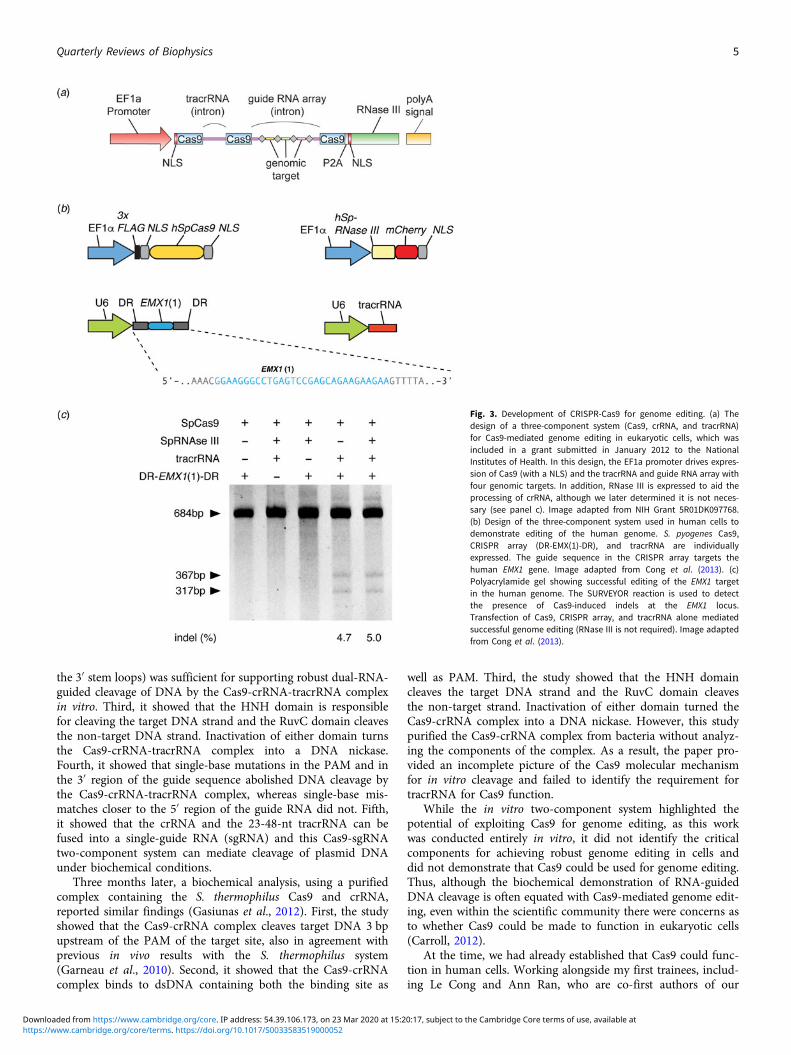
the 3′ stem loops) was sufficient for supporting robust dual-RNA-guided cleavage of DNA by the Cas9-crRNA-tracrRNA complexin vitro. Third, it showed that the HNH domain is responsiblefor cleaving the target DNA strand and the RuvC domain cleavesthe non-target DNA strand. Inactivation of either domain turnsthe Cas9-crRNA-tracrRNA complex into a DNA nickase.Fourth, it showed that single-base mutations in the PAM and inthe 3′ region of the guide sequence abolished DNA cleavage bythe Cas9-crRNA-tracrRNA complex, whereas single-base mis-matches closer to the 5′ region of the guide RNA did not. Fifth,it showed that the crRNA and the 23-48-nt tracrRNA can befused into a single-guide RNA (sgRNA) and this Cas9-sgRNAtwo-component system can mediate cleavage of plasmid DNAunder biochemical conditions.
Three months later, a biochemical analysis, using a purifiedcomplex containing the S. thermophilus Cas9 and crRNA,reported similar findings (Gasiunas et al., 2012). First, the studyshowed that the Cas9-crRNA complex cleaves target DNA 3 bpupstream of the PAM of the target site, also in agreement withprevious in vivo results with the S. thermophilus system(Garneau et al., 2010). Second, it showed that the Cas9-crRNAcomplex binds to dsDNA containing both the binding site as
well as PAM. Third, the study showed that the HNH domaincleaves the target DNA strand and the RuvC domain cleavesthe non-target strand. Inactivation of either domain turned theCas9-crRNA complex into a DNA nickase. However, this studypurified the Cas9-crRNA complex from bacteria without analyz-ing the components of the complex. As a result, the paper pro-vided an incomplete picture of the Cas9 molecular mechanismfor in vitro cleavage and failed to identify the requirement fortracrRNA for Cas9 function.
While the in vitro two-component system highlighted thepotential of exploiting Cas9 for genome editing, as this workwas conducted entirely in vitro, it did not identify the criticalcomponents for achieving robust genome editing in cells anddid not demonstrate that Cas9 could be used for genome editing.Thus, although the biochemical demonstration of RNA-guidedDNA cleavage is often equated with Cas9-mediated genome edit-ing, even within the scientific community there were concerns asto whether Cas9 could be made to function in eukaryotic cells(Carroll, 2012).
At the time, we had already established that Cas9 could func-tion in human cells. Working alongside my first trainees, includ-ing Le Cong and Ann Ran, who are co-first authors of our
Fig. 3. Development of CRISPR-Cas9 for genome editing. (a) Thedesign of a three-component system (Cas9, crRNA, and tracrRNA)for Cas9-mediated genome editing in eukaryotic cells, which wasincluded in a grant submitted in January 2012 to the NationalInstitutes of Health. In this design, the EF1a promoter drives expres-sion of Cas9 (with a NLS) and the tracrRNA and guide RNA array withfour genomic targets. In addition, RNase III is expressed to aid theprocessing of crRNA, although we later determined it is not neces-sary (see panel c). Image adapted from NIH Grant 5R01DK097768.(b) Design of the three-component system used in human cells todemonstrate editing of the human genome. S. pyogenes Cas9,CRISPR array (DR-EMX(1)-DR), and tracrRNA are individuallyexpressed. The guide sequence in the CRISPR array targets thehuman EMX1 gene. Image adapted from Cong et al. (2013). (c)Polyacrylamide gel showing successful editing of the EMX1 targetin the human genome. The SURVEYOR reaction is used to detectthe presence of Cas9-induced indels at the EMX1 locus.Transfection of Cas9, CRISPR array, and tracrRNA alone mediatedsuccessful genome editing (RNase III is not required). Image adaptedfrom Cong et al. (2013).
Quarterly Reviews of Biophysics 5
https://www.cambridge.org/core/terms. https://doi.org/10.1017/S0033583519000052Downloaded from https://www.cambridge.org/core. IP address: 54.39.106.173, on 23 Mar 2020 at 15:20:17, subject to the Cambridge Core terms of use, available at
publication (Cong et al., 2013), we focused on two orthologs ofCas9 that had been previously studied using bacterial geneticsand had complementary advantages: S. thermophilus Cas9(StCas9), which was small enough to be packaged into anadeno-associated viral vector (AAV) for in vivo delivery, and S.pyogenes Cas9 (SpCas9), which had a less restrictive PAMsequence (SpCas9, PAM 5′-NGG, can target on average every12·7 bp in the human genome, whereas StCas9, PAM5′-NNAGAAW, can target on average every 106·6 bp in thehuman genome), and thus broader targeting potential. First, wefound that both SpCas9 and StCas9 can be engineered to mediategenome editing in human and mouse cells. However, Cas9 aggre-gated in the nucleolus, pointing to the obstacle of correct subcel-lular localization when moving a bacterial system into eukaryoticcells. After experimenting with a number of nuclear localizationsignals (NLSs), we found that the combination of a monopartiteand a bipartite NLS allowed Cas9 to localize efficiently into thehuman cell nucleus without any aggregation in the nucleolus.Second, we found that although the natural bacteria expressedmultiple isoforms of the tracrRNA, all of which can provideCRISPR immunity in bacteria (Deltcheva et al., 2011), only the89-nt isoform was stably expressed in human cells and wasimportant for achieving robust genome editing. Third, we foundthat across 16 target sites in human and mouse cells, the three-component system for SpCas9 and StCas9 can mediate robustediting of the genome. Fourth, we found that a CRISPR arrayencoding multiple spacers can be processed by human cells intoindividual guide RNAs to target multiple genes in the genome.Fifth, we showed that DSBs introduced by Cas9 can stimulatehomologous recombination, leading to targeted gene insertion,and that Cas9 nickase activity can also stimulate homologousrecombination in cells, while avoiding the formation ofDSB-induced indels. Sixth, we also explored a two-componentdesign. We found that the additional 3′ stem loops on thetracrRNA are important for gene editing, as the three-componentsystem achieved significantly more robust genome editing inhuman cells than the two-component design employed, whichfailed to edit at a number of genomic sites. Together, these resultsestablished a foundation for the molecular mechanism by whichCRISPR-Cas9 can mediate robust genome editing and furtherunderscores that the ability of CRISPR-Cas9 to function ineukaryotic cells cannot be predicted from in vitro studies (Conget al., 2013).
If Cas9 could function in eukaryotic cells, however, it wouldunlock the potential for a range of sought after applications inresearch, biotechnology, and medicine. It was therefore not sur-prising that, in addition to our efforts to develop Cas9 for genomeediting, other groups were inspired by the biochemical character-ization of Cas9 (Jinek et al., 2012) to explore applications of Cas9as well. Concurrent with our study, a second report of gene edit-ing using Cas9 was published (Mali et al., 2013b). Shortly there-after, additional studies also reported the use of Cas9 in humanand animal cells (Cho et al., 2013; Hwang et al., 2013; Jineket al., 2013) and the use of a catalytically inactivated variant ofCas9 to achieve targeted gene repression (Qi et al., 2013).
Initial impact of Cas9-mediated genome editing
Following the demonstration of Cas9-mediated genome editingin eukaryotic cells, many outstanding scientists contributed tothe advancement and application of the technology, pushing thefield ahead at a remarkable rate. We continued to develop the
technology by focusing on three major areas: (1) further under-standing the biology of Cas9 so as to improve and extend its util-ity; (2) developing applications of Cas9, including genome-widescreening, a Cas9 knock-in mouse, and conversion of Cas9 to acatalytically inactive programmable DNA-binding scaffold; and(3) exploring the natural diversity of CRISPR-Cas systems to iden-tify other Cas effectors with unique properties that may be advan-tageous for technological development. Through these endeavorswe had the opportunity to collaborate with a number of talentedresearchers from diverse backgrounds, further amplifying theimpact of CRISPR-based technologies.
One way the immediate impact of Cas9 can be seen is in itsrapid adoption for other organisms, which highlights the broadutility of this tool as well as the robustness and ease-of-use ofthe system. Catalyzed by the success of Cas9-mediated genomeediting in human cells, within a year, groups from around theworld reported the successful application of Cas9 in a numberof eukaryotic model organisms, including yeast (DiCarlo et al.,2013), mice (Wang et al., 2013), Drosophila (Gratz et al., 2013),C. elegans (Friedland et al., 2013), Arabidopsis (Li et al., 2013),Xenopus (Nakayama et al., 2013), and non-human primates(Niu et al., 2014). Cas9 was also successfully deployed in a num-ber of agriculturally important species in that first year, such asrice and wheat (Shan et al., 2013), sorghum (Jiang et al., 2013),and maize (Liang et al., 2014). Before the year’s end, the firstreports were published on the use of Cas9 to correct a cataract-causing mutation in a mouse, leading to reversal of the diseasephenotype (Wu et al., 2013). In parallel, a number of improve-ments and extensions of the technology were reported in quicksuccession.
The impact of the CRISPR-based technologies is due in nosmall part to the open sharing culture of the CRISPR field,which has enabled applications and further development ofCRISPR-based technologies to flourish. This has been facilitatedthrough on-line resources, such as the creation of numerous web-based tools for guide design (Hsu et al., 2013; Bae et al., 2014;Schmid-Burgk et al., 2014; Labun et al., 2016; Pinello et al.,2016; Concordet and Haeussler, 2018; Listgarten et al., 2018),and through the annual CRISPR meetings. CRISPR reagentshave also been shared widely and openly. To date, more than350 laboratories from around the world have made theirCRISPR-based reagents accessible through the non-profit molec-ular reagent sharing organization Addgene. For my own group,we have made it a priority to help researchers benefit from theCRISPR technological advances we made by disseminatingreagents as well as know-how for CRISPR-based technologies.Through a combination of direct mailing as well as distributionthrough Addgene, we have been able to share more than 52 000CRISPR reagents to researchers at more than 2300 institutionsspanning 62 countries.
From Cas9 to beyond: Cas12 and Cas13
The development of other molecular technologies, such as restric-tion enzymes (Loenen et al., 2014) and green fluorescent proteins(Rodriguez et al., 2017), has benefitted significantly from explora-tions of natural diversity. Similarly, my own experience with thedevelopment of optogenetics has taught me the power of explor-ing the diversity of microbial opsins. Therefore, we turned to thenatural diversity of CRISPR-Cas systems to identify other Caseffectors with the potential to expand the capabilities ofCRISPR-based technologies. By mining the microbial diversity
6 Feng Zhang
https://www.cambridge.org/core/terms. https://doi.org/10.1017/S0033583519000052Downloaded from https://www.cambridge.org/core. IP address: 54.39.106.173, on 23 Mar 2020 at 15:20:17, subject to the Cambridge Core terms of use, available at
for signatures of CRISPR-Cas systems (e.g., conserved genes andCRISPR-like repeat sequences), we discovered and elucidated thefunctions of two new types of CRISPR-Cas systems and developedthem to significantly expand the CRISPR toolbox (Shmakov et al.,2015, 2017; Zetsche et al., 2015a; Smargon et al., 2017) (Fig. 4).These discoveries prompted other investigations of microbialdiversity, revealing additional subtypes of CRISPR-Cas systems(Burstein et al., 2017; Harrington et al., 2018; Konermannet al., 2018; Shmakov et al., 2018; Yan et al., 2018b) and providinginsight into the origin, evolution, and function of these elegantsystems.
SaCas9
Beyond its immediate utility in the lab, there was enormous inter-est in using Cas9-mediated genome editing as a therapeutic thatcould theoretically treat thousands of genetic diseases. One limi-tation to the therapeutic use of SpCas9, however, was its relativelylarge size, which made delivering it challenging. We thereforesought to identify smaller Cas9 orthologs that worked efficientlyin mammalian cells while maintaining a broad targeting range.We characterized a number of CRISPR-Cas9 systems and profiledtheir mammalian genome editing activity. One Cas9 orthologfrom Staphylococcus aureus (SaCas9, PAM 5′-NNGRRT) showedthe highest levels of activity in human cells (Ran et al., 2015).SaCas9 is more than 1 kb shorter than SpCas9, which allowedus to deliver it, along with a guide RNA, on a single-AAV vectorfor in vivo use (Ran et al., 2015). SaCas9 is now being developedas the first in vivo genome editing medicine for humans (seebelow) (Allergan, 2019). SpCas9 is also being advanced for ther-apeutic applications. However, due to its large size, clinical trialsemploying SpCas9 are focused on electroporation of patient cellsex vivo (Vertex, 2018a, 2018b).
Cas12
We next went beyond Cas9 orthologs to study other CRISPR-Cassystems, beginning with a putative new type of class 2CRISPR-Cas system, type V, characterized by the Cas12 familyof effector proteins. The first Cas12 enzyme, classified as typeV-A and referred to as Cas12a (previously known as Cpf1) wasidentified in the genomes of Prevotella and Francisella and con-tained a large protein of unknown function (Schunder et al.,2013; Vestergaard et al., 2014; Makarova et al., 2015). Cas12a isa distinct enzyme unrelated to Cas9. A number of Francisella spe-cies contain Cas12a in association with putative CRISPR arrays,including F. novicida. Heterologous expression of the F. novicidaCRISPR-Cas12a locus in E. coli led to interference of plasmidDNA transformation, establishing CRISPR-Cas12a as a bonafide CRISPR-Cas system and revealing that Cas12a requires aT-rich PAM sequence preceding the DNA target site (Zetscheet al., 2015a). In contrast to Cas9, the Cas12a system does notcontain a tracrRNA, and its DNA cleavage results in a 5′ overhanginstead of a blunt DSB (Zetsche et al., 2015a). Also, unlike Cas9,which utilizes host RNase III to process its CRISPR array, Cas12aitself has RNase activity and processes its own pre-crRNA arrayinto individual crRNAs (Fonfara et al., 2016).
A search for Cas12a orthologs identified two-Cas12a enzymes,from Acidaminococcus and Lachnospiraceae, with strong cleavageactivity in human cells, comparable to SpCas9 (Zetsche et al.,2015a). Apart from expanding the range of genomic targets thatcan be edited given that it has a different PAM than Cas9,
Cas12a-mediated editing has several advantages over Cas9: it issignificantly more specific (Kleinstiver et al., 2016b; Kim et al.,2017b), which is important for therapeutic applications; it offersa simplified guide design because it does not require tracrRNA;it generates over-hanging ends, rather than the blunt ends createdby Cas9, which may be beneficial for the introduction of newsequences (Moreno-Mateos et al., 2017); it has smaller molecularsize which is more suitable for viral packaging, and it is ideallysuited for multiplex genome editing because multiple guideRNAs can be easily expressed as a single transcript and subse-quently processed into individual guide RNAs by Cas12a itself(Zetsche et al., 2016).
Relative to the Cas9 family of Cas effectors, Cas12 is a muchmore diverse family. Indeed a number of subtypes of Cas12 sys-tems have recently been reported (denoted type V-A – V-I).The Cas12b effectors (previously known as C2c1) target DNA,but in contrast to Cas12a, they are dual-RNA guided, requiringa tracrRNA (Shmakov et al., 2015). Although initial characteriza-tion of Cas12b revealed thermophilic nuclease activity, which pre-vented application in mammalian cells, subsequent exploration ofthe Cas12b diversity and protein engineering made possible thedevelopment of two-Cas12b systems with robust genome editingactivity in human cells (Teng et al., 2018; Strecker et al., 2019).Comparison of Cas12b with SpCas9 showed that Cas12b has sub-stantially reduced off-target activity, indicating it is inherentlymore specific than wild-type SpCas9 when targeting the humangenome (Teng et al., 2018; Strecker et al., 2019). AdditionalCas12 effectors have also been identified from bacterial genomicdatabases, including Cas12c (Shmakov et al., 2015), Cas12d(CasY) and Cas12e (CasX), both of which were found in metage-nomic samples (Burstein et al., 2017), and three subtypes ofCas12f (Cas14) (Harrington et al., 2018). Two-Cas12e orthologs,DpbCasX and PlmCasX, have recently been shown to achieve tar-geted gene knockout in human cells (Liu et al., 2019). A recenteffort to holistically identify CRISPR-Cas systems from morethan 10 terabytes of genomic and metagenomic data led to theidentification of a number of new type V subtype loci, includingboth DNA- and RNA-targeting Cas12 systems (Yan et al., 2019).
Cas13
The type VI family of CRISPR-Cas systems, signified by theRNA-guided RNA-targeting Cas13 effector, was first found byusing the highly conserved adaptation protein Cas1 as the searchseed to identify all genomic fragments that contain putativeCRISPR-Cas systems. Focusing on conserved proteins of unknownfunction located within each CRISPR locus, we discovered a familyof well-conserved large proteins carrying the higher eukaryotic–prokaryotic nuclease (HEPN) domain, which suggested they areputative RNases (Shmakov et al., 2015). Subsequent expansion ofthe search to use CRISPR repeats as the search seed led to the iden-tification of additional Cas13 subtypes, including Cas13b, Cas13c,and Cas13d (Shmakov et al., 2017; Smargon et al., 2017;Konermann et al., 2018; Yan et al., 2018b).
Using E. coli heterologously expressing type VI CRISPR-Cassystems, we showed that CRISPR-Cas13a and Cas13b systemsconfer resistance to RNA phages, and that they are single-effectorRNases guided by crRNAs (Abudayyeh et al., 2016; Smargonet al., 2017). This finding paved the way for an entirely new setof molecular technologies operating at the level of RNA, ratherthan DNA, and offering a safer therapeutic approach to treatingdisease (see below). Similar to Cas12a, Cas13 proteins also
Quarterly Reviews of Biophysics 7
https://www.cambridge.org/core/terms. https://doi.org/10.1017/S0033583519000052Downloaded from https://www.cambridge.org/core. IP address: 54.39.106.173, on 23 Mar 2020 at 15:20:17, subject to the Cambridge Core terms of use, available at
contain an RNase processing domain with the ability to cleavetheir corresponding CRISPR array into individual maturecrRNAs (East-Seletsky et al., 2016; Smargon et al., 2017;Konermann et al., 2018). Cas13 cleaves RNA at sites outside ofthe target region complementary to the crRNA. Analysis of cleav-age products of Leptotrichia shahii Cas13a (LshCas13a) showed
that cut sites do not vary even for crRNAs targeting different posi-tions on the same target, indicating that cut sites are likely dic-tated by a combination of the target RNA secondary structureand sequence features (Abudayyeh et al., 2017; Smargon et al.,2017). Further exploration of the RNase activity uncovered the‘collateral effect’ of Cas13 – recognition of the target RNA by
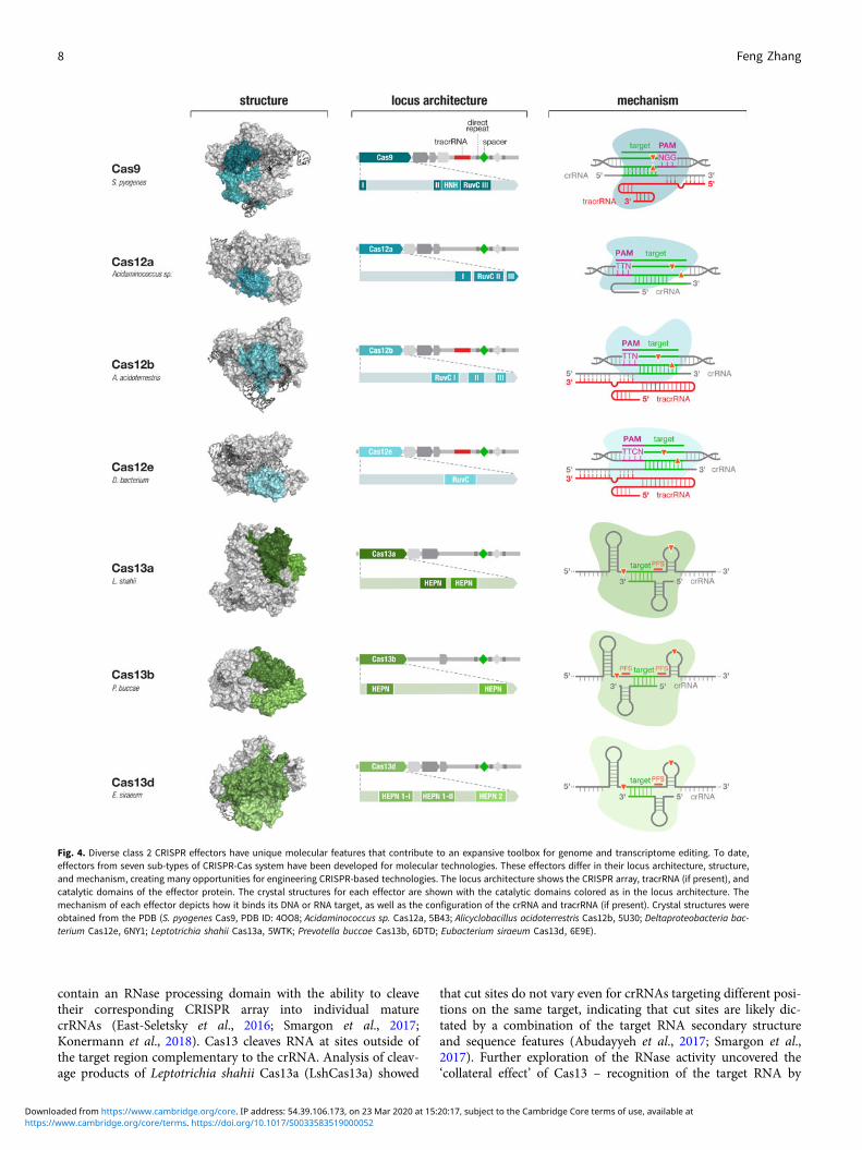
Fig. 4. Diverse class 2 CRISPR effectors have unique molecular features that contribute to an expansive toolbox for genome and transcriptome editing. To date,effectors from seven sub-types of CRISPR-Cas system have been developed for molecular technologies. These effectors differ in their locus architecture, structure,and mechanism, creating many opportunities for engineering CRISPR-based technologies. The locus architecture shows the CRISPR array, tracrRNA (if present), andcatalytic domains of the effector protein. The crystal structures for each effector are shown with the catalytic domains colored as in the locus architecture. Themechanism of each effector depicts how it binds its DNA or RNA target, as well as the configuration of the crRNA and tracrRNA (if present). Crystal structures wereobtained from the PDB (S. pyogenes Cas9, PDB ID: 4OO8; Acidaminococcus sp. Cas12a, 5B43; Alicyclobacillus acidoterrestris Cas12b, 5U30; Deltaproteobacteria bac-terium Cas12e, 6NY1; Leptotrichia shahii Cas13a, 5WTK; Prevotella buccae Cas13b, 6DTD; Eubacterium siraeum Cas13d, 6E9E).
8 Feng Zhang
https://www.cambridge.org/core/terms. https://doi.org/10.1017/S0033583519000052Downloaded from https://www.cambridge.org/core. IP address: 54.39.106.173, on 23 Mar 2020 at 15:20:17, subject to the Cambridge Core terms of use, available at
the Cas13-crRNA complex leads Cas13 to become a promiscuousRNase, cleaving non-target bystander RNAs at preferred cut sites(Abudayyeh et al., 2016). Collateral activity may play a role in pro-grammed cell death in bacteria, although this remains to be fullyexplored. This collateral activity has been exploited to expand theapplications of Cas effectors into new categories, including thedevelopment of sensitive, low-cost, and rapid diagnostics assaysfor viral and bacterial infections (see below).
Cas13a, b, c, and d have all been adapted for use in mamma-lian cells to mediate targeted RNA knockdown (Abudayyeh et al.,2017; Cox et al., 2017; Konermann et al., 2018). Interestingly,although in bacteria, each Cas13 ortholog exhibits varying levelsof nucleotide preference in sequences flanking the protospacer,referred to as the protospacer flanking site (PFS), the presenceof the PFS is not a strict requirement for RNA targeting in mam-malian cells (Abudayyeh et al., 2017). Additionally, although col-lateral activity has been observed in vitro and in bacterial cells(Abudayyeh et al., 2016; Meeske and Marraffini, 2018), it hasnot been detected in mammalian cells (Abudayyeh et al., 2017;Konermann et al., 2018), suggesting that, similarly for Cas9 andCas12, the differences between biochemical, bacterial, and mam-malian environments can substantially affect the behavior of Caseffectors.
Development of a molecular toolbox based on Cas effectors
DNA and RNA cleavage through the nuclease activities of Caseffectors is only one way CRISPR technology can be applied.The ability to customize the binding specificity of Cas effectorsusing a short-guide RNA creates many additional opportunitiesfor developing new capabilities for manipulating DNA andRNA. There are two-main categories of molecular tools basedon Cas proteins (Fig. 5), with the first category utilizing theintrinsic RNA-guided nuclease activity of each effector, and thesecond category exploiting nuclease-inactivated Cas proteins(dCas) as RNA-guided nucleic acid binding domains to targeteffector modules to modulate, monitor, or modify target DNAor RNA. As tools based on Cas effectors rely on the specificityof RNA-guided target recognition, another area of focus hasbeen to assess the specificity of Cas effectors as well as engineeringsolutions to enhance their specificity. Below is an overview of thebroad range of molecular tools that have been developed based onCas proteins as well as efforts to address the most critical chal-lenges facing CRISPR-based tools.
Leveraging natural and engineered properties of diverse Caseffectors
The opportunities for developing Cas effectors as molecular tech-nologies are further amplified by the natural diversity within eachfamily of class 2 CRISPR-Cas systems. Based on the current pub-licly accessible bacterial genomic and metagenomic sequencingdata, there are over 100 000 Cas9 family members, over 70 000Cas12 family members, and over 5000 Cas13 family members.Within each family, members can exhibit a number of differencesin terms of their size, guide RNA requirement, binding motif (e.g.,PAM and PFS), targeting specificity, and suitability for functionin eukaryotic cells. In the case of Cas13 family members, theycan also exhibit different cleavage motif preferences.
A number of Cas9 orthologs have been discovered (Bolotinet al., 2005; Makarova et al., 2011, 2015; Zhang et al., 2013;Chylinski et al., 2014; Fonfara et al., 2014; Ran et al., 2015;
Shmakov et al., 2017), and an increasing number of these havebeen developed for use as genome editing tools beyond SpCas9and StCas9 (Esvelt et al., 2013; Hou et al., 2013; Karvelis et al.,2015; Ran et al., 2015; Hirano et al., 2016; Lee et al., 2016; Kimet al., 2017a). The natural diversity of these enzymes has allowedexpanded applications, for example some smaller Cas9 orthologs,such as S. aureus Cas9 (SaCas9), Neisseria meningitidis Cas9(NmeCas9), and Campylobacter jejunii Cas9 (CjCas9) have beenefficiently delivered in vivo using a single-vector strategy (Ranet al., 2015; Kim et al., 2017a; Ibraheim et al., 2018).
While exploration of natural Cas diversity provides one avenuefor expanding and improving CRISPR-based tools, a complemen-tary approach uses structure-guided engineering to modify andimprove Cas effector function. Over the past several years a num-ber of crystal structures have been solved for different members ofCas9 (Anders et al., 2014; Jinek et al., 2014; Nishimasu et al.,2014, 2015; Hirano et al., 2016; Yamada et al., 2017), Cas12(Dong et al., 2016; Yamano et al., 2016; Yang et al., 2016; Stellaet al., 2017; Swarts et al., 2017; Wu et al., 2017; Liu et al.,2019), and Cas13 (Knott et al., 2017; Liu et al., 2017a, 2017b;Zhang et al., 2018a, 2018b; Slaymaker et al., 2019) families.These structures include the apo forms with just the effectorprotein alone, or the effector in complex with its guide RNAalone or guide RNA in complex with target DNA or RNA,providing structural insights into target recognition and cleavage.These structural studies have been complemented by otherbiochemical and biophysical studies into the target searchmechanism of Cas effectors (Sternberg et al., 2014; Knightet al., 2015; Ma et al., 2016a).
Expanding the targeting range of DNA-targeting Cas proteins
The DNA targeting range of Cas9 and Cas12 is defined by thePAM sequence, a short-sequence flanking the target sequence isrequired for DNA targeting. A shorter PAM sequence providesa broader targeting range whereas longer PAM sequences aremore restrictive. For example, wild-type SpCas9 (which has anNGG PAM) can target roughly ten times more sites in thehuman exome than wild-type SaCas9 (which has an NNGRRTPAM) (Scott and Zhang, 2017). In order to increase the flexibilityof Cas-mediated DNA targeting, a combination of approaches hasbeen used to expand the number of targetable PAM sequences.First, by exploring phylogenetic diversity, a number of Cas effec-tor orthologs have been identified with distinct PAM require-ments. In the case of Cas12a, a survey of more than a dozenorthologs identified one, from Moraxella bovoculi, with robustindel activity in human cells and tolerance of a shorter PAM,expanding the available targeting landscape (Zetsche et al.,2017). Ultimately, however, only a handful of Cas effectors havebeen successfully developed for function in eukaryotic cells(Ran et al., 2015; Zetsche et al., 2015a; Abudayyeh et al.,2017; Cox et al., 2017; Kim et al., 2017a; Chatterjee et al., 2018;Ibraheim et al., 2018; Konermann et al., 2018; Teng et al., 2018;Liu et al., 2019; Strecker et al., 2019) limiting the extent of thisapproach.
A second approach for expanding the DNA targeting range ofCas9 and Cas12 is to engineer new variants, either throughstructure-guided design or directed evolution. Based on the crystalstructures of Cas effectors in complex with guide RNA and targetDNA (Anders et al., 2014; Nishimasu et al., 2015; Yamano et al.,2016), targeted mutagenesis has been used to generate new pro-tein variants with altered PAM sequences. At the same time, a
Quarterly Reviews of Biophysics 9
https://www.cambridge.org/core/terms. https://doi.org/10.1017/S0033583519000052Downloaded from https://www.cambridge.org/core. IP address: 54.39.106.173, on 23 Mar 2020 at 15:20:17, subject to the Cambridge Core terms of use, available at
number of groups have used directed evolution strategies to evolvenew variants of Cas effectors with unique properties, includingdifferent PAM preferences. These efforts have led to the develop-ment of a number of Cas9 and Cas12a variants with a
significantly broadened DNA targeting range (Kleinstiver et al.,2015a, 2015b; Gao et al., 2017; Hu et al., 2018; Nishimasuet al., 2018). Collectively, these variants enable targeting of virtu-ally any genomic site.
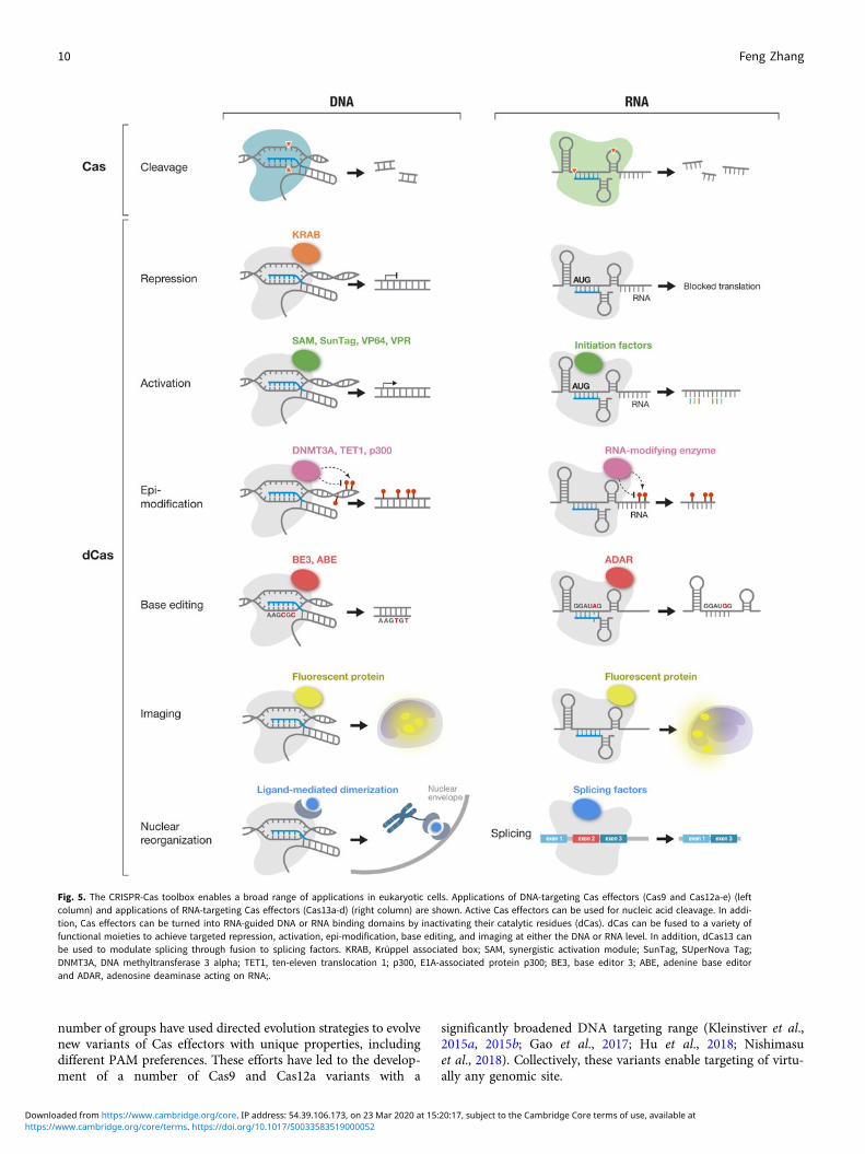
Fig. 5. The CRISPR-Cas toolbox enables a broad range of applications in eukaryotic cells. Applications of DNA-targeting Cas effectors (Cas9 and Cas12a-e) (leftcolumn) and applications of RNA-targeting Cas effectors (Cas13a-d) (right column) are shown. Active Cas effectors can be used for nucleic acid cleavage. In addi-tion, Cas effectors can be turned into RNA-guided DNA or RNA binding domains by inactivating their catalytic residues (dCas). dCas can be fused to a variety offunctional moieties to achieve targeted repression, activation, epi-modification, base editing, and imaging at either the DNA or RNA level. In addition, dCas13 canbe used to modulate splicing through fusion to splicing factors. KRAB, Krüppel associated box; SAM, synergistic activation module; SunTag, SUperNova Tag;DNMT3A, DNA methyltransferase 3 alpha; TET1, ten-eleven translocation 1; p300, E1A-associated protein p300; BE3, base editor 3; ABE, adenine base editorand ADAR, adenosine deaminase acting on RNA;.
10 Feng Zhang
https://www.cambridge.org/core/terms. https://doi.org/10.1017/S0033583519000052Downloaded from https://www.cambridge.org/core. IP address: 54.39.106.173, on 23 Mar 2020 at 15:20:17, subject to the Cambridge Core terms of use, available at

Assessing the specificity of class 2 Cas effectors
One of the most critical technical requirements for the applicationof class 2 Cas effectors is their targeting specificity. When apply-ing Cas9 or Cas12 as an active nuclease, minimizing off targets isparticularly important because a range of undesirable genomicalterations could arise through the cell’s endogenous DNA repairmechanisms, such as translocations between different cleavagesites and large-scale deletions. For nuclease as well as dCas bind-ing applications, it is important that the effector binds selectivelyto the DNA or RNA targeted by the guide RNA. In the case ofactive nuclease applications, off-target editing activity due topseudo-specific interactions between the Cas effector and thegenome (arising when there are less than perfect matches betweenthe target and guide RNA) can give rise to additional DSBs thatlead to either small insertions and deletions (indels) or largergenomic alterations.
An initial study to characterize off-target indels used compu-tational analyses to identify loci in the genome that share ahigh degree of homology to the target site, and then assayed edit-ing events at these computationally predicted off-target loci usingdeep sequencing (Fu et al., 2013). The study found that SpCas9can indeed induce off-target edits at genomic loci that carriedthree or fewer mismatches compared with the guide sequence.Additional studies using different approaches also showed thatCas9 can indeed introduce off-target edits (Hsu et al., 2013;Mali et al., 2013a; Pattanayak et al., 2013). As the early approachescovered only a very limited set of off-target sites, subsequentinvestigations focused on developing genome-wide unbiasedapproaches including in vitro assays like Digenome-seq (Kimet al., 2015), CIRCLE-seq (circularization for in vitro reportingof cleavage effects by sequencing) (Tsai et al., 2017), andSITE-seq (selective enrichment and identification of tagged geno-mic DNA ends by sequencing) (Cameron et al., 2017) and cellularassays like GUIDE-seq (genome-wide, unbiased identification ofDSBs enabled by sequencing) (Tsai et al., 2015), BLESS (directin situ breaks labeling, enrichment on streptavidin and next-generation sequencing) and BLISS (breaks labeling in situ andsequencing) (Crosetto et al., 2013; Yan et al., 2017), linearamplification-mediated PCR followed by high-throughputgenome-wide translocation sequencing (LAM-HTGTS) (Frocket al., 2015), and VIVO (verification of in vivo off-targets)(Akcakaya et al., 2018). The use of these assays found that theediting specificity of SpCas9 varied widely depending on theguide RNA. When these unbiased techniques were used to profilethe specificity of Cas9 orthologs as well as Cas12 family members,it was found that SaCas9 as well as Cas12a and Cas12b are muchmore specific than SpCas9, with most guide RNAs exhibiting nodetectable off-target editing (Kleinstiver et al., 2016b; Yan et al.,2017; Strohkendl et al., 2018; Tycko et al., 2018; Strecker et al.,2019). It is worth noting, however, that the functional impact ofoff-target edits will vary depending on their location. For exam-ple, off-target indels within coding regions, regulatory elements,and non-coding RNAs are likely to have more undesirable effects.
A number of studies have also explored the landscape of largergenomic alterations arising from Cas9 activity. For translocations,the use of LAM-HTGTS showed that the frequency ofSpCas9-induced translocation varies considerably with the guideRNA, from undetectable to ∼3% (Frock et al., 2015), and theuse of a tagmentation strategy found SpCas9-mediated transloca-tion rates of ∼2·5% for two-different guides (Giannoukos et al.,2018). For large deletions, the use of PacBio and Sanger
sequencing with SpCas9-edited hemizygous embryonic stemcells showed that 10 out of 48 edited alleles represented deletionslarger than 250 bp (ranging up to nearly 6 kb) (Kosicki et al.,2018). The same study also identified a number of other eventssuch as transversions, duplications, and other structural rear-rangements (Kosicki et al., 2018).
Similar approaches have not been applied to the high-specificity variants of Cas9 or to Cas12a/b, all of which show sub-stantially fewer indel off-targets, and it will be interesting to see ifthe number of large deletions and structural rearrangements issimilarly reduced with these other Cas effectors. These studies,along with empirical testing of guide RNAs, will inform thebest choice of the Cas effector for applications requiring particu-larly high levels of editing specificity. In addition, methods havebeen developed to quantify the on-target editing outcomes ofCas9 (Miyaoka et al., 2016, 2018).
When assessing the RNA targeting specificity of Cas13, theconsiderations are slightly different. Although off-target cleavagearising from pseudo-specific binding remains a concern, an addi-tional issue with Cas13 is potential collateral cleavage ofbystander transcripts. To assess the likelihood of off-targetRNA knockdown, the effect of an increasing number of mis-matches between the guide sequence and its RNA target wasexamined. From these studies it was found that Cas13 can tolerateup to a single mismatch throughout the guide sequence and stillcleave the target RNA (Abudayyeh et al., 2017). Additionally,transcriptome-wide sequencing revealed that Cas13 can achievehighly specific knockdown of the target transcript without signif-icant off-target effects. In contrast, short-hairpin RNA (shRNA)knockdown of the same transcript led to downregulation of hun-dreds of off-targets (Abudayyeh et al., 2017). In addition, bio-chemical analysis of target RNA binding by Cas13a revealedthat perfect matching in a central seed region of the guidesequence is required for binding, but a different guide region isrequired for the activation of RNase activity (Tambe et al.,2018). Together, these studies provided detailed insight into thepotential off-target effects due to pseudo-specific binding aswell as suggested that collateral activity is not significant in mam-malian cells, which has been supported by additional studies(Konermann et al., 2018).
Improving the targeting specificity of Cas effectors
A number of approaches have been developed to improve the spe-cificity of DNA editing by Cas9, and many of these approacheshave also been applied or are relevant to Cas12 as well. Theapproaches can be divided into strategies that either seek toreduce overall exposure to the nuclease or directly improve specif-icity through engineering of the system.
For the first category of approaches, we observed early on thatthe editing specificity can be improved by more than 10-fold byintroducing less Cas9 into cells (Hsu et al., 2013). In agreementwith this observation, several other groups have demonstratedthat using either mRNA to deliver Cas9 or deliveringCas9-sgRNA ribonucleoprotein (RNP) complexes directly intothe target cell can significantly increase the editing specificity(Cho et al., 2014; Lin et al., 2014). In addition to use of differentdelivery methods to limit the dosage of Cas9 in cells, it is also pos-sible to engineer Cas9 so that its nuclease activity becomes drug-or light-inducible (Davis et al., 2015; Nihongaki et al., 2015;Truong et al., 2015; Wright et al., 2015; Zetsche et al., 2015b;Liu et al., 2016a; Nguyen et al., 2016; Rose et al., 2017). This
Quarterly Reviews of Biophysics 11
https://www.cambridge.org/core/terms. https://doi.org/10.1017/S0033583519000052Downloaded from https://www.cambridge.org/core. IP address: 54.39.106.173, on 23 Mar 2020 at 15:20:17, subject to the Cambridge Core terms of use, available at
inducible approach has also been applied to Cas12a (Tak et al.,2017).
For the second category of approaches, various strategies havebeen used, beginning with engineering the system to increase thenumber of target DNA bases that must be specifically recognizedby Cas9 in order to activate nuclease activity. The first strategydoubles the number of DNA bases that need to be recognizedto introduce a DSB by utilizing a Cas9 nickase and two-juxtaposed guide RNAs to create two off-set nicks (Mali et al.,2013a; Ran et al., 2013). This method was found to reduce off-target edits beyond the detection limit (Ran et al., 2013).Related to this double-nicking approach, a second strategy usesdCas9-FokI fusions and a pair of juxtaposed guide RNAs to facil-itate the introduction of a DSB (Guilinger et al., 2014; Tsai et al.,2014). The specificity of Cas9 targeting can also be enhanced bymodifying the guide RNA. One study showed that SpCas9 target-ing can be significantly improved using truncated-guide RNAswith 17 nt of the targeting sequence (Fu et al., 2014), whichdecreases the tolerance for mismatches. More recently, it wasshown that the use of bridged or locked-nucleic acids can alsoimprove Cas9 specificity by slowing the reaction rate (Cromwellet al., 2018), and that engineering the guide RNA to create a hair-pin in the spacer region improves specificity of a number of Cas9and Cas12 enzymes (Kocak et al., 2019).
In addition to these strategies, rational engineering of Cas9 andCas12 has been used to create high-specificity variants, offering asimpler solution to the specificity challenge. We developed thefirst of these variants, eSpCas9, which exhibits substantiallyreduced off-target activity while maintaining on-target efficiency(Slaymaker et al., 2015). A number of groups have subsequentlyused structural information or directed evolution to develop addi-tional high-specificity variants of Cas9 (Kleinstiver et al., 2016a;Chen et al., 2017; Casini et al., 2018; Hu et al., 2018; Lee et al.,2018; Vakulskas et al., 2018) and Cas12a (Kleinstiver et al., 2019).
The specificity of Cas effectors will continue to be improvedthrough rational engineering and directed evolution, which willbe particularly important for the clinical use of Cas effectors. Itis important to note that the development of variants of Cas effec-tors with increased specificity needs to be complemented withhigher sensitivity assays, such as the recently developed genome-wide off-target analysis by the two-cell embryo injection (GOTI)method (Zuo et al., 2019), for detecting the presence of off-targetactivity, particularly large deletions and chromosomalrearrangements.
dCas platforms
In addition to their utility as nucleases, class 2 Cas effectors canalso be inactivated to turn the proteins into RNA-guided DNA-or RNA-binding domains (Fig. 5). These inactivated variantscan be used for a wide variety of powerful applications by servingas programmable nucleic acid binding scaffolds for the recruit-ment of a variety of effector functions. To deactivate the nucleaseactivity of Cas9, alanine substitutions are introduced into the cat-alytic residues of the HNH and RuvC nuclease domains(Sapranauskas et al., 2011). In early 2013, using this mutant ver-sion of Cas9, termed dead Cas9 (dCas9), it was shown that dCas9could achieve programmable gene repression in bacteria andmammalian cells by simply binding to the genome and blockingtranscription (Qi et al., 2013). Since then, many new applicationshave been developed by using dCas9 to recruit effectors that mod-ulate, modify, or visualize DNA or RNA (for examples see:
(Bikard et al., 2013; Chen et al., 2013; Gilbert et al., 2013, 2014;Konermann et al., 2013, 2014; Maeder et al., 2013; Perez-Pineraet al., 2013; Tanenbaum et al., 2014; Hilton et al., 2015; Maet al., 2015, 2016b; Thakore et al., 2015)). Similar to Cas9, theRuvC nuclease domain of Cas12 (Zetsche et al., 2015a), and theHEPN nuclease domains of Cas13 (Abudayyeh et al., 2016) canalso be inactivated to generate dCas12 and dCas13, respectively.
In the case of Cas9, we showed that it is also possible to use atruncated guide sequence that does not trigger the nuclease activ-ity of Cas9 (Dahlman et al., 2015). Using this strategy, researcherscan simultaneously use Cas9 as a nuclease to cleave one set ofgenomic targets and as a DNA binding domain for a differentset of genomic targets simply by using guide RNAs with fulllength (20-nt) or truncated (12-nt) guide sequences, respectively.This approach is particularly relevant when using transgenicmouse lines expressing the nuclease-active form of Cas9 (Plattet al., 2014). Using this truncated guide RNA strategy, DNA bind-ing experiments can be conducted without creating an additionalmouse line expressing dCas9 (Liao et al., 2017).
There are a several ways to recruit effectors to dCas. The sim-plest method is to directly fuse the effector protein to either theN- or C-terminus of the Cas protein (Gilbert et al., 2013;Konermann et al., 2013; Abudayyeh et al., 2017; Tak et al.,2017). However, in some applications, particularly for the recruit-ment of fluorescent proteins for imaging, a second strategy hasbeen used where a SunTag is attached to dCas9 to attract effectorsthat are fused to a single-chain variable fragment antibody frag-ment with SunTag affinity (Tanenbaum et al., 2014). Yet anotherapproach is to engineer the guide RNA such that exposed hairpinscan serve as potential sites for insertion of RNA aptamers. Byengineering new guide RNAs carrying the MS2 aptamer insertedinto stem loops on the guide RNA, we showed that effectordomains can be recruited via MS2 binding (Konermann et al.,2014). Subsequent studies have shown that other aptamers suchas PP7 and com can also be inserted into the guide RNA toallow for multiplexing applications (Zalatan et al., 2015; Liuet al., 2016a).
The applications of dCas are quite broad. Initial work showedthat simply by recruiting dCas9 to target loci, gene expressioncould be repressed in both bacterial and human cells (Bikardet al., 2013; Qi et al., 2013). Fusions of dCas9 to transcriptionalrepressors, such as Krüppel-associated box (KRAB), have alsobeen used to programmably repress gene expression (Gilbertet al., 2013) in human cell lines. dCas9-KRAB fusions havebeen combined with inducible Cas9 systems for fine-tuned regu-latory control of gene networks (Mandegar et al., 2016). dCas9can also be used to facilitate transcriptional activation of targetgenes (Bikard et al., 2013; Gilbert et al., 2013; Konermannet al., 2013, 2014; Maeder et al., 2013; Perez-Pinera et al., 2013).Additionally, dCas9 has been fused with epigenetic modifiers toachieve targeted histone acetylation (Hilton et al., 2015), histonedemethylation (Kearns et al., 2015), and DNA methylation anddemethylation (Liu et al., 2016b; Vojta et al., 2016; Xu et al.,2016). A number of groups have used dCas9 for genomic locusand chromosome imaging as well as spatial manipulation of geno-mic organization (Chen et al., 2013; Morgan et al., 2017; Wanget al., 2018). Through the use of either orthogonal Cas enzymesor aptamers and multiple fluorophores, multiplex locus imagingcan be achieved (Chen et al., 2016; Liu et al., 2016a). Similarly,RNA can be imaged using dCas effectors, including dCas9(Nelles et al., 2016) and dCas13a (Abudayyeh et al., 2017).dCas13 has also been fused to hnRNP1, a negative regulator of
12 Feng Zhang
https://www.cambridge.org/core/terms. https://doi.org/10.1017/S0033583519000052Downloaded from https://www.cambridge.org/core. IP address: 54.39.106.173, on 23 Mar 2020 at 15:20:17, subject to the Cambridge Core terms of use, available at
splicing, to achieve targeting exon skipping (Konermann et al.,2018). Through fusion to the engineered peroxidase APEX2,dCas9 can be used to identify proteins associated with a specificgenomic locus (Myers et al., 2018). Additional functional plat-forms have also been developed, such as the fusion of a Cas9 nick-ase with an error-prone polymerase to create EvolvR, a system forrapid diversification of the DNA sequence within a few hundredbase-pair window (Halperin et al., 2018). Another system,CRISPR-X, uses dCas9 and modified-guide RNAs to recruit cyti-dine deaminase variants to generate localized windows of varia-tion, which may have applications for directed evolution (Hesset al., 2016).
Targeted base editing of DNA and RNA
Another exciting application of dCas enzymes has been the devel-opment of programmable DNA and RNA base editors, which canachieve the precise chemical change of one base to another(reviewed comprehensively by (Rees and Liu, 2018)). Base editorsare particularly promising for the development of therapeuticapplications, as more than half of the known pathological variantsare point mutations. Furthermore, both DNA and RNA base edit-ing provide the possibility of making targeted changes withoutrelying on homologous recombination, which is inefficient espe-cially in post-mitotic cells such as neurons.
DNA base editors are generated by fusing dCas9 or Cas9 nick-ase (Komor et al., 2016; Nishida et al., 2016; Gaudelli et al., 2017)or dCas12 (Li et al., 2018c) to single-strand DNA deaminases(Fig. 5). The first type of base editor developed used dCas9 orCas9 nickase to bring a single-stranded DNA cytosine deaminasesuch as AID or APOBEC to mediate C • G to T • A conversionson target DNA. Binding of DNA by dCas9 or Cas9 nickase formsan R-loop which exposes a short stretch of single-stranded DNAfor deamination by the tethered cytosine deaminase (Komoret al., 2016; Nishida et al., 2016). Application of the cytosinebase editor in a variety of animal and plant cell types can leadto high levels of targeted base conversion (Rees and Liu, 2018).To expand the types of base changes achievable, a second typeof base editor capable of converting A • T to G • C was createdby fusing dCas9 or Cas9 nickase to an evolved form of the bacte-rial tRNA-specific adenine deaminase TadA (Gaudelli et al.,2017). TadA naturally acts on single-stranded RNA, but throughan impressive series of directed evolution steps, TadA was con-verted into a DNA deaminase. DNA base editing has alreadybeen applied in animal models of disease, highlighting its poten-tial for therapeutic use (Villiger et al., 2018). This powerful tech-nology is being rapidly optimized to increase specificity, efficacy,and precision (Rees and Liu, 2018).
RNA base editors have been engineered by fusing dCas13 tothe adenine deaminase ADAR to achieve a precise, targetedA-to-I conversion (inosine is read out by cells as guanosine)(Fig. 5) (Cox et al., 2017). Because ADAR acts on an RNA duplexformed between the target RNA and the guide RNA at adenosinesin A • C mismatch bubbles, a specific adenine can be targeted fordeamination by intentional mis-pairing with a cytosine on theguide RNA (Cox et al., 2017). The ability to direct adenine deam-ination with single-nucleotide precision, which is not currentlypossible with DNA editing, has inspired efforts to use directedevolution to convert ADAR into a duplex RNA-acting cytosinedeaminase, an activity which has not been found in nature, todevelop a precise C to U RNA editor. RNA and DNA base editingcomplement each other to expand the range of applications. In
particular, RNA editing does not depend on the presence ofDNA-repair machinery for base conversion and therefore canbe applied in virtually all cell types. Furthermore, because RNAediting can be potentially temporally restricted when pairedwith transient delivery systems, it can serve as a reversible editingsystem, which further expands the therapeutic potential ofCRISPR-based technologies.
The specificity of base editors has recently been comprehen-sively profiled. Although cytosine DNA base editors have beenfound to generate a large number of off-target edits throughoutthe genome, the adenine base editor is able to achieve specificediting of the target site (Jin et al., 2019; Kim et al., 2019; Zuoet al., 2019). A study in mice also examined potential off-targeteffects of adenine base editing and similarly found that A • Tto G • C conversion was quite specific (Liu et al., 2018d).Additionally, a recent study looking at the specificity of base edi-tors found substantial off-target editing in the transcriptome(Grunewald et al., 2019). Future refinements aimed at reducingnon-specific interactions with DNA should significantly increasethe specificity of cytosine base editors. In addition to improvingthe specificity of the deaminase domain, specificity may also beimproved through the use of more specific Cas effectors such asdSaCas9 or dCas12a rather than dSpCas9. As DNA base editorsdo not rely on the introduction of DSBs, the likelihood of largedeletions or translocations is significantly lower than withnuclease-based approaches. The specificity of RNA base editorshas also been comprehensively profiled using high-coverage tran-scriptome analysis. Although the initial version of the RNA baseediting platform showed broad transcriptome-wide off-targetediting, structure-guided engineering of ADAR reduced non-specific interactions and improved the targeting specificity by∼1000 fold (Cox et al., 2017). The promising results with bothDNA and RNA base editing are prompting rapid improvementsin technology.
Additional applications of CRISPR-based technologies
CRISPR-based technologies have also been coopted for informa-tion recording, either about cell fate, activity, or even non-biological data. Many of these approaches rely on the ability ofCRISPR-Cas systems to create traceable scars that can be readout through sequencing and then related back to specific events.One of the first such uses was lineage tracing to reconstruct cel-lular or organismal development (McKenna et al., 2016; Friedaet al., 2017; Kalhor et al., 2017). Trackers have also been builtthat can record information about the cellular state, such as thepresence of small molecules, metabolites, external stimuli, or tran-scriptional activity (Perli et al., 2016; Schmidt et al., 2018; Tangand Liu, 2018). Cas effectors have also been used to build syn-thetic gene circuits to advance synthetic biology applications(Nissim et al., 2014; Zalatan et al., 2015; Nakamura et al.,2019). Finally, using the adaptation modules of CRISPR-Cas sys-tems, the Cas1 and Cas2 enzymes (Heler et al., 2015; Nunez et al.,2015; Silas et al., 2016), an approach has been developed for stor-ing non-biological data (Shipman et al., 2016, 2017; Schmidtet al., 2018).
Advancing biological research
CRISPR-based tools have been deployed widely in the life sci-ences, and due to their accessibility and ease of use, they are con-tributing to the advancement of biological studies on nearly every
Quarterly Reviews of Biophysics 13
https://www.cambridge.org/core/terms. https://doi.org/10.1017/S0033583519000052Downloaded from https://www.cambridge.org/core. IP address: 54.39.106.173, on 23 Mar 2020 at 15:20:17, subject to the Cambridge Core terms of use, available at
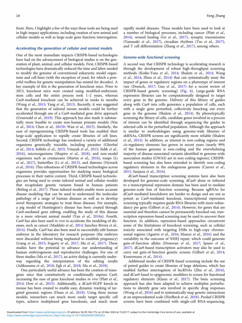
front. Here, I highlight a few of the ways these tools are being usedin high-impact applications, including creation of new animal andcellular models as well as large-scale gene function interrogation.
Accelerating the generation of cellular and animal models
One of the most immediate impacts CRISPR-based technologieshave had on the advancement of biological studies is on the gen-eration of plant, animal, and cellular models. First, CRISPR-basedtechnologies have dramatically reduced the time and labor neededto modify the genome of conventional eukaryotic model organ-isms and cell lines (with the exception of yeast, for which a pow-erful toolbox for genetic manipulation has existed for decades). Akey example of this is the generation of knockout mice. Prior to2013, knockout mice were created using modified-embryonicstem cells and the entire process took 1–2 years, whereasCas9-mediated knockout can be achieved in weeks to months(Wang et al., 2013; Yang et al., 2013). Recently, it was suggestedthat the generation of tailored mouse models could be furtheraccelerated through use of a Cas9-mediated gene drive approach(Grunwald et al., 2019). This approach has also made it substan-tially more feasible to create non-human primate models (Niuet al., 2014; Chen et al., 2015b; Wan et al., 2015). Similarly, theease of reprogramming CRISPR-based tools has enabled theirlarge-scale application to rapidly create libraries of cell lines.Second, CRISPR technology has rendered a variety of additionalorganisms genetically tractable, including parasites (Ghorbalet al., 2014; Sollelis et al., 2015; Vinayak et al., 2015; Sidik et al.,2016), microorganisms (Shapiro et al., 2018), and non-modelorganisms such as crustaceans (Martin et al., 2016), wasps (Liet al., 2017), butterflies (Li et al., 2015), and diatoms (Nymarket al., 2016). This robustness of CRISPR-based technologies acrossorganisms provides opportunities for studying many biologicalprocesses in their native context. Third, CRISPR-based technolo-gies are being used to create tailored animal and cellular modelsthat recapitulate genetic variants found in human patients(Birling et al., 2017). These tailored models enable more accuratedisease modeling that can be used to understand the molecularpathology of a range of human diseases as well as to developnovel therapeutic strategies to treat these diseases. For example,a pig model of Huntington’s disease has been created throughCas9-mediated gene editing, enabling the study of this diseasein a more relevant animal model (Yan et al., 2018a). Fourth,Cas9 has also been used in vivo to accelerate the modeling of dis-eases, such as cancer (Maddalo et al., 2014; Sanchez-Rivera et al.,2014). Finally, Cas9 has also been used to successfully edit humanembryos in the laboratory for research purposes (the embryoswere discarded without being implanted to establish pregnancy)(Liang et al., 2015; Fogarty et al., 2017; Ma et al., 2017). Thesestudies have the potential to advance our understanding ofhuman embryogenesis and reproductive challenges. For one ofthese studies (Ma et al., 2017), an active dialog is currently under-way regarding the interpretation of the editing results(Adikusuma et al., 2018; Egli et al., 2018; Ma et al., 2018).
One particularly useful advance has been the creation of trans-genic mice that constitutively or conditionally express Cas9,increasing the ease of gene knockout studies in vivo (Platt et al.,2014; Dow et al., 2015). Additionally, a dCas9-EGFP knock-inmouse has been created to enable easy, dynamic tracking of tar-geted genomic regions (Duan et al., 2018). Using these mousemodels, researchers can much more easily target specific celltypes, achieve multiplexed gene knockouts, and much more
rapidly model diseases. These models have been used to look ata number of biological processes, including cancer (Platt et al.,2014), wound healing (Ge et al., 2017), synaptic transmission(Yamasaki et al., 2017), circadian rhythms (Tso et al., 2017),and T cell differentiation (Zhang et al., 2017), among others.
Genome-wide functional screening
A second way that CRISPR technology is accelerating research isthrough the development of robust high-throughput screeningmethods (Koike-Yusa et al., 2014; Shalem et al., 2014; Wanget al., 2014; Zhou et al., 2014) that can systematically assay theimpact of genes or regulatory regions on a phenotype of interest(see (Doench, 2017; Guo et al., 2017) for a recent review ofCRISPR-based genetic screening) (Fig. 6). Large-guide RNAexpression libraries can be computationally designed to targetevery gene in the genome. Delivery of this library of guidesalong with Cas9 into cells generates a population of cells, eachwith a single gene perturbed, collectively knocking out everygene in the genome (Shalem et al., 2014). By phenotypicallyscreening the library of cells, candidate genes involved in a processof interest can be identified through sequencing the guides inselected cells in the perturbed population. Although the approachis similar to methodologies using genome-wide libraries ofshRNAs, CRISPR screens are significantly more reliable (Shalemet al., 2014). In addition, as interest in non-coding regions andcis-regulatory elements has grown in recent years (nearly 99%of the human genome is non-coding and the overwhelmingmajority of disease-associated variants identified in genome-wideassociation studies (GWAS) are in non-coding regions), CRISPR-based screening has also been extended to identify non-codingregulatory elements in the endogenous genome (Canver et al.,2015; Sanjana et al., 2016).
dCas9-based transcription screening systems have also beendeveloped for genome-scale screening. dCas9 alone or tetheredto a transcriptional repression domain has been used to mediategenome-scale loss of function screening. Because sgRNAs fordCas9-mediated knockdown are harder to design and are not aspotent as Cas9-mediated knockout, transcriptional repressionscreening typically requires guide RNA libraries with more redun-dancy per gene (Gilbert et al., 2014). However, for genes that areessential and therefore cannot be permanently knocked-out, tran-scription repression-based screening may be used to uncover theirfunction. In addition, repression-based approaches circumventsome of the limitations of knockout screening, such as cellulartoxicity associated with targeting DSBs to high-copy chromo-somal regions (Aguirre et al., 2016; Munoz et al., 2016) and thevariability in the outcome of NHEJ repair, which could generategain-of-function alleles (Donovan et al., 2017; Ipsaro et al.,2017). dCas9-based transcription activators may also be used tocarry out gain-of-function genetic screens (Gilbert et al., 2014;Konermann et al., 2014).
Additional modes of CRISPR-based screening include the useof paired guides to create libraries of large deletions, which hasenabled further interrogation of lncRNAs (Zhu et al., 2016),and dCas9 fused to epigenomic modifiers to screen for functionalregulatory elements (Klann et al., 2017). The basic screeningapproach has also been adapted to achieve multiplex perturba-tions to identify gene sets involved in specific drug responses(Wong et al., 2016) and to systematically map genetic interactionsat an unprecedented scale (Horlbeck et al., 2018). Pooled CRISPRscreens have been combined with single-cell RNA-sequencing,
14 Feng Zhang
https://www.cambridge.org/core/terms. https://doi.org/10.1017/S0033583519000052Downloaded from https://www.cambridge.org/core. IP address: 54.39.106.173, on 23 Mar 2020 at 15:20:17, subject to the Cambridge Core terms of use, available at
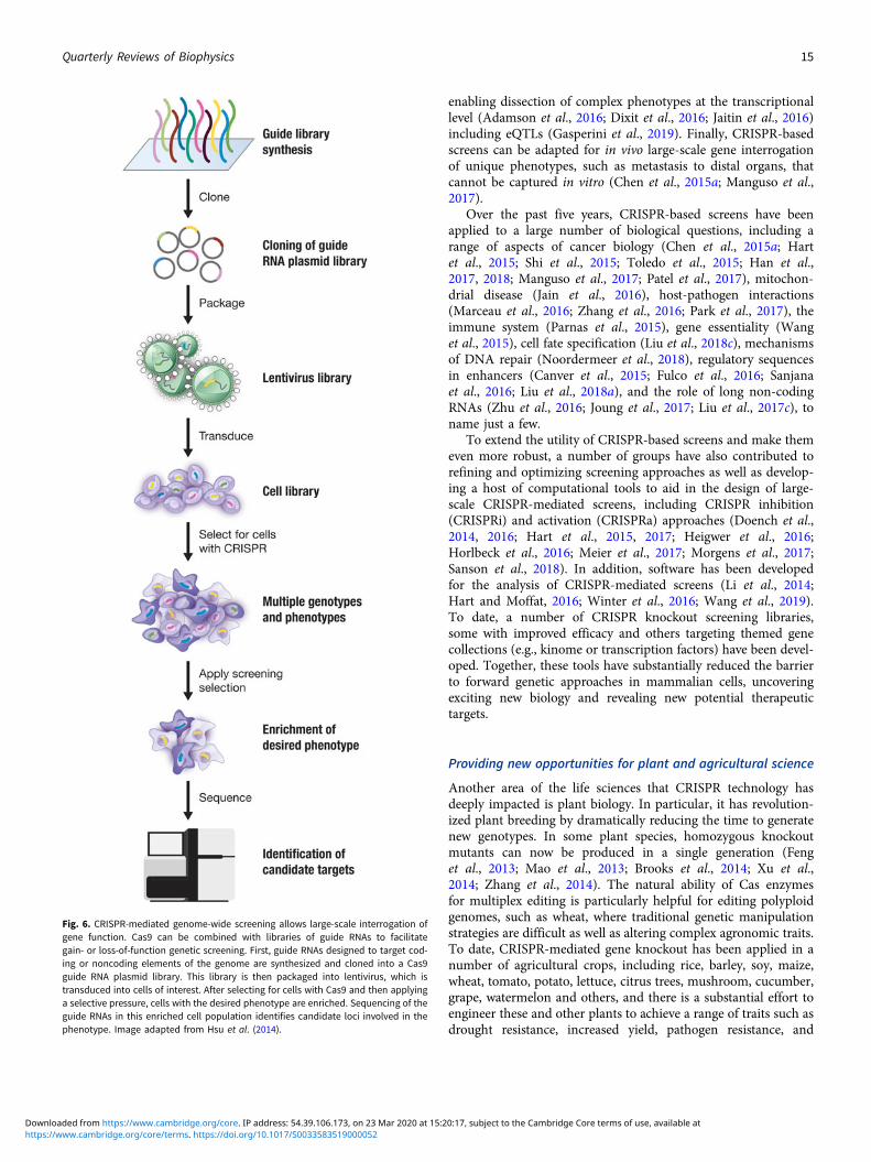
enabling dissection of complex phenotypes at the transcriptionallevel (Adamson et al., 2016; Dixit et al., 2016; Jaitin et al., 2016)including eQTLs (Gasperini et al., 2019). Finally, CRISPR-basedscreens can be adapted for in vivo large-scale gene interrogationof unique phenotypes, such as metastasis to distal organs, thatcannot be captured in vitro (Chen et al., 2015a; Manguso et al.,2017).
Over the past five years, CRISPR-based screens have beenapplied to a large number of biological questions, including arange of aspects of cancer biology (Chen et al., 2015a; Hartet al., 2015; Shi et al., 2015; Toledo et al., 2015; Han et al.,2017, 2018; Manguso et al., 2017; Patel et al., 2017), mitochon-drial disease (Jain et al., 2016), host-pathogen interactions(Marceau et al., 2016; Zhang et al., 2016; Park et al., 2017), theimmune system (Parnas et al., 2015), gene essentiality (Wanget al., 2015), cell fate specification (Liu et al., 2018c), mechanismsof DNA repair (Noordermeer et al., 2018), regulatory sequencesin enhancers (Canver et al., 2015; Fulco et al., 2016; Sanjanaet al., 2016; Liu et al., 2018a), and the role of long non-codingRNAs (Zhu et al., 2016; Joung et al., 2017; Liu et al., 2017c), toname just a few.
To extend the utility of CRISPR-based screens and make themeven more robust, a number of groups have also contributed torefining and optimizing screening approaches as well as develop-ing a host of computational tools to aid in the design of large-scale CRISPR-mediated screens, including CRISPR inhibition(CRISPRi) and activation (CRISPRa) approaches (Doench et al.,2014, 2016; Hart et al., 2015, 2017; Heigwer et al., 2016;Horlbeck et al., 2016; Meier et al., 2017; Morgens et al., 2017;Sanson et al., 2018). In addition, software has been developedfor the analysis of CRISPR-mediated screens (Li et al., 2014;Hart and Moffat, 2016; Winter et al., 2016; Wang et al., 2019).To date, a number of CRISPR knockout screening libraries,some with improved efficacy and others targeting themed genecollections (e.g., kinome or transcription factors) have been devel-oped. Together, these tools have substantially reduced the barrierto forward genetic approaches in mammalian cells, uncoveringexciting new biology and revealing new potential therapeutictargets.
Providing new opportunities for plant and agricultural science
Another area of the life sciences that CRISPR technology hasdeeply impacted is plant biology. In particular, it has revolution-ized plant breeding by dramatically reducing the time to generatenew genotypes. In some plant species, homozygous knockoutmutants can now be produced in a single generation (Fenget al., 2013; Mao et al., 2013; Brooks et al., 2014; Xu et al.,2014; Zhang et al., 2014). The natural ability of Cas enzymesfor multiplex editing is particularly helpful for editing polyploidgenomes, such as wheat, where traditional genetic manipulationstrategies are difficult as well as altering complex agronomic traits.To date, CRISPR-mediated gene knockout has been applied in anumber of agricultural crops, including rice, barley, soy, maize,wheat, tomato, potato, lettuce, citrus trees, mushroom, cucumber,grape, watermelon and others, and there is a substantial effort toengineer these and other plants to achieve a range of traits such asdrought resistance, increased yield, pathogen resistance, and
Fig. 6. CRISPR-mediated genome-wide screening allows large-scale interrogation ofgene function. Cas9 can be combined with libraries of guide RNAs to facilitategain- or loss-of-function genetic screening. First, guide RNAs designed to target cod-ing or noncoding elements of the genome are synthesized and cloned into a Cas9guide RNA plasmid library. This library is then packaged into lentivirus, which istransduced into cells of interest. After selecting for cells with Cas9 and then applyinga selective pressure, cells with the desired phenotype are enriched. Sequencing of theguide RNAs in this enriched cell population identifies candidate loci involved in thephenotype. Image adapted from Hsu et al. (2014).
Quarterly Reviews of Biophysics 15
https://www.cambridge.org/core/terms. https://doi.org/10.1017/S0033583519000052Downloaded from https://www.cambridge.org/core. IP address: 54.39.106.173, on 23 Mar 2020 at 15:20:17, subject to the Cambridge Core terms of use, available at

decreased time to ripening (Schindele et al., 2018). Cas-basedapproaches have also succeeded in plants that have traditionallybeen inaccessible to targeted gene changes, such as woody plants(Bewg et al., 2018). Methods are also being developed to achievetransgene-free gene editing in plants that rely on transformingplants with Cas9-guide RNA RNP complexes to avoid introducingforeign DNA (Woo et al., 2015; Liang et al., 2017)
Similar to the extension of Cas enzymes for many purposes inother systems, there is a growing toolbox of CRISPR-based technol-ogies tailored to plant biology. For example, Cas12a has proven to beparticularly effective in plants (Zaidi et al., 2017). Base editingapproaches have also been applied successfully in a number ofplant species including rice, maze, tomato, and wheat (Shimataniet al., 2017; Zong et al., 2017; Kang et al., 2018; Li et al., 2018a;Hua et al., 2019). Cas13a has been used for interference againstRNA viruses in tobacco, providing a new strategy for conferringimmunity (Aman et al., 2018). Together, this toolbox is advancingbasic plant biology studies and holds substantial potential to con-tribute to global food security without relying on transgenes. Todate, numerous crop strains have been generated throughCRISPR-mediated genome editing, including tomatoes with higheryield (Soyk et al., 2017), reduced-gluten wheat, virus-resistantcacao, caffeine-free coffee, and mushrooms that were engineeredto resist browning, which have received USDA approval (https://www.nature.com/news/gene-edited-crispr-mushroom-escapes-us-regulation-1·19754).
Advancing human health
The ability to precisely manipulate the genome, and in particularour ability to edit DNA and RNA, offers enormous potential forimproving human health by offering a platform that can be tai-lored to any of thousands of genetic disorders (reviewed in(Cox et al., 2015)). Achieving this potential, however, will requirea suite of highly specific and efficient Cas enzymes and a toolboxof delivery modalities that can be seamlessly combined to addressthe specific challenges of individual diseases. To date, the field hasproduced a spectacular diversity of editing tools, some of whichare now entering clinical trials. There are still outstanding chal-lenges in the development of CRISPR-based therapeutics, how-ever, notably delivery and potential immunogenicity. Below Idiscuss some of the ways CRISPR-based technologies are advanc-ing human health and highlight areas where additional research isneeded.
Applications for elimination of bacterial and viral pathogens
CRISPR-Cas systems have been applied to improve human healthin a range of ways, including the generation of new antibacterialagents. Several groups reported that CRISPR-Cas systems couldbe packaged in phages and used to selectively treat targeted bac-teria, generating programmable, sequence-specific antimicrobialagents (Bikard et al., 2014; Citorik et al., 2014). There have alsobeen multiple studies that leverage the natural function ofCRISPR-Cas systems to treat viral infections. For example, itwas shown that in a cellular model of HIV, Cas9 could be pro-grammed to target integrated copies of the HIV virome as wellas prevent HIV infection (Hu et al., 2014). With many viral infec-tions, the persistence of latent virus in the body represents amajor therapeutic challenge. Using a mouse model of HepatitisB virus (HBV) chronic infection, it was demonstrated that Cas9targeting the genome of HBV can reduce viral load (Ramanan
et al., 2015). Finally, CRISPR-based technologies are being usedto eliminate endogenous retroviruses in pigs to generate animalsthat may be suitable sources of organs for transplantation intohuman patients (Yang et al., 2015; Niu et al., 2017).
Applications for detection of bacterial and viral pathogens
Another way that CRISPR-Cas systems are advancing humanhealth is through the development of CRISPR-based diagnostics.Our discovery of the collateral RNase activity of Cas13 – uponbinding to the target RNA, Cas13a becomes activated as a non-specific RNase and can cleave the target RNA as well as otherRNA molecules in the vicinity that do not have complementaritywith the guide RNA target sequence – has made possible thedevelopment of a new approach for nucleic acid detection(Abudayyeh et al., 2016). With programmable target recognitiontriggering non-specific collateral cleavage of reporter molecules,these systems can be used to detect target molecules of interestat very low levels (Fig. 7). This technology has a range of applica-tions, notably in the detection and monitoring of infectious dis-eases in the field, such as Zika and Ebola, and in highlysensitive genotyping, such as the detection of cancer-associatedalleles in circulating DNA. Moreover, CRISPR diagnostics canbe used in agricultural and industrial settings to ensure food safetyand prevent the spread of contaminating agents.
A number of modalities can be integrated with collateral-cleavage of reporter RNA to provide diagnostic readout. Usinggel electrophoresis for visualization, diagnostic reporting can beachieved by assessing cleavage of a fluorescently tagged reporterRNA by Cas13 upon crRNA-guided recognition of target RNA(Abudayyeh et al., 2016). Using a fluorimeter for readout, detec-tion of target nucleic acid triggers Cas13 collateral activity andunlocks fluorescent reporters such as the commercially availableRNaseAlert by unleashing the fluorophore from its quencher toyield fluorescence. Direct application of Cas13 collateral RNaseactivity with RNaseAlert achieved detection sensitivity in thepicomolar range (∼ 1 000 000 molecules per test) (East-Seletskyet al., 2016). By integrating Cas13 collateral activity with isother-mal amplification, we developed a technique called SHERLOCK,which allowed for significantly increased detection sensitivity tothe attomolar range (Gootenberg et al., 2017), enabling clinicalapplications. Finally, using colorimetric paper test strips,Cas13-based diagnostics can be applied in low-resource settingsor in the field. To achieve this low-cost modality, we developeda lateral flow paper-based test, similar to a commonly-used preg-nancy test strip, by exploiting Cas13 collateral activity to cleaveand unlock two-different affinity molecules so that the presenceof the target can be read out as two-stained lines on the paperstrip (Gootenberg et al., 2018). Cas13-based diagnostics testscan also be lyophilized and easily reconstituted through the addi-tion of water, which makes it possible for SHERLOCK to be dis-tributed and stored without requiring refrigeration (Gootenberget al., 2017), which is especially important for application in low-resource settings in the developing world.
CRISPR-based detection technology is rapidly expanding.Recently, Cas12 was found to have natural ssDNA collateral activ-ity that is triggered by binding DNA (Chen et al., 2018; Li et al.,2018b). Cas12 can be similarly combined with isothermal ampli-fication in a technique called DETECTOR or integrated into theSHERLOCK platform for multiplex detection (Chen et al., 2018;Gootenberg et al., 2018; Li et al., 2018b). Different types of Casproteins with collateral activity can be combined to achieve
16 Feng Zhang
https://www.cambridge.org/core/terms. https://doi.org/10.1017/S0033583519000052Downloaded from https://www.cambridge.org/core. IP address: 54.39.106.173, on 23 Mar 2020 at 15:20:17, subject to the Cambridge Core terms of use, available at
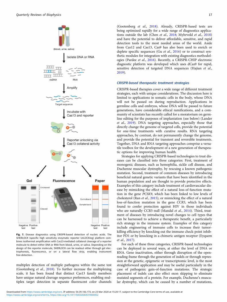
multiplex detection of multiple pathogens within the same test(Gootenberg et al., 2018). To further increase the multiplexingscale, it has been found that distinct Cas13 family membershave unique natural cleavage sequence preferences, enabling mul-tiplex target detection in separate fluorescent color channels
(Gootenberg et al., 2018). Already, CRISPR-based tests arebeing optimized rapidly for a wide range of diagnostics applica-tions outside the lab (Chen et al., 2018; Myhrvold et al., 2018)and have the potential to deliver affordable, sensitive, and rapiddetection tools to the most needed areas of the world. Asidefrom Cas12 and Cas13, Cas9 has also been used to enrich ordeplete specific sequences (Gu et al., 2016) or to construct syn-thetic modules for integration with existing diagnostics methodol-ogies (Pardee et al., 2016). Recently, a CRISPR-CHIP electronicdiagnostic platform was developed which uses dCas9 for rapid,sensitive detection of targeted DNA sequences (Hajian et al.,2019).
CRISPR-based therapeutic treatment strategies
CRISPR-based therapies cover a wide range of different treatmentstrategies, each with unique considerations. The discussion here islimited to applications in somatic cells in the body, whose DNAwill not be passed on during reproduction. Applications ingermline cells and embryos, whose DNA will be passed to futuregenerations, have considerable ethical ramifications, and a com-munity of scientists has recently called for a moratorium on germ-line editing for the purposes of implantation (see below) (Landeret al., 2019). DNA targeting approaches, especially those thatdirectly change the genome of targeted cells, provide the potentialfor one-time treatments with curative results. RNA targetingapproaches, by contrast, do not permanently change the genome,and provide the potential for transient and reversible treatments.Together, DNA and RNA targeting approaches comprise a versa-tile toolbox for the development of a new generation of therapeu-tic options for improving human health.
Strategies for applying CRISPR-based technologies to treat dis-eases can be classified into three categories: First, treatment ofmonogenic diseases, such as hemophilia, sickle cell disease, andDuchenne muscular dystrophy, by rescuing a known pathogenicmutation. Second, treatment of common diseases by introducingbeneficial natural genetic variants that have been identified in thehuman population and are thought to provide protective effects.Examples of this category include treatment of cardiovascular dis-ease by mimicking the effect of a natural loss-of-function muta-tion in the gene PCSK9, which has been linked to low levels ofcholesterol (Ran et al., 2015), or mimicking the effect of a naturalloss-of-function mutation in the gene CCR5, which has beenfound to confer protection against HIV in those individualswho are naturally CCR5-null (Mandal et al., 2014). Third, treat-ment of diseases by introducing novel changes to cell types thatcan be harnessed to achieve a therapeutic benefit, a particularlyrich strategy in the immune system. Examples of this categoryinclude engineering of immune cells to increase their tumor-killing efficiency by knocking out the immune check-point inhib-itor PD1 or by knocking in a chimeric antigen receptor (Eyquemet al., 2017).
For each of these three categories, CRISPR-based technologiescan be deployed in several ways, at either the level of DNA orRNA. Gene inactivation, either through disruption of the open-reading frame through the generation of indels or through repres-sion at the genetic, epigenetic or transcriptomic level, is the moststraightforward application and may be useful particularly in thecase of pathogenic gain-of-function mutations. The strategicplacement of indels can also effect exon skipping to eliminatemutated segments of a protein. For example, Duchenne’s muscu-lar dystrophy, which can be caused by a number of mutations,
Fig. 7. Disease diagnostics using CRISPR-based detection of nucleic acids. TheSHERLOCK (specific high sensitivity enzymatic reporter UnLOCKing) platform com-bines isothermal amplification with Cas13-mediated collateral cleavage of a reportermolecule to detect either DNA or RNA from blood, urine, or saliva. Depending on thedesign of the reporter molecule, SHERLOCK can be readout either through gel elec-trophoresis, fluorescence, or on a lateral flow strip, enabling instrumentfree-detection.
Quarterly Reviews of Biophysics 17
https://www.cambridge.org/core/terms. https://doi.org/10.1017/S0033583519000052Downloaded from https://www.cambridge.org/core. IP address: 54.39.106.173, on 23 Mar 2020 at 15:20:17, subject to the Cambridge Core terms of use, available at
including deletions and frame-shift mutations, can be treated withNHEJ-based exon skipping strategies, leading to restoration of thereading frame and a functional gene product (Long et al., 2016;Nelson et al., 2016; Tabebordbar et al., 2016). In other situations,gene upregulation using dCas-activation platforms can achieve atherapeutic effect. This approach is appealing for X-linked dom-inant diseases, such as Fragile X, where upregulating the silentwild-type copy of the gene could ameliorate the phenotype;proof-of-concept experiments have shown that a dCas9-Tetfusion can erase methylation at the inactivated FMR1 locus, lead-ing to reactivation (Liu et al., 2018b).
One particularly promising approach to treat a range ofpathologies is base editing at either the DNA or RNA level.Because base editing does not introduce DSBs, it offers a safer,less restricted route to correcting pathogenic mutations, themajority of which are single-nucleotide changes that either dis-rupt regulatory regions or result in truncated or abnormal proteinvariants. Several proof-of-concept studies have been performed inhuman cells and in mice demonstrating that base editing can leadto measurable expression levels of the corrected transcript (Reesand Liu, 2018). The ability to edit RNA opens additional possibil-ities for treatment, including transient alterations to the transcrip-tome. Such transient RNA editing would allow reversiblealteration of protein function, such as editing β-catenin to tempo-rarily alter cellular signaling pathways to drive cellular regenera-tion (Bastakoty and Young, 2016).
Although the most technically challenging, HR-mediatedapproaches to achieve targeted insertion of the desired-DNAsequence have the potential to correct the broadest swath of path-ogenic mutations. Currently, this approach is somewhat limited,although techniques for increasing the efficiency of gene insertioneither through homologous recombination or independent path-ways are continuing to be developed (Maruyama et al., 2015;Paquet et al., 2016; Richardson et al., 2016, 2018; Schmid-Burgket al., 2016; Suzuki et al., 2016; Kan et al., 2017; Canny et al.,2018). Gene insertion strategies may be most usefully deployedin disorders where a small increase in the corrected genotype islikely to have an outsized phenotypic impact, as may be thecase when edited cells have a selection or fitness advantage overnon-edited cells.
Any of the above CRISPR-based therapeutic strategies can beachieved either ex vivo or in vivo (Fig. 8). Ex vivo approachesoffer substantial advantages in terms of safety and efficiency ofediting but are limited to certain cell types that can be manipu-lated in the lab and subsequently engrafted, such as T cells(Roth et al., 2018), hematopoietic stem cells (Dever et al., 2016),and intestinal stem cell-derived organoids (Schwank et al.,2013). By contrast, in vivo approaches may be applicable to awider range of tissues, but the potential for off-targets, particu-larly if editing at the DNA level, is a safety concern. Currently,in vivo delivery modes for gene therapies typically rely on AAVvectors, which have been approved by the U.S. Food and DrugAdministration (FDA). Although promising, AAV vectors haverelatively limited cargo capacity, making it challenging to deliverSpCas9 and guide RNA effectively. Other Cas9 orthologs (SaCas9,CjCas9, and NmeCas9) are smaller than SpCas9, making thembetter suited for AAV delivery (Ran et al., 2015; Kim et al.,2017a; Ibraheim et al., 2018). Recently, high levels of AAV inte-gration into the genome have been reported, which may havelong-term safety implications (Nelson et al., 2019). Another chal-lenge with AAV or any other type of in vivo delivery modality,such as lipid nanoparticles (Finn et al., 2018), is achieving cell-
type specificity to ensure that only the pathological tissues are tar-geted. In vivo applications must also take into account potentialimmunogenicity of the therapy, which may be particularly rele-vant for SpCas9 and SaCas9, as they are derived from pathogenicbacteria (Charlesworth et al., 2019).
CRISPR in the clinic
Clinical trials to treat patients with CRISPR-based therapies are nowunderway to treat a handful of diseases. Currently, there are two setsof clinical trials entering phase 1 testing. The first set of trials are exvivo, using SpCas9 to treat β-thalassemia and sickle cell disease(Vertex, 2018a, 2018b). The second trial is in vivo, using SaCas9and delivery by AAV into the retina to treat Type 10 Leber congen-ital amaurosis (LCA10) (Allergan, 2019), which causes blindness.Pre-clinical studies have recently been published reporting the useof SaCas9 to correct a splice-site mutation causing LCA10, showingthat in human retinal explants, humanized mice, and non-humanprimates, editing rates exceed the threshold of 10% thought to beclinically relevant for disease amelioration (Maeder et al., 2019). Anumber of additional studies are developing Cas9, Cas12, andCas13-based strategies to treat a wide array of diseases includinggenetic disorders and cancer, providing new hope for patients cur-rently lacking treatment options.
Ethical considerations
In November 2018, it emerged that a scientist had reportedly usedCas9 to edit human embryos, creating at least two-geneticallymodified babies. This shocking news highlighted the far-reachingethical challenges that CRISPR-based technologies present forsociety. Although a number of stakeholders, including ethicists,scientists, clinicians, and policy makers, have voiced concernsover the use of CRISPR-based technologies in germline genomeediting (Baltimore et al., 2015), the exact nature of whether clin-ical uses of germline editing should be permitted remains a con-tentious topic. To advance discussions around this topic, a groupof specialists, myself included, from seven countries called for a5-year moratorium for the use of clinical germline editing, argu-ing that given a combination of scientific, technical, medical, andmoral considerations, society as a whole needs to wait and estab-lish consensus before proceeding with any form of clinical germ-line editing (Lander et al., 2019).
Indeed, this is an enormously complicated issue. Clinicalgermline editing applications can be divided into two types:genetic correction and genetic enhancement, as described above.With increasing knowledge about human genetic variation atthe population level, it is reasonable to expect that the outcomeof converting a rare disease-causing variant to a common variantthat does not lead to disease will be predictable and beneficial. Bycontrast, genetic enhancement relies on information about rarevariants in the human population, such as APOE-4 or CCR5null, which are much less well understood. For example, althoughloss of CCR5 appears to prevent HIV infection, it increases sus-ceptibility to West Nile virus, and the selective pressures on thisallele are not well understood (Telenti, 2009). Introducing suchchanges to the genome will likely have unpredictable conse-quences. Even in the future, when significant advances in ourunderstanding of biology and human genetic variation becomesufficient to predict the outcome of genome editing for enhance-ment, whether society as a whole should adopt germline editingstill needs to be vigorously debated. The moral quandaries are
18 Feng Zhang
https://www.cambridge.org/core/terms. https://doi.org/10.1017/S0033583519000052Downloaded from https://www.cambridge.org/core. IP address: 54.39.106.173, on 23 Mar 2020 at 15:20:17, subject to the Cambridge Core terms of use, available at
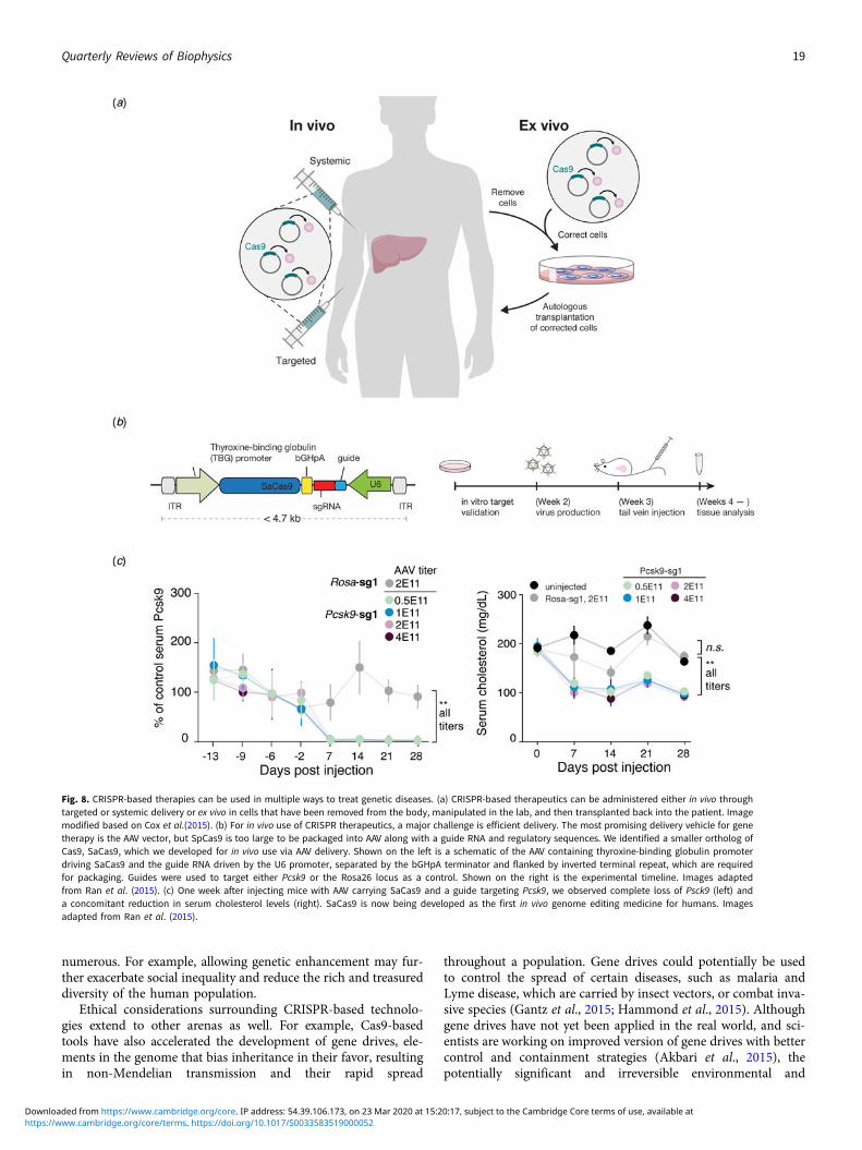
numerous. For example, allowing genetic enhancement may fur-ther exacerbate social inequality and reduce the rich and treasureddiversity of the human population.
Ethical considerations surrounding CRISPR-based technolo-gies extend to other arenas as well. For example, Cas9-basedtools have also accelerated the development of gene drives, ele-ments in the genome that bias inheritance in their favor, resultingin non-Mendelian transmission and their rapid spread
throughout a population. Gene drives could potentially be usedto control the spread of certain diseases, such as malaria andLyme disease, which are carried by insect vectors, or combat inva-sive species (Gantz et al., 2015; Hammond et al., 2015). Althoughgene drives have not yet been applied in the real world, and sci-entists are working on improved version of gene drives with bettercontrol and containment strategies (Akbari et al., 2015), thepotentially significant and irreversible environmental and
Fig. 8. CRISPR-based therapies can be used in multiple ways to treat genetic diseases. (a) CRISPR-based therapeutics can be administered either in vivo throughtargeted or systemic delivery or ex vivo in cells that have been removed from the body, manipulated in the lab, and then transplanted back into the patient. Imagemodified based on Cox et al.(2015). (b) For in vivo use of CRISPR therapeutics, a major challenge is efficient delivery. The most promising delivery vehicle for genetherapy is the AAV vector, but SpCas9 is too large to be packaged into AAV along with a guide RNA and regulatory sequences. We identified a smaller ortholog ofCas9, SaCas9, which we developed for in vivo use via AAV delivery. Shown on the left is a schematic of the AAV containing thyroxine-binding globulin promoterdriving SaCas9 and the guide RNA driven by the U6 promoter, separated by the bGHpA terminator and flanked by inverted terminal repeat, which are requiredfor packaging. Guides were used to target either Pcsk9 or the Rosa26 locus as a control. Shown on the right is the experimental timeline. Images adaptedfrom Ran et al. (2015). (c) One week after injecting mice with AAV carrying SaCas9 and a guide targeting Pcsk9, we observed complete loss of Psck9 (left) anda concomitant reduction in serum cholesterol levels (right). SaCas9 is now being developed as the first in vivo genome editing medicine for humans. Imagesadapted from Ran et al. (2015).
Quarterly Reviews of Biophysics 19
https://www.cambridge.org/core/terms. https://doi.org/10.1017/S0033583519000052Downloaded from https://www.cambridge.org/core. IP address: 54.39.106.173, on 23 Mar 2020 at 15:20:17, subject to the Cambridge Core terms of use, available at

ecological consequences of gene drives also demand careful con-sideration (Lunshof, 2015; Courtier-Orgogozo et al., 2017).
Looking ahead
The accumulation of genomic sequences, which initially poweredthe discovery of CRISPR systems, has continued apace (Fig. 9).The availability of microbial sequences, driven in large partthrough developments in metagenomics, is particularly compel-ling. Until quite recently, our sampling of microbial genomicswas severely taxonomically limited compared to the predicteddiversity of organisms. Even today, roughly 50% of the∼ 200000 available bacterial genomes encompass just 20 species, leavingout a vast swath of diversity. Concerted efforts, such as the EarthMicrobiome Project (earthmicrobiome.org), are underway to sys-tematically sample genomes across microbial taxa to gain a morecomprehensive understanding of prokaryotic natural diversity.
It is clear that we have only begun to scratch the surface of thefull microbial diversity. For example, the discovery of single-effector RNA targeting systems highlights the diversity withinCRISPR-Cas systems, which themselves are only a sliver of themicrobial defense systems that exist in nature. The continuedsearch for CRISPR effectors as well as the large diversity of auxiliaryproteins associated with CRISPR loci remain a rich source for explo-ration and development. Further revealing the sophistication of thearsenal used in microbial warfare, numerous anti-Cas9 andanti-Cas12 proteins (Pawluk et al., 2016; Rauch et al., 2017;Marino et al., 2018; Watters et al., 2018) as well as novel bacterialanti-phage defense systems (Doron et al., 2018) have recentlybeen discovered. These findings prefigure yet-to-be-discoveredadaptive immune systems hidden within the immense diversity ofthe microbial world. Hints that this may be the case lie in the diver-sity of immune systems in animals – while most vertebrates have anantibody-based adaptive immune system, immunity in even thejawless vertebrates is based on different distinct antibody-like pro-teins called variable lymphocyte receptors (VLRs) (Han et al.,2008). The existence of VLRs within a narrow branch of the animalkingdom suggests that perhaps within similarly narrow phyla of themicrobial diversity there may be unique adaptive immune systemsthat operate through distinct mechanisms. Perhaps some bacteriahave evolved protein-based adaptive immune systems that employpowerful diversification mechanisms to generate diverse proteinsthat provide critical survival functions for those microbes in theirnative environments. The technological potential of novel adaptiveimmune systems is also tantalizing – by virtue of being adaptive,these systems are naturally reprogrammable for the recognition ofdiverse substrates. Harnessing such reprogrammable systems couldlikely provide many new biotechnological platforms for the recogni-tion of proteins, metabolites, or patterns of glycosylation.
Fully understanding and harnessing this natural diversity willrequire solving a number of open challenges, both computationaland experimental. For example, further understanding of themolecular diversity of microbial species will require comprehen-sive genome sequencing and repeated sampling of diverse envi-ronments to capture the population dynamics in microbialcommunities. How do we deconvolute and assemble metage-nomic data to obtain more refined genetic sequence and informa-tion about these organisms? How do we more accurately predictthe function of novel protein sequences? Could protein structurehelp predict protein function? Perhaps advances in artificial intel-ligence may be applied to better infer the function of microbialproteins. Indeed, one of the key assumptions often used for the
study of bacterial protein sequences is the idea of ‘guilt by associ-ation’, where genes located within the same neighborhood oroperon are likely related to each other. This functional organiza-tion by neighborhood may perhaps suggest that there is a certainsyntax or grammar to the organization of bacterial genomes, andapproaches developed for natural language processing and deeplearning may be borrowed to make advances here.
There are also experimental challenges that need to be furtherresolved. How can we study microbes at scale without cultivatingthem? How do we cultivate those microbes that we cannot cur-rently culture so that we can study them comprehensively? Howdo we further accelerate some of the basic molecular biology tech-niques, like gene assembly, protein purification and structuredetermination? Solutions to some of these problems may arisethrough the work itself, much as CRISPR-based technologiesare now being used to manipulate microbial genomes in thelab, while some may come through the intersection of molecularbiology with other research fields, notably nanoscience and min-iaturization and parallelization.
Indeed, it often seems that some of the biggest leaps forward inscience are arrived at tangentially, and this should encourage all ofus to probe the literature of unrelated fields for information thatmay be applicable in new situations and dare to move our ownresearch programs not just forward, but also sideways. InFebruary 2011, through a serendipitous encounter, I learnedabout CRISPR, and my imagination was captured by this fascinat-ing and elegant mechanism of microbial adaptive immunity. Thispath has taken me from the development of CRISPR-Cas9 forgenome editing to the exploration of the expansive world ofCRISPR-Cas systems and microbial diversity. It has been particu-larly exciting to witness how this technology has flourishedthrough the contributions of so many talented scientists whoshare the same spirit of openness and generosity.
This is a time of plenty for curious biologists – we have onlyglimpsed a tiny sliver of the diversity of life at the molecularlevel, and every few months, new genetic ‘wonders’ are reported:plankton with shattered chromosomes (Blanc-Mathieu et al.,2017), lamprey genome rearrangements (Smith et al., 2018),phage-encoded diversity generating retroelements (Doulatov et al.,2004; Benler et al., 2018), and marine microorganisms that usenovel DNA repair systems (Deng et al., 2018). Each natural systemtempts us to explore new paths, determining the mechanism andfunction behind these systems, and opening new opportunities totinker, ultimately leading to a healthier and more sustainable future.
Fig. 9. The rapidly expanding database of microbial sequences holds many hiddentreasures. Exploration of the natural bacterial and archaeal diversity will undoubtedlyreveal new molecular systems that can provide the substrate for the development ofnew technologies. Data obtained from NCBI.
20 Feng Zhang
https://www.cambridge.org/core/terms. https://doi.org/10.1017/S0033583519000052Downloaded from https://www.cambridge.org/core. IP address: 54.39.106.173, on 23 Mar 2020 at 15:20:17, subject to the Cambridge Core terms of use, available at
Acknowledgements. I would like to thank my mentors, former and currentlab members, collaborators, and colleagues in the CRISPR field for helpful dis-cussions and inspiration. I would also like to thank the National Institutes ofHealth, National Science Foundation, Howard Hughes Medical Institute,New York Stem Cell Foundation, McKnight Foundation, Gates Foundation,Keck Foundation, Klingenstein Foundation, Searles Scholars Program,Damon Runyan Cancer Research Foundation, Vallee Foundation, MathersFoundation, Paul G. Allen Family Foundation, Simons Foundation, thePoitras Center for Affective Disorders, Merkin Foundation, HarvardNeurodiscovery Center, Jim and Patricia Poitras, Lisa Yang and Hock Tan,Bob Metcalfe, Tom Harriman, Jane Pauley, and David Cheng for their pastand current research funding. I am an inventor on a number of issued patentsand patent applications relating to CRISPR-based technologies. I am aco-founder and scientific advisor of Editas Medicine, Pairwise Plants,Sherlock Biosciences, Arbor Biotechnologies, and Beam Therapeutics.
References
Abudayyeh OO, Gootenberg JS, Konermann S, Joung J, Slaymaker IM,Cox DBT, Shmakov S, Makarova KS, Semenova E, Minakhin L,Severinov K, Regev A, Lander ES, Koonin EV and Zhang F (2016)C2c2 is a single-component programmable RNA-guided RNA-targetingCRISPR effector. Science 353, aaf5573.
Abudayyeh OO, Gootenberg JS, Essletzbichler P, Han S, Joung J,Belanto JJ, Verdine V, Cox DBT, Kellner MJ, Regev A, Lander ES,Voytas DF, Ting AY and Zhang F (2017) RNA targeting with CRISPR–Cas13. Nature 550, 280–284.
Adamson B, Norman TM, Jost M, Cho MY, Nuñez JK, Chen Y, Villalta JE,Gilbert LA, Horlbeck MA, Hein MY, Pak RA, Gray AN, Gross CA,Dixit A, Parnas O, Regev A and Weissman JS (2016) A multiplexedsingle-cell CRISPR screening platform enables systematic dissection of theunfolded protein response. Cell 167, 1867–1882.
Adikusuma F, Piltz S, Corbett MA, Turvey M, Mccoll SR, Helbig KJ,Beard MR, Hughes J, Pomerantz RT and Thomas PQ (2018) Large dele-tions induced by Cas9 cleavage. Nature 560, E8–E9.
Aguirre AJ, Meyers RM, Weir BA, Vazquez F, Zhang CZ, Ben-David U,Cook A, Ha G, Harrington WF, Doshi MB, Kost-Alimova M, Gill S,Xu H, Ali LD, Jiang G, Pantel S, Lee Y, Goodale A, Cherniack AD,Oh C, Kryukov G, Cowley GS, Garraway LA, Stegmaier K,Roberts CW, Golub TR, Meyerson M, Root DE, Tsherniak A andHahn WC (2016) Genomic copy number dictates a gene-independentcell response to CRISPR/Cas9 targeting. Cancer Discovery 6, 914–929.
Akbari OS, Bellen HJ, Bier E, Bullock SL, Burt A, Church GM, Cook KR,Duchek P, Edwards OR, Esvelt KM, Gantz VM, Golic KG, Gratz SJ,Harrison MM, Hayes KR, James AA, Kaufman TC, Knoblich J,Malik HS, Matthews KA, O’connor-Giles KM, Parks AL, Perrimon N,Port F, Russell S, Ueda R and Wildonger J (2015) BIOSAFETY.Safeguarding gene drive experiments in the laboratory. Science 349, 927–929.
Akcakaya P, Bobbin ML, Guo JA, Malagon-Lopez J, Clement K, Garcia SP,Fellows MD, Porritt MJ, Firth MA, Carreras A, Baccega T, Seeliger F,Bjursell M, Tsai SQ, Nguyen NT, Nitsch R, Mayr LM, Pinello L,Bohlooly YM, Aryee MJ, Maresca M and Joung JK (2018) In vivoCRISPR editing with no detectable genome-wide off-target mutations.Nature 561, 416–419.
ALLERGAN (2019) Single Ascending Dose Study in Participants WithLCA10. ClinicalTrial.gov Identifier: NCT03872479 (clinicaltrials.gov/ct2/show/NCT03872479).
Aman R, Ali Z, Butt H, Mahas A, Aljedaani F, Khan MZ, Ding S andMahfouz M (2018) RNA virus interference via CRISPR/Cas13a system inplants. Genome Biology 19, 1.
Anders C, Niewoehner O, Duerst A and Jinek M (2014) Structural basis ofPAM-dependent target DNA recognition by the Cas9 endonuclease. Nature513, 569–573.
Bae S, Park J and Kim JS (2014) Cas-OFFinder: a fast and versatile algorithmthat searches for potential off-target sites of Cas9 RNA-guided endonucle-ases. Bioinformatics 30, 1473–1475.
Baltimore D, Berg P, Botchan M, Carroll D, Charo RA, Church G, Corn JE,Daley GQ, Doudna JA, Fenner M, Greely HT, Jinek M, Martin GS,
Penhoet E, Puck J, Sternberg SH, Weissman JS and Yamamoto KR(2015) Biotechnology. A prudent path forward for genomic engineeringand germline gene modification. Science 348, 36–38.
Barrangou R and Horvath P (2017) A decade of discovery: CRISPR functionsand applications. Nature Microbiology 2, 17092.
Barrangou R, Fremaux C, Deveau H, Richards M, Boyaval P, Moineau S,Romero DA and Horvath P (2007) CRISPR provides acquired resistanceagainst viruses in prokaryotes. Science 315, 1709–1712.
Bastakoty D and Young PP (2016) Wnt/beta-catenin pathway in tissue injury:roles in pathology and therapeutic opportunities for regeneration. FASEBJournal 30, 3271–3284.
Benler S, Cobian-Guemes AG, Mcnair K, Hung SH, Levi K, Edwards R andRohwer F (2018) A diversity-generating retroelement encoded by a globallyubiquitous Bacteroides phage. Microbiome 6, 191.
Bewg WP, Ci D and Tsai CJ (2018) Genome editing in trees: from multiplerepair pathways to long-term stability. Frontiers in Plant Science 9, 1732.
Bikard D, Jiang W, Samai P, Hochschild A, Zhang F and Marraffini LA(2013) Programmable repression and activation of bacterial gene expressionusing an engineered CRISPR-Cas system. Nucleic Acids Research 41, 7429–7437.
Bikard D, Euler CW, Jiang W, Nussenzweig PM, Goldberg GW,Duportet X, Fischetti VA and Marraffini LA (2014) ExploitingCRISPR-Cas nucleases to produce sequence-specific antimicrobials.Nature Biotechnology 32, 1146–1150.
Birling MC, Herault Y and Pavlovic G (2017) Modeling human disease inrodents by CRISPR/Cas9 genome editing. Mamm Genome 28, 291–301.
Blanc-Mathieu R, Krasovec M, Hebrard M, Yau S, Desgranges E, Martin J,Schackwitz W, Kuo A, Salin G, Donnadieu C, Desdevises Y,Sanchez-Ferandin S, Moreau H, Rivals E, Grigoriev IV, Grimsley N,Eyre-Walker A and Piganeau G (2017) Population genomics of picophyto-plankton unveils novel chromosome hypervariability. Science Advances 3,e1700239.
Boch J, Scholze H, Schornack S, Landgraf A, Hahn S, Kay S, Lahaye T,Nickstadt A and Bonas U (2009) Breaking the code of DNA binding spe-cificity of TAL-type III effectors. Science 326, 1509–1512.
Bolotin A, Quinquis B, Sorokin A and Ehrlich SD (2005) Clustered regularlyinterspaced short palindrome repeats (CRISPRs) have spacers of extrachro-mosomal origin. Microbiology (Reading England) 151(Pt 8), 2551–2561.
Boyden ES, Zhang F, Bamberg E, Nagel G and Deisseroth K (2005)Millisecond-timescale, genetically targeted optical control of neural activity.Nature Neuroscience 8, 1263–1268.
Brooks C, Nekrasov V, Lippman ZB and Van Eck J (2014) Efficient geneediting in tomato in the first generation using the clustered regularly inter-spaced short palindromic repeats/CRISPR-associated9 system. PlantPhysiology 166, 1292–1297.
Brouns SJ, Jore MM, Lundgren M, Westra ER, Slijkhuis RJ, Snijders AP,Dickman MJ, Makarova KS, Koonin EV and Van Der Oost J (2008)Small CRISPR RNAs guide antiviral defense in prokaryotes. Science 321,960–964.
Burstein D, Harrington LB, Strutt SC, Probst AJ, Anantharaman K,Thomas BC, Doudna JA and Banfield JF (2017) New CRISPR-Cas sys-tems from uncultivated microbes. Nature 542, 237–241.
Cameron P, Fuller CK, Donohoue PD, Jones BN, Thompson MS,Carter MM, Gradia S, Vidal B, Garner E, Slorach EM, Lau E,Banh LM, Lied AM, Edwards LS, Settle AH, Capurso D, Llaca V,Deschamps S, Cigan M, Young JK and May AP (2017) Mappingthe genomic landscape of CRISPR-Cas9 cleavage. Nature Methods 14,600–606.
Canny MD, Moatti N, Wan LCK, Fradet-Turcotte A, Krasner D, Mateos-Gomez PA, Zimmermann M, Orthwein A, Juang YC, Zhang W,Noordermeer SM, Seclen E, Wilson MD, Vorobyov A, Munro M,Ernst A, Ng TF, Cho T, Cannon PM, Sidhu SS, Sicheri F andDurocher D (2018) Inhibition of 53BP1 favors homology-dependentDNA repair and increases CRISPR-Cas9 genome-editing efficiency.Nature Biotechnology 36, 95–102.
Canver MC, Smith EC, Sher F, Pinello L, Sanjana NE, Shalem O, Chen DD,Schupp PG, Vinjamur DS, Garcia SP, Luc S, Kurita R, Nakamura Y,Fujiwara Y, Maeda T, Yuan G-C, Zhang F, Orkin SH and Bauer DE
Quarterly Reviews of Biophysics 21
https://www.cambridge.org/core/terms. https://doi.org/10.1017/S0033583519000052Downloaded from https://www.cambridge.org/core. IP address: 54.39.106.173, on 23 Mar 2020 at 15:20:17, subject to the Cambridge Core terms of use, available at
(2015) BCL11A enhancer dissection by Cas9-mediated in situ saturatingmutagenesis. Nature 527, 192–197.
Carroll D (2012) A CRISPR approach to gene targeting. Molecular Therapy20, 1658–1660.
Casini A, Olivieri M, Petris G, Montagna C, Reginato G, Maule G,Lorenzin F, Prandi D, Romanel A, Demichelis F, Inga A andCereseto A (2018) A highly specific SpCas9 variant is identified by invivo screening in yeast. Nature Biotechnology 36, 265–271.
Charlesworth CT, Deshpande PS, Dever DP, Camarena J, Lemgart VT,Cromer MK, Vakulskas CA, Collingwood MA, Zhang L, Bode NM,Behlke MA, Dejene B, Cieniewicz B, Romano R, Lesch BJ, Gomez-Ospina N, Mantri S, Pavel-Dinu M, Weinberg KI and Porteus MH(2019) Identification of preexisting adaptive immunity to Cas9 proteinsin humans. Nature Medicine 25, 249–254.
Chatterjee P, Jakimo N and Jacobson JM (2018) Minimal PAM specificity ofa highly similar SpCas9 ortholog. Science Advances 4, eaau0766.
Chen B, Gilbert LA, Cimini BA, Schnitzbauer J, Zhang W, Li GW, Park J,Blackburn EH, Weissman JS, Qi LS and Huang B (2013) Dynamic imag-ing of genomic loci in living human cells by an optimized CRISPR/Cas sys-tem. Cell 155, 1479–1491.
Chen S, Sanjana NE, Zheng K, Shalem O, Lee K, Shi X, Scott DA, Song J,Pan JQ, Weissleder R, Lee H, Zhang F and Sharp PA (2015a)Genome-wide CRISPR screen in a mouse model of tumor growth andmetastasis. Cell 160, 1246–1260.
Chen Y, Zheng Y, Kang Y, Yang W, Niu Y, Guo X, Tu Z, Si C, Wang H,Xing R, Pu X, Yang SH, Li S, Ji W and Li XJ (2015b) Functional disrup-tion of the dystrophin gene in rhesus monkey using CRISPR/Cas9. HumanMolecular Genetics 24, 3764–3774.
Chen B, Hu J, Almeida R, Liu H, Balakrishnan S, Covill-Cooke C, Lim WAand Huang B (2016) Expanding the CRISPR imaging toolset withStaphylococcus aureus Cas9 for simultaneous imaging of multiple genomicloci. Nucleic Acids Research 44, e75–e75.
Chen JS, Dagdas YS, Kleinstiver BP, Welch MM, Sousa AA, Harrington LB,Sternberg SH, Joung JK, Yildiz A and Doudna JA (2017) Enhanced proof-reading governs CRISPR–Cas9 targeting accuracy. Nature 550, 407–410.
Chen JS, Ma E, Harrington LB, Da Costa M, Tian X, Palefsky JM andDoudna JA (2018) CRISPR-Cas12a target binding unleashes indiscriminatesingle-stranded DNase activity. Science 360, 436–439.
Cho SW, Kim S, Kim JM and Kim JS (2013) Targeted genome engineering inhuman cells with the Cas9 RNA-guided endonuclease. NatureBiotechnology 31, 230–232.
Cho SW, Kim S, Kim Y, Kweon J, Kim HS, Bae S and Kim JS (2014)Analysis of off-target effects of CRISPR/Cas-derived RNA-guided endonu-cleases and nickases. Genome Research 24, 132–141.
Chylinski K, Makarova KS, Charpentier E and Koonin EV (2014)Classification and evolution of type II CRISPR-Cas systems. Nucleic AcidsResearch 42, 6091–6105.
Citorik RJ, Mimee M and Lu TK (2014) Sequence-specific antimicrobialsusing efficiently delivered RNA-guided nucleases. Nature Biotechnology32, 1141–1145.
Concordet JP and Haeussler M (2018) CRISPOR: intuitive guide selection forCRISPR/Cas9 genome editing experiments and screens. Nucleic AcidsResearch 46, W242–W245.
Cong L, Ran FA, Cox D, Lin SL, Barretto R, Habib N, Hsu PD, Wu XB,Jiang WY, Marraffini LA and Zhang F (2013) Multiplex genome engineer-ing using CRISPR/Cas systems. Science 339, 819–823.
Courtier-Orgogozo V, Morizot B and Boete C (2017) Agricultural pest con-trol with CRISPR-based gene drive: time for public debate: should we usegene drive for pest control? EMBO Reports 18, 878–880.
Cox DB, Platt RJ and Zhang F (2015) Therapeutic genome editing: prospectsand challenges. Nature Medicine 21, 121–131.
Cox DBT, Gootenberg JS, Abudayyeh OO, Franklin B, Kellner MJ, Joung Jand Zhang F (2017) RNA editing with CRISPR-Cas13. Science 550,eaaq0180.
Cromwell CR, Sung K, Park J, Krysler AR, Jovel J, Kim SK and Hubbard BP(2018) Incorporation of bridged nucleic acids into CRISPR RNAs improvesCas9 endonuclease specificity. Nature Communications 9, 1448.
Crosetto N, Mitra A, Silva MJ, Bienko M, Dojer N, Wang Q, Karaca E,Chiarle R, Skrzypczak M, Ginalski K, Pasero P, Rowicka M andDikic I (2013) Nucleotide-resolution DNA double-strand break mappingby next-generation sequencing. Nature Methods 10, 361–366.
Dahlman JE, Abudayyeh OO, Joung J, Gootenberg JS, Zhang F andKonermann S (2015) Orthogonal gene knockout and activation with a cat-alytically active Cas9 nuclease. Nature Biotechnology 33, 1159–1161.
Davis KM, Pattanayak V, Thompson DB, Zuris JA and Liu DR (2015) Smallmolecule–triggered Cas9 protein with improved genome-editing specificity.Nature Chemical Biology 11, 316–318.
Deltcheva E, Chylinski K, Sharma CM, Gonzales K, Chao Y, Pirzada ZA,Eckert MR, Vogel J and Charpentier E (2011) CRISPR RNA maturationby trans-encoded small RNA and host factor RNase III.Nature 471, 602–607.
Deng W, Henriet S and Chourrout D (2018) Prevalence of mutation-pronemicrohomology-mediated end joining in a chordate lacking the c-NHEJDNA repair pathway. Current Biology 28, 3337–3341 e3334.
Deveau H, Barrangou R, Garneau JE, Labonte J, Fremaux C, Boyaval P,Romero DA, Horvath P and Moineau S (2008) Phage response toCRISPR-encoded resistance in Streptococcus thermophilus. Journal ofBacteriology 190, 1390–1400.
Dever DP, Bak RO, Reinisch A, Camarena J, Washington G, Nicolas CE,Pavel-Dinu M, Saxena N, Wilkens AB, Mantri S, Uchida N, Hendel A,Narla A, Majeti R, Weinberg KI and Porteus MH (2016) CRISPR/cas9beta-globin gene targeting in human haematopoietic stem cells. Nature539, 384–389.
Dicarlo JE, Norville JE, Mali P, Rios X, Aach J and Church GM (2013)Genome engineering in Saccharomyces cerevisiae using CRISPR-Cas sys-tems. Nucleic Acids Research 41, gkt135–gk4343.
Dixit A, Parnas O, Li B, Chen J, Fulco CP, Jerby-Arnon L, Marjanovic ND,Dionne D, Burks T, Raychowdhury R, Adamson B, Norman TM,Lander ES, Weissman JS, Friedman N and Regev A (2016) Perturb-Seq:dissecting molecular circuits with scalable single-cell RNA profiling ofpooled genetic screens. Cell 167, 1853–1866.
Doench JG (2017) Am I ready for CRISPR? A user’s guide to genetic screens.Nature Publishing Group 19, 67–80.
Doench JG, Hartenian E, Graham DB, Tothova Z, Hegde M, Smith I,Sullender M, Ebert BL, Xavier RJ and Root DE (2014) Rational designof highly active sgRNAs for CRISPR-Cas9-mediated gene inactivation.Nature Biotechnology 32, 1262–1267.
Doench JG, Fusi N, Sullender M, Hegde M, Vaimberg EW, Donovan KF,Smith I, Tothova Z, Wilen C, Orchard R, Virgin HW, Listgarten J andRoot DE (2016) Optimized sgRNA design to maximize activity andminimize off-target effects of CRISPR-Cas9. Nature Biotechnology 34,184–191.
Doetschman T, Gregg RG, Maeda N, Hooper ML, Melton DW,Thompson S and Smithies O (1987) Targetted correction of a mutantHPRT gene in mouse embryonic stem cells. Nature 330, 576–578.
Dong D, Ren K, Qiu X, Zheng J, Guo M, Guan X, Liu H, Li N, Zhang B,Yang D, Ma C, Wang S, Wu D, Ma Y, Fan S, Wang J, Gao N andHuang Z (2016) The crystal structure of Cpf1 in complex with CRISPRRNA. Nature 532, 522–526.
Donovan KF, Hegde M, Sullender M, Vaimberg EW, Johannessen CM,Root DE and Doench JG (2017) Creation of novel protein variants withCRISPR/Cas9-mediated mutagenesis: turning a screening by-product intoa discovery tool. PLoS One 12, e0170445.
Doron S, Melamed S, Ofir G, Leavitt A, Lopatina A, Keren M, Amitai Gand Sorek R (2018) Systematic discovery of antiphage defense systems inthe microbial pangenome. Science 359, eaar4120.
Doudna JA and Charpentier E (2014) Genome editing. The new frontier ofgenome engineering with CRISPR-Cas9. Science 346, 1258096.
Doulatov S, Hodes A, Dai L, Mandhana N, Liu M, Deora R, Simons RW,Zimmerly S and Miller JF (2004) Tropism switching in Bordetella bacter-iophage defines a family of diversity-generating retroelements. Nature 431,476–481.
Dow LE, Fisher J, O’rourke KP, Muley A, Kastenhuber ER, Livshits G,Tschaharganeh DF, Socci ND and Lowe SW (2015) Inducible in vivogenome editing with CRISPR-Cas9. Nature Biotechnology 33, 390–394.
22 Feng Zhang
https://www.cambridge.org/core/terms. https://doi.org/10.1017/S0033583519000052Downloaded from https://www.cambridge.org/core. IP address: 54.39.106.173, on 23 Mar 2020 at 15:20:17, subject to the Cambridge Core terms of use, available at
Duan J, Lu G, Hong Y, Hu Q, Mai X, Guo J, Si X, Wang F and Zhang Y(2018) Live imaging and tracking of genome regions in CRISPR/dCas9knock-in mice. Genome Biology 19, 192.
East-Seletsky A, O’connell MR, Knight SC, Burstein D, Cate JHD, Tjian Rand Doudna JA (2016) Two distinct RNase activities of CRISPR-C2c2enable guide-RNA processing and RNA detection. Nature 538, 270–273.
Egli D, Zuccaro MV, Kosicki M, Church GM, Bradley A and Jasin M (2018)Inter-homologue repair in fertilized human eggs? Nature 560, E5–E7.
Esvelt KM, Mali P, Braff JL, Moosburner M, Yaung SJ and Church GM(2013) Orthogonal Cas9 proteins for RNA-guided gene regulation and edit-ing. Nature 10, 1116–1121.
Eyquem J, Mansilla-Soto J, Giavridis T, Van Der Stegen SJ, Hamieh M,Cunanan KM, Odak A, Gonen M and Sadelain M (2017) Targeting aCAR to the TRAC locus with CRISPR/Cas9 enhances tumour rejection.Nature 543, 113–117.
Fellmann C, Gowen BG, Lin PC, Doudna JA and Corn JE (2017)Cornerstones of CRISPR-Cas in drug discovery and therapy. NatureReviews Drug Discovery 16, 89–100.
Feng Z, Zhang B, Ding W, Liu X, Yang DL, Wei P, Cao F, Zhu S, Zhang F,Mao Y and Zhu JK (2013) Efficient genome editing in plants using aCRISPR/Cas system. Cell Research 23, 1229–1232.
Finn JD, Smith AR, Patel MC, Shaw L, Youniss MR, Van Heteren J,Dirstine T, Ciullo C, Lescarbeau R, Seitzer J, Shah RR, Shah A, Ling D,Growe J, Pink M, Rohde E, Wood KM, Salomon WE, Harrington WF,Dombrowski C, Strapps WR, Chang Y and Morrissey DV (2018) A singleadministration of CRISPR/Cas9 lipid nanoparticles achieves robust andpersistentiIn vivo genome editing. Cell Reports 22, 2227–2235.
Fogarty NME, Mccarthy A, Snijders KE, Powell BE, Kubikova N,Blakeley P, Lea R, Elder K, Wamaitha SE, Kim D, Maciulyte V,Kleinjung J, Kim JS, Wells D, Vallier L, Bertero A, Turner JMA andNiakan KK (2017) Genome editing reveals a role for OCT4 in humanembryogenesis. Nature 550, 67–73.
Fonfara I, Le Rhun A, Chylinski K, Makarova KS, Lécrivain A-L,Bzdrenga J, Koonin EV and Charpentier E (2014) Phylogeny of Cas9determines functional exchangeability of dual-RNA and Cas9 amongorthologous type II CRISPR-Cas systems. Nucleic Acids Research 42,2577–2590.
Fonfara I, Richter H, Bratovič M, Le Rhun A and Charpentier E (2016) TheCRISPR-associated DNA-cleaving enzyme Cpf1 also processes precursorCRISPR RNA. Nature 532, 517–521.
Frieda KL, Linton JM, Hormoz S, Choi J, Chow KK, Singer ZS, Budde MW,Elowitz MB and Cai L (2017) Synthetic recording and in situ readout oflineage information in single cells. Nature 541, 107–111.
Friedland AE, Tzur YB, Esvelt KM, Colaiácovo MP, Church GM andCalarco JA (2013) Heritable genome editing in C. elegans via aCRISPR-Cas9 system. Nature Methods 10, 741–743.
Frock RL, Hu J, Meyers RM, Ho YJ, Kii E and Alt FW (2015) Genome-widedetection of DNA double-stranded breaks induced by engineered nucleases.Nature Biotechnology 33, 179–186.
Fulco CP, Munschauer M, Anyoha R, Munson G, Grossman SR, Perez EM,Kane M, Cleary B, Lander ES and Engreitz JM (2016) Systematic mappingof functional enhancer-promoter connections with CRISPR interference.Science 354, 769–773.
Fu Y, Foden JA, Khayter C, Maeder ML, Reyon D, Joung JK and Sander JD(2013) High-frequency off-target mutagenesis induced by CRISPR-Casnucleases in human cells. Nature Biotechnology 31, 822–826.
Fu Y, Sander JD, Reyon D, Cascio VM and Joung JK (2014) ImprovingCRISPR-Cas nuclease specificity using truncated guide RNAs. NatureBiotechnology 32, 279–284.
Gantz VM, Jasinskiene N, Tatarenkova O, Fazekas A, Macias VM, Bier Eand James AA (2015) Highly efficient Cas9-mediated gene drive for pop-ulation modification of the malaria vector mosquito Anopheles stephensi.Proceedings of the National Academy of Sciences 112, E6736–E6743.
Gao C (2018) The future of CRISPR technologies in agriculture. NatureReviews Molecular Cell Biology 19, 275–276.
Gao L, Cox DBT, Yan WX, Manteiga JC, Schneider MW, Yamano T,Nishimasu H, Nureki O, Crosetto N and Zhang F (2017) EngineeredCpf1 variants with altered PAM specificities. Nature Biotechnology 163, 759.
Garcia-Doval C and Jinek M (2017) Molecular architectures and mechanismsof class 2 CRISPR-associated nucleases. Current Opinion in StructuralBiology 47, 157–166.
Garneau JE, Dupuis ME, Villion M, Romero DA, Barrangou R, Boyaval P,Fremaux C, Horvath P, Magadan AH and Moineau S (2010) TheCRISPR/Cas bacterial immune system cleaves bacteriophage and plasmidDNA. Nature 468, 67–71.
Gasiunas G, Barrangou R, Horvath P and Siksnys V (2012) Cas9-crRNAribonucleoprotein complex mediates specific DNA cleavage for adaptiveimmunity in bacteria. Proceedings of the National Academy of Sciences ofthe United States of America 109, E2579–E2586.
Gasperini M, Hill AJ, Mcfaline-Figueroa JL, Martin B, Kim S, Zhang MD,Jackson D, Leith A, Schreiber J, Noble WS, Trapnell C, Ahituv N andShendure J (2019) A genome-wide framework for mapping gene regulationvia cellular genetic screens. Cell 176, 377–390.
Gaudelli NM, Komor AC, Rees HA, Packer MS, Badran AH, Bryson DI andLiu DR (2017) Programmable base editing of A•T to G•C in genomic DNAwithout DNA cleavage. Nature 551, 464–471.
Ge Y, Gomez NC, Adam RC, Nikolova M, Yang H, Verma A, Lu CP,Polak L, Yuan S, Elemento O and Fuchs E (2017) Stem cell lineage infi-delity drives wound repair and cancer. Cell 169, 636–650.
Ghorbal M, Gorman M, Macpherson CR, Martins RM, Scherf A andLopez-Rubio J-J (2014) Genome editing in the humanmalaria parasite plas-modium falciparum using the CRISPR-Cas9 system. Nature Biotechnology32, 819–821.
Giannoukos G, CiullaDM,MarcoE,AbdulkerimHS,Barrera LA, BothmerA,Dhanapal V, Gloskowski SW, Jayaram H, Maeder ML, Skor MN, Wang T,Myer VE and Wilson CJ (2018) UDitas, a genome editing detection methodfor indels and genome rearrangements. BMC Genomics 19, 212.
Gilbert LA, Larson MH, Morsut L, Liu Z, Brar GA, Torres SE, Stern-Ginossar N, Brandman O, Whitehead EH, Doudna JA, Lim WA,Weissman JS and Qi LS (2013) CRISPR-mediated modular RNA-guidedregulation of transcription in eukaryotes. Cell 154, 442–451.
Gilbert LA, Horlbeck MA, Adamson B, Villalta JE, Chen Y, Whitehead EH,Guimaraes C, Panning B, Ploegh HL, Bassik MC, Qi LS, Kampmann Mand Weissman JS (2014) Genome-Scale CRISPR-mediated control of generepression and activation. Cell 159, 647–661.
Gootenberg JS, Abudayyeh OO, Lee JW, Essletzbichler P, Dy AJ, Joung J,Verdine V, Donghia N, Daringer NM, Freije CA, Myhrvold C,Bhattacharyya RP, Livny J, Regev A, Koonin EV, Hung DT, Sabeti PC,Collins JJ and Zhang F (2017) Nucleic acid detection with CRISPR-Cas13a/C2c2. Science 356, 438–442.
Gootenberg JS, Abudayyeh OO, Kellner MJ, Joung J, Collins JJ andZhang F (2018) Multiplexed and portable nucleic acid detection platformwith Cas13, Cas12a, and Csm6. Science 360, 439–444.
Gratz SJ, Cummings AM, Nguyen JN, Hamm DC, Donohue LK,Harrison MM, Wildonger J and O’connor-Giles KM (2013) Genomeengineering of drosophila with the CRISPR RNA-guided Cas9 nuclease.Genetics 194, 1029–1035.
Grunewald J, Zhou R, Garcia SP, Iyer S, Lareau CA, Aryee MJ and Joung JK(2019) Transcriptome-wide off-target RNA editing induced byCRISPR-guided DNA base editors. Nature 569, 433–437.
Grunwald HA, Gantz VM, Poplawski G, Xu XS, Bier E and Cooper KL(2019) Super-Mendelian inheritance mediated by CRISPR-Cas9 in thefemale mouse germline. Nature 566, 105–109.
Guilinger JP, Thompson DB and Liu DR (2014) Fusion of catalytically inac-tive Cas9 to FokI nuclease improves the specificity of genome modification.Nature Biotechnology 32, 577–582.
Guo X, Chitale P and Sanjana NE (2017) Target discovery for precisionmedicine using high-throughput genome engineering. Advances inExperimental Medicine and Biology 1016, 123–145.
Gu W,Crawford ED,O’donovan BD,WilsonMR, ChowED, RetallackH andDerisi JL (2016) Depletion of abundant sequences by hybridization (DASH):usingCas9 to remove unwanted high-abundance species in sequencing librar-ies and molecular counting applications. Genome Biology 17, 41.
Hajian R, Balderston S, Tran T, Deboer T, Etienne J, Sandhu M,Wauford NA, Chung J-Y, Nokes J, Athaiya M, Paredes J, Peytavi R,Goldsmith B, Murthy N, Conboy IM and Aran K (2019) Detection of
Quarterly Reviews of Biophysics 23
https://www.cambridge.org/core/terms. https://doi.org/10.1017/S0033583519000052Downloaded from https://www.cambridge.org/core. IP address: 54.39.106.173, on 23 Mar 2020 at 15:20:17, subject to the Cambridge Core terms of use, available at
unamplified target genes via CRISPR–Cas9 immobilized on a graphenefield-effect transistor. Nature Biomedical Engineering. http://doi.org/10.1038/s41551-019-0371-x.
Hale CR, Zhao P, Olson S, Duff MO, Graveley BR, Wells L, Terns RM andTerns MP (2009) RNA-guided RNA cleavage by a CRISPR RNA-Cas pro-tein complex. Cell 139, 945–956.
Halperin SO, Tou CJ, Wong EB, Modavi C, Schaffer DV and Dueber JE(2018) CRISPR-guided DNA polymerases enable diversification of allnucleotides in a tunable window. Nature 560, 248–252.
Hammond A, Galizi R, Kyrou K, Simoni A, Siniscalchi C, Katsanos D,Gribble M, Baker D, Marois E, Russell S, Burt A, Windbichler N,Crisanti A and Nolan T (2015) A CRISPR-Cas9 gene drive system target-ing female reproduction in the malaria mosquito vector Anopheles gam-biae. Nature Biotechnology 34, 78–83.
Han BW, Herrin BR, Cooper MD and Wilson IA (2008) Antigen recognitionby variable lymphocyte receptors. Science 321, 1834–1837.
Han K, Jeng EE, Hess GT, Morgens DW, Li A and Bassik MC (2017)Synergistic drug combinations for cancer identified in a CRISPR screenfor pairwise genetic interactions. Nature Biotechnology 35, 463–474.
Han R, Li L, Ugalde AP, Tal A, Manber Z, Barbera EP, Chiara VD, Elkon Rand Agami R (2018) Functional CRISPR screen identifies AP1-associatedenhancer regulating FOXF1 to modulate oncogene-induced senescence.Genome Biology 19, 118.
Harrington LB, Burstein D, Chen JS, Paez-Espino D, Ma E, Witte IP,Cofsky JC, Kyrpides NC, Banfield JF and Doudna JA (2018)Programmed DNA destruction by miniature CRISPR-Cas14 enzymes.Science 362, 839–842.
Hart T and Moffat J (2016) BAGEL: a computational framework for identifyingessential genes from pooled library screens. BMC Bioinformatics 17, 164.
Hart T, Chandrashekhar M, Aregger M, Steinhart Z, Brown KR,Macleod G, Mis M, Zimmermann M, Fradet-Turcotte A, Sun S,Mero P, Dirks P, Sidhu S, Roth FP, Rissland OS, Durocher D,Angers S and Moffat J (2015) High-resolution CRISPR screens reveal fit-ness genes and genotype-specific cancer liabilities. Cell 163, 1515–1526.
Hart T, Tong AHY, Chan K, Van Leeuwen J, Seetharaman A, Aregger M,Chandrashekhar M, Hustedt N, Seth S, Noonan A, Habsid A,Sizova O, Nedyalkova L, Climie R, Tworzyanski L, Lawson K,Sartori MA, Alibeh S, Tieu D, Masud S, Mero P, Weiss A, Brown KR,Usaj M, Billmann M, Rahman M, Constanzo M, Myers CL,Andrews BJ, Boone C, Durocher D and Moffat J (2017) Evaluation anddesign of genome-wide CRISPR/SpCas9 knockout screens. G3 (Bethesda)7, 2719–2727.
Heigwer F, Zhan T, Breinig M, Winter J, Brugemann D, Leible S andBoutros M (2016) CRISPR library designer (CLD): software for multispe-cies design of single guide RNA libraries. Genome Biology 17, 55.
Heler R, Samai P, Modell JW, Weiner C, Goldberg GW, Bikard D andMarraffini LA (2015) Cas9 specifies functional viral targets duringCRISPR-Cas adaptation. Nature 519, 199–202.
Hess GT, Fresard L, Han K, Lee CH, Li A, Cimprich KA, Montgomery SBand Bassik MC (2016) Directed evolution using dCas9-targeted somatichypermutation in mammalian cells. Nature Methods 13, 1036–1042.
Hess GT, Tycko J, Yao D and Bassik MC (2017) Methods and applications ofCRISPR-mediated base editing in eukaryotic genomes. Molecular Cell 68,26–43.
Hilton IB, D’ippolito AM, Vockley CM, Thakore PI, Crawford GE,Reddy TE and Gersbach CA (2015) Epigenome editing by a CRISPR-Cas9-based acetyltransferase activates genes from promoters and enhancers.Nature Biotechnology 33, 510–517.
Hirano H, Gootenberg JS, Horii T, Abudayyeh OO, Kimura M, Hsu PD,Nakane T, Ishitani R, Hatada I, Zhang F, Nishimasu H and Nureki O(2016) Structure and engineering of Francisella novicida Cas9. Cell 164,950–961.
Horlbeck MA, Gilbert LA, Villalta JE, Adamson B, Pak RA, Chen Y,Fields AP, Park CY, Corn JE, Kampmann M and Weissman JS (2016)Compact and highly active next-generation libraries for CRISPR-mediatedgene repression and activation. Elife 5, e19760.
Horlbeck MA, Xu A, Wang M, Bennett NK, Park CY, Bogdanoff D,Adamson B, Chow ED, Kampmann M, Peterson TR, Nakamura K,
Fischbach MA, Weissman JS and Gilbert LA (2018) Mapping the geneticlandscape of human cells. Cell 174, 953–967.
Horvath P, Romero DA, Coute-Monvoisin AC, Richards M, Deveau H,Moineau S, Boyaval P, Fremaux C and Barrangou R (2008) Diversity,activity, and evolution of CRISPR loci in Streptococcus thermophilus.Journal of Bacteriology 190, 1401–1412.
Horvath P, Coute-Monvoisin AC, Romero DA, Boyaval P, Fremaux C andBarrangou R (2009) Comparative analysis of CRISPR loci in lactic acid bac-teria genomes. International Journal of Food Microbiology 131, 62–70.
Hotta A and Yamanaka S (2015) From genomics to gene therapy: inducedpluripotent stem cells meet genome editing. Annual Review of Genetics49, 47–70.
Hou Z, Zhang Y, Propson NE, Howden SE, Chu LF, Sontheimer EJ andThomson JA (2013) Efficient genome engineering in human pluripotentstem cells using Cas9 from Neisseria meningitidis. Proceedings of theNational Academy of Sciences of the United States of America 110,15644–15649.
Hsu PD, Scott DA, Weinstein JA, Ran FA, Konermann S, Agarwala V, Li Y,Fine EJ, Wu X, Shalem O, Cradick TJ, Marraffini LA, Bao G andZhang F (2013) DNA targeting specificity of RNA-guided Cas9 nucleases.Nature Biotechnology 31, 827–832.
Hsu PD, Lander ES and Zhang F (2014) Development and applications ofCRISPR-Cas9 for genome engineering. Cell 157, 1262–1278.
Hua K, Tao X and Zhu JK (2019) Expanding the base editing scope in rice byusing Cas9 variants. Plant Biotechnology Journal 17, 499–504.
Hu W, Kaminski R, Yang F, Zhang Y, Cosentino L, Li F, Luo B, Alvarez-Carbonell D, Garcia-Mesa Y, Karn J, Mo X and Khalili K (2014)RNA-directed gene editing specifically eradicates latent and prevents newHIV-1 infection. Proceedings of the National Academy of Sciences of theUnited States of America 111, 11461–11466.
Hu JH, Miller SM, Geurts MH, Tang W, Chen L, Sun N, Zeina CM, Gao X,Rees HA, Lin Z and Liu DR (2018) Evolved Cas9 variants with broad PAMcompatibility and high DNA specificity. Nature 556, 57–63.
Hwang WY, Fu Y, Reyon D, Maeder ML, Tsai SQ, Sander JD, Peterson RT,Yeh JR and Joung JK (2013) Efficient genome editing in zebrafish using aCRISPR-Cas system. Nature Biotechnology 31, 227–229.
Ibraheim R, Song CQ, Mir A, Amrani N, Xue W and Sontheimer EJ (2018)All-in-one adeno-associated virus delivery and genome editing by Neisseriameningitidis Cas9 in vivo. Genome Biology 19, 137.
Ipsaro JJ, Shen C, Arai E, Xu Y, Kinney JB, Joshua-Tor L, Vakoc CR andShi J (2017) Rapid generation of drug-resistance alleles at endogenousloci using CRISPR-Cas9 indel mutagenesis. PLoS One 12, e0172177.
Ishino Y, Shinagawa H, Makino K, Amemura M and Nakata A (1987)Nucleotide sequence of the iap gene, responsible for alkaline phosphataseisozyme conversion in Escherichia coli, and identification of the gene prod-uct. Journal of Bacteriology 169, 5429–5433.
Ishino Y, Krupovic M and Forterre P (2018) History of CRISPR-Cas fromencounter with a mysterious repeated sequence to genome editing technol-ogy. Journal of Bacteriology 200, e00580.
Jackson RN and Wiedenheft B (2015) A conserved structural chassis formounting Versatile CRISPR RNA-guided immune responses. MolecularCell 58, 722–728.
Jacob F (1977) Evolution and tinkering. Science 196, 1161–1166.Jain IH, Zazzeron L, Goli R, Alexa K, Schatzman-Bone S, Dhillon H,
Goldberger O, Peng J, Shalem O, Sanjana NE, Zhang F, Goessling W,Zapol WM and Mootha VK (2016) Hypoxia as a therapy for mitochon-drial disease. Science 352, 54–61.
Jaitin DA, Weiner A, Yofe I, Lara-Astiaso D, Keren-Shaul H, David E,Salame TM, Tanay A, Van Oudenaarden A and Amit I (2016)Dissecting immune circuits by linking CRISPR-pooled screens with single-cell RNA-Seq. Cell 167, 1883–1896.
Jansen R, Embden JD, Gaastra W and Schouls LM (2002) Identification ofgenes that are associated with DNA repeats in prokaryotes. MolecularMicrobiology 43, 1565–1575.
Jiang F and Doudna JA (2017) CRISPR-Cas9 Structures and mechanisms.Annual Review of Biophysics 46, 505–529.
Jiang W, Zhou H, Bi H, Fromm M, Yang B and Weeks DP (2013)Demonstration of CRISPR/Cas9/sgRNA-mediated targeted gene
24 Feng Zhang
https://www.cambridge.org/core/terms. https://doi.org/10.1017/S0033583519000052Downloaded from https://www.cambridge.org/core. IP address: 54.39.106.173, on 23 Mar 2020 at 15:20:17, subject to the Cambridge Core terms of use, available at
modification in Arabidopsis, tobacco, sorghum and rice. Nucleic AcidsResearch 41, e188–e188.
Jinek M, Chylinski K, Fonfara I, Hauer M, Doudna JA and Charpentier E(2012) A programmable dual-RNA-guided DNA endonuclease in adaptivebacterial immunity. Science 337, 816–821.
Jinek M, East A, Cheng A, Lin S, Ma E and Doudna J (2013) RNA-pro-grammed genome editing in human cells. Elife 2, e00471.
Jinek M, Jiang F, Taylor DW, Sternberg SH, Kaya E, Ma E, Anders C,Hauer M, Zhou K, Lin S, Kaplan M, Iavarone AT, Charpentier E,Nogales E and Doudna JA (2014) Structures of Cas9 endonucleasesreveal RNA-mediated conformational activation. Science 343, 1247997–1247997.
Jin S, Zong Y, Gao Q, Zhu Z, Wang Y, Qin P, Liang C, Wang D, Qiu J-L,Zhang F and Gao C (2019) Cytosine, but not adenine, base editors inducegenome-wide off-target mutations in rice. Science 364, 292–295.
Jost M and Weissman JS (2018) CRISPR approaches to small molecule targetidentification. ACS Chemical Biology 13, 366–375.
Joung JK and Sander JD (2013) TALENs: a widely applicable technology fortargeted genome editing. Nature Reviews Molecular Cell Biology 14, 49–55.
Joung J, Engreitz JM, Konermann S, Abudayyeh OO, Verdine VK, Aguet F,Gootenberg JS, Sanjana NE, Wright JB, Fulco CP, Tseng Y-Y, Yoon CH,Boehm JS, Lander ES and Zhang F (2017) Genome-scale activation screenidentifies a lncRNA locus regulating a gene neighbourhood. Nature 548,343–346.
Kalhor R, Mali P and Church GM (2017) Rapidly evolving homing CRISPRbarcodes. Nature Methods 14, 195–200.
Kang BC, Yun JY, Kim ST, Shin Y, Ryu J, Choi M, Woo JW and Kim JS(2018) Precision genome engineering through adenine base editing inplants. Nat Plants 4, 427–431.
Kan Y, Ruis B, Takasugi T and Hendrickson EA (2017) Mechanisms of pre-cise genome editing using oligonucleotide donors. Genome Research 27,1099–1111.
Karvelis T, Gasiunas G, Young J, Bigelyte G, Silanskas A, Cigan M andSiksnys V (2015) Rapid characterization of CRISPR-Cas9 protospacer adja-cent motif sequence elements. Genome Biology 16, 253.
Kearns NA, Pham H, Tabak B, Genga RM, Silverstein NJ, Garber M andMaehr R (2015) Functional annotation of native enhancers with a Cas9–histone demethylase fusion. Nature Methods 12, 401–403.
Kim H and Kim JS (2014) A guide to genome engineering with programma-ble nucleases. Nature Reviews Genetics 15, 321–334.
Kim D, Bae S, Park J, Kim E, Kim S, Yu HR, Hwang J, Kim J-I and Kim J-S(2015) Digenome-seq: genome-wide profiling of CRISPR-Cas9 off-targeteffects in human cells. Nature Methods 12, 237–243.
Kim E, Koo T, Park SW, Kim D, Kim K, Cho HY, Song DW, Lee KJ,Jung MH, Kim S, Kim JH, Kim JH and Kim JS (2017a) In vivo genomeediting with a small Cas9 orthologue derived from Campylobacter jejuni.Nature Communications 8, 14500.
Kim H, Kim S-T, Ryu J, Kang B-C, Kim J-S and Kim S-G (2017b) CRISPR/Cpf1-mediated DNA-free plant genome editing. Nature Communications 8,14406.
Kim D, Kim DE, Lee G, Cho SI and Kim JS (2019) Genome-wide target spe-cificity of CRISPR RNA-guided adenine base editors. Nature Biotechnology37, 430–435.
Klann TS, Black JB, Chellappan M, Safi A, Song L, Hilton IB, Crawford GE,Reddy TE and Gersbach CA (2017) CRISPR-Cas9 epigenome editingenables high-throughput screening for functional regulatory elements inthe human genome. Nature Biotechnology 35, 561–568.
Kleinstiver BP, Prew MS, Tsai SQ, Nguyen NT, Topkar VV, Zheng Z andJoung JK (2015a) Broadening the targeting range of Staphylococcus aureusCRISPR-Cas9 by modifying PAM recognition. Nature Biotechnology 33,1293–1298.
Kleinstiver BP, Prew MS, Tsai SQ, Topkar VV, Nguyen NT, Zheng Z,Gonzales APW, Li Z, Peterson RT, Yeh J-RJ, Aryee MJ and Joung JK(2015b) Engineered CRISPR-Cas9 nucleases with altered PAM specificities.Nature 523, 481–485.
Kleinstiver BP, Pattanayak V, Prew MS, Tsai SQ, Nguyen NT, Zheng Z andJoung JK (2016a) High-fidelity CRISPR–Cas9 nucleases with no detectablegenome-wide off-target effects. Nature 529, 490–495.
Kleinstiver BP, Tsai SQ, Prew MS, Nguyen NT, Welch MM, Lopez JM,Mccaw ZR, Aryee MJ and Joung JK (2016b) Genome-wide specificitiesof CRISPR-Cas Cpf1 nucleases in human cells. Nature Biotechnology 34,869–874.
Kleinstiver BP, Sousa AA, Walton RT, Tak YE, Hsu JY, Clement K,Welch MM, Horng JE, Malagon-Lopez J, Scarfo I, Maus MV,Pinello L, Aryee MJ and Joung JK (2019) Engineered CRISPR-Cas12a var-iants with increased activities and improved targeting ranges for gene, epi-genetic and base editing. Nature Biotechnology 37, 276–282.
Knight SC, Xie L, Deng W, Guglielmi B, Witkowsky LB, Bosanac L,Zhang ET, El Beheiry M, Masson JB, Dahan M, Liu Z, Doudna JAand Tjian R (2015) Dynamics of CRISPR-Cas9 genome interrogation inliving cells. Science 350, 823–826.
Knott GJ, East-Seletsky A, Cofsky JC, Holton JM, Charles E, O’connell MRand Doudna JA (2017) Guide-bound structures of an RNA-targetingA-cleaving CRISPR-Cas13a enzyme. Nature Structural &MolecularBiology 24, 825–833.
Kocak DD, Josephs EA, Bhandarkar V, Adkar SS, Kwon JB andGersbach CA (2019) Increasing the specificity of CRISPR systems withengineered RNA secondary structures. Nature Biotechnology. http://doi.org/10.1038/s41587-019-0095-1.
Koike-Yusa H, Li Y, Tan EP, Velasco-Herrera Mdel C and Yusa K (2014)Genome-wide recessive genetic screening in mammalian cells with a lenti-viral CRISPR-guide RNA library. Nature Biotechnology 32, 267–273.
Komor AC, Kim YB, Packer MS, Zuris JA and Liu DR (2016) Programmableediting of a target base in genomic DNA without double-stranded DNAcleavage. Nature 533, 420–424.
Konermann S, BrighamMD, Trevino AE, Hsu PD, Heidenreich M, Cong L,Platt RJ, Scott DA, Church GM and Zhang F (2013) Optical control ofmammalian endogenous transcription and epigenetic states. Nature 500,472–476.
Konermann S, Brigham MD, Trevino AE, Joung J, Abudayyeh OO,Barcena C, Hsu PD, Habib N, Gootenberg JS, Nishimasu H, Nureki Oand Zhang F (2014) Genome-scale transcriptional activation by an engi-neered CRISPR-Cas9 complex. Nature 517, 583–588.
Konermann S, Lotfy P, Brideau NJ, Oki J, Shokhirev MN and Hsu PD(2018) Transcriptome engineering with RNA-targeting type VI-DCRISPR effectors. Cell 173, 665–676.
Koonin EV and Makarova KS (2017) Mobile genetic elements and evolutionof CRISPR-Cas systems: all the way there and back. Genome Biology andEvolution 9, 2812–2825.
Kosicki M, Tomberg K and Bradley A (2018) Repair of double-strand breaksinduced by CRISPR-Cas9 leads to large deletions and complex rearrange-ments. Nature Biotechnology 36, 765–771.
Labun K, Montague TG, Gagnon JA, Thyme SB and Valen E (2016)CHOPCHOP v2: a web tool for the next generation of CRISPR genomeengineering. Nucleic Acids Research 44, W272–W276.
Lander ES, Baylis F, Zhang F, Charpentier E, Berg P, Bourgain C,Friedrich B, Joung JK, Li J, Liu D, Naldini L, Nie JB, Qiu R, Schoene-Seifert B, Shao F, Terry S, Wei W and Winnacker EL (2019) Adopt amoratorium on heritable genome editing. Nature 567, 165–168.
Lee CM, Cradick TJ and Bao G (2016) The Neisseria meningitidis CRISPR-Cas9 system enables specific genome editing in mammalian cells.MolecularTherapy 24, 645–654.
Lee JK, Jeong E, Lee J, Jung M, Shin E, Kim Y-H, Lee K, Jung I, Kim D,Kim S and Kim J-S (2018) Directed evolution of CRISPR-Cas9 to increaseits specificity. Nature Communications 9, 3048.
Lemay ML, Horvath P and Moineau S (2017) The CRISPR-Cas app goesviral. Current Opinion in Microbiology 37, 103–109.
Liang Z, Zhang K, Chen K and Gao C (2014) Targeted mutagenesis in Zeamays using TALENs and the CRISPR/Cas system. Journal of Genetics andGenomics=Yi chuan xue bao 41, 63–68.
Liang P, Xu Y, Zhang X, Ding C, Huang R, Zhang Z, Lv J, Xie X, Chen Y,Li Y, Sun Y, Bai Y, Songyang Z, Ma W, Zhou C and Huang J (2015)CRISPR/Cas9-mediated gene editing in human tripronuclear zygotes.Protein Cell 6, 363–372.
Liang Z, Chen K, Li T, Zhang Y, Wang Y, Zhao Q, Liu J, Zhang H, Liu C,Ran Y and Gao C (2017) Efficient DNA-free genome editing of bread wheat
Quarterly Reviews of Biophysics 25
https://www.cambridge.org/core/terms. https://doi.org/10.1017/S0033583519000052Downloaded from https://www.cambridge.org/core. IP address: 54.39.106.173, on 23 Mar 2020 at 15:20:17, subject to the Cambridge Core terms of use, available at
using CRISPR/Cas9 ribonucleoprotein complexes. Nature Communications8, 14261.
Liao HK, Hatanaka F, Araoka T, Reddy P, Wu MZ, Sui Y, Yamauchi T,Sakurai M, O’keefe DD, Nunez-Delicado E, Guillen P, Campistol JM,Wu CJ, Lu LF, Esteban CR and Izpisua Belmonte JC (2017) In vivo targetgene activation via CRISPR/Cas9-mediated trans-epigenetic modulation.Cell 171, 1495–1507.
Link KH and Breaker RR (2009) Engineering ligand-responsive gene-controlelements: lessons learned from natural riboswitches. Gene Therapy 16,1189–1201.
Lin S, Staahl BT, Alla RK and Doudna JA (2014) Enhanced homology-directed human genome engineering by controlled timing of CRISPR/Cas9 delivery. Elife 3, e04766.
Listgarten J, Weinstein M, Kleinstiver BP, Sousa AA, Joung JK, Crawford J,Gao K, Hoang L, Elibol M, Doench JG and Fusi N (2018) Prediction ofoff-target activities for the end-to-end design of CRISPR guide RNAs.Nature Biomedical Engineering 2, 38–47.
Liu KI, Ramli MNB, Woo CWA, Wang Y, Zhao T, Zhang X, Yim GRD,Chong BY, Gowher A, Chua MZH, Jung J, Lee JHJ and Tan MH(2016a) A chemical-inducible CRISPR-Cas9 system for rapid control ofgenome editing. Nature Chemical Biology 12, 980–987.
Liu XS, Wu H, Ji X, Stelzer Y, Wu X, Czauderna S, Shu J, Dadon D,Young RA and Jaenisch R (2016b) Editing DNA methylation in the mam-malian genome. Cell 167, 233–247.
Liu L, Li X, Ma J, Li Z, You L, Wang J, Wang M, Zhang X and Wang Y(2017a) The molecular architecture for RNA-guided RNA cleavage byCas13a. Cell 170, 714–726.
Liu L, Li X, Wang J, Wang M, Chen P, Yin M, Li J, Sheng G and Wang Y(2017b) Two distant catalytic sites are responsible for C2c2 RNase activities.Cell 168, 121–134.
Liu SJ, Horlbeck MA, Cho SW, Birk HS, Malatesta M, He D, Attenello FJ,Villalta JE, Cho MY, Chen Y, Mandegar MA, Olvera MP, Gilbert LA,Conklin BR, Chang HY, Weissman JS and Lim DA (2017c) CRISPRi-based genome-scale identification of functional long noncoding RNA lociin human cells. Science 355, eaah7111.
Liu N, Lee CH, Swigut T, Grow E, Gu B, Bassik MC and Wysocka J (2018a)Selective silencing of euchromatic L1s revealed by genome-wide screens forL1 regulators. Nature 553, 228–232.
Liu XS, Wu H, Krzisch M, Wu X, Graef J, Muffat J, Hnisz D, Li CH,Yuan B, Xu C, Li Y, Vershkov D, Cacace A, Young RA and Jaenisch R(2018b) Rescue of Fragile X syndrome neurons by DNA methylation editingof the FMR1 gene. Cell 172, 979–992.
Liu Y, Yu C, Daley TP, Wang F, Cao WS, Bhate S, Lin X, Still 2nd C, Liu H,Zhao D, Wang H, Xie XS, Ding S, Wong WH, Wernig M and Qi LS(2018c) CRISPR activation screens systematically identify factors thatdrive neuronal fate and reprogramming. Cell Stem Cell 23, 758–771.
Liu Z, Lu Z, Yang G, Huang S, Li G, Feng S, Liu Y, Li J, Yu W, Zhang Y,Chen J, Sun Q and Huang X (2018d) Efficient generation of mouse modelsof human diseases via ABE- and BE-mediated base editing. NatureCommunications 9, 2338.
Liu J-J, Orlova N, Oakes BL, Ma E, Spinner HB, Baney KLM, Chuck J,Tan D, Knott GJ, Harrington LB, Al-Shayeb B, Wagner A,Brötzmann J, Staahl BT, Taylor KL, Desmarais J, Nogales E andDoudna JA (2019) Casx enzymes comprise a distinct family of RNA-guided genome editors. Nature 566, 218–223.
Li J-F, Norville JE, Aach J, Mccormack M, Zhang D, Bush J, Church GMand Sheen J (2013) Multiplex and homologous recombination-mediatedgenome editing in Arabidopsis and Nicotiana benthamiana using guideRNA and Cas9. Nature Biotechnology 31, 688–691.
Li W, Xu H, Xiao T, Cong L, Love MI, Zhang F, Irizarry RA, Liu JS,Brown M and Liu XS (2014) MAGeCK enables robust identification ofessential genes from genome-scale CRISPR/Cas9 knockout screens.Genome Biology 15, 554.
Li X, Fan D, Zhang W, Liu G, Zhang L, Zhao L, Fang X, Chen L, Dong Y,Chen Y, Ding Y, Zhao R, Feng M, Zhu Y, Feng Y, Jiang X, Zhu D,Xiang H, Feng X, Li S, Wang J, Zhang G, Kronforst MR and Wang W(2015) Outbred genome sequencing and CRISPR/Cas9 gene editing in but-terflies. Nature Communications 6, 8212.
Li M, Au LYC, Douglah D, Chong A, White BJ, Ferree PM and Akbari OS(2017) Generation of heritable germline mutations in the jewel waspNasonia vitripennis using CRISPR/Cas9. Scientific Reports 7, 901.
Li C, Zong Y, Wang Y, Jin S, Zhang D, Song Q, Zhang R and Gao C (2018a)Expanded base editing in rice and wheat using a Cas9-adenosine deaminasefusion. Genome Biology 19, 59.
Li S-Y, Cheng Q-X, Wang J-M, Li X-Y, Zhang Z-L, Gao S, Cao R-B,Zhao G-P and Wang J (2018b) CRISPR-Cas12a-assisted nucleic acid detec-tion. Cell Discovery 4, 20.
Li X, Wang Y, Liu Y, Yang B, Wang X, Wei J, Lu Z, Zhang Y, Wu J,Huang X, Yang L and Chen J (2018c) Base editing with a Cpf1-cytidinedeaminase fusion. Nature Biotechnology 36, 324–327.
Loenen WA, Dryden DT, Raleigh EA, Wilson GG and Murray NE (2014)Highlights of the DNA cutters: a short history of the restriction enzymes.Nucleic Acids Research 42, 3–19.
Long C, Amoasii L, Mireault AA, Mcanally JR, Li H, Sanchez-Ortiz E,Bhattacharyya S, Shelton JM, Bassel-Duby R and Olson EN (2016)Postnatal genome editing partially restores dystrophin expression in amouse model of muscular dystrophy. Science 351, 400–403.
Lunshof J (2015) Regulate gene editing in wild animals. Nature 521, 127.Maddalo D, Manchado E, Concepcion CP, Bonetti C, Vidigal JA, Han YC,
Ogrodowski P, Crippa A, Rekhtman N, De Stanchina E, Lowe SW andVentura A (2014) In vivo engineering of oncogenic chromosomal rear-rangements with the CRISPR/Cas9 system. Nature 516, 423–427.
Maeder ML, Linder SJ, Cascio VM, Fu Y, Ho QH and Joung JK (2013)CRISPR RNA-guided activation of endogenous human genes. NatureMethods 10, 977–979.
Maeder ML, Stefanidakis M, Wilson CJ, Baral R, Barrera LA,Bounoutas GS, Bumcrot D, Chao H, Ciulla DM, Dasilva JA, Dass A,Dhanapal V, Fennell TJ, Friedland AE, Giannoukos G,Gloskowski SW, Glucksmann A, Gotta GM, Jayaram H, Haskett SJ,Hopkins B, Horng JE, Joshi S, Marco E, Mepani R, Reyon D, Ta T,Tabbaa DG, Samuelsson SJ, Shen S, Skor MN, Stetkiewicz P, Wang T,Yudkoff C, Myer VE, Albright CF and Jiang H (2019) Development ofa gene-editing approach to restore vision loss in Leber congenital amaurosistype 10. Nature Medicine 25, 229–233.
Makarova KS, Aravind L, Grishin NV, Rogozin IB and Koonin EV (2002) ADNA repair system specific for thermophilic archaea and bacteria predictedby genomic context analysis. Nucleic Acids Research 30, 482–496.
Makarova KS, Grishin NV, Shabalina SA, Wolf YI and Koonin EV (2006) Aputative RNA-interference-based immune system in prokaryotes: computa-tional analysis of the predicted enzymatic machinery, functional analogieswith eukaryotic RNAi, and hypothetical mechanisms of action. BiologyDirect 1, 7.
Makarova KS, Haft DH, Barrangou R, Brouns SJJ, Charpentier E,Horvath P, Moineau S, Mojica FJM, Wolf YI, Yakunin AF, Van DerOost J and Koonin EV (2011) Evolution and classification of theCRISPR–Cas systems. Nature Reviews Microbiology 9, 467–477.
Makarova KS, Wolf YI, Alkhnbashi OS, Costa F, Shah SA, Saunders SJ,Barrangou R, Brouns SJJ, Charpentier E, Haft DH, Horvath P,Moineau S, Mojica FJM, Terns RM, Terns MP, White MF,Yakunin AF, Garrett RA, Van Der Oost J, Backofen R and Koonin EV(2015) An updated evolutionary classification of CRISPR-Cas systems.Nature Reviews Microbiology 13, 722–736.
Mali P, Aach J, Stranges PB, Esvelt KM, Moosburner M, Kosuri S, Yang Land Church GM (2013a) CAS9 transcriptional activators for target specif-icity screening and paired nickases for cooperative genome engineering.Nature Biotechnology 31, 833–838.
Mali P, Yang LH, Esvelt KM, Aach J, Guell M, Dicarlo JE, Norville JE andChurch GM (2013b) RNA-guided Human genome engineering via Cas9.Science 339, 823–826.
Mandal PK, Ferreira LM, Collins R, Meissner TB, Boutwell CL, Friesen M,Vrbanac V, Garrison BS, Stortchevoi A, Bryder D, Musunuru K,Brand H, Tager AM, Allen TM, Talkowski ME, Rossi DJ andCowan CA (2014) Efficient ablation of genes in human hematopoieticstem and effector cells using CRISPR/Cas9. Cell Stem Cell 15, 643–652.
Mandegar MA, Huebsch N, Frolov EB, Shin E, Truong A, Olvera MP,Chan AH, Miyaoka Y, Holmes K, Spencer CI, Judge LM, Gordon DE,
26 Feng Zhang
https://www.cambridge.org/core/terms. https://doi.org/10.1017/S0033583519000052Downloaded from https://www.cambridge.org/core. IP address: 54.39.106.173, on 23 Mar 2020 at 15:20:17, subject to the Cambridge Core terms of use, available at
Eskildsen TV, Villalta JE, Horlbeck MA, Gilbert LA, Krogan NJ,Sheikh SP, Weissman JS, Qi LS, So PL and Conklin BR (2016) CRISPRinterference efficiently induces specific and reversible gene silencing inhuman iPSCs. Cell Stem Cell 18, 541–553.
Manguso RT, Pope HW, Zimmer MD, Brown FD, Yates KB, Miller BC,Collins NB, Bi K, Lafleur MW, Juneja VR, Weiss SA, Lo J, Fisher DE,Miao D, Van Allen E, Root DE, Sharpe AH, Doench JG andHaining WN (2017) In vivo CRISPR screening identifies Ptpn2 as a cancerimmunotherapy target. Nature 547, 413–418.
Mao Y, Zhang H, Xu N, Zhang B, Gou F and Zhu JK (2013) Application ofthe CRISPR-Cas system for efficient genome engineering in plants.Molecular Plant 6, 2008–2011.
Marceau CD, Puschnik AS, Majzoub K, Ooi YS, Brewer SM, Fuchs G,Swaminathan K, Mata MA, Elias JE, Sarnow P and Carette JE (2016)Genetic dissection of Flaviviridae host factors through genome-scaleCRISPR screens. Nature 535, 159–163.
Marino ND, Zhang JY, Borges AL, Sousa AA, Leon LM, Rauch BJ,Walton RT, Berry JD, Joung JK, Kleinstiver BP and Bondy-Denomy J(2018) Discovery of widespread type I and type V CRISPR-Cas inhibitors.Science 362, 240–242.
Marraffini LA (2015) CRISPR-Cas immunity in prokaryotes. Nature 526,55–61.
Marraffini LA and Sontheimer EJ (2008) CRISPR interference limits hori-zontal gene transfer in staphylococci by targeting DNA. Science 322,1843–1845.
Martin A, Serano JM, Jarvis E, Bruce HS, Wang J, Ray S, Barker CA,O’connell LC and Patel NH (2016) CRISPR/cas9 mutagenesis reveals ver-satile roles of Hox genes in crustacean limb specification and evolution.Current Biology: CB 26, 14–26.
Maruyama T, Dougan SK, Truttmann MC, Bilate AM, Ingram JR andPloegh HL (2015) Increasing the efficiency of precise genome editingwith CRISPR-Cas9 by inhibition of nonhomologous end joining. NatureBiotechnology 33, 538–542.
Mastroianni M, Watanabe K, White TB, Zhuang F, Vernon J, Matsuura M,Wallingford J and Lambowitz AM (2008) Group II intron-based gene tar-geting reactions in eukaryotes. PLoS One 3, e3121.
Ma H, Naseri A, Reyes-Gutierrez P, Wolfe SA, Zhang S and Pederson T(2015) Multicolor CRISPR labeling of chromosomal loci in human cells.Proceedings of the National Academy of Sciences of the United States ofAmerica 112, 3002–3007.
Ma H, Tu LC, Naseri A, Huisman M, Zhang S, Grunwald D and Pederson T(2016a) CRISPR-Cas9 nuclear dynamics and target recognition in livingcells. Journal of Cell Biology 214, 529–537.
Ma H, Tu LC, Naseri A, Huisman M, Zhang S, Grunwald D and Pederson T(2016b) Multiplexed labeling of genomic loci with dCas9 and engineeredsgRNAs using CRISPRainbow. Nature Biotechnology 34, 528–530.
Ma H, Marti-Gutierrez N, Park SW, Wu J, Lee Y, Suzuki K, Koski A, Ji D,Hayama T, Ahmed R, Darby H, Van Dyken C, Li Y, Kang E, Park AR,Kim D, Kim ST, Gong J, Gu Y, Xu X, Battaglia D, Krieg SA, Lee DM,Wu DH, Wolf DP, Heitner SB, Belmonte JCI, Amato P, Kim JS,Kaul S and Mitalipov S (2017) Correction of a pathogenic gene mutationin human embryos. Nature 548, 413–419.
Ma H,Marti-GutierrezN, Park SW,Wu J,HayamaT,DarbyH,VanDykenC,Li Y, Koski A, Liang D, Suzuki K, Gu Y, Gong J, Xu X, Ahmed R, Lee Y,Kang E, Ji D, Park AR, Kim D, Kim ST, Heitner SB, Battaglia D,Krieg SA, Lee DM, Wu DH, Wolf DP, Amato P, Kaul S, Belmonte JCI,Kim JS and Mitalipov S (2018) Ma et al. Reply. Nature 560, E10–E23.
Mckenna A, Findlay GM, Gagnon JA, Horwitz MS, Schier AF andShendure J (2016) Whole-organism lineage tracing by combinatorial andcumulative genome editing. Science 353, aaf7907.
Meeske AJ and Marraffini LA (2018) RNA guide complementarity preventsself-targeting in type VI CRISPR systems. Molecular Cell 71, 791–801.
Meier JA, Zhang F and Sanjana NE (2017) GUIDES: sgRNA design forloss-of-function screens. Nature Methods 14, 831–832.
Miyaoka Y, Berman JR, Cooper SB, Mayerl SJ, Chan AH, Zhang B, Karlin-Neumann GA and Conklin BR (2016) Systematic quantification of HDRand NHEJ reveals effects of locus, nuclease, and cell type on genome-editing. Scientific Reports 6, 23549.
Miyaoka Y, Mayerl SJ, Chan AH and Conklin BR (2018) Detection andquantification of HDR and NHEJ induced by genome editing at endoge-nous gene loci using droplet digital PCR. Methods in Molecular Biology1768, 349–362.
Mojica FJ and Rodriguez-Valera F (2016) The discovery of CRISPR inarchaea and bacteria. FEBS Journal 283, 3162–3169.
Mojica FJ, Diez-Villasenor C, Garcia-Martinez J and Soria E (2005)Intervening sequences of regularly spaced prokaryotic repeats derive fromforeign genetic elements. Journal of Molecular Evolution 60, 174–182.
Mojica FJ, Diez-Villasenor C, Garcia-Martinez J and Almendros C (2009)Short motif sequences determine the targets of the prokaryotic CRISPRdefence system. Microbiology (Reading England) 155(Pt 3), 733–740.
Moreno-Mateos MA, Fernandez JP, Rouet R, Vejnar CE, Lane MA, Mis E,Khokha MK, Doudna JA and Giraldez AJ (2017) CRISPR-Cpf1 mediatesefficient homology-directed repair and temperature-controlled genomeediting. Nature Communications 8, 2024.
Morgan SL, Mariano NC, Bermudez A, Arruda NL, Wu F, Luo Y,Shankar G, Jia L, Chen H, Hu JF, Hoffman AR, Huang CC, Pitteri SJand Wang KC (2017) Manipulation of nuclear architecture throughCRISPR-mediated chromosomal looping. Nature Communications 8,15993.
Morgens DW, Wainberg M, Boyle EA, Ursu O, Araya CL, Tsui CK,Haney MS, Hess GT, Han K, Jeng EE, Li A, Snyder MP, Greenleaf WJ,Kundaje A and Bassik MC (2017) Genome-scale measurement of off-target activity using Cas9 toxicity in high-throughput screens. NatureCommunications 8, 15178.
Moscou MJ and Bogdanove AJ (2009) A simple cipher governs DNA recog-nition by TAL effectors. Science 326, 1501.
Munoz DM, Cassiani PJ, Li L, Billy E, Korn JM, Jones MD, Golji J,Ruddy DA, Yu K, Mcallister G, Deweck A, Abramowski D, Wan J,Shirley MD, Neshat SY, Rakiec D, De Beaumont R, Weber O,Kauffmann A, Mcdonald 3rd ER, Keen N, Hofmann F, Sellers WR,Schmelzle T, Stegmeier F and Schlabach MR (2016) CRISPR screens pro-vide a comprehensive assessment of cancer vulnerabilities but generatefalse-positive hits for highly amplified genomic regions. Cancer Discovery6, 900–913.
Myers SA, Wright J, Peckner R, Kalish BT, Zhang F and Carr SA (2018)Discovery of proteins associated with a predefined genomic locus viadCas9-APEX-mediated proximity labeling. Nature Methods 15, 437–439.
Myhrvold C, Freije CA, Gootenberg JS, Abudayyeh OO, Metsky HC,Durbin AF, Kellner MJ, Tan AL, Paul LM, Parham LA, Garcia KF,Barnes KG, Chak B, Mondini A, Nogueira ML, Isern S, Michael SF,Lorenzana I, Yozwiak NL, Macinnis BL, Bosch I, Gehrke L, Zhang Fand Sabeti PC (2018) Field-deployable viral diagnostics using CRISPR-Cas13. Science 360, 444–448.
Nakamura M, Srinivasan P, Chavez M, Carter MA, Dominguez AA, LaRussa M, Lau MB, Abbott TR, Xu X, Zhao D, Gao Y, Kipniss NH,Smolke CD, Bondy-Denomy J and Qi LS (2019) Anti-CRISPR-mediatedcontrol of gene editing and synthetic circuits in eukaryotic cells. NatureCommunications 10, 194.
Nakayama T, Fish MB, Fisher M, Oomen-Hajagos J, Thomsen GH andGrainger RM (2013) Simple and efficient CRISPR/Cas9-mediated targetedmutagenesis in Xenopus tropicalis.Genesis (New York N.Y. : 2000) 51, 835–843.
Nelles DA, Fang MY, O’connell MR, Xu JL, Markmiller SJ, Doudna JA andYeo GW (2016) Programmable RNA tracking in live cells with CRISPR/Cas9. Cell 165, 488–496.
Nelson CE, Hakim CH, Ousterout DG, Thakore PI, Moreb EA,Rivera RMC, Madhavan S, Pan X, Ran FA, Yan WX, Asokan A,Zhang F, Duan D and Gersbach CA (2016) In vivo genome editingimproves muscle function in a mouse model of Duchenne muscular dystro-phy. Science 351, 403–407.
Nelson CE, Wu Y, Gemberling MP, Oliver ML, Waller MA, Bohning JD,Robinson-Hamm JN, Bulaklak K, Castellanos Rivera RM, Collier JH,Asokan A and Gersbach CA (2019) Long-term evaluation of AAV-CRISPR genome editing for Duchenne muscular dystrophy. NatureMedicine 25, 427–432.
Nguyen DP, Miyaoka Y, Gilbert LA, Mayerl SJ, Lee BH, Weissman JS,Conklin BR and Wells JA (2016) Ligand-binding domains of nuclear
Quarterly Reviews of Biophysics 27
https://www.cambridge.org/core/terms. https://doi.org/10.1017/S0033583519000052Downloaded from https://www.cambridge.org/core. IP address: 54.39.106.173, on 23 Mar 2020 at 15:20:17, subject to the Cambridge Core terms of use, available at
receptors facilitate tight control of split CRISPR activity. NatureCommunications 7, 12009.
Nihongaki Y, Kawano F, Nakajima T and Sato M (2015) PhotoactivatableCRISPR-Cas9 for optogenetic genome editing. Nature Biotechnology 33,755–760.
Nishida K, Arazoe T, Yachie N, Banno S, Kakimoto M, Tabata M,Mochizuki M, Miyabe A, Araki M, Hara KY, Shimatani Z andKondo A (2016) Targeted nucleotide editing using hybrid prokaryoticand vertebrate adaptive immune systems. Science 353, aaf8729–aaf8729.
Nishimasu H, Ran FA, Hsu PD, Konermann S, Shehata SI, Dohmae N,Ishitani R, Zhang F and Nureki O (2014) Crystal structure of Cas9 in com-plex with guide RNA and target DNA. Cell 156, 935–949.
Nishimasu H, Cong L, Yan WX, Ran FA, Zetsche B, Li Y, Kurabayashi A,Ishitani R, Zhang F and Nureki O (2015) Crystal structure ofStaphylococcus aureus Cas9. Cell 162, 1113–1126.
Nishimasu H, Shi X, Ishiguro S, Gao L, Hirano S, Okazaki S, Noda T,Abudayyeh OO, Gootenberg JS, Mori H, Oura S, Holmes B,Tanaka M, Seki M, Hirano H, Aburatani H, Ishitani R, Ikawa M,Yachie N, Zhang F and Nureki O (2018) Engineered CRISPR-Cas9 nucle-ase with expanded targeting space. Science 361, 1259–1262.
Nissim L, Perli SD, Fridkin A, Perez-Pinera P and Lu TK (2014)Multiplexed and programmable regulation of gene networks with an inte-grated RNA and CRISPR/Cas toolkit in human cells. Molecular Cell 54,698–710.
Niu Y, Shen B, Cui Y, Chen Y, Wang J, Wang L, Kang Y, Zhao X, Si W,Li W, Xiang AP, Zhou J, Guo X, Bi Y, Si C, Hu B, Dong G, Wang H,Zhou Z, Li T, Tan T, Pu X, Wang F, Ji S, Zhou Q, Huang X, Ji W andSha J (2014) Generation of gene-modified Cynomolgus monkey via Cas9/RNA-mediated gene targeting in one-cell embryos. Cell 156, 836–843.
Niu D, Wei HJ, Lin L, George H, Wang T, Lee IH, Zhao HY, Wang Y,Kan Y, Shrock E, Lesha E, Wang G, Luo Y, Qing Y, Jiao D, Zhao H,Zhou X, Wang S, Wei H, Guell M, Church GM and Yang L (2017)Inactivation of porcine endogenous retrovirus in pigs usingCRISPR-Cas9. Science 357, 1303–1307.
Noordermeer SM, Adam S, Setiaputra D, Barazas M, Pettitt SJ, Ling AK,Olivieri M, Álvarez-Quilón A, Moatti N, Zimmermann M,Annunziato S, Krastev DB, Song F, Brandsma I, Frankum J,Brough R, Sherker A, Landry S, Szilard RK, Munro MM, Mcewan A,De Rugy TG, Lin Z-Y, Hart T, Moffat J, Gingras A-C, Martin A, VanAttikum H, Jonkers J, Lord CJ, Rottenberg S and Durocher D (2018)The shieldin complex mediates 53BP1-dependent DNA repair. Nature560, 117–121.
Nunez JK, Harrington LB, Kranzusch PJ, Engelman AN and Doudna JA(2015) Foreign DNA capture during CRISPR-Cas adaptive immunity.Nature 527, 535–538.
Nymark M, Sharma AK, Sparstad T, Bones AM and Winge P (2016) ACRISPR/Cas9 system adapted for gene editing in marine algae. ScientificReports 6, 24951.
Paquet D, Kwart D, Chen A, Sproul A, Jacob S, Teo S, Olsen KM, Gregg A,Noggle S and Tessier-Lavigne M (2016) Efficient introduction of specifichomozygous and heterozygous mutations using CRISPR/Cas9. Nature533, 125–129.
Pardee K, Green AA, Takahashi MK, Braff D, Lambert G, Lee JW,Ferrante T, Ma D, Donghia N, Fan M, Daringer NM, Bosch I,Dudley DM, O’connor DH, Gehrke L and Collins JJ (2016) Rapid, low-cost detection of Zika virus using programmable biomolecular components.Cell 165, 1255–1266.
Park RJ, Wang T, Koundakjian D, Hultquist JF, Lamothe-Molina P,Monel B, Schumann K, Yu H, Krupzcak KM, Garcia-Beltran W,Piechocka-Trocha A, Krogan NJ, Marson A, Sabatini DM, Lander ES,Hacohen N and Walker BD (2017) A genome-wide CRISPR screen iden-tifies a restricted set of HIV host dependency factors. Nature Genetics 49,193–203.
Parnas O, Jovanovic M, Eisenhaure TM, Herbst RH, Dixit A, Ye CJ,Przybylski D, Platt RJ, Tirosh I, Sanjana NE, Shalem O, Satija R,Raychowdhury R, Mertins P, Carr SA, Zhang F, Hacohen N andRegev A (2015) A genome-wide CRISPR screen in primary immune cellsto dissect regulatory networks. Cell 162, 675–686.
Patel SJ, Sanjana NE, Kishton RJ, Eidizadeh A, Vodnala SK, Cam M,Gartner JJ, Jia L, Steinberg SM, Yamamoto TN, Merchant AS,Mehta GU, Chichura A, Shalem O, Tran E, Eil R, Sukumar M,Guijarro EP, Day CP, Robbins P, Feldman S, Merlino G, Zhang F andRestifo NP (2017) Identification of essential genes for cancer immunother-apy. Nature 548, 537–542.
Pattanayak V, Lin S, Guilinger JP, Ma E, Doudna JA and Liu DR (2013)High-throughput profiling of off-target DNA cleavage reveals RNA-programmed Cas9 nuclease specificity. Nature Biotechnology 31, 839–843.
Pawluk A, Amrani N, Zhang Y, Garcia B, Hidalgo-Reyes Y, Lee J, Edraki A,Shah M, Sontheimer EJ, Maxwell KL and Davidson AR (2016) Naturallyoccurring off-switches for CRISPR-Cas9. Cell 167, 1829–1838.
Perez-Pinera P, Kocak DD, Vockley CM, Adler AF, Kabadi AM,Polstein LR, Thakore PI, Glass KA, Ousterout DG, Leong KW,Guilak F, Crawford GE, Reddy TE and Gersbach CA (2013) RNA-guidedgene activation by CRISPR-Cas9-based transcription factors. NatureMethods 10, 973–976.
Perli SD, Cui CH and Lu TK (2016) Continuous genetic recording with self-targeting CRISPR-Cas in human cells. Science 353, aag0511.
Pinello L, Canver MC, Hoban MD, Orkin SH, Kohn DB, Bauer DE andYuan GC (2016) Analyzing CRISPR genome-editing experiments withCRISPResso. Nature Biotechnology 34, 695–697.
Platt RJ, Chen S, Zhou Y, Yim MJ, Swiech L, Kempton HR, Dahlman JE,Parnas O, Eisenhaure TM, Jovanovic M, Graham DB, Jhunjhunwala S,Heidenreich M, Xavier RJ, Langer R, Anderson DG, Hacohen N,Regev A, Feng G, Sharp PA and Zhang F (2014) CRISPR-Cas9 knockinmice for genome editing and cancer modeling. Cell 159, 440–455.
Plessis A, Perrin A, Haber JE and Dujon B (1992) Site-specific recombina-tion determined by I-SceI, a mitochondrial group I intron-encoded endo-nuclease expressed in the yeast nucleus. Genetics 130, 451–460.
Porteus MH (2015) Towards a new era in medicine: therapeutic genome edit-ing. Genome Biology 16, 286.
Pourcel C, Salvignol G and Vergnaud G (2005) CRISPR elements in Yersiniapestis acquire new repeats by preferential uptake of bacteriophage DNA,and provide additional tools for evolutionary studies. Microbiology(Reading England) 151(Pt 3), 653–663.
Qi LS, Larson MH, Gilbert LA, Doudna JA, Weissman JS, Arkin AP andLim WA (2013) Repurposing CRISPR as an RNA-guided platform forsequence-specific control of gene expression. Cell 152, 1173–1183.
Quiberoni A, Moineau S, Rousseau GM, Reinheimer J and Ackermann HW(2010) Streptococcus thermophilus bacteriophages. International DairyJournal 20, 657–664.
Ramanan V, Shlomai A, Cox DBT, Schwartz RE, Michailidis E, Bhatta A,Scott DA, Zhang F, Rice CM and Bhatia SN (2015) CRISPR/cas9 cleavageof viral DNA efficiently suppresses hepatitis B virus. Scientific Reports 5, 10833.
Ran FA, Hsu PD, Lin C-Y, Gootenberg JS, Konermann S, Trevino AE,Scott DA, Inoue A, Matoba S, Zhang Y and Zhang F (2013) Double nick-ing by RNA-guided CRISPR Cas9 for enhanced genome editing specificity.Cell 154, 1380–1389.
Ran FA, Cong L, Yan WX, Scott DA, Gootenberg JS, Kriz AJ, Zetsche B,Shalem O, Wu X, Makarova KS, Koonin EV, Sharp PA and Zhang F(2015) In vivo genome editing using Staphylococcus aureus Cas9. Nature520, 186–191.
Rauch BJ, Silvis MR, Hultquist JF, Waters CS, Mcgregor MJ, Krogan NJand Bondy-Denomy J (2017) Inhibition of CRISPR-Cas9 with bacterio-phage proteins. Cell 168, 150–158.
Rees HA and Liu DR (2018) Base editing: precision chemistry on thegenome and transcriptome of living cells. Nature Publishing Group 19,770–788.
Richardson CD, Ray GJ, Dewitt MA, Curie GL and Corn JE (2016)Enhancing homology-directed genome editing by catalytically active andinactive CRISPR-Cas9 using asymmetric donor DNA. NatureBiotechnology 34, 339–344.
Richardson CD, Kazane KR, Feng SJ, Zelin E, Bray NL, Schafer AJ, Floor SNand Corn JE (2018) CRISPR-Cas9 genome editing in human cells occurs viathe Fanconi anemia pathway. Nature Genetics 50, 1132–1139.
Rodriguez EA, Campbell RE, Lin JY, Lin MZ, Miyawaki A, Palmer AE,Shu X, Zhang J and Tsien RY (2017) The growing and glowing toolbox
28 Feng Zhang
https://www.cambridge.org/core/terms. https://doi.org/10.1017/S0033583519000052Downloaded from https://www.cambridge.org/core. IP address: 54.39.106.173, on 23 Mar 2020 at 15:20:17, subject to the Cambridge Core terms of use, available at
of fluorescent and photoactive proteins. Trends in Biochemical Sciences 42,111–129.
Rose JC, Stephany JJ, Valente WJ, Trevillian BM, Dang HV, Bielas JH,Maly DJ and Fowler DM (2017) Rapidly inducible Cas9 and DSB-ddPCR to probe editing kinetics. Nature Methods 14, 891–896.
Roth TL, Puig-Saus C, Yu R, Shifrut E, Carnevale J, Li PJ, Hiatt J, Saco J,Krystofinski P, Li H, Tobin V, Nguyen DN, Lee MR, Putnam AL,Ferris AL, Chen JW, Schickel JN, Pellerin L, Carmody D, Alkorta-Aranburu G, Del Gaudio D, Matsumoto H, Morell M, Mao Y, Cho M,Quadros RM, Gurumurthy CB, Smith B, Haugwitz M, Hughes SH,Weissman JS, Schumann K, Esensten JH, May AP, Ashworth A,Kupfer GM, Greeley SAW, Bacchetta R, Meffre E, Roncarolo MG,Romberg N, Herold KC, Ribas A, Leonetti MD and Marson A (2018)Reprogramming human T cell function and specificity with non-viralgenome targeting. Nature 559, 405–409.
Rouet P, Smih F and Jasin M (1994) Introduction of double-strand breaksinto the genome of mouse cells by expression of a rare-cutting endonucle-ase. Molecular and Cellular Biology 14, 8096–8106.
Rudin N, Sugarman E and Haber JE (1989) Genetic and physical analysis ofdouble-strand break repair and recombination in Saccharomyces cerevisiae.Genetics 122, 519–534.
Sanchez-Rivera FJ, Papagiannakopoulos T, Romero R, Tammela T,Bauer MR, Bhutkar A, Joshi NS, Subbaraj L, Bronson RT, Xue W andJacks T (2014) Rapid modelling of cooperating genetic events in cancerthrough somatic genome editing. Nature 516, 428–431.
Sanjana NE, Wright J, Zheng K, Shalem O, Fontanillas P, Joung J, Cheng C,Regev A and Zhang F (2016) High-resolution interrogation of functionalelements in the noncoding genome. Science 353, 1545–1549.
Sanson KR, Hanna RE, Hegde M, Donovan KF, Strand C, Sullender ME,Vaimberg EW, Goodale A, Root DE, Piccioni F and Doench JG (2018)Optimized libraries for CRISPR-Cas9 genetic screens with multiple modal-ities. Nature Communications 9, 5416.
Sapranauskas R, Gasiunas G, Fremaux C, Barrangou R, Horvath P andSiksnys V (2011) The Streptococcus thermophilus CRISPR/Cas system pro-vides immunity in Escherichia coli. Nucleic Acids Research 39, gkr606–gk9282.
Schindele P, Wolter F and Puchta H (2018) Transforming plant biology andbreeding with CRISPR/Cas9, Cas12 and Cas13. FEBS letters 592, 1954–1967.
Schmid-Burgk JL, Schmidt T, Gaidt MM, Pelka K, Latz E, Ebert TS andHornung V (2014) Outknocker: a web tool for rapid and simple genotypingof designer nuclease edited cell lines. Genome Research 24, 1719–1723.
Schmid-Burgk JL, Honing K, Ebert TS and Hornung V (2016) CRISPaintallows modular base-specific gene tagging using a ligase-4-dependentmechanism. Nature Communications 7, 12338.
Schmidt F, Cherepkova MY and Platt RJ (2018) Transcriptional recording byCRISPR spacer acquisition from RNA. Nature 562, 380–385.
Schunder E, Rydzewski K, Grunow R and Heuner K (2013) First indicationfor a functional CRISPR/Cas system in Francisella tularensis. InternationalJournal of Medical Microbiology 303, 51–60.
Schwank G, Koo BK, Sasselli V, Dekkers JF, Heo I, Demircan T, Sasaki N,Boymans S, Cuppen E, Van Der Ent CK, Nieuwenhuis EE,Beekman JM and Clevers H (2013) Functional repair of CFTR byCRISPR/Cas9 in intestinal stem cell organoids of cystic fibrosis patients.Cell Stem Cell 13, 653–658.
Scott DA and Zhang F (2017) Implications of human genetic variation inCRISPR-based therapeutic genome editing. Nature Medicine 23, 1095–1101.
Shalem O, Sanjana NE, Hartenian E, Shi X, Scott DA, Mikkelsen TS,Heckl D, Ebert BL, Root DE, Doench JG and Zhang F (2014)Genome-scale CRISPR-Cas9 knockout screening in human cells. Science343, 84–87.
Shalem O, Sanjana NE and Zhang F (2015) High-throughput functionalgenomics using CRISPR-Cas9. Nature Reviews Genetics 16, 299–311.
Shan Q, Wang Y, Li J, Zhang Y, Chen K, Liang Z, Zhang K, Liu J, Xi JJ,Qiu J-L and Gao C (2013) Targeted genome modification of crop plantsusing a CRISPR-Cas system. Nature Biotechnology 31, 686–688.
Shapiro RS, Chavez A and Collins JJ (2018) CRISPR-based genomic tools forthe manipulation of genetically intractable microorganisms. Nature ReviewsMicrobiology 16, 333–339.
Shimatani Z, Kashojiya S, Takayama M, Terada R, Arazoe T, Ishii H,Teramura H, Yamamoto T, Komatsu H, Miura K, Ezura H, Nishida K,Ariizumi T and Kondo A (2017) Targeted base editing in rice and tomatousing a CRISPR-Cas9 cytidine deaminase fusion. Nature Biotechnology 35,441–443.
Shipman SL, Nivala J, Macklis JD and Church GM (2016) Molecular record-ings by directed CRISPR spacer acquisition. Science 353, aaf1175.
Shipman SL, Nivala J, Macklis JD and Church GM (2017) CRISPR-Casencoding of a digital movie into the genomes of a population of living bac-teria. Nature 547, 345–349.
Shi J, Wang E, Milazzo JP, Wang Z, Kinney JB and Vakoc CR (2015)Discovery of cancer drug targets by CRISPR-Cas9 screening of proteindomains. Nature Biotechnology 33, 661–667.
Shmakov S, Abudayyeh OO, Makarova KS, Wolf YI, Gootenberg JS,Semenova E, Minakhin L, Joung J, Konermann S, Severinov K,Zhang F and Koonin EV (2015) Discovery and functional characterizationof diverse class 2 CRISPR-Cas systems. Molecular Cell 60, 385–397.
Shmakov S, Smargon A, Scott D, Cox D, Pyzocha N, Yan W,Abudayyeh OO, Gootenberg JS, Makarova KS, Wolf YI, Severinov K,Zhang F and Koonin EV (2017) Diversity and evolution of class 2CRISPR-Cas systems. Nature Reviews Microbiology 15, 169–182.
Shmakov SA, Makarova KS, Wolf YI, Severinov KV and Koonin EV (2018)Systematic prediction of genes functionally linked to CRISPR-Cas systemsby gene neighborhood analysis. Proceedings of the National Academy ofSciences of the United States of America 115, E5307–E5316.
Sidik SM, Huet D, Ganesan SM, Huynh M-H, Wang T, Nasamu AS,Thiru P, Saeij JPJ, Carruthers VB, Niles JC and Lourido S (2016) Agenome-wide CRISPR screen in toxoplasma identifies essentialApicomplexan genes. Cell 166, 1423–1435.
Silas S, Mohr G, Sidote DJ, Markham LM, Sanchez-Amat A, Bhaya D,Lambowitz AM and Fire AZ (2016) Direct CRISPR spacer acquisitionfrom RNA by a natural reverse transcriptase-Cas1 fusion protein. Science351, aad4234.
Slaymaker IM, Gao L, Zetsche B, Scott DA, Yan WX and Zhang F (2015)Rationally engineered Cas9 nucleases with improved specificity. Science351, 84–88.
Slaymaker IM, Mesa P, Kellner MJ, Kannan S, Brignole E, Koob J,Feliciano PR, Stella S, Abudayyeh OO, Gootenberg JS, Strecker J,Montoya G and Zhang F (2019) High-resolution structure of Cas13band biochemical characterization of RNA targeting and cleavage. CellReports 26, 1–11.
Smargon AA, Cox DBT, Pyzocha NK, Zheng K, Slaymaker IM,Gootenberg JS, Abudayyeh OA, Essletzbichler P, Shmakov S,Makarova KS, Koonin EV and Zhang F (2017) Cas13b Is a type VI-BCRISPR-associated RNA-guided RNase differentially regulated by accessoryproteins Csx27 and Csx28. Molecular Cell 65, 618–630.
Smith JJ, Timoshevskaya N, Ye C, Holt C, Keinath MC, Parker HJ,Cook ME, Hess JE, Narum SR, Lamanna F, Kaessmann H,Timoshevskiy VA, Waterbury CKM, Saraceno C, Wiedemann LM,Robb SMC, Baker C, Eichler EE, Hockman D, Sauka-Spengler T,Yandell M, Krumlauf R, Elgar G and Amemiya CT (2018) The sea lam-prey germline genome provides insights into programmed genome rear-rangement and vertebrate evolution. Nature Genetics 50, 270–277.
Sollelis L, Ghorbal M, Macpherson CR, Martins RM, Kuk N, Crobu L,Bastien P, Scherf A, Lopez-Rubio J-J and Sterkers Y (2015) First efficientCRISPR-Cas9-mediated genome editing in Leishmania parasites. CellularMicrobiology 17, 1405–1412.
Sontheimer EJ and Barrangou R (2015) The bacterial origins of the CRISPRgenome-editing revolution. Human Gene Therapy 26, 413–424.
Sorek R, Kunin V and Hugenholtz P (2008) CRISPR--a widespread systemthat provides acquired resistance against phages in bacteria and archaea.Nature Reviews Microbiology 6, 181–186.
Soyk S, Lemmon ZH, Oved M, Fisher J, Liberatore KL, Park SJ, Goren A,Jiang K, Ramos A, Van Der Knaap E, Van Eck J, Zamir D, Eshed Y andLippman ZB (2017) Bypassing negative epistasis on yield in tomatoimposed by a domestication gene. Cell 169, 1142–1155 e1112.
Stella S, Alcón P and Montoya G (2017) Structure of the Cpf1 endonucleaseR-loop complex after target DNA cleavage. Nature 546, 559–563.
Quarterly Reviews of Biophysics 29
https://www.cambridge.org/core/terms. https://doi.org/10.1017/S0033583519000052Downloaded from https://www.cambridge.org/core. IP address: 54.39.106.173, on 23 Mar 2020 at 15:20:17, subject to the Cambridge Core terms of use, available at
Sternberg SH, Redding S, Jinek M, Greene EC and Doudna JA (2014) DNAinterrogation by the CRISPR RNA-guided endonuclease Cas9. Nature 507,62–67.
Strecker J, Jones S,Koopal B, Schmid-Burgk J, ZetscheB,GaoL,MakarovaKS,Koonin EV and Zhang F (2019) Engineering of CRISPR-Cas12b for humangenome editing. Nature Communications 10, 866.
Strohkendl I, Saifuddin FA, Rybarski JR, Finkelstein IJ and Russell R(2018) Kinetic basis for DNA target specificity of CRISPR-Cas12a.Molecular Cell 71, 816–824.
Suzuki K, Tsunekawa Y, Hernandez-Benitez R, Wu J, Zhu J, Kim EJ,Hatanaka F, Yamamoto M, Araoka T, Li Z, Kurita M, Hishida T,Li M, Aizawa E, Guo S, Chen S, Goebl A, Soligalla RD, Qu J, Jiang T,Fu X, Jafari M, Esteban CR, Berggren WT, Lajara J, Nunez-Delicado E, Guillen P, Campistol JM, Matsuzaki F, Liu GH,Magistretti P, Zhang K, Callaway EM, Zhang K and Belmonte JC(2016) In vivo genome editing via CRISPR/Cas9 mediated homology-inde-pendent targeted integration. Nature 540, 144–149.
Swarts DC, Van Der Oost J and Jinek M (2017) Structural basis for guideRNA processing and seed-dependent DNA targeting by CRISPR-Cas12a.Molecular Cell 66, 221–233.
Tabebordbar M, Zhu K, Cheng JKW, Chew WL, Widrick JJ, Yan WX,Maesner C, Wu EY, Xiao R, Ran FA, Cong L, Zhang F,Vandenberghe LH, Church GM and Wagers AJ (2016) In vivo geneediting in dystrophic mouse muscle and muscle stem cells. Science 351,407–411.
Tak YE, Kleinstiver BP, Nuñez JK, Hsu JY, Horng JE, Gong J, Weissman JSand Joung JK (2017) Inducible and multiplex gene regulation usingCRISPR-Cpf1-based transcription factors. Nature Methods 14, 1163–1166.
Tambe A, East-Seletsky A, Knott GJ, Doudna JA and O’connell MR (2018)RNA binding and HEPN-nuclease activation Are decoupled in CRISPR-Cas13a. Cell Reports 24, 1025–1036.
Tanenbaum ME, Gilbert LA, Qi LS, Weissman JS and Vale RD (2014) Aprotein-tagging system for signal amplification in gene expression and fluo-rescence imaging. Cell 159, 635–646.
Tang W and Liu DR (2018) Rewritable multi-event analog recording in bac-terial and mammalian cells. Science 360, eaap8992.
Telenti A (2009) Safety concerns about CCR5 as an antiviral target. Curr OpinHIV AIDS 4, 131–135.
Teng F, Cui T, Feng G, Guo L, Xu K, Gao Q, Li T, Li J, Zhou Q and Li W(2018) Repurposing CRISPR-Cas12b for mammalian genome engineering.Cell Discovery 4, 63.
Thakore PI, D’ippolito AM, Song L, Safi A, Shivakumar NK, Kabadi AM,Reddy TE, Crawford GE and Gersbach CA (2015) Highly specific epige-nome editing by CRISPR-Cas9 repressors for silencing of distal regulatoryelements. Nature Methods 12, 1143–1149.
Thomas KR and Capecchi MR (1987) Site-directed mutagenesis by genetargeting in mouse embryo-derived stem cells. Cell 51, 503–512.
Toledo CM, Ding Y, Hoellerbauer P, Davis RJ, Basom R, Girard EJ, Lee E,Corrin P, Hart T, Bolouri H, Davison J, Zhang Q, Hardcastle J,Aronow BJ, Plaisier CL, Baliga NS, Moffat J, Lin Q, Li XN, Nam DH,Lee J, Pollard SM, Zhu J, Delrow JJ, Clurman BE, Olson JM andPaddison PJ (2015) Genome-wide CRISPR-Cas9 screens reveal loss ofredundancy between PKMYT1 and WEE1 in Glioblastoma stem-likecells. Cell Reports 13, 2425–2439.
Truong D-JJ, Kühner K, Kühn R, Werfel S, Engelhardt S, Wurst W andOrtiz O (2015) Development of an intein-mediated split-Cas9 system forgene therapy. Nucleic Acids Research 43, 6450–6458.
Tsai SQ and Joung JK (2016) Defining and improving the genome-wide spec-ificities of CRISPR-Cas9 nucleases. Nature Reviews Genetics 17, 300–312.
Tsai SQ, Wyvekens N, Khayter C, Foden JA, Thapar V, Reyon D,Goodwin MJ, Aryee MJ and Joung JK (2014) Dimeric CRISPR RNA-guided FokI nucleases for highly specific genome editing. NatureBiotechnology 32, 569–576.
Tsai SQ, Zheng Z, Nguyen NT, Liebers M, Topkar VV, Thapar V,Wyvekens N, Khayter C, Iafrate AJ, Le LP, Aryee MJ and Joung JK(2015) GUIDE-seq enables genome-wide profiling of off-target cleavageby CRISPR-Cas nucleases. Nature Biotechnology 33, 187–197.
Tsai SQ, Nguyen NT, Malagon-Lopez J, Topkar VV, Aryee MJ andJoung JK (2017) CIRCLE-seq: a highly sensitive in vitro screen for genome-wide CRISPR-Cas9 nuclease off-targets. Nature Methods 14, 607–614.
Tso CF, Simon T, Greenlaw AC, Puri T, Mieda M and Herzog ED (2017)Astrocytes regulate daily rhythms in the Suprachiasmatic nucleus andbehavior. Current Biology 27, 1055–1061.
Tycko J, Barrera LA, Huston NC, Friedland AE, Wu X, Gootenberg JS,Abudayyeh OO, Myer VE, Wilson CJ and Hsu PD (2018) Pairwise libraryscreen systematically interrogates Staphylococcus aureus Cas9 specificity inhuman cells. Nature Communications 9, 2962.
Urnov FD, Rebar EJ, Holmes MC, Zhang HS and Gregory PD (2010)Genome editing with engineered zinc finger nucleases. Nature ReviewsGenetics 11, 636–646.
Vakulskas CA, Dever DP, Rettig GR, Turk R, Jacobi AM, Collingwood MA,Bode NM, Mcneill MS, Yan S, Camarena J, Lee CM, Park SH,Wiebking V, Bak RO, Gomez-Ospina N, Pavel-Dinu M, Sun W,Bao G, Porteus MH and Behlke MA (2018) A high-fidelity Cas9 mutantdelivered as a ribonucleoprotein complex enables efficient gene editing inhuman hematopoietic stem and progenitor cells. Nature Medicine 24,1216–1224.
Van Der Oost J, Westra ER, Jackson RN and Wiedenheft B (2014)Unravelling the structural and mechanistic basis of CRISPR-Cas systems.Nature Reviews Microbiology 12, 479–492.
VERTEX (2018a) A Safety and Efficacy Study Evaluating CTX001 in SubjectsWith Severe Sickle Cell Disease. ClinicalTrial.gov Identifier: NCT03745287,(clinicaltrials.gov/ct2/show/NCT03745287).
VERTEX (2018b) A Safety and Efficacy Study Evaluating CTX001 in SubjectsWith Transfusion-Dependent β-Thalassemia. ClinicalTrial.gov Identifier:NCT03655678 (clinicaltrials.gov/ct2/show/NCT03655678).
Vestergaard G, Garrett RA and Shah SA (2014) CRISPR adaptive immunesystems of Archaea. RNA Biology 11, 156–167.
Villiger L, Grisch-Chan HM, Lindsay H, Ringnalda F, Pogliano CB,Allegri G, Fingerhut R, Haberle J, Matos J, Robinson MD, Thony Band Schwank G (2018) Treatment of a metabolic liver disease by in vivogenome base editing in adult mice. Nature Medicine 24, 1519–1525.
Vinayak S, Pawlowic MC, Sateriale A and Brooks CF (2015) Genetic mod-ification of the diarrhoeal pathogen cryptosporidium parvum. Nature 523,477–480.
Vojta A, Dobrinić P, Tadić V, Bočkor L, Korać P, Julg B, Klasić M andZoldoš V (2016) Repurposing the CRISPR-Cas9 system for targetedDNA methylation. Nucleic Acids Research 44, 5615–5628.
Voytas DF and Gao C (2014) Precision genome engineering and agriculture:opportunities and regulatory challenges. PLoS Biology 12, e1001877.
Wang H, Yang H, Shivalila CS, Dawlaty MM, Cheng AW, Zhang F andJaenisch R (2013) One-Step generation of mice carrying mutations in multi-ple genes by CRISPR/Cas-mediated genome engineering. Cell 153, 910–918.
Wang T, Wei JJ, Sabatini DM and Lander ES (2014) Genetic screens inhuman cells using the CRISPR-Cas9 system. Science 343, 80–84.
Wang T, Birsoy K, Hughes NW, Krupczak KM, Post Y, Wei JJ, Lander ESand Sabatini DM (2015) Identification and characterization of essentialgenes in the human genome. Science 350, 1096–1101.
Wang H, Xu X, Nguyen CM, Liu Y, Gao Y, Lin X, Daley T, Kipniss NH, LaRussa M and Qi LS (2018) CRISPR-mediated programmable 3D genomepositioning and nuclear organization. Cell 175, 1405–1417.
Wang B, Wang M, Zhang W, Xiao T, Chen CH, Wu A, Wu F, Traugh N,Wang X, Li Z, Mei S, Cui Y, Shi S, Lipp JJ, Hinterndorfer M, Zuber J,Brown M, Li W and Liu XS (2019) Integrative analysis of pooledCRISPR genetic screens using MAGeCKFlute. Nat Protoc 14, 756–780.
Wan H, Feng C, Teng F, Yang S, Hu B, Niu Y, Xiang AP, Fang W, Ji W,Li W, Zhao X and Zhou Q (2015) One-step generation of p53 gene biallelicmutant Cynomolgus monkey via the CRISPR/Cas system. Cell Research 25,258–261.
Watters KE, Fellmann C, Bai HB, Ren SM and Doudna JA (2018) Systematicdiscovery of natural CRISPR-Cas12a inhibitors. Science 362, 236–239.
Winter J, Breinig M, Heigwer F, Brugemann D, Leible S, Pelz O, Zhan Tand Boutros M (2016) Carpools: an R package for exploratory data analysisand documentation of pooled CRISPR/Cas9 screens. Bioinformatics 32,632–634.
30 Feng Zhang
https://www.cambridge.org/core/terms. https://doi.org/10.1017/S0033583519000052Downloaded from https://www.cambridge.org/core. IP address: 54.39.106.173, on 23 Mar 2020 at 15:20:17, subject to the Cambridge Core terms of use, available at
Wong AS, Choi GC, Cui CH, Pregernig G, Milani P, Adam M, Perli SD,Kazer SW, Gaillard A, Hermann M, Shalek AK, Fraenkel E and Lu TK(2016) Multiplexed barcoded CRISPR-Cas9 screening enabled byCombiGEM. Proceedings of the National Academy of Sciences of theUnited States of America 113, 2544–2549.
Woo JW, Kim J, Kwon SI, Corvalan C, Cho SW, Kim H, Kim SG, Kim ST,Choe S andKim JS (2015)DNA-free genome editing in plants with preassem-bled CRISPR-Cas9 ribonucleoproteins. Nature Biotechnology 33, 1162–1164.
Wright AV, Sternberg SH, Taylor DW, Staahl BT, Bardales JA, Kornfeld JEand Doudna JA (2015) Rational design of a split-Cas9 enzyme complex.Proceedings of the National Academy of Sciences of the United States ofAmerica 112, 2984–2989.
Wu Y, Liang D, Wang Y, Bai M, Tang W, Bao S, Yan Z, Li D and Li J (2013)Correction of a genetic disease in mouse via use of CRISPR-Cas9. Cell StemCell 13, 659–662.
Wu D, Guan X, Zhu Y, Ren K and Huang Z (2017) Structural basis of strin-gent PAM recognition by CRISPR-C2c1 in complex with sgRNA. CellResearch 27, 705–708.
Xiong X, Chen M, Lim WA, Zhao D and Qi LS (2016) CRISPR/cas9 forhuman genome engineering and disease research. Annual Review ofGenomics and Human Genetics 17, 131–154.
Xu R, Li H, Qin R, Wang L, Li L, Wei P and Yang J (2014) Gene targetingusing the Agrobacterium tumefaciens-mediated CRISPR-Cas system in rice.Rice (N Y) 7, 5.
Xu X, Tao Y, Gao X, Zhang L, Li X, Zou W, Ruan K, Wang F, Xu G-L andHu R (2016) A CRISPR-based approach for targeted DNA demethylation.Cell Discovery 2, 16009.
Yamada M, Watanabe Y, Gootenberg JS, Hirano H, Ran FA, Nakane T,Ishitani R, Zhang F, Nishimasu H and Nureki O (2017) Crystal structureof the minimal Cas9 from Campylobacter jejuni reveals the moleculardiversity in the CRISPR-Cas9 systems. Molecular Cell 65, 1109–1121.
Yamano T, Nishimasu H, Zetsche B, Hirano H, Slaymaker IM, Li Y,Fedorova I, Nakane T, Makarova KS, Koonin EV, Ishitani R, Zhang Fand Nureki O (2016) Crystal structure of Cpf1 in complex with guideRNA and target DNA. Cell 165, 949–962.
Yamasaki T, Hoyos-Ramirez E, Martenson JS, Morimoto-Tomita M andTomita S (2017) GARLH family proteins stabilize GABAA receptors atsynapses. Neuron 93, 1138–1152.
Yang H, Wang H, Shivalila CS, Cheng AW, Shi L and Jaenisch R (2013)One-Step generation of mice carrying reporter and conditional alleles byCRISPR/Cas-mediated genome engineering. Cell 154, 1370–1379.
Yang L, Guell M, Niu D, George H, Lesha E, Grishin D, Aach J, Shrock E,Xu W, Poci J, Cortazio R, Wilkinson RA, Fishman JA and Church G(2015) Genome-wide inactivation of porcine endogenous retroviruses(PERVs). Science 350, 1101–1104.
Yang H, Gao P, Rajashankar KR and Patel DJ (2016) PAM-dependent targetDNA recognition and cleavage by C2c1 CRISPR-Cas endonuclease. Cell167, 1814–1828.
Yan WX, Mirzazadeh R, Garnerone S, Scott D, Schneider MW, Kallas T,Custodio J, Wernersson E, Li Y, Gao L, Federova Y, Zetsche B,Zhang F, Bienko M and Crosetto N (2017) BLISS is a versatile and quan-titative method for genome-wide profiling of DNA double-strand breaks.Nature Communications 8, 15058.
Yan S, Tu Z, Liu Z, Fan N, Yang H, Yang S, YangW, Zhao Y, Ouyang Z, Lai C,Yang H, Li L, Liu Q, Shi H, Xu G, Zhao H, Wei H, Pei Z, Li S, Lai L andLi XJ (2018a) A Huntingtin Knockin Pig model recapitulates features of selec-tive neurodegeneration in Huntington’s disease. Cell 173, 989–1002.
Yan WX, Chong S, Zhang H, Makarova KS, Koonin EV, Cheng DR andScott DA (2018b) Cas13d Is a compact RNA-targeting type VI CRISPReffector positively modulated by a WYL-domain-containing accessory pro-tein. Molecular Cell 70, 327–339.
Yan WX, Hunnewell P, Alfonse LE, Carte JM, Keston-Smith E,Sothiselvam S, Garrity AJ, Chong S, Makarova KS, Koonin EV,Cheng DR and Scott DA (2019) Functionally diverse type V CRISPR-Cas systems. Science 363, 88–91.
Zaidi SS-E-A, Mahfouz MM and Mansoor S (2017) CRISPR-Cpf1: a new toolfor plant genome editing. Trends in Plant Science 22, 550–553.
Zalatan JG, Lee ME, Almeida R, Gilbert LA, Whitehead EH, La Russa M,Tsai JC, Weissman JS, Dueber JE, Qi LS and Lim WA (2015)Engineering complex synthetic transcriptional programs with CRISPRRNA scaffolds. Cell 160, 339–350.
Zetsche B, Gootenberg JS, Abudayyeh OO, Slaymaker IM, Makarova KS,Essletzbichler P, Volz SE, Joung J, Van Der Oost J, Regev A,Koonin EV and Zhang F (2015a) Cpf1 Is a single RNA-guided endonucle-ase of a class 2 CRISPR-Cas system. Cell 163, 759–771.
Zetsche B, Volz SE and Zhang F (2015b) A split-Cas9 architecture for induc-ible genome editing and transcription modulation. Nature Biotechnology 33,139–142.
Zetsche B, Heidenreich M, Mohanraju P, Fedorova I, Kneppers J,Degennaro EM, Winblad N, Choudhury SR, Abudayyeh OO,Gootenberg JS, Wu WY, Scott DA, Severinov K, Van Der Oost J andZhang F (2016) Multiplex gene editing by CRISPR–Cpf1 using a singlecrRNA array. Nature Biotechnology 35, 31–34.
Zetsche B, Strecker J, Abudayyeh OO, Gootenberg JS, Scott DA andZhang F (2017) A survey of genome editing activity for 16 Cpf1 orthologs.bioRxiv. https://doi.org/10.1101/134015.
Zhang F, Wang LP, Brauner M, Liewald JF, Kay K, Watzke N, Wood PG,Bamberg E, Nagel G, Gottschalk A and Deisseroth K (2007) Multimodalfast optical interrogation of neural circuitry. Nature 446, 633–639.
Zhang F, Cong L, Lodato S, Kosuri S, Church GM and Arlotta P (2011)Efficient construction of sequence-specific TAL effectors for modulatingmammalian transcription. Nature Biotechnology 29, 149–153.
Zhang Y, Heidrich N, Ampattu BJ, Gunderson CW, Seifert HS, Schoen C,Vogel J and Sontheimer EJ (2013) Processing-independent CRISPR RNAslimit natural transformation in Neisseria meningitidis. Molecular Cell 50,488–503.
Zhang H, Zhang J, Wei P, Zhang B, Gou F, Feng Z, Mao Y, Yang L,Zhang H, Xu N and Zhu JK (2014) The CRISPR/Cas9 system producesspecific and homozygous targeted gene editing in rice in one generation.Plant Biotechnology Journal 12, 797–807.
Zhang R, Miner JJ, Gorman MJ, Rausch K, Ramage H, White JP, Zuiani A,Zhang P, Fernandez E, Zhang Q, Dowd KA, Pierson TC, Cherry S andDiamond MS (2016) A CRISPR screen defines a signal peptide processingpathway required by flaviviruses. Nature 535, 164–172.
Zhang S, Takaku M, Zou L, Gu AD, Chou WC, Zhang G, Wu B, Kong Q,Thomas SY, Serody JS, Chen X, Xu X, Wade PA, Cook DN, Ting JPYand Wan YY (2017) Reversing SKI-SMAD4-mediated suppression is essen-tial for TH17 cell differentiation. Nature 551, 105–109.
Zhang B, Ye W, Ye Y, Zhou H, Saeed AFUH, Chen J, Lin J, Perčulija V,Chen Q, Chen C-J, Chang M-X, Choudhary MI and Ouyang S (2018a)Structural insights into Cas13b-guided CRISPR RNA maturation and rec-ognition. Cell Research 28, 1198–1201.
Zhang C, Konermann S, Brideau NJ, Lotfy P, Wu X, Novick SJ,Strutzenberg T, Griffin PR, Hsu PD and Lyumkis D (2018b) Structuralbasis for the RNA-guided ribonuclease activity of CRISPR-Cas13d. Cell175, 212–223.
Zhou Y, Zhu S, Cai C, Yuan P, Li C, Huang Y and Wei W (2014) High-throughput screening of a CRISPR/Cas9 library for functional genomicsin human cells. Nature 509, 487–491.
Zhu S, Li W, Liu J, Chen C-H, Liao Q, Xu P, Xu H, Xiao T, Cao Z, Peng J,Yuan P, Brown M, Liu XS and Wei W (2016) Genome-scale deletionscreening of human long non-coding RNAs using a paired-guide RNACRISPR-Cas9 library. Nature Biotechnology 34, 1279–1286.
Zong Y, Wang Y, Li C, Zhang R, Chen K, Ran Y, Qiu JL, Wang D andGao C (2017) Precise base editing in rice, wheat and maize with a Cas9-cytidine deaminase fusion. Nature Biotechnology 35, 438–440.
Zuo E, Sun Y, Wei W, Yuan T, Ying W, Sun H, Yuan L, Steinmetz LM,Li Y and Yang H (2019) Cytosine base editor generates substantialoff-target single-nucleotide variants in mouse embryos. Science 364,289–292.
Quarterly Reviews of Biophysics 31
https://www.cambridge.org/core/terms. https://doi.org/10.1017/S0033583519000052Downloaded from https://www.cambridge.org/core. IP address: 54.39.106.173, on 23 Mar 2020 at 15:20:17, subject to the Cambridge Core terms of use, available at