Development of a PCR Assay for Diagnosing Trematode ...
8
Am. J. Trop. Med. Hyg., 96(1), 2017, pp. 221–228 doi:10.4269/ajtmh.16-0165 Copyright © 2017 by The American Society of Tropical Medicine and Hygiene Development of a PCR Assay for Diagnosing Trematode (Opisthorchis and Haplorchis) Infections in Human Stools Pheophet Lamaningao, 1,2 Seiji Kanda, 1,2 * Sakhone Laimanivong, 3 Takaki Shimono, 1,2 Andrew Waleluma Darcy, 1 Amphay Phyaluanglath, 4 Nobuyuki Mishima, 1 and Toshimasa Nishiyama 1 1 Department of Public Health, Kansai Medical University, Japan; 2 Regenerative Research Center for Intractable Diseases, Kansai Medical University, Japan; 3 Center of Malariology, Parasitology, and Entomology, Ministry of Health, Lao People’s Democratic Republic; 4 Clinical Laboratory, Mahosot Hospital, Ministry of Health, Lao People’s Democratic Republic Abstract. We developed a combined conventional polymerase chain reaction (PCR) and real-time PCR (qPCR)- based assay for detecting and discriminating between Opisthorchis viverrini and Haplorchis taichui parasite infections. The first PCR amplifies the mitochondrial cytochrome c oxidase subunit I (COI) genes of parasites, and differential diagnosis is achieved by performing qPCR with specific primers and SYBR Green I. The detection limit of the assay was found to be 2.0 × 10 2 plasmid copies in a test in which a stool sample was spiked with a single egg, which is equivalent to 5 eggs per gram (EPG). The testing of 34 clinical stool samples that had been demon- strated to contain “Opisthorchis-like” eggs by microscopy showed that the novel assay exhibited a sensitivity of 100% for “Opisthorchis-like” parasitic infections, and 71% and 91% of these samples were found to be infected with O. viverrini and H. taichui, respectively. A further four parasitic infections were diagnosed in the 16 negative samples, and the microscopic findings of these samples were confirmed to be false negatives by sequencing analysis. The assay also displayed high specificity during the testing of 10 samples containing other common parasites. The fact that our qPCR SYBR Green I–based assay detected submicroscopic traces of parasitic DNA and was able to differentiate between parasites that produce eggs with similar morphologies indicates that it has a good potential for develop- ment of diagnostic application to use in areas where multiple parasites coexist. INTRODUCTION Two parasites Opisthorchis viverrini (Opisthorchiidae family) and Haplorchis taichui (Heterophyidae family) are known to cause food-borne trematode diseases. The life cycles of both parasites involve the intermediate hosts, snails and fish, with humans being the definitive host. They cause infections in human through eating raw fresh water fish. These parasite infections have a major public health issue in the Lao People’s Democratic Republic (Lao PDR) and Thailand. Opisthorchis viverrini infection is known to associate with cholangiocarcinoma (CCA), 1 whereas H. taichui infection is considered to be relatively innocuous, it remains a concern because it caused severe pathological changes in tissue. 2 The identification of two trematodes in Opisthorchiidae and Heterophyidae families, as well as in Lecithodendriidae and Echinostomatidae families is still based on microscopic obser- vation of their egg from fecal samples. This can be a challenge as they have similar egg morphology and requires more expe- rienced microscopist to make any distinction. Therefore, there was a coexisting of two parasites with the eggs of similar morphology, they often described as “Opisthorchis-like” eggs in the clinical laboratory of Lao. 3,4 The adult worms recovered from purged stools are still required for making definitive species identification. 5–7 A novel method to detect and discriminate between a carcinogenic fluke and non- carcinogenic fluke may be useful to provide accurate infor- mation of the disease and help put in place public health interventions that will prevent CCA in the region. Several different PCR-based techniques for discriminating between O. viverrini and H. taichui have been developed. These tests used DNA from adult worms, metacercariae, or human stool specimens and exhibited varying degrees of success. Some critical issues that affect such assays are low DNA concentrations, PCR inhibitors, and a lack of specificity during the initial egg identification. Previous comparative studies have detected discrepancies between the O. viverrini egg detection results obtained by micros- copy and PCR. 8 The latter study suggested that PCR is not able to detect O. viverrini, probably due to incorrect identification during the microscopic examinations of fecal eggs. The presence of DNA polymerase inhibitors in stool extracts has also been reported to be a problem, and some studies have tried to improve PCR outcomes by cleaning up stool specimens during the DNA extraction process to remove inhibitors. 9–12 Other PCR-based techniques involving the purification of the PCR template, sequencing analysis, temperature optimization, or the use of restriction enzymes to cut the PCR products post-amplification have also been tested. 13–15 Recently, multiplex PCR, loop-mediated isothermal amplifica- tion, qPCR-based fluorescence resonance energy transfer, and high-resolution melting analysis have been success- fully used to detect O. viverrini and discriminate it from Clonorchis sinensis. 16–19 However, there have not been any reports about PCR assays for discriminating between O. viverrini and H. taichui. SYBR Green I, a dye that is used in qPCR, is a widely accepted and cheap molecular detection tool. 20 T m peak values, which are dependent on the GC/AT ratios and lengths of the nucleotide sequences of amplified PCR products, are used to identify specific DNA molecules during fluorescence melting curve analysis. 21 Some researchers have reported that T m peak values can be used to distinguish between dif- ferent parasite 22 and bacterial 23 species. This technique is particularly useful in instances where the most widely used qPCR probes are not able to amplify mutated DNA. 24 In this study, we developed a protocol involving a combi- nation of conventional PCR and qPCR SYBR Green I–based *Address correspondence to Seiji Kanda, 2-5-1, Shinmachi, Hirakata, Osaka, Japan, 573-1010. E-mail: [email protected] 221
Transcript of Development of a PCR Assay for Diagnosing Trematode ...

Am. J. Trop. Med. Hyg., 96(1), 2017, pp. 221–228doi:10.4269/ajtmh.16-0165Copyright © 2017 by The American Society of Tropical Medicine and Hygiene
Development of a PCR Assay for Diagnosing Trematode (Opisthorchis and Haplorchis)Infections in Human Stools
Pheophet Lamaningao,1,2 Seiji Kanda,1,2* Sakhone Laimanivong,3 Takaki Shimono,1,2 Andrew Waleluma Darcy,1
Amphay Phyaluanglath,4 Nobuyuki Mishima,1 and Toshimasa Nishiyama11Department of Public Health, Kansai Medical University, Japan; 2Regenerative Research Center for Intractable Diseases, Kansai Medical
University, Japan; 3Center of Malariology, Parasitology, and Entomology, Ministry of Health, Lao People’s Democratic Republic;4Clinical Laboratory, Mahosot Hospital, Ministry of Health, Lao People’s Democratic Republic
Abstract. We developed a combined conventional polymerase chain reaction (PCR) and real-time PCR (qPCR)-based assay for detecting and discriminating between Opisthorchis viverrini and Haplorchis taichui parasiteinfections. The first PCR amplifies the mitochondrial cytochrome c oxidase subunit I (COI) genes of parasites, anddifferential diagnosis is achieved by performing qPCR with specific primers and SYBR Green I. The detection limitof the assay was found to be 2.0 × 10² plasmid copies in a test in which a stool sample was spiked with a singleegg, which is equivalent to 5 eggs per gram (EPG). The testing of 34 clinical stool samples that had been demon-strated to contain “Opisthorchis-like” eggs by microscopy showed that the novel assay exhibited a sensitivity of100% for “Opisthorchis-like” parasitic infections, and 71% and 91% of these samples were found to be infected withO. viverrini and H. taichui, respectively. A further four parasitic infections were diagnosed in the 16 negative samples,and the microscopic findings of these samples were confirmed to be false negatives by sequencing analysis. Theassay also displayed high specificity during the testing of 10 samples containing other common parasites. The fact thatour qPCR SYBR Green I–based assay detected submicroscopic traces of parasitic DNA and was able to differentiatebetween parasites that produce eggs with similar morphologies indicates that it has a good potential for develop-ment of diagnostic application to use in areas where multiple parasites coexist.
INTRODUCTION
Two parasites Opisthorchis viverrini (Opisthorchiidaefamily) and Haplorchis taichui (Heterophyidae family) areknown to cause food-borne trematode diseases. The lifecycles of both parasites involve the intermediate hosts,snails and fish, with humans being the definitive host.They cause infections in human through eating raw freshwater fish. These parasite infections have a major publichealth issue in the Lao People’s Democratic Republic(Lao PDR) and Thailand. Opisthorchis viverrini infectionis known to associate with cholangiocarcinoma (CCA),1
whereas H. taichui infection is considered to be relativelyinnocuous, it remains a concern because it caused severepathological changes in tissue.2
The identification of two trematodes in Opisthorchiidae andHeterophyidae families, as well as in Lecithodendriidae andEchinostomatidae families is still based on microscopic obser-vation of their egg from fecal samples. This can be a challengeas they have similar egg morphology and requires more expe-rienced microscopist to make any distinction. Therefore, therewas a coexisting of two parasites with the eggs of similarmorphology, they often described as “Opisthorchis-like”eggs in the clinical laboratory of Lao.3,4 The adult wormsrecovered from purged stools are still required for makingdefinitive species identification.5–7 A novel method to detectand discriminate between a carcinogenic fluke and non-carcinogenic fluke may be useful to provide accurate infor-mation of the disease and help put in place public healthinterventions that will prevent CCA in the region.Several different PCR-based techniques for discriminating
between O. viverrini and H. taichui have been developed.These tests used DNA from adult worms, metacercariae,
or human stool specimens and exhibited varying degreesof success. Some critical issues that affect such assaysare low DNA concentrations, PCR inhibitors, and a lack ofspecificity during the initial egg identification. Previouscomparative studies have detected discrepancies betweenthe O. viverrini egg detection results obtained by micros-copy and PCR.8 The latter study suggested that PCR isnot able to detect O. viverrini, probably due to incorrectidentification during the microscopic examinations of fecaleggs. The presence of DNA polymerase inhibitors in stoolextracts has also been reported to be a problem, and somestudies have tried to improve PCR outcomes by cleaningup stool specimens during the DNA extraction process toremove inhibitors.9–12
Other PCR-based techniques involving the purificationof the PCR template, sequencing analysis, temperatureoptimization, or the use of restriction enzymes to cut thePCR products post-amplification have also been tested.13–15
Recently, multiplex PCR, loop-mediated isothermal amplifica-tion, qPCR-based fluorescence resonance energy transfer,and high-resolution melting analysis have been success-fully used to detect O. viverrini and discriminate it fromClonorchis sinensis.16–19 However, there have not beenany reports about PCR assays for discriminating betweenO. viverrini and H. taichui.SYBR Green I, a dye that is used in qPCR, is a widely
accepted and cheap molecular detection tool.20 Tm peakvalues, which are dependent on the GC/AT ratios and lengthsof the nucleotide sequences of amplified PCR products, areused to identify specific DNA molecules during fluorescencemelting curve analysis.21 Some researchers have reportedthat Tm peak values can be used to distinguish between dif-ferent parasite22 and bacterial23 species. This technique isparticularly useful in instances where the most widely usedqPCR probes are not able to amplify mutated DNA.24
In this study, we developed a protocol involving a combi-nation of conventional PCR and qPCR SYBR Green I–based
*Address correspondence to Seiji Kanda, 2-5-1, Shinmachi, Hirakata,Osaka, Japan, 573-1010. E-mail: [email protected]
221

melting curve analysis for discriminating between O. viverriniand H. taichui parasite infections in human stool speci-mens, which were examined and confirmed for the presenceand absence of “Opisthorchis-like” eggs by routine micro-scopic technique.
MATERIALS AND METHODS
Collection and preparation of stool samples. The Cen-ter of Malariology, Parasitology, and Entomology (CMPE),Vientiane City, Lao PDR, provided a pooled stool samplein April 2012. The pooled sample contained a mixtureof purged stool specimens that had been found to con-tain “Opisthorchis-like” eggs. All of the samples were col-lected from individuals from central Khammouane Province,which is a region known for its relatively high incidence ofparasite infections.25
A further 50 clinical stool specimens were obtained frompatients at Mahosot Hospital, Vientiane, Lao PDR. Patientsthat were suspected of having parasitic infections at theoutpatient department were asked to provide a stool sam-ple of approximately 2 g in a wide-mouthed container,which was then subjected to a routine parasitic microscopiclaboratory examination using direct fecal smear method.A maximum of three direct fecal smear samples were pro-duced from each sample before a diagnostic result wasobtained. The abovementioned clinical samples included sam-ples that were collected from 34 patients that had been diag-nosed with O. viverrini infections (O. viverrini is a term thatused in local hospitals; however, in this study we will refer tothese samples as containing “Opisthorchis-like” eggs). Theremaining 16 fecal specimens were found to be “Opisthorchis-like” eggs parasite-negative during microscopic examinations.Although other coexisting parasitic eggs were found in someof these specimens such as Taenia spp., Ascaris lumbricoides,Trichuris trichiura, hookworms, Enterobius vermicularis, andStrongyloides stercoralis (Table 3).All samples were first processed by making a 50 mL sus-
pension containing approximately 2 g stool material in0.85% saline solution. The suspension was strained througha mesh of 1 × 1 mm to remove any debris and then strainedagain with a mesh measuring 70 × 70 μm. Next, thestrained filtrate was washed with 50 mL 0.85% salinesolution and centrifuged for 5 minutes at 2,000 rpmbefore the supernatant was discarded. This washing pro-cess was performed twice before the sediment containingthe parasitic eggs was stored at −30°C for further analysis.Genomic DNA (gDNA) extraction. The gDNA of the par-
asitic eggs was extracted from the fecal pellets using a stoolextraction kit (QIAGEN, Hilden, Germany) for DNA purifica-tion. The spin column silica membrane–based nucleic acid
purification system together with the InhibitEX matrix tabletincluded in the kit for reducing stool inhibition were usedaccording to the manufacturer’s instructions. The DNA con-centration was quantified by spectrophotometry.Amplification of the target COI gene and PCR-
inhibited test. Partial fragments of the target mitochondrialcytochrome c oxidase subunit I (COI) gene were amplifiedusing a standard 50 μL PCR reaction mix containing Emer-ald Amp MAX PCR master mix (Takara Bio, Shiga, Japan)as a source of Taq DNA polymerase. The concentrationof the gDNA was adjusted to 10 ng/μL with distilled water,and 10 μL of the mixture was used as a DNA templatetogether with previously described primers (MCOI-A as aforward primer and MCOI-B as a reverse primer) (Table 1).These primers were used to amplify trematode parasiteDNA in a phylogenetic study of food-borne trematodesand an intraspecies investigation of O. viverrini in Lao PDRand Thailand.26,27 The PCR amplification conditions involved40 cycles of 94°C for 10 seconds, 40°C for 30 seconds, and72°C for 30 seconds with a final extension step of 15 minutesat 72°C.All extracted gDNA samples (50) that were infected and
negative of “Opisthorchis-like” eggs were subjected for PCRinhibitor test. The specific primer was used for screeninghuman beta actin DNA detection.Cloning and DNA sequencing of the COI genes from
the pooled stool samples. The partial COI gene PCRproducts of the pooled sample were cloned with the TAPCR cloning kit using the pTAC-2 vector (size: 2,786 bp;BioDynamics Laboratory, Tokyo, Japan) to identify thedifferent parasites in the pooled sample. After perform-ing PCR and confirming the size of the target amplicon(444 bp) during electrophoresis on 1.5% agarose gel, thePCR products were ligated into the pTAC-2 vector as perthe manufacturer’s instructions. The vector was then trans-formed into Escherichia coli DH5α cells using the heatshock method, before the cells were spread onto a mediaplate containing selective antibiotics and X-Gal (Wako PureChemical Industries, Osaka, Japan). Colonies were selectedbased on the size of the target fragment during sequencinganalysis using the BigDye® Terminator v3.1 cycle sequenc-ing kit (Becton Dickinson Biosciences, Bedford, MA). Theplasmid sequences were then compared with the sequencesin GenBank database (National Institute for BiotechnologyInformation, Bethesda, MD) to determine the identity of theparasites in the pooled sample.Primer design and qPCR. The plasmid sequences of the
partial COI gene PCR products of O. viverrini and H. taichuiwere used to design the primers for use in the qPCR. Thesequences were intraspecies aligned with sequences fromthe GenBank database and then interspecies aligned with
TABLE 1Primers for PCR and real-time PCR SYBR Green I–based assays
No. Primer name Direction Nucleotide sequence (5′–3′) Length (bp) Product size (bp)
1 MCOI-A F TTT TTT GGG CAT CCT GAG GTT TA 23 4442 MCOI-B R TAA AGA AAG AAC ATA ATG AAA ATG AGC 273 PKZ1613F F CTT CTT TGG TTA TGC GGG TTT AGT TC 26 2184 PKZ1613R R TTA TCA GAT CCC AAA ACA GGC CC 235 QV301F F GTC ATA TCT GTA CGA CGT TCA CTG GT 26 1556 QV301R R GCT AAA AAA CAC AGC GGT GCC 21
F = forward; R = reverse; PCR = polymerase chain reaction.
222 LAMANINGAO AND OTHERS

the sequences of the clones in the pooled sample. Primerswere then designed based on the conserved regions foreach species, as shown in Table 1.The qPCR was performed using a reaction mixture contain-
ing 10 μL of 0.2 μM each forward and reverse primer, 5 μLof 2× SsoAdvanced universal SYBR Green supermix Taq (asa source of Taq DNA polymerase; Bio-Rad Laboratories,Hercules, CA), and 1 μL of the prepared plasmid DNA or a100 times diluted solution of the COI gene PCR product of oneof the clinical stool samples as a DNA template. The qPCR wascarried out using the Rotor-Gene Q rPCR system (QIAGEN)at an initial temperature of 95°C for 30 seconds, followed by40 cycles of 15 seconds at 95°C and 30 seconds at 60°C.Sensitivity and specificity of the primers. To determine
the sensitivity and the limit of detection, 1.6 × 10⁷ copies ofthe COI gene plasmids of the two parasites were subjectedto 5-fold serial dilution, and then the diluted samples weresubjected to qPCR using the conditions that were described.Amplification curve and Tm peak value analyses were usedto calculate the correlation coefficient and consensus peakvalues of the O. viverrini and H. taichui primers. Moreover,O. viverrini and H. tachui gDNA from egg was used to eval-uate the detection limit of the assay. The egg gDNA wasextracted under two different conditions, i.e., it was 1) directlyextracted from 1, 2, or 4 egg(s) and 2) extracted from 200-mgstool specimens (which had been demonstrated to be par-asite-negative by microscopic methods and our presentprotocol) that had been spiked with 1, 2, or 4 egg(s). Tennanogram of the gDNA that was directly extracted from theeggs was subjected to 10-fold serial dilution. The DNA con-centration of the resultant solution was quantified by spec-trophotometry, and 1 μL was used as a DNA template. Asfor the gDNA extracted from the egg-spiked stool speci-mens, it was subjected to the same process as the directlyextracted gDNA and was then used in the combined PCRassay to determine the detection limit of the assay.The analytical specificity of each primer was evaluated by
using gDNA between two species O. viverrini and H. taichui,as well as of other parasites such as Haplorchis pumilio,Echinostoma malayanum, S. stercoralis, A. lumbricoides,Ancylostoma duodenale, Trichuris trichura, Taenia saginata,and Schistosoma mansoni. The reaction was run using theindividual templates for each parasite or a mixture of thetemplates for all parasites under the combined PCR condi-tions described above.The clinical stool specimens (50), which were infected and
negative for “Opisthorchis-like” eggs, were also tested usingthe specific primers for O. viverrini and H. taichui under thecombined PCR conditions mentioned above. The positiveO. viverrini and/or H. taichui sample qPCR products weresequenced to confirm specific primer of amplification.Ethics statement. This study was approved by the
National Ethics Committee for Health Research, Ministryof Health, Lao PDR (Reference No. 276/NECHR), and alsoby the institutional review board of Kansai Medical Univer-sity (Reference No. 0758). Written consent was obtainedfrom each participant.
RESULTS
Cloning and sequencing analysis of the COI genes inthe pooled sample. We cloned the products detected
during the PCR analysis of the pooled sample and obtained21 COI gene sequences. When the sequences werecompared with the sequences in GenBank database, itwas found that five sequences matched the O. viverrinisequence (JF739555), eight matched the H. taichui sequence(KF214770), and three matched the T. saginata sequence(AB821273). Five other clones did not match any knownsequence in the GenBank database. The confirmed clonednucleic acid sequences of O. viverrini and H. taichui weresubjected to multiple sequence alignment.Furthermore, comparisons were performed among the
sequences from the pooled stool samples, the clonesof O. viverrini and H. taichui COI gene sequences, andthe sequences in GenBank (JF739555 and KF214770),and cross-species identity values were calculated. TheO. viverrini COI gene clone sequences were highly homol-ogous with the O. viverrini GenBank sequence (JF739555),and The H. taichui COI gene clone sequences were highlyhomologous with the H. taichui sequence in GenBank data-base (KF214770) (both cross-species homology values were99.50%). These findings suggested that the pooled samplecontained a mixture of eggs from O. viverrini and H. taichui,and that the cloned sequences could be used to designspecific primers that would not cross-react with each other.In addition, plasmids containing these sequences could beused as positive or negative controls for each parasite.Primers and qPCR SYBR Green I–based assay. The
primers used to amplify the O. viverrini COI gene were asfollows: forward, QV301F and reverse, QV301R. Similarly, theprimers used to amplify the H. taichui COI gene were asfollows: forward, PKZ1613F and reverse, PKZ1613R (Table 1).The sensitivity analysis of 5-fold serial dilutions of the
two parasites’ COI gene products produced parallel ampli-fication curves for O. viverrini and H. taichui, as shown inFigure 1A and C, respectively. The detection limit of theassay was determined after 30 cycles and was calculatedto be 2.0 × 10² copies for both parasites using standardcurves that exhibited strong linear relationships. The corre-lation coefficients for O. viverrini (R2 ≥ 0.9984; Figure 1B)and H. taichui (R2 ≥ 0.9958; Figure 1D) indicated that thefluorescence levels observed during the PCR were a goodindicator of the relative concentrations of the parasites’ COIgene plasmids. During the melting curve analysis, consen-sus peaks for each dilution were observed (Figure 1E), andthe mean Tm peak values (±SD) for O. viverrini and H. taichuiwere 83.21 (±0.05) and 82.14 (±0.08), respectively (Table 2).Regarding the sensitivity tests performed using 10-foldserial dilutions of 10 ng pure O. viverrini and H. tachui gDNA(from 1, 2, or 4 egg[s]), the qPCR assay exhibited a detec-tion limit of 1 × 10⁴, which was equivalent to 1 pg gDNA.As for the results obtained using the egg-spiked (1, 2, or4 egg[s]) samples (200 mg) of uninfected human feces, thecombined PCR assay was able to detect the presence ofa single egg, which was equivalent to a detection limit of5 eggs per gram (EPG).The specificity of the primers for O. viverrini and H. taichui
was assessed using the amplification curves and Tm peakvalues for the O. viverrini, H. taichui template DNA oran artificial mixture for templates. The findings suggestedthat there was no cross-amplification because the specificprimers for O. viverrini did not amplify the H. taichui COI geneand vice versa. In addition, neither the O. viverrini-specific nor
223DIAGNOSIS OPISTHORCHIS, HAPLORCHIS INFECTION IN HUMAN STOOLS

the H. taichui-specific primers amplified COI gene DNAof H. pumilio, E. malayanum, S. stercoralis, A. lumbricoides,A. duodenale, T. trichura, T. saginata, and S. mansoni or amixture of the templates and the negative control (no DNAtemplate). These results indicate that the primers could beused to validate the detection of O. viverrini and H. taichuiin clinical stool samples.Evaluation of the clinical stool samples. qPCR was
used to evaluate the partial parasitic COI gene PCR prod-ucts obtained from human fecal specimens. The results for
the 50 clinical stool samples that were examined with thenewly designed primers for each species are shown inTable 3. The Tm peak values of the O. viverrini and H.taichui amplification curves were similar to those of theknown COI gene plasmids, as shown in Table 2. Of the 34stool specimens that were shown to contain “Opisthorchis-like” eggs by routine microscopy, O. viverrini was detectedin 24 specimens (71%), and Figure 2A shows a representa-tive amplification curve and the corresponding Tm peakvalue for these specimens. On the other hand, H. taichui
FIGURE 1. Analytical sensitivity of the specific primers for Opisthorchis viverrini and Haplorchis taichui during the real-time polymerase chainreaction SYBR Green I–based assay. (A) and (C) show the amplification curves obtained using 5-fold serial dilutions of the COI plasmids forO. viverrini and H. taichui, respectively, whereas (B) and (D) show the corresponding correlation coefficients for the relationship between thethreshold cycle and the relative concentrations of the plasmids. The templates (1–8) were diluted from 1.6 × 10⁷ copies, and the minimum detec-tion limit was found to be 2.0 × 10² copies. The negative control (9) (no DNA template) was not amplified. (E) Melting curve analysis showing con-sensus Tm peak values of 83.21 (±0.05) for O. viverrini and 82.14 (±0.08) for H. Taichui (see the vertical [a] and [b] dashed lines, respectively).
224 LAMANINGAO AND OTHERS
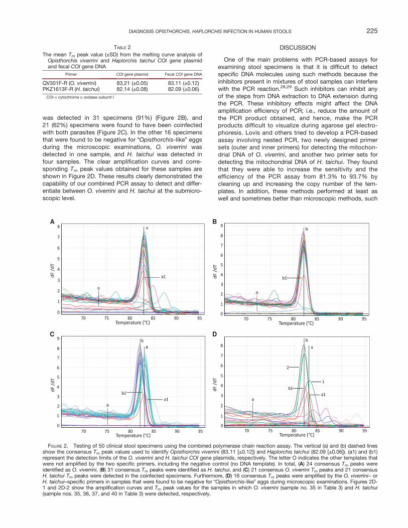
was detected in 31 specimens (91%) (Figure 2B), and21 (62%) specimens were found to have been coinfectedwith both parasites (Figure 2C). In the other 16 specimensthat were found to be negative for “Opisthorchis-like” eggsduring the microscopic examinations, O. viverrini wasdetected in one sample, and H. taichui was detected infour samples. The clear amplification curves and corre-sponding Tm peak values obtained for these samples areshown in Figure 2D. These results clearly demonstrated thecapability of our combined PCR assay to detect and differ-entiate between O. viverrini and H. taichui at the submicro-scopic level.
DISCUSSION
One of the main problems with PCR-based assays forexamining stool specimens is that it is difficult to detectspecific DNA molecules using such methods because theinhibitors present in mixtures of stool samples can interferewith the PCR reaction.28,29 Such inhibitors can inhibit anyof the steps from DNA extraction to DNA extension duringthe PCR. These inhibitory effects might affect the DNAamplification efficiency of PCR; i.e., reduce the amount ofthe PCR product obtained, and hence, make the PCRproducts difficult to visualize during agarose gel electro-phoresis. Lovis and others tried to develop a PCR-basedassay involving nested PCR, two newly designed primersets (outer and inner primers) for detecting the mitochon-drial DNA of O. viverrini, and another two primer sets fordetecting the mitochondrial DNA of H. taichui. They foundthat they were able to increase the sensitivity and theefficiency of the PCR assay from 81.3% to 93.7% bycleaning up and increasing the copy number of the tem-plates. In addition, these methods performed at least aswell and sometimes better than microscopic methods, such
FIGURE 2. Testing of 50 clinical stool specimens using the combined polymerase chain reaction assay. The vertical (a) and (b) dashed linesshow the consensus Tm peak values used to identify Opisthorchis viverrini (83.11 [±0.12]) and Haplorchis taichui (82.09 [±0.06]). (a1) and (b1)represent the detection limits of the O. viverrini and H. taichui COI gene plasmids, respectively. The letter O indicates the other templates thatwere not amplified by the two specific primers, including the negative control (no DNA template). In total, (A) 24 consensus Tm peaks wereidentified as O. viverrini, (B) 31 consensus Tm peaks were identified as H. taichui, and (C) 21 consensus O. viverrini Tm peaks and 21 consensusH. taichui Tm peaks were detected in the coinfected specimens. Furthermore, (D) 16 consensus Tm peaks were amplified by the O. viverrini– orH. taichui–specific primers in samples that were found to be negative for “Opisthorchis-like” eggs during microscopic examinations. Figures 2D-1 and 2D-2 show the amplification curves and Tm peak values for the samples in which O. viverrini (sample no. 35 in Table 3) and H. taichui(sample nos. 35, 36, 37, and 40 in Table 3) were detected, respectively.
TABLE 2The mean Tm peak value (±SD) from the melting curve analysis ofOpisthorchis viverrini and Haplorchis taichui COI gene plasmidand fecal COI gene DNA
Primer COI gene plasmid Fecal COI gene DNA
QV301F-R (O. viverrini) 83.21 (±0.05) 83.11 (±0.12)PKZ1613F-R (H. taichui) 82.14 (±0.08) 82.09 (±0.06)
COI = cytochrome c oxidase subunit I
225DIAGNOSIS OPISTHORCHIS, HAPLORCHIS INFECTION IN HUMAN STOOLS

as the Kato-Katz technique and formalin–ether concentra-tion.15 However, despite the advantages of the nested PCRtechnique, false-positive and false-negative diagnoses weredetected when ribosomal DNA-, internal transcribed spacer1(ITS1)-, and ITS2 gene-based PCR assays were used todiscriminate between O. viverrini, C. sinensis, H. taichui,and H. pumilio in an analysis of the same human stool speci-
mens, and it was concluded that PCR cannot discriminatebetween these parasite species based on the size of theamplicon produced during agarose gel electrophoresis.8 Todiscriminate between O. viverrini and H. taichui, Thaenkhamand others14 designed primers for amplifying the same COIgene region in adults, metacercariae, and eggs of both par-asites. After PCR amplification at the optimal annealing
TABLE 3Case data of samples showing presence and absence of “Opisthorchis-like” eggs by microscopy and Opisthorchis viverrini and Haplorchis
taichui positive (+) and negative (−) samples by combined PCR
Sample no. Sex Age (years)
Direct fecal smear microscope–based technique Combined PCR
Parasite eggs identified O. viverrini H. taichui
Presence of “Opisthorchis-like” eggs and other parasitic eggs from 34 samples1 M 58 “Opisthorchis-like” egg,
Hookworm− +
2 M 40 “Opisthorchis-like” egg + +3 F 65 “Opisthorchis-like” egg,
Taenia sp., Blastocystis hominis+ +
4 M 17 “Opisthorchis-like” egg + +5 M 23 “Opisthorchis-like” egg + +6 F 34 “Opisthorchis-like” egg + −7 M 67 “Opisthorchis-like” egg + +8 F 44 “Opisthorchis-like” egg + +9 F 58 “Opisthorchis-like” egg − +
10 F 59 “Opisthorchis-like” egg + +11 M 38 “Opisthorchis-like” egg − +12 F 70 “Opisthorchis-like” egg − +13 M 22 “Opisthorchis-like” egg,
Trichuris trichura+ +
14 M 38 “Opisthorchis-like” egg + +15 F 54 “Opisthorchis-like” egg + −16 F 43 “Opisthorchis-like” egg,
B. hominis, Hookworm, Trichomonas hominis+ −
17 F 35 “Opisthorchis-like” egg + +18 M 82 “Opisthorchis-like” egg + +19 M 30 “Opisthorchis-like” egg + +20 F 23 “Opisthorchis-like” egg + +21 M 34 “Opisthorchis-like” egg + +22 F 23 “Opisthorchis-like” egg,
T. trichura+ +
23 M 27 “Opisthorchis-like” egg + +24 M 57 “Opisthorchis-like” egg + +25 M 60 “Opisthorchis-like” egg − +26 F 23 “Opisthorchis-like” egg − +27 M 25 “Opisthorchis-like” egg + +28 M 20 “Opisthorchis-like” egg + +29 F 44 “Opisthorchis-like” egg − +30 F 36 “Opisthorchis-like” egg − +31 F 35 “Opisthorchis-like” egg − +32 M 35 “Opisthorchis-like” egg + +33 M 58 “Opisthorchis-like” egg − +34 M 32 “Opisthorchis-like” egg + +
Absence of “Opisthorchis-like” eggs from 16 samples35 F 39 Enterobius vermicularis + +36 M 33 Ascaris lumbricoides, Hookworm − +37 F 26 T. trichura, Hookworm − +38 F 52 Strongyloides stercoralis − −39 M 57 A. lumbricoides − −40 F 47 A. lumbricoides − +41 F 22 Trichuris trichura − −42 M 29 Taenia spp. − −43 F 73 S. stercoralis − −44 F 27 Hookworm − −45 F 42 – − −46 F 44 – − −47 F 30 – − −48 F 43 – − −49 F 26 – − −50 M 40 – − −
F = female; M = male; PCR = polymerase chain reaction.
226 LAMANINGAO AND OTHERS

temperature, it was shown that these two parasite speciescannot be identified based on the sizes of their ampliconson agarose gel electrophoresis. Thus, the amplicons weresubjected to restriction enzyme digestion, and the geneticcharacteristics of O. viverrini, C. sinensis, and H. taichuiwere determined. However, if a weak amplicon is producedduring PCR or a mutation is present in the target DNAsequence, this might reduce the sensitivity of the PCRrestriction fragment length polymorphism technique.30
This study describes a protocol for diagnosing parasiteinfections based on the use of COI gene DNA derived fromparasite eggs as a genetic marker, which attempted tojump over PCR inhibition in stool specimens as well. Asstarted from the step of gDNA egg extraction from stoolpellet by using a commercial stool DNA extraction kit,which included with InhibitEX matrix tablet, the InhibitEXmatrix tablet helped to reduce PCR inhibitors and followedwith screening of human beta actins to confirm PCR qual-ity from the gDNA extraction process.In this technique, a PCR involving a universal primer pair
that has been demonstrated to be useful for amplifying theCOI gene DNA of multiple parasites is used to increase thepurity and enhance the amplification of the target region ofthe COI gene. In the latter stage, the PCR product wasdiluted with distilled water and it was enough for subjectinginto qPCR without cleanup of PCR product, and then qPCRwas performed with species-specific primers. The uniquefeatures of the amplification curves and Tm peak values ofspecific DNA molecules are used to identify the causativeparasite. We initially used this assay to detect O. viverriniand H. taichui COI gene plasmids that had been producedby DNA cloning, and then it was used to validate clinicalstool specimens. The stool specimens had been found tocontain “Opisthorchis-like” eggs and other parasite eggsduring microscopic examinations.To the best of our knowledge, this is the first study to use
fluorescent melting curve analysis to evaluate COI gene prod-ucts to differentiate between two genospecies (O. viverriniand H. taichui). No cross-reactivity was detected when qPCRwas performed using partial COI gene DNA for O. viverrini,H. taichui, or a mixture of other parasites template, and inpositive cases, only one sharp Tm peak was detected. Fur-thermore, the differences between the Tm peak values ofO. viverrini and H. taichui were greater than 1°C (Table 2).These findings suggest that our qPCR assay with species-specific primers is able to distinguish between O. viverriniand H. taichui with high specificity. In agreement with this,the assay was able to detect O. viverrini and H. taichui instool samples that had been found to be “Opisthorchis-like”eggs-negative in microscopic examinations. Moreover, theassay was able to detect 1 pg of pure O. viverrini andH. tachui egg DNA (which was derived from a single egg) and5 EPG in spiked egg stool specimens, which are similar tothe findings obtained for other qPCR tests.31 However, nocross-reactivity was detected during the testing of 10 clinicalsamples containing other parasites, which confirmed thespecificity of the primers relative to the other parasites thatcommonly cause coinfections in Lao PDR25 (Table 3).The fact that our assay exhibited 100% sensitivity to
parasitic infections (O. viverrini, 71%; H. taichui, 91%) whenit was used to test samples that had been found tocontain “Opisthorchis-like” eggs demonstrated that it is
a good detection tool, while the fact that it detected para-sites in 25% of the samples that had been found to be“Opisthorchis-like” eggs-negative during the microscopicexaminations showed that it has a clear advantage over thecurrent microscopic diagnostic techniques. Sequencinganalysis confirmed that the four false-negative specimensincluded one coinfected specimen and three H. taichui–infected specimens (Table 3). Another reason for enhancedsubmicroscopic detection capability of our PCR assaycould be, the use of an increased quantity of stool material(2 g) for detecting O. viverrini eggs.32 These results suggestthat our assay is highly sensitive and specific for detectingand differentiating between O. viverrini and H. taichui.Inhabitants infested with O. viverrini and/or H. taichui par-
asites consumed raw fresh water fish cooked in traditionalstyle, which were infected with parasitic metacercariae as sin-gle and/or mix. The prevalence rates were previously reportedin Lao PDR community survey.33,34 These studies usedexpelled adult worms to confirm the types of parasites pres-ent in each subject and showed that there is geographicalvariation in the prevalence of the two parasites and a needfor local information about effective interventions and prioriti-zation.35 As molecular technology is becoming more afford-able, it is hoped that the findings of this study will not onlycontribute to the development of better detection methods,but also aid research develop better diagnostic methods.
CONCLUSIONS
As the eggs of O. viverrini and H. taichui are very simi-lar in terms of their size and morphology, it is difficult todistinguish between them during microscopic examina-tions of stool samples from infected patients. To differen-tiate between these two parasites, we designed newspecies-specific primers and used a combined PCR assayinvolving SYBR Green I and Tm peak value analysis, whichexhibited high sensitivity and specificity.
ReceivedMarch 2, 2016. Accepted for publication September 22, 2016.
Published online November 7, 2016.
Acknowledgments: We would like to thank all of their colleagueswho were involved in this study, especially the Mahosot Hospitallaboratory staff for their assistance and collaboration andChanpheng Thammavong, Vimon Soukkaseuam, and ViengxayVanisaveth for their help with the coordination between MahosotHospital and the Center of Malariology, Parasitology, and Entomol-ogy, Ministry of Health, Vientiane, Lao PDR.
Authors’ addresses: Pheophet Lamaningao, Seiji Kanda, and TakakiShimono, Department of Public Health, Kansai Medical University,Osaka, Japan, E-mails: [email protected], [email protected], and [email protected]. SakhoneLaimanivong, Center of Malariology, Parasitology, and Entomol-ogy, Ministry of Health, Vientiane, Lao PDR, E-mail: [email protected]. Andrew Waleluma Darcy, Nobuyuki Mishima, andToshimasa Nishiyama, Department of Public Health, Kansai Medi-cal University, Osaka, Japan, E-mails: [email protected],[email protected], and [email protected]. AmphayPhyaluanglath, Clinical Laboratory, Mahosot Hospital, Ministry ofHealth, Vientiane, Lao PDR, E-mail: [email protected].
REFERENCES
1. Sripa B, Kaewkes S, Sithithaworn P, Mairiang E, Laha T, SmoutM, Pairojkul C, Bhudhisawasdi V, Tesana S, Thinkamrop B,
227DIAGNOSIS OPISTHORCHIS, HAPLORCHIS INFECTION IN HUMAN STOOLS

Bethony JM, Loukas A, Brindley PJ, 2007. Liver fluke inducescholangiocarcinoma. PLoS Med 4: e201.
2. Belizario VY Jr, de Leon WU, Bersabe MJ, Purnomo Baird JK,Bangs MJ, 2004. A focus of human infection by Haplorchistaichui (Trematoda: Heterophyidae) in the southern Philippines.J Parasitol 90: 1165–1169.
3. Chai JY, Sohn WM, Jung BK, Yong TS, Eom KS, Min DY,Insisiengmay B, Insisiengmay S, Phommasack B, Rim HJ,2015. Intestinal helminths recovered from humans in XiengKhouang Province, Lao PDR with a particular note onHaplorchis pumilio infection. Korean J Parasitol 53: 439–445.
4. Sayasone S, Vonghajack Y, Vanmany M, Rasphone O, TesanaS, Utzinger J, Akkhavong K, Odermatt P, 2009. Diversity ofhuman intestinal helminthiasis in Lao PDR. Trans R Soc TropMed Hyg 103: 247–254.
5. Chai JY, Han ET, Guk SM, Shin EH, Sohn WM, Yong TS, EomKS, Lee KH, Jeong HG, Ryang YS, Hoang EH, PhommasackB, Insisiengmay B, Lee SH, Rim HJ, 2007. High prevalenceof liver and intestinal fluke infections among residents ofSavannakhet Province in Laos. Korean J Parasitol 45: 213–218.
6. Chai JY, Han ET, Shin EH, Sohn WM, Yong TS, Eom KS, MinDY, Um JY, Park MS, Hoang EH, Phommasack B,Insisiengmay B, Lee SH, Rim HJ, 2009. High prevalence ofHaplorchis taichui, Phaneropsolus molenkampi, and otherhelminth infections among people in Khammouane province,Lao PDR. Korean J Parasitol 47: 243–247.
7. Chai JY, Yong TS, Eom KS, Min DY, Shin EH, Banouvong V,Insisiengmay B, Insisiengmay S, Phommasack B, Rim HJ,2010. Prevalence of the intestinal flukes Haplorchis taichuiand H. yokogawai in a mountainous area of Phongsaly Prov-ince, Lao PDR. Korean J Parasitol 48: 339–342.
8. Sato M, Thaenkham U, Dekumyoy P, Waikagul J, 2009. Dis-crimination of O. viverrini, C. sinensis, H. pumilio and H.taichui using nuclear DNA-based PCR targeting ribosomalDNA ITS regions. Acta Trop 109: 81–83.
9. Duenngai K, Sithithaworn P, Rudrappa UK, Iddya K, Laha T,Stensvold CR, Strandgaard H, Johansen MV, 2008. Improve-ment of PCR for detection of Opisthorchis viverrini DNA inhuman stool samples. J Clin Microbiol 46: 366–368.
10. Stensvold CR, Saijuntha W, Sithithaworn P, WongratanacheewinS, Strandgaard H, Ornbjerg N, Johansen MV, 2006. Evaluationof PCR based coprodiagnosis of human opisthorchiasis.Acta Trop 97: 26–30.
11. Wongratanacheewin S, Pumidonming W, Sermswan RW,Pipitgool V, Maleewong W, 2002. Detection of Opisthorchisviverrini in human stool specimens by PCR. J Clin Micro-biol 40: 3879–3880.
12. Muller B, Schmidt J, Mehlhorn H, 2007. PCR diagnosis of infec-tions with different species of Opisthorchiidae using a rapidclean-up procedure for stool samples and specific primers.Parasitol Res 100: 905–909.
13. Thaenkham U, Blair D, Nawa Y, Waikagul J, 2012. FamiliesOpisthorchiidae and Heterophyidae: are they distinct? Para-sitol Int 61: 90–93.
14. Thaenkham U, Visetsuk K, Dung do T, Waikagul J, 2007. Dis-crimination of Opisthorchis viverrini from Haplorchis taichuiusing COI sequence marker. Acta Trop 103: 26–32.
15. Lovis L, Mak TK, Phongluxa K, Soukhathammavong P, SayasoneS, Akkhavong K, Odermatt P, Keiser J, Felger I, 2009. PCRDiagnosis of Opisthorchis viverrini and Haplorchis taichuiinfections in a Lao Community in an area of endemicity andcomparison of diagnostic methods for parasitological fieldsurveys. J Clin Microbiol 47: 1517–1523.
16. Arimatsu Y, Kaewkes S, Laha T, Hong SJ, Sripa B, 2012. Rapiddetection of Opisthorchis viverrini copro-DNA using loop-mediated isothermal amplification (LAMP). Parasitol Int 61:178–182.
17. Kaewkong W, Intapan PM, Sanpool O, Janwan P, ThanchomnangT, Laummaunwai P, Lulitanond V, Doanh PN, MaleewongW, 2013. Molecular differentiation of Opisthorchis viverriniand Clonorchis sinensis eggs by multiplex real-time PCRwith high resolution melting analysis. Korean J Parasitol 51:689–694.
18. Sanpool O, Intapan PM, Thanchomnang T, Janwan P,Lulitanond V, Doanh PN, Van Hien H, Dung do T, Maleewong
W, Nawa Y, 2012. Rapid detection and differentiation ofClonorchis sinensis and Opisthorchis viverrini eggs in humanfecal samples using a duplex real-time fluorescence reso-nance energy transfer PCR and melting curve analysis. Para-sitol Res 111: 89–96.
19. Le TH, Van De N, Blair D, Sithithaworn P, McManus DP, 2006.Clonorchis sinensis and Opisthorchis viverrini: developmentof a mitochondrial-based multiplex PCR for their identifica-tion and discrimination. Exp Parasitol 112: 109–114.
20. Wittwer CT, Herrmann MG, Moss AA, Rasmussen RP, 1997.Continuous fluorescence monitoring of rapid cycle DNAamplification. Biotechniques 22: 130–131, 134–138.
21. Ririe KM, Rasmussen RP, Wittwer CT, 1997. Product differenti-ation by analysis of DNA melting curves during the polymer-ase chain reaction. Anal Biochem 245: 154–160.
22. Nicolas L, Milon G, Prina E, 2002. Rapid differentiation of OldWorld Leishmania species by LightCycler polymerase chainreaction and melting curve analysis. J Microbiol Methods 51:295–299.
23. Pietila J, He Q, Oksi J, Viljanen MK, 2000. Rapid differentia-tion of Borrelia garinii from Borrelia afzelii and Borreliaburgdorferi sensu stricto by LightCycler fluorescence melt-ing curve analysis of a PCR product of the recA gene. J ClinMicrobiol 38: 2756–2759.
24. Papin JF, Vahrson W, Dittmer DP, 2004. SYBR green-basedreal-time quantitative PCR assay for detection of West NileVirus circumvents false-negative results due to strain vari-ability. J Clin Microbiol 42: 1511–1518.
25. Eom KS, Yong TS, Sohn WM, Chai JY, Min DY, Rim HJ, JeonHK, Banouvong V, Insisiengmay B, Phommasack B, 2014.Prevalence of helminthic infections among inhabitants of LaoPDR. Korean J Parasitol 52: 51–56.
26. Saijuntha W, Sithithaworn P, Wongkham S, Laha T, Chilton NB,Petney TN, Barton M, Andrews RH, 2008. Mitochondrial DNAsequence variation among geographical isolates of Opisthorchisviverrini in Thailand and Lao PDR, and phylogenetic relation-ships with other trematodes. Parasitology 135: 1479–1486.
27. Ando K, Sithithaworn P, Nuchjungreed C, Tesana S,Srisawangwong T, Limviroj W, Chinzei Y, 2001. Nucleotidesequence of mitochondrial CO I and ribosomal ITS II genesof Opisthorchis viverrini in northeast Thailand. SoutheastAsian J Trop Med Public Health 32 (Suppl 2): 17–22.
28. Wilde J, Eiden J, Yolken R, 1990. Removal of inhibitory sub-stances from human fecal specimens for detection of groupA rotaviruses by reverse transcriptase and polymerase chainreactions. J Clin Microbiol 28: 1300–1307.
29. Abu Al-Soud W, Radstrom P, 2000. Effects of amplificationfacilitators on diagnostic PCR in the presence of blood,feces, and meat. J Clin Microbiol 38: 4463–4470.
30. Shepard GC, Lawson HL, Hawkins GA, Owen J, 2011. BsaXI/RFLP analysis of initial or selectively reamplified PCR prod-uct is unreliable in detecting the V617F mutation in JAK2.Int J Lab Hematol 33: 267–271.
31. Cai XQ, Yu HQ, Li R, Yue QY, Liu GH, Bai JS, Deng Y,Qiu DY, Zhu XQ, 2014. Rapid detection and differentia-tion of Clonorchis sinensis and Opisthorchis viverrini usingreal-time PCR and high resolution melting analysis.ScientificWorldJournal 2014: 893981.
32. Umesha KR, Kumar S, Parvathi A, Duenngai K, Sithithaworn P,Karunasagar I, 2008. Opisthorchis viverrini: detection bypolymerase chain reaction (PCR) in human stool samples.Exp Parasitol 120: 353–356.
33. Giboda M, Ditrich O, Scholz T, Viengsay T, Bouaphanh S,1991. Human Opisthorchis and Haplorchis infections in Laos.Trans R Soc Trop Med Hyg 85: 538–540.
34. Chai JY, Yong TS, Eom KS, Min DY, Jeon HK, Kim TY, JungBK, Sisabath L, Insisiengmay B, Phommasack B, Rim HJ,2013. Hyperendemicity of Haplorchis taichui infection amongriparian people in Saravane and Champasak province, LaoPDR. Korean J Parasitol 51: 305–311.
35. Sripa B, Bethony JM, Sithithaworn P, Kaewkes S, MairiangE, Loukas A, Mulvenna J, Laha T, Hotez PJ, Brindley PJ,2011. Opisthorchiasis and Opisthorchis-associated cholangio-carcinoma in Thailand and Laos. Acta Trop 120 (Suppl 1):S158–S168.
228 LAMANINGAO AND OTHERS