DEVELOPMENT OF A BEAD-BASED MULTIPLEXED PCR ASSAY FOR THE
32
DEVELOPMENT OF A BEAD-BASED MULTIPLEXED PCR ASSAY FOR THE SIMULTANEOUS DETECTION OF MULTIPLE MYCOPLASMA SPECIES By DANIEL J. RIGHTER A thesis submitted in partial fulfillment of the requirements for the degree of Master of Veterinary Science WASHINGTON STATE UNIVERSITY Department of Veterinary Microbiology & Pathology DECEMBER 2010
Transcript of DEVELOPMENT OF A BEAD-BASED MULTIPLEXED PCR ASSAY FOR THE

DEVELOPMENT OF A BEAD-BASED MULTIPLEXED PCR ASSAY FOR THE
SIMULTANEOUS DETECTION OF MULTIPLE MYCOPLASMA SPECIES
By
DANIEL J. RIGHTER
A thesis submitted in partial fulfillment of the requirements for the degree of
Master of Veterinary Science
WASHINGTON STATE UNIVERSITY Department of Veterinary Microbiology & Pathology
DECEMBER 2010
ii
To the Faculty of Washington State University:
The members of the Committee appointed to examine the thesis of DANIEL J. RIGHTER find it satisfactory and recommend that it be accepted.
Terry F. McElwain, D.V.M., Ph.D., Chair
Fred R. Rurangirwa, B.V.Sc., M.S., Ph.D.
Douglas R. Call, Ph.D.
Timothy V. Baszler, DVM, Ph.D.

iii
ACKNOWLEDGMENT
I would to thank Terry McElwain for the tremendous amount of guidance throughout all
phases of this project, Fred Rurangirwa for additional project oversight and techniques required
for cultivating and extracting DNA from mycoplasma, Doug Call for making his laboratory
available at all times and for his assistance with the manuscript, Tim Baszler for serving on the
committee, Jonathon Anderson for conducting preliminary assay development experiments, Lisa
Orfe for technical assistance, especially as it pertains to primer and probe design, Dan Bradway
for culturing Mycoplasma mycoides subsp. capri, and additional technical assistance, Niles
Reichardt for providing bi-monthly lists of field samples to be collected by WADDL‟s Central
Processing staff and Jenn Baisley for saving field samples.
The funding for this project was provided by USDA, NIFA, NAHLN Cooperative
Agreement, the Washington Animal Disease Diagnostic Laboratory, and the WSU CVM
Department of Veterinary Microbiology and Pathology.

iv
DEVELOPMENT OF A BEAD-BASED MULTIPLEXED PCR ASSAY FOR THE
SIMULTANEOUS DETECTION OF MULTIPLE MYCOPLASMA SPECIES
Abstract
By Daniel J. Righter, D.V.M., M.S.
Washington State University
December 2010
Chair: Terry F. McElwain
We describe the development and analytical validation of a specific and sensitive 7-plex
polymerase chain reaction assay coupled to a bead-based liquid suspension array for high
throughput detection of multiple Mycoplasma spp. of ruminants. The assay employs a
combination of newly designed and previously validated primer-probe sets that target genetic
loci specific for Mycoplasma bovis, Mycoplasma mycoides cluster, Mycoplasma mycoides subsp.
mycoides SC (MmmSC) and Mycoplasma capricolum subspecies capripneumoniae (Mccp).
PCR products were hybridized to specific oligonucleotide probes bound to spectrally unique, 5.6
µm diameter, carboxylated beads, and subsequently analyzed by flow cytometry. Analytical
sensitivity for the targeted Mycoplasma species ranged from 10 fg to 1 pg of purified gDNA
extracted from broth cultures (approximately 8-800 MmmSC genome equivalents). In silico
comparison of primers and probes, and analytical assessment with a range of near-neighbor
Mycoplasma species and multiple bacterial respiratory pathogens of ruminants demonstrated
100% analytical specificity of the assay. To assess assay performance and diagnostic specificity,
192 bovine respiratory samples were analyzed by incorporating a high throughput DNA
extraction platform. The assay correctly classified all 192 field samples as negative for MmmSC

v
or Mccp with an apparent 100% diagnostic specificity for these pathogens. 33/192 field samples
were positive for M. bovis and all were confirmed by PCR or sequence verification of the uvrC
gene. The results from this study indicate that the bead-based liquid suspension array will
provide a reliable, analytically sensitive and specific platform to simultaneously interrogate
ruminant respiratory samples for multiple Mycoplasma species, including Mycoplasma mycoides
cluster organisms exotic to the United States. Sequential addition of primer-probe sets to the
assay did not significantly impact analytical sensitivity of individual primer-probe combinations,
suggesting that expanding the array to include more Mycoplasma species will not compromise
overall performance.

vi
TABLE OF CONTENTS
Page
ACKNOWLEDGEMENT .............................................................................................. iii
ABSTRACT .................................................................................................................. iv
LIST OF TABLES ........................................................................................................ vii
LIST OF FIGURES ..................................................................................................... viii
DEDICATION ............................................................................................................... ix
INTRODUCTION .......................................................................................................... 1
MATERIALS & METHODS .......................................................................................... 4
RESULTS ..................................................................................................................... 10
DISCUSSION ............................................................................................................... 17
BIBLIOGRAPHY ......................................................................................................... 21

vii
LIST OF TABLES
1. Bacteria used for determining analytical specificity ............................................. 3
2. Primer-probe sets used for multiplex assay .......................................................... 6
3. Analytical sensitivity of the multiplex assay ...................................................... 14
4. Interassay variability (CV%).............................................................................. 16

viii
LIST OF FIGURES
1. Analytical specificity graph #1 .......................................................................... 11
2. Analytical specificity graph #2 .......................................................................... 12
3. Analytical sensitivity ......................................................................................... 14

ix
Dedication
This thesis is dedicated to my wife Jamie Lee for her unwavering emotional support and sacrifice
over the last three years.

1
Introduction
Mycoplasma bovis and members of the Mycoplasma mycoides cluster are significant
ruminant respiratory pathogens of economic importance (10, 11, 12, 16, 17, 18). M. bovis is
associated with pneumonia in young calves and feedlot cattle (17, 19, 20). While all five taxa of
the recently reclassified M. mycoides cluster are pathogenic, four are associated with three
disease syndromes that involve the respiratory tract of small ruminants or cattle, and are listed as
notifiable agents by the OIE (World Organization for Animal Health): (i) contagious agalactiae
of sheep and goats (Mycoplasma capricolum subsp. capricolum and Mycoplasma mycoides
subsp. capri (including the recently reclassified serovar Mycoplasma mycoides subsp. mycoides
biotype Large Colony) (ii) contagious bovine pleuropneumonia of cattle (Mycoplasma mycoides
subsp. mycoides biotype Small Colony), and (iii) contagious caprine pleuropneumonia of goats
(Mycoplasma capricolum subsp. capripneumoniae) (21). The fifth, recently reclassified M.
mycoides cluster member, Mycoplasma leachii sp. nov. (21), (formerly Mycoplasma sp. bovine
group 7 of Leach) has been isolated from calves with pneumonia (23). There are also reports of
members of the M. mycoides cluster being isolated from non-traditional hosts. For instance, M.
mycoides subsp. capri has been isolated from cattle, M. leachii and M. mycoides subsp. mycoides
biotype Small Colony from goats, and M. capricolum subsp. capripneumoniae has been isolated
from sheep (18, 22, 23).
Identification of specific Mycoplasma spp. from the ruminant respiratory tract typically
involves cultivation followed by immunologic-based assays or PCR. Members of the M.
mycoides cluster are particularly difficult to differentiate because of phenotypic and genotypic
features that lead to serological cross-reactivity and challenges with sequence verification (9).
Cross-reactions by growth inhibition assays are frequently seen among M. mycoides subsp.

2
mycoides biotype Small Colony and M. mycoides subsp. capri, as well as between M. leachii and
M. capricolum subps. capripneumoniae (9). Further, in the case of small ruminants, M.
mycoides subsp. capri grows relatively well, which could result in concurrent infections with the
more fastidious M. capricolum subsp. capripneumoniae or M. capricolum subsp. capricolum not
being detected by preliminary cultivation techniques (23) without specifically investigating their
presence by PCR.
Because of the unique challenges for efficiently identifying M. bovis, accurate
identification in the case of the M. mycoides cluster, and the importance of early detection of the
causative agents of contagious bovine and caprine pleuropneumonia, the aim of the present study
was to develop a single assay capable of resolving these targets from ruminant respiratory
samples. A 7-plex PCR coupled to a liquid bead array assay was employed that targets two
genetic loci each for M. bovis, M. capricolum subsp. capripneumoniae, and M. mycoides subsp.
mycoides biotype Small Colony, and one primer-probe set targeting the M. mycoides cluster as a
whole. This assay was chosen for its diagnostic efficiency, capacity to overcome the limited
number of target organisms detectable using multiplex rt-PCR assays, its ability to identify
multiple respiratory pathogens in a single sample, and for the opportunity to include in the future
primer-probe sets targeting other Mycoplasma spp. and non-mycoplasma microbial pathogens.

3
TABLE 1. Bacteria used for determining analytical specificity
Strain Source Target(s) for strain verification
M. agalactiae ATCC 35890 16s rRNA/16s-23s intergenic spacer region
M. alkalescens Cornell; 2-1-1-46 strain 16s rRNA/16s-23s intergenic spacer region
M. arginini ATCC 23243 16s rRNA/16s-23s intergenic spacer region
M. bovigenitalium ATCC 14173 16s rRNA
M. bovirhinis IOM Mollicutes Collection; PG43 strain
16s-23s intergenic spacer region
M. bovis ATCC 25025 16s rRNA/16s-23s intergenic spacer region
M. bovoculi ATCC 29104 16s rRNA
M. californicum ATCC 33461 16s rRNA/16s-23s intergenic spacer region
M. canadense ATCC 29418 16s rRNA
M. canis ATCC 19525 16s rRNA
M. dispar ATCC 27140 16s rRNA
M. ovipneumoniae WADDL1 16s rRNA2
M. putrefaciens IOM Mollicutes Collection; KS-1 strain
16s rRNA
M. mycoides cluster
M. capricolum subsp. capricolum IOM Mollicutes Collection; goat 14 strain
16s rRNA/fusA/putative integral membrane protein H2 gene
M. capricolum subsp. capripneumoniae FADDL3 16s rRNA/fusA/arcD
M. leachii sp. nov. IOM Mollicutes Collection; N29 Leach strain
16s rRNA/fusA/glk
M. mycoides subsp. capri Bradway 16s rRNA/fusA/glk
M. mycoides subsp. capri (serovar Large Colony)
IOM Mollicutes Collection; 4933 strain
16s rRNA/fusA/glk
M. mycoides subsp. mycoides Small Colony
FADDL, Gladysdale strain 16s rRNA/fusA/glk
Non-mycoplasma bacteria
Arcanobacterium pyogenes WADDL N/A4
Escherichia coli WADDL N/A
Histophilus somni WADDL N/A
Mannheimia hemolytica WADDL N/A
Pasteurella multocida WADDL N/A
1Washington Animal Disease Diagnostic Laboratory
2WADDL rt-PCR (25, 26)
3United States Foreign Animal Disease Diagnostic Laboratory
4 Classified by biochemical assays

4
Materials and methods
Bacterial strains and growth conditions: The bacteria used in this study are listed in Table 1.
Mycoplasma strains were cultured at 37°C under 5% CO2 in PPLO or general Mycoplasma
Broth (Hardy Diagnostics, USA). Mycoplasma dispar was grown at 37°C under 5% CO2 in
modified Friis broth (UC Davis Biological Media Services, CA, USA). Agar plates streaked for
isolation of Arcanobacterium pyogenes, Escherichia coli, Histophilus somni, Mannheimia
hemolytica and Pasteurella multocida were provided by the Washington Animal Disease
Diagnostic Laboratory (WADDL, Pullman, WA) for DNA extraction.
DNA extraction and sequence analysis: Mycoplasma strains were pelleted by centrifuging
broth cultures at 10,000 X g for 10 min. Pellets of the mycoplasma strains and non-mycoplasma
bacterial colonies on agar were re-suspended in 200 µl of 1X PBS, and DNA was extracted using
the DNeasy tissue kit (Qiagen, USA). DNA from M. mycoides subsp. mycoides biotype Small
Colony and M. capricolum subsp. capripneumoniae was extracted and safety treated (see below)
at the United States Foreign Animal Disease Diagnostic Laboratory (FADDL) (Plum Island, NY)
prior to being shipped. Invitrogen‟s (Carlsbad, CA) Easy-DNA™ kit was used, per
manufacturer‟s instructions (E. coli specific protocol), to extract 5 to 12 ml of the mycoplasma
cultures. The DNA was suspended in Tris buffer (pH 8.0, Ambion, Austin, TX) and submitted
for safety treatment for removal from BSL3 facilities. The extracted DNA was safety treated by
adding 10 µl of 3 M NaCl (1/10 volume), 3.5 µl 3 M Acetate (1/30 volume), and 5 µl
ribonuclease A (0.5 Kunitz units/100 µl, USB, Cat. 70194Z) followed by boiling the sample for
10 min, slowly cooled, and frozen at 70°C prior to being shipped.

5
The identity of Mycoplasma spp. was confirmed by amplification and sequence analysis
of DNA targets listed in Table 1. Modifications of previously described PCR assay conditions
were used to amplify 16s rRNA, fusA, glk, arcD, and putative integral membrane protein H2
gene sequences (7, 8, 9, 10, 11, 12). For sequence analysis of the 16s-23s rRNA intergenic
spacer region, an alignment of the partial 16s rRNA sequence, complete 16s-23s intergenic
spacer sequence, and partial 23s rRNA gene sequence of two M. bovis strains available in
GenBank (accession nos. AY729934, AY765211), three M. bovigenitalium strains (AY780797,
AY973571, AY974058), and one strain each of M. agalactiae (AY770623), M. alkalescens
(AY816348), M. californicum (AY736031), and M. canadense (AY800341) was created using
Vector NTI software (Invitrogen,USA). Primers L-ITS and R-ITS were developed (L-ITS:
AACCATGGGAGCTGGTAATG; R-ITS: GTTCGCTATCGGTGTCTGGT) using primer3
(http://frodo.wi.mit.edu/primer3/) based on conserved regions of this alignment that would lead
to an expected 939 bp amplicon. The PCR conditions for amplifying the 16-23s rRNA spacer
region for sequencing consisted of those optimized for the multiplex PCR assay (see
“development and optimization of multiplex PCR” section below). Bands of the expected size
in a 1.5% agarose gel were visualized under ultraviolet light, excised, and purified using Freeze
„N Squeeze DNA gel extraction spin column kit (Bio-Rad Laboratories, USA). Purified PCR
products (10 µl) were combined with 5 µl of the forward or reverse primer (5 µM) and submitted
for sequence analysis (Genewiz Inc., USA).

6
TABLE 2. Primer-probe sets used for multiplex assay
targeted
mycoplasma primer/probe sequence 5'-3' gene target size (bp) reference
M. mycoides cluster
MYC1 TTCTAAATTAGTTACTCGTGCA ribosomal protein S7
457-466 1
MYC2 AATAAACTGTATTCTCTAGCCA
MYC probe ATTGGAGGAGCTAACTATCAAGTTC this study
M. mycoides subsp. mycoides
SC
SG1352 GTTTTTTAGGAATTTTGTAAATAGCTCAATT hypothetical
prolipoprotein 90 2 SG1353 AGTGAAGTTTCTAAATCAATCCAATCAG
SGAB010 probe TCAATTAGACTGTAATGAAATAT
M. mycoides subsp. mycoides
SC
MSC-382F ATGCAAGAAGTTATTAATGTTTATCATTC 52 amino acid hypothetical
protein
106 3
MSC-382R CGTAATATATTTGTTTAACATATGGAATAA
MSC-382 probe CTTTTATTAAGCTCATCTCTTGG this study
M. capricolum subsp.
capripneumoniae
MccpF CGCTCACATAGCCAATCATC lppa (lipoprotein
A)
152 4
MccpR TCGTTTTTAAGAGAAAATCAAGCA
Mccp probe CAAGCTGATGAACATAAAAATGATG this study
M. capricolum subsp.
capripneumoniae
CPN1F ATGTTTGAAGGGGCAGAGAA putative integral membrane
protein (membrane protein H2)
216 this study CPN1R TTTGGGCTTAACCAAAGGTG
CPN1 probe ATGACCCTAACAAAAGACCGGAT
M. bovis
Mb-F GCTTCAGTATTTTGACGG
uvrC 303 5 Mb-R GGTTTAGCTCCATAACCAGA
PMyb probe CATATATAAGTGAGACTAACTTATT
M. bovis
mb-mp1F TATTGGATCAACTGCTGGAT mb-mp81 (membrane
lipoprotein P81 gene)
447 6 mb-mp1R AGATGCTCCACTTATCTTAG
mb-mp1 probe GCAGCTAAAGGAAAAGGCGC this study

7
Development and optimization of multiplex PCR: The primers and probes used in the
multiplex assay are listed in Table 2 (1, 2, 3, 4, 5, 6). Primers CPN1F and CPN1R were designed
from a highly conserved region of the putative integral membrane protein H2 gene (13) present
in each of ten M. capricolum subsp. capripneumoniae strain sequences available in Genbank
(accession numbers: AF378153, AF378154, AF378155, AF378156, AF378157, AF378158,
AF378159, AF162991, EU827517, and GU992420) using CLC DNA Workbench 5.6 software
(Cambridge, MA, USA) and primer3. Probes mb-mp1, CPN1, Mccp and MSC-382 were
designed using a combination of CLC DNA workbench 5.6 software and Oligo Calculator
version 3.5 (http://www.basic.northwestern.edu/biotools/oligocalc.html) from sequences
available in Genbank (accession numbers: AY627040 (mb-mp1 probe), AF111424 (Mccp
probe), AF162991 (CPN1 probe) and, BX293980 (MSC-382 probe)).
The PCR assay was optimized using the PCR Optimization Kit (Roche, USA), for
assessment of optimal pH, MgCl2 concentration, and affects of several additives (DMSO,
glycerol, (NH4)2SO4 and gelatin). The annealing temperature was optimized using a temperature
gradient setting on the iCycler thermocycler ((Bio-Rad Laboratories, USA). The cycling
parameters consisted of an initial denaturation step of 95°C for 5 min followed by 35 cycles of
94°C for 15 sec, 55°C for 30 sec, and 72°C for 1 min with a final extension step of 72°C for 7
min. The amount of DNA was standardized at 10 ng (1 ng/µl) of genomic DNA for each of the
targeted mycoplasmas using the NanoDrop ND1000 spectrophotometer (Thermo Scientific,
USA). Reactions with greatest band intensities in agarose gels were observed with the following
constituents (50 µl final reaction volume): 5 µl 1X FastStart reaction buffer with MgCl2
(FastStart High Fidelity PCR System, Roche, USA), 1 µl dNTP mix (10 mM) (Roche, USA), 1.2
8
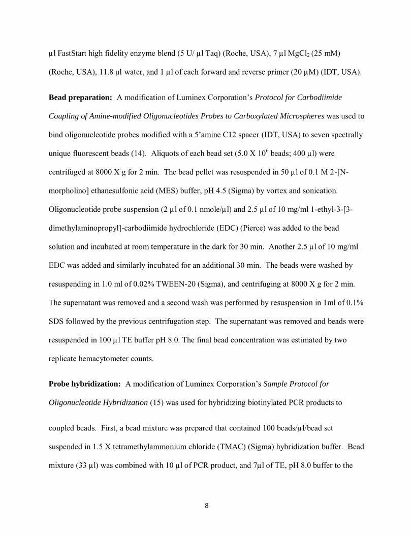
µl FastStart high fidelity enzyme blend (5 U/ µl Taq) (Roche, USA), 7 µl MgCl2 (25 mM)
(Roche, USA), 11.8 µl water, and 1 µl of each forward and reverse primer (20 µM) (IDT, USA).
Bead preparation: A modification of Luminex Corporation‟s Protocol for Carbodiimide
Coupling of Amine-modified Oligonucleotides Probes to Carboxylated Microspheres was used to
bind oligonucleotide probes modified with a 5‟amine C12 spacer (IDT, USA) to seven spectrally
unique fluorescent beads (14). Aliquots of each bead set (5.0 X 10⁶ beads; 400 µl) were
centrifuged at 8000 X g for 2 min. The bead pellet was resuspended in 50 µl of 0.1 M 2-[N-
morpholino] ethanesulfonic acid (MES) buffer, pH 4.5 (Sigma) by vortex and sonication.
Oligonucleotide probe suspension (2 µl of 0.1 nmole/µl) and 2.5 µl of 10 mg/ml 1-ethyl-3-[3-
dimethylaminopropyl]-carbodiimide hydrochloride (EDC) (Pierce) was added to the bead
solution and incubated at room temperature in the dark for 30 min. Another 2.5 µl of 10 mg/ml
EDC was added and similarly incubated for an additional 30 min. The beads were washed by
resuspending in 1.0 ml of 0.02% TWEEN-20 (Sigma), and centrifuging at 8000 X g for 2 min.
The supernatant was removed and a second wash was performed by resuspension in 1ml of 0.1%
SDS followed by the previous centrifugation step. The supernatant was removed and beads were
resuspended in 100 µl TE buffer pH 8.0. The final bead concentration was estimated by two
replicate hemacytometer counts.
Probe hybridization: A modification of Luminex Corporation‟s Sample Protocol for
Oligonucleotide Hybridization (15) was used for hybridizing biotinylated PCR products to
coupled beads. First, a bead mixture was prepared that contained 100 beads/µl/bead set
suspended in 1.5 X tetramethylammonium chloride (TMAC) (Sigma) hybridization buffer. Bead
mixture (33 µl) was combined with 10 µl of PCR product, and 7µl of TE, pH 8.0 buffer to the

9
wells of a 96-well PCR plate. The hybridization reaction was carried out in an iCycler
Thermocycler set with a single denaturation step of 95°C for 5 minutes followed by a 30 minute
hybridization step at 55⁰C. The hybridization products were transferred to a 96-well 1.2 µm
filter plate (Millipore) and washed 3 times using 100 µl 1 X TMAC buffer. Beads were
resuspended in 100 µl 0.75 X TMAC reporter buffer containing 4 µg/ml recombinant Phycolink
streptavidin-R-phycoerythrin (Prozyme, USA). Reporter mix was incubated in the dark on the
Bio-Plex 100 Suspension Array System platform (Bio-Rad Laboratories, USA) at 45°C for 5
min, and each well was subsequently analyzed by the instrument at a 45°C holding temperature.
Assessment of diagnostic specificity: 192 bovine lung samples submitted to WADDL from
Washington, Idaho, Utah, Oregon, Montana, Colorado, Nevada, and New York during 2008-
2009 were stored at -70°C prior to analysis in 2010. The majority of samples were suspect
bacterial or viral pneumonia cases; however, other diseases such as enteritis, encephalitis, sepsis,
metritis, salmonellosis, clostridial myositis, malignant catarrhal fever, bovine viral diarrhea,
toxicity, renal failure, and abortion were also represented. Cattle breeds included Angus,
Hereford, Holstein, Charlois, Limousin, Brown Swiss, Shorthorn, Jersey, and variety of crosses.
The age of cattle ranged from fetuses to aged-adults (11 years-old); however, the majority of
lung samples were from calves less than 1 year-old. Heifers, steers and adult cows and bulls
were all represented.
The samples were processed by swabbing a freshly cut surface of each lung with a
flocked swab (Hardy Diagnostics, USA). Each swab was rinsed in a 96 well storage plate
containing 400 µl 1X PBS, pH 7.2. DNA was extracted using the BioSprint 96 One-For-All Vet
Kit (Qiagen, USA) on the BioSprint 96 robotic workstation.

10
Results
Analytical specificity: The seven primer-probe sets (Table 2) were initially evaluated using 1
ng of purified genomic DNA (gDNA) from a representative strain of each target Mycoplasma
species. The suspension array detected three probes for Mycoplasma mycoides subsp. mycoides
biotype Small Colony and Mycoplasma capricolum subsp. capripneumoniae samples (each
sample positive for the M. mycoides cluster probe (MYC), and two probes specific for each
subspecies (SGAB010; MSC-382 and Mccp; CPN1, respectively)), one probe for the remaining
members of the M. mycoides cluster samples (MYC probe), and two species specific probes for
the M. bovis sample (PMyb and mb-mp1). No probe cross-reactivity was observed (Fig.1), and
the signal to noise ratio of the mean fluorescence intensities comparing signal among target and
non-target strains ranged from 20.8 (Mccp probe) to 88.3 (PMyb probe). Members of this group
of seven target Mycoplasma spp. served as both positive and negative internal controls for the
remaining analytical specificity experiments. No probe cross-reactivity was seen among the
remaining mycoplasma strains or common bacterial respiratory pathogens of cattle (Fig. 2).
From these data, we established a threshold for classifying positive and negative results.
A sample was considered positive if the mean fluorescence intensity of a probe was greater than
each of two calculated values: (i) mean fluorescence intensity of the negative internal control
samples plus three times the standard deviation and (ii) two times the mean fluorescence
intensity of the negative internal control samples.

11
Figure 1. Preliminary analytical specificity assessing 1 ng gDNA of the M. mycoides cluster members and M.
bovis. Error bars represent standard error of the mean. Two probes are specific for M. bovis (mb-mp1;
PMyb), M. mycoides subsp. mycoides Small Colony (SGAB010; MSC-382), M. capricolum subsp.
capripneumoniae (Mccp; CPN1), and one probe (MYC) is specific for the M. mycoides cluster members.
MmmSC=M. mycoides subsp. mycoides Small Colony, Mccp=M. capricolum subsp. capripneumoniae,
Mmc=M. mycoides subsp. capri, Mcc=M. capricolum subsp. capricolum, MBG7=M. leachii, MmmLC=M.
mycoides subsp. mycoides biotype Large Colony, bovis=M. bovis.
0
1000
2000
3000
4000
5000
6000
7000
8000
9000
10000
blank MmmSC Mccp Mmc Mcc MBG7 MmmLC bovis water
Mean
Flu
ore
scen
ce In
ten
sit
ymb-mp1 SGAB010 Mccp CPN1 Pmyb MYC MSC-382

12
Figure 2. Analytical specificity run assessing Mycoplasma species and non-mycoplasma bacteria commonly
isolated from the ruminant respiratory tract. Error bars represent standard error of the mean. Two probes
are specific for M. bovis (mb-mp1; PMyb), M. mycoides subsp. mycoides Small Colony (SGAB010; MSC-382),
M. capricolum subsp. capripneumoniae (Mccp; CPN1), and one probe (MYC) is specific for the M. mycoides
cluster members.MmmSC=M. mycoides subsp. mycoides Small Colony, Mccp=M. capricolum subsp.
capripneumoniae, Mmc=M. mycoides subsp. capri, Mcc=M. capricolum subsp. capricolum, MBG7=M. leachii,
MmmLC=M. mycoides subsp. mycoides biotype Large Colony, bovis=M. bovis.
0 1000 2000 3000 4000 5000 6000 7000 8000 9000 10000
MmmSC
Mccp
Mmc
Mcc
MBG7
MmmLC
bovis
water
agalactiae
bovoculi
canis
alkalescens
arginini
californicum
bovirhinis
putrefaciens
canadense
bovigenitalium
dispar
ovipneumoniae
P. multocida
M. hemolytica
H. somni
A. pyogenes
E. coli
mb-mp1 SGAB010 Mccp CPN1 Pmyb MYC MSC-382

13
Analytical sensitivity and intra-assay variability: The limit of detection (LOD) of all seven
probes for each target Mycoplasma spp. was determined by preparing ten-fold serial dilutions
that ranged from 1 ng to 1 fg of purified genomic DNA (gDNA). Both multiplex amplification
and detection steps of the assay were performed in triplicate for each dilution series, and the limit
of detection was defined as the highest dilution at which all three replicates were above the
negative cut-off value. The cut-off value for classifying a sample as positive was calculated
similar to previously described except a PCR control sample containing water in place of non-
target gDNA served as the negative control. The limit of detection of probes and corresponding
target Mycoplasma spp. are listed in Table 3, and mean fluorescence intensities with standard
error of the means for determining intra-assay variability are illustrated in Figure 3. The LOD
for M. bovis was 10 fg gDNA, and 100 fg for M. capricolum subsp. capricolum, M. leachii, M.
mycoides subsp. capri, 1 pg for M. mycoides subsp. mycoides biotype Small Colony, and 10 pg
for M. capricolum subsp. capripneumoniae. Based on complete genome sequence data
available in GenBank, 1 pg gDNA of M. mycoides subsp. mycoides biotype Small Colony, M.
mycoides subsp. capri, and M. capricolum subsp. capricolum strains represents approximately
103 genome copies.

14
TABLE 3. Analytical sensitivity of the multiplex assay
Mycoplasma Probes
MYC SGAB010 MSC-382 Mccp CPN1 PMyb mb-mp1
M. mycoides subsp. mycoides SC 10 pg1 1 pg 1 pg M. capricolum subsp.
capripneumoniae 100 pg
10 pg 10 pg M. mycoides subsp. capri
(serovar MmmLC) 100 fg
M. mycoides subsp. capri 100 fg
M. capricolum subsp. capricolum 100 fg
M. leachii 100 fg
M. bovis 10 fg 10 fg 1Lowest quantity of target DNA at which all 3 replicates were positive
0
2000
4000
6000
8000
10000
12000
1 fg 10 fg 100 fg 1 pg 10 pg 100 pg 1 ng water
M. bovis gDNA
0
2000
4000
6000
8000
10000
12000
1 fg 10 fg 100 fg 1 pg 10 pg 100 pg 1 ng water
Mccp gDNA
0
2000
4000
6000
8000
10000
12000
1 fg 10 fg 100 fg 1 pg 10 pg 100 pg 1 ng water
MmmSC gDNA
0
2000
4000
6000
8000
10000
12000
1 fg 10 fg 100 fg 1 pg 10 pg 100 pg 1 ng water
Mcc gDNA
A B
C D
Figure 3. Analytical sensitivity of the multiplex assay. The y-axis represents mean fluorescence intensity.
Panel A=serial dilutions of M. bovis gDNA. Panel B= M. mycoides subsp. mycoides biotype Small Colony
gDNA. Panel C=M. capricolum subsp. capripneumoniae. Panel D=M. capricolum subsp. capricolum. Error
bars represent standard error of the mean. Each probe is designated a specific color (see Figs. 1 & 2).

15
Interassay variability: To assess repeatability of the multiplex assay, amplification and
detection steps were performed in triplicate using high, medium, and low concentrations based
on LOD of target gDNA on 3 separate days. Aliquoted samples of gDNA at the specified
concentrations were prepared on a single day and frozen at -20°C. Aliquots were thawed and a
fresh PCR master mix was prepared on each of the three days. Table 4 summarizes the 3-day
interassay coefficient of variation for each probe at varying concentrations of target gDNA. The
coefficient of variation percentage was less than 30% for the majority of the cases. Not
surprisingly, in some cases lower template concentrations yielded higher variance estimates.
Assay performance on field samples: To initiate stage two validation (OIE) of the assay,
lung samples from 192 bovine cases submitted to WADDL during 2008-2009 were analyzed.
Ten microliters of gDNA extracted from each bovine lung sample (N=192) was combined with
40 µl of master mix for a single multiplex PCR step. The probe hybridization and detections
steps were performed in duplicate using 10 µl of PCR product. Each 96-well plate had five wells
reserved for the following control samples: 1 ng MmmSC gDNA, 1ng Mccp gDNA, 1 ng M.
bovis gDNA, water PCR control, and a blank (mixture containing the probe hybridization and
SAPE reporter buffers).

16
Table 4. Interassay variability (CV%)
Probes
Mycoplasma MYC SGAB010 MSC-382 CPN1 Mccp PMyb mb-mp1
M. mycoides
subsp. mycoides
SC
1 ng 2.9 1 9.5 17.6 100 pg 4.0 1.5 14.4 1 pg ND2 32.8 17.2
M. capricolum
subsp. capripneumoniae
1 ng 21.0
10.3 16.7 100 pg 29.7
30.2 1.4
10 pg ND 4.5 6.5 M. capricolum
subsp. capricolum
100 pg 4.9 1 pg 8.8 100 fg 12.1
M. mycoides
subsp. capri
(serovar
MmmLC)
100 pg 7.2 1 pg 26.6
100 fg 37.7
M. leachii
100 pg 3.0 1 pg 6.1 100 fg 38.2
M. bovis
100 pg 9.8 8.5 1 pg
9.8 8.6
100 fg 5.4 9.7 1 Percentage coefficient of variation 2 Not done
Mycoplasma culture was performed by WADDL on 43/192 field samples, and 14/43
were positive for mycoplasma growth. Each of these 14 samples was identified as M. bovis by
the bead-based assay using the cut-off value algorithm for determining analytical specificity. A
total of 33 samples were positive in the bead-based assay for M. bovis. Of these 33 samples, 32
were positive based on the PMyb probe and 13 based on the mb-mp1 probe. One sample was
positive for probe mb-mp1 and negative for PMyb, while 20 samples were positive for probe
PMyb and negative for mb-mp1. All 33 samples positive for M. bovis were either positive for
mycoplasma growth by WADDL (n=14), or confirmed by sequence verification (n=19) of the
uvrC gene using the primer set in Table 2. Two culture negative samples were positive for M.

17
bovis in the multiplex assay and were confirmed as M. bovis by uvrC gene sequence verification.
All 192 field samples were negative for M. mycoides subsp. mycoides biotype Small Colony, M.
capricolum subsp. capripneumoniae and M. mycoides cluster group (probes SGAB010, MSC-
382, CPN1, Mccp, and MYC).
Discussion
Diagnosis and differentiation of Mycoplasma spp. is challenging. The OIE recognizes
multiple tests for identifying the causative agents of contagious bovine and caprine
pleuropneumonia including culture and biochemical testing, and serological and PCR assays (27,
28). Multiple PCR assays have been described that specifically identify Mycoplasma species and
members of the M. mycoides cluster. To our knowledge, there is no single assay that can be
used to accomplish this task. Multiplex PCR combined with suspension bead array offers the
opportunity to interrogate a single sample for multiple bacterial species. We utilized this
platform to determine the potential for a single assay to accurately accomplish this. As an initial
assessment, the assay was designed for the purpose of utilizing the routine diagnostic stream of
ruminant respiratory cases in the United States for surveillance of mycoplasma causing bovine
and caprine pleuropneumonia. Primers and probes for which analytical data had previously been
published were adapted to the multiplex suspension array format. In addition, five new probe
sequences and one new primer-probe set were designed so that two probes were available for
each of the three specific strains targeted.
The limit of detection varied by probe. In general, there was no improvement of
analytical sensitivity when comparing primer-probe sets in a singleplex or multiplex format for
M. mycoides subsp. mycoides biotype Small Colony and M. capricolum subsp. capripneumoniae

18
with the exception of MYC probe where a 10-fold increase in the LOD was observed for M.
capricolum subsp. capripneumoniae. The decreased sensitivity observed with M. mycoides
subsp. mycoides biotype Small Colony and M. capricolum subsp. capripneumoniae probes in the
multiplex assay could be related in part to safety treatment and/or shipment of DNA prepared for
these pathogens (DNA from M. mycoides subsp. mycoides biotype Small Colony and M.
capricolum subsp. capripneumoniae was generously provided by the United States Foreign
Animal Disease Diagnostic Laboratory, Plum Island, NY). This assumption is based on the
relative drop in the MYC probe sensitivity for these two strains when compared to the other M.
mycoides cluster members in a singleplex format, and by a comparison of the limit of detection
of gDNA prepared from two different M. leachii strains. M. leachii genomic DNA prepared with
the DNeasy tissue kit had a 1000-fold increase in analytical sensitivity over another M. leachii
strain whose DNA had been safety treated and shipped by FADDL (data not shown). Moreover,
when the MYC primer-probe set was tested on M. mycoides subsp. mycoides biotype Small
Colony and M. capricolum subsp. capripneumoniae in a singleplex format, the limit of detection
remained 10-100-fold less sensitive than the remaining M. mycoides cluster members.
Additional target and non-target Mycoplasma species also need to be evaluated for a
more complete assessment of analytical and diagnostic sensitivity and specificity. Nevertheless,
six of seven primer sets were previously evaluated using a more broad range of strains within the
same taxa and other closely related Mycoplasma species, suggesting conservation of the targeted
loci. The one novel primer-probe set designed in this study (CPN1) amplifies a sequence
encoding a putative integral membrane protein that is highly conserved among all ten strains
available in GenBank. Notably, 5/7 probes were developed specifically for this assay, and their
performance with other field isolates needs to be determined.

19
Preliminary evaluation of the assay‟s performance on field samples is encouraging. The
diagnostic specificity is 100% (95% CI 98-100%) for M. mycoides subsp. mycoides biotype
Small Colony and M. capricolum subsp. capripneumonia with an assumption that these
organisms are not present in the United States (US declared free by the OIE; 29). Mycoplasma
culture results were available for 43 of the field samples tested. Of these 43 samples, the
multiplex assay correctly identified M. bovis as present in all 14 that were culture positive. Of
the 29 culture negative samples, 2 were positive in the multiplex assay and were confirmed as
positive M. bovis samples by uvrC amplification and sequencing. This suggests that the
multiplex assay may offer increased diagnostic sensitivity for M. bovis over culture, while
requiring much less time. Clearly, however, diagnostic sensitivity of the assay must be
thoroughly assessed using a larger number of known positive samples, not only for M. bovis, but
for M. mycoides subsp. mycoides biotype Small Colony and M. capricolum subsp.
capripneumoniae, the latter two of which will require studies outside the United States in
endemic regions. Specificity of the assay analyzed in silico and using type strains of multiple
mycoplasma and common prokaryotic respiratory pathogens of ruminants was 100%.
The likelihood of a false negative result arising from a mutated field strain is expected to
decrease by targeting two loci. The utility of 2 primer-probe sets was demonstrated for M. bovis
in this study, as one known M. bovis positive lung sample from the field set was negative for
PMyb probe, while nearly two-thirds of the known M. bovis positive samples were negative with
the mb-mp1 probe. It is unclear whether the decreased diagnostic sensitivity associated with the
mb-mp1 probe is due to a mutation in M. bovis field isolates or if it is the result of competing
amplification reactions with the PMyb primer set.
20
Previous studies involving development and validation of a multiplex PCR-coupled
liquid suspension array indicate that this form of microbial detection can be highly sensitive and
specific, and is an efficient method for screening multiple pathogens in a single assay, exceeding
the capacity of real-time PCR (24). By combining multiplex amplification steps to a liquid
suspension array, up to 100 gene targets can be resolved using the flow cytometer platform.
Clearly, this number is unattainable from a single multiplex PCR assay; however, the quantity of
pathogens screened in a single well of a 96-well plate can be increased by combining products
from several multiplex PCR assays. During the development phase of this assay, no decrease in
analytical sensitivity was observed by increasing the number of primer-probe sets from three to
seven (data not shown). This finding is encouraging for further development aimed at
discriminating additional members of the M. mycoides cluster and other Mycoplasma species.
Moreover, additional multiplex PCR assays targeting non-mycoplasma bacteria or viral
pathogens could be developed to create a syndrome-specific assay capable of screening a more
complete set of pathogens from a single ruminant respiratory sample. This would encourage use
of the assay in the routine diagnostic workflow for simultaneous diagnosis of respiratory
pathogens and surveillance for foreign mycoplasma strains, facilitating rapid identification of
accidental or deliberate introduction of these pathogens into non-endemic countries.
21
BIBLIOGRAPHY
1. Dedieu L, Mady V, Lefevre PC. Development of a selective polymerase chain reaction assay for the detection of Mycoplasma mycoides subsp. Mycoides S.C. (contagious bovine pleuropneumonia agent). Vet Microbiol. 1994 Dec;42(4):327-39.
2. Gorton TS, Barnett MM, Gull T, French RA, Lu Z, Kutish GF, Adams LG, Geary SJ. Development of real-time diagnostic assays specific for Mycoplasma mycoides subspecies mycoides Small Colony. Vet Microbiol. 2005 Nov 30;111(1-2):51-8
3. Lorenzon S, Manso-Silván L, Thiaucourt F. Specific real-time PCR assays for the detection and quantification of Mycoplasma mycoides subsp. mycoides SC and Mycoplasma capricolum subsp. capripneumoniae. Mol Cell Probes. 2008 Oct-Dec;22(5-6):324-8
4. Fitzmaurice J, Sewell M, King CM, McDougall S, McDonald WL, O'Keefe JS., A real-time polymerase chain reaction assay for the detection of Mycoplasma agalactiae. N Z Vet J. 2008 Oct;56(5):233-6.
5. Lee KH, Lee JW, Wang SW, Liu LY, Lee MF, Chuang ST, Shy YM, Chang CL, Wu MC, Chi CH. Development of a novel biochip for rapid multiplex detection of seven mastitis-causing pathogens in bovine milk samples. J Vet Diagn Invest. 2008 Jul;20(4):463-71
6. Foddai A, Idini G, Fusco M, Rosa N, de la Fe C, Zinellu S, Corona L, Tola S. Rapid differential diagnosis of Mycoplasma agalactiae and Mycoplasma bovis based on a multiplex-PCR and a PCR-RFLP. Mol Cell Probes. 2005 Jun;19(3):207-12
7. van Kuppeveld FJ, van der Logt JT, Angulo AF, van Zoest MJ, Quint WG, Niesters HG, Galama JM, Melchers WJ. Genus- and species-specific identification of mycoplasmas by 16S rRNA amplification. Appl Environ Microbiol. 1992 Aug;58(8):2606-15.
8. Kong F, James G, Gordon S, Zelynski A, Gilbert GL. Species-specific PCR for identification of common contaminant mollicutes in cell culture. Appl Environ Microbiol. 2001 Jul;67(7):3195-200.
9. Manso-Silván L, Perrier X, Thiaucourt F. Phylogeny of the Mycoplasma mycoides cluster based on analysis of five conserved protein-coding sequences and possible implications for the taxonomy of the group. Int J Syst Evol Microbiol. 2007 Oct;57(Pt 10):2247-58.
10. Woubit S, Manso-Silván L, Lorenzon S, Gaurivaud P, Poumarat F, Pellet MP, Singh VP, Thiaucourt F. A PCR for the detection of mycoplasmas belonging to the Mycoplasma mycoides cluster: application to the diagnosis of contagious agalactia. Mol Cell Probes. 2007 Oct-Dec;21(5-6):391-9.
11. Woubit S, Lorenzon S, Peyraud A, Manso-Silván L, Thiaucourt F. A specific PCR for the identification of Mycoplasma capricolum subsp. capripneumoniae, the causative

22
agent of contagious caprine pleuropneumonia (CCPP). Vet Microbiol. 2004 Nov 30;104(1-2):125-32.
12. Thiaucourt F, Lorenzon S, David A, Breard A. Phylogeny of the Mycoplasma mycoides cluster as shown by sequencing of a putative membrane protein gene. Vet Microbiol. 2000 Mar 15;72(3-4):251-68.
13. Lorenzon S, Wesonga H, Ygesu L, Tekleghiorgis T, Maikano Y, Angaya M, Hendrikx P, Thiaucourt F. Genetic evolution of Mycoplasma capricolum subsp. capripneumoniae strains and molecular epidemiology of contagious caprine pleuropneumonia by sequencing of locus H2. Vet Microbiol. 2002 Mar 1;85(2):111-23.
14. http://www.luminexcorp.com/uploads/data/Nucleic%20Acids%20Protocols%20FAQs/Oligonucleotide%20Coupling%20Protocol%200407%2010208.pdf
15. http://www.luminexcorp.com/uploads/data/Nucleic%20Acids%20Protocols%20FAQs/Oligonucleotide%20Hybridization%20Protocol%200106%2010222.pdf
16. Bergonier D, Berthelot X, Poumarat F. Contagious agalactia of small ruminants: current knowledge concerning epidemiology, diagnosis and control. Rev Sci Tech. 1997 Dec;16(3):848-73.
17. Caswell JL, Archambault M. Mycoplasma bovis pneumonia in cattle. Anim Health Res Rev. 2007 Dec;8(2):161-86.
18. Bölske G, Mattsson JG, Bascuñana CR, Bergström K, Wesonga H, Johansson KE. Diagnosis of contagious caprine pleuropneumonia by detection and identification of Mycoplasma capricolum subsp. capripneumoniae by PCR and restriction enzyme analysis. J Clin Microbiol. 1996 Apr;34(4):785-91.
19. Caswell JL, Bateman KG, Cai HY, Castillo-Alcala F. Mycoplasma bovis in respiratory disease of feedlot cattle. Vet Clin North Am Food Anim Pract. 2010 Jul;26(2):365-79.
20. Gagea MI, Bateman KG, Shanahan RA, van Dreumel T, McEwen BJ, Carman S, Archambault M, Caswell JL. Naturally occurring Mycoplasma bovis-associated pneumonia and polyarthritis in feedlot beef calves. J Vet Diagn Invest. 2006 Jan;18(1):29-40
21. Manso-Silván L, Vilei EM, Sachse K, Djordjevic SP, Thiaucourt F, Frey J. Mycoplasma leachii sp. nov. as a new species designation for Mycoplasma sp. bovine group 7 of Leach, and reclassification of Mycoplasma mycoides subsp. mycoides LC as a serovar of Mycoplasma mycoides subsp. capri. Int J Syst Evol Microbiol. 2009 Jun;59(Pt 6):1353-8.
22. Vilei EM, Korczak BM, Frey J. Mycoplasma mycoides subsp. capri and Mycoplasma mycoides subsp. mycoides LC can be grouped into a single subspecies. Vet Res. 2006 Nov-Dec;37(6):779-90.
23. Maigre L, Citti C, Marenda M, Poumarat F, Tardy F. Suppression-subtractive hybridization as a strategy to identify taxon-specific sequences within the Mycoplasma

23
mycoides Cluster: design and validation of an M. capricolum subsp. capricolum-specific PCR assay. Int J Syst Evol Microbiol. 2009 Jun;59(Pt 6):1353-8.
24. Wilson WJ, Erler AM, Nasarabadi SL, Skowronski EW, Imbro PM. A multiplexed PCR-coupled liquid bead array for the simultaneous detection of four biothreat agents. Mol Cell Probes. 2005 Apr;19(2):137-44. Epub 2004 Dec 31.
25. Besser TE, Cassirer EF, Potter KA, VanderSchalie J, Fischer A, Knowles DP, Herndon DR, Rurangirwa FR, Weiser GC, Srikumaran S. Association of Mycoplasma ovipneumoniae infection with population-limiting respiratory disease in free-ranging Rocky Mountain bighorn sheep (Ovis canadensis canadensis). J Clin Microbiol. 2008 Feb;46(2):423-30.
26. McAuliffe L, Hatchell FM, Ayling RD, King AI, Nicholas RA. Detection of Mycoplasma ovipneumoniae in Pasteurella-vaccinated sheep flocks with respiratory disease in England. Vet Rec. 2003 Nov 29;153(22):687-8.
27. http://www.oie.int/eng/Normes/mmanual/2008/pdf/2.04.09_CBPP.pdf
28. http://www.oie.int/eng/Normes/mmanual/2008/pdf/2.07.06_CCPP.pdf
29. http://www.oie.int/wahis/public.php?page=home