DEVELOPMENT AND POTENTIAL APPLICATIONS OF …529562/FULLTEXT01.pdf · países en todo el mundo,...
25
TRITA-LWR Degree Project ISSN 1651-064X LWR-EX-11-07 DEVELOPMENT AND POTENTIAL APPLICATIONS OF NANOMATERIALS FOR ARSENIC REMOVAL FROM CONTAMINATED GROUNDWATER Rajender Kumar March 2011
Transcript of DEVELOPMENT AND POTENTIAL APPLICATIONS OF …529562/FULLTEXT01.pdf · países en todo el mundo,...

TRITA-LWR Degree Project
ISSN 1651-064X
LWR-EX-11-07
DEVELOPMENT AND POTENTIAL
APPLICATIONS OF NANOMATERIALS
FOR ARSENIC REMOVAL FROM
CONTAMINATED GROUNDWATER
Rajender Kumar
March 2011

Rajender Kumar TRITA LWR Degree Project 11:07
ii
© Rajender Kumar 2011
Degree Project at the Masters level
KTH-International Groundwater Arsenic Research Group (GARG)
Department of Land and Water Resources Engineering
Royal Institute of Technology (KTH)
SE-100 44 STOCKHOLM, Sweden
Reference to this publication should be written as: Kumar, R (2011) Development and Potential Applica-tions of Nanomaterials for Arsenic Removal from Contaminated Groundwater. TRITA LWR Degree project 11:07, 17p.
Development and potential applications of nanomaterials for arsenic removal from contaminated groundwater
iii
SUMMARY IN SWEDISH
Föroreningar av geokemiska ursprung såsom arsenik (As) drabbar miljontals människor i flera länder globalt, såsom Latinamerika, Taiwan, Kina, Nepal, Bangladesh och Indien. Den aktuella studien är om utvecklingen av nanomaterial för grundvattnet behandling. Användningen av järn-baserade nanopartiklar för att sanera arsenik förorenat grundvatten utnyttjar den utökade reaktivitet. Egenskaper såsom stor yta, potential för att bygga själv, hög specificitet, hög reaktivitet, och katalytisk potential för att nanopartiklar utmärkta kandidater för att avlägsna föroreningar från dricksvatten. Magnetiska nanopartiklar var synteti-seras av samfällning metod. För att förhindra tätorten och oxidationer av Fe3O4 olika organiska och oor-ganiska föreningar används som ytskikt modification. Synthesized magnetiska nanopartiklar har fram-gångsrikt använts för remedition arsenik-förorenat grundvatten. Bindningsförmåga As (III) bestämdes med Fe3O4 NPS och differenent ytmodifierade NPs APTES dvs överdragna, TSC bestruket och Chitosan belagda NPs.
SUMMARY IN SPANISH
La contaminación de origen geoquímico, como el arsénico (As) afecta millones de personas en varios países en todo el mundo, como América Latina, Taiwán, China, Nepal, Bangladesh y la India. El estudio actual se centra en el desarrollo de nanomateriales para el tratamiento de las aguas subterráneas. El uso de nanopartículas a base de hierro para la remediación de arsénico de aguas subterráneas contaminadas con la mayor reactividad. Características tales como una gran área superficial, la posibilidad de construirse a sí misma, una alta especificidad, alta reactividad, y el potencial catalítico de las nanopartículas de candidatos excelentes para la eliminación de contaminantes del agua potable. Nanopartículas magnéticas fueron sintetizados por el método de co-precipitación. Para evitar la aglomeración y la oxidación de Fe3O4 diver-sos compuestos orgánicos e inorgánicos utilizados para la modificación de la superficie. Sintetizado nano-partículas magnéticas se han utilizado con éxito remediación arsénico de las aguas subterráneas contami-nadas. Capacidad de enlace de As (III) se determinó con Fe3O4 fuentes de energía nuclear y la superficie de fuentes de energía nuclear differenent APTES modificado es decir, recubiertos, revestidos TSC fuentes de energía nuclear y el quitosano-revestido.

Rajender Kumar TRITA LWR Degree Project 11:07
iv

Development and potential applications of nanomaterials for arsenic removal from contaminated groundwater
v
ACKNOWLEDGEMENTS
Water is a valuable resource under increasing demand worldwide and is exposed to numerous sources of pollution. The occurrence of arsenic in natural waters, mainly in groundwater, is a worldwide problem. The largest population at risk among the 21 countries with known groundwater arsenic contamination is in Bangladesh, followed by West Bengal in India. I take pride in carrying out research on the applications of nanotechnology in remediation of As contaminated groundwater using Magnetic nanoparticles. No research can be accomplished without facilitate of research supervisor, cooperation of family members, and support of friends. There is always a sense of gratitude which one express to others for helpful ser-vices they render during all the phases of life. I too, would like to thank all those who helped me directly or indirectly in getting this mighty task of research through.
I should thank first of all to my advisor Prof. Prosun Bhatacharya, KTH-International Groundwater Arsenic Research Group (GARG), Department of Land and Water Resources Engineering, Royal Insti-tute of Technology (KTH), for his ever helping nature, immense knowledge of the subject, a great sense of precision, parental care and ever smiling face are remarkable, without which I could not have complet-ed this.
I express my deep sense of gratitude and obedience to Associate Prof. Gunaratna Kuttuva Rajarao, Divi-sion of Environmental microbiology, School of Biotechnology for providing all the infrastructure and laboratory facilities during whole of the research period and her valuable suggestions when required and caring nature.
I wish to extend my thanks to Mr. Chuka Okoli, PhD student, Division of Environmental microbiology, School of Biotechnology for providing all the laboratory instructions, their valuable suggestions in exper-imental setup and moral support during whole of the research period.
I acknowledge with sincere thanks to Mr. Alireza, Division of Chemical Technology for the analysis of samples on FTIR.
I would also like to special thanks to my friends Mr. Anil Sangwan and Saurabh Hsengar PhD scholar „thin film laboratory, physic department Indian Institute of Technology, New Delhi in samples analysis on TEM.
I thank to the staff members of Department of Land and Water Resources Engineering and Division of Environmental microbiology, School of Biotechnology, Royal Institute of Technology (KTH), for his ever helping nature whole hearted co-operation and assistance throughout this research work.
Further thanks should also go to European Union for the granting a scholarship within the Erasmus Mundus programme for International Master in Material and Sensor System for Environmental Technol-ogy at UPV, Valencia and KTH, Sweden.
I thank to the staff members of computer center and library who provide the facility to enable interna-tional online scientific resources, journals and books these were very helpful to completing this research.
I would also thank to IMMSSET Co-ordiantor Ampro Ribes at UPV, Valencia, Sigbritt Karlsson and Emma Strömberg at KTH, Sweden for her ever helping nature whole hearted co-operation and support throughout the study period.
My special thanks go to my wife, Divya Bhatia for their loving, carrying and moral support during the whole period of research.
I would also like thanks my all friends, Mr. Ramnath Lakshmanan, Ida Bodlun, Karin Larsdotter, Duke, Kisan Koirala, Yitagesu Arse for their cordial and friendly attitude which provided impetus to me for completing the research in time.
I admire the patience shown by my parents during the research and grateful for their constant encourage-ment and love rendered to me during this work. I am also thankful to my family members for his everlast-ing patience, for all his moral support, and love to complete the present study, without his cooperation it would have been very difficult to complete this manuscript.
Last, but not the least, my sincere thanks to all of those whom I am not recalling this moment, who helped me in any way during my research work.

Rajender Kumar TRITA LWR Degree Project 11:07
vi

Development and potential applications of nanomaterials for arsenic removal from contaminated groundwater
vii
TABLE OF CONTENT
Summary in Swedish ............................................................................................................. iii Summary in Spanish .............................................................................................................. iii Acknowledgements ................................................................................................................. v Table of Figures .................................................................................................................. viii Abstract.................................................................................................................................... 1 1 Introduction ................................................................................................................... 1 2 Magnetic Nanoparticles and its applications ................................................................ 2
2.1 General ..................................................................................................................... 2
2.2 Magnetic nanoparticles ............................................................................................ 3
2.3 Synthesis of magnetic NPs ...................................................................................... 3 2.3.1 Co-precipitation method ...................................................................................................................... 3 2.3.2 Microemulsion method ........................................................................................................................ 4 2.3.3 Hydrothermal synthesis ........................................................................................................................ 4
2.4 Surface modification ................................................................................................ 4
2.5 Magnetic behavior of iron oxide NPs ...................................................................... 4
2.6 Application of NPs in groundwater ......................................................................... 5 3 MATERIALS AND METHODS .................................................................................. 5
3.1 Chemicals ................................................................................................................. 5
3.2 Synthesis of Magnetic NPs ...................................................................................... 5
3.3 Surface Modification ................................................................................................ 6 3.3.1 3-aminopropyl-triethoxysilane (3- APTES) ...................................................................................... 6 3.3.2 Fe3O4–Chitosan NPs ............................................................................................................................ 6 3.3.3 Trisodium citrate (TSC) coated NPs.................................................................................................. 6
3.4 Characterization methods ........................................................................................ 6 3.4.1 X-ray Diffraction XRD ........................................................................................................................ 6 3.4.2 Transmission Electron Microscope (TEM) ...................................................................................... 7 3.4.3 Fourier Transform Infrared Spectroscopy (FTIR) .......................................................................... 7
3.5 Binding capacity of Arsenic species from Groundwater .......................................... 8 4 RESULTS AND DISCUSSION .................................................................................... 8
4.1 Transmission electron micrograph .......................................................................... 8
4.2 X-ray Diffraction (XRD) analysis ........................................................................... 10
4.3 Fourier-transform infrared (FTIR) spectroscopy .................................................. 10
4.4 Potential applications of Nano-biomaterial in arsenic(III) removal ..................... 11
4.5 Binding capacity of As(III)and As(V)from groundwater using NPs .................... 11 5 Conclusions ................................................................................................................. 12 6 Future Work ................................................................................................................. 13 7 References ................................................................................................................... 14

Rajender Kumar TRITA LWR Degree Project 11:07
viii
TABLE OF FIGURES
1. Variation of the coercivity (HC) of magnetic nanoparticles….…………………..4
2. Synthesized Fe3O4 NPs ………………………………………………………………6
3. Structure of APTES coated Fe3O4 NPs……………………………………………..7
4. Structure of Chitosan coated- Fe3O4 NPs……………………………………………8
5. WAG- WE 10500 Arsenator…………………………………………………………...9
6. (A) HR-TEM image (B) TEM image of Fe3O4 NPs……………………………….9
7. (A) HR-TEM image (B) TEM image of Chitosan -Fe3O4 NPs……………………9
8. (A) TEM image (B) HR-TEM image of APTES coated Fe3O4 NPs………………9
9. (A) HR-TEM image (B) TEM image of TSC - coated Fe3O4 NPs………………..9
10. FT-IR spectrum of the Fe3O4 nanoparticles synthesized.………………………...10
11. XRD pattern of the different type of synthesized NPs…………………………….11
12. Binding capacity of As (III) using Fe3O4 NPs……………………………………..11
13. Binding capacity of As (III) using Chitosan coated-Fe3O4 NPs…………………..11
14. Binding capacity of As (III) using TSC coated-Fe3O4 NPs……………………….12
15. Binding capacity of As (III) using APTES coated-Fe3O4 NPs……………………12
16. Removal of total arsenic from groundwater with different types of MNPs at initial concentration (500 µg/L) and NPs 100 µL………………………………………….12
17. Removal of total arsenic from groundwater with different NPs initial concentra-tion (1000 µg/L) with NPs 100 µL…………………………………………..……….12

Development and potential applications of nanomaterials for arsenic removal from contaminated groundwater
1
ABSTRACT
In this study, a magnetic nanomaterial was used for the binding of anionic arsenic species from contami-nated groundwater. Iron oxide (Fe3O4) magnetic nanoparticles (NPs) and the surface modified Fe3O4 NPs with 3-aminopropyl-triethoxysilane (3-APTES), Trisodium citrare (TSC) and Chitosan were synthesized with the co-precipitation method. Structural characterizations showed that the four kinds of NPs had different sizes an average particle range size of 15-20 nm was observed with Transmission Electron Mi-croscopy. X-ray diffraction was used to identify the crystalline structure of synthesized Fe3O4 and surface modified NPs. Molecular structure and functional groups present in synthesized magnetic NPs Fe3O4 were identify with infrared analysis. The synthesized Fe3O4 NPs and surface coated NPs were used for determine the binding capacity of Arsenic ions from the synthetic groundwater. The binding of As(III) increased as the dissolved As(III) concentration increased in the solution. From the experiments it was found chitosan-coated NPs are best than other coated and uncoated NPs for arsenite removal from the solution. It was found that if only As(III) ions were present in the water without other anions and cations the binding capacity of the magnetic NPs is very high. The binding capacity of As ions was decreased with presence of other anions and cations in the groundwater because they interfere with arsenic binding sites which presence on the magnetic NPs.
Key words: Iron oxide nanoparticles (NPs), surface modification, structural charcaterisation, Arsenic species
1 INTRODUCTION
Hundreds of millions of people all over the world, in developed and developing countries, depend directly on groundwater for their daily domestic needs. The rapid development of indus-trial, urban and agricultural sectors is adding contaminants to the water resources systems at an increasing rate. Unfortunately, industrialized nations have contaminated much land and the groundwater associated with it. In some situa-tions these contaminants join the aquifers and cause groundwater quality problems.
Pollutants of geochemical origin such as arsenic (As) affects millions of people in several coun-tries globally, such as South America, Taiwan, China, Nepal, Bangladesh and India (Bhattachar-ya et al. 1997, 2002). Most of the affected part is South-East Asia. Much of the world‟s total pro-duction of arsenic, around 30,000 tons per an-num, is intentionally released into the environ-ment as industrial or agricultural wastewater (Chen et al., 1994; Das et al., 1995; Karim, 2000; Chakraborti et al., 2002). Bangladesh indicate that as much as 80% of the country and an estimated 40 million people are at risk of arsenic poisoning and related diseases resulting from drinking arsenic-contaminated groundwater (Alam and Satter, 2000). Arsenic can occur in the environ-ment in several oxidation states (-3, 0, +3 and +5) but in natural waters is mostly found in inorganic form as oxy-anions of trivalent arsenite As(III) or pentavalent arsenate As(V). Organic As forms may be produced by biological activity,
mostly in surface waters, but are rarely quantita-tively important. Organic forms may however occur where waters are significantly impacted by industrial pollution. Even natural concentrations of arsenic in groundwater can limit its suitability for drinking. Highly toxic forms of arsenic have also been found in significant concentrations in the natural environment resulting from excessive exploitation of groundwater. Arsenic (As) distri-bution and toxicology in the environment is a serious issue, with millions of individuals world-wide being affected by As toxicosis. Long-term exposure to arsenic has been associated with cancer of the skin, lungs, urinary tract, kidneys and liver, keratosis and melanosis (Bhattacharyya et al., 2003; Kapaj et al., 2006). Inorganic forms of arsenic dissolved in drinking water are the most significant forms of natural exposure. Or-ganic forms of arsenic that may be present in food are much less toxic to humans. Arsenic contamination and its mitigation is a priority area in drinking water quality within the World Health Organization (WHO) and other national and international agencies, and a multitude of studies. The WHO limit for arsenic in drinking water is 10 ppb, though many countries still use a limit of 50 ppb (CGWB, 1999; Nath et al., 2008). Many wells in Bangladesh and neighboring regions exceed these limits by a large factor. High con-centration as 5000 µg/L of arsenic in groundwa-ter and two fold concentrations found in arsenic rich areas when mining has contaminated groundwater. Arsenic levels even increases upto 50,000 µg/L due to geothermal influence. It has

Rajender Kumar TRITA LWR Degree Project 11:07
2
also been found that anthropogenic sources, mining and herbicide industry are the first to mention, because there have been reported arse-nic concentrations in drainages from 34,000 to 850,000 µg/L (Smedley and Kinniburgh, 2002). The aim of this study was to establish an innova-tive water treatment technology for elimination of As from drinking water. These methods in-clude coagulation and flocculation, precipitation, adsorption and ion exchange, membrane filtra-tion. Alternative methods like ozone oxidation, bioremediation and electrochemical treatments mainly used in the removal of arsenic. Most of these methods suffer from some drawbacks such as high capital and operational costs for the treatment and disposal of the residual metal sludge. Conventional adsorbents used in arsenic removal are activated carbons and alumina, soils and resins which can be coated with different materials like iron or alumina (Chakravarty et al., 2002). The major disadvantages of these tech-niques are difficult separation (centrifugation or filtering), waste formation (both liquid and sludge) and in many cases poor adsorption capac-ity. The development of novel systems for reme-diation and pre-concentration is required because unlike organic pollutants, metals can be a recircu-lating contaminant (Ireland, 1991) and (Vernet, 1992) and cannot be metabolized or decom-posed. Therefore, efforts are made to develop low cost materials to remove As from drinking water.
Nanotechnology has been used in fields such as medicine, biotechnology and electronics, its beneficial application to drinking water treatment has begun only recently. Because of their size, nanomaterials can exhibit an array of novel prop-erties that can be used to develop new technolo-gies and improve existing ones. Characteristics such as large surface area, potential for self as-sembly, high specificity, high reactivity, and catalytic potential make nanoparticles excellent candidates for removal of contaminants from drinking water (Li et al., 2003). They also have been widely studied for arsenic adsorption (Peng et al., 2005). Studies of magnetic nanoparticle systems have attracted much interest in the last few years from both fundamental and applicative point of view. Over the last decade, additional control and design of magnetic nanoparticles was achieved by developing core shell structured nanosystems. This type of particles have drawn the attention of physicist, chemists and material scientists since they allow the tailoring of the combined surface and core properties providing an increased number of applications like catalysis,
and coatings to hyperthermia and magneto-resistant materials (Wang, et al., 1999). Main advantages of using maghemite nanoparticles as an adsorbent are: (1) maghemite nanoparticles have adsorption capacity due to its large surface area, (2) simple and rapid separation of metal-loaded magnetic adsorbent from treated water can be achieved via an external magnetic field, (3) no secondary pollutants are produced, (4) the amount of chemicals used is diminished and (5) maghemite is available commercially and it can also be easily synthesized with different methods (White et al., 2009). The overall objective of the proposed research is to specifically design nano-materials and their surface modification that can be used to selectively removal of arsenic species from ground water and investigation of structure and size of the particles.
2 MAGNETIC NANOPARTICLES
AND ITS APPLICATIONS
2.1 General
Nanoscience is the study of materials on the nanoscale level between approximately 1 and 100 nm (Rotello, 2003). Nanotechnology includes the integration of these nanoscale structures into larger material components and systems, keeping the control and construction of new and im-proved materials at the nanoscale. Nanomaterial not only has the characters of general nanometer material, but also has the advantages of good biologic compatibility, better photocatalytic activity, non toxicity, stable medical performance, easy preparation. The unusual properties of nanoparticles (NPs) have accelerated growth in the production of nanoscale materials and the rapid increase of their applications in many areas has captured the attention of researchers, gov-ernment and industry worldwide. Nanostructures or nonomaterial currently used in electronics, biomedicine, pharmaceuticals, cosmetics, envi-ronmental analysis and remediation, catalysis and material sciences. In general, the nanomaterials are divided into different categories such as carbon-based materials fullerenes and carbon nanotubes and inorganic nanomaterials based on metal oxides (zinc oxide, iron oxide, titanium dioxide and cerium oxide etc), metals (gold, silver and iron) and quantum dots (cadmium sulfide and cadmium selenide) (Roco and Brainbridge, 2001). Among these novel nanomaterials, iron oxide magnetic nanoparticles play an important role in nanotechnology advances. Iron oxides exist in many forms in nature, with magnetite (Fe3O4), maghemite (g-Fe2O3), and hematite (a-

Development and potential applications of nanomaterials for arsenic removal from contaminated groundwater
3
Fe2O3) being probably the most common (Cor-nell and Schwertmann, 2003). Nanoscience and nanoengineering can provide cost-effective op-tions to restore contaminated groundwater (Biswas and Wu, 2005). Many researchers have found application of Magnetic Nanomaterials, in water purification to reduce concentrations of toxic components (e.g. metal ions, radionuclides, organic and inorganic compounds, as well as bacteria and viruses) to sub-ppb (parts per bil-lion) levels (Savage and Diallo, 2005).
2.2 Magnetic nanoparticles
Magnetic nanoparticles composites, magnetic nanoparticles embedded in carbon matrix, are currently the subject of intense research due to the promising applications in numerous fields, including magnetic data storage (Yan et. al., 2003) and (Yu et. al., 1999), ferrofluid (Nasri et. al., 1996), drug delivery and magnetic resonance for biomedicines (Bystrzejewski et. al., 2005) as well as magnetically separable absorbents (Fuertes and Tartaj, 2006) and (Sun et. al., 2006). In these composites, the role of the carbon matrix is mainly to protect the magnetic nanoparticles against environmental degradation effects and isolate these blocks from each other to weaken and/or avoid the magnetic interaction. It is be-lieved that magnetic nanoparticles exhibit the finite-size effect or high ratio of surface-to-volume, resulting in a higher adsorption capacity for metal removal. In addition, the easy separa-tion of metal loaded magnetic adsorbent from solution can be achieved using an external mag-netic field. Thus, an efficient, economic, scalable and non-toxic synthesis of Fe3O4 nanoparticles is highly preferred for potential applications and fundamental research (Sun and Zeng, 2002; Si et. al., 2004; Wan et al., 2006).
2.3 Synthesis of magnetic NPs
For the synthesis of magnetic NPs, the main objective is not simply to obtain nanoscale mate-rials. For most real world applications, experi-mental conditions need to be tightly controlled in order to obtain NPs with at least the following characteristics: identical particles in terms of size (a uniform size distribution), shape or morpholo-gy, chemical composition and crystal structure (ideally, core and surface composition must be the same, unless specifically designed for other purposes), and monodispersity (no aggregation) (Rotello, 2003). Iron oxide NPs, Fe3O4 and Fe2O3 have been synthesised with a number methods involving different compositions and phases (Neveu et al., 2002), (Grasset et al., 2002) and (Sun and Zeng, 2002). During the last few years,
many publications describing efficient synthetic methods to obtain shape-controlled, highly sta-ble, and monodisperse magnetic NPs have been produced. The techniques Co-precipitation (Wil-lis et al., 2005) and (Cushing et al., 2004), thermal decomposition and/or reduction (Park et al., 2004), and hydrothermal synthesis (Wang et al., 2005) are among the most used methods and also easily scalable with high synthetic yields. The study of NPs involves how to control the for-mation of two and three-dimensional assemblies of molecular scale building blocks into well-defined (Rosi and Mirkin, 2005).
2.3.1 Co-precipitation method
The Co-precipitation is a common and simple method for the synthesis of magnetic iron oxide NPs. Mostly ammonium hydroxide or sodium hydroxide NaOH base solution added in an aqueous solution of ferrous (Fe2+) and ferric (Fe3+) ions in a 1:2 stoichiometry and produced a black precipitate of spherical magnetite NPs of uniform sizes about 10 nm in an oxygen free environment (Massart, 1981, Faivre et al., 2004 and Vassierres et. al., 1998). With this method a monodispersed NPs can be formed by uniform nucleation followed by crystal growth without further nucleation, (Nielsen, 1964 and Walton, 1979). However, multiple nucleations can also result in uniform NPs by Oswald ripening (De-nouden and Thompson, 1991), where large uni-form crystals form by crystal growth through the dissolution of small crystallites. Uniform particles of larger sizes can also be obtained by aggregation of small crystallites through coalescence (Ocana et al., 1995, Morales et al., 1992). Crystal growth in solution is interface-controlled up to a certain critical size and beyond that size, the growth is diffusion controlled, in which the addition of base to an aqueous solution of ferrous (Fe2+) and ferric (Fe3+) ions in a 1:2 stoichiometry produced a black precipitate of spherical magnetite NPs of uniform sizes in an oxygen free environment (Turnbull, 1953). It is important to have an oxy-gen free environment during the synthesis other-wise, magnetite can be further oxidised to ferric hydroxide in the reaction medium. Recently, Sen et al., 2006 compared Sugimoto's and Massart's methods for magnetite synthesis. They found that Sugimoto's method produced larger particles (30 to 200 nm) of rhombic (stirred condition) and spherical (static condition) morphologies whereas Massart's method produced smaller (< 20 nm) spherical particles. The size and morphology of magnetite NPs could be controlled by varying the
Rajender Kumar TRITA LWR Degree Project 11:07
4
ratio of ferrous and hydroxide ions in Sugimoto's method.
2.3.2 Microemulsion method
In this approach synthesis routes employing water in oil microemulsion, a certain amount of water is added to a large amount of non-polar solvent (oil) and, in the presence of appropriate amphiphilic surfactant molecules, homogeneously distributed droplets of water stabilized by the surfactant molecules act as micro or nano-reactors for the nucleation and controlled growth of iron oxide NPs (Munshi et al., 1997). In these syntheses an improvement in the dispersity of the particles was reported, due to the size confine-ment offered by the water pool inside each mi-celle. Even in these cases however the limited reaction temperature achievable leads to a low crystallinity of the material and additionally the products are obtained in low yields. Highly crys-talline maghemite particles with well defined nanometer sizes by the micro-emulsion method at high temperature using iron(III) acetyl ace-tonate as an iron precursor was reported by Lee et al., 2005. The size controlled synthesis of ultra-small magnetite (12 and 16 nm) NPs using Fe(acac)3 as an iron source by the microemulsion route also reported by Sun et al., 2002.
2.3.3 Hydrothermal synthesis
Hydrothermal synthesis is an alternative tech-nique for the preparation of highly crystalline iron oxide NPs (Wang et al., 2005). In this, mix-ture of iron salts dissolved in aqueous media in a sealed container and heated above the boiling temperature of water, and consequently the reaction pressure is increased much above at-mospheric pressure. The high temperatures and pressures strongly improve the quality of the nanocrystals and their magnetic features. Unfor-tunately, there is no straightforward way to con-trol the size and the shape of the final particles as compare to the microemulsion technique (Li et al., 2008).
2.4 Surface modification
However, Fe3O4 NPs tend to aggregate due to strong magnetic dipole–dipole attractions be-tween particles. So, stabilizers such as surfactants, oxide (Santra et al., 2001 and Beydoun et al., 2000), metal NPs (Mikhaylova et al., 2004) or polymeric compounds (Matsuno et al., 2004) with some specific functional groups have been used to modify these particles to increase the stability. Functionalization and modification of the surface of the Fe3O4 NPs with various biocompatible and biodegradable polymers have been widely exam-
ined because the introduction of new substance could also bring new functions for magnetite (Kim et al., 2006). Magnetic particles are usually composed of the magnetic cores to ensure a strong magnetic response and a polymeric shell to provide favourable functional groups and features for various applications.
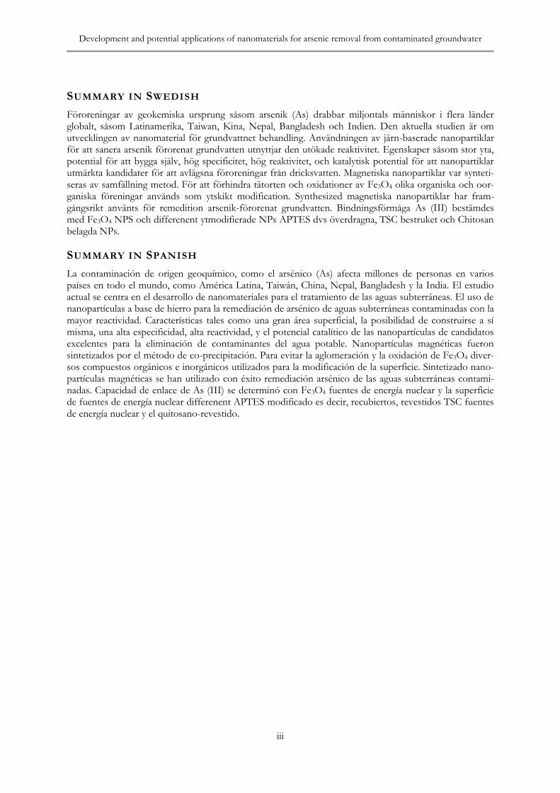
2.5 Magnetic behavior of iron oxide NPs
On the basis of magnetic response magnetic materials can classified as paramagnets, ferro-magnets, ferrimagnets or antiferromagnets (Fig. 1) (Figuerola et al., 2010).
Paramagnetic material in the individual atom magnetic moments are randomly aligned (or uncoupled) magnetic dipoles with respect to each other and the crystal has a zero net magnetic moment. This type of material has no coercivity nor remanence, means when no external mag-netic, the internal magnetic dipoles randomize again, no extra force required to demagnetize the material and hence the initial zero net magnetic moment is spontaneously recovered. NPs with isuch magnetic behaviour are called superpara-magnetic (SPM). Ferromagnetic material is fer-romagnetic means atoms can align parallel one to the other, hence exhibiting an enhanced collec-tive response even in the absence of an external magnetic field. In this case three main parameters can describe the strength and magnetization of the material and are: the coercive field, HC, the external field of opposite sign required reducing the magnetization back to zero; this parameter represents also the minimum energy required for the reversal. However, such behaviors are strong-ly size dependent and consequently one
Fig. 1 Variation of the coercivity (HC) of magnetic NPs with size

Development and potential applications of nanomaterials for arsenic removal from contaminated groundwater
5
important thing should keep in mind that, at a particular temperature, the magnetic behavior of any material can be altered by tuning its size (O‟Handley, 2000). Antiferromagnetic magnetic dipoles can align antiparallel in the lattice, which means that they will cancel each other (repulsion of magnetic dipoles). This type of magnetic exchange can lead to two different situations, namely antiferromagnetism, when the magnetic dipoles or interacting spins have the same value and hence the material shows a net zero magneti-zation.
2.6 Application of NPs in groundwater
The unusual properties of NPs have accelerated growth in the production of nanoscale materials and the rapid increase of their applications in many areas has captured the attention of re-searchers, government and industry worldwide.
Manufactured NPs are currently used in different areas such as electronics, biomedicine, pharma-ceuticals, cosmetics, environmental analysis and remediation, catalysis and material sciences, due to the relative ease with which they can be pre-pared and manipulated, their generally high reac-tivity and surface area and the tuneable nature of their optical and other properties (Poole and Owens, 2003). Nanoscience and nanoengineering can provide low cost and effective options to restore contaminated groundwater (Biswas and Wu, 2005). Savage and Diallo, 2005 was found nanomaterials, including NPs, application in water purification to reduce concentrations of toxic components (e.g. metal ions, radio nuclides, organic and inorganic compounds, as well as bacteria and viruses) to sub-ppb (parts per bil-lion) levels. The nanoscale iron particles are very effective for the transformation and detoxifica-tion of a wide variety of common environmental contaminants, such as chlorinated organic sol-vents, organochlorine pesticides, trinitrotoluenes, phenols, and herbicide molinate, amino carbox-ylic acids and p-hydroxybenzoic acid. Carbon nanotubes are used for the removal of arsenate from water (Peng et al., 2005) as well as other toxic metals such as Pb2+, Cu2+ and Cd2+ (Li et al., 2003). They show a high capacity for adsorp-tion of lead (Li et al., 2002) and are able to re-move chemicals of concern such as bisphenol A (Cai et al., 2003). NPs of magnetite (Fe3O4) coat-ed with mesoporous silica can be used in the removal of many harmful agents present in the environment (Wu et al., 2004), but also in biolog-ical cell separations and waste remediation. NPs, nano crystalline titanium dioxide, nanoscale zero valent iron and modified zero valent iron parti-
cles have shown promising results for the re-moval of arsenic (Pena et al., 2005, Kanel et al., 2006 and Jegadeesan et al., 2005). However, the same disadvantages as in conventional adsorption apply in the use of nano adsorbents. Novel in-vention in adsorption technology is the use of magnetic NPs as an adsorbent. Recently, it has been discovered in the USA that removal of arsenic with magnetite (Fe3O4) NPs is possible and the adsorption capacity for both As(III)and As(V)is at its highest level with small-diameter NPs (10 nm) (Mayo et al., 2007). Use of another type of magnetic NPs, maghemite (γ-Fe2O3), for arsenate removal in drinking water will give valuable information of nanoscale iron metal oxides behavior and suitability to arsenate ad-sorption. γ-Fe2O3 has been found very effective in Cr(VI) removal. It is expected that this type of adsorbent can perform equally well for As re-moval due to the similarity between As and Cr.
3 MATERIALS AND
METHODS
3.1 Chemicals
In this study the reagents ferric chloride hexa-hydrate (FeCl3•6H2O), ferrous chloride tetrahy-drate (FeCl2•4H2O), sodium hydroxide (NaOH) and ammonium hydroxide (NH4OH, 26% of ammonia) all analytical grade were used. 3-aminopropyl)-triethoxysilane, Trisodium citrate, Chitosan and Acetic acid (CH3COOH), acetone and ethanol all are chemical grade.
3.2 Synthesis of Magnetic NPs
Magnetic NPs were synthesized by the co-precipitation method (Massart, 1981; Kim et al., 2001). 2M of FeCl3•6H2O and 1M FeCl2•4H2O (2M:1M) was dissolved in 200 ml in deionised water with vigorous stirring at 80 0C for 10 minutes under oxygen free condition to prevent oxidation. Drop wise ammonium solution (26%) was added in mixed solution under mechanical stirring until the pH of the solution reached around 10, color changed from yellow to brown and brown precipitate heated at 80 0C for 30 minutes under vigorous stirring. Fe3O4 particles were separated centrifugally at 15000 rpm for 10 min and washed three times with deionized water. Finally, magnetic NPs were dried under vacuum at 70 0C for determine the concentration of NPs present in the solution.

Rajender Kumar TRITA LWR Degree Project 11:07
6
OH-
OH-
OH-
OH-
OH-
OH-
OH-
OH-
OH-
OH-
OH-
OH-
OH-
OH-
OH-
OH-
OH-
OH-
OH-
OH-
Fig. 2 Synthesized Fe3O4 NPs
The possible reaction for the formation of Fe3O4 particles shown below:
Fe2+ + 2 Fe3+ + 8NH3H2O =
Fe3O4↓+8NH4+ + 4H2O
The pH range of the synthesis iron oxide NPs should be 8-14 with maintaining molar ratio of Fe3+/Fe2+ (2:1) under a non oxidizing condition. Otherwise iron oxide NPs might be oxidize to λ -Fe2O3. In the absence of any surface modifica-tion Fe3O4 NPs tend to aggregate due to strong magnetic dipole–dipole attractions between particles. Due to this interaction between the particles, the nanoparticles tend to be agglomer-ate and form larger molecules and increase the size of the particles. For prevent the agglomera-tion and oxidations of Fe3O4 different organic and inorganic compounds are used for the sur-face modification.
3.3 Surface Modification
3.3.1 3-aminopropyl-triethoxysilane (3- APTES)
3-APTES as amino-silane coupling agent was used for the surface modification of Fe3O4 NPs. The aminosilane agent of aminopropyl trietho ysilane (APTES) is considered as a candidate for modification on the surface of Fe3O4 NPs direct-ly, for the advantages of the biocompatibility as well as high density of surface functional group of –NH2, allowing for connecting to other target-ing biomolecules. The APTES Fe3O4 NPs were also prepared with co-precipitation method. The 4 ml solution NPs concentration 18 mg/ml were dissolved in 35 ml of deionized water. 3-APTES 620 microliter was added drop wise into the mixture under oxygen free atmosphere at 80 0C for 3 h (Liu et al., 2004). After that the solution was cooled to room temperature.
The prepared APTES-modified were collected with a magnet, and washed with ethanol, fol-lowed by deionized water for three times. Finally,
APTES-modified Fe3O4 NPs were dried under vacuum at 70 0C.
3.3.2 Fe3O4–Chitosan NPs
Chitosan with presence of amino groups can be used for functionalization. It is a suitable kind of polymer to be used to modify the Fe3O4 NPs. The high content of amino groups also makes possible many chemical modifications in polymer with the purpose of improving selectivity. Chi-tosan is a partially acetylated glucosamine biopol-ymer with many useful features such as hydro-philicity, biocompatibility, biodegradability (Hu et al., 2007; Guo et al., 2005). Liang and Zhang (2007) prepared superparamagnetic Fe3O4 NPs decorated with carboxymethylated chitosan papain. Monodisperse chitosan/polyacrylic ac-id/Fe3O4 NPs were synthesized which could be used for MRI (Feng et al. 2009). For preparation of Fe3O4–chitosan NPs, chitosan (0.136 g) dis-solved in 5ml of CH3COOH and 45mL deionised water (Tran et al., 2010). The suspension was kept at room temperature for 24 h without stir-ring and separated by washing several times in deionised water.
3.3.3 Trisodium citrate (TSC) coated NPs
Among various small molecules, citrate moiety has been extensively used for the preparation of aqueous stable iron oxide NPs as well as their conjugation to bio-molecule sand drugs by ex-ploiting the uncoordinated di-carboxyl groups present on the surface of NPs. Trisodium citrate modified NPs was obtained using 10 mililitter of iron oxide (Fe3O4 conc. 18 mg/ml) was diluted in 20 ml of deionised water and stirred 400rpm for 30 min at 80-90 0C. 0.2 g of TSC was dissolved in 4 ml of water and the solution pored drop-wise into Fe3O4 solution under vigorous string and kept it 30 min at 80-90 0C. After that the suspen-sion was cooled down at room temperature and washed 3 times with Milli-Q water. Surface coat-ed NPs were collected with an external magnetic field. The obtained TSC coated Fe3O4 particles were suspended in 20 ml of water. The pH of the solution was measured between 6.5 and 7.0.
3.4 Characterization methods
3.4.1 X-ray Diffraction XRD
X-ray diffraction (XRD) is a versatile, non-destructive technique that reveals detailed infor-mation about the chemical composition and crystallographic structure of natural and manufac- tured materials. X-ray diffraction is based on constructive interference of monochromatic X-rays and a crystalline sample.

Development and potential applications of nanomaterials for arsenic removal from contaminated groundwater
7
Fig. 3 Structure of APTES coated- Fe3O4 NPs
The interaction of the incident rays with the sample produces constructive interference (and a diffracted ray) when conditions satisfy Bragg's Law (nλ=2d sin θ). This law relates the wave-length of electromagnetic radiation to the diffrac-tion angle and the lattice spacing in a crystalline sample. These diffracted X-rays are then detect-ed, processed and counted. By scanning the sample through a range of 2θangles, all possible diffraction directions of the lattice should be attained due to the random orientation of the powdered material. Conversion of the diffraction peaks to d-spacings allows identification of the mineral because each mineral has a set of unique d-spacings. Typically, this is achieved by compari-son of d-spacings with standard reference pat-terns. The fine dried powdered sample was fed into the Rontgen diffractometer system (Model Siemens D5000, XRD). The system with a ten-sion of 45kV and a current of 40 mA was turned on. The 2θ data was collected using a continuous scan mode with start angle 200 end angle 700 for Fe3O4 NPs and chitosan coated Fe3O4 NPs and with start angle 200 and end angle 800 for 3- APTES coated Fe3O4 and TSC coated Fe3O4. The average crystalline size of the sample was estmated using DebyeScherrer‟s forulla from the width at half maximum (β) of the main intense peak.
D= 0.9λ/ β cos θ
Where, D is the size of the particles, λ the wave-length of the emitted X-rays, β is the full width at
half maximum of the corresponding peak of the XRD and θ the angle of diffraction.
3.4.2 Transmission Electron Microscope (TEM)
The Transmission Electron Microscope (TEM) is a valuable tool for analysing crystalline solids.
Imaging of microstructures and lattice defects which affect the mechanical properties of the solids can be accomplished as well as measuring of the crystallographic orientations of the investi-gated crystals using electron diffraction methods. It is a microscopy technique whereby a beam of electrons is transmitted through an ultra thin specimen, interacting with the specimen as it passes through. An image is formed from the interaction of the electrons transmitted through the specimen; the image is magnified and focused onto an imaging device, such as a fluorescent screen, on a layer of photographic film, or to be detected by a sensor such as a CCD camera.
For Transmission electron microscopy (TEM), Philips model CM12 operating voltage at 100 kV was used for the sample analysis. Chemical analy-sis was also done using high resolution transmis-sion electron microscopy (HRTEM), Tecnai G20-Stwin along with energy dispersive X-ray analysis (EDAX).
3.4.3 Fourier Transform Infrared Spectroscopy (FTIR)
FTIR analysis is a technique that provides infor-mation about the chemical bonding or molecular structure of materials, whether organic or inor-ganic. During FTIR analysis, a spot on the spec-imen is subjected to a modulated IR beam. The specimen's transmittance and reflectance of the infrared rays at different frequencies is translated into an IR absorption plot consisting of reverse peaks. The resulting FTIR spectral pattern is analyzed and matched with known signatures of identified materials in the FTIR library. Multigas 2030 HS, FTIR was used for the measurement and identify the functional groups present in the iron oxide NPs.

Rajender Kumar TRITA LWR Degree Project 11:07
8
OH-
OH-
OH-
OH-
OH-
OH-
OH-
OH-
OH-
OH-
OH-
OH-
OH-
OH-
OH-
OH-
OH-
OH-
OH-
OH-
Fe3O4 NPs
Chitosan- Fe3O4 NPs
Chitosan
Glutaraldehyde
Fig. 4 Structure of Chitosan coated- Fe3O4 NPs
3.5 Binding capacity of Arsenic species from Groundwater
To determine the binding capacity of As from the groundwater with iron oxide NPs and coated NPs were analysed using HACH EZ- Arsenic kit 2822800 and WAG- WE 10500 Arsenator (Fig. 5). The As(V)and the As(III)stock solutions were prepared by dissolving the corresponding As oxides (As2O5 and As2O3) in de-ionized water. Effect of arenite concentration i.e. 10 µg/ml, 50 µg/ml and 100 µg/ml and reaction time 0, 15, 30, 45 and 60 min on binding capacity of As(III)with iron oxide, APTES coated, TSC coated and chitosan coated Fe3O4 particles was analyzed. The experiments were performed in 1 ml solution with 10 µL of NPs concentration (18 mg/ml) at room temperature on rotating shaker. For the experiment in synthetic groundwater with pres-ence of arsenic species was prepared in laborato-ry. The composition of the GW was Cl- 38.2 mg/L, NH4
+ 19.4 mg/L, HCO3- 168 mg/L, Na+
-63.03 mg/L, NO3- - 49.1 mg/L, K+ - 39.09
mg/L, Mn+- 3.62 mg/L, SO4- 30.82 mg/L, Fe-
14.26 mg/L, PO4- 19.6 mg/L , F- 4.0 mg/L and
varying concentration of As(III) and As(V) ions.
4 RESULTS AND
DISCUSSION
4.1 Transmission electron micrograph
In order to obtain more direct information on particle size and morphology, the typical HRTEM and TEM micrographs for the Fe3O4 NPs and surface modified NPs are shown (Fig. 6-9). HRTEM images provide further insight into the structure of the product. The HR-TEM images demonstrate that highly crystalline Fe3O4
NPs with lattice fringes were formed in the syn-thesis (Fig. 6 A). The TEM image of the uncoat-ed Fe3O4 NPs and the particles are roughly spherical and polydisperse. Usually, spherical shapes are formed because the nucleation rate per unit area is isotopic at the interface between the Fe3O4 magnetic NPs (Kim et al., 2006). We can see that the uncoated NPs are easy to agglomer-ates (Fig. 6B). The size of the particles which taken by micrograph is around 50 nm. TEM image are indicates the all particles are crystalline in nature. It is noted from images, when NPs underwent surface functionalization, the size of

Development and potential applications of nanomaterials for arsenic removal from contaminated groundwater
9
Fig. 5 WAG- WE 10500 Arsenator
the NPs changed little from uncoated particles (Fig. 7, 8 and 9). The coupling of chitosan, amino silane and sodium citrate would not significantly work on the thickness of the coating. Fe3O4 NPs coated with chitosan were shown in “figure 7.” HRTEM image shows that the NPs are structur-ally uniform with lattice fringe spacing. From “figure 7B” observed that the Fe3O4-chitosan NPs were quasispherical and had good dispersity. The structure of Chitosan-coated magnetite NPs was looser then uncoated Fe3O4 NPs. The aver-age size of the Fe3O4-chitosan NPs was larger than uncoated Fe3O4 NPs. The enlargement of the NPs size indicated the formation of the Fe3O4-chitosan NPs with core-shell structure.
From the magnified image a slight aggregation can be observed which was due to the crosslink-ing among the different NPs. APTES, a common amino-silane coupling agent, has found its exten-sive applications in surface-decoration by forming a monolayer of amino-silane.
Additionally, the modified magnetic NPs with amino-silane shell are non-toxic, biocompatible and injectable (Hervé et al., 2008). The particles size and morphology of magnetite particles, amino-silane treated magnetite were also investi-gated and shown in “figure 8”. Particles with an approximate spherical shape were observed “figure 8A”. APTES coating reduced the aggre-gation and improved the particle dispersion as shown in. With further increase of the magnifi-cation factor, the lattice of the Fe3O4 can be clearly observed in HRTEM image (Fig. 8B).
Among various small molecules, citrate moiety has been extensively used for the preparation of aqueous stable iron oxide NPs as well as their conjugation to biomolecules and drugs by ex-ploiting the uncoordinated carboxyl groups pre-sent on the surface of NPs. A new method was developed for reversible association of drug (DOX) to citrate-stabilized iron oxide NPs pre-pared by agitating bare NPs in citric acid solution (Munnier et al. 2008). Coated magnetite core-shell NPs were also fabricated by Khosroshahi and Ghazanfari, 2010. Due to an electrostatic interaction between the iron oxide Fe3O4 NPs and trisodium citrate surfactant, stable magnetic
Fig. 9 (A) HR-TEM image (B) TEM im-age of TSC - coated Fe3O4 NPs
A B
Fig. 8 (A) TEM image (B) HR-TEM im-age of APTES coated Fe3O4 NPs
B A
Fig. 7 (A) HR-TEM image (B) TEM im-age of chitosan coated Fe3O4 NPs
A B
A B
Fig. 6 (A) HR-TEM image (B) TEM im-age of Fe3O4 NPs
A B
A B
B

Rajender Kumar TRITA LWR Degree Project 11:07
10
fluid containing dispersed Fe3O4 NPs was ob-tained in this study (Fig. 9). From TEM micro-graph, it is clearly observed that Fe3O4 NPs are almost spherical in shape and size of about 8-10 nm (σ≤10%).
4.2 X-ray Diffraction (XRD) analysis
X-ray diffraction was used to identify the crystal-line structure of synthesized Fe3O4 NPs and surface modified NPs. The X-ray diffraction pattern was observed and found that standard Fe3O4 crystal has six diffraction peaks i.e. (220), (311), (400), (422), (511), (440) (Fig. 10) . These binding energies data are consistent with the reported values of Fe3O4 in the literature (Qu et al., 2010). On the other hand, it was found that there is no extra peaks were detected in APTES coated Fe3O4, chitosan coated and TSC coated NPs which could be assigned to impurities as shown in “figure 10”. In addition, the broad peak
appears in the range of 180 ∼280 due to surface
modified agents were coated onto the surface of
Fe3O4 NPs, and the intensity of crystallization peak of coated Fe3O4 NPs was slightly lower
than that of Fe3O4 NPs because of coating.
4.3 Fourier-transform infrared (FTIR) spectroscopy
The surface chemical structure of Fe3O4 NPs was characterized by FTIR spectroscopy (Fig. 11). The FT-IR spectrum of iron oxide exhibits strong bands in the low-frequency region 1000–500 cm-1 due to the iron oxide skeleton, which is consistent with the magnetite (Fe3O4) spectrum Waldron, 1955. The adsorption peaks at around 563 or 580 cm−1 were the characteristic absorp-tion of FeO bond which confirmed the presence of magnetite NPs. The characteristics of stretch-ing vibration peak at 3400 cm-1 due to the pres-ence of OH. It can be deduced that Fe(OH)2, Fe(OH)3 and FeOOH formed resulting from hydrolyzation on the surface of Fe3O4 (Lu et al., 2010). While the peak at 1600 cm-1 also shows existence of Fe–O.
Fig. 10 XRD pattern of the different type of synthesized nanoparticles

Development and potential applications of nanomaterials for arsenic removal from contaminated groundwater
11
0
0.1
0.2
0.3
0.4
0.5
0.6
0 1000 2000 3000 4000 5000
Wavelength (cm-1)
Tra
nsm
itta
nce
(%
)
Fig. 11 FTIR spectrum of the Fe3O4 NPs synthesized
4.4 Potential applications of Nano-biomaterial in arsenic(III) removal
The binding capacity of As(III)with the NPs was determined in different initial concentration of solutions 10 µg/ml, 50 µg/ml and 100 µg/mL at different time interval from 0, 15, 30, 45 and 60 min in 1ml As(III)solution with 10 µL of nano-particle solution concentration 18 mg/ml). Bind-ing capacity of As(III)was determined with Fe3O4 NPs and different surface modified NPs i.e. APTES coated, TSC coated and Chitosan coated NPs. The samples were placed on a rotatory shaker. NPs were separate from the solution using magnetic separator. HACH EZ- Arsenic kit 2822800 was used for the analysis of arsenic in the samples. Samples were diluted for 15 times and 9.6 ml of solution was taken into the reaction bottle. A test strip was inserted into the cap and the pad complete converse the small opening. After that, reagents A1 and A2 added in to the reaction bottle and attach the cap immediately to the reaction bottle and kept it for 20 minutes for the reaction. After 20 minutes test strip was removed and immediately develop color compare with color code chart. The removal of As(III) with Fe3O4 NPs was found 96.8%, 97% and 93% with initial concentration of 100µg/ml, 50 and 10µg/ml respectively (Fig. 12). The maximum removal was found 99.5%, 99% and 97% with chitosan coated NPs in 60 minute at initial con-centration of As(III) 100 µg/mL, 50 and 10 µg/mL (Fig. 13). Whereas, removal was found 99.5%, 98.8% and 92.5% with TSC coated NPs in 60 minute at initial concentration of As(III) 100 µg/mL, 50 and 10 µg/mL respectively (Fig. 14). Similarly with APTES coated NPs were 97.25%, 96% and 85% at concentration of As(III) 100 µg/mL, 50 and 10 µg/mL (Fig. 15). The binding of As(III) increased as the dissolved As(III)concentration increased in the solution. From this experiment was found the maximum
arsenite removal from the solution with chitosan-coated NPs than other coated and uncoated NPs.
4.5 Binding capacity of As(III)and As(V)from groundwater using NPs
Binding capacity of total arsenic was determine using Wag - WE 10500 wagtech digital arsentor. Compostion of synthetic groundwater was given above was used with presence of 250 µg/L As(III)and 250 µg/L As(V)for removal of As with different type of magnetic nanoparticles. For this experiment, 10 ml synthetic groundwater was taken and added 100 µL magnetic NPs (concen-tration 18 mg/ml) in the sample. Samples were kept on rotatory shaker at 100 rpm for 5 minutes and 1hr. Each experiment was performed in duplicate. Magnetic NPs were separate with magnetic separator from the first sample after 5 min and then after 1 hr from second sample as the dissolved As(III)concentration increased in the solution.
86
88
90
92
94
96
98
0 15 30 45 60
Time (min)
% B
ind
ing
cap
acit
y
10 µg/mL 50 µg/mL 100 µg/mL
Fig. 12 Binding capacity of As(III)using Fe3O4 NPs
94
95
96
97
98
99
100
0 15 30 45 60
Time (min)
% B
ind
ing
Cap
acit
y
10 µg/mL 50 µg/mL 100 µg/mL
Fig. 13 Binding capacity of As(III)using Chitosan coated-Fe3O4 NPs

Rajender Kumar TRITA LWR Degree Project 11:07
12
75
80
85
90
95
100
105
0 15 30 45 60
Time (min)
% B
ind
ing
cap
acit
y10 µg/mL 50 µg/mL 100 µg/mL
Fig. 14 Binding capacity of As(III)using TSC Coated-Fe3O4 NPs
95
96
97
98
99
100
0 15 30 45 60
Time (min)
% B
ind
ing
cap
acit
y
10 µg/mL 50 µg/mL 100 µg/mL
Fig. 15 Binding capacity of As(III)using APTES coated-Fe3O4 NPs
From this experiment was also found the maxi-mum binding capacity of arsenite with chitosan-coated NPs from the aqueous solution than other coated and uncoated NPs.
As shown in “figure 16” the maximum removal of arsenic was determine with presence of chi-tosan coated magnetic NPs and found binding capacity As ion is ˜40% in initial period of con-tact time at 5 minutes after 1hr the binding of As ions was increased and analysed 50% of As ions decreased from the solution. From the previous results it was found that if only As( III) ions were present in the water without other anions and cations the binding capacity of the magnetic NPs is very high. It was determine the binding capaci-ty of As ion was decreased with presence of other anions and cations in the groundwater and they interfere with arsenic binding sites which pres-ence on the magnetic NPs. Similarly if concentra-tion of As(III)and As(V)ions increases in the groundwater the binding capacity of As ions not decreases much.
05
1015
2025
3035
4045
50
contr
ol
Fe3O
4
Chito
san
APTES
TSC
Differnt types of NPs
Co
ncen
trati
on
(µg
/L)
5 min
1 hr
Fig. 16 Removal of total arsenic from groundwater with different types of NPs at initial concentration (500 µg/L) and NPs 100 µL
0
20
40
60
80
100
contr
ol
Fe3O
4
Chitosa
n
APTESTSC
Different types of NPs
co
ncen
trati
on
(µ
g/L
)
5 min
1 hr
Fig. 17 Removal of total arsenic from groundwater with different NPs initial con-centration (1000 µg/L) with NPs 100 µL
As shown in “figure 17” the removal of As ions was not much increases with increases concentra-tion 1000 µg/L and maximum removal was also obtained with chitosan coated NPs in the initial periods in 5 minutes. Whereas, with APTES coated NPs binding capacity decrease with in-creases contact time may be due to the release of bound ions in the solution.
5 CONCLUSIONS
The synthesis of Fe3O4 NPs using co-precipitation was successfully used for this re-search work. Different chemical compounds were used i.e. 3-aminopropyl-triethoxysilane (3-APTES), Ttrisodium citrare (TSC) and Chitosan for surface modification of Fe3O4 NPs. The analyses of TEM and XRD indicated that the Fe3O4 NPs are roughly spherical and polydisperse with a mean diameter of 15 nm, and after surface modification Fe3O4 NPs mean diameter of parti-cles are in the range of 15-20 nm. The surface modified particles tend to have a uniform and stable distribution in the solution. The crystalline structure of synthesized Fe3O4 NPs and surface

Development and potential applications of nanomaterials for arsenic removal from contaminated groundwater
13
modified NPs was identify with XRD and the intensity of crystallization peak of coated NPs was slightly lower than that of uncoated NPs because of coating. Synthesized magnetic nano particles were successfully used for removal of arsenic contaminated groundwater.
Binding capacity of As(III)was determined with Fe3O4 NPs and different surface modified NPs i.e. APTES coated, TSC coated and Chitosan coated NPs. Binding capacity of As(III)was very high and found more than 90% with uncoated and coated magnetic particles. Whereas, binding capacity was increased with increases concentra-tion of As(III)ions 10 µg/ml-100 µg/ml. If As(III) and As(V) present with other anions and cations in the groundwater the binding of capaci-ty of total arsenic was decreased.
6 FUTURE WORK
There are some suggestions for future study: There should be requiring continuous research work in the same because this research work not covers all the possibilities of nanomaterials for groundwater decontamination. We put forward the need for future research focused on a better understanding of the mechanisms involved as well as the study of the potential interference in arsenic binding caused by anions and cations present in solution. Among the many applications of nanotechnology that have environmental implications, remediation of contaminated groundwater using magnetic NPs is one of the most well-known examples of a rapidly emerging technology with considerable potential benefits.

Rajender Kumar TRITA LWR Degree Project 11:07
14
7 REFERENCES
Alam MB, Sattar MA. 2000. Assessment of arsenic contamination in soils and waters in some areas of Bangladesh. Water Science and Technology. 42: 185-193.
Beydoun D, Amal R, Low GKC, McEvoy S. 2000. Novel Photocatalyst: Titania-Coated Magnetite. Activ-ity and Photodissolution. Journal of Physical Chemistry. 104: 4387-4392.
Bhattacharya P, Chatterjee D, Jacks G. 1997. Occurrence of arsenic contaminated groundwater in alluvial aquifers from Delta Plains, Eastern India: Options for safe drinking water supply. International Journal of Water Research and Management. 13:79-92.
Bhattacharya P, Frisbie SH, Smith E, Naidu R, Jacks G, Sarkar B. 2002. Arsenic in the Environment: A Global Perspective. In: B.Sarkar (Ed.) Handbook of Heavy Metals in the Environment. Marcell Dekker Inc., New York, pp. 147-215.
Bhattacharyya R, Chatterjee D, Nath B, Jana J, Jacks G, Vahter M. 2003. High arsenic groundwater: mobilization, metabolism and mitigation-an overview in the Bengal Delta Plain. Molecular and Cellular Biochemistry. 253: 347–355.
Biswas, Wu, CY. 2005. Nanoparticles and the environment. Journal of the Air and Waste Management 55: 708-746.
Bystrzejewski M, Huczko A, Lange H. 2005. Arc plasma route to carbon-encapsulated magnetic nanopar-ticles for biomedical applications. Sensors and Actuators B: Chemical. 109: 81-85.
Cai YQ, Jiang GB, Liu, JF, Zhou QX. 2003. Multiwalled carbon nanotubes as a solid-phase extraction adsorbent for the determination of bisphenol a, 4-nnonylphenol, and 4-tert-octylphenol. Analytical Chemistry. 75: 2517–2521.
CGWB, 1999. High Incidence of As in groundwater in West Bengal. Central Groundwater Board, India. Ministry of Water Resources, Government of India.
Chakravarty S, Dureja V, Bhattacharyya G, Maity S, Bhattacharjee S. 2002. Removal of arsenic from groundwater using low cost ferruginous manganese ore. Water Research. 36: 625-632.
Chen S-L, Dzeng SR, Yang M-H. 1994. Arsenic species in groundwaters of the black foot disease area, Taiwan. Environmental Science and Technology. 28: 877–881.
Cornell RM, Schwertmann HCU. 2003. The iron oxides: structure, properties, reactions, occurrences and uses (2nd Eds), WILEY-VCH.
Cornell RM, Schwertmann U. 2003. The Iron Oxides: Structure, Properties, Reactions, Occurrences and Uses, (second eds), Wiley-VCH, Weinheim.
Cushing BL, Kolesnichenko VL, O‟Connor CJ. 2004. Recent advances in the liquid-phase syntheses of inorganic nanoparticles. Chemical Reviews. 104:3893–3946.
Das D, Chatterjee A, Mandal BK, Samanta G, Chakroborty D, Chanda B. 1995. Arsenic in groundwater in six districts of West Bengal, India: the biggest arsenic calamity in the world. Part 2. Arsenic concen-tration in drinking water, hair, nails, urine, skin-scales and liver tissues (biopsy) of the affected people. Analyst. 120: 917–924.
Denouden CJJ, Thompson RW. 1991. Analysis of the formation of monodisperse populations by homo-geneous nucleation. Journal of Colloid and Interface Science. 143: 77–84.
Faivre D, Agrinier P, Menguy N, Zuddas P, Pachana K, Gloter A, Laval J-Y, Guyot F. 2004. Mineralog-ical and isotopic properties of inorganic nanocrystalline magnetites. Geochimica et Cosmochimica Acta. 68: 4395-4403.
Figuerola A, Corato R- Di, Mannaa L, Pellegrinoa, T. 2010. From iron oxide nanoparticles towards ad-vanced iron-based inorganic materials designed for biomedical applications. Pharmacological Research. 62: 126–143.
Fuertes AB, Tartaj P. 2006. A facile route for the preparation of superparamagnetic porous carbons. Chemistry of Material. 18, 1675-1679.

Development and potential applications of nanomaterials for arsenic removal from contaminated groundwater
15
Grasset F, Labhsetwar N, Li D, Park DC, Saito N, Haneda H. 2002. Synthesis and magnetic characteriza-tion of zinc ferrite nanoparticles with different environments: powder, colloidal solution, and zinc fer-rite-silica core-shell nanoparticles. Langmuir. 18: 8209-8216.
Guo TY, Xia YQ, Wang J, Song MD, Zhang BH. 2005. Chitosan beads as molecularly imprinted polymer matrix for selective separation of proteins. Biomaterials. 26: 5737–5745.
Hervé K, Douziech-Eyrolles L, Munnier E, Cohen-Jonathan S, Soucé M, Marchais H, Limelette P, Warmont F, Saboungi ML, Dubois P, Chourpa I. 2008. The development of stable aqueous suspen-sions of PEGylated SPIONs for biomedical applications. Nanotechnology. 19: 465608.
Hu Y, Ding Y, Ding D, Sun MJ, Zhang LY, Jiang XQ, Yang CZ. 2007. Hollow chitosan/poly (acrylic acid) nanospheres as drug carriers. Biomacromolecules. 8: 1069–1076.
Ireland MP. 1991. Heavy metal sources – Uptake and distribution. In Biological Monitoring of Heavy Metals, (Eds.) Dillon, H.K. & Ho, M.H. John Wiley & Sons, New York: 263-276.
Jegadeesan G, Mondal, K, Lalvani, SB. 2005. Arsenate remediation using nanosized modified zerovalent iron particles. Environmental Progress. 24: 289–296.
Kanel SR, Greneche J-M, Choi H. 2006. Arsenic (V) removal from groundwater using nano scale zero-valent iron as a colloidal reactive barrier material. Environmental Science and Technology. 40: 2045–2050.
Kapaj S, Peterson H, Liber K, Bhattacharya P. 2006. Human health effects from chronic arsenic poison-ing - a review. Journal of Environmental Science and Health, Part A Toxic/Hazardous Substances and Environ-mental Engineering. 41: 2399–2428.
Khosroshahi ME, Ghazanfari L. 2010. Preparation and characterization of silica-coated iron-oxide biona-noparticles under N2 gas. Physica E. 42: 1824-1829.
Kim DH, Lee SH, Im KH, Kim KN, Kim KM, Shim IB, Lee MH, Lee YK. 2006. Surface-modified mag-netite nanoparticles for hyperthermia: preparation, characterization, and cytotoxicity studies. Current Applied Physics, 6: 242-249.
Kim DK, Zhang Y, Voit W, Rao KV, Muhammed M. 2001. Synthesis and characterization of surfactant-coated superparamagnetic monodispersed iron oxide nanoparticles. Journal of Magnetism and Magnetic Materials. 225: 30-36.
Lee Y, Lee J, Bae CJ, Park JG, Noh HJ, Park JH, Hyeon T. 2005. Large-scale synthesis of uniform and crystalline magnetite nanoparticles using reverse micelles as nanoreactors under reflux conditions. Ad-vanced Functional Materials. 15: 503–509.
Li G, Jiang Y, Huang K, Ding P, Chen J. 2008. Preparation and properties of magnetic Fe3O4–chitosan nanoparticles, Journal of Alloys and Compounds. 466: 451-456.
Li YH, Wang S, Wei J, Zhang X, Xu C, Luan Z, Wu D, Wei B. 2002. Lead adsorption on carbon nano-tubes. Chemical Physics Letters 357, 263–266.
Li YH, Wang SG, Luan ZK, Ding J, Xu CL, Wu DH. 2003. Adsorption of cadmium (II) from aqueous solution by surface oxidized carbon nanotubes. Carbon 41:1057–1062.
Liang YY, Zhang LM. 2007. Bioconjugation of papain on superparamagnetic nanoparticles decorated with carboxymethylated chitosan. Biomacromolecules. 8:1480–1486.
Liu X, Ma Z, Xing J, Liu H. 2004. Preapartion and characterisation of amino silane modified supermag-natic silica nanospheres. Journal of Magnetism and Magnetic Materials. 27: 1-6.
Massart R. 1981. Preparation of aqueous magnetic liquids in alkaline and acidic media. Magnetics, IEEE Transactions on. 17: 1247–1248.
Matsuno R, Yamamoto K, Otsuka H, Takahara A. 2004. Polystyrene- and Poly(3-vinylpyridine)-Grafted Magnetite Nanoparticles Prepared through Surface-Initiated Nitroxide-Mediated Radical Polymeriza-tion. Macromolecules. 37: 2203-2209.
Mayo JT, Yavuz C, Yean S, Cong L, Shipley H, Yu W, Falkner J, Kan A, Tomson M, Colvin VL. 2007. The effect of nanocrystalline magnetite size on arsenic removal. Science and Technology of Advance Materials. 8: 71–75.
Mikhaylova M, Kim DK, Bobrysheva N, Osmolowsky M, Semenov V, Tsakalakos T, Muhammed M. 2004. Superparamagnetism of Magnetite Nanoparticles: Dependence on Surface Modification. Lang-muir. 20: 2472–2477.

Rajender Kumar TRITA LWR Degree Project 11:07
16
Morales MP, Gonzalezcarreno T, Serna CJ. 1992. The formation of alpha-Fe2O3 monodispersed particles in solution. Journal of Materials Research. 7: 2538–2545.
Munnier E, Cohen-Jonathan S, Linassier C, Douziech-Eyrolles L, Marchais H, Soucé M Hervé K, Dubois P, Chourpa I. 2008. Novel method of doxorubicin–SPION reversible association for magnetic drug targeting. International Journal of Pharmaceutics 363: 170-176.
Munshi N, De TK, Maitra A. 1997. Size modulation of polymeric nanoparticles under controlled dynam-ics of microemulsion droplets, Journal of Colloid and Interface Science. 190: 387–391.
Nasri R, Siblini A, Jorat L, Noyel G. 1996. Magneto dielectric behavior of the magnetic fluid manganese ferrite in carbon tetrachloride. Journal of Magnetism and Magnetic Materials. 161: 309-315.
Nath B, Sahu SJ, Jana J, Mukherjee-Goswami A, Roy S, Sarkar MJ, Sotiropoulou S, Chaniotakis NA. 2003. Carbon nanotube array-based biosensor. Analytical and Bioanalytical Chemistry. 375: 103–105.
Neveu A, Bee MR, Talbot D. 2002. Size-selective chemical synthesis of tartrate stabilized cobalt ferrite ionic magnetic fluid. Journal Colloid Interface Science. 255: 293–298.
Nielsen AE. 1964. Kinetics of Precipitation. Pergamon Press, New York.
O‟Handley RC. 2000. Modern Magnetic Materials Principles and Applications. Wiley.
Ocana M, Rodriguezclemente R, Serna CJ. 1995. Uniform colloidal particles in solution-formation mech-anisms. Advanced Materials 7: 212–216.
Park J, An K, Hwang Y, Park JG, Noh HJ, Kim JY. 2004. Ultra large-scale syntheses of monodisperse nanocrystals, Nature Materials. 3: 891–895.
Pena ME, Korfiatis GP, Patel M, Lippincott L, Meng X. 2005. Adsorption of As(V)and As(III)by nano-crystalline titanium dioxide.Water Research. 39: 2327–2337.
Peng X, Luan Z, Ding J, Di Z, Li Y, Tian B. 2005. Ceria nanoparticles supported on carbon nanotubes for the removal of arsenate from water. Materials Letters. 59: 399-403.
Poole CP, Owens FJ. 2003. Introduction to Nanotechnology. Russian Journal of Applied Chemistry. 79:1213-1214.
Qu J, Liu G, Wang Y, Hong R. 2010. Preparation of Fe3O4–chitosan nanoparticles used for hyperthermia. Advanced Powder Technology. 21: 461-467.
Roco M, Brainbridge WS. 2001. Societal Implications of Nanoscience and Nanotechnology, Kluwer Aca-demic Pubishers.
Rosi NL, Mirkin CA. 2005. Nanostructures in Biodiagnostics. Chemical Reviews. 105:1547–1562.
Rotello VM. 2003. Nanoparticles: Building Blocks for Nanotechnology (1st Ed.), Springer, New York.
Santra S, Tapec R, Theodoropoulou N, Dobson J, Hebard A, Tan WH. 2001. Synthesis and Characteriza-tion of Silica-Coated Iron Oxide Nanoparticles in Microemulsion: The Effect of Nonionic Surfactants. Langmuir. 17: 2900–2906.
Savage N, Diallo MS. 2005. Nanomaterials and water purification: opportunities and challenges. Journal of Nanoparticle Research. 7: 331–342.
Sen T, Magdassi S, Nizri G, Bruce IJ. 2006. Dispersion of magnetic nanoparticles in suspension. Micro and Nano Letters 1:39–42.
Si S, Kotal A, Mandal TK, Giri S, Nakamura H, Kohara T. 2004. Size-controlled synthesis of magnetite nanoparticles in the presence of polyelectrolytes. Chemistry Materials. 16: 3489–3496.
Smedley PL, Kinniburgh DG. 2002. A review of the source, behaviour and distribution of arsenic in natural waters. Applied Geochemistry. 17: 517–568.
Sun SH, Zeng H. 2002. Size-controlled synthesis of magnetite nanoparticles. Journal of the American Chemi-cal Society. 124: 8204–8205.
Sun ZH, Wang LF, Liu PP, Wang SC, Sun B, Jiang DZ, Xiao FS. 2006. Magnetically motive porous sphere composite and its excellent properties for the removal of pollutants in water by adsorption and desorption cycles. Advanced Materials. 18: 1968-1971.
Tran HV, Tran, LD, Nguyen TN. 2010. Preparation of chitosan/magnetite compositebeads and their application for removal of Pb(II) and Ni(II) from aqueous solution. Materials Science and Engineering. 30: 304-310.

Development and potential applications of nanomaterials for arsenic removal from contaminated groundwater
17
Turnbull D. 1953. The kinetics of precipitation of barium sulfate from aqueous solution. Acta Metallurgica 1: 684-691.
Vassierres L, Chaneac C, Tronc E, Jolivet JP. 1998. Size Tailoring of Magnetite Particles Formed by Aqueous Precipitation: An Example of Thermodynamic Stability of Nanometric Oxide Particles. Jour-nal of Colloid and Interface Science. 205: 205–212.
Vernet, JP. 1992. Impact of Heavy Metals on the Environment, Trace metals in the environment, Else-vier, New York, 311-325 .
Walton AG. 1979. The Formation and Properties of Precipitates, Krieger R, New York.
Wan SR, Huang JS, Yan HS, Liu KL. 2006. Size-controlled preparation of magnetite nanoparticles in the presence of graft copolymers. Material Chemistry. 16: 298–303.
Wang X, Zhuang J, Peng Q, Li Y. 2005. A general strategy for nanocrystal synthesis. Nature. 437: 121–124.
Wang, C-Y, Chen, Z-Y, Cheng, B, Zhu, Y-R, Liu H-J. 1999. The preparation, surface modification, and characterization of metallic a-Fe nanoparticles. Materials Science and Engineering. 60: 223–226.
White BR, Stackhouse BT, Holcombe JA. 2009. Magnetic У-Fe2O3 nanoparticles coated with poly-l-cysteine for chelation of As(III), Cu(II), Cd(II), Ni(II), Pb(II) and Zn(II). Journal of Hazardous Materials. 161: 848-853.
Willis, NJ, Turro, O‟Brien S. 2005. Spectroscopic characterization of the surface of iron oxide nanocrys-tals, Chemistry Materials. 17: 5970–5975.
Wu PG, Zhu JH, Xu ZH. 2004. Template-assisted synthesis of mesoporous magnetic nanocomposite particles. Advanced Functional Materials. 14: 345-351.
Yu M, Liu Y, Moser A, Weller D, Sellmyer DJ. 1999. Nanocomposite CoPt:C films for extremely high-density recording. Applied Physics Letters. 75: 3992 -3995.
Yan ML, Li XZ, Gao L, Liou SH, Sellmyer DJ, Veerdonk RJM, Wierman KW. 2003. Fabrication of nonepitaxially grown double-layered FePt:C/FeCoNi thin films for perpendicular recording. Applied Physics Letters. 83: 3332-3335.